Embryonic Development of Caspian kutum, Rutilus frisii kutum
Upload
khangminh22Category
view
9download
0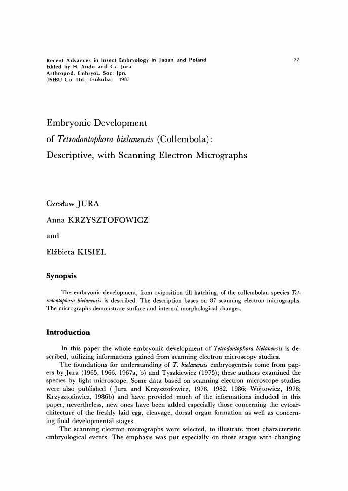
Recent Advances in Insect Embryology in Japan and Poland Edited by H. Ando and Cz. Jura Arthropod. EmbryoL. Soc. Jpn. (lSEBU Co. Ltd., Tsukuba) 1987
Embryonic Development
of Tetrodontophora bielanensis (Collembola):
Descriptive, with Scanning Electron Micrographs
Czeslaw JURA
Anna KRZYSZTOFOWICZ
and
ElZbieta KISIEL
Synopsis
77
The embryonic development, from oviposition till hatching, of the collembolan species Tet-rodontophora bielanensis is described. The description bases on 87 scanning electron micrographs. The micrographs demonstrate surface and internal morphological changes.
Introduction
In this paper the whole embryonic development of Tetrodontophora bielanensis is de-scribed, utilizing informations gained from scanning electron microscopy studies.
The foundations for understanding of T. bielanensis embryogenesis come from pap-ers by Jura (1965, 1966, 1967a, b) and Tyszkiewicz (1975); these authors examined the species by light microscope. Some data based on scanning electron microscope studies were also published (Jura and Krzysztofowicz, 1978, 1982, 1986; W6jtowicz, 1978; Krzysztofowicz, 1986b) and have provided much of the informations included in this paper, nevertheless, new ones have been added especially those concerning the cytoar-chitecture of the freshly laid egg, cleavage, dorsal organ formation as well as concern-ing final developmental stages.
The scanning electron micrographs were selected, to illustrate most characteristic embryological events. The emphasis was put especially on those stages with changing
78 Cz. Jura, A. Krzysztofowicz and E. Kisiel
surface characteristics, but some mention is made also of concurrent internal changes in embryo morphology. Making selection the authors intended to summarise the results obtained until now, to make basis for further study.
Material and Methods
For observations the specimens of Tetrodontophora bielanensis were collected in the vicinity of Krak6w, especially of Bielany Hills, where they can be found in great num-bers in the forest under loose bark of trees, dead leaves, on moss, or on mushrooms.
In nature oviposition occurs during the first days of November, especially after 2-3 days with temperature below ot. The eggs are usually laid in a group of from 15-25. Every female deposits about 60 eggs. The females deposit their eggs mainly in the clefts and caves of ground under the forest litter, and sometimes between the dead leaves of the littter. In such environment, humid and with temperatures ranging be-tween + 4 t - ot, the embryos developed during the period of about six autumn-winter-spring months.
The fertilization of eggs is internal. The males of T. bielanensis deposit sperma-tophores, in groups of 10 - 20 (Krzysztofowicz and Byczkowska-Smyk, 1966; Krzysz-tofowicz, 1967, 1980a). Before laying eggs, the females collect spermatophores and in-troduce them to their reproductive systems.
The animals collected at the beginning of November and kept under laboratory conditions, in cool chamber (+ 4 t) in glass dishes filled with fragments of decaying wood, moss and leaves, laid eggs in mid November. The animals were fed with mushrooms collected in nature.
The developing embryos were also kept in glass dishes, filled with humid forest lit-ter, in cool chamber at ± 4 t. Under such condition the developmental period lasts, as in nature, about six months.
The eggs were examined daily under dissecting microscope and fixed in appropri-ate stage of development. Observations are especially instructive when eggs are place under dissecting microscope in embryological hollow black slides filled with water. Through semitransparent chorion the progress of the developmental processes, going on inside the egg, may be easily observed.
Embryos were fixed in 2% osmium tetroxide in phosphate buffer of pH 7.3 for 19 hr and dehydrated in grade aceton. The eggs had to be pricked to facilitate penetration of fixative into the egg interior. Best results were obtained by pricking under fixative with a fine tungsten needle. Some procedures require removal of chorion, before or af-ter fixation. Before fixation this was done chemically , by immersing the eggs in a 3 % solution of sodium hypochlorite for about a minute. After fixation, the eggs were de-chorionated manually by using fine forceps. Manually, if required, was also removed vitelline membrane. Some eggs were fractured manually by means of fine needle.
Fixed embryos were dried by the critical-point technique, using CO2, The dried embryos were coated with carbon and gold and examined in a JEOL ISN scanning electron microscope.
The developmental age of each illustrated embryo is indicated, but all timing data are approximate since the progress of development varies among individual eggs.
Embryonic development of Tetrodontophora 79
Among eggs from different stocks of females, as a result of very small temperature fluc-tuations, the variations were rather high, even up to 20 days.
Observations
Egg
The egg, immediately after oviposition, is yellowish in colour, perfectly spherical, and measures about 0.5 mm in diameter. Externally it is not possible to find any detail which would allow to determine either anterior or posterior pole, ventral or dorsal side of the egg.
Egg envelopes
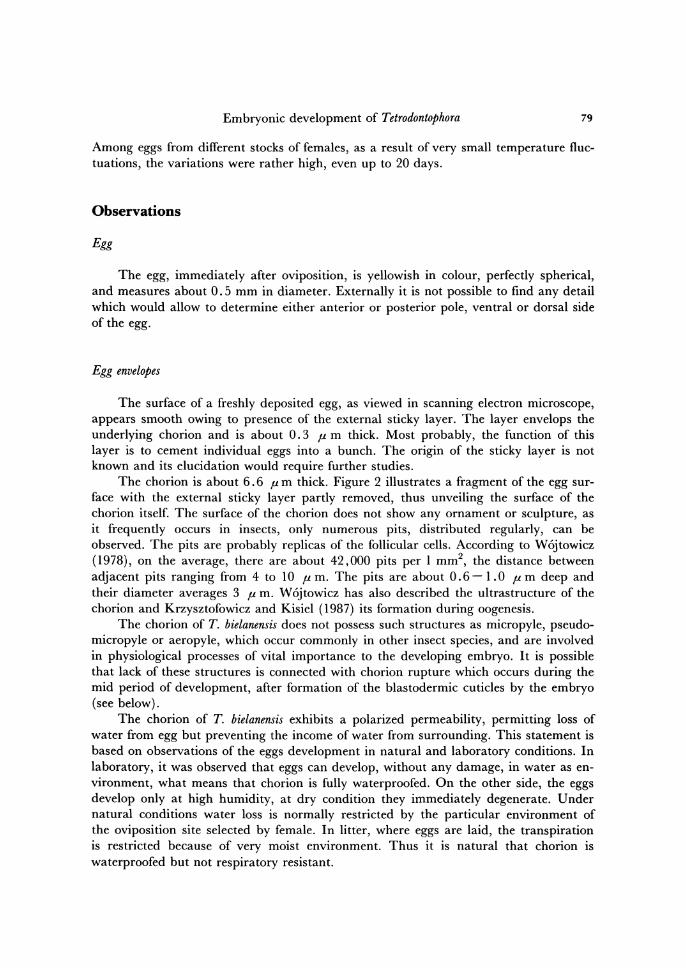
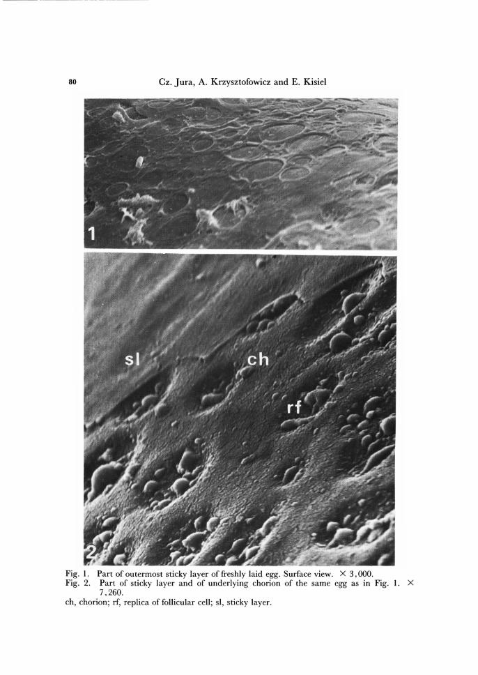
The surface of a freshly deposited egg, as viewed in scanning electron microscope, appears smooth owing to presence of the external sticky layer. The layer envelops the underlying chorion and is about 0.3 fL m thick. Most probably, the function of this layer is to cement individual eggs into a bunch. The origin of the sticky layer is not known and its elucidation would require further studies.
The chorion is about 6.6 fL m thick. Figure 2 illustrates a fragment of the egg sur-face with the external sticky layer partly removed, thus unveiling the surface of the chorion itself. The surface of the chorion does not show any ornament or sculpture, as it frequently occurs in insects, only numerous pits, distributed regularly, can be observed. The pits are probably replicas of the follicular cells. According to W6jtowicz (1978), on the average, there are about 42,000 pits per 1 mm2, the distance between adjacent pits ranging from 4 to 10 fL m. The pits are about 0.6-1.0 fL m deep and their diameter averages 3 fL m. W6jtowicz has also described the ultrastructure of the chorion and Krzysztofowicz and Kisiel (1987) its formation during oogenesis.
The chorion of T. bielanensis does not possess such structures as micropyle, pseudo-micropyle or aeropyle, which occur commonly in other insect species, and are involved in physiological processes of vital importance to the developing embryo. It is possible that lack of these structures is connected with chorion rupture which occurs during the mid period of development, after formation of the blastodermic cuticles by the embryo (see below).
The chorion of T. bielanensis exhibits a polarized permeability, permitting loss of water from egg but preventing the income of water from surrounding. This statement is based on observations of the eggs development in natural and laboratory conditions. In laboratory, it was observed that eggs can develop, without any damage, in water as en-vironment, what means that chorion is fully waterproofed. On the other side, the eggs develop only at high humidity, at dry condition they immediately degenerate. Under natural conditions water loss is normally restricted by the particular environment of the oviposition site selected by female. In litter, where eggs are laid, the transpiration is restricted because of very moist environment. Thus it is natural that chorion is waterproofed but not respiratory resistant.
--- - --- ------ -- ---- - --- - ---
80 Cz. Jura, A. Krzysztofowicz and E. Kisiel
Fig. I. Part of outermost sticky layer of freshly laid egg. Surface view. X 3,000. Fig. 2. Part of sticky layer and of underlying chorion of the same egg as in Fig. 1. X
7,260. ch, chorion; rf, replica of follicular cell; si, sticky layer.
Embryonic development of Tetrodontophora
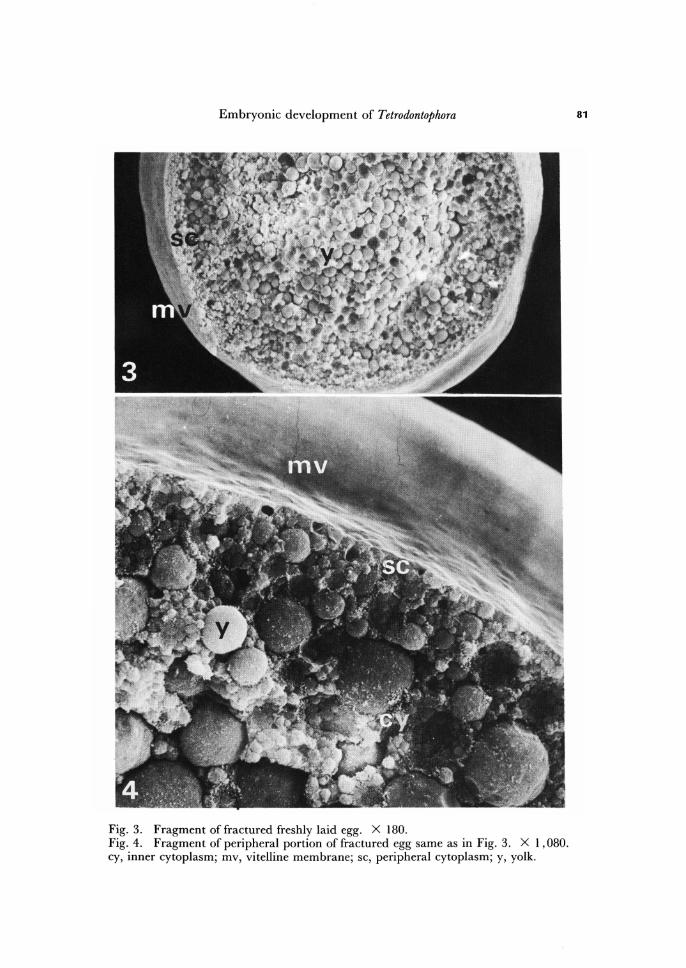
Fig. 3. Fragment of fractured freshly laid egg. X 180. Fig. 4. Fragment of peripheral portion of fractured egg same as in Fig. 3. X 1,080. cy, inner cytoplasm; mv, vitelline membrane; sc, peripheral cytoplasm; y, yolk.
81
82 Cz. Jura, A. Krzysztofowicz and E. Kisiel
The surface of the vitelline membrane, the second egg envelope underlying cho-rion, is completely smooth (Figs. 3, 4). The vitelline membrane is about 0.6 p m thick and, in opposite to chorion, it does Qot ruptures during the mid period of embryo de-velopment (see below).
Egg cytoarchitecture
The description of general egg cytoarchitecture is based on fractured eggs (Figs. 3 -5).
The. egg proper of T. bielanensis, as in other insects, contains three major compo-nents: periplasm, endoplasm with nucleus, and yolk. We shall describe these three components, but neglecting the nucleus which can not be observed under scanning electron microscope.
Periplasm and endoplasm consists of a cytoplasmic matrix that is rich in free ribo-somes and encloses a multitude of organelles and other components (Komorowska, 1970; Fryc, 1971; Klaja, 1971; Malcher, 1971; Adamiec, 1975; Gancarzewicz, 1975; Sikora, 1975; Bilinski, 1976; Klag, 1982a, b, 1983, 1984).
By means of scanning electron microscope it is not possible to distinguish any axial polarity in T. bielanensis egg. However, Gancarzewicz (1975) and Klag (1982) documented that one pole of freshly laid egg is rich in components containing RNA, which according to Klag are germ-cells determinants.
Periplasm, as viewed in scanning electron microscope, consists of cytoplasmic mat-rix which lacks yolk particles, and is extremely thin (Figs. 3, 4). In this respect T. bielanensis egg does not agree with corelation between oogenesis type and periplasm thickness established for Pterygota, where egg cytoarchitecture in general reflects the condition of oogenesis as well as the future course of embryogenesis. In Pterygota the eggs with thin periplasm are produced in result of panoistic oogenesis and have short germ band, whether the eggs with thick periplasm are produced in result of polytrophic oogenesis and have long germ band (for review see Counce, 1973, and Sander, 1983). In case of T. bielanensis the oogenesis is polytrophic (Krzysztofowicz, 1971, 1975), eggs have extremely thin periplasm, but extremely long germ band (Jura, 1986).
The endoplasm consists of a network of strands, occupying the space between yolk components, varying in thickness that here and there form cytoplasmic islands of diffe-rent sizes (Figs. 4, 5). Peripherally, the endoplasm merges into the periplasm without any change in the ultrastructural aspect.
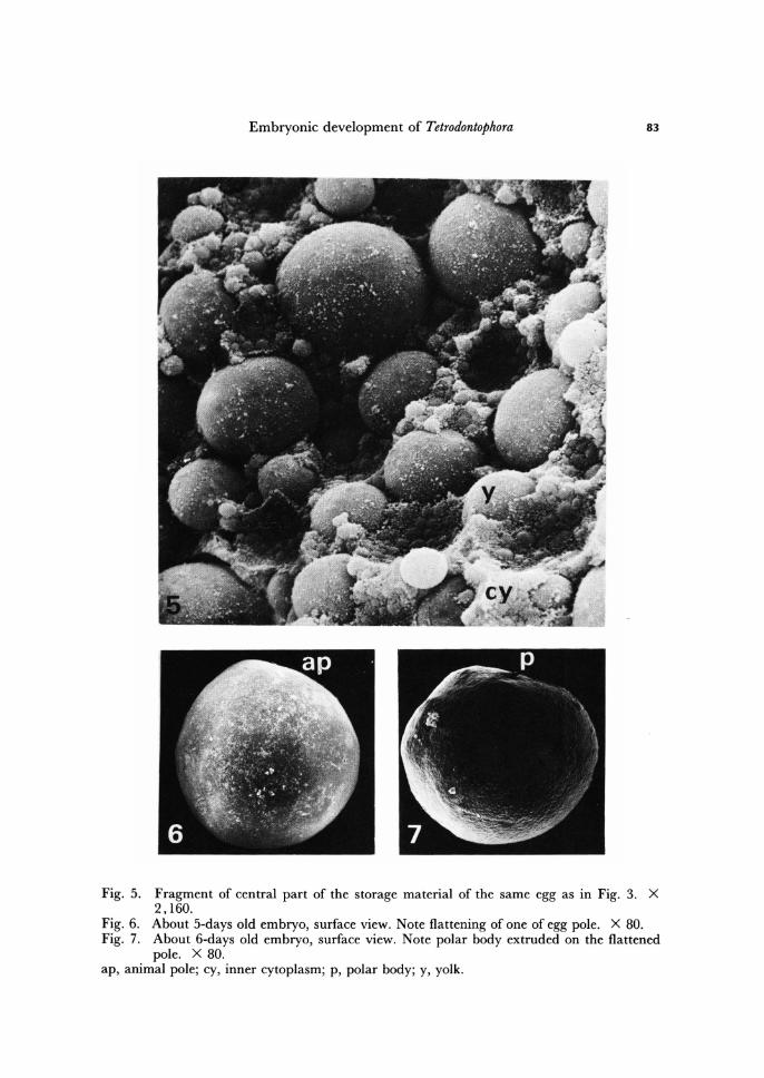
The egg of T. bielanensis is very rich in storage substances. The meshes of the endo-plasm are filled by storage substances that can be classified as proteid yolk bodies, lipid droplets, and glycogen granules (Fryc, 1971; Klaja, 1971; Malcher, 1971; Ksi~zkiewicz-Ilijeva, 1974; Bilinski, 1976). All these components are arranged concentrically with some regularity. The components showing the largest diameters are concentrated in the egg centre, whether the diameter of yolk components diminishes toward the egg periphery. This concerns especially proteid yolk bodies (Figs. 3-5).
Proteid yolk bodies dominate, but the egg is also lipid-rich and this probably is being related with the low temperature at which it develops. The lipids occur in the form of smaller globule-bodies than those of proteid-bodies. In the freshly laid egg,
Embryonic development of Tetrodontophora 83
Fig. 5. Fragment of central part of the storage material of the same egg as in Fig. 3. X 2,160.
Fig. 6. About 5-days old embryo, surface view. Note flattening of one of egg pole. X 80. Fig. 7. About 6-days old embryo, surface view. Note polar body extruded on the flattened
pole. X 80. ap, animal pole; cy, inner cytoplasm; p, polar body; y, yolk.
84 Cz. Jura, A. Krzysztofowicz and E. Kisiel
lipid globules occur in the largest number in the vicinity of periplasm, whether the gly-cogen granules are distributed more or less uniformly in periplasm as well as in endo-plasm.
Experimental studies showed that the egg of T. bielanensis is equiped with some ex-tracaryotic information, elaborated during oogenesis. This information is most probably localized in the egg periphery and is necessary for such process as cell membrane formation during early cleavage. The stratification of such components as RNA, pro-teins, glycogen or lipids, in result of centrifugation, does not influence the normal formation of the embryo (Ksiqikiewicz-Ilijeva, 1974). On the other hand, in the fertil-ized egg with partially injured periplasm cytokinesis does not occur (Jura, 1975, 1986).
Maturation
As we have mentioned, the freshly laid egg is perfectly spherical. After about 6 days from oviposition the first changes in this egg shape occur.
During the first 5 days of development the fertilized egg smugly fills the space en-closed by the vitelline membrane. Beginning with 6th day of development the egg shor-tens and flattens on one pole (Fig. 6), apparently by changing volume since its chorion-vitelline membrane girth remains constant. Fluid-filled pocket appears between the vitelline membrane and the flattened pole of the egg.
The egg remains flattened on one pole until the end of the first cleavage (see be-low). Meantime, the polar bodies emerge and occupy the pocket (Fig. 7).
Most probably, the flattening and lengthening of the egg involves the passage of a fluid out and into the egg. Also the occupation of the pocket by the polar bodies sug-gests that the egg flattening may facilitate polar bodies formation.
The flattened egg portion represents the pole on which latter first cleavage furrows will appear, and because of this, we may call it as animal egg pole (compare Figs. 7, 8).
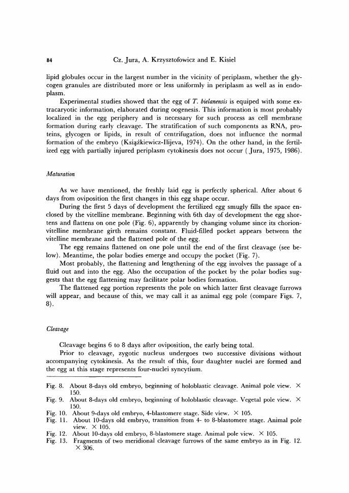
Cleavage
Cleavage begins 6 to 8 days after oviposition, the early being total. Prior to cleavage, zygotic nucleus undergoes two successive divisions without
accompanying cytokinesis. As the result of this, four daughter nuclei are formed and the egg at this stage represents four-nuclei syncytium.
Fig. 8. About 8-days old embryo, beginning of holoblastic cleavage. Animal pole view. X 150.
Fig. 9. About 8-days old embryo, beginning of holoblastic cleavage. Vegetal pole view. X 150.
Fig. 10. About 9-days old embryo, 4-blastomere stage. Side view. X 105. Fig. 11. About lO-days old embryo, transition from 4- to 8-blastomere stage. Animal pole
view. X 105. Fig. 12. About lO-days old embryo, 8-blastomere stage. Animal pole view. X 105. Fig. 13. Fragments of two meridional cleavage furrows of the same embryo as in Fig. 12.
X 306.
Embryonic development of Tetrodontophora 85
86
Fig. 14. Fig. 15. Fig. 16. Fig. 17. Fig. 18.
Cz. Jura, A. Krzysztofowicz and E. Kisiel
About IO-days old embryo, late 8-blastomere stage. Side view. X 105. About 12-days old embryo, transition from 8- to 16-blastomere stage. X 105. Fractured embryo at 16-blastomere stage. Note central yolk mass. 8-blastomere stage with "additional blastomere" (see text) . X 105. Fragment of the surface of the embryo about 14-days old during the transItion from 16- to 32-blastomere stage. Note cleavage furrow formation in the lower blastomere. X 369.
cf, cleavage furrow; cym, central yolk mass; sb, superficial blastomere.
Embryonic development of Tetrodontophora 87
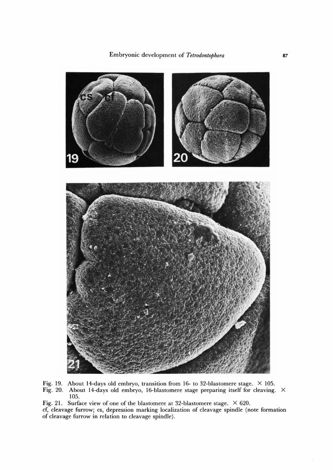
Fig. 19. About 14-days old embryo, transition from 16- to 32-blastomere stage. X 105. Fig. 20. About 14-days old embryo, 16-blastomere stage preparing itself for cleaving. X
105. Fig. 21. Surface view of one of the blastomere at 32-blastomere stage. X 620. cf, cleavage furrow; cs, depression marking localization of cleavage spindle (note formation of cleavage furrow in relation to cleavage spindle).
88 Cz. Jura, A. Krzysztofowicz and E. Kisiel
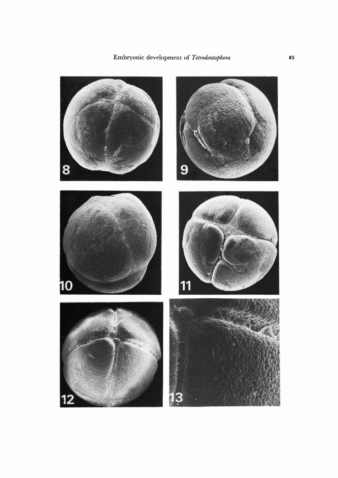
During the next day of development, the nuclei migrate from the centre to the opposite quarters of the egg and then on the flattened pole two cleavage furrows appear (Fig. 8) perpendicular to each other. These two first furrows may be named as meridional since they are localized on animal pole. The furrows deepen, extend to the sides, toward the opposite pole, and increasingly engirdle the whole egg. Within 10-15 hr the embryo becomes subdivided into four blastomeres (Figs. 8-10). Later a third furrow system appears, a horizontal one, which runs parallel to the equator of the egg (Figs. 11, 12). The resulting 8-blastomere stage consists of four animal-pole blasto-meres and four vegetal-pole ones, of equal size. The complete 8-cell stage is shown in Figs. 12 and 14.
Thus, the early cleavage is holoblastic, synchronous and equal. However, this was not the rule for all embryos studies. Within about 25 % of the embryos, the early cleav-age was somewhat unequal. Moreover, some embryos showed one or two" additional" flimsy blastomeres (Fig. 17). It was not possiblle to determine whether these additional blastomeres were equiped with nuclei, or were purely cytoplasmic structures, since under scanning electron microscope the nuclei could not be observed. Nevertheless, further development of such embryos was quite normal.
During described cleavage divisions, the cells of the embryo must gain new surface areas, which are composed of new periplasm + new cell membrane portion. Jura and Krzysztofowicz (1982) have noted that when holoblastic cleavage is going on the grooves, marking the cleavage planes, are wide but shallow (Fig. 13). The egg surface, in other planes than the grooves, remains smooth and seems to be stationary. Under scanning electron microscope no traces of insertion of preexisting surface area, from the bottoms of the grooves into the embryo interior, or splitting of ooplasm, can be observed. This remains in deep contrast with events accompanying the cleavage pro-cesses in a frog egg (for review see Beams and Kessel, 1976). The evidences obtained for T. bielanensis suggest that in this species during holoblastic cleavage formation of quite new surface areas and synthesis of new cell membranes, is necessary to complete the separation of cells.
After reaching 8-blastomere stage, the further cleavage furrows do not involve the whole yolk material of blastomeres. Innermost parts of blastomeres are cut off as anucleate yolk masses, in result of formation of new cell membranes of blastomeres above previous ones. The separated yolk masses fuse into a single central yolk material, filling the interior of the embryo, leaving the layer of pyramidal blastomeres at the egg surface (Fig. 16). Thus, during the transition from 8- to 16-blastomere stage the super-ficial cleavage is initiated and the stereoblastula is formed. At the centre of stereoblas-tula the yolk globules are uniformly distributed, previous blastomere boundaries had disappeared and no even traces of them could be seen.
Since 16-blastomere stage, the layer of superficial blastomeres of the embryo of T. bielanensis is equivalent to early cellular blastoderm, very characteristic embryonic structure of Thysanura and Pterygota species. Also beginning with 16-blastomere stage further cleavages are asynchronous and unequal.
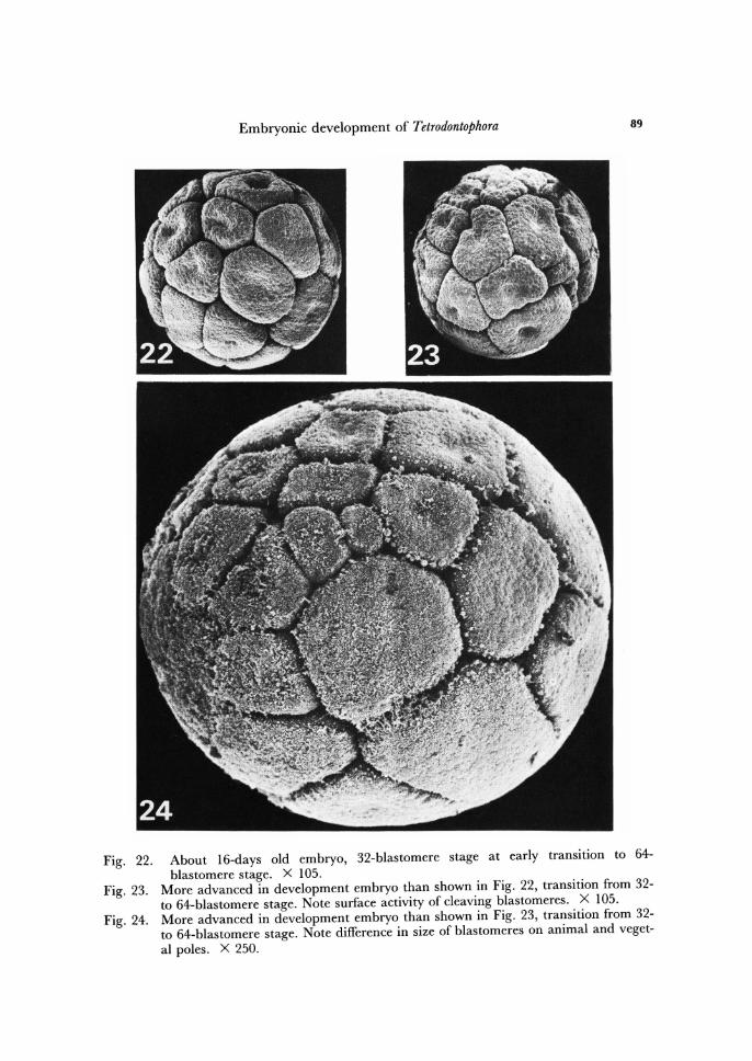
During the transition from 16- to 32- and than up to about 3, ODD-blastomere stage the superficial blastomeres further diminish in size (Figs. 16 - 35), and at every division separate off at their bases additional yolk, increasing the central yolk mass (Figs. 26-28).
Embryonic development of Tetrodontophora 89
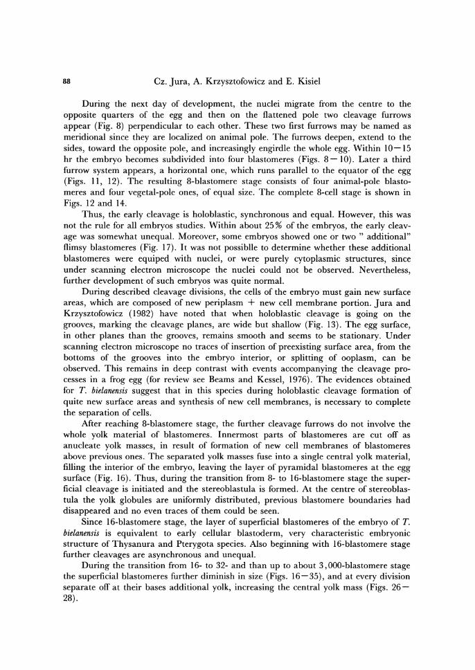
Fig. 22. About 16-days old embryo, 32-blastomere stage at early transition to 64-blastomere stage. X 105.
Fig. 23. More advanced in development embryo than shown in Fig. 22, transition from 32-to 64-blastomere stage. Note surface activity of cleaving blastomeres. X 105.
Fig. 24. More advanced in development embryo than shown in Fig. 23, transition from 32-to 64-blastomere stage. Note difference in size of blastomeres on animal and veget-al poles. X 250.
90 Cz. Jura, A. Krzysztofowicz and E . Kisiel
Fig. 25. About 17-days old embryo, fragment of surface view of 64-blastomere stage. X 340.
Fig. 26. Fragment of fractured embryo during transition from 16- to 32-blastomere stage. X 700.
sb, superficial blastomere; y, yolk.
Embryonic development of Tetrodontophora 91
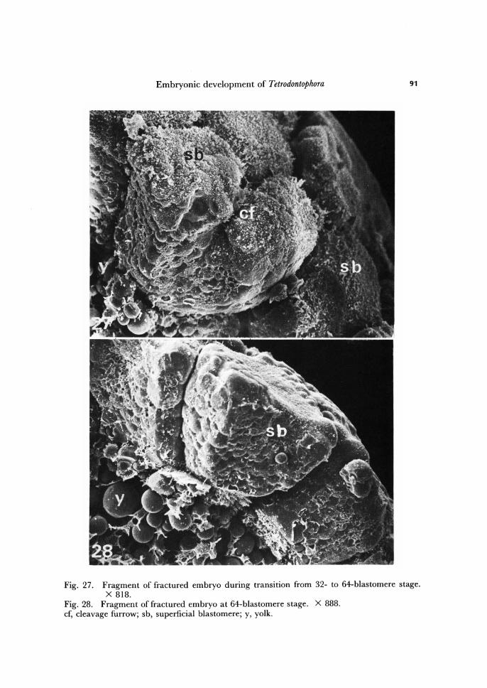
Fig. 27. Fragment of fractured embryo during transition from 32- to 64-blastomere stage. X 818.
Fig. 28. Fragment of fractured embryo at 64-blastomere stage. X 888. cf, cleavage furrow; sb, superficial blastomere; y, yolk.
92 Cz. Jura, A. Krzysztofowicz and E. Kisiel
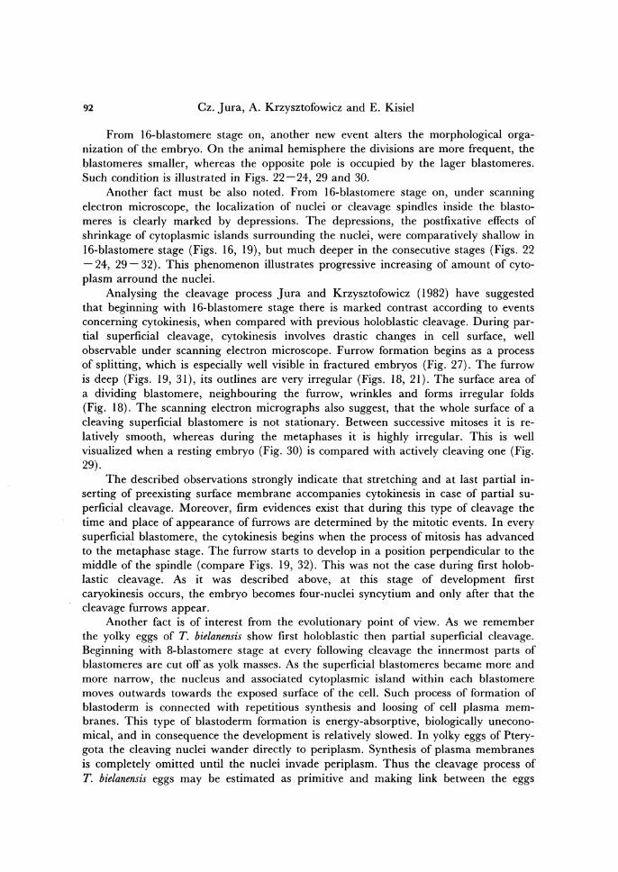
From 16-blastomere stage on, another new event alters the morphological orga-nization of the embryo. On the animal hemisphere the divisions are more frequent, the blastomeres smaller, whereas the opposite pole is occupied by the lager blastomeres. Such condition is illustrated in Figs. 22-24, 29 and 30.
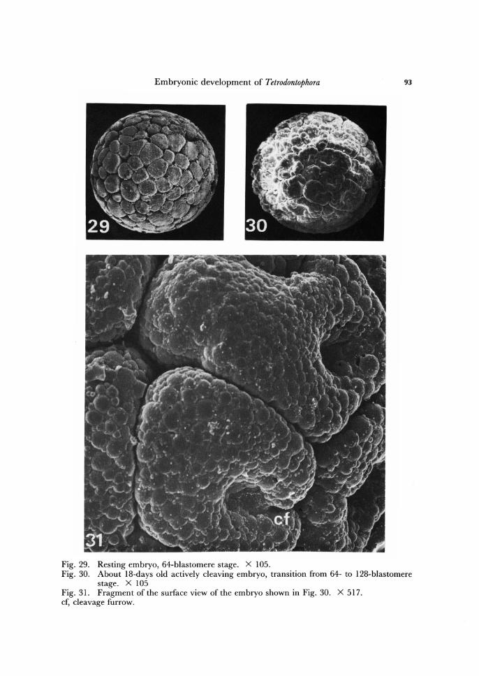
Another fact must be also noted. From 16-blastomere stage on, under scanning electron microscope, the localization of nuclei or cleavage spindles inside the bias to-meres is clearly marked by depressions. The depressions, the postfixative effects of shrinkage of cytoplasmic islands surrounding the nuclei, were comparatively shallow in 16-blastomere stage (Figs. 16, 19), but much deeper in the consecutive stages (Figs. 22 - 24, 29 - 32). This phenomenon illustrates progressive increasing of amount of cyto-plasm arround the nuclei.
Analysing the cleavage process Jura and Krzysztofowicz (1982) have suggested that beginning with 16-blastomere stage there is marked contrast according to events concerning cytokinesis, when compared with previous holoblastic cleavage. During par-tial superficial cleavage, cytokinesis involves drastic changes in cell surface, well observable under scanning electron microscope. Furrow formation begins as a process of splitting, which is especially well visible in fractured embryos (Fig. 27). The furrow is deep (Figs. 19, 31), its outlines are very irregular (Figs. 18, 21). The surface area of a dividing blastomere, neighbouring the furrow, wrinkles and forms irregular folds (Fig. 18). The scanning electron micrographs also suggest, that the whole surface of a cleaving superficial blastomere is not stationary. Between successive mitoses it is re-latively smooth, whereas during the metaphases it is highly irregular. This is well visualized when a resting embryo (Fig. 30) is compared with actively cleaving one (Fig. 29).
The described observations strongly indicate that stretching and at last partial in-serting of preexisting surface membrane accompanies cytokinesis in case of partial su-perficial cleavage. Moreover, firm evidences exist that during this type of cleavage the time and place of appearance of furrows are determined by the mitotic events. In every superficial blastomere, the cytokinesis begins when the process of mitosis has advanced to the metaphase stage. The furrow starts to develop in a position perpendicular to the middle of the spindle (compare Figs. 19, 32). This was not the case during first holob-lastic cleavage. As it was described above, at this stage of development first caryokinesis occurs, the embryo becomes four-nuclei syncytium and only after that the cleavage furrows appear.
Another fact is of interest from the evolutionary point of view. As we remember the yolky eggs of T. bielanensis show first holoblastic then partial superficial cleavage. Beginning with 8-blastomere stage at every following cleavage the innermost parts of blastomeres are cut off as yolk masses. As the superficial blastomeres became more and more narrow, the nucleus and associated cytoplasmic island within each blastomere moves outwards towards the exposed surface of the cell. Such process of formation of blastoderm is connected with repetitious synthesis and loosing of cell plasma mem-branes. This type of blastoderm formation is energy-absorptive, biologically unecono-mical, and in consequence the development is relatively slowed. In yolky eggs of Ptery-gota the cleaving nuclei wander directly to periplasm. Synthesis of plasma membranes is completely omitted until the nuclei invade periplasm. Thus the cleavage process of T. bielanensis eggs may be estimated as primitive and making link between the eggs
Embryonic development of Tetrodontophora 93
Fig. 29. Resting embryo, 64-blastomere stage. X 105. Fig. 30. About 18-days old actively cleaving embryo, transition from 64- to 128-blastomere
stage. X 105 Fig. 31. Fragment of the surface view of the embryo shown in Fig. 30. X 517. cf, cleavage furrow.
94 Cz. Jura, A. Krzysztofowicz and E. Kisiel
Fig. 32. Fragment of the surface view of embryo at about 120-blastomere stage. X 510. Fig. 33. Fragment offractured embryo at about 120-blastomere stage. X 517. cs, depression marking localization of cleavage spindle; ib, inner yolk material of blasto-mere; n, depression marking localization of nucleus; su, surface of blastomere.
Embryonic development of Tetrodontophora
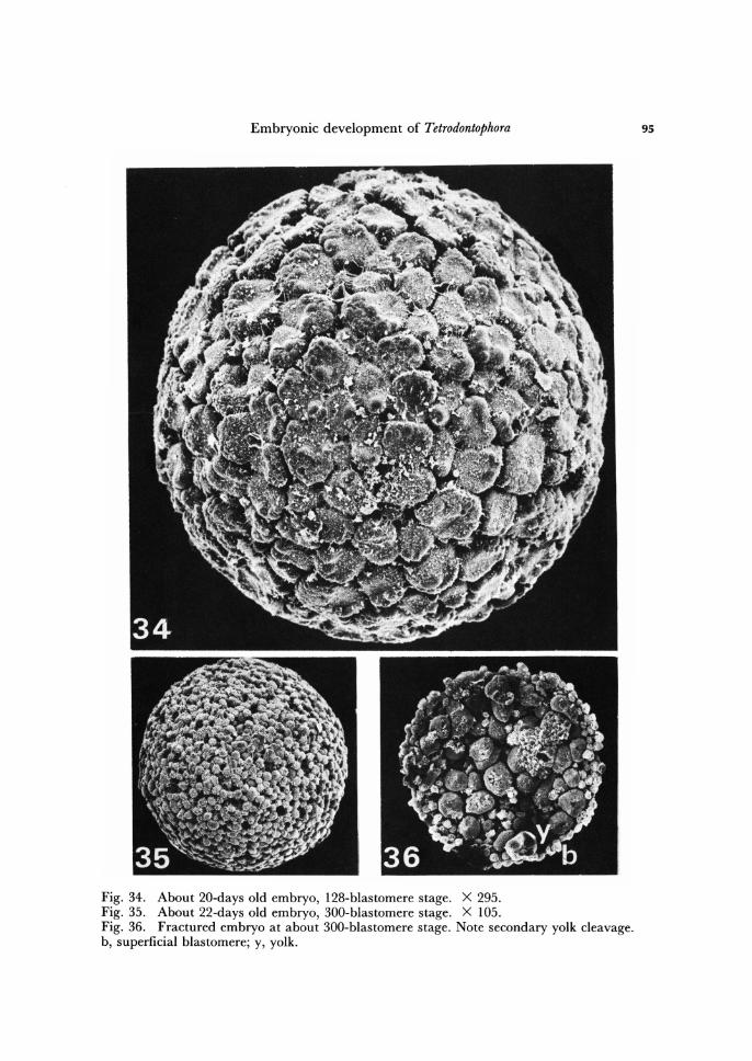
Fig. 34. About 20-days old embryo, 128-blastomere stage. X 295. Fig. 35. About 22-days old embryo, 300-blastomere stage. X 105. Fig. 36. Fractured embryo at about 300-blastomere stage. Note secondary yolk cleavage. b, superficial blastomere; y, yolk.
95
96 Cz. Jura, A. Krzysztofowicz and E. Kisiel
cleaving purely totally and those ones cleaving purely superficially. The processes connected with cleavage stage of development, which could not be
observed under scanning electron microscope, like formation of yolk cells and primary germ cells, were analysed by light. and transmission electron microscopes by Jura (1965, 1967a, b) and Klag (1982a, b, 1983, 1984). Jura (1986) also analysed the reg-ulative capacity of the embryo from fertilization till blastoderm stage. The morpholo-gical significance of yolk cells was studied by Jura (1966), Jura and Krzysztofowicz (1977).
Secondary yolk cleavage
During the holoblastic cleavage the storage material of the egg becomes divided into individual blastomeres, then, this material left in the centre of the embryo, in re-sult of superficial cleavage up to about 64-blastomere stage, represents an aggregation of yolk globules uniformly distributed within the mesh of the inner ooplasm (Figs. 16, 28). Following 64-blastomere stage the central yolk mass becomes temporarily divided by membranes into large blocks or spherules, each containing one or more vitel-lophages. At about 200-blastomere stage whole central yolk is segmented into irregular blocks of different size (Fig. 36). This organization of storage material, which may be called as secondary yolk cleavage, lasts comparatively short period of time. Before the embryo reaches mature blastoderm stage (see below) the yolky blocks disintegrate into separate yolk globules and storage material again assumes uniform organization.
Mature blastoderm
The layer of superficial blastomeres, formed in result of superficial cleavage, up to about 3, OOO-blastomere stage, only roughly resembles cellular blastoderm of pterygote insects. At early superficial cleavage phase the blastomeres are truncated, pyramidal in shape and have large amount of yolk (compare Figs. 26 - 28). As the superficial cleav-age progresses, the blastomeres loose the character of yolky pyramids and become more spherical. At the stage of about 1,500 blastomeres the organization of the embryo is as follows.
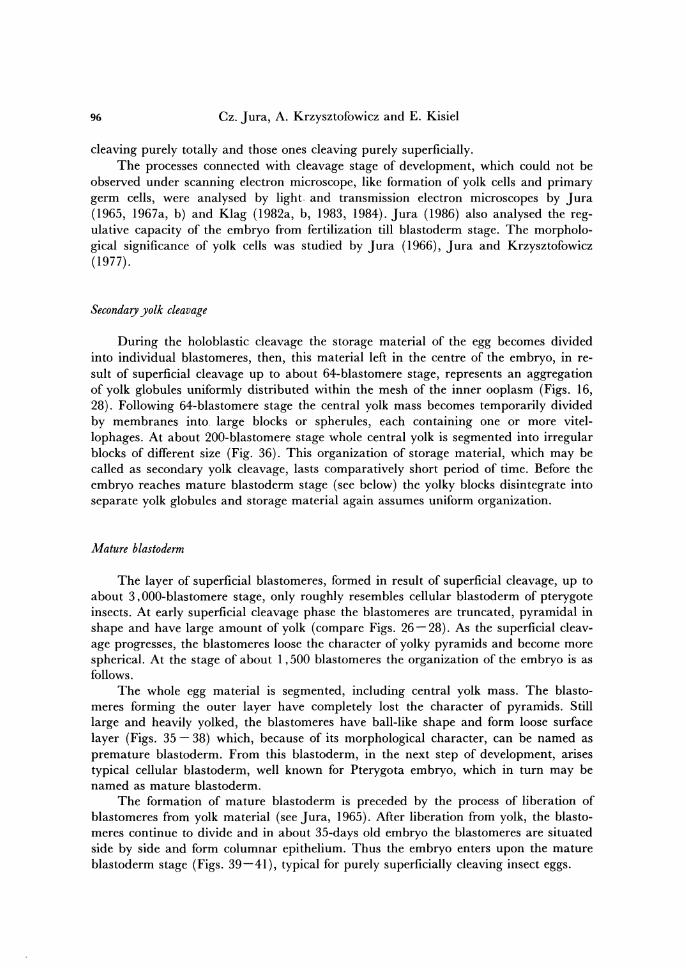
The whole egg material is segmented, including central yolk mass. The blasto-me res forming the outer layer have completely lost the character of pyramids. Still large and heavily yolked, the blastomeres have ball-like shape and form loose surface layer (Figs. 35 - 38) which, because of its morphological character, can be named as premature blastoderm. From this blastoderm, in the next step of development, arises typical cellular blastoderm, well known for Pterygota embryo, which in turn may be named as mature blastoderm.
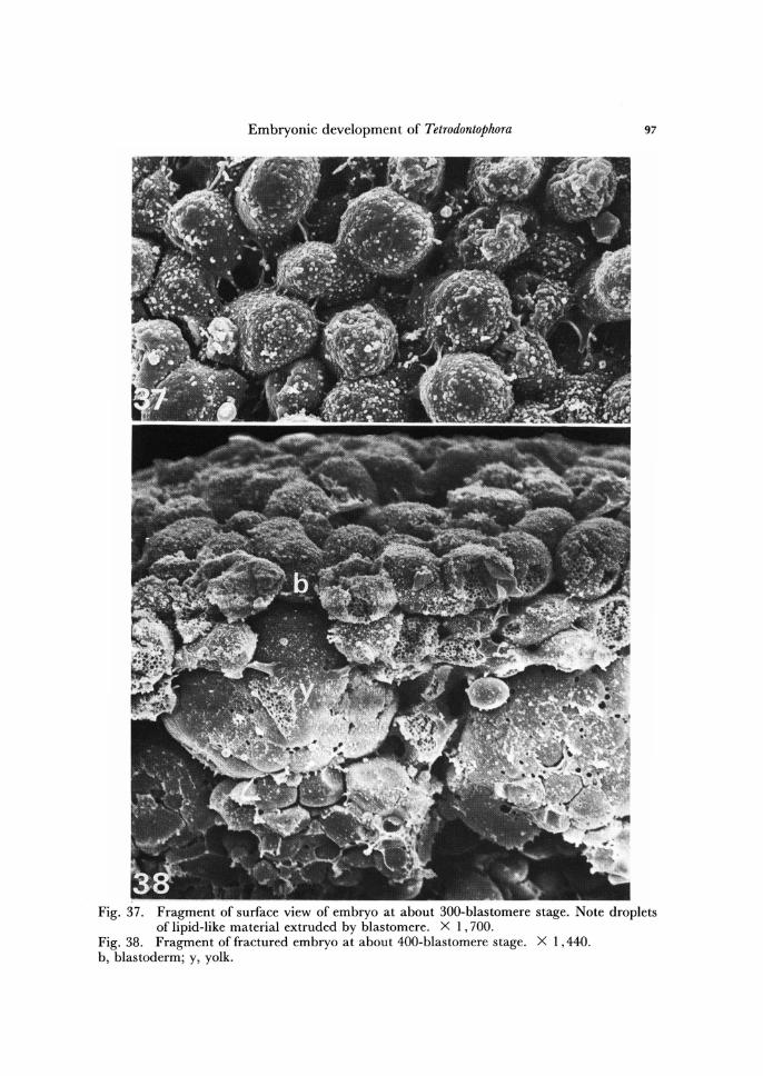
The formation of mature blastoderm is preceded by the process of liberation of blastomeres from yolk material (see Jura, 1965). After liberation from yolk, the bias to-meres continue to divide and in about 35-days old embryo the blastomeres are situated side by side and form columnar epithelium. Thus the embryo enters upon the mature blastoderm stage (Figs. 39 - 41), typical for purely superficially cleaving insect eggs.
Embryonic development of Tetrodontophora 97
Fig. 37. Fragment of surface view of embryo at about 300-blastomere stage. Note droplets of lipid-like material extruded by blastomere. X 1,700.
Fig. 38. Fragment of fractured embryo at about 400-blastomere stage. X 1,440. b, blastoderm; y, yolk.
98 Cz. Jura, A. Krzysztofowicz and E. Kisiel
Fig. 39. About 35-days old embryo, 3,000-blastomere stage (premature blastoderm stage). X 105.
Fig. 40. About 40-days old embryo, mature blastoderm stage. X 150. Fig. 41. Fragment of fractured embryo same as shown in Fig. 40. Note tertiary yolk cleav-
age. X 1,287: b, blastoderm; y, yolk.
Embryonic development of Tetrodontophora 99
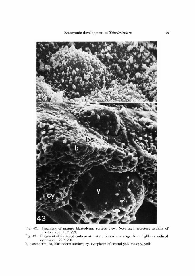
Fig. 42. Fragment of mature blastoderm, surface view. Note high secretory activity of blastomeres. X 7,293.
Fig. 43 . Fragment of fractured embryo at mature blastoderm stage. Note highly vacuolized cytoplasm. X 7,200.
b, blastoderm; bs, blastoderm surface; cy, cytoplasm of central yolk mass; y, yolk.
100 Cz. Jura, A. Krzysztofowicz and E. Kisiel
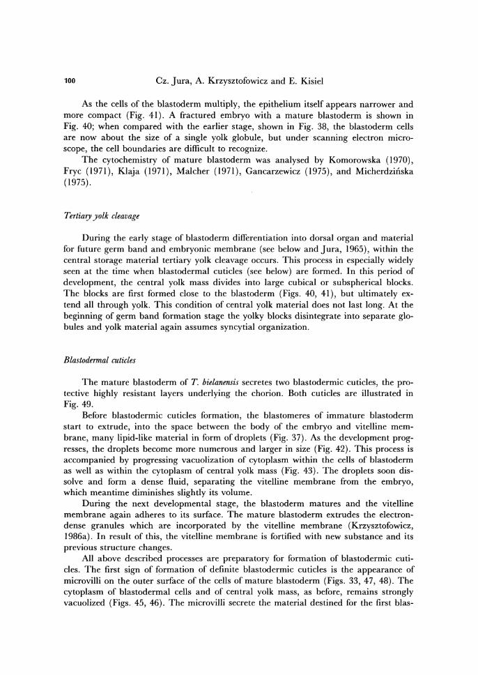
As the cells of the blastoderm multiply, the epithelium itself appears narrower and more compact (Fig. 41). A fractured embryo with a mature blastoderm is shown in Fig. 40; when compared with the earlier stage, shown in Fig. 38, the blastoderm cells are now about the size of a single yolk globule, but under scanning electron micro-scope, the cell boundaries are difficult to recognize.
The cytochemistry of mature blastoderm was analysed by Komorowska (1970), Fryc (1971), Klaja (1971), Malcher (1971), Gancarzewicz (1975), and Micherdziiiska (1975).
Tertiary yolk cleavage
During the early stage of blastoderm differentiation into dorsal organ and material for future germ band and embryonic membrane (see below and Jura, 1965), within the central storage material tertiary yolk cleavage occurs. This process in especially widely seen at the time when blastodermal cuticles (see below) are formed. In this period of development, the central yolk mass divides into large cubical or subspherical blocks. The blocks are first formed close to the blastoderm (Figs. 40, 41), but ultimately ex-tend all through yolk. This condition of central yolk material does not last long. At the beginning of germ band formation stage the yolky blocks disintegrate into separate glo-bules and yolk material again assumes syncytial organization.
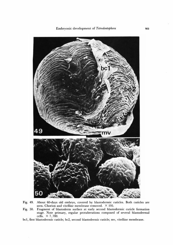
Blastodermal cuticles
The mature blastoderm of T. bielanensis secretes two blastodermic cuticles, the pro-tective highly resistant layers underlying the chorion. Both cuticles are illustrated in Fig. 49.
Before blastodermic cuticles formation, the blastomeres of immature blastoderm start to extrude, into the space between the body of the embryo and vitelline mem-brane, many lipid-like material in form of droplets (Fig. 37). As the development prog-resses, the droplets become more numerous and larger in size (Fig. 42). This process is accompanied by progressing vacuolization of cytoplasm within the cells of blastoderm as well as within the cytoplasm of central yolk mass (Fig. 43). The droplets soon dis-solve and form a dense fluid, separating the vitelline membrane from the embryo, which meantime diminishes slightly its volume.
During the next developmental stage, the blastoderm matures and the vitelline membrane again adheres to its surface. The mature blastoderm extrudes the electron-dense granules which are incorporated by the vitelline membrane (Krzysztofowicz, 1986a). In result of this, the vitelline membrane is fortified with new substance and its previous structure changes.
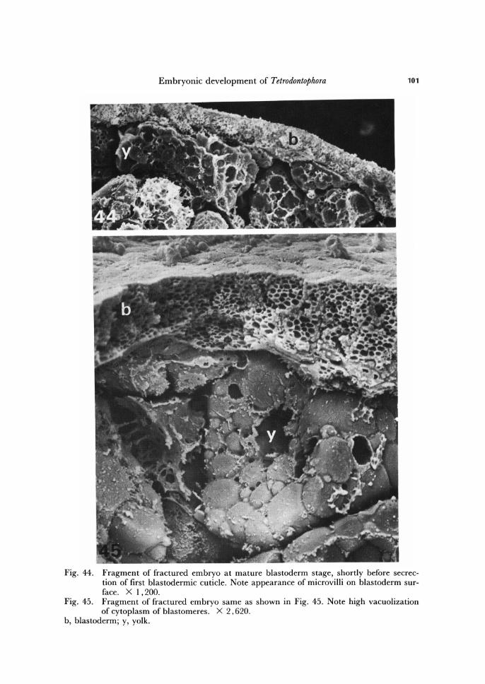
All above described processes are preparatory for formation of blastodermic cuti-cles. The first sign of formation of definite blastodermic cuticles is the appearance of microvilli on the outer surface of the cells of mature blastoderm (Figs. 33, 47, 48). The cytoplasm of blastodermal cells and of central yolk mass, as before, remains strongly vacuolized (Figs. 45, 46). The microvilli secrete the material destined for the first blas-
Embryonic development of Tetrodontophora 101
Fig. 44. Fragment of fractured embryo at mature blastoderm stage, shortly before secrec-tion of first blastodermic cuticle. Note appearance of microvilli on blastoderm sur-face. X 1.200.
Fig. 45. Fragment of fractured embryo same as shown in Fig. 45. Note high vacuolization of cytoplasm of blastomeres. X 2.620.
b, blastoderm; y, yolk.
102 Cz. Jura, A. Krzysztofowicz and E. Kisiel
Fig. 46. Fragment of fractured embryo shortly before blastodermic cuticle formation stage. Note high vacuolization of cytoplasm of central yolk mass. X 2,700.
Fig. 47 . Fragment of fractured embryo at mature blastoderm stage shortly before formation of blastodermic cuticles. Note numerous microvilli of blastoderm surface. X 8,140.
Fig. 48. Fragment of surface view of the same embryo as in Fig. 47. Note microvilli. X 4,400.
b, blastoderm; cb, cytoplasm of blastoderm; cy, cytoplasm of central yolk mass ; mi, micro-villi; y, yolk.
Embryonic development of Tetrodontophora 103
Fig. 49. About 60-days old embryo, covered by blastodermic cuticles. Both cuticles are seen. Chorion and vitelline membrane removed. X 195.
Fig. 50. Fragment of blastoderm surface at early second blastodermic cuticle formation stage. Note primary, regular protuberations composed of several blastodermal cells. X 7,700.
bel, first blastodermic cuticle; bc2, second blastodermic cuticle; mv, vitelline membrane.
104 Cz. Jura, A. Krzysztofowicz and E. Kisiel
todermic cuticle. In about two months old embryo the first blastodermic cuticle is already secreted.
This protective layer is comparatively thin and smooth (Fig. 49). By the beginning of third month of development, within the blastoderm morpholo-
gical changes occur leading to its differentiation into two different portions. On one pole of the egg the blastodermal become higher and in consequence of this within the blastoderm two regions become distinguishable. The portion of blastoderm with higher cells represents the material for the dorsal organ, the rest - for the future embryonic rudiment (germ band) and embryonic envelope (amnion-serosa).
By the time the dorsal organ is formed (see below), the blastoderm develops the second blastodermic cuticle. Every cell of the blastoderm contribute to the formation of this cuticle, including cells differentiating meantime into dorsal organ.
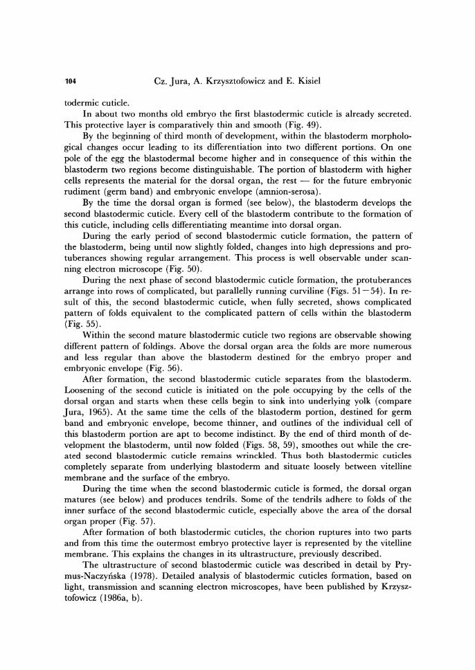
During the early period of second blastodermic cuticle formation, the pattern of the blastoderm, being until now slightly folded, changes into high depressions and pro-tuberances showing regular arrangement. This process is well observable under scan-ning electron microscope (Fig. 50).
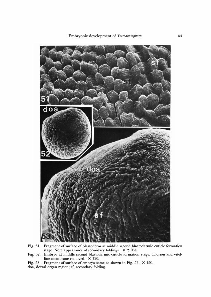
During the next phase of second blastodermic cuticle formation, the protuberances arrange into rows of complicated, but parallelly running curviline (Figs. 51 - 54). In re-sult of this, the second blastodermic cuticle, when fully secreted, shows complicated pattern of folds equivalent to the complicated pattern of cells within the blastoderm (Fig. 55).
Within the second mature blastodermic cuticle two regions are observable showing different pattern of foldings. Above the dorsal organ area the folds are more numerous and less regular than above the blastoderm destined for the embryo proper and embryonic envelope (Fig. 56).
After formation, the second blastodermic cuticle separates from the blastoderm. Loosening of the second cuticle is initiated on the pole occupying by the cells of the dorsal organ and starts when these cells begin to sink into underlying yolk (compare Jura, 1965). At the same time the cells of the blastoderm portion, destined for germ band and embryonic envelope, become thinner, and outlines of the individual cell of this blastoderm portion are apt to become indistinct. By the end of third month of de-velopment the blastoderm, until now folded (Figs. 58, 59), smoothes out while the cre-ated second blastodermic cuticle remains wrinckled. Thus both blastodermic cuticles completely separate from underlying blastoderm and situate loosely between vitelline membrane and the surface of the embryo.
During the time when the second blastodermic cuticle is formed, the dorsal organ matures (see below) and produces tendrils. Some of the tendrils adhere to folds of the inner surface of the second blastodermic cuticle, especially above the area of the dorsal organ proper (Fig. 57).
After formation of both blastodermic cuticles, the chorion ruptures into two parts and from this time the outermost embryo protective layer is represented by the vitelline membrane. This explains the changes in its ultrastructure, previously described.
The ultrastructure of second blastodermic cuticle was described in detail by Pry-mus-Naczynska (1978). Detailed analysis of blastodermic cuticles formation, based on light, transmission and scanning electron microscopes, have been published by Krzysz-tofowicz (1986a, b).
Embryonic development of Tetrodontophora 105
Fig. 51. Fragment of surface of blastoderm at middle second blastodermic cuticle formation stage. Note appearance of secondary foldings. X 2,964.
Fig. 52. Embryo at middle second blastodermic cuticle formation stage. Chorion and vitel-line membrane removed. X 120.
Fig. 53. Fragment of surface of embryo same as shown in Fig. 52. X 450. doa, dorsal organ region; sf, secondary folding.
106 Cz. Jura, A. Krzysztofowicz and E. Kisiel
Fig. 54. Fragment of second blastodermic cuticle at secondary folding phase. X 2,400. Fig. 55. About gO-days old embryo covered with mature second blastodermic cuticle. Side
view. X 80. Fig. 56. About gO-days old embryo covered with mature second blastodermic cuticle. Dor-
sal organ pole view. X 202. doa, dorsal organ area.
Embryonic development of Tetrodontophora 107
Primary dorsal organ
The primary dorsal organ is a very characteristic structure of every collembolan embryo of the species until now studied.
In T. bielanensis embryo, the dorsal organ arises through enlargement of a circular patch of cells of blastoderm, 2 - 4 days earlier before the germ band can be disting-uished.
The cells elongate and the dorsal organ gradually invaginates into the yolk, at the time when both blastodermic cuticles become secreted. During the invagination, the cell boundaries disappear, the nuclei migrate to the deepest parts of the cells and the dorsal organ becomes mushroom-shaped with fibrillar cytoplasm in its neck region (for more details of dorsal organ formation see Jura, 1965, 1976).
Komorowska (1972) using histochemical methods and cytophotometry documented, that the nuclei of dorsal organ contain four times more DNA when com-pared with the nuclei of the rest of blastoderm. Micherdzinska (1975) noted, that the cells of dorsal organ are especially rich in RNA content, but this content decreases at the time when dorsal organ shows a secretory activity (see below).
The dorsal organ of T. bielanensis assumes its mature organization by the end of third month of development. The mature dorsal organ has a typical glandular orga-nization. At mature stage it secretes through its neck radiating tendrils, which fan out (Fig. 61) beneath the second blastodermic cuticle and progressively elongate to the opposite pole of the egg (Figs. 60-67).
During the early period of dorsal organ activity the tendrils are radiating only arround the inner edge of its neck (Fig. 63), but soon the tendrils fill the whole opening of the neck (compare Figs. 60, 62-65).
The tendrils are very numerous. A single tendril is round on cross-section, it is ab-out 2 pm thick and has a lenght of 0.01-0.10 mm (Figs. 68-70). Within the fan, the tendrils are cemented by a dense substance, also secreted by the dorsal organ, which in fixed state coagulates arround tendrils in form of irregular net (Fig. 68).
At about the time when the embryo appendage rudiments become visible (see be-low), the tendrils flatten. The free ends of tendrils, occupying the outermost circles within the fan, split into several subelements and send out thin threads, which densely invest the whole embryo.
Meantime, as the above described processes advance, the space between second blastodermic cuticle and the embryo surface becomes filled with more or less liquid substance, dorsal organ derivate which metabolize some components of the yolk mate-rial. The substance when fixed coagulate arround the embryo in form of delicate film. The film is especially well observable at the level of developing, meantime, the rudi-ments of head appendages (Fig. 76).
We have previously mentioned, that the second blastodermic cuticle shows a spe-cialized area of a saucer-like appearance, localized above the dorsal organ (Fig. 56). Below this area, the free ends of some tendrils, 2 - 4 in number, join together and attach to the second blastodermic cuticle (Fig. 69) and form with its folds plastron-like structure localized above the dorsal organ. The significance of this structure is not known.
The primary dorsal organ is a transitory embryonic organ, and degenerates at the

108 Cz. Jura, A. Krzysztofowicz and E. Kisiel
Fig. 57. Fragment of second blastodermic cuticle of the dorsal organ area. Inner surface view. Note tendrils attached to the inner cuticle folds . X 990.
Fig. 58. About 90-days old embryo, fragment of blastoderm surface after secretion of second blastodermic cuticle. X 720.
Fig. 59. About 90-days old embryo after secretion of blastodermic cuticles. Both cuticles removed. X 90.
t, tendrils.

Embryonic development of Tetrodontophora
Fig. 60. Dorsal organ at early stage of secretion of tendrils. Surface view. X 672. Fig. 61. Dorsal organ at early stage of secretion of tendrils. Side view. X 220. Fig. 62. Dorsal organ at middle stage of secretion of tendrils. Surface view. X 220. od, dorsal organ orifice; t, tendrils.
109

110 Cz. Jura, A. Krzysztofowicz and E. Kisiel
Fig. 63. Fragment of dorsal organ at middle stage of secretion of tendrils. Side view. X 1,419.
Fig. 64. Dorsal organ at advanced stage of tendril secretion. Note tendrils filling its orifice. X 450.
od, dorsal organ orifice.
Embryonic development of Tetrodontophora 111
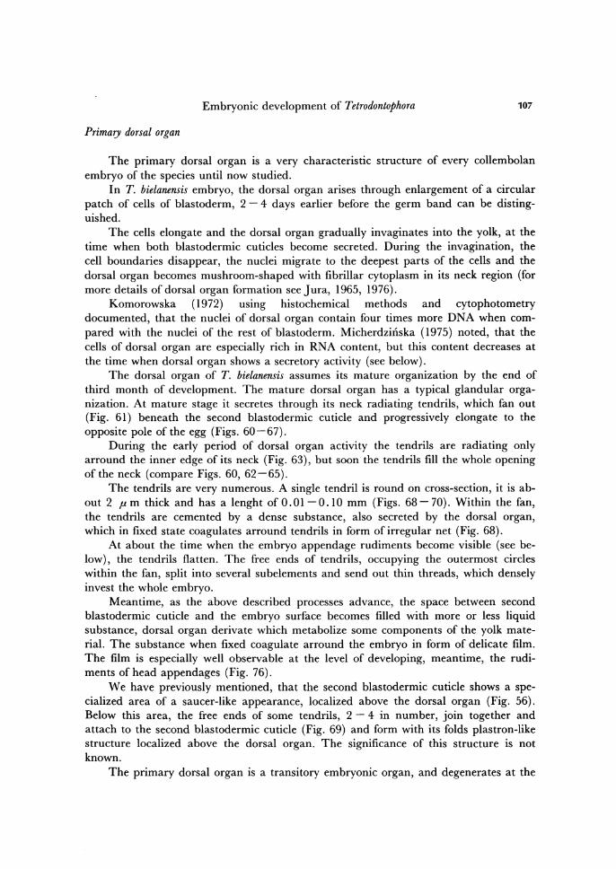
Fig. 65. Dorsal organ at more advanced stage of secretion of tendrils than that shown in Fig. 64. X 774.
Fig. 66. About lOO-days old embryo at advanced stage of secretion of tendrils by dorsal organ. Note papilla-like evaginations of rudiments of appendages. X 132.
Fig. 67. About l20-days old embryo wholly invested by tendrils. Dorsal organ pole view. X 206.
t, tendrils.
112 Cz. Jura, A. Krzysztofowicz and E. Kisiel
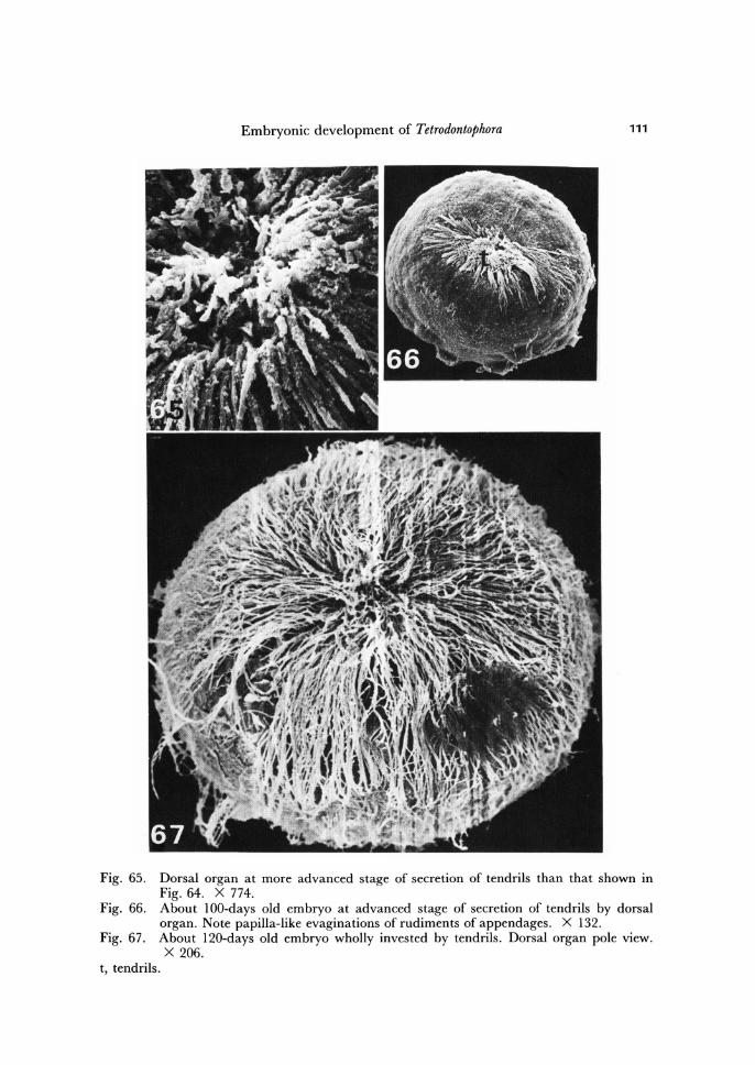
Fig. 68. Fragments of tendrils with attached dense substance. X 5,400. Fig. 69. Fragments of two tendrils connected by ends and attached to mner surface of
second blastodermic cuticle of dorsal organ area. X 6,820. bc, blastodermic cuticle; t, tendril.
Embryonic development of Tetrodontophora 113
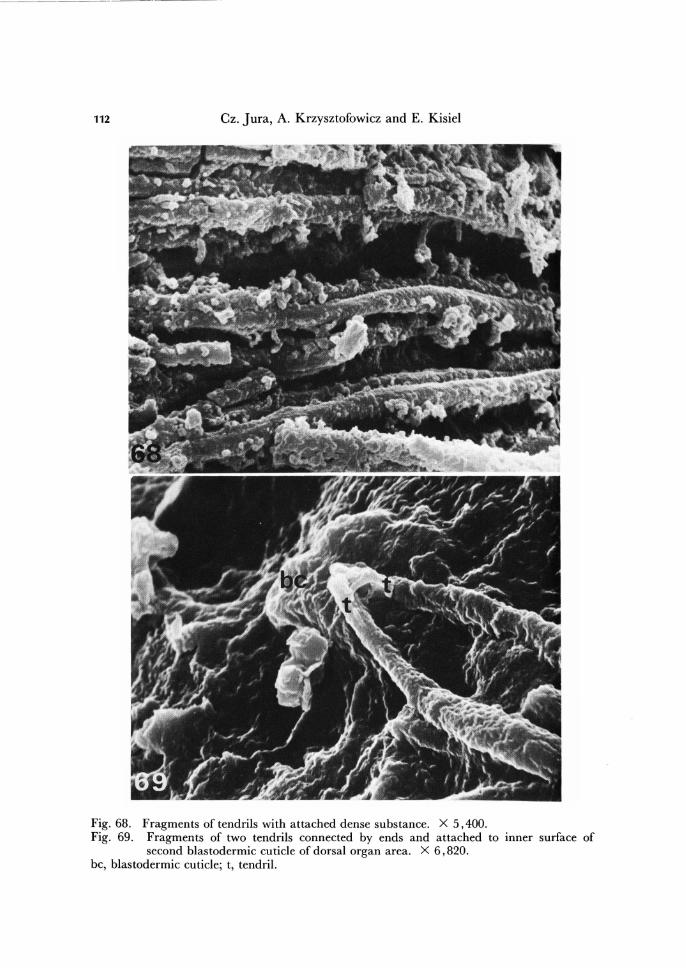
Fig. 70. Fragments of tendrils before embryo invagination into the yolk. X 11,650. Fig. 71. About ISO-days old embryo after invagination into the yolk. Dorsal organ pole
view. Note involution of dorsal organ. X 150. Fig. 72. Fragment of embryo at dorsal-closure stage. Dorsal organ pole view. Xl, 600. od, dorsal organ orifice.
114 Cz. Jura, A. Krzysztofowicz and E. Kisiel
end of embryogenesis. Already after invagination of the embryo into the yolk, the ten-drils dissolve, and at the time when dorsal closure begins, this is when the ectodermal cells of the embryo start to replace the amnio-serosal cells, the tendrils are no longer observable (Figs. 71, 72). However, during some time a narrow opening is visible lead-ing to the degenerating dorsal organ (Fig. 72).
The fluid originating from dissolved tendrils, as well as from dense substance sur-rounding the tendrils, fills the space created beneath the embryo invaginating into the yolk (Fig. 85; compare also Fig. 1, Jura, 1967b).
After invagination, the embryo flattens on both sides (Fig. 71) and elongates in anterio-posterior direction. The fluid fills also the space between the embryo sides and the second blastodermic cuticle. The increasing of the volume of the fluid as well as its pressure leads to the cracking of the chorion, but the second blastodermic cuticle stretches without breaking because of presence of folds which smooth out. The vitelline membrane and first blastodermic cuticle also do not break, most probably, because of their high extensibility.
The function of the dorsal organ is widely discussed in insect embryological litera-ture. It is postulated that this structure functions as organ for gas exchange, or as organ for absorbing the water from environment (for review see Tamarelle, 1975).
The dorsal organ in T. bielanensis reaches its maximum activity, in fluid and ten-drils secretion, when invagination of the embryo into the yolk is taking place. The swelling of the second blastodermic cuticle and the chorion rupture accompanies in-vagination. It seems clear, therefore, that the dorsal organ plays a part in the change of the shape of the embryo, essential for its invagination and elongation. It is also reason-able to suggest, that the radiating tendrils act in strengthening of the second blas-todermic cuticle, which stretches to accommodate the shape of the embryo. Jura (1967b) published experimental evidences, that in absence of functional dorsal organ the embryo of T. bielanensis fails to undergo invagination. In consequence of this, the body segments and appendages develop flatly on over the surface of the yolk material, further morphogenetic processes are disturbed and larva does not hatch.
Body segments and appendages
The period in which the part of blastoderm, destined for the embryo proper and
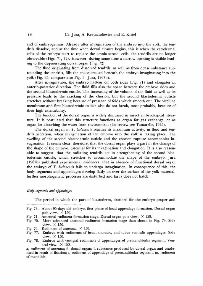
Fig. 73.
Fig. 74. Fig. 75.
Fig. 76. Fig. 77.
Fig. 78.
About 95-days old embryo, first phase of head appendage formation. Dorsal organ pole view. X 150. Antennal rudiment formation stage. Dorsal organ pole view. X 150. More advanced antennal rudiment formation stage than shown in Fig. 74. Side view. X 150. Rudiment of antenna. X 750. Embryo with rudiments of head, thoracic, and tubus ventralis appendages. Side view. X 150. Embryo with vestigial rudiments of appendages of premandibular segment. Ven-tral view. X 150.
a, rudiment of antenna; d, dorsal organ; f, substance produced by dorsal organ and conde-nsed in result of fixation; i, rudiment of appendage of premandibular segment; m, rudiment of mandible.
Embryonic development of Tetrodontophora 115
116 Cz. Jura, A. Krzysztofowicz and E. Kisiel
Embryonic development of Tetrodontophora 117
the embryonic envelope, is uniformly thick does not last long. The blastodermic cuti-des and dorsal organ formation phase is accomplished with differentiation of uniform blastoderm into germ band and amnio-serosa.
The germ band appears in form of a band (Jura, 1965), on the opposite pole in re-lation to developing dorsal organ. The cells of germ band gradually become columnar, between them distinct boundaries appear, and in result of many cell divisions and cell migrations the germ band becomes first two-layered, then many-layered.
The cells of the blastoderm, which occupy the sides of the embryo between the dorsal organ and germ band, become much flattened and thinner and remain one-layered. This thin walled part of blastoderm represents amnio-serosa.
At the mature dorsal organ stage, the head lobes within the germ band become already evident and this is the first indication of germ band differentiation. The head lobes represent the region bearing labrum, mouth and antennae.
At the head lobe stage, the germ band is very long its head and tail ends nearly meeting at the dorsal organ. The formation of appendage rudiments precedes the se-questration of germ band into definite body segments. In case of T. bielanensis it is possible to distinguish three phases in development of the rudiments of appendages. The rudiments of head appendages appear first, are followed by thoracic, and a little later by abdominal ones. All rudiments of appendages appear as papilla-like evagina-tions of germ band (Figs. 73 -77).
Of the head appendages as first appear rudiments of maxillae. The rudiments of the first thoracic appendages appear almost simultaneously with rudiments of maxillae. These are most conspicuous rudiments during early period of appendage formation (Fif. 73). Subsequently: rudiments of antennae, mandibles, intercalary appendages, and second and third thoracic appendages appear (Figs. 73 - 78). As last appear rudiments of abdominal appendages (Fig. 77).
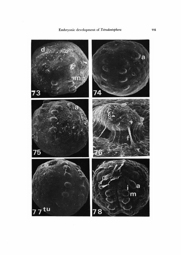
The antennal, intercalary, mandibular, maxillary I and 11 appendage rudiments mark dearly five head segments of T. bielanensis embryo. But, because between the antennal segment and the most frontal part of the germ band incisions appear (Figs. 82, 83), we postulate that the head is make up of six segments:
I) The preantennal segment, deveoid of appendages. 2) The antennary segment. 3) The intercalary segment (premandibular), with vestigial appendages. 4) The mandibular segment. 5) The maxillary I segment. 6) The maxillary 11 segment.
Fig. 79. Orifice of stomodaeum. Xl, 280. Fig. 80. Embryo more advanced in development than shown in Fig. 71. Ventral view. X
84. Fig. 81. Advanced appendage rudiment stage. Dorsal organ pole view. X 180. Fig. 82. Advanced appendage rudiment formation stage. Beginning of involution of rudi-
ments of premandibular segment. X 246. a, rudiment of antenna; d, dorsal organ; i, rudiment of appendage of premandibular seg-ment; in, incisions marking border between preantennal and antennal segments; m, rudi-ment of mandible; s, orifice of stomodaeum; tv, rudiment of tubus ventralis; xl, rudiment of first maxilla; x2, rudiment of second maxilla.
118 Cz. Jura, A. Krzysztofowicz and E. Kisiel
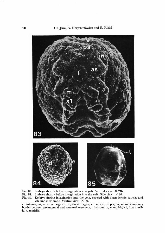
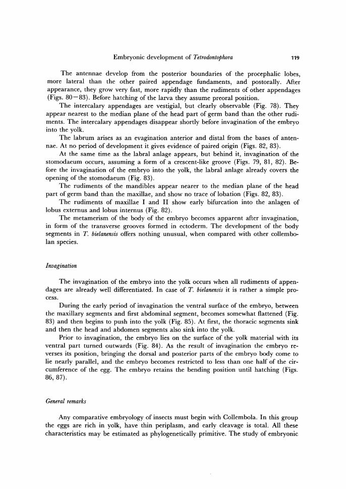
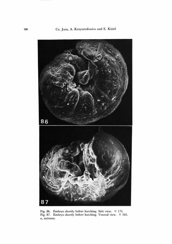
Fig. 83. Embryo shortly before invagination into yolk. Ventral view. X 246. Fig. 84. Embryo shortly before invagination into the yolk. Side view. X 90. Fig. 85. Embryo during invagination into the yolk, covered with blastodermic cuticles and
vitelline membrane. Ventral view. X 90. a, antenna; as, antennal segment; d, dorsal organ; e, embryo proper; in, incision marking border between preantennal and antennal segments; I, labrum; m, mandible; xl, first maxil-la; t, tendrils.
Embryonic development of Tetrodontophora 119
The antennae develop from the posterior boundaries of the procephalic lobes, more lateral than the other paired appendage fundaments, and postorally. After appearance, they grow very fast, more . rapidly than the rudiments of other appendages (Figs. 80-83). Before hatching of the larva they assume preoral position.
The intercalary appendages are vestigial, but clearly observable (Fig. 78). They appear nearest to the median plane of the head part of germ band than the other rudi-ments. The intercalary appendages disappear shortly before invagination of the embryo into the yolk.
The labrum arises as an evagination anterior and distal from the bases of anten-nae. At no period of development it gives evidence of paired origin (Figs. 82, 83).
At the same time as the labral anlage appears, but behind it, invagination of the stomodaeum occurs, assuming a form of a crescent-like groove (Figs. 79, 81, 82). Be-fore the invagination of the embryo into the yolk, the labral anlage already covers the opening of the stomodaeum (Fig. 83).
The rudiments of the mandibles appear nearer to the median plane of the head part of germ band than the maxillae, and show no trace of lobation (Figs. 82, 83).
The rudiments of maxillae I and 11 show early bifurcation into the anlagen of lobus externus and lobus internus (Fig. 82).
The metamerism of the body of the embryo becomes apparent after invagination, in form of the transverse grooves formed in ectoderm. The development of the body segments in T. bielanensis offers nothing unusual, when compared with other collembo-lan species.
Invagination
The invagination of the embryo into the yolk occurs when all rudiments of appen-dages are already well differentiated. In case of T. bielanensis it is rather a simple pro-cess.
During the early period of invagination the ventral surface of the embryo, between the maxillary segments and first abdominal segment, becomes somewhat flattened (Fig. 83) and then begins to push into the yolk (Fig. 85). At first, the thoracic segments sink and then the head and abdomen segments also sink into the yolk. .
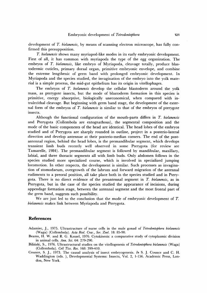
Prior to invagination, the embryo lies on the surface of the yolk material with its ventral part turned outwards (Fig. 84). As the result of invagination the embryo re-verses its position, bringing the dorsal and posterior parts of the embryo body come to lie nearly parallel, and the embryo becomes restricted to less than one half of the cir-cumference of the egg. The embryo retains the bending position until hatching (Figs. 86,87).
General remarks
Any comparative embryology of insects must begin with Collembola. In this group the eggs are rich in yolk, have thin periplasm, and early cleavage is total. All these characteristics may be estimated as phylogenetically primitive. The study of embryonic
120 Cz. Jura, A. Krzysztofowicz and E. Kisiel
Fig. 86. Embryo shortly before hatching. Side view. X 175. Fig. 87. Embryo shortly before hatching. Ventral view. X 165. a, antenna.
Embryonic development of Tetrodontophora 121
development of T. bielanensis, by means of scanning electron microscope, has fully con-firmed this presupposition.
T. bielanensis shows many myriapod-like modes in its early embryonic development. First of all, it has common with myriapods the type of the egg organization. The embryos of T. bielanensis, like embryo of Myriapoda, cleavage totally, produce blas-todermic cuticles, primary dorsal organ, primitive embryonic envelope, and combine the extreme lengthenic of germ band with prolonged embryonic development. In Myriapoda and the species studied, the invagination of the embryo into the yolk mate-rial is a simple process, the mid-gut epithelium has its origin in vitellophages.
The embryos of T. bielanensis develop the cellular blastoderm around the yolk mass, as pterygote insects, but the mode of blastoderm formation in this species is primitive, energy absorptive, biologically uneconomical, when compared with in-tralecithal cleavage. But beginning with germ band stage, the development of the exter-nal form of the embryos of T. bielanensis is similar to that of the embryos of pterygote insects.
Although the functional configuration of the mouth-parts differs in T. bielanensis and Pterygota (Collembola are entognathous), the segmental composition and the mode of the basic components of the head are identical. The head lobes of the embryos studied and of Pterygota are sharply rounded in outline, project in a posterio-Iateral direction and develop antennae at their posterio-median corners. The end of the post-antenna 1 region, behind the head lobes, is the premandibular segment, which develops transient limb buds recently well observed in some Pterygota (for review see Tamarelle, 1984). The premandibular segment is followed by mandibular, maxillary, labial, and three thoracic segments all with limb buds. Only abdomen follows in the species studied more specialized course, which is involved in specialized jumping locomotion. In other respects, the development is similar. Such processes as invagina-tion of stomodaeum, overgrowth of the labrum and forward migration of the antennal rudiments to a preoral position, all take place both in the species studied and in Ptery-gota. There is no direct evidence of the preantennal segment in T. bielanensis, as in Pterygota, but in the case of the species studied the appearance of incisions, during appendage formation stage, between the antenna 1 segment and the most frontal part of the germ band, suggests such possibility.
We are just led to the conclusion that the mode of embryonic development of T. bielanensis makes link between Myriapoda and Pterygota.
References
Adamiec, ]., 1975. Ultrastructure of nurse cells in the male gonad of Tetrodontophora bieianensis (Waga) (Collembola). Acta Bioi. Crac., Ser. Zooi. 18: 85-90.
Beams, H. W. and R. G. Kessel, 1976. Cytokinesis: a comparative study of cytoplasmic division in animal cells. Ann. Sci. 64: 279-290.
Bilinski, S., 1976. Ultrastructural studies on the vitellogenesis of Tetrodontophora bieianensis (Waga) (Collembola). Cell Tiss. Res. 168: 399-410.
Counce, S. ]., 1973. The causal analysis of insect embryogenesis. In S. ]. Counce and C. H. Wadding ton (eds. ), Developmental Systems: Insects, Vol. 2, 1-156. Academic Press, Lon-don, New York.
122 Cz. Jura, A. Krzysztofowicz and E. Kisiel
Fryc, W., 1971. Histochemical analysis of early developmental stages of Tetrodontophora bielanensis (Waga) (Collembola). Lipids. Zeu;. Nauk. U}, 2001. 17: 39-46.
Gancarzewicz, B., 1975. Histochemical analysis of RNA in early stages of embryonic develop-ment of Tetrodontophora bielanensis (Waga) (Collembola). Zeu;. Nauk. U}, Zool. 21: 85-94.
jura, Cz., 1965. Embryonic development of Tetrodontophora bielanensis (Waga) (Collembola) from oviposition till germ band formation stage. Acta Bio1. Grac., Ser. Zooz. 8: 141-157.
-----, 1966. Origin of the endoderm and embryogenesis of the alimentary system in Tet-rodontophora bielanensis (Waga) (Collembola). Acta BioI. Grac., Ser. Zool. 9: 93-102.
-----, 1967a. Origin of germ cells and gonads formation in embryogenesis of Tetrodont ophora bie1anensis (Waga) (Collembola). Acta Bio1. Grac., Ser. Zool. 10: 97-103.
-----, 1967b. The significance and function of the primary dorsal organ in embryonic development of Tetrodontophora bie1anensis (Waga) (Collembola). Acta Bio1. Grac., Ser. Zool. 10: 301-311.
-----, 1972. The development of apterygote insects. In S. j. Counce and C. H. Wad-dington (eds. ), Developmental Systems: Insects, Vol. I, 49-94. Academic Press, London, New York.
-----, 1975. Cleavage without cytokinesis of the eggs of Tetrodontophora bie1anensis (Waga) (Collembola) after periplasm damage with thermocauter. Acta Bioi. Grac., Ser. Zool. 18: 97-102.
-----, 1986. Effects of cautheryzation of Tetrodontophora bielanensis (Collembola) eggs from oviposition till premature blastoderm stage. Instructive events in early development. Proc. Arthropod. Embryol. Soc.}pn. 21: 1-12.
-----, and A. Krzysztofowicz, 1977. Ultrastructural changes in embryonic midgut cells devel9ping into larval midgut epithelium of Tetrodontophora bielanensis (Waga) (Collembola). Rev. Ecoz. Bioi. Sol 14: 10-15.
-----, and , 1978. Scanning electron microscopy of Tetrodontophora .bielanensis (Waga) (Collembola). The structure of the dorsal organ. 1st Int. Sem. Apterygota, Accad. delle Se. Detta Da-Fisiocritiei, 105-113.
-----, and , 1982. Scanning electron microscopy of Tetrodontophora bielanensis (Waga) (Collembola) embryogenesis. Fertilized egg to 64-blastomere stage. Acta Bio1. Aud. Sci. Hung. 33: 39-48.
-----, and , 1986. Scanning electron microscopy of Tetrodontophora bielanensis (Waga) (Collembola) embryogenesis. Segmentation and appendage formation of head. Acta Bioi. Grac., Ser. 2001. 28: 7-11.
Klag, j., 1982a. Germ line of Tetrodontophora bielanensis (Insecta, Collembola). Ultrastructural study on the origin of primordial germ cells. j. Embryo1. Exp. Morpho1. 72: 183-195.
-----, 1982b. Germ line of Tetrodontophora bielanensis (Insecta, Collembola). 2. Ultras-tructural differentiation of primordial germ cells during early embryogenesis. Gytobios 33: 173-182.
-----, 1983. Germ line of Tetrodontophora bielanensis (Insecta, Collembola). 3. Migration of primordial germ cells. Gytobios 38: 38-53.
-----, 1984. Germ line of Tetrodontophora bielanensis (Insecta, Collembola). 4. Nucleolus-like bodies extruded in toto from nuclei of primordial germ cells become part of the "nuage" . Gytobios 40: 7-20.
Klaja, E., 1971. Histochemical analysis of early developmental stages of Tetrodontophora bielanensis (Waga) (Collembola). PAS-positive substances. Zesz. Nauk. U}, Zool. 17: 47-57.
Komorowska, B., 1970. Alkaline phosphatase in early stages of embryonic development of Tetro-dontophora bielanensis (Waga) (Collembola). Histochemical analysis. Zesz. Nauk. U}, 2001. 16: 93-105.
------., 1972. Cytophotometric analysis of nuclear DNA in the cells of primary dorsal organ and germ band of Tetrodontophora bielanensis (Waga) (Collembola). Zesz. Nauk. U}, Zool. 18: 15-25.
Embryonic development of Tetrodontophora 123
Krzysztofowicz, A., 1967. Spermatogeneza u Tetrodontophorabielanensis (Waga) (Collembola). Zesz. Nauk. Vj, Zool. 13: 27-69.
-----, 1971. Histochemical and autoradiographic analysis of RNA synthesis in trophic cells of the female gonad in Tetrodontophora bielanensis (Waga) (Collembola). Acta Biol. Crac., Ser. Zool. 14: 299-305.
-----, 1975. Histochemical and ultrastructural analysis of blocking nurse cells in the female gonad of Tetrodontophora bielanensis (Waga) (Collembola). Acta Biol. Crac., Ser. Zool. 18: 45-53.
-----, 1980a. Ultrastructure of the spermatophores in Tetrodontophora bielanensis (Waga) (Collembola). Folia Biol., Krak6w 28: 79-82.
-----, 1980b. The ultrastructure of spermatogonia and germ cells of germarium in Tetro-dontophora bielanensis (Waga) (Collembola). Folia Biol., KrakOw 28: 363-369.
-----, 1986a. Ultrastructural studies on embryonic development of Tetrodontophora bielanensis (Waga) (Collembola). Formation of the 1st and 2nd blastodermal cuticles. Part I. TEM studies. Acta Biol. Crac., Ser. Zool. 28: 19-26.
-----, 1986b. Ultrastructural studies on embryonic development of Tetrodontophora bielanensis (Waga) (Collembola). Formation of the 1st and 2nd blastodermal cuticles. Part 11. SEM studies. Acta Biol. Crac., Ser. Zool. 28: 27-31.
-----, and W. Byczkowska-Smyk, 1966. Preliminary investigations on the fine structure of spermatozoa of Tetrodontophora bielanensis (Waga) (Collembola). Acta Biol. Crac., Ser. Zool. 9: 97-103.
-----, and , 1968. Studies on the fine structure of spermatids in the Tetro-dontophora bielanensis (Waga) (Collembola). Part 11. Acta Biol. Crac., Ser. Zool. 11: 29-36.
-----, and , 1972. Fine structure of first spermatocytes of Tetrodontophora bielanensis (Waga) (Collembola). Ill. Zesz. Nauk. Vj, Zool. 18: 7-13.
-----, and E. Kisiel, 1987. Oogenesis of Tetrodontophora bielanensis (Waga) (Collembola). Preliminary ultrastructural studies of the first egg envelope formation. In H. Ando and Cz. Jura (eds. ), Recent Advances in Insect Embryology in Japan and Poland, 37-49. Arthro-pod. Embryo!. Soc. Jpn. (ISEBU Co. Ltd., Tsukuba).
Ksi~zkiewicz-Ilijeva, M., 1974. Development of the eggs of Tetrodontophora bielanensis (Waga) (Col-lembola) when centrifuged before cleavage. Acta Biol. Crac., Ser. Zool. 17: 70-75.
Malcher, B., 1971. Glycogen .in the early developmental stages of Tetrodontophora bielanensis (Waga) (Collembola). Zesz. Nauk. Vj, Zool. 17: 59-67.
Micherdzinska, E., 1975. RNA of the late blastodermal cells and of the primary dorsal organ of Tetrodontophora bielanensis (Waga) (Collembola). Histochemical analysis. Zesz. Nauk. Vj, Zool. 21: 95-103.
Prymus-Naczynska, A., 1978. Ultrastructure of the blastodermal cuticle of Tetrodontophora bielanensis (Waga) (Collembola). Folia Biol., Krak6w 26: 87-93.
Sander, K., 1983. Extracaryotic determinants, a link between oogenesis and embryonic pattern formation in insects. Proc. Arthropod. Embryol. Soc.jpn. 19: 1-12.
Sikora, A., 1975. Ultrastructure of the nurse cells in the female gonad of Tetrodontophora bielanensis (Waga) (Collembola). Acta Biol. Crac., Ser. Zool. 18: 79-84.
Tamarelle, M., 1975. Nouvelles observations sur ultrastructure de l'organe dorsal des Collem-boles. Mise un evidence d'un type de differenciation "en cocarde" chez Proisotoma minuta. C. R. Acad. Sci. Paris 280: 897-899.
-----, 1984. Transient rudiments of second antennae on the "intercalary segment" of embryos of Anurida maritima Guer. (Collembola: Arthropleona) and Hyphantria cunea Drury (Lepidoptera: Arctiidae). Int. j. Insect Morphol. Embryol. 13: 331-336.
Tyszkiewicz, K., 1976. The embryogenesis of the central nervous system of Tetrodontophora bielanensis (Waga) (Collembola). Acta Biol. Crac., Ser. Zool. 19: 1-20.
W6jtowicz, A., 1978. Fine structure of the egg chorion of Tetrodontophora bielanensis (Waga) (Col-lembola). Folia Biol., Krak6w 26: 323-327.
124 Cz. Jura, A. Krzysztofowicz and E. Kisiel
Authors' address: Prof. Cz. Jura, Prof. A. Krzysztofowicz and MSc. E. Kisiel Department of Sys-tematic Zoology, InstItute of Zoology, Jagiellonian University, ul. Karasia 6, 30-060 Krak6w, Poland
Copyright © 2022 FDOKUMEN