Interaction with the 5D3 Monoclonal Antibody Is Regulated by Intramolecular Rearrangements but Not...
12
Interaction with the 5D3 Monoclonal Antibody Is Regulated by Intramolecular Rearrangements but Not by Covalent Dimer Formation of the Human ABCG2 Multidrug Transporter * Received for publication, April 28, 2008, and in revised form, July 16, 2008 Published, JBC Papers in Press, July 21, 2008, DOI 10.1074/jbc.M803230200 Csilla O ¨ zvegy-Laczka ‡1 , Roza ´ lia Laczko ´ ‡ , Csilla Hegedu ˝s ‡ , Thomas Litman § , Gyo ¨ rgy Va ´ rady ‡ , Katalin Goda ¶ , Tama ´ s Hegedu ˝s , Nikolay V. Dokholyan**, Brian P. Sorrentino ‡‡ , Andra ´ s Va ´ radi §§ , and Bala ´ zs Sarkadi ‡2 From the ‡ Membrane Research Group of the Hungarian Academy of Sciences, Semmelweis University and National Blood Center, 1113 Budapest, Hungary, § Bioinformatics Centre, University of Copenhagen, DK-2100 Copenhagen, Denmark, ¶ Medical and Health Science Center, Department of Biophysics and Cell Biology, University of Debrecen, Nagyerdei Square 98, 4012 Debrecen, Hungary, Department of Biochemistry and Biophysics, Cystic Fibrosis Treatment and Research Center, University of North Carolina, Chapel Hill, North Carolina 27599, **Department of Biochemistry and Biophysics, School of Medicine, University of North Carolina, Chapel Hill, North Carolina 27599, ‡‡ Division of Experimental Hematology, Department of Hematology/Oncology, St. Jude Children’s Research Hospital, Memphis, Tennessee 38105, and §§ Institute of Enzymology, Hungarian Academy of Sciences, 1113 Budapest, Hungary Human ABCG2 is a plasma membrane glycoprotein working as a homodimer or homo-oligomer. The protein plays an impor- tant role in the protection/detoxification of various tissues and may also be responsible for the multidrug-resistant phenotype of cancer cells. In our previous study we found that the 5D3 monoclonal antibody shows a function-dependent reactivity to an extracellular epitope of the ABCG2 transporter. In the cur- rent experiments we have further characterized the 5D3- ABCG2 interaction. The effect of chemical cross-linking and the modulation of extracellular S–S bridges on the transporter function and 5D3 reactivity of ABCG2 were investigated in depth. We found that several protein cross-linkers greatly increased 5D3 labeling in ABCG2 expressing HEK cells; how- ever, there was no correlation between covalent dimer forma- tion, the inhibition of transport activity, and the increase in 5D3 binding. Dithiothreitol treatment, which reduced the extracel- lular S–S bridge-forming cysteines of ABCG2, had no effect on transport function but caused a significant decrease in 5D3 binding. When analyzing ABCG2 mutants carrying Cys-to-Ala changes in the extracellular loop, we found that the mutant C603A (lacking the intermolecular S–S bond) showed compara- ble transport activity and 5D3 reactivity to the wild-type ABCG2. However, disruption of the intramolecular S–S bridge (in C592A, C608A, or C592A/C608A mutants) in this loop abol- ished 5D3 binding, whereas the function of the protein was pre- served. Based on these results and ab initio folding simulations, we propose a model for the large extracellular loop of the ABCG2 protein. Human ABCG2 (also called as MXR/BCRP/ABCP) is a plasma membrane glycoprotein that belongs to the large family of ATP-binding cassette (ABC) 3 proteins. ABCG2 mediates the energy-dependent transport of various compounds out of the cell. The protein is abundantly expressed in the intestine, the blood-brain barrier, and the placenta, influencing the absorption and fetal penetration of many toxic agents and food constituents (1). ABCG2 is also present in the liver where it is supposed to have an important role in the excretion of toxic metabolites into the bile (2, 3). ABCG2 is a marker protein of stem cells (4), where its physiological role is not yet clearly understood. It has been documented that ABCG2 expression is up-regulated under hypoxic conditions and that the protein can bind and/or transport porphyrins (5, 6); therefore it may play an important role in the protection of stem cells under hypoxic conditions. Overexpression of ABCG2 has been demonstrated in various tumor cells as well (1), where the transporter may be responsible for the emergence of a multidrug-resistant tumor phenotype that often leads to the failure of chemotherapy treat- ment in cancer patients. Because ABCG2 is a half-transporter, bearing only one of each of the characteristic ABC family domains (the ATP-bind- ing domain and transmembrane domain), ABCG2 has to form a * This work was supported by grants from the Hungarian Scientific Research Fund (OTKA) (AT 048986 and NK72057) National Research and Develop- ment Programmes (NKFP), FP6-INTHER, FP6-MEMTRANS, NEDO, and National Health Council (ETT). The costs of publication of this article were defrayed in part by the payment of page charges. This article must there- fore be hereby marked “advertisement” in accordance with 18 U.S.C. Sec- tion 1734 solely to indicate this fact. 1 Recipient of Postdoctoral Fellowship PD45957 from OTKA (Hungary) and the Ja ´ nos Bolyai Scholarship of the Hungarian Academy of Sciences. 2 To whom correspondence should be addressed: Membrane Research Group of the Hungarian Academy of Sciences, Semmelweis University and National Blood Center, 1113 Budapest, Dioszegi u. 64, Hungary. Tel/Fax: 361-372-4353; E-mail: [email protected]. 3 The abbreviations used are: ABC, ATP-binding cassette; AMP-PNP, adeno- sine 5-(,-imidotriphosphate); BM[PEO] 3 , 1,8-bis-maleimidotriethyl- eneglycol; BMPH, N-[-maleimidopropionic acid]hydrazide, trifluoroacetic acid salt; DMD, discrete molecular dynamics; DPBS, Dulbecco’s modified phosphate-buffered saline; DTT, dithiothreitol; ECL3, third extracellular loop of ABCG2; EDC, [1-ethyl-3-(3-dimethylaminopropyl)carbodiimide hydrochloride]; FTC, fumitremorgin C; HEK, human embryonic kidney; MX, mitoxantrone; MXR, mitoxantrone resistance protein; PFA, paraformalde- hyde; PMPI, N-(p-maleimidophenyl)isocyanate; sulfo-EGS, ethylene glycol bis(sulfosuccinimidyl succinate); sulfo-MBS, m-maleimidobenzoyl-N-hy- doxysuccinimide ester. THE JOURNAL OF BIOLOGICAL CHEMISTRY VOL. 283, NO. 38, pp. 26059 –26070, September 19, 2008 © 2008 by The American Society for Biochemistry and Molecular Biology, Inc. Printed in the U.S.A. SEPTEMBER 19, 2008 • VOLUME 283 • NUMBER 38 JOURNAL OF BIOLOGICAL CHEMISTRY 26059 at SEMMELWEIS UNIV OF MEDICI on November 12, 2008 www.jbc.org Downloaded from
-
Upload
independent -
Category
Documents
-
view
2 -
download
0
Transcript of Interaction with the 5D3 Monoclonal Antibody Is Regulated by Intramolecular Rearrangements but Not...
Interaction with the 5D3 Monoclonal Antibody Is Regulatedby Intramolecular Rearrangements but Not by CovalentDimer Formation of the Human ABCG2Multidrug Transporter*
Received for publication, April 28, 2008, and in revised form, July 16, 2008 Published, JBC Papers in Press, July 21, 2008, DOI 10.1074/jbc.M803230200
Csilla Ozvegy-Laczka‡1, Rozalia Laczko‡, Csilla Hegedus‡, Thomas Litman§, Gyorgy Varady‡, Katalin Goda¶,Tamas Hegedus�, Nikolay V. Dokholyan**, Brian P. Sorrentino‡‡, Andras Varadi§§, and Balazs Sarkadi‡2
From the ‡Membrane Research Group of the Hungarian Academy of Sciences, Semmelweis University and National Blood Center,1113 Budapest, Hungary, §Bioinformatics Centre, University of Copenhagen, DK-2100 Copenhagen, Denmark, ¶Medical andHealth Science Center, Department of Biophysics and Cell Biology, University of Debrecen, Nagyerdei Square 98,4012 Debrecen, Hungary, �Department of Biochemistry and Biophysics, Cystic Fibrosis Treatment and Research Center,University of North Carolina, Chapel Hill, North Carolina 27599, **Department of Biochemistry and Biophysics, School ofMedicine, University of North Carolina, Chapel Hill, North Carolina 27599, ‡‡Division of Experimental Hematology, Department ofHematology/Oncology, St. Jude Children’s Research Hospital, Memphis, Tennessee 38105,and §§Institute of Enzymology, Hungarian Academy of Sciences, 1113 Budapest, Hungary
Human ABCG2 is a plasma membrane glycoprotein workingas a homodimer or homo-oligomer. The protein plays an impor-tant role in the protection/detoxification of various tissues andmay also be responsible for the multidrug-resistant phenotypeof cancer cells. In our previous study we found that the 5D3monoclonal antibody shows a function-dependent reactivity toan extracellular epitope of the ABCG2 transporter. In the cur-rent experiments we have further characterized the 5D3-ABCG2 interaction.The effect of chemical cross-linking and themodulation of extracellular S–S bridges on the transporterfunction and 5D3 reactivity of ABCG2 were investigated indepth. We found that several protein cross-linkers greatlyincreased 5D3 labeling in ABCG2 expressing HEK cells; how-ever, there was no correlation between covalent dimer forma-tion, the inhibition of transport activity, and the increase in 5D3binding. Dithiothreitol treatment, which reduced the extracel-lular S–S bridge-forming cysteines of ABCG2, had no effect ontransport function but caused a significant decrease in 5D3binding. When analyzing ABCG2 mutants carrying Cys-to-Alachanges in the extracellular loop, we found that the mutantC603A (lacking the intermolecular S–S bond) showed compara-ble transport activity and 5D3 reactivity to the wild-typeABCG2. However, disruption of the intramolecular S–S bridge(in C592A, C608A, or C592A/C608Amutants) in this loop abol-ished 5D3 binding, whereas the function of the protein was pre-served. Based on these results and ab initio folding simulations,
we propose a model for the large extracellular loop of theABCG2 protein.
Human ABCG2 (also called as MXR/BCRP/ABCP) is aplasmamembrane glycoprotein that belongs to the large familyof ATP-binding cassette (ABC)3 proteins. ABCG2mediates theenergy-dependent transport of various compounds out ofthe cell. The protein is abundantly expressed in the intestine,the blood-brain barrier, and the placenta, influencing theabsorption and fetal penetration of many toxic agents and foodconstituents (1). ABCG2 is also present in the liver where it issupposed to have an important role in the excretion of toxicmetabolites into the bile (2, 3). ABCG2 is a marker protein ofstem cells (4), where its physiological role is not yet clearlyunderstood. It has been documented that ABCG2 expression isup-regulated under hypoxic conditions and that the protein canbind and/or transport porphyrins (5, 6); therefore itmay play animportant role in the protection of stem cells under hypoxicconditions. Overexpression of ABCG2 has been demonstratedin various tumor cells as well (1), where the transporter may beresponsible for the emergence of a multidrug-resistant tumorphenotype that often leads to the failure of chemotherapy treat-ment in cancer patients.Because ABCG2 is a half-transporter, bearing only one of
each of the characteristic ABC family domains (the ATP-bind-ing domain and transmembrane domain), ABCG2has to forma
* This work was supported by grants from the Hungarian Scientific ResearchFund (OTKA) (AT 048986 and NK72057) National Research and Develop-ment Programmes (NKFP), FP6-INTHER, FP6-MEMTRANS, NEDO, andNational Health Council (ETT). The costs of publication of this article weredefrayed in part by the payment of page charges. This article must there-fore be hereby marked “advertisement” in accordance with 18 U.S.C. Sec-tion 1734 solely to indicate this fact.
1 Recipient of Postdoctoral Fellowship PD45957 from OTKA (Hungary) andthe Janos Bolyai Scholarship of the Hungarian Academy of Sciences.
2 To whom correspondence should be addressed: Membrane ResearchGroup of the Hungarian Academy of Sciences, Semmelweis University andNational Blood Center, 1113 Budapest, Dioszegi u. 64, Hungary. Tel/Fax:361-372-4353; E-mail: [email protected].
3 The abbreviations used are: ABC, ATP-binding cassette; AMP-PNP, adeno-sine 5�-(�,�-imidotriphosphate); BM[PEO]3, 1,8-bis-maleimidotriethyl-eneglycol; BMPH, N-[�-maleimidopropionic acid]hydrazide, trifluoroaceticacid salt; DMD, discrete molecular dynamics; DPBS, Dulbecco’s modifiedphosphate-buffered saline; DTT, dithiothreitol; ECL3, third extracellularloop of ABCG2; EDC, [1-ethyl-3-(3-dimethylaminopropyl)carbodiimidehydrochloride]; FTC, fumitremorgin C; HEK, human embryonic kidney; MX,mitoxantrone; MXR, mitoxantrone resistance protein; PFA, paraformalde-hyde; PMPI, N-(p-maleimidophenyl)isocyanate; sulfo-EGS, ethylene glycolbis(sulfosuccinimidyl succinate); sulfo-MBS, m-maleimidobenzoyl-N-hy-doxysuccinimide ester.
THE JOURNAL OF BIOLOGICAL CHEMISTRY VOL. 283, NO. 38, pp. 26059 –26070, September 19, 2008© 2008 by The American Society for Biochemistry and Molecular Biology, Inc. Printed in the U.S.A.
SEPTEMBER 19, 2008 • VOLUME 283 • NUMBER 38 JOURNAL OF BIOLOGICAL CHEMISTRY 26059
at SE
MM
ELW
EIS
UN
IV O
F M
ED
ICI on N
ovember 12, 2008
ww
w.jbc.org
Dow
nloaded from

homodimer or homo-oligomer to become functionally active(7, 8). The ABCG2 homodimer is covalently linked via a disul-fide bond formed by cysteines at position 603, localized in thelarge �55-amino acid-long third extracellular loop (ECL3) ofthe protein (9, 10). Interestingly, mutation of Cys-603 to Ala,Gly, or Ser does not remarkably influence the expression andfunctionality of the transporter (9–11). In ECL3, ABCG2 hastwo other cysteines at positions 592 and 608. These two resi-dues are indicated as forming an intramolecular disulfidebridge that influences plasma membrane targeting and sub-strate specificity of the transporter (10–13).Being a stem cell marker protein and one of the most impor-
tant ABC multidrug transporters, a sensitive method for thedetection of ABCG2 expression is of great interest. There areseveralmethods for detectingABCG2 expression in various celltypes (14); however, only a limited number of these use intactcells, which is essential when enrichment and further culturingof ABCG2-expressing cells (e.g. stem cells) is required. Onesuch example is the flow cytometric application of the 5D3antibody, which allows the easy detection and sorting ofABCG2-expressing intact cells.The 5D3monoclonal antibodywas generated by immunizing
mice with murine cells expressing human ABCG2 (4). Thisantibody recognizes a yet undefined, extracellular epitope ofABCG2. Previously, we have shown that 5D3 binding stronglydepends on the conformation of ABCG2 (15). Namely, inhibi-tion of protein function by the specific inhibitor Ko143 or byusing anABCG2 substrate flavopiridol at a high, inhibitory con-centration, as well as ATP depletion of the cells, greatlyincreases 5D3 binding, called a “5D3 shift” (15). On the otherhand, mimicking the ATP-bound state by using a non-hydro-lyzable ATP analog, AMP-PNP, or by arresting ABCG2 bysodium orthovanadate significantly reduces 5D3 binding (15).We and others have also demonstrated that 5D3 can inhibit thefunction of ABCG2 (15, 16). Not only is the 5D3 antibody agood candidate for the detection of ABCG2 in flow cytometry-based assays, but this antibody-protein interaction may alsofacilitate structural studies at a molecular level, such as in thecrystallization of ABCG2. However, because 5D3 reactivity issensitive to conformational changes of ABCG2, proper assayconditions must be determined and accurately controlled.The aim of the present study was to further characterize the
conditions influencing 5D3 binding to ABCG2. We have ana-lyzed in detail how covalent cross-linkings of two ABCG2 pro-teins influence 5D3 binding and attempted to unravel the roleof the intra- and intermolecular disulfide bonds in 5D3 epitopeformation. We found several protein cross-linkers that signifi-cantly increased 5D3 binding to ABCG2, resulting in a covalentABCG2 dimer formation and/or inhibition of transport func-tion. However, we also found a cross-linker that caused a 5D3shift without covalent cross-linking of the twoABCG2 proteinsor without the inhibition of ABCG2 function.Whenwe admin-istered dithiothreitol (DTT) to intact cells to reduce extracellu-lar cysteines, we found that this treatment abolished 5D3binding without any effect on the transport function. Charac-terization of 5D3 binding to ABCG2 proteins bearing muta-tions in extracellular cysteines revealed that the intermolecularS–S bond has only a minor effect on 5D3 binding, but disrup-
tion of the intramolecular S–S bridge has a dramatic effect onantibody recognition. Based on these data, we suggest that theepitope of 5D3 is located in the third, large extracellular loop ofABCG2. Additionally, we have generated a model showing theconformation of the third extracellular loop and revealing howconformational changes mediated by the disruption of theextracellular S–S bonds may influence 5D3 epitope formation.
EXPERIMENTAL PROCEDURES
Materials
Protein cross-linkers BM[PEO]3, BMPH, EDC, PMPI, sulfo-EGS, and sulfo-MBS were purchased from Pierce. BXP-21monoclonal antibody (3) and Ko143 (17) were kind gifts fromDrs. George Scheffer and Rik Scheper and from Dr. G. J.Koomen, respectively.
Expression Vectors, Cell Lines, and Cell Culturing
pCIN4 bicistronic mammalian expression vectors contain-ing the cDNAs of ABCG2-R482G, or additional Cys to Alamutations, were generated as described previously (10).HEK293 cell lines expressing various ABCG2 mutants weregenerated by transfection of the cells using the FuGENE� 6(Roche Applied Science). Stable cell lines were obtained bymaintaining the cells in Dulbecco’s modified Eagle’s mediumsupplemented with 10% fetal calf serum, 50 units/ml penicillin,50 units/ml streptomycin, 5 mmol/liter glutamine, and 0.5mg/ml G418 (Invitrogen) at 37 °C in 5% CO2. To obtain a cellline showing higher ABCG2-C592A/C608A expression, HEK-C592A/C608A cells were sorted based on rhodamine123 extru-sion capacity in a FACSAria flow cytometer. The sorted HEK-C592A/C608A cell line was used throughout this study.Generation of HEK293, A431, or PLB985 cells expressing wild-type ABCG2 was described previously (15, 18, 19).
Generation of 5D3-Alexa647, 5D3-Fab and 5D3-Fab-Alexa647
The 5D3 monoclonal antibody was purified from the super-natant of a hybridoma using affinity chromatography. Fab frag-ments of the antibody were prepared by papain digestion andseparated fromFc fragments and thewhole antibody on proteinA-Sepharose column as described previously (20). Fab frag-ments and the monoclonal antibody preparations were morethan 97% pure as determined by SDS-PAGE. The antibody andthe Fab fragments were labeled with Alexa647 succinimidylester (Molecular Probes, Invitrogen) and separated from theunconjugated dye by gel filtration on a Sephadex G-50 column(21). The dye-to-protein labeling ratio was 3.28 and 0.98 for theantibody and Fab preparations, respectively.
Immunodetection of ABCG2
Western Blotting—HEK cells were suspended in a Laemmlibuffer containing 2% of the reducing agent �-mercaptoethanolor without it, as indicated in Fig. 1A. Western blot analysis wasperformed as described previously (22) by using the BXP-21monoclonal antibody in a 500� dilution and a goat anti-mousehorseradish peroxidase-conjugated secondary antibody(5000� dilution, Jackson ImmunoResearch).Flow Cytometry—5D3 binding in intact cells was examined
by suspending HEK cells in HPMI buffer (120 mM NaCl, 5 mM
Interaction of ABCG2 with the 5D3 Monoclonal Antibody
26060 JOURNAL OF BIOLOGICAL CHEMISTRY VOLUME 283 • NUMBER 38 • SEPTEMBER 19, 2008
at SE
MM
ELW
EIS
UN
IV O
F M
ED
ICI on N
ovember 12, 2008
ww
w.jbc.org
Dow
nloaded from
KCl, 400 �M MgCl2, 40 �M CaCl2, 10 mM HEPES, 10 mM
NaHCO3, 10 mM glucose, and 5 mM Na2HPO4) containing0.05% bovine serum albumin (Sigma). Aliquots of the cell sus-pension (5� 105 cells in 100�l) were incubatedwithAlexa647-conjugated 5D3 antibody (2 �g/ml final concentration) for 45min at 37 °C. 5D3-Alexa647 binding was determined in aFACSCalibur cytometer at 635 nm excitation and 661/16 nmemission (FL4) wavelengths. When indirect labeling was per-formed, cells were incubated with unlabeled 5D3 and mouseIgG2b as an isotype control (both used in 1 �g/ml final concen-tration) for 30 min at 37 °C. After washing, the phycoerythrin-conjugated goat anti-mouse secondary antibody (GAM-PE,Beckman Coulter) was used, and its fluorescence was deter-mined at 488-nm excitation and 585/42-nm emission (FL2)wavelengths. Labeling with 5D3-Fab was carried out the sameway as with thewhole 5D3 antibody.When labelingwas carriedout in the presence of anABCG2 inhibitor (1�MKo143 or 5�M
FTC), the cells were preincubated with these agents for 10 minat 37 °C before labeling, and the inhibitors were presentthroughout the antibody labeling procedure.
Confocal Microscopy
HEK cells were seeded onto 8-well Nunc Lab-Tek II cham-bered coverglass (NalgeNunc International) at 3� 104/well celldensity, and grown for 48 h in Dulbecco’s modified Eagle’smedium containing 10% fetal calf serum. For cell surface label-ing, the cells were gently washed with Dulbecco’s modifiedphosphate-buffered saline (DPBS), fixed with 1% paraformal-dehyde in DPBS for 15 min at room temperature, and thenblocked for 1 h at room temperature in DPBS containing 0.5%bovine serum albumin. The samples were then incubated for1 h at room temperature with the 5D3 antibody conjugatedwith allophycocyanin (R&D Systems), diluted 5� in DPBS con-taining 0.5% bovine serum albumin, and finally washed withDPBS.For immunostaining of permeabilized cells, samples were
gently washed and then fixed with 4% paraformaldehyde inDPBS for 15 min at room temperature. After a few washes withDPBS, the cells were further fixed and permeabilized in pre-chilled methanol for 5 min at �20 °C. Following further wash-ing steps, the cells were blocked for 1 h at room temperature inDPBS containing 2% bovine serum albumin, 1% fish gelatin,0.1% Triton-X 100, and 5% goat serum (blocking buffer). Thesamples were then incubated for 1 h at room temperature withBXP-21 antibody diluted 100� in blocking buffer. After wash-ing with DPBS, the cells were incubated for 1 h at room tem-perature with Alexa Fluor 488-conjugated goat anti-mouse IgG(H�L) (Molecular Probes) diluted 250� in blocking buffer. Asisotype controls, allophycocyanin-conjugated mouse IgG2b(eBioscience) (5 �g/ml) and mouse IgG2a (Dako) (2.5 �g/ml)plus Alexa Fluor 488-conjugated goat anti-mouse IgG (1:250)were used. The stained samples were studied with an OlympusFV500-IX confocal laser scanning microscope using an Olym-pus PLAPO 60� (1.4) oil immersion objective (OlympusEuropa GmbH) at room temperature. Green and deep red flu-orescencewas acquired above 505 and 650 nm, using excitationat 488 and 633 nm, respectively.
Cellular Dye Uptake and Calculation of ABCG2 TransportActivity
Formeasurement of ABCG2 activity, 5� 105 HEK cells weresuspended in 100 �l of HPMI buffer containing 2 �M rhoda-mine123, 5 �Mmitoxantrone (MX) or 1 �M pheophorbide A inthe presence or absence of 5 �M FTC for 30 min at 37 °C. Cellswere thenwashed and resuspended in ice-cold phosphate-buff-ered saline, and fluorescence was determined in a FACSCaliburcytometer. Dead cells in the rhodamine123 uptake experimentswere excluded based on TOPRO-3 staining (Molecular Probes)and inMX and pheophorbide A transport experiments by pro-pidium iodide (Sigma) staining. Mean fluorescence valuesmeasured in the absence (M0) and the presence of inhibitor(Mi) were determined and activity was calculated as follows:(Mi � M0)/Mi.
Measurement of Hoechst 33342 Transport Activity
Hoechst 33342 transport was determined as described pre-viously (23). The effect of 5D3 (12�g in 100�l for 3� 105 cells)on Hoechst 33342 transport was measured as described previ-ously (15). The effect of 5D3-Fab onHoechst 33342 uptake wasdetermined in the sameway, except that 24 or 48�g of 5D3-Fabwas used in 100 �l for 3 � 105 cells.Treatment of ABCG2 with Cross-linkers or Dithiothreitol—
5 � 105 HEK cells were suspended in 100 �l of phosphate-buffered saline containing paraformaldehyde (PFA) (0.001–1%), BM[PEO]3 (0.5 mM), BMPH (1mM), PMPI (1 mM), EDC (2mM), sulfo-EGS (2 mM), sulfo-MBS (2 mM), or dithiothreitol(1–50 mM), and incubated at 37 °C for 10 min. After washingwith 1 ml of HPMI containing 0.05% bovine serum albumin,5D3 labeling or functional analysis of ABCG2was performed asdescribed above. Alternatively, cells were suspended in SDS-sample loading buffer and analyzed by Western blotting (seeabove). In some experiments PFA fixation and DTT treatmentwere used in combination. In these cases, cells were fixed firstwith PFA (or treated with DTT) as described above, washedwith 1 ml of HPMI, and then treated with DTT (or fixed withPFA), washed again with 1 ml of HPMI-0.05% bovine serumalbumin, and finally labeled with 5D3.
Ab Initio Folding Simulations and Three-dimensionalCharacterization of the ECL3 of ABCG2
The folding simulations of the third extracellular loop ofABCG2 were performed as described previously (24). Briefly, alinear ECL3 peptide (residues 563–618) was used to generate alarge, diverse pool of structures using a dynamic sampling algo-rithm, discrete molecular dynamics (DMD) (25–28). Thesearch for low energy conformers was performed by replicaexchange (28, 29) DMD simulations (26–29) in two steps. Inthe first round, accessible conformers of ECL3 were sampledwithin a higher temperature range (0.5–0.78 �/kb), and a pre-liminary decoy set composed of structures with low potentialenergy values (lower than �261 �) was constructed. Then eachdecoy in the preliminary set was subjected to a second round ofreplica exchange DMD, with exchange temperatures in a lowerrange (0.3–0.58 �/kb). The final decoy set consisted of 101 lowenergy structures (lower than �338 �), which were relaxed in afinal step of equilibrium simulation at low temperature (0.2
Interaction of ABCG2 with the 5D3 Monoclonal Antibody
SEPTEMBER 19, 2008 • VOLUME 283 • NUMBER 38 JOURNAL OF BIOLOGICAL CHEMISTRY 26061
at SE
MM
ELW
EIS
UN
IV O
F M
ED
ICI on N
ovember 12, 2008
ww
w.jbc.org
Dow
nloaded from

�/kb). The distance between the two ends of the ECL3 was setbetween 9 and 12 Å, based on existing ABC protein crystalstructures (30). The S–S distance between Cys-592 and Cys-608 were set to maintain the disulfide bond.To characterize the major conformations accessed by ECL3,
decoys were grouped into clusters using the k-means algo-rithm88 (MatLab, MathWorks, Inc.) applying C� root-mean-square deviation as the similarity metric between two struc-tures. We selected a single, representative ECL3 structure forpresentation (Fig. 8), which was obtained from the most popu-lated cluster having the glycosylation site and cysteine 603(responsible for intermolecular dimer interaction) on the sur-face of the structure.
RESULTS
Effect of Paraformaldehyde Fixation on ABCG2 Protein 5D3Antibody Interaction—5D3 is a conformation-sensitive mono-clonal antibody that recognizes a yet undefined extracellularepitope of the human ABC half-transporter, ABCG2. PFA gen-erally used cross-linking fixative. Previously, we had found thatin a PLB985 cell line expressing ABCG2, PFA fixation (1% finalconcentration) as well as inhibition of the function of wild-typeABCG2, e.g. byKo143 or FTC, results in a significant increase in5D3 labeling (5D3 shift (15)).We found that PFA also increased5D3 binding in other ABCG2-expressing human cell lines(A431 and HEK). When we analyzed PFA-fixed samples byWestern blotting, using the anti-ABCG2 antibody BXP-21 gen-erated against an intracellular epitope of ABCG2, we found thatin PFA-fixed cells two higher molecular mass forms of ABCG2appeared (Fig. 1A, lane 3). These bands have �200–250-kDarelativemolecularweight that, despite an unusually slowmobil-ity, has been suggested to correspond to the dimeric form of theprotein. When the ABCG2 homodimer is linked by a disulfidebridge (8–12, 31), the covalent ABCG2 dimer can be alsodetected under nonreducing conditions by Western blotting(see Fig. 1A, lane 2).When we analyzed HEK-ABCG2 cells not treated with any
cross-linkers but suspended in a nonreducing buffer (without�-mercaptoethanol), we found the same slower mobility bandsas in PFA-fixed samples (Fig. 1A, lane 2). These data suggestthat the higher apparent molecular mass forms observed inPFA-treated samples aremost probably covalently cross-linkedABCG2 dimers (see more on cross-linkers below).To analyze whether there is a correlation between covalent
dimer formation upon PFA fixation and increased 5D3 binding,and whether PFA cross-linking inhibits ABCG2 function, wetreated intact HEK cells expressing ABCG2 (R482G) withincreasing concentrations of PFA and analyzed 5D3 binding,ABCG2 function, and covalent dimer formation. Throughoutthis study we used both the wild-type and the R482G mutantvariant of ABCG2 in HEK cells, because the background of thecysteine mutations was this latter variant (10). The R482Gmutant also allowed the measurement of transport activity fol-lowed by rhodamine123 extrusion, characteristic of themutantprotein. In all functional experiments the R482G protein vari-ant showed the same behavior regarding 5D3 binding as thewild-type ABCG2, that is PFA or Ko143 caused a significant5D3 shift (Fig. 1B).
When analyzing the effect of increasing amounts of PFA, wefound that 5D3 labeling showed a saturating curve. We couldalready detect a slight increase in 5D3 binding at 0.05% PFAconcentration as compared with the untreated cells, and 5D3labeling reached its maximum when cells were fixed with0.5–1% PFA (Fig. 1C). Increased antibody binding caused byPFA fixation was specific, as there was no 5D3 labeling in con-trol cells transfected with an empty vector (and having noABCG2 expression), and the fluorescence in cells incubatedwith an isotype controlmAbdid not increase uponPFA fixation(Fig. 1C).To examine whether fixation-mediated increase in 5D3
labeling of ABCG2 positively correlates with the inhibition ofthe protein transport function, we also analyzed the effect ofPFA on the transport function of ABCG2. Fig. 1C also showsrhodamine123 transport activity (activity factor) of the HEK-ABCG2-R482G cells treated with increasing concentrations ofPFA.We found thatABCG2 gradually lost its activity uponPFAfixation, but functional inactivation was observed only in cellsfixed with 0.5% or more PFA. Notably, there was already anabout 80% increase in 5D3 labeling in the 0.1% PFA-fixed sam-ples, whereas rhodamine123 extrusion was hardly inhibited(less than 20%) at this PFA concentration. When we analyzedPFA-fixed cells byWestern blotting (Fig. 1D), we found that therelative amount of covalently linked ABCG2 dimer greatlyincreased in cells fixed with 0.5% or more PFA; thus, covalentcross-linking seemed to correlate with the inhibited transportfunction but not with the 5D3 shift.Effect of Various Protein Cross-linkers onABCG2Protein 5D3
Antibody Interaction—To further investigate whether there is acorrelation between covalent cross-linking of the ABCG2monomers and an increased 5D3 binding, we treated ABCG2expressing cells with different specific protein cross-linkers andanalyzed them for 5D3 binding, functionality and dimer forma-tion. We chose several non-cell-permeable protein cross-link-ers, reacting with different amino acid side chains on the cellsurface (Table 1). As Fig. 2 shows, we found several proteincross-linkers (BM[PEO]3, BMPH, sulfo-MBS, and PMPI) thatincreased 5D3 binding up to the level observed in FTC or PFAtreated cells, whereas they did not affect the labeling of controlcells or the fluorescence measured with the isotype antibodycontrol (not shown).To examine whether the observed increase in 5D3 binding in
these protein cross-linker treated samples was due to the inhi-bition of the function of ABCG2, we analyzed rhodamine123uptake in these cells (Fig. 2A, right panel). We found that EDC,sulfo-EGS, sulfo-MBS, and PMPI did not inhibit the transportfunction of ABCG2, whereas the others abolished rhoda-mine123 extrusion. We also analyzed protein cross-linkertreated cells for covalent ABCG2 dimer formation by Westernblotting and found that BMPH and EDC, two compounds thatcaused a 5D3 shift, did not result in a covalently linked ABCG2(Fig. 2B). The experiments shown in Fig. 2 were performed inHEK-ABCG2-R482G cells, but experiments repeated in bothPLB985 and A431 cells, expressing the wild-type ABCG2 pro-tein, provided the same results (data not shown).In summary, we found that the effect of cross-linkers was
highly variable as to function, 5D3 labeling, and covalent dimer
Interaction of ABCG2 with the 5D3 Monoclonal Antibody
26062 JOURNAL OF BIOLOGICAL CHEMISTRY VOLUME 283 • NUMBER 38 • SEPTEMBER 19, 2008
at SE
MM
ELW
EIS
UN
IV O
F M
ED
ICI on N
ovember 12, 2008
ww
w.jbc.org
Dow
nloaded from

formation (see Table 1). These results suggest that covalentcross-linking of two (ormore) ABCG2s is neither sufficient nornecessary for an increased 5D3 binding and/or for transportinhibition. From these data it seems likely that fixation of theepitope on a monomeric ABCG2 protein, even without theinhibition of transport function, may be responsible for anincreased 5D3 binding.Effect of DTT Treatment on 5D3 Binding—ABCG2 has three
cysteines localized on the cell surface in the large third extra-
cellular loop of the protein. It has been suggested that Cys-603is responsible for intermolecular S–S bond formation, whereasthe other two cysteines, Cys-592 and Cys-608, form anintramolecular S–S bridge (9–12). To find out whether theextracellular S–S bonds formed by cysteines are important for5D3 recognition, we treated HEK-ABCG2 cells with the reduc-ing agent DTT, which has a slow or insignificant cellular per-meation. We found that treatment with 10 mM DTT caused asignificant decrease in 5D3 binding in native (nonfixed) cells;
FIGURE 1. A, effect of PFA fixation on covalent ABCG2 dimer formation. HEK-ABCG2-R482G cells were lysed, dissolved in disaggregation buffer containing thereducing agent �-mercaptoethanol or without it (as indicated on the figure), and subjected to 7.5% SDS-PAGE. ABCG2 in cells fixed with 0.5% PFA prior to celllysis is also shown. 15 �g of protein was loaded into each lane. ABCG2 was detected by the BXP-21 antibody. B, effect of PFA fixation on 5D3 labeling of ABCG2.HEK293 cells transfected with empty pCIN4 or pCIN4-ABCG2(R482G) were labeled with Alexa647-conjugated 5D3. Fluorescence was detected using aFACSCalibur cytometer. The solid line represents 5D3 binding of 0.5% PFA-treated and the dotted line for nontreated (native) cells. The dashed line representslabeling of cells in the presence of the specific ABCG2 inhibitor, Ko143. C, 5D3 labeling and activity of ABCG2 in cells treated with increasing concentrations ofPFA. Mock or pCIN4-ABCG2(R482G)-transfected HEK293 cells were fixed with increasing concentrations of PFA, washed, and then labeled with 5D3 antibodyor mouse IgG2b (isotype control (IT)). Fluorescence of goat anti-mouse phycoerythrin-conjugated secondary antibody was detected in a FACSCalibur cytom-eter. Fluorescence values are shown as the percentage of maximum labeling obtained in ABCG2-expressing cells fixed with 1% PFA and labeled with 5D3.Rhodamine123 uptake is shown in the presence of increasing concentrations of PFA. HEK-ABCG2 cells were fixed with increasing concentrations of PFA,washed, and then incubated with 2 �M rhodamine123 or 2 �M rhodamine123 � 1 �M Ko143. Rhodamine123 fluorescence in living cells was detected in aFACSCalibur cytometer. Rhodamine123 extrusion activity of ABCG2 is shown as the percentage of maximum activity. Activities were calculated as follows: (Mi �M0)/Mi where Mi is the mean rhodamine123 fluorescence in the presence of Ko143 and M0 is the mean fluorescence without Ko143. Shown is the average of twoindependent experiments. D, Western blot analysis of ABCG2 in HEK cells treated with increasing concentrations of PFA. HEK-ABCG2 cells were fixed withincreasing concentrations of PFA or without it (first lane), washed, and then suspended in disaggregation buffer. Samples (each containing 15 �g of protein)were then subjected to 7.5% Laemmli gel electrophoresis and electroblotting. ABCG2 was visualized by the BXP-21 antibody.
Interaction of ABCG2 with the 5D3 Monoclonal Antibody
SEPTEMBER 19, 2008 • VOLUME 283 • NUMBER 38 JOURNAL OF BIOLOGICAL CHEMISTRY 26063
at SE
MM
ELW
EIS
UN
IV O
F M
ED
ICI on N
ovember 12, 2008
ww
w.jbc.org
Dow
nloaded from
this low 5D3 binding persisted even if the cells were fixed byPFA after the DTT treatment (Fig. 3B). DTT treatment had noeffect on the negligible 5D3 labeling of HEK cells transfectedwith the empty vector (Fig. 3A), nor did it affect the fluores-cence of HEK-ABCG2 cells incubated with an isotype controlantibody. In parallel experiments we analyzed the effect of DTTon the function of ABCG2 and found that even in cells treatedwith 10–50mMDTT,ABCG2was fully active (data not shown).This latter finding is in harmony with the results of Mitomo etal. (31), who demonstrated that �-mercaptoethanol, anotherreducing agent, does not influence the functionality of ABCG2.We also examined the expression level and dimerization of
ABCG2 in DTT-treated samples by Western blotting. Wefound that 10 mMDTT treatment resulted in the complete lossof ABCG2 dimer formation observed in samples dissolved inloading buffer without the reducing agent �-mercaptoethanol.However, DTT had no effect on the covalent cross-linking ofABCG2s by PFA (not shown). These results suggested thatintact S–S bridges in theABCG2protein on the cell surfacemayplay an important role in the 5D3 epitope formation.Expression, Function, and 5D3 Binding of ABCG2 Variants
Carrying Cys-to-Ala Mutations in the Third ExtracellularLoop—To investigate whether DTT treatment causes adecrease in 5D3 binding due to the reduction of the intramo-lecular or intermolecular S–S bonds, or which of these bondsare important for 5D3 epitope formation, we expressedABCG2mutants having single Cys-to-Ala changes or the combinationof these mutations in HEK cells.Fig. 4A shows that all Cys-to-Alamutants (except forC603A/
C608A that was expressed in very low amount and exclusivelyin an underglycosylated form) could be detected by Westernblotting, using the ABCG2-specific BXP-21 antibody. Themutants C592A and C603A showed expression levels compa-rable to that of the wild-type ABCG2, whereas the amount ofdoublemutant C592A/C608Aor the triple Alamutant proteinswas about 50% of that seen for the wild-type ABCG2.To analyze whether the ABCG2 cysteinemutants were func-
tional, we performed fluorescent dye uptake experiments byflow cytometry and fluorometry (Fig. 4B). We analyzed thetransport of four different fluorescent ABCG2 substrate com-pounds, MX, pheophorbide A, Hoechst 33342, and rhoda-mine123 (rhodamine123 is transported only by the R482GABCG2 mutant). We found that all of the expressed cysteinemutants were functional, bearing dye extrusion capacity, butthere was a significant variation in their transport activities rel-ative to the different transported compounds.Hoechst 33342 and pheophorbide A were transported by all
of themutants (except for C603A/C608A); however, themitox-
antrone transport capacity of themutants, lacking the intramo-lecular or both disulfide bonds, was significantly weaker thanthat of the R482G or C603A variants. There was also a differ-ence in rhodamine123 uptake, with the C592A and C592A/C603A mutants showing practically no transport activity (Fig.4B). These data are in harmony with the findings of Henriksenet al. (10), who report that some of theCys-to-Alamutants havealtered substrate specificity.When cells expressing the differentCys-to-Alamutantswere
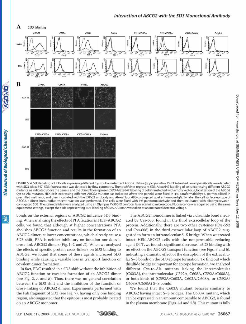
labeled with the 5D3 antibody, we found that only the C603Avariant had a clearly detectable 5D3 labeling and the C592A/C608A mutant showed some weak 5D3 binding capacity (Fig.5A, upper panel). Similar to that seen in the case of the wild-type ABCG2, PFA fixation (Fig. 5A, lower panel) or Ko143treatment (not shown) of the cells expressing the C603Amutant and the double mutant C592A/C608A resulted in anincreased 5D3 binding. However, the othermutant variants didnot show any labeling, even in PFA-fixed or Ko143-inhibitedsamples.To rule out the possibility that lowprotein expression level or
protein dislocalization was responsible for the absence of label-ing ofmost of themutants, and to test whether they could reachthe plasma membrane (necessary for recognition by 5D3 anti-body), we analyzed ABCG2 immunofluorescence by confocalmicroscopy.Fig. 5B shows that all mutants could be detected by the
BXP-21 antibody, recognizing an intracellular epitope ofABCG2, and all of them were present in the plasma membrane(except for the hardly expressedC603A/C608A doublemutant,which was found, for the most part, intracellularly). However,5D3 labeling analyzed by confocal microscopy gave the sameresult as the flow cytometry measurements, that is, only thecells expressing the wild-type ABCG2, C603A, and the C592A/C608A variants (the latter one seen only at increased detectorvoltage) could bind the 5D3 antibody.It has been shown that the 5D3 antibody inhibits the trans-
port function ofABCG2 (as judged in a sensitiveHoechst 33342assay), although this inhibition is only partial and requires highconcentrations of the antibody (15). To analyze a potential cor-relation between 5D3 binding and transport inhibition, weadded high concentrations of 5D3 to the HEK cells and thenanalyzed ABCG2-specific Hoechst 33342 transport activity.We found that, in contrast to a 30–40% inhibition found in thecase of the wild-type ABCG2, 5D3 did not influence theHoechst 33342 transport activity of the C592A/C608A andC592A/C603A/C608A mutants (data not shown). All of thesedata strongly suggest that the ABCG2 mutant proteins lackingthe cysteines required for intramolecular S–S bridge forma-
TABLE 1Effects of protein cross-linkers on 5D3 binding, transport activity, and covalent dimer formation of ABCG2
Cross-linker Side chainscross-linked
Spacer armlength
Increased5D3 binding
Inhibition oftransport function
Cross-linked ABCG2onWestern blot
ÅBM�PEO�3 SH2–H2 14.7 Yes Yes YesBMPH CH3–SH2 8.1 Yes Yes NoEDC COOH–NH2 0 Yes No NoSulfo-EGS NH2–NH2 16.1 No No YesSulfo-MBS NH2–SH2 9.9 Yes No YesPMPI SH2–OH 8.7 Yes No Yes
Interaction of ABCG2 with the 5D3 Monoclonal Antibody
26064 JOURNAL OF BIOLOGICAL CHEMISTRY VOLUME 283 • NUMBER 38 • SEPTEMBER 19, 2008
at SE
MM
ELW
EIS
UN
IV O
F M
ED
ICI on N
ovember 12, 2008
ww
w.jbc.org
Dow
nloaded from
tion are expressed in comparable amounts, reach the cell sur-face, and work as active transporters in a manner similar to thewild-type ABCG2, but these variants (except for the C592A/C608A mutant showing weak 5D3 binding) are unable to bindthe 5D3 antibody.Effect ofDTTon 5D3Labeling of theCys-to-AlaMutants—To
test whether decreased 5D3 binding inABCG2-expressing cellstreated with DTT was due to the reduction of the extracellular
cysteines, we also examined the effect of DTT on 5D3 labelingof the mutants C603A and C592A/C608A in native or PFA-fixed cells. Fig. 6,A andB, shows thatDTT is still effective in thereduction of 5D3 binding in the case of the C603A mutant buthas practically no effect on 5D3 labeling of the C592A/C608Amutant. When we analyzed 5D3 fluorescence in ABCG2-ex-pressing HEK cells treated with increasing concentrations ofDTT, we found that DTT treatment caused a gradual decrease
FIGURE 2. A, effect of protein cross-linkers on 5D3 labeling and function of ABCG2. HEK-ABCG2-R482G cells were incubated with different protein cross-linkers,washed, and then labeled with 5D3-Alexa647 (left panel) or incubated with 2 �M rhodamine123 (right panel) in the presence or absence of the inhibitor Ko143.Fluorescence was determined by flow cytometry. Left panel, dotted lines represent labeling of mock-transfected native cells, and dashed lines stand formock-transfected cells treated with cross-linker (or FTC). The solid lines represent 5D3 labeling of native, and heavy solid lines show cross-linker (or FTC)-treatedABCG2-expressing cells. Right panel, rhodamine123 uptake of cross-linker (or FTC) nontreated HEK-ABCG2 cells (solid line) or cross-linker (or FTC)-treatedHEK-ABCG2 cells (heavy solid line). Only living cells are shown in the right panel. B, Western blot analysis of the effect of protein cross-linkers on ABCG2.HEK-ABCG2 cells were incubated with different protein cross-linkers, washed, and then suspended in Laemmli buffer. Proteins were separated by 7.5%SDS-PAGE. After electroblotting, ABCG2 was detected by using the BXP-21 antibody. Each lane represents 15 �g of protein (�/0, in the case of PFA, its inhibitoryeffect depends on the concentration used; see Fig. 1C). sEGS, sulfo-EGS; sMBS, sulfo-MBS.
Interaction of ABCG2 with the 5D3 Monoclonal Antibody
SEPTEMBER 19, 2008 • VOLUME 283 • NUMBER 38 JOURNAL OF BIOLOGICAL CHEMISTRY 26065
at SE
MM
ELW
EIS
UN
IV O
F M
ED
ICI on N
ovember 12, 2008
ww
w.jbc.org
Dow
nloaded from

in 5D3 binding that reached its minimum (almost the fluores-cence of the background) at 10–50 mM DTT in ABCG2 andC603A-expressing cells, whereas DTThad no effect on labelingof the C592A/C608A double mutant (Fig. 6C). These experi-ments suggest that the intramolecular and not the intermolec-ular S–S bonds are important in 5D3 epitope formation.Experiments with 5D3-Fab—To analyze whether the 5D3
shift is caused by the stabilization of the epitopes in a dimerform of ABCG2, and thus an intact, bivalent 5D3 is required for
labeling, we incubated HEK-ABCG2 cells with the Alexa647-conjugated Fab fragment of 5D3.We found a lower affinity butspecific binding of 5D3 Fab to ABCG2 that was greatlyincreased by PFA fixation or by Ko143. DTT treatment alsoreduced Fab binding (Fig. 7). These experiments also suggestthat 5D3 binding toABCG2depends on the conformation of anepitope found on a monomeric ABCG2.
DISCUSSION
ABCG2 is a marker protein of the side population of stemcells and is also important in tumor cells where it can mediatethe emergence of a multidrug-resistant phenotype. On onehand, a sensitive method for the detection of low amounts ofABCG2 may allow the enrichment and selection of ABCG2-expressing cells, such as stem cells. On the other hand, moni-toring the presence of ABCG2 in tumor samples from patientscan help to find the most effective chemotherapy treatmentusing non-ABCG2 substrate drugs. The conformation-sensi-tive antibody 5D3 is a good candidate for the detection ofABCG2 for the above mentioned purposes. However, to estab-lish a reliable method it is essential to find the optimum condi-tions to be able to detect even low amounts of endogenousABCG2. In our previous paper we showed that 5D3 bindingdepends on the actual conformation of ABCG2 (15). We coulddefine two ABCG2 conformations based on the 5D3 bindingcapacity. The high affinity form was observed in ABCG2 inhib-ited by the specific inhibitor Ko143 in ATP-depleted cells orafter fixation of the cells with PFA, whereas stabilization ofABCG2 in a vanadate-trapped form resulted in decreased 5D3binding, representing a low affinity form.In this study we have further analyzed how alterations in
ABCG2 structure, covalent cross-linking, or changes in the S–S
FIGURE 3. Effect of DTT treatment on 5D3 binding. HEK293 cells transfectedwith empty pCIN4 (A) or pCIN4-ABCG2(R482G) (B) were incubated with orwithout 10 mM DTT, washed, and then labeled with 5D3 or mouse IgG2b andgoat anti-mouse phycoerythrin-conjugated secondary antibody. The upperpanels represent labeling of PFA nontreated cells (native), and the lower pan-els show labeling of cells that were fixed with 1% PFA after DTT treatment.Dotted lines, isotype control; dot-dash-dotted lines, isotype control of DTT-treated cells; solid lines, 5D3; dashed lines, 5D3 labeling of DTT-treated cells.
FIGURE 4. A, expression of various ABCG2 mutants carrying Cys-to-Ala changes. HEK cells were transfected with pCIN4 vectors encoding different Cys-to-AlaABCG2 mutants or R482G (indicated as ABCG2). Cells were lysed, dissolved in disaggregation buffer, and subjected to 7.5% SDS-PAGE. ABCG2 was detected bythe BXP-21 antibody. To demonstrate differences in the expression level of the mutants, various amounts of samples were loaded onto the gel as indicated inthe figure. B, mitoxantrone, pheophorbide A, rhodamine123, and Hoechst 33342 transport activity of Cys-to-Ala mutants. HEK cells expressing different ABCG2mutants or R482G (indicated as ABCG2) were incubated with 5 �M mitoxantrone, 1 �M pheophorbide A, 2 �M rhodamine123, or 1 �M Hoechst 33342 in theabsence or presence of 1 �M Ko143. Fluorescence of mitoxantrone, pheophorbide A, and rhodamine123 was detected in a FACSCalibur cytometer, and activityfactors were calculated from mean fluorescence values as described under “Experimental Procedures.” Fluorescence due to Hoechst 33342 accumulation wasdetermined in a spectrofluorimeter. The transport rate was determined as described under “Experimental Procedures.” Shown are the average transportactivities obtained from two independent experiments.
Interaction of ABCG2 with the 5D3 Monoclonal Antibody
26066 JOURNAL OF BIOLOGICAL CHEMISTRY VOLUME 283 • NUMBER 38 • SEPTEMBER 19, 2008
at SE
MM
ELW
EIS
UN
IV O
F M
ED
ICI on N
ovember 12, 2008
ww
w.jbc.org
Dow
nloaded from
bonds on the external regions of ABCG2 influence 5D3 bind-ing.When analyzing the effects of PFA fixation inHEK-ABCG2cells, we found that although at higher concentrations PFAabolishes ABCG2 function and results in the formation of anABCG2 dimer, at lower concentrations, which already cause a5D3 shift, PFA is neither inhibitory on function nor does itcross-link ABCG2 dimers (Fig. 1, C and D). When we analyzedthe effects of specific protein cross-linkers on 5D3 binding toABCG2, we found that some of these agents increased 5D3binding while causing a variable loss in transport function orcovalent dimer formation.In fact, EDC resulted in a 5D3 shift without the inhibition of
ABCG2 function or covalent formation of an ABCG2 dimer(see Fig. 2, A and B). Thus, there was no general correlationbetween the 5D3 shift and the inhibition of the function orcross-linking of ABCG2 dimers. Experiments performed withthe Fab fragment of 5D3 (see Fig. 7), having only one bindingregion, also suggested that the epitope is most probably locatedon an ABCG2 monomer.
The ABCG2 homodimer is linked via a disulfide bond medi-ated by Cys-603, found in the third extracellular loop of theprotein. Additionally, there are two other cysteines (Cys-592and Cys-608) in the third extracellular loop of ABCG2, sug-gested to form an intramolecular S–S bridge. When we treatedintact HEK-ABCG2 cells with the nonpermeable reducingagentDTT,we found a significant decrease in 5D3 bindingwithno effect on the ABCG2 transport function (see Figs. 3 and 6),indicating a dramatic effect of the disruption of the extracellu-lar S–S bonds on the 5D3 epitope formation. To find out whichdisulfide bridge is important for epitope formation,we analyzeddifferent Cys-to-Ala mutants lacking the intermolecular(C603A), the intramolecular (C592A, C608A, C592A/C608A),or both kinds of (C592A/C603A, C603A/C608A, or C592A/C603A/C608A) S–S bonds.We found that the C603A mutant behaves similarly to
ABCG2 having intact S–S bridges. The C603A mutant, whichcan be expressed in an amount comparable to ABCG2, is foundin the plasmamembrane (Figs. 4A and 5B). This mutant is fully
FIGURE 5. A, 5D3 labeling of HEK cells expressing different Cys-to-Ala mutants of ABCG2. Native (upper panel) or 1% PFA-treated (lower panel) cells were labeledwith 5D3-Alexa647. 5D3 fluorescence was detected by flow cytometry. Then solid lines represent 5D3-Alexa647 labeling of cells expressing different ABCG2mutants, as indicated above the panels, and the dotted lines represent 5D3-Alexa647 labeling of cells transfected with empty vector. B, localization of the ABCG2Cys-to-Ala mutants. HEK cells expressing different ABCG2 mutants (as indicated above the panels) were fixed in 4% paraformaldehyde, permeabilized inprechilled methanol, and then incubated with the BXP-21 antibody and Alexa Fluor 488-conjugated goat anti-mouse IgG. To label the cell surface epitope ofABCG2, a direct immunofluorescent reaction was performed. The cells were fixed with 1% paraformaldehyde and then incubated with allophycocyanin-conjugated 5D3. The stained slides were analyzed using an Olympus FV500-IX confocal laser scanning microscope. Fluorescence was acquired using the sameequipment settings, except the slide representing 5D3 labeling of C592A/C608A was taken at an increased detector voltage.
Interaction of ABCG2 with the 5D3 Monoclonal Antibody
SEPTEMBER 19, 2008 • VOLUME 283 • NUMBER 38 JOURNAL OF BIOLOGICAL CHEMISTRY 26067
at SE
MM
ELW
EIS
UN
IV O
F M
ED
ICI on N
ovember 12, 2008
ww
w.jbc.org
Dow
nloaded from

active and has the same substratespecificity as ABCG2 (Fig. 4B).Cys-603 has a clearly detectable5D3 binding that can be increasedby inhibition of ABCG2 functionby Ko143 or treatment with pro-tein cross-linkers. DTT treatmentalso decreased 5D3 binding of thismutant (Fig. 6), suggesting thatthis cysteine is not important forthe observed effect of S–S bridgereduction.The single mutants, lacking the
intramolecular S–S bond, i.e.C592A, C608A, as well as theC592A/C603A/C608A variant, hadclearly detectable expression levels,were present in the plasma mem-brane, and were functional foractive transport with somewhataltered substrate specificities (Figs.4 and 5). However, these mutantsdid not show any specific 5D3 bind-ing either in a PFA-fixed or in aKo143-inhibited form, and more-over, 5D3 did not inhibit their func-tion. These experiments suggestthat the intramolecular S–S bridgein the third extracellular loop ofABCG2 has a crucial, either director indirect (e.g. stabilizing theproper conformation), role in theformation of the 5D3 epitope as wellas in the substrate specificity of thetransporter.The role of the intramolecular
S–S bond in 5D3 epitope formationhas already been suggested (9, 10).Kage et al. (9) analyzed 5D3 labelingin intact cells expressing variousCys-to-Ser mutants by flow cytom-etry. They found that C592S andC608S had impaired 5D3 binding;however, these twomutants showedvery low expression levels in thisstudy (9). In our experiments wewere able to express C592A andC608A mutants in comparable lev-els to the wild-type ABCG2. More-over, we demonstrated that eventhough these mutants were func-tional and properly localized to theplasma membrane, they could notbe labeled with 5D3 even in thepresence of an inhibitor or PFA.Henriksen et al. (10) also sug-
gested the role of the intramolecularS–S bridge of ABCG2 in 5D3 bind-
FIGURE 6. A and B, effect of DTT on labeling of the Cys-to-Ala mutants. HEK293 cells expressing different ABCG2mutants were treated with 10 mM DTT, washed, and then labeled with 5D3-Alexa647 (A). B shows the labeling ofsamples fixed with 0.5% PFA prior to antibody labeling. Fluorescence was determined in a FACSCalibur cytometer.Dotted lines, mock-transfected HEK; dot-dash-dotted lines, DTT-treated, mock-transfected HEK cells; solid lines, HEKcells expressing different ABCG2 mutants; dashed lines, HEK cells expressing different ABCG2 mutants treated withDTT. C, HEK cells expressing different ABCG2 mutants were treated with increasing concentrations of DTT, fixed with0. 5% PFA, and labeled with 5D3-Alexa647. 5D3 fluorescence was determined by flow cytometry. Mean valuesobtained from at least in two independent experiments are shown.
Interaction of ABCG2 with the 5D3 Monoclonal Antibody
26068 JOURNAL OF BIOLOGICAL CHEMISTRY VOLUME 283 • NUMBER 38 • SEPTEMBER 19, 2008
at SE
MM
ELW
EIS
UN
IV O
F M
ED
ICI on N
ovember 12, 2008
ww
w.jbc.org
Dow
nloaded from

ing; they detected no 5D3 labeling by confocal microscopy forC592A, C608A, and the C592A/C608A double mutant. In har-mony with this study, we could not detect any labeling for thesingle Cys-to-Ala mutants. However, in our hands the C592A/C608A doublemutant showed a weak 5D3 binding both in flowcytometry and confocal microscopy, and the 5D3 shift uponPFA or Ko143 treatment could also be observed (Fig. 5). DTThad no effect on the 5D3 labeling of the C592A/C608A variant(Fig. 6), and the excess amount of 5D3 did not inhibit the func-tion of this mutant.The single Cys-592 or Cys-608mutants showed an increased
cytoplasmic accumulation (9, 10, 13), whereas the simultane-ous removal of cysteines 592 and 608promotedprotein stabilityand proper targeting (10). Thus such a double mutation mayallow the development of a favorable conformation within theABCG2 protein, allowing some 5D3 labeling.Based on these data we suggest that protein cross-linking
most probably stabilizes the epitope of 5D3 present within asingle ABCG2 protein. The effect of DTT treatment on 5D3labeling together with experiments on the extracellular Cys-to-Alamutants revealed that the third extracellular loop, and espe-cially the intramolecular S–S bond within this region, has acrucial role in 5D3 epitope formation and probably in the sub-strate interactions of ABCG2.To visualize the possible molecular arrangement of the third
extracellular loop (ECL3) of the ABCG2 protein, we performeda molecular dynamic simulation of the folding of this relativelylarge protein sequence. Currently, there are no atomic struc-tures available for eukaryotic ABC transporters, and molecularmodeling is usually guided by the recently solved structures ofbacterial transporters. Although a recent communicationoffers a homology model for ABCG2 (30), it does not includeECL3, as this loop shows no homology to any ABC proteinsequences with a known structure. Data for other ABC trans-porters, however, suggest that the adjacent transmembraneregions of these proteins are found near each other, within adistance of 9–12 Å. Therefore in our simulation algorithm, wefixed theN- andC-terminal regions of the ECL3 at this distanceat the membrane surface. We have also included the informa-tion that the cysteines forming intramolecular S–S bonds (Cys-592 and Cys-608), as well as a cysteine involved in the intermo-
lecular S–S bridge formation, are most probably located on thesurface of this loop. It is interesting to note, that many of theABCG-type proteins have this large, conserved, extracellularregion, with similar arrangements of cysteines and potentialglycosylation sites (11).The conformation obtained, as seen in Fig. 8, corresponds to
these requirements and suggests the presence of a stabilizedantiparallel loop with �-sheets in the region of the three cys-teines, surrounding the glycosylation site.When eitherCys-592or Cys-608 was replaced by alanines in the in silico model, thestructure of the antiparallel loop collapsed and the folding lostits conserved characteristics (data not shown). The combina-tion of the experimental and the simulation results suggest awell defined structure in this area of ECL3, which may beimportant in mediating ABCG2 interaction with the plasmamembrane or other proteins. Moreover, the conformation ofthis loop seems tomodulate the substrate and antibody bindingto this membrane transporter.
Acknowledgments—We greatly appreciate the technical help of EvaKrizsan, Zsuzsanna Andrasi, and Judit Kis. We appreciate the kindgifts of Ko143 fromDrs. J. D. Allen andG. J. Koomen and anti-ABCG2BXP-21 antibody obtained fromDrs. George Scheffer andRik Scheper.
REFERENCES1. Sarkadi, B., Homolya, L., Szakacs, G., and Varadi, A. (2006) Physiol. Rev.
86, 1179–12362. Chandra, P., and Brouwer, K. L. (2004) Pharm. Res. (N. Y.) 21, 719–7353. Maliepaard, M., Scheffer, G. L., Faneyte, I. F., van Gastelen, M. A., Pijnen-
borg, A. C., Schinkel, A. H., van De Vijver, M. J., Scheper, R. J., and Schel-lens, J. H. (2001) Cancer Res. 61, 3458–3464
4. Zhou, S., Schuetz, J. D., Bunting, K. D., Colapietro, A. M., Sampath, J.,Morris, J. J., Lagutina, I., Grosveld, G. C., Osawa, M., Nakauchi, H., andSorrentino, B. P. (2001) Nat. Med. 7, 1028–1034
5. Krishnamurthy, P., Ross, D. D., Nakanishi, T., Bailey-Dell, K., Zhou, S.,Mercer, K. E., Sarkadi, B., Sorrentino, B. P., and Schuetz, J. D. (2004) J. Biol.Chem. 279, 24218–24225
6. Krishnamurthy, P., and Schuetz, J. D. (2005) Biometals 18, 349–358
FIGURE 7. Direct labeling of HEK cells by Alexa647-conjugated 5D3-Fab.HEK293 cells transfected with the pCIN4 vector or pCIN4 containing the cDNAof ABCG2(R482G) were incubated with 2 �g/ml 5D3-Fab-Alexa647. Fluores-cence was determined by flow cytometry. The dotted lines represent labelingof 10 mM DTT-treated cells; solid lines stand for native cells incubated onlywith 5D3-Fab, heavy solid lines show labeling of 0.5% PFA-fixed cells, anddashed lines indicate fluorescence in the presence of the inhibitor Ko143.
FIGURE 8. Model of the third extracellular loop of ABCG2 (amino acids563– 618) obtained by ab initio folding employing discrete moleculardynamics. The structure is colored blue to red from the N to the C terminus.Cysteines involved in intramolecular and intermolecular interactions are rep-resented with sticks and are colored magenta. The glycosylation site (Asn-596) is represented by a blue stick.
Interaction of ABCG2 with the 5D3 Monoclonal Antibody
SEPTEMBER 19, 2008 • VOLUME 283 • NUMBER 38 JOURNAL OF BIOLOGICAL CHEMISTRY 26069
at SE
MM
ELW
EIS
UN
IV O
F M
ED
ICI on N
ovember 12, 2008
ww
w.jbc.org
Dow
nloaded from
7. Ozvegy, C., Litman, T., Szakacs, G., Nagy, Z., Bates, S., Varadi, A., andSarkadi, B. (2001) Biochem. Biophys. Res. Commun. 285, 111–117
8. Kage, K., Tsukahara, S., Sugiyama, T., Asada, S., Ishikawa, E., Tsuruo, T.,and Sugimoto, Y. (2002) Int. J. Cancer 97, 626–630
9. Kage, K., Fujita, T., and Sugimoto, Y. (2005) Cancer Sci. 96, 866–87210. Henriksen, U., Fog, J. U., Litman, T., and Gether, U. (2005) J. Biol. Chem.
280, 36926–3693411. Wakabayashi, K., Nakagawa, H., Adachi, T., Kii, I., Kobatake, E., Kudo, A.,
and Ishikawa, T. (2006) J. Exp. Ther. Oncol. 5, 205–22212. Takada, T., Suzuki, H., and Sugiyama, Y. (2005) Pharm. Res. (N. Y.) 22,
458–46413. Wakabayashi, K., Nakagawa, H., Tamura, A., Koshiba, S., Hoshijima, K.,
Komada, M., and Ishikawa, T. (2007) J. Biol. Chem. 282, 27841–2784614. Robey, R. W., Polgar, O., Deeken, J., To, K. W., and Bates, S. E. (2007)
Cancer Metastasis Rev. 26, 39–5715. Ozvegy-Laczka, C., Varady, G., Koblos, G., Ujhelly, O., Cervenak, J.,
Schuetz, J. D., Sorrentino, B. P., Koomen, G. J., Varadi, A., Nemet, K., andSarkadi, B. (2005) J. Biol. Chem. 280, 4219–4227
16. Abbott, B. L., Colapietro, A. M., Barnes, Y., Marini, F., Andreeff, M., andSorrentino, B. P. (2002) Blood 100, 4594–4601
17. Allen, J. D., van Loevezijn, A., Lakhai, J. M., van der Valk, M., van Tell-ingen, O., Reid, G., Schellens, J. H., Koomen, G. J., and Schinkel, A. H.(2002)Mol. Cancer Ther. 1, 417–425
18. Robey, R. W., Honjo, Y., Morisaki, K., Nadjem, T. A., Runge, S., Risbood,M., Poruchynsky,M. S., and Bates, S. E. (2003)Br. J. Cancer 89, 1971–1978
19. Elkind, N. B., Szentpetery, Z., Apati, A., Ozvegy-Laczka, C., Varady, G.,Ujhelly, O., Szabo, K., Homolya, L., Varadi, A., Buday, L., Keri, G., Nemet,K., and Sarkadi, B. (2005) Cancer Res. 65, 1770–1777
20. Szollosi, J., Horejsi, V., Bene, L., Angelisova, P., andDamjanovich, S. (1996)J. Immunol. 157, 2939–2946
21. Spack, E. G., Jr., Packard, B., Wier, M. L., and Edidin, M. (1986) Anal.Biochem. 158, 233–237
22. Ozvegy, C., Varadi, A., and Sarkadi, B. (2002) J. Biol. Chem. 277,47980–47990
23. Ozvegy-Laczka, C., Hegedus, T., Varady, G., Ujhelly, O., Schuetz, J. D.,Varadi, A., Keri, G., Orfi, L., Nemet, K., and Sarkadi, B. (2004)Mol. Phar-macol. 65, 1485–1495
24. Hegedus, T., Serohijos, A. W., Dokholyan, N. V., He, L., and Riordan, J. R.(2008) J. Mol. Biol. 378, 1052–1063
25. Dokholyan, N. V., Buldyrev, S. V., Stanley, H. E., and Shakhnovich, E. I.(1998) Folding Des. 3, 577–587
26. Ding, F., and Dokholyan, N. V. (2005) Trends Biotechnol. 23, 450–45527. Ding, F., and Dokholyan, N. V. (2006) PLoS Comput. Biol. 2, e8528. Sugita, Y., and Okamoto, Y. (1999) Chem. Phys. Lett. 314, 141–15129. Ding, F., Tsao, D., Nie, H., and Dokholyan, N. V. (2008) Structure (Lond.)
16, 1010–101830. Hazai, E., and Bikadi, Z. (2007) J. Struct. Biol. 162, 63–7431. Mitomo, H., Kato, R., Ito, A., Kasamatsu, S., Ikegami, Y., Kii, I., Kudo, A.,
Kobatake, E., Sumino, Y., and Ishikawa, T. (2003) Biochem. J. 373,767–774
Interaction of ABCG2 with the 5D3 Monoclonal Antibody
26070 JOURNAL OF BIOLOGICAL CHEMISTRY VOLUME 283 • NUMBER 38 • SEPTEMBER 19, 2008
at SE
MM
ELW
EIS
UN
IV O
F M
ED
ICI on N
ovember 12, 2008
ww
w.jbc.org
Dow
nloaded from