Molecular Regulation of Cytoskeletal Rearrangements During T Cell Signalling
320
-
Upload
independent -
Category
Documents
-
view
1 -
download
0
Transcript of Molecular Regulation of Cytoskeletal Rearrangements During T Cell Signalling


Results and Problems in Cell Differentiation
43
Series EditorsD. Richter, H. Tiedge
Eckart D. Gundelfinger, Constanze I. Seidenbecher,Burkhart Schraven (Eds.)
Cell Communicationin Nervousand Immune SystemWith 28 Figures, 8 in Color, and 3 Tables
123

Professor Dr. Eckart D. Gundelfinger
Dr. Constanze I. Seidenbecher
Leibniz Institut für NeurobiologieAbteilung Neurochemie und MolekularbiologieBrenneckestr. 639118 [email protected]
Professor Dr. Burkhart Schraven
Institut für ImmunologieOtto-von-Guericke-Universität MagdeburgLeipzigerstrasse 443120 [email protected]
ISSN 0080-1844ISBN-10 3-540-36828-0 Springer Berlin Heidelberg New YorkISBN-13 978-3-540-36828-1 Springer Berlin Heidelberg New York
Library of Congress Control Number: 2006932575
This work is subject to copyright. All rights are reserved, whether the whole or part of the materialis concerned, specifically the rights of translation, reprinting, reuse of illustrations, recitation, broad-casting, reproduction on microfilm or in any other way, and storage in data banks. Duplication ofthis publication or parts thereof is permitted only under the provisions of the German Copyright Lawof September 9, 1965, in its current version, and permission for use must always be obtained fromSpringer. Violations are liable for prosecution under the German Copyright Law.
Springer is a part of Springer Science+Business Media
springer.com
c© Springer-Verlag Berlin Heidelberg 2006Printed in Germany
The use of registered names, trademarks, etc. in this publication does not imply, even in the absenceof a specific statement, that such names are exempt from the relevant protective laws and regulationsand therefore free for general use.
Cover design: Design & Production GmbH, HeidelbergTypesetting and Production: LE-TEX Jelonek, Schmidt & Vöckler GbR, Leipzig
Printed on acid-free paper 31/3100/YL – 5 4 3 2 1 0
Preface
Signal exchange between cells is a key feature of life from humble monadsto human beings. Appropriate communication is of particular importancebetween cells of multi-cellular organisms. Various basic mechanisms of cell–cell communication have evolved during phylogenesis, which were subjectto organ, tissue and cell type-specific adaptation. These mechanisms rangefrom long-distance communication via hormones to more and more local pro-cesses, e.g. via cytokines, chemokines or neuromodulators/neurotransmitters,and eventually direct physical interactions of molecules anchored at cell sur-faces. Accordingly, highly specialized transient or stable cell–cell contact siteshave evolved that mediate signaling between cells. With few exceptions (e.g.lipophilic hormones, gases) intercellular communication depends on specificsignal detection devices at the cell surface coupled to a signal transductionapparatus that mediates the signal transfer across the cell membrane and acti-vates intracellular effector systems, which generate intracellularly decipherablesignals.
Prime examples for tissues of intensely communicating cells are the nervousand the immune systems. Although at the first glance these systems appear verydifferent, both have developed sophisticated mechanisms for the formation ofmemory, though of quite different quality and significance for the organism.Memory formation in the immune system serves the recognition and toleranceof the organism’s own cells and tissues as well as the effective recognition of anddefense from invading pathogens. It is based on a complex network of cellularcommunication and signaling processes between cells of this “dispersed” organand with target cells. Brain mechanisms of learning and memory, on the otherhand, are indispensable for survival of an organism in its natural and socialenvironment. They are based on the function and plasticity of the probablymost complex cell junction: the chemical synapse. However, other cell–cellconnections, such as gap junctions or specialized neuron-glia interaction sites,play an essential part in brain performance and plasticity.
This collection of reviews, contributed by internationally recognized im-munologists and molecular and cellular neurobiologists, juxtaposes cellularcommunication devices and signaling mechanisms in the immune and thenervous system and discusses mechanisms of interaction between the two sys-tems, the significance of which has only been fully appreciated in recent years.

VI Preface
Thus messengers produced by one of the two systems, such as cytokines orneuropeptides, can modulate cellular communication in the other system aswell. Moreover, the central nervous system (CNS) has long been consideredan immune-privileged organ lacking the classical immune response. Basedon recent studies this view had to be revised and refined, and the particularrole of the immune system in neuropathological as well as in neuroprotectiveand neurorepair processes has been recognized. This implies that the poten-tially harmful effects of the immune system in the CNS have to be tightlycontrolled by precise communication between cells of neural and immunesystems.
The first four review articles deal with chemical synapses of the CNS. Thishighly sophisticated asymmetric cell–cell contact is designed for particularcommunication between neurons via chemical substances, the neurotrans-mitters. Neurotransmitters are stored in little membranous containers, i.e.synaptic vesicles, and released from the presynaptic cell in response to incom-ing electrical signals in a regulated manner. Different postsynaptic deviseshave evolved to detect excitatory (the first chapter) or inhibitory (the secondchapter) transmitters and transduce the signals into the postsynaptic cell. Alsothe site of regulated neurotransmitter release from the presynaptic neuron—the active zone—is a complex molecular machine that organizes the synapticvesicle cycle (the third chapter). The gap between the pre- and the postsynap-tic membranes, the synaptic cleft, is a specialized extracellular compartmentarranged by various cell adhesion molecules and components of the extracel-lular matrix that is thought to contribute importantly to synaptic assembly andplasticity (the fourth chapter).
Though known for more than 50 years, the electrical synapses have hada shadowy existence for a long time and only during recent years have theiridentity and their physiological relevance been studied in more detail. Thefifth chapter discusses the role of gap junctions that form electrical synapsesin the CNS. The next chapter discusses another intriguing cell–cell contact sitethat determines the capacity and efficacy of the vertebrate nervous system isthe neuron-glia interaction at the so-called nodes of Ranvier, which facilitatesrapid propagation of electrical signals along myelinated axons.
Also within the immune system the term “synapse” has been meanwhilewell established. Here, the so-called immunological synapse describes themolecular and biophysical events that occur when immunocompetent cellsinteract with each other at the beginning of the adaptive immune response.T-cells, the major components of the adaptive immune system are by them-selves incapable of detecting complete bacteria or viruses. Rather, the majorstructure on the T-cell surface that initiates T-cell activation, the T-cell receptor(TCR) only recognizes small, 9 to 12 amino acid-long bacterial or viral (anti-genic) peptides. These have to be generated by particular immunocompetentcells, which have collectively been termed antigen presenting cells (APCs).Although it is well known that B-cells, dendritic cells and macrophages rep-

Preface VII
resent the major types of APCs that activate T-cells, it is still unclear whether,for example, endothelial cells, which are spread throughout the whole body,are also capable of presenting antigens to T-cells, at least in particular organssuch as the liver, or under particular conditions such as inflammation. Thesequestions are addressed in the seventh chapter, which also discusses the im-munological consequences of the interaction between T-cells and endothelialcells.
Importantly, the mere generation of antigenic peptides is not sufficient toactivate T-cells. This is due to the fact that the TCR only signals when anti-genic peptides are presented to the T-cell by APCs in conjunction with self-molecules, the so-called major histocompatibility complex (MHC) molecules.The detection of antigen/MHC by the TCR occurs at the beginning of theimmune response at the interface between the T-cell and the APC and thisfirst physical contact between the two cells induces the formation of the im-munological synapse. Consequently, the immunological synapse is a highlydynamic structure that changes its morphology and molecular compositionduring the initial phase of the immune response. During the past decade nu-merous groups have begun to dissect the molecular events that either regulatethe formation of the immunological synapse or occur after immunologicalsynapse formation by applying sophisticated microscopic and biochemicaltechniques. As a result, several models of the molecular composition and thefunction of the immunological synapse have evolved. The eighth and ninthchapters focus on the biophysics and the morphological changes of the im-munological synapse under different conditions of stimulation and furtherdiscuss the role of the immunological synapse during T-cell activation. Whilethese two articles primarily deal with the dynamics of APC/T-cell interactionsand the morphological changes of the immunological synapse on the micro-scopic level, the following two chapters focus on the signaling events thatregulate particular aspects of immunological synapse formation and T-cellactivation. The first of these discusses alterations of the cytoskeleton and thesecond the regulation of intimate membrane contacts via adhesion moleculesand integrins.
The final two chapters shed some light on the communication betweenthe immune and the nervous system and the control of immune responses inthe nervous system. To gain access to the CNS, immune cells have to crossthe blood–brain barrier provided by composed of endothelial cells. How thisprocess is mediated and controlled under physiological and pathological con-ditions is discussed in the penultimate chapter. Endocannabinoids, the endoge-nous ligands for the “Marihuana” receptors, seem to be intricately involved inthe neural control of the immune system. The current view of how the CNSendocannabinoid system contributes to the immune surveillance is discussedin the final chapter.
This book is dedicated to our colleague Werner Hoch, who intended tocontribute an article on the neuromuscular junction, the supposedly best-

VIII Preface
studied vertebrate synapse and long-time focus of Werner’s scientific work.Werner passed away last summer—unexpectedly and much too early for all ofus.
Magdeburg, 2006 Eckart D. GundelfingerConstanze I. Seidenbecher
Burkhart Schraven

Contents
Molecular Organization and Assemblyof the Postsynaptic Density of Excitatory Brain SynapsesEunjoon Kim, Jaewon Ko . . . . . . . . . . . . . . . . . . . . . . . . . 11 Introduction . . . . . . . . . . . . . . . . . . . . . . . . . . 12 Components of the PSD . . . . . . . . . . . . . . . . . . . . 13 Assembly of the PSD . . . . . . . . . . . . . . . . . . . . . . 34 Synaptic Adhesion and PSD Proteins . . . . . . . . . . . . . 45 Membrane Proteins and PSD Proteins . . . . . . . . . . . . . 56 Spine Formation and PSD Proteins . . . . . . . . . . . . . . 57 Postsynaptic Signaling and PSD Proteins . . . . . . . . . . . 78 Regulation of Synaptic Transmission and Plasticity
by PSD Proteins . . . . . . . . . . . . . . . . . . . . . . . . . 99 Dynamic Regulation of the Assembly of the PSD . . . . . . . 1110 Transport of PSD Proteins by Motor Proteins . . . . . . . . . 1211 Conclusions . . . . . . . . . . . . . . . . . . . . . . . . . . . 13References . . . . . . . . . . . . . . . . . . . . . . . . . . . . . . . . . . 13
Molecular Organization and Assemblyof the Central Inhibitory PostsynapseI. Lorena Arancibia-Carcamo, Stephen J. Moss . . . . . . . . . . . 251 Introduction . . . . . . . . . . . . . . . . . . . . . . . . . . 251.1 GABAA Receptor Structure . . . . . . . . . . . . . . . . . . . 261.2 GABAA Receptor Localization within the Brain . . . . . . . . 272 Formation and Maintenance of Inhibitory Synapses . . . . . 303 Trafficking and Targeting of GABAA Receptors . . . . . . . . 324 Receptor Stability . . . . . . . . . . . . . . . . . . . . . . . . 375 Concluding Remarks . . . . . . . . . . . . . . . . . . . . . . 40References . . . . . . . . . . . . . . . . . . . . . . . . . . . . . . . . . . 41
Molecular Organization and Assemblyof the Presynaptic Active Zone of Neurotransmitter ReleaseAnna Fejtova, Eckart D. Gundelfinger . . . . . . . . . . . . . . . 491 Introduction . . . . . . . . . . . . . . . . . . . . . . . . . . 50

X Contents
2 General Aspects of Structuraland Functional Organization of the Active Zone . . . . . . . 50
2.1 Neurotransmitter Release . . . . . . . . . . . . . . . . . . . 502.2 Ultrastructural Organization of the Active Zone . . . . . . . 513 Molecular Organization of the Active Zone . . . . . . . . . . 523.1 The UNC13/Munc13 Protein Family –
Key Actors in SV Priming . . . . . . . . . . . . . . . . . . . 553.2 RIMs – Multidomain Rab3 Effectors
with a Major Organizing Role . . . . . . . . . . . . . . . . . 563.3 CAST/ERC Proteins –
Major Structural Proteins of the CAZ? . . . . . . . . . . . . . 583.4 Piccolo and Bassoon –
Two Related Giants Scaffolding the CAZ . . . . . . . . . . . . 584 Synaptic Ribbons –
Specializations of the CAZ at High-Throughput Synapses . . 605 Developmental Assembly of the Active Zone –
The Active Zone Precursor Vesicle Hypothesis . . . . . . . . 62References . . . . . . . . . . . . . . . . . . . . . . . . . . . . . . . . . . 64
Extracellular Matrix and Synaptic FunctionsAlexander Dityatev, Renato Frischknecht,
Constanze I. Seidenbecher . . . . . . . . . . . . . . . . . . . . . . . 691 Introduction . . . . . . . . . . . . . . . . . . . . . . . . . . 702 Synaptogenic Activity of Agrin . . . . . . . . . . . . . . . . 713 Synaptic Functions of Laminins . . . . . . . . . . . . . . . . 724 Synaptic Functions of Integrins . . . . . . . . . . . . . . . . 755 Thrombospondins and Synaptogenesis . . . . . . . . . . . . 766 Synaptic Functions of Reelin . . . . . . . . . . . . . . . . . . 777 Clustering of Glutamate Receptors by Neuronal Pentraxins . 798 HB-GAM and N-syndecan in Synaptic Plasticity . . . . . . . 809 Tenascin-R, GABAergic Transmission and Metaplasticity . . 8110 Tenascin-C and Synaptic Plasticity . . . . . . . . . . . . . . 8311 NG2 and Glia-neuron Synapses . . . . . . . . . . . . . . . . 8412 Lecticans Differentially Contribute
to Synapse Formation and Function . . . . . . . . . . . . . 8513 Receptor Protein Tyrosin Phosphatase and Phosphacan . . . 8714 Matrix Metalloproteases and the Modulation
of Perisynaptic Matrix . . . . . . . . . . . . . . . . . . . . . 88References . . . . . . . . . . . . . . . . . . . . . . . . . . . . . . . . . . 90
Electrical Synapses – Gap Junctions in the BrainCarola Meier, Rolf Dermietzel . . . . . . . . . . . . . . . . . . . . 991 Introduction . . . . . . . . . . . . . . . . . . . . . . . . . . 992 Gap Junctions in Development . . . . . . . . . . . . . . . . . 101

Contents XI
3 Gap Junctions in Glial Cells . . . . . . . . . . . . . . . . . . 1024 Challenges in Expression Analysis of Gap Junction Proteins . 1045 Some History on Electrical Synapses . . . . . . . . . . . . . 1056 Gap Junctions in Neurons – Electrical Synapses . . . . . . . 1066.1 Connexin Gap Junctions . . . . . . . . . . . . . . . . . . . . 1066.2 Pannexin Gap Junctions . . . . . . . . . . . . . . . . . . . . 1167 Conclusions . . . . . . . . . . . . . . . . . . . . . . . . . . . 118References . . . . . . . . . . . . . . . . . . . . . . . . . . . . . . . . . . 119
Neuron-Glia Interactions at the Node of RanvierMatthew N. Rasband . . . . . . . . . . . . . . . . . . . . . . . . . . 1291 Introduction . . . . . . . . . . . . . . . . . . . . . . . . . . 1292 Domain Structure of the Myelinated Axon . . . . . . . . . . 1312.1 Node of Ranvier . . . . . . . . . . . . . . . . . . . . . . . . . 1312.2 Paranode . . . . . . . . . . . . . . . . . . . . . . . . . . . . 1342.3 Juxtaparanode . . . . . . . . . . . . . . . . . . . . . . . . . 1353 Examples of Neuron-Glia Interactions . . . . . . . . . . . . . 1363.1 Neuron-Glia Interactions Regulate Nav Channel Clustering
at Nodes of Ranvier . . . . . . . . . . . . . . . . . . . . . . . 1373.2 Neuron-Glia Interactions at the Paranode . . . . . . . . . . . 1403.3 Neuron-Glia Interactions
Regulate Juxtaparanodal K+ Channel Localization . . . . . . 1424 Conclusions . . . . . . . . . . . . . . . . . . . . . . . . . . . 143References . . . . . . . . . . . . . . . . . . . . . . . . . . . . . . . . . . 144
Cognate Interaction Between Endothelial Cells and T CellsPercy A. Knolle . . . . . . . . . . . . . . . . . . . . . . . . . . . . . . 1511 Organ-Specific Phenotypes and Functions
of Endothelial Cells . . . . . . . . . . . . . . . . . . . . . . . 1512 Establishment of Interaction Between Endothelial Cells
and T Cells . . . . . . . . . . . . . . . . . . . . . . . . . . . 1543 Endothelial Cells as Antigen-Presenting Cells . . . . . . . . . 1554 MHC Expression and Antigen Presentation
by Endothelial Cells . . . . . . . . . . . . . . . . . . . . . . . 1555 Co-stimulation by Endothelial Cells . . . . . . . . . . . . . . 1586 Consequences of Cognate Interaction
Between Antigen-Presenting Endothelial Cells and T Cells . . 1597 Modulation of CD8 T Cell Function by Endothelial Cells . . . 1618 Modulation of CD4 T Cell Function . . . . . . . . . . . . . . 1639 Transmigration of T Cells in Response
to Antigen Presentation by Endothelial Cells . . . . . . . . . 16410 Mediation of Antigen-Specific T Cell Recruitment
in the Absence of Antigen Presentation by Endothelial Cells . 167

XII Contents
11 Final Remarks . . . . . . . . . . . . . . . . . . . . . . . . . . 168References . . . . . . . . . . . . . . . . . . . . . . . . . . . . . . . . . . 168
Impact of the Immunological Synapse on T Cell SignalingMichael L. Dustin . . . . . . . . . . . . . . . . . . . . . . . . . . . . 1751 Introduction . . . . . . . . . . . . . . . . . . . . . . . . . . 1762 New Model for Sustained Signaling Through
the Immunological Synapse . . . . . . . . . . . . . . . . . . 1773 In Vivo Functions of Immunological Synapse
and Hemisynapse . . . . . . . . . . . . . . . . . . . . . . . . 1834 In Vivo Analysis of CD4+ T Cell Priming
and Tolerance Induction . . . . . . . . . . . . . . . . . . . . 1845 In Vivo Analysis of CD8+ T Cell Priming
and Tolerance Induction . . . . . . . . . . . . . . . . . . . . 1876 Importance of Stable Interactions In Vivo . . . . . . . . . . . 1897 Effector Sites . . . . . . . . . . . . . . . . . . . . . . . . . . 1908 Summary . . . . . . . . . . . . . . . . . . . . . . . . . . . . 193References . . . . . . . . . . . . . . . . . . . . . . . . . . . . . . . . . . 193
The Biophysics of T Lymphocyte Activation In Vitro and In VivoPeter Reichardt, Matthias Gunzer . . . . . . . . . . . . . . . . . . 1991 Introduction . . . . . . . . . . . . . . . . . . . . . . . . . . 1992 The Role of T Cells Within the Immune System:
A T Cell Biography . . . . . . . . . . . . . . . . . . . . . . . 2012.1 T Cell Selection in the Thymus . . . . . . . . . . . . . . . . . 2012.2 T Cell Circulation and Homeostasis . . . . . . . . . . . . . . 2022.3 T Cell Activation in the Lymph Node:
A Marketplace Analogy . . . . . . . . . . . . . . . . . . . . . 2022.4 Homing of Activated T Cells . . . . . . . . . . . . . . . . . . 2052.5 T Cell Regulation . . . . . . . . . . . . . . . . . . . . . . . . 2063 The Biophysics of T Cell Activation . . . . . . . . . . . . . . 2063.1 Classical In Vitro Studies of T Cell–APC Interaction . . . . . 2073.2 The Single Encounter Model of T Cell Activation . . . . . . . 2083.3 Challenges Introduced by Live Cell Imaging Experiments . . 2083.4 A Serial Encounter Model of T Cell Activation . . . . . . . . 2103.5 The Road to Intravital Imaging:
Phase Models of T Cell Activation . . . . . . . . . . . . . . . 2103.6 The Role of the APC . . . . . . . . . . . . . . . . . . . . . . 2123.7 Summary: A Multitude of T Cell–APC Interaction Kinetics . 2124 Conclusions and Outlook . . . . . . . . . . . . . . . . . . . . 213References . . . . . . . . . . . . . . . . . . . . . . . . . . . . . . . . . . 214

Contents XIII
Molecular Regulation of Cytoskeletal RearrangementsDuring T Cell SignallingTheresia E. B. Stradal, Rico Pusch, Stefanie Kliche . . . . . . . . 2191 Introduction . . . . . . . . . . . . . . . . . . . . . . . . . . 2191.1 The Actin Cytoskeleton . . . . . . . . . . . . . . . . . . . . . 2201.2 The Microtubule System . . . . . . . . . . . . . . . . . . . . 2212 Arp2/3-Dependent Actin Assembly
and the WASP/Scar Family of Proteins . . . . . . . . . . . . . 2232.1 WASP and the Wiskott–Aldrich Syndrome . . . . . . . . . . 2232.2 WAVE Proteins Induce Actin Assembly Downstream of Rac . 2253 T Cell Signalling Leading to Cytoskeletal Rearrangements . . 2263.1 T Cell Migration . . . . . . . . . . . . . . . . . . . . . . . . 2263.2 Immunological Synapse Formation/TCR Signalling . . . . . 2294 Conclusions and Outlook . . . . . . . . . . . . . . . . . . . . 234References . . . . . . . . . . . . . . . . . . . . . . . . . . . . . . . . . . 234
Membrane-Proximal Signaling Events in Beta-2 Integrin ActivationBettina Kellersch, Waldemar Kolanus . . . . . . . . . . . . . . . 2451 Introduction . . . . . . . . . . . . . . . . . . . . . . . . . . 2452 The Integrin Cytoskeletal Anchor Talin . . . . . . . . . . . . 2473 Rap1 and RAPL Control Cell Adhesion . . . . . . . . . . . . 2474 DNAM-1 . . . . . . . . . . . . . . . . . . . . . . . . . . . . . 2495 SKAP-55 and ADAP/Fyb/SLAP-130 . . . . . . . . . . . . . . 2506 Cytohesins, Cytohesin-binding Proteins,
Integrins and the Cytoskeleton . . . . . . . . . . . . . . . . . 2517 Cytohesins in Downstream Signaling
and Gene Activation Events . . . . . . . . . . . . . . . . . . 253References . . . . . . . . . . . . . . . . . . . . . . . . . . . . . . . . . . 254
Regulation of Immune Cell Entry into the Central Nervous SystemBritta Engelhardt . . . . . . . . . . . . . . . . . . . . . . . . . . . 2591 The Immune Privilege of the CNS Reconsidered . . . . . . . 2592 Where do Immune Cells Enter the Immune Privileged CNS? . 2603 How do Immune Cells Leave the Bloodstream
and Enter the Tissue? . . . . . . . . . . . . . . . . . . . . . . 2624 The Challenges of Studying Leukocyte Trafficking
into the CNS . . . . . . . . . . . . . . . . . . . . . . . . . . . 2645 Immunosurveillance of the CNS: Immune Cell Entry
into the Healthy CNS . . . . . . . . . . . . . . . . . . . . . . 2656 Immune Cell Migration Across the Inflamed BBB . . . . . . 2676.1 Tethering and Rolling Versus Capture . . . . . . . . . . . . . 2686.2 G-Protein Signalling . . . . . . . . . . . . . . . . . . . . . . 2696.3 Firm Adhesion . . . . . . . . . . . . . . . . . . . . . . . . . 2706.4 Diapedesis: Transcellular or Paracellular? . . . . . . . . . . . 271

XIV Contents
7 Conclusions . . . . . . . . . . . . . . . . . . . . . . . . . . . 274References . . . . . . . . . . . . . . . . . . . . . . . . . . . . . . . . . . 274
Cell–cell communication by Endocannabinoidsduring Immune Surveillance of the Central Nervous SystemOliver Ullrich, Regine Schneider-Stock, Frauke Zipp . . . . . . 2811 Keeping Control: CNS Immune Surveillance . . . . . . . . . 2812 Loosing Control: Inflammatory Escalation
during Multiple Sclerosis . . . . . . . . . . . . . . . . . . . . 2833 Maintaining the Balance:
The Brain Endocannabinoid System . . . . . . . . . . . . . . 2853.1 Endogenous Ligands . . . . . . . . . . . . . . . . . . . . . . 2853.2 Release, Uptake and Deactivation . . . . . . . . . . . . . . . 2873.3 Signal Transduction . . . . . . . . . . . . . . . . . . . . . . 2883.4 Cell-Cell-Communication . . . . . . . . . . . . . . . . . . . 2894 Gaining Control: Therapeutic Intervention
in CNS Inflammation . . . . . . . . . . . . . . . . . . . . . . 2924.1 Microglia as Therapeutic Target . . . . . . . . . . . . . . . . 2924.2 Cannabinoid System as Therapeutic Target . . . . . . . . . . 293References . . . . . . . . . . . . . . . . . . . . . . . . . . . . . . . . . . 294
Subject Index . . . . . . . . . . . . . . . . . . . . . . . . . . . . . . . . 307

Results Probl Cell Differ (43)E. Gundelfinger, C. Seidenbecher, B. Schraven:Cell Communication in Nervous and Immune SystemDOI 10.1007/011/Published online: 23 March 2006© Springer-Verlag Berlin Heidelberg 2006
Molecular Organization and Assemblyof the Postsynaptic Density of Excitatory Brain Synapses
Eunjoon Kim (�) · Jaewon Ko
National Creative Research Initiative Center for Synaptogenesis and Departmentof Biological Sciences, Korea Advanced Institute of Science and Technology (KAIST),Kuseong-dong, Yuseong-ku, 305-701 Daejeon, [email protected]
Abstract The postsynaptic density (PSD) is a postsynaptic membrane specialization atexcitatory synapses. The PSD is made of macromolecular multiprotein complexes, whichcontain a variety of synaptic proteins including membrane, scaffolding, and signal-ing proteins. By coaggregating with postsynaptic cell adhesion molecules, PSD proteinspromote the formation and maturation of excitatory synapses. PSD proteins organizesignaling pathways to coordinate structural and functional changes in synapses, andthey regulate trafficking and recycling of glutamate receptors, which determines synap-tic strength and plasticity. Synaptic activity dynamically regulates the assembly of thePSD through mechanisms including protein phosphorylation, palmitoylation, and pro-tein degradation. PSD proteins associate with diverse motor proteins, suggesting that theyfunction as adaptors linking motors to their specific cargoes.
1Introduction
The postsynaptic density (PSD) is an electron-dense postsynaptic membranespecialization located in dendritic spines, where most of the excitatory synap-tic transmission occurs. During the last decade, a large number of PSD com-ponents have been identified. In this review, we will not give a comprehensivedescription of known PSD proteins and synaptic mechanisms; rather, we willfocus on how key PSD proteins are assembled to form the PSD and coordinatestructural and functional plasticity of excitatory synapses.
2Components of the PSD
PSD proteins are characterized by their biochemical enrichment in PSD frac-tions and by the presence of multiple protein–protein interaction domains(Fig. 1). These proteins were mostly identified through molecular and cellu-lar methods, such as yeast two-hybrid screening. However, recent proteomicstudies have identified a large number of PSD proteins, which include both

2 E. Kim · J. Ko
Fig. 1 Schematic diagram of PSD proteins. Examples of PSD proteins are shown alongwith their domain structures. PDZ domains are shown as gray ellipses. Other do-mains are indicated: Act1, actin regulatory domain 1; Act2, actin regulatory domain 2;AD, association domain; Ank, ankyrin repeats; ArfGAP, Arf GTPase-activating protein;C2, calcium/lipid binding domain 2; CAM kinase, Ca2+/calmodulin-dependent kinase(CAME)-like domain; CC, coiled coil domain; CRIB, Cdc42/Rac-interactive binding;EVH1, ENA/VASP homology domain 1; GH1, GKAP homology domain 1; GK, guanylatekinase-like domain; GKBD, PSD-95 GK binding domain; GRKBD, GRK2 binding domain;Kinase, serine/threonine kinase domain; L27, domain initially found in LIN2 and LIN7;PDZ, PSD-95/Dlg/ZO-1 domain; LRR, leucine rich repeat; PH, pleckstrin homology do-main; RapGAP, Rap GTPase-activating protein; RasGAP, Ras GTPase-activating protein;RCB, Rac binding domain; SAM, sterile α motif; SH3, Src homology 3 domain; SHD,Spa2 homology domain; WW, domain with two conserved Trp (W) residues. Proteins:CaMKIIα, Ca2+/calmodulin-dependent kinase II alpha; CASK/LIN2, vertebrate homologof lin2; GIT1, GRK-interacting protein 1; GKAP, GK associated protein; GRIP/ABP, glu-tamate receptor interacting protein/AMPA receptor binding protein; IRSp53, insulinreceptor tyrosine kinase substrate p53; PICK1, protein interacting with C-kinase; PSD-93, postsynaptic density protein 93; PSD-95, postsynaptic density protein 95; SAP97;synapse-associated protein 97, SAP102, synapse-associated protein 102; Shank, SH3 andankyrin repeat-containing protein; SPAR, spine-associated RapGAP; S-SCAM, synapticscaffolding molecule; Veli/LIN7, vertebrate homolog of lin7. Scale bar, 100 amino acids
known and novel proteins (Husi et al. 2000; Walikonis et al. 2000; Satoh et al.2002; Jordan et al. 2004; Li et al. 2004; Peng et al. 2004; Yoshimura et al. 2004;Collins et al. 2005). Although it is not yet clear whether the newly identifiedproteins represent real PSD components, the proteomic results have certainlyenhanced our understanding of the molecular profile of the PSD. For instance,a recent proteomic study identified a total of 374 proteins as components

Postsynaptic Density of Excitatory Brain Synapses 3
of the PSD. These proteins were further divided into distinct groups basedupon their known or predicted functions (Peng et al. 2004), which include re-ceptors/channels, cell adhesion molecules (CAMs), scaffolding proteins, actincytoskeleton proteins, membrane trafficking proteins, GTPases and regula-tors, protein kinases/phosphatases, translation proteins, and mitochondrialproteins.
Proteomic analysis has provided additional information on PSD proteinssuch as the patterns of their phosphorylation and their relative abundance.For instance, a recent quantitative mass spectrometry study revealed thatPSD-95 is approximately five-fold more abundant in the PSD than the NR1subunit of NMDA receptors (NMDARs) (Peng et al. 2004). The greater abun-dance of PSD-95 family proteins may explain why neurons from PSD-95-deficient mice show normal synaptic NMDAR localization (Migaud et al.1998) and why PSD-95 overexpression in cultured neurons does not enhancesynaptic localization of NMDARs (El-Husseini et al. 2000a).
3Assembly of the PSD
Abundant multidomain PSD proteins are thought to play a key role in PSDassembly. The PSD-95 family is one of the most extensively studied groupof PSD proteins and includes PSD-95/SAP90, SAP97, PSD-93/chapsyn-110,and SAP102. These proteins have several domains for protein–protein in-teractions, including the PDZ domain, an approximately 90-aa module forbinding to C-terminal peptides; the SH3 domain; and the guanylate kinase(GK) domain (Fig. 1). Accordingly, PSD-95 associates with diverse membraneand signaling proteins at excitatory synapses (Funke et al. 2004; Kim andSheng 2004). In addition, PSD-95 binds to other scaffolding proteins such asGKAP/SAPAP and Shank/ProSAP, which are localized in the deeper layers ofthe PSD. These are linked to additional synaptic proteins, therefore adding tothe size of the PSD complexes.
Assembly of the PSD may involve the sequential recruitment of PSD pro-teins. Negative-stain electron microscopy reveals that PSD-95 has a C-shapedconformation, in which the N- and C-terminal regions bind to each other (Na-kagawa et al. 2004). Consistently, the weak binding of full-length PSD-93 toMAP1A, which strongly binds to the isolated GK domain of PSD-93, is partiallyrestored by the binding of peptides to the N-terminal PDZ domains (Brenmanet al. 1998), suggesting that binding of an initial ligand molecule to PSD-95changes it from a closed to an open conformation, promoting the binding ofa second ligand molecule. This notion is supported by the finding that thebinding of GKAP changes the conformation of Shank (Romorini et al. 2004).
Protein multimerization may contribute to the organization of the PSD. Forexample, homo- and/or hetero-multimerization has been observed for several

4 E. Kim · J. Ko
PSD proteins, including PSD-95, SAP97, Shank, Homer, GRIP/ABP, PICK1,and GIT1 (Funke et al. 2004; Kim and Sheng 2004; Romorini et al. 2004).Multimerization of PSD proteins may increase the number and diversity ofdocking sites in the PSD and the affinity and stability of the protein–proteininteractions.
PSD assembly seems to involve a gradual recruitment of the components(Bresler et al. 2001, 2004; Marrs et al. 2001; Okabe et al. 2001a,b; Bresler et al.2004), in contrast to the generation of presynaptic active zones by recruit-ment of a small number of preassembled mobile packets (Ziv and Garner2004). For instance, live imaging of GFP-labeled PSD proteins including NR1,PSD-95, Shank2/ProSAP1, and Shank3/ProSAP2 suggests that they are grad-ually recruited (Bresler et al. 2001; Bresler et al. 2004), although modulartransport of NR1 and PSD-95 has been reported (Prange and Murphy 2001;Washbourne et al. 2002; Washbourne et al. 2004a). This gradual accumulationmay contribute to the homeostatic changes at excitatory synapses.
4Synaptic Adhesion and PSD Proteins
The assembly of the PSD coincides with the formation of presynaptic struc-tures (Friedman et al. 2000; Marrs et al. 2001; Okabe et al. 2001a), indicativeof a trans-synaptic interaction. Neuroligin is a postsynaptic CAM that pro-motes synapse formation by interacting with presynaptic β-neurexin throughits ectodomain and with PSD-95 through its PDZ-binding C-terminus(Washbourne et al. 2004b). The initial clustering of neuroligin induces co-aggregation of PSD-95 and other postsynaptic proteins (Graf et al. 2004; Namand Chen 2005). Thus, CAM-interacting PSD proteins seem to couple ex-tracellular adhesion signals to the development of postsynaptic multiproteincomplexes.
PDZ interactions may be generally involved in CAM-mediated synapse for-mation. SynCAM associates with synaptic PDZ proteins including CASK andsyntenin (Biederer et al. 2002). Dasm1, a synaptic Ig superfamily member in-volved in the regulation of dendritic arborization and synaptic maturation,binds to Shank and S-SCAM PDZ proteins (Shi et al. 2004a, b). Sidekick,a synaptic CAM that determines synaptic connectivity in the retina, hasa PDZ domain-binding motif at its C-terminus (Yamagata et al. 2002).
In addition to being passively recruited to sites of CAM aggregation, PSDproteins seem to have some active roles. Neuroligin isoforms show distinctsynaptic localization: neuroligin-1 is located mainly at excitatory synapseswhereas neuroligin-2 is located at inhibitory synapses (Song et al. 1999;Graf et al. 2004; Varoqueaux et al. 2004; Chih et al. 2005; Levinson et al.2005). Consistently, neuroligins regulate the development of both excitatoryand inhibitory synapses (Graf et al. 2004; Prange et al. 2004; Chih et al.

Postsynaptic Density of Excitatory Brain Synapses 5
2005; Levinson et al. 2005). Of note, overexpression of PSD-95 translocatesneuroligin-2 to excitatory synapses (Graf et al. 2004; Levinson et al. 2005) andincreases the ratio of excitatory to inhibitory synapses (Prange et al. 2004).Conversely, siRNA knockdown of PSD-95 decreases the ratio of excitatory toinhibitory synapses (Prange et al. 2004). However, synaptic localization ofneuroligin-1 does not require its PSD-95-binding C-terminus (Dresbach et al.2004), suggesting that PSD-95 may promote synaptic localization of neuro-ligins through PDZ-independent mechanisms. Although further details re-main to be determined, these results suggest that PSD proteins may activelydrive synaptic CAMs to excitatory synapses and determine the balance be-tween excitatory and inhibitory synapses.
5Membrane Proteins and PSD Proteins
PSD proteins have diverse effects on interacting membrane proteins. PSD-95clusters NMDARs, stargazin (a transmembrane AMPA receptor [AMPAR]regulatory protein or TARP), and potassium channels in heterologous cells(Chen et al. 2000; Kim and Sheng 2004). PSD-95 promotes NMDAR inser-tion to the surface membrane, decreases channel internalization, protectsNMDARs from calpain-mediated cleavage, and decreases glutamate sensitiv-ity and desensitization of NMDARs (Yamada et al. 1999; Li et al. 2003; Donget al. 2004; Lin et al. 2004). In addition, PSD-95 promotes synaptic localiza-tion of the NR2A subunit of NMDARs but reduces that of NR2B (Losi et al.2003), changing the subunit composition of synaptic NMDARs. Stargazin mu-tants lacking PSD-95 binding lose their synaptic localization (Chen et al. 2000;Chetkovich et al. 2002). A mutant GluR2 AMPAR subunit that lacks GRIPbinding shows a reduced synaptic localization (Osten et al. 2000). At the sin-gle channel level, PSD-95 increases the open probability of NMDAR (Lin et al.2004) and decreases the conductance of Kir2.3 K+ channel (Nehring et al.2000). Together, these results suggest that PSD proteins regulate the synap-tic delivery, clustering, stabilization, subunit composition, ligand sensitivity,endocytosis, and intrinsic functional properties of interacting membraneproteins.
6Spine Formation and PSD Proteins
Polymerization of F-actin in dendritic spines regulates spine morphogenesisand synaptic plasticity (Segal 2005). The Rac1, RhoA, and Cdc42 Rho fam-ily small GTPases are important regulators of F-actin remodeling in spines.For instance, Rac1 promotes spine density and maturation, whereas RhoA

6 E. Kim · J. Ko
has an opposite role (Luo et al. 1996; Nakayama et al. 2000; Tashiro et al. 2000;Tashiro and Yuste 2004).
PSD proteins directly associate with the Rac1 signaling pathway proteins(Fig. 2), promoting their recruitment to synapses and possibly facilitatingtheir functional coupling. GRIP1 interacts with the EphB receptor tyrosinekinase (Torres et al. 1998), which regulates spine morphogenesis throughits action on downstream effectors including the Rac1 guanine-nucleotideexchange factor (GEF) kalirin-7 and the Cdc42 GEF intersectin (Irie and Yam-aguchi 2002; Henkemeyer et al. 2003; Penzes et al. 2003). NMDAR associateswith ligand-activated EphB receptor (Dalva et al. 2000) as well as with theRac1 GEF Tiam1 (Tolias et al. 2005). PSD-95 interaction is required for synap-tic localization of kalirin-7 (Penzes et al. 2001). Shank promotes synapticlocalization of βPIX, a GEF for Rac1/Cdc42, and βPIX-associated PAK1 (p21-activated kinase) (Park et al. 2003), a downstream effector of Rac1 and Cdc42acting on LIMK-1 and MLC for actin regulation (Meng et al. 2002; Zhanget al. 2005). GIT1, a multi-domain protein enriched in the PSD and implicatedin AMPAR trafficking (Ko et al. 2003), also regulates synaptic localization ofβPIX and spine morphogenesis (Zhang et al. 2003, 2005). Finally, both PSD-95
Fig. 2 Association of PSD proteins with Rac1 signaling pathway proteins. PSD proteinsassociate with Rac1 signaling pathway proteins and regulate their synaptic localizationand/or activity, which may contribute to the organization of the Rac1 signaling path-way in dendritic spines. Specific protein–protein interactions are indicated by the directcontacts of proteins or dotted lines. Signaling flows are indicated by arrows. Abp1, actin-binding protein 1; Arp2/3, actin-related 2/3 complex; βPIX, PAK-interactive exchangefactor; Cofilin, actin depolymerizing factor; EphB, ephrin receptor type B; LIMK, LIM ki-nase; MLC, myosin regulatory light chain; NR1, NMDA receptor subunit 1; NR2A, NMDAreceptor subunit 2A; PAK1, p21-activated kinase

Postsynaptic Density of Excitatory Brain Synapses 7
and Shank regulate the synaptic localization of IRSp53, a downstream effec-tor of Rac1 and Cdc42 that regulates spine formation (Bockmann et al. 2002;Soltau et al. 2002; Choi et al. 2005).
In addition to Rac1 pathway proteins, PSD proteins associate with di-verse actin remodeling or spine regulatory proteins. Shank interacts with theactin-binding protein Abp1 (Qualmann et al. 2004), the actin-crosslinkingprotein α-fodrin (Bockers et al. 2001), and cortactin (a protein promotingactin nucleation through Arp2/3) (Du et al. 1998). CASK binds to synde-can, a cell surface heparan sulfate proteoglycan, which requires its C-terminalPDZ-binding motif for spine promotion (Hsueh et al. 1998; Ethell and Yam-aguchi 1999). Actin-binding proteins neurabin and spinophilin interact withLfc, a Rho GEF that regulates spine morphology (Ryan et al. 2005).
Consistent with the implication of core PSD proteins in spine morpho-genesis, overexpression of PSD-95 promotes spine density (El-Husseini et al.2000a), whereas degradation of PSD-95 induced by the SNK polo-like ki-nase reduces spine numbers (Pak and Sheng 2003). Similarly, overexpressionof Shank1 promotes maturation of spines (Sala et al. 2001), whereas siRNAknockdown of Shank3 reduces spine number (Roussignol et al. 2005).
In addition to promoting synaptic localization of actin remodeling pro-teins, PSD proteins regulate spine morphogenesis by functionally modulatingtheir activity in spines. Synaptic localization of cortactin, SPAR (a GTPase-activating protein [GAP] for Rap), and SynGAP (Ras/Rap GAP) does notdepend on their interaction with PSD-95 or Shank but rather on F-actin bind-ing (Pak et al. 2001; Hering and Sheng 2003; Vazquez et al. 2004). Consistently,SynGAP requires PSD-95 interaction for its activity (Vazquez et al. 2004).
7Postsynaptic Signaling and PSD Proteins
The strength and plasticity of excitatory synapses are regulated by signalingpathways in dendritic spines (Kennedy et al. 2005). The molecular organiza-tion of these pathways is coordinated by PSD proteins. A well-known exampleis the association of PSD-95 with neuronal nitric oxide synthase, which couplesNMDAR activation to nitric oxide generation (Aarts et al. 2002; Bredt 2005).
SynGAP is a neuron-specific GAP for Ras and Rap small GTPases (Chenet al. 1998; Kim et al. 1998), which regulate AMPAR trafficking and synapticplasticity (Zhu et al. 2002, 2005). SynGAP is activated by Ca2+/calmodulin-dependent protein kinase II (CaMKII)-dependent phosphorylation (Oh et al.2004) and thus may couple NMDAR activation to the regulation of the Ras-ERK pathway and AMPAR trafficking.
Genetic analyses of mice have shed light on the function of SynGAP. Ho-mozygote SynGAP-deficient mice die within a few days of birth (Komiyamaet al. 2002; Kim et al. 2003; Vazquez et al. 2004), and conditional SynGAP

8 E. Kim · J. Ko
knockout mice with a delayed loss of SynGAP show enhanced neuronal apop-tosis (Knuesel et al. 2005). Also, heterozygotic mice exhibit increased basalERK activity, increased synaptic AMPAR clustering, reduced long-term po-tentiation (LTP), and impaired spatial learning (Komiyama et al. 2002; Kimet al. 2003). Cultured neurons from homozygotic mice show an accelerateddevelopment of dendritic spines and synapses as well as an increase in spinesize (Vazquez et al. 2004). These results suggest that SynGAP is a key regulatorof neuronal development, synaptic structure and function, and memory.
How does SynGAP regulate synaptic function? A recent study demon-strates that SynGAP and CaMKII preferentially associate with NR2B butnot NR2A (Kim et al. 2005), suggesting that SynGAP may contribute to theselective association of NR2B with long-term depression (LTD) (Liu et al.2004). Consistently, SynGAP overexpression reduces AMPAR insertion intothe plasma membrane, whereas siRNA knockdown of SynGAP prolongsNMDAR-mediated Ras-ERK activation. These results suggest that SynGAPcouples the activation of NR2B-containing NMDARs to the inhibition of theRas-ERK pathway and AMPAR surface insertion. Similarly, MUPP1, a proteinwith multiple PDZ domains, interacts both SynGAP and CaMKII (Krapivin-sky et al. 2004), bringing the NMDAR-CaMKII complex into the vicinity ofSynGAP. Disruption of the MUPP1-SynGAP interaction results in dephospho-rylation of SynGAP and inactivation of p38 MAPK, suggesting that MUPP1couples NMDAR activation to the regulation of SynGAP and the p38 MAPKsignaling pathway.
AKAP79/150 is a synaptic scaffold that anchors cAMP-dependent proteinkinase (PKA), protein kinase C (PKC), and protein phosphatase 2B (cal-cineurin). Although AKAP79 binds to PSD-95 and SAP97 (Colledge et al.2000), synaptic localization of AKAP79 depends on the binding of F-actinbut not PSD-95 (Gomez et al. 2002). Thus, PSD-95 and/or SAP97 may bringAKAP-associated kinases and phosphatases close to their synaptic substrates,including the SAP97-bound GluR1 AMPAR subunit (Leonard et al. 1998). Insupport of this, the SAP97-AKAP79 complex promotes the basal phosphory-lation of GluR1 on Ser 845 (Colledge et al. 2000), a key determinant of thefunction and recycling of AMPARs and synaptic plasticity (Banke et al. 2000;Ehlers 2000; Lee et al. 2000, 2003a). This complex also confers calcium- andprotein phosphatase 2B-mediated downregulation of GluR1 currents (Tavalinet al. 2002). Consistently, disruption of the interaction between AKAP79/150and cAMP-dependent protein kinase reduces synaptic AMPAR levels and oc-cludes LTD (Snyder et al. 2005).
Tyrosine phosphorylation regulates NMDAR activity (Salter and Kalia2004), AMPAR trafficking (Ahmadian et al. 2004; Hayashi and Huganir 2004),and synaptic localization of β-catenin, a protein that links cadherins tothe actin cytoskeleton for the regulation of synaptic structure and function(Murase et al. 2002). PSD-95 associates with Src family non-receptor tyro-sine kinases (Kalia and Salter 2003) and their upstream activator CAKβ/Pyk2

Postsynaptic Density of Excitatory Brain Synapses 9
(Huang et al. 2001), suggesting that PSD-95 recruits these kinases to theirsynaptic substrates. LAR, a synaptic receptor protein tyrosine phosphatase,binds to the multidomain protein liprin-α, which in turn associates with theAMPAR-GRIP complex (Wyszynski et al. 2002; Dunah et al. 2005). The phos-phatase activity of LAR and the LAR-liprin-α-GRIP interaction are requiredfor the maintenance of dendritic spines and excitatory synapses (Dunah et al.2005). In addition, LAR directly dephosphorylates β-catenin and regulates itssynaptic localization. These results suggest that liprin-α may link LAR to itsspecific substrates through protein–protein interactions.
8Regulation of Synaptic Transmission and Plasticity by PSD Proteins
The role of PSD-95 family proteins in the regulation of synaptic trans-mission and plasticity has been studied extensively. Overexpression ofPSD-95 enhances AMPAR-mediated excitatory postsynaptic currents (EPSCs)(El-Husseini et al. 2000a; Beique and Andrade 2003; Stein et al. 2003;Ehrlich and Malinow 2004). Conversely, siRNA knockdown of PSD-95 reducesAMPAR EPSCs (Nakagawa et al. 2004). Indicative of a role in LTP expression,PSD-95 overexpression drives GluR1 AMPAR to synapses, converts silentsynapses into mature ones, occludes LTP, and enhances LTD (Beique and An-drade 2003; Stein et al. 2003; Ehrlich and Malinow 2004). PSD-95-deficientmice, however, exhibit an enhanced LTP and a reduced LTD, a finding that re-mains to be explained (Migaud et al. 1998; Yao et al. 2004). Because PSD-95does not directly associate with AMPARs, the LTP-promoting effect of PSD-95 could be mediated by stargazin, which directly interacts with AMPARs andPSD-95 and regulates AMPAR trafficking and bidirectional synaptic plasticity(Chen et al. 2000; Schnell et al. 2002; Tomita et al. 2005a,b).
SAP97 promotes AMPA EPSCs and occludes LTP (Rumbaugh et al. 2003;Nakagawa et al. 2004), consistent with the implication that SAP97 participatesin the trafficking of GluR1 AMPARs (Leonard et al. 1998; Hayashi et al. 2000;Sans et al. 2001). Conversely, siRNA knockdown of SAP97 depletes surfaceGluR1 and GluR2 and reduces EPSCs for AMPA and, notably, NMDA (Naka-gawa et al. 2004), suggesting that SAP97 has a broader role in the regulationof synaptic function.
GRIP/ABP, PICK1, and NSF bind to the C-terminal region of AMPAR sub-units and regulate their trafficking and recycling. Here we describe relativelyrecent findings, because previous results have been summarized in several ex-cellent reviews (Barry and Ziff 2002; Malinow and Malenka 2002; Bredt andNicoll 2003; Sheng and Hyoung Lee 2003; Collingridge et al. 2004; Palmer et al.2005).
GRIP/ABP family proteins, which include GRIP1 and GRIP2, contain mul-tiple PDZ domains and are thought to help anchor GluR2/3 at synaptic or

10 E. Kim · J. Ko
intracellular sites (Daw et al. 2000; Osten et al. 2000). As with PSD-95, a splicevariant of GRIP/ABP is directed to the plasma membrane and dendriticspines by palmitoylation in the N-terminus (DeSouza et al. 2002). Also, a re-gion in the middle of GRIP/ABP including PDZ4-6 mediates association withintracellular membrane clusters (Fu et al. 2003).
GRIP/ABP interacts not only with AMPARs but also with diverse synapticproteins. The association of GRIP with liprin-α and liprin-α-associated GIT1is important for dendritic and surface clustering of AMPARs (Wyszynski et al.2002; Ko et al. 2003). GRIP associates with GRASP-1, a neuronal Ras GEF(Ye et al. 2000), possibly coupling Ras signaling to AMPAR trafficking. GRIPinteracts with Eph receptors and their ephrin ligands, regulating LTP (Con-tractor et al. 2002). The in vivo function of GRIP at synapses, however, hasnot yet been explored, partly because of the embryonic lethality of a GRIP1knockout (Bladt et al. 2002; Takamiya et al. 2004).
Another PSD protein PICK1 binds to GluR2/3 and PKCα through its PDZdomain. PICK1 targets activated PKCα to spines (Perez et al. 2001), and phos-phorylation of synaptic GluR2 on Ser 880 by PKCα decreases its binding toGRIP but not to PICK1 (Chung et al. 2000; Matsuda et al. 2000) and may leadto PICK1-dependent internalization of AMPARs (Perez et al. 2001) and LTDexpression. Consistent with these findings, LTD induction causes the phos-phorylation of GluR2 on Ser 880 (Matsuda et al. 2000; Kim et al. 2001), andsynthetic peptides blocking the interaction of PICK1 with GluR2/3 inhibit theexpression of LTD both in hippocampus and cerebellum (Xia et al. 2000; Kimet al. 2001). In addition to AMPARs, PICK1 and GRIP interact with kainate re-ceptors (GluR5, GluR6 and GluR7), which are required for the maintenance ofkainate receptor-mediated synaptic transmission (Hirbec et al. 2003). PICK1also binds and regulates the trafficking of other membrane proteins includ-ing metabotropic glutamate receptors (Boudin et al. 2000; Dev et al. 2000;Hirbec et al. 2002; Perroy et al. 2002), UNC5H (netrin receptor) (Williamset al. 2003), dopamine plasma membrane transporter (Torres et al. 2001), andErbB2 receptor tyrosine kinase (Jaulin-Bastard et al. 2001).
NSF, an ATPase involved in membrane fusion, binds to a C-terminal re-gion of GluR2 that is distinct from the PICK1/GRIP-binding site. Inhibition ofNSF binding to GluR2 using synthetic peptides causes a decrease in the sur-face expression of AMPARs, rundown of AMPA EPSCs, and occlusion of LTD(Palmer et al. 2005). NSF, with the aid of SNAP, disassembles the GluR2-PICK1complex (Hanley et al. 2002), suggesting that NSF maintains synaptic AM-PARs by inhibiting the PICK1-dependent removal of AMPARs. In addition,NSF has been shown to be involved in the recycling of internalized AM-PARs, inhibiting their lysosomal degradation (Lee et al. 2004). AP2, a clathrinadaptor complex that mediates endocytosis, interacts with a C-terminal re-gion of GluR2 that overlaps with the NSF-binding site and is required forNMDA-induced AMPAR internalization and hippocampal LTD (Lee et al.2002), suggesting that AP2 is involved in regulated internalization of AM-

Postsynaptic Density of Excitatory Brain Synapses 11
PARs. Of note, NMDAR activation promotes S-nitrosylation of NSF, bindingof NSF to GluR2, and surface expression of GluR2, suggesting that this mech-anism may contribute to NMDAR-dependent LTP induction (Huang et al.2005). Recent reports have also shown that the interaction of NSF and PICK1with GluR2 is required for activity-dependent delivery of GluR2-containingAMPARs into synapses in cerebellar stellate cells, contributing to the plas-tic changes in calcium permeability of AMPARs in these cells (Gardner et al.2005; Liu and Cull-Candy 2005).
9Dynamic Regulation of the Assembly of the PSD
A large number of PSD proteins contain the PDZ domain (Funke et al. 2004;Kim and Sheng 2004). Therefore, phosphorylation-dependent inhibition ofPDZ interaction would be an efficient way of regulating PSD assembly. In-deed, C-terminal phosphorylation of potassium channels (Tanemoto et al.2002), β1-adrenergic receptor (Hu et al. 2002), and stargazin (Chetkovichet al. 2002; Choi et al. 2002) inhibits their PDZ-mediated interaction withPSD-95. The C-terminal phosphorylation of NR2B on Ser 1480 by casein ki-nase II disrupts interaction with PSD-95 and decreases the surface expressionof NR2B (Chung et al. 2004). Similarly, PKC-dependent phosphorylation ofGluR2 on Ser 880 disrupts its PDZ interaction with GRIP but does not affectits binding to PICK1 (Chung et al. 2000; Matsuda et al. 2000). Also, CaMKIIphosphorylation of SAP97 on Ser 232 in the PDZ1 domain disrupts its inter-action with NR2A (Gardoni et al. 2003), a process that may regulate synapticlocalization of NMDARs.
PDZ-independent phosphorylation also regulates assembly of the PSD.Cdk5 phosphorylates PSD-95 in its N-terminal domain, inhibiting the mul-timerization and channel clustering activity of PSD-95 and reducing the sizeof synaptic PSD-95 clusters (Morabito et al. 2004). Also, CaMKII phospho-rylates SAP97 on Ser 39 in the N-terminal L27 domain, promoting synapticlocalization of SAP97 and GluR1 (Mauceri et al. 2004).
Palmitoylation occurs on diverse neuronal proteins, including receptors,scaffolding proteins, and signaling proteins (El-Husseini Ael and Bredt 2002).Palmitoylation of PSD-95 at the N-terminal residues Cys3 and Cys5 promotessynaptic localization of PSD-95, whereas synaptic activity leads to depalmi-toylation and dispersal of PSD-95 (El-Husseini Ael et al. 2002). Chapsyn-110/PSD-93 and GRIP/ABP are similarly palmitoylated in their N-terminalregions (El-Husseini et al. 2000b; DeSouza et al. 2002). Recent studies haveidentified palmitoyl transferases that specifically modify PSD-95 and othersynaptic proteins (Fukata et al. 2004; Huang et al. 2004).
Protein degradation by the ubiquitin-proteosome pathway seems to regu-late the composition of the PSD. Mdm2, an E3 ubiquitin ligase, interacts with

12 E. Kim · J. Ko
and ubiquitinates PSD-95 (Colledge et al. 2003). The polo-like kinase SNK,which is induced by synaptic activity, phosphorylates the Rap GAP SPAR andinduces loss of SPAR, PSD-95, and dendritic spines (Pak and Sheng 2003).Furthermore, it has been proposed that synaptic activity regulates the ubiq-uitination of PSD-95 and other scaffolding proteins, including GKAP, Shank,and AKAP79 (Ehlers 2003).
10Transport of PSD Proteins by Motor Proteins
Most of the PSD proteins are thought to be translated in the cell bodyand then transported to their target synapses. The kinesin superfamily ofmicrotubule-based motor proteins mediates molecular transport in neu-rons (Hirokawa and Takemura 2005). Although not as extensively studiedas axonal transport, a variety of findings support the presence of dendritictransport of postsynaptic cargoes. In addition to the association of post-synaptic proteins with motor proteins (see below), the minus end-directedkinesin KIFC2 is detected mainly in dendrites (Hanlon et al. 1997; Saito et al.1997). In fact, the movement of KIF1A and KIF17 kinesins has been di-rectly visualized in the dendrites of living neurons (Guillaud et al. 2003; Leeet al. 2003b).
Because the known number of kinesins in the human genome (approxi-mately 45 kinesins) is far less than that of their cargoes, multidomain adap-tors are thought to function as “motor receptors”, linking kinesins to di-verse cargoes through protein–protein interactions (Hirokawa and Takemura2005). In neurons, synaptic scaffolding proteins seem to function as mo-tor receptors during their delivery to synapses. In support of this, PSD-95, SAP97, and S-SCAM associate with the kinesin motor protein KIF1Bα
through a PDZ interaction (Mok et al. 2002). Distinct domains of SAP97 inter-act with the kinesin-like motor GAKIN and the actin-based motor myosin-VI(Wu et al. 2002; Asaba et al. 2003), which is implicated in the regulation ofsynapse formation and AMPAR trafficking (Osterweil et al. 2005). In add-ition, GKAP binds to myosin-V, linking myosin-V to GKAP-associated pro-teins including PSD-95 and Shank (Naisbitt et al. 2000). Furthermore, theLIN-10 PDZ protein links the kinesin motor KIF17 to NMDARs through theLIN-2/LIN-7/LIN-10 complex (Setou et al. 2000; Ho et al. 2003).
GRIP1 interacts with conventional kinesin (KIF5), linking it to AMPARsand directing KIF5 to dendrites (Setou et al. 2002). Because GRIP asso-ciates with EphB receptors and ephrin ligands, KIF5 may also transport theseproteins in dendrites. Indeed, siRNA knockdown of GRIP1 causes a mislocal-ization of GluR2, EphB2 receptor, and KIF5 (Hoogenraad et al. 2005). Theseresults suggest that GRIP1 acts as an adaptor protein that links KIF5 to EphBreceptors and AMPARs.

Postsynaptic Density of Excitatory Brain Synapses 13
The GRIP-interacting protein liprin-α also associates with KIF1A, anotherkinesin motor (Shin et al. 2003). The role for liprin-α in neuronal transportwas demonstrated in a recent genetic study in Drosophila, in which liprin-α-deficient axons showed reduced anterograde transport but increased initiationof retrograde transport (Miller et al. 2005). The association of two differ-ent kinesin motors (KIF1A and KIF5) with AMPARs suggests redundancy inmolecular transport mechanisms for physiologically important cargoes.
11Conclusions
This review mainly described how PSD proteins coordinate the dynamicchanges in the structure and function of excitatory synapses. Accumulat-ing data indicate that apparently different aspects of synaptic functionsare closely associated and may be regulated by common PSD proteins. Forinstance, regulation of spine morphogenesis is intimately associated withthe regulation of synaptic strength and plasticity. Synaptic cell adhesionmolecules may not only induce synapse formation but also promote spinemorphogenesis and synaptic maturation. Another point to be considered isthat the same PSD proteins may have different functions in different spa-tiotemporal contexts. For instance, synaptic scaffolds may function as motorreceptors for the transport of specific cargoes during early stages of neuronaldevelopment, whereas they mainly participate in the regulation of receptortrafficking and recycling in mature neurons. Future studies may clarify theseissues and increase our understanding of the molecular organization of thedynamic excitatory synapse.
Acknowledgements We acknowledge the support of the National Creative Research Initia-tive Program of the Korean Ministry of Science and Technology.
References
Aarts M, Liu Y, Liu L, Besshoh S, Arundine M, Gurd JW, Wang YT, Salter MW, Tymi-anski M (2002) Treatment of ischemic brain damage by perturbing NMDA receptor-PSD-95 protein interactions. Science 298:846–850
Ahmadian G, Ju W, Liu L, Wyszynski M, Lee SH, Dunah AW, Taghibiglou C, Wang Y, Lu J,Wong TP, Sheng M, Wang YT (2004) Tyrosine phosphorylation of GluR2 is requiredfor insulin-stimulated AMPA receptor endocytosis and LTD. EMBO J 23:1040–1050
Asaba N, Hanada T, Takeuchi A, Chishti AH (2003) Direct interaction with a kinesin-related motor mediates transport of mammalian discs large tumor suppressor homo-logue in epithelial cells. J Biol Chem 278:8395–8400
Banke TG, Bowie D, Lee H, Huganir RL, Schousboe A, Traynelis SF (2000) Controlof GluR1 AMPA receptor function by cAMP-dependent protein kinase. J Neurosci20:89–102

14 E. Kim · J. Ko
Barry MF, Ziff EB (2002) Receptor trafficking and the plasticity of excitatory synapses.Curr Opin Neurobiol 12:279–286
Beique JC, Andrade R (2003) PSD-95 regulates synaptic transmission and plasticity in ratcerebral cortex. J Physiol 546:859–867
Biederer T, Sara Y, Mozhayeva M, Atasoy D, Liu X, Kavalali ET, Sudhof TC (2002)SynCAM, a synaptic adhesion molecule that drives synapse assembly. Science297:1525–1531
Bladt F, Tafuri A, Gelkop S, Langille L, Pawson T (2002) Epidermolysis bullosa and em-bryonic lethality in mice lacking the multi-PDZ domain protein GRIP1. Proc Natl AcadSci USA 99:6816–6821
Bockers TM, Mameza MG, Kreutz MR, Bockmann J, Weise C, Buck F, Richter D, Gundel-finger ED, Kreienkamp HJ (2001) Synaptic scaffolding proteins in rat brain. Ankyrinrepeats of the multidomain Shank protein family interact with the cytoskeletal proteinalpha-fodrin. J Biol Chem 276:40104–40112
Bockmann J, Kreutz MR, Gundelfinger ED, Bockers TM (2002) ProSAP/Shank postsynap-tic density proteins interact with insulin receptor tyrosine kinase substrate IRSp53.J Neurochem 83:1013–1017
Boudin H, Doan A, Xia J, Shigemoto R, Huganir RL, Worley P, Craig AM (2000) Presynapticclustering of mGluR7a requires the PICK1 PDZ domain binding site. Neuron 28:485–497
Bredt DS (2005) Membrane-associated guanylate kinases regulate adhesion and plasticityat cell junctions. Annu Rev Biochem 74:219–245
Bredt DS, Nicoll RA (2003) AMPA receptor trafficking at excitatory synapses. Neuron40:361–379
Brenman JE, Topinka JR, Cooper EC, McGee AW, Rosen J, Milroy T, Ralston HJ, Bredt DS(1998) Localization of postsynaptic density-93 to dendritic microtubules and interac-tion with microtubule-associated protein 1A. J Neurosci 18:8805–8813
Bresler T, Ramati Y, Zamorano PL, Zhai R, Garner CC, Ziv NE (2001) The dynamics ofSAP90/PSD-95 recruitment to new synaptic junctions. Mol Cell Neurosci 18:149–167
Bresler T, Shapira M, Boeckers T, Dresbach T, Futter M, Garner CC, Rosenblum K, Gun-delfinger ED, Ziv NE (2004) Postsynaptic density assembly is fundamentally differentfrom presynaptic active zone assembly. J Neurosci 24:1507–1520
Chen HJ, Rojas-Soto M, Oguni A, Kennedy MB (1998) A synaptic Ras-GTPase activatingprotein (p135 SynGAP) inhibited by CaM kinase II. Neuron 20:895–904
Chen L, Chetkovich DM, Petralia RS, Sweeney NT, Kawasaki Y, Wenthold RJ, Bredt DS,Nicoll RA (2000) Stargazin regulates synaptic targeting of AMPA receptors by twodistinct mechanisms. Nature 408:936–943
Chetkovich DM, Chen L, Stocker TJ, Nicoll RA, Bredt DS (2002) Phosphorylation ofthe postsynaptic density-95 (PSD-95)/discs large/zona occludens-1 binding site ofstargazin regulates binding to PSD-95 and synaptic targeting of AMPA receptors.J Neurosci 22:5791–5796
Chih B, Engelman H, Scheiffele P (2005) Control of excitatory and inhibitory synapseformation by neuroligins. Science 307:1324–1328
Choi J, Ko J, Park E, Lee JR, Yoon J, Lim S, Kim E (2002) Phosphorylation of stargazin byprotein kinase A regulates its interaction with PSD-95. J Biol Chem 277:12359–12363
Choi J, Ko J, Racz B, Burette A, Lee JR, Kim S, Na M, Lee HW, Kim K, Weinberg RJ, Kim E(2005) Regulation of dendritic spine morphogenesis by insulin receptor substrate 53,a downstream effector of Rac1 and Cdc42 small GTPases. J Neurosci 25:869–879
Chung HJ, Huang YH, Lau LF, Huganir RL (2004) Regulation of the NMDA receptor com-plex and trafficking by activity-dependent phosphorylation of the NR2B subunit PDZligand. J Neurosci 24:10248–10259

Postsynaptic Density of Excitatory Brain Synapses 15
Chung HJ, Xia J, Scannevin RH, Zhang X, Huganir RL (2000) Phosphorylation ofthe AMPA receptor subunit GluR2 differentially regulates its interaction with PDZdomain-containing proteins. J Neurosci 20:7258–7267
Colledge M, Dean RA, Scott GK, Langeberg LK, Huganir RL, Scott JD (2000) Targeting ofPKA to Glutamate Receptors through a MAGUK-AKAP Complex. Neuron 27:107
Colledge M, Snyder EM, Crozier RA, Soderling JA, Jin Y, Langeberg LK, Lu H, Bear MF,Scott JD (2003) Ubiquitination regulates PSD-95 degradation and AMPA receptor sur-face expression. Neuron 40:595–607
Collingridge GL, Isaac JT, Wang YT (2004) Receptor trafficking and synaptic plasticity.Nat Rev Neurosci 5:952–962
Collins MO, Yu L, Coba MP, Husi H, Campuzano I, Blackstock WP, Choudhary JS,Grant SG (2005) Proteomic analysis of in vivo phosphorylated synaptic proteins. J BiolChem 280:5972–5982
Contractor A, Rogers C, Maron C, Henkemeyer M, Swanson GT, Heinemann SF (2002)Trans-synaptic Eph receptor-ephrin signaling in hippocampal mossy fiber LTP. Science296:1864–1869
Dalva MB, Takasu MA, Lin MZ, Shamah SM, Hu L, Gale NW, Greenberg ME (2000) EphBreceptors interact with NMDA receptors and regulate excitatory synapse formation.Cell 103:945–956
Daw MI, Chittajallu R, Bortolotto ZA, Dev KK, Duprat F, Henley JM, Collingridge GL,Isaac JT (2000) PDZ proteins interacting with C-terminal GluR2/3 are involved ina PKC-dependent regulation of AMPA receptors at hippocampal synapses. Neuron28:873–886
DeSouza S, Fu J, States BA, Ziff EB (2002) Differential palmitoylation directs the AMPA re-ceptor-binding protein ABP to spines or to intracellular clusters. J Neurosci 22:3493–3503
Dev KK, Nakajima Y, Kitano J, Braithwaite SP, Henley JM, Nakanishi S (2000) PICK1 in-teracts with and regulates PKC phosphorylation of mGLUR7. J Neurosci 20:7252–7257
Dong YN, Waxman EA, Lynch DR (2004) Interactions of postsynaptic density-95 and theNMDA receptor 2 subunit control calpain-mediated cleavage of the NMDA receptor.J Neurosci 24:11035–11045
Dresbach T, Neeb A, Meyer G, Gundelfinger ED, Brose N (2004) Synaptic targeting ofneuroligin is independent of neurexin and SAP90/PSD95 binding. Mol Cell Neurosci27:227–235
Du Y, Weed SA, Xiong WC, Marshall TD, Parsons JT (1998) Identification of a novel cor-tactin SH3 domain-binding protein and its localization to growth cones of culturedneurons. Mol Cell Biol 18:5838–5851
Dunah AW, Hueske E, Wyszynski M, Hoogenraad CC, Jaworski J, Pak DT, Simonetta A,Liu G, Sheng M (2005) LAR receptor protein tyrosine phosphatases in the developmentand maintenance of excitatory synapses. Nat Neurosci 8:458–467
Ehlers MD (2000) Reinsertion or degradation of AMPA receptors determined by activity-dependent endocytic sorting. Neuron 28:511–525
Ehlers MD (2003) Activity level controls postsynaptic composition and signaling via theubiquitin-proteasome system. Nat Neurosci 6:231–242
Ehrlich I, Malinow R (2004) Postsynaptic density 95 controls AMPA receptor incor-poration during long-term potentiation and experience-driven synaptic plasticity.J Neurosci 24:916–927
El-Husseini AE, Schnell E, Chetkovich DM, Nicoll RA, Bredt DS (2000a) PSD-95 involve-ment in maturation of excitatory synapses. Science 290:1364–1368
El-Husseini AE, Topinka JR, Lehrer-Graiwer JE, Firestein BL, Craven SE, Aoki C, BredtDS (2000b) Ion channel clustering by membrane-associated guanylate kinases. Dif-

16 E. Kim · J. Ko
ferential regulation by N-terminal lipid and metal binding motifs. J Biol Chem275:23904–23910
El-Husseini Ael D, Bredt DS (2002) Protein palmitoylation: a regulator of neuronal devel-opment and function. Nat Rev Neurosci 3:791–802
El-Husseini Ael D, Schnell E, Dakoji S, Sweeney N, Zhou Q, Prange O, Gauthier-CampbellC, Aguilera-Moreno A, Nicoll RA, Bredt DS (2002) Synaptic strength regulated bypalmitate cycling on PSD-95. Cell 108:849–863
Ethell IM, Yamaguchi Y (1999) Cell surface heparan sulfate proteoglycan syndecan-2 in-duces the maturation of dendritic spines in rat hippocampal neurons. J Cell Biol144:575–586
Friedman HV, Bresler T, Garner CC, Ziv NE (2000) Assembly of new individual excitatorysynapses: time course and temporal order of synaptic molecule recruitment. Neuron27:57–69
Fu J, deSouza S, Ziff EB (2003) Intracellular membrane targeting and suppression ofSer880 phosphorylation of glutamate receptor 2 by the linker I-set II domain of AMPAreceptor-binding protein. J Neurosci 23:7592–7601
Fukata M, Fukata Y, Adesnik H, Nicoll RA, Bredt DS (2004) Identification of PSD-95palmitoylating enzymes. Neuron 44:987–996
Funke L, Dakoji S, Bredt DS (2004) Membrane-Associated Guanylate Kinases RegulateAdhesion and Plasticity at Cell Junctions. Annu Rev Biochem 74:219–245
Gardner SM, Takamiya K, Xia J, Suh JG, Johnson R, Yu S, Huganir RL (2005) Calcium-permeable AMPA receptor plasticity is mediated by subunit-specific interactions withPICK1 and NSF. Neuron 45:903–915
Gardoni F, Pagliardini S, Setola V, Bassanini S, Cattabeni F, Battaglia G, Di Luca M (2003)The NMDA receptor complex is altered in an animal model of human cerebral hetero-topia. J Neuropathol Exp Neurol 62:662–675
Gomez LL, Alam S, Smith KE, Horne E, Dell’Acqua ML (2002) Regulation of A-kinaseanchoring protein 79/150-cAMP-dependent protein kinase postsynaptic targeting byNMDA receptor activation of calcineurin and remodeling of dendritic actin. J Neurosci22:7027–7044
Graf ER, Zhang X, Jin SX, Linhoff MW, Craig AM (2004) Neurexins induce differentiation ofGABA and glutamate postsynaptic specializations via neuroligins. Cell 119:1013–1026
Guillaud L, Setou M, Hirokawa N (2003) KIF17 dynamics and regulation of NR2B traffick-ing in hippocampal neurons. J Neurosci 23:131–140
Hanley JG, Khatri L, Hanson PI, Ziff EB (2002) NSF ATPase and alpha-/beta-SNAPs dis-assemble the AMPA receptor-PICK1 complex. Neuron 34:53–67
Hanlon DW, Yang Z, Goldstein LS (1997) Characterization of KIFC2, a neuronal kinesinsuperfamily member in mouse. Neuron 18:439–451
Hayashi T, Huganir RL (2004) Tyrosine phosphorylation and regulation of the AMPAreceptor by SRC family tyrosine kinases. J Neurosci 24:6152–6160
Hayashi Y, Shi SH, Esteban JA, Piccini A, Poncer JC, Malinow R (2000) Driving AMPA re-ceptors into synapses by LTP and CaMKII: requirement for GluR1 and PDZ domaininteraction. Science 287:2262–2267
Henkemeyer M, Itkis OS, Ngo M, Hickmott PW, Ethell IM (2003) Multiple EphB receptortyrosine kinases shape dendritic spines in the hippocampus. J Cell Biol 163:1313–1326
Hering H, Sheng M (2003) Activity-dependent redistribution and essential role of cor-tactin in dendritic spine morphogenesis. J Neurosci 23:11759–11769
Hirbec H, Perestenko O, Nishimune A, Meyer G, Nakanishi S, Henley JM, Dev KK (2002)The PDZ proteins PICK1, GRIP, and syntenin bind multiple glutamate receptor sub-types. Analysis of PDZ binding motifs. J Biol Chem 277:15221–15224

Postsynaptic Density of Excitatory Brain Synapses 17
Hirbec H, Francis JC, Lauri SE, Braithwaite SP, Coussen F, Mulle C, Dev KK, Coutinho V,Meyer G, Isaac JT, Collingridge GL, Henley JM, Couthino V (2003) Rapid and differ-ential regulation of AMPA and kainate receptors at hippocampal mossy fibre synapsesby PICK1 and GRIP. Neuron 37:625–638
Hirokawa N, Takemura R (2005) Molecular motors and mechanisms of directional trans-port in neurons. Nat Rev Neurosci 6:201–214
Ho A, Morishita W, Hammer RE, Malenka RC, Sudhof TC (2003) A role for Mints intransmitter release: Mint 1 knockout mice exhibit impaired GABAergic synaptic trans-mission. Proc Natl Acad Sci USA 100:1409–1414
Hoogenraad CC, Milstein AD, Ethell IM, Henkemeyer M, Sheng M (2005) GRIP1 controlsdendrite morphogenesis by regulating EphB receptor trafficking. Nat Neurosci 8:906–915
Hsueh YP, Yang FC, Kharazia V, Naisbitt S, Cohen AR, Weinberg RJ, Sheng M (1998) Dir-ect interaction of CASK/LIN-2 and syndecan heparan sulfate proteoglycan and theiroverlapping distribution in neuronal synapses. J Cell Biol 142:139–151
Hu LA, Chen W, Premont RT, Cong M, Lefkowitz RJ (2002) G protein-coupled receptorkinase 5 regulates beta 1-adrenergic receptor association with PSD-95. J Biol Chem277:1607–1613
Huang K, Yanai A, Kang R, Arstikaitis P, Singaraja RR, Metzler M, Mullard A, Haigh B,Gauthier-Campbell C, Gutekunst CA, Hayden MR, El-Husseini A (2004) Huntingtin-interacting protein HIP14 is a palmitoyl transferase involved in palmitoylation andtrafficking of multiple neuronal proteins. Neuron 44:977–986
Huang Y, Man HY, Sekine-Aizawa Y, Han Y, Juluri K, Luo H, Cheah J, Lowenstein C,Huganir RL, Snyder SH (2005) S-nitrosylation of N-ethylmaleimide sensitive factormediates surface expression of AMPA receptors. Neuron 46:533–540
Huang Y, Lu W, Ali DW, Pelkey KA, Pitcher GM, Lu YM, Aoto H, Roder JC, Sasaki T,Salter MW, MacDonald JF (2001) CAKbeta/Pyk2 kinase is a signaling link for induc-tion of long-term potentiation in CA1 hippocampus. Neuron 29:485–496
Husi H, Ward MA, Choudhary JS, Blackstock WP, Grant SG (2000) Proteomic analysis ofNMDA receptor-adhesion protein signaling complexes. Nat Neurosci 3:661–669
Irie F, Yamaguchi Y (2002) EphB receptors regulate dendritic spine development via in-tersectin, Cdc42 and N-WASP. Nat Neurosci 5:1117–1118
Jaulin-Bastard F, Saito H, Le Bivic A, Ollendorff V, Marchetto S, Birnbaum D, Borg JP(2001) The ERBB2/HER2 receptor differentially interacts with ERBIN and PICK1 PSD-95/DLG/ZO-1 domain proteins. J Biol Chem 276:15256–15263
Jordan BA, Fernholz BD, Boussac M, Xu C, Grigorean G, Ziff EB, Neubert TA (2004)Identification and verification of novel rodent postsynaptic density proteins. Mol CellProteomics 3:857–871
Kalia LV, Salter MW (2003) Interactions between Src family protein tyrosine kinases andPSD-95. Neuropharmacology 45:720–728
Kennedy MB, Beale HC, Carlisle HJ, Washburn LR (2005) Integration of biochemicalsignalling in spines. Nat Rev Neurosci 6:423–434
Kim CH, Chung HJ, Lee HK, Huganir RL (2001) Interaction of the AMPA receptor subunitGluR2/3 with PDZ domains regulates hippocampal long-term depression. Proc NatlAcad Sci USA 98:11725–11730
Kim E, Sheng M (2004) PDZ domain proteins of synapses. Nat Rev Neurosci 5:771–781Kim JH, Liao D, Lau LF, Huganir RL (1998) SynGAP: a synaptic RasGAP that associates
with the PSD-95/SAP90 protein family. Neuron 20:683–691Kim JH, Lee HK, Takamiya K, Huganir RL (2003) The role of synaptic GTPase-activating
protein in neuronal development and synaptic plasticity. J Neurosci 23:1119–1124

18 E. Kim · J. Ko
Kim MJ, Dunah AW, Wang YT, Sheng M (2005) Differential Roles of NR2A- and NR2B-Containing NMDA Receptors in Ras-ERK Signaling and AMPA Receptor Trafficking.Neuron 46:745–760
Knuesel I, Elliott A, Chen HJ, Mansuy IM, Kennedy MB (2005) A role for synGAP inregulating neuronal apoptosis. Eur J Neurosci 21:611–621
Ko J, Kim S, Valtschanoff JG, Shin H, Lee JR, Sheng M, Premont RT, Weinberg RJ, Kim E(2003) Interaction between liprin-alpha and GIT1 is required for AMPA receptor tar-geting. J Neurosci 23:1667–1677
Komiyama NH, Watabe AM, Carlisle HJ, Porter K, Charlesworth P, Monti J, StrathdeeDJ, O’Carroll CM, Martin SJ, Morris RG, O’Dell TJ, Grant SG (2002) SynGAP reg-ulates ERK/MAPK signaling, synaptic plasticity, and learning in the complex withpostsynaptic density 95 and NMDA receptor. J Neurosci 22:9721–9732
Krapivinsky G, Medina I, Krapivinsky L, Gapon S, Clapham DE (2004) SynGAP-MUPP1-CaMKII synaptic complexes regulate p38 MAP kinase activity and NMDA receptor-dependent synaptic AMPA receptor potentiation. Neuron 43:563–574
Lee HK, Barbarosie M, Kameyama K, Bear MF, Huganir RL (2000) Regulation of distinctAMPA receptor phosphorylation sites during bidirectional synaptic plasticity. Nature405:955–959
Lee HK, Takamiya K, Han JS, Man H, Kim CH, Rumbaugh G, Yu S, Ding L, He C,Petralia RS, Wenthold RJ, Gallagher M, Huganir RL (2003a) Phosphorylation of theAMPA receptor GluR1 subunit is required for synaptic plasticity and retention ofspatial memory. Cell 112:631–643
Lee JR, Shin H, Ko J, Choi J, Lee H, Kim E (2003b) Characterization of the movement ofthe kinesin motor KIF1A in living cultured neurons. J Biol Chem 278:2624–2629
Lee SH, Simonetta A, Sheng M (2004) Subunit rules governing the sorting of internalizedAMPA receptors in hippocampal neurons. Neuron 43:221–236
Lee SH, Liu L, Wang YT, Sheng M (2002) Clathrin adaptor AP2 and NSF interact withoverlapping sites of GluR2 and play distinct roles in AMPA receptor trafficking andhippocampal LTD. Neuron 36:661–674
Leonard AS, Davare MA, Horne MC, Garner CC, Hell JW (1998) SAP97 is associatedwith the alpha-amino-3-hydroxy-5-methylisoxazole-4- propionic acid receptor GluR1subunit. J Biol Chem 273:19518–19524
Levinson JN, Chery N, Huang K, Wong TP, Gerrow K, Kang R, Prange O, Wang YT, El-Husseini A (2005) Neuroligins mediate excitatory and inhibitory synapse formation:involvement of PSD-95 and neurexin-1beta in neuroligin-induced synaptic specificity.J Biol Chem 280:17312–17319
Li B, Otsu Y, Murphy TH, Raymond LA (2003) Developmental decrease in NMDA recep-tor desensitization associated with shift to synapse and interaction with postsynapticdensity-95. J Neurosci 23:11244–11254
Li KW, Hornshaw MP, Van Der Schors RC, Watson R, Tate S, Casetta B, Jimenez CR,Gouwenberg Y, Gundelfinger ED, Smalla KH, Smit AB (2004) Proteomics analysis ofrat brain postsynaptic density. Implications of the diverse protein functional groupsfor the integration of synaptic physiology. J Biol Chem 279:987–1002
Lin Y, Skeberdis VA, Francesconi A, Bennett MV, Zukin RS (2004) Postsynaptic dens-ity protein-95 regulates NMDA channel gating and surface expression. J Neurosci24:10138–10148
Liu L, Wong TP, Pozza MF, Lingenhoehl K, Wang Y, Sheng M, Auberson YP, Wang YT(2004) Role of NMDA receptor subtypes in governing the direction of hippocampalsynaptic plasticity. Science 304:1021–1024

Postsynaptic Density of Excitatory Brain Synapses 19
Liu SJ, Cull-Candy SG (2005) Subunit interaction with PICK and GRIP controls Ca2+permeability of AMPARs at cerebellar synapses. Nat Neurosci 8:768–775
Losi G, Prybylowski K, Fu Z, Luo J, Wenthold RJ, Vicini S (2003) PSD-95 regulates NMDAreceptors in developing cerebellar granule neurons of the rat. J Physiol 548:21–29
Luo L, Hensch TK, Ackerman L, Barbel S, Jan LY, Jan YN (1996) Differential effects of theRac GTPase on Purkinje cell axons and dendritic trunks and spines. Nature 379:837–840
Malinow R, Malenka RC (2002) AMPA receptor trafficking and synaptic plasticity. AnnuRev Neurosci 25:103–126
Marrs GS, Green SH, Dailey ME (2001) Rapid formation and remodeling of postsynapticdensities in developing dendrites. Nat Neurosci 4:1006–1013
Matsuda S, Launey T, Mikawa S, Hirai H (2000) Disruption of AMPA receptor GluR2 clus-ters following long-term depression induction in cerebellar Purkinje neurons. EMBO J19:2765–2774
Mauceri D, Cattabeni F, Di Luca M, Gardoni F (2004) CaMKII-dependent phosphorylationdrives synapse-associated protein 97 into spines. J Biol Chem 279:23813–23821
Meng Y, Zhang Y, Tregoubov V, Janus C, Cruz L, Jackson M, Lu WY, MacDonald JF,Wang JY, Falls DL, Jia Z (2002) Abnormal spine morphology and enhanced LTP inLIMK-1 knockout mice. Neuron 35:121–133
Migaud M, Charlesworth P, Dempster M, Webster LC, Watabe AM, Makhinson M, He Y,Ramsay MF, Morris RG, Morrison JH, O’Dell TJ, Grant SG (1998) Enhanced long-term potentiation and impaired learning in mice with mutant postsynaptic density-95protein. Nature 396:433–439
Miller KE, DeProto J, Kaufmann N, Patel BN, Duckworth A, Van Vactor D (2005) Dir-ect observation demonstrates that Liprin-alpha is required for trafficking of synapticvesicles. Curr Biol 15:684–689
Mok H, Shin H, Kim S, Lee JR, Yoon J, Kim E (2002) Association of the kinesin su-perfamily motor protein KIF1Balpha with postsynaptic density-95 (PSD-95), synapse-associated protein-97, and synaptic scaffolding molecule PSD-95/discs large/zonaoccludens-1 proteins. J Neurosci 22:5253–5258
Morabito MA, Sheng M, Tsai LH (2004) Cyclin-dependent kinase 5 phosphorylates the N-terminal domain of the postsynaptic density protein PSD-95 in neurons. J Neurosci24:865–876
Murase S, Mosser E, Schuman EM (2002) Depolarization drives beta-Catenin into neu-ronal spines promoting changes in synaptic structure and function. Neuron 35:91–105
Naisbitt S, Valtschanoff J, Allison DW, Sala C, Kim E, Craig AM, Weinberg RJ, Sheng M(2000) Interaction of the postsynaptic density-95/guanylate kinase domain-associatedprotein complex with a light chain of myosin-V and dynein. J Neurosci 20:4524–4534
Nakagawa T, Futai K, Lashuel HA, Lo I, Okamoto K, Walz T, Hayashi Y, Sheng M (2004)Quaternary structure, protein dynamics, and synaptic function of SAP97 controlled byL27 domain interactions. Neuron 44:453–467
Nakayama AY, Harms MB, Luo L (2000) Small GTPases Rac and Rho in the mainte-nance of dendritic spines and branches in hippocampal pyramidal neurons. J Neurosci20:5329–5338
Nam CI, Chen L (2005) Postsynaptic assembly induced by neurexin–neuroligin interac-tion and neurotransmitter. Proc Natl Acad Sci USA 102:6137–6142
Nehring RB, Wischmeyer E, Doring F, Veh RW, Sheng M, Karschin A (2000) Neuronalinwardly rectifying K(+) channels differentially couple to PDZ proteins of the PSD-95/SAP90 family. J Neurosci 20:156–162

20 E. Kim · J. Ko
Oh JS, Manzerra P, Kennedy MB (2004) Regulation of the neuron-specific Ras GTPase-activating protein, synGAP, by Ca2+/calmodulin-dependent protein kinase II. J BiolChem 279:17980–17988
Okabe S, Miwa A, Okado H (2001a) Spine formation and correlated assembly of presy-naptic and postsynaptic molecules. J Neurosci 21:6105–6114
Okabe S, Urushido T, Konno D, Okado H, Sobue K (2001b) Rapid redistribution of thepostsynaptic density protein PSD-Zip45 (Homer 1c) and its differential regulation byNMDA receptors and calcium channels. J Neurosci 21:9561–9571
Osten P, Khatri L, Perez JL, Kohr G, Giese G, Daly C, Schulz TW, Wensky A, Lee LM,Ziff EB (2000) Mutagenesis reveals a role for ABP/GRIP binding to GluR2 in synapticsurface accumulation of the AMPA receptor. Neuron 27:313–325
Osterweil E, Wells DG, Mooseker MS (2005) A role for myosin VI in postsynaptic struc-ture and glutamate receptor endocytosis. J Cell Biol 168:329–338
Pak DT, Sheng M (2003) Targeted protein degradation and synapse remodeling by aninducible protein kinase. Science 302:1368–1373
Pak DT, Yang S, Rudolph-Correia S, Kim E, Sheng M (2001) Regulation of dendritic spinemorphology by SPAR, a PSD-95-associated RapGAP. Neuron 31:289–303
Palmer CL, Cotton L, Henley JM (2005) The molecular pharmacology and cell biology ofalpha-amino-3-hydroxy-5-methyl-4-isoxazolepropionic acid receptors. Pharmacol Rev57:253–277
Park E, Na M, Choi J, Kim S, Lee JR, Yoon J, Park D, Sheng M, Kim E (2003) The Shankfamily of postsynaptic density proteins interacts with and promotes synaptic accumu-lation of the beta PIX guanine nucleotide exchange factor for Rac1 and Cdc42. J BiolChem 278:19220–19229
Peng J, Kim MJ, Cheng D, Duong DM, Gygi SP, Sheng M (2004) Semi-quantitative pro-teomic analysis of rat forebrain postsynaptic density fractions by mass spectrometry.J Biol Chem 279:21003–21011
Penzes P, Beeser A, Chernoff J, Schiller MR, Eipper BA, Mains RE, Huganir RL (2003)Rapid Induction of Dendritic Spine Morphogenesis by trans-Synaptic EphrinB-EphBReceptor Activation of the Rho-GEF Kalirin. Neuron 37:263–274
Penzes P, Johnson RC, Sattler R, Zhang X, Huganir RL, Kambampati V, Mains RE, Eip-per BA (2001) The neuronal Rho-GEF Kalirin-7 interacts with PDZ domain-containingproteins and regulates dendritic morphogenesis. Neuron 29:229–242
Perez JL, Khatri L, Chang C, Srivastava S, Osten P, Ziff EB (2001) PICK1 targets activatedprotein kinase Calpha to AMPA receptor clusters in spines of hippocampal neuronsand reduces surface levels of the AMPA-type glutamate receptor subunit 2. J Neurosci21:5417–5428
Perroy J, El Far O, Bertaso F, Pin JP, Betz H, Bockaert J, Fagni L (2002) PICK1 is requiredfor the control of synaptic transmission by the metabotropic glutamate receptor 7.EMBO J 21:2990–2999
Prange O, Murphy TH (2001) Modular transport of postsynaptic density-95 clusters andassociation with stable spine precursors during early development of cortical neurons.J Neurosci 21:9325–9333
Prange O, Wong TP, Gerrow K, Wang YT, El-Husseini A (2004) A balance between excita-tory and inhibitory synapses is controlled by PSD-95 and neuroligin. Proc Natl AcadSci USA 101:13915–13920
Qualmann B, Boeckers TM, Jeromin M, Gundelfinger ED, Kessels MM (2004) Linkage ofthe actin cytoskeleton to the postsynaptic density via direct interactions of Abp1 withthe ProSAP/Shank family. J Neurosci 24:2481–2495

Postsynaptic Density of Excitatory Brain Synapses 21
Romorini S, Piccoli G, Jiang M, Grossano P, Tonna N, Passafaro M, Zhang M, Sala C (2004)A functional role of postsynaptic density-95-guanylate kinase-associated protein com-plex in regulating Shank assembly and stability to synapses. J Neurosci 24:9391–9404
Roussignol G, Ango F, Romorini S, Tu JC, Sala C, Worley PF, Bockaert J, Fagni L (2005)Shank expression is sufficient to induce functional dendritic spine synapses in aspinyneurons. J Neurosci 25:3560–3570
Rumbaugh G, Sia GM, Garner CC, Huganir RL (2003) Synapse-associated protein-97isoform-specific regulation of surface AMPA receptors and synaptic function in cul-tured neurons. J Neurosci 23:4567–4576
Ryan XP, Alldritt J, Svenningsson P, Allen PB, Wu GY, Nairn AC, Greengard P (2005) TheRho-Specific GEF Lfc Interacts with Neurabin and Spinophilin to Regulate DendriticSpine Morphology. Neuron 47:85–100
Saito N, Okada Y, Noda Y, Kinoshita Y, Kondo S, Hirokawa N (1997) KIFC2 is a novelneuron-specific C-terminal type kinesin superfamily motor for dendritic transport ofmultivesicular body-like organelles. Neuron 18:425–438
Sala C, Piech V, Wilson NR, Passafaro M, Liu G, Sheng M (2001) Regulation of dendriticspine morphology and synaptic function by Shank and Homer. Neuron 31:115–130
Salter MW, Kalia LV (2004) Src kinases: a hub for NMDA receptor regulation. Nat RevNeurosci 5:317–328
Sans N, Racca C, Petralia RS, Wang YX, McCallum J, Wenthold RJ (2001) Synapse-associated protein 97 selectively associates with a subset of AMPA receptors early intheir biosynthetic pathway. J Neurosci 21:7506–7516
Satoh K, Takeuchi M, Oda Y, Deguchi-Tawarada M, Sakamoto Y, Matsubara K, Nagasu T,Takai Y (2002) Identification of activity-regulated proteins in the postsynaptic densityfraction. Genes Cells 7:187–197
Schnell E, Sizemore M, Karimzadegan S, Chen L, Bredt DS, Nicoll RA (2002) Direct inter-actions between PSD-95 and stargazin control synaptic AMPA receptor number. ProcNatl Acad Sci USA 99:13902–13907
Segal M (2005) Dendritic spines and long-term plasticity. Nat Rev Neurosci 6:277–284Setou M, Nakagawa T, Seog DH, Hirokawa N (2000) Kinesin Superfamily Motor Pro-
tein KIF17 and mLin-10 in NMDA Receptor-Containing Vesicle Transport. Science288:1796–1802
Setou M, Seog DH, Tanaka Y, Kanai Y, Takei Y, Kawagishi M, Hirokawa N (2002)Glutamate-receptor-interacting protein GRIP1 directly steers kinesin to dendrites. Na-ture 417:83–87
Sheng M, Hyoung Lee S (2003) AMPA receptor trafficking and synaptic plasticity: majorunanswered questions. Neurosci Res 46:127–134
Shi SH, Cheng T, Jan LY, Jan YN (2004a) The immunoglobulin family member dendrite ar-borization and synapse maturation 1 (Dasm1) controls excitatory synapse maturation.Proc Natl Acad Sci USA 101:13346–13351
Shi SH, Cox DN, Wang D, Jan LY, Jan YN (2004b) Control of dendrite arborization by anIg family member, dendrite arborization and synapse maturation 1 (Dasm1). Proc NatlAcad Sci USA 101:13341–13345
Shin H, Wyszynski M, Huh KH, Valtschanoff JG, Lee JR, Ko J, Streuli M, Weinberg RJ,Sheng M, Kim E (2003) Association of the kinesin motor KIF1A with the multimodu-lar protein liprin-alpha. J Biol Chem 278:11393–11401
Snyder EM, Colledge M, Crozier RA, Chen WS, Scott JD, Bear MF (2005) Role forA kinase-anchoring proteins (AKAPS) in glutamate receptor trafficking and long termsynaptic depression. J Biol Chem 280:16962–16968

22 E. Kim · J. Ko
Soltau M, Richter D, Kreienkamp HJ (2002) The Insulin Receptor Substrate IRSp53 LinksPostsynaptic shank1 to the Small G-Protein cdc42. Mol Cell Neurosci 21:575–583
Song JY, Ichtchenko K, Sudhof TC, Brose N (1999) Neuroligin 1 is a postsynaptic cell-adhesion molecule of excitatory synapses. Proc Natl Acad Sci USA 96:1100–1105
Stein V, House DR, Bredt DS, Nicoll RA (2003) Postsynaptic density-95 mimics andoccludes hippocampal long-term potentiation and enhances long-term depression.J Neurosci 23:5503–5506
Takamiya K, Kostourou V, Adams S, Jadeja S, Chalepakis G, Scambler PJ, Huganir RL,Adams RH (2004) A direct functional link between the multi-PDZ domain proteinGRIP1 and the Fraser syndrome protein Fras1. Nat Genet 36:172–177
Tanemoto M, Fujita A, Higashi K, Kurachi Y (2002) PSD-95 mediates formation of a func-tional homomeric Kir5.1 channel in the brain. Neuron 34:387–397
Tashiro A, Yuste R (2004) Regulation of dendritic spine motility and stability by Rac1 andRho kinase: evidence for two forms of spine motility. Mol Cell Neurosci 26:429–440
Tashiro A, Minden A, Yuste R (2000) Regulation of dendritic spine morphology by the rhofamily of small GTPases: antagonistic roles of Rac and Rho. Cereb Cortex 10:927–938
Tavalin SJ, Colledge M, Hell JW, Langeberg LK, Huganir RL, Scott JD (2002) Regulationof GluR1 by the A-kinase anchoring protein 79 (AKAP79) signaling complex sharesproperties with long-term depression. J Neurosci 22:3044–3051
Tolias KF, Bikoff JB, Burette A, Paradis S, Harrar D, Tavazoie S, Weinberg RJ, GreenbergME (2005) The Rac1-GEF Tiam1 couples the NMDA receptor to the activity-dependentdevelopment of dendritic arbors and spines. Neuron 45:525–538
Tomita S, Stein V, Stocker TJ, Nicoll RA, Bredt DS (2005a) Bidirectional synaptic plasticityregulated by phosphorylation of stargazin-like TARPs. Neuron 45:269–277
Tomita S, Adesnik H, Sekiguchi M, Zhang W, Wada K, Howe JR, Nicoll RA, Bredt DS(2005b) Stargazin modulates AMPA receptor gating and trafficking by distinct do-mains. Nature 435:1052–1058
Torres GE, Yao WD, Mohn AR, Quan H, Kim KM, Levey AI, Staudinger J, Caron MG(2001) Functional interaction between monoamine plasma membrane transportersand the synaptic PDZ domain-containing protein PICK1. Neuron 30:121–134
Torres R, Firestein BL, Dong H, Staudinger J, Olson EN, Huganir RL, Bredt DS, Gale NW,Yancopoulos GD (1998) PDZ proteins bind, cluster, and synaptically colocalize withEph receptors and their ephrin ligands. Neuron 21:1453–1463
Varoqueaux F, Jamain S, Brose N (2004) Neuroligin 2 is exclusively localized to inhibitorysynapses. Eur J Cell Biol 83:449–456
Vazquez LE, Chen HJ, Sokolova I, Knuesel I, Kennedy MB (2004) SynGAP regulates spineformation. J Neurosci 24:8862–8872
Walikonis RS, Jensen ON, Mann M, Provance DW Jr, Mercer JA, Kennedy MB (2000)Identification of proteins in the postsynaptic density fraction by mass spectrometry.J Neurosci 20:4069–4080
Washbourne P, Bennett JE, McAllister AK (2002) Rapid recruitment of NMDA receptortransport packets to nascent synapses. Nat Neurosci 5:751–759
Washbourne P, Liu XB, Jones EG, McAllister AK (2004a) Cycling of NMDA receptorsduring trafficking in neurons before synapse formation. J Neurosci 24:8253–8264
Washbourne P, Dityatev A, Scheiffele P, Biederer T, Weiner JA, Christopherson KS, El-Husseini A (2004b) Cell adhesion molecules in synapse formation. J Neurosci 24:9244–9249
Williams ME, Wu SC, McKenna WL, Hinck L (2003) Surface expression of the netrin re-ceptor UNC5H1 is regulated through a protein kinase C-interacting protein/proteinkinase-dependent mechanism. J Neurosci 23:11279–11288
Postsynaptic Density of Excitatory Brain Synapses 23
Wu H, Nash JE, Zamorano P, Garner CC (2002) Interaction of SAP97 with minus-end-directed actin motor myosin VI. Implications for AMPA receptor trafficking. J BiolChem 277:30928–30934
Wyszynski M, Kim E, Dunah AW, Passafaro M, Valtschanoff JG, Serra-Pages C, Streuli M,Weinberg RJ, Sheng M (2002) Interaction Between GRIP and Liprin-a/SYD2 Requiredfor AMPA Receptor Targeting. Neuron 34:39–52
Xia J, Chung HJ, Wihler C, Huganir RL, Linden DJ (2000) Cerebellar long-term depressionrequires PKC-regulated interactions between GluR2/3 and PDZ domain-containingproteins. Neuron 28:499–510
Yamada Y, Chochi Y, Takamiya K, Sobue K, Inui M (1999) Modulation of the channel ac-tivity of the epsilon2/zeta1-subtype N-methyl D-aspartate receptor by PSD-95. J BiolChem 274:6647–6652
Yamagata M, Weiner JA, Sanes JR (2002) Sidekicks: synaptic adhesion molecules thatpromote lamina-specific connectivity in the retina. Cell 110:649–660
Yao WD, Gainetdinov RR, Arbuckle MI, Sotnikova TD, Cyr M, Beaulieu JM, Torres GE,Grant SG, Caron MG (2004) Identification of PSD-95 as a regulator of dopamine-mediated synaptic and behavioral plasticity. Neuron 41:625–638
Ye B, Liao D, Zhang X, Zhang P, Dong H, Huganir RL (2000) GRASP-1: a neuronal RasGEFassociated with the AMPA receptor/GRIP complex. Neuron 26:603–617
Yoshimura Y, Yamauchi Y, Shinkawa T, Taoka M, Donai H, Takahashi N, Isobe T, Ya-mauchi T (2004) Molecular constituents of the postsynaptic density fraction revealedby proteomic analysis using multidimensional liquid chromatography-tandem massspectrometry. J Neurochem 88:759–768
Zhang H, Webb DJ, Asmussen H, Horwitz AF (2003) Synapse formation is regulated bythe signaling adaptor GIT1. J Cell Biol 161:131–142
Zhang H, Webb DJ, Asmussen H, Niu S, Horwitz AF (2005) A GIT1/PIX/Rac/PAK sig-naling module regulates spine morphogenesis and synapse formation through MLC.J Neurosci 25:3379–3388
Zhu JJ, Qin Y, Zhao M, Van Aelst L, Malinow R (2002) Ras and Rap control AMPA receptortrafficking during synaptic plasticity. Cell 110:443–455
Zhu Y, Pak D, Qin Y, McCormack SG, Kim MJ, Baumgart JP, Velamoor V, Auberson YP,Osten P, van Aelst L, Sheng M, Zhu JJ (2005) Rap2-JNK Removes Synaptic AMPAReceptors during Depotentiation. Neuron 46:905–916
Ziv NE, Garner CC (2004) Cellular and molecular mechanisms of presynaptic assembly.Nat Rev Neurosci 5:385–399

Results Probl Cell Differ (43)E. Gundelfinger, C. Seidenbecher, B. Schraven:Cell Communication in Nervous and Immune SystemDOI 10.1007/017/Published online: 10 June 2006© Springer-Verlag Berlin Heidelberg 2006
Molecular Organization and Assemblyof the Central Inhibitory Postsynapse
I. Lorena Arancibia-Carcamo1 · Stephen J. Moss2 (�)1Department of Pharmacology, University College London, Gower Street,London WC1E 6BT, UK
2Department of Neuroscience, School of Medicine, University of Pennsylvania,215 Stemmler Hall, Philadelphia, PA 19104/6074, [email protected]
Abstract γ -Amino butyric acid type A (GABAA) receptors are the major sites of fastsynaptic inhibition in the brain. GABAA receptors play an important role in regulatingneuronal excitability and in addition have been implicated in numerous neurological dis-orders. In order to understand synaptic inhibition it is important to comprehend thecellular mechanisms, that neurons utilize to regulate the accumulation and regulation ofGABAA receptors at postsynaptic inhibitory specializations. Over the past decade a num-ber of GABAA receptor interacting proteins have been identified allowing us to furtherunderstand the trafficking, targeting and clustering of these receptors as well as the reg-ulation of receptor stability. In the following review we examine the proteins identifiedas GABAA receptor binding partners and other components of the inhibitory postsynap-tic scaffold, and how they contribute to the construction of inhibitory synapses and thedynamic modulation of synaptic inhibition.
1Introduction
Fast inhibitory neurotransmission in the adult central nervous system ismediated by the neurotransmitter GABA (γ -amino butyric acid) acting onGABA type A receptors. GABAA receptors are the targets for various clinicaldrugs including benzodiazepines, barbiturates and anaesthetics. In additionthey have been implicated in a number of neurological disorders includingepilepsy (Olsen et al. 1999; Coulter 2001), anxiety (Malizia 1999), substanceabuse (Morrow et al. 2001), Huntington’s disease (Kunig et al. 2000), ischemia(Schwartz-Bloom and Sah 2001), Angelman’s Syndrome (DeLorey et al. 1998)and schizophrenia (Lewis 2000; Nutt and Malizia 2001; Blum and Mann 2002).
Functional changes in the activity of ligand-gated ion channels areachieved by either regulating their activity, their cell surface expression levelsor their localization within the plasma membrane. The regulation of in-hibitory neurotransmission by phosphorylation of GABAA receptors has beenextensively reviewed elsewhere (Moss and Smart 2001; Brandon et al. 2002;Kittler and Moss 2003; Song et al. 2004). Therefore, here we will discuss how

26 I.L. Arancibia-Carcamo · S.J. Moss
recently identified GABAA receptor-associated proteins regulate the cell sur-face dynamics of these receptors, their accumulation at inhibitory synapses,and the efficacy of synaptic inhibition.
1.1GABAA Receptor Structure
GABAA receptors are chloride-permeable channels, which are members of theligand gated ion channel superfamily. Members of this family are homologousin structure and include muscle and neuronal nicotinic acetylcholine recep-tors, glycine receptors and serotonin receptors (Schofield et al. 1987; Unwin1993). GABAA receptors are hetero-oligomeric pentamers composed of ho-mologous subunits, which share a common structure encompassing a largeextracellular amino terminus, four transmembrane (TM) domains and a large
Fig. 1 GABAA receptor structure. a GABAA receptors are members of the ligand gatedion channel superfamily. Receptor subunits consist of four hydrophobic transmembranedomains (TM 1–4), where TM2 is believed to line the pore of the channel. The largeextracellular N-terminus is the site for ligand binding as well as the site of action ofvarious drugs. Each receptor subunit also contains a large intracellular domain betweenTM3 and TM4, which is the site for various protein interactions as well as the site forpost-translational modifications that modulate receptor activity. b Five subunits fromseven subunit subfamilies assemble to form a heteropentameric chloride permeable chan-nel. Despite the extensive heterogeneity of GABAA receptors most synaptic receptors arethought to consist of 2α, 2β and 1γ subunit

Molecular Organization and Assembly of the Central Inhibitory Postsynapse 27
intracellular loop between TM3 and TM4 (Fig. 1). In the mammalian nervoussystem, 16 genes encoding GABAA receptor subunits have been identified.These genes can be divided into seven subunit subfamilies according to aminoacid sequence homology: α(1–6), β(1–3), γ (1–3), δ, ε, π and θ. In additionmany of these genes undergo alternative splicing, the most predominant ofthese being the γ2 subunit, which exists in a short (γ2S) and long (γ2L)form. The large diversity of GABAA receptor subunits generates the poten-tial for a bewildering heterogeneity of receptor structure. However, studies onreceptor assembly have revealed that only a limited number of receptor sub-unit combinations are likely to exist on the neuronal cell surface (Sieghartet al. 1999). With the exception of the β1, β3 and γ2S subunits, expressionof individual GABAA receptor subunits in heterologous systems results in anaccumulation in the endoplasmic reticulum (ER) and no cell surface expres-sion (Connolly et al. 1999; Ebert et al. 1999). Homomeric channels formedfrom β3 subunits are constitutively active (Krishek et al. 1996; Wooltortonet al. 1997) whereas homomeric β1 receptors appear to be GABA gated de-pending on the species origin of the subunit being expressed (Krishek et al.1996). On the other hand, expression of the γ2S subunit alone does not resultin the formation of functional channels, consistent with the suggestion that itcan access the cell surface as a monomer (Connolly et al. 1999). The α and β
subunits co-expressed in cultured cells result in the formation of functionalGABA gated channels at the cell surface, however, these differ from most nativereceptors in that they lack benzodiazepine sensitivity (Angelotti and Macdon-ald 1993). Cells expressing α, β and γ subunits, conversely, form GABA gated,benzodiazepine-sensitive chloride channels (Angelotti and Macdonald 1993).A combination of immunocytochemical, pharmacological and electrophysio-logical studies are all in agreement that most GABAA receptors expressed inthe brain consist of two α, two β and one γ subunit (Chang et al. 1996; Tretteret al. 1997; Farrar et al. 1999; Knight et al. 2000) where the γ subunit can bereplaced by δ, ε, or π depending on the cellular/subcellular localization of thereceptor (Shivers et al. 1989; Davies et al. 1997; Neelands and Macdonald 1999).
1.2GABAA Receptor Localization within the Brain
GABAA receptors with different subunit composition have been shown tohave different pharmacological and physiological properties (Mohler et al.2001; Rudolph et al. 2001; Mody and Pearce 2004; Farrant and Nusser 2005).In addition, a number of immunocytochemical studies have revealed differ-ential subcellular targeting of GABAA receptors depending on their subunitcomposition as well as differential expression throughout different brain re-gions. Furthermore, there is evidence to suggest that GABAA receptor subunitexpression varies in development as well as with disease (Laurie et al. 1992a;Cossart et al. 2005).

28 I.L. Arancibia-Carcamo · S.J. Moss
1.2.1Regional Distribution of GABAA Receptors
GABAA receptors have ubiquitous expression in the brain, with α1, β2/3, andγ2 being the most abundant subunits (Laurie et al. 1992a; Wisden et al. 1992;Pirker et al. 2000). Whereas the α1 subunit is found to be present in mostbrain regions the α4 subunit is the least abundant isoform in the brain andappears to be restricted to the thalamus, the striatum and the molecular layerof the dentate gyrus (Wisden et al. 1992; Pirker et al. 2000). In contrast, α5 isfound enriched in the CA1 region of the hippocampus whereas α6 is found al-most exclusively in the granular layer of the cerebellum (Laurie et al. 1992b;Pirker et al. 2000).
All three β subunits are widely distributed in the brain although β1 is ex-pressed at much lower levels in comparison to β2 and β3 (Pirker et al. 2000).Some brain regions, however, appear to have higher concentrations of onesubunit and consequently lower concentrations of other β subunits. For ex-ample, β2 subunits are highly expressed in the thalamus in comparison to β1and β3 subunits that showed little immunoreactivity in this region (Wisdenet al. 1992; Pirker et al. 2000). In contrast, β3 is found to be expressed at highlevels in the striatum whereas β2 is still present but at lower levels (Pirkeret al. 2000). In addition, in the hippocampus β1 and β3 are found at higherconcentrations than β2 and are mainly found in principal neurons whereasβ2 shows stronger staining on interneurons (Pirker et al. 2000).
The gamma subunits are all found widely distributed with γ2 being, byfar, the most abundant one. The γ1 subunit is found at higher concentrationsin the pallidum and substantia nigra whereas γ3 is diffusely distributed atlow concentrations throughout the brain, although slightly more concentratedin the cerebral cortex (Laurie et al. 1992b; Wisden et al. 1992; Pirker et al.2000). Despite being widely expressed, γ1 and γ3 do not substitute for the γ2subunit in γ2 knockout mice, which show a lethal phenotype (Essrich et al.1998).
Immunohistochemical and in situ hybridization studies are all in agree-ment that the δ subunit of GABAA receptors is specifically concentrated in thegranular layer of the cerebellum and in the thalamus, where it is thought tobe assembled only in receptors that contain α6 and α4 subunits, respectively(Laurie et al. 1992b; Wisden et al. 1992; Pirker et al. 2000).
1.2.2GABAA Receptor Subcellular Localization
In addition to differential subunit expression throughout brain regions, im-munocytochemical and immunogold electron microscopy studies have re-vealed that GABAA receptor subunit composition varies between cell typesand undergoes differential subcellular targeting.

Molecular Organization and Assembly of the Central Inhibitory Postsynapse 29
Studies in cultured hippocampal neurons and cerebral slices have revealedthat α1–3 and α5 subunit expression is located in the soma, dendrites andaxons. The α2 subunit, however, is found to be concentrated in the axon ini-tial segment (AIS) of the majority of cells, where it co-localizes with gephyrinindicating a synaptic localization (Nusser et al. 1996; Brunig et al. 2002). Incontrast, α1 is found in somata and both proximal and distal dendrites, show-ing both diffuse and clustered staining. The α1 subunit was not found in theAIS on its own, but only in those neurons that also expressed α2, indicat-ing that subunit composition of GABAA receptors in the AIS consists of onecopy of α1 and one of α2 (Brunig et al. 2002). Clusters of α1 are found toco-localize with gephyrin, whereas diffuse α1 staining indicates that a consid-erable amount of α1 containing receptors are found extrasynaptically (Bruniget al. 2002).
The subcellular targeting of α3 containing receptors appears to be depen-dent on the type of cell where it is expressed. In a subset of hippocampalcells, characterized by a round cell body and numerous short dendrites, α3staining is generally diffuse and with no gephyrin co-localization althoughin the AIS large clusters can also be found (Brunig et al. 2002). In contrast,in pyramidal-like cells α3 was found in clusters at postsynaptic sites (Bruniget al. 2002). Staining of α5, on the other hand, was found to be diffuse in allhippocampal cells and never co-localized with gephyrin clusters (Brunig et al.2002).
Pharmacological studies have revealed the importance of receptor compo-sition in defining the biophysical properties of GABAA receptors (reviewedin Farrant and Nusser 2005). The type of α subunit, for example, plays animportant role in defining the sensitivity of the receptor to GABA and thedeactivation rate of the receptor (Lavoie et al. 1997; Bohme et al. 2004; Far-rant and Nusser 2005). In addition, the γ2 and δ subunits can also play a rolein modulating receptor sensitivity to GABA and deactivation, as well as be-ing important in determining channel opening times (Fisher and Macdonald1997; Haas and Macdonald 1999; Brown et al. 2002; Farrant and Nusser 2005).In the cerebellum, electrophysiological studies have identified two types ofGABAA receptor-mediated inhibition: a phasic and a tonic inhibition and thishas been attributed to synaptic and extrasynaptic GABAA receptors, respec-tively (Brickley et al. 1996, 1999). Immunocytochemical studies on electronmicroscopic sections confirmed that in cerebellar granule cells synaptic andextrasynaptic GABAA receptor subunit composition differed. Synapses incerebellar granule neurons were found to be positive for α1, α6, β2/3 andγ2 immunogold staining whereas the δ subunit was found specifically in ex-trasynaptic membranes associated with α6 subunits (Nusser et al. 1998a).Furthermore, consequent electrophysiological studies have confirmed a rolefor the γ2 subunit in mediating phasic inhibition and a role for the δ subunitin mediating tonic inhibition (Brickley et al. 1999; Brickley et al. 2001; Stellet al. 2003).

30 I.L. Arancibia-Carcamo · S.J. Moss
2Formation and Maintenance of Inhibitory Synapses
A number of proteins have been implicated in regulating the clustering andtargeting of GABAA receptors to postsynaptic inhibitory specializations. Re-cent findings have implicated neuroligins and neurexins as key players in bothexcitatory and inhibitory synapse formation, reviewed in (Washbourne et al.2004; Levinson and El-Husseini 2005).
Neuroligins are postsynaptic transmembrane proteins encoded by fourdifferent genes. All four neuroligins (NL 1–4) have been shown to bind thepresynaptic transmembrane protein β-neurexin but not its splice isoform α-neurexin in vitro and in vivo (Ichtchenko et al. 1996; Dean et al. 2003). Anelegant study by Scheiffele et al. demonstrated that the interaction betweenneuroligins and β-neurexin played an important role in triggering excita-tory synaptogenesis in non-neuronal cells (Scheiffele et al. 2000; Fu et al.2003). Furthermore, neuroligins have been shown to interact with PSD-95and this interaction has been suggested to provide a link for the recruitmentof neurotransmitter receptors and accessory proteins to postsynaptic sites(Washbourne et al. 2004; Chih et al. 2005). Interestingly, suppression of in-dividual neuroligins using small-hairpin RNAs (shRNA) results in reducedsynapse formation (Chih et al. 2005), whereas NL1 and NL2 knockout miceare both viable and behaviourally normal (Song et al. 1999; Varoqueaux et al.2004). The basis for this discrepancy between in vitro and in vivo observa-tions will have to be revealed in future.
Until recently, neuroligins have been shown to play a role in excitatorysynaptogenesis. However, it is now evident that whereas NL1 is found at ex-citatory synapses, NL2 is exclusively found at inhibitory synapses where itco-localizes with GABAA receptors (Varoqueaux et al. 2004; Chih et al. 2005;Levinson et al. 2005). Using a number of neuron-fibroblast co-culture assays,Craig and colleagues demonstrated that expression of β-neurexin is able toinduce clusters of the inhibitory postsynaptic marker gephyrin. In the samestudy, expression of NL2 in fibroblasts was able to induce the clustering ofGAD-65 positive GABAergic synaptic vesicles in opposing terminals (Grafet al. 2004).
Despite their subcellular distribution, overexpression of NL1, 2 or 3 can in-duce the formation of both excitatory and inhibitory synapses (Prange et al.2004; Chih et al. 2005; Levinson et al. 2005). Interestingly, expression of NL1together with PSD-95 results in the formation of new excitatory but not in-hibitory synapses (Prange et al. 2004). Furthermore overexpression of PSD-95causes a redistribution of NL2 to excitatory synapses, presumably by an in-teraction between PSD-95 and the PDZ domain in NL2 (Levinson et al. 2005).Overall these data suggest that the neurolign–neurexin complex plays an im-portant role in excitatory and inhibitory synaptogenesis, although it appearsthat, in addition, postsynaptic scaffolding proteins may be required for con-

Molecular Organization and Assembly of the Central Inhibitory Postsynapse 31
trolling the balance between excitatory and inhibitory synapses (Levinsonand El-Husseini 2005).
GABAA receptor clustering at postsynaptic sites is thought to be dependenton the 93 kDa protein gephyrin. Gephyrin was initially identified as a pro-tein involved in mediating glycine receptor clustering via a direct interactionwith the intracellular domain of the β subunit of these receptors (Pfeiffer et al.1982; Meyer et al. 1995; Kneussel et al. 1999a). Gephyrin is specifically local-ized to glycinergic synapses in spinal cord, retina and hippocampal neurons.Depletion of gephyrin expression using antisense oligonucleotides inhibitedsynaptic clustering of glycine receptors (Kirsch et al. 1993) and neuronal cul-tures from gephyrin knockout mice show diffuse staining of glycine receptorsconfirming the importance of gephyrin in glycine receptor clustering (Fenget al. 1998).
In hippocampal and cortical neurons, where GABA is the major inhibitoryneurotransmitter and glycine receptor expression is relatively low, gephyrinwas found to be expressed in clusters at postsynaptic sites where it co-localized with GABAA receptors (Craig et al. 1996; Essrich et al. 1998). Fur-thermore a study by Essrich and colleagues showed that in cultures fromγ2 knockout mice there are no GABAA receptors accumulated at postsy-naptic sites and this loss is parallel to a loss in gephyrin clusters (Essrichet al. 1998). In addition hippocampal neurons treated with gephyrin anti-sense oligonucleotides, hippocampal cultures, and spinal cord sections fromgephyrin knockout mice all show a loss of α2 and γ2 clusters (Essrich et al.1998; Kneussel et al. 1999b). However, the overall levels of α2 and γ2 recep-tor subunits in gephyrin knockout mice do not differ from control animalsbut are accumulated intracellularly, suggesting that the clustering of GABAAreceptors at postsynaptic sites requires an interaction between gephyrin andγ2 subunits (Essrich et al. 1998; Kneussel et al. 1999b). However, despite theseobservations it has not so far been possible to show a direct interaction be-tween gephyrin and GABAA receptors.
Gephyrin binds tubulin via a region with high homology to the microtu-bule-associated proteins MAP-2 and Tau (Ramming et al. 2000). In addition,the N-terminal domain of gephyrin has the ability to trimerize whereas theC-terminal domain can form dimers (Schwarz et al. 2001). These obser-vations suggest that gephyrin can assemble as a lattice and act as a scaf-fold at postsynaptic sites. Recent observations from gephyrin knockout miceare in agreement with gephyrin acting as a scaffold rather than as a pro-tein required for clustering at GABAergic synapses. Hippocampal culturesfrom gephyrin knockout mice only showed a 23% reduction in the ampli-tude of GABA currents compared to wild type neurons (Levi et al. 2004).In addition, synaptic aggregation of α1 containing GABAA receptors wasunaffected by gephyrin depletion, whereas α2 containing receptor levels atthe cell surface were reduced by 65%, and γ2 containing receptors by 43%(Levi et al. 2004). These results were consistent with previous observations
32 I.L. Arancibia-Carcamo · S.J. Moss
in immature neurons where GABAA receptor clusters form before gephyrincan be detected at postsynaptic sites (Dumoulin et al. 2000; Danglot et al.2003). Together, these results implicate a role in the clustering of a sub-set of GABAA receptors, however, gephyrin co-localizes with all GABAA re-ceptors independent of subunit composition. A recent study looked at thecell surface dynamics of GABAA receptors in the presence and absence ofgephyrin (Jacob et al. 2005). Using FRAP (fluorescence recovery after pho-tobleaching) and RNAi technology, they observed that in the absence ofgephyrin clustered GABAA receptors were three times more motile thanthose in control neurons, indicating that gephyrin plays a role in enhanc-ing the confinement of GABAA receptors at synaptic sites (Jacob et al. 2005).This suggests that gephyrin, much like PSD-95 at excitatory synapses, actsas a scaffold and aids in the stabilization of previously clustered GABAAreceptors.
Another protein found highly concentrated at inhibitory synapses is dys-trophin (Knuesel et al. 1999). In brain slices of 2–3 month old rats, dystrophinco-localizes with α2 and γ2 GABAA receptor subunit clusters found in thesoma and dendrites of pyramidal cells as well as α1 and γ2 clusters in thesoma and proximal dendrites of Purkinje cells (Knuesel et al. 1999). Interest-ingly, not all GABAA receptor clusters are found co-localized with dystrophin,indicating that dystrophin is found at a subset of inhibitory synapses (Knueselet al. 1999). In brain sections from mdx mutant mice, which lack dystrophin,GABAA receptor clusters in pyramidal and Purkinje cells are reduced in sizeand number whereas gephyrin clusters appear unaltered (Knuesel et al. 1999).Dystrophin is found associated with α-dystroglycan and β-dystroglycan toform the dystrophin-associated glycoprotein complex (DGC). Analysis ofboth α- and β-dystroglycan localization in cultured hippocampal neuronsrevealed that, like dystrophin, dystroglycan is found at a subset of GABAer-gic terminals, although this co-localization was only evident in late stages ofdevelopment (Levi et al. 2002). In cultured hippocampal neurons from dys-troglycan conditional knockout mice, there was no evidence of dystophinaccumulation yet GABAA receptor and gephyrin synaptic accumulation wasunaltered (Levi et al. 2002). Together, these results indicate that the DGC isnot required for GABAA receptor clustering, but might play a stabilizing rolefor the receptor at inhibitory synapses.
3Trafficking and Targeting of GABAA Receptors
Given the selective subcellular expression of GABAA receptors it is clear thatthe trafficking, targeting and clustering of these receptors is a tightly con-trolled and regulated process. A number of GABAA receptor-associated pro-teins have been recently identified and implicated in these processes (Table 1);

Molecular Organization and Assembly of the Central Inhibitory Postsynapse 33
Table 1 Proteins associated with GABAA receptors
Protein Cellular Interaction Putative Refs.localization with function
GABAAR
NL2 Synapses No Synapse formation Graf et al. 2004;Varoqueaux et al. 2004;Chih et al. 2005
Gephyrin Synapses No GABAAR Craig et al. 1996;clustering/scaffold Essrich et al. 1998;
Levi et al. 2004Dystrophin Synapses No Stabilization Knuesel et al. 1999GABARAP Golgi γ2 subunit Trafficking Wang et al. 1999;
Kittler et al. 2001;Leil et al. 2004;Chen et al. 2005
PRIP-1 Synapses β3 subunit Regulation of GABAAR Kanematsu et al. 2002;phosphorylation Terunuma et al. 2004state/trafficking
Plic-1 Intracellular α and β Surface number Bedford et al. 2001compartments subunits regulation
AP2 Clathrin- β and γ Endocytosis of Kittler et al. 2000coated pits subunits GABAARs
HAP1A Endosomes β subunits Post-endocytic Kittler et al. 2004sorting of GABAARs
BIG-2 Intracellular β subunits Trafficking Charych et al. 2004compartments
GODZ Golgi γ subunits Palmitoylation of Keller et al. 2004γ subunit
GRIF-1 Intracellular β2 subunit Unknown Beck et al. 2002gC1qR Intracellular β3 subunit Unknown Schaerer et al. 2001
compartments/mitochondria
those that have been identified to regulate receptor cell surface expression arediscussed below (Fig. 2).
Evidence from neuronal cultures from mice deficient in γ2 subunit ex-pression revealed that although this subunit is not essential for assemblyand membrane expression it is essential for the synaptic localization, clus-tering and function of GABAA receptors. Using the γ2 subunit as the bait ina yeast two-hybrid screen allowed for the identification of a 17 kDa GABAAreceptor-associated protein (GABARAP) (Wang et al. 1999). GABARAP hasbeen shown to bind γ subunits both in vitro and in vivo. Although it is notfound at inhibitory synapses it appears to co-localize with GABAA receptorsin intracellular compartments resembling the Golgi and subsynaptic cister-nae (Wang et al. 1999; Kittler et al. 2001). Sequence analysis of GABARAP re-

34 I.L. Arancibia-Carcamo · S.J. Moss
vealed similarity to the small microtubule binding proteins GATE-16 and thelight chain-3 (LC3) subunit of MAP-1A and 1B. Further analysis has shownthat GABARAP is able to bind microtubules as well as N-ethylmaleimide sen-sitive factor (NSF), supporting a role for GABARAP in GABAA receptor intra-cellular transport (Wang and Olsen 2000; Kittler et al. 2001). A functional role

Molecular Organization and Assembly of the Central Inhibitory Postsynapse 35
for GABARAP in the trafficking of GABAA receptors to the plasma membranewas recently confirmed in a set of immunochemical and electrophysiologi-cal studies where co-expression of GABARAP and GABAA receptors in COS-7cells resulted in an increase in the levels of GABAA receptors expressed at thecell surface (Leil et al. 2004) as well as an increase of GABA-mediated currentsin oocytes (Chen et al. 2005).
More recently, a yeast two-hybrid screen identified GRIP-1 as a bindingpartner for GABARAP (Kittler et al. 2004a). GRIP-1 was found to interactwith GABARAP and, in addition, immunostaining in cultured hippocampalneurons showed extensive co-localization with the γ2 subunit of GABAA re-ceptors (Kittler et al. 2004a). GRIP-1, previously identified as a glutamatereceptor interacting protein, is a 7 PDZ domain protein that plays an essentialrole in AMPA receptor clustering (Dong et al. 1997). GRIP-1 and the 4-PDZdomain splice form GRIP-1c (PDZ 4-7) have also been observed at inhibitorysynapses in cultured neurons and in brain slices (Burette et al. 1999; Donget al. 1999; Wyszynski et al. 1999; Charych et al. 2004b; Li et al. 2005). The roleof GRIP-1 at inhibitory synapses remains unknown, although its ability to in-teract with GABARAP suggest that it may be involved in the synaptogenesis ofinhibitory synapses or in the regulation of GABAA receptor function (Kittleret al. 2004a).
Another recently identified GABARAP-associated protein is the phospholi-pase-C related inactive protein type 1 (PRIP-1) (Kanematsu et al. 2002).PRIP-1 does not associate with the γ2 subunit of GABAA receptors but ap-pears to compete with this for GABARAP binding (Kanematsu et al. 2002).Furthermore, PRIP-1 knockout mice showed an impairment in GABAA re-ceptor modulation by γ2 selective pharmacological agents such as benzo-diazepines and zinc (Kanematsu et al. 2002). These findings suggested thatPRIP-1 might play a role in the regulation of GABAA receptor traffickingby GABARAP, ensuring that only mature αβγ receptors are delivered tothe plasma membrane (Kanematsu et al. 2002). Further studies with PRIP-1 and GABAA receptors have revealed a different model by which PRIP-1modulates GABAA receptor function in a manner that is independent ofGABARAP (Terunuma et al. 2004). PRIP-1 has been shown to bind the cata-
Fig. 2� Trafficking of GABAA receptors. GABAA receptors are assembled in the endoplas-mic reticulum and exit into the Golgi complex is thought to be mediated by aid of theguanidine exchange factor BIG2. The microtubule binding protein GABARAP associateswith the γ2 subunit of GABAA receptors at the Golgi and aids in the trafficking to theplasma membrane. In addition, the ubiquitin like protein Plic-1 is localized in intracel-lular compartments where it binds α and β subunits. Binding of Plic-1 to the GABAAreceptor is thought to regulate membrane targeting by preventing degradation in theproteasome. GABAA receptors are thought to be inserted into the plasma membrane atextrasynaptic sites where they are allowed to diffuse into synaptic sites and are clusteredthere by an unknown interaction with the scaffolding protein Gephyrin

36 I.L. Arancibia-Carcamo · S.J. Moss
lytic subunit of protein phosphatase 1α (PP1α) (Yoshimura et al. 2001) andthe GABAA receptor β3 subunit (Terunuma et al. 2004). Given the func-tional modulation of GABAA receptors by phosphorylation of GABAA re-ceptor β3 subunits by PKA, Terunuma et al. investigated the role of PRIP-1in the regulation of the phosphorylation state of GABAA receptors. IndeedPP1a activity was significantly enhanced in PRIP-1 knockout mice resultingin a reduction of β3 phosphorylation by PKA (Terunuma et al. 2004). Fur-thermore PRIP-1 is also a substrate for PKA and in its phosphorylated statePRIP-1 dissociates from PP1a allowing for phosphatase activity (Terunumaet al. 2004). The authors propose a model where PRIP-1 regulates GABAAreceptor phosphorylation and thus synaptic inhibition (Terunuma et al.2004).
GABAA receptors also interact with the ubiquitin-related protein Plic-1.Plic-1 is a 67 kDa protein with a ubiquitin-like N-terminus and a carboxy-terminal ubiquitin-associated domain (UBA) (Wu et al. 1999; Kleijnen et al.2000). Plic proteins have been shown to bind the proteasome and ubiqui-tin ligases and, hence, are thought to regulate ubiquitin-dependent proteindegradation in the proteasome by interfering with the ubiquitination ma-chinery (Kleijnen et al. 2000). The interaction between Plic-1 and GABAAreceptors was originally identified from a yeast two-hybrid screen using theintracellular loop of the α1 GABAA receptor subunit as the bait (Bedford et al.2001). Further studies using yeast two-hybrid screens and GST pull-downassays revealed an interaction between Plic-1 and all α(1–6) and β(1–3) sub-units of GABAA receptors, indicating that Plic-1 function may be relevantfor the majority of receptor subtypes expressed in the brain (Bedford et al.2001). Immunocytochemical studies found Plic-1 to be mainly expressed in-tracellularly in the cell body and dendritic axonal processes, consistent withits proteasome binding properties (Kleijnen et al. 2000; Bedford et al. 2001).Despite its subcellular localization, a significant number of GABAA receptorswere found to co-localize with Plic-1, immunostaining beneath the plasmamembrane and synaptic sites (Bedford et al. 2001). Interestingly, binding tothe intracellular loops of α and β subunits is mediated by the UBA domainof Plic-1. Functional studies revealed that blockade of this interaction re-sulted in reduced cell surface expression of GABAA receptors as demonstratedby electrophysiological studies in both HEK293 cells and hippocampal slices(Bedford et al. 2001). Consistent with these results, overexpression of Plic-1 inrecombinant systems caused an increase in GABAA receptors expressed at thecell surface without affecting internalization rates (Bedford et al. 2001). Al-though the mechanisms by which Plic-1 modulates GABAA receptor functionremain unknown, these results suggest that Plic-1 regulates GABAA recep-tor cell surface numbers by facilitating membrane insertion, presumably byinhibiting their degradation by the proteasome.
The GABAA receptor γ2 subunit, which is essential for the synaptic clus-tering of GABAA receptors (Essrich et al. 1998), has recently been shown

Molecular Organization and Assembly of the Central Inhibitory Postsynapse 37
to be a substrate for palmitoylation (Keller et al. 2004; Rathenberg et al.2004). Palmitoylation is a post-translational modification that involves at-tachment of the fatty acid palmitate to cysteine residues and has beenshown to be involved in the membrane targeting and subcellular traffick-ing of various proteins, including the neuronal scaffold proteins PSD-95and GRIP (DeSouza et al. 2002; El-Husseini Ael et al. 2002; Smotrys andLinder 2004). Palmitoylation of the γ2 subunit of GABAA receptors is de-pendent on the palmitoylase transferase GODZ (Golgi-specific DHHC zincfinger domain protein) (Keller et al. 2004). GODZ was found to interactwith a 14-amino acid cysteine-rich domain conserved in the intracellularloops of all γ (1–3) subunits in a SOS-recruitment yeast two-hybrid assay,but not by pull-down assays, suggesting that the interaction between GODZand γ subunits is transient (Keller et al. 2004). Similarly GODZ was foundto be localized in the Golgi where there was partial overlap with the γ2subunit in HEK293 cells but not in neurons (Keller et al. 2004). Interest-ingly, mutating the cysteine residues within the intracellular loop of theγ2 subunit resulted in a loss of GABAA receptor clusters at the cell sur-face, as did treatment with the palmitoylation inhibitor 2-BrP (Rathenberget al. 2004). From these studies it is evident that GABAA receptor palmi-toylation plays a critical role in the trafficking and clustering of GABAAreceptors, although the exact mechanisms by which this is achieved remainunknown.
The brefeldin A-inhibited GDP/GTP exchange factor 2 (BIG2) has alsobeen identified in a yeast two-hybrid screen as a GABAA receptor β subunitinteracting protein (Charych et al. 2004a). BIG2 is mainly found concentratedin the trans-Golgi network although a small proportion is found in vesicle-like structures along dendrites and near postsynaptic sites (Charych et al.2004a). BIG2 has previously been implicated in vesicular transport by itsability to catalyse the GDP–GTP exchange on ARF GTPases. Interestingly, co-expression of BIG2 with the GABAA receptor β3 subunit results in an increasein β3 exit from the ER (Charych et al. 2004a), suggesting that BIG2 is involvedin the post-Golgi vesicular trafficking of GABAA receptors.
Other GABAA receptor interacting proteins have been identified includingGRIF-1 (Beck et al. 2002) and gC1qR (Schaerer et al. 2001). These proteins,however, are not enriched at synapses and their functional significance re-mains to be established.
4Receptor Stability
Regulating the number of GABAA receptors at the cell surface is an importantmechanism for regulating the strength of synaptic inhibition (Nusser et al.1997, 1998b; Kittler et al. 2000; Kittler and Moss 2003). Therefore, a significant
38 I.L. Arancibia-Carcamo · S.J. Moss
amount of research has focussed on detailing the mechanisms that controlthe stability of cell surface receptor populations and their accumulation atinhibitory synapses.
Studies on both glycine and AMPA receptors using single particle track-ing have revealed that there are significant rates of exchange between synapticand extrasynaptic pools of these ion channels and that synaptic receptorsexhibit reduced rates of lateral mobility compared to their extrasynapticcounterparts (Choquet and Triller 2003; Dahan et al. 2003; Tardin et al. 2003).More recently a study by Thomas et al. (2005) used a novel electrophysio-logical tagging method to examine the lateral movement of GABAA receptorsin cultured hippocampal neurons (Thomas et al. 2005). Their results demon-strate that in common with glycine and AMPA receptors, GABAA receptorsare dynamic along the plasma membrane and can readily diffuse in andout of synaptic sites, providing a molecular mechanism to regulate the ef-ficacy of synaptic inhibition (Thomas et al. 2005). Another recent study isalso in agreement with the ability of GABAA receptors to diffuse along theplasma membrane. In addition, this study revealed that extrasynaptic recep-tors are more mobile than their synaptic counterparts and that this differencein motility is dependent upon the postsynaptic protein gephyrin (Jacob et al.2005).
In addition to the ability to undergo lateral diffusion along the plasmamembrane, GABAA receptors have been shown to undergo extensive endo-cytosis in both heterologous and neuronal systems. Furthermore, a numberof associated proteins have been identified as playing a key role in regulatingGABAA receptor internalization and post-endocytic sorting.
GABAA receptor internalization is blocked by hypertonic sucrose in bothheterologous recombinant systems and in cultured hippocampal neurons(Kittler et al. 2000), conditions which have been shown to impair clathrinfunction (Heuser and Anderson 1989). In addition, blocking dynamin func-tion by using a peptide that disrupts the association between dynamin andamphiphysin, or by overexpression of the catalytically inactive dynaminK44A mutant, also results in a reduction of GABAA receptor internalization(Kittler et al. 2000; Herring et al. 2003; van Rijnsoever et al. 2005). Together,these suggest that GABAA receptors are internalized via clathrin-mediatedendocytosis (Fig. 3). Moreover, both β and γ subunits have been shownto bind the clathrin adaptor protein complex AP2, an integral member ofthe clathrin endocytosis machinery (Kittler et al. 2000). In addition, a formof clathrin-independent GABAA receptor endocytosis has been reported inHEK293 cells although there is not yet any evidence that this mechanism alsooccurs in neurons (Cinar and Barnes 2001).
A study using recombinant receptors in HEK293 cells revealed the im-portance of a dileucine motif present in all three β subunit isoforms for theinternalization of GABAA receptors (Herring et al. 2003). In addition, a novelAP2 binding motif has recently been identified in all β subunits of the GABAA
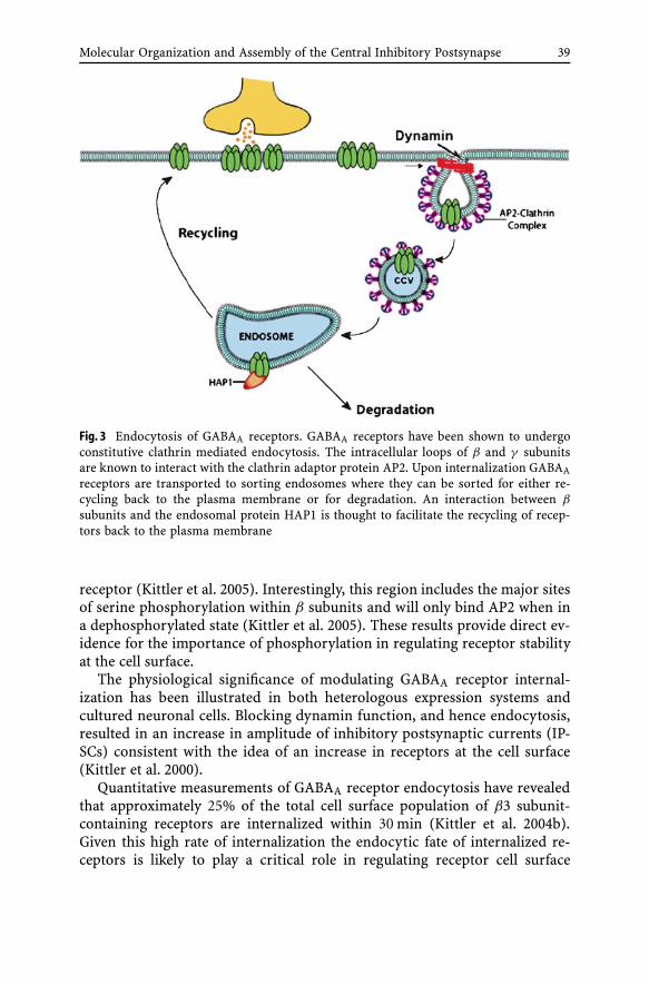
Molecular Organization and Assembly of the Central Inhibitory Postsynapse 39
Fig. 3 Endocytosis of GABAA receptors. GABAA receptors have been shown to undergoconstitutive clathrin mediated endocytosis. The intracellular loops of β and γ subunitsare known to interact with the clathrin adaptor protein AP2. Upon internalization GABAAreceptors are transported to sorting endosomes where they can be sorted for either re-cycling back to the plasma membrane or for degradation. An interaction between β
subunits and the endosomal protein HAP1 is thought to facilitate the recycling of recep-tors back to the plasma membrane
receptor (Kittler et al. 2005). Interestingly, this region includes the major sitesof serine phosphorylation within β subunits and will only bind AP2 when ina dephosphorylated state (Kittler et al. 2005). These results provide direct ev-idence for the importance of phosphorylation in regulating receptor stabilityat the cell surface.
The physiological significance of modulating GABAA receptor internal-ization has been illustrated in both heterologous expression systems andcultured neuronal cells. Blocking dynamin function, and hence endocytosis,resulted in an increase in amplitude of inhibitory postsynaptic currents (IP-SCs) consistent with the idea of an increase in receptors at the cell surface(Kittler et al. 2000).
Quantitative measurements of GABAA receptor endocytosis have revealedthat approximately 25% of the total cell surface population of β3 subunit-containing receptors are internalized within 30 min (Kittler et al. 2004b).Given this high rate of internalization the endocytic fate of internalized re-ceptors is likely to play a critical role in regulating receptor cell surface

40 I.L. Arancibia-Carcamo · S.J. Moss
expression levels. It is emerging that internalized GABAA receptors can besorted for two endocytic fates. The majority of internalized receptors con-stitutively recycle back to the plasma membrane over short time frames(tens of minutes) but over longer time periods (6 h) 25% of surface recep-tors are degraded upon internalization (Kittler et al. 2004b). Furthermore,internalized receptors have been shown to localize to a subsynaptic pool as-sociated with postsynaptic proteins such as gephyrin (van Rijnsoever et al.2005). Recently huntingtin-associated protein-1 (HAP1) has been identifiedas a GABAA receptor-associated protein (Kittler et al. 2004b). HAP1 bindsthe intracellular loop of β subunits in vitro and in vivo. Overexpression ofHAP1 in cortical neurons results in a decrease of GABAA receptor degrada-tion and consequently an increase in GABAA receptor recycling (Kittler et al.2004b). These results identify HAP1 as a regulator of GABAA receptor endo-cytic sorting (Fig. 3).
5Concluding Remarks
GABAA receptors are critical mediators of fast synaptic inhibition and drugtargets. Therefore, comprehending the cellular mechanisms neurons utilize toregulate their cell surface stability and accumulation at inhibitory synapses isof fundamental importance.
Accumulating experimental evidence has begun to illustrate that GABAAreceptors are dynamic entities on the neuronal surface, and that these pro-teins show significant levels of both endocytosis and exocytosis. These pro-cesses can be regulated by receptor-associated proteins (Table 1) and havebeen shown to regulate the efficacy of synaptic inhibition. Moreover, themechanisms that regulate the clustering and accumulation of GABAA recep-tors at synaptic sites are also being actively pursued. It will be importantto assess the role that these putative regulatory processes play in models ofsynaptic plasticity, such as rebound potentiation in the cerebellum, in whichalterations in GABAA receptor cell surface number are believed to be of sig-nificance.
Finally there is a growing list of CNS pathologies such as Status epilepticus,where altered GABAA receptor functional expression is of major significance(Coulter 2001). Therefore, assessing the physiological and pathological role ofGABAA receptor membrane trafficking is an active area of current investiga-tion.
Acknowledgements The authors would like to thank Dr. J.T. Kittler for critical reading ofthis manuscript. ILAC is the recipient of a Wellcome trust prize Studentship and SJM issupported by Grants from the MRC (UK), the Wellcome trust and NIH NINDS grantsNS047478, NS048045 & NS051195.

Molecular Organization and Assembly of the Central Inhibitory Postsynapse 41
References
Angelotti TP, Macdonald RL (1993) Assembly of GABAA receptor subunits: alpha 1 beta 1and alpha 1 beta 1 gamma 2S subunits produce unique ion channels with dissimilarsingle-channel properties. J Neurosci 13:1429–1440
Beck M, Brickley K, Wilkinson HL, Sharma S, Smith M, Chazot PL, Pollard S, Stephen-son FA (2002) Identification, molecular cloning, and characterization of a novelGABAA receptor-associated protein, GRIF-1. J Biol Chem 277:30079–30090
Bedford FK, Kittler JT, Muller E, Thomas P, Uren JM, Merlo D, Wisden W, Triller A,Smart TG, Moss SJ (2001) GABA(A) receptor cell surface number and subunit stabilityare regulated by the ubiquitin-like protein Plic-1. Nat Neurosci 4:908–916
Blum BP, Mann JJ (2002) The GABAergic system in schizophrenia. Int J Neuropsychophar-macol 5:159–179
Bohme I, Rabe H, Luddens H (2004) Four amino acids in the alpha subunits determinethe gamma-aminobutyric acid sensitivities of GABAA receptor subtypes. J Biol Chem279:35193–35200
Brandon N, Jovanovic J, Moss S (2002) Multiple roles of protein kinases in the modula-tion of gamma-aminobutyric acid(A) receptor function and cell surface expression.Pharmacol Ther 94:113–122
Brickley SG, Cull-Candy SG, Farrant M (1996) Development of a tonic form of synapticinhibition in rat cerebellar granule cells resulting from persistent activation of GABAAreceptors. J Physiol 497 (Pt 3):753–759
Brickley SG, Cull-Candy SG, Farrant M (1999) Single-channel properties of synaptic andextrasynaptic GABAA receptors suggest differential targeting of receptor subtypes.J Neurosci 19:2960–2973
Brickley SG, Revilla V, Cull-Candy SG, Wisden W, Farrant M (2001) Adaptive regulation ofneuronal excitability by a voltage-independent potassium conductance. Nature 409:88–92
Brown N, Kerby J, Bonnert TP, Whiting PJ, Wafford KA (2002) Pharmacological character-ization of a novel cell line expressing human alpha(4)beta(3)delta GABA(A) receptors.Br J Pharmacol 136:965–974
Brunig I, Scotti E, Sidler C, Fritschy JM (2002) Intact sorting, targeting, and clusteringof gamma-aminobutyric acid A receptor subtypes in hippocampal neurons in vitro.J Comp Neurol 443:43–55
Burette A, Wyszynski M, Valtschanoff JG, Sheng M, Weinberg RJ (1999) Characterizationof glutamate receptor interacting protein-immunopositive neurons in cerebellum andcerebral cortex of the albino rat. J Comp Neurol 411:601–612
Chang Y, Wang R, Barot S, Weiss DS (1996) Stoichiometry of a recombinant GABAAreceptor. J Neurosci 16:5415–5424
Charych EI, Yu W, Miralles CP, Serwanski DR, Li X, Rubio M, De Blas AL (2004a)The brefeldin A-inhibited GDP/GTP exchange factor 2, a protein involved in vesicu-lar trafficking, interacts with the beta subunits of the GABA receptors. J Neurochem90:173–189
Charych EI, Yu W, Li R, Serwanski DR, Miralles CP, Li X, Yang BY, Pinal N, Waliko-nis R, De Blas AL (2004b) A four PDZ domain-containing splice variant form ofGRIP1 is localized in GABAergic and glutamatergic synapses in the brain. J Biol Chem279:38978–38990
Chen ZW, Chang CS, Leil TA, Olcese R, Olsen RW (2005) GABAA Receptor-associatedprotein regulates GABAA receptor cell-surface number in Xenopus laevis oocytes. MolPharmacol 68:152–159

42 I.L. Arancibia-Carcamo · S.J. Moss
Chih B, Engelman H, Scheiffele P (2005) Control of excitatory and inhibitory synapseformation by neuroligins. Science 307:1324–1328
Choquet D, Triller A (2003) The role of receptor diffusion in the organization of thepostsynaptic membrane. Nat Rev Neurosci 4:251–265
Cinar H, Barnes EM Jr (2001) Clathrin-independent endocytosis of GABA(A) receptors inHEK 293 cells. Biochemistry 40:14030–14036
Connolly CN, Uren JM, Thomas P, Gorrie GH, Gibson A, Smart TG, Moss SJ (1999) Sub-cellular localization and endocytosis of homomeric gamma2 subunit splice variants ofgamma-aminobutyric acid type A receptors. Mol Cell Neurosci 13:259–271
Cossart R, Bernard C, Ben-Ari Y (2005) Multiple facets of GABAergic neurons andsynapses: multiple fates of GABA signalling in epilepsies. Trends Neurosci 28:108–115
Coulter DA (2001) Epilepsy-associated plasticity in gamma-aminobutyric acid receptorexpression, function, and inhibitory synaptic properties. Int Rev Neurobiol 45:237–252
Craig AM, Banker G, Chang W, McGrath ME, Serpinskaya AS (1996) Clustering ofgephyrin at GABAergic but not glutamatergic synapses in cultured rat hippocampalneurons. J Neurosci 16:3166–3177
Dahan M, Levi S, Luccardini C, Rostaing P, Riveau B, Triller A (2003) Diffusion dynamicsof glycine receptors revealed by single-quantum dot tracking. Science 302:442–445
Danglot L, Triller A, Bessis A (2003) Association of gephyrin with synaptic and extrasy-naptic GABAA receptors varies during development in cultured hippocampal neurons.Mol Cell Neurosci 23:264–278
Davies PA, Hanna MC, Hales TG, Kirkness EF (1997) Insensitivity to anaesthetic agentsconferred by a class of GABA(A) receptor subunit. Nature 385:820–823
Dean C, Scholl FG, Choih J, DeMaria S, Berger J, Isacoff E, Scheiffele P (2003) Neurexinmediates the assembly of presynaptic terminals. Nat Neurosci 6:708–716
DeLorey TM, Handforth A, Anagnostaras SG, Homanics GE, Minassian BA, Asatourian A,Fanselow MS, Delgado-Escueta A, Ellison GD, Olsen RW (1998) Mice lacking the beta3subunit of the GABAA receptor have the epilepsy phenotype and many of the behav-ioral characteristics of Angelman syndrome. J Neurosci 18:8505–8514
DeSouza S, Fu J, States BA, Ziff EB (2002) Differential palmitoylation directs the AMPAreceptor-binding protein ABP to spines or to intracellular clusters. J Neurosci 22:3493–3503
Dong H, Zhang P, Liao D, Huganir RL (1999) Characterization, expression, and distribu-tion of GRIP protein. Ann NY Acad Sci 868:535–540
Dong H, O’Brien RJ, Fung ET, Lanahan AA, Worley PF, Huganir RL (1997) GRIP:a synaptic PDZ domain-containing protein that interacts with AMPA receptors. Nature386:279–284
Dumoulin A, Levi S, Riveau B, Gasnier B, Triller A (2000) Formation of mixed glycine andGABAergic synapses in cultured spinal cord neurons. Eur J Neurosci 12:3883–3892
Ebert V, Scholze P, Fuchs K, Sieghart W (1999) Identification of subunits mediating clus-tering of GABA(A) receptors by rapsyn. Neurochem Int 34:453–463
El-Husseini Ael D, Schnell E, Dakoji S, Sweeney N, Zhou Q, Prange O, Gauthier-Campbell C, Aguilera-Moreno A, Nicoll RA, Bredt DS (2002) Synaptic strength regu-lated by palmitate cycling on PSD-95. Cell 108:849–863
Essrich C, Lorez M, Benson JA, Fritschy JM, Luscher B (1998) Postsynaptic clusteringof major GABAA receptor subtypes requires the gamma 2 subunit and gephyrin. NatNeurosci 1:563–571
Farrant M, Nusser Z (2005) Variations on an inhibitory theme: phasic and tonic activationof GABA(A) receptors. Nat Rev Neurosci 6:215–229
Molecular Organization and Assembly of the Central Inhibitory Postsynapse 43
Farrar SJ, Whiting PJ, Bonnert TP, McKernan RM (1999) Stoichiometry of a ligand-gatedion channel determined by fluorescence energy transfer. J Biol Chem 274:10100–10104
Feng G, Tintrup H, Kirsch J, Nichol MC, Kuhse J, Betz H, Sanes JR (1998) Dual require-ment for gephyrin in glycine receptor clustering and molybdoenzyme activity. Science282:1321–1324
Fisher JL, Macdonald RL (1997) Single channel properties of recombinant GABAA recep-tors containing gamma 2 or delta subtypes expressed with alpha 1 and beta 3 subtypesin mouse L929 cells. J Physiol 505(Pt 2):283–297
Fu Z, Washbourne P, Ortinski P, Vicini S (2003) Functional excitatory synapses in HEK293cells expressing neuroligin and glutamate receptors. J Neurophysiol 90:3950–3957
Graf ER, Zhang X, Jin SX, Linhoff MW, Craig AM (2004) Neurexins induce differentiation ofGABA and glutamate postsynaptic specializations via neuroligins. Cell 119:1013–1026
Haas KF, Macdonald RL (1999) GABAA receptor subunit gamma2 and delta subtypesconfer unique kinetic properties on recombinant GABAA receptor currents in mousefibroblasts. J Physiol 514(Pt 1):27–45
Herring D, Huang R, Singh M, Robinson LC, Dillon GH, Leidenheimer NJ (2003)Constitutive GABAA receptor endocytosis is dynamin-mediated and dependent ona dileucine AP2 adaptin-binding motif within the beta 2 subunit of the receptor. J BiolChem 278:24046–24052
Heuser JE, Anderson RG (1989) Hypertonic media inhibit receptor-mediated endocytosisby blocking clathrin-coated pit formation. J Cell Biol 108:389–400
Ichtchenko K, Nguyen T, Sudhof TC (1996) Structures, alternative splicing, and neurexinbinding of multiple neuroligins. J Biol Chem 271:2676–2682
Jacobs TC, Bogdanov YD, Magnus C, Saliba RS, Kittler JT, Haydon PG, Moss SJ (2005)Gephyrin regulates the cell surface dynamics of synaptic GABA A receptors. J Neurosci25:10469–10478
Kanematsu T, Jang IS, Yamaguchi T, Nagahama H, Yoshimura K, Hidaka K, Matsuda M,Takeuchi H, Misumi Y, Nakayama K, Yamamoto T, Akaike N, Hirata M, Nakayama K(2002) Role of the PLC-related, catalytically inactive protein p130 in GABA(A) receptorfunction. Embo J 21:1004–1011
Keller CA, Yuan X, Panzanelli P, Martin ML, Alldred M, Sassoe-Pognetto M, Luscher B(2004) The gamma2 subunit of GABA(A) receptors is a substrate for palmitoylationby GODZ. J Neurosci 24:5881–5891
Kirsch J, Wolters I, Triller A, Betz H (1993) Gephyrin antisense oligonucleotides preventglycine receptor clustering in spinal neurons. Nature 366:745–748
Kittler JT, Moss SJ (2003) Modulation of GABAA receptor activity by phosphorylationand receptor trafficking: implications for the efficacy of synaptic inhibition. Curr OpinNeurobiol 13:341–347
Kittler JT, Arancibia-Carcamo IL, Moss SJ (2004a) Association of GRIP1 with a GABA(A)receptor associated protein suggests a role for GRIP1 at inhibitory synapses. BiochemPharmacol 68:1649–1654
Kittler JT, Delmas P, Jovanovic JN, Brown DA, Smart TG, Moss SJ (2000) Constitutive endo-cytosis of GABAA receptors by an association with the adaptin AP2 complex modulatesinhibitory synaptic currents in hippocampal neurons. J Neurosci 20:7972–7977
Kittler JT, Rostaing P, Schiavo G, Fritschy JM, Olsen R, Triller A, Moss SJ (2001) The sub-cellular distribution of GABARAP and its ability to interact with NSF suggest a rolefor this protein in the intracellular transport of GABA(A) receptors. Mol Cell Neurosci18:13–25
Kittler JT, Thomas P, Tretter V, Bogdanov YD, Haucke V, Smart TG, Moss SJ (2004b)Huntingtin-associated protein 1 regulates inhibitory synaptic transmission by mod-

44 I.L. Arancibia-Carcamo · S.J. Moss
ulating gamma-aminobutyric acid type A receptor membrane trafficking. Proc NatlAcad Sci USA 101:12736–12741
Kittler JT, Chen G, Honing S, Bogdanov YD, McAinsh K, Arancibia-Carcamo IL, Jo-vanovic JN, Pangalos MN, Haucke V, Yan Z, Moss SJ (2005) Phospho-dependentbinding of the clathrin AP2 adaptor complex to GABAA receptors regulates the effi-cacy of inhibitory synaptic transmission. Proc Natl Acad Sci USA 102:14871–6
Kleijnen MF, Shih AH, Zhou P, Kumar S, Soccio RE, Kedersha NL, Gill G, Howley PM(2000) The hPLIC proteins may provide a link between the ubiquitination machineryand the proteasome. Mol Cell 6:409–419
Kneussel M, Hermann A, Kirsch J, Betz H (1999a) Hydrophobic interactions mediatebinding of the glycine receptor beta-subunit to gephyrin. J Neurochem 72:1323–1326
Kneussel M, Brandstatter JH, Laube B, Stahl S, Muller U, Betz H (1999b) Loss of postsynapticGABA(A) receptor clustering in gephyrin-deficient mice. J Neurosci 19:9289–9297
Knight AR, Stephenson FA, Tallman JF, Ramabahdran TV (2000) Monospecific antibod-ies as probes for the stoichiometry of recombinant GABA(A) receptors. ReceptorsChannels 7:213–226
Knuesel I, Mastrocola M, Zuellig RA, Bornhauser B, Schaub MC, Fritschy JM (1999) Shortcommunication: altered synaptic clustering of GABAA receptors in mice lacking dys-trophin (mdx mice). Eur J Neurosci 11:4457–4462
Krishek BJ, Moss SJ, Smart TG (1996) Homomeric beta 1 gamma-aminobutyric acidA receptor-ion channels: evaluation of pharmacological and physiological properties.Mol Pharmacol 49:494–504
Kunig G, Leenders KL, Sanchez-Pernaute R, Antonini A, Vontobel P, Verhagen A, Gun-ther I (2000) Benzodiazepine receptor binding in Huntington’s disease: [11C]flumaze-nil uptake measured using positron emission tomography. Ann Neurol 47:644–648
Laurie DJ, Wisden W, Seeburg PH (1992a) The distribution of thirteen GABAA receptorsubunit mRNAs in the rat brain. III. Embryonic and postnatal development. J Neurosci12:4151–4172
Laurie DJ, Seeburg PH, Wisden W (1992b) The distribution of 13 GABAA receptorsubunit mRNAs in the rat brain. II. Olfactory bulb and cerebellum. J Neurosci 12:1063–1076
Lavoie AM, Tingey JJ, Harrison NL, Pritchett DB, Twyman RE (1997) Activation anddeactivation rates of recombinant GABA(A) receptor channels are dependent onalpha-subunit isoform. Biophys J 73:2518–2526
Leil TA, Chen ZW, Chang CS, Olsen RW (2004) GABAA receptor-associated protein traf-fics GABAA receptors to the plasma membrane in neurons. J Neurosci 24:11429–11438
Levi S, Logan SM, Tovar KR, Craig AM (2004) Gephyrin is critical for glycine receptorclustering but not for the formation of functional GABAergic synapses in hippocampalneurons. J Neurosci 24:207–217
Levi S, Grady RM, Henry MD, Campbell KP, Sanes JR, Craig AM (2002) Dystroglycan isselectively associated with inhibitory GABAergic synapses but is dispensable for theirdifferentiation. J Neurosci 22:4274–4285
Levinson JN, El-Husseini A (2005) New players tip the scales in the balance betweenexcitatory and inhibitory synapses. Mol Pain 1:12
Levinson JN, Chery N, Huang K, Wong TP, Gerrow K, Kang R, Prange O, Wang YT, El-Husseini A (2005) Neuroligins mediate excitatory and inhibitory synapse formation:involvement of PSD-95 and neurexin-1beta in neuroligin-induced synaptic specificity.J Biol Chem 280:17312–17319
Lewis DA (2000) GABAergic local circuit neurons and prefrontal cortical dysfunction inschizophrenia. Brain Res Brain Res Rev 31:270–276

Molecular Organization and Assembly of the Central Inhibitory Postsynapse 45
Li RW, Serwanski DR, Miralles CP, Li X, Charych E, Riquelme R, Huganir RL, de Blas AL(2005) GRIP1 in GABAergic synapses. J Comp Neurol 488:11–27
Malizia AL (1999) What do brain imaging studies tell us about anxiety disorders? J Psy-chopharmacol 13:372–378
Meyer G, Kirsch J, Betz H, Langosch D (1995) Identification of a gephyrin binding motifon the glycine receptor beta subunit. Neuron 15:563–572
Mody I, Pearce RA (2004) Diversity of inhibitory neurotransmission through GABA(A)receptors. Trends Neurosci 27:569–575
Mohler H, Crestani F, Rudolph U (2001) GABA(A)-receptor subtypes: a new pharmacol-ogy. Curr Opin Pharmacol 1:22–25
Morrow AL, VanDoren MJ, Penland SN, Matthews DB (2001) The role of GABAergic neu-roactive steroids in ethanol action, tolerance and dependence. Brain Res Brain Res Rev37:98–109
Moss SJ, Smart TG (2001) Constructing inhibitory synapses. Nat Rev Neurosci 2:240–250Neelands TR, Macdonald RL (1999) Incorporation of the pi subunit into functional
gamma-aminobutyric Acid(A) receptors. Mol Pharmacol 56:598–610Nusser Z, Cull-Candy S, Farrant M (1997) Differences in synaptic GABA(A) receptor
number underlie variation in GABA mini amplitude. Neuron 19:697–709Nusser Z, Sieghart W, Somogyi P (1998a) Segregation of different GABAA receptors to
synaptic and extrasynaptic membranes of cerebellar granule cells. J Neurosci 18:1693–1703
Nusser Z, Hajos N, Somogyi P, Mody I (1998b) Increased number of synaptic GABA(A)receptors underlies potentiation at hippocampal inhibitory synapses. Nature 395:172–177
Nusser Z, Sieghart W, Benke D, Fritschy JM, Somogyi P (1996) Differential synaptic lo-calization of two major gamma-aminobutyric acid type A receptor alpha subunits onhippocampal pyramidal cells. Proc Natl Acad Sci USA 93:11939–11944
Nutt DJ, Malizia AL (2001) New insights into the role of the GABA(A)-benzodiazepinereceptor in psychiatric disorder. Br J Psychiatry 179:390–396
Olsen RW, DeLorey TM, Gordey M, Kang MH (1999) GABA receptor function andepilepsy. Adv Neurol 79:499–510
Pfeiffer F, Graham D, Betz H (1982) Purification by affinity chromatography of the glycinereceptor of rat spinal cord. J Biol Chem 257:9389–9393
Pirker S, Schwarzer C, Wieselthaler A, Sieghart W, Sperk G (2000) GABA(A) receptors:immunocytochemical distribution of 13 subunits in the adult rat brain. Neuroscience101:815–850
Prange O, Wong TP, Gerrow K, Wang YT, El-Husseini A (2004) A balance between excita-tory and inhibitory synapses is controlled by PSD-95 and neuroligin. Proc Natl AcadSci USA 101:13915–13920
Ramming M, Kins S, Werner N, Hermann A, Betz H, Kirsch J (2000) Diversity andphylogeny of gephyrin: tissue-specific splice variants, gene structure, and sequencesimilarities to molybdenum cofactor-synthesizing and cytoskeleton-associated pro-teins. Proc Natl Acad Sci USA 97:10266–10271
Rathenberg J, Kittler JT, Moss SJ (2004) Palmitoylation regulates the clustering and cellsurface stability of GABAA receptors. Mol Cell Neurosci 26:251–257
Rudolph U, Crestani F, Mohler H (2001) GABA(A) receptor subtypes: dissecting theirpharmacological functions. Trends Pharmacol Sci 22:188–194
Schaerer MT, Kannenberg K, Hunziker P, Baumann SW, Sigel E (2001) Interaction be-tween GABA(A) receptor beta subunits and the multifunctional protein gC1q-R. J BiolChem 276:26597–26604

46 I.L. Arancibia-Carcamo · S.J. Moss
Scheiffele P, Fan J, Choih J, Fetter R, Serafini T (2000) Neuroligin expressed in nonneu-ronal cells triggers presynaptic development in contacting axons. Cell 101:657–669
Schofield PR, Darlison MG, Fujita N, Burt DR, Stephenson FA, Rodriguez H, Rhee LM,Ramachandran J, Reale V, Glencorse TA et al. (1987) Sequence and functional ex-pression of the GABA A receptor shows a ligand-gated receptor super-family. Nature328:221–227
Schwartz-Bloom RD, Sah R (2001) gamma-Aminobutyric acid(A) neurotransmission andcerebral ischemia. J Neurochem 77:353–371
Schwarz G, Schrader N, Mendel RR, Hecht HJ, Schindelin H (2001) Crystal structuresof human gephyrin and plant Cnx1 G domains: comparative analysis and functionalimplications. J Mol Biol 312:405–418
Shivers BD, Killisch I, Sprengel R, Sontheimer H, Kohler M, Schofield PR, Seeburg PH(1989) Two novel GABAA receptor subunits exist in distinct neuronal subpopulations.Neuron 3:327–337
Sieghart W, Fuchs K, Tretter V, Ebert V, Jechlinger M, Hoger H, Adamiker D (1999)Structure and subunit composition of GABA(A) receptors. Neurochem Int 34:379–385
Smotrys JE, Linder ME (2004) Palmitoylation of intracellular signaling proteins: regula-tion and function. Annu Rev Biochem 73:559–587
Song JY, Ichtchenko K, Sudhof TC, Brose N (1999) Neuroligin 1 is a postsynaptic cell-adhesion molecule of excitatory synapses. Proc Natl Acad Sci USA 96:1100–1105
Song R, Segal G, Messing J (2004) Expression of the sorghum 10-member kafirin genecluster in maize endosperm. Nucleic Acids Res 32:e189
Stell BM, Brickley SG, Tang CY, Farrant M, Mody I (2003) Neuroactive steroids re-duce neuronal excitability by selectively enhancing tonic inhibition mediated by deltasubunit-containing GABAA receptors. Proc Natl Acad Sci USA 100:14439–14444
Tardin C, Cognet L, Bats C, Lounis B, Choquet D (2003) Direct imaging of lateral move-ments of AMPA receptors inside synapses. Embo J 22:4656–4665
Terunuma M, Jang IS, Ha SH, Kittler JT, Kanematsu T, Jovanovic JN, Nakayama KI,Akaike N, Ryu SH, Moss SJ, Hirata M (2004) GABAA receptor phospho-dependentmodulation is regulated by phospholipase C-related inactive protein type 1, a novelprotein phosphatase 1 anchoring protein. J Neurosci 24:7074–7084
Thomas P, Mortensen M, Hosie AM, Smart TG (2005) Dynamic mobility of functionalGABAA receptors at inhibitory synapses. Nat Neurosci 8:889
Tretter V, Ehya N, Fuchs K, Sieghart W (1997) Stoichiometry and assembly of a recombi-nant GABAA receptor subtype. J Neurosci 17:2728–2737
Unwin N (1993) Nicotinic acetylcholine receptor at 9 A resolution. J Mol Biol 229:1101–1124
van Rijnsoever C, Sidler C, Fritschy JM (2005) Internalized GABA-receptor subunits aretransferred to an intracellular pool associated with the postsynaptic density. Eur JNeurosci 21:327–338
Varoqueaux F, Jamain S, Brose N (2004) Neuroligin 2 is exclusively localized to inhibitorysynapses. Eur J Cell Biol 83:449–456
Wang H, Olsen RW (2000) Binding of the GABA(A) receptor-associated protein(GABARAP) to microtubules and microfilaments suggests involvement of the cy-toskeleton in GABARAP–GABA(A) receptor interaction. J Neurochem 75:644–655
Wang H, Bedford FK, Brandon NJ, Moss SJ, Olsen RW (1999) GABA(A)-receptor-associated protein links GABA(A) receptors and the cytoskeleton. Nature 397:69–72
Washbourne P, Dityatev A, Scheiffele P, Biederer T, Weiner JA, Christopherson KS, El-Husseini A (2004) Cell adhesion molecules in synapse formation. J Neurosci 24:9244–9249

Molecular Organization and Assembly of the Central Inhibitory Postsynapse 47
Wisden W, Laurie DJ, Monyer H, Seeburg PH (1992) The distribution of 13 GABAA recep-tor subunit mRNAs in the rat brain. I. Telencephalon, diencephalon, mesencephalon.J Neurosci 12:1040–1062
Wooltorton JR, Moss SJ, Smart TG (1997) Pharmacological and physiological characteri-zation of murine homomeric beta3 GABA(A) receptors. Eur J Neurosci 9:2225–2235
Wu AL, Wang J, Zheleznyak A, Brown EJ (1999) Ubiquitin-related proteins regulate in-teraction of vimentin intermediate filaments with the plasma membrane. Mol Cell4:619–625
Wyszynski M, Valtschanoff JG, Naisbitt S, Dunah AW, Kim E, Standaert DG, Weinberg R,Sheng M (1999) Association of AMPA receptors with a subset of glutamate receptor-interacting protein in vivo. J Neurosci 19:6528–6537
Yoshimura K, Takeuchi H, Sato O, Hidaka K, Doira N, Terunuma M, Harada K, Ogawa Y,Ito Y, Kanematsu T, Hirata M (2001) Interaction of p130 with, and consequent inhibi-tion of, the catalytic subunit of protein phosphatase 1alpha. J Biol Chem 276:17908–17913

Results Probl Cell Differ (43)E. Gundelfinger, C. Seidenbecher, B. Schraven:Cell Communication in Nervous and Immune SystemDOI 10.1007/012/Published online: 23 June 2006© Springer-Verlag Berlin Heidelberg 2006
Molecular Organization and Assemblyof the Presynaptic Active Zoneof Neurotransmitter Release
Anna Fejtova (�) · Eckart D. Gundelfinger
Leibniz Institute for Neurobiology,Department of Neurochemistry and Molecular Biology, Brennecke Str. 6,39118 Magdeburg, [email protected]
Abstract At chemical synapses, neurotransmitter is released at a restricted region of thepresynaptic plasma membrane, called the active zone. At the active zone, a matrix of pro-teins is assembled, which is termed the presynaptic grid or cytomatrix at the active zone(CAZ). Components of the CAZ are thought to localize and organize the synaptic vesi-cle cycle, a series of membrane trafficking events underlying regulated neurotransmitterexocytosis. This review is focused on a set of specific proteins involved in the structuraland functional organization of the CAZ. These include the multi-domain Rab3-effectorproteins RIM1α and RIM2α; Bassoon and Piccolo, two multi-domain CAZ scaffoldingproteins of enormous size; as well as members of the CAST/ERC family of CAZ-specificstructural proteins. Studies on ribbon synapses of retinal photoreceptor cells have fos-tered understanding the molecular design of the CAZ. In addition, the analysis of thedelivery pathways for Bassoon and Piccolo to presynaptic sites during development hasproduced new insights into assembly mechanisms of brain synapses during develop-ment. Based on these studies, the active zone transport vesicle hypothesis was formulated,which postulates that active zones, at least in part, are pre-assembled in neuronal cellbodies and transported as so-called Piccolo-Bassoon transport vesicles (PTVs) to sitesof synaptogenesis. Several PTVs can fuse on demand with the presynaptic membrane torapidly form an active zone.
AbbreviationsC1 domain conserved domain 1 of protein kinase CC2 domain conserved domain 2 of protein kinase CCAST CAZ-associated structural proteinCAZ cytomatrix assembled at the active zoneCtBP C-terminal binding protein (or connected to Bassoon and Piccolo)DAG diacylglycerolERC ELKS-Rab6-interacting molecule-CASTGIT protein interacting with G-protein-coupled receptor kinasesPBH domain Piccolo-Bassoon homology domainPDZ PSD-95-Discs large-ZO-1 domainPSD postsynaptic densityPTV Piccolo-Bassoon transport vesicleRIM Rab3-interacting molecule

50 A. Fejtova · E.D. Gundelfinger
RIM-BP RIM-binding proteinSV synaptic vesicle
1Introduction
Chemical synapses are important sites for intercellular communication.They constitute asymmetric cell-cell contact sites between a presynaptic anda postsynaptic cell devised to translate presynaptic electrical signals intochemical signals in the form of exocytosed neurotransmitter. This messen-ger can be detected by the postsynaptic cell and transformed back into anappropriate postsynaptic signal. The presynaptic active zone is the restrictedarea of the presynaptic plasma membrane, at which synaptic vesicles can fuseand release their neurotransmitter content in a regulated (Ca2+-dependent)manner. The active zone is aligned precisely with the region of postsynapticmembrane that harbors the neurotransmitter reception apparatus and is de-lineated by the postsynaptic density (PSD). As defined here, the active zoneconsists of two major parts, the active zone plasma membrane and the asso-ciated cytoskeletal matrix, which is called cytomatrix assembled at the activezone (CAZ) or presynaptic grid (Dresbach et al. 2001; Gundelfinger et al.2006). In this review, we will discuss recent discoveries concerning the mo-lecular organization of the active zone and its developmental assembly duringthe major period of synaptogenesis during brain development.
2General Aspects of Structuraland Functional Organization of the Active Zone
2.1Neurotransmitter Release
At the active zone, a series of membrane trafficking events underlies regulatedexocytosis of neurotransmitter from synaptic vesicles (SV) and the retrievaland recycling of vesicular membranes: the SV cycle (Sudhof 1995, 2004). SVthat are tethered to the active zonal membrane have to undergo a primingprocess to become fusion competent, i.e. to become responsive to influx ofCa2+ through voltage-gated Ca2+ channels located in the active zone mem-brane. Several Proteins, including Ca2+ and diacylglycerol (DAG)-regulatedUNC13/Munc13 proteins, SM (Sec1/Munc18-like) proteins and RIM1/UNC10,are involved in this priming process (Rosenmund et al. 2003, see below).Upon membrane depolarization by action potentials, Ca2+ channels open

Molecular Organization and Assembly of the Presynaptic Active Zone 51
and a Ca2+-sensing machinery detects Ca2+ ions entering the cell. Synapto-tagmins are the major presynaptic Ca2+ sensors (Chapman 2002). However,other proteins, such as complexins, are also thought to convey the Ca2+ sig-nal to the fusion machinery (Reim et al. 2001). The actual fusion process ismediated via SNAREs, including synaptobrevins/VAMPs as vesicular SNAREproteins and syntaxin and SNAP-25 as target membrane SNAREs (for review,see Jahn et al. 2003). After membrane fusion, SV may either fully collapse intothe active zone membrane or release their neurotransmitter content througha fusion pore. In the latter case, the vesicle may be rapidly retrieved and re-filled in a “kiss-and-run” or “kiss-and-stay” mechanisms (Gundelfinger et al.2003; Sudhof 2004). In the former case, SV vesicles are retrieved by clathrin-mediated endocytosis in the periactive zone area that surrounds the activezone. Exocytosis and endocytosis are thought to be spatially and temporallytightly linked (Dresbach et al. 2001; Gundelfinger et al. 2003; Murthy and DeCamilli 2003).
Recycling and refilling processes are thought to occur somewhat remotefrom the active zonal membrane (Sudhof 1995, 2004). Depending on theirfunctional state, different pools of SV can be defined: the cycling pool of vesi-cles, which includes vesicles that are docked and immediately available forrelease and those that are rapidly recycling and reused, and the reserve orresting pool of vesicles. The latter pool includes SV that are inactive and arerecruited only after massive stimulation (Sudhof 2000, 2004).
2.2Ultrastructural Organization of the Active Zone
The presynaptic active zone is precisely aligned with the postsynaptic neu-rotransmitter reception apparatus defined by the PSD. At the ultrastructurallevel, both sides of the synaptic contact site appear as electron-dense struc-tures that are tightly associated with the plasma membrane. In the electronmicroscope, the CAZ or presynaptic grid appears as a more or less regu-lar array of electron-dense cone-shaped particles, which extend ∼ 50 nminto the cytoplasm (Gray 1963; Pfenninger et al. 1972; Phillips et al. 2001).A meshwork of cytoskeletal filaments connects the 50-nm pyramid-shapedparticles; in addition filamentous strands extend deeply from the active zoneplasma membrane into the presynaptic bouton. At the frog neuromuscularjunction, which has been studied by electron microscope tomography, activezone material extends 50–75 nm into the cytoplasm of the terminal and hasa regularly arranged ultrastructure consisting of “pegs”, “ribs” and “beams”(Harlow et al. 2001).
The CAZ is thought to define and organize neurotransmitter release sites,keeping them aligned with the PSD. CAZ components mediate the replace-ment of exocytosed SV (Dresbach et al. 2001; Garner et al. 2002). Moreover,the CAZ may play an important role in the spatial and temporal coordi-

52 A. Fejtova · E.D. Gundelfinger
nation of exocytic and endocytic events at the active zone (Gundelfingeret al. 2003).
3Molecular Organization of the Active Zone
Several CAZ-specific constituents have been identified and characterizedat the molecular level, including the UNC13/Munc13 proteins, the Rab3-interacting molecules (RIMs), Bassoon, Piccolo/Aczonin and the ERC2/CAST(Garner et al. 2000; Rosenmund et al. 2003; Ziv and Garner 2004). These pro-teins are physically interconnected and thus thought to form the molecular
Fig. 1 Molecular organization of the CAZ. The CAZ-specific proteins RIMs, Munc13s,Bassoon, Piccolo and CAST/ERC are thought to localize and organize membrane traf-ficking events of the synaptic vesicle cycle and connect it to the active zone mem-brane proteins including voltage-gated Ca2+ channels and cell adhesion molecules suchas the neurexins. Further components that are not exclusive CAZ components in-clude Ca2+/calmodulin kinase domain-containing membrane-associated guanylate kinaseCASK, the transcriptional co-repressor CtBP1/BARS50, the RIM-binding proteins (RIM-BP), the prenylated Rab3 acceptor protein PRA1, the ARF-GTPase-activating protein GIT,the receptor tyrosine phosphatase LAR and its interacting protein Liprin, componentsof the SNARE complex and its control elements (e.g. Munc18). The interaction betweenPiccolo and the actin-binding protein Abp1 is thought to link the active zone to theneighboring endocytic zone. For further details see: Gundelfinger, Altrock, Fejtova, Activezone. In: Encyclopedic Reference of Neuroscience (Field editor: M. Takahashi, SpringerBerlin, in press). The picture was taken from the website of the IfN(http://www.ifn-magdeburg.de/en/departments/neurochemistry_and_molecular_biology)
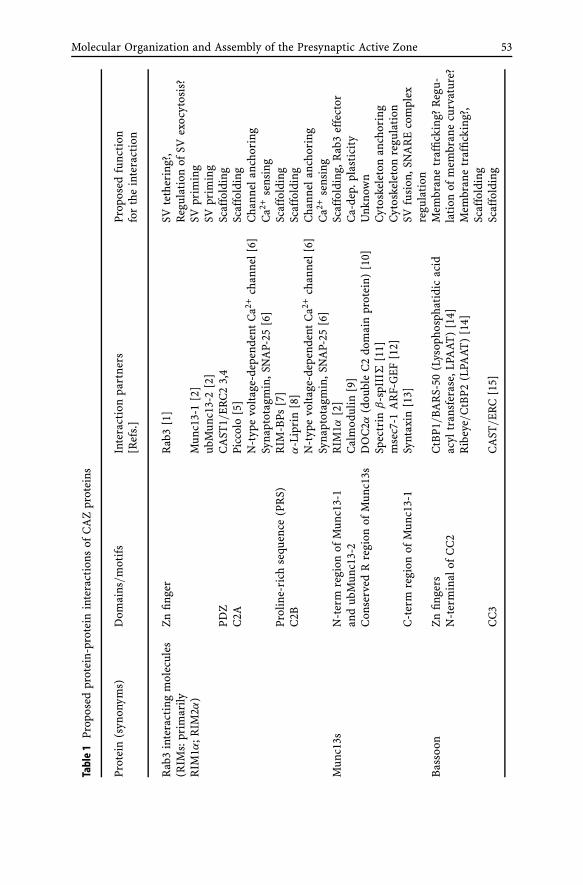
Molecular Organization and Assembly of the Presynaptic Active Zone 53
Tabl
e1
Prop
osed
prot
ein-
prot
ein
inte
ract
ions
ofC
AZ
prot
eins
Prot
ein
(syn
onym
s)D
omai
ns/
mot
ifs
Inte
ract
ion
part
ners
Prop
osed
func
tion
[Ref
s.]
for
the
inte
ract
ion
Rab
3in
tera
ctin
gm
olec
ules
Zn
finge
rR
ab3
[1]
SVte
ther
ing?
,(R
IMs:
prim
arily
Reg
ulat
ion
ofSV
exoc
ytos
is?
RIM
1α;R
IM2α
)M
unc1
3-1
[2]
SVpr
imin
gub
Mun
c13-
2[2
]SV
prim
ing
PDZ
CA
ST1/
ER
C2
3,4
Scaf
fold
ing
C2A
Picc
olo
[5]
Scaf
fold
ing
N-t
ype
volt
age-
depe
nden
tC
a2+ch
anne
l[6]
Cha
nnel
anch
orin
gSy
napt
otag
min
,SN
AP-
25[6
]C
a2+se
nsin
gPr
olin
e-ri
chse
quen
ce(P
RS)
RIM
-BPs
[7]
Scaf
fold
ing
C2B
α-L
ipri
n[8
]Sc
affo
ldin
gN
-typ
evo
ltag
e-de
pend
ent
Ca2+
chan
nel[
6]C
hann
elan
chor
ing
Syna
ptot
agm
in,S
NA
P-25
[6]
Ca2+
sens
ing
Mun
c13s
N-t
erm
regi
onof
Mun
c13-
1R
IM1α
[2]
Scaf
fold
ing,
Rab
3ef
fect
oran
dub
Mun
c13-
2C
alm
odul
in[9
]C
a-de
p.pl
asti
city
Con
serv
edR
regi
onof
Mun
c13s
DO
C2α
(dou
ble
C2
dom
ain
prot
ein)
[10]
Unk
now
nSp
ectr
inβ
-spI
IIΣ
[11]
Cyt
oske
leto
nan
chor
ing
mse
c7-1
AR
F-G
EF[1
2]C
ytos
kele
ton
regu
lati
onC
-ter
mre
gion
ofM
unc1
3-1
Synt
axin
[13]
SVfu
sion
,SN
AR
Eco
mpl
exre
gula
tion
Bas
soon
Zn
finge
rsC
tBP1
/BA
RS-
50(L
ysop
hosp
hati
dic
acid
Mem
bran
etr
affic
king
?R
egu-
N-t
erm
inal
ofC
C2
acyl
tran
sfer
ase,
LPA
AT)
[14]
lati
onof
mem
bran
ecu
rvat
ure?
Rib
eye/
CtB
P2(L
PAAT
)[1
4]M
embr
ane
traf
ficki
ng?,
Scaf
fold
ing
CC
3C
AST
/ER
C[1
5]Sc
affo
ldin
g

54 A. Fejtova · E.D. Gundelfinger
Tabl
e1
(con
tinu
ed)
Prot
ein
(syn
onym
s)D
omai
ns/
mot
ifs
Inte
ract
ion
part
ners
Prop
osed
func
tion
[Ref
s.]
for
the
inte
ract
ion
Picc
olo
(Acz
onin
)Q
dom
ain
Act
in-b
indi
ngpr
otei
nA
bp1
[16]
Act
inbi
ndin
g,lin
kto
endo
cyto
sis
Zn
finge
rsPR
A1
[17]
Unk
now
nPR
SG
IT(A
RF-
GA
P)[1
8]G
TPa
sere
gula
tion
,mem
bran
etr
affic
king
PRS
Profi
lin(a
ctin
bind
ing
prot
ein)
[19]
Act
inre
gula
tion
CC
3C
AST
/ER
C[1
5]Sc
affo
ldin
gPD
ZcA
MP-
GEF
II[5
]G
TPa
sere
gula
tion
C2A
RIM
2[5
]Sc
affo
ldin
g,R
ab3
effe
ctor
C2B
Picc
olo
[5]
Scaf
fold
ing
L-ty
pevo
ltag
e-de
pend
ent
Ca2+
chan
nel[
5]C
hann
elan
chor
ing
CA
Z-a
ssoc
iate
dst
ruct
ural
Coi
led-
coil
regi
ons
Bas
soon
,Pic
colo
[15]
Scaf
fold
ing
prot
eins
(CA
STs,
ERC
s)α
-Lip
rin
[20]
Scaf
fold
ing,
Tran
spor
t?C
-ter
mPD
Zbi
ndin
gm
otif
RIM
s[3
]Sc
affo
ldin
gR
IMbi
ndin
gpr
otei
nsSH
3do
mai
ns(o
neof
3)R
IMs
[7]
Scaf
fold
ing
(RIM
-BPs
)SH
3do
mai
ns(o
neof
3)C
a2+ch
anne
lsC
a v2.
2(N
-typ
e),
Cha
nnel
anch
orin
gC
a v1.
3(L
-typ
e)[2
1]α
-Lip
rins
(SY
D-2
)N
-ter
mC
Cre
gion
CA
ST/
ERC
[20]
Scaf
fold
ing,
Tran
spor
t?C
-ter
mSA
Mdo
mai
nsK
IF1A
(kin
esin
mot
or)
[22]
Tran
spor
tLA
R(r
ecep
tror
tyro
sine
phos
pata
se)
[23]
Rec
epto
ran
chor
ing
GR
IP[2
4]R
ecep
tor
clus
teri
ng

Molecular Organization and Assembly of the Presynaptic Active Zone 55
Table 1 continued
References to Table 1:1. Wang Y, Okamoto M, Schmitz F, Hofmann K, Sudhof TC (1997) Nature 388:5932. Betz A, et al. (2001) Neuron 30:1833. Ohtsuka T, et al. (2002) J Cell Biol 158:5774. Wang Y, Liu X, Biederer T, Sudhof TC (2002) Proc Natl Acad Sci USA 99:144645. Shibasaki T, Sunaga Y, Fujimoto K, Kashima Y, Seino S (2003) J Biol Chem6. Coppola T, et al. (2001) J Biol Chem 276:327567. Wang Y, Sugita S (2000) Sudhof TC, J Biol Chem 275:200338. Schoch S, et al. (2002) Nature 415:3219. Junge HJ, et al. (2004) Cell 118:389
10. Orita S, et al. (1997) J Biol Chem 272:1608111. Sakaguchi G, et al. (1998) Biochem Biophys Res Commun 248:84612. Neeb A, Koch H, Schurmann A, Brose N (1999) Eur J Cell Biol 78:53313. Betz A, Okamoto M, Benseler F, Brose N (1997) J Biol Chem 272:252014. tom Dieck S, et al. (2005) J Cell Biol 168:82515. Takao-Rikitsu E, et al. (2004) J Cell Biol 164:30116. Fenster SD, et al. (2003) J Biol Chem 278:2026817. Fenster SD, et al. (2000) Neuron 25:20318. Kim S, et al. (2003) J Biol Chem 278:629119. Wang X, et al. (1999) J Cell Biol 147:15120. Ko J, Na M, Kim S, Lee JR, Kim E (2003) J Biol Chem 278:4237721. Hibino H, et al. (2002) Neuron 34:41122. Shin H, et al. (2003) J Biol Chem 278:1139323. Pulido R, Serra-Pages C, Tang M, Streuli M (1995) Proc Natl Acad Sci USA 92:1168624. Wyszynski M, et al. (2002) Neuron 34:39
scaffold of the CAZ (Fig. 1, Table 1). In addition, functions in organizing theSV cycle have been assigned to some of the CAZ-specific proteins.
3.1The UNC13/Munc13 Protein Family – Key Actors in SV Priming
UNC13 proteins, first identified in “uncoordinate” mutants of Caenorhab-ditis elegans, contain two or three C2 domains, which are supposed to beinvolved in Ca2+ and phospholipid binding, and one C1 domain that canbind the second messenger DAG and β-phorbol esters (for review, see Broseet al. 2000). Alternative splicing of the C. elegans unc13 gene gives rise totwo isoforms differing in their N-terminal sequence. In vertebrates, fourUNC13 genes have been identified encoding Munc13-1, Munc13-2, Munc13-3, and Munc13-4. In analogy to worm UNC13, two isoforms arise from theMunc13-2 gene: bMunc13-2 is similarly to Munc13-1 and Munc13-3 brainspecific and ubMunc13-2 is ubiquitously expressed (Koch et al. 2000). Ge-netic studies in C. elegans, Drosophila and mice indicate that UNC13 proteinshave an important role in the attainment of fusion competence of docked

56 A. Fejtova · E.D. Gundelfinger
SVs, a process generally called SV “priming” (Brose et al. 2000). Accordingto our current view, Munc13s bind the presynaptic membrane SNARE pro-tein syntaxin and promote the formation of a loose SNARE complex, whichis a prerequisite for Ca2+-dependent SV fusion (Rosenmund et al. 2003). Sev-eral second messengers and regulatory proteins that interact with UNC13proteins can fine-tune their function. For example, DAG and phorbol estersbind the C1 domain of Munc13s. Electrophysiological studies on mice ex-pressing a DAG-insensitive Munc13-1 variant have demonstrated that DAGbinding to Munc13 is essential for the maintenance of efficient transmis-sion during trains of action potentials and that Munc13s, and not proteinkinase C, are the targets for DAG and phorbol ester-mediated potentiationof transmitter release (Rhee et al. 2002). Moreover, Munc13s possess an evo-lutionary conserved Ca2+-dependent calmodulin (CaM) binding motif. TheCa2+ sensor-effector complex regulates synaptic vesicle priming and synapticefficacy by sensing residual Ca2+ concentrations; in this way, it controls short-term plasticity characteristics during periods of repeated synaptic activity(Junge et al. 2004).
Rosenmund and colleagues demonstrated a differential contribution ofMunc13-1 and Munc13-2 to the regulation of presynaptic short-term plasticity(Rosenmund et al. 2002). While synapses containing Munc13-1 depress dur-ing trains of action potentials, synapses containing Munc13-2 augment whenrepeatedly stimulated. As both isoforms can be localized to distinct presynap-tic boutons of a single neuron, the same train of action potentials can lead todivergent responses at these synapses. These differences in regulation of short-term plasticity by Munc13 isoforms vanish when CaM insensitive mutants ofMunc13-1 and Munc13-2 are expressed (Junge et al. 2004).
3.2RIMs – Multidomain Rab3 Effectors with a Major Organizing Role
While invertebrates possess only a single gene for the RIM (Rab3-interactingmolecule) homologue UNC10, in vertebrates four RIM-related genes havebeen identified that code for RIM1α, RIM2α, β and γ , RIM3γ and RIM4γ iso-forms. The RIM2 gene is transcribed from alternative promoters to give ariseto α, β and γ isoform. The diversity of RIM family members is even increasedby alternative splicing of α and β isoforms (Wang et al. 2000; Wang and Sud-hof 2003). The α isoforms comprise an N-terminal zinc finger domain, a PDZ(PSD-95/SAP90-DlgA-ZO-1) domain and two C2 domains, the RIM2 β iso-form lacks the zinc finger domain, and γ isoforms include just one C-terminalC2 domain. RIMs are expressed predominantly in brain; both α isoforms arespecific constituents of the CAZ.
RIM1α was first identified as an effector of the monomeric GTPase proteinsof the Rab3 subfamily that are associated with SVs (Wang et al. 1997). This sug-gested a role for RIMs in the regulation of neurotransmitter release. Genetic

Molecular Organization and Assembly of the Presynaptic Active Zone 57
studies in C. elegans revealed a function of the RIM homologue UNC10 in SVpriming, similar to that of UNC13 proteins (Koushika et al. 2001). The func-tional relationship of the molecules was supported by showing an interactionof zinc finger of RIM1α, which interacts also with Rab3, with the N-terminalregion of Munc13-1 and ubMunc13-2 (Betz et al. 2001). A detailed study ofRIM1α knock-out mice revealed multiple modulatory roles for RIM1α duringsynaptic transmission (Schoch et al. 2002). RIM1α is phosphorylated by proteinkinase A (PKA) and its phosphorylation is necessary for binding the adaptorprotein 14-3-3. This interaction is crucial for manifestation of mossy fibers LTPthat has a principal presynaptic component and is dependent on PKA activa-tion, as shown by the absence of mossy fiber LTP in RIM1α-deficient mice, byexpression of non-phosphorylable RIM1 or by expression of RIM1α-bindingunstable 14-3-3 mutant (Castillo et al. 2002; Lonart et al. 2003; Simsek-Duranet al. 2004). Schaffer collateral synapses on CA1 pyramidal cells exhibited de-creased neurotransmitter-release probability and abnormalities in short-termplasticity in RIM1 mutants. Remarkably, the effect of RIM1α was inverse in ex-citatory as compared with inhibitory synapses, suggesting diversity of releasecontrolling protein complexes present at these synapses (Schoch et al. 2002). Incontrast to well-understood role of RIM1α in mossy fiber LTP, the molecularbasis of these phenotypes has not yet been established.
The interaction with Munc13s and Rab3 can account only partially for theabove-mentioned findings. Additional interaction partners of RIM describedrecently could play a role here (see Table 1, Fig. 1). For instance, RIMs can as-sociate via their C2 domains with the α1B (Cav2.2) pore-forming subunit ofN-type Ca2+-channels, but not with the α1D (Cav1.3) subunit of L-type chan-nels. This interaction seems to be independent of the Ca2+ concentration and iscompetitive with the binding of channels to the C2 domain of syntaxin1 (Cop-pola et al. 2001). The C2 domains of RIM further bind the target membraneSNARE protein SNAP-25 and the C2B domain can oligomerize with the C2 do-main of the presynaptic Ca2+ sensor synaptotagmin 1 in a Ca2+-dependentmanner (Coppola et al. 2001; Schoch et al. 2002). RIM1α and RIM2α and β
contain a conserved SH3-domain binding motif mediating an interaction withthe SH3 domain of RIM binding proteins RIM-BP1 and RIM-BP2 that bind viathe same domain α1B (Cav2.2) and α1D (Cav1.3) subunits of N- and L-typeCa2+ channels, respectively (Wang et al. 2000; Hibino et al. 2002). As RIM-BPscontain three SH3 domains they potentially can bind RIMs and Ca2+ chan-nels simultaneously. The above-described interactions could contribute to thechanges in short- and long-term plasticity observed in RIM1α-deficient mice.They link RIMs physically with the Ca2+-triggered machinery that executesand fine-tunes the SV exocytosis. Experiments performed in pancreatic β cellsrevealed an interaction of PDZ domain of RIM2α with the cAMP-guanidinenucleotide exchange factor II (cAMP-GEFII) that mediates cAMP-dependentPKA-independent exocytosis in pancreatic β-cells. This implicates a role ofRIM2α in regulation of exocytosis also in secretory cells (Ozaki et al. 2000).
58 A. Fejtova · E.D. Gundelfinger
The C2B domain of RIMs can bind α-liprins in a Ca2+-independent man-ner (Schoch et al. 2002). Liprins were originally identified as intracellularbinding partners for LAR receptor protein tyrosine phosphatases implicatedin axonal guidance, synaptogenesis and morphogenesis of non-neuronal tis-sues (Serra-Pages et al. 1998; Dunah et al. 2005). In this way, liprins areresponsible for the appropriate localization and clustering of LAR. Studies onC. elegans syd-2 (liprin) mutants have shown liprin to be required for properassembly of the active zone (Zhen and Jin 1999) and that RIM is mis-localizedin syd-2 mutants (discussed in Schoch et al. 2002). The interaction of RIMswith liprins can play a role in formation and organization of molecular scaf-fold at the active zone.
RIMs interact also with CAST/ERC protein family members and with Pic-colo (Ohtsuka et al. 2002; Wang et al. 2002; Shibasaki et al. 2003) (see Fig. 1,Table 1 for details).
3.3CAST/ERC Proteins – Major Structural Proteins of the CAZ?
The CAST/ERC (CAZ-associated structural protein/ELKS-Rab6-interactingprotein-CAST) protein family is conserved from worms to mammals, whereit is represented by two genes: CAST1/ERC2 and ERC1. The latter is ex-pressed in two alternatively spliced isoforms diverging in their C-terminus,i.e. brain-specific ERC1b and ubiquitously expressed ERC1a (Ohtsuka et al.2002; Wang et al. 2002; Deguchi-Tawarada et al. 2004). CAST/ERC2 andERC1b are localized at the active zone. Their C-termini resemble canoni-cal PDZ recognition motifs that mediate interaction with the PDZ domainof RIMs. This interaction seems to be essential for targeting RIMs to theactive zone during synaptogenesis (Wang et al. 2000; Ohtsuka et al. 2002).CAST/ERC proteins have three coiled-coiled domains. The second coiled-coildomain of CAST/ERC2 can interact with Piccolo or Bassoon, thereby forminga tetrameric complex composed of CAST/ERC2, Bassoon or Piccolo, RIM1αand RIM’s binding partner Munc-13 (Takao-Rikitsu et al. 2004). Disruptionof CAST/ERC binding to Bassoon inhibits neurotransmitter release by notyet understood mechanisms. Furthermore, CAST/ERCs can interact with α-liprins (Ko et al. 2003). Over-expression of N-terminal, the portion of CASTbearing the interaction domain, enhances synaptic accumulation of α-liprins.Thus, this interaction could contribute to the molecular organization of activezone.
3.4Piccolo and Bassoon – Two Related Giants Scaffolding the CAZ
Piccolo/Aczonin (Cases-Langhoff et al. 1996; Wang et al. 1999; Fenster et al.2000) and Bassoon (tom Dieck et al. 1998) are giant proteins of 530 kDa and

Molecular Organization and Assembly of the Presynaptic Active Zone 59
420 kDa, respectively, sharing significant sequence similarity. They are evo-lutionarily conserved in vertebrates, but have no identifiable homologues inworms or flies. Bassoon and probably also Piccolo can be post-translationallymodified by N-myristoylation (Dresbach et al. 2003). They contain 10 regionsof particular sequence similarity called Piccolo-Bassoon homology (PBH) do-mains. Three of the PBH domains include coiled-coil domains. Embeddedwithin the first two PBH domains are two zinc fingers that exhibit limitedhomology to the zinc fingers of RIM α-isoforms. The zinc fingers of Piccolohave been found to bind the prenylated Rab acceptor (PRA1) (Fenster et al.2000), a small protein that can interact with both VAMP2/synaptobrevin 2as well as with Rab3. This may indicate a link to the exocytic apparatusfor SVs (Martincic et al. 1997). The N-terminal proline-rich Q domain ofPiccolo, which is absent from Bassoon, interacts with the F-actin-binding pro-tein Abp1 and heterologous over-expression of this domain interferes withclathrin-mediated endocytosis (Fenster et al. 2003). Moreover, Piccolo, butnot Bassoon, also interacts with GIT proteins that function as signaling inte-grators in processes of membrane trafficking, reorganization of the actin cy-toskeleton and endocytosis (Kim et al. 2003). Both Bassoon and Piccolo inter-act with both members of the CtBP (C-terminal binding protein/Connectedto Bassoon and Piccolo) protein family (tom Dieck et al. 2005). CtBPs wereinitially described as nuclear regulator of gene expression interacting withnumerous transcription factors (Furusawa et al. 1999). In retinal cells, CtBP2is transcribed from a specific promoter giving rise to protein Ribeye, whichis a core component of photoreceptor ribbons (Schmitz et al. 2000). CtBP1acts in control of gene expression, but was also localized at the Golgi com-plex, where it regulates vesicle fission from the trans-Golgi network and isa substrate for brefeldin A-ADP-ribolysation, hence its synonymous nameBARS50 (brefeldin A-ADP-ribosylation substrate of 50 kDa) (Schaeper et al.1995; Weigert et al. 1999). Recently, CtBP1/BARS50 was described also asneuronal synaptic protein (tom Dieck et al. 2005). Additional common in-teraction partners of Bassoon and Piccolo are members of the CAST/ERCprotein family (see above).
A unique PDZ and C2A and C2B domains structurally related to thoseof RIMs are located in the C-terminal region of Piccolo. This C-terminal re-gion of Piccolo has no counterpart in Bassoon. The C2A domain of Piccolohas unique Ca2+-binding properties. It binds Ca2+ with high specificity butlow affinity, and the binding causes major conformational changes of over-all domain. Moreover, Ca2+ binding is a prerequisite of interaction of C2Adomain with many interaction partners of Piccolo (see below). Piccolo couldtherefore function as a Ca2+ sensor in processes of short-term presynapticplasticity where Ca2+ levels rise after repeated stimuli (Gerber et al. 2001).Piccolo is expressed in many alternatively spliced variants (Fenster and Gar-ner 2002; Garcia et al. 2004). Alternative splicing of nine residues withinthe C2A domain has an enormous effect on its Ca2+ binding properties; it

60 A. Fejtova · E.D. Gundelfinger
increases its Ca2+ affinity, abrogates Ca2+-dependent dimerization and abol-ishes the Ca2+-induced conformational change, resulting in characteristics oftypical C2 domains (Garcia et al. 2004). The C2B domain that can be alter-natively spliced out lacks the four of five aspartate residues usually crucialfor Ca2+ binding in C2 domains. It also has other abnormalities of tertiarystructure and therefore it is probably incapable of Ca2+ binding (Gerber et al.2001).
Similar to large RIM isoforms, Piccolo can interact with cAMP-GEFII viaits PDZ domain, as has been shown for pancreatic β-cells (Fujimoto et al.2002). Treatment of β-cells with antisense oligonucleotides against Piccolo re-sults in reduced cAMP-induced PKA-independent secretion of insulin. TheC2A domain of Piccolo can form homodimers as well as heterodimers withRIM2 in a Ca2+-dependent manner. Moreover, both C2 domains of Piccolohave been shown to interact with the α1C (Cav1.2), but not with α1D (Cav1.3)subunit of L-type voltage-dependent Ca2+ channels that regulates Ca2+ influxinto pancreatic β-cells (Shibasaki et al. 2003). These findings suggest that Pic-colo could together with RIM2s contribute to the organization of a specialmicrodomain in pancreatic β-cells that is involved in secretion, and therebyfunction as Ca2+ sensor during cAMP-induced PKA-independent exocytosisof insulin granules. Whether the above-described functional interactions ofPiccolo can also play a role during neurotransmitter release in neurons re-mains to be clarified.
Recent studies of mice lacking functional Bassoon provided some insightinto the synaptic function of this protein (Altrock et al. 2003; Dick et al. 2003).These mice express a residual 180-kDa Bassoon protein, which lacks the cen-tral region involved in proper anchoring of the protein to the CAZ (Alt-rock et al. 2003; Dresbach et al. 2003). Studies on autapse-forming micro-island cultures and hippocampal slices from Bsn mutants suggest that a sig-nificant fraction of hippocampal glutamatergic synapses is functionally inac-tive, but can morphologically not be distinguished from functional synapses.This phenotype is partly reminiscent of the phenotype of Munc13-1 mutantmice and suggests a function of Bassoon in priming of SVs by yet unknownmolecular mechanisms.
4Synaptic Ribbons – Specializations of the CAZ at High-Throughput Synapses
Ribbon synapses are highly specialized synapses adapted to tonic or highthroughput neurotransmitter release that requires high precision and relia-bility. These synapses are characterized by electron-dense structures asso-ciated with the active zone membrane – the ribbons, which are thought torepresent specializations of the CAZ (Garner et al. 2000; Zhai and Bellen2004). Ribbon synapses are found photoreceptor cells and bipolar cells of

Molecular Organization and Assembly of the Presynaptic Active Zone 61
the retina as well as in hair cells of the inner ear (for review, see Fuchset al. 2003; Sterling and Matthews 2005). A current model suggests thatsynaptic ribbons serve to tether SVs and to convey them efficiently to theactive zone (von Gersdorff 2001). Alternatively, ribbons were proposed asan anchoring device for SV that mediates release of transmitter from mul-tiple vesicles by compound fusion. Basically all CAZ-specific proteins in-cluding RIM1 (Wang et al. 1997), Bassoon (Brandstatter et al. 1999), Pic-colo (Dick et al. 2001), CAST/ERC2 (tom Dieck et al. 2005) and Munc13-1(tom Dieck et al. 2005) have also been identified as components of synap-tic ribbons of photoreceptor cells. In addition, Ribeye, an isoform of thetranscription co-repressor CtBP2 (see above), has been identified as a spe-cific structural component of retinal photoreceptor ribbons (Schmitz et al.2000).
Bsn mutant mice, which lack the central part of the protein (Altrock et al.2003), display a very distinctive synaptic ribbon phenotype. In retinal pho-toreceptors, synaptic ribbons are not associated with the active zone butfloat freely in the presynaptic terminals, suggesting that Bassoon is involvedin anchoring of ribbons to the presynaptic membrane (Dick et al. 2003).As a consequence synaptic transmission from photoreceptors is impaired.A similar observation has been made for ribbons of cochlear inner hair cells(Khimich et al. 2005). Here, a detailed electrophysiological analysis revealeda reduction of the readily releasable SV pool and impaired synchronous au-ditory signaling.
To gain a more detailed insight into the molecular organization of synapticribbons in particular, and the CAZ in general, the association of CAZ com-ponents with floating ribbons and the presynaptic membrane was assessedin Bsn mutants and compared with wild-type mice. While Piccolo, Ribeye,CtBP1, RIM1 and the ribbon-associated motorprotein KIF3a were found infloating ribbons, RIM2, Munc13-1 and CAST were associated with the activezone as defined by the presence of voltage-gated Ca2+ channels (tom Diecket al. 2005). Bassoon was exclusively localized at the interface between theribbon and the active zone associated arciform density in wild-type ribbonsynapses (Brandstatter et al. 1999; tom Dieck et al. 2005). As the binding sitefor Ribeye is lacking in the mutant Bassoon protein, it was suggested thatribbon anchoring may depend on physical interaction between Bassoon andRibeye. Bassoon’s binding partner on the other, i.e. active zonal, side maybe CAST/ERC2, which has also been shown to interact with Bassoon (Takao-Rikitsu et al. 2004 see above). The differential distribution of Bassoon andPiccolo along synaptic ribbons may explain why Piccolo cannot compensatefor the lack of functional Bassoon in the mutant photoreceptor cell. Detailedanalyses of the exact distribution and localization of CAZ components at con-ventional synapses by EM immunogold studies will have to unravel, whethera similar molecular organization forms the scaffold of the CAZ at conven-tional brain synapses.

62 A. Fejtova · E.D. Gundelfinger
5Developmental Assembly of the Active Zone –The Active Zone Precursor Vesicle Hypothesis
During development, a previously undetermined region of axonal plasmamembrane differentiates into a presynaptic active zone with a highly complexand organized molecular composition that warrants neurotransmitter releasewith high specificity and fidelity in precise alignment with the postsynapticreceptive apparatus.
Our understanding of the molecular and cellular mechanism underlyingthis process is surprisingly vague compared with our knowledge about thepresynaptic function. Synapse formation is initiated by axonal growth conesor highly mobile dendritic filopodia that grow out and sense their environmentuntil they reach a suitable counterpart (reviewed by Vaughn 1989). New axo-dendritic contacts are stabilized by adhesion molecules (reviewed by Scheiffele2003). The activity-induced SV recycling can be observed within 30 min afterthe axo-dendritic contact formation and the active-zone protein Bassoon isrecruited to these sites already within 10 min (Ahmari et al. 2000; Fried-man et al. 2000). The amazing velocity of this process is reasonable assumingthat the synapse formation and remodeling underlies the process of learningand memory formation, but is difficult to imagine from a molecular perspec-tive. The hypothesis, initially brought up by Vaughn in the late 1980s, thatpropose the formation of presynaptic specializations from preassembled mo-lecular complexes associated with vesicular structures, became attractive inthis context (Vaughn 1989). This hypothesis was largely supported by electronmicroscopy (EM) studies demonstrating presence of different pleomorphicvesicular structures, dense-core vesicles, sometimes with spicules projectingfrom their surface, and coated vesicles in nascent synapses (Ahmari et al. 2000;Zhai et al. 2001). Similarly, live imaging studies indicated presence of mobileclusters labeled with a green fluorescent protein (GFP)-tagged SV moleculesynaptobrevin/VAMP2 (Ahmari et al. 2000) or with a GFP-tagged active zoneprotein Bassoon (Zhai et al. 2001; Shapira et al. 2003) traveling within ax-ons of young cultured hippocampal neurons and their rapid accumulation atthe sites of new axo-dendritic contacts. The electron-microscopic, immuno-cytochemical and biochemical analysis of these clusters revealed two distinctvesicular structures: first, clear vesicles of 50–60 nm baring SV specific proteinslike VAMP2/synaptobrevin 2, synapsin, synaptotagmin and second, a previ-ously unknown, dense-core vesicles of 80 nm containing active zone specificproteins such as Piccolo, Bassoon and CAST/ERC that were termed Piccolo-Bassoon transport vesicles (PTVs) (Vaughn 1989; Ahmari et al. 2000; Zhaiet al. 2001; Ohtsuka et al. 2002). These two vesicle populations show similarmobility characteristics, but they seem not to form part of the same mobilecluster. PTVs are not stained with synapsin I, synaptophysin or synaptobrevinantibody (Zhai et al. 2001; Shapira et al. 2003) in contrast to the mobile SV

Molecular Organization and Assembly of the Presynaptic Active Zone 63
precursors described by Ahmari and co-workers (Ahmari et al. 2000). BesidesBassoon, Piccolo and its interaction partners CASTs/ERCs and CtBP-1, PTVscarry also RIM, Munc13, Munc18, syntaxin, SNAP-25, Rab3 and α1 and β1subunits of N-type Ca2+ channel implied in SV exocytosis and cell-adhesionmolecules like N-cadherin (Zhai et al. 2001; Ohtsuka et al. 2002; Shapira et al.2003; and our unpublished observations). Several proteins such as syntaxin,SNAP-25, Rab3 or N-cadherin were found to be associated with both PTVsand SV precursors. PTVs were proposed to represent the active zone precursorvesicles, capable to fuse with the presynaptic plasma membrane and therebyconstitute a new active zone (Fig. 2). Quantitative immunocytochemical andlive imaging studies confirmed that presynaptic sites could rapidly be assem-bled in unitary fashion from PTVs and that recruitment of 2–5 PTVs precedesregulated SV recycling (Shapira et al. 2003; Bresler et al. 2004). The activezone components delivered by PTV are believed to provide a scaffold for re-cruiting of SV possibly by trapping of preformed SV clusters (Ahmari et al.2000; Friedman et al. 2000). The mechanism by which the PTVs are targetedtowards and fuse at the place of the new axo-dendritic contacts is currentlynot known, but it is reasonable to assume that they are targeted by an inter-action of PTV-associated molecules with a protein complex organized by thecytoplasmic domains of cell-adhesion molecules such as neurexin, NCAM or
Fig. 2 The active zone transport vesicle hypothesis: PTVs bud from the trans-Golgi-complexand travel along the axon via microtubule-based transport and are directed to nascent activezones by unknown signaling mechanisms. Fusion of PTVs with the axonal plasma mem-brane results in formation of an active zone. The topology of membrane fusion predicts thatthe CAZ proteins traveling on PTVs become located immediately underneath the plasmamembrane, their contents are released, and transmembrane proteins become incorporatedinto the plasma membrane. Synaptic vesicle precursors are also generated at the Golgi-complex (left) and move along the axon along microtubules using independent pathways(adapted from Dresbach, Altrock, Gundelfinger, Neuroforum 03/2003)

64 A. Fejtova · E.D. Gundelfinger
SynCAM that are present at newly formed axo-dendritic contacts. However, thenature of these interactions is currently completely obscure and remains to becharacterized.
Several recent studies have focused on synaptic targeting of various pro-teins associated with PTVs. Munc13 seems to be recruited to synapses by itsinteraction partner RIM (Schoch et al. 2002). However, other RIM interact-ing molecules are recruited in a RIM independent way (Schoch et al. 2002).RIM itself requires for its synaptic localization the PDZ domain that mediatesthe interaction with CAST/ERC2 (Ohtsuka et al. 2002; Wang et al. 2002). ERC2was identified to regulate the targeting of its interaction partner α-liprin (Koet al. 2003). Moreover, ERC2 interacts with Bassoon and Piccolo, but disrup-tion of the ERC2-interaction domain of Bassoon or the Bassoon-interactingdomain of ERC2 did not lead to their mis-localization (Takao-Rikitsu et al.2004). A targeting study of various Bassoon deletion constructs revealed thatboth its myristoylated N-terminal region and its central region could be in-dependently targeted to the active zone (Dresbach et al. 2003). However,these studies did not rule out the possibility of oligomerization of Bassoonand ERC2 deletion mutants with endogenous wild-type proteins. EM andimmunocytochemical studies as well as imaging of GFP-tagged Bassoon inneurons suggest that PTVs are assembled at the Golgi apparatus within thecell body and transported along the microtubuli towards the axonal endings(Dresbach et al., 2006).
In conclusion, various studies during recent years have confirmed thatpresynaptic specializations are formed at the site of new axo-dendritic con-tacts by incorporating preassembled molecular complexes that are built upin the cell body and transported associated with vesicular organelles alongthe microtubules into axons toward presynaptic sites of synaptogenesis. PTVscarrying preassembled components of active zone have been described inyoung neurons, but they are sparse in differentiated neurons. Whether theturnover of active zone components and synaptogenesis in adult CNS relyon alternative transport and assembly mechanisms is an urgent question thatremains to be solved.
Acknowledgements Work on active zone function and assembly in the authors’ lab is sup-ported by the Deutsche Forschungsgemeinschaft, the European Commission (SynScaff),the Swiss National Fonds, the Fonds der Chemischen Industrie and the Max Planck Prizeawarded by the Alexander von Humboldt Foundation and the Max Planck Society.
References
Ahmari SE, Buchanan J, Smith SJ (2000) Assembly of presynaptic active zones from cyto-plasmic transport packets. Nat Neurosci 3:445–451
Altrock WD, tom Dieck S, Sokolov M, Meyer AC, Sigler A, Brakebusch C, Fassler R,Richter K, Boeckers TM, Potschka H, Brandt C, Loscher W, Grimberg D, Dresbach T,

Molecular Organization and Assembly of the Presynaptic Active Zone 65
Hempelmann A, Hassan H, Balschun D, Frey JU, Brandstatter JH, Garner CC, Rosen-mund C, Gundelfinger ED (2003) Functional inactivation of a fraction of excitatorysynapses in mice deficient for the active zone protein Bassoon. Neuron 37:787–800
Betz A, Thakur P, Junge HJ, Ashery U, Rhee JS, Scheuss V, Rosenmund C, Rettig J,Brose N (2001) Functional interaction of the active zone proteins Munc13-1 and RIM1in synaptic vesicle priming. Neuron 30:183–196
Brandstatter JH, Fletcher EL, Garner CC, Gundelfinger ED, Wassle H (1999) Differentialexpression of the presynaptic cytomatrix protein Bassoon among ribbon synapses inthe mammalian retina. Eur J Neurosci 11:3683–3693
Bresler T, Shapira M, Boeckers T, Dresbach T, Futter M, Garner CC, Rosenblum K, Gun-delfinger ED, Ziv NE (2004) Postsynaptic density assembly is fundamentally differentfrom presynaptic active zone assembly. J Neurosci 24:1507–1520
Brose N, Rosenmund C, Rettig J (2000) Regulation of transmitter release by Unc-13 andits homologues. Curr Opin Neurobiol 10:303–311
Cases-Langhoff C, Voss B, Garner AM, Appeltauer U, Takei K, Kindler S, Veh RW, DeCamilli P, Gundelfinger ED, Garner CC (1996) Piccolo, a novel 420 kDa protein asso-ciated with the presynaptic cytomatrix. Eur J Cell Biol 69:214–223
Castillo PE, Schoch S, Schmitz F, Sudhof TC, Malenka RC (2002) RIM1alpha is requiredfor presynaptic long-term potentiation. Nature 415:327–330
Chapman ER (2002) Synaptotagmin: a Ca(2+) sensor that triggers exocytosis? Nat RevMol Cell Biol 3:498–508
Coppola T, Magnin-Luthi S, Perret-Menoud V, Gattesco S, Schiavo G, Regazzi R (2001)Direct interaction of the Rab3 effector RIM with Ca2+ channels, SNAP-25, and synap-totagmin. J Biol Chem 276:32756–32762
Deguchi-Tawarada M, Inoue E, Takao-Rikitsu E, Inoue M, Ohtsuka T, Takai Y (2004)CAST2: identification and characterization of a protein structurally related to thepresynaptic cytomatrix protein CAST. Genes Cells 9:15–23
Dick O, Hack I, Altrock WD, Garner CC, Gundelfinger ED, Brandstatter JH (2001) Lo-calization of the presynaptic cytomatrix protein Piccolo at ribbon and conventionalsynapses in the rat retina: comparison with Bassoon. J Comp Neurol 439:224–234
Dick O, tom Dieck S, Altrock WD, Ammermuller J, Weiler R, Garner CC, GundelfingerED, Brandstatter JH (2003) The presynaptic active zone protein bassoon is essentialfor photoreceptor ribbon synapse formation in the retina. Neuron 37:775–786
Dresbach T, Qualmann B, Kessels MM, Garner CC, Gundelfinger ED (2001) The presynap-tic cytomatrix of brain synapses. Cell Mol Life Sci 58:94–116
Dresbach T, Hempelmann A, Spilker C, tom Dieck S, Altrock WD, Zuschratter W, GarnerCC, Gundelfinger ED (2003) Functional regions of the presynaptic cytomatrix pro-tein bassoon: significance for synaptic targeting and cytomatrix anchoring. Mol CellNeurosci 23:279–291
Dresbach T, Torres V, Wittenmayer N, Altrock WD, Zamorano P, Zuschratter W,Nawrotzki R, Ziv NE, Garner CC, Gundelfinger ED (2006) Assembly of active zone pre-cursor vesicles: obligatory trafficking of presynaptic cytomatrix proteins Bassoon andPiccolo via a trans-Golgi compartment. J Biol Chem 281:6038–6047
Dunah AW, Hueske E, Wyszynski M, Hoogenraad CC, Jaworski J, Pak DT, Simonetta A,Liu G, Sheng M (2005) LAR receptor protein tyrosine phosphatases in the developmentand maintenance of excitatory synapses. Nat Neurosci 8:458–467
Fenster SD, Garner CC (2002) Gene structure and genetic localization of the PCLO geneencoding the presynaptic active zone protein Piccolo. Int J Dev Neurosci 20:161–171
Fenster SD, Kessels MM, Qualmann B, Chung WJ, Nash J, Gundelfinger ED, Garner CC(2003) Interactions between Piccolo and the actin/dynamin-binding protein Abp1 linkvesicle endocytosis to presynaptic active zones. J Biol Chem 278:20268–20277

66 A. Fejtova · E.D. Gundelfinger
Fenster SD, Chung WJ, Zhai R, Cases-Langhoff C, Voss B, Garner AM, Kaempf U, KindlerS, Gundelfinger ED, Garner CC (2000) Piccolo, a presynaptic zinc finger protein struc-turally related to bassoon. Neuron 25:203–214
Friedman HV, Bresler T, Garner CC, Ziv NE (2000) Assembly of new individual excitatorysynapses: time course and temporal order of synaptic molecule recruitment. Neuron27:57–69
Fuchs PA, Glowatzki E, Moser T (2003) The afferent synapse of cochlear hair cells. CurrOpin Neurobiol 13:452–458
Fujimoto K, Shibasaki T, Yokoi N, Kashima Y, Matsumoto M, Sasaki T, Tajima N,Iwanaga T, Seino S (2002) Piccolo, a Ca2+ sensor in pancreatic beta-cells. Involvementof cAMP-GEFII.Rim2.Piccolo complex in cAMP-dependent exocytosis. J Biol Chem277:50497–50502
Furusawa T, Moribe H, Kondoh H, Higashi Y (1999) Identification of CtBP1 and CtBP2 ascorepressors of zinc finger-homeodomain factor deltaEF1. Mol Cell Biol 19:8581–8590
Garcia J, Gerber SH, Sugita S, Sudhof TC, Rizo J (2004) A conformational switch in thePiccolo C2A domain regulated by alternative splicing. Nat Struct Mol Biol 11:45–53
Garner CC, Kindler S, Gundelfinger ED (2000) Molecular determinants of presynapticactive zones. Curr Opin Neurobiol 10:321–327
Garner CC, Zhai RG, Gundelfinger ED, Ziv NE (2002) Molecular mechanisms of CNSsynaptogenesis. Trends Neurosci 25:243–251
Gerber SH, Garcia J, Rizo J, Sudhof TC (2001) An unusual C(2)-domain in the active-zoneprotein piccolo: implications for Ca(2+) regulation of neurotransmitter release. EmboJ 20:1605–1619
Gray EG (1963) Electron microscopy of presynaptic organelles of the spinal cord. J Anat97:101–106
Gundelfinger ED, Kessels MM, Qualmann B (2003) Temporal and spatial coordination ofexocytosis and endocytosis. Nat Rev Mol Cell Biol 4:127–139
Gundelfinger ED, Altrock WD, Fejtova A (2005) Active zone. In: Hirokawa N, Takahashi M(eds) Encyclopedic reference of neuroscience. Springer
Harlow ML, Ress D, Stoschek A, Marshall RM, McMahan UJ (2001) The architecture ofactive zone material at the frog’s neuromuscular junction. Nature 409:479–484
Hibino H, Pironkova R, Onwumere O, Vologodskaia M, Hudspeth AJ, Lesage F (2002) RIMbinding proteins (RBPs) couple Rab3-interacting molecules (RIMs) to voltage-gatedCa(2+) channels. Neuron 34:411–423
Jahn R, Lang T, Sudhof TC (2003) Membrane fusion. Cell 112:519–533Junge HJ, Rhee JS, Jahn O, Varoqueaux F, Spiess J, Waxham MN, Rosenmund C, Brose
N (2004) Calmodulin and Munc13 form a Ca2+ sensor/effector complex that controlsshort-term synaptic plasticity. Cell 118:389–401
Khimich D, Nouvian R, Pujol R, Tom Dieck S, Egner A, Gundelfinger ED, Moser T (2005)Hair cell synaptic ribbons are essential for synchronous auditory signalling. Nature434:889–894
Kim S, Ko J, Shin H, Lee JR, Lim C, Han JH, Altrock WD, Garner CC, Gundelfinger ED,Premont RT, Kaang BK, Kim E (2003) The GIT family of proteins forms multimers andassociates with the presynaptic cytomatrix protein Piccolo. J Biol Chem 278:6291–6300
Ko J, Na M, Kim S, Lee JR, Kim E (2003) Interaction of the ERC family of RIM-bindingproteins with the liprin-alpha family of multidomain proteins. J Biol Chem 278:42377–42385
Koch H, Hofmann K, Brose N (2000) Definition of Munc13-homology-domains and char-acterization of a novel ubiquitously expressed Munc13 isoform. Biochem J 349:247–253

Molecular Organization and Assembly of the Presynaptic Active Zone 67
Koushika SP, Richmond JE, Hadwiger G, Weimer RM, Jorgensen EM, Nonet ML (2001)A post-docking role for active zone protein Rim. Nat Neurosci 4:997–1005
Lonart G, Schoch S, Kaeser PS, Larkin CJ, Sudhof TC, Linden DJ (2003) Phosphoryla-tion of RIM1alpha by PKA triggers presynaptic long-term potentiation at cerebellarparallel fiber synapses. Cell 115:49–60
Martincic I, Peralta ME, Ngsee JK (1997) Isolation and characterization of a dual preny-lated Rab and VAMP2 receptor. J Biol Chem 272:26991–26998
Murthy VN, De Camilli P (2003) Cell biology of the presynaptic terminal. Annu RevNeurosci 26:701–728
Ohtsuka T, Takao-Rikitsu E, Inoue E, Inoue M, Takeuchi M, Matsubara K, Deguchi-Tawarada M, Satoh K, Morimoto K, Nakanishi H, Takai Y (2002) Cast: a novel proteinof the cytomatrix at the active zone of synapses that forms a ternary complex withRIM1 and munc13–1. J Cell Biol 158:577–590
Ozaki N, Shibasaki T, Kashima Y, Miki T, Takahashi K, Ueno H, Sunaga Y, Yano H, Mat-suura Y, Iwanaga T, Takai Y, Seino S (2000) cAMP-GEFII is a direct target of cAMP inregulated exocytosis. Nat Cell Biol 2:805–811
Pfenninger K, Akert K, Moor H, Sandri C (1972) The fine structure of freeze-fracturedpresynaptic membranes. J Neurocytol 1:129–149
Phillips GR, Huang JK, Wang Y, Tanaka H, Shapiro L, Zhang W, Shan WS, Arndt K, FrankM, Gordon RE, Gawinowicz MA, Zhao Y, Colman DR (2001) The presynaptic particleweb: ultrastructure, composition, dissolution, and reconstitution. Neuron 32:63–77
Reim K, Mansour M, Varoqueaux F, McMahon HT, Sudhof TC, Brose N, Rosenmund C(2001) Complexins regulate a late step in Ca2+-dependent neurotransmitter release.Cell 104:71–81
Rhee JS, Betz A, Pyott S, Reim K, Varoqueaux F, Augustin I, Hesse D, Sudhof TC,Takahashi M, Rosenmund C, Brose N (2002) Beta phorbol ester- and diacylglycerol-induced augmentation of transmitter release is mediated by Munc13s and not byPKCs. Cell 108:121–133
Rosenmund C, Rettig J, Brose N (2003) Molecular mechanisms of active zone function.Curr Opin Neurobiol 13:509–519
Rosenmund C, Sigler A, Augustin I, Reim K, Brose N, Rhee JS (2002) Differential controlof vesicle priming and short-term plasticity by Munc13 isoforms. Neuron 33:411–424
Schaeper U, Boyd JM, Verma S, Uhlmann E, Subramanian T, Chinnadurai G (1995) Mo-lecular cloning and characterization of a cellular phosphoprotein that interacts witha conserved C-terminal domain of adenovirus E1A involved in negative modulationof oncogenic transformation. Proc Natl Acad Sci USA 92:10467–10471
Scheiffele P (2003) Cell-cell signaling during synapse formation in the CNS. Annu RevNeurosci 26:485–508
Schmitz F, Konigstorfer A, Sudhof TC (2000) RIBEYE, a component of synaptic ribbons:a protein’s journey through evolution provides insight into synaptic ribbon function.Neuron 28:857–872
Schoch S, Castillo PE, Jo T, Mukherjee K, Geppert M, Wang Y, Schmitz F, Malenka RC,Sudhof TC (2002) RIM1alpha forms a protein scaffold for regulating neurotransmitterrelease at the active zone. Nature 415:321–326
Serra-Pages C, Medley QG, Tang M, Hart A, Streuli M (1998) Liprins, a family ofLAR transmembrane protein-tyrosine phosphatase-interacting proteins. J Biol Chem273:15611–15620
Shapira M, Zhai RG, Dresbach T, Bresler T, Torres VI, Gundelfinger ED, Ziv NE, Gar-ner CC (2003) Unitary assembly of presynaptic active zones from Piccolo-Bassoontransport vesicles. Neuron 38:237–252

68 A. Fejtova · E.D. Gundelfinger
Shibasaki T, Sunaga Y, Fujimoto K, Kashima Y, Seino S (2003) Interaction of ATP sensor,cAMP sensor, Ca2+ sensor, and voltage-dependent calcium channel in insulin granuleexocytosis. J Biol Chem 279:7956–7961
Simsek-Duran F, Linden DJ, Lonart G (2004) Adapter protein 14-3-3 is required fora presynaptic form of LTP in the cerebellum. Nat Neurosci 7:1296–1298
Sterling P, Matthews G (2005) Structure and function of ribbon synapses. Trends Neurosci28:20–29
Sudhof TC (1995) The synaptic vesicle cycle: a cascade of protein-protein interactions.Nature 375:645–653
Sudhof TC (2000) The synaptic vesicle cycle revisited. Neuron 28:317–320Sudhof TC (2004) The synaptic vesicle cycle. Annu Rev Neurosci 27:509–547Takao-Rikitsu E, Mochida S, Inoue E, Deguchi-Tawarada M, Inoue M, Ohtsuka T, Takai Y
(2004) Physical and functional interaction of the active zone proteins, CAST, RIM1,and Bassoon, in neurotransmitter release. J Cell Biol 164:301–311
tom Dieck S, Altrock WD, Kessels MM, Qualmann B, Regus H, Brauner D, Fejtova A,Bracko O, Gundelfinger ED, Brandstatter JH (2005) Molecular dissection of the pho-toreceptor ribbon synapse: physical interaction of Bassoon and RIBEYE is essential forthe assembly of the ribbon complex. J Cell Biol 168:825–836
tom Dieck S, Sanmarti-Vila L, Langnaese K, Richter K, Kindler S, Soyke A, Wex H,Smalla KH, Kampf U, Franzer JT, Stumm M, Garner CC, Gundelfinger ED (1998) Bas-soon, a novel zinc-finger CAG/glutamine-repeat protein selectively localized at theactive zone of presynaptic nerve terminals. J Cell Biol 142:499–509
Vaughn JE (1989) Fine structure of synaptogenesis in the vertebrate central nervous sys-tem. Synapse 3:255–285
von Gersdorff H (2001) Synaptic ribbons: versatile signal transducers. Neuron 29:7–10Wang X, Kibschull M, Laue MM, Lichte B, Petrasch-Parwez E, Kilimann MW (1999)
Aczonin, a 550-kD putative scaffolding protein of presynaptic active zones, shareshomology regions with Rim and Bassoon and binds profilin. J Cell Biol 147:151–162
Wang Y, Sudhof TC (2003) Genomic definition of RIM proteins: evolutionary amplifica-tion of a family of synaptic regulatory proteins. Genomics 81:126–137
Wang Y, Sugita S, Sudhof TC (2000) The RIM/NIM family of neuronal C2 domain pro-teins. Interactions with Rab3 and a new class of Src homology 3 domain proteins.J Biol Chem 275:20033–20044
Wang Y, Liu X, Biederer T, Sudhof TC (2002) A family of RIM-binding proteins regulatedby alternative splicing: implications for the genesis of synaptic active zones. Proc NatlAcad Sci USA 99:14464–14469
Wang Y, Okamoto M, Schmitz F, Hofmann K, Sudhof TC (1997) Rim is a putative Rab3effector in regulating synaptic-vesicle fusion. Nature 388:593–598
Weigert R, Silletta MG, Spano S, Turacchio G, Cericola C, Colanzi A, Senatore S, Man-cini R, Polishchuk EV, Salmona M, Facchiano F, Burger KN, Mironov A, Luini A, CordaD (1999) CtBP/BARS induces fission of Golgi membranes by acylating lysophospha-tidic acid. Nature 402:429–433
Zhai RG, Bellen HJ (2004) The architecture of the active zone in the presynaptic nerveterminal. Physiology (Bethesda) 19:262–270
Zhai RG, Vardinon-Friedman H, Cases-Langhoff C, Becker B, Gundelfinger ED, Ziv NE,Garner CC (2001) Assembling the presynaptic active zone: a characterization of anactive one precursor vesicle. Neuron 29:131–143
Zhen M, Jin Y (1999) The liprin protein SYD-2 regulates the differentiation of presynaptictermini in C. elegans. Nature 401:371–375
Ziv NE, Garner CC (2004) Cellular and molecular mechanisms of presynaptic assembly.Nat Rev Neurosci 5:385–399

Results Probl Cell Differ (43)E. Gundelfinger, C. Seidenbecher, B. Schraven:Cell Communication in Nervous and Immune SystemDOI 10.1007/025/Published online: 10 June 2006© Springer-Verlag Berlin Heidelberg 2006
Extracellular Matrix and Synaptic Functions
Alexander Dityatev1 (�) · Renato Frischknecht2 ·Constanze I. Seidenbecher2
1Institut für Neurophysiologie und Pathophysiologie, UniversitätsklinikumHamburg-Eppendorf,Martinistr. 52, 20246 Hamburg, [email protected]
2Leibniz-Institut für Neurobiologie,Brenneckestr. 6, 39118 Magdeburg, Germany
Abstract Comprehensive analysis of neuromuscular junction formation and recent dataon synaptogenesis and long-term potentiation in the central nervous system revealeda number of extracellular matrix (ECM) molecules regulating different aspects of synap-tic differentiation and function. The emerging mechanisms comprise interactions of ECMcomponents with their cell surface receptors coupled to tyrosine kinase activities (agrin,integrin ligands, and reelin) and interactions with ion channels and transmitter receptors(Narp, tenascin-R and tenascin-C). These interactions may shape synaptic transmissionand plasticity of excitatory synapses either via regulation of Ca2+ entry and postsynap-tic expression of transmitter receptors or via control of GABAergic inhibition. The ECMmolecules, derived from both neurons and glial cells and secreted into the extracellu-lar space in an activity-dependent manner, may also shape synaptic plasticity throughsetting diffusion constraints for neurotransmitters, trophic factors and ions.
AbbreviationsAChR acetylcholine receptorAMPA alpha-amino-3-hydroxy-5-methyl-4 isoxazolepropionic acidApoER2 apolipoprotein E receptor 2CA1, 3 hippocampal cornu ammonis regions 1,3CaMKII Ca2+/calmodulin-dependent protein kinase IICNS central nervous systemCREB cAMP-responsive element-binding proteinCS chondroitin sulfateCSPG chondroitin sulfate proteoglycanECM extracellular matrixEPSC excitatory postsynaptic currentFN fibronectinGABA gamma-aminobutyric acidGIRK G-protein-coupled inwardly rectifying K+ channelGluR1 glutamate receptor subunit 1HA hyaluronic acidHB-GAM heparin-binding growth-associated moleculeHNK-1 human natural killer cell antigen 1HSPG heparan sulfate proteoglycan

70 A. Dityatev et al.
IAP integrin-associated proteinKO knock outLTP long-term potentiationMAPK mitogen-activated protein kinaseMMP matrix metalloproteaseMuSK muscle-specific receptor tyrosine kinaseNarp/NP2 neuronal activity-regulated pentraxinNMDA N-methyl-D-aspartateNMJ neuromuscular junctionNP1 neuronal pentraxin 1NR2B NMDA receptor 2BOPC oligodendrocyte progenitor cellsPKC protein kinase CPSA-NCAM polysialylated neural cell adhesion moleculePSI phosphacan short isoformRAP receptor-associated proteinRPTP receptor protein tyrosine phosphataseSAP90/PSD-95 synapse-associated protein 90 kDa/post-synaptic density protein of 95 kDSV2 synaptic vesicle protein 2TN-C tenascin-CTN-R tenascin-RTSP thrombospondinVDCC voltage-dependent calcium channelVLDL very low density lipoprotein
1Introduction
The architecture of a tissue is determined by recognition mechanisms that in-volve not only cell-cell interactions, but also interactions between cells andthe extracellular matrix (ECM). In animals, organized groups of cells are sur-rounded by a matrix formed by collagens, proteoglycans and glycoproteins.Molecules in the matrix not only interact with each other, but also can ac-tivate signal transduction pathways via diverse cell-surface receptors. Thesepathways orchestrate the inputs from equally diverse ECM molecules to coor-dinate cell functions such as proliferation, migration, and morphological andbiochemical differentiation.
In the nervous system, ECM molecules also coordinate synaptogenesis andsynaptic functions. The importance of ECM in the synaptic organization isprobably most spectacularly highlighted by the structure of the neuromuscu-lar junction (NMJ). An important feature of the NMJ is the prominent basallamina of ECM that runs through the synaptic cleft. This matrix containsacetylcholine esterase, which degrades the transmitter and thus terminatesthe presynaptic signal, and houses many of the signaling and structuralmolecules that are now known to be essential players in the development

Extracellular Matrix and Synaptic Functions 71
and stability of the NMJ. Although a large proportion of extracellular spacein the brain is devoid of massive accumulations of ECM components, theextrasynaptic space contains distinct aggregates of ECM molecules, in par-ticular around synapses. Conspicuous structures that are enriched in ECMmolecules in the central nervous system (CNS) are the so-called perineu-ronal nets, which surround cell bodies and proximal dendrites in a mesh-likestructure that interdigitates with synaptic contacts. Perineuronal nets are het-erogeneous in structure and composition, and nets that are associated withdifferent sets of neurons are characterized by unique molecular compositions.Apart from these spectacular ECM structures, immunoreactivity for manyECM molecules appears as diffuse staining, documenting the widespreadoccurrence of these molecules in the extrasynaptic space, the structural andmolecular organization of which remains to be elucidated.
Over the last decades, substantial knowledge has accumulated with regardto the morphogenetic effects of ECM molecules during development, trau-matic lesion and regeneration in the nervous system. In addition, the ECMand associated molecules are involved in physiological processes in the adultbrain, such as synaptogenesis and synaptic plasticity, which will be the focusof this review.
2Synaptogenic Activity of Agrin
The nerve-derived alternatively spliced z+ isoform of the heparan sulfateproteoglycan (HSPG) agrin proved to be necessary for postsynaptic differen-tiation of the neuromuscular junction (for a review see Sanes and Lichtman2001). Mice lacking only the z-exons with normal expression of all otheragrin isoforms have a marked reduction in acetylcholine receptor (AChR)clusters and postsynaptic differentiation, and an overgrowth of motor neu-ron terminals well beyond their normal domain in the central end-plateband of the muscle (Burgess et al. 1999). Activity of agrin involves signal-ing via the transmembrane muscle specific receptor tyrosine kinase MuSK.Mice that lack MuSK show a phenotype similar to that of agrin knockout(KO) mice, with a complete lack of AChR clustering and presynaptic ab-normalities (DeChiara et al. 1996). In addition, muscle fibers cultured fromMuSK KO mice do not respond to agrin, and agrin is capable of activat-ing MuSK in a variety of heterologous in vitro systems. A crucial effectordownstream of tyrosine phosphorylation activity mediated by MuSK is thecytoplasmic scaffolding protein rapsyn that serves to scaffold together AChRsin the plasma membrane and anchor them to the actin cytoskeleton (Phillipset al. 1991). Ablation of the rapsyn gene in mice also results in a com-plete failure of AChR clustering, although the receptors still appear to beexpressed, albeit diffusely, in the central portion of the muscle and the ex-

72 A. Dityatev et al.
tent of the presynaptic overgrowth is less severe in rapsyn KO than in agrinand MuSK KO mice.
Recent findings indicate that agrin is important not only for the forma-tion of NMJs but also for the emergence of synapses between cholinergicpreganglionic axons and sympathetic neurons in the superior cervical gan-glion (Gingras et al. 2002) and for splanchnic nerve-chromaffin cell cholin-ergic synapses in rat adrenal gland slices (Martin et al. 2005). In the lat-ter, agrin decreased gap junction-mediated electrical coupling that precedesan increase in nicotinic synaptic transmission. This developmental switchfrom predominantly electrical to chemical communication is fully opera-tional within one hour and depends on the activation of Src family-relatedtyrosine kinases.
In hippocampal cultures, the density of presynaptic boutons and vesicularturnover were reduced when agrin expression or function was suppressed byantisense oligonucleotides and specific antibodies (Ferreira 1999; Bose et al.2000). However, synaptogenesis occurs normally in primary hippocampaland cortical neurons derived from agrin-deficient mice (Li et al. 1999; Serpin-skaya et al. 1999), suggesting the existence of functional redundant mechan-isms for agrin and the activation of these compensatory mechanisms in agrindeficient cultures during development. Application of recombinant agrin tocultured cortical neurons induces multiple events that involve tyrosine kinaseactivation and results in modulation of intracellular Ca2+ levels and activity ofMAPK and CaMKII, phosphorylation of CREB and induction of expression ofthe immediate early gene c-fos (Hilgenberg et al. 1999; Hilgenberg and Smith2004). In line with these findings, agrin KO mice show reduced c-fos expres-sion and resistance to excitotoxicity and seizures (Hilgenberg et al. 2002). Insummary, in several types of synaptic connections, particularly in cholinergicsynapses, agrin stimulates activities of tyrosine kinases that affect importantaspects of synaptic organization and signaling.
3Synaptic Functions of Laminins
Laminins are trimeric molecules consisting of an alpha chain, a beta chain,and a gamma chain, arranged in a cross-like structure with their C-terminiintercoiled. Mammals express at least five alpha chains, three beta chains andthree gamma chains. Distinct genes encode each subunit, and the expressionpatterns are often dynamic during development. The laminins are known toact as ligands for integrins and other cell-surface receptors such as dystrogly-can. In the NMJ laminins are dynamically and precisely localized on musclefibres, and even within the synaptic cleft.
At the presynaptic site of the NMJ, β2 chain-containing laminin bindsdirectly to the N-type calcium channels and indices clustering of chan-

Extracellular Matrix and Synaptic Functions 73
Fig. 1 A summary of mechanisms, by which ECM molecules may shape synaptogene-sis and synaptic plasticity. Neuronal pentraxins NP1 and NP2 interact with each otherand form a scaffold in the extracellular space, thus promoting clustering of postsynapticAMPA subtype of glutamate (Glu) receptors (AMPARs) and associated proteins, such asStargazin, PSD-95 and NMDA receptors (NMDARs). Scaffolds containing laminins clusterpresynaptic voltage-dependent Ca2+ channels (VDCCs). Similarly, heparan sulfate proteo-glycans (HSPGs) in association with PSA-NCAM serve to accumulate FGF and amplifysignaling via FGF receptors. Heparan sulfates and PSA also potentiate AMPA receptors.Reelin and integrin ligands may bind to their cell surface receptors (ApoER2 and αβ in-tegrins) to trigger intracellular cascades resulting in activation of tyrosine kinases of theSrc family and tyrosine phosphorylation of effectors, including the NMDA receptor andthe L-type VDCC (L-VDCC). Additionally, integrin ligands may activate Ca2+/calmodulindependent kinase II that regulates the number and activity of AMPA receptors. Tenascin-C affects L-VDCC via an unknown mechanism that may involve a direct interaction withchannels or signaling via integrins
nels, which in turn recruits other presynaptic components (Fig. 1). Surpris-ingly, recruitment occurs independently of Ca2+ influx through the channels(Nishimune et al. 2004). Perturbation of this interaction results in the disas-sembly of neurotransmitter release sites, the so-called active zones, resem-bling defects previously observed in an autoimmune neuromuscular disorder

74 A. Dityatev et al.
Lambert–Eaton myasthenic syndrome. These abnormalities correlate withphysiological defects in quantal release and use-dependent modulation ofsynaptic transmission at the NMJ (Knight et al. 2003). In mice lacking anotherlaminin, α4, active zones and junctional folds of the NMJ form in normalnumbers, but not precisely apposed to each other (Patton et al. 2001), sug-gesting that laminins may be important players in the alignment of pre- andpostsynaptic machineries.
Additional data shows a link between vesicle recycling and laminin thatis found in synaptosomes in complex with SV2, a synaptic vesicle trans-membrane proteoglycan. SV2 binds with high affinity to purified laminin-1,indicating that a synaptic vesicle component might act as a laminin re-ceptor on the presynaptic plasma membrane. These data imply that theremay be a link between laminin-dependent regulation of adhesion and thenumber of unbound SV2 molecules that could recycle at synapses (Sonet al. 2000).
In the CNS, data on synaptogenic function of laminins are scarce. Moststriking is a report on abnormal rod photoreceptor synapses in laminin β2chain-deficient mice (Libby et al. 1999). In the normal retina, rod photore-ceptors form synaptic triads with three postsynaptic elements that invaginateinto the base of the photoreceptor in a way that two horizontal cell dendritesare positioned laterally and one bipolar cell dendrite lies centrally. In con-trast, in β2 chain-deficient animals, another type of synaptic configuration,dyads, with only one or two horizontal cell processes apposed to the presy-naptic specialization (ribbon), are most common. In these mice, “floatingsynapses”, where a fully formed ribbon without any postsynaptic elementapposed, are observed unusually often.
This may be just the tip of the iceberg, since messenger RNAs for alllaminin chains could be detected in the CNS, e.g. in the hippocampus. Im-munohistochemical analysis showed that α5, β1 and γ1 are most highlyexpressed in neuronal cell layers. Thus, laminin-10 (α5-β1-γ1) may be a ma-jor isoform in the mouse hippocampus. Interestingly, laminins includinglaminin-10 are degraded by the tissue plasminogen activator/plasmin pro-tease system (Indyk et al. 2003). Preincubation of cultured hippocampalslices with plasmin inhibited stabilization of long-term potentiation (LTP)at hippocampal CA1 synapses, whereas preincubation with laminin antibod-ies prevented both the degradation of laminins and the impairment of LTPby plasmin (Nakagami et al. 2000). These data support a view that laminin-mediated interactions between cells and the ECM might be necessary for themaintenance of LTP. A recent genetic study demonstrates that a defect in α2-containing laminins causes a disruption in long-term synaptic plasticity atthe Purkinje cell-parallel fibre synapse in the cerebellum (Anderson et al.2005). Additional support for the role of laminins in LTP is based on exten-sive data showing that integrins as the main laminin receptors are involved insynaptic plasticity and synaptogenesis.

Extracellular Matrix and Synaptic Functions 75
4Synaptic Functions of Integrins
Integrins are transmembrane heterodimeric receptors that are composed of α
and β subunits. There are 16 different α and eight different β mammalian in-tegrin subunits, which associate to form 22 recognized αβ heterodimers. Eachintegrin recognizes specific ligands, often extracellular matrix molecules suchas laminin and fibronectin (FN), or cell adhesion molecules. Many distinctintegrin heterodimers are expressed in the developing and adult hippocam-pus, as detected by in situ hybridization and RT-PCR (Pinkstaff et al. 1999;Chan et al. 2003). Three integrin subunits, α8, β8, and β1, have been local-ized by electron microscopic studies to dendritic spines of pyramidal neuronsor granule cells in the hippocampus, where they are associated with the post-synaptic densities (Einheber et al. 1996; Nishimura et al. 1998; Schuster et al.2001). At the light microscopic level, the α5 and α3 subunits were found tobe expressed on dendrites of pyramidal cells of the hippocampus and theneocortex (Bi et al. 2001; Chan et al. 2003).
Initial evidence for a potential role of integrins in synaptic plasticity hasbeen gathered by attenuating the stability of hippocampal LTP using broad-spectrum peptide ligands of the integrins (Staubli et al. 1990, 1998). Injectionof snake toxins (disintegrins), which preferentially inhibit the binding of lig-ands to β1- or β3-containing integrins, was very efficient in blocking thestabilization of LTP (Chun et al. 2001), whereas the blocking antibody to α3subunit facilitated depotentiation, i.e. reversal of LTP by low-frequency stim-ulation (Kramar et al. 2002). Since laminin, an important ECM componentin the nervous system, modulates stabilization of LTP and the α3 subunitwas found to be associated with the β1 subunit in synaptosomal preparations(Chan et al., 2003), it is plausible that interactions between α3β1 integrinreceptors and laminins are important for stabilization of LTP.
Another heterodimer, αVβ3 integrin, turned out to be necessary for matu-ration of excitatory hippocampal synapses, converting immature hippocam-pal synaptic contacts, which express the NR2B subunit of NMDA receptorsand have a high glutamate release probability, into mature synapses, whichlack NR2B and have a lower release probability (Chavis and Westbrook2001). Since RGD peptides and β1 antibodies interfere with the formation ofsynapses in organotypic slice cultures, β1-containing integrins may also playa role in synaptogenesis (Nikonenko et al. 2003b). A recent study suggests thatinteraction between astrocytes and neurons may elicit protein kinase C (PKC)activation via integrin receptors. This activation, initially focal, soon spreadthroughout the entire neuron and facilitates synaptogenesis throughout theneuron (Hama et al. 2004).
The roles of integrins in synaptic plasticity and learning are also supportedby genetic studies. In Drosophila, the disruption of Volado, a gene encodingtwo forms of α-integrin, impairs olfactory learning (Grotewiel et al. 1998).

76 A. Dityatev et al.
Disruption of the integrin-associated protein IAP produces memory deficitsin mice (Chang et al. 1999). Heterozygous mice with genetically reducedexpression of the α3 subunit fail to maintain CA1 LTP. Mice with reducedexpression of the α3 and α5 subunits also exhibit a defect in paired-pulsefacilitation. Mice with reduced expression of α3, α5, and α8 subunits are de-fective in CA1 LTP and spatial memory in the water maze but have normalfear conditioning (Chan et al. 2003). These results demonstrate that multipleintegrins are involved in synaptic plasticity and provide the evidence of theirrequirement for learning and memory in vertebrates.
Several recent studies highlight potential mechanisms by which integrinsmay affect synaptic functions. Infusion of a broad spectrum integrin lig-and (the peptide GRGDSP) into rat hippocampal slices reversibly increasedthe slope and amplitude of excitatory postsynaptic potentials mediated byAMPA receptors (Kramar et al. 2003). Biochemical analysis directly demon-strated that the peptide and the native ligand FN trigger β1-dependent rapidand robust increases in tyrosine phosphorylation of focal adhesion kinase,proline-rich tyrosine kinase 2 and Src family kinases (Bernard-Trifilo et al.2005). In agreement with these data, the ligand-induced increase in AMPAreceptor-mediated currents was blocked by inhibitors of the Src tyrosinekinases (Kramar et al. 2003). Src activity was also found to underlie ty-rosine phosphorylation of NMDA receptor subunits NR2A and NR2B anda strong increase in NMDA receptor-mediated currents (Fig. 1) (Lin et al.2003; Bernard-Trifilo et al. 2005). The same signaling pathway may be in-volved in the regulation of activity of L-type Ca2+ channels (Davis et al. 2001;Gall et al. 2003), also playing an important role in induction of some forms ofsynaptic plasticity.
In summary, available data suggest that at least αVβ3, α3β1, α5β1 in-tegrins are involved in regulation of synaptic functions. It is likely thatactivity-dependent release of ECM components or their partial proteolyticcleavage—resulting in relief of cryptic integrin-binding sites—may triggerintegrin signaling during LTP, which involves activation of Src tyrosine kinaseand modulation of NMDA receptors and the L-type Ca2+ channels.
5Thrombospondins and Synaptogenesis
The thrombospondins (TSPs) constitute a gene family with five membersin vertebrates. All TSPs are extracellular multimeric, multidomain calcium-binding glycoproteins that function at cell surfaces and in the extracellularmatrix (Adams 2001). Their well-known functions are to promote cell attach-ment and to regulate the cytoskeleton, migration, and angiogenesis. In thenervous system, four TSPs are expressed: TSP1 and TSP2 by astrocytes dur-ing postnatal development, TSP3 predominantly in embryonic brains, and
Extracellular Matrix and Synaptic Functions 77
TSP4 by CNS neurons in adulthood rather than during postnatal develop-ment. TSP4 is highly concentrated in synaptic layers in the adult retina andbrain, as well as at the mature NMJ.
TSP has recently been identified as a component of the CNS ECM thatplays a crucial role in synapse formation in vitro and in vivo. Using puri-fied cultures of neurons, it was found that neurons in the absence of glialcells can survive and extend neurites, but they form only a few synapses. Thenumber of synapses, however, could be increased by many folds if a culturemedium conditioned by factors secreted by glial cells is added to the neu-rons (Slezak and Pfrieger 2003). A recent report provides evidence that TSPis the astrocyte-derived signal that increases synapses on purified neurons(Christopherson et al. 2005). This study shows that astrocytes in vivo andin vitro express high levels of TSP1 and 2 mRNA. Addition of purified recom-binant TSP2 to cultures of neurons resulted in a dramatic increase in synapsenumber, similar to the effects of astrocyte-conditioned media (Christopher-son et al. 2005), indicating that TSP2 is indeed sufficient to increase synapsenumber. To determine if TSP2 is a necessary signal from astrocytes for theirsynapse-promoting ability, TSP2 was removed from astrocyte conditionedmedia with a TSP2 specific antibody. TSP2 depletion abolished the synapse-inducing activity of astrocyte-conditioned media. These findings indicatethat TSP2 is sufficient and necessary for astrocyte-conditioned media to in-duce synaptogenesis between retinal ganglion cells in vitro.
Since TSP1 had a similar synapse-promoting effect as TSP2 and appearedto be expressed in vivo, both TSPs may play a role in synaptogenesis in vivo.This was verified by analysis of the double KO mice deficient in TSP1 andTSP2. Double KO brains had about 30% less synapses at early postnatal agesthan wild-type controls, and this deficit in synapses was maintained intoadulthood (Christopherson et al. 2005). Since no significant effect on synapsenumber was seen in the TSP1 or TSP2 single KOs, it is likely that they mayserve redundant functions in the nervous system, each being able to regulatesynaptogenesis. How these TSPs send signals to neurons remains to be deter-mined. Since multiple integrins and integrin-associated proteins are amongthe TSP receptors that are highly localized to synapses in the CNS, signalingof TSPs via integrins is an attractive hypothesis.
6Synaptic Functions of Reelin
Integrins have also been recognized as receptors for an ECM glycoproteincalled reelin, named after the spontaneously occurring reeler mouse mutant,which shows abnormal migration of neurons during embryonic development.These deficits result in an outside-in instead of inside-out positioning of mi-grating neuronal cell bodies in the cerebral cortex. Strikingly, mice deficient

78 A. Dityatev et al.
in receptors for reelin, the very low density lipoprotein (VLDL) receptor andapoE receptor 2 (apoER2), and cytoplasmic adaptor protein mDab1, whichmediates signaling through VLDL and apoER2 receptors, also display thereeler phenotype.
Several studies suggest that reelin together with VLDL and apoER2 re-ceptors are involved in synaptic plasticity (Fig. 1). Reeler mice show abnor-mal LTP in some layers of the hippocampus (Ishida et al. 1994) and evenheterozygous reeler mice exhibit a decrease of dendritic spine density oncortical and CA1 pyramidal neurons of the hippocampus (Liu et al. 2001).Application of reelin to wild-type hippocampal slices produces a significantincrease in LTP (Weeber et al. 2002). Application of receptor-associated pro-tein RAP, a specific and broad-spectrum inhibitor of ligand binding to alllow density lipoprotein receptor family members, almost completely blocksLTP (Liu et al. 2001; Weeber et al. 2002). A modest decrease in short-termpotentiation is observed in VLDL receptor-deficient mice, although LTP inthese mutants is nearly identical to that of the wild-type mice. However,apoER2 mice were strongly impaired in LTP. Importantly, when reelin wasapplied to slices from VLDL receptor-deficient or apoER2-deficient mice, theLTP-enhancing effect of reelin was abolished. When VLDL receptor- andapoER2-deficient mice were tested in the contextual fear-conditioning learn-ing paradigm, which depends on the hippocampus, both exhibited significantdeficits. So, each type of LDL receptor seems to play a role in hippocampalsynaptic plasticity and associative memory formation, by signaling down-stream of reelin.
A recent study uncovered the underlying mechanisms (Beffert et al. 2005).It provides evidence that apoER2 is present in the postsynaptic densities ofexcitatory synapses, where it forms a functional complex with NMDA re-ceptors. Reelin signaling through apoER2 markedly enhances LTP througha mechanism that requires the presence of a stretch of amino acid residuesin the intracellular domain of apoER2 encoded by a single exon. This exonis alternatively spliced in an activity-dependent manner and is required forreelin-induced tyrosine phosphorylation of NMDA receptor subunits. Miceconstitutively lacking the exon perform poorly in learning and memory tasks.
It appeared that this mechanism is also important for maturation ofsynapses in hippocampal cultures (Sinagra et al. 2005). In this system, chronicblockade of the function of reelin with antisense oligonucleotides or thefunction-blocking antibody prevents the decrease of NR1/NR2B-mediatedwhole-cell currents, which is a hallmark of synaptic maturation. Conversely,exogenously added recombinant reelin accelerated the maturational changesin NMDA-evoked currents. Importantly, the change in NMDAR subunits wasalso blocked by chronic treatment with an inhibitor of the Src kinase sig-naling pathway or by an antagonist of the low density lipoprotein receptors(Sinagra et al. 2005). Thus, tyrosine phosphorylation appeared to be criticalin mediating effects of reelin on synaptic functions. The importance of these

Extracellular Matrix and Synaptic Functions 79
findings is underscored by reduction of reelin expression in psychotic pa-tients and similarity of cellular and synaptic abnormalities observed in thesepatients and in reelin-deficient mice.
7Clustering of Glutamate Receptors by Neuronal Pentraxins
In addition to clustering of neurotransmitter receptors via the intracellularpostsynaptic scaffold, a mechanism involving aggregation of AMPA receptorsthrough direct interaction with ECM molecules—the complex of neuronalpentraxin NP1 and neuronal activity-regulated pentraxin (Narp or NP2)—has been described (Xu et al. 2003). These two proteins are covalently linkedby disulfide bonds to form highly organized complexes, and their relativeratio in the complex is dynamically dependent upon the neuron’s activity his-tory and developmental stage. Narp is a secreted protein that is selectivelyenriched at excitatory synapses on the dendritic shafts of cultured spiny andaspiny hippocampal interneurons, but is not present at excitatory synapseson dendritic spines in vitro and in vivo. In Narp-transfected cells, the pro-tein interacts with itself, forming large surface clusters that co-aggregate withAMPA receptor subunits (Fig. 1) (O’Brien et al. 1999). Narp molecules co-cluster and co-immunoprecipitate with AMPA receptor subunits GluR1 – 3,but not with the AMPA receptor subunit GluR4, NMDA receptor subunitsNR1 and NR1/2A or kainate receptor subunit GluR6. Expression of dominant-negative Narp mutant protein that prevents its accumulation at synapsesled to a marked decrease in the ability of transfected cells to induce GluR1clusters (O’Brien et al. 2002). Application of exogenous Narp to cultured hip-pocampal neurons induced clusters of AMPA receptors. Surprisingly, this alsoled to clustering of NMDA receptors on interneurons, but not on pyramidalcells (Mi et al. 2002). Because Narp does not directly aggregate NMDA re-ceptors, the underlying mechanism seems to involve a recruitment of NMDAreceptors via its binding to SAP90/PSD-95, that in turn may associate withthe AMPA receptor binding protein stargazin (Mi et al. 2004). As expressionof Narp is up-regulated after induction of LTP (O’Brien et al. 1999), it is verylikely that deposition of Narp in the ECM will be crucial for expression of LTPin excitatory synapses on interneurons.
Narp is substantially more effective than NP1 in assays of cell surfacecluster formation, co-clustering of AMPA receptors, and excitatory synapto-genesis, yet their combined expression results in synergistic effects. Thesedata support a model, in which Narp can regulate the latent synaptogenicactivity of NP1 by forming mixed pentraxin assemblies. This mechanismappears to contribute to both activity-independent and activity-dependentexcitatory synaptogenesis in vitro (Xu et al. 2003), although its relevance tosynaptogenesis in vivo remains to be demonstrated.

80 A. Dityatev et al.
Another potentially interesting link between AMPA receptors and ECMproteoglycans was uncovered in experiments showing that heparin increasesthe open probability of purified AMPA receptors reconstituted in lipid bilay-ers (Hall et al. 1996; Sinnarajah et al. 1999). These data suggest that membersof the HSPG family may directly interact with AMPA receptors to affect theiractivity and, possibly, stimulate their aggregation.
8HB-GAM and N-syndecan in Synaptic Plasticity
Another molecule that binds to heparin is the heparin-binding growth-associated molecule (HB-GAM). It has been implicated in the regulation ofneurite outgrowth, axon guidance and synaptogenesis in vitro (for a reviewsee Rauvala and Peng 1997). HB-GAM is expressed in an activity-dependentmanner (Lauri et al. 1996). In the adult hippocampus, application of HB-GAM inhibited NMDA receptor-dependent LTP, but did not affect L-typeVDCC-dependent LTP, that was induced by application of the K+ channelblocker tetraethylammonium. This indicates that the action of HB-GAM islimited to NMDA receptor-dependent LTP (Lauri et al. 1998). In HB-GAM-deficient mice, a lower threshold for induction of LTP was observed, whichwas restored to the wild-type level by application of HB-GAM (Amet et al.2001). The observation that LTP is attenuated in transgenic mice that over-express HB-GAM is consistent with these findings. These changes in LTP areaccompanied by behavioral alterations. Paradoxically, mice over-expressingHB-GAM (with impaired LTP) learn more quickly in the water maze andare less anxious in the elevated plus-maze than wild-type mice, whereasHB-GAM-deficient mice (with a low threshold for induction of LTP) learnless well in the water maze and are more anxious in the elevated plus-maze(Pavlov et al. 2002).
As HB-GAM binds to the HSPG syndecan-3 (N-syndecan), its role in LTPis of great interest. Injection of heparin or removal of heparan sulfate byheparitinase treatment inhibited LTP, showing that LTP depends on endoge-nous heparan sulfates. The importance of HSPGs is further underscored byrecent demonstration that HSPGs in association with polysialylated neuralcell adhesion molecule PSA-NCAM regulate synaptogenesis and LTP-inducedformation of perforated synapses (Fig. 1) (Dityatev et al. 2004). Syndecan-3 was identified as one of the major HSPGs that is expressed in the hip-pocampus, and injection of purified syndecan-3 was found to inhibit LTP(Lauri et al. 1999). Mice lacking syndecan-3, on the other hand, exhibited en-hanced levels of CA1 LTP and were not responsive to HB-GAM. Behavioralanalysis of syndecan-3-deficient mice revealed their impaired performance inhippocampus-dependent learning tasks. These data indicate that syndecan-3can act as a receptor for HB-GAM and thereby influences synaptic plastic-

Extracellular Matrix and Synaptic Functions 81
ity and hippocampus-dependent memory (Kaksonen et al. 2002). Interest-ingly, interaction of syndecan-3 with the intracellular cytoskeleton-regulatingmolecules cortactin and fyn kinase is important for neurite outgrowth, andthis interaction is increased after induction of LTP (Lauri et al. 1999). Sincerecent findings indicate that impaired LTP in HB-GAM over-expressing micecan be rescued by an inhibitor of GABAA receptors and there is an increasein inhibitory currents in these mutants (Taira, personal communication), itis plausible that HB-GAM signals via syndecan-3 and fyn kinase to promotesynaptic strength of excitatory synapses on interneurons and by this mech-anism enhances GABAergic inhibition and “restrains” synaptic plasticity.
9Tenascin-R, GABAergic Transmission and Metaplasticity
Another example of how ECM molecules affect GABAergic transmission isprovided by the glycoprotein tenascin-R (TN-R). TN-R is enriched at thenodes of Ranvier and in the perineuronal nets and is essential for assemblyof these nets since distribution of net-associated ECM molecules is alteredin TN-R-deficient mice (Weber et al. 1999; Bruckner et al. 2000). Further-more, a recent study revealed rather general changes in the organizationof extracellular space in TN-R KO brains, as detected via diffusion meas-urements (Sykova et al. 2005). Also, TN-R-deficient mutant mice showedimpaired CA1 LTP (Bukalo et al. 2001; Saghatelyan et al. 2001) and sev-eral studies revealed that this abnormality is related to a deficiency in theunusual carbohydrate epitope HNK-1 (a structure containing a 3′-sulfatedglucuronic acid, which was first discovered on human natural killer cells—hence the name) carried by tenascin-R. First, mice deficient in glucuronyl-transferase and HNK-1 sulfotransferase, which are the final pathway en-zymes in the synthesis of the HNK-1 carbohydrate, showed a similar re-duction in CA1 LTP as was found in TN-R-deficient mice (Senn et al.2002; Yamamoto et al. 2002). Second, reduced LTP in TN-R- and HNK-1sulfotransferase-deficient mice was accompanied by increased basal excita-tory synaptic transmission in synapses formed on CA1 pyramidal neurons(Saghatelyan et al. 2001; Senn et al. 2002). Consistent with the in vitro data,intrahippocampal recordings in vivo revealed an increase in the amplitudeof auditory evoked potentials and gamma-oscillations in TN-R KO mice(Gurevicius et al. 2004). Third, the amplitudes of unitary perisomatic in-hibitory postsynaptic currents were smaller in TN-R mutants and HNK-1antibody-treated wild-type mice (Saghatelyan et al. 2000, 2001). These datacorrelated with the results of a quantitative electron microscopic analysis ofTN-R-deficient mice, which detected a strong reduction in the density andan abnormal architecture of symmetric perisomatic synapses in the CA1area of the hippocampus (Nikonenko et al. 2003a). Changes in the density

82 A. Dityatev et al.
and spatial arrangement of synaptic vesicles in the synaptic terminals alsoprovided ultrastructural evidence for reduced inhibitory synaptic activityin TN-R mutants.
Mechanisms underlying the action of HNK-1 on GABAergic transmis-sion have been elucidated in experiments with the HNK-1 antibody, whichproduced a strong and specific reduction in perisomatic inhibitory currentswhen applied to hippocampal slices (Saghatelyan et al. 2000). Antagoniststo GABAB receptors and G-protein-coupled inwardly rectifying K+ chan-nels (GIRKs) abolished the effects of the HNK-1 antibody on perisomaticinhibition, indicating that activation of GIRKs via GABAB receptors is in-volved in HNK-1 carbohydrate function. Interestingly, synthetic HNK-1 car-bohydrate or its peptidomimetic inhibited GABAB receptor-activated GIRKcurrents, although not GIRKs directly. As the HNK-1 carbohydrate and itspeptidomimetic bind to GABAB receptors, the results imply that the HNK-1antibody relieves a constitutive block of GABAB receptors exerted by endoge-nous HNK-1 carbohydrate in perisomatic synapses (Saghatelyan et al. 2003).
Is the action of HNK-1 pre- or postsynaptic? Infusion of K+ channel-blocking Cs+ ions into postsynaptic CA1 pyramidal neurons diminished theeffects of the HNK-1 antibody on perisomatic inhibitory currents, indicat-ing that the HNK-1 carbohydrate regulates the activation of postsynaptic K+
channels (Saghatelyan et al. 2003). However, the consequences of this activa-tion impinge on presynaptic GABA release. As elevation of extracellular K+
concentration mimicked and occluded the effects of the HNK-1 antibody oninhibitory currents, it is plausible that outflow of K+ from postsynaptic cellsinduces changes in excitability and/or presynaptic machinery. However, influ-ences of other retrograde messengers are not fully ruled out. The functionallink between the HNK-1 carbohydrate and TN-R as its cognate “carrier” wasdirectly demonstrated by experiments, in which HNK-1 antibody was appliedto hippocampal slices from mice deficient in recognition molecules known tocarry the HNK-1 carbohydrate: The antagonistic effect of HNK-1 antibody oninhibitory currents was abolished in TN-R mutant mice, but not in NCAM-deficient mice, indicating that TN-R is the predominant carrier of the HNK-1carbohydrate that modulates perisomatic inhibition (Saghatelyan et al. 2000).
Thus, TN-R/HNK-1 deficiency leads to reduction in GABAergic inhibitionand impairment in LTP. A recent study (Bukalo et al. 2005) suggests thatthe latter is due to changes in the threshold for induction of LTP at thesesynapses, which was evaluated using pairing of low-frequency presynapticstimulation with depolarization of postsynaptic CA1 pyramidal cells to – 20,– 10 and 0 mV. In TN-R KO mice, the threshold was increased by 10 mVin comparison to wild types and this difference appeared between the sec-ond and third week of postnatal development, i.e. during maturation of theGABAergic system. To evaluate whether changes in LTP are related to re-duced GABAA and/or increased GABAB receptor-mediated currents in TN-RKO mice, it was examined whether manipulations of these currents could

Extracellular Matrix and Synaptic Functions 83
lead to a recovery of LTP. Pre-treatment of hippocampal slices with eithera GABAB receptor antagonist CGP54626 or one of the GABAA receptor ago-nists, muscimol or zolpidem, restored theta-burst-induced LTP in TN-R KOmice. Zolpidem could also normalize the threshold for pairing-induced LTP.Furthermore, in vivo injection of TN-R KO mice with muscimol completelyrestored both LTP and levels of excitatory synaptic transmission. These ob-servations provide first evidence that abnormal levels of inhibition in TN-R-/-mice cause metaplastic changes in LTP (Bukalo et al. 2005).
10Tenascin-C and Synaptic Plasticity
It was first recognized that TN-C may be relevant to synaptic plasticity whenTN-C was found to be up-regulated in the hippocampus both at the mRNAand protein levels within several hours after induction of LTP in the den-tate gyrus (Nakic et al. 1998). In TN-C KO mice, theta-burst stimulation ofSchaffer collaterals induced reduced LTP, whereas CA1 LTD was completelyabolished (Evers et al. 2002). Recordings of LTP in the presence of nifedipine,an antagonist of L-type VDCCs, did not affect LTP in TN-C-deficient mice,but reduced LTP in wild-type mice to the levels seen in the mutant, imply-ing a link between VDCCs and TN-C in the regulation of synaptic plasticity.Furthermore, chemical induction of a VDCC-dependent LTP in the CA1 re-gion by application of the K+ channel blocker tetraethylammonium resultedin impaired LTP in TN-C-deficient mice. As NMDA receptor-mediated re-sponses and three forms of L-type VDCC-independent LTP seemed to benormal in TN-C-deficient mice, all data point to a specific role for TN-C inL-type VDCC-mediated signaling (Fig. 1).
In a parallel study (Strekalova et al. 2002), it was found that TN-C-deficientmice, as well as wild-type mice that had received an intrahippocampal injec-tion of the fragment comprising TN-C FN-like repeats 6 – 8, were impairedin memory recall in a step-down avoidance task. These findings correlatedwith electrophysiological experiments, which showed a strong reduction ofLTP in the CA1 hippocampal area after injection of the fragment. In con-trast, the TN-C fragment comprising FN-like repeats 3 – 5 had no effect onLTP and memory. The combined observations not only support the view thatTN-C is acutely involved in synaptic plasticity, but also that a particular do-main exerts this function. It is noteworthy in this context that the fragmentcontaining FN-like repeats 6 – 8 binds to another ECM molecule, i.e. FN. Thefragment, thus, could act via competition with endogenous TN-C, disruptingthe targeting of TN-C to the ECM scaffold via FN.
The hypothesis that TN-C affects signaling via L-type Ca2+ channels wasrecently supported by optical measurements of the intracellular Ca2+ concen-tration. A difference between Ca2+ signals triggered by theta-burst stimula-

84 A. Dityatev et al.
tion in normal physiological solution and in the presence of nifedipine wasused as a measure of Ca2+ influx via L-type Ca2+ channels. The measure-ments revealed a clear reduction in Ca2+ influx in TN-C KO mice (Volgushevand Balaban, personal communication). More work is necessary to dissectthe mechanisms of L-type Ca2+ channel regulation by TN-C and to eluci-date whether this mechanism is related to impaired vibrissectomy-inducedplasticity in the somatosensory cortex and numerous morphological corti-cal abnormalities in TN-C KO mice (Cybulska-Klosowicz et al. 2004; Irintchevet al. 2005).
11NG2 and Glia-neuron Synapses
NG2 is a single pass transmembrane chondroitin sulfate proteoglycan(CSPG), which has been reported to interact with type VI collagen and there-fore acts as a surface ECM receptor (Nishiyama et al. 1991; Nishiyama andStallcup 1993). It consists of a large extracellular domain comprising twolaminin G domains at the N-terminus and has a short cytoplasmic tail of76 amino acids. The protein is expressed by a class of immature glial cellswith a stellate morphology, often referred to as oligodendrocyte progenitorcells (OPC) and by a novel type of postmitotic glial cells (Bergles et al. 2000;Diers-Fenger et al. 2001; Schneider et al. 2001; Butt et al. 2002; Greenwoodand Butt 2003). Surprisingly, activation of CA3 pyramidal neurons and Schaf-fer collaterals resulted in rapid inward currents in NG2-positive OPC fromthe CA1 region of rat hippocampal slice cultures. Paired stimuli producedlarger currents, similar to paired-pulse facilitation of EPSCs in CA1 pyra-midal neurons (Bergles et al. 2000). These currents were blocked by AMPAreceptor antagonists and indicating that they were mediated by AMPA recep-tors. The quantal nature of these responses and their rapid kinetics indicatedthat they were produced by the exocytosis of vesicles filled with glutamatedirectly juxtaposed to these receptors. Electron microscopic studies revealedsynaptic junctions on NG2-positive cells in both young and adult hippocam-pus (Bergles et al. 2000; Lin and Bergles 2004a). It has recently been foundthat these synapses are not only glutamatergic but can also be GABAergic (Linand Bergles 2004b). Furthermore, it became obvious that NG2-expressingcells in the cerebellum receive synaptic input from climbing fibres (Lin andBergles 2004b; Lin et al. 2005).
An active role of NG2 in the formation of the neuron-glial synapses is sug-gested by the result of a yeast two-hybrid screen using the cytoplasmic partof NG2 as bait. A PDZ binding motif at the C-terminus of NG2 has been re-ported to interact with the glutamate receptor interacting protein 1 (GRIP1)(Stegmuller et al. 2003). The authors were able to co-immunoprecipitate NG2,GRIP1 and GluR2 from cultured primary oligodendrocytes, thus suggesting

Extracellular Matrix and Synaptic Functions 85
the existence of a tripartite complex in vivo. This finding strongly suggestsa role of NG2 in glia-neuronal signaling processes.
12Lecticans Differentially Contribute to Synapse Formation and Function
A final group of extracellular molecules to be discussed in the context ofsynaptic functioning are the CSPGs of the lectican family (for a review seeYamaguchi 2000). This family comprises four abundant components of brainECM: aggrecan and versican as broadly expressed CSPGs and neurocan andbrevican as nervous system-specific family members. Because of manifoldpost-translational modifications each lectican occurs in several isoforms: al-ternative splicing, differential addition of CS side chains, variable glycosy-lation and controlled proteolytic cleavage yield a whole plethora of discretemolecular species with slightly different spatial and temporal expression pat-terns, interaction partners and network-forming properties.
Lecticans can be considered as connectors between neuronal surfaces,where they are able to bind with their C-terminal globular domains, andhyaluronic acid (HA), the central organizing polysaccharide of the ECM,which is bound by the N-terminal globular domains of the CSPGs. Thus, thelecticans together with link proteins, HA and glycoproteins like the tenascinsform huge carbohydrate-protein aggregates, e.g. the perineuronal nets and tiethem to the cell surfaces (Fig. 2).
First evidence for a plasticity-relevant role of these proteoglycans origi-nates from the pioneering work by Sue Hockfield and colleagues (Zarembaet al. 1989; Lander et al. 1997) who described the activity-dependent regula-tion of the biosynthesis of the CSPG epitopes CAT-301, -315 and -316, whichlater turned out to be differentially glycosylated aggrecan isoforms (Matthewset al. 2002).
Versican, also termed PG-M, has a splice variant V1, which is a componentof CNS matrix that is expressed rather early during development. It was re-cently reported to promote the maturation of presynaptic varicosities withinretinal arbors innervating the embryonic chick optic tectum (Yamagata andSanes 2005). Utilizing RNAi techniques the authors demonstrated a selectiveeffect of versican on the shaping of large varicosities.
Neurocan is another typical proteoglycan in the early developing rodentbrain. Constitutive neurocan deficiency in mutant mice results in reducedplasticity of Schaffer collateral synapses onto CA1 hippocampal pyramidalneurons in vitro, as measured in LTP and GABA blockade paradigms (Zhouet al. 2001). In these mice, no significant differences in basic synaptic trans-mission and in induction of synaptic potentiation could be detected. How-ever, maintenance of LTP (> 2 hrs after induction) was clearly reduced ascompared to wild-type controls. Picrotoxin-induced disinhibition appeared

86 A. Dityatev et al.
Fig. 2 Hypothetical model that illustrates potential mechanisms for the modulation ofsynaptic function and plasticity by secreted proteoglycans of the lectican family. Brevi-can (BC) and neurocan (NC) are secreted into the extracellular space, where they bind tohyaluronic acid (HA), tenascin-C (TN-C), tenascin-R (TN-R) (Aspberg et al. 1997; Hag-ihara et al. 1999) or sulfated cell-surface glycolipids (SGGLs) (Miura et al. 1999, 2001)to form huge sugar-protein-aggregates, which are concentrated in close proximity tosynapses but omit the synaptic cleft. (1) Chemical and/or electrical insulation of synapticcontact sites, maintenance of ion homeostasis, pH and diffusion properties. (2) Low-affinity binding of neurotrophins (NT) like brain-derived neurotrophic factor (BDNF).(3) Putative (peri-) synaptic proteoglycan receptors transducing signals from the ECM topostsynaptic neurons
also to be restricted to the first two hours after application and declined af-terwards. Since late LTP phases are characterized by synaptic remodeling, itis plausible to assume a role for neurocan in such processes.
Brevican, also termed BEHAB (brain enriched hyaluronan binding pro-tein), is the closest relative of neurocan and one of the most abundant CSPGsin mature rodent CNS. It is widely distributed in the brain, but highly concen-trated in perineuronal nets and in synaptic layers, where it localizes perisy-naptically (Seidenbecher et al. 1997). Biochemical fractionation reveals an en-richment of membrane-bound brevican in synaptic fractions (Seidenbecheret al. 2002). Application of anti-brevican antibodies to naive rat hippocampalslices leads to a drastic reduction of CA1 LTP already 30 min after tetanizationof the Schaffer collaterals (Brakebusch et al. 2002). This finding is corrobo-rated by LTP studies in brevican-deficient mice. Although basic properties ofexcitatory and inhibitory synaptic transmission in the mutants are normal,LTP maintenance is dramatically impaired within the same time frame as inthe antibody experiment (Brakebusch et al. 2002). The observed LTP defects,however, seem not to be accompanied by learning and memory deficits in theKO mice.

Extracellular Matrix and Synaptic Functions 87
Even though the brevican antibody should primarily neutralize the coreprotein it is not completely clear to what extent the plasticity-reducing ef-fect is mediated via the attached CS side chains. Treatment of hippocampalslices with the bacterial enzyme chondroitinase ABC, which removes CS frombrevican and the other CSPGs, led to a 50% reduction in LTP expression.Furthermore, CS digestion also diminished LTD maintenance, although basaltransmission as well as short-term plasticity remained unaffected (Bukaloet al. 2001).
Putative mechanisms how lecticans might influence synaptic transmissionare discussed in Fig. 2:(1) Perisynaptic nets of ECM may insulate synaptic contact sites to prevent
spill-over of released neurotransmitters to neighboring synapses or ex-trasynaptic neurotransmitter receptors. They maintain ion homeostasis,pH and diffusion properties in perisynaptic microenvironments.
(2) Some proteoglycans are known as low-affinity co-receptors for neu-rotrophins (NT) like brain-derived neurotrophic factor (BDNF). Thesesoluble factors bind with high affinity to their neurotrophin receptors(NT-R), e.g., TrkB. NT-mediated signal transduction largely influencessynaptic transmission. Proteoglycans may contribute to these processesvia trapping and concentrating neurotrophins at synapses and presentingthem to their cognate receptors.
(3) An intriguing hypothesis is based on the existence of hitherto not yetcharacterized proteoglycan receptors, which might actively transduce sig-nals from the ECM to postsynaptic neurons, thus directly influencingsynaptic transmission properties. A candidate molecule could be NCAM,a cell adhesion molecule implicated in synaptic plasticity (reviewed inRonn et al. 1997; Welzl and Stork 2003). Neurocan as well as phosphacanare known to bind to NCAM (for a review see Margolis et al. 1996).
13Receptor Protein Tyrosin Phosphatase β (RPTPβ) and Phosphacan
Phosphacan/RPTPβ is a CSPG, which is expressed predominantly in thebrain. It occurs in four alternative splicing variants resulting in two trans-membrane and two secreted isoforms. The full-length molecule is a receptorprotein tyrosine phosphatase (RPTPβ long) and consists of a N-terminalcarbonic anhydrase-like domain, a FNIII domain, a CS glycosaminoglycan at-tachment region, a transmembrane region and two intracellular phosphatasedomains. A short receptor form (RPTPβ short) lacks a large part of the CSglycosaminoglycan attachment region. The two secreted isoforms are termedphosphacan and PSI (phosphacan short isoform) and are the soluble formsof RPTPβ long and RPTPβ short, respectively (Canoll et al. 1996; Heck et al.2005). The extracellular domains are known to interact with a number of pro-

88 A. Dityatev et al.
teins including members of the Ig-superfamily of cell adhesion molecules,growth factors and extracellular matrix proteins including the tenascins.Phosphacan expression is regulated in an activity-dependent manner, for in-stance, in the hypothalamic supraoptic nucleus (Miyata et al. 2004). Proteinsof the RPTPβ family are expressed in close vicinity to synapses, where theyare well in place to bridge and stabilize perisynaptic glial processes nearsynapses via an interaction with the contactin-Caspr complex. This notionis supported by the finding that RPTPβ variants are distributed abnormallyin contactin-deficient mice (Murai et al. 2002). These mice showed impairedLTD, accompanied by a small increase in synapse density in the CA1 region.Both findings could be due to a reduced number of glial processes that inter-mingle between synapses.
The involvement of the RPTPβ family in synaptic plasticity was furthersupported by the analysis of RPTPβ knockout mice (also termed Ptprz).These mice exhibited enhanced hippocampal LTP in an age-dependent man-ner, which could be overruled by pharmacological inhibition of the Rho-associated kinase (ROCK), the major downstream effector of Rho (Niisatoet al. 2005). Interestingly, this abnormality was accompanied by impairmentsin the Morris water maze task, suggesting that enhanced LTP is not necessar-ily beneficial for learning. Thus, it has become clear that the RPTPβ familyis important for synaptic function through a number of direct and indirectpathways.
14Matrix Metalloproteases and the Modulation of Perisynaptic Matrix
Matrix metalloproteases (MMPs) are well known for their ability to digestcomponents of the extracellular matrix, such as brevican, aggrecan and agrin(Kuno et al. 2000; Nakamura et al. 2000; VanSaun and Werle 2000). At theNMJ, MMP-3 is expressed and is able to cleave agrin in vitro and in vivo. End-plates from MMP-3 deficient mice have an increased volume of AChR-stainedregions, leaving only small regions devoid of the receptor, and increased agrinimmunofluorescence. An increased number and size of junctional folds, andadditional ectopic folds accompany changes in expression of agrin and AChR(VanSaun et al. 2003; Werle and VanSaun 2003). MMP-3 can be produced bySchwann cells in its pro-form and is activated via cleavage by the serine pro-tease plasmin (Goldberg et al. 1990). Thus, a signaling cascade might exist,which involves plasmin, plasminogen activator and MMP-3, which regulatesremoval of agrin from the NMJ and thus shapes NMJ synaptic properties.
In the hippocampus, several MMPs are expressed. Remarkably, expres-sion of MMP-3 and -9 is increased transiently during water maze acquisi-tion in a NMDA receptor-dependent manner. Inhibition of MMP activitywith MMP-3 and -9 antisense oligonucleotides and/or MMP inhibitor FN-439
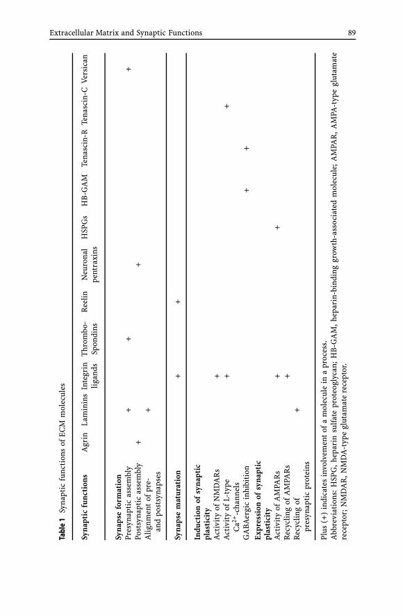
Extracellular Matrix and Synaptic Functions 89
Tabl
e1
Syna
ptic
func
tion
sof
ECM
mol
ecul
es
Syna
ptic
func
tion
sA
grin
Lam
inin
sIn
tegr
inT
hrom
bo-
Ree
linN
euro
nal
HSP
Gs
HB
-GA
MTe
nasc
in-R
Tena
scin
-CV
ersi
can
ligan
dsSp
ondi
nspe
ntra
xins
Syna
pse
form
atio
nPr
esyn
apti
cas
sem
bly
++
+Po
stsy
napt
icas
sem
bly
++
Alig
nmen
tof
pre-
+an
dpo
stsy
naps
es
Syna
pse
mat
urat
ion
++
Indu
ctio
nof
syna
ptic
plas
tici
tyA
ctiv
ity
ofN
MD
AR
s+
Act
ivit
yof
L-ty
pe+
+C
a2+-c
hann
els
GA
BAer
gic
inhi
biti
on+
+E
xpre
ssio
nof
syna
ptic
plas
tici
tyA
ctiv
ity
ofA
MPA
Rs
++
Rec
yclin
gof
AM
PAR
s+
Rec
yclin
gof
+pr
esyn
apti
cpr
otei
ns
Plus
(+)
indi
cate
sin
volv
emen
tof
am
olec
ule
ina
proc
ess.
Abb
revi
atio
ns:
HSP
G,
hepa
rin
sulfa
tepr
oteo
glyc
an;
HB
-GA
M,
hepa
rin-
bind
ing
grow
th-a
ssoc
iate
dm
olec
ule;
AM
PAR
,A
MPA
-typ
egl
utam
ate
rece
ptor
;NM
DA
R,N
MD
A-t
ype
glut
amat
ere
cept
or.

90 A. Dityatev et al.
impaired LTP and acquisition of spatial learning. The learning-dependentchanges in MMP expression were also shown to modify the stability ofthe actin-binding protein cortactin, which plays an essential role in reg-ulating the dendritic cytoskeleton and synaptic efficiency (Meighan et al.2006). The importance of regulated ECM remodeling by MMP in the CNSis further supported by a study of mice deficient in the tissue inhibitor ofmetalloproteinases-1 (TIMP-1), which display an impairment in olfactorymaze learning (Jourquin et al. 2005).
Conclusions
Taken together, a fascinating picture of complex contributions of ECMmolecules to the genesis, function and plasticity of peripheral and CNSsynapses has emerged during the last 15 years (Table 1) (Sanes and Licht-man 2001; Dityatev and Schachner 2003). This appears to be a rather shorttime, if one considers the sparse knowledge about molecular components, or-ganizing principles, and binding receptors of neural ECM 15 years ago. Nowwe can conclude that brain matrix components are in fact much more thanjust a passive microenvironment repellent for neurite outgrowth to stabilizethe status quo of mature CNS architecture but a highly dynamic network,which intensely communicates with glial and neuronal cells and contributesto neural synaptic transmission. Thus, presently, chemical synapses may beconsidered as a structural and functional “Quadriga” of presynaptic termi-nal, postsynaptic element, glial endfeet infiltrating synaptic contact sites anda highly specialized perisynaptic ECM (Fig. 2).
Acknowledgements Work in our laboratories is supported by the Deutsche Forschungs-gemeinschaft grants Di 702/4-1 and -2, and Gu 230/5-1 and by the Swiss National Fonds.
References
Adams JC (2001) Thrombospondins: multifunctional regulators of cell interactions. AnnuRev Cell Dev Biol 17:25–51
Amet LE, Lauri SE, Hienola A, Croll SD, Lu Y, Levorse JM, Prabhakaran B, Taira T, Rau-vala H, Vogt TF (2001) Enhanced hippocampal long-term potentiation in mice lackingheparin-binding growth-associated molecule. Mol Cell Neurosci 17:1014–1024
Anderson JL, Head SI, Morley JW (2005) Synaptic plasticity in the dy2J mouse model oflaminin alpha2-deficient congenital muscular dystrophy. Brain Res 1042:23–28
Aspberg A, Miura R, Bourdoulous S, Shimonaka M, Heinegard D, Schachner M, RuoslahtiE, Yamaguchi Y (1997) The C-type lectin domains of lecticans, a family of aggregat-ing chondroitin sulfate proteoglycans, bind tenascin-R by protein-protein interactionsindependent of carbohydrate moiety. Proc Natl Acad Sci USA 94:10116–10121
Beffert U, Weeber EJ, Durudas A, Qiu S, Masiulis I, Sweatt JD, Li WP, Adelmann G,Frotscher M, Hammer RE, Herz J (2005) Modulation of synaptic plasticity and mem-

Extracellular Matrix and Synaptic Functions 91
ory by Reelin involves differential splicing of the lipoprotein receptor Apoer2. Neuron47:567–579
Bergles DE, Roberts JD, Somogyi P, Jahr CE (2000) Glutamatergic synapses on oligoden-drocyte precursor cells in the hippocampus. Nature 405:187–191
Bernard-Trifilo JA, Kramar EA, Torp R, Lin CY, Pineda EA, Lynch G, Gall CM (2005) Inte-grin signaling cascades are operational in adult hippocampal synapses and modulateNMDA receptor physiology. J Neurochem 93:834–849
Bi X, Lynch G, Zhou J, Gall CM (2001) Polarized distribution of alpha5 integrin in den-drites of hippocampal and cortical neurons. J Comp Neurol 435:184–193
Bose CM, Qiu D, Bergamaschi A, Gravante B, Bossi M, Villa A, Rupp F, Malgaroli A (2000)Agrin controls synaptic differentiation in hippocampal neurons. J Neurosci 20:9086–9095
Brakebusch C, Seidenbecher CI, Asztely F, Rauch U, Matthies H, Meyer H, Krug M,Bockers TM, Zhou X, Kreutz MR, Montag D, Gundelfinger ED, Fassler R (2002)Brevican-deficient mice display impaired hippocampal CA1 long-term potentiationbut show no obvious deficits in learning and memory. Mol Cell Biol 22:7417–7427
Bruckner G, Grosche J, Schmidt S, Hartig W, Margolis RU, Delpech B, Seidenbecher CI,Czaniera R, Schachner M (2000) Postnatal development of perineuronal nets in wild-type mice and in a mutant deficient in tenascin-R. J Comp Neurol 428:616–629
Bukalo O, Schachner M, Dityatev A (2001) Modification of extracellular matrix by enzy-matic removal of chondroitin sulfate and by lack of tenascin-R differentially affectsseveral forms of synaptic plasticity in the hippocampus. Neuroscience 104:359–369
Bukalo O NA, Schachner M, Dityatev A (2005) GABAergic inhibition and metaplastic-ity: insights from analysis of mice deficient in the extracellular matrix glycoproteintenascin-R. Program No 5001, 2005 Abstract Viewer/Itinerary Planner Washington,DC: Society for Neuroscience, 2005 Online
Burgess RW, Nguyen QT, Son YJ, Lichtman JW, Sanes JR (1999) Alternatively spliced iso-forms of nerve- and muscle-derived agrin: their roles at the neuromuscular junction.Neuron 23:33–44
Butt AM, Kiff J, Hubbard P, Berry M (2002) Synantocytes: new functions for novel NG2expressing glia. J Neurocytol 31:551–565
Canoll PD, Petanceska S, Schlessinger J, Musacchio JM (1996) Three forms of RPTP-betaare differentially expressed during gliogenesis in the developing rat brain and duringglial cell differentiation in culture. J Neurosci Res 44:199–215
Chan CS, Weeber EJ, Kurup S, Sweatt JD, Davis RL (2003) Integrin requirement for hip-pocampal synaptic plasticity and spatial memory. J Neurosci 23:7107–7116
Chang HP, Lindberg FP, Wang HL, Huang AM, Lee EH (1999) Impaired memory retentionand decreased long-term potentiation in integrin-associated protein-deficient mice.Learn Mem 6:448–457
Chavis P, Westbrook G (2001) Integrins mediate functional pre- and postsynaptic matu-ration at a hippocampal synapse. Nature 411:317–321
Christopherson KS, Ullian EM, Stokes CC, Mullowney CE, Hell JW, Agah A, Lawler J,Mosher DF, Bornstein P, Barres BA (2005) Thrombospondins are astrocyte-secretedproteins that promote CNS synaptogenesis. Cell 120:421–433
Chun D, Gall CM, Bi X, Lynch G (2001) Evidence that integrins contribute to multiplestages in the consolidation of long term potentiation in rat hippocampus. Neuro-science 105:815–829
Cybulska-Klosowicz A, Zakrzewska R, Pyza E, Kossut M, Schachner M (2004) Reducedplasticity of cortical whisker representation in adult tenascin-C-deficient mice aftervibrissectomy. Eur J Neurosci 20:1538–1544

92 A. Dityatev et al.
Davis MJ, Wu X, Nurkiewicz TR, Kawasaki J, Gui P, Hill MA, Wilson E (2001) Regulationof ion channels by protein tyrosine phosphorylation. Am J Physiol Heart Circ Physiol281:H1835–1862
DeChiara TM, Bowen DC, Valenzuela DM, Simmons MV, Poueymirou WT, Thomas S,Kinetz E, Compton DL, Rojas E, Park JS, Smith C, DiStefano PS, Glass DJ, Burden SJ,Yancopoulos GD (1996) The receptor tyrosine kinase MuSK is required for neuromus-cular junction formation in vivo. Cell 85:501–512
Diers-Fenger M, Kirchhoff F, Kettenmann H, Levine JM, Trotter J (2001) AN2/NG2protein-expressing glial progenitor cells in the murine CNS: isolation, differentiation,and association with radial glia. Glia 34:213–228
Dityatev A, Schachner M (2003) Extracellular matrix molecules and synaptic plasticity.Nat Rev Neurosci 4:456–468
Dityatev A, Dityateva G, Sytnyk V, Delling M, Toni N, Nikonenko I, Muller D, Schach-ner M (2004) Polysialylated neural cell adhesion molecule promotes remodeling andformation of hippocampal synapses. J Neurosci 24:9372–9382
Einheber S, Schnapp LM, Salzer JL, Cappiello ZB, Milner TA (1996) Regional and ultra-structural distribution of the alpha 8 integrin subunit in developing and adult rat brainsuggests a role in synaptic function. J Comp Neurol 370:105–134
Evers MR, Salmen B, Bukalo O, Rollenhagen A, Bosl MR, Morellini F, Bartsch U, Dity-atev A, Schachner M (2002) Impairment of L-type Ca2+ channel-dependent forms ofhippocampal synaptic plasticity in mice deficient in the extracellular matrix glycopro-tein tenascin-C. J Neurosci 22:7177–7194
Ferreira A (1999) Abnormal synapse formation in agrin-depleted hippocampal neurons.J Cell Sci 112(Pt 24):4729–4738
Gall CM, Pinkstaff JK, Lauterborn JC, Xie Y, Lynch G (2003) Integrins regulate neuronalneurotrophin gene expression through effects on voltage-sensitive calcium channels.Neuroscience 118:925–940
Gingras J, Rassadi S, Cooper E, Ferns M (2002) Agrin plays an organizing role in theformation of sympathetic synapses. J Cell Biol 158:1109–1118
Goldberg GI, Frisch SM, He C, Wilhelm SM, Reich R, Collier IE (1990) Secreted proteases.Regulation of their activity and their possible role in metastasis. Ann NY Acad Sci580:375–384
Greenwood K, Butt AM (2003) Evidence that perinatal and adult NG2-glia are not con-ventional oligodendrocyte progenitors and do not depend on axons for their survival.Mol Cell Neurosci 23:544–558
Grotewiel MS, Beck CD, Wu KH, Zhu XR, Davis RL (1998) Integrin-mediated short-termmemory in Drosophila. Nature 391:455–460
Gurevicius K, Gureviciene I, Valjakka A, Schachner M, Tanila H (2004) Enhanced corti-cal and hippocampal neuronal excitability in mice deficient in the extracellular matrixglycoprotein tenascin-R. Mol Cell Neurosci 25:515–523
Hagihara K, Miura R, Kosaki R, Berglund E, Ranscht B, Yamaguchi Y (1999) Immuno-histochemical evidence for the brevican-tenascin-R interaction: colocalization in per-ineuronal nets suggests a physiological role for the interaction in the adult rat brain.J Comp Neurol 410:256–264
Hall RA, Vodyanoy V, Quan A, Sinnarajah S, Suppiramaniam V, Kessler M, Bahr BA(1996) Effects of heparin on the properties of solubilized and reconstituted rat brainAMPA receptors. Neurosci Lett 217:179–183
Hama H, Hara C, Yamaguchi K, Miyawaki A (2004) PKC signaling mediates global en-hancement of excitatory synaptogenesis in neurons triggered by local contact withastrocytes. Neuron 41:405–415
Extracellular Matrix and Synaptic Functions 93
Heck N, Klausmeyer A, Faissner A, Garwood J (2005) Cortical neurons express PSI,a novel isoform of phosphacan/RPTPbeta. Cell Tissue Res 321:323–333
Hilgenberg LG, Smith MA (2004) Agrin signaling in cortical neurons is mediated by a ty-rosine kinase-dependent increase in intracellular Ca2+ that engages both CaMKII andMAPK signal pathways. J Neurobiol 61:289–300
Hilgenberg LG, Hoover CL, Smith MA (1999) Evidence of an agrin receptor in corticalneurons. J Neurosci 19:7384–7393
Hilgenberg LG, Ho KD, Lee D, O’Dowd DK, Smith MA (2002) Agrin regulates neuronalresponses to excitatory neurotransmitters in vitro and in vivo. Mol Cell Neurosci19:97–110
Indyk JA, Chen ZL, Tsirka SE, Strickland S (2003) Laminin chain expression suggeststhat laminin-10 is a major isoform in the mouse hippocampus and is degraded bythe tissue plasminogen activator/plasmin protease cascade during excitotoxic injury.Neuroscience 116:359–371
Irintchev A, Rollenhagen A, Troncoso E, Kiss JZ, Schachner M (2005) Structural and func-tional aberrations in the cerebral cortex of tenascin-C deficient mice. Cereb Cortex15:950–962
Ishida A, Shimazaki K, Terashima T, Kawai N (1994) An electrophysiological and im-munohistochemical study of the hippocampus of the reeler mutant mouse. Brain Res662:60–68
Jourquin J, Tremblay E, Bernard A, Charton G, Chaillan FA, Marchetti E, Roman FS,Soloway PD, Dive V, Yiotakis A, Khrestchatisky M, Rivera S (2005) Tissue inhibitorof metalloproteinases-1 (TIMP-1) modulates neuronal death, axonal plasticity, andlearning and memory. Eur J Neurosci 22:2569–2578
Kaksonen M, Pavlov I, Voikar V, Lauri SE, Hienola A, Riekki R, Lakso M, Taira T,Rauvala H (2002) Syndecan-3-deficient mice exhibit enhanced LTP and impairedhippocampus-dependent memory. Mol Cell Neurosci 21:158–172
Knight D, Tolley LK, Kim DK, Lavidis NA, Noakes PG (2003) Functional analysis of neu-rotransmission at beta2-laminin deficient terminals. J Physiol 546:789–800
Kramar EA, Bernard JA, Gall CM, Lynch G (2002) Alpha3 integrin receptors contribute tothe consolidation of long-term potentiation. Neuroscience 110:29–39
Kramar EA, Bernard JA, Gall CM, Lynch G (2003) Integrins modulate fast excitatorytransmission at hippocampal synapses. J Biol Chem 278:10722–10730
Kuno K, Okada Y, Kawashima H, Nakamura H, Miyasaka M, Ohno H, Matsushima K(2000) ADAMTS-1 cleaves a cartilage proteoglycan, aggrecan. FEBS Lett 478:241–245
Lander C, Kind P, Maleski M, Hockfield S (1997) A family of activity-dependent neuronalcell-surface chondroitin sulfate proteoglycans in cat visual cortex. J Neurosci 17:1928–1939
Lauri SE, Taira T, Kaila K, Rauvala H (1996) Activity-induced enhancement of HB-GAMexpression in rat hippocampal slices. Neuroreport 7:1670–1674
Lauri SE, Rauvala H, Kaila K, Taira T (1998) Effect of heparin-binding growth-associatedmolecule (HB-GAM) on synaptic transmission and early LTP in rat hippocampalslices. Eur J Neurosci 10:188–194
Lauri SE, Kaukinen S, Kinnunen T, Ylinen A, Imai S, Kaila K, Taira T, Rauvala H(1999) Regulatory role and molecular interactions of a cell-surface heparan sul-fate proteoglycan (N-syndecan) in hippocampal long-term potentiation. J Neurosci19:1226–1235
Li Z, Hilgenberg LG, O’Dowd DK, Smith MA (1999) Formation of functional synapticconnections between cultured cortical neurons from agrin-deficient mice. J Neurobiol39:547–557

94 A. Dityatev et al.
Libby RT, Lavallee CR, Balkema GW, Brunken WJ, Hunter DD (1999) Disruption oflaminin beta2 chain production causes alterations in morphology and function in theCNS. J Neurosci 19:9399–9411
Lin B, Arai AC, Lynch G, Gall CM (2003) Integrins regulate NMDA receptor-mediatedsynaptic currents. J Neurophysiol 89:2874–2878
Lin SC, Bergles DE (2004a) Synaptic signaling between neurons and glia. Glia 47:290–298Lin SC, Bergles DE (2004b) Synaptic signaling between GABAergic interneurons and
oligodendrocyte precursor cells in the hippocampus. Nat Neurosci 7:24–32Lin SC, Huck JH, Roberts JD, Macklin WB, Somogyi P, Bergles DE (2005) Climbing fiber
innervation of NG2-expressing glia in the mammalian cerebellum. Neuron 46:773–785Liu WS, Pesold C, Rodriguez MA, Carboni G, Auta J, Lacor P, Larson J, Condie BG,
Guidotti A, Costa E (2001) Down-regulation of dendritic spine and glutamic acid de-carboxylase 67 expressions in the reelin haploinsufficient heterozygous reeler mouse.Proc Natl Acad Sci USA 98:3477–3482
Margolis RK, Rauch U, Maurel P, Margolis RU (1996) Neurocan and phosphacan: two ma-jor nervous tissue-specific chondroitin sulfate proteoglycans. Perspect Dev Neurobiol3:273–290
Martin AO, Alonso G, Guerineau NC (2005) Agrin mediates a rapid switch from electricalcoupling to chemical neurotransmission during synaptogenesis. J Cell Biol 169:503–514
Matthews RT, Kelly GM, Zerillo CA, Gray G, Tiemeyer M, Hockfield S (2002) Aggrecanglycoforms contribute to the molecular heterogeneity of perineuronal nets. J Neurosci22:7536–7547
Mi R, Tang X, Sutter R, Xu D, Worley P, O’Brien RJ (2002) Differing mechanisms for glu-tamate receptor aggregation on dendritic spines and shafts in cultured hippocampalneurons. J Neurosci 22:7606–7616
Mi R, Sia GM, Rosen K, Tang X, Moghekar A, Black JL, McEnery M, Huganir RL, O’BrienRJ (2004) AMPA receptor-dependent clustering of synaptic NMDA receptors is me-diated by Stargazin and NR2A/B in spinal neurons and hippocampal interneurons.Neuron 44:335–349
Miura R, Ethell IM, Yamaguchi Y (2001) Carbohydrate-protein interactions betweenHNK-1-reactive sulfoglucuronyl glycolipids and the proteoglycan lectin domain me-diate neuronal cell adhesion and neurite outgrowth. J Neurochem 76:413–424
Miura R, Aspberg A, Ethell IM, Hagihara K, Schnaar RL, Ruoslahti E, Yamaguchi Y (1999)The proteoglycan lectin domain binds sulfated cell surface glycolipids and promotescell adhesion. J Biol Chem 274:11431–11438
Miyata S, Akagi A, Hayashi N, Watanabe K, Oohira A (2004) Activity-dependent regula-tion of a chondroitin sulfate proteoglycan 6B4 phosphacan/RPTPbeta in the hypotha-lamic supraoptic nucleus. Brain Res 1017:163–171
Murai KK, Misner D, Ranscht B (2002) Contactin supports synaptic plasticity associatedwith hippocampal long-term depression but not potentiation. Curr Biol 12:181–190
Nakagami Y, Abe K, Nishiyama N, Matsuki N (2000) Laminin degradation by plasminregulates long-term potentiation. J Neurosci 20:2003–2010
Nakamura H, Fujii Y, Inoki I, Sugimoto K, Tanzawa K, Matsuki H, Miura R, Yamaguchi Y,Okada Y (2000) Brevican is degraded by matrix metalloproteinases and aggrecanase-1(ADAMTS4) at different sites. J Biol Chem 275:38885–38890
Nakic M, Manahan-Vaughan D, Reymann KG, Schachner M (1998) Long-term potentia-tion in vivo increases rat hippocampal tenascin-C expression. J Neurobiol 37:393–404
Niisato K, Fujikawa A, Komai S, Shintani T, Watanabe E, Sakaguchi G, Katsuura G,Manabe T, Noda M (2005) Age-dependent enhancement of hippocampal long-term

Extracellular Matrix and Synaptic Functions 95
potentiation and impairment of spatial learning through the Rho-associated kinasepathway in protein tyrosine phosphatase receptor type Z-deficient mice. J Neurosci25:1081–1088
Nikonenko A, Schmidt S, Skibo G, Bruckner G, Schachner M (2003a) Tenascin-R-deficientmice show structural alterations of symmetric perisomatic synapses in the CA1 regionof the hippocampus. J Comp Neurol 456:338–349
Nikonenko I, Toni N, Moosmayer M, Shigeri Y, Muller D, Sargent Jones L (2003b) In-tegrins are involved in synaptogenesis, cell spreading, and adhesion in the postnatalbrain. Brain Res Dev Brain Res 140:185–194
Nishimune H, Sanes JR, Carlson SS (2004) A synaptic laminin-calcium channel interac-tion organizes active zones in motor nerve terminals. Nature 432:580–587
Nishimura SL, Boylen KP, Einheber S, Milner TA, Ramos DM, Pytela R (1998) Synapticand glial localization of the integrin alphavbeta8 in mouse and rat brain. Brain Res791:271–282
Nishiyama A, Stallcup WB (1993) Expression of NG2 proteoglycan causes retention oftype VI collagen on the cell surface. Mol Biol Cell 4:1097–1108
Nishiyama A, Dahlin KJ, Prince JT, Johnstone SR, Stallcup WB (1991) The primary struc-ture of NG2, a novel membrane-spanning proteoglycan. J Cell Biol 114:359–371
O’Brien R, Xu D, Mi R, Tang X, Hopf C, Worley P (2002) Synaptically targeted narpplays an essential role in the aggregation of AMPA receptors at excitatory synapses incultured spinal neurons. J Neurosci 22:4487–4498
O’Brien RJ, Xu D, Petralia RS, Steward O, Huganir RL, Worley P (1999) Synaptic clusteringof AMPA receptors by the extracellular immediate-early gene product Narp. Neuron23:309–323
Patton BL, Cunningham JM, Thyboll J, Kortesmaa J, Westerblad H, Edstrom L, Tryggva-son K, Sanes JR (2001) Properly formed but improperly localized synaptic specializa-tions in the absence of laminin alpha4. Nat Neurosci 4:597–604
Pavlov I, Voikar V, Kaksonen M, Lauri SE, Hienola A, Taira T, Rauvala H (2002) Roleof heparin-binding growth-associated molecule (HB-GAM) in hippocampal LTP andspatial learning revealed by studies on overexpressing and knockout mice. Mol CellNeurosci 20:330–342
Phillips WD, Maimone MM, Merlie JP (1991) Mutagenesis of the 43-kD postsynaptic pro-tein defines domains involved in plasma membrane targeting and AChR clustering.J Cell Biol 115:1713–1723
Pinkstaff JK, Detterich J, Lynch G, Gall C (1999) Integrin subunit gene expression isregionally differentiated in adult brain. J Neurosci 19:1541–1556
Rauvala H, Peng HB (1997) HB-GAM (heparin-binding growth-associated molecule) andheparin-type glycans in the development and plasticity of neuron-target contacts. ProgNeurobiol 52:127–144
Ronn LC, Pedersen N, Jahnsen H, Berezin V, Bock E (1997) Brain plasticity and the neuralcell adhesion molecule (NCAM). Adv Exp Med Biol 429:305–322
Saghatelyan AK, Gorissen S, Albert M, Hertlein B, Schachner M, Dityatev A (2000) Theextracellular matrix molecule tenascin-R and its HNK-1 carbohydrate modulate peri-somatic inhibition and long-term potentiation in the CA1 region of the hippocampus.Eur J Neurosci 12:3331–3342
Saghatelyan AK, Dityatev A, Schmidt S, Schuster T, Bartsch U, Schachner M (2001)Reduced perisomatic inhibition, increased excitatory transmission, and impairedlong-term potentiation in mice deficient for the extracellular matrix glycoproteintenascin-R. Mol Cell Neurosci 17:226–240

96 A. Dityatev et al.
Saghatelyan AK, Snapyan M, Gorissen S, Meigel I, Mosbacher J, Kaupmann K, Bettler B,Kornilov AV, Nifantiev NE, Sakanyan V, Schachner M, Dityatev A (2003) Recognitionmolecule associated carbohydrate inhibits postsynaptic GABA(B) receptors: a mech-anism for homeostatic regulation of GABA release in perisomatic synapses. Mol CellNeurosci 24:271–282
Sanes JR, Lichtman JW (2001) Induction, assembly, maturation and maintenance ofa postsynaptic apparatus. Nat Rev Neurosci 2:791–805
Schneider S, Bosse F, D’Urso D, Muller H, Sereda MW, Nave K, Niehaus A, Kempf T,Schnolzer M, Trotter J (2001) The AN2 protein is a novel marker for the Schwanncell lineage expressed by immature and nonmyelinating Schwann cells. J Neurosci21:920–933
Schuster T, Krug M, Stalder M, Hackel N, Gerardy-Schahn R, Schachner M (2001) Immu-noelectron microscopic localization of the neural recognition molecules L1, NCAM,and its isoform NCAM180, the NCAM-associated polysialic acid, beta1 integrin andthe extracellular matrix molecule tenascin-R in synapses of the adult rat hippocampus.J Neurobiol 49:142–158
Seidenbecher C, Richter K, Gundelfinger ED (1997) Brevican, a conditional proteogly-can from rat brain: characterization of secreted and GPI-anchored isoforms. In:Teelken AW, Korf J (eds) Neurochemistry. Plenum Press, New York, pp 901–904
Seidenbecher CI, Smalla KH, Fischer N, Gundelfinger ED, Kreutz MR (2002) Brevicanisoforms associate with neural membranes. J Neurochem 83:738–746
Senn C, Kutsche M, Saghatelyan A, Bosl MR, Lohler J, Bartsch U, Morellini F, SchachnerM (2002) Mice deficient for the HNK-1 sulfotransferase show alterations in synapticefficacy and spatial learning and memory. Mol Cell Neurosci 20:712–729
Serpinskaya AS, Feng G, Sanes JR, Craig AM (1999) Synapse formation by hippocampalneurons from agrin-deficient mice. Dev Biol 205:65–78
Sinagra M, Verrier D, Frankova D, Korwek KM, Blahos J, Weeber EJ, Manzoni OJ, Chavis P(2005) Reelin, very-low-density lipoprotein receptor, and apolipoprotein E receptor 2control somatic NMDA receptor composition during hippocampal maturation in vitro.J Neurosci 25:6127–6136
Sinnarajah S, Suppiramaniam V, Kumar KP, Hall RA, Bahr BA, Vodyanoy V (1999) Hep-arin modulates the single channel kinetics of reconstituted AMPA receptors from ratbrain. Synapse 31:203–209
Slezak M, Pfrieger FW (2003) New roles for astrocytes: regulation of CNS synaptogenesis.Trends Neurosci 26:531–535
Son YJ, Scranton TW, Sunderland WJ, Baek SJ, Miner JH, Sanes JR, Carlson SS (2000) Thesynaptic vesicle protein SV2 is complexed with an alpha5-containing laminin on thenerve terminal surface. J Biol Chem 275:451–460
Staubli U, Vanderklish P, Lynch G (1990) An inhibitor of integrin receptors blocks long-term potentiation. Behav Neural Biol 53:1–5
Staubli U, Chun D, Lynch G (1998) Time-dependent reversal of long-term potentiation byan integrin antagonist. J Neurosci 18:3460–3469
Stegmuller J, Werner H, Nave KA, Trotter J (2003) The proteoglycan NG2 is complexedwith alpha-amino-3-hydroxy-5-methyl-4-isoxazolepropionic acid (AMPA) receptors bythe PDZ glutamate receptor interaction protein (GRIP) in glial progenitor cells. Impli-cations for glial-neuronal signaling. J Biol Chem 278:3590–3598
Strekalova T, Sun M, Sibbe M, Evers M, Dityatev A, Gass P, Schachner M (2002) Fi-bronectin domains of extracellular matrix molecule tenascin-C modulate hippocampallearning and synaptic plasticity. Mol Cell Neurosci 21:173–187

Extracellular Matrix and Synaptic Functions 97
Sykova E, Vorisek I, Mazel T, Antonova T, Schachner M (2005) Reduced extracellularspace in the brain of tenascin-R- and HNK-1-sulphotransferase deficient mice. Eur JNeurosci 22:1873–1880
VanSaun M, Werle MJ (2000) Matrix metalloproteinase-3 removes agrin from synapticbasal lamina. J Neurobiol 43:140–149
VanSaun M, Herrera AA, Werle MJ (2003) Structural alterations at the neuromuscularjunctions of matrix metalloproteinase 3 null mutant mice. J Neurocytol 32:1129–1142
Weber P, Bartsch U, Rasband MN, Czaniera R, Lang Y, Bluethmann H, Margolis RU,Levinson SR, Shrager P, Montag D, Schachner M (1999) Mice deficient for tenascin-Rdisplay alterations of the extracellular matrix and decreased axonal conduction veloci-ties in the CNS. J Neurosci 19:4245–4262
Weeber EJ, Beffert U, Jones C, Christian JM, Forster E, Sweatt JD, Herz J (2002) Reelin andApoE receptors cooperate to enhance hippocampal synaptic plasticity and learning.J Biol Chem 277:39944–39952
Welzl H, Stork O (2003) Cell adhesion molecules: key players in memory consolidation?News Physiol Sci 18:147–150
Werle MJ, VanSaun M (2003) Activity dependent removal of agrin from synaptic basallamina by matrix metalloproteinase 3. J Neurocytol 32:905–913
Xu D, Hopf C, Reddy R, Cho RW, Guo L, Lanahan A, Petralia RS, Wenthold RJ, O’Brien RJ,Worley P (2003) Narp and NP1 form heterocomplexes that function in developmentaland activity-dependent synaptic plasticity. Neuron 39:513–528
Yamagata M, Sanes JR (2005) Versican in the developing brain: lamina-specific expressionin interneuronal subsets and role in presynaptic maturation. J Neurosci 25:8457–8467
Yamaguchi Y (2000) Lecticans: organizers of the brain extracellular matrix. Cell Mol LifeSci 57:276–289
Yamamoto S, Oka S, Inoue M, Shimuta M, Manabe T, Takahashi H, Miyamoto M, AsanoM, Sakagami J, Sudo K, Iwakura Y, Ono K, Kawasaki T (2002) Mice deficient in ner-vous system-specific carbohydrate epitope HNK-1 exhibit impaired synaptic plasticityand spatial learning. J Biol Chem 277:27227–27231
Zaremba S, Guimaraes A, Kalb RG, Hockfield S (1989) Characterization of an activity-dependent, neuronal surface proteoglycan identified with monoclonal antibody Cat-301. Neuron 2:1207–1219
Zhou XH, Brakebusch C, Matthies H, Oohashi T, Hirsch E, Moser M, Krug M, Seiden-becher CI, Boeckers TM, Rauch U, Buettner R, Gundelfinger ED, Fassler R (2001)Neurocan is dispensable for brain development. Mol Cell Biol 21:5970–5978

Results Probl Cell Differ (43)E. Gundelfinger, C. Seidenbecher, B. Schraven:Cell Communication in Nervous and Immune SystemDOI 10.1007/013/Published online: 30 March 2006© Springer-Verlag Berlin Heidelberg 2006
Electrical Synapses – Gap Junctions in the Brain
Carola Meier (�) · Rolf Dermietzel
Department of Neuroanatomy and Molecular Brain Research, Ruhr-University Bochum,Universitätsstrasse 150, 44801 Bochum, [email protected]
Abstract In the nervous system, interneuronal communication can occur via indirect ordirect transmission. The mode of indirect communication involves chemical synapses, inwhich transmitters are released into the extracellular space to subsequently bind to thepostsynaptic cell membrane. Direct communication is mediated by electrical synapses,and will be the focus of this review.
The most prevalent group of electrical synapses are neuronal gap junctions (bothterms are used interchangeably in this article), which directly connect the intracellularspace of two cells by gap junction channels. The structural components of gap junc-tion channels in the nervous system are connexin proteins, and, as recently identified,pannexin proteins.
Connexin gap junction channels enable the intercellular, bidirectional transport ofions, metabolites, second messengers and other molecules smaller than 1 kD. More than20 connexin genes have been found in the mouse and human genome. With the cloningof connexin36 (Cx36), a connexin protein with predominantly neuronal expression, thebiochemical correlate of electrotonic transmission between neurons was identified. Weoutline the distribution of Cx36 as well as two other neuronal connexins (Cx57 and Cx45)in the nervous system, describing their spatial and temporal expression patterns. One fo-cus in this review was the retina, as it shows many and diverse electrical synapses whoseconnexin components have been identified in fish and mammals. In view of the func-tion of neuronal gap junctions, the network of inhibitory interneurons will be reviewedin detail, focussing on the hippocampus.
Although in vivo data on pannexin proteins are still restricted to information onmRNA expression, electrophysiological data and the expression pattern in the nervoussystem have been included.
1Introduction
Communication between cells, particularly those of the nervous system, isessential for the existence and survival of any living animal. In the centralnervous system, the essential feature of neuronal cell interaction is a highoperational speed, which is achieved by synapses.
Synapses can mediate neuronal communication by two means: indirecttransmission, i.e. chemical synapses acting via neurotransmitter release, ordirect transmission, i.e. electrical synapses acting via ion, metabolite, and sec-ond messenger transmission between cells. The vast majority of electrical

100 C. Meier · R. Dermietzel
synapses are gap junction channels, the structural correlate of which are con-nexin proteins, and also pannexin proteins (Sect. 6.2), the latter being moreand more recognized in their channel-forming capacity.
Connexin gap junctions are formed by the apposition of two contactinghemichannels, each in the membrane of one cell. The components of onehemichannel, the connexon, are six transmembrane proteins named connex-ins (Kumar and Gilula 1996). To date, 20 and 21 connexin genes have beenidentified in the mouse and human genome, respectively (Willecke et al. 2002;Sohl et al. 2004). Most, but not all, of the mouse connexin genes correspondto human orthologous connexin sequences (Sohl et al. 2004). Connexins (Cx)are generally named after their approximate molecular weight, i.e. Cx32 fora 32 kD protein. This family of proteins possesses several connexin-specificcharacteristics, for instance four transmembrane domains, two extracellular
Fig. 1 Schematic representation of gap junctions and their connexin constituents.a Connexin protein with cytoplasmic C- (COOH) and N- (NH2) terminus, four trans-membrane domains (M1–M4), two extracellular (EL1 and EL2) and one cytoplasmicloop (CL) domain. b Single gap junction channel, formed by close apposition of twohemichannels, each assembled by six connexin proteins. c Gap junction plaque, charac-terized by the reduced distance between cell membranes (3.5 nm within the gap junctionarea)

Electrical Synapses – Gap Junctions in the Brain 101
loops, and cytoplasmic C- and N-termini (Kumar and Gilula, 1996) (Fig. 1a).The two cytoplasmic domains, particularly the C-terminus, were recentlyshown to be important for signal transduction, phosphorylation events andassociation with kinases, tight junction proteins and the cytoskeleton (Giep-mans, 2004). The N-terminal domain, at least in the Cx36 protein, has beenimplicated in the determination of its subcellular localization (Zoidl et al.2002). Connexons (Fig. 1b) may have only one type of connexins (homomericconnexons), or several (heteromeric), and the gap junction channel can beformed by two identical (homotypic) or different (heterotypic) connexons.The most common assembly of gap junction channels is the gap junctionplaque (Fig. 1c), characterized by the reduced space between membranes ofcontacting cells. However, additional gap junction formations have recentlybeen described in the retina (Rash et al. 2004).
Gap junction channels allow the bidirectional intercellular passage ofmolecules smaller than 1 kD, thereby mediating ionic and metabolic commu-nication between directly connected cells. With their speed, simplicity andreciprocity, gap junctions can fulfil very specialized tasks in neuronal cir-cuits, which are different form those accomplished by chemical synapses.Gap junctions in the brain show distinct spatial and temporal expression pat-terns. In neuronal cells, the abundance of gap junctions and their implicationfor neuronal circuits has only been recognized recently. Electrical synapsesare now being extensively examined in mammals, and advances in electro-physiology, transgenic animal technology, cell imaging, and ultrastructuraltechniques (freeze-fracture replica immunolabelling, FRIL) are allowing thedetailed analysis of expression and function of connexins in developing andmature nervous system.
2Gap Junctions in Development
Connexin gap junctions are prevalent during the early phase of neurogenesis(prenatal phase) (Dermietzel et al. 1989) while chemical synapses emerge inthe postnatal phase (Blue and Parnavelas, 1983; Kandler and Katz, 1998). Ap-parently, both modes of communication serve for different functions, and itis to be expected to find them in different spatial or temporal settings. Directintercellular transfer of molecules can be detected from the eight cell stageembryo onwards (McLachlin and Kidder, 1986), and gap junction-formingproteins (i.e. connexins) are already present at the zygote stage (Barron et al.1989). In general, connexin-containing gap junctions allow the intercellularexchange of ions (electrical coupling) and transmission of biologically activemolecules (metabolic coupling) between adjacent cells in multicellular organ-isms. During early cortical development, gap junctions are likely to mediateneuronal communication by metabolic interaction. Significant changes hap-
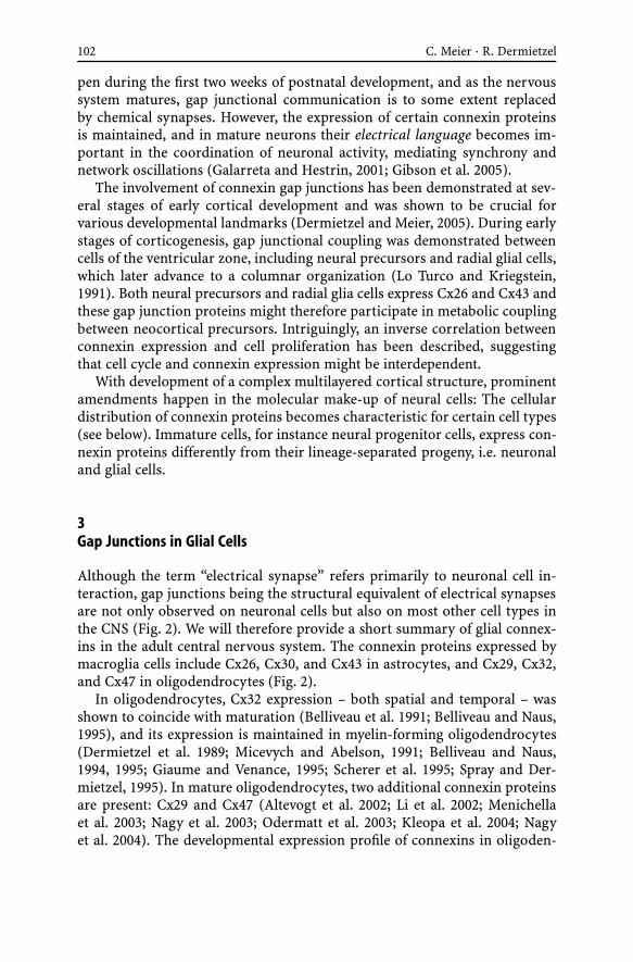
102 C. Meier · R. Dermietzel
pen during the first two weeks of postnatal development, and as the nervoussystem matures, gap junctional communication is to some extent replacedby chemical synapses. However, the expression of certain connexin proteinsis maintained, and in mature neurons their electrical language becomes im-portant in the coordination of neuronal activity, mediating synchrony andnetwork oscillations (Galarreta and Hestrin, 2001; Gibson et al. 2005).
The involvement of connexin gap junctions has been demonstrated at sev-eral stages of early cortical development and was shown to be crucial forvarious developmental landmarks (Dermietzel and Meier, 2005). During earlystages of corticogenesis, gap junctional coupling was demonstrated betweencells of the ventricular zone, including neural precursors and radial glial cells,which later advance to a columnar organization (Lo Turco and Kriegstein,1991). Both neural precursors and radial glia cells express Cx26 and Cx43 andthese gap junction proteins might therefore participate in metabolic couplingbetween neocortical precursors. Intriguingly, an inverse correlation betweenconnexin expression and cell proliferation has been described, suggestingthat cell cycle and connexin expression might be interdependent.
With development of a complex multilayered cortical structure, prominentamendments happen in the molecular make-up of neural cells: The cellulardistribution of connexin proteins becomes characteristic for certain cell types(see below). Immature cells, for instance neural progenitor cells, express con-nexin proteins differently from their lineage-separated progeny, i.e. neuronaland glial cells.
3Gap Junctions in Glial Cells
Although the term “electrical synapse” refers primarily to neuronal cell in-teraction, gap junctions being the structural equivalent of electrical synapsesare not only observed on neuronal cells but also on most other cell types inthe CNS (Fig. 2). We will therefore provide a short summary of glial connex-ins in the adult central nervous system. The connexin proteins expressed bymacroglia cells include Cx26, Cx30, and Cx43 in astrocytes, and Cx29, Cx32,and Cx47 in oligodendrocytes (Fig. 2).
In oligodendrocytes, Cx32 expression – both spatial and temporal – wasshown to coincide with maturation (Belliveau et al. 1991; Belliveau and Naus,1995), and its expression is maintained in myelin-forming oligodendrocytes(Dermietzel et al. 1989; Micevych and Abelson, 1991; Belliveau and Naus,1994, 1995; Giaume and Venance, 1995; Scherer et al. 1995; Spray and Der-mietzel, 1995). In mature oligodendrocytes, two additional connexin proteinsare present: Cx29 and Cx47 (Altevogt et al. 2002; Li et al. 2002; Menichellaet al. 2003; Nagy et al. 2003; Odermatt et al. 2003; Kleopa et al. 2004; Nagyet al. 2004). The developmental expression profile of connexins in oligoden-

Electrical Synapses – Gap Junctions in the Brain 103
Fig. 2 Schematic representation of connexin expression in astrocytes, oligodendrocytes andneurons. Connexin36 gap junctions in the brain occur between neurons only. Astrocytesform gap junctions consisting of Cx43, Cx26 and Cx30, whereas oligodendrocytes expressCx29, Cx32 and Cx47, with each of these proteins forming gap junctions with distinct partnerconnexins. The exception is Cx29, the coupling partner of which is still unknown
drocytes parallels that of myelin proteins (Altevogt et al. 2002). Expression ofCx45 has been demonstrated in developing oligodendrocyte precursors, how-ever, studies on Cx45-transgenic animals seem to exclude its expression inmature oligodendrocytes (Maxeiner et al. 2003).
Intriguingly, gap junctions between oligodendrocytes themselves arerarely if ever observed (Massa and Mugnaini, 1982; Waxman and Black,1984; Rash et al. 2001a). However, oligodendrocytes are integrated into theastrocytic network by gap junctional communication, creating a “panglialsyncytium” (Massa and Mugnaini, 1982) consisting of heterotypic channels.It has been suggested that oligodendrocyte Cx32 associates mainly with as-trocytic Cx26, whereas oligodendrocyte Cx47 associates with astrocytic Cx43and Cx30 (Rash et al. 2001a,b; Altevogt and Paul, 2004).
There is also extensive astrocyte–astrocyte coupling observed in the glialsyncytium, and mature astrocytes were shown to express a number of con-

104 C. Meier · R. Dermietzel
nexin proteins, including Cx43, Cx30 and Cx26 (Nagy and Rash, 2000). Thesethree connexin proteins have been identified in astrocytes of adult rat brainand spinal cord and were ultrastructurally identified as astrocytic gap junc-tions by immunolabelling on freeze-fracture replicas (Nagy and Rash, 2000).Studies on Cx43-deficient astrocytes also imply that the pattern of connexinexpression can be modulated: In Cx43-deficient cells, the additional expressionof Cx40, Cx45 and Cx46 was detected by RT-PCR (Dermietzel et al. 2000a).
4Challenges in Expression Analysis of Gap Junction Proteins
Quantitative and qualitative changes of connexin expression were demon-strated under certain developmental and pathological circumstances (seeabove). Thus, analysis of gap junctional coupling can only provide a depictionof the actual connexin expression, irrespective of a putative diverse mo-lecular make-up at different developmental stages or under the influence ofpathological conditions including de- and regeneration. Examples of patho-logical changes in connexin expression include an increase of Cx46 protein inSchwann cells during peripheral nerve regeneration (Chandross et al. 1996),neuronal expression of Cx45 (increase) and Cx36 (decrease) upon kainatetreatment (Condorelli et al. 2003), increased expression of Cx32 and Cx36 inCA1 neurons after global ischemia (Oguro et al. 2001), and the expression ofCx30 in apoptotic neuronal cells after kainate-induced seizures (Condorelliet al. 2002). One example of temporary developmental expression is the tran-sient coupling of developing muscle fibres. In the chick, the earliest develop-mental stage at which Cx36 mRNA was detected by in situ hybridization wasembryonic day 2 (i.e. HH 13). During embryonic development, expressionof Cx36 mRNA was assigned to myotomes (co-localization with MyoD andFGF-8), whereas a few days later, Cx36 expression was also detected in limbmuscles (co-localization with Pax3 and MyoD). In view of skeletal muscle be-ing one of the few tissues that do not show gap junctional communication inthe adult animal, the expression of Cx36 during development seemed some-what surprising. However, several studies have investigated the occurrence ofgap junctions ultrastructurally, and from these studies it became evident thatdeveloping skeletal muscle cells are indeed initially coupled through gap junc-tions, but with the formation of muscle fibres become uncoupled (Rash andStaehelin, 1974; Keeter et al. 1975; Kalderon et al. 1977; Schmalbruch, 1982).
In the nervous system, extensive cell branching, long cellular processes,and the intermingling of various cell types impose an additional challengein gap junction research. Particularly, the assignment of a certain connexinto a specific cell or cell type turned out to be a general problem, as thecellular identity is often obscured and cannot be resolved due to technicalrestrictions, for instance the limit of resolution. In addition, the detection

Electrical Synapses – Gap Junctions in the Brain 105
of connexin mRNA does not necessarily reflect its translation into protein(Traub et al. 1998; Condorelli et al. 2003); the presence of connexin proteinsdoes not automatically result in junctional conductance as observed in thecase of Cx29 (Altevogt et al. 2002).
With more than 20 isoforms of connexin genes, another problem becameevident: Any technique based on binding of antibody to antigen (i.e. im-munohistochemistry) or on hybridization of probe to mRNA (i.e. in situhybridization) involves the risk of cross-reactivity between different connexinisoforms. Merely connexin-deficient animals provide unambiguous negativecontrols for the specificity of antibodies and in situ hybridization probes. Thegeneration of transgenic animals in which the connexin coding sequence hasbeen replaced by reporter genes allows for easier identification and interpre-tation of the expression profiles of connexins (Sohl et al. 2004). Using thetransgenic technique, the reassignment of cellular expression sites has alreadybeen achieved in some cases, for instance for Cx45 and Cx47 (Maxeiner et al.2003; Odermatt et al. 2003). However, every technique has its limitation, andin the case of transgenic animals some expression sites might not be detectedbecause of weak promotor activity or a generally low abundance of the trans-lated product in heterozygous animals. The only way to overcome the describeddifficulties is theapplicationof abroadspectrumof techniques, including trans-genic animals (Meier et al. 2002), and approaches that combine microsamplingof tissue with molecular biological methods (Weickert et al. 2005).
5Some History on Electrical Synapses
In general, synapses can be defined as specialized sites of functional interac-tion between neurons. As neuronal gap junctions comprise defined areas ofneuronal membranes, provide for direct cell-to-cell contact, and enable theexchange of molecules between neurons, they fulfil the requirements for be-ing designated synaptic contacts. Initially, their predominant function wasthought to be the intercellular transport of ions, and neuronal gap junctionswere therefore named “electrical synapses”. Although this terminology alsoincludes a number of junctional specializations that are not gap junctions(Bennett, 1977; Faber and Korn, 1989; Jefferys, 1995), we will use the terms“neuronal gap junction” and “electrical synapse” synonymously.
The concept that neurons communicate electrically originated from phar-macological, ultrastructural, and physiological observations described morethan 50 years ago (Bennett, 1997; Zoidl and Dermietzel, 2002; Connors andLong, 2004). Nevertheless, the tug-of-war for the acknowledgement and ac-ceptance of electrical versus chemical synapses lasted for several decades.Application of computational and molecular methods and technical advancesin ultrastructural analysis have provided new insights into the mechanisms

106 C. Meier · R. Dermietzel
of synaptic transmission, and novel ideas on the impact and importance ofneuronal gap junctions have been brought forward.
In the 1950s, the first compelling evidence for the presence of electricalsynapses was gathered by studies on lower vertebrates. Initially, electro-tonic transmission was identified between segments of crayfish cord giantfibres (Furshpan and Potter, 1957, 1959; Grundfest, 1959). Watanabe (Watan-abe, 1958) demonstrated that neurons in the cardiac ganglion of the mantidshrimp, Squilla, are electrically coupled – and he suggested that coupling wasresponsible for synchronous firing of these cells, a hypothesis that would beproven right about 40 years later (see below). In the study by Bennett andco-workers (Bennett et al. 1959), electrical synapses were for the first timeidentified ultrastructurally in supramedullary neurons of a teleost fish (thepufferfish), and these were also shown to fire synchronously. Because of thesefindings, the co-existence of electrical and chemical synapses became gener-ally accepted, however, the functional impact of electrical synapses was stillattributed to the nervous system of lower vertebrates. In consequence, elec-trical synapses were thought to represent primitive phylogenetic devices forintercellular neuronal communication that have been abandoned in mam-mals. This hypothesis had to be revised with the identification of electro-tonic transmission in higher vertebrates: In the 1970s, several areas with gapjunction-mediated electrical transmission were defined in the mammaliancentral nervous system, including the olfactory bulb (Pinching and Powell,1971), neocortex (Sloper, 1972; Peinado et al. 1993), the mesencephalic nu-cleus (Baker and Llinas, 1971), inferior olive (Llinas et al. 1974) and thecerebellum (Sotelo and Llinas, 1972).
With the cloning of Cx36, the impact of neuronal connexins in the mam-malian central nervous system has become much more evident (see below).With innovative methods like freeze-fracture replica immunolabelling, the as-tonishing abundance of electric synapses begins to be unravelled (Rash et al.2000, 2001b). However, in some cell types known to be electrically coupled,Cx36 is essentially absent. With the identification of pannexin proteins andthe demonstration of their junctional properties, the pannexin family of gapjunction proteins moved into the focus of electrical synapse research.
6Gap Junctions in Neurons – Electrical Synapses
6.1Connexin Gap Junctions
Of all connexins expressed in cells of the nervous system, only few can beassigned to neurons – and for some others, their cellular correlate is still ofdebate. At least ten of the mammalian connexins are abundant in the cen-

Electrical Synapses – Gap Junctions in the Brain 107
tral nervous system, however, there seems to be a prevailing separation ofneuronal and glial connexins. Hardly any of the “glial” connexins describedabove are expressed in mature neuronal cells (Rash et al. 2001a,b; Odermattet al. 2003), although some might be present in neurons during develop-ment or under pathological conditions (Nagy et al. 2001; Condorelli et al.2003; Honma et al. 2004). Intriguingly, in brain areas displaying high elec-trical coupling, i.e. hippocampus and retina, neurons were devoid of any ofthe known connexin proteins, which were cloned before 1998. This apparentdiscrepancy was beginning to be resolved with the discovery of a novel fam-ily of connexin proteins, whose expression was attributed predominantly toneurons of the central nervous system.
Cloning of this neuronal subfamily of connexin genes commenced in fishwith the identification of Cx35 (O’Brien et al. 1996, 1998; White et al. 1999),which was followed by the cloning of its orthologue, Cx36, in mouse (Con-dorelli et al. 1998; Sohl et al. 1998) and man (Belluardo et al. 1999). Mo-lecular analysis revealed that Cx35 and Cx36 differ from other connexins bythe insertion of a single intron interrupting the coding region, thus plac-ing Cx35/Cx36 in a new subgroup of gap junction proteins, designated theγ -group (Condorelli et al. 1998; O’Brien et al. 1998; Sohl et al. 1998). Ini-tial electrophysiological studies (Srinivas et al. 1999) provided first functionalevidence for Cx36 being a main player of electrotonic synapses in neurons.
6.1.1Spatial Expression of Cx36
The spatial expression of Cx36 was studied by in situ hybridization (Con-dorelli et al. 1998; Belluardo et al. 1999), Northern blot analysis (Sohl et al.1998; Al-Ubaidi et al. 2000), RT-PCR (Belluardo et al. 1999; Srinivas et al.1999), immunolabelling at the light and electron microscopic level (Bellu-ardo et al. 2000; Rash et al. 2000; Teubner et al. 2000; Meier et al. 2002), andin transgenic animals expressing reporter genes under the Cx36 promotor(Deans et al. 2001; Degen et al. 2004). In concurrence, Cx36 is widely ex-pressed in neurons of the central nervous system. High expression levels werefound in the following brain regions: olfactory bulb, spinal cord, dorsal rootganglia, brainstem nuclei, cerebellum, thalamus and hypothalamus, septum,basal ganglia, basal forebrain, piriform cortex, amygdala, pineal gland, infe-rior olive, hippocampus, cortex and retina (Condorelli et al. 2000; Connorsand Long, 2004). Further proof of the neuronal expression of Cx36 came fromprimary cell cultures, in which Cx36 mRNA was identified in neurons but inneither micro- nor macroglial cells (Condorelli et al. 2000).
Despite its predominantly neuronal expression, Cx36 was also detected intissues outside the nervous system. Using transgenic mice expressing lacZunder the Cx36 promotor, Degen et al. (2004) were able to demonstrate Cx36expression in the adrenal medulla, and in β-cells of the pancreas, a find-

108 C. Meier · R. Dermietzel
ing previously also reported on the basis of immunohistochemical results(Serre-Beinier et al. 2000). Pancreatic expression was also confirmed in chickembryos, where Cx36 expression is initially restricted to the mesoderm andits derivatives (Berthoud et al. 2004).
6.1.2Temporal Expression of Cx36
Concerning the temporal expression of Cx36 in the nervous system, neuronsin the embryonic and early postnatal rat cortex were shown to be extensivelycoupled via gap junctions (Connors et al. 1983; Lo Turco and Kriegstein, 1991;Peinado et al. 1993; Rorig and Sutor, 1996; Rozental et al. 1998), with first de-tection of Cx36 mRNA in the central nervous system at embryonic day 9.5in the mouse (Gulisano et al. 2000). High levels of Cx36 are expressed in theperinatal period, reaching a peak of expression at P7 (Sohl et al. 1998). Ac-cording to Belluardo et al. (2000), the entire neuronal population of certainbrain areas is expressing Cx36 during early postnatal development, irrespec-tive of their Cx36 expression in later life. After P7, Cx36 expression showsa progressive decline to adult levels (Sohl et al. 1998; Belluardo et al. 2000).This developmental decrease also parallels the decline of dye coupling be-tween neurons of the rat neocortex (Connors et al. 1983; Rorig et al. 1995;Rorig and Sutor, 1996).
6.1.3Cx36 and Neuronal Network Oscillations
Nevertheless, Cx36 expression persists in some neuronal subpopulationsthroughout life – and these expression sites seem to have major influence onneuronal network function. In the adult central nervous system, gap junc-tional coupling seems to be prevalent between interneurons that influence orentrain networks of principal cells to oscillate. Synchronization of neuronalensembles occurring at different frequency bands [θ waves 5–10 Hz (Buzsakiet al. 1983; Buzsaki, 2002), γ oscillations 30–80 Hz (Singer and Gray, 1995;Steriade et al. 1996; Traub et al. 1998), and fast spiking oscillations/ripples100–200 Hz (Ylinen et al. 1995)] has been implicated to underlie a numberof cognitive processes including cognition, learning, memory and perception(Gray and Singer, 1989; Miltner et al. 1999; Rodriguez et al. 1999). There areseveral lines of evidence supporting the notion that electrical synapses arecrucially involved in the establishment and perpetuation of these oscillatorynetworks (Buzsaki et al. 1992; Traub et al. 1996; Buzsaki, 2001; Buzsaki andDraguhn, 2004). With the identification of the neuronal expression of Cx36,the molecular correlate of electrical synapses had been identified, and, ascell-type specific functions seem likely, we will address the expression andfunction of Cx36 with respect to neuronal subtypes.

Electrical Synapses – Gap Junctions in the Brain 109
Of all adult neurons expressing Cx36, gamma-aminobutyric acid (GABA-ergic) interneurons comprise the population described best, based on mor-phological and electrophysiological evidence. In several brain regions, dyecoupling and synchronization of inhibitory interneurons had previously beendemonstrated (Michelson and Wong, 1994; Gulyas et al. 1996). In addition,GABAergic interneurons were shown to be involved in oscillatory neuronalactivity, and this effect was clearly mediated by gap junctions (Jefferys et al.1996; Galarreta and Hestrin, 1999; Gibson et al. 1999). With the identifica-tion of Cx36 and its localization in GABAergic interneurons, evidence grewthat Cx36 might be the crucial gap junction protein mediating oscillatory be-haviour.
In the hippocampus, the majority of Cx36-positive cells are GABAergicinterneurons of the CA1 and CA3 region (Belluardo et al. 2000), a findingthat was also confirmed by detection of a reporter gene product (Deans et al.2001; Degen et al. 2004). In the subiculum, i.e. the region between cornu am-monis and the entorhinal cortex, as many as 81% of parvalbumin-positiveneurons express Cx36 (Belluardo et al. 2000). The neuronal expression ofCx36 was initially confirmed by co-localization with the neuronal markerprotein NeuN, and further classification of neurons demonstrated the pres-ence of Cx36 in parvalbumin-expressing neurons, a distinct population ofGABAergic neurons (Belluardo et al. 2000). This observation is in agreementwith the ultrastructural detection of gap junctions on parvalbumin-positiveGABAergic neurons in the rat hippocampus (Katsumaru et al. 1988) and stria-tum (Kita et al. 1990). In the hilus of the hippocampus, dendro-dendriticgap junctions have been described in interneurons (rat and guinea-pig). Inaddition, Cx36 expression was also demonstrated for parvalbumin-negativeneurons (Belluardo et al. 2000). This is consistent with the finding thatcoupling of other neuron-types, for instance somatostatin-immunopositiveneurons and low threshold spiking interneurons, is also mediated by gapjunctions.
The evidence for Cx36 in principal pyramidal cells of the hippocampus ismore ambiguous. Despite detection of Cx36 mRNA in pyramidal cells of theCA3 region in the hippocampus and expression of a reporter gene in pyra-midal cells of the CA3 region (Deans et al. 2001), Cx36 protein could notbe detected by immunohistochemistry (Belluardo et al. 2000). Initial reportson immunohistochemical labelling of Cx36 in pyramidal cells (Teubner et al.2000) have later been questioned upon application of those antibodies to tis-sue derived from Cx36-deficient animals, demonstrating non-specific binding(Meier et al. 2002). Possible explanations for the divergent detection of mRNAand protein include (a) that Cx36 protein expression might be below the limitof detection, and (b) that riboprobes might be less specific than the antibody,thereby possibly cross-reacting with other homologous gap junction proteinsyet to be identified. Puzzling data were also provided by studies of neuronaldye coupling, which demonstrated that pyramidal neurons of CA3, but also of

110 C. Meier · R. Dermietzel
the CA1 region, are extensively coupled, indicating that gap junctional coup-ling occurs despite the absence of Cx36 protein (see below).
In summary, consensus of the expression analysis has been that Cx36 isstrongly expressed in pyramidal cells and interneurons during early postnataldevelopment, but then disappears from the majority of cells in the pyramidallayer (Deans et al. 2001), and is primarily present in interneurons in adults.
Studies on connexin protein expression and formation of gap junctionplaques experienced a major advancement by development of the techniqueof freeze-fracture replica immunogold labelling (FRIL) (Fujimoto, 1995; Rashet al. 1998) as this technique allows the simultaneous detection of immuno-labelled integral membrane proteins and the identification of gap junctionalultrastructure and cell type. Employing the FRIL-technique, Cx36-containinggap junction plaques were exclusively identified in neurons of brain andspinal cord (Rash et al. 2000, 2001b; Rash et al. 2001a), and neurons were pos-tulated to share gap junctions only with other neurons, but not with glial cells(Rash et al. 2001b). In several brain areas the ultrastructural identification ofCx36 gap junctions is in agreement with previous mRNA localization, includ-ing the nuclear complex of the inferior olive, retina, olfactory bulb and spinalcord (Rash et al. 2000).
The first pioneering work of Traub and co-workers provided a theoreti-cal basis for the impact of few and dispersed gap junctions in neurons.Using computer simulations, this group was able to study the mechanismsof various forms of oscillations in the hippocampus (Traub et al. 1996, 1999,2001, 2003; Traub and Bibbig, 2000). In these models, electrical synapseswere placed at axonal processes of inhibitory neurons, thereby generatingsites of axo-axonal contact. These gap junctions were shown to mediate highfrequency oscillations of 100–200 Hz, termed fast pre-potentials or ripples(Traub and Bibbig, 2000). The most surprising detail in these models wasthe demonstration of very few gap junctional channels being required to re-sult in high frequency oscillations. The theoretical findings obtained fromthese simulations corroborated the observation that gap junction uncouplingagents abolish both ultrafast ripples and γ -frequency oscillations in neuronalnetworks of the hippocampus (Draguhn et al. 1998; Schmitz et al. 2001).
However, fundamental experimental evidence for Cx36 being the struc-tural correlate of neuronal network oscillations was first provided by electro-physiological studies on Cx36-deficient animals, assessing functional conse-quences of impaired electrical signalling on neuronal network activity (Deanset al. 2001; Hormuzdi et al. 2001). Interestingly, deletion of the Cx36 geneaffected only a subset of oscillatory activities, with others being left un-changed: The most robust effect in the Cx36-deficient mouse was the loweramplitude of γ -frequency oscillations (Deans et al. 2001; Hormuzdi et al.2001), a component which was previously shown to depend on the presenceof electrical synapses (Traub et al. 2000). Disturbance of oscillations in theγ -frequency band resulted in a reduced coherence of inhibitory input onto
Electrical Synapses – Gap Junctions in the Brain 111
pyramidal cells as well as depolarized interneurons of the stratum pyrami-dale (Hormuzdi et al. 2001). This major impact can be explained by the factthat single hippocampal inhibitory neurons innervate hundreds of principleneurons (Somogyi et al. 1983; Sik et al. 1995; Halasy et al. 1996). The study byDeans et al. (2001) revealed the absence of electrical coupling and spikeletsin low threshold spiking types of inhibitory interneurons, which are knownto be connected by electrical synapses (Gibson et al. 1999). These findingswere also supported by in vitro results as cultured parvalbumin-positive den-tate gyrus basket cells generally displayed gap junctional coupling. However,upon ablation of the Cx36 gene reciprocal coupling was absent (Venance et al.2000), and identical observations were made on GABAergic interneurons ofthe hippocampal CA3 stratum oriens (Hormuzdi et al. 2001).
Fast oscillations occur mostly as sharp wave–ripple complexes in which thesharp wave represents synaptic excitation of CA1 pyramidal cells by a mas-sive, synchronous input from CA3 via the Schaffer pathway (Buzsaki et al.1992; Ylinen et al. 1995; Draguhn et al. 1998). Ripples were assigned to rapidcoupling potentials of pyramidal neurons, which were responsive to a numberof agents affecting gap junctions (Draguhn et al. 1998). Schmitz et al. (2001)have taken these studies a step further and showed that the gap junctions me-diating this fast spiking rhythm are located between axons of principal cells,thus being axo-axonal (Schmitz et al. 2001). In the study by Hormuzdi et al.(2001), ripples were still present in Cx36-deficient tissue and appeared to beunaffected by the absence of Cx36. However, when the effect Cx36 imposes onripples was investigated in detail, Maier et al. (2002) demonstrated in a strin-gent in vitro analysis of slice preparations a reduction in frequency and theoccurrence of spontaneous sharp waves and ripples in Cx36-deficient micecompared to wild-type animals.
Taken together, expression data and functional analysis contributed to thefinding that Cx36 gap junctions are present on inhibitory interneurons andare crucial components of γ -frequency oscillations. However, their implica-tion in slower frequency θ waves and high frequency ripples is still uncertain.As the aforementioned data were collected from slice cultures, in vivo analysismight provide less ambiguous evidence. The first in vivo studies have mean-while been performed (Buhl et al. 2003) and present electrophysiological an-alysis of running or sleeping animals. In this study, γ -frequency oscillationswere selectively impaired in Cx36-deficient mice, whereas neither θ wavesnor high frequency ripples were affected in vivo. Further functional impli-cations of Cx36 have been obtained by investigation of motor and cognitivebehaviour: Motor performance was not affected in Cx36-deficient animals(Kistler et al. 2002; Frisch et al. 2005), however, these animals were incapableof distinguishing between newly presented and known old objects in objectrecognition tasks (Frisch et al. 2005).
In addition to the hippocampus, GABAergic interneurons are also en-countered in the cerebral cortex, thalamus, striatum and cerebellum. In

112 C. Meier · R. Dermietzel
the thalamus, Cx36 expression is absent or weak in most thalamic nuclei,with the exception of the reticular thalamic nucleus. Interestingly, this nu-cleus also contains GABAergic, parvalbumin-positive interneurons that areinvolved in genesis of thalamocortical oscillations (Pinault and Deschenes,1992; Destexhe et al. 1996). Furthermore, Cx36 was detected in neurons ofthe centrolateral thalamic nucleus, which has been assigned to the intralam-inar thalamocortical cell group, and these neurons are possibly involved inthe distribution of coherent 40 Hz oscillations characterizing the magne-toencephalographic activity during wakefulness and REM sleep (Llinas andRibary, 1993; Steriade et al. 1993). Functional analysis on Cx36-deficient braintissue demonstrated that coupling of thalamic reticular neurons is almost en-tirely dependent on Cx36 (Landisman et al. 2002), and it was proposed thatelectrical synapses coordinate the rhythmic activity of small clusters of neu-rons within the thalamic reticular nucleus (Long et al. 2004).
In the striatum, parvalbumin-positive GABAergic neurons were shown topossess gap junctions (Kita et al. 1990). As this population comprises 3–5%of striatal neurons, the proportion is well in agreement with the low num-ber of scattered cells expressing Cx36 mRNA (Condorelli et al. 2000). Asinterneurons receive information from the cerebral cortex, it is feasible thatthese Cx36-expressing neurons might participate in synchronization of stria-tal spiny neurons.
In the cerebellum, the subpopulation of parvalbumin-containing neuronsof the molecular layer expresses Cx36 at detectable level. These inhibitoryGABAergic interneurons, which are likely to correspond to stellate and/orbasket neurons, display intense coupling including the generation of syn-chronous activity (Mann-Metzer and Yarom, 1999). In addition to its localiza-tion in interneurons of the cerebellar cortex, Cx36 mRNA was also detected inthe deep cerebellar nuclei by in situ hybridization (Condorelli et al. 2000), andthis data was confirmed in transgenic mice using a lacZ-Cx36 reporter gene(Degen et al. 2004). However, Cx36 protein could not be detected in GABAer-gic neurons of the cerebellar nuclei (Degen et al. 2004), a finding which wasof particular interest in view of projections between cerebellar nuclei and thenuclear complex of the inferior olive: Cx36 protein was shown to be presentin all nuclei of the inferior olive, but to be absent in neurons of the deepcerebellar nuclei. In contrast, the N36-βgal reporter protein was expressedin many but not all nuclei of the inferior olive and was present in all deepcerebellar nuclei (Degen et al. 2004). Although oscillations of olivary neuronsstill occur in Cx36-deficient mice (Long et al. 2002), the absence of electricalcoupling nevertheless results in changes of olivary neuron oscillations, andalso comprises gap junctional changes on the ultrastructural level in that theinterneuronal space was abnormally wide (De Zeeuw et al. 2003).
Expression of Cx36 and the implication of electrotonic synapses in the net-work of dopaminergic neurons are hardly explored: Cx36 mRNA expressionwas detected in the substantia nigra (pars compacta and pars reticulata) and
Electrical Synapses – Gap Junctions in the Brain 113
the ventral tegmental area, with the cellular identity of Cx36-positive cellsdemonstrated by double labelling with tyrosine hydroxylase (Condorelli et al.2000). However, the functional impact of Cx36 in the dopaminergic neuronalnetwork has not been demonstrated yet, although there is electrophysiolog-ical evidence for electrotonic coupling of dopaminergic neurons (Freemanet al. 1985; Vandecasteele et al. 2005).
Connexin36 expression has also been described in various other neuronalcells, and shall be summarized briefly. In spinal cord, neurons of all lam-inae express Cx36 mRNA, with highest levels of expression being detectedin motoneurons (Condorelli et al. 2000), however, gap junctional couplingamongst spinal motoneurons was only observed during early postnatal devel-opment (Fulton et al. 1991). In hypohalamic neurons, dye- and electrotoniccoupling has been demonstrated in paraventricular and supraoptic nuclei(Andrew et al. 1981; Yuste et al. 1992; Micevych et al. 1996). Gap junctions inthe olfactory bulb have already been demonstrated (Landis et al. 1974), andmitral cells were shown to contain Cx36 using immunohistochemistry andtransgenic mice (Meier et al. 2002; Degen et al. 2004). When the role of gapjunctions between mitral cells was investigated in Cx36-deficient mice, elec-trical coupling and correlated spiking between mitral cells was entirely absent(Christie et al. 2005).
6.1.4Connexin Gap Junctions in the Retina
From the expression studies and functional analysis summarized above, it istempting to postulate that gap junctions form communication compartments.The retina represents a two-dimensional stratified tissue of the brain withpopulations of well-characterized neurons (Haverkamp and Wassle, 2000).Because of its layered organization, the retina comprises a system in whichcell interaction, electrical communication, and its ultrastructural correlatecan straightforwardly be investigated.
Microinjection studies applying neurobiotin as a gap junction permeabletracer have led to the discovery of a high incidence of coupled cells and re-vealed a diversity of coupling patterns so far unmatched in any other partof the brain (Vaney, 1999). The predominant connexin in retinal neurons isCx36. A number of interneurons, including AII amacrine cells (Feigenspanet al. 2001; Guldenagel et al. 2001; Mills et al. 2001), principle cells in the formof alpha ganglion cells (Schubert et al. 2005), cone photoreceptors (Lee et al.2003; Feigenspan et al. 2004), as well as OFF cone bipolar cells (Feigenspanet al. 2004), have been described as expressing Cx36 (Fig. 3). Although Cx36has previously been associated with the rod pathways of the retina (Millset al. 2001; Deans et al. 2002; Lee et al. 2003), its expression in rods has beenquestioned in another study (Feigenspan et al. 2004). The pattern of retinalconnexin expression has become more complex by the discovery of cell-
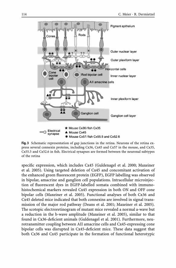
114 C. Meier · R. Dermietzel
Fig. 3 Schematic representation of gap junctions in the retina. Neurons of the retina ex-press several connexin proteins, including Cx36, Cx45 and Cx57 in the mouse, and Cx35,Cx55.5 and Cx52.6 in fish. Electrical synapses are formed between the neuronal subtypesof the retina
specific expression, which includes Cx45 (Guldenagel et al. 2000; Maxeineret al. 2005). Using targeted deletion of Cx45 and concomitant activation ofthe enhanced green fluorescent protein (EGFP), EGFP labelling was observedin bipolar, amacrine and ganglion cell populations. Intracellular microinjec-tion of fluorescent dyes in EGFP-labelled somata combined with immuno-histochemical markers revealed Cx45 expression in both ON and OFF conebipolar cells (Maxeiner et al. 2005). Functional analyses of both Cx36 andCx45 deleted mice indicated that both connexins are involved in signal trans-mission of the major rod pathway (Deans et al. 2001; Maxeiner et al. 2005).The scotopic electroretinogram of mutant mice revealed a normal a-wave buta reduction in the b-wave amplitude (Maxeiner et al. 2005), similar to thatfound in Cx36-deficient animals (Guldenagel et al. 2001). Furthermore, neu-rotransmitter coupling between AII amacrine cells and Cx45-expressing conebipolar cells was disrupted in Cx45-deficient mice. These data suggest thatboth Cx36 and Cx45 participate in the formation of functional heterotypic

Electrical Synapses – Gap Junctions in the Brain 115
electrical synapses between these two types of retinal neurons that make upthe major cone pathway.
Studies on zebrafish retina indicated that additional connexins are in-volved in the formation of electrical synapses. Cloning of four zebrafishconnexins (Dermietzel et al. 2000b; Zoidl et al. 2004) and evaluation of theircell-specific distribution showed that two of these connexins (zfCx55.5 andzfCx52.6) are retina-specific and exclusively expressed in horizontal cells.Furthermore, these connexins show essentially different electrophysiologi-cal properties under functional expression conditions. In recently generatedtransgenic mice, Cx57 has been described as showing an identical patternwith restriction to horizontal cells (Hombach et al. 2004) as discovered forthe two zebrafish connexins. This site-restrictiveness is a further indicationthat neurons can be endowed with their specific connexin complement andthat multiple connexins with the potential for compatible heterotypic channelformation are likely to shape the functional connectivity of retinal circuits.Obviously, the network behaviour of electrical synapses seems to dependlargely on the type of connexins expressed in individual classes of neuronsand their combinatorial configuration in terms of homotypic and/or he-terotypic combinations.
A further degree of complexity can be achieved if one considers the sus-ceptibility of individual connexins to phosphorylation through kinase acti-vation. In the case of horizontal cell electrical coupling a clear link betweendopamine and the PKA pathway has consistently been reported (Piccolinoet al. 1984; Hampson et al. 1992; Weiler et al. 2000). Dopamine exerts its in-fluence on the size of the receptor field by decoupling horizontal cells. Thismechanism becomes most effective under light exposure when dopaminesecretion increases. This example shows a clear interference of neurotrans-mitter activity and functional behaviour of an electrically coupled network.
Even more intriguing are data provided by Mills and Massey (Millsand Massey, 1995) who showed that an individual class of neurons can befurnished with electrical synapses of different functional phenotypes. AIIamacrine cells of the retina are coupled homotypically with AII amacrineneurons, and heterotypically with cone bipolar cells. After injecting a singleAII amacrine cell, Mills and Massey found that neurobiotin (relative mo-lecular mass, 286 Dalton) passed easily through both types of gap junctions,but that biotin-X cadaverine (relative molecular mass, 442 Dalton) passedthrough AII/bipolar cell gap junctions poorly compared to AII/AII gap junc-tions. Thus, the AII/bipolar cell channel has a lower permeability to largemolecules than does the AII/AII amacrine cell channel. More strikingly, thetwo pathways are also regulated differently. Dopamine and cyclic AMP ag-onists, known to diminish AII–AII coupling, did not change the relativelabelling intensity after intracellular dye injection of AII to bipolar cells. How-ever, nitric oxide and cGMP agonists selectively reduced labelling in bipolarcells relative to AII amacrine cells, perhaps by acting at the bipolar side of this

116 C. Meier · R. Dermietzel
gap junction. This suggests that gating mechanisms of both types of electricalsynapses are driven by different signalling pathways, which are apt to con-trol the network switch between rod and cone pathways associated with light(Mills and Massey, 1995).
Further neurotransmitter effects have been described for serotonin in thesomatosensory cortex of postnatal rats (Rorig and Sutor, 1996), suggestingextensive interactions between synaptic transmission and information flowthrough gap junctions. Obviously, the interaction between gap junctions andchemical synaptic transmission represents a powerful tool that helps to sculp-ture synaptic circuits in the developing brain as well as to increase the efficacyof synaptic transmission in adult networks (Roerig and Feller, 2000). In add-ition, gap junctional communication seems to be modulated not only byclassical neurotransmitters, but also seems to include neuroactive gaseoussubstrates, i.e. the nitric oxide (NO)/cGMP system (see above). Decouplingof retinal horizontal cells occurs upon NO release and it was suggested thatthe effect of NO on the responsiveness of horizontal cells might be due to anaction on green- and red-sensitive cones (Pottek et al. 1997). Apparently, theinteraction of neuroactive transmitters and electrical synapses includes notonly wiring transmission but also entails volume transmission as well.
6.2Pannexin Gap Junctions
Most of the neuronal connexins were identified at sites that previously wereshown to be electrically coupled, albeit in the absence of Cx36 expression,and can therefore account for electrical synapse activity in these regions (Sohlet al. 2004, 2005). However, with the identification of a novel family of gapjunction proteins named pannexins (see below), some of these assignmentsmight need to be reconsidered.
Connexin36 is one of the major players in the electrical coupling betweenhippocampal interneurons. Although deletion of Cx36 resulted in the disrup-tion of γ -frequency network oscillations in vitro and in vivo (Hormuzdi et al.2001; Maier et al. 2002; Buhl et al. 2003), high frequency oscillations in thehippocampus, which are likely to involve pyramidal cells (Schmitz et al. 2001),were unaffected (Hormuzdi et al. 2001). Explanation for the latter observa-tion can be provided by either of two hypotheses: The gap junction protein inpyramidal cells causing high frequency oscillations might be (1) a connexinprotein different from Cx36 or (2) a distinctly different class of proteins.
Theoretical support for the latter hypothesis initially arose from in silicioanalysis of the vertebrate genomes identifying orthologous DNA sequencesin the human genome that show similarity to the family of innexin proteins(i.e. “invertebrate analogue of connexins”) (Phelan et al. 1998; Phelan andStarich, 2001). Innexins were suggested to be specific invertebrate gap junc-tion proteins, representing the functional correlate of connexin proteins in

Electrical Synapses – Gap Junctions in the Brain 117
invertebrates (Phelan et al. 1998; Phelan and Starich, 2001; Hua et al. 2003),i.e. forming gap junction communication channels (Phelan et al. 1998; Lan-desman et al. 1999; Stebbings et al. 2000). Although unrelated to connexinsat the sequence level, innexins also share certain topological features withconnexin proteins, including the presence of four transmembrane domains(Bruzzone et al. 1996; Phelan and Starich, 2001).
With the cloning of innexin homologues in vertebrates, these proteinshad to be reclassified, and innexins were subsequently incorporated in thegroup of pannexin proteins (latin: pan – all, throughout; nexus – connec-tion, bond), a name indicating the ubiquitous distribution of this proteinfamily in invertebrates as well as chordates (Panchin et al. 2000). Mean-while, three pannexin-encoding genes have been cloned from the human andmouse genomes, respectively, and named PANX1 (Panx1), PANX2 (Panx2)and PANX3 (Panx3) (Panchin et al. 2000; Bruzzone et al. 2003; Baranova et al.2004; Dykes et al. 2004). Expression analysis on mRNA level has revealeda distinct distribution of these three genes in developing and adult animals(Bruzzone et al. 2003; Baranova et al. 2004) with Panx1 and Panx2 being co-expressed in a number of tissues. Pannexin-1 and Panx2 mRNAs are bothpresent in the central nervous system, including cortex, striatum, olfactorybulb, hippocampus, thalamus, cerebellum and inferior olive (Bruzzone et al.2003; Ray et al. 2005; Weickert et al. 2005). In contrast, expression of Panx3seems to be absent from the adult rat nervous system with its expressionsites being in skin, osteoblasts and synovial fibroblasts (Panchin et al. 2000;Bruzzone et al. 2003; Baranova et al. 2004). However, RT-PCR analysis fromhuman adult hippocampus samples revealed an amplification product (Bara-nova et al. 2004). Another apparent difference between species was observedin the expression of Panx2, which appears to be absent from spinal cord in hu-man tissue (Baranova et al. 2004), whereas in mouse, Panx2 expression wasshown to be higher than that of Panx1 (Bruzzone et al. 2003).
The presence of pannexin genes in the nervous system points to a possiblefunction of the gene product in neuronal communication. This implicationis supported by the notion that Panx1 and Panx2 mRNAs are expressed inhippocampal pyramidal cells (Bruzzone et al. 2003; Ray et al. 2005; Weickertet al. 2005), which were previously characterized by the near-absence of Cx36,but are nevertheless dye-coupled (MacVicar and Dudek, 1980; MacVicaret al. 1982), and electrically coupled (MacVicar and Dudek, 1981; MacVicarand Jahnsen, 1985). Direct proof for the functional implication of pannexinchannels was provided by Bruzzone and co-workers (Bruzzone et al. 2003)who demonstrated in paired Xenopus oocytes that rodent Panx1 – aloneor in combination with Panx2 – formed intercellular channels. HeteromericPanx1/Panx2 channels, albeit functional, displayed a reduction in currentamplitude, and a slower gating mechanism. Neither Panx2 nor Panx3 in-duced membrane currents above those recorded from controls. Intriguingly,there is pharmacological evidence that pannexin channels respond to some

118 C. Meier · R. Dermietzel
inhibitors known to block gap junction channels composed by connexin pro-teins, whereas others proved ineffective (Bruzzone et al. 2005). Pannexin-1 ex-pressed in single Xenopus oocytes was shown to form functional hemichan-nels (Bruzzone et al. 2003), which is of particular interest in view of functionalevidence demonstrating the mechanosensitivity of pannexin channels caus-ing the release of ATP to the extracellular space upon mechanical stress (Baoet al. 2004).
Taken together, the expression patterns and electrophysiological proper-ties of Panx1 and Panx2 provide evidence that pannexins represent a secondfamily of gap junction proteins in vertebrates. However, functionality in theCNS still needs to be determined, and studies on pannexin-deficient mice willneed to demonstrate the functional impact of pannexin channels on high-frequency oscillations and neuronal network synchronization. To understandthe channel-forming properties of pannexins, elucidation of their ultrastruc-ture is also essential.
7Conclusions
The impact of electrical synapses in the mammalian nervous system has beenrecognized over the past few years, and electrotonic coupling has been ac-cepted as a major player in neuronal interaction. One major breakthroughwas achieved with the cloning of Cx36, as identification of a gap junctionprotein predominantly expressed in neurons led to the detailed analysis ofneuronal networks and the impact of electrical synapses in their synchro-nization. Connexin36-deficient mice allowed the possibility of investigatingneuronal interaction in the absence of electrical Cx36 synapses. These stud-ies revealed a major involvement of Cx36 in synchronizing neuronal networkoscillations in various neuronal systems, for instance in the synchronizationof γ -frequencies in GABAergic interneurons of the hippocampus. With theidentification of the family of pannexin gap junction proteins, the studieswill have to be extended to the analysis of the influence of pannexins onelectrical communication between neurons, a task that will certainly be ad-dressed upon generation of pannexin-deficient animals. With these importantstudies, it will also be able to resolve the question of whether connexinand pannexin gap junction channels function separately or are somewhatorchestrated.
The second major topic, which arose with the identification of the in-volvement of electrical synapses in neuronal networks, is the functional andstructural interaction of chemical and electrical transmission. In several stud-ies, chemical synapses were shown to influence electrical coupling and viceversa (Montoro and Yuste, 2004). Examples of neurotransmitter action ofelectrical synapses include the effects of dopamine, in that extensive coupling
Electrical Synapses – Gap Junctions in the Brain 119
of horizontal cells in the turtle retina was greatly reduced by application ofdopamine (Dowling, 1991), and dye communication between cortical neuronsof the neonatal rat largely disappeared upon dopamine application to brainslices (Rorig et al. 1995).
Further implications for a possible interaction of electrical and chemicalsynapses were provided by ultrastructural evidence for the presence of so-called mixed synapses. The system in which these interactions were studiedmost extensively is the Mauthner cell of the goldfish (Pereda et al. 1994); elec-trical transmission at club endings of these cells was already described in 1962(Furshpan and Furukawa, 1962). More recently, these electrical synapses havebeen visualized by the FRIL technique, and their connexin components iden-tified as being Cx35, the fish orthologue of mammalian Cx36 (Pereda et al.2004; Rash et al. 2004). Interestingly, these gap junction plaques were foundin close vicinity to neurotransmitter receptors, i.e. the NMDA-receptor 1 sub-unit of glutamate receptors (NR1), pointing to the interaction of chemical andelectrical transmission. Mixed synapses have meanwhile also been identifiedin neurons of the nuclear inferior olivary complex (Rash et al. 2004), and itseems likely that the occurrence of mixed synapses will not be restricted tothese sites.
Acknowledgements We thank Helga Schulze for expertly done figures. The authors alsowish to thank John E. Rash for intense and stimulating scientific discussions. Work in thelaboratory was supported by grants of the German research foundation to R.D. (SFB 509and DE292/11-4).
References
Al-Ubaidi MR, White TW, Ripps H, Poras I, Avner P, Gomes D, Bruzzone R (2000) Func-tional properties, developmental regulation, and chromosomal localization of murineconnexin36, a gap-junctional protein expressed preferentially in retina and brain.J Neurosci Res 59:813–826
Altevogt BM, Paul DL (2004) Four classes of intercellular channels between glial cells inthe CNS. J Neurosci 24:4313–4323
Altevogt BM, Kleopa KA, Postma FR, Scherer SS, Paul DL (2002) Connexin29 is uniquelydistributed within myelinating glial cells of the central and peripheral nervous sys-tems. J Neurosci 22:6458–6470
Andrew RD, MacVicar BA, Dudek FE, Hatton GI (1981) Dye transfer through gap junc-tions between neuroendocrine cells of rat hypothalamus. Science 211:1187–1189
Baker R, Llinas R (1971) Electrotonic coupling between neurones in the rat mesencephalicnucleus. J Physiol 212:45–63
Bao L, Locovei S, Dahl G (2004) Pannexin membrane channels are mechanosensitiveconduits for ATP. FEBS Lett 572:65–68
Baranova A, Ivanov D, Petrash N, Pestova A, Skoblov M, Kelmanson I, Shagin D,Nazarenko S, Geraymovych E, Litvin O, Tiunova A, Born TL, Usman N, Staroverov D,Lukyanov S, Panchin Y (2004) The mammalian pannexin family is homologous to theinvertebrate innexin gap junction proteins. Genomics 83:706–716

120 C. Meier · R. Dermietzel
Barron DJ, Valdimarsson G, Paul DL, Kidder GM (1989) Connexin32, a gap junction pro-tein, is a persistent oogenetic product through preimplantation development of themouse. Dev Genet 10:318–323
Belliveau DJ, Naus CC (1994) Cortical type 2 astrocytes are not dye coupled nor dothey express the major gap junction genes found in the central nervous system. Glia12:24–34
Belliveau DJ, Naus CC (1995) Cellular localization of gap junction mRNAs in developingrat brain. Dev Neurosci 17:81–96
Belliveau DJ, Kidder GM, Naus CC (1991) Expression of gap junction genes during post-natal neural development. Dev Genet 12:308–317
Belluardo N, Trovato-Salinaro A, Mudo G, Hurd YL, Condorelli DF (1999) Structure,chromosomal localization, and brain expression of human Cx36 gene. J Neurosci Res57:740–752
Belluardo N, Mudo G, Trovato-Salinaro A, Le Gurun S, Charollais A, Serre-Beinier V, Am-ato G, Haefliger JA, Meda P, Condorelli DF (2000) Expression of connexin36 in theadult and developing rat brain. Brain Res 865:121–138
Bennett MV (1997) Gap junctions as electrical synapses. J Neurocytol 26:349–366Bennett MV, Crain SM, Grundfest H (1959) Electrophysiology of supramedullary neurons
in Spheroides maculatus. II. Properties of the electrically excitable membrane. J GenPhysiol 43:189–219
Bennett MVL (1977) Electrical transmission: a functional analysis and comparison withchemical transmission. M.D. Williams and Wilkins, Baltimore
Berthoud VM, Singh R, Minogue PJ, Ragsdale CW, Beyer EC (2004) Highly restrictedpattern of connexin36 expression in chick somite development. Anat Embryol (Berl)209:11–18
Blue ME, Parnavelas JG (1983) The formation and maturation of synapses in the visualcortex of the rat. I. Qualitative analysis. J Neurocytol 12:599–616
Bruzzone R, White TW, Paul DL (1996) Connections with connexins: the molecular basisof direct intercellular signaling. Eur J Biochem 238:1–27
Bruzzone R, Barbe MT, Jakob NJ, Monyer H (2005) Pharmacological properties of ho-momeric and heteromeric pannexin hemichannels expressed in Xenopus oocytes.J Neurochem 92:1033–1043
Bruzzone R, Hormuzdi SG, Barbe MT, Herb A, Monyer H (2003) Pannexins, a family ofgap junction proteins expressed in brain. Proc Natl Acad Sci USA 100:13644–13649
Buhl DL, Harris KD, Hormuzdi SG, Monyer H, Buzsaki G (2003) Selective impairment ofhippocampal gamma oscillations in connexin-36 knock-out mouse in vivo. J Neurosci23:1013–1018
Buzsaki G (2001) Electrical wiring of the oscillating brain. Neuron 31:342–344Buzsaki G (2002) Theta oscillations in the hippocampus. Neuron 33:325–340Buzsaki G, Draguhn A (2004) Neuronal oscillations in cortical networks. Science
304:1926–1929Buzsaki G, Leung LW, Vanderwolf CH (1983) Cellular bases of hippocampal EEG in the
behaving rat. Brain Res 287:139–171Buzsaki G, Horvath Z, Urioste R, Hetke J, Wise K (1992) High-frequency network oscilla-
tion in the hippocampus. Science 256:1025–1027Chandross KJ, Kessler JA, Cohen RI, Simburger E, Spray DC, Bieri P, Dermietzel R
(1996) Altered connexin expression after peripheral nerve injury. Mol Cell Neurosci7:501–518
Christie JM, Bark C, Hormuzdi SG, Helbig I, Monyer H, Westbrook GL (2005) Connexin36mediates spike synchrony in olfactory bulb glomeruli. Neuron 46:761–772

Electrical Synapses – Gap Junctions in the Brain 121
Condorelli DF, Belluardo N, Trovato-Salinaro A, Mudo G (2000) Expression of Cx36 inmammalian neurons. Brain Res Brain Res Rev 32:72–85
Condorelli DF, Trovato-Salinaro A, Mudo G, Mirone MB, Belluardo N (2003) Cellular ex-pression of connexins in the rat brain: neuronal localization, effects of kainate-inducedseizures and expression in apoptotic neuronal cells. Eur J Neurosci 18:1807–1827
Condorelli DF, Mudo G, Trovato-Salinaro A, Mirone MB, Amato G, Belluardo N (2002)Connexin-30 mRNA is up-regulated in astrocytes and expressed in apoptotic neuronalcells of rat brain following kainate-induced seizures. Mol Cell Neurosci 21:94–113
Condorelli DF, Parenti R, Spinella F, Trovato Salinaro A, Belluardo N, Cardile V, Cicirata F(1998) Cloning of a new gap junction gene (Cx36) highly expressed in mammalianbrain neurons. Eur J Neurosci 10:1202–1208
Connors BW, Long MA (2004) Electrical synapses in the mammalian brain. Annu RevNeurosci 27:393–418
Connors BW, Benardo LS, Prince DA (1983) Coupling between neurons of the developingrat neocortex. J Neurosci 3:773–782
De Zeeuw CI, Chorev E, Devor A, Manor Y, Van Der Giessen RS, De Jeu MT, HoogenraadCC, Bijman J, Ruigrok TJ, French P, Jaarsma D, Kistler WM, Meier C, Petrasch-Parwez E, Dermietzel R, Sohl G, Gueldenagel M, Willecke K, Yarom Y (2003) Defor-mation of network connectivity in the inferior olive of connexin 36-deficient mice iscompensated by morphological and electrophysiological changes at the single neuronlevel. J Neurosci 23:4700–4711
Deans MR, Gibson JR, Sellitto C, Connors BW, Paul DL (2001) Synchronous activity ofinhibitory networks in neocortex requires electrical synapses containing connexin36.Neuron 31:477–485
Deans MR, Volgyi B, Goodenough DA, Bloomfield SA, Paul DL (2002) Connexin36 isessential for transmission of rod-mediated visual signals in the mammalian retina.Neuron 36:703–712
Degen J, Meier C, Van Der Giessen RS, Sohl G, Petrasch-Parwez E, Urschel S, Dermiet-zel R, Schilling K, De Zeeuw CI, Willecke K (2004) Expression pattern of lacZ reportergene representing connexin36 in transgenic mice. J Comp Neurol 473:511–525
Dermietzel R, Meier C (2005) Gap junction expression in brain tissues with focus ondevelopment. In: Winterhager E (ed) Gap junctions in development and disease.Springer, Berlin Heidelberg New York, pp 83–110
Dermietzel R, Traub O, Hwang TK, Beyer E, Bennett MV, Spray DC, Willecke K (1989)Differential expression of three gap junction proteins in developing and mature braintissues. Proc Natl Acad Sci USA 86:10148–10152
Dermietzel R, Gao Y, Scemes E, Vieira D, Urban M, Kremer M, Bennett MV, Spray DC(2000a) Connexin43 null mice reveal that astrocytes express multiple connexins. BrainRes Brain Res Rev 32:45–56
Dermietzel R, Kremer M, Paputsoglu G, Stang A, Skerrett IM, Gomes D, Srinivas M,Janssen-Bienhold U, Weiler R, Nicholson BJ, Bruzzone R, Spray DC (2000b) Molecularand functional diversity of neural connexins in the retina. J Neurosci 20:8331–8343
Destexhe A, Bal T, McCormick DA, Sejnowski TJ (1996) Ionic mechanisms underlyingsynchronized oscillations and propagating waves in a model of ferret thalamic slices.J Neurophysiol 76:2049–2070
Dowling JE (1991) Retinal neuromodulation: the role of dopamine. Vis Neurosci 7:87–97Draguhn A, Traub RD, Schmitz D, Jefferys JG (1998) Electrical coupling underlies high-
frequency oscillations in the hippocampus in vitro. Nature 394:189–192Dykes IM, Freeman FM, Bacon JP, Davies JA (2004) Molecular basis of gap junctional
communication in the CNS of the leech Hirudo medicinalis. J Neurosci 24:886–894
122 C. Meier · R. Dermietzel
Faber DS, Korn H (1989) Electrical field effects: their relevance in central neural net-works. Physiol Rev 69:821–863
Feigenspan A, Teubner B, Willecke K, Weiler R (2001) Expression of neuronal connexin36in AII amacrine cells of the mammalian retina. J Neurosci 21:230–239
Feigenspan A, Janssen-Bienhold U, Hormuzdi S, Monyer H, Degen J, Sohl G, Willecke K,Ammermuller J, Weiler R (2004) Expression of connexin36 in cone pedicles and OFF-cone bipolar cells of the mouse retina. J Neurosci 24:3325–3334
Freeman AS, Meltzer LT, Bunney BS (1985) Firing properties of substantia nigra dopamin-ergic neurons in freely moving rats. Life Sci 36:1983–1994
Frisch C, De Souza-Silva MA, Sohl G, Guldenagel M, Willecke K, Huston JP, Dere E (2005)Stimulus complexity dependent memory impairment and changes in motor perform-ance after deletion of the neuronal gap junction protein connexin36 in mice. BehavBrain Res 157:177–185
Fujimoto K (1995) Freeze-fracture replica electron microscopy combined with SDS di-gestion for cytochemical labeling of integral membrane proteins. Application to theimmunogold labeling of intercellular junctional complexes. J Cell Sci 108 (Pt 11):3443-3449
Furshpan EJ, Potter DD (1957) Mechanism of nerve-impulse transmission at a crayfishsynapse. Nature 180:342–343
Furshpan EJ, Potter DD (1959) Transmission at the giant motor synapses of the crayfish.J Physiol 145:289–325
Furshpan EJ, Furukawa T (1962) Intracellular and extracellular responses of the severalregions of the Mauthner cell of the goldfish. J Neurophysiol 25:732–771
Galarreta M, Hestrin S (1999) A network of fast-spiking cells in the neocortex connectedby electrical synapses. Nature 402:72–75
Galarreta M, Hestrin S (2001) Electrical synapses between GABA-releasing interneurons.Nat Rev Neurosci 2:425–433
Giaume C, Venance L (1995) Gap junctions in brain glial cells and development. PerspectDev Neurobiol 2:335–345
Gibson JR, Beierlein M, Connors BW (1999) Two networks of electrically coupled in-hibitory neurons in neocortex. Nature 402:75–79
Gibson JR, Beierlein M, Connors BW (2005) Functional properties of electrical synapsesbetween inhibitory interneurons of neocortical layer 4. J Neurophysiol 93:467–480
Giepmans BN (2004) Gap junctions and connexin-interacting proteins. Cardiovasc Res62:233–245
Gray CM, Singer W (1989) Stimulus-specific neuronal oscillations in orientation columnsof cat visual cortex. Proc Natl Acad Sci USA 86:1698–1702
Grundfest H (1959) General physiology and pharmacology of synapses and someimplications for the mammalian central nervous system. J Nerv Ment Dis 128:473–496
Guldenagel M, Sohl G, Plum A, Traub O, Teubner B, Weiler R, Willecke K (2000) Expres-sion patterns of connexin genes in mouse retina. J Comp Neurol 425:193–201
Guldenagel M, Ammermuller J, Feigenspan A, Teubner B, Degen J, Sohl G, Willecke K,Weiler R (2001) Visual transmission deficits in mice with targeted disruption of thegap junction gene connexin36. J Neurosci 21:6036–6044
Gulisano M, Parenti R, Spinella F, Cicirata F (2000) Cx36 is dynamically expressed dur-ing early development of mouse brain and nervous system. Neuroreport 11:3823–3828
Gulyas AI, Hajos N, Freund TF (1996) Interneurons containing calretinin are specializedto control other interneurons in the rat hippocampus. J Neurosci 16:3397–3411

Electrical Synapses – Gap Junctions in the Brain 123
Halasy K, Buhl EH, Lorinczi Z, Tamas G, Somogyi P (1996) Synaptic target selectivityand input of GABAergic basket and bistratified interneurons in the CA1 area of therat hippocampus. Hippocampus 6:306–329
Hampson EC, Vaney DI, Weiler R (1992) Dopaminergic modulation of gap junction per-meability between amacrine cells in mammalian retina. J Neurosci 12:4911–4922
Haverkamp S, Wassle H (2000) Immunocytochemical analysis of the mouse retina.J Comp Neurol 424:1–23
Hombach S, Janssen-Bienhold U, Sohl G, Schubert T, Bussow H, Ott T, Weiler R, WilleckeK (2004) Functional expression of connexin57 in horizontal cells of the mouse retina.Eur J Neurosci 19:2633–2640
Honma S, De S, Li D, Shuler CF, Turman JE Jr (2004) Developmental regulation of con-nexins 26, 32, 36, and 43 in trigeminal neurons. Synapse 52:258–271
Hormuzdi SG, Pais I, LeBeau FE, Towers SK, Rozov A, Buhl EH, Whittington MA,Monyer H (2001) Impaired electrical signaling disrupts gamma frequency oscillationsin connexin 36-deficient mice. Neuron 31:487–495
Hua VB, Chang AB, Tchieu JH, Kumar NM, Nielsen PA, Saier MH Jr. (2003) Sequence andphylogenetic analyses of 4 TMS junctional proteins of animals: connexins, innexins,claudins and occludins. J Membr Biol 194:59–76
Jefferys JG (1995) Nonsynaptic modulation of neuronal activity in the brain: electriccurrents and extracellular ions. Physiol Rev 75:689–723
Jefferys JG, Traub RD, Whittington MA (1996) Neuronal networks for induced 40 Hzrhythms. Trends Neurosci 19:202–208
Kalderon N, Epstein ML, Gilula NB (1977) Cell-to-cell communication and myogenesis.J Cell Biol 75:788–806
Kandler K, Katz LC (1998) Coordination of neuronal activity in developing visual cortexby gap junction-mediated biochemical communication. J Neurosci 18:1419–1427
Katsumaru H, Kosaka T, Heizmann CW, Hama K (1988) Gap junctions on GABAergicneurons containing the calcium-binding protein parvalbumin in the rat hippocampus(CA1 region). Exp Brain Res 72:363–370
Keeter JS, Pappas GD, Model PG (1975) Inter- and intramyotomal gap junctions in theaxolotl embryo. Dev Biol 45:21–33
Kistler WM, De Jeu MT, Elgersma Y, Van Der Giessen RS, Hensbroek R, Luo C,Koekkoek SK, Hoogenraad CC, Hamers FP, Gueldenagel M, Sohl G, Willecke K, DeZeeuw CI (2002) Analysis of Cx36 knockout does not support tenet that olivary gapjunctions are required for complex spike synchronization and normal motor perform-ance. Ann NY Acad Sci 978:391–404
Kita H, Kosaka T, Heizmann CW (1990) Parvalbumin-immunoreactive neurons in the ratneostriatum: a light and electron microscopic study. Brain Res 536:1–15
Kleopa KA, Orthmann JL, Enriquez A, Paul DL, Scherer SS (2004) Unique distributionsof the gap junction proteins connexin29, connexin32, and connexin47 in oligodendro-cytes. Glia 47:346–357
Kumar NM, Gilula NB (1996) The gap junction communication channel. Cell 84:381–388
Landesman Y, White TW, Starich TA, Shaw JE, Goodenough DA, Paul DL (1999) Innexin-3forms connexin-like intercellular channels. J Cell Sci 112 (Pt 14):2391–2396
Landis DM, Reese TS, Raviola E (1974) Differences in membrane structure between ex-citatory and inhibitory components of the reciprocal synapse in the olfactory bulb.J Comp Neurol 155:67–91
Landisman CE, Long MA, Beierlein M, Deans MR, Paul DL, Connors BW (2002) Electricalsynapses in the thalamic reticular nucleus. J Neurosci 22:1002–1009

124 C. Meier · R. Dermietzel
Lee EJ, Han JW, Kim HJ, Kim IB, Lee MY, Oh SJ, Chung JW, Chun MH (2003) The im-munocytochemical localization of connexin 36 at rod and cone gap junctions in theguinea pig retina. Eur J Neurosci 18:2925–2934
Li X, Lynn BD, Olson C, Meier C, Davidson KG, Yasumura T, Rash JE, Nagy JI (2002) Con-nexin29 expression, immunocytochemistry and freeze-fracture replica immunogoldlabelling (FRIL) in sciatic nerve. Eur J Neurosci 16:795–806
Llinas R, Ribary U (1993) Coherent 40-Hz oscillation characterizes dream state in hu-mans. Proc Natl Acad Sci USA 90:2078–2081
Llinas R, Baker R, Sotelo C (1974) Electrotonic coupling between neurons in cat inferiorolive. J Neurophysiol 37:560–571
Lo Turco JJ, Kriegstein AR (1991) Clusters of coupled neuroblasts in embryonic neocor-tex. Science 252:563–566
Long MA, Landisman CE, Connors BW (2004) Small clusters of electrically coupled neu-rons generate synchronous rhythms in the thalamic reticular nucleus. J Neurosci24:341–349
Long MA, Deans MR, Paul DL, Connors BW (2002) Rhythmicity without synchrony in theelectrically uncoupled inferior olive. J Neurosci 22:10898–10905
MacVicar BA, Dudek FE (1980) Dye coupling between CA3 pyramidal cells in slices of rathippocampus. Brain Res 196:494–497
MacVicar BA, Dudek EF (1981) Electronic coupling between pyrymidal cells: A directdemonstration in rat hippocampal slices. Science 213:782–785
MacVicar BA, Jahnsen H (1985) Uncoupling of CA3 pyramidal neurons by propionate.Brain Res 330:141–145
MacVicar BA, Ropert N, Krnjev́ic K (1982) Dye-coupling between pyramidal cells of rathippocampus in vivo. Brain Res 238:239–244
Maier N, Guldenagel M, Sohl G, Siegmund H, Willecke K, Draguhn A (2002) Reductionof high-frequency network oscillations (ripples) and pathological network dischargesin hippocampal slices from connexin 36-deficient mice. J Physiol 541:521–528
Mann-Metzer P, Yarom Y (1999) Electrotonic coupling interacts with intrinsic propertiesto generate synchronized activity in cerebellar networks of inhibitory interneurons.J Neurosci 19:3298–3306
Massa PT, Mugnaini E (1982) Cell junctions and intramembrane particles of astrocytesand oligodendrocytes: a freeze-fracture study. Neuroscience 7:523–538
Maxeiner S, Kruger O, Schilling K, Traub O, Urschel S, Willecke K (2003) Spatiotemporaltranscription of connexin45 during brain development results in neuronal expressionin adult mice. Neuroscience 119:689–700
Maxeiner S, Dedek K, Janssen-Bienhold U, Ammermuller J, Brune H, Kirsch T, Pieper M,Degen J, Kruger O, Willecke K, Weiler R (2005) Deletion of connexin45 in mouse reti-nal neurons disrupts the rod/cone signaling pathway between AII amacrine and ONcone bipolar cells and leads to impaired visual transmission. J Neurosci 25:566–576
McLachlin JR, Kidder GM (1986) Intercellular junctional coupling in preimplantationmouse embryos: effect of blocking transcription or translation. Dev Biol 117:146–155
Meier C, Petrasch-Parwez E, Habbes HW, Teubner B, Guldenagel M, Degen J, Sohl G,Willecke K, Dermietzel R (2002) Immunohistochemical detection of the neuronal con-nexin36 in the mouse central nervous system in comparison to connexin36-deficienttissues. Histochem Cell Biol 117:461–471
Menichella DM, Goodenough DA, Sirkowski E, Scherer SS, Paul DL (2003) Connexins arecritical for normal myelination in the CNS. J Neurosci 23:5963–5973
Micevych PE, Abelson L (1991) Distribution of mRNAs coding for liver and heart gapjunction proteins in the rat central nervous system. J Comp Neurol 305:96–118

Electrical Synapses – Gap Junctions in the Brain 125
Micevych PE, Popper P, Hatton GI (1996) Connexin 32 mRNA levels in the rat supraopticnucleus: up-regulation prior to parturition and during lactation. Neuroendocrinology63:39–45
Michelson HB, Wong RK (1994) Synchronization of inhibitory neurones in the guinea-pighippocampus in vitro. J Physiol 477 (Pt 1):35–45
Mills SL, Massey SC (1995) Differential properties of two gap junctional pathways madeby AII amacrine cells. Nature 377:734–737
Mills SL, O’Brien JJ, Li W, O’Brien J, Massey SC (2001) Rod pathways in the mammalianretina use connexin 36. J Comp Neurol 436:336–350
Miltner WH, Braun C, Arnold M, Witte H, Taub E (1999) Coherence of gamma-band EEGactivity as a basis for associative learning. Nature 397:434–436
Montoro RJ, Yuste R (2004) Gap junctions in developing neocortex: a review. Brain ResBrain Res Rev 47:216–226
Nagy JI, Rash JE (2000) Connexins and gap junctions of astrocytes and oligodendrocytesin the CNS. Brain Res Brain Res Rev 32:29–44
Nagy JI, Dudek FE, Rash JE (2004) Update on connexins and gap junctions in neuronsand glia in the mammalian nervous system. Brain Res Brain Res Rev 47:191–215
Nagy JI, Ionescu AV, Lynn BD, Rash JE (2003) Connexin29 and connexin32 at oligo-dendrocyte and astrocyte gap junctions and in myelin of the mouse central nervoussystem. J Comp Neurol 464:356–370
Nagy JI, Li X, Rempel J, Stelmack G, Patel D, Staines WA, Yasumura T, Rash JE (2001)Connexin26 in adult rodent central nervous system: demonstration at astrocyticgap junctions and colocalization with connexin30 and connexin43. J Comp Neurol441:302–323
O’Brien J, al-Ubaidi MR, Ripps H (1996) Connexin 35: a gap-junctional protein expressedpreferentially in the skate retina. Mol Biol Cell 7:233–243
O’Brien J, Bruzzone R, White TW, Al-Ubaidi MR, Ripps H (1998) Cloning and expres-sion of two related connexins from the perch retina define a distinct subgroup of theconnexin family. J Neurosci 18:7625–7637
Odermatt B, Wellershaus K, Wallraff A, Seifert G, Degen J, Euwens C, Fuss B, BussowH, Schilling K, Steinhauser C, Willecke K (2003) Connexin 47 (Cx47)-deficient micewith enhanced green fluorescent protein reporter gene reveal predominant oligoden-drocytic expression of Cx47 and display vacuolized myelin in the CNS. J Neurosci23:4549–4559
Oguro K, Jover T, Tanaka H, Lin Y, Kojima T, Oguro N, Grooms SY, Bennett MV, Zukin RS(2001) Global ischemia-induced increases in the gap junctional proteins connexin 32(Cx32) and Cx36 in hippocampus and enhanced vulnerability of Cx32 knock-out mice.J Neurosci 21:7534–7542
Panchin Y, Kelmanson I, Matz M, Lukyanov K, Usman N, Lukyanov S (2000) A ubiquitousfamily of putative gap junction molecules. Curr Biol 10:R473–474
Peinado A, Yuste R, Katz LC (1993) Extensive dye coupling between rat neocortical neu-rons during the period of circuit formation. Neuron 10:103–114
Pereda AE, Nairn AC, Wolszon LR, Faber DS (1994) Postsynaptic modulation of synapticefficacy at mixed synapses on the Mauthner cell. J Neurosci 14:3704–3712
Pereda AE, Rash JE, Nagy JI, Bennett MV (2004) Dynamics of electrical transmission atclub endings on the Mauthner cells. Brain Res Brain Res Rev 47:227–244
Phelan P, Starich TA (2001) Innexins get into the gap. Bioessays 23:388–396Phelan P, Stebbings LA, Baines RA, Bacon JP, Davies JA, Ford C (1998) Drosophila
Shaking-B protein forms gap junctions in paired Xenopus oocytes. Nature391:181–184

126 C. Meier · R. Dermietzel
Piccolino M, Neyton J, Gerschenfeld HM (1984) Decrease of gap junction permeability in-duced by dopamine and cyclic adenosine 3′ : 5′-monophosphate in horizontal cells ofturtle retina. J Neurosci 4:2477–2488
Pinault D, Deschenes M (1992) Control of 40-Hz firing of reticular thalamic cells byneurotransmitters. Neuroscience 51:259–268
Pinching AJ, Powell TP (1971) The neuropil of the glomeruli of the olfactory bulb. J CellSci 9:347–377
Pottek M, Schultz K, Weiler R (1997) Effects of nitric oxide on the horizontal cell networkand dopamine release in the carp retina. Vision Res 37:1091–1102
Rash JE, Staehelin LA (1974) Freeze-cleave demonstration of gap junctions between skele-tal myogenic cells in vivo. Dev Biol 36:455–461
Rash JE, Yasumura T, Dudek FE (1998) Ultrastructure, histological distribution, andfreeze-fracture immunocytochemistry of gap junctions in rat brain and spinal cord.Cell Biol Int 22:731–749
Rash JE, Yasumura T, Dudek FE, Nagy JI (2001a) Cell-specific expression of connexinsand evidence of restricted gap junctional coupling between glial cells and betweenneurons. J Neurosci 21:1983–2000
Rash JE, Dudek FE, Yasumura T, Davidson K, Furman CS, Nagy JI (2001b) Identificationof cells expressing Cx43, Cx30, Cx26 Cx32 and Cx36 in gap junctions of rat brain andspinal cord. Cell Comm Adhesion:1–344
Rash JE, Staines WA, Yasumura T, Patel D, Furman CS, Stelmack GL, Nagy JI (2000) Im-munogold evidence that neuronal gap junctions in adult rat brain and spinal cordcontain connexin-36 but not connexin-32 or connexin-43. Proc Natl Acad Sci USA97:7573–7578
Rash JE, Pereda A, Kamasawa N, Furman CS, Yasumura T, Davidson KG, Dudek FE, OlsonC, Li X, Nagy JI (2004) High-resolution proteomic mapping in the vertebrate centralnervous system: Close proximity of connexin35 to NMDA glutamate receptor clus-ters and co-localization of connexin36 with immunoreactivity for zonula occludensprotein-1 (ZO-1). J Neurocytol 33:131–151
Ray A, Zoidl G, Weickert S, Wahle P, Dermietzel R (2005) Site-specific and developmentalexpression of pannexin 1 in the mouse nervous system. Eur J Neurosci 21:3277–3290
Rodriguez E, George N, Lachaux JP, Martinerie J, Renault B, Varela FJ (1999) Perception’sshadow: long-distance synchronization of human brain activity. Nature 397:430–433
Roerig B, Feller MB (2000) Neurotransmitters and gap junctions in developing neuralcircuits. Brain Res Brain Res Rev 32:86–114
Rorig B, Sutor B (1996) Regulation of gap junction coupling in the developing neocortex.Mol Neurobiol 12:225–249
Rorig B, Klausa G, Sutor B (1995) Dye coupling between pyramidal neurons in devel-oping rat prefrontal and frontal cortex is reduced by protein kinase A activation anddopamine. J Neurosci 15:7386–7400
Rozental R, Morales M, Mehler MF, Urban M, Kremer M, Dermietzel R, Kessler JA,Spray DC (1998) Changes in the properties of gap junctions during neuronal differ-entiation of hippocampal progenitor cells. J Neurosci 18:1753–1762
Scherer SS, Deschenes SM, Xu YT, Grinspan JB, Fischbeck KH, Paul DL (1995) Con-nexin32 is a myelin-related protein in the PNS and CNS. J Neurosci 15:8281–8294
Schmalbruch H (1982) Skeletal muscle fibers of newborn rats are coupled by gap junc-tions. Dev Biol 91:485–490
Schmitz D, Schuchmann S, Fisahn A, Draguhn A, Buhl EH, Petrasch-Parwez E, Dermiet-zel R, Heinemann U, Traub RD (2001) Axo-axonal coupling. a novel mechanism forultrafast neuronal communication. Neuron 31:831–840

Electrical Synapses – Gap Junctions in the Brain 127
Schubert T, Degen J, Willecke K, Hormuzdi SG, Monyer H, Weiler R (2005) Connexin36mediates gap junctional coupling of alpha-ganglion cells in mouse retina. J CompNeurol 485:191–201
Serre-Beinier V, Le Gurun S, Belluardo N, Trovato-Salinaro A, Charollais A, Haefliger JA,Condorelli DF, Meda P (2000) Cx36 preferentially connects beta-cells within pancreaticislets. Diabetes 49:727–734
Sik A, Penttonen M, Ylinen A, Buzsaki G (1995) Hippocampal CA1 interneurons: an invivo intracellular labeling study. J Neurosci 15:6651–6665
Singer W, Gray CM (1995) Visual feature integration and the temporal correlation hypoth-esis. Annu Rev Neurosci 18:555–586
Sloper JJ (1972) Gap junctions between dendrites in the primate neocortex. Brain Res44:641–646
Sohl G, Maxeiner S, Willecke K (2005) Expression and functions of neuronal gap junc-tions. Nat Rev Neurosci 6:191–200
Sohl G, Degen J, Teubner B, Willecke K (1998) The murine gap junction gene connexin36is highly expressed in mouse retina and regulated during brain development. FEBSLett 428:27–31
Sohl G, Odermatt B, Maxeiner S, Degen J, Willecke K (2004) New insights into the ex-pression and function of neural connexins with transgenic mouse mutants. Brain ResBrain Res Rev 47:245–259
Somogyi P, Nunzi MG, Gorio A, Smith AD (1983) A new type of specific interneuron inthe monkey hippocampus forming synapses exclusively with the axon initial segmentsof pyramidal cells. Brain Res 259:137–142
Sotelo C, Llinas R (1972) Specialized membrane junctions between neurons in the verte-brate cerebellar cortex. J Cell Biol 53:271–289
Spray DC, Dermietzel R (1995) X-linked dominant Charcot-Marie-Tooth disease andother potential gap-junction diseases of the nervous system. Trends Neurosci18:256–262
Srinivas M, Rozental R, Kojima T, Dermietzel R, Mehler M, Condorelli DF, Kessler JA,Spray DC (1999) Functional properties of channels formed by the neuronal gap junc-tion protein connexin36. J Neurosci 19:9848–9855
Stebbings LA, Todman MG, Phelan P, Bacon JP, Davies JA (2000) Two Drosophila innexinsare expressed in overlapping domains and cooperate to form gap-junction channels.Mol Biol Cell 11:2459–2470
Steriade M, McCormick DA, Sejnowski TJ (1993) Thalamocortical oscillations in thesleeping and aroused brain. Science 262:679–685
Steriade M, Amzica F, Contreras D (1996) Synchronization of fast (30–40 Hz) spontaneouscortical rhythms during brain activation. J Neurosci 16:392–417
Teubner B, Degen J, Sohl G, Guldenagel M, Bukauskas FF, Trexler EB, Verselis VK, DeZeeuw CI, Lee CG, Kozak CA, Petrasch-Parwez E, Dermietzel R, Willecke K (2000)Functional expression of the murine connexin 36 gene coding for a neuron-specificgap junctional protein. J Membr Biol 176:249–262
Traub RD, Bibbig A (2000) A model of high-frequency ripples in the hippocampusbased on synaptic coupling plus axon-axon gap junctions between pyramidal neurons.J Neurosci 20:2086–2093
Traub RD, Schmitz D, Jefferys JG, Draguhn A (1999) High-frequency population oscilla-tions are predicted to occur in hippocampal pyramidal neuronal networks intercon-nected by axoaxonal gap junctions. Neuroscience 92:407–426
Traub RD, Whittington MA, Colling SB, Buzsaki G, Jefferys JG (1996) Analysis of gammarhythms in the rat hippocampus in vitro and in vivo. J Physiol 493(Pt 2):471–484

128 C. Meier · R. Dermietzel
Traub RD, Spruston N, Soltesz I, Konnerth A, Whittington MA, Jefferys GR (1998)Gamma-frequency oscillations: a neuronal population phenomenon, regulated bysynaptic and intrinsic cellular processes, and inducing synaptic plasticity. Prog Neu-robiol 55:563–575
Traub RD, Bibbig A, Fisahn A, LeBeau FE, Whittington MA, Buhl EH (2000) A model ofgamma-frequency network oscillations induced in the rat CA3 region by carbachol invitro. Eur J Neurosci 12:4093–4106
Traub RD, Kopell N, Bibbig A, Buhl EH, LeBeau FE, Whittington MA (2001) Gap junc-tions between interneuron dendrites can enhance synchrony of gamma oscillations indistributed networks. J Neurosci 21:9478–9486
Traub RD, Pais I, Bibbig A, LeBeau FE, Buhl EH, Hormuzdi SG, Monyer H, Whitting-ton MA (2003) Contrasting roles of axonal (pyramidal cell) and dendritic (interneu-ron) electrical coupling in the generation of neuronal network oscillations. Proc NatlAcad Sci USA 100:1370–1374
Vandecasteele M, Glowinski J, Venance L (2005) Electrical synapses between dopaminer-gic neurons of the substantia nigra pars compacta. J Neurosci 25:291–298
Vaney DI (1999) Neuronal coupling in the central nervous system: lessons from the retina.Novartis Found Symp 219:113–125; discussion 125–133
Venance L, Rozov A, Blatow M, Burnashev N, Feldmeyer D, Monyer H (2000) Connexinexpression in electrically coupled postnatal rat brain neurons. Proc Natl Acad SciUSA 97:10260–10265
Watanabe A (1958) The interaction of electrical activity among neurons of lobster cardiacganglion. Jpn J Physiol 8:305–318
Waxman SG, Black JA (1984) Freeze-fracture ultrastructure of the perinodal astrocyteand associated glial junctions. Brain Res 308:77–87
Weickert S, Ray A, Zoidl G, Dermietzel R (2005) Expression of neural connexins and pan-nexin1 in the hippocampus and inferior olive: a quantitative approach. Brain Res MolBrain Res 133:102–109
Weiler R, Pottek M, He S, Vaney DI (2000) Modulation of coupling between retinal ho-rizontal cells by retinoic acid and endogenous dopamine. Brain Res Brain Res Rev32:121–129
White TW, Deans MR, O’Brien J, Al-Ubaidi MR, Goodenough DA, Ripps H, Bruzzone R(1999) Functional characteristics of skate connexin35, a member of the gamma sub-family of connexins expressed in the vertebrate retina. Eur J Neurosci 11:1883–1890
Willecke K, Eiberger J, Degen J, Eckardt D, Romualdi A, Guldenagel M, Deutsch U, Sohl G(2002) Structural and functional diversity of connexin genes in the mouse and humangenome. Biol Chem 383:725–737
Ylinen A, Bragin A, Nadasdy Z, Jando G, Szabo I, Sik A, Buzsaki G (1995) Sharp wave-associated high-frequency oscillation (200 Hz) in the intact hippocampus: networkand intracellular mechanisms. J Neurosci 15:30–46
Yuste R, Peinado A, Katz LC (1992) Neuronal domains in developing neocortex. Science257:665–669
Zoidl G, Dermietzel R (2002) On the search for the electrical synapse: a glimpse at thefuture. Cell Tissue Res 310:137–142
Zoidl G, Meier C, Petrasch-Parwez E, Zoidl C, Habbes HW, Kremer M, Srinivas M,Spray DC, Dermietzel R (2002) Evidence for a role of the N-terminal domain in sub-cellular localization of the neuronal connexin36 (Cx36). J Neurosci Res 69:448–465
Zoidl G, Bruzzone R, Weickert S, Kremer M, Zoidl C, Mitropoulou G, Srinivas M,Spray DC, Dermietzel R (2004) Molecular cloning and functional expression ofzfCx52.6: a novel connexin with hemichannel-forming properties expressed in ho-rizontal cells of the zebrafish retina. J Biol Chem 279:2913–2921

Results Probl Cell Differ (43)E. Gundelfinger, C. Seidenbecher, B. Schraven:Cell Communication in Nervous and Immune SystemDOI 10.1007/014/Published online: 30 March 2006© Springer-Verlag Berlin Heidelberg 2006
Neuron-Glia Interactions at the Node of Ranvier
Matthew N. Rasband
Department of Neuroscience, University of Connecticut Health Center,263 Farmington Avenue, Farmington, CT 06030-3401, [email protected]
Abstract Rapid, faithful, and efficient action potential propagation in mammalian axonsis a consequence of myelin and clustered Na+ channels. Both myelination and node ofRanvier formation require complex intercellular interactions between neurons and gliathat result in profound molecular, morphological, and functional changes in each celltype. This review will focus on the molecular and cellular mechanisms that underlieneuron-glia interactions at the node of Ranvier. In particular, the proteins and proteincomplexes, and how they participate in node of Ranvier formation and maintenance, willbe discussed. Traditionally, myelinating glia have been viewed as merely passive playersin neuronal function, conferring on the axons they ensheath various electrical propertiesthat facilitate action potential conduction. However, it is now recognized that this view isincomplete. This review will discuss several examples illustrating how myelinating glia ac-tively regulate the excitable properties of axons including the kinds of channels expressedand their subcellular localization.
1Introduction
Intercellular communication is essential for proper nervous system function.The prototypical example of this interaction is found at the synapse. Here,pre- and postsynaptic neurons form functionally, morphologically, biochem-ically, and molecularly distinct structures that permit rapid, faithful and effi-cient transmission of information. However, before synaptic transmission canever take place, action potentials must be conducted along axons that traverserelatively long distances. As with the synapse, axons must also facilitate rapid,faithful, and efficient action potential propagation while minimizing boththe metabolic and space requirements needed to accomplish this. Vertebrateshave solved these problems through the development of 1) the myelin sheath(Fig. 1A) and 2) the clustering of voltage-gated Na+ (Nav) channels at reg-ularly spaced gaps in the myelin sheath called nodes of Ranvier (Fig. 1B,C).Like the synapse, the myelin sheath and the node of Ranvier are functionally,morphologically, biochemically, and molecularly distinct structures.
Myelin is a multilammelar structure that ensheathes axons and is com-posed of both lipid and protein. Myelin is made in the central nervous system(CNS) by oligodendrocytes and in the peripheral nervous system (PNS) by

130 M.N. Rasband
Fig. 1 Myelinated axons have several subdomains: nodes, paranodes, juxtaparanodes, andinternodes. A Electron micrograph and cross-section of peripheral nerve showing myeli-nated axons. B Electron micrograph and cross-section of a node of Ranvier. C Peripheralnode of Ranvier immunofluorescently labeled using antibodies against Nav channels. Theoutline of the myelin is visible using phase-contrast imaging. D Cartoon model illustratingmany of the proteins found at nodes, paranodes, and juxtaparanodes (for details see text).Electron micrographs were kindly provided by Yang Yang. Scale bars: A = 2 µm; C = 5 µm
Schwann cells. Traditionally, the myelin membrane has been viewed as a pas-sive contributor to action potential conduction: myelin increases membraneresistance and decreases membrane capacitance, thereby conserving ioniccharge as the axolemma is depolarized during action potential propagation.However, this myopic view of myelin as simply a passive cellular structureis now recognized as incomplete, since oligodendrocytes and Schwann cells

Neuron-Glia Interactions at the Node of Ranvier 131
actively regulate many neuronal properties, including axonal ion channel ex-pression and localization.
Although the process of myelination itself provides an excellent exampleof reciprocal subcellular differentiation between neurons and glia (Michailovet al. 2004), the node of Ranvier is a particularly elegant and beautifulexample. Furthermore, recent work has led to a dramatic increase in ourknowledge of the molecules and mechanisms regulating node of Ranvier for-mation. This chapter will focus first on the proteins present at the nodeof Ranvier (and its associated domains, see Section 2.1), and then describeexperiments examining some of these molecules (e.g. their function, localiza-tion, and/or redistribution under developmental or pathological conditions)that illustrate how the node of Ranvier is a prime example of neuron-gliainteractions.
2Domain Structure of the Myelinated Axon
Myelinated axons can be subdivided into several distinct domains: node ofRanvier, paranode, juxtaparanode, and internode (Fig. 1D). Each domain ischaracterized by a unique set of ion channels, cell adhesion molecules, scaf-folding proteins, and cytoskeletal components.
2.1Node of Ranvier
Morphologically, the node of Ranvier appears as a gap in the myelin sheath.A number of other structural features have been identified that are typical ofnodes of Ranvier in both the CNS and PNS (see Peters et al. 1976). For ex-ample, electron microscopy of nodes reveals an electron dense cytoplasmicundercoating of the axolemma, and flanking paranodal “loops” of membranebound cytoplasm (Fig. 1B). In general most features of CNS nodes are alsofound at PNS nodes. However, some differences do exist and these will beemphasized below.
Not surprisingly, nodes of Ranvier are molecularly distinct structures.Functionally, the most important and significant component of the nodeis the Nav channel. The main Nav channel subunit expressed at nodes ofRanvier is Nav1.6 (Fig. 2A; Caldwell et al. 2000). However, during early de-velopment, Nav1.2 is also found at nodes, and in the CNS precedes detectionof Nav1.6 (Boiko et al. 2001). Thus, there is some developmental regulationof Nav channel subtype (for more details, see Section 3.1). Nav channels areoften associated with accessory β-subunits, which can modulate the proper-ties of Nav channels and promote their surface expression (Isom 2001). Navβ1is detected at nodes of Ranvier, and has been suggested to be important for

132 M.N. Rasband
Fig. 2 Components of nodes, paranodes, juxtaparanodes, and Schwann cell microvilli.A Nodal Nav1.6 (top; arrow) and paranodal Caspr (bottom). B Paranodal Caspr (bottom)and Moesin found in Schwann cell microvilli (top); the dotted lines show the outermostedges of the Schwann cell microvilli staining. C Nodal Nav1.6 (top) and juxtaparanodalKv1.2 (bottom; arrowheads). Scale bars = 5 µm
neuron-glia interactions as well as regulating the fastest conducting axons,since these features are both disrupted in Navβ1-null mice (Chen et al. 2004).Besides the accessory Nav β-subunits, the cytoplasmic protein FGF homolo-gous factor 2B (FHF2B) has also been shown to associate with Nav1.6 at nodesof Ranvier (Wittmack et al. 2004). Surprisingly, FHF2B is only found at nodesof Ranvier in peripheral sensory axons. FHF2B does modify the biophysicalproperties of Nav1.6, and the changes may account for the subtle differencesin the sodium currents reported in sensory versus motor axons (Wittmacket al. 2004).
The molecular identities of two nodal K+ channels were discovered by De-vaux et al. (2003, 2004). These channels include KCNQ2 (also known as Kv7.2)and Kv3.1b; surprisingly, the latter is detected only in a subset of CNS nodes.The precise function of Kv3.1b is unknown, since mice deficient for thesesubunits show no change in conduction properties; it is possible that someother Kv3.X subunit compensates for the loss of Kv3.1b. In contrast, KCNQ2channels appear to be functional at nodes of Ranvier, since application of theKCNQ2 agonist retigabine reduces axonal excitability (Devaux et al. 2004).
Several different cell adhesion molecules (CAMs) have also been describedat nodes of Ranvier, including two members of the L1 family of CAMs (Nr-CAM and Neurofascin-186; Davis et al. 1996). However, the functions ofthese proteins are not well understood. Based on their localization duringearly developmental myelination, it has been suggested that they act as pi-oneer molecules, serving as nucleation sites for recruitment of cytoplasmicscaffolding proteins such as AnkyrinG, since these CAMs have a conservedcytoplasmic AnkyrinG binding domain (Lambert et al. 1997; Jenkins et al.2001). The extracellular domains of these proteins may interact with glialCAMs to direct their localization along the axon (Poliak and Peles 2003); in-

Neuron-Glia Interactions at the Node of Ranvier 133
deed, this function for nodal CAMs has now been verified in the PNS (seebelow, and Eshed et al. 2005). Interestingly, NrCAM-null mice have nodes ofRanvier, but Nav channel and AnkyrinG clustering is delayed compared withcontrol mice (Custer et al. 2003). It is likely that Neurofascin-186 compensatesfor the loss of NrCAM, but with a short delay. The GPI-anchored CAM Con-tactin has also been shown to be present at nodes of Ranvier in the CNS (Rioset al. 2000), where it may participate in neuron-glia interactions and/or mod-ulate the surface expression of Nav channels (Kazarinova-Noyes et al. 2001).Finally, Navβ1 has several properties of a CAM and has been shown to be ableto interact with the other CAMs described above (McEwen and Isom 2004).The significance of these interactions is unknown, but may be reflected in thesubtle structural paranodal/nodal abnormalities observed in the Navβ1-nullmouse (Chen et al. 2004).
The scaffolding proteins AnkyrinG and βIV spectrin are also found inhigh densities at nodes of Ranvier. AnkyrinG has been shown to interact withβIV spectrin, Nav channels, and the CAMs Neurofascin-186 and NrCAM (Tu-via et al. 1997; Komada and Soriano 2002; Garrido et al. 2003a). As such,AnkyrinG may act as the main scaffold, linking transmembrane proteins tothe actin-spectrin based cytoskeleton. Consistent with this idea, mice witha cerebellum specific deletion in AnkyrinG show abnormal clustering of Navchannels and Neurofascin-186 at axon initial segments, a membrane domainthat in many ways (functionally, molecularly, and ultrastructurally) is simi-lar to the node of Ranvier (Zhou et al. 1998; Ango et al. 2004). However,mutations in βIV spectrin have also been shown to compromise nodal mem-brane structure and lead to lower densities of Nav channels and AnkyrinG(Komada and Soriano 2002; Yang et al. 2004). Thus, future experiments willbe needed to determine whether AnkyrinG recruits βIV spectrin (and othercomponents) to nodes of Ranvier and axon initial segments, or vice versa.
One major difference between nodes of Ranvier in the PNS and CNS isthat the former is contacted by numerous finger-like protrusions from theSchwann cells called microvilli (Fig. 1D). The microvilli have been shown tocontain a variety of signaling, adhesion, and structural proteins, includingERMs (Ezrin, Radixin, and Moesin), EBP50 (Ezrin binding protein 50 kD),erbB2, Syndecans, NSF, Rho, and Gliomedin (Fig. 2B; Melendez-Vasquez et al.2001, 2004; Scherer et al. 2001; Goutebroze et al. 2003; Taylor et al. 2004; Es-hed 2005; Guertin et al. 2005). Interestingly, many of these microvilli proteinsaccumulate at the tips of myelinating Schwann cells during early develop-ment (Melendez-Vasquez et al. 2004). Some experiments have even suggestedthat these microvilli may be important contributors to Nav channel cluster-ing (Gatto et al. 2003). The observation that mutant animals with aberrantSchwann cell microvilli also have disrupted Nav channel clusters is consistentwith a role for microvilli in both forming and maintaining nodes (Saito et al.2003; Yang et al. 2004). In fact, this role for microvilli has now been clearlyestablished with the identification of Gliomedin, a cell-adhesion molecule

134 M.N. Rasband
found at the tips of the Schwann cell microvilli, as the Schwann cell recep-tor for nodal NF-186 (Eshed et al. 2005). Indeed, it now appears that in thePNS Gliomedin directs the initial events surrounding node of Ranvier forma-tion (see below). Although CNS nodes do not have microvilli, some nodesappear to be partially enveloped by a process from astrocytes or NG2+ glialcells that have recently been termed “synantocytes” (Butt et al. 2002). Theseperinodal processes may function in a manner similar to Schwann cell mi-crovilli, or they have also been proposed to be involved in buffering ions fromthe perinodal space; future experiments will need to test these possibilities.
Other molecules in the extracellular space may contribute to CNS node for-mation, maintenance, and/or function. For example, the molecules tenascin(matrix molecule) and Oligodendrocyte Myelin Glycoprotein (OMGp; GPIanchored CAM) are detected at CNS nodes of Ranvier (Bjartmar et al. 1994;OMGp, M.N. Rasband, unpublished results). The function(s) of these pro-teins are unknown. Interestingly, mice lacking Tenascin-R show a significantreduction in compound action potential conduction velocity, although bothnodes of Ranvier and myelin appear normal (Weber et al. 1999). OMGp hasrecently been shown to bind to the Nogo Receptor and inhibit neurite out-growth (Wang et al. 2002). Given this function, it is easy to speculate thatOMGp may participate in limiting collateral sprouting from nodes of Ranvier.
2.2Paranode
The paranodes flank nodes of Ranvier and are the largest vertebrate junc-tional adhesion complex (Peters et al. 1976; Rosenbluth 1995). Although inlongitudinal sections the paranodal loops appear as independent entities(Fig. 1B), in fact they are a continuous helical structure that winds around theaxon. Paranodal loops appose the axon very closely and beneath each loop ofcytoplasm a set of electron dense bands (called transverse bands) can be de-tected. Structurally, these junctions are very similar to the Drosophila salivaryepithelial septate junctions. The septate junctions function to provide a para-cellular diffusion barrier, a role that is analogous to that performed by theparanodal loops in myelin. Moreover, the protein components of paranodesshare homology with invertebrate septate junction proteins. Important in-sights into the formation and function of paranodes have come from analysisof Drosophila mutants with disrupted septate junctions. For example, deletionof the protein neurexin leads to septate junction breakdown and loss of thediffusion barrier. The mammalian homologue to neurexin is the ContactinASsociated PRotein (Caspr; also known as Paranodin or NCP1; Einheber et al.1997; Menegoz et al. 1997; Peles et al. 1997; Bhat et al. 2001). Caspr is an ax-onal transmembrane protein that is found at very high densities at paranodes(Fig. 2A,B). Interestingly, Caspr-null mice lack transverse bands, have dis-rupted nodal domain structure, show lateral diffusion of nodal ion channels
Neuron-Glia Interactions at the Node of Ranvier 135
into paranodes, and have a reduced compound action potential conductionvelocity (Bhat et al. 2001).
As its name suggests, Caspr interacts in cis with contactin, a GPI an-chored cell adhesion molecule (Peles et al. 1997); Drosophila contactin isalso found at epithelial septate junctions (Faivre-Sarrailh et al. 2004). Con-tactin is required for the cell-surface expression of Caspr (Faivre-Sarrailhet al. 2000). Furthermore, mice lacking contactin show disrupted nodal struc-ture and have a very similar phenotype to that observed in the Caspr-nullmouse (Boyle et al. 2001). Another component of Drosophila septate junc-tions is neuroglian (NRG). This protein is a binding partner for neurexin andflies with a mutant form of NRG have disrupted septae (Genova and Fehon2003). Remarkably, the mammalian homologue to NRG is the CAM Neuro-fascin, and a glial specific splice variant, Neurofascin-155 (NF-155; recall thatthe neuron specific splice variant, NF-186, is found at high densities at nodesof Ranvier), has been localized to the paranode (Tait et al. 2000). While someevidence suggests that Caspr, contactin, and NF-155 form a tripartite proteincomplex (Charles et al. 2002), other data argue that heterophilic interactionsbetween only Contactin and NF-155 underlie the paranodal structure (Gol-lan et al. 2002). Whatever the specifics of the protein complex, the importanceof both Contactin and Caspr is underscored by the observation that micedeficient for either one of these proteins lack appropriate paranodal junc-tions and have disrupted ion channel localization (Bhat et al. 2001; Boyleet al. 2001).
Integral membrane proteins are often restricted to specific membranedomains through interactions with cytoskeletal scaffolding or adaptor pro-teins. Caspr has now been shown to colocalize and interact with protein 4.1B(Ohara et al. 2000; Denisenko-Nehrbass et al. 2003). The 4.1 family of proteinslinks membrane proteins to the actin-spectrin based cytoskeleton. Duringdevelopment, protein 4.1B appears at paranodes after Caspr, suggesting thatthe primary role of protein 4.1B is to anchor and maintain the paranode,rather than precipitate clustering of Caspr and formation of the paranode(Denisenko-Nehrbass et al. 2003). It will be interesting to determine whetherspecific spectrins and/or actins are restricted to the paranode, in a mannersimilar to the restricted localization of βIV spectrin to nodes of Ranvier.
2.3Juxtaparanode
The juxtaparanode begins at the innermost axoglial junction of the paranode.This domain extends into the internode for 5–15 µm, and is characterized byhigh densities of voltage-gated Kv1 channels (Fig. 2C; Chiu and Ritchie 1980;Wang et al. 1993). Kv1 channels are composed of a tetramer of pore-forming,voltage-sensitive α-subunits, and up to four cytoplasmic β-subunits that havebeen shown to promote channel surface expression (Manganas and Trim-
136 M.N. Rasband
mer 2000). Kv1 channel α-subunits can form heterotetrameric channels, butonly a relatively small number of subunit combinations have been detected(Rhodes et al. 1995; Shamotienko et al. 1997; Rasband and Trimmer 2001;Rasband et al. 2001). Based on colocalization and co-immunoprecipitationexperiments, juxtaparanodal Kv1 channel complexes are thought to consistof Kv1.1/Kv1.2/Kvβ2, Kv1.1/Kv1.4/Kvβ2, or Kv1.1/Kv1.2/Kv1.4/Kvβ2 hetero-multimers (Wang et al. 1993; Rasband and Trimmer 2001; Rasband et al.2001). It is clear that some axons contain one repertoire of channel subunits,while another contains a different set. The reasons for this are unknown.Since Kv1 channels are restricted to juxtaparanodes, it is reasonable to as-sume that these channels play important roles in nervous system function.However, the only clear evidence for a role in regulating action potential con-duction is seen during early development when these Kv1 channels stabilizenodal excitability (Vabnick et al. 1999). Thus, the normal function of thesechannels remains an enigma (for a more complete description of the poten-tial role(s) of juxtaparanodal Kv1 channels, see Chiu et al. 1999; Rasband2004).
As at the node of Ranvier and the paranode, a larger protein complex ex-ists at the juxtaparanode that has been shown to be important in regulatingthe localization of Kv1 channels (see Section 3.3). Further, like the node andparanode, there are both CAMs and scaffolding proteins that link to the Kv1channel. Another member of the Caspr family of proteins, Caspr2, was shownto bind indirectly to Kv1 channels through a PDZ-domain protein (Poliaket al. 1999). Surprisingly, although the PDZ protein PSD-95 is present at jux-taparanodes and interacts with Kv1 channels, mice lacking juxtaparanodalPSD-95 have no apparent defects in Kv1 channel localization (Rasband et al.2002). Thus, another yet to be identified scaffolding protein must link Kv1channels and Caspr2.
Similar to its close homologue Caspr, Caspr2 also interacts with a GPI-anchored CAM, Transient Axonal Glycoprotein or TAG-1 (Traka et al. 2002;recall that Caspr interacts with the GPI-anchored protein Contactin), andprotein 4.1B (Caspr2 has a 4.1B binding domain similar to Caspr; Denisenko-Nehrbass et al. 2003). Interestingly, TAG-1 is found in both the axonal andglial juxtaparanodal membranes (Traka et al. 2002; Poliak et al. 2003); how-ever, other functions unrelated to myelinated nerve fibers have been sug-gested for this protein (Furley et al. 1990). The functions of TAG-1 and Caspr2will be discussed in detail in Section 3.3.
3Examples of Neuron-Glia Interactions
As described above, nodes, paranodes, and juxtaparanodes each have theirown set of defining proteins. This section will summarize experiments show-

Neuron-Glia Interactions at the Node of Ranvier 137
ing that neuron-glia interactions are essential for the formation and mainte-nance of these domains.
3.1Neuron-Glia Interactions Regulate Nav Channel Clustering at Nodes of Ranvier
High-density clusters of Nav channels are the hallmark of nodes of Ranvier.As described above, these ∼ 1 µm long clusters are found at regularly spacedgaps in the myelin sheath along the entire length of axons, which in humanscan be 1 m or more in length. Thus, a single nodal cluster of Nav channelsoccupies approximately 1/1 000 000th of the axonal membrane. How is thisprecise targeting possible? Clearly, some exquisite neurobiological mechan-isms must exist to establish and maintain these precisely defined domains.Does the neuron specify and maintain the nodal Nav channel cluster throughintrinsic determinants and mechanisms, or do extrinsic factors contributedby glia influence Nav channel clustering? Or is there a combination of intrin-sic and extrinsic factors that work together to establish and maintain Navchannel clusters at nodes of Ranvier?
Some of the first experiments to address the mechanism(s) of Nav channelclustering focused on remyelination and developmental myelination in thePNS (Dugandzija-Novakovic et al. 1995; Vabnick et al. 1996). An importantproperty of Schwann cells is that after injury or disease, they can fully re-myelinate demyelinated axons. However, the process of remyelination resultsin the formation of much shorter myelin segments. Thus, many new nodesof Ranvier must be established and maintained to allow for normal actionpotential conduction through the remyelinated zone. Dugandzija-Novakovicet al. (1995) induced acute and focal demyelination in sciatic nerve axonsusing a single intraneural injection of the detergent lysolecithin, then care-fully determined the localization of Nav channels after demyelination, andduring the process of remyelination. Similarly, Vabnick et al. (1996) character-ized the localization of Nav channels during developmental myelination. Bothstudies showed that as Schwann cells begin to form myelin sheaths, Nav chan-nels accumulate at the edges of the newly forming sheath and are “pushed”towards adjacent Schwann cells. Finally, these Nav channel clusters flankingeach sheath fuse into new nodes of Ranvier. Importantly, in both studies,when myelination was inhibited, by blocking Schwann cell proliferation, Navchannel clusters failed to form.
Consistent with these in vivo observations, Ching et al. (1999) showedthat in an in vitro myelinating model Nav channels were efficiently clusteredand formed nodes of Ranvier in dorsal root ganglion axons, but only whenSchwann cells were in contact with the axon and allowed to form myelin.Taken together, these early experiments suggested a model for PNS node for-mation where extrinsic factors (i.e. contact with myelinating Schwann cells)mediate Nav channel clustering. This long sought for contact-dependent, ex-

138 M.N. Rasband
trinsic factor has now been identified as a Schwann cell microvilli proteincalled Gliomedin (Eshed et al. 2005). During development Gliomedin bindsdirectly to axonal NF-186 and NrCAM. These CAMs then begin to accumu-late at the leading edges of myelinating Schwann cells (where the microvilliare located). NF-186 then recruits AnkyrinG to newly forming nodes of Ran-vier. Finally, AnkyrinG acts as a scaffold for the binding and localization ofother nodal proteins such as Nav channels (Garrido et al. 2003b). It is notclear yet whether AnkyrinG brings Nav channels with it from the cell body,or whether it is clustered first and Nav channels arrive later. Some data sug-gest that AnkyrinG is detected at new nodes before Nav channels are clustered(Rasband et al. 1999). While this model is very concise and simple, it may bethat specific recruitment of Nav channels by Gliomedin and exclusion of Navchannels from sites of close axoglial contact (i.e. paranodes) both contributeto the restricted localization of Nav channels at nodes of Ranvier. Future stud-ies examining mice lacking Gliomedin will no doubt help to establish whetherparanodal axoglial contact also contributes to Nav channel clustering.
Is contact with myelinating oligodendrocytes necessary for Nav channelclustering in the CNS? Since nearly every molecule found at nodes, paran-odes, and juxtaparanodes in the CNS is also found at nodes in the PNS (withthe important exception of the microvilli proteins and especially Gliomedin;Eshed et al. 2005), it is not difficult to imagine that CNS node of Ranvier for-mation requires myelinating oligodendrocytes, just as PNS node formationrequires myelinating Schwann cells. Early in vitro studies showed that thenumber of Nav channel clusters dramatically increases in retinal ganglion cellaxons when cells are grown in oligodendrocyte conditioned medium; astro-cyte conditioned medium does not have a similar effect (Kaplan et al. 1997).Furthermore, the spacing between these individual clusters is consistent withinternodal distance in vivo. While these results are consistent with oligoden-drocytes providing an extrinsic factor that initiates Nav channel clustering,an important difference is that the results of Kaplan et al. (1997) argue thatcontact is not an essential component of Nav channel clustering in the CNS.
The identification of components of the axoglial junction allowed fora more direct assessment of the role of neuron-glia contact in CNS node for-mation. Rasband et al. (1999a) used a combination of electrophysiology andimmunofluorescence microscopy to examine the events surrounding nodeof Ranvier formation and axonal function in optic nerve axons. Specifically,antibodies against Caspr and Nav channels were used to determine whenparanodes began to form and when Nav channel clusters first appeared. In-terestingly, paranodal axoglial junctions, defined by Caspr immunoreactivity,were detected prior to the accumulation of any Nav channel clusters. EarlyNav channel clusters were often elongated and were always immediately ad-jacent to Caspr-labeled axoglial junctions, without any overlap in Caspr andNav channel immunoreactivity. As myelination progressed, and adjacent ax-oglial junctions approached one another, the Nav channel clusters became
Neuron-Glia Interactions at the Node of Ranvier 139
much more focal. The patterns of immunoreactivity during node formationincluded heminodes (single channel cluster adjacent to axoglial junction), bi-nary clusters (doublet of Nav channel clusters flanked on one side with Casprstaining), and focal Nav channel clusters (flanked by Caspr on each side);these three patterns of Nav channel immunoreactivity are very reminiscentof the patterns seen during PNS myelination both in vivo and in vitro (Vab-nick et al. 1996; Ching et al. 1999). During this same period of active nodeformation and myelination, action potential conduction velocity increasedsignificantly. Rasband et al. (1999) quantified the appearance of Caspr-labeledparanodal axoglial junctions from postnatal day 5 through postnatal day 60and found that these sites preceded the appearance of Nav channel clusters byabout 2 days. Taken together, the morphology, timing, and close appositionof Caspr and Nav channel localization strongly suggest that in vivo Nav chan-nels cluster only after oligodendrocytes form axoglial junctions and initiatemyelination.
In support of the idea that myelination and neuron-glia interactions areimportant for Nav channel clustering in the CNS, Boiko et al. (2001) examinedtwo different Nav channel subtypes, Nav1.2 and Nav1.6 in optic nerve axons.Importantly, Nav1.2 had been previously described in unmyelinated axons inthe CNS (Gong et al. 1999), whereas Nav1.6 had been reported at nodes ofRanvier and axon initial segments (Caldwell et al. 2000). Given this differencein distribution, these authors wondered if myelination was in some way re-sponsible for the differential expression of the two Nav channel subtypes. Thechoice of the optic nerve as a model was very important, since the anatomyof the optic nerve is such that as axons leave the retina and pass throughthe optic nerve head they are unmyelinated and remain unmyelinated untilthey pass through the lamina cribrosa, which is some distance from the op-tic nerve head. Thus, retinal ganglion cell axons have both a myelinated andan unmyelinated segment. Using antibodies against Nav1.2 and Nav1.6, Boikoet al. (2001) found that in the very same axon, Nav1.2 was found exclusively inthe unmyelinated zone, whereas Nav1.6 was found only at nodes of Ranvier.The simplest explanation was that myelination directly regulates the kinds ofchannels found in the membrane, allowing Nav1.6 to be inserted at nodes ofRanvier, but excluding Nav1.2 from nodes and regions covered with myelinmembrane. The details of this regulation are unknown.
To test directly whether myelin influences the kinds of channels expressedin axons, Boiko et al. (2001) determined the types of Nav channels expressedin optic nerve axons during early development. Initially, optic nerve axonsare unmyelinated, yet are electrically active due to low densities of uniformlydistributed Nav channels (Rasband et al. 1999a). What kinds of Nav channelsare found in these immature optic nerve axons that only days later becomemyelinated? Interestingly, the earliest Nav channel clusters are composed ofNav1.2 subunits, consistent with the localization of Nav1.2 to unmyelinatedaxons in other regions of the CNS. These results suggest that in the CNS nodal

140 M.N. Rasband
Nav channel clusters form from a pre-existing pool of Nav1.2 found in ax-ons. During the next 2 weeks of most active myelination and node of Ranvierformation, Nav1.6 channels gradually replaced Nav1.2 channels at nodes ofRanvier. Thus, one interpretation is that myelination caused a switch in thekinds of Nav channels expressed at nodes of Ranvier.
The importance of myelin and axoglial contact in regulating the kinds ofNav channels expressed at nodes of Ranvier has been more directly verifiedin a variety of experimental models. For example, in the hypomyelinatingmutant mouse Shiverer, compact myelin and axoglial junction formationare perturbed. As a consequence, Nav1.2 expression levels are dramaticallyincreased (Westenbroek et al. 1992), and Nav channels fail to cluster appro-priately (Rasband et al. 1999a; Boiko et al. 2001). When clusters do appear,they are almost invariably adjacent to some form of axoglial contact (rem-iniscent of paranodal axoglial junctions), and they almost always consist ofNav1.2 subunits.
One potential complication that arises from the Shiverer mice is that theseanimals have developmental hypomyelination, making it difficult to concludethat myelin actively regulates the kinds of channels expressed in axons. Toaddress this more directly, two models of adult-onset demyelination havebeen used: experimental autoimmune encephalitis (or EAE), and a transgenicmouse with two extra copies of the proteolipid protein (Plp) gene (Kagawaet al. 1994). In both models, subsequent to central demyelination, there isa dramatic increase in the amounts of Nav1.2 channels found in the axon(Craner et al. 2003; Rasband et al. 2003). In the case of the Plp over-expressor,the amount of Nav1.2 channels in demyelinated optic nerve axons increasesby more than 6-fold. The results described here do not appear to be a phe-nomenon unique to mice. In fact, post-mortem brain tissue from multiplesclerosis patients shows a dramatic increase in Nav1.2 channels in demyeli-nated lesions (Craner et al. 2004). Taken together, these results all point to theconclusion that myelinating oligodendrocytes directly regulate the kinds ofNav channels that are found in the axon and their localization, rather than theneuron dictating these properties.
3.2Neuron-Glia Interactions at the Paranode
Since the principle site of neuron-glia interaction is the paranode, it is con-ceivable that this site may contain the signaling machinery responsible forregulating Nav channel subtype expression and localization. This possibil-ity was tested in the Caspr-null mouse. These mice have apparently normalmyelin, but fail to form normal paranodal contacts: transverse bands donot form and many paranodal loops are everted and face away from theaxon. Importantly, Nav1.2 is still detected at more than 50% of nodes inmature mice, indicating that in the absence of appropriate axoglial junc-

Neuron-Glia Interactions at the Node of Ranvier 141
tion formation, Nav1.6 channels fail to completely replace Nav1.2 (Rioset al. 2003).
Several other paranodal mutant mice have been generated throughthe deletion of enzymes that catalyze the production of the galactolipidsgalactocerebroside (GalC) and sulfatide. For example, both the [UDP-galactose:ceramide galactosyltransferase-null (CGT-null)] and [cerebrosidesulfotransferase-null (CST-null)] have a similar phenotype: aberrant paran-odal structure, impaired action potential conduction, and altered ion channellocalization (Dupree et al. 1999; Honke et al. 2002; Ishibashi et al. 2002). Inmany respects, the phenotype of these animals is identical to the Caspr- andContactin-null mice. While it is easy to understand the structural abnormal-ities in the Caspr- and Contactin-null mice, it is more difficult to understandhow loss of galactolipids leads to the same phenotype. One suggestion toexplain these results is that galactolipids may be structural components ofparanodes. However, one additional clue is the observation that with the lossof galactolipids, there is a total absence of NF-155 from paranodes (Poliaket al. 1999). To explain these various results, Schafer et al. (2004) showed thatNF-155 has important biochemical characteristics of a lipid raft-associatedprotein. For example, a fraction of NF-155 is resistant to detergent extractionat 4 ◦C, but becomes soluble in the same detergent at 37 ◦C. More impor-tantly, this same fraction of detergent insoluble NF-155 floats at very lowsucrose densities and becomes soluble when cholesterol is removed fromcell membranes. NF-155 acquired these biochemical properties concomitantwith paranode formation, suggesting that the recruitment of NF-155 to lipidrafts may depend on binding to its axonal ligand; importantly, both Casprand Contactin were also recruited to the same lipid rafts and had the samebiochemical properties as NF-155. To directly test the possibility that neuron-glia interactions were responsible for the association of NF-155 with lipidrafts, the biochemical properties of NF-155 were examined in paranodal mu-tants. Although no overall reduction in NF-155 was detected in these animals,NF-155 failed to partition into lipid rafts. Taken together, these results suggestthat trans interactions between axonal binding partners (e.g. Caspr and/orContactin) and glial NF-155, and cis interactions between NF-155 and myelingalactolipids are important for stabilization and formation of the paranode.
What are the molecular interactions that underlie the paranode? SinceCaspr, contactin, and NF-155 are all localized to the paranode, and all havebeen shown to be important in forming the septate junctions in Drosophilaepithelia, and since all are cell adhesion molecules, it is not difficult to imag-ine a tripartite CAM complex underlying the paranode. In fact, in in vitropull-down assays Charles et al. (2002) demonstrated that NF-155-Fc fusionproteins could bind to Caspr and Contactin in lysates from brain. Further-more, they showed this same NF-155-Fc fusion protein could bind to thesurface of cells transfected with Caspr and Contactin, but not Contactinalone, suggesting that all three proteins are involved in neuron-glia interac-

142 M.N. Rasband
tions at the paranode. In direct contrast to these experiments, Gollan et al.(2003) showed that in vitro NF-155 bound to Contactin alone and that bind-ing of Caspr to Contactin actually inhibited the ability of NF-155 to interactwith Contactin. Although the data in Gollan et al. (2003) were quite convinc-ing, one question that remained unanswered is what is the function of Casprat paranodes if it is not needed for interaction with NF-155? One possibilityarises from the fact that Caspr binds to protein 4.1B and that paranodal pro-teins have properties of raft-associated proteins. If the basis of the paranodalinteractions is only through Contactin and NF-155, perhaps this interaction issufficient to set up the paranodal lipid rafts (both axonal and glial) describedabove. Contactin-Caspr heterodimers may be recruited into these lipid raftsbased on the GPI anchors in Contactin. Caspr would then be appropriatelypositioned to participate in paranodal cytoskeletal interactions that couldfurther stabilize the paranodal structure. This model relies on neuron-gliainteractions establishing an environment into which Caspr-Contactin com-plexes are recruited. Future experiments will need to determine if singleContactin molecules exist at the paranode or if all Contactin is complexedwith Caspr at these sites.
One problem in understanding the specifics of the protein interactions atthe paranode is that the detergent insolubility of the paranode makes co-immunoprecipitation experiments difficult. More stringent detergents thatdisrupt these lipid rafts also appear to disrupt interactions between NF-155and Contactin/Caspr. It may be that individual paranodal protein interactionsare very weak, and that the basis of the strong adhesion complex at the para-node is the sum of these many weak interactions.
3.3Neuron-Glia Interactions Regulate Juxtaparanodal K+ Channel Localization
As described above, a variety of paranodal mutant mice exist. One interest-ing common phenotype among all of these animals is that Kv1 K+ channels,normally restricted to juxtaparanodes (Fig. 2C), invade into paranodal zoneswhenever transverse bands are absent or paranodal loops are everted (Dupreeet al. 1999; Bhat et al. 2001; Boyle et al. 2001; Ishibashi et al. 2002). Theseobservations have led to the conclusion that paranodes also function as a dif-fusion barrier, to exclude Kv1 channels from paranodal and nodal domains.
While the results above suggest that the neuron-glia interactions at theparanode act as a passive barrier to limit Kv1 channel localization, thereis also evidence that myelinating glia actively influence how Kv1 channelsare distributed in the axon. One of the first experiments to show this wasdone using the lysolecithin model of peripheral demyelination. In these ex-periments, Rasband et al. (1998) examined the localization of Kv1 channelsduring remyelination. They found that Kv1 channels are initially not detectedat newly forming nodes of Ranvier. However, only a few days later Kv1 chan-

Neuron-Glia Interactions at the Node of Ranvier 143
nels can be detected at nodes colocalized with newly formed clusters of Navchannels. These Kv1 channels do not remain in the nodal gap, instead theyare redistributed from nodal, through paranodal, and finally into their properjuxtaparanodal locations. When remyelination was inhibited, Kv1 channelsaccumulated in the axolemma, but failed to be clustered at any particularlocation.
Consistent with these observations, studies in hypomyelinating and dys-myelinating mutant mice have also shown that Kv1 channel localization isdisrupted in the absence of appropriate myelination and junction forma-tion (Wang et al. 1995; Baba et al. 1999; Rasband et al. 1999b). Furthermore,in some mutant mice Kv1 channel expression appears to be dramaticallyupregulated (Wang et al. 1995). Taken together, these results suggest thatmyelinating glia actively regulate the locations of both Nav channels and Kvchannels.
What molecular mechanisms underlie the neuron-glia interactions thatregulate Kv1 channel localization at juxtaparanodes? This question has nowbeen answered through the use of Caspr2 and TAG-1 knockout mice (Poliaket al. 2003; Traka et al. 2003) as well as a more complete picture of the pro-tein interactions occurring at the juxtaparanode. We know that Kv1 channelsbind to Caspr2 indirectly, through a yet to be identified PDZ domain con-taining scaffolding protein (Poliak et al. 1999; Rasband et al. 2002). Caspr2in turn interacts in cis with TAG-1 in the axonal membrane. Surprisingly,the Caspr2/TAG-1 heterodimer binds in trans to glial TAG-1 to complete theneuron-glia protein complex. Animals lacking either TAG-1 or Caspr2 haveappropriate numbers of Kv1 channels, but these channels fail to cluster atjuxtaparanodes. It is important to emphasize that there are no defects in para-nodal structure or myelin, pointing to the specific loss of the neuron-gliainteraction as the cause of the aberrant localization of Kv1 channels (Poliaket al. 2003; Traka et al. 2003). Taken together, these results demonstrate the es-sential contribution of neuron-glia interactions in regulating the localizationof Kv1 channels to juxtaparanodes.
4Conclusions
This chapter has focused on several examples that illustrate how neuron-glia interactions regulate the proper formation and function of the node ofRanvier and its associated domains (i.e. paranode and juxtaparanode). De-spite the rapid progress that has been made in recent years to define theseinteractions, much remains unknown. For example, it is interesting to notethat virtually every protein component of a node of Ranvier is also foundat the axon initial segment. However, in contrast to nodes, axon initial seg-ments do not require any extrinsic input (neuron-glia interactions) to form

144 M.N. Rasband
high-density clusters of Nav1.6 channels. Thus, intrinsic determinants aresufficient to establish the axon initial segment membrane domains, yet extrin-sic factors are required for node of Ranvier formation. What is the interplaybetween intrinsic and extrinsic factors at nodes? Other intriguing questionsmay include the following: what is the signaling cascade whereby oligoden-drocytes actively regulate the kinds and amounts of Nav and/or Kv1 channelsin an axon? How is the positioning of nodes achieved? What are the moleculardetails of the paranodal axoglial apparatus? And finally, to date we only knowof one glial paranodal protein, NF-155. Surely there must be additional pro-teins found at this site. What are these, what are their functions, and how dothey inform the oligodendrocyte or Schwann cell that a paranode has beenformed? While all of these questions are very interesting from an academicand scientific perspective and will further our understanding of the nervoussystem, their answers will no doubt have far reaching consequences as theyinfluence future treatments for the many debilitating and deadly diseases andinjuries that result in demyelination and loss of neuron-glia interactions.
Acknowledgements Supported by NIH NS044916.
References
Ango F, di Cristo G, Higashiyama H, Bennett V, Wu P, Huang ZJ (2004) Ankyrin-based subcellular gradient of neurofascin, an immunoglobulin family protein, directsGABAergic innervation at purkinje axon initial segment. Cell 119:257–272
Baba H, Akita H, Ishibashi T, Inoue Y, Nakahira K, Ikenaka K (1999) Completion ofmyelin compaction, but not the attachment of oligodendroglial processes triggers K+channel clustering. J Neurosci Res 58:752–764
Bhat MA, Rios JC, Lu Y, Garcia-Fresco GP, Ching W, St Martin M, Li J, Einheber S,Chesler M, Rosenbluth J, Salzer JL, Bellen HJ (2001) Axon-glia interactions andthe domain organization of myelinated axons requires neurexin IV/Caspr/Paranodin.Neuron 30:369–383
Bjartmar C, Karlsson B, Hildebrand C (1994) Cellular and extracellular components atnodes of Ranvier in rat white matter. Brain Res 667:111–114
Boiko T, Rasband MN, Levinson SR, Caldwell JH, Mandel G, Trimmer JS, Matthews G(2001) Compact myelin dictates the differential targeting of two sodium channel iso-forms in the same axon. Neuron 30:91–104
Boyle ME, Berglund EO, Murai KK, Weber L, Peles E, Ranscht B (2001) Contactin orches-trates assembly of the septate-like junctions at the paranode in myelinated peripheralnerve. Neuron 30:385–397
Butt AM, Kiff J, Hubbard P, Berry M (2002) Synantocytes: new functions for novel NG2expressing glia. J Neurocytol 31:551–565
Caldwell JH, Schaller KL, Lasher RS, Peles E, Levinson SR (2000) Sodium channelNa(v)1.6 is localized at nodes of Ranvier, dendrites, and synapses. Proc Natl Acad SciUSA 97:5616–5620
Charles P, Tait S, Faivre-Sarrailh C, Barbin G, Gunn-Moore F, Denisenko-Nehrbass N,Guennoc AM, Girault JA, Brophy PJ, Lubetzki C (2002) Neurofascin is a glial receptor

Neuron-Glia Interactions at the Node of Ranvier 145
for the paranodin/Caspr-contactin axonal complex at the axoglial junction. Curr Biol12:217–220
Chen C, Westenbroek RE, Xu X, Edwards CA, Sorenson DR, Chen Y, McEwen DP, O’Mal-ley HA, Bharucha V, Meadows LS, Knudsen GA, Vilaythong A, Noebels JL, SaundersTL, Scheuer T, Shrager P, Catterall WA, Isom LL (2004) Mice lacking sodium chan-nel beta1 subunits display defects in neuronal excitability, sodium channel expression,and nodal architecture. J Neurosci 24:4030–4042
Ching W, Zanazzi G, Levinson SR, Salzer JL (1999) Clustering of neuronal sodium chan-nels requires contact with myelinating Schwann cells. J Neurocytol 28:295–301
Chiu SY, Ritchie JM (1980) Potassium channels in nodal and internodal axonal membraneof mammalian myelinated fibres. Nature 284:170–171
Chiu SY, Zhou L, Zhang CL, Messing A (1999) Analysis of potassium channel functions inmammalian axons by gene knockouts. J Neurocytol 28:349–364
Craner MJ, Lo AC, Black JA, Waxman SG (2003) Abnormal sodium channel distributionin optic nerve axons in a model of inflammatory demyelination. Brain 126:1552–1561
Craner MJ, Newcombe J, Black JA, Hartle C, Cuzner ML, Waxman SG (2004) Molecu-lar changes in neurons in multiple sclerosis: altered axonal expression of Nav1.2and Nav1.6 sodium channels and Na+/Ca2+ exchanger. Proc Natl Acad Sci USA101:8168–8173
Custer AW, Kazarinova-Noyes K, Sakurai T, Xu X, Simon W, Grumet M, Shrager P (2003)The role of the ankyrin-binding protein NrCAM in node of Ranvier formation. J Neu-rosci 23:10032–10039
Davis JQ, Lambert S, Bennett V (1996) Molecular composition of the node of Ranvier:identification of ankyrin-binding cell adhesion molecules neurofascin (mucin+/thirdFNIII domain-) and NrCAM at nodal axon segments. J Cell Biol 135:1355–1367
Denisenko-Nehrbass N, Oguievetskaia K, Goutebroze L, Galvez T, Yamakawa H, Ohara O,Carnaud M, Girault JA (2003) Protein 4.1B associates with both Caspr/paranodinand Caspr2 at paranodes and juxtaparanodes of myelinated fibres. Eur J Neurosci17:411–416
Devaux J, Alcaraz G, Grinspan J, Bennett V, Joho R, Crest M, Scherer SS (2003) Kv3.1b isa novel component of CNS nodes. J Neurosci 23:4509–4518
Devaux JJ, Kleopa KA, Cooper EC, Scherer SS (2004) KCNQ2 is a nodal K+ channel.J Neurosci 24:1236–1244
Dugandzija-Novakovic S, Koszowski AG, Levinson SR, Shrager P (1995) Clustering ofNa+ channels and node of Ranvier formation in remyelinating axons. J Neurosci15:492–503
Dupree JL, Girault J-A, Popko B (1999) Axo-glial interactions regulate the localization ofaxonal paranodal proteins. J Cell Biol 147:1145–1151
Einheber S, Zanazzi G, Ching W, Scherer S, Milner TA, Peles E, Salzer JL (1997) Theaxonal membrane protein Caspr, a homologue of neurexin IV, is a component ofthe septate-like paranodal junctions that assemble during myelination. J Cell Biol139:1495–1506
Eshed Y, Feinberg K, Poliak S, Sabanay H, Sarig-Nadir O, Spiegel I, Bermingham JR Jr,Peles E (2005) Gliomedin mediates Schwann cell-axon interaction and the molecularassembly of the nodes of Ranvier. Neuron 47:215–229
Faivre-Sarrailh C, Banerjee S, Li J, Hortsch M, Laval M, Bhat MA (2004) Drosophila con-tactin, a homolog of vertebrate contactin, is required for septate junction organizationand paracellular barrier function. Development 131:4931–4942
Faivre-Sarrailh C, Gauthier F, Denisenko-Nehrbass N, Le Bivic A, Rougon G, Girault JA(2000) The glycosylphosphatidyl inositol-anchored adhesion molecule F3/contactin

146 M.N. Rasband
is required for surface transport of paranodin/contactin-associated protein (caspr).J Cell Biol 149:491–502
Furley AJ, Morton SB, Manalo D, Karagogeos D, Dodd J, Jessell TM (1990) The ax-onal glycoprotein TAG-1 is an immunoglobulin superfamily member with neuriteoutgrowth-promoting activity. Cell 61:157–170
Garrido JJ, Fernandes F, Moussif A, Fache MP, Giraud P, Dargent B (2003a) Dynamiccompartmentalization of the voltage-gated sodium channels in axons. Biol Cell 95:437–445
Garrido JJ, Giraud P, Carlier E, Fernandes F, Moussif A, Fache MP, Debanne D, Dargent B(2003b) A targeting motif involved in sodium channel clustering at the axonal initialsegment. Science 300:2091–2094
Gatto CL, Walker BJ, Lambert S (2003) Local ERM activation and dynamic growth conesat Schwann cell tips implicated in efficient formation of nodes of Ranvier. J Cell Biol162:489–498
Genova JL, Fehon RG (2003) Neuroglian, Gliotactin, and the Na+/K+ ATPase are essentialfor septate junction function in Drosophila. J Cell Biol 161:979–989
Gollan L, Sabanay H, Poliak S, Berglund EO, Ranscht B, Peles E (2002) Retention of a celladhesion complex at the paranodal junction requires the cytoplasmic region of Caspr.J Cell Biol 157:1247–1256
Gollan L, Salomon D, Salzer JL, Peles E (2003) Caspr regulates the processing of contactinand inhibits its binding to neurofascin. J Cell Biol 163:1213–1218
Gong B, Rhodes KJ, Bekele-Arcuri Z, Trimmer JS (1999) Type I and type II Na(+) chan-nel alpha-subunit polypeptides exhibit distinct spatial and temporal patterning, andassociation with auxiliary subunits in rat brain. J Comp Neurol 412:342–352
Goutebroze L, Carnaud M, Denisenko N, Boutterin MC, Girault JA (2003) Syndecan-3 andsyndecan-4 are enriched in Schwann cell perinodal processes. BMC Neurosci 4:29
Guertin AD, Zhang DP, Mak KS, Alberta JA, Kim HA (2005) Microanatomy of axon/glialsignaling during Wallerian degeneration. J Neurosci 25:3478–3487
Honke K, Hirahara Y, Dupree J, Suzuki K, Popko B, Fukushima K, Fukushima J, Naga-sawa T, Yoshida N, Wada Y, Taniguchi N (2002) Paranodal junction formation andspermatogenesis require sulfoglycolipids. Proc Natl Acad Sci USA 99:4227–4232
Ishibashi T, Dupree JL, Ikenaka K, Hirahara Y, Honke K, Peles E, Popko B, Suzuki K,Nishino H, Baba H (2002) A myelin galactolipid, sulfatide, is essential for maintenanceof ion channels on myelinated axon but not essential for initial cluster formation.J Neurosci 22:6507–6514
Isom LL (2001) Sodium channel beta subunits: anything but auxiliary. Neuroscientist7:42–54
Jenkins SM, Kizhatil K, Kramarcy NR, Sen A, Sealock R, Bennett V (2001) FIGQY phos-phorylation defines discrete populations of L1 cell adhesion molecules at sites ofcell-cell contact and in migrating neurons. J Cell Sci 114:3823–3835
Kagawa T, Ikenaka K, Inoue Y, Kuriyama S, Tsujii T, Nakao J, Nakajima K, Aruga J,Okano H, Mikoshiba K (1994) Glial cell degeneration and hypomyelination caused byoverexpression of myelin proteolipid protein gene. Neuron 13:427–442
Kaplan MR, Meyer-Franke A, Lambert S, Bennett V, Duncan ID, Levinson SR, Bar-res BA (1997) Induction of sodium channel clustering by oligodendrocytes. Nature386:724–728
Kazarinova-Noyes K, Malhotra JD, McEwen DP, Mattei LN, Berglund EO, Ranscht B,Levinson SR, Schachner M, Shrager P, Isom LL, Xiao ZC (2001) Contactin associateswith Na+ channels and increases their functional expression. J Neurosci 21:7517–7525

Neuron-Glia Interactions at the Node of Ranvier 147
Komada M, Soriano P (2002) [Beta]IV-spectrin regulates sodium channel clusteringthrough ankyrin-G at axon initial segments and nodes of Ranvier. J Cell Biol156:337–348
Lambert S, Davis JQ, Bennett V (1997) Morphogenesis of the node of Ranvier: co-clustersof ankyrin and ankyrin-binding integral proteins define early developmental interme-diates. J Neurosci 17:7025–7036
Manganas LN, Trimmer JS (2000) Subunit composition determines Kv1 potassium chan-nel surface expression. J Biol Chem 275:29685–29693
McEwen DP, Isom LL (2004) Heterophilic interactions of sodium channel beta1 subunitswith axonal and glial cell adhesion molecules. J Biol Chem 279:52744–52752
Melendez-Vasquez CV, Einheber S, Salzer JL (2004) Rho kinase regulates Schwann cellmyelination and formation of associated axonal domains. J Neurosci 24:3953–3963
Melendez-Vasquez CV, Rios JC, Zanazzi G, Lambert S, Bretscher A, Salzer JL (2001) Nodesof Ranvier form in association with ezrin-radixin-moesin (ERM)-positive Schwanncell processes. Proc Natl Acad Sci USA 98:1235–1240
Menegoz M, Gaspar P, Le Bert M, Galvez T, Burgaya F, Palfrey C, Ezan P, Arnos F, Gi-rault JA (1997) Paranodin, a glycoprotein of neuronal paranodal membranes. Neuron19:319–331
Michailov GV, Sereda MW, Brinkmann BG, Fischer TM, Haug B, Birchmeier C, Role L,Lai C, Schwab MH, Nave KA (2004) Axonal neuregulin-1 regulates myelin sheaththickness. Science 304:700–703
Ohara R, Yamakawa H, Nakayama M, Ohara O (2000) Type II brain 4.1 (4.1B/KIAA0987),a member of the protein 4.1 family, is localized to neuronal paranodes. Brain Res MolBrain Res 85:41–52
Peles E, Nativ M, Lustig M, Grumet M, Schilling J, Martinez R, Plowman GD, Schles-singer J (1997) Identification of a novel contactin-associated transmembrane receptorwith multiple domains implicated in protein–protein interactions. EMBO Journal16:978–988
Peters A, Palay SL, Webster Hd (1976) The fine structure of the nervous system: theneurons and supporting cells. W. B. Saunders Company, Philadelphia, PA
Poliak S, Gollan L, Martinez R, Custer A, Einheber S, Salzer JL, Trimmer JS, Shrager P,Peles E (1999) Caspr2, a new member of the neurexin superfamily, is localized atthe juxtaparanodes of myelinated axons and associates with K+ channels. Neuron24:1037–1047
Poliak S, Peles E (2003) The local differentiation of myelinated axons at nodes of Ranvier.Nat Rev Neurosci 4:968–980
Poliak S, Salomon D, Elhanany H, Sabanay H, Kiernan B, Pevny L, Stewart CL, Xu X,Chiu SY, Shrager P, Furley AJ, Peles E (2003) Juxtaparanodal clustering of Shaker-like K+ channels in myelinated axons depends on Caspr2 and TAG-1. J Cell Biol162:1149–1160
Rasband MN (2004) It’s juxta potassium channel. J Neurosci Res 76:749–757Rasband MN, Kagawa T, Park EW, Ikenaka K, Trimmer JS (2003) Dysregulation of ax-
onal sodium channel isoforms after adult-onset chronic demyelination. J Neurosci Res73:465–470
Rasband MN, Park EW, Vanderah TW, Lai J, Porreca F, Trimmer JS (2001) Distinct potas-sium channels on pain-sensing neurons. Proc Natl Acad Sci USA 98:13373–13378
Rasband MN, Park EW, Zhen D, Arbuckle MI, Poliak S, Peles E, Grant SGN, Trimmer JS(2002) Clustering of neuronal potassium channels is independent of their interactionwith PSD-95. J Cell Biol 195:663–672

148 M.N. Rasband
Rasband MN, Peles E, Trimmer JS, Levinson SR, Lux SE, Shrager P (1999a) Dependenceof nodal sodium channel clustering on paranodal axoglial contact in the developingCNS. J Neurosci 19:7516–7528
Rasband MN, Trimmer JS (2001) Subunit composition and novel localization of K+ chan-nels in spinal cord. J Comp Neurol 429:166–176
Rasband MN, Trimmer JS, Peles E, Levinson SR, Shrager P (1999b) K+ channel distri-bution and clustering in developing and hypomyelinated axons of the optic nerve.J Neurocytol 28:319–331
Rasband MN, Trimmer JS, Schwarz TL, Levinson SR, Ellisman MH, Schachner M,Shrager P (1998) Potassium channel distribution, clustering, and function in remyeli-nating rat axons. J Neurosci 18:36–47
Rhodes KJ, Keilbaugh SA, Barrezueta NX, Lopez KL, Trimmer JS (1995) Association andcolocalization of K+ channel alpha- and beta-subunit polypeptides in rat brain. J Neu-rosci 15:5360–5371
Rios JC, Melendez-Vasquez CV, Einheber S, Lustig M, Grumet M, Hemperly J, Peles E,Salzer JL (2000) Contactin-associated protein (Caspr) and contactin form a com-plex that is targeted to the paranodal junctions during myelination. J Neurosci20:8354–8364
Rios JC, Rubin M, St Martin M, Downey RT, Einheber S, Rosenbluth J, Levinson SR,Bhat M, Salzer JL (2003) Paranodal interactions regulate expression of sodium chan-nel subtypes and provide a diffusion barrier for the node of Ranvier. J Neurosci23:7001–7011
Rosenbluth J (1995) Glial membranes and axoglial junctions. In: Neuroglia. Oxford Uni-versity Press, New York
Saito F, Moore SA, Barresi R, Henry MD, Messing A, Ross-Barta SE, Cohn RD,Williamson RA, Sluka KA, Sherman DL, Brophy PJ, Schmelzer JD, Low PA, WrabetzL, Feltri ML, Campbell KP (2003) Unique role of dystroglycan in peripheral nervemyelination, nodal structure, and sodium channel stabilization. Neuron 38:747–758
Schafer DP, Bansal R, Hedstrom KL, Pfeiffer SE, Rasband MN (2004) Does paranodeformation and maintenance require partitioning of neurofascin 155 into lipid rafts?J Neurosci 24:3176–3185
Scherer SS, Xu T, Crino P, Arroyo EJ, Gutmann DH (2001) Ezrin, radixin, and moesin arecomponents of Schwann cell microvilli. J Neurosci Res 65:150–164
Shamotienko OG, Parcej DN, Dolly JO (1997) Subunit combinations defined for K+channel Kv1 subtypes in synaptic membranes from bovine brain. Biochemistry36:8195–8201
Tait S, Gunn-Moore F, Collinson JM, Huang J, Lubetzki C, Pedraza L, Sherman DL, Col-man DR, Brophy PJ (2000) An oligodendrocyte cell adhesion molecule at the site ofassembly of the paranodal axo-glial junction. J Cell Biol 150:657–666
Taylor CM, Marta CB, Claycomb RJ, Han DK, Rasband MN, Coetzee T, Pfeiffer SE (2004)Proteomic mapping provides powerful insights into functional myelin biology. ProcNatl Acad Sci USA 101:4643–4648
Traka M, Dupree JL, Popko B, Karagogeos D (2002) The neuronal adhesion protein TAG-1is expressed by Schwann cells and oligodendrocytes and is localized to the juxtapara-nodal region of myelinated fibers. J Neurosci 22:3016–3024
Traka M, Goutebroze L, Denisenko N, Bessa M, Nifli A, Havaki S, Iwakura Y, Fuka-mauchi F, Watanabe K, Soliven B, Girault JA, Karagogeos D (2003) Association ofTAG-1 with Caspr2 is essential for the molecular organization of juxtaparanodal re-gions of myelinated fibers. J Cell Biol 162:1161–1172

Neuron-Glia Interactions at the Node of Ranvier 149
Tuvia S, Garver TD, Bennett V (1997) The phosphorylation state of the FIGQY tyrosine ofneurofascin determines ankyrin-binding activity and patterns of cell segregation. ProcNatl Acad Sci USA 94:12957–12962
Vabnick I, Novakovic SD, Levinson SR, Schachner M, Shrager P (1996) The clusteringof axonal sodium channels during development of the peripheral nervous system.J Neurosci 16:4914–4922
Vabnick I, Trimmer JS, Schwarz TL, Levinson SR, Risal D, Shrager P (1999) Dynamicpotassium channel distributions during axonal development prevent aberrant firingpatterns. J Neurosci 19:747–758
Wang H, Allen ML, Grigg JJ, Noebels JL, Tempel BL (1995) Hypomyelination alters K+channel expression in mouse mutants shiverer and Trembler. Neuron 15:1337–1347
Wang H, Kunkel DD, Martin TM, Schwartzkroin PA, Tempel BL (1993) HeteromultimericK+ channels in terminal and juxtaparanodal regions of neurons. Nature 365:75–79
Wang KC, Koprivica V, Kim JA, Sivasankaran R, Guo Y, Neve RL, He Z (2002) Oligo-dendrocyte-myelin glycoprotein is a Nogo receptor ligand that inhibits neurite out-growth. Nature 417:941–944
Weber P, Bartsch U, Rasband MN, Czaniera R, Lang Y, Bluethmann H, Margolis RU,Levinson SR, Shrager P, Montag D, Schachner M (1999) Mice deficient for tenascin-Rdisplay alterations of the extracellular matrix and decreased axonal conduction veloci-ties in the CNS. J Neurosci 19:4245–4262
Westenbroek RE, Noebels JL, Catterall WA (1992) Elevated expression of type II Na+channels in hypomyelinated axons of shiverer mouse brain. J Neurosci 12:2259–2267
Wittmack EK, Rush AM, Craner MJ, Goldfarb M, Waxman SG, Dib-Hajj SD (2004) Fi-broblast growth factor homologous factor 2B: association with Nav1.6 and selectivecolocalization at nodes of Ranvier of dorsal root axons. J Neurosci 24:6765–6775
Yang Y, Lacas-Gervais S, Morest DK, Solimena M, Rasband MN (2004) BetaIV spec-trins are essential for membrane stability and the molecular organization of nodes ofRanvier. J Neurosci 24:7230–7240
Zhou D, Lambert S, Malen PL, Carpenter S, Boland LM, Bennett V (1998) AnkyrinG isrequired for clustering of voltage-gated Na channels at axon initial segments and fornormal action potential firing. J Cell Biol 143:1295–1304

Results Probl Cell Differ (43)E. Gundelfinger, C. Seidenbecher, B. Schraven:Cell Communication in Nervous and Immune SystemDOI 10.1007/018/Published online: 7 April 2006© Springer-Verlag Berlin Heidelberg 2006
Cognate Interaction Between Endothelial Cells and T Cells
Percy A. Knolle
Institute for Molecular Medicine and Experimental Immunology,Friedrich-Wilhelms-Universität Bonn, Sigmund-Freud-Str. 25, 53105 Bonn, [email protected]
Abstract Endothelial cells lining the blood vessels form a barrier between circulatingimmune cells and parenchymal tissue. While the molecular mechanisms involved inantigen-independent recruitment of leukocytes into infected tissue have been exten-sively studied, the mechanisms involving antigen-specific recruitment of T cells intotissue have remained largely elusive. Here I shall review the experimental evidence thatendothelial cells function as antigen-presenting cells and in this function contributefirst to regulation of immune responses and second, to antigen-specific recruitment ofT cells.
1Organ-Specific Phenotypes and Functions of Endothelial Cells
The vascular system comprises a network of vessels linking the heart withorgans and tissues to maintain homeostasis in response to physiological andpathological conditions. Endothelial cells line the inner surface of blood andlymphatic vessels and show remarkable functional differentiation in diversemicroenvironments. They may either form tight, continuous monolayers—such as the blood–brain barrier—or form a discontinuous layer of cells,which allows rapid exchange of fluids and particles to occur. Endothelial cellscontribute to the development and modelling of vasculature, blood fluidity,coagulation, nutrient exchange and organ development. Clearly, different or-gan microenvironments require a different functional range of endothelialcells to support organ-specific functions, to vary blood pressure and to main-tain tissue homeostasis. Transcriptome analysis revealed large differences ingene expression in endothelial cells isolated from various sites. Remarkabledifferences in gene expression patterns were detected for macrovascular vsmicrovascular endothelial cells, for arterial vs venous endothelial cells and formicrovascular endothelial cells from various organs (Chi et al. 2003). Theseresults suggest that endothelial cells undergo extensive functional differen-tiation in order to support the local physiology of the respective organ. En-dothelial cell diversity is further demonstrated by the presence of organ- andtissue-specific endothelial cell receptors, termed vascular signatures, whichwere identified by phage display analysis (Pasqualini and Ruoslahti 1996).
152 P.A. Knolle
Differentiation of endothelial cells is driven by the local organ-specific mi-croenvironment and requires continuous “education”, because isolated en-dothelial cells rapidly lose their functional characteristics and change theirgene expression profile (Lacorre et al. 2004). Results obtained from experi-ments where endothelial cells were serially passaged before functional analy-sis should therefore be viewed with caution.
The adaptation of endothelial cells to organ-specific functions is enor-mous and often not only complements a particular organ function, but is alsoa critical prerequisite for this function. For instance, microvascular endothe-lial cells of the brain constitute the so-called blood–brain barrier. Endothelialcells forming this interface must ascertain dynamic regulation of ion balancebetween the peripheral circulation and the central nervous system, facilitatenutrient transport, and function as a barrier to prevent access of poten-tially harmful molecules or pathogens. Anatomically, endothelial cells of theblood–brain barrier are distinguished from those in the periphery by largernumbers of mitochondria, lack of fenestrations and minimal pinocytotic ac-tivity. A number of molecules contribute to tight junction formation, suchas JAM-1, claudins, ZO-1 and ZO-2, thus assuring barrier function (Hawkinsand Davis 2005). Transport of molecules and access by pathogens to thecentral nervous system occur via transcytotic transport through brain en-dothelial cells (Descamps et al. 1996).
Lung microvascular endothelial cells are faced with other functional de-mands. Normal pulmonary function depends on the maintenance of fluidbalance. Given the vast surface area of the pulmonary microcirculation andthe vicinity to the alveolar epithelial barrier, where gas exchange occurs, tis-sue fluid balance must be maintained within the entire organ to prevent fluidleakage into the alveolar space. Lung endothelial cells serve to maintain localvascular permeability, and expression of tight junction proteins is operationalin mediating this function (Boitano et al. 2004). However, albumin transcy-tosis by lung microvascular endothelial cells is important to maintain tissuecolloid osmotic pressure and to maintain barrier integrity. The developmentof pathophysiological changes within the lung is often associated with an ac-tivation of endothelial cells, which subsequently triggers shape changes ofendothelial cells. These mechanisms may result in increased local paracellu-lar movement of fluid into the interstitial compartment. As a consequence,gas exchange problems are generated which finally may lead to organ failure(Mehta et al. 2004).
Liver sinusoidal endothelial cells represent a family of scavenger endothe-lial cells, which are most active with respect to scavenging of macromoleculesfrom blood passing through the liver (Smedsrod 2004). Liver endothelial cellsshow perforations, so-called fenestrae, which allow diffusion of moleculessmaller than 20 nm from the bloodstream into the space of Dissé. As themetabolic function of the liver entails both clearance of waste molecules fromthe bloodstream and extraction of nutrients for metabolic activity, the enor-

Cognate Interaction Between Endothelial Cells and T Cells 153
mous scavenger activity of liver sinusoidal endothelial cells in part defines thephysiological function of the liver. Molecules initially taken up by receptor-mediated endocytosis into scavenger liver sinusoidal endothelial cells aretransported subsequently by transcytosis to hepatocytes (Tavassoli et al.1986). Although the liver endothelium lacks a basement membrane, it appearsthat these cells constitute a physical barrier that at least restricts access ofblood-borne leukocytes to liver tissue (Limmer et al. 1998; Bowen et al. 2004).
Lymphatic endothelial cells are largely responsible for uptake of moleculesfrom interstitial tissue. Although endothelial cells of lymphatic capillariesshare many common properties with endothelial cells from blood vessels,they bear a number of distinct structural features that reflect their spe-cific function and allow their molecular characterization (Podgrabinska et al.2002). Lymphatic endothelial cells often lack a basement membrane, theycontain numerous invaginations and show overlapping intercellular junctionsthat are only sparsely distributed. Lymphatic endothelial cells are further con-nected to the extracellular matrix by anchoring filaments, thus integratingthese endothelial cells into the interstitium (Scavelli et al. 2004). Apart fromparticipating in interstitial fluid balance regulation, lymphatic vessels serveas an entry and trafficking pathway for antigen-presenting cells on their wayfrom the tissue to lymph nodes.
High endothelial venules (HEV), lining the blood vessels of lymph nodes,function to recruit subsets of T and B lymphocytes from the circulationinto lymphatic tissue (Miyasaka and Tanaka 2004). HEV possess a coupleof unique features, which are not observed in normal venular endothelialcells. For example, they have a thick basal lamina, a prominent perivas-cular sheath and a tall and plump appearance. In particular, HEV fromperipheral lymph nodes express highly glycosylated and sulphated formsof sialomucins, CD34, podocalyxin, endoglycan and endomucin (Miyasakaand Tanaka 2004). Under physiological conditions, expression of certainchemokines (e.g. CCL19, CCL21, CXCL12 and CXCL13) by HEV is op-erative in controlling lymphocyte trafficking to lymph nodes and Peyer’spatches. It is of interest to note that entry into lymph nodes occurs forantigen-presenting cells mostly via the lymphatic vessels and for lymphocytesthrough HEV. Both antigen-presenting cells and naïve T cells express identicalchemokine receptors, e.g. CCR7, allowing them to meet in certain microenvi-ronments within peripheral lymph nodes.
These examples illustrate the vast functional repertoire of endothelial cellsin different organ microenvironments, which is accompanied by characteris-tic differences in gene expression profiles. As endothelial cells serve so manydifferent functions, it is impossible to draw general conclusions on the im-mune function of “the endothelial cell”. Rather, we should view endothelialcells as distinct organ-specific cells that participate in shaping the local im-mune milieu required to maintain homeostasis under physiological as well aspathological conditions.

154 P.A. Knolle
2Establishment of Interaction Between Endothelial Cells and T Cells
Leukocytes pass through blood vessels at considerable speed, reaching up to900 µm/s. The well-known multi-step paradigm of lymphocyte binding toendothelial cells involves sequential engagement of selectins as well as adhe-sion molecules and G-protein coupled chemokine receptors, finally resultingin firm leukocyte adhesion to the endothelial cell, which is a prerequisitefor subsequent transendothelial migration to enter the tissue (see Fig. 1).A recent report from Shamri and colleagues (2005) clearly illustrated thatleukocyte activation and firm binding to endothelial cells required endothe-lial cell-bound chemokines, occurred within less than a second after initialcontact, and involved the induction of a high-affinity extended lymphocytefunction-associated antigen-1 (LFA-1) conformation. A detailed analysis ofthe molecular mechanisms governing leukocyte–endothelial interactions isgiven in the chapter by Kolanus (this volume).
In general, most cells interacting with endothelial cells are not T lym-phocytes but neutrophils. A large body of experimental evidence has beenaccumulated that describes the different steps involved in initial binding, ad-hesion, firm tethering and transendothelial migration. Most important to theinitiation of this cascade is the activation of endothelial cells through pro-inflammatory stimuli such as tumour necrosis factor alpha (TNF-α) or inter-feron gamma (IFN-γ), because endothelial cells prevent leukocyte adhesionunder steady-state conditions through constitutive release of prostacyclins,NO or adenosine. However, the molecular mechanisms leading to the ac-cumulation of antigen-specific T lymphocytes at local sites of infection andallowing the antigen-specific immune system to eradicate local infection stillremain unclear. In the following sections I will review the current knowledgeon the cellular and molecular mechanisms that enable endothelial cells to
Fig. 1 Mode of T cell – EC interaction. Transmigration of lymphocytes across endothelialcells along a chemotactic gradient

Cognate Interaction Between Endothelial Cells and T Cells 155
engage in cognate interactions with T cells and the implication of these pro-cesses for T cell recruitment.
3Endothelial Cells as Antigen-Presenting Cells
As endothelial cells form a barrier between leukocytes travelling in the blood-stream and the underlying tissue they are ideally positioned to convey in-formation from tissue to the pool of circulating lymphocytes. Chemokinesreleased from parenchymal cells may be transported by endothelial cells fromthe basolateral side to the luminal side, and there contribute to antigen-independent recruitment of leukocytes to the vessel wall (Shamri et al. 2005).Endothelial cells further attract leukocytes via expression of certain adhesionmolecules, e.g. leukocytes migrate to the skin as a consequence of expressionof common leukocyte antigen (CLA) or to the gut and liver via MADCAM-1.To recruit antigen-specific T lymphocytes, endothelial cells need to interactin a major histocompatibility complex (MHC)-restricted cognate fashion withpassenger T lymphocytes. In general, the functions of professional antigen-presenting cells, such as dendritic cells, can be divided into at least threedifferent categories: (1) constitutive MHC I and MHC II expression in combi-nation with the ability to efficiently ingest and process antigen; (2) provisionof sufficient co-stimulation to prime naïve CD4 or CD8 T lymphocytes; and(3) the induction of T lymphocyte differentiation and function. At least mi-crovascular endothelial cells from some organs appear to fulfil these criteria(Choi et al. 2004; Marelli-Berg and Jarmin 2004; Valujskikh and Heeger 2003),as will be described below.
4MHC Expression and Antigen Presentation by Endothelial Cells
Microvascular endothelial cells from the human kidney (Muczynski et al.2003), human heart (McDougall et al. 1997), skin (Vora et al. 1994), gut (Har-aldsen et al. 1998) and liver (Lohse et al. 1996) constitutively express MHCclass II molecules. In general, most microvascular endothelial cells from hu-mans show constitutive expression of MHC class II molecules, whereas mostmurine microvascular endothelial cells seem to require pro-inflammatorymediators in order to up-regulate expression of MHC class II molecules.Although this seems to point to a fundamental difference between microvas-cular endothelial cells from mouse and man, it has to be kept in mind thatmost animal experiments are normally performed using mice that are keptunder specific pathogen-free (SPF) conditions. The observation that evenhuman microvascular endothelial cells rapidly lose their constitutive MHC

156 P.A. Knolle
class II expression in culture supports the notion that continuous interactionwith microorganisms and leukocytes in vivo is operational in maintaining ex-pression of MHC-II molecules on endothelial cells, presumably via secretionof IFN-γ from T cells (McDougall et al. 1997). Nevertheless, it is important tokeep in mind that most studies using endothelial cells as antigen-presentingcells are conducted with endothelial cells that were pre-stimulated with pro-inflammatory mediators, such as IFN-γ or TNF-α.
Besides expression of MHC molecules, a further prerequisite to functionas an antigen-presenting cell is the ability to ingest sufficient amounts ofexogenous antigen that is subsequently proteolytically processed in endolyso-somal compartments and loaded onto MHC class II molecules. However,little is known about the efficiency of endothelial cells to take up anti-gens. Whereas dendritic cells employ macropinocytosis in combination withreceptor-mediated endocytosis, only certain types of endothelial cells func-tion as scavenger cells and take up significant amounts of soluble antigensfrom the circulation (Smedsrod 2004). Microvascular endothelial cells fromliver and lymph nodes bear numerous scavenger receptors and are mostactive in eliminating soluble macromolecules from the circulation, therebyaccounting for the clearance function of the so-called reticulo-endothelial cellsystem (Wake et al. 2001). Indeed, intravascular injection of fluorochrome-labelled molecules results in uptake of this model antigen in all sinusoidalendothelial cells in the liver as well as in some microvascular endothelialcells in spleen and lymph node—suggesting that these types of endothelialcells are most efficient in antigen uptake—but not in endothelial cells in thekidney or the lung (P. Knolle, unpublished observation). Given these dif-ferences between microvascular endothelial cells from various organs, onewould expect that non-scavenger endothelial cells are unlikely to function asantigen-presenting cells. However, since many studies addressing the antigen-presenting capacity of endothelial cells use peptides, alloantigens or super-antigens rather than nominal antigens, thus bypassing the need for antigenuptake and antigen processing by endothelial cells, the question as to whichtype of endothelial cell is indeed capable of antigen presentation remainselusive.
Presumably, the best way to evaluate the antigen-presenting capacity ofa certain cell population is to test it for its ability to cross-present exoge-nous antigens on MHC class I molecules to CD8 T cells. Cross-presentationwas originally recognized as a complex and exclusive feature of antigen-presenting cells that is restricted to dendritic cells, macrophages and B cells.Microvascular endothelial cells from pancreas have the capacity to cross-present soluble antigens to CD8 T cells in vivo (Greening et al. 2003; Savinovet al. 2003). Moreover, even endothelial cells isolated from murine aorta areable to cross-present exogenous antigens (Bagai et al. 2005). It should benoted, however, that antigen presentation by endothelial cells in these reportsalways required pre-conditioning via pro-inflammatory mediators or whole-

Cognate Interaction Between Endothelial Cells and T Cells 157
body irradiation of the animals. This suggests that endothelial cells operateas responder cells and require information from other cell populations of theimmune system, such as dendritic cells or macrophages, before they can func-tion as antigen-presenting cells. Consequently, the initial decision on whetheror not to mount an immune response is not driven by the endothelial cellitself but rather by professional antigen-presenting cells.
Along the same line, additional reports support the view that endothelialcells fail to function as fully professional antigen-presenting cells (Marelli-Berg et al. 2000a; Kummer et al. 2005). Thus, endothelial cells do not presentimmunodominant epitopes, which lowers the risk of cytotoxic T lymphocyte(CTL)-mediated immune attack against the endothelium. Although the exactmolecular mechanisms underlying the failure to present immunodominantepitopes to CTL remain unclear, it is likely that proteasomal processing andTAP-mediated transportation function in a different fashion in endothelialcells as compared to dendritic cells or macrophages. The detailed analysis ofthe antigen-presenting capacity of endothelial cells is most relevant, becauseonly cells capable of processing and presenting exogenous antigens are able tocommunicate information about the presence of infectious pathogens to theimmune system. If antigen presentation by endothelial cells was restricted toendogenous antigens, this would be relevant for the development of autoim-munity (towards endogenously presented autoantigens) and endothelitis inorgan transplants (towards directly presented alloantigens).
Liver sinusoidal endothelial cells (LSEC) seem to differ from endothelialcells isolated from other organs. LSEC not only function as scavenger cellsthat efficiently clear the circulation of macromolecules binding to scavengerreceptors, mannose receptors and many others, but also bear resemblance toimmature dendritic cells with respect to their ability to function as antigen-presenting cells. Exogenous antigens are presented by LSEC in an MHCclass II restricted fashion to naïve CD4 T cells. Importantly, in contrast toother endothelial cells, this ability to prime naïve CD4 T cells does not requireprevious stimulation of LSEC with a pro-inflammatory mediator (Knolle et al.1999). Although priming of naïve CD4 T cells by antigen-presenting LSEC isless efficient than priming by fully professional antigen-presenting cells suchas dendritic cells, it demonstrates that microvascular endothelial cells of theliver are fundamentally different from endothelial cells isolated from otherorgans (Haraldsen et al. 1998). Similarly, LSEC have the capacity to presentsoluble exogenous antigens in an MHC class I restricted fashion to CD8 T cells(Limmer et al. 2000). Presentation of antigen by LSEC is a process with con-siderable efficiency, because even orally ingested antigens are cross-presentedby LSEC within two hours after intragastric application (Limmer et al. 2005).
In general, additional studies are needed to evaluate in detail the func-tional repertoire of the different microvascular endothelial cells in variousorgans with respect to antigen-presenting cell function. However, isolationof microvascular endothelial cells is experimentally difficult and often leads

158 P.A. Knolle
to only low numbers of cells. Thus, the discrepant results obtained from dif-ferent groups working with microvascular endothelial cells often result fromimportant methodological differences in isolation procedures or experimen-tal conditions (Katz et al. 2004). In order to assess the functional repertoireof endothelial cells one must also keep in mind that these cells are organ-resident sessile cells that normally exert their function in a resting, post-mitotic state. If cells are not allowed to adhere they undergo a process termed“anoikis”, which is characterized by loss of specific functions and leads fi-nally to apoptosis. During anoikis endothelial cells fail to ingest antigensvia receptor-mediated endocytosis and do not display antigen-presenting cellfunction (P. Knolle, unpublished observation).
Moreover, the unique molecular composition of organ microenvironmentsmay profoundly influence the immune function of organ-resident endothelialcells. This complexity may become even larger in the case of organ damage,inflammation and fibrosis. Only studies that combine well-defined in vitroconditions and endothelial cell-specific in vivo models will significantly fur-ther our knowledge of the immune function of endothelial cells.
5Co-stimulation by Endothelial Cells
Co-stimulation by antigen-presenting cells is required to stimulate naïve orresting T lymphocytes and may be further categorized in membrane-boundco-stimulatory molecules (such as CD80, CD86 and CD40) or soluble co-stimulation (most importantly interleukin-12 (IL-12)). Endothelial cells fromhumans (Karmann et al. 1995) and mice (Grewal and Flavell 1998) expressCD40 and may thus serve to interact with CD154 (CD40L)-expressing T lym-phocytes. The induction of CD154 expression on T lymphocytes is driventhrough LFA-3 expressed on human endothelial cells and results in a pro-longed RNA half-life of CD40L in LFA-3-stimulated T lymphocytes (Karmannet al. 1996). In mice, CD80 and CD86 are the major co-stimulatory molecules(Lenschow et al. 1996); in man LFA-3 and CD80/CD86 seem to be equally im-portant to trigger T lymphocyte stimulation. Human endothelial cells consti-tutively express LFA-3 (Smith and Thomas 1990) and thus can engage CD2 onhuman T lymphocytes, but they fail to constitutively express CD80 or CD86(Denton et al. 1999). In contrast, murine endothelial cells show low-level ex-pression of CD80/CD86, which operates in CD28-mediated co-stimulation ofCD4 and CD8 T lymphocytes (Lohse et al. 1996; Marelli-Berg et al. 2000b;Kreisel et al. 2002).
Recently, additional members of the B7 family have been discovered andtheir expression was detected on human and mouse endothelial cells. B7RP1(ICOS-L) binds to CD28 on T lymphocytes. The molecule is constitutively ex-pressed on human endothelial cells and functions in co-stimulation of CD4

Cognate Interaction Between Endothelial Cells and T Cells 159
T lymphocytes, thereby leading to sustained cytokine secretion by CD4 mem-ory T cells (Khayyamian et al. 2002). Another B7 family member is B7-H1(PDL1), which binds to PD1 on T lymphocytes, a molecule described tofunction as a negative regulator of T cell activation. B7-H1 is constitutivelyexpressed on murine (Rodig et al. 2003; Freeman et al. 2000) and humanendothelial cells (Mazanet and Hughes 2002), and its expression is asso-ciated with reduced effector function of T lymphocytes, i.e. reduction ofcytokine expression. The expression levels of CD80 and CD86, as well asB7H1 or B7RP1, increase after exposure of human and mouse endothelialcells to pro-inflammatory stimuli. However, the functional consequences ofco-signalling molecule expression for T cell stimulation, i.e. co-stimulation orco-inhibition, depends on complex interactions of co-stimulatory moleculeson antigen-presenting cells with their respective ligands on T cells. A predic-tion solely on the basis of expression levels of single molecules is not possible(Chen 2004).
Cytokines released from antigen-presenting cells function as additionalco-stimulatory signals and are also most relevant for efficient T cell activation(Curtsinger et al. 2003). Endothelial cells release IL-1alpha when appropri-ately stimulated with pro-inflammatory molecules, such as TNF-α or IFN-γ(Kurt-Jones et al. 1987). In response to contact with viruses, endothelial cellscan release type I IFN. Importantly, type I IFN can function as a third sig-nal besides MHC and CD80/CD86 to trigger full activation of T lymphocytes(Curtsinger et al. 2005). More importantly, the expression of IL-12 (Lienen-luke et al. 2000) and IL-18 (Gerdes et al. 2002) has been demonstrated forhuman endothelial cells. But so far, there are no reports describing the expres-sion of IL-12 by murine or human microvascular endothelial cells, so thereis still debate as to whether microvascular endothelial cells can also providesufficient “signal 3” to drive T lymphocyte differentiation towards immunity.
Taken together, the experimental data support the notion that endothelialcells provide some co-stimulation. But as only the combination of sufficientco-stimulation and the ability to ingest, process and present antigens on MHCmolecules renders a cell population capable of initiating antigen-specific im-mune responses, most endothelial cells have to be seen as non-professionalantigen-presenting cells.
6Consequences of Cognate InteractionBetween Antigen-Presenting Endothelial Cells and T Cells
The ability of a cell population to function as an antigen-presenting cell relieson the combination of the characteristics described above: antigen uptake,antigen processing and antigen presentation in combination with qualitativeand quantitative aspects of co-stimulation. It is important to note that almost

160 P.A. Knolle
all the data reported so far on endothelial cells as antigen-presenting cellsrefer to experimental conditions where endothelial cells were pre-stimulatedwith IFN-γ or TNF-α, or where the cells were exposed to low-dose irradi-ation. As low-dose irradiation causes endothelial cell irritation and can evenresult in endothelial cell damage leading to development of apoptosis (Garcia-Barros et al. 2003), one has to assume that irradiation represents anotherpotent stimulus for endothelial cell activation.
As already described above, only one type of microvascular endothelial cellhas been described so far that bears a similarity to dendritic cells and doesnot require pre-stimulation in order to function as an antigen-presentingcell, i.e. the liver sinoidal endothelial cell. Organ-resident, sessile LSEC areefficient in antigen uptake, antigen processing and MHC-restricted antigenpresentation to CD4 and CD8 T cells. Due to the constitutive low-level ex-pression of co-stimulatory molecules, LSEC stimulate cytokine release andproliferation of naïve CD4 T cells. However, in contrast to dendritic cells,antigen-presenting LSEC fail to promote differentiation of naïve CD4 T cellsinto Th1 cells. Rather, CD4 T cells primed by antigen-presenting LSEC releasesubstantial amounts of IL-4 and IL-10 (TH2 cytokines) upon antigen-specificre-stimulation (Knolle et al. 1999). We have observed that these T cells exertregulatory function towards stimulation of third-party naïve T cells, i.e. theyprevented T cell proliferation induced by antigen-presenting dendritic cells(P. Knolle, unpublished observation). Similar to CD4+ T cells, LSEC can primeproliferation and initial cytokine release from naïve CD8+ T cells in responseto exogenous, soluble antigens. As the presentation of exogenous antigens onMHC class I molecules (termed cross-presentation) was previously believedto be restricted to professional antigen-presenting cells, the ability of LSECto fulfil this function underlines their intermediate position between non-professional antigen-presenting endothelial and fully professional dendriticcells (Limmer et al. 2000). It is, however, important to note that CD8 T cellsthat were primed by cross-presenting LSEC developed into tolerant T cellsand lost both their ability to release cytokines such as IL-2 and IFN-γ andtheir cytotoxic activity (Limmer et al. 2000). Thus, despite the fact that LSECare the only endothelial cell population with significant antigen-presentingcapacity, they induce T cell tolerance in CD4 and CD8 T cells.
Induction of CD8 T cell tolerance by LSEC is an active process because itinvolves initial proliferation and cytokine release before the T cells becomeunresponsive to further stimulation or tolerant, i.e. fail to release effector cy-tokines and fail to exert antigen-specific cytotoxicity (Limmer et al. 2000).These experiments revealed that LSEC actively contribute to peripheral im-mune tolerance. Indeed, the liver has been implicated in the induction oftolerance in a number of experimental systems where antigen was distributedvia the bloodstream (Cantor and Dumont 1967). This immunoregulatoryfunction of the liver may be linked to the fact that the liver is the site wherethe bulk of circulating antigens are eliminated from the bloodstream and

Cognate Interaction Between Endothelial Cells and T Cells 161
where lymphocytes pass through sinusoidal vessels with a narrow diameterat low blood flow, which forces them into interaction with LSEC. Thus, theanatomic site of the liver seems perfectly suited to modulate the immune re-sponse towards circulating antigens.
It is important to note that the induction of CD8 T cell tolerance towardscirculating antigens by LSEC is also operative under physiological situations.Indeed, the liver has been suspected to be involved in induction of toler-ance towards orally ingested antigens (Callery et al. 1989a). Although thegeneration of regulatory T cells in gut-associated lymphatic tissue certainlyremains the mainstay of oral tolerance, several observations argue that addi-tional tolerance mechanisms have to exist to avoid immune reactivity againstinnocuous oral antigens. Following ingestion, oral antigens are rapidly takenup from the intestine and are found within 10 min circulating in peripheralblood (Peng et al. 1989). As blood from the gastrointestinal tract first entersthe liver via the portal vein, it was assumed that the liver should have a role ininducing oral tolerance. Indeed, the generation of a portacaval shunt preventsinduction of oral tolerance (Cantor and Dumont 1967; Callery et al. 1989a).
Within the liver, many different cell populations contribute to inductionof tolerance towards orally ingested antigens (Callery et al. 1989b; Watan-abe et al. 2002; Watanabe et al. 2003), but LSEC seem to play a particu-lar role as they induce tolerance towards oral antigens specifically in CD8T cells (Limmer et al. 2005). The tolerance-inducing property of LSEC vsthe inflammation-inducing function of professional antigen-presenting cellsmay be especially important for the development of the CD8-dependent au-toimmunity that is sometimes observed after oral feeding of autoantigens(Blanas et al. 1996). Clearly, the rapid systemic distribution of oral antigenscertainly leads to uptake of antigens by both LSEC and dendritic cells, which,in the latter case, results in T cell activation in the spleen (Gutgemann et al.1998). As the site where antigen is first encountered by the immune systemdetermines the outcome of the ensuing immune response, i.e. first antigencontact in the liver leads to tolerance and first encounter in lymphatic tis-sue leads to immunity (Bowen et al. 2004), it is important to know whichpopulations of antigen-presenting cells first encounter and process antigenand present it to the immune system. Variation in antigen uptake by differ-ent antigen-presenting cells or the presence of additional pro-inflammatorystimuli may alter an otherwise tolerogenic situation into an immunogenicsituation.
7Modulation of CD8 T Cell Function by Endothelial Cells
With the exception of LSEC, no data suggest a role for endothelial cells fromother organs in initiating antigen-specific immune responses. Rather, the ma-

162 P.A. Knolle
jority of available data focus on modulation of the function of T cells thathad already been primed by professional antigen-presenting cells within lym-phatic tissues by endothelial cells. In these settings endothelial cells requirea first stimulation by pro-inflammatory mediators or by other non-specificstimulatory events (such as low-dose irradiation) before they can engagein immuno-modulation. The key aspect reviewed in this and the followingsection is the relevance of cognate interaction between antigen-presentingendothelial cells and previously activated or memory T cells.
By examining the role of endothelial cells during organ transplant re-jection it became apparent that endothelial cells are capable of stimulatingallogeneic CD8 T cells to differentiate into CTL (Biedermann and Pober1998). However, compared with other antigen-presenting cells, endothelialcells were less efficient as antigen-presenting cells and required the additionof exogenous IL-2 when co-cultured with T cells. More detailed investigationsrevealed that endothelial cells only have the capacity to stimulate alloantigen-specific memory CD8 but not naïve CD8 T cells because they cannot providethe appropriate co-stimulatory signals (Dengler and Pober 2000). Similarly,CTL stimulated by human vascular endothelial cells undergo clonal expan-sion and show considerable endothelial cell-specific cytotoxicity, but oftenfail to secrete larger amounts of IFN-γ upon antigen-specific re-stimulation(Biedermann and Pober 1998, 1999). The endothelial cell-specific cytotoxicitymay be of relevance in pathophysiological situations such as during chronicgraft versus host disease. Thus, endothelium-specific CTL from the host canrecognize allogeneic endothelial cells resident within the transplanted organand subsequently attack these cells. This attack could lead to endothelitis(which is a first marker of transplant rejection), loss of blood vessels and mayterminally result in organ failure (Biedermann et al. 2002) (see Fig. 2).
Fig. 2 Mode of T cell – EC interaction. Antigen presentation by EC and recognition bycytotoxic T cells leads to killing of EC and direct access of T cells to the tissue

Cognate Interaction Between Endothelial Cells and T Cells 163
As antigen presentation predisposes endothelial cells to CTL attack, nu-merous mechanisms have been described that may be involved in attenu-ating the target role of endothelial cells. For example, alloantigen-specificCTL stimulated by endothelial cells show strong expression of the activationmarkers CD69 and CD25 (indicating that they had been activated) but havelower levels of perforin. This may lead to a reduced cytotoxic potential of theactivated CTL towards endothelial cells (Dengler and Pober 2000). Further-more, clonal expansion of CTL induced by endothelial cells is less efficientby a factor of 5–40 compared to T cells that were stimulated by professionalantigen-presenting cells which restricts the numbers of CTLS that are capableof attacking endothelial cell specific CTL (Dengler et al. 2001). It is reasonableto propose that both mechanisms operate in concert to limit antigen-specificimmune responses against antigen-presenting endothelial cells.
Various additional mechanisms have been described that may further con-tribute to the immune protection of endothelial cells. Binding of IL-11 toits receptor on human endothelial cells leads to cytoprotection against CTL-mediated attack (Mahboubi et al. 2000). Although the molecular mechanismunderlying endothelial cytoprotection is unclear, tyrosine phosphorylation ofSTAT3 and STAT1 is involved. Moreover, resting murine lung microvascularendothelial cells stimulate proliferation of CD8 T cell clones and to a smallextent also naïve CD8 T cells, but cytokine-stimulated endothelial cells failto promote proliferation of either naïve T cells or T cell clones (Marelli-Berg et al. 2000a). However, in these experiments cytokine stimulation ren-dered endothelial cells more sensitive towards CTL-mediated killing (Marelli-Berg et al. 2000a). Further evidence indicated that human endothelial cellspresent a different antigen repertoire than professional antigen-presentingcells (Kummer et al. 2005). In particular, and as already mentioned before,endothelial cells appear to present less immunodominant peptides, which al-lows them to escape CTL that had been primed by professional dendritic cells(Kummer et al. 2005). Certain microvascular endothelial cells, especially liversinusoidal endothelial cells, express granzyme inhibitors and thereby escapeCTL-mediated killing (Vermijlen et al. 2002). Collectively, all these data sup-port the notion that endothelial cells modulate CTL function in order to at-tenuate endothelial cell-specific immune responses, and that endothelial cellshave developed cell-intrinsic mechanisms to escape CTL-mediated killing.
8Modulation of CD4 T Cell Function
Similar to the ability to stimulate CD8 T cells, human microvascular gut en-dothelial cells present complex protein antigens in a MHC class II restrictedfashion to CD4 T cell clones (Haraldsen et al. 1998), and the ability of humanmicrovascular endothelial cells to take up and to present nominal antigens

164 P.A. Knolle
on MHC II molecules was reported in the same study. However, stimulationwith IFN-γ was required before endothelial cells could function as antigen-presenting cells. Using the lectin PHA to stimulate T cells, another studyshowed that human endothelial cells can induce cytokine expression in naïveCD4 T cells (Ma and Pober 1998). However, although this initial stimulationleads to release of IL-2 and IFN-γ, it fails to promote differentiation of naïveCD4 T cells into Th1 cells (Ma and Pober 1998). This suggests that endothe-lial cells differ from fully professional antigen-presenting cells in their abilityto induce TH1 responses, possibly because they do not express co-stimulatorymolecules such as CD80/CD86 and also do not release IL-12 (both are hall-marks of classical antigen-presenting cells). Indeed, supplementation of IL-12to co-cultures enables endothelial cells to fully promote TH1 T helper celldifferentiation (Ma and Pober 1998).
Somewhat different results were obtained using allogeneic stimulation in-stead of lectins. Co-culture of IFN-γ re-activated human endothelial cells withCD4 T cells (mixed lymphocyte reaction) does not initiate a primary allo-response. However, upon allo-specific re-stimulation of T cells an allo-specificnon-responsiveness was induced in naïve CD45RA+ naïve T cells, whereasreactivity of CD45RO+ memory T cells was not affected (Marelli-Berg et al.1996). The molecular mechanisms responsible for these interesting regulatorycapacities of endothelial cells remain unknown. Nevertheless, it is clear thathuman endothelial cells have the capacity to function as antigen-processingand -presenting cells, but it is also clear that they require pre-stimulation orconditioning before they can exert these functions.
9Transmigration of T Cells in Response to Antigen Presentationby Endothelial Cells
Lymphocyte re-circulation and recruitment of antigen-specific T cells to sitesof inflammation are most relevant for successful immune surveillance (Liuet al. 2004; Engelhardt and Wolburg 2004). The efficiency of T cells in reach-ing an inflamed tissue is regulated at several levels. During T cell primingin lymphatic tissue by antigen-presenting dendritic cells a number of phe-notypic changes, such as surface expression of adhesion molecules, occurthat promote T cell migration (Mackay 1993). These pre-activated T cells arethen recruited by the endothelium and subsequently are allowed to migrateto sites of inflammation. Receptor-mediated interactions between endothelialcells and T cells regulate this multi-step process. As described above, antigen-independent interactions are operative in recruiting T cells to endothelialcells. The subsequent step of transmigration through the layer of endothelialcells to enter the tissue depends on a number of additional receptor-mediatedinteractions, e.g. homotypic interactions of CD31 and CD99. Given the abil-

Cognate Interaction Between Endothelial Cells and T Cells 165
ity of endothelial cells to function as antigen-presenting cells, especially forpreviously activated T cells, it seems reasonable to assume that additionalcognate MHC-mediated interactions between endothelial cells and T cellsalso promote the recruitment of antigen-specific T cells to sites of inflam-mation. Indeed, in models of bacterial infection or allergen challenge, anaccumulation of pathogen- or allergen-specific T cells was observed at thesites of inflammation (Kawai et al. 1998; Borgonovo et al. 1997). This sup-ports the idea that antigen-specific T cells are indeed preferentially recruitedby endothelial cells. Similarly, an accumulation of alloantigen-specific T cellsis often observed in organ transplants (Suitters et al. 1990; Zeevi et al. 1986),and Marelli-Berg and co-workers (1999) have shown that MHC class II re-stricted antigen presentation on human endothelial cells leads to preferentialrecruitment of antigen-specific CD4 T cells over non-specific CD4 T cells byat least a factor of 4. However, T cell transmigration across an endothelialcell layer that presents the cognate antigen poses a conceptual problem, sinceit has recently been shown that T cell receptor (TCR) engagement deliversa migration-stop signal to LFA-1-driven T cell migration (Dustin et al. 1997).
This contradiction may be solved by the observation that IFN-γ reversesthe TCR-mediated stop signal, thereby allowing migration of antigen-specificT cells into inflamed tissue (Tay et al. 2003). The exact molecular mechanismsthat regulate Stop and Go remain unclear, but may entail increased expressionof chemokines by IFN-γ-activated endothelial cells or modulation clusteringof adhesion molecules at the endothelial membrane (Tay et al. 2003). As en-dothelial cells in the study by Marelli-Berg et al.(1999) were stimulated withIFN-γ prior to the transmigration experiments, it is likely that IFN-γ was op-erative in promoting antigen-specific T cell transmigration. It is of interest tonote that most endothelial cells require IFN-γ to acquire antigen-presentingcell function, and they also depend on this cytokine to promote transmigra-tion of antigen-specific T cells. The inflammatory milieu as defined by therelease of a sufficient amount of IFN-γ is therefore most important to driverecruitment of antigen-specific T cells to local sites of infection (see Fig. 3).
So far, few data are available on the consequences of cognate interac-tion between endothelial cells and antigen-specific T cells under flow condi-tions. However, analysis of such events would be most important as T cellsrapidly change their shape and migratory behaviour (Shamri et al. 2005)under flow conditions, and may have a different susceptibility for antigen-triggered transmigration compared to steady-state conditions in static adhe-sion assays. Compelling evidence exists that antigen-presenting endothelialcells preferentially recruit antigen-specific T cells in vivo. For example, inthe non-obese diabetic (NOD) mouse model adoptive transfer of insulin-specific CD8 T cells leads to development of diabetes (Savinov et al. 2003).In this model, insulin-specific CD8 T cells directly recognized microvascu-lar endothelial cells isolated from the pancreas. Further, expression of CCL21was also required for efficient antigen-specific transmigration and elicitation

166 P.A. Knolle
Fig. 3 Mode of T cell – EC interaction. In the presence of soluble mediators antigen pre-sentation by EC may proceed from the migration stop signal for T cells to transmigrationacross EC
of T cell effector function in the tissue. This study clearly demonstrated thatpancreatic endothelial cells have the capacity to cross-present insulin andto recruit insulin-specific CD8 T cells to the pancreas. Although no exoge-nous IFN-γ was supplied in these experiments, animals were subjected tolow-dose irradiation, which is known to lead to a pro-inflammatory envi-ronment likely to be rich in mediators such as IFN-γ (J. Alferink, personalcommunication).
A further report described that circulating HY-specific CD8 T cells are re-cruited to tissue only in male mice (which carry the HY as an endogenousautoantigen) but not in female mice (which do not express HY). Thus it ap-pears as if cognate recognition of antigen on endothelial cells induces trans-migration of antigen-specific cells through the endothelium (Marelli-Berget al. 2004). Intravital microscopy in the cremasteric vascular bed revealedthat cognate recognition of the endothelium results in enhanced diapedesisof T cells into the tissue without affecting rolling or adhesion of T cells(Marelli-Berg et al. 2004). Similar to the reports referred to previously, ac-cumulation of antigen-specific T cells required prior systemic treatment ofanimals with IFN-γ. Interestingly, preferential transmigration in this modelwas only observed at some sites, although antigen was ubiquitously expressedin endothelial cells (Marelli-Berg et al. 2004).
There is debate as to whether transmigration of T cells across endothe-lial cells modulates their subsequent functional repertoire (Nourshargh andMarelli-Berg 2005). It is clear that activated T cells acquire endothelial cellsurface determinants during transendothelial migration (Brezinschek et al.1999) and this may of course influence subsequent T cell function. Fromthe elegant in vivo study mentioned above (Savinov et al. 2003) it is, how-ever, also clear that antigen-specific transmigration of T cells does not leadto an alteration in T cell function (with regard to the antigen specificity),because recruitment of insulin-specific CD8 T cells into the pancreas in-evitably resulted in insulitis and development of clinical diabetes (Savinovet al. 2003).

Cognate Interaction Between Endothelial Cells and T Cells 167
On the other hand, transmigration through endothelial cells seems toaugment the functional repertoire of T cells, i.e. augmentation of T cell sen-sitivity to antigen-specific stimulation and increased motility (Denton et al.1999; Sancho et al. 1999; Berg et al. 2002). The molecular mechanisms ofthese functional changes have not been fully elucidated but it is evident thatintegrin-mediated signalling events are involved, although they are them-selves not sufficient to increase the sensitivity of T cells for stimulation (Berget al. 2002). It is also important to note that the above described func-tional changes (hyper-motility and hyper-responsiveness) occurred even inthe absence of antigen-specific stimulation by endothelial cells. This suggeststhat trans-endothelial migration of T cells is not strictly dependent on priorantigen-specific stimulation, but amplifies the sensitivity of T cells duringsubsequent antigen-specific stimulation.
10Mediation of Antigen-Specific T Cell Recruitmentin the Absence of Antigen Presentation by Endothelial Cells
Antigen-specific recruitment of T lymphocytes may not only be achievedthrough antigen presentation by endothelial cells. Using a model of skin con-tact sensitivity, it was demonstrated that antigen rapidly enters the bloodcirculation by diffusion or active transport through tissue and endothelialcells. Minutes after application to the skin, antigen is found to stimulateVα14Jα18 invariant natural killer T (NKT) cells in the liver to release IL-4, which in turn stimulates peritoneal B1 B cells to express IgM antibodies(Campos et al. 2003). The antigen-presenting cell involved in activation of in-variant NKT cells has not been identified, but it is likely that CD1-expressingLSEC and Kupffer cells contribute to this process. Circulating IgM antibodiesthen form complexes with the antigen at the site of initial antigen application,and trigger local complement activation and release of vasoactive mediatorsthat finally lead to T cell recruitment at the original site of antigen applica-tion (Campos et al. 2003). Expression of adhesion molecules, such as CD54and CD106, by endothelial cells is triggered through local release of vasoac-tive serotonin and TNF-α, thereby promoting the interaction with effectorT cells. This example nicely illustrates the extraordinary position of the liverin scavenging antigens from the systemic circulation and its function as a sen-tinel organ to detect the presence of foreign antigens. Furthermore, it revealsadditional mechanisms that promote antigen-specific recruitment of effectorT cells to peripheral sites of inflammation. Circulation of large numbers ofeffector T cells alone is not sufficient to drive delayed-type hypersensitivity(DTH) responses following antigen challenge; rather, generation of antigen-specific antibodies is required for elicitation of these responses (Askenaseet al. 2004).

168 P.A. Knolle
11Final Remarks
Cumulative evidence from various in vitro and in vivo studies reveals thatmicrovascular endothelial cells fulfil different physiological functions in dif-ferent organs. Microvascular endothelial cells further engage in regulation ofthe immune response at various levels:
1. They contribute to binding and recruitment of circulating leukocytesin inflammatory conditions through increased expression of adhesion-mediating surface molecules and chemokines. This function occurs in-dependently of antigen-specific interaction.
2. In the presence of inflammation, in particular in the presence of IFN-γ,most microvascular endothelial cells gain antigen-presenting cell func-tion and have the ability to stimulate cytokine release and proliferationof resting T cells as well as memory T cells. However, endothelial cells arein general less efficient antigen-presenting cells as compared to dendriticcells, and thus may contribute to skewing of effector immune responses.
3. Antigen presentation by the endothelium in inflammatory conditionsleads to preferential recruitment of effector T cells and may thus improvethe efficiency of the adaptive immune response towards pathogens. IFN-γ-mediated signalling is essential for both antigen-presenting cell func-tion and antigen-specific transmigration of T cells, and strengthens thenotion that most endothelial cells depend in their immune-regulatoryfunction on a pro-inflammatory environment.
4. In their function as antigen-presenting cells microvascular endothe-lial cells become themselves the target of antigen-specific immune re-sponses. Obviously, efficient cytoprotective mechanisms must exist toprevent immune-mediated vascular damage.
5. While immune regulation through endothelial cells mostly occurs afterT cell responses have been initiated in lymphatic tissue, some microvas-cular endothelial cells, in particular liver sinusoidal endothelial cells,have the capacity to initiate primary immune responses. However, T cellpriming in the liver through antigen-presenting LSEC leads to develop-ment of T cell tolerance. As LSEC bear strong phenotypic similaritiesto lymphatic endothelial cells, more studies are needed to understandthe function of lymphatic endothelial cells, especially their function asantigen-presenting cells.
References
Askenase PW, Szczepanik M, Itakura A, Kiener C, Campos RA (2004) Extravascular T-cellrecruitment requires initiation begun by Valpha14+ NKT cells and B-1 B cells. TrendsImmunol 25:441–449

Cognate Interaction Between Endothelial Cells and T Cells 169
Bagai R, Valujskikh A, Canaday DH, Bailey E, Lalli PN, Harding CV, Heeger PS (2005)Mouse endothelial cells cross-present lymphocyte-derived antigen on class I MHC viaa TAP1- and proteasome-dependent pathway. J Immunol 174:7711–7715
Berg LP, James MJ, Alvarez-Iglesias M, Glennie S, Lechler RI, Marelli-Berg FM (2002)Functional consequences of noncognate interactions between CD4+ memory T lym-phocytes and the endothelium. J Immunol 168:3227–3234
Biedermann BC, Pober JS (1998) Human endothelial cells induce and regulate cytolyticT cell differentiation. J Immunol 161:4679–4687
Biedermann BC, Pober JS (1999) Human vascular endothelial cells favor clonal expansionof unusual alloreactive CTL. J Immunol 162:7022–7030
Biedermann BC, Sahner S, Gregor M, Tsakiris DA, Jeanneret C, Pober JS, Gratwohl A(2002) Endothelial injury mediated by cytotoxic T lymphocytes and loss of microves-sels in chronic graft versus host disease. Lancet 359:2078–2083
Blanas E, Carbone FR, Allison J, Miller JF, Heath WR (1996) Induction of autoimmunediabetes by oral administration of autoantigen. Science 274:1707–1709
Boitano S, Safdar Z, Welsh DG, Bhattacharya J, Koval M (2004) Cell–cell interac-tions in regulating lung function. Am J Physiol Lung Cell Mol Physiol 287:L455–L459
Borgonovo B, Casorati G, Frittoli E, Gaffi D, Crimi E, Burastero SE (1997) Recruitmentof circulating allergen-specific T lymphocytes to the lung on allergen challenge inasthma. J Allergy Clin Immunol 100:669–678
Bowen DG, Zen M, Holz L, Davis T, McCaughan GW, Bertolino P (2004) The site of pri-mary T cell activation is a determinant of the balance between intrahepatic toleranceand immunity. J Clin Invest 114:701–712
Brezinschek RI, Oppenheimer-Marks N, Lipsky PE (1999) Activated T cells acquire en-dothelial cell surface determinants during transendothelial migration. J Immunol162:1677–1684
Callery MP, Kamei T, Flye MW (1989a) The effect of portacaval shunt on delayed-hypersensitivity responses following antigen feeding. J Surg Res 46:391–394
Callery MP, Kamei T, Flye MW (1989b) Kupffer cell blockade inhibits induction of toler-ance by the portal venous route. Transplantation 47:1092–1094
Campos RA, Szczepanik M, Itakura A, Akahira-Azuma M, Sidobre S, Kronenberg M, Aske-nase PW (2003) Cutaneous immunization rapidly activates liver invariant Valpha14NKT cells stimulating B-1 B cells to initiate T cell recruitment for elicitation of contactsensitivity. J Exp Med 198:1785–1796
Cantor H, Dumont A (1967) Hepatic suppression of sensitization to antigen absorbed intothe portal system. Nature 215:744–745
Chen L (2004) Co-inhibitory molecules of the B7-CD28 family in the control of T-cellimmunity. Nat Rev Immunol 4:336–347
Chi JT, Chang HY, Haraldsen G, Jahnsen FL, Troyanskaya OG, Chang DS, Wang Z, Rock-son SG, van de Rijn M, Botstein D, Brown PO (2003) Endothelial cell diversity revealedby global expression profiling. Proc Natl Acad Sci USA 100:10623–10628
Choi J, Enis DR, Koh KP, Shiao SL, Pober JS (2004) T lymphocyte–endothelial cell interac-tions. Annu Rev Immunol 22:683–709
Curtsinger JM, Lins DC, Mescher MF (2003) Signal 3 determines tolerance versus full ac-tivation of naive CD8 T cells: dissociating proliferation and development of effectorfunction. J Exp Med 197:1141–1151
Curtsinger JM, Valenzuela JO, Agarwal P, Lins D, Mescher MF (2005) Type I IFNs pro-vide a third signal to CD8 T cells to stimulate clonal expansion and differentiation.J Immunol 174:4465–4469

170 P.A. Knolle
Dengler TJ, Pober JS (2000) Human vascular endothelial cells stimulate memory but notnaive CD8+ T cells to differentiate into CTL retaining an early activation phenotype.J Immunol 164:5146–5155
Dengler TJ, Johnson DR, Pober JS (2001) Human vascular endothelial cells stimulatea lower frequency of alloreactive CD8+ pre-CTL and induce less clonal expan-sion than matching B lymphoblastoid cells: development of a novel limiting dilu-tion analysis method based on CFSE labeling of lymphocytes. J Immunol 166:3846–3854
Denton MD, Geehan CS, Alexander SI, Sayegh MH, Briscoe DM (1999) Endothelial cellsmodify the costimulatory capacity of transmigrating leukocytes and promote CD28-mediated CD4(+) T cell alloactivation. J Exp Med 190:555–566
Descamps L, Dehouck MP, Torpier G, Cecchelli R (1996) Receptor-mediated transcytosisof transferrin through blood–brain barrier endothelial cells. Am J Physiol 270:H1149–H1158
Dustin ML, Bromley SK, Kan Z, Peterson DA, Unanue ER (1997) Antigen receptor engage-ment delivers a stop signal to migrating T lymphocytes. Proc Natl Acad Sci USA94:3909–3913
Engelhardt B, Wolburg H (2004) Transendothelial migration of leukocytes: through thefront door or around the side of the house? Eur J Immunol 34:2955–2963
Freeman GJ, Long AJ, Iwai Y, Bourque K, Chernova T, Nishimura H, Fitz LJ, Malenkovich N,Okazaki T, Byrne MC, Horton HF, Fouser L, Carter L, Ling V, Bowman MR, Car-reno BM, Collins M, Wood CR, Honjo T (2000) Engagement of the PD-1 immunoin-hibitory receptor by a novel B7 family member leads to negative regulation oflymphocyte activation. J Exp Med 192:1027–1034
Garcia-Barros M, Paris F, Cordon-Cardo C, Lyden D, Rafii S, Haimovitz-Friedman A,Fuks Z, Kolesnick R (2003) Tumor response to radiotherapy regulated by endothelialcell apoptosis. Science 300:1155–1159
Gerdes N, Sukhova GK, Libby P, Reynolds RS, Young JL, Schonbeck U (2002) Expressionof interleukin (IL)-18 and functional IL-18 receptor on human vascular endothelialcells, smooth muscle cells, and macrophages: implications for atherogenesis. J ExpMed 195:245–257
Greening JE, Tree TI, Kotowicz KT, van Halteren AG, Roep BO, Klein NJ, Peakman M(2003) Processing and presentation of the islet autoantigen GAD by vascular endothe-lial cells promotes transmigration of autoreactive T cells. Diabetes 52:717–725
Grewal IS, Flavell RA (1998) CD40 and CD154 in cell-mediated immunity. Annu Rev Im-munol 16:111–135
Gutgemann I, Fahrer AM, Altman JD, Davis MM, Chien YH (1998) Induction of rapidT cell activation and tolerance by systemic presentation of an orally administeredantigen. Immunity 8:667–673
Hawkins BT, Davis TP (2005) The blood–brain barrier/neurovascular unit in health anddisease. Pharmacol Rev 57:173–185
Haraldsen G, Sollid LM, Bakke O, Farstad IN, Kvale D, Molberg J, Norstein J, Stang E,Brandtzaeg P (1998) Major histocompatibility complex class II-dependent antigen pre-sentation by human intestinal endothelial cells. Gastroenterology 114:649–656
Karmann K, Hughes CC, Schechner J, Fanslow WC, Pober JS (1995) CD40 on humanendothelial cells: inducibility by cytokines and functional regulation of adhesionmolecule expression. Proc Natl Acad Sci USA 92:4342–4346
Karmann K, Hughes CC, Fanslow WC, Pober JS (1996) Endothelial cells augment the ex-pression of CD40 ligand on newly activated human CD4+ T cells through a CD2/LFA-3signaling pathway. Eur J Immunol 26:610–617

Cognate Interaction Between Endothelial Cells and T Cells 171
Katz SC, Pillarisetty VG, Bleier JI, Shah AB, DeMatteo RP (2004) Liver sinusoidal endothe-lial cells are insufficient to activate T cells. J Immunol 173:230–235
Kawai T, Shimauchi H, Eastcott JW, Smith DJ, Taubman MA (1998) Antigen direction ofspecific T-cell clones into gingival tissues. Immunology 93:11–19
Khayyamian S, Hutloff A, Buchner K, Grafe M, Henn V, Kroczek RA, Mages HW (2002)ICOS-ligand, expressed on human endothelial cells, costimulates Th1 and Th2 cy-tokine secretion by memory CD4+ T cells. Proc Natl Acad Sci USA 99:6198–6203
Knolle PA, Schmitt E, Jin S, Germann T, Duchmann R, Hegenbarth S, Gerken G, Lohse AW(1999) Induction of cytokine production in naive CD4(+) T cells by antigen-presentingmurine liver sinusoidal endothelial cells but failure to induce differentiation towardTh1 cells. Gastroenterology 116:1428–1440
Kreisel D, Krupnick AS, Balsara KR, Riha M, Gelman AE, Popma SH, Szeto WY, Turka LA,Rosengard BR (2002) Mouse vascular endothelium activates CD8+ T lymphocytes ina B7-dependent fashion. J Immunol 169:6154–6161
Kummer M, Lev A, Reiter Y, Biedermann BC (2005) Vascular endothelial cells have im-paired capacity to present immunodominant, antigenic peptides: a mechanism of celltype-specific immune escape. J Immunol 174:1947–1953
Kurt-Jones EA, Fiers W, Pober JS (1987) Membrane interleukin 1 induction on humanendothelial cells and dermal fibroblasts. J Immunol 139:2317–2324
Lacorre DA, Baekkevold ES, Garrido I, Brandtzaeg P, Haraldsen G, Amalric F, Girard JP(2004) Plasticity of endothelial cells: rapid dedifferentiation of freshly isolated highendothelial venule endothelial cells outside the lymphoid tissue microenvironment.Blood 103:4164–4172
Lenschow DJ, Walunas TL, Bluestone JA (1996) CD28/B7 system of T cell costimulation.Annu Rev Immunol 14:233–258
Lienenluke B, Germann T, Kroczek RA, Hecker M (2000) CD154 stimulation of interleukin-12 synthesis in human endothelial cells. Eur J Immunol 30:2864–2870
Limmer A, Sacher T, Alferink J, Kretschmar M, Schonrich G, Nichterlein T, Arnold B,Hammerling GJ (1998) Failure to induce organ-specific autoimmunity by breaking oftolerance: importance of the microenvironment. Eur J Immunol 28:2395–2406
Limmer A, Ohl J, Kurts C, Ljunggren HG, Reiss Y, Groettrup M, Momburg F, Arnold B,Knolle PA (2000) Efficient presentation of exogenous antigen by liver endothelial cellsto CD8+ T cells results in antigen-specific T-cell tolerance. Nat Med 6:1348–1354
Limmer A, Ohl J, Wingender G, Berg M, Jüngerkes F, Schumak B, Djandji D, Scholz K,Klevenz A, Hegenbarth S, Momburg F, Hämmerling GJ, Arnold B, Knolle PA (2005)Cross-presentation of oral antigens by liver sinusoidal endothelial cells leads to CD8T cell tolerance. Eur J Immunol 35:2970–2981
Liu Y, Shaw SK, Ma S, Yang L, Luscinskas FW, Parkos CA (2004) Regulation of leukocytetransmigration: cell surface interactions and signaling events. J Immunol 172:7–13
Lohse AW, Knolle PA, Bilo K, Uhrig A, Waldmann C, Ibe M, Schmitt E, Gerken G,Meyer zum Buschenfelde KH (1996) Antigen-presenting function and B7 expression ofmurine sinusoidal endothelial cells and Kupffer cells. Gastroenterology 110:1175–1181
Ma W, Pober JS (1998) Human endothelial cells effectively costimulate cytokine produc-tion by, but not differentiation of, naive CD4+ T cells. J Immunol 161:2158–2167
Mackay CR (1993) Homing of naive, memory and effector lymphocytes. Curr Opin Im-munol 5:423–427
Mahboubi K, Biedermann BC, Carroll JM, Pober JS (2000) IL-11 activates human endothe-lial cells to resist immune-mediated injury. J Immunol 164:3837–3846
Marelli-Berg FM, Jarmin SJ (2004) Antigen presentation by the endothelium: a green lightfor antigen-specific T cell trafficking? Immunol Lett 93:109–113

172 P.A. Knolle
Marelli-Berg FM, Hargreaves RE, Carmichael P, Dorling A, Lombardi G, Lechler RI (1996)Major histocompatibility complex class II-expressing endothelial cells induce allospe-cific nonresponsiveness in naive T cells. J Exp Med 183:1603–1612
Marelli-Berg FM, Frasca L, Weng L, Lombardi G, Lechler RI (1999) Antigen recognitioninfluences transendothelial migration of CD4+ T cells. J Immunol 162:696–703
Marelli-Berg FM, Scott D, Bartok I, Peek E, Dyson J, Lechler RI (2000a) Activated murineendothelial cells have reduced immunogenicity for CD8+ T cells: a mechanism ofimmunoregulation? J Immunol 165:4182–4189
Marelli-Berg FM, Peek E, Lidington EA, Stauss HJ, Lechler RI (2000b) Isolation of en-dothelial cells from murine tissue. J Immunol Methods 244:205–215
Marelli-Berg FM, James MJ, Dangerfield J, Dyson J, Millrain M, Scott D, Simpson E, Nour-shargh S, Lechler RI (2004) Cognate recognition of the endothelium induces HY-specific CD8+ T-lymphocyte transendothelial migration (diapedesis) in vivo. Blood103:3111–3116
Mazanet MM, Hughes CC (2002) B7-H1 is expressed by human endothelial cells and sup-presses T cell cytokine synthesis. J Immunol 169:3581–3588
McDouall RM, Batten P, McCormack A, Yacoub MH, Rose ML (1997) MHC class II ex-pression on human heart microvascular endothelial cells: exquisite sensitivity tointerferon-gamma and natural killer cells. Transplantation 64:1175–1180
Mehta D, Bhattacharya J, Matthay MA, Malik AB (2004) Integrated control of lung fluidbalance. Am J Physiol Lung Cell Mol Physiol 287:L1081–L1090
Miyasaka M, Tanaka T (2004) Lymphocyte trafficking across high endothelial venules:dogmas and enigmas. Nat Rev Immunol 4:360–370
Muczynski KA, Ekle DM, Coder DM, Anderson SK (2003) Normal human kidney HLA-DR-expressing renal microvascular endothelial cells: characterization, isolation, andregulation of MHC class II expression. J Am Soc Nephrol 14:1336–1348
Nourshargh S, Marelli-Berg FM (2005) Transmigration through venular walls: a key regu-lator of leukocyte phenotype and function. Trends Immunol 26:157–165
Pasqualini R, Ruoslahti E (1996) Organ targeting in vivo using phage display peptidelibraries. Nature 380:364–366
Peng HJ, Turner MW, Strobel S (1989) The kinetics of oral hyposensitization to a pro-tein antigen are determined by immune status and the timing, dose and frequency ofantigen administration. Immunology 67:425–430
Podgrabinska S, Braun P, Velasco P, Kloos B, Pepper MS, Skobe M (2002) Molecular char-acterization of lymphatic endothelial cells. Proc Natl Acad Sci USA 99:16069–16074
Rodig N, Ryan T, Allen JA, Pang H, Grabie N, Chernova T, Greenfield EA, Liang SC,Sharpe AH, Lichtman AH, Freeman GJ (2003) Endothelial expression of PD-L1 andPD-L2 down-regulates CD8+ T cell activation and cytolysis. Eur J Immunol 33:3117–3126
Sancho D, Yanez-Mo M, Tejedor R, Sanchez-Madrid F (1999) Activation of peripheralblood T cells by interaction and migration through endothelium: role of lympho-cyte function antigen-1/intercellular adhesion molecule-1 and interleukin-15. Blood93:886–896
Savinov AY, Wong FS, Stonebraker AC, Chervonsky AV (2003) Presentation of antigenby endothelial cells and chemoattraction are required for homing of insulin-specificCD8+ T cells. J Exp Med 197:643–656
Scavelli C, Weber E, Agliano M, Cirulli T, Nico B, Vacca A, Ribatti D (2004) Lymphatics atthe crossroads of angiogenesis and lymphangiogenesis. J Anat 204:433–449

Cognate Interaction Between Endothelial Cells and T Cells 173
Shamri R, Grabovsky V, Gauguet JM, Feigelson S, Manevich E, Kolanus W, Robinson MK,Staunton DE, von Andrian UH, Alon R (2005) Lymphocyte arrest requires instanta-neous induction of an extended LFA-1 conformation mediated by endothelium-boundchemokines. Nat Immunol 6:497–506
Smedsrod B (2004) Clearance function of scavenger endothelial cells. Comp Hepatol3 Suppl 1:S22
Smith ME, Thomas JA (1990) Cellular expression of lymphocyte function associatedantigens and the intercellular adhesion molecule-1 in normal tissue. J Clin Pathol43:893–900
Suitters AJ, Rose ML, Dominguez MJ, Yacoub MH (1990) Selection for donor-specific cy-totoxic T lymphocytes within the allografted human heart. Transplantation 49:1105–1109
Tavassoli M, Kishimoto T, Soda R, Kataoka M, Harjes K (1986) Liver endothelium medi-ates the uptake of iron–transferrin complex by hepatocytes. Exp Cell Res 165:369–379
Tay SS, McCormack A, Lawson C, Rose ML (2003) IFN-gamma reverses the stop signalallowing migration of antigen-specific T cells into inflammatory sites. J Immunol170:3315–3322
Valujskikh A, Heeger PS (2003) Emerging roles of endothelial cells in transplant rejection.Curr Opin Immunol 15:493–498
Vermijlen D, Luo D, Froelich CJ, Medema JP, Kummer JA, Willems E, Braet F, Wisse E(2002) Hepatic natural killer cells exclusively kill splenic/blood natural killer-resistanttumor cells by the perforin/granzyme pathway. J Leukoc Biol 72:668–676
Vora M, Yssel H, de Vries JE, Karasek MA (1994) Antigen presentation by human dermalmicrovascular endothelial cells. Immunoregulatory effect of IFN-gamma and IL-10.J Immunol 152:5734–5741
Wake K, Kawai Y, Smedsrod B (2001) Re-evaluation of the reticulo-endothelial system.Ital J Anat Embryol 106:261–269
Watanabe T, Yoshida M, Shirai Y, Yamori M, Yagita H, Itoh T, Chiba T, Kita T, Wakatsuki Y(2002) Administration of an antigen at a high dose generates regulatory CD4+ T cellsexpressing CD95 ligand and secreting IL-4 in the liver. J Immunol 168:2188–2199
Watanabe T, Katsukura H, Shirai Y, Yamori M, Nishi T, Chiba T, Kita T, Wakatsuki Y (2003)A liver tolerates a portal antigen by generating CD11c+ cells, which select Fas ligand+Th2 cells via apoptosis. Hepatology 38:403–412
Zeevi A, Fung J, Zerbe TR, Kaufman C, Rabin BS, Griffith BP, Hardesty RL, Duquesnoy RJ(1986) Allospecificity of activated T cells grown from endomyocardial biopsies fromheart transplant patients. Transplantation 41:620–626

Results Probl Cell Differ (43)E. Gundelfinger, C. Seidenbecher, B. Schraven:Cell Communication in Nervous and Immune SystemDOI 10.1007/019/Published online: 4 May 2006© Springer-Verlag Berlin Heidelberg 2006
Impact of the Immunological Synapse on T Cell Signaling
Michael L. Dustin
Program in Molecular Pathogenesis, Skirball Institute of Biomolecular Medicine,and Department of Pathology, New York University School of Medicine, 540 1st Ave,New York, NY 10016, [email protected]
Abstract T cell activation requires interactions of T cell antigen receptors and peptidespresented by major histocompatibility complex molecules in an adhesive junction be-tween the T cell and antigen-presenting cell (APC). Stable junctions with bull’s-eyesupramolecular activation clusters have been defined as immunological synapses (IS).These structures maintain T cell–APC interaction and allow directed secretion. T cells canalso be activated by asymmetric hemisynapses (HS) that allow migration during signalintegration. IS and HS dominate in different stages of T cell priming. Optimal effectorfunctions may also depend upon cyclical use of IS and HS.
Keywords Immunological synapses · Signaling · Intravital microscopy · Cell migration ·Autoimmunity
AbbreviationsAPC Antigen-presenting cellCFP Cyan fluorescent proteinCFSE Carboxyfluorescein succinimidyl esterCNS Central nervous systemDC Dendritic celleGFP Enhanced green fluorescent proteinHEV High endothelial venulesHS HemisynapseICAM Intercellular adhesion moleculeIS Immunological synapseLFA Lymphocyte function associatedMBP Myelin basic proteinMHCp Major histocompatibility complex–peptide complexNKT Natural killer T cellSMAC Supramolecular activation clusterTCR T cell receptorTIRFM Total internal reflection fluorescence microscopyTPLSM Two-photon laser scanning microscopyYFP Yellow fluorescent protein
176 M.L. Dustin
1Introduction
The partnership between dendritic cells (DC) and T lymphocytes (T cells)defends the body against microbes, parasites, abnormal cells, and environ-mental toxins that breach the barrier function of skin and epithelial surfaces(Banchereau and Steinman 1998). Diverse tools including those of biochem-istry, cell biology, genetics, and imaging have been employed to understandthe mechanistic basis of this partnership. In recent years imaging approacheshave become increasingly useful as molecular technologies for labeling cellsand proteins, and imaging hardware and software have improved. In vitroimaging led to the initial definition of the immunological synapse (IS) basedon cell polarity and segregation of adhesion molecules and T cell antigen re-ceptors (TCR) in the interface between T cells and antigen-presenting cells(APC)(Dustin and Colman 2002). In vivo imaging has led to a basic un-derstanding of the dynamics of T cell–APC interactions in the lymph nodeand the effect of antigen, which causes formation of long-lived T cell–APCcontacts that may be the in vivo counterparts of immunological synapses(Mempel et al. 2004b). Both in vitro and in vivo systems have emphasized theability of T cells to integrate signals without the formation of a long-lived IS.Therefore, a challenge for in vitro molecular imaging is to provide insight intohow T cells integrate signals from IS and more dynamic interactions.
One of the basic biological questions in immunology is what distinguishesT cell responses to DC that lead to tolerance or priming. One concept is thatthe outcome of antigen presentation depends upon the activation status ofthe DC (Steinman et al. 2003). Immature DC patrol the tissue spaces andboundaries of the body and gather antigenic structures, both self and for-eign. Induced or spontaneous maturation of DC triggers their migration tothe lymph node and concurrent processing of antigens to generate peptidesthat bind to major histocompatibility complex molecules (MHCp), which arethen presented at the cell surface. DC migrate to the lymph node via the lym-phatics and then migrate in the parenchyma and join DC networks in theT cell zones where they encounter many T cells. The level of costimulatorymolecules expressed by the DC is determined by the level of cytokines likeTNF produced in response to various endogenous or exogenous activators ofinnate immunity (Fujii et al. 2004). This level of innate stimulation appears tocontrol whether the antigen-dependent T cell–DC interactions lead to toler-ance or priming of an immune response over a period of 5–7 days (Redmondet al. 2003).
Once T cells are primed they can either exit the lymph node to migrate tosites of inflammation or remain within the lymph node to help B cells or con-tribute to central memory (Lanzavecchia and Sallusto 2002). There has beenno work to date on antigen-specific effector T cells in peripheral effector sites,but I will discuss a recent paper on the initial stages of T cell help for B cells

Impact of the Immunological Synapse on T Cell Signaling 177
(Okada et al. 2005). There has also been little work on memory T cell interac-tions with DC during secondary stimulation; however, the availability of micewith stably integrated fluorescent proteins that do not dilute out during celldivision should enable such studies.
Peripheral tissue scanning by DC is only one mode of innate immunesurveillance of tissues. Two striking examples are the surveillance of the brainby the dynamic processes of microglial cells (Davalos et al. 2005; Nimmer-jahn et al. 2005) and the active patrolling of liver sinusoids by natural killerT cells, an innate-like T cell, and leukocytes (Frevert et al. 2005; Geissmannet al. 2005).
In this chapter I will summarize a new view of sustained T cell activationthrough the IS and the implications of how migrating cells integrate signalsthrough a “hemisynapse” (HS). How the IS and HS work together in T celltolerance and immune surveillance will then be discussed.
2New Model for Sustained Signaling Through the Immunological Synapse
Studies on the IS bring together three parallel lines of experimentation inimmunology: (1) signal transduction, (2) cell adhesion, and (3) directed se-cretion.
TCR signaling is based on a tyrosine kinase cascade that leads to rapidactivation of phospholipase C γ (Weiss and Littman 1994). The key tyro-sine kinases are Lck, which initiates phosphorylation of immunotyrosineactivation motifs (ITAM) in the cytoplasmic domain of the TCR, ZAP-70,which is recruited to phosphorylated ITAM, and phosphorylates LAT andITK, which phosphorylate phospholipase C γ that is recruited to phosphory-lated LAT. Phospholipase C γ activation leads to generation of inositol-1,4,5-triphosphate, which induces Ca2+ mobilization from intracellular stores anddiacylglycerol leading to activation of protein kinase C and Ras exchangefactors (Dustin and Chan 2000). The triggering of the cascade is based onrecruitment of Lck-associated coreceptors to the TCR and on TCR oligomerformation.
Members of the integrin and immunoglobulin families mediate T cell ad-hesion to APC. These interactions greatly extend the sensitivity of TCR tosmall numbers of MHCp bearing agonist peptides (Bachmann et al. 1997).Costimulatory molecules are also configured as adhesion molecules, buthave regulatory features that make them less effective as adhesion molecules(Bromley et al. 2001). By definition, adhesion enhances the physical inter-action of T cells with APC and the interaction of TCR and MHCp, whilecostimulation enhances TCR signaling or produces independent signals thatintegrate with the TCR signal to influence T cell activation. However, themajor T cell adhesion molecules have some costimulatory activity. For ex-

178 M.L. Dustin
ample, LFA-1 contributes to the adhesion of T cells in many contexts, con-tributes to TCR–MHCp interactions, and provides signals that enhance Ca2+,phosphatidylinositol-3-kinase, and MAPK pathway activation (Dustin et al.2004). There are also negative costimulators. For example, CTLA-4 and PD-1negatively regulate T cell expansion at intermediate and late periods of acti-vation (Egen et al. 2002; Okazaki et al. 2002).
Directed secretion is a hallmark of the neural synapses and is one of themost compelling parallels between the IS and neural synapse (Dustin andColman 2002). Early studies on the mechanism of T cell-mediated killing sug-gested that killing worked by exocytosis of preformed granules containinglytic molecules with activity-like complement (Young et al. 1986). It was laterfound that the primary role of perforin was to induce the target cell to takea “poison pill” by introducing granzyme A or B into the cytoplasm, whichinitiates a proapoptotic caspase cascade (Darmon et al. 1995). Evidence thatT cell polarity was related to directed secretion was provided by the seminalstudies of Geiger and Kupfer, showing that the microtubule organizing centerand Golgi apparatus reorients toward the target cell for killing (Geiger et al.1982; Kupfer et al. 1983). Kupfer continued with a series of studies on the mo-lecular makeup of the T cell/B cell interface with the first description of CD4,LFA-1, IL-4, talin, and protein kinase C-θ polarization to the interface be-tween T cells and B cells (Kupfer et al. 1986, 1987, 1991; Kupfer and Singer1989; Monks et al. 1997). All of these studies were performed with fixed cellsso temporal information was deduced from populations of images for cellsfixed at different times.
In 1998 Kupfer published a paper on the organization of LFA-1, talin, TCR,and protein kinase C-θ in the interface between antigen-specific T cells andantigen-presenting B cells (Monks et al. 1998). LFA-1 and talin were shown toform a ring in the interface, and TCR and protein kinase C-θ were shown tocluster in the middle. These structures were defined as supramolecular acti-vation clusters (SMAC). The TCR cluster marked the cSMAC, while the LFA-1ring marked the pSMAC. It was implied that TCR signaling was initiated andsustained by the cSMAC. My collaborators and I published a paper in parallelin which live T cells interacting with supported planar bilayers were imagedin real time to visualize segregated adhesive domains composed of LFA-1–ICAM-1 and CD2–CD58 interactions (Dustin et al. 1998). It was positedthat the segregation of the adhesion molecules was driven by the differ-ent topology of the LFA-1–ICAM-1 (40-nm domain) and CD2–CD58 (15-nmdomain) interactions (van der Merwe et al. 2000). The antigen-dependentorganization of these domains into a bull’s-eye pattern, similar to that re-ported in several international meetings by Kupfer, was an active process. Weproposed the definition of “immunological synapse” (IS) for the bull’s-eyepattern described by Kupfer and colleagues and our studies with adhesionmolecules, linking a specific molecular pattern to the widely discussed con-cept (Norcross 1984; Paul and Seder 1994). Taking these two studies together
Impact of the Immunological Synapse on T Cell Signaling 179
the IS was defined as a specialized cell–cell junction composed of a cSMACand a pSMAC (van der Merwe et al. 2000). IS has subsequently been ap-plied to a more diverse array of structures, but we will adhere to this originaldefinition.
The formation of the cSMAC was first evaluated in live T cell-supportedplanar bilayer models (Grakoui et al. 1999). It was shown that TCR are en-gaged first in the periphery within 30 s and then these TCR clusters translo-cate to the center of the IS to form the cSMAC within 5 min. Examinationof cell–cell systems showed a similar pattern with peripheral TCR clustersmerging in the center to form the cSMAC (Krummel et al. 2000; Lee et al.2002). This process could take up to 30 min with naive T cells. IS formationis enhanced by CD28–CD80-mediated costimulation (Wülfing et al. 2002), butCD28–CD80 interactions are dependent upon TCR–MHCp interactions (An-dres et al. 2004; Bromley et al. 2001; Egen and Allison 2002), perhaps dueto induced local rearrangements of actin that are compatible with enhancedCD28–CD80 interaction. Thus, costimulation mediated by CD28 interactionwith CD80/86 is a positive feedback loop in IS formation and function.
The IS pattern was highly correlated with full T cell activation in vitro inmultiple studies using both T cell–B cell, T cell–MHCp, and ICAM-1 bear-ing planar bilayer and NK cell–target cell IS (Grakoui et al. 1999; Irvine et al.2002; Monks et al. 1998). While it was recognized early on that the TCR sig-naling was initiated well before the cSMAC was formed, it was still positedthat the cSMAC might be involved in sustained signaling. While kinases canbe localized to the cSMAC at 1–5 min, there is a consensus that the cSMAChas relatively low levels of phosphotyrosine, activated phosphoLck, or acti-vated phospho-ZAP-70 at later times (Freiberg et al. 2002; Lee et al. 2002,2003). Phosphotyrosine staining was retained in the cSMAC at 1 h in CD2AP-deficient T cells. Since CD2AP regulates TCR degradation it was argued thatthe cSMAC is engaged in continuous signaling, which is made occult by TCRdegradation processes (Lee et al. 2003).
A striking property of the cSMAC is that TCR–MHCp interactions inthe cSMAC are stable, as measured by fluorescence photobleaching recovery(Grakoui et al. 1999). Since each TCR–agonist MHCp interaction has a half-life of 5–30 s, it would be expected that half of the TCR–MHCp interactionsin the interface would exchange free MHCp over a period of several minutes.However, the TCR–MHCp interactions in the cSMAC do not exchange withMHCp in the bilayer over a period of 1 h. The basis of this stabilization is notclear; it may represent a simple avidity effect where MHCp have a very highprobability of rebinding the same TCR they have just dissociated from, ratherthan diffusing away (Lee et al. 2003). I will show later how this dramatic effectcan be used to acutely discern differences in signaling between the cSMACand other structures in the IS. Sustained signaling by T cells appears to bemaintained by new TCR–MHCp interactions, since it can be acutely inhibitedby antibodies to MHCp that compete for TCR binding (Huppa et al. 2003).

180 M.L. Dustin
This acute inhibition by anti-MHCp appears to be inconsistent with a cen-tral role of stable TCR–MHCp interactions in the cSMAC with TCR–MHCpinteractions. However, in the absence of other TCR-containing structuresafter 5–30 min attention has continued to focus on the cSMAC as a signalingstructure, even though the formation of a cSMAC is not required for T cellactivation (Lee et al. 2003; Purtic et al. 2005).
To search for other TCR-containing structures in the IS, my lab has em-ployed total internal reflection fluorescence microscopy (TIRFM) with the liveT cell-supported planar bilayer system. This is a uniquely advantageous com-bination for increasing sensitivity to small, low-contrast structures. Through-the-lens TIRFM is based on using very high-resolution oil immersion objec-tives with a laser focused at the outer edge of the back aperture to steer a shaftof illumination to the sample at an angle that exceeds the “critical angle” (Ax-
Fig. 1 Modes of T cell interaction deduced from in vitro and in vivo studies. A En faceview of the IS with cSMAC, pSMAC, and dSMAC as defined by Kupfer (Freiberg et al.2002; Monks et al. 1998). TCR microclusters as defined by Varma et al. (personal com-munication) are indicated. Varma et al. propose that these structures mediate sustainedsignaling. Arrows on schematic indicate direction of TCR microclusters movement. B Enface view of hemisynapse formed by migrating T cell on antigen-positive APC. In migrat-ing cells the leading edge is a lamellipodium, followed by the lamella and trailed by theuropod. Sims et al. (personal communication) and Varma et al. present data that equatestructures of the IS with structures of the HS. C Antigen-specific T cells push antigenbearing small resting B cells through the 3D tissue environment (Gunzer et al. 2004). Thissuggests that IS and HS character can coexist in the same cell with the dSMAC /lamel-lipodium edge as a line of symmetry. We propose that under these conditions a distincturopod-like structure will form at the distal pole to the IS. This may be the conditionfor formation of the distal pole complex discovered by Burkhardt and colleagues (Culli-nan et al. 2002). For simplicity the 3D tissue/matrix environment against which the lateralsurfaces of the migrating T cells are engaged is not depicted
Impact of the Immunological Synapse on T Cell Signaling 181
elrod 1989). Under these conditions all the light is reflected off the interfacebetween the cover glass and the cell, but an evanescent wave is generated thatcan excite fluorescence within 200 nm of the interface (Fig. 1). This method isdifficult to apply at a cell/cell interface because these structures are many mi-crons away from the interface, but it can be very effective for examination ofthe interface with cells and the supported planar bilayer, which is only 2 nmoff the surface of the cover glass (Klopfenstein et al. 2002). Thus, the entire IScan be illuminated with lateral and axial resolution of ∼ 200 nm, a uniquelyoptimal situation in light microscopy. TIRFM is used for single fluorophoreimaging so as a long as contrast exists, a small fluorescent structure can bedetected (Douglass and Vale 2005; Klopfenstein et al. 2002).
Application of TIRFM to the IS led to a striking discovery. While the fieldhad seen the IS formation process as a single wave of TCR–MHCp movementfrom the periphery to the cSMAC, TIRFM revealed continued formation ofTCR–MHCp clusters in the periphery of the IS (Campi et al. 2005; Yokosukaet al. 2005). TCR clustering had been recognized as an important concepton theoretical grounds, based on work with growth factor receptors (Fangeret al. 1986; Schreiber et al. 1983) and the FcεRI receptor (Erickson et al.1986; Stauffer and Meyer 1997), but the minimal clusters sufficient to sus-tain TCR signaling had been assumed to be too small for direct visualization(van der Merwe et al. 2000; Germain 1997). Initially, TCR microclusters cancontain up to ∼ 150 TCR each (Campi et al. 2005), at which point they arereadily detectable by conventional methods, but after the cSMAC forms at5 min the TCR clusters contain only tens of TCR per cluster and could onlybe detected by TIRFM (Campi et al. 2005; Yokosuka et al. 2005). Microclus-ters are detected over the entire range of MHCp densities leading to cSMACformation (Grakoui et al. 1999; Varma, Campi, Yokosuka, Saito, and Dustin,personal communication). The role of TCR microclusters in signaling wassupported by the demonstration that peripheral microclusters were stainedwith anti-phosphotyrosine antibodies and they recruit ZAP-70-GFP and SLP-76-GFP (Campi et al. 2005; Yokosuka et al. 2005).
Peripheral TCR clusters in the IS appeared to be associated with sustainedsignaling, but the role of the cSMAC in signaling was still unclear. TCR mi-croclusters only last about 2 min, from their initial formation at the peripheryof the IS to the time when they join in the cSMAC, so any treatment that pre-vents formation of new microclusters would eliminate these structures within2 min. Varma, Campi, Yokosuka, Saito, and Dustin (personal communication)tested the effect of anti-MHCp antibodies on different structures in the IS.They found that the avidity of TCR–MHCp interactions in microclusters andin the cSMAC is high enough to resist dissociation by antibodies for greaterthan 10 min, but anti-MHCp Ab blocks new formation of TCR microclusterswithin seconds. Under these conditions cytoplasmic Ca2+ signaling returnedto baseline within 2 min. It appears that the persistence of Ca2+ elevation for2 min is related to the time it takes the microclusters that have already formed

182 M.L. Dustin
to complete movement to the cSMAC. Hundreds of TCR–MHCp interactionsin the cSMAC were insufficient to sustain signaling after 2 min. Anti-MHCpalso blocked the appearance of ZAP-70 in peripheral microclusters, but didnot alter the levels of ZAP-70 associated with the cSMAC (Varma et al., per-sonal communication). These findings collectively redirect our attention fromthe cSMAC to the more dynamic structures in the periphery of the IS.
Kupfer described the most peripheral part of the IS as a distal SMAC (dS-MAC) (Freiberg et al. 2002). The dynamics of the dSMAC are very similar tothe lamellipodium of a spreading or migrating fibroblast in that it displayscycles of extension and retraction referred to as contractile oscillations (Gi-annone et al. 2004; Sims, Xenias, Dubin-Thaler, Hofman, Wiggins, Sheetz, andDustin, personal communication). The dSMAC is also rich in CD45, the trans-membrane tyrosine phosphatase that primes activation of Src family kinasesincluding Lck and Fyn (Freiberg et al. 2002; Johnson et al. 2000). Therefore,actin-dependent TCR clustering by agonist ligands will exclude CD45, settingconditions for rapid tyrosine phosphorylation of partially activated Lck andTCR signaling.
The TCR microclusters traverse the pSMAC, a domain rich in the largerLFA-1–ICAM-1 interaction. The pSMAC is not a solid ring, but a mesh-work of micron-scale LFA-1-rich domains with interspersed holes lackingLFA-1–ICAM-1 interaction (Grakoui et al. 1999). The microclusters appear tonavigate these holes in a tortuous fashion (Yokosuka et al. 2005). Thus, theTCR nanoclusters do not take a straight-line path to the cSMAC, but zigzagthrough the pSMAC, perhaps because of the obstacles formed by the dynamicLFA-1 clusters. It is notable that ZAP-70 recruitment and activation appears tobe maximal as the TCR microclusters traverse the pSMAC (Campi et al. 2005;Yokosuka et al. 2005). Thus, the close juxtaposition of TCR microclusters andsurrounding integrin microclusters appears to be an optimal condition forTCR signaling, but signaling turns off as or shortly after the TCR clusters jointhe cSMAC.
It has been observed in many studies that when T cells migrate on ICAM-1-containing surfaces they produce “focal zones” of LFA-1–ICAM-1 interactionthat accumulate in the lamella, the force-generating structure in amoeboidcell locomotion (Grakoui et al. 1999; Gupton et al. 2005; Heissmeyer et al.2004; Ponti et al. 2004; Smith et al. 2005; Somersalo et al. 2004; Sumen et al.2004). These focal zones can vary in shape from crescents to wedges thatare analogous to a half or quarter IS. Hypothetically, in a complete IS theinward directed forces generated in the pSMAC are balanced and the cellmoves slowly or not at all (Dustin 2004). If half or more of the IS is elimi-nated then the remaining portion mediates cell movement because the forcesgenerated by the lamella are unbalanced. Thus, I propose that the contactstructure used by a migrating T cell to integrate the signal from an APC canbe defined as a hemisynapse (HS). HS signal by forming TCR microclustersin the leading lamellipodia, while the microclusters signal as they translo-

Impact of the Immunological Synapse on T Cell Signaling 183
cate through the LFA-1-rich focal zone and are inactivated in the uropod,the trailing structure that is analogous to the cSMAC (R. Varma, T.N. Sims,and M.L. Dustin, unpublished observation). This model suggests a way to re-solve the controversy regarding IS formation and signaling between groupsthat typically observe IS formation during T cell activation and those that ob-serve migration only (Gunzer et al. 2000). Since TCR signaling is initiated andsustained in the leading lamellipodium by TCR microclusters, the formationof these structures by migrating T cells is fully compatible with integrationof signals. The uropod and cSMAC may differ in that a cSMAC receives andpreserves, to some degree, TCR–MHCp complexes and associated molecules,although evidence documented above suggests that TCR accumulated in thecSMAC do not signal, at least not for elevation of Ca2+. In addition, theuropod may only maintain connections using membrane nanotubes, whichmay prolong signaling connections between immune cells but are likely tobreak beyond a few tens of micrometers (Onfelt and Davis 2004; Onfelt et al.2004; Watkins and Salter 2005). The major differences between IS and HSare that IS form a cSMAC, while HS form nanotubes, and T cells forming ISspend more time with the APC than cells forming HS, which move seriallyfrom APC to APC.
3In Vivo Functions of Immunological Synapse and Hemisynapse
TCR signal integration through nanoclusters formed in the IS or HS providesa framework for thinking about the results from recent in vivo studies. Sincewe can now understand the ability of T cells to signal while migrating, we canconsider regulation of T cell migration not in terms of signaling changes, butin terms of controlling the network of spatiotemporal cell–cell interactionsthat recruit different cells into the immune response or allow them to executeeffector functions. Another consideration is directed secretion, which is likelybest sustained through a stable IS, but may also be executed from a HS forshorter periods.
Naive T cell priming is a central process in initiation of immune responses.Priming of naive T cells requires interactions with dendritic cells in vivo (Junget al. 2002). However, not all antigen-specific T cell–DC interactions will leadto priming. Antigen presentation by dendritic cells in the steady state (ab-sence of inflammation) results in induction of peripheral tolerance (Hawigeret al. 2001, 2004). Presentation under steady-state conditions leads to T cellproliferation followed by induction of antigen-specific nonresponsiveness inthe effector cells or their deletion (Vidard et al. 1994). A second mechan-ism of peripheral tolerance is the induction of antigen-specific regulatoryT cells (Mucida et al. 2005). The steady-state process of inducing tolerancepreconditions the active peripheral T cell repertoire to react selectively with

184 M.L. Dustin
pathogen-associated foreign antigens under conditions of infection or tissuedamage, since T cells specific for self-antigens and benign foreign antigensare deleted or anergic (Steinman et al. 2003). The dynamics of priming orpriming versus tolerance has been the subject of many of the initial wave ofintravital microscopy studies.
Stand alone studies on priming of naive T cells have been carried out usingthree different TCR transgenic models, two different adjuvant systems, andboth explanted intact lymph node and intravital imaging approaches. Con-sistent themes are emerging and hypotheses can be developed regarding thebasis for differences between studies.
4In Vivo Analysis of CD4+ T Cell Priming and Tolerance Induction
Stoll et al. (2002) examined the dynamics of T cell priming by transferredmature DC. CFSE-labeled 5C.C7 TCR Tg T cells were introduced into thelymph nodes by intravenous adoptive transfer and DiI- or DiD-labeled DCwere introduced into specific draining lymph nodes by subcutaneous injec-tion. 5C.C7 is an I–Ek-restricted TCR that binds moth cytochrome C (MCC)peptide 91–103 as an agonist, so DC were either pulsed with this peptideor a control peptide. The lymph nodes were then explanted, embedded, andimaged with a conventional confocal microscope. This is the only study oflymph node T cell dynamics by conventional confocal microscopy, which lim-its depth of penetration to tens of micrometers. T cells displayed low basalmotility in the absence of antigen, which may be attributed to low perfusion(media movement over the surface of the lymph node). Nonetheless, manymore 5C.C7 T cells interacted with antigen-pulsed DC compared to unpulsedDC. The antigen-specific interactions appeared to be stable in that T cellsand DC formed extensive interfaces that excluded CD43, a characteristic ofin vitro IS. Naive 5C.C7 T cells expressing CD43-GFP were prepared by retro-viral transduction of bone marrow stem cells followed by reconstitution ofirradiate mice. A remarkable transformation took place at 36 h when the acti-vated antigen-specific blasts initiated rapid migration. This 36-h period tookplace in vivo, not in the organ culture, but nonetheless the imaging conditionswere the same so it is apparent that recently activated blasts “break” IS-likeinteractions after 36 h when they initiated rapid migration under conditionswhere naive T cells were incapable of migration. Subsequent encounters withantigen-positive DC did not lead to IS. This study suggested that CD4 T cellsremain in IS until they start to proliferate such that all communication mighttake place through IS-like structures.
A series of studies by Miller et al. (2002, 2003, 2004a,b) using two-photonlaser scanning microscopy (TPLSM) shed light on repertoire scanning andpriming in a similar hybrid in vivo/explant system, with some modifications

Impact of the Immunological Synapse on T Cell Signaling 185
compared to Stoll et al (2002). For information on TPLSM, which allows imag-ing of cells at depths of up to 500 µm in tissues, I refer the reader to twooutstanding reviews (Cahalan et al. 2002; Zipfel et al. 2003). Subsequently inthis review, all studies use TPLSM unless otherwise noted. Adoptively trans-ferred DO11.10 TCR Tg T cells were activated with ovalbumin in alum (Milleret al. 2002, 2004b). Alum is an adjuvant that primes Th2 responses, perhapsdue to the activation of IL-4 producing APC (Jordan et al. 2004). Unlike theStoll et al. (2002) system, where the node was immobilized by implantingit in a drop of agarose and imaging it from the immobilized side, Milleret al. (2002) attached the lymph node to the bottom surface of a dish via thehillus and then imaged it from above with a continuous perfusion of highlyoxygenated media. These conditions, and perfusion may be the key differ-ence, resulted in the first observations of dramatic motility of naive T cellsin the lymph node, which is similar to that observed by intravital microscopy(Miller et al. 2003), and established the current paradigm for repertoire scan-ning (Miller et al. 2004a). In studies on priming, purified DO11.10 T cells werelabeled with CMTMR and adoptively transferred i.v., while DC were labeledin situ by subcutaneous injection of CFSE with the alum/antigen mixture.Therefore, all DC that were present at the injection site and then migratedto the lymph node would be labeled. Resident DC in the lymph node thatwere exposed to antigen draining via the lymph or immigrant DC that en-tered the tissue hours after the injection would not be labeled because thereactive dye would be hydrolyzed in a few minutes. It is not clear how effectivethe alum is at preventing drainage of soluble antigen to the lymph node aheadof immigrant DC, which may have an impact on early priming (Itano et al.2003). Nonetheless, this was a highly effective method for examining T cellinteractions with DC that migrated from the injection site.
The fundamental findings from this series of experiments were: (1) T cellsmove rapidly and, to a first approximation, randomly in the T cell zones(Miller et al. 2002); (2) similar movement of T cells is observed in vivo usingintravital microscopy of the inguinal lymph node (Miller et al. 2003); (3) DCmove slowly, but contact many T cells by probing with many veil-like pro-cesses, such that each DC can contact up to 5000 T cells per hour (Miller et al.2004a); and (4) antigen-specific interactions can be more dynamic than thoseobserved by Stoll et al. (2002) with stable interaction between 3 and 16 h, withresumption of antigen-specific swarming and rapid migration by 16–24 h andbeyond (Miller et al. 2004b). Even at the most stable phases of interaction theapparent contact area size and the changes over time suggested a more dy-namic situation than in vitro IS analysis; however, no molecular imaging wasperformed. The same model was also used to study oral tolerance in mesen-teric and peripheral lymph nodes (Zinselmeyer et al. 2005). In conditions ofpriming and tolerance T cell clusters were formed and it was assumed, basedon the similarity of T cell patterns to those observed by Miller et al. (2004,2004b), that these clusters formed around DC. Under conditions of priming

186 M.L. Dustin
the clusters were larger and longer-lived, but stable, IS-like interactions wereobserved in both priming and tolerance induction. The prevalence of a spec-trum of interaction types, from rapid migration to swarming in addition tostable interactions, suggests that HS-like interactions also have a role in invivo priming and tolerance.
Steady-state DC have been visualized in vivo using CD11c promoter YFPtransgenic (CD11c-YFP) mice (Lindquist et al. 2004). Lymph node DCs in-clude immigrants from the tissues and resident cells that may enter directlyfrom the blood. While tissue inflammation stimulates synchronous migrationof many DC to the draining lymph node, there is a poorly understood steady-state migration of DC from the tissues to the lymph nodes that is likely tobe critical for peripheral tolerance to tissue antigens. These immigrant DCare mature in that they express high levels of MHC class II on their surfaces,but they are not activated since they only express low to intermediate levelsof CD80 and CD86. The CD11c promoter was used to generate a number oftransgenic strains that were tested for bright enough fluorescence to be usefulin two-photon intravital microscopy at up to 300 µm depth. One founder ofseveral tested had high expression in CD11chi myeloid DC. In the T cell zonesof inguinal lymph nodes of live mice, steady-state DC form extensive sessilenetworks (Lindquist et al. 2004). Distinct DC behaviors were described in thesubcapular sinus (migrating DC), superficial surface of B cell follicles (layerof dim stationary DC), interfollicular zones (clusters of DC trapping T andB cells), and T cell zones (DC networks). Immigrant DC migrated from thesubcapular sinus to the T cell zones rapidly and join the T cell zone networks.Thus, rapid T cell migration through the DC networks and DC outreach topassing T cells through formation of long membrane processes are the mech-anisms that drive repertoire scanning.
CD4+ T cell priming and tolerance was studied in vivo using the CD11c-YFP Tg mice and a strategy for targeting antigen to DC by attaching antigenicpeptides to mAb specific for the scavenger receptor DEC-205 (Hawiger et al.2001; Shakhar et al. 2005). Under these conditions all of the DEC-205 positiveDC, which include CD11c high and low DC types, will present antigen. In theabsence of innate immune stimulation, presentation of antigen to the threedifferent MHC class II restricted TCR tested thus far (3A9/HEL, 2D2/MOG,OTII/OVA) leads to tolerance (Hawiger et al. 2001, 2004; Shakhar et al. 2005).Tolerance involves initial activation and expansion of T cells followed bydeath of the expanded cells between days 3 and 7 after initial activation. In-duction of tolerance requires both early exposure to antigen, which inducesactivation and proliferation, and late exposure to antigen after day 3 to inducedeletion (M. Nussenzweig, personal communication).
To perform these experiments, OTII TCR Tg/chicken β-actin promoter-GFP Tg T cells (specific) and non-TCR Tg/CFP Tg T cells (nonspecific) weretransferred into CD11c-YFP Tg hosts that had been injected 4 h prior withDEC-205-Ova peptide such that the specific and nonspecific T cells entered

Impact of the Immunological Synapse on T Cell Signaling 187
lymph nodes that already contained antigen-presenting DC. Blood flow inthe intravital inguinal lymph node preparation was followed using red quan-tum dots, which excite well at the same TPLSM wavelength. Tolerance wasinduced with the DEC-205-Ova alone and priming was induced by additionof anti-CD40 mAb to induce activation of DC. This study is distinguishedfrom other studies on CD4+ T cells and other studies on tolerance by usingintravital microscopy in which blood and lymph flow are intact. Imagingwas initiated in three time frames: 0–6, 6–12, and 12–18 h after T cell trans-fer. Specific T cells showed rapid arrest near high endothelial venules (HEV)within an hour of injection, regardless of whether the conditions favoredtolerance or priming. Over the next 18 h the specific T cell regained rapidmotility in the DC networks and did not systemically form stable interac-tions at later times. Some statistically significant differences were detectedbetween tolerance and priming, in that under conditions of priming the rateof return to control migration velocity was slower than that for tolerizingconditions. Since activation and proliferation are induced under both to-lerizing and priming conditions, there was a positive correlation betweenIS-like stable interactions and induction of T cell activation. It appearedthat critical signals for tolerance were integrated during later, dynamic HS-like T cell–DC interactions. In fact, continued interaction of T cells withantigen-positive DC appears to be critical for full expansion of CD4+ T cells(Obst et al. 2005). Since all three studies on CD4+ T cells (Stoll et al., Milleret al., and Shakhar et al.) all concur that T cell–DC interactions are dynamicafter proliferation is initiated, it seems very likely that both IS-like stableand HS-like migrating interactions play a key role in CD4+ T cells primingand tolerance.
5In Vivo Analysis of CD8+ T Cell Priming and Tolerance Induction
The priming of CD8 T cells has been shown to require a short time of inter-action with antigen-presenting cells followed by a long antigen-independentexpansion process (Kaech and Ahmed 2001; van Stipdonk et al. 2003; Wongand Pamer 2001). As mentioned in the previous section, this is very differ-ent from the current understanding of antigen requirements for CD4+ T cellexpansion (Iezzi et al. 1998; Obst et al. 2005). However, induction of antigen-dependent tolerance of CD8+ T cells appears to require sustained contact withantigen-positive DC after 72 h (Redmond et al. 2003). Thus, while priming ofCD8+ T cell responses may be imprinted by early interactions with mature,activated DC, the peripheral deletion of autoreactive CD8+ T cells appears notto be imprinted, but to require sustained interactions with DC for longer than3 days. These biological issues need to be considered in the interpretation ofthe imaging data.

188 M.L. Dustin
Bousso and Robey (2003) studied the priming of LCMV gp33 specific P14TCR Tg T cells using transferred dendritic cells and a similar explanted lymphnode imaging approach as Miller et al. (2002). They focused on one time pointthat was 24 h after transfer. It is assumed that DC migrated rapidly to the lymphnode, since they were injected into very proximal subcutaneous sites. Boussoet al. provided the first estimate of DC repertoire scanning rate at 500 T cells perDC per hour. This is tenfold lower than the subsequent estimate by Miller et al.of 5000 T cells per DC per hour, and the major difference may be in the pres-ence of fine dendritic processes that greatly increase the effective target sizeof the DC in the latter study. This may be an imaging issue or an issue relatedto handling of the DC altering their morphology. Like Stoll et al., Bousso et al.emphasize the formation of stable IS-like interactions, but only at this one timepoint. A unique aspect of this study is that it is the only intact lymph node studywhere antigen dose was varied. They found that stable IS-like interactions wereformed over the entire range that would lead to priming.
The first kinetic study of T cell–DC interaction in vivo was reported byMempel et al. (2004a). They performed extensive studies with the P14 linealso used before by Bousso and Robey (2003), but they imaged the popliteallymph node of live anesthetized mice with intact blood and lymph flow. Whilestudies were performed with the DO11.10 CD4+ T cells, this data set was verylimited compared to the more complete study by Miller et al. (2004b). Mempelet al. used LPS-treated mature CMTMR-labeled DC that were injected into thefoot pad, which then drained uniquely to the ipsilateral popliteal lymph node.They injected CFSE-labeled P14 T cells, allowed the cells an hour to enter thelymph node via the HEV, and then injected mAb to L-selectin throughoutthe rest of the experiment to block further entry. This ensured that the onetemporally defined cohort of T cells is followed. The study by Mempel et al.is the first to clearly demonstrate three phases of interaction during prim-ing. Antigen-specific T cells began to encounter DC soon after entering thelymph node, but for the first 8 h the encounters were short lived. These short-lived encounters did activate the T cells since CD69 was upregulated in thistime frame. Signal integration in this period (phase 1) suggests HS-like in-teractions that might involve TCR nanocluster formation. In the 8–12 h timeperiod the contacts were all long-lived IS-like interactions. After this period(phase 2) cytokine production was initiated. At 24–26 h there was a mixedpicture of stable, intermediate, and short-lived interaction, and by 44–48 h allinteractions were again short lived. This period (phase 3) was characterizedby proliferation. Thus, although it was found that CD8+ T cells only requirea few hours of stimulation to fully commit to many rounds of cell division, thein vivo profile of T cell–DC interaction is very similar to what was reportedsubsequently for CD4+ T cells, which integrate signals over longer periods.It is possible that CD8+ T cells in vitro immediately form IS-like interac-tions with antigen-rich DC and integrate signals quickly to commit fully toan effector program. In contrast, the sparse antigen-positive DC in vivo may
Impact of the Immunological Synapse on T Cell Signaling 189
require a longer period of signal integration requiring phase 1 and 2 beforebecoming antigen independent in phase 3. More work would be required todetermine if CD8+ T cells do or do not integrate meaningful signals throughHS-like interactions in phase 3.
Hugues et al. (2004) studied tolerance versus priming of CD8+ OTI TCRTg T cells using an explant system very similar to that of Miller et al. (2002).They used the DEC-205 antigen delivery approach after CFSE-labeled OTIT cells were transferred, and labeled all DC in the explanted lymph nodesby injecting fluorescently labeled anti-CD11c IgG into the parenchyma priorto imaging. Thus, unlike Shakhar et al. (2005), Hugues et al. delivered anti-gen to DC after T cells were equilibrated in the lymph nodes, rather thaninjecting T cells into mice that had been equilibrated with the antigen. Theyalso performed the imaging with explanted lymph nodes in which DC hadCD11c and perhaps FcR engaged by anti-CD11c mAb, rather than perform-ing intravital microscopy in a mouse in which DC express YFP. In this studyOTI T cells were found to engage in a spectrum of interactions with DC thatwas biased toward stable interactions in priming conditions and biased to dy-namic interactions in conditions of tolerance. Hugues et al. interpreted thisset of experiments as indicating that priming involves stable, IS-like interac-tions, whereas tolerance induction involves HS-like interactions. It is not clearif the differences between the findings of Hugues et al. and Shakhar et al. aredue to differences between CD4+ and CD8+ systems or due to technical dif-ferences in the way the experiments were performed. In the study by Shakharet al. the area around the HEV played a key role in early IS-like interactions,and it might be argued that this region may not function in the same wayin the absence of blood flow. Zinselmeyer et al. (2005) used a totally differ-ent antigen delivery route and also found that both priming and toleranceinvolved stable, IS-like T cell migration patterns. It will be important to re-visit the issue of T cell–DC dynamics during CD8+ T cell tolerance inductionin light of these discrepancies.
6Importance of Stable Interactions In Vivo
Priming and tolerance require T cell activation. Tolerance induction by dele-tion of effector cells appears to require prolonged TCR signaling beyond3 days. Thus far, activation is associated with stable, IS-like interaction in nineof ten data sets. Only one data set on tolerance induction is associated withactivation, but shows no stable, IS-like interactions, and this is contradictedby two other studies that readily detected stable interactions under conditionsof tolerance induction following primary activation. In most cases the for-mation of stable IS-like interaction is highly correlated to T cell activation,but not to the generation of effector cells or memory, which is only corre-

190 M.L. Dustin
lated with more subtle changes in the dynamics of interaction. CD4+ T cellsand CD8+ T cells undergoing tolerance induction all integrate signals at a latetime (> 24 h) and all show rapid migration with short-duration interactionswith DC in this time frame. Thus, migratory HS-like signal integration islikely to be critical for full activation of CD4+ T cells and tolerance inductionof CD4+ and CD8+ T cells.
7Effector Sites
At the time of writing this review, there are only a few papers on intravital mi-croscopy of T cell–APC interactions at effector sites. While there are a numberof other studies that report on various lymphoid cells in the intestine and intumors, I have not included these because there was no concept of antigen-specific interaction; rather, the movement of cell types was documented oftenwith only a limited understanding of the cell type involved. The primary stud-ies that I will discuss have focused on natural killer T (NKT) cells in the liver(Geissmann et al. 2005), helper T cells in the lymph node (Okada et al. 2005),and activated effector cells in the central nervous system (CNS) (Kawakamiet al. 2005). These studies all suggest that effector cells form stable, IS-likeinteractions with antigen-positive APC.
Geissmann et al. (2005) took advantage of the expression pattern of thechemokine receptor CXCR6 in the liver to follow NKT cells in vivo. CXCR6is highly expressed on activated T cells and NKT cells. In the liver of healthymice NKT cells represent 30% of the mononuclear cells and 70% of theCXCR6+ cells. Unutmaz et al. (2000) replaced the major coding exon ofCXCR6 with GFP by homologous recombination. The CXCR6gfp/+ mice havea normal number of NKT cells in the liver, which are GFPh, whereas theCXCR6gfp/gfp mice have three- to fivefold reduced numbers of NKT cells inthe liver, which remain GFPhi (Geissmann et al. 2005). The reason for the re-duced numbers of NKT cells in the CXCR6gfp/gfp mice appears to be reducedNKT cell survival in the absence of CXCR6. The high levels of GFP expressedin the NKT cells afforded the opportunity to track these cells in the liver ofmice by intravital microscopy and to determine the effect of antigen. It hadbeen reported that a population of leukocytes identified as Kupffer cells mi-grated within the sinusoids of the liver (MacPhee et al. 1992). Geissmann et al.discovered that Kupffer cells stained by high molecular weight rhodaminedextran are stationary cells, while NKT cells migrate rapidly within the sinu-soids with or against blood flow. This rapid migration within the fenestratedsinusoids allowed wild-type NKT cells to visit each hepatocyte in the liveron average every 15 min. Geissmann et al. never observed GFP+ cells ex-travasating. NKT cells have a dominant Vα14 rearrangement that producesa high-affinity TCR for CD1d with α-galactosylceramide (AGC), which corres-

Impact of the Immunological Synapse on T Cell Signaling 191
ponds structurally to a class of bacterial lipids (Kinjo et al. 2005; Mattner et al.2005). When AGC was injected i.v. most of the NKT cells became activatedwithin 2 h based on cytokine production, and 60% of the GFP+ cells stoppedmigrating and formed long-lived IS-like interactions within 20 min (Geiss-mann et al. 2005). NKT cells have a previously activated memory/effectorphenotype in vivo, such that the rapid IS-like response provides the firstevidence of interactions with IS-like stability in the effector phase of an im-mune response. We have also found that conventional effector T cells patrolliver sinusoids (T. Cameron, unpublished observation). This suggests that pa-trolling of sinusoids is typical of immune surveillance by activated T cellsin the liver.
Okada et al. (2005) studied the T cell–B cell interactions during recogni-tion of hen egg white lyozyme (HEL) or I–Ab-HEL 74–88 complexes by MD4IgG Tg B cells and TCR7 TCR Tg T cells, respectively. The mice were im-munized with HEL in alum and explanted lymph nodes were imaged exactlyas in the report by Miller et al. (2004). T cell proliferation was extensive byday 2, whereas B cells did not proliferate extensively until day 3. The B cellsslowed down in the first 1–3 h and then regained speed and migrated to-ward the junction between the T zone/B zone interface. This migration wasguided by CCL21 gradients in the follicle and required CCR7 expression onthe B cells. This is the first demonstration of chemokine-directed migration inan intact lymph node. By 30 h the B and T cells are activated, and long-livedantigen-specific T–B conjugates begin to form at the T zone/B zone interface.The interactions are strikingly different than priming T cell–DC interactionsin that the B cells continue to migrate rapidly and drag the T cells behindthem. This could be explained by the T cell forming an IS-like stable in-teraction with the B cell via the B cell uropod, but the B cell continuing tomigrate under the influence of chemokinetic and chemotactic factors in theenvironment. This is also different from an earlier study in which activatedT cells interacted with naive B cells, as viewed by conventional confocal mi-croscopy in the inguinal lymph node (Gunzer et al. 2004). In this case theactivated T cells pushed the B cells, which rounded up and became sessileafter T cell contact. While the study of Gunzer et al. (2004) opens an inter-esting physical possibility that the lateral surfaces of a T cell can function inmigration while the front part forms an IS, it is less physiological than thefindings of Okada et al., who studied a classical T cell-dependent antibodyresponse. Again, effector cells form stable IS-like interactions rapidly afterencountering APC.
Kawakami et al. (2005) studied the dynamics of T cells in acute spinalcord sliced from rats with early experimental allergic encephalitis (EAE) le-sions. The EAE lesions were induced by injecting five million GFP+ myelinbasic protein (MBP)-specific cloned T cells i.v. Acute spinal cord slices wereprepared 4 days after T cell transfer, at which time antigen-specific and non-antigen-specific ovalbumin-specific cloned T cells were also found in the

192 M.L. Dustin
lesions. This is characteristic of inflamed sites, which are equally attractivefor extravasation of antigen-specific and non-antigen-specific cells. Trackingof MBP-specific T cells revealed that 40% were immobile over long periodswhereas only 5% of ovalbumin-specific cells were similarly stationary. The re-maining cells were migrating in the living brain tissue. Staining of the livesections in real time with antibodies to LFA-1, TCR, and MHC class II re-vealed polarization of these molecules toward the shared interface betweenMBP-specific T cells and class II positive APC, but rarely with Ova-specificT cells. While these images were not of sufficient resolution, nor were thestaining methods really refined enough, to detect SMACs, nonetheless, evi-dence for IS-like structures in the tissue sections was compelling. Prior his-tological analyses in lymph nodes (Reichert et al. 2001) and in the meningesduring the CD8+ T cell response to lymphocytic choriomeningitis virus (Mc-Gavern et al. 2002) have also detected IS-like structures in fixed tissue, but thestudy by Kawakami et al. (2005) was the first to be able to reference this stain-ing to the dynamics of interaction in ex vivo live tissue. While the use of intactantibodies could be criticized, for example, Fab fragments with nonblockingAb would be a better choice, this study provided a strong step toward imagingof SMACs in vivo.
Innate immune surveillance of the CNS was studied by Davalos et al. (2005)and Nimmerjahn et al. (2005). microglial cells are the innate immune cellsof the central nervous system in the steady state. These cells can be visual-ized in live mice in which the CX3CR1 chemokine receptor’s major codingexon is replaced by eGFP. Microglial cells are the only cells in the CNS thatexpress GFP in CX3CR1+/gfp mice. Both of these studies used the thinnedskull technology developed by Gan and colleagues (Grutzendler et al. 2002)to image microglial cells in the intact brain of anesthetized mice. Parenchy-mal microglial cells are referred to as “ramified” because each cell projectsmany long processes in three dimensions to cover a territory of 65 000 µm3.The processes extend and retract at a velocity of 1–2 µm/min. Focal injuryin the CNS results in a rapid response by the ramified microglial cells inwhich the cell bodies remain in place, but all the processes within a radiusof 75 µm from the site of injury, i.e., dozens of individual processes, convergeon the site while maintaining tethers to the cell bodies. While the biologi-cal function of this response is not known with certainty, the released ATPis necessary and sufficient to trigger the response, and a reasonable biolog-ical hypothesis is that the cells are sealing off local injuries within 30 min.This does not appear to be a classical phagocytic response, and in fact clas-sical phagocytes like neutrophils and monocytes appear to be excluded fromsmall injuries that are completely surrounded by microglial foot processes(J. Kim, unpublished observations). Whether microglial cells that respondto focal injuries are competent to present antigen to CD4+ or CD8+ T cellsis not known.

Impact of the Immunological Synapse on T Cell Signaling 193
8Summary
Recent evidence suggests that the basic units of sustained T cell receptor sig-naling are small TCR clusters or microclusters dynamically generated near theleading edge of migrating T cells forming HS-like structures, or in the dSMACof T cells forming stable IS-like structures. Retention of many TCR in the cS-MAC is only possible when the T cell stays with one APC, which requires a stopsignal, but even then the peripheral TCR microclusters are required to sustainsignaling. The specific function of the cSMAC is unknown, but it is not sufficientto sustain Ca2+ signals in T cells. In vivo, activation of T cells associated withactivation and tolerance requires both stable IS-like and migratory HS-like in-teractions. IS-like interactions are a common feature of the effector phase andthese interactions are initiated rapidly after antigen recognition.
Acknowledgements I thank R. Varma, G. Campi, T. Sims, T. Cameron, J. Kim, G. Shakhar,and M. Nussenzweig for valuable discussions.
References
Andres PG, Howland KC, Dresnek D, Edmondson S, Abbas AK, Krummel MF (2004) CD28signals in the immature immunological synapse. J Immunol 172:5880–5886
Axelrod D (1989) Total internal reflection fluorescence microscopy. Methods Cell Biol30:245–270
Bachmann MF, McKall-Faienza K, Schmits R, Bouchard D, Beach J, Speiser DE, Mak TW,Ohashi PS (1997) Distinct roles for LFA-1 and CD28 during activation of naive T cells:adhesion versus costimulation. Immunity 7:549–557
Banchereau J, Steinman RM (1998) Dendritic cells and the control of immunity. Nature392:245–252
Bousso P, Robey E (2003) Dynamics of CD8+ T cell priming by dendritic cells in intactlymph nodes. Nat Immunol 4:579–585
Bromley SK, Iaboni A, Davis SJ, Whitty A, Green JM, Shaw AS, Weiss A, Dustin ML (2001)The immunological synapse and CD28–CD80 interactions. Nat Immunol 2:1159–1166
Cahalan MD, Parker I, Wei SH, Miller MJ (2002) Two-photon tissue imaging: seeing theimmune system in a fresh light. Nat Rev Immunol 2:872–880
Campi G, Varma R, Dustin ML (2005) Actin and agonist MHC–peptide complex-dependent T cell receptor microclusters as scaffolds for signaling. J Exp Med 202:1031–1036
Cullinan P, Sperling AI, Burkhardt JK (2002) The distal pole complex: a novel membranedomain distal to the immunological synapse. Immunol Rev 189:111–122
Darmon AJ, Nicholson DW, Bleackley RC (1995) Activation of the apoptotic proteaseCPP32 by cytotoxic T-cell-derived granzyme B. Nature 377:446–448
Davalos D, Grutzendler J, Yang G, Kim JV, Zuo Y, Jung S, Littman DR, Dustin ML, Gan WB(2005) ATP mediates rapid microglial response to local brain injury in vivo. Nat Neu-rosci 8:752–758
194 M.L. Dustin
Douglass AD, Vale RD (2005) Single-molecule microscopy reveals plasma membranemicrodomains created by protein–protein networks that exclude or trap signalingmolecules in T cells. Cell 121:937–950
Dustin ML (2004) Stop and go traffic to tune T cell responses. Immunity 21:305–314Dustin ML, Bivona TG, Philips MR (2004) Membranes as messengers in T cell adhesion
signaling. Nat Immunol 5:363–372Dustin ML, Chan AC (2000) Signaling takes shape in the immune system. Cell 103:283–
294Dustin ML, Colman DR (2002) Neural and immunological synaptic relations. Science
298:785–789Dustin ML, Olszowy MW, Holdorf AD, Li J, Bromley S, Desai N, Widder P, Rosenberger F,
van der Merwe PA, Allen PM, Shaw AS (1998) A novel adapter protein orchestrates re-ceptor patterning and cytoskeletal polarity in T cell contacts. Cell 94:667–677
Egen JG, Allison JP (2002) Cytotoxic T lymphocyte antigen-4 accumulation in the im-munological synapse is regulated by TCR signal strength. Immunity 16:23–35
Egen JG, Kuhns MS, Allison JP (2002) CTLA-4: new insights into its biological functionand use in tumor immunotherapy. Nat Immunol 3:611–618
Erickson J, Kane P, Goldstein B, Holowka D, Baird B (1986) Cross-linking of IgE–receptorcomplexes at the cell surface: a fluorescence method for studying the binding of mono-valent and bivalent haptens to IgE. Mol Immunol 23:769–781
Fanger BO, Austin KS, Earp HS, Cidlowski JA (1986) Cross-linking of epidermal growthfactor receptors in intact cells: detection of initial stages of receptor clustering and de-termination of molecular weight of high-affinity receptors. Biochemistry 25:6414–6420
Freiberg BA, Kupfer H, Maslanik W, Delli J, Kappler J, Zaller DM, Kupfer A (2002) Stagingand resetting T cell activation in SMACs. Nat Immunol 3:911–917
Frevert U, Engelmann S, Zougbede S, Stange J, Ng B, Matuschewski K, Liebes L, Yee H(2005) Intravital observation of Plasmodium berghei sporozoite infection of the liver.PLoS Biol 3:e192
Fujii S, Liu K, Smith C, Bonito AJ, Steinman RM (2004) The linkage of innate to adaptiveimmunity via maturing dendritic cells in vivo requires CD40 ligation in addition toantigen presentation and CD80/86 costimulation. J Exp Med 199:1607–1618
Geiger B, Rosen D, Berke G (1982) Spatial relationships of microtubule-organizing centersand the contact area of cytotoxic T lymphocytes and target cells. J Cell Biol 95:137–143
Geissmann F, Cameron TO, Sidobre S, Manlongat N, Kronenberg M, Briskin MJ, Dus-tin ML, Littman DR (2005) Intravascular immune surveillance by CXCR6+ NKT cellspatrolling liver sinusoids. PLoS Biol 3:e113
Germain RN (1997) T-cell signaling: the importance of receptor clustering. Curr Biol7:R640–R644
Giannone G, Dubin-Thaler BJ, Dobereiner HG, Kieffer N, Bresnick AR, Sheetz MP (2004)Periodic lamellipodial contractions correlate with rearward actin waves. Cell 116:431–443
Grakoui A, Bromley SK, Sumen C, Davis MM, Shaw AS, Allen PM, Dustin ML (1999) Theimmunological synapse: a molecular machine controlling T cell activation. Science285:221–227
Grutzendler J, Kasthuri N, Gan WB (2002) Long-term dendritic spine stability in the adultcortex. Nature 420:812–816
Gunzer M, Schafer A, Borgmann S, Grabbe S, Zanker KS, Brocker EB, Kampgen E, Friedl P(2000) Antigen presentation in extracellular matrix: interactions of T cells with den-dritic cells are dynamic, short lived, and sequential. Immunity 13:323–332
Impact of the Immunological Synapse on T Cell Signaling 195
Gunzer M, Weishaupt C, Hillmer A, Basoglu Y, Friedl P, Dittmar KE, Kolanus W, Varga G,Grabbe S (2004) A spectrum of biophysical interaction modes between T cells anddifferent antigen-presenting cells during priming in 3D collagen and in vivo. Blood104:2801–2809
Gupton SL, Anderson KL, Kole TP, Fischer RS, Ponti A, Hitchcock-DeGregori SE, Danu-ser G, Fowler VM, Wirtz D, Hanein D, Waterman-Storer CM (2005) Cell migrationwithout a lamellipodium: translation of actin dynamics into cell movement mediatedby tropomyosin. J Cell Biol 168:619–631
Hawiger D, Inaba K, Dorsett Y, Guo M, Mahnke K, Rivera M, Ravetch JV, Steinman RM,Nussenzweig MC (2001) Dendritic cells induce peripheral T cell unresponsivenessunder steady-state conditions in vivo. J Exp Med 194:769–779
Hawiger D, Masilamani RF, Bettelli E, Kuchroo VK, Nussenzweig MC (2004) Immunolog-ical unresponsiveness characterized by increased expression of CD5 on peripheralT cells induced by dendritic cells in vivo. Immunity 20:695–705
Heissmeyer V, Macian F, Im SH, Varma R, Feske S, Venuprasad K, Gu H, Liu YC, Dus-tin ML, Rao A (2004) Calcineurin imposes T cell unresponsiveness through targetedproteolysis of signaling proteins. Nat Immunol 5:255–265
Hugues S, Fetler L, Bonifaz L, Helft J, Amblard F, Amigorena S (2004) Distinct T cell dy-namics in lymph nodes during the induction of tolerance and immunity. Nat Immunol5:1235–1242
Huppa JB, Gleimer M, Sumen C, Davis MM (2003) Continuous T cell receptor signalingrequired for synapse maintenance and full effector potential. Nat Immunol 4:749–755
Iezzi G, Karjalainen K, Lanzavecchia A (1998) The duration of antigenic stimulation de-termines the fate of naive and effector T cells. Immunity 8:89–95
Irvine DJ, Purbhoo MA, Krogsgaard M, Davis MM (2002) Direct observation of ligandrecognition by T cells. Nature 419:845–849
Itano AA, McSorley SJ, Reinhardt RL, Ehst BD, Ingulli E, Rudensky AY, Jenkins MK (2003)Distinct dendritic cell populations sequentially present antigen to CD4 T cells andstimulate different aspects of cell-mediated immunity. Immunity 19:47–57
Johnson KG, Bromley SK, Dustin ML, Thomas ML (2000) A supramolecular basis forCD45 regulation during T cell activation. Proc Natl Acad Sci USA 97:10138–10143
Jordan MB, Mills DM, Kappler J, Marrack P, Cambier JC (2004) Promotion of B cell im-mune responses via an alum-induced myeloid cell population. Science 304:1808–1810
Jung S, Unutmaz D, Wong P, Sano G, De los Santos K, Sparwasser T, Wu S, Vuthoori S,Ko K, Zavala F et al. (2002) In vivo depletion of CD11c(+) dendritic cells abrogatespriming of CD8(+) T cells by exogenous cell-associated antigens. Immunity 17:211–220
Kaech SM, Ahmed R (2001) Memory CD8+ T cell differentiation: initial antigen encountertriggers a developmental program in naive cells. Nat Immunol 2:415–422
Kawakami N, Nagerl UV, Odoardi F, Bonhoeffer T, Wekerle H, Flugel A (2005) Live imag-ing of effector cell trafficking and autoantigen recognition within the unfoldingautoimmune encephalomyelitis lesion. J Exp Med 201:1805–1814
Kinjo Y, Wu D, Kim G, Xing GW, Poles MA, Ho DD, Tsuji M, Kawahara K, Wong CH, Kro-nenberg M (2005) Recognition of bacterial glycosphingolipids by natural killer T cells.Nature 434:520–525
Klopfenstein DR, Tomishige M, Stuurman N, Vale RD (2002) Role of phosphatidylinos-itol(4,5)bisphosphate organization in membrane transport by the Unc104 kinesinmotor. Cell 109:347–358
Krummel MF, Sjaastad MD, Wulfing C, Davis MM (2000) Differential clustering of CD4and CD3ζ during T cell recognition. Science 289:1349–1352
196 M.L. Dustin
Kupfer A, Dennert G, Singer SJ (1983) Polarization of the Golgi apparatus and themicrotubule-organizing center within cloned natural killer cells bound to their targets.Proc Natl Acad Sci USA 80:7224–7228
Kupfer A, Mosmann TR, Kupfer H (1991) Polarized expression of cytokines in cell conju-gates of helper T cells and splenic B cells. Proc Natl Acad Sci USA 88:775–779
Kupfer A, Singer SJ (1989) The specific interaction of helper T cells and antigen-presenting B cells. IV. Membrane and cytoskeletal reorganizations in the bound T cellas a function of antigen dose. J Exp Med 170:1697–1713
Kupfer A, Singer SJ, Janeway CA Jr, Swain SL (1987) Coclustering of CD4 (L3T4) moleculewith the T-cell receptor is induced by specific direct interaction of helper T cells andantigen-presenting cells. Proc Natl Acad Sci USA 84:5888–5892
Kupfer A, Swain SL, Janeway CA Jr, Singer SJ (1986) The specific direct interaction ofhelper T cells and antigen-presenting B cells. Proc Natl Acad Sci USA 83:6080–6083
Lanzavecchia A, Sallusto F (2002) Progressive differentiation and selection of the fittest inthe immune response. Nat Rev Immunol 2:982–987
Lee KH, Dinner AR, Tu C, Campi G, Raychaudhuri S, Varma R, Sims TN, Burack WR,Wu H, Wang J et al. (2003) The immunological synapse balances T cell receptor sig-naling and degradation. Science 302:1218–1222
Lee KH, Holdorf AD, Dustin ML, Chan AC, Allen PM, Shaw AS (2002) T cell receptor sig-naling precedes immunological synapse formation. Science 295:1539–1542
Lindquist RL, Shakhar G, Dudziak D, Wardemann H, Eisenreich T, Dustin ML, Nussen-zweig MC (2004) Visualizing dendritic cell networks in vivo. Nat Immunol 5:1243–1250
MacPhee PJ, Schmidt EE, Groom AC (1992) Evidence for Kupffer cell migration along liversinusoids, from high-resolution in vivo microscopy. Am J Physiol 263:G17–G23
Mattner J, Debord KL, Ismail N, Goff RD, Cantu C 3rd, Zhou D, Saint-Mezard P, Wang V,Gao Y, Yin N et al. (2005) Exogenous and endogenous glycolipid antigens activateNKT cells during microbial infections. Nature 434:525–529
McGavern DB, Christen U, Oldstone MB (2002) Molecular anatomy of antigen-specificCD8(+) T cell engagement and synapse formation in vivo. Nat Immunol 3:918–925
Mempel TR, Henrickson SE, Von Andrian UH (2004a) T cell priming by dendritic cells inlymph nodes occurs in three distinct phases. Nature 427:154–159
Mempel TR, Scimone ML, Mora JR, Von Andrian UH (2004b) In vivo imaging of leuko-cyte trafficking in blood vessels and tissues. Curr Opin Immunol 16:406–417
Miller MJ, Hejazi AS, Wei SH, Cahalan MD, Parker I (2004a) T cell repertoire scanning ispromoted by dynamic dendritic cell behavior and random T cell motility in the lymphnode. Proc Natl Acad Sci USA 101:998–1003
Miller MJ, Safrina O, Parker I, Cahalan MD (2004b) Imaging the single cell dynamics ofCD4+ T cell activation by dendritic cells in lymph nodes. J Exp Med 200:847–856
Miller MJ, Wei SH, Cahalan MD, Parker I (2003) Autonomous T cell trafficking examinedin vivo with intravital two-photon microscopy. Proc Natl Acad Sci USA 100:2604–2609
Miller MJ, Wei SH, Parker I, Cahalan MD (2002) Two-photon imaging of lymphocytemotility and antigen response in intact lymph node. Science 296:1869–1873
Monks CR, Freiberg BA, Kupfer H, Sciaky N, Kupfer A (1998) Three-dimensional segrega-tion of supramolecular activation clusters in T cells. Nature 395:82–86
Monks CR, Kupfer H, Tamir I, Barlow A, Kupfer A (1997) Selective modulation of proteinkinase C-theta during T-cell activation. Nature 385:83–86
Mucida D, Kutchukhidze N, Erazo A, Russo M, Lafaille JJ, Curotto de Lafaille MA (2005)Oral tolerance in the absence of naturally occurring Tregs. J Clin Invest 115:1923–1933

Impact of the Immunological Synapse on T Cell Signaling 197
Nimmerjahn A, Kirchhoff F, Helmchen F (2005) Resting microglial cells are highly dy-namic surveillants of brain parenchyma in vivo. Science 308:1314–1318
Norcross MA (1984) A synaptic basis for T-lymphocyte activation. Ann Immunol (Paris)135D:113–134
Obst R, van Santen HM, Mathis D, Benoist C (2005) Antigen persistence is requiredthroughout the expansion phase of a CD4(+) T cell response. J Exp Med 201:1555–1565
Okada T, Miller MJ, Parker I, Krummel MF, Neighbors M, Hartley SB, O’Garra A, Caha-lan MD, Cyster JG (2005) Antigen-engaged B cells undergo chemotaxis toward theT zone and form motile conjugates with helper T cells. PLoS Biol 3:e150
Okazaki T, Iwai Y, Honjo T (2002) New regulatory coreceptors: inducible costimulator andPD-1. Curr Opin Immunol 14:779–782
Onfelt B, Davis DM (2004) Can membrane nanotubes facilitate communication betweenimmune cells? Biochem Soc Trans 32:676–678
Onfelt B, Nedvetzki S, Yanagi K, Davis DM (2004) Cutting edge: membrane nanotubesconnect immune cells. J Immunol 173:1511–1513
Paul WE, Seder RA (1994) Lymphocyte responses and cytokines. Cell 76:241–251Ponti A, Machacek M, Gupton SL, Waterman-Storer CM, Danuser G (2004) Two distinct
actin networks drive the protrusion of migrating cells. Science 305:1782–1786Purtic B, Pitcher LA, van Oers NS, Wulfing C (2005) T cell receptor (TCR) clustering in the
immunological synapse integrates TCR and costimulatory signaling in selected T cells.Proc Natl Acad Sci USA 102:2904–2909
Redmond WL, Hernandez J, Sherman LA (2003) Deletion of naive CD8 T cells requirespersistent antigen and is not programmed by an initial signal from the tolerogenicAPC. J Immunol 171:6349–6354
Reichert P, Reinhardt RL, Ingulli E, Jenkins MK (2001) Cutting edge: in vivo identificationof TCR redistribution and polarized IL-2 production by naive CD4 T cells. J Immunol166:4278–4281
Schreiber AB, Libermann TA, Lax I, Yarden Y, Schlessinger J (1983) Biological role of epi-dermal growth factor–receptor clustering: investigation with monoclonal anti-receptorantibodies. J Biol Chem 258:846–853
Shakhar G, Lindquist RL, Skokos D, Dudziak D, Huang JH, Nussenzweig MC, Dustin ML(2005) Stable T cell–dendritic cell interactions precede the development of both toler-ance and immunity in vivo. Nat Immunol 6:707–714
Smith A, Carrasco YR, Stanley P, Kieffer N, Batista FD, Hogg N (2005) A talin-dependentLFA-1 focal zone is formed by rapidly migrating T lymphocytes. J Cell Biol 170:141–151
Somersalo K, Anikeeva N, Sims TN, Thomas VK, Strong RK, Spies T, Lebedeva T, Syku-lev Y, Dustin ML (2004) Cytotoxic T lymphocytes form an antigen-independent ringjunction. J Clin Invest 113:49–57
Stauffer TP, Meyer T (1997) Compartmentalized IgE receptor-mediated signal transduc-tion in living cells. J Cell Biol 139:1447–1454
Steinman RM, Hawiger D, Nussenzweig MC (2003) Tolerogenic dendritic cells. Annu RevImmunol 21:685–711
Stoll S, Delon J, Brotz TM, Germain RN (2002) Dynamic imaging of T cell–dendritic cellinteractions in lymph nodes. Science 296:1873–1876
Sumen C, Dustin ML, Davis MM (2004) T cell receptor antagonism interferes with MHCclustering and integrin patterning during immunological synapse formation. J CellBiol 166:579–590
Unutmaz D, Xiang W, Sunshine MJ, Campbell J, Butcher E, Littman DR (2000) The pri-mate lentiviral receptor Bonzo/STRL33 is coordinately regulated with CCR5 and itsexpression pattern is conserved between human and mouse. J Immunol 165:3284–3292

198 M.L. Dustin
van der Merwe PA, Davis SJ, Shaw AS, Dustin ML (2000) Cytoskeletal polarization andredistribution of cell-surface molecules during T cell antigen recognition. Semin Im-munol 12:5–21
van Stipdonk MJ, Hardenberg G, Bijker MS, Lemmens EE, Droin NM, Green DR, Schoen-berger SP (2003) Dynamic programming of CD8+ T lymphocyte responses. Nat Im-munol 4:361–365
Vidard L, Colarusso LJ, Benacerraf B (1994) Specific T-cell tolerance may be preceded bya primary response. Proc Natl Acad Sci USA 91:5627–5631
Watkins SC, Salter RD (2005) Functional connectivity between immune cells mediated bytunneling nanotubules. Immunity 23:309–318
Weiss A, Littman DR (1994) Signal transduction by lymphocyte antigen receptors. Cell76:263–274
Wong P, Pamer EG (2001) Cutting edge: antigen-independent CD8 T cell proliferation.J Immunol 166:5864–5868
Wülfing C, Sumen C, Sjaastad MD, Wu LC, Dustin ML, Davis MM (2002) Costimulationand endogenous MHC ligands contribute to T cell recognition. Nat Immunol 3:42–47
Yokosuka T, Sakata-Sogawa K, Kobayashi W, Hiroshima M, Hashimoto-Tane A, Toku-naga M, Dustin ML, Saito T (2005) Newly generated T cell receptor microclustersinitiate and sustain T cell activation by recruitment of Zap70 and SLP-76. Nat Im-munol 6:1253–1262
Young JD, Cohn ZA, Podack ER (1986) The ninth component of complement and thepore-forming protein (perforin 1) from cytotoxic T cells: structural, immunological,and functional similarities. Science 233:184–190
Zinselmeyer BH, Dempster J, Gurney AM, Wokosin D, Miller M, Ho H, Millington OR,Smith KM, Rush CM, Parker I et al. (2005) In situ characterization of CD4+ T cell be-havior in mucosal and systemic lymphoid tissues during the induction of oral primingand tolerance. J Exp Med 201:1815–1823
Zipfel WR, Williams RM, Webb WW (2003) Nonlinear magic: multiphoton microscopy inthe biosciences. Nat Biotechnol 21:1369–1377
Results Probl Cell Differ (43)E. Gundelfinger, C. Seidenbecher, B. Schraven:Cell Communication in Nervous and Immune SystemDOI 10.1007/021/Published online: 21 April 2006© Springer-Verlag Berlin Heidelberg 2006
The Biophysics of T Lymphocyte ActivationIn Vitro and In Vivo
Peter Reichardt1 · Matthias Gunzer2 (�)1Junior Research Group of Immunodynamics,German Research Centre for Biotechnology (GBF), Mascheroder Weg 1,38124 Braunschweig, Germany
2Immunodynamics, German Research Centre for Biotechnology (GBF),Mascheroder Weg 1, 38124 Braunschweig, [email protected]
Abstract T cell activation is crucial for the development of specific immune reactions. Itrequires physical contact between T cells and antigen-presenting cells (APC). Since thesecells are initially located at distinct positions in the body, they have to migrate and findeach other within secondary lymphoid organs. After encountering each other both cellshave to maintain a close membrane contact sufficiently long to ensure successful sig-naling. Thus, there is the necessity to temporarily synchronize the motile behavior ofthese cells. Initially, it had been proposed that during antigen recognition, T cells re-ceive a stop signal and maintain a stable contact with APC for several hours when anappropriate APC has been encountered. However, direct cell observation via time-lapsemicroscopy in vitro and in vivo has revealed a different picture. While long contacts canbe observed, many interactions appear to be very short and sequential despite efficientsignaling. Thus, two concepts addressing the biophysics of T cell activation have emerged.The single encounter model proposes that after a period of dynamic searching, a T cellstops to interact with one appropriately presenting APC until signaling is completed. Theserial encounter model suggests that T cells are able to collect a series of short signalsby different APC until a critical activation threshold is achieved. Future research needsto clarify the relative importance of short and dynamic versus long-lived T cell–APC en-counters for the outcome of T cell activation. Furthermore, a thorough understanding ofthe molecular events underlying the observed complex motility patterns will make thesephenomena amenable for intervention, which might result in the identification of newtypes of immune modulating drugs.
Keywords Biophysics · Cell–cell interaction · DC · T cell activation
1Introduction
T lymphocytes are positioned at the crossroads of the human immune sys-tem. They are central for efficiently mounting an adaptive immune response.To fulfill their tasks, T cells undergo a complex maturation process and dis-play a characteristic migratory pattern where they physically and functionallyinteract with many cell types of the body. Thus, for proper functioning of

200 P. Reichardt · M. Gunzer
the immune system, the ability to migrate in an autonomous fashion is anindispensable prerequisite (Friedl and Brocker 2002). In the case of the denovo induction of a specific immune response, two diverse cell types, a T celland an antigen-presenting cell (APC; mainly dendritic cells (DC), B cells, ormacrophages (Mφ)), have to physically engage and need to exchange spe-cific signals. T cells recognize a complex of a peptide antigen in combinationwith a major histocompatibility complex (MHC) molecule on the surface ofAPC via their antigen-specific T cell receptor (TCR). During thymic devel-opment, the clonotypic TCR are generated by a chance recombination ofa huge number of genetic building blocks, also including mechanisms to addvariety. This is done by each developing T cell individually, until a function-ing TCR is obtained. Thus, although each T cell expresses only one type ofTCR, the diversity of the TCR in the whole T cell compartment is immense(Nikolich-Zugich et al. 2004; Goldrath and Bevan 1999). However, althoughthere are millions of different TCR, each one is able to recognize a spectrumof peptide–MHC complexes with different affinity; thus, each antigen will berecognized by a number of individual T cells, albeit with distinct efficiency.This system may help in finding a balance between the ability to recognizea broad set of antigens and to mount a specific immune response in a shortenough time (Nikolich-Zugich et al. 2004).
Only contacts with peptide–MHC complexes of sufficiently high TCR affin-ity will ultimately lead to an activation-associated change in one or bothpartners, e.g., blast formation and proliferation in T and B cells, or antibodyproduction and antibody class switch in B cells only. However, all cell typesthat need to interact initially reside in very diverse areas of the body. More-over, only very few of a body’s APC will carry the specific antigen looked forby a specific T cell. In contrast to hormonal signaling, which allows commu-nication between far apart cells by means of small, transportable molecules,from all that we know it is not possible to communicate between an APCand a T cell to indicate which antigen has entered the body, other thanby direct physical cell–cell contact. We will see later how, despite these ob-stacles, the body ensures efficient T cell activation leading to a successfulimmune response by creating unique immunological “marketplaces” of in-tense cell communication and cell–cell contact: the lymph nodes. To betterunderstand the functional importance of the migration and cell–cell interac-tions of T lymphocytes, we will first follow a developing T cell along its wayfrom the bone marrow to the thymus, where precursor T cells prepare fortheir central task, the encounter with a foreign antigen and the appropriateresponse to it. The activation of T cells by antigen occurs in the lymph nodes,the anatomy of which we will therefore briefly discuss. Mapping of the T cel-l’s workplace will then open the way for having a closer look at experimentaldata obtained from direct observations of cell–cell interactions, and at themechanistic models generated from these data that try to explain how cellularcontacts will enable a T cell to become activated in vivo.

Biophysics of T Cell Activation 201
2The Role of T Cells Within the Immune System: A T Cell Biography
2.1T Cell Selection in the Thymus
Like all hematopoetic cells, T lymphocytes originate in the bone marrow. AsT cell precursors, they leave the bone marrow via the bloodstream to be-gin a rigorous two-step test for functionality and self-destructive potentialin the thymus, termed positive and negative selection. Details of the thymicselection processes have been described in a number of excellent reviews(Goldrath and Bevan 1999; Sebzda et al. 1999; Correia-Neves et al. 2001).Very briefly, thymocytes must try to establish contact via their then only half-completed TCR with MHC molecules on specialized cortical thymic epithelialcells and DC, respectively, which both present a large variety of self peptides.During these interactions the T cell precursors are sorted by the strength ofsignals resulting from binding of their TCR to the peptide–MHC complexespresented in the thymus (Bousso et al. 2002; Richie et al. 2002). T cells arepositively selected as a result of a weak TCR signal, whereas no signal re-sults in death by neglect and a too strong signal results in negative selection(Alberola-Ila et al. 1995).
For effective interactions with stromal cells, thymocytes decrease theirmotility, and establish sustained calcium oscillations that may enhance theexpression of genes enabling positive selection (Bhakta et al. 2005). The sig-naling strength mediated by the TCR is believed to be the key to both positiveand negative selection. Thus, T cells are selected for survival if the signals re-ceived from antigen-presenting DC residing in the medulla of the thymus donot exceed a certain threshold. If they do, suspicion is raised that this highaffinity toward the selecting peptide might pose a risk for this T cell to laterdevelop into an autoreactive T cell, thereby priming the organism for the de-velopment of autoimmune disease. Consequently, the potentially dangerousT cell is driven into apoptosis. The duration and strength of the signal also par-ticipate in the decision as to which of the two main T cell lineages a particularT cell will belong in the future: to the CD8+ (killer) T cells bearing direct cy-totoxic potential or to the CD4+ (helper) T cells providing support to CD8 cellsfor killing, to B cells for producing antibodies, and to macrophages/monocytesto kill intracellular pathogens such as listeria (Yasutomo K et al. 2000). Whilethere is evidence that positive and negative selection occur spatially distinctfrom each other and consecutively in time (Bousso et al. 2002; Witt et al. 2005),both processes are aimed at achieving a single goal, namely to provide thehighest possible number of T cell specificities available for fighting invaderswhile minimizing the risk of attacking self tissue. This process is rigorous:more than 90% of all T lymphocytes entering the thymus fall prey to it and diewithout ever leaving the thymus (Palmer 2003).

202 P. Reichardt · M. Gunzer
2.2T Cell Circulation and Homeostasis
At the end of the thymic selection process, the now mature naïve (i.e., antigen-inexperienced) T cells are released into the blood and circulate through theperiphery (Butcher and Picker 1996). This process comprises the migrationof T cells through secondary lymphoid organs and peripheral nonlymphoidtissues. The persistent recirculation is fundamental to the maintenance of im-mune surveillance. A variety of proteins on the T cell surface facilitate theirinteraction with resident endothelial, epithelial, stromal, and immune cellsand with matrix components of secondary lymphatic organs such as spleen,lymph nodes, and the lymphoid tissue of the respiratory tract and intestine(Butcher and Picker 1996; Potsch et al. 1999; Moser and Loetscher 2001). Ini-tially, naïve T cells will randomly enter secondary lymphoid organs (e.g., lymphnodes) (Mackay 1993; Bradley and Watson 1996)). Upon scanning the environ-ment for a cognate antigen, T cells will predominantly encounter self peptideswith which they undergo serial antigen-unspecific, short-lived contacts provid-ing subthreshold survival signals and being implicated in T cell homeostasis(Brocker 1997). It could be shown that recirculating naïve T cells constitutivelycontact high numbers of DC as they migrate through the perifollicular regionsof the lymph node (Miller et al. 2004a). The lymph node is also the place wherethe most important cell–cell interactions occur with respect to proinflamma-tory T cell activation toward antigens of invading organisms. Here, antigendrained from the site of infection/inflammation arrives within afferent lym-phatic vessels, in soluble form or bound to APC, mostly DC (Itano et al. 2003).The close proximity in the lymph node of foreign antigens, APC, and T cells isessential for the transformation of a resting naïve T cell into an activated, armedeffector T lymphocyte.
2.3T Cell Activation in the Lymph Node: A Marketplace Analogy
In order to get activated, T cells needs to find APC with the correct peptide–MHC complexes on their surface. However, there exists an intrinsic informa-tion problem. A T cell does not have any knowledge of whether the antigen forwhich its TCR is specific has been taken up somewhere in the body. Nor doesa DC know which T cell is specific for the pathogen that it just had phagocy-tosed, and whose processed peptide antigens are now presented on its MHCmolecules. Likewise, a B cell cannot tell which T cell can provide help for theantigen that fits into its B cell receptor (BCR), a membrane-bound version ofthe antibody that is being produced by this particular B cell.
The way nature has solved this problem is probably best explained by anal-ogy with a town’s marketplace. A marketplace is a defined region where all thetown’s inhabitants go to check what is being offered by the merchants. Mer-

Biophysics of T Cell Activation 203
chants go to this place and set up their booths, because they know that theywill have a very high chance of making contact with the town’s inhabitants.Inhabitants entering have a preference for specific goods, but they don’t knowwho is selling this particular product. Therefore, they have to migrate over theentire marketplace and interact with many merchants to check the goods. Asa result, two individuals who did not know of each other’s existence before,and were initially far apart, meet and interact fruitfully.
Transferred to the immune system, the marketplace is a lymph node, themerchants would be APC, and the customers would be T cells. The “goods”that are being presented are self and foreign peptide antigens, which areloaded onto MHC molecules of the APC. Therefore, powerful APC such asDC, which carry 1–8×10⁶ MHC complexes on their surface (Cella et al. 1997),most likely present several hundred if not thousands of different peptides,and thus can serve more than one distinct T cell clone (Fig. 1) (Cella et al.1997; Kedl et al. 2000).
However, it is not only the T cell that moves toward the lymph node; DC maytravel a long way, too. DC usually reside in peripheral tissues of the body andform a tight network of gatekeepers for invading foreign antigens (Fig. 2) (Mell-man and Steinman 2001). They leave their position once they have taken upa sample of this antigen by phagocytosis and start a migration process, whichends when the DC reaches the lymph node draining the area of infection, usu-ally 1–2 days later (Itano et al. 2003). This process has been particularly welldocumented for Langerhans cells (LC), the DC of the epidermis (Romani et al.2001). Activating tissue-resident LC, e.g., by painting contact sensitizers on theskin or simply by explanting skin and placing it in culture, induces the rapidemigration of skin-resident LC into the lymph vessels of the dermis (Lukas et al.1996) and from there into draining lymph nodes (Mehling et al. 2001).
The lymph node architecture comprises an inner medullary area, rich inplasma cells and macrophages, and an outer cortical region. The node is sur-rounded by a sinus and then a capsule. Afferent lymphatics drain into thissinus, thereby bringing new antigens to the node for presentation to T cells(Itano et al. 2003; Gretz et al. 1997, 2000; Ushiki et al. 1995). An unstimulatednode’s cortex contains primary follicles which are mainly composed of B cellsand some DC surrounded by a DC-rich T cell zone. Lymphocytes entering thelymph node from the bloodstream exit the blood vessels by crossing the wallsof specialized postcapillary or high endothelial venules (HEV), around whichDC may line up in strategic positions to immediately interact with newly ar-riving T cells (Mempel et al. 2004; Bajenoff et al. 2003). In fact, DC will bethe major antigen presenters as they are also the most effective professionalAPC (Fuchs and Matzinger 1992). However, B cells may also actively partici-pate in antigen presentation if they were able to internalize soluble antigen viatheir B cell receptor, process the antigen and load the generated peptides ontoMHC molecules, and then transport these MHC–peptide complexes to the cellsurface (Rodriguez-Pinto and Moreno 2005). B cells will then migrate with di-

204 P. Reichardt · M. Gunzer
Fig. 1 The lymph node as a busy marketplace for T cell activation and B cell help
rectional preference toward the B-zone/T-zone boundary by upregulating thechemokine receptor CCR7, which enables the B cells to sense a CCR7–ligandgradient that extends from the T zone into the border of the B cell follicle(Okada et al. 2005; Reif et al. 2002).
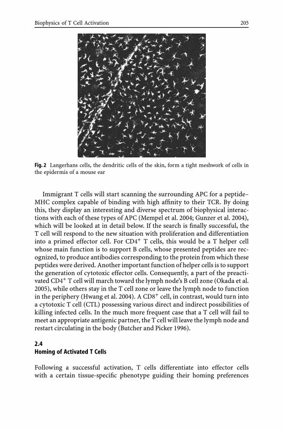
Biophysics of T Cell Activation 205
Fig. 2 Langerhans cells, the dendritic cells of the skin, form a tight meshwork of cells inthe epidermis of a mouse ear
Immigrant T cells will start scanning the surrounding APC for a peptide–MHC complex capable of binding with high affinity to their TCR. By doingthis, they display an interesting and diverse spectrum of biophysical interac-tions with each of these types of APC (Mempel et al. 2004; Gunzer et al. 2004),which will be looked at in detail below. If the search is finally successful, theT cell will respond to the new situation with proliferation and differentiationinto a primed effector cell. For CD4+ T cells, this would be a T helper cellwhose main function is to support B cells, whose presented peptides are rec-ognized, to produce antibodies corresponding to the protein from which thesepeptides were derived. Another important function of helper cells is to supportthe generation of cytotoxic effector cells. Consequently, a part of the preacti-vated CD4+ T cell will march toward the lymph node’s B cell zone (Okada et al.2005), while others stay in the T cell zone or leave the lymph node to functionin the periphery (Hwang et al. 2004). A CD8+ cell, in contrast, would turn intoa cytotoxic T cell (CTL) possessing various direct and indirect possibilities ofkilling infected cells. In the much more frequent case that a T cell will fail tomeet an appropriate antigenic partner, the T cell will leave the lymph node andrestart circulating in the body (Butcher and Picker 1996).
2.4Homing of Activated T Cells
Following a successful activation, T cells differentiate into effector cellswith a certain tissue-specific phenotype guiding their homing preferences

206 P. Reichardt · M. Gunzer
(Butcher and Picker 1996; Kannagi 2002; Springer 1990, 1994). Key factorsthat collectively determine the homing of leukocytes are interactions be-tween integrins or selectins and their tissue adhesion molecules, as wellas interactions between chemokine receptors and their specific ligands, thechemokines (Butcher and Picker 1996; Kannagi 2002; Springer 1990, 1994).In addition, the presence of inflammatory stimuli modulates the homing be-havior of immune cells (Hwang et al. 2004). Activated T cells start homingto peripheral tissues, where they exert proinflammatory regulatory effectorfunctions or recirculate as memory cells (Sallusto et al. 2004). At the targetlocation, T cells leave the vessels and enter the local tissue ready to exerttheir effector function (Butcher and Picker 1996; Gunzer et al. 2005). Alterna-tively, antigen-primed T cells might become memory cells, an option whichmay occur early during T cell development as well as after differentiationinto an effector cell (Seder and Ahmed 2003). Memory cells either reside cen-trally in lymphoid tissues or in the periphery, with a very high likelihoodof expanding rapidly upon reencountering the stimulating antigen (Sallustoet al. 1999).
2.5T Cell Regulation
A special case emerges when the T cell/APC contact does not generate a fullT cell activation status aimed at performing the optimal immune response,but rather results in T cells suppressing the immune response. It is currentlya matter of debate as to whether a regulatory cell is a differentiation statusof any given T cell, or rather a specific type of T cell that is preferentiallyexpanded in situations where suboptimal activation is prevailing. Evidencefor naturally occurring regulatory cells has been obtained (Jonuleit et al.2001). However, antigen presentation by immature and semimature DC invivo might lead to incomplete T cell activation and de novo induction ofregulation (Jonuleit et al. 2001; Probst et al. 2003). Regulatory T cells have at-tracted great interest in recent years and certainly play a very important rolein maintaining an efficient and safe immune system (O’Neill et al. 2004; Sak-aguchi 2005). The role of cell–cell interactions during this process has not yetbeen studied in detail (Fontenot et al. 2005).
3The Biophysics of T Cell Activation
The direct microscopic observation of T cells revealed that upon migration,T cells (which appear round in liquid cell culture) show a very dynamic cellshape with clear polarity: a tail is formed by a motile uropod, as well as a headconsisting of the cytoskeletal leading edge by which cell–cell contacts are ini-
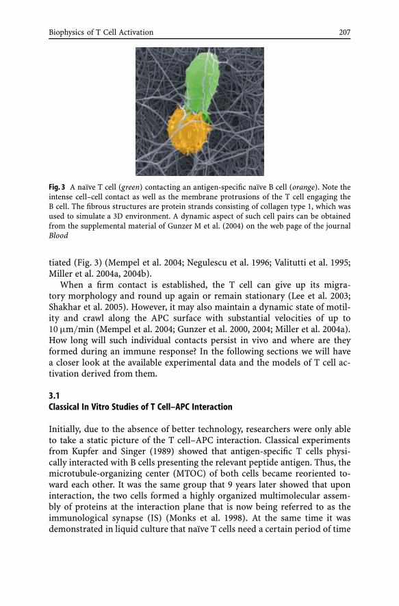
Biophysics of T Cell Activation 207
Fig. 3 A naïve T cell (green) contacting an antigen-specific naïve B cell (orange). Note theintense cell–cell contact as well as the membrane protrusions of the T cell engaging theB cell. The fibrous structures are protein strands consisting of collagen type 1, which wasused to simulate a 3D environment. A dynamic aspect of such cell pairs can be obtainedfrom the supplemental material of Gunzer M et al. (2004) on the web page of the journalBlood
tiated (Fig. 3) (Mempel et al. 2004; Negulescu et al. 1996; Valitutti et al. 1995;Miller et al. 2004a, 2004b).
When a firm contact is established, the T cell can give up its migra-tory morphology and round up again or remain stationary (Lee et al. 2003;Shakhar et al. 2005). However, it may also maintain a dynamic state of motil-ity and crawl along the APC surface with substantial velocities of up to10 µm/min (Mempel et al. 2004; Gunzer et al. 2000, 2004; Miller et al. 2004a).How long will such individual contacts persist in vivo and where are theyformed during an immune response? In the following sections we will havea closer look at the available experimental data and the models of T cell ac-tivation derived from them.
3.1Classical In Vitro Studies of T Cell–APC Interaction
Initially, due to the absence of better technology, researchers were only ableto take a static picture of the T cell–APC interaction. Classical experimentsfrom Kupfer and Singer (1989) showed that antigen-specific T cells physi-cally interacted with B cells presenting the relevant peptide antigen. Thus, themicrotubule-organizing center (MTOC) of both cells became reoriented to-ward each other. It was the same group that 9 years later showed that uponinteraction, the two cells formed a highly organized multimolecular assem-bly of proteins at the interaction plane that is now being referred to as theimmunological synapse (IS) (Monks et al. 1998). At the same time it wasdemonstrated in liquid culture that naïve T cells need a certain period of time

208 P. Reichardt · M. Gunzer
in contact with APC to reach their full effector potential. This was ∼ 6 h forthe best possible APC, but could be as long as 30 h for weak APC. There-fore powerful APC, like DC, were able to induce T cell proliferation withina few hours of contact, even at low doses of antigen, while weak APC, suchas B cells, needed long contact times, even at high levels of pMHC occupancy(Iezzi et al. 1998). In addition, the first approaches using live cell imagingof T cells migrating on lipid bilayers found that, in the absence of recogniz-able peptide–MHC complexes, T cells were highly mobile and migrated onICAM-1 layers (Dustin et al. 1997). However, when a specific peptide–MHCcomplex was available within the lipid bilayers in addition to ICAM-1, theT cells stopped moving and remained at the MHC–peptide-containing spot(Dustin et al. 1997).
3.2The Single Encounter Model of T Cell Activation
The above data were used to establish a model of T cell activation in lymphnodes stating that naïve T cells are actively motile in lymphoid tissues,thereby scanning the surface of all available APC for specific peptide–MHCcomplexes. However, as soon as a high-affinity (cognate) MHC–peptide com-plex is discovered, T cells stop and remain at this location, forming a stablecell contact with the APC for several hours until full activation is reached.During that time, an immunological synapse is formed, which is essentialfor signaling and which is possibly also involved in stopping T cell motility(Dustin and Chan 2000; Lanzavecchia et al. 1999). The concept of a cognatecell pair stopping and thus stabilizing its contact was appealing, as it pro-vided a solution to the apparent problem that in vivo a rather low numberof motile peptide-loaded APC had to activate an also very limited number ofmotile T cells (Nikolich-Zugich et al. 2004) specific for an invading pathogen.It appeared logical that, in the rare event of a specific APC hitting a specificT cell, this interaction had to be stabilized in order to maximize the chanceof a successful T cell activation. Thus, the complete stopping of the two cellu-lar partners was considered a general rule for all situations of specific T cellactivation.
3.3Challenges Introduced by Live Cell Imaging Experiments
This concept, however, was challenged by the first observations employinglive cell imaging of antigen presentation in liquid cultures with T cell blastsand with macrophages serving as APC. Here, it was found that T cells migrateover APC bodies, stopping only for a limited amount of time on one APC, butthen resume motility and wander on to the next APC. From these observa-tions it was concluded that a T cell sequentially could gain signals from more

Biophysics of T Cell Activation 209
Fig. 4 T cells (green) interacting with an antigen-specific DC (orange) within 3D collagen.Note the huge size of the DC relative to the T cells, as well as the ability of the DC to“serve” three T cells at the same time. In vivo, DC can contact up to 200 cells at any giventime point (Miller MJ et al. 2004a)
than one APC (Underhill et al. 1999). However, one point of critique regardingthese experiments was that the particular behavior of the T cells used in thisstudy could result from their preactivated state, and thus might not reflect thebehavior of a naïve, nonactivated T cell. Therefore, in a new approach, naïveT cell receptor (TCR) transgenic T cells plus peptide-loaded highly activatedDC were used as an antigen-specific activation pair, and the two types of cellswere embedded in a 3D collagen matrix as an approximation of the 3D envi-ronment present within lymphatic tissue. Using this experimental system, itwas shown that it was a general feature of naïve T cells to form contacts withspecific antigen-loaded DC only transiently, for a few minutes rather thanhours (Fig. 4) (Gunzer et al. 2000).
In addition, T cells were observed to engage multiple DC in a serial fash-ion. It was assumed that, although each of these contacts was able to inducesignals within the T cell, none of these short-lived contacts was sufficientto activate the T cell but rather a sequence of several contacts was needed(Gunzer et al. 2000). It could later be shown that T cells may indeed gradu-ally increase the level of signaling intermediates (Borovsky et al. 2002), andthat the same level of activation can be reached by a sequence of interruptedcontacts or with one stable contact (Faroudi et al. 2003). The intracellularevents underlying these phenomena are not yet fully elucidated. Accumula-tion of transcription factors such as cJun over time following suboptimal TCRligation has been demonstrated (Rosette et al. 2001); it was postulated that
210 P. Reichardt · M. Gunzer
a counting molecule exists, allowing for a stepwise buildup of activated sig-naling intermediates and finally leading to T cell activation (Rosette et al.2001). The question as to whether such a system works like a capacitor, which“deloads” in one stroke once it has reached the loading capacity, or whethera more analog system exists, which can generate a smooth response reflectingdifferent levels of activation, is currently only a matter of speculation.
3.4A Serial Encounter Model of T Cell Activation
These findings were put together in the serial encounter model for T cell acti-vation (Friedl and Gunzer 2001; Rachmilewitz and Lanzavecchia 2002). Basedon the findings in 3D collagen matrices, the serial encounter model statedthat there is no difference in the biophysical dynamics of T cells contactinglow-affinity or cognate APC. However, only cognate APC were able to delivera signal to the T cell. Therefore, the number of available pMHC complexeson one cell would not be essential, as it has been shown that even one pMHCmolecule is sufficient to trigger T cell responses (Irvine et al. 2002). Since onecontact was not enough, the T cell was forced to undergo additional contactsto other APC presenting the same antigen. Such a mechanism would be help-ful in “measuring” the number of APC in the migratory reach of a T cell, andthereby inhibiting unnecessary immune activation in the case of minor in-fections with very low numbers of antigen-specific APC (Friedl and Gunzer2001).
3.5The Road to Intravital Imaging: Phase Models of T Cell Activation
The results of T cell biophysics obtained in 3D collagen matrices were chal-lenged mainly by the fact that, in true lymphatic tissue, collagen fibers are notdirectly accessible to migrating cells. Rather, they are ensheathed by follicu-lar reticulum cells of the lymphatic environment (Gretz et al. 2000; KaldjianPet al. 2001). Thus, the behavior of T cells in 3D collagen might not necessar-ily represent the in vivo reality (Dustin and de Fougerolles 2001; Dustin et al.2001; Lanzavecchia and Sallusto (2001).
The goal to observe T cell activation in true lymphatic tissue was finallyreached by two groups working with naïve T cells and DC in explanted lymphnodes (Miller et al. 2002; Stoll et al. 2002) and by a group examining thy-mocytes in reaggregate thymic organ cultures (Bousso et al. 2002). All threestudies were able to demonstrate effective migration of T cells in lymphatictissue. This was very remarkable given the extreme crowding of cells in theseorgans (Ushiki et al. 1995). In fact, for T and B cells, the measured motil-ity parameters in intact lymphatic organs were identical to the values thatwere obtained for cells migrating in artificial 3D systems, which are only

Biophysics of T Cell Activation 211
sparsely populated such that individual cells only rarely touch each other(Gunzer et al. 2004; Friedl et al. 1994, 1995, 1998). Within T cell zones oflymph nodes DC, in contrast, were found to be more static. Rather than mi-grating within lymph nodes, they remained mainly stationary, but displayedhighly motile dendrites which were constantly reaching for T cells (Milleret al. 2004a; Lindquist et al. 2004). Interestingly, the studies arrived at oppo-site conclusions concerning the duration of antigen-specific contacts betweenT cells and DC. While the studies by Bousso et al. (2002) and Miller et al.(2002) showed that contacts between T cells and APC were an equal mix-ture of short-lived and more extended interactions, Stoll et al. (2002) foundexclusively long-lasting contacts between adoptively transferred mature DCand naïve T cells upon antigen presentation. It was possible that the differen-tial results were due to differences in treatment of explanted tissues, or thatchanges in the behavior of cells after removal of the lymph nodes occurred.Therefore, it was clear that it was necessary to approach imaging in lymphnodes in situ, i.e., in the living animal.
This goal was initially reached by Miller et al. (2003), albeit only by lookingat T cell migration. The first intravital study of naïve T cells interacting withDC in a lymph node was completed by Mempel et al. (2004). By monitoringCD8+ T cells, this study showed that the T cell activation followed a three-stepmechanism. T cells which had just entered the lymph node engaged antigen-specific DC in short, transient encounters during the first 6–8 h. After thistime, interactions became more stable (in the range of one or more hours)and the production of effector cytokines was induced. This phase lasted for∼ 12 h. A third stage was again characterized by profound motility and onlytransient encounters with DC. It was during this phase that T cell divisionsbecame detectable (Mempel et al. 2004). These findings were essentially con-firmed for CD4+ T cell activation in vivo (Shakhar et al. 2005) and in anexplanted lymph node model (Miller et al. 2004b). In addition, the groupof Amigorena (Hugues et al. 2004) showed that CD8+ T cells contacting DCunder a regime inducing tolerance rather that activation displayed short-livedinteractions at any time point, while Mempel et al.’s (2004) three-phase modelwith long interactions in a middle phase could be confirmed for an activa-tory situation (Hugues et al. 2004). Thus, current data suggest that a completeabsence of static interactions to DC is associated with less efficient T cell ac-tivation or even T cell tolerization for CD8 cells, while there seems to be noobvious difference in interaction behavior of CD4 T cells, whether the out-come is anergy or activation (Shakhar et al. 2005). In any case, most availablestudies clearly show the presence of dynamic interactions at different timepoints during effective T cell stimulation.
The exact function and underlying molecular mechanisms of each respec-tive phase are unclear; however, the sequence may reflect expression changesof surface receptors and cytoskeletal regulators during T cell activation. It ispossible that the longevity of T cell–DC interactions in a phase might indicate
212 P. Reichardt · M. Gunzer
the formation of a mature synapse-like contact zone (Lee et al. 2002). Whileboth dynamic and stable contacts contribute to T cell activation and prolifer-ation, long-lasting interactions may be needed for maximum IL-2 productionand the attainment of full effector functions (Hurez et al. 2003). The phasemodel or kinetic transition model of T cell interaction expands the serial en-counter model by postulating an integration of several signals of differentduration and strengths into a common pathway of T cell activation.
3.6The Role of the APC
The results of recent studies lead to another important question. What cellmakes the decision on maintaining the interaction and comigration? TheT cell or the APC? The studies by Mempel et al. (2004) and Miller et al. (2004b)concluded that it was the status of the T cell which decided the duration ofAPC interaction. However, even in the rather homogeneous population ofnaïve TCR transgenic T cells, interactions with DC were shown to differ sub-stantially in vitro, ranging from short-lived to long-lived (Hurez et al. 2003).The first study that addressed the role of the APC in deciding the duration ofinteraction used two types of effective APC—DC and activated B cells— aswell as naïve B cells, as a model for a weak, ineffective APC. The APC werepeptide-loaded and incubated separately with naïve TCR transgenic T cells. Itwas found that the T cells contacted effective APC (DC and activated B cells)in a short-lived manner, while they formed long-lasting contacts with ineffi-cient APC (naïve B cells). This pattern was demonstrated both in 3D in vitrosystems and in lymph nodes in vivo (Gunzer et al. 2004). The findings wereeven more unexpected as the long-lasting contacts with naïve B cells wereshown to be the least efficient in terms of T cell activation, while the muchmore dynamic contacts to DC and activated B cells rapidly induced T cellactivation (Gunzer et al. 2004).
3.7Summary: A Multitude of T Cell–APC Interaction Kinetics
For the original problem of how to synchronize the migration of cells ini-tially separated in space, live cell imaging experiments of the past 5 years haveoffered a wide range of solutions. Synchronization can occur transiently bylimiting the migration of T cells to the surface of APC, either with an almostunchanged velocity compared to free migration or in a more stable manner,with strongly reduced motility down to complete stopping of T cells on thesurface of DC. In a most extreme form, hour-long coordinated migration ofa cell pair occurs, as in the example of naïve B cells as APC (Gunzer et al.2004). During the comigration, very motile T cells were shown to push B cellsin vivo through lymph node parenchyma, whereas B cells, when activated,

Biophysics of T Cell Activation 213
may take the lead themselves and pull T cells (Gunzer et al. 2004). The latterfinding was later confirmed for B cells that were activated by loading anti-gen onto their B cell receptor (Okada et al. 2005). Another issue covered bythe studies mentioned was directional migration. In the marketplace analogy,this would mean that customers on the market would somehow be prefer-entially attracted to or by specific booths. Currently available data do notsupport this idea. In contrast, there is evidence that migration of T cells inlymph nodes is entirely random and that encounters with APC are stochas-tic events (Miller et al. 2003; Wei et al. 2003). However, other researcherscall these conclusions into question (Germain and Jenkins 2004). What isagreed upon is the sole fact that the lymph node per se is attractive to T cells,B cells, and APC by means of the chemokines CCL19 and CCL21 binding tothe chemokine receptor CCR7 (Forster et al. 1999).
Lastly, it was shown that the character of interaction is not only modulatedby antigenic load and type of APC, but also by the architecture of the sur-rounding tissue which may influence the dynamics of cell interactions. Tightdendritic cell networks within the lymph node facilitate in vivo T cell contactswith DC and the transition between them, thus allowing the T cells to be incontact with DC for most of the time (Lindquist et al. 2004; Westermann et al.2005).
4Conclusions and Outlook
Great advances in imaging techniques in recent years have led to uniqueinsights into the complexity of cell–cell interactions in vivo. However, theresulting models of T cell activation still vary in their interpretation of phys-ical interaction modes of T cells, and many aspects yet remain descriptive.There is now evidence from several research groups for a multistep cell in-teraction process during T cell activation. However, the molecular basis forthe different interaction kinetics observed remains unclear. Other questionsin need of clarification are, e.g., what is the relative importance of long overshort contacts for T cell activation? Are both contact types necessary or isone dispensable in vivo? By what molecular principle do short cell contactsdiffer from long ones? Do short contacts “prepare” cells for long ones andif yes, how? How exactly is the surrounding tissue influencing the durationor outcome of interaction? Apart from studying effector cell induction, themechanism of APC activation (especially of B cells) needs to be investigated.Experimental models need to be expanded to include locations other than in-guinal or polietal lymph nodes, e.g., lymphatic sites in the gut and the lung.More physiologically relevant models together with pathogen-related anti-gens are needed, as well as ways to overcome one major obstacle for in vivoimaging studies, the low number of precursor cells in wild-type animals.

214 P. Reichardt · M. Gunzer
In conclusion, T cells display an intriguingly complex pattern of cellularinteraction with APC during their activation. A more complete understand-ing of the underlying mechanisms in vivo is needed to modulate or mimicsuch processes. This would be one prerequisite to make them amenable fortherapeutic intervention. Based on the excellent advances in intravital imag-ing made in recent years, we can be very positive about future insights gainedfrom this powerful methodology.
Acknowledgement We thank Priyanka Narang for critical reading of the manuscript.
References
Alberola-Ila J et al. (1995) Selective requirement for MAP kinase activation in thymocytedifferentiation. Nature 373(6515):620–623
Bajenoff M, Granjeaud S, Guerder S (2003) The strategy of T cell antigen-presenting cellencounter in antigen-draining lymph nodes revealed by imaging of initial T cell acti-vation. J Exp Med 198(5):715–724
Bhakta NR, Oh DY, Lewis RS (2005) Calcium oscillations regulate thymocyte motilityduring positive selection in the three-dimensional thymic environment. Nat Immunol6(2):143–151
Borovsky Z et al. (2002) Serial triggering of T cell receptors results in incremental accu-mulation of signaling intermediates. J Biol Chem 277(24):21529–21536
Bousso P et al. (2002) Dynamics of thymocyte–stromal cell interactions visualized bytwo-photon microscopy. Science 296(5574):1876–1880
Bradley LM, Watson SR (1996) Lymphocyte migration into tissue: the paradigm derivedfrom CD4 subsets. Curr Opin Immunol 8(3):312–320
Brocker T (1997) Survival of mature CD4 T lymphocytes is dependent on major histo-compatibility complex class-II expressing dendritic cells. J Exp Med 186(8):1223–1232
Butcher EC, Picker LJ (1996) Lymphocyte homing and homeostasis. Science 272(5258):60–66
Cella M et al. (1997) Inflammatory stimuli induce accumulation of MHC class II com-plexes on dendritic cells. Nature 388(6644):782–787
Correia-Neves M et al. (2001) The shaping of the T cell repertoire. Immunity 14(1):21–32Dustin ML, Chan AC (2000) Signaling takes shape in the immune system. Cell 103(2):283–
294Dustin ML, de Fougerolles AR (2001) Reprogramming T cells: the role of extracellu-
lar matrix in coordination of T cell activation and migration. Curr Opin Immunol13(3):286–290
Dustin ML et al. (1997) Antigen receptor engagement delivers a stop signal to migratingT lymphocytes. Proc Natl Acad Sci USA 94(8):3909–3913
Dustin ML, Allen PM, Shaw AS (2001) Environmental control of immunological synapseformation and duration. Trends Immunol 22(4):192–194
Faroudi M et al. (2003) Cutting edge: T lymphocyte activation by repeated immunologicalsynapse formation and intermittent signaling. J Immunol 171(3):1128–1132
Fontenot JD et al. (2005) Regulatory T cell lineage specification by the forkhead transcrip-tion factor foxp3. Immunity 22(3):329–341
Forster R et al. (1999) CCR7 coordinates the primary immune response by establishingfunctional microenvironments in secondary lymphoid organs. Cell 99(1):23–33

Biophysics of T Cell Activation 215
Friedl P, Brocker EB (2002) TCR triggering on the move: diversity of T cell interactionswith antigen-presenting cells. Immunol Rev 186:83–89
Friedl P, Gunzer M (2001) Interaction of T cells with APCs: the serial encounter model.Trends Immunol 22(4):187–191
Friedl P et al. (1994) Locomotor phenotypes of unstimulated CD45RAhigh andCD45ROhigh CD4+ and CD8+ lymphocytes in three-dimensional collagen lattices.Immunology 82(4):617–624
Friedl P, Noble PB, Zanker KS (1995) T lymphocyte locomotion in a three-dimensionalcollagen matrix. Expression and function of cell adhesion molecules. J Immunol154(10):4973–4985
Friedl P et al. (1998) CD4+ T lymphocytes migrating in three-dimensional collagen latticeslack focal adhesions and utilize beta1 integrin-independent strategies for polarization,interaction with collagen fibers and locomotion. Eur J Immunol 28(8):2331–2343
Fuchs EJ, Matzinger P (1992) B cells turn off virgin but not memory T cells. Science258(5085):1156–1159
Germain RN, Jenkins MK (2004) In vivo antigen presentation. Curr Opin Immunol16(1):120–125
Goldrath AW, Bevan MJ (1999) Selecting and maintaining a diverse T cell repertoire.Nature 402(6759):255–262
Gretz JE, Anderson AO, Shaw S (1997) Cords, channels, corridors and conduits: criticalarchitectural elements facilitating cell interactions in the lymph node cortex. ImmunolRev 156:11–24
Gretz JE et al. (2000) Lymph-borne chemokines and other low molecular weightmolecules reach high endothelial venules via specialized conduits while a functionalbarrier limits access to the lymphocyte microenvironments in lymph node cortex.J Exp Med 192(10):1425–1440
Gunzer M et al. (2000) Antigen presentation in extracellular matrix: interactions of T cellswith dendritic cells are dynamic, short lived, and sequential. Immunity 13(3):323–332
Gunzer M et al. (2004) A spectrum of biophysical interaction modes between T cells anddifferent antigen-presenting cells during priming in 3D collagen and in vivo. Blood104(9):2801–2809
Gunzer M et al. (2005) Systemic administration of a TLR7 ligand leads to transient im-mune incompetence due to peripheral-blood leukocyte depletion. Blood 106(7):2424–2432
Hugues S et al. (2004) Distinct T cell dynamics in lymph nodes during the induction oftolerance and immunity. Nat Immunol 5(12):1235–1242
Hurez V et al. (2003) Restricted clonal expression of IL-2 by naive T cells reflects differ-ential dynamic interactions with dendritic cells. J Exp Med 198(1):123–132
Hwang JM et al. (2004) A critical temporal window for selectin-dependent CD4+ lym-phocyte homing and initiation of late-phase inflammation in contact sensitivity. J ExpMed 199(9):1223–1234
Iezzi G, Karjalainen K, Lanzavecchia A (1998) The duration of antigenic stimulation de-termines the fate of naive and effector T cells. Immunity 8(1):89–95
Irvine DJ, Purbhoo MA, Krogsgaard M, Davis MM (2002) Direct observation of ligandrecognition by T cells. Nature 419(6909):845–849
Itano AA et al. (2003) Distinct dendritic cell populations sequentially present antigen to CD4T cells and stimulate different aspects of cell-mediated immunity. Immunity 19(1):47–57
Jonuleit H et al. (2001) Identification and functional characterization of humanCD4(+)CD25(+) T cells with regulatory properties isolated from peripheral blood.J Exp Med 193(11):1285–1294

216 P. Reichardt · M. Gunzer
Kaldjian EP et al. (2001) Spatial and molecular organization of lymph node T cell cor-tex: a labyrinthine cavity bounded by an epithelium-like monolayer of fibroblasticreticular cells anchored to basement membrane-like extracellular matrix. Int Immunol13(10):1243–1253
Kannagi R (2002) Regulatory roles of carbohydrate ligands for selectins in the homing oflymphocytes. Curr Opin Struct Biol 12(5):599–608
Kedl RM et al. (2000) T cells compete for access to antigen-bearing antigen-presentingcells. J Exp Med 192(8):1105–1113
Kupfer A, Singer SJ (1989) The specific interaction of helper T cells and antigen-presenting B cells. IV. Membrane and cytoskeletal reorganizations in the bound T cellas a function of antigen dose. J Exp Med 170(5):1697–1713
Lanzavecchia A, Sallusto F (2001) Antigen decoding by T lymphocytes: from synapses tofate determination. Nat Immunol 2(6):487–492
Lanzavecchia A, Lezzi G, Viola A (1999) From TCR engagement to T cell activation: a ki-netic view of T cell behavior. Cell 96(1):1–4
Lee KH et al. (2002) T cell receptor signaling precedes immunological synapse formation.Science 295(5559):1539–1542
Lee KH et al. (2003) The immunological synapse balances T cell receptor signaling anddegradation. Science 302(5648):1218–1222
Lindquist RL et al. (2004) Visualizing dendritic cell networks in vivo. Nat Immunol5(12):1243–1250
Lukas M et al. (1996) Human cutaneous dendritic cells migrate through dermal lymphaticvessels in a skin organ culture model. J Invest Dermatol 106(6):1293–1299
Mackay CR (1993) Homing of naive, memory and effector lymphocytes. Curr Opin Im-munol 5(3):423–427
Mehling A et al. (2001) Overexpression of CD40 ligand in murine epidermis results inchronic skin inflammation and systemic autoimmunity. J Exp Med 194(5):615–628
Mellman I, Steinman RM (2001) qDendritic cells: specialized and regulated antigen pro-cessing machines. Cell 106(3):255–258
Mempel TR, Henrickson SE, Von Andrian UH (2004) T cell priming by dendritic cells inlymph nodes occurs in three distinct phases. Nature 427(6970):154–159
Miller MJ et al. (2002) Two-photon imaging of lymphocyte motility and antigen responsein intact lymph node. Science 296(5574):1869–1873
Miller MJ et al. (2003) Autonomous T cell trafficking examined in vivo with intravital two-photon microscopy. Proc Natl Acad Sci USA 100(5):2604–2609
Miller MJ et al. (2004a) T cell repertoire scanning is promoted by dynamic dendritic cellbehavior and random T cell motility in the lymph node. Proc Natl Acad Sci USA101(4):998–1003
Miller MJ et al. (2004b) Imaging the single cell dynamics of CD4+ T cell activation bydendritic cells in lymph nodes. J Exp Med 200(7):847–856
Monks CR et al. (1998) Three-dimensional segregation of supramolecular activation clus-ters in T cells. Nature 395(6697):82–86
Moser B, Loetscher P (2001) Lymphocyte traffic control by chemokines. Nat Immunol2(2):123–128
Negulescu PA et al. (1996) Polarity of T cell shape, motility, and sensitivity to antigen.Immunity 4(5):421–430
Nikolich-Zugich J, Slifka MK, Messaoudi I (2004) The many important facets of T-cellrepertoire diversity. Nat Rev Immunol 4(2):123–132
Okada T et al. (2005) Antigen-engaged B cells undergo chemotaxis toward the T zone,form motile conjugates with helper T cells. PLoS Biol 3(6):e150

Biophysics of T Cell Activation 217
O’Neill EJ et al. (2004) Natural and induced regulatory T cells. Ann NY Acad Sci1029(1):180–192
Palmer E (2003) Negative selection—clearing out the bad apples from the T cell reper-toire. Nat Rev Immunol 3(5):383–391
Potsch C, Vohringer D, Pircher H (1999) Distinct migration patterns of naive and effectorCD8 T cells in the spleen: correlation with CCR7 receptor expression and chemokinereactivity. Eur J Immunol 29(11):3562–3570
Probst HC et al. (2003) Inducible transgenic mice reveal resting dendritic cells as potentinducers of CD8+ T cell tolerance. Immunity 18(5):713–720
Rachmilewitz J, Lanzavecchia A (2002) A temporal and spatial summation modelfor T cell activation: signal integration and antigen decoding. Trends Immunol23(12):592–595
Reif K et al. (2002) Balanced responsiveness to chemoattractants from adjacent zonesdetermines B cell position. Nature 416:94–99
Richie LI et al. (2002) Imaging synapse formation during thymocyte selection: inabilityof CD3zeta to form a stable central accumulation during negative selection. Immunity16(4):595–606
Rodriguez-Pinto D, Moreno JB (2005) B cells can prime naive CD4+ T cells in vivo inthe absence of other professional antigen-presenting cells in a CD154-CD40-dependentmanner. Eur J Immunol 35(4):1097–1105
Romani N et al. (2001) Migration of dendritic cells into lymphatics—the Langerhans cellexample: routes, regulation, and relevance. Int Rev Cytol 207:237–270
Rosette C et al. (2001) The impact of duration versus extent of TCR occupancy on T cellactivation: a revision of the kinetic proofreading model. Immunity 15(1):59–70
Sakaguchi S (2005) Naturally arising Foxp3-expressing CD25+CD4+ regulatory T cells inimmunological tolerance to self and non-self. Nat Immunol 6(4):345–352
Sallusto F et al. (1999) Two subsets of memory T lymphocytes with distinct homingpotentials and effector functions. Nature 401(6754):708–712
Sallusto F, Geginat J, Lanzavecchia A (2004) Central memory and effector memory T cellsubsets: function, generation, and maintenance. Annu Rev Immunol 22(1):745–763
Sebzda E et al. (1999) Selection of the T cell repertoire. Annu Rev Immunol 17(1):829–874Seder RA, Ahmed R (2003) Similarities and differences in CD4+ and CD8+ effector and
memory T cell generation. Nat Immunol 4(9):835–842Shakhar G et al. (2005) Stable T cell–dendritic cell interactions precede the development
of both tolerance and immunity in vivo. Nat Immunol 6(7):707–714Springer TA (1990) Adhesion receptors of the immune system. Nature 346(6283):425–434Springer TA (1994) Traffic signals for lymphocyte recirculation and leukocyte emigration:
the multistep paradigm. Cell 76(2):301–314Stoll S, Delon J, Brotz TM, Germain RN (2002) Dynamic imaging of T cell–dendritic cell
interactions in lymph nodes. Science 296(5574):1873–1876Underhill DM et al. (1999) Dynamic interactions of macrophages with T cells during
antigen presentation. J Exp Med 190(12):1909–1914Ushiki T, Ohtani O, Abe K (1995) Scanning electron microscopic studies of reticular
framework in the rat mesenteric lymph node. Anat Rec 241(1):113–122Valitutti S et al. (1995) Sustained signaling leading to T cell activation results from
prolonged T cell receptor occupancy. Role of T cell actin cytoskeleton. J Exp Med181:577–584
Wei SH et al. (2003) A stochastic view of lymphocyte motility and trafficking within thelymph node. Immunol Rev 195:136–159

218 P. Reichardt · M. Gunzer
Westermann J et al. (2005) Naive, effector, and memory T lymphocytes efficiently scandendritic cells in vivo: contact frequency in T cell zones of secondary lymphoid or-gans does not depend on LFA-1 expression and facilitates survival of effector T cells.J Immunol 174(5):2517-2524
Witt CM et al. (2005) Directed migration of positively selected thymocytes visualized inreal time. PLoS Biol 3(6):e160
Yasutomo K et al. (2000) The duration of antigen receptor signalling determines CD4+versus CD8+ T-cell lineage fate. Nature 404(6777):506–510

Results Probl Cell Differ (43)E. Gundelfinger, C. Seidenbecher, B. Schraven:Cell Communication in Nervous and Immune SystemDOI 10.1007/022/Published online: 4 May 2006© Springer-Verlag Berlin Heidelberg 2006
Molecular Regulation of Cytoskeletal RearrangementsDuring T Cell Signalling
Theresia E. B. Stradal1 · Rico Pusch2 · Stefanie Kliche2 (�)1Signalling and Motility Group, German Research Centre for Biotechnology (GBF),Mascheroder Weg 1, 38124 Braunschweig, Germany
2Institute of Immunology, Otto von Guericke University, Leipziger Strasse 44,39120 Magdeburg, [email protected]
Abstract Regulation of the cytoskeleton in cells of the haematopoietic system is essentialfor fulfilling diverse tasks such as migration towards a chemoattractant, phagocytosis orcell–cell communication. This is particularly true for the many types of T cells, which areat the foundation of the adaptive immune system in vertebrates. Deregulation of actinfilament turnover is known to be involved in the development of severe immunodefi-ciencies or immunoproliferative diseases. Therefore, molecular dissection of signallingcomplexes and effector molecules, which leads to controlled cytoskeletal assembly, hasbeen the focus of immunological research in the last decade.
In the past, cytoskeletal remodelling was frequently understood as the finish line ofsignalling, while today it becomes increasingly evident that actin and microtubule dy-namics are required for proper signal transmission in many processes such as T cellactivation. Significant effort is made in many laboratories to further elucidate the con-tribution of cytoskeletal remodelling to immune function.
The objective of this article is to summarise the current knowledge on how actin andmicrotubules are reorganised to support the formation of structures as diverse as theimmunological synapse and peripheral protrusions during cell migration.
Keywords TCR · CD28 · Actin · Tubulin · Cytoskeleton · Immunological synapse
1Introduction
Small GTPases act as molecular switches on which signalling inputs con-verge and are transduced into a coordinated array of output pathways leadingto the desired cell response. The Ras family members, Ras and Rap1, havebeen assigned important functions for cytoskeletal reorganisation, partic-ularly in integrin activation to initiate adhesion and migration (for reviewsee Bos et al. 2003; Kinbara et al. 2003). Rho family GTPases, the bestcharacterised of which comprise RhoA, Rac1 and Cdc42, regulate differ-ent sub-compartments of the actin cytoskeleton (reviewed by Hall 1998).While Rho activation triggers the generation of the contractile forces re-

220 T.E.B. Stradal et al.
quired, for instance, for force coupling through cell substrate adhesions, Racand Cdc42 stimulate the protrusion of lamellipodia and filopodia, respec-tively (Nobes and Hall 1995). Moreover, Cdc42 appears to be crucial for cellpolarisation in cells as diverse as budding yeast or cytotoxic T cells (Etienne-Manneville 2004b). In their activated state, these GTPases specifically interactwith a plethora (> 80 known) of different effector proteins, some of which sig-nal directly to actin cytoskeletal reorganisation. However, the exact functionsof the various effector proteins in these processes are only just beginning toemerge.
1.1The Actin Cytoskeleton
The highly dynamic and reversible polymerisation of actin is at the baseof a number of complex cellular processes, such as chemotaxis or adhe-sion and cell–cell communication. Dysregulation of the actin cytoskeleton inhaematopoietic cells has already been found to cause severe immunopatholo-gies, such as Wiskott–Aldrich syndrome (Badour et al. 2004; Burns et al.2004; Orange et al. 2004; Thrasher 2002) or BCR-Abl-induced leukaemia (Da-ley et al. 1990; Druker et al. 2001; Goldman and Melo 2003; Hantschel andSuperti-Furga 2004; Pendergast and Witte 1987).
As a result of activatory signals, the polymerisation of actin filaments isinitiated and maintained by nucleation and branching off from pre-existingfilaments, processes thought to be catalysed by the Arp2/3 complex. Re-cently, proteins of the formin family were added to the list of cellular actinfilament nucleators acting downstream of Rho-GTPases, and while the mech-anism of creating long unbranched filaments was elucidated (Romero et al.2004), the cellular function of the three diaphanous related formins (DRFs)and other formin family members in vertebrate cells is still far from be-ing understood (Higgs 2005; Higgs and Pollard 2001; Zigmond 2004). TheArp2/3 complex consists of two actin-related and five additional subunits,and is essential for the protrusion of cellular projections such as lamellipo-dia (Machesky and Insall 1998; Pollard and Borisy 2003). The nucleatingactivity of the Arp2/3 complex can be stimulated, for instance, by proteinsof the WASP/WAVE family of nucleation promotion factors (NPFs), whichall share a unique C-terminal domain designed to efficiently activate theArp2/3 complex (Millard et al. 2004; Stradal et al. 2004). The protein re-gions outside this module, however, define two subfamilies: in mammals,one subfamily comprises the haematopoietic WASP (Wiskott–Aldrich syn-drome protein) and the more ubiquitously expressed N-WASP (neural WASP),while the second subfamily is built of three WAVEs (WASP family verprolin-homologous proteins), also known as Scar (suppressor of cAMP receptor)isoforms.

Cytoskeletal Rearrangements in T Cells 221
Differential roles for both protein subfamilies, WASP/N-WASP and WAVEs,will be discussed below with a focus on their potential as well as establishedroles in the spatio-temporal activation of T cells.
1.2The Microtubule System
Microtubules are involved in cell division, cell motility, morphogenesisand cell polarity, translocalisation of organelles and transport of vesicles(Etienne-Manneville 2004a; Sancho et al. 2002; Vyas et al. 2002). T lympho-cytes are highly motile premitotic cells harbouring a well-developed mi-crotubule cytoskeleton (see Fig. 1) upon activation they have the ability tomigrate, to form intimate cell–cell contacts and finally to enter the cell cycleand undergo mitosis. The microtubule building block is formed by het-erodimers of α- and β-tubulins, and head-to-tail association of these dimersleads to the formation of linear protofilaments. The lateral association of atleast 13 protofilaments forms a cylindrical microtubule. Microtubules are in-herent polar structures with a dynamic plus end and a minus end that canbe stabilised by embedding the filament into a microtubule organising cen-tre (MTOC). The dynamic behaviour of microtubules in vivo is describedby the term dynamic instability, as a microtubule can frequently switchbetween phases of growth and rapid depolymerisation. Post-translationalenzymatic modifications of tubulin, which can occur after polymerisationinto tubules, lead to variations in microtubule stability and altered affin-ity for microtubule binding proteins (MacRae 1997; Westermann and Weber2003).
Dynamic microtubules can be locally stabilised during cell migration andpolarisation (Gundersen et al. 2004). Microtubule stabilisation in fibroblastsoccurs primarily at the leading edge and results from capturing of the micro-tubule plus-ends by plasma membrane–microtubule tip complex interactions(Brunner 2002; Gundersen and Cook 1999; Gundersen et al. 2004). These sta-bilised microtubules accumulate post-translationally modified tubulin, suchas acetylated or detyrosinated tubulin (for reviews see MacRae 1997; Wester-mann and Weber 2003). Acetylation of α-tubulins occurs on a conservedlysine residue at position 40 at the amino terminus of α-tubulins (LeDizet andPiperno 1987; L’Hernault and Rosenbaum 1985). To date, the enzymes thatare responsible for the tubulin acetylation in mammalian cells remain elu-sive (Polevoda and Sherman 2002). The enzymes that catalyse the opposingreaction, deacetylation, have been described. HDAC6 (histone deacetylase 6),a member of the histone deacetylase family, deacetylate tubulin and micro-tubules in vitro, and suppression of HDAC6 by pharmacolocical inhibitors(trichostatin (TSA) and tubacin) or by small interfering RNA (siRNA) leads toa marked increase in tubulin acetylation in fibroblasts (Haggarty et al. 2003;Hubbert et al. 2002; Zhang et al. 2003).
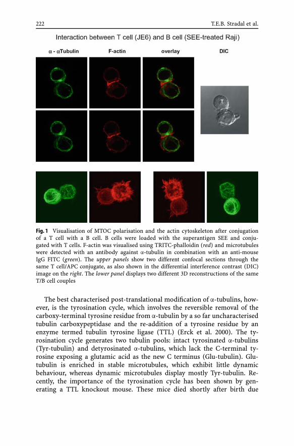
222 T.E.B. Stradal et al.
Fig. 1 Visualisation of MTOC polarisation and the actin cytoskeleton after conjugationof a T cell with a B cell. B cells were loaded with the superantigen SEE and conju-gated with T cells. F-actin was visualised using TRITC-phalloidin (red) and microtubuleswere detected with an antibody against α-tubulin in combination with an anti-mouseIgG FITC (green). The upper panels show two different confocal sections through thesame T cell/APC conjugate, as also shown in the differential interference contrast (DIC)image on the right. The lower panel displays two different 3D reconstructions of the sameT/B cell couples
The best characterised post-translational modification of α-tubulins, how-ever, is the tyrosination cycle, which involves the reversible removal of thecarboxy-terminal tyrosine residue from α-tubulin by a so far uncharacterisedtubulin carboxypeptidase and the re-addition of a tyrosine residue by anenzyme termed tubulin tyrosine ligase (TTL) (Erck et al. 2000). The ty-rosination cycle generates two tubulin pools: intact tyrosinated α-tubulins(Tyr-tubulin) and detyrosinated α-tubulins, which lack the C-terminal ty-rosine exposing a glutamic acid as the new C terminus (Glu-tubulin). Glu-tubulin is enriched in stable microtubules, which exhibit little dynamicbehaviour, whereas dynamic microtubules display mostly Tyr-tubulin. Re-cently, the importance of the tyrosination cycle has been shown by gen-erating a TTL knockout mouse. These mice died shortly after birth due

Cytoskeletal Rearrangements in T Cells 223
to disorganisation of the neuronal networks (Erck et al. 2005). Below, theinvolvement of microtubule dynamics and modifications in cell polarisa-tion, during cell–cell interaction or cell migration in T lymphocytes will bediscussed.
2Arp2/3-Dependent Actin Assembly and the WASP/Scar Family of Proteins
2.1WASP and the Wiskott–Aldrich Syndrome
In mammals, most is known about the loss of function of the haematopoieticWAS gene. In addition to the intense research on the inherited Wiskott–Aldrich syndrome (WAS), two independent WASP knockout mice havebeen reported (Snapper et al. 1998; Zhang et al. 1999), displaying a mildhaematopoietic phenotype reminiscent of less severe WAS in humans (Burnset al. 2004). In 1996, 2 years after the discovery of WASP as the genemutated in this disease (Derry et al. 1994), two groups independently es-tablished the direct interaction of this protein with the Rho family GTPaseCdc42 (Aspenstrom et al. 1996; Symons et al. 1996), raising the possibilityof WASP acting in the translation of Cdc42 signalling to actin cytoskeletalreorganisation. The subsequent recognition of the Arp2/3-activating fea-tures of WASP/Scar family members (Machesky and Insall 1998; Macheskyet al. 1999; Rohatgi et al. 1999) provided the direct connection to actinfilament assembly. Hence, a potential Cdc42 → WASP/N-WASP → Arp2/3pathway appeared firmly established and provided a first clue on how Cdc42could induce actin assembly. The most striking cytoskeletal phenotypesin WAS patients and WASP knockout mice are impaired T cell activationupon T cell/antigen-presenting cell (APC) conjugation (Gallego et al. 1997),the lack of surface microvilli on T lymphocytes (Kenney et al. 1986) (al-though this was questioned recently (Majstoravich et al. 2004)), as well asthe failure of haematopoietic cells to form podosomes or to efficiently mi-grate in vitro (Jones et al. 2002). Indeed, both WASP and N-WASP localiseto podosomes and appear essential for their formation (Mizutani et al.2002).
2.1.1Regulation of WASP
The activity of the ubiquitously expressed N-WASP is regulated througha plethora of binding partners such as Cdc42 (Rohatgi et al. 1999), PIP2(phosphatidylinositol 4,5-bisphosphate) (Rohatgi et al. 2000), WASP-inter-acting protein (WIP) (Moreau et al. 2000), and SH3 domain harbouring
224 T.E.B. Stradal et al.
signalling adapters like Nck (Rohatgi et al. 2001) or Grb2 (Carlier et al.2000). Thus, these signalling intermediates are thought to play key rolesin the regulation of actin rearrangements, providing the diversity neces-sary to satisfy the various requirements. Although many interaction part-ners of N-WASP are known, most of them were implicated in N-WASP ac-tivation or modulation of its activity. Until recently, it was widely agreedthat N-WASP, which is autoinhibited in the cytoplasm via an intramolecu-lar bond, exists as a monomer prior to activation. Subsequently, however,biochemical approaches indicated that N-WASP appears constitutively boundto WIP family members, such as the brain-specific CR16 (Ho et al. 2001),or to WIP (= WASPIP) (Ho et al. 2004), which is highly expressed in thehaematopoietic system (Anton et al. 2002). Furthermore, Ho and colleagues(2004) concluded from in vitro actin polymerisation assays that WIP bind-ing to N-WASP stabilises its inactive conformation. However, other studiessuggest a dependency on the binding of WIP to the N terminus of WASPduring T cell activation (Silvin et al. 2001), and a positive cooperation be-tween WIP and WASP/N-WASP has also been described in other systems(Martinez-Quiles et al. 2001; Moreau et al. 2000). Moreover, WIP-deficientT cells display a phenotype significantly reminiscent of WASP deficiency,as exemplified by impaired proliferation, reduced interleukin-2 (IL-2) se-cretion or disturbed immunological synapse (IS) formation after T cellreceptor (TCR) ligation (Anton et al. 2002). Thus, it is still controversialas to whether the in vitro findings of Ho and colleagues (2004) describ-ing WIP as an inhibitor of WASP/N-WASP function can be confirmed invivo.
Beyond WIP, only little is known about the direct interaction partners ofWASP as compared to N-WASP. In most cases, newly identified interactionpartners of N-WASP are assumed to interact with WASP in a similar man-ner, although this was rarely tested. One exception is the finding that theEna/VASP family member, VASP (vasodilator-stimulated phosphoprotein),interacts with WASP but not N-WASP and promotes actin assembly at theplasma membrane (Castellano et al. 2001). A WASP/VASP-containing com-plex was reported to occur in macrophages during FCγ-receptor-mediatedphagocytosis (Coppolino et al. 2001), and sequestration of Ena/VASP proteinsin Jurkat T cells using a fragment of the listerial virulence factor ActA (Sechiet al. 2002) was found to impede full interaction with anti-CD3-coated beads.The relevance of these interactions in vivo remains to be established.
2.1.2WASP and Endocytosis
WASP is implicated in the clathrin-mediated endocytosis of the T cell anti-gen receptor (McGavin et al. 2001; Zhang et al. 1999), and both WASPand N-WASP are known to interact with various endocytic proteins, e.g.

Cytoskeletal Rearrangements in T Cells 225
syndapin (also named PACSIN) proteins (reviewed by Kessels and Qual-mann 2004), intersectin-1 (Hussain et al. 2001) and indirectly with thelarge GTPase dynamin (reviewed by Engqvist-Goldstein and Drubin 2003;Schafer 2002). Hence, WASP and N-WASP are thought to play a role inreceptor-mediated endocytosis (Gundelfinger et al. 2003; Qualmann andKessels 2002).
In exciting recent studies, direct visualisation of the recruitment of actinand N-WASP accompanying the internalisation of single clathrin-coated pitswas achieved by employing two-colour total internal reflection fluorescence(TIRF) microscopy (Merrifield et al. 2002, 2004). Moreover, endocytic defectswere observed in cells ablated for N-WASP expression by genetic deletion aswell as by RNA interference. In N-WASP null fibroblasts, impaired actin as-sembly at internalising clathrin-coated pits was accompanied by a significantreduction in internalisation rates of epidermal growth factor receptor (EGFR)(Benesch et al. 2005), and after RNAi-mediated knockdown of N-WASP addi-tional severe alterations in cell surface distribution of EGFR and transferrinreceptor (TfR) were observed in HeLa cells (Innocenti et al. 2005).
Moreover, several lines of evidence substantiate a physiological functionof N-WASP/WASP in actin assembly events at the plasma membrane or en-domembranes. For instance, N-WASP –/– fibroblasts fail to support the move-ment of endosomal vesicles evoked by increased PIP2 or phosphotyrosinelevels (Benesch et al. 2002; Taunton 2001), a phenotype which can be fullyrestored not only by the re-expression of N-WASP but also by expression ofWASP. Thus, both the haematopoietic WASP and N-WASP may drive actinassemblies for complex cellular functions related to membrane and proteintraffic, which require further analysis.
2.2WAVE Proteins Induce Actin Assembly Downstream of Rac
All haematopoietic cells express members of the WAVE subfamily of pro-teins in addition to WASP. WAVE proteins mediate Arp2/3-driven lamellipo-dia and membrane ruffle formation (Machesky and Insall 1998; Miki et al.1998), which occur concomitant to Rac1 activation. However, unlike WASPor N-WASP, WAVE proteins lack a motif capable of direct interaction witha Rho-GTPase, and instead are linked to Rac1 indirectly.
Several studies have established how WAVE2 function is controlled bya multi-protein complex consisting of at least four components, which linksRac to Arp2/3 activation (Eden et al. 2002; Innocenti et al. 2004; Kunda et al.2003; Rogers et al. 2003; Steffen et al. 2004). This complex harbours the specif-ically Rac1-associated protein 1 (Sra-1) and Nck-associated protein 1 (Nap1),Abi (Abl interacting) proteins and WAVE. The constitutively assembled com-plex was found to be recruited to and activated at the plasma membrane uponactivation of Rac1. However, the role of the WAVE complex in the formation of

226 T.E.B. Stradal et al.
structures other than lamellipodia or in cells other than fibroblasts remainedelusive (see also below).
Mammals express three WAVE isoforms, two of which are mostly restrictedto the brain (WAVE1 and WAVE3) and one (WAVE2) being expressed ubiqui-tously (Sossey-Alaoui et al. 2003). Conventional WAVE2 knockout mice havenot helped the understanding of WAVE function in haematopoietic cells dueto early embryonic lethality (Yamazaki et al. 2003; Yan et al. 2003). It willgreatly improve our understanding of WAVE complex function in the immunesystem when the results from conditional targeting of WAVE genes, and sub-sequent deletion of proteins in cells of the haematopoietic lineage, becomeavailable.
3T Cell Signalling Leading to Cytoskeletal Rearrangements
The Rho family of GTPases is most prominently involved in the regulation ofcytoskeletal rearrangements. We will now briefly summarise two prominentT cell behaviours, cell migration along chemotactic gradients and activationof TCR/CD3 complexes through interaction with an APC, which both involvesignalling cascades leading to activation of GTPases (Cantrell 2003) and sub-sequent rearrangement of the actin and microtubule cytoskeleton.
3.1T Cell Migration
Haematopoietic cells including T cells display a type of movement that hasbeen termed amoeboid movement, which differs significantly in phenotypicappearance from fibroblastic migratory behaviour (Entschladen and Zanker2000). This amoeboid type of migration (Fig. 2) was repeatedly compared tothe chemotactic movements of Dictyostelium discoideum (Parent 2004; VanHaastert and Devreotes 2004). Directional cell migration entails the establish-ment and maintenance of polarity, as established by formation of protrusiveactin-based structures at the cell front (Pollard and Borisy 2003; Small et al.2002). This allows the exploration of new space and is accompanied by an-chorage of these newly formed cellular structures through specialised adhe-sion sites, which then support dragging of the cell body forward (Beningoand Wang 2002; Hestermann et al. 2002; Horwitz and Parsons 1999) and—in most cell types—reorientation of the microtubule cytoskeleton (Etienne-Manneville and Hall 2003; Gundersen 2002; Palazzo et al. 2001). For immunecells, and also for the well-studied D. discoideum, chemotactic movementis initiated when chemoattractants bind to seven-membrane-span receptorsthat couple to heterotrimeric G proteins. This leads to activation of a plethoraof effector and adaptor proteins through the dissociated Gα and βγ subunits

Cytoskeletal Rearrangements in T Cells 227
Fig. 2 Schematic representation of a migrating T lymphocyte. The leading front of thecell displays F-actin-rich lamellipodia-like protrusions called pseudopodia; the trailingedge of the cell is termed the uropod and is enriched in myosin II. In contrast to mostother cell types, the MTOC of migrating T cells is packed into the uropod and it is un-known whether or how the MTOC or microtubules influence T cell movement duringchemotaxis
(Parent 2004), ultimately leading to cellular polarisation and movement. Con-sequently, pre-treatment of Jurkat T cells with pertussis toxin, a Gαi inhibitor,abolishes chemotaxis towards SDF-1α (Okabe et al. 2002). In comparison toother cell types, T lymphocytes are the smallest and fastest migrating cells(Entschladen et al. 1998; Miller et al. 2003; Nikolai et al. 1998; Smith et al.2003).
The chemokine SDF-1α (denotes stromal cell-derived factor-1α, alsotermed CXCL12), originally characterised as a pre-B cell stimulatory factor, isa potent chemoattractant for T lymphocytes and other leukocytes (Kim andBroxmeyer 1999). Targeted disruption of the SDF-1 gene (or its unique recep-tor CXCR4) in mice is perinatally lethal with defects in lymphopoiesis andbone marrow myelopoiesis (Nagasawa et al. 1996). Little is known about thesignalling pathways that mediate SDF-1 effects. SDF-1α stimulation enhancesthe tyrosine phosphorylation of various focal adhesion molecules, such asCrk and paxillin and, most importantly, T lymphocyte chemotaxis requiresphosphatidylinositol (PI) 3-kinase activity and is abrogated after chemical in-hibition of this enzyme with either wortmannin or Ly294002. (Dutt et al. 1998;Okabe et al. 2002; Wang et al. 2000). Data from D. discoideum underscorethe fact that recruitment of PI 3-kinase and localised PIP3 production at theleading front of chemotaxing cells are upstream of pseudopod protusion, asalso occurs in the presence of the actin polymerisation inhibitor latrunculin(Janetopoulos et al. 2004).
But where in chemotaxis do WASP- and WAVE-mediated Arp2/3 activationcome into play? Acquisition of polarity is at the onset of directed migrationand accompanied by drastic redistribution of cytoskeletal components, dur-ing which F-actin and numerous actin-binding proteins are enriched at thefront or leading edge and myosin II is assembled on the sides and at the back

228 T.E.B. Stradal et al.
or trailing edge, also termed uropod (Entschladen and Zanker 2000; Hogget al. 2003).
WASP-deficient monocytes display impaired chemotaxis in response tomonocyte chemoattractant protein-1 (MCP-1) (Badolato et al. 1998) and Fcγreceptor-mediated phagocytosis is inhibited (Lorenzi et al. 2000). T lympho-cytes from WAS patients display abnormal chemotaxis in response to theT cell chemoattractant SDF-1α (Haddad et al. 2001). In this study Cdc42was shown to be crucial for the chemotactic response, and thus maybe foractivation of WASP, by sequestering specifically activated Cdc42 using the re-combinant Cdc42 interactive motif (CRIB domain) of WASP (Haddad et al.2001). WASP was tyrosine phosphorylated in response to SDF-1α stimulationin Jurkat T cells, a phenomenon that was described earlier to augment WASPactivation (Cory et al. 2002, 2003). Moreover, SDF-1α induced Crk-associatedsubstrate (Cas), Nck, and focal adhesion kinase (FAK) phosphorylation in Ju-rkat T cells in response to SDF-1α. Consequently, chemotaxis in these cells canbe inhibited using herbimycin, a protein tyrosine kinase inhibitor. Notably, asopposed to defective chemotaxis, the random motility of, for example, WASpatient macrophages was found to be indistinguishable from that of controlcells (Jones et al. 2002; Zicha et al. 1998), suggesting that WASP deficiencydoes not interfere with the formation of cellular protrusions, but rather withthe establishment and/or maintenance of polarity towards the chemotacticstimulus.
Data from the chemotaxis of Dictyostelium support this view: Dic-tyostelium harbours two copies of the WASP gene and one for Scar (= WAVE).A Dictyostelim strain was designed that expresses very low levels of WASPafter genetic deletion of one gene and tetracycline-dependent suppression ofthe expression of the other gene (Myers et al. 2005). Cells of this strain weremore spread and flattened as compared to wild-type (WT) cells, but wereunable to establish polarity resulting in a higher angular deviation and thusa four times lower speed of movement towards the chemoattractant (Myerset al. 2005). In contrast, Scar null D. discoideum cells (Bear et al. 1998; Blagget al. 2003) were less well spread and hence significantly smaller than WTcells. They displayed strongly reduced F-actin levels and were unable to ac-cumulate F-actin at the leading front during chemotaxis, although they wereable to polarise within the chemotactic gradient (Bear et al. 1998). So bothdeficiencies, WASP (Myers et al. 2005) and Scar (Bear et al. 1998; Blagg et al.2003), were significantly impaired in chemotaxis and showed reduced speedand total F-actin levels, but while in WASP-deficient cells polarisation wasabrogated, in Scar null cells pseudopod formation was defective.
One striking difference that has been reported between migrating T cellsand other cell types, including fibroblasts and D. discoideum, is that theMTOC becomes packed into the uropod of T cells (Entschladen et al. 2000;Ratner et al. 1997), while in, for example, Dictyostelium the MTOC is locatedin front of the nucleus, heading and reorienting in the direction of the leading

Cytoskeletal Rearrangements in T Cells 229
front pseudopods (Ueda et al. 1997). So far, no direct involvement of MTOCpolarisation in T cell migration has been found. It is noteworthy that depoly-merisation of microtubules in T cells even leads to an increase in locomotoryactivity (Nikolai et al. 1998), thus supporting the hypothesis that they do notutilise microtubule–focal contact interactions as described, for example, forfibroblasts, in the process of migration.
A role for tubulin acetylation in cell motility has been proposed on thebasis that HDAC6 overexpression increased the chemotactic movement ofNIH3T3 cells, whereas inhibition of HDAC6 blocks cell migration (Haggartyet al. 2003; Hubbert et al. 2002).
The microtubule network is also associated with a growing number of sig-nalling molecules that are probably involved in T cell migration. In T cells,paxillin localises not only to areas of LFA-1/ICAM-1 and VLA-4/fibronectinattachment sites, but also to the MTOC and direct binding of paxillin toγ-tubulin was described (Herreros et al. 2000; Sancho et al. 2000). LFA-1 en-gagement promotes translocalisation of Pyk-2 (Rodriguez-Fernandez et al.1999), PKCβI and PKCδ (Volkov et al. 1998) to the MTOC of the migratingcell. PKCβI also co-localises with microtubules, and LFA-1 has been isolatedas part of a PKCβI-tubulin-rich complex in T cells, which is thought to beimportant for LFA-1-mediated locomotion of activated T cells (Volkov et al.1998, 2001). For a more detailed discussion on the important role of ad-hesion in chemotaxis beyond the described MTOC interactions of adhesionmolecules, the reader is directed to recent excellent reviews on the subject(DeMali et al. 2003; Kinbara et al. 2003; Laudanna et al. 2002).
3.2Immunological Synapse Formation/TCR Signalling
Formation of the IS is initiated by T cell receptor (TCR) recognition of a pep-tide/MHC (major histocompatibility complex) complex. This interaction in-duces a tyrosine phosphorylation cascade and is first seen as a ring surround-ing a central cluster of LFA-1 (leukocyte function-associated molecule 1),which later inverts to give rise to the mature appearance of the IS (seeFig. 3a and b).
3.2.1TCR Activation
Ligand recognition causes the T cell to stop migrating and to form a stablecell–cell contact with the corresponding APC, termed immunological synapse(Peacock and Jirik 1999). T cell–APC contact formation involves reorientationof the MTOC (including associated vesicles and Golgi), and the recruitment ofreceptors and signalling molecules to the nascent immunological synapse (forschematic overview see Fig. 3b) (reviewed by Huppa and Davis 2003). When

230 T.E.B. Stradal et al.
Fig. 3 a Schematic overview of a T cell building up contact with a B cell to form the im-munological synapse (IS). The contact area between the two cells forms supramolecularactivation clusters (SMACs) with at least two distinct areas: the peripheral p-SMAC andthe central c-SMAC, which contain defined sets of proteins. Some T cell surface moleculesbecome excluded from the zone of cell–cell contact during synapse formation (upper ar-row), whereas others become recruited into the IS. The T cell MTOC is located in theultimate vicinity of the TCR-containing c-SMAC in the mature synapse, and secretoryvesicles become transported to this site in a microtubule-based fashion. Depending onthe T cell type, they contain stimulatory cytokines (like IL-2 in CD4+ T cells) or cytolyticagents (such as granzyme or perforin) in CD8+ T cells. b Front view of the SMACs ina mature IS. The cell–cell contact region is cholesterol enriched (raft-type membrane).In the c-SMAC the TCR, phosphatidylinositol 3-kinase (PI3K) and protein kinase C theta(PKCΘ) are enriched and active, while adhesion molecules such as the integrin LFA-1can be found in the outer p-SMAC. Accumulating evidence suggests that WASP-basedactin polymerisation and focal adhesion like F-actin anchorage, as well as membrane–microtubule anchorage, occur predominantly in the peripheral p-SMAC zone
a T cell encounters an APC carrying the appropriate MHC/peptide complex,within seconds a tyrosine phosphorylation cascade is initiated starting fromthe engaged TCRs. Phosphorylation of immunoreceptor tyrosine-based acti-vation motifs (ITAMs) of the CD3/zeta complex through the src-kinases Lckand Fyn creates the docking sites for zeta-associated protein of 70 kDa (ZAP-70), which in turn binds to the ITAMs via its tandem Src homology 2 (SH2)domain, thereby placing ZAP-70 in proximity to Lck. Lck phosphorylates andactivates ZAP-70, which in turn phosphorylates T cell signalling componentsincluding the transmembrane adaptor protein linker for activating T cells(LAT) (reviewed by Kliche et al. 2004; Togni et al. 2004; Horejsi et al. 2004)and SH2-domain-containing leukocyte protein of 76 kDa (SLP-76). As a con-sequence of phosphorylation, LAT and SLP-76 recruit other adaptor proteinsincluding Grb2, Gads as well as phospholipase Cγ1, the guanine nucleotideexchange factor (GEF) Vav1 and the Tec kinase Itk (Horejsi et al. 2004; Lucaset al. 2003). In this stage of T cell activation, the molecules are being translo-cated into the contact area between both cells and, in addition to tyrosine

Cytoskeletal Rearrangements in T Cells 231
phosphorylation, a first upsurge of actin polymerisation can be detected. In-hibition of both src-kinases by PP2 or in Lck-deficient Jurkat T cells (Badouret al. 2004; Bromley et al. 2001a) or actin assembly (Valitutti et al. 1995) in-terrupts T cell activation. This early time point of TCR signalling is believedto be the one where activation of small GTPases through various GEFs, suchas Vav1, occurs (Tybulewicz et al. 2003), which is required for formation ofthe actin-based structures. Within the next 15–20 min, all molecules in thenascent immunological synapse become organised in so-called supramolecu-lar activation clusters (SMACs) with a central (c-SMAC) and a peripheral(p-SMAC) zone, as described in great detail in excellent recent reviews (Brom-ley et al. 2001a; Friedl and Gunzer 2001; Friedl and Storim 2004; Kupfer andKupfer 2003).
MTOC Polarisation During T Cell–APC Conjugation
T cells, upon engaging to an APC, reorient their MTOCs together with asso-ciated organelles such as the Golgi apparatus, granules and vesicles towardsthe target cell (Friedl and Storim 2004). Recent studies showed that this re-orientation was required for both cytotoxic T lymphocyte (CTL)-mediatedcytotoxicity and T helper functions (Miletic et al. 2003; Poenie et al. 2004;Sancho et al. 2000; Vyas et al. 2002). In activated T helper (CD4+ T cells),site-directed secretion of cytokines such as IL-2 and IL-4 is accompanied byand seems to depend on translocalisation of the MTOC (Kupfer et al. 1991,1994; Reichert et al. 2001), as also exemplified by the polarised secretion ofcytokines after experimentally induced TCR cross-linking (Poo et al. 1988).One to five minutes after the initial contact with the B cell, the MTOC ofthe T cell is moving towards the site of cell–cell contact (Kuhne et al. 2003;Tskvitaria-Fuller et al. 2003). The signals exchanged by interaction of T cell–B cell pairs subsequently lead to activation, gene transcription, proliferationand differentiation within both cells (Bromley et al. 2001b; Gunzer et al.2004). Furthermore, when multiple B cells were attached to the T helper cells,the B cells next to the MTOC-polarised Golgi apparatus were stimulated toproliferate (Gunzer et al. 2000; Reichert et al. 2001; Depoil et al. 2005).
Recently, Serrador and co-workers showed that TCR engagement enhancesthe formation of acetylated tubulin. Acetylated microtubules were concen-trated at the T cell–B cell contact site and were surrounded by the TCR com-plex and LFA-1. Overexpression of HDAC6 but not a kinase dead HDAC6 mu-tant disorganised the TCR complex and LFA-1 at the immunological synapse.In addition, MTOC translocation towards the contact site and IL-2 produc-tion were impaired in HDAC6-overexpressing T cells (Serrador et al. 2004).These results suggested that a subset of acetylated microtubules is neces-sary for polarisation of the MTOC and stabilisation of the immunologicalsynapse, and influences T cell activation. It will be interesting to see if ei-ther Vav1, SLP-76, ZAP-70 or LAT, which have been shown to regulate MTOC

232 T.E.B. Stradal et al.
polarisation to the contact site, are modulating the formation of acetylatedmicrotubules.
In cytotoxic T cells, reorientation of the MTOC allows the Golgi appa-ratus and lytic granules to be deposited into the contact site (Kupfer et al.1985; Stinchcombe et al. 2001). The function of MTOC polarisation in cyto-toxic T cells is to direct effector activities to a specific target cell by confiningthe delivery of lytic granules and cytokines to a small restricted area ofcontact, an event that takes place within 5 min after target cell encounter(Stinchcombe et al. 2001). The inability of non-lytic tumor-infiltrating lym-phocytes to polarise the MTOC was described to preclude translocalisationof lytic granules and to prevent target lysis (Radoja et al. 2001). Using mod-ulated polarisation microscopy to study MTOC translocalisation in livingcytotoxic T cells, Kuhn and colleagues (Kuhn and Poenie 2002; Kuhn et al.2001) showed that the MTOC follows a straight line as it moves towards thesynapse. Upon reaching the synapse, the MTOC begins to oscillate slightlyalong the contact zone. The oscillating movements of the MTOC suggest thatcortical dynein anchored at the synapse probably generates tension on micro-tubules. Analysis of the 3D array of microtubules in CTL target pairs showedthat microtubules anchored to the same regions of the contact site as thatwhere the integrin LFA-1 was clustered in the p-SMAC. Furthermore, in cyto-toxic T cells interacting simultaneously with two susceptible target cells, theMTOC was seen to oscillate between the targets in a straight line, suggestingthat the MTOC retained its connection with both target cells while cytolysisprobably occurs sequentially (Kuhn and Poenie 2002).
Recent studies have shed more light on the signalling pathways requiredfor MTOC polarisation. Repositioning of the MTOC is a complex event anddepends on engagement of the TCR but not on CD28 or the integrin LFA-1(Kuhne et al. 2003; Lowin-Kropf et al. 1998; Sedwick et al. 1999). The TCRdependence of MTOC polarisation required Lck and subsequent ZAP-70 ac-tivation and the presence of phosphorylated ITAMs (Lowin-Kropf et al. 1998;Sedwick et al. 1999). Using Jurkat T cell lines deficient for ZAP-70, Lck, LATand SLP-76, several groups could show that these signalling molecules arerequired for MTOC polarisation (Blanchard et al. 2002; Kuhne et al. 2003; Sed-wick et al. 1999). The adaptor protein SLP-76 is linked to the actin cytoskele-ton by its ability to interact with Vav1, Cdc42, Nck and the Wiskott–Aldrichsyndrome protein (WASP) (Krawczyk et al. 2002; Zeng et al. 2003). More-over, a report in which Tian and colleagues (2000) described the fact that theCdc42 effector CIP4 can recruit WASP to microtubules suggests that the actincytoskeleton and MTOC repositioning may be intimately connected. Clearlymore research is needed to further characterise the signalling componentsinvolved in MTOC polarisation and function.
Finally, accumulating evidence suggests that TCR-mediated signallingpeaks before the mature synapse is formed (reviewed by Davis and Dustin2004; Lee et al. 2002), and this is when actin reorganisations as well as MTOC
Cytoskeletal Rearrangements in T Cells 233
reorientation are initiated. Hence, models are questioned in which the du-ration of T cell–B cell interaction (> 12 h) was hypothesised to be requiredfor sustained TCR signalling. In elegant studies and using T cells from micegenetically deficient for CD2ap (CD2-associated protein), Lee and colleagues(2002, 2003) showed that extended TCR signalling even impairs formation ofa mature IS.
3.2.2Co-Stimulation Through CD28: A Matter of Cell Fate
Full activation of T cells results in proliferation, which allows clonal ex-pansion specific for the antigenic determinants causative of T cell/APCrecognition. This requires not only TCR-proximal signalling but also co-stimulation through CD28 with CD80 or CD86 (Acuto and Michel 2003; Ruddand Schneider 2003). CD28-deficient mice or mice treated with antagonists ofCD28–CD80/CD86 interaction display reduced responses to an array of im-mune challenges, including infectious pathogens (Compton and Farrell 2002;King et al. 1996; Mittrucker et al. 2001; Shahinian et al. 1993), allograft antigens(Salomon and Bluestone 2001), graft versus host disease (Via et al. 1996), con-tact hypersensitivity (Krinzman et al. 1996) and asthma (Kondo et al. 1996).Correspondingly, the lack of CD28-mediated co-stimulation of T cells resultsin impaired proliferation in vitro and in vivo, reduced T helper cell differenti-ation (Gudmundsdottir et al. 1999; Lucas et al. 1995) and reduced expressionof Th2-type cytokines (Rulifson et al. 1997). In summary, animal and cell-based studies have confirmed that a lack of interaction of CD28 with its naturalligands attenuates the T cell activation cascade (Lenschow et al. 1996).
In humans, T cells lacking detectable expression of CD28 are the most re-liable biological indicator of senescence in the immune system and are symp-tomatic for age-related immunoincompetence (reviewed by Vallejo 2005).While CD28 plays a crucial role in enhancing cytokine production for the dif-ferentiation and survival of T cells, as well as control of the cell cycle (forreview see, e.g., Acuto and Michel 2003; Bluestone 1995), little is known aboutits relationship to actin cytoskeleton remodelling. Stimulation of CD28 in theabsence of TCR stimulation leads to its tyrosine phosphorylation (Tsuchidaet al. 1999) and subsequent recruitment PI3K, Vav1, Itk or Grb2 (Hehner et al.2000; Kim et al. 1998; Pages et al. 1994; August et al. 1994; Michel et al. 2001;Yang et al. 1999). Nevertheless, no specific roles for CD28-dependent actinremodelling were described, hence leading to a model in which CD28 (andrelated co-stimulatory or co-inhibitory receptors) controls priming, differen-tiation and maturation of the T cell response rather than induction of specificcytoskeletal structures (reviewed by Chen 2004).

234 T.E.B. Stradal et al.
4Conclusions and Outlook
In the future combined structural, cell biological, biochemical and gene in-activation approaches will be instrumental in studying the multi-proteinassemblies driving cytoskeletal remodelling in cells of the immune sys-tem. Determination of the constituents of the involved signalling complexesfollowed by RNAi-mediated gene silencing and their genetic deletion willhelp in understanding the exact sequence of events that result in differ-ential T cell responses to diverse stimuli from outside. The identificationof new key regulatory components will eventually give rise to new thera-peutic drugs for better treatment of disorders involving deregulation of theimmune system.
Acknowledgements The work was funded in part by the DFG (KL1292/5-1 to SK andFor471 to TEBS). We thank Dr. Martin Kliche for preparation of the schematic figures andDr. Klemens Rottner for helpful discussions and critically reading the manuscript.
References
Acuto O, Michel F (2003) CD28-mediated co-stimulation: a quantitative support for TCRsignalling. Nat Rev Immunol 3:939–951
Anton IM, de la Fuente MA, Sims TN, Freeman S, Ramesh N, Hartwig JH, Dustin ML,Geha RS (2002) WIP deficiency reveals a differential role for WIP and the actin cy-toskeleton in T and B cell activation. Immunity 16:193–204
Aspenstrom P, Lindberg U, Hall A (1996) Two GTPases, Cdc42 and Rac, bind directlyto a protein implicated in the immunodeficiency disorder Wiskott–Aldrich syndrome.Curr Biol 6:70–75
August A, Gibson S, Kawakami Y, Kawakami T, Mills GB, Dupont B (1994) CD28 is asso-ciated with and induces the immediate tyrosine phosphorylation and activation of theTec family kinase ITK/EMT in the human Jurkat leukemic T cell line. Proc Natl AcadSci USA 91:9347–9351
Badolato R, Sozzani S, Malacarne F, Bresciani S, Fiorini M, Borsatti A, Albertini A, Manto-vani A, Ugazio AG, Notarangelo LD (1998) Monocytes from Wiskott–Aldrich patientsdisplay reduced chemotaxis and lack of cell polarization in response to monocytechemoattractant protein-1 and formyl-methionyl-leucyl-phenylalanine. J Immunol161:1026–1033
Badour K, Zhang J, Siminovitch KA (2004) Involvement of the Wiskott–Aldrich syndromeprotein and other actin regulatory adaptors in T cell activation. Semin Immunol16:395–407
Bear JE, Rawls JF, Saxe CL 3rd (1998) SCAR a WASP-related protein isolated as a suppres-sor of receptor defects in late Dictyostelium development. J Cell Biol 142:1325–1335
Benesch S, Lommel S, Steffen A, Stradal TE, Scaplehorn N, Way M, Wehland J, Rott-ner K (2002) Phosphatidylinositol 4,5-bisphosphate (PIP2)-induced vesicle move-ment depends on N-WASP and involves Nck WIP and Grb2. J Biol Chem 277:37771–37776

Cytoskeletal Rearrangements in T Cells 235
Benesch S, Polo S, Lai FP, Anderson KI, Stradal TE, Wehland J, Rottner K (2005) N-WASPdeficiency impairs EGF internalization and actin assembly at clathrin-coated pits.J Cell Sci 118:3103–3115
Beningo KA, Wang YL (2002) Flexible substrata for the detection of cellular tractionforces. Trends Cell Biol 12:79–84
Blagg SL, Stewart M, Sambles C, Insall RH (2003) PIR121 regulates pseudopod dynamicsand SCAR activity in Dictyostelium. Curr Biol 13:1480–1487
Blanchard N, Di Bartolo V, Hivroz C (2002) In the immune synapse ZAP-70 controls T cellpolarization and recruitment of signaling proteins but not formation of the synapticpattern. Immunity 17:389–399
Bluestone JA (1995) New perspectives of CD28-B7-mediated T cell costimulation. Immu-nity 2:555–559
Bos JL, de Bruyn K, Enserink J, Kuiperij B, Rangarajan S, Rehmann H, Riedl J, de Rooij J,van Mansfeld F, Zwartkruis F (2003) The role of Rap1 in integrin-mediated cell adhe-sion. Biochem Soc Trans 31:83–86
Bromley SK, Burack WR, Johnson KG, Somersalo K, Sims TN, Sumen C, Davis MM,Shaw AS, Allen PM, Dustin ML (2001a) The immunological synapse. Annu Rev Im-munol 19:375–396
Bromley SK, Iaboni A, Davis SJ, Whitty A, Green JM, Shaw AS, Weiss A, Dustin ML(2001b) The immunological synapse and CD28-CD80 interactions. Nat Immunol2:1159–1166
Brunner D (2002) How to grab a microtubule on the move. Dev Cell 3:2–4Burns S, Cory GO, Vainchenker W, Thrasher AJ (2004) Mechanisms of WASp-mediated
hematologic and immunologic disease. Blood 104:3454–3462Cantrell DA (2003) GTPases and T cell activation. Immunol Rev 192:122–130Carlier MF, Nioche P, Broutin-L’Hermite I, Boujemaa R, Le Clainche C, Egile C, Garbay C,
Ducruix A, Sansonetti P, Pantaloni D (2000) GRB2 links signaling to actin assemblyby enhancing interaction of neural Wiskott–Aldrich syndrome protein (N-WASp) withactin-related protein (ARP2/3) complex. J Biol Chem 275:21946–21952
Castellano F, Le Clainche C, Patin D, Carlier MF, Chavrier P (2001) A WASp-VASP com-plex regulates actin polymerization at the plasma membrane. Embo J 20:5603–5614
Chen L (2004) Co-inhibitory molecules of the B7-CD28 family in the control of T cellimmunity. Nat Rev Immunol 4:336–347
Compton HL, Farrell JP (2002) CD28 costimulation and parasite dose combine to influ-ence the susceptibility of BALB/c mice to infection with Leishmania major. J Immunol168:1302–1308
Coppolino MG, Krause M, Hagendorff P, Monner DA, Trimble W, Grinstein S, Wehland J,Sechi AS (2001) Evidence for a molecular complex consisting of Fyb/SLAP SLP-76Nck VASP and WASP that links the actin cytoskeleton to Fcgamma receptor signallingduring phagocytosis. J Cell Sci 114:4307–4318
Cory GO, Cramer R, Blanchoin L, Ridley AJ (2003) Phosphorylation of the WASP-VCA do-main increases its affinity for the Arp2/3 complex and enhances actin polymerizationby WASP. Mol Cell 11:1229–1239
Cory GO, Garg R, Cramer R, Ridley AJ (2002) Phosphorylation of tyrosine 291 enhancesthe ability of WASp to stimulate actin polymerization and filopodium formation.Wiskott–Aldrich Syndrome protein. J Biol Chem 277:45115–45121
Daley GQ, Van Etten RA, Baltimore D (1990) Induction of chronic myelogenous leukemiain mice by the P210bcr/abl gene of the Philadelphia chromosome. Science 247:824–830
Davis DM, Dustin ML (2004) What is the importance of the immunological synapse?Trends Immunol 25:323–327

236 T.E.B. Stradal et al.
DeMali KA, Wennerberg K, Burridge K (2003) Integrin signaling to the actin cytoskele-ton. Curr Opin Cell Biol 15:572–582
Depoil D, Zaru R, Guiraud M, Chauveau A, Harriague J, Bismuth G, Utzny C, Muller S,Valitutti S (2005) Immunological synapses are versatile structures enabling selectiveT cell polarization. Immunity 22:185–194
Derry JM, Ochs HD, Francke U (1994) Isolation of a novel gene mutated in Wiskott–Aldrich syndrome. Cell 78:635–644
Druker BJ, Talpaz M, Resta DJ, Peng B, Buchdunger E, Ford JM, Lydon NB, Kantarjian H,Capdeville R, Ohno-Jones S, Sawyers CL (2001) Efficacy and safety of a specific in-hibitor of the BCR-ABL tyrosine kinase in chronic myeloid leukemia. N Engl J Med344:1031–1037
Dutt P, Wang JF, Groopman JE (1998) Stromal cell-derived factor-1 alpha and stem cellfactor/kit ligand share signaling pathways in hemopoietic progenitors: a potentialmechanism for cooperative induction of chemotaxis. J Immunol 161:3652–3658
Eden S, Rohatgi R, Podtelejnikov AV, Mann M, Kirschner MW (2002) Mechanism ofregulation of WAVE1-induced actin nucleation by Rac1 and Nck. Nature 418:790–793
Engqvist-Goldstein AE, Drubin DG (2003) Actin assembly and endocytosis: from yeast tomammals. Annu Rev Cell Dev Biol 19:287–332
Entschladen F, Gunzer M, Scheuffele CM, Niggemann B, Zanker KS (2000) T lymphocytesand neutrophil granulocytes differ in regulatory signaling and migratory dynamicswith regard to spontaneous locomotion and chemotaxis. Cell Immunol 199:104–114
Entschladen F, Maaser K, Gunzer M, Niggemann B, Zanker KS, Friedl P (1998) Distinctdynamics and regulatory signal transduction of cell migration: lessons from dendriticcells tumor cells and T lymphocytes. In: Mihich E, Croce C (eds) The biology oftumors. Plenum, New York, pp 43–49
Entschladen F, Zanker KS (2000) Locomotion of tumor cells: a molecular comparison tomigrating pre- and postmitotic leukocytes. J Cancer Res Clin Oncol 126:671–681
Erck C, Frank R, Wehland J (2000) Tubulin-tyrosine ligase: a long-lasting enigma. Neu-rochem Res 25:5–10
Erck C, Peris L, Andrieux A, Meissirel C, Gruber AD, Vernet M, Schweitzer A, Saoudi Y,Pointu H, Bosc C et al (2005) A vital role of tubulin-tyrosine-ligase for neuronalorganization. Proc Natl Acad Sci USA 102:7853–7858
Etienne-Manneville S (2004a) Actin and microtubules in cell motility: which one is incontrol? Traffic 5:470–477
Etienne-Manneville S (2004b) Cdc42—the centre of polarity. J Cell Sci 117:1291–1300Etienne-Manneville S, Hall A (2003) Cdc42 regulates GSK-3beta and adenomatous poly-
posis coli to control cell polarity. Nature 421:753–756Friedl P, Gunzer M (2001) Interaction of T cells with APCs: the serial encounter model.
Trends Immunol 22:187–191Friedl P, Storim J (2004) Diversity in immune-cell interactions: states and functions of the
immunological synapse. Trends Cell Biol 14:557–567Gallego MD, Santamaria M, Pena J, Molina IJ (1997) Defective actin reorganization and
polymerization of Wiskott–Aldrich T cells in response to CD3-mediated stimulation.Blood 90:3089–3097
Goldman JM, Melo JV (2003) Chronic myeloid leukemia—advances in biology and newapproaches to treatment. N Engl J Med 349:1451–1464
Gudmundsdottir H, Wells AD, Turka LA (1999) Dynamics and requirements of T cellclonal expansion in vivo at the single-cell level: effector function is linked to prolif-erative capacity. J Immunol 162:5212–5223

Cytoskeletal Rearrangements in T Cells 237
Gundelfinger ED, Kessels MM, Qualmann B (2003) Temporal and spatial coordination ofexocytosis and endocytosis. Nat Rev Mol Cell Biol 4:127–139
Gundersen GG (2002) Evolutionary conservation of microtubule-capture mechanisms.Nat Rev Mol Cell Biol 3:296–304
Gundersen GG, Cook TA (1999) Microtubules and signal transduction. Curr Opin CellBiol 11:81–94
Gundersen GG, Gomes ER, Wen Y (2004) Cortical control of microtubule stability andpolarization. Curr Opin Cell Biol 16:106–112
Gunzer M, Schafer A, Borgmann S, Grabbe S, Zanker KS, Brocker EB, Kampgen E,Friedl P (2000) Antigen presentation in extracellular matrix: interactions of T cellswith dendritic cells are dynamic, short lived and sequential. Immunity 13:323–332
Gunzer M, Weishaupt C, Hillmer A, Basoglu Y, Friedl P, Dittmar KE, Kolanus W, Varga G,Grabbe S (2004) A spectrum of biophysical interaction modes between T cells anddifferent antigen-presenting cells during priming in 3-D collagen and in vivo. Blood104:2801–2809
Haddad E, Zugaza JL, Louache F, Debili N, Crouin C, Schwarz K, Fischer A,Vainchenker W, Bertoglio J (2001) The interaction between Cdc42 and WASP is re-quired for SDF-1-induced T lymphocyte chemotaxis. Blood 97:33–38
Haggarty SJ, Koeller KM, Wong JC, Butcher RA, Schreiber SL (2003) Multidimen-sional chemical genetic analysis of diversity-oriented synthesis-derived deacetylaseinhibitors using cell-based assays. Chem Biol 10:383–396
Hall A (1998) Rho GTPases and the actin cytoskeleton. Science 279:509–514Hantschel O, Superti-Furga G (2004) Regulation of the c-Abl and Bcr-Abl tyrosine ki-
nases. Nat Rev Mol Cell Biol 5:33–44Hehner SP, Hofmann TG, Dienz O, Droge W, Schmitz ML (2000) Tyrosine-phosphorylated
Vav1 as a point of integration for T cell receptor- and CD28-mediated activation ofJNK p38 and interleukin-2 transcription. J Biol Chem 275:18160–18171
Herreros L, Rodriguez-Fernandez JL, Brown MC, Alonso-Lebrero JL, Cabanas C, Sanchez-Madrid F, Longo N, Turner CE, Sanchez-Mateos P (2000) Paxillin localizes to thelymphocyte microtubule organizing center and associates with the microtubule cy-toskeleton. J Biol Chem 275:26436–26440
Hestermann A, Rehberg M, Graf R (2002) Centrosomal microtubule plus end trackingproteins and their role in Dictyostelium cell dynamics. J Muscle Res Cell Motil 23:621–630
Higgs HN (2005) Formin proteins: a domain-based approach. Trends Biochem Sci 30:342–353
Higgs HN, Pollard TD (2001) Regulation of actin filament network formation throughARP2/3 complex: activation by a diverse array of proteins. Annu Rev Biochem 70:649–676
Ho HY, Rohatgi R, Ma L, Kirschner MW (2001) CR16 forms a complex with N-WASP inbrain and is a novel member of a conserved proline-rich actin-binding protein family.Proc Natl Acad Sci USA 98:11306–11311
Ho HY, Rohatgi R, Lebensohn AM, Le M, Li J, Gygi SP, Kirschner MW (2004) Toca-1 me-diates Cdc42-dependent actin nucleation by activating the N-WASP-WIP complex. Cell118:203–216
Hogg N, Laschinger M, Giles K, McDowall A (2003) T cell integrins: more than juststicking points. J Cell Sci 116:4695–4705
Horejsi V, Zhang W, Schraven B (2004) Transmembrane adaptor proteins: organizers ofimmunoreceptor signalling. Nat Rev Immunol 4:603–616

238 T.E.B. Stradal et al.
Horwitz AR, Parsons JT (1999) Cell migration—movin’ on. Science 286:1102–1103Hubbert C, Guardiola A, Shao R, Kawaguchi Y, Ito A, Nixon A, Yoshida M, Wang XF,
Yao TP (2002) HDAC6 is a microtubule-associated deacetylase. Nature 417:455–458Huppa JB, Davis MM (2003) T cell-antigen recognition and the immunological synapse.
Nat Rev Immunol 3:973–983Hussain NK, Jenna S, Glogauer M, Quinn CC, Wasiak S, Guipponi M, Antonarakis SE,
Kay BK, Stossel TP, Lamarche-Vane N, McPherson PS (2001) Endocytic pro-tein intersectin-l regulates actin assembly via Cdc42 and N-WASP. Nat Cell Biol3:927–932
Innocenti M, Zucconi A, Disanza A, Frittoli E, Areces LB, Steffen A, Stradal TE,Di Fiore PP, Carlier MF, Scita G (2004) Abi1 is essential for the formation and acti-vation of a WAVE2 signalling complex. Nat Cell Biol 6:319–327
Innocenti M, Gerboth S, Rottner K, Lai FPL, Hertzog M, Stradal TEB, Frittoli E, Didry D,Polo S, Disanza A et al (2005) Abi1 regulates the activity of N-WASP and WAVE indistinct actin-based processes. Nat Cell Biol 7:969–976
Janetopoulos C, Ma L, Devreotes PN, Iglesias PA (2004) Chemoattractant-induced phos-phatidylinositol 3,4,5-trisphosphate accumulation is spatially amplified and adaptsindependent of the actin cytoskeleton. Proc Natl Acad Sci USA 101:8951–8956
Jones GE, Zicha D, Dunn GA, Blundell M, Thrasher A (2002) Restoration of podosomesand chemotaxis in Wiskott–Aldrich syndrome macrophages following induced expres-sion of WASp. Int J Biochem Cell Biol 34:806–815
Kenney D, Cairns L, Remold-O’Donnell E, Peterson J, Rosen FS, Parkman R (1986) Mor-phological abnormalities in the lymphocytes of patients with the Wiskott–Aldrichsyndrome. Blood 68:1329–1332
Kessels MM, Qualmann B (2004) The syndapin protein family: linking membrane traf-ficking with the cytoskeleton. J Cell Sci 117:3077–3086
Kim CH, Broxmeyer HE (1999) Chemokines: signal lamps for trafficking of T and B cellsfor development and effector function. J Leukoc Biol 65:6–15
Kim HH, Tharayil M, Rudd CE (1998) Growth factor receptor-bound protein 2 SH2/SH3domain binding to CD28 and its role in co-signaling. J Biol Chem 273:296–301
Kinbara K, Goldfinger LE, Hansen M, Chou FL, Ginsberg MH (2003) Ras GTPases: inte-grins’ friends or foes? Nat Rev Mol Cell Biol 4:767–776
King CL, Xianli J, June CH, Abe R, Lee KP (1996) CD28-deficient mice generate an im-paired Th2 response to Schistosoma mansoni infection. Eur J Immunol 26:2448–2455
Kliche S, Lindquist JA, Schraven B (2004) Transmembrane adapters: structure biochem-istry and biology. Semin Immunol 16:367–377
Kondo S, Kooshesh F, Wang B, Fujisawa H, Sauder DN (1996) Contribution of the CD28molecule to allergic and irritant-induced skin reactions in CD28 –/– mice. J Immunol157:4822–4829
Krawczyk C, Oliveira-dos-Santos A, Sasaki T, Griffiths E, Ohashi PS, Snapper S, Alt F,Penninger JM (2002) Vav1 controls integrin clustering and MHC/peptide-specific celladhesion to antigen-presenting cells. Immunity 16:331–343
Krinzman SJ, De Sanctis GT, Cernadas M, Mark D, Wang Y, Listman J, Kobzik L, Dono-van C, Nassr K, Katona I et al (1996) Inhibition of T cell costimulation abrogatesairway hyperresponsiveness in a murine model. J Clin Invest 98:2693–2699
Kuhn JR, Poenie M (2002) Dynamic polarization of the microtubule cytoskeleton duringCTL-mediated killing. Immunity 16:111–121
Kuhn JR, Wu Z, Poenie M (2001) Modulated polarization microscopy: a promising newapproach to visualizing cytoskeletal dynamics in living cells. Biophys J 80:972–985
Cytoskeletal Rearrangements in T Cells 239
Kuhne MR, Lin J, Yablonski D, Mollenauer MN, Ehrlich LI, Huppa J, Davis MM, WeissA (2003) Linker for activation of T cells zeta-associated protein-70 and Src homology 2domain-containing leukocyte protein-76 are required for TCR-induced microtubule-organizing center polarization. J Immunol 171:860–866
Kunda P, Craig G, Dominguez V, Baum B (2003) Abi Sra1 and Kette control the stabilityand localization of SCAR/WAVE to regulate the formation of actin-based protrusions.Curr Biol 13:1867–1875
Kupfer A, Kupfer H (2003) Imaging immune cell interactions and functions: SMACs andthe immunological synapse. Semin Immunol 15:295–300
Kupfer A, Dennert G, Singer SJ (1985) The reorientation of the Golgi apparatus and themicrotubule-organizing center in the cytotoxic effector cell is a prerequisite in the lysisof bound target cells. J Mol Cell Immunol 2:37–49
Kupfer A, Mosmann TR, Kupfer H (1991) Polarized expression of cytokines in cell conju-gates of helper T cells and splenic B cells. Proc Natl Acad Sci USA 88:775–779
Kupfer H, Monks CR, Kupfer A (1994) Small splenic B cells that bind to antigen-specificT helper (Th) cells and face the site of cytokine production in the Th cells selectivelyproliferate: immunofluorescence microscopic studies of Th–B antigen-presenting cellinteractions. J Exp Med 179:1507–1515
Laudanna C, Kim JY, Constantin G, Butcher E (2002) Rapid leukocyte integrin activationby chemokines. Immunol Rev 186:37–46
LeDizet M, Piperno G (1987) Identification of an acetylation site of Chlamydomonasalpha-tubulin. Proc Natl Acad Sci USA 84:5720–5724
Lee KH, Holdorf AD, Dustin ML, Chan AC, Allen PM, Shaw AS (2002) T cell receptorsignaling precedes immunological synapse formation. Science 295:1539–1542
Lee KH, Dinner AR, Tu C, Campi G, Raychaudhuri S, Varma R, Sims TN, Burack WR,Wu H, Wang J et al (2003) The immunological synapse balances T cell receptor sig-naling and degradation. Science 302:1218–1222
Lenschow DJ, Walunas TL, Bluestone JA (1996) CD28/B7 system of T cell costimulation.Annu Rev Immunol 14:233–258
L’Hernault SW, Rosenbaum JL (1985) Chlamydomonas alpha-tubulin is posttranslation-ally modified by acetylation on the epsilon-amino group of a lysine. Biochemistry24:473–478
Lorenzi R, Brickell PM, Katz DR, Kinnon C, Thrasher AJ (2000) Wiskott–Aldrich syn-drome protein is necessary for efficient IgG-mediated phagocytosis. Blood 95:2943–2946
Lowin-Kropf B, Shapiro VS, Weiss A (1998) Cytoskeletal polarization of T cells is regu-lated by an immunoreceptor tyrosine-based activation motif-dependent mechanism.J Cell Biol 140:861–871
Lucas JA, Miller AT, Atherly LO, Berg LJ (2003) The role of Tec family kinases in T celldevelopment and function. Immunol Rev 191:119–138
Lucas PJ, Negishi I, Nakayama K, Fields LE, Loh DY (1995) Naive CD28-deficient T cellscan initiate but not sustain an in vitro antigen-specific immune response. J Immunol154:5757–5768
Machesky LM, Insall RH (1998) Scar1 and the related Wiskott–Aldrich syndrome proteinWASP regulate the actin cytoskeleton through the Arp2/3 complex. Curr Biol 8:1347–1356
Machesky LM, Mullins RD, Higgs HN, Kaiser DA, Blanchoin L, May RC, Hall ME, Pol-lard TD (1999) Scar a WASp-related protein activates nucleation of actin filaments bythe Arp2/3 complex. Proc Natl Acad Sci USA 96:3739–3744

240 T.E.B. Stradal et al.
MacRae TH (1997) Tubulin post-translational modifications—enzymes and their mech-anisms of action. Eur J Biochem 244:265–278
Majstoravich S, Zhang J, Nicholson-Dykstra S, Linder S, Friedrich W, Siminovitch KA,Higgs HN (2004) Lymphocyte microvilli are dynamic actin-dependent structures thatdo not require Wiskott–Aldrich syndrome protein (WASp) for their morphology.Blood 104:1396–1403
Martinez-Quiles N, Rohatgi R, Anton IM, Medina M, Saville SP, Miki H, Yamaguchi H,Takenawa T, Hartwig JH, Geha RS, Ramesh N (2001) WIP regulates N-WASP-mediatedactin polymerization and filopodium formation. Nat Cell Biol 3:484–491
McGavin MK, Badour K, Hardy LA, Kubiseski TJ, Zhang J, Siminovitch KA (2001) Theintersectin 2 adaptor links Wiskott–Aldrich syndrome protein (WASp)-mediated actinpolymerization to T cell antigen receptor endocytosis. J Exp Med 194:1777–1787
Merrifield CJ, Feldman ME, Wan L, Almers W (2002) Imaging actin and dynamin recruit-ment during invagination of single clathrin-coated pits. Nat Cell Biol 4:691–698
Merrifield CJ, Qualmann B, Kessels MM, Almers W (2004) Neural Wiskott–Aldrichsyndrome protein (N-WASP) and the Arp2/3 complex are recruited to sites of clathrin-mediated endocytosis in cultured fibroblasts. Eur J Cell Biol 83:13–18
Michel F, Attal-Bonnefoy G, Mangino G, Mise-Omata S, Acuto O (2001) CD28 as a mo-lecular amplifier extending TCR ligation and signaling capabilities. Immunity 15:935–945
Miki H, Suetsugu S, Takenawa T (1998) WAVE: a novel WASP-family protein involved inactin reorganization induced by Rac. Embo J 17:6932–6941
Miletic AV, Swat M, Fujikawa K, Swat W (2003) Cytoskeletal remodeling in lymphocyteactivation. Curr Opin Immunol 15:261–268
Millard TH, Sharp SJ, Machesky LM (2004) Signalling to actin assembly via theWASP (Wiskott–Aldrich syndrome protein)-family proteins and the Arp2/3 complex.Biochem J 380:1–17
Miller MJ, Wei SH, Cahalan MD, Parker I (2003) Autonomous T cell trafficking examinedin vivo with intravital two-photon microscopy. Proc Natl Acad Sci USA 100:2604–2609
Mittrucker HW, Kursar M, Kohler A, Hurwitz R, Kaufmann SH (2001) Role of CD28for the generation and expansion of antigen-specific CD8(+) T lymphocytes duringinfection with Listeria monocytogenes. J Immunol 167:5620–5627
Mizutani K, Miki H, He H, Maruta H, Takenawa T (2002) Essential role of neural Wiskott–Aldrich syndrome protein in podosome formation and degradation of extracellularmatrix in src-transformed fibroblasts. Cancer Res 62:669–674
Moreau V, Frischknecht F, Reckmann I, Vincentelli R, Rabut G, Stewart D, Way M (2000)A complex of N-WASP and WIP integrates signalling cascades that lead to actin poly-merization. Nat Cell Biol 2:441–448
Myers SA, Han JW, Lee Y, Firtel RA, Chung CY (2005) A Dictyostelium homologue ofWASP is required for polarized F-actin assembly during chemotaxis. Mol Biol Cell16:2191–2206
Nagasawa T, Hirota S, Tachibana K, Takakura N, Nishikawa S, Kitamura Y, Yoshida N,Kikutani H, Kishimoto T (1996) Defects of B cell lymphopoiesis and bone-marrowmyelopoiesis in mice lacking the CXC chemokine PBSF/SDF-1. Nature 382:635–638
Nikolai G, Niggemann B, Werner M, Zanker KS, Friedl P (1998) Direct and rapid induc-tion of migration in human CD4+ T lymphocytes within three-dimensional collagenmatrices mediated by signalling via CD3 and/or CD2. Immunology 95:62–68
Nobes CD, Hall A (1995) Rho rac and cdc42 GTPases regulate the assembly of multimolec-ular focal complexes associated with actin stress fibers lamellipodia and filopodia. Cell81:53–62

Cytoskeletal Rearrangements in T Cells 241
Okabe S, Fukuda S, Broxmeyer HE (2002) Activation of Wiskott–Aldrich syndrome pro-tein and its association with other proteins by stromal cell-derived factor-1alpha isassociated with cell migration in a T lymphocyte line. Exp Hematol 30:761–766
Orange JS, Stone KD, Turvey SE, Krzewski K (2004) The Wiskott–Aldrich syndrome. CellMol Life Sci 61:2361–2385
Pages F, Ragueneau M, Rottapel R, Truneh A, Nunes J, Imbert J, Olive D (1994) Bindingof phosphatidylinositol-3-OH kinase to CD28 is required for T cell signalling. Nature369:327–329
Palazzo AF, Joseph HL, Chen YJ, Dujardin DL, Alberts AS, Pfister KK, Vallee RB, Gunder-sen GG (2001) Cdc42 dynein and dynactin regulate MTOC reorientation independentof Rho-regulated microtubule stabilization. Curr Biol 11:1536–1541
Parent CA (2004) Making all the right moves: chemotaxis in neutrophils and Dic-tyostelium. Curr Opin Cell Biol 16:4–13
Peacock JW, Jirik FR (1999) TCR activation inhibits chemotaxis toward stromal cell-derived factor-1: evidence for reciprocal regulation between CXCR4 and the TCR.J Immunol 162:215–223
Pendergast AM, Witte ON (1987) Role of the ABL oncogene tyrosine kinase activity inhuman leukaemia. Baillieres Clin Haematol 1:1001–1020
Poenie M, Kuhn J, Combs J (2004) Real-time visualization of the cytoskeleton and effectorfunctions in T cells. Curr Opin Immunol 16:428–438
Polevoda B, Sherman F (2002) The diversity of acetylated proteins. Genome Biol 3:re-views 0006
Pollard TD, Borisy GG (2003) Cellular motility driven by assembly and disassembly ofactin filaments. Cell 112:453–465
Poo WJ, Conrad L, Janeway CA Jr (1988) Receptor-directed focusing of lymphokine re-lease by helper T cells. Nature 332:378–380
Qualmann B, Kessels MM (2002) Endocytosis and the cytoskeleton. Int Rev Cytol 220:93–144
Radoja S, Saio M, Schaer D, Koneru M, Vukmanovic S, Frey AB (2001) CD8(+) tumor-infiltrating T cells are deficient in perforin-mediated cytolytic activity due to defectivemicrotubule-organizing center mobilization and lytic granule exocytosis. J Immunol167:5042–5051
Ratner S, Sherrod WS, Lichlyter D (1997) Microtubule retraction into the uropod and itsrole in T cell polarization and motility. J Immunol 159:1063–1067
Reichert P, Reinhardt RL, Ingulli E, Jenkins MK (2001) Cutting edge: in vivo identificationof TCR redistribution and polarized IL-2 production by naive CD4 T cells. J Immunol166:4278–4281
Rodriguez-Fernandez JL, Gomez M, Luque A, Hogg N, Sanchez-Madrid F, Cabanas C(1999) The interaction of activated integrin lymphocyte function-associated antigen 1with ligand intercellular adhesion molecule 1 induces activation and redistribution offocal adhesion kinase and proline-rich tyrosine kinase 2 in T lymphocytes. Mol BiolCell 10:1891–1907
Rogers SL, Wiedemann U, Stuurman N, Vale RD (2003) Molecular requirements for actin-based lamella formation in Drosophila S2 cells. J Cell Biol 162:1079–1088
Rohatgi R, Ma L, Miki H, Lopez M, Kirchhausen T, Takenawa T, Kirschner MW (1999) Theinteraction between N-WASP and the Arp2/3 complex links Cdc42-dependent signalsto actin assembly. Cell 97:221–231
Rohatgi R, Ho HY, Kirschner MW (2000) Mechanism of N-WASP activation by CDC42and phosphatidylinositol 4,5-bisphosphate. J Cell Biol 150:1299–1310

242 T.E.B. Stradal et al.
Rohatgi R, Nollau P, Ho HY, Kirschner MW, Mayer BJ (2001) Nck and phosphatidyli-nositol 4,5-bisphosphate synergistically activate actin polymerization through theN-WASP-Arp2/3 pathway. J Biol Chem 276:26448–26452
Romero S, Le Clainche C, Didry D, Egile C, Pantaloni D, Carlier MF (2004) Formin isa processive motor that requires profilin to accelerate actin assembly and associatedATP hydrolysis. Cell 119:419–429
Rudd CE, Schneider H (2003) Unifying concepts in CD28 ICOS and CTLA4 co-receptorsignalling. Nat Rev Immunol 3:544–556
Rulifson IC, Sperling AI, Fields PE, Fitch FW, Bluestone JA (1997) CD28 costimulationpromotes the production of Th2 cytokines. J Immunol 158:658–665
Salomon B, Bluestone JA (2001) Complexities of CD28/B7: CTLA-4 costimulatory path-ways in autoimmunity and transplantation. Annu Rev Immunol 19:225–252
Sancho D, Nieto M, Llano M, Rodriguez-Fernandez JL, Tejedor R, Avraham S, Cabanas C,Lopez-Botet M, Sanchez-Madrid F (2000) The tyrosine kinase PYK-2/RAFTK regu-lates natural killer (NK) cell cytotoxic response and is translocated and activated uponspecific target cell recognition and killing. J Cell Biol 149:1249–1262
Sancho D, Vicente-Manzanares M, Mittelbrunn M, Montoya MC, Gordon-Alonso M,Serrador JM, Sanchez-Madrid F (2002) Regulation of microtubule-organizing centerorientation and actomyosin cytoskeleton rearrangement during immune interactions.Immunol Rev 189:84–97
Schafer DA (2002) Coupling actin dynamics and membrane dynamics during endocyto-sis. Curr Opin Cell Biol 14:76–81
Sechi AS, Buer J, Wehland J, Probst-Kepper M (2002) Changes in actin dynamics at theT cell/APC interface: implications for T cell anergy? Immunol Rev 189:98–110
Sedwick CE, Morgan MM, Jusino L, Cannon JL, Miller J, Burkhardt JK (1999) TCR LFA-1and CD28 play unique and complementary roles in signaling T cell cytoskeletal re-organization. J Immunol 162:1367–1375
Serrador JM, Cabrero JR, Sancho D, Mittelbrunn M, Urzainqui A, Sanchez-Madrid F(2004) HDAC6 deacetylase activity links the tubulin cytoskeleton with immunesynapse organization. Immunity 20:417–428
Shahinian A, Pfeffer K, Lee KP, Kundig TM, Kishihara K, Wakeham A, Kawai K,Ohashi PS, Thompson CB, Mak TW (1993) Differential T cell costimulatory require-ments in CD28-deficient mice. Science 261:609–612
Silvin C, Belisle B, Abo A (2001) A role for Wiskott–Aldrich syndrome protein in T cellreceptor-mediated transcriptional activation independent of actin polymerization.J Biol Chem 276:21450–21457
Small JV, Stradal T, Vignal E, Rottner K (2002) The lamellipodium: where motility begins.Trends Cell Biol 12:112–120
Smith L, Wang Z, Smith JB (2003) Caspase processing activates atypical protein kinase Czeta by relieving autoinhibition and destabilizes the protein. Biochem J 375:663–671
Snapper SB, Rosen FS, Mizoguchi E, Cohen P, Khan W, Liu CH, Hagemann TL, Kwan SP,Ferrini R, Davidson L et al (1998) Wiskott–Aldrich syndrome protein-deficient micereveal a role for WASP in T but not B cell activation. Immunity 9:81–91
Sossey-Alaoui K, Head K, Nowak N, Cowell JK (2003) Genomic organization and ex-pression profile of the human and mouse WAVE gene family. Mamm Genome 14:314–322
Steffen A, Rottner K, Ehinger J, Innocenti M, Scita G, Wehland J, Stradal TEB (2004) Sra-1and Nap1 link Rac to actin assembly driving lamellipodia formation. Embo J 23:749–759

Cytoskeletal Rearrangements in T Cells 243
Stinchcombe JC, Bossi G, Booth S, Griffiths GM (2001) The immunological synapse ofCTL contains a secretory domain and membrane bridges. Immunity 15:751–761
Stradal TEB, Rottner K, Disanza A, Confalioneri S, Innocenti M, Scita G (2004) Regulationof actin dynamics by WASP and WAVE family proteins. Trends Cell Biol 14:303–311
Symons M, Derry JM, Karlak B, Jiang S, Lemahieu V, McCormick F, Francke U,Abo A (1996) Wiskott–Aldrich syndrome protein, a novel effector for the GTPaseCDC42Hs, is implicated in actin polymerization. Cell 84:723–734
Taunton J (2001) Actin filament nucleation by endosomes, lysosomes and secretory vesi-cles. Curr Opin Cell Biol 13:85–91
Thrasher AJ (2002) WASp in immune-system organization and function. Nat Rev Im-munol 2:635–646
Tian L, Nelson DL, Stewart DM (2000) Cdc42-interacting protein 4 mediates binding ofthe Wiskott–Aldrich syndrome protein to microtubules. J Biol Chem 275:7854–7861
Togni M, Lindquist J, Gerber A, Kolsch U, Hamm-Baarke A, Kliche S, Schraven B (2004)The role of adaptor proteins in lymphocyte activation. Mol Immunol 41:615–630
Tskvitaria-Fuller I, Rozelle AL, Yin HL, Wulfing C (2003) Regulation of sustained actindynamics by the TCR and costimulation as a mechanism of receptor localization.J Immunol 171:2287–2295
Tsuchida M, Manthei ER, Knechtle SJ, Hamawy MM (1999) CD28 ligation induces rapidtyrosine phosphorylation of the linker molecule LAT in the absence of Syk and ZAP-70tyrosine phosphorylation. Eur J Immunol 29:2354–2359
Tybulewicz VL, Ardouin L, Prisco A, Reynolds LF (2003) Vav1: a key signal transducerdownstream of the TCR. Immunol Rev 192:42–52
Ueda M, Graf R, MacWilliams HK, Schliwa M, Euteneuer U (1997) Centrosome position-ing and directionality of cell movements. Proc Natl Acad Sci USA 94:9674–9678
Valitutti S, Dessing M, Aktories K, Gallati H, Lanzavecchia A (1995) Sustained signalingleading to T cell activation results from prolonged T cell receptor occupancy. Role ofT cell actin cytoskeleton. J Exp Med 181:577–584
Vallejo AN (2005) CD28 extinction in human T cells: altered functions and the programof T cell senescence. Immunol Rev 205:158–169
Van Haastert PJ, Devreotes PN (2004) Chemotaxis: signalling the way forward. Nat RevMol Cell Biol 5:626–634
Via CS, Rus V, Nguyen P, Linsley P, Gause WC (1996) Differential effect of CTLA4Igon murine graft-versus-host disease (GVHD) development: CTLA4Ig prevents bothacute and chronic GVHD development but reverses only chronic GVHD. J Immunol157:4258–4267
Volkov Y, Long A, Kelleher D (1998) Inside the crawling T cell: leukocyte function-associated antigen-1 cross-linking is associated with microtubule-directed transloca-tion of protein kinase C isoenzymes beta(I) and delta. J Immunol 161:6487–6495
Volkov Y, Long A, McGrath S, Ni Eidhin D, Kelleher D (2001) Crucial importance of PKC-beta(I) in LFA-1-mediated locomotion of activated T cells. Nat Immunol 2:508–514
Vyas YM, Maniar H, Dupont B (2002) Visualization of signaling pathways and cortical cy-toskeleton in cytolytic and noncytolytic natural killer cell immune synapses. ImmunolRev 189:161–178
Wang JF, Park IW, Groopman JE (2000) Stromal cell-derived factor-1alpha stimulates ty-rosine phosphorylation of multiple focal adhesion proteins and induces migrationof hematopoietic progenitor cells: roles of phosphoinositide-3 kinase and protein ki-nase C. Blood 95:2505–2513
Westermann S, Weber K (2003) Post-translational modifications regulate microtubulefunction. Nat Rev Mol Cell Biol 4:938–947
244 T.E.B. Stradal et al.
Yamazaki D, Suetsugu S, Miki H, Kataoka Y, Nishikawa S, Fujiwara T, Yoshida N, Tak-enawa T (2003) WAVE2 is required for directed cell migration and cardiovasculardevelopment. Nature 424:452–456
Yan C, Martinez-Quiles N, Eden S, Shibata T, Takeshima F, Shinkura R, Fujiwara Y, Bron-son R, Snapper SB, Kirschner MW et al (2003) WAVE2 deficiency reveals distinct rolesin embryogenesis and Rac-mediated actin-based motility. Embo J 22:3602–3612
Yang WC, Ghiotto M, Barbarat B, Olive D (1999) The role of Tec protein-tyrosine kinasein T cell signaling. J Biol Chem 274:607–617
Zeng R, Cannon JL, Abraham RT, Way M, Billadeau DD, Bubeck-Wardenberg J,Burkhardt JK (2003) SLP-76 coordinates Nck-dependent Wiskott–Aldrich syndromeprotein recruitment with Vav-1/Cdc42-dependent Wiskott–Aldrich syndrome proteinactivation at the T cell–APC contact site. J Immunol 171:1360–1368
Zhang J, Shehabeldin A, da Cruz LA, Butler J, Somani AK, McGavin M, Kozieradzki I,dos Santos AO, Nagy A, Grinstein S et al (1999) Antigen receptor-induced activationand cytoskeletal rearrangement are impaired in Wiskott–Aldrich syndrome protein-deficient lymphocytes. J Exp Med 190:1329–1342
Zhang Y, Li N, Caron C, Matthias G, Hess D, Khochbin S, Matthias P (2003) HDAC-6interacts with and deacetylates tubulin and microtubules in vivo. Embo J 22:1168–1179
Zicha D, Allen WE, Brickell PM, Kinnon C, Dunn GA, Jones GE, Thrasher AJ (1998)Chemotaxis of macrophages is abolished in the Wiskott–Aldrich syndrome. Br JHaematol 101:659–665
Zigmond SH (2004) Formin-induced nucleation of actin filaments. Curr Opin Cell Biol16:99–105

Results Probl Cell Differ (43)E. Gundelfinger, C. Seidenbecher, B. Schraven:Cell Communication in Nervous and Immune SystemDOI 10.1007/024/Published online: 13 May 2006© Springer-Verlag Berlin Heidelberg 2006
Membrane-Proximal Signaling Eventsin Beta-2 Integrin Activation
Bettina Kellersch · Waldemar Kolanus (�)
Life and Medical Sciences Institute (LIMES),Molecular Immune and Cell Biology Program Unit,Laboratory of Molecular Immunology, University of Bonn,Karlrobert-Kreiten Str. 13, 53115 Bonn, [email protected]
Abstract In the immune system, integrins have essential roles in leukocyte traffick-ing and function. These include immune cell attachment to endothelial and antigen-presenting cells, cytotoxicity, and extravasation into tissues. The integrin leukocytefunction-associated antigen-1 (LFA-1), which is exclusively expressed on hematopoieticcells, has been intensely studied since this receptor is important for many functions ofthe immune system. LFA-1 is involved in a) the interaction between T-cells and anti-gen presenting cells, b) the adhesion of cells to post-capillary high endothelial venulesor to activated endothelium at sites of inflammation (extravasation), c) the control of celldifferentiation and proliferation, and d) the regulation of T-cell effector functions. There-fore, a precise understanding of the spatial and temporal control of LFA-1 interaction withits cellular counter-receptors, the intercellular adhesion molecules (ICAM) -1, -2 and -3,in the various contexts, is of high interest. LFA-1 mediated adhesion is induced by sev-eral extracellular stimuli in different cell types. In T-cells, LFA-1 becomes activated uponsignaling from the T-cell receptor (TCR), and upon cytokine and chemokine sensing. Ad-hesion of monocytes to ICAM-1 is induced by lipopolysaccharide (LPS), a componentof the bacterial cell wall. To investigate the regulation of LFA-1 adhesiveness, researchhas focused on the identification of interaction partners of the intracellular portions ofthe integrin alpha and beta subunits. This review will highlight recent developments ontransmembrane and intracellular signaling proteins, which have been implicated in beta-2integrin activation.
Keywords ADAP · Affinity regulation · Beta-2 integrins · CD18 · CD11a · Cytohesin-1 ·DNAM · Inside-out · LFA-1 · Rap1 · SKAP-55
1Introduction
Integrins are heterodimeric plasma membrane receptors, each composed ofan alpha and a beta subunit, respectively. These proteins mediate importantcell–cell contacts as well as adhesion to the extracellular matrix. They repre-sent a large and diverse family of proteins: 18 alpha subunits and 8 beta sub-units have been identified to date which range in molecular weights between

246 B. Kellersch · W. Kolanus
120–170 kDa and 90–100 kDa, respectively. However, due to tissue selectiveexpression and structurally restricted pairing there are probably less than30 different functional integrin cell surface heterodimers expressed through-out most mammalian cell types. Since cell–cell interactions and cell commu-nication through extracellular components of tissues are so fundamentallyimportant for many biological processes, integrins have been implicated inmany functional contexts, the most important of which are maintenance oftissue integrity, leukocyte biology, organ development, wound healing (Hynes1992) and neurological memory (Grotewiel et al. 1998).
The adhesive properties of integrins are regulated through intracellularsignals (inside-out signaling), which in turn are triggered by various extra-cellular stimuli. The process of integrin activation includes the regulationof ligand binding affinity and/or avidity. Affinity for ligands is modulatedthrough conformational changes of integrin extracellular domains which areinduced and controlled by intracellular events. Enhanced avidity for ligandsis achieved by clustering of these receptors at the cell surface, a process thatmight require temporal dissociation of integrins from cytoskeletal restraintsallowing lateral mobility of the receptors within the plasma membrane envi-ronment.
In the immune system, integrins have essential roles in leukocyte traf-ficking and function. These include immune cell attachment to endothelialand antigen-presenting cells, cytotoxicity, and extravasation into tissues. Theintegrin LFA-1 (leukocyte function-associated antigen-1 (LFA-1), which is ex-clusively expressed on hematopoietic cells, has been intensely studied sincethis receptor is important for many functions of the immune system (re-viewed in: Kinashi 2005). LFA-1 is involved in a) the interaction betweenT-cells and antigen-presenting cells, b) the adhesion of cells to post-capillaryhigh endothelial venules or to activated endothelium at sites of inflammation(extravasation), c) the control of cell differentiation and proliferation, andd) the regulation of T-cell effector functions. Therefore, a precise understand-ing of the spatial and temporal control of LFA-1 interaction with its cellularcounter-receptors, the intercellular adhesion molecules (ICAM) -1, -2 and -3,in the various contexts, is of high interest.
LFA-1 mediated adhesion is induced by several extracellular stimuli in dif-ferent cell types. In T-cells, LFA-1 becomes activated upon signaling from theT-cell receptor (TCR), and upon cytokine and chemokine sensing. Adhesionof monocytes to ICAM-1 is induced by lipopolysaccharide (LPS), a compon-ent of the bacterial cell wall. To investigate the regulation of LFA-1 adhesive-ness, research has focused on the identification of interaction partners of theintracellular portions of the integrin alpha and beta subunits.
This review will highlight recent developments on transmembrane and in-tracellular signaling proteins, which have been implicated in beta-2 integrinactivation.

Membrane-Proximal Signaling Events in Beta-2 Integrin Activation 247
2The Integrin Cytoskeletal Anchor Talin
Talin is a 60 nm elongated, flexible, actin-bundling protein consisting ofa ∼ 50 kDa globular head domain and a ∼ 220 kDa rod domain. Two talinmolecules form an anti-parallel homodimer. The talin head domain containsa FERM domain (band four point one – ezrin – radixin – moesin) whichbinds to the NPxY/F motif within the cytoplasmic tails of the integrin betachains. Through its ability to bind multiple proteins, such as integrin betachain cytoplasmic tails, F-actin, or the actin binding protein vinculin, talinapparently links integrins to the actin cytoskeleton. On one hand, this mayaid in “freezing” integrins in a relatively immobile state. On the other hand,talin enhances the affinity of the integrins for extracellular ligands, probablyby spatial separation of the cytoplasmic tails of the alpha and beta subunits,as demonstrated by intramolecular fluorescence resonance electron trans-fer (FRET) (Kim et al. 2003). Talin mediated separation of the cytoplasmicportions of the alpha and beta subunits likely results in a conformationalextension of the extracellular domains. This phenomenon has first been dis-covered through mutational analysis of the integrin alpha and beta cytoplas-mic tails. An arginine in the conserved, membrane proximal GFFKR motifin integrin alpha chains forms a charge pair with an aspartate in the beta-subunit, that interaction being referred to as the “hinge”-domain. Mutants,in which the membrane proximal GFFKR motif of the integrin alpha chain isaltered, assume a high affinity conformation (O’Toole et al. 1991). Similarly,expression of a recombinant talin head domain enhances integrin activa-tion (Calderwood et al. 1999). Upon Ca++ signalling, the Ca++-dependentprotease calpain becomes activated and cleaves talin between the head androd domain. Subsequently, the bound integrin is released from the actin cy-toskeleton which enhances the lateral mobility of the integrin within theplasma membrane and facilitates integrin clustering.
Consequently, siRNA-mediated knock-down of talin leads to a decreasedactivation of integrins (Tadokoro et al. 2003) and to a reduced adhesion ofhuman peripheral blood lymphocytes (PBL) to ICAM-1 (Shamri et al. 2005).Therefore it has been proposed that talin is required for firm adhesion, andreinforces the cytoskeletal connection that occurs after ligand binding andafter clustering of the receptors. Thus, talin appears to have multiple and,possibly, sequential roles in the control of integrin activation.
3Rap1 and RAPL Control Cell Adhesion
Rap1 is a small GTPase of the ras family which is crucially involved in integrinactivation (Bos 2005; Kinashi 2005). The cycling of Rap1 between the inactive
248 B. Kellersch · W. Kolanus
GDP-bound and the active GTP-bound conformation is regulated throughseveral unique guanine-nucleotide exchange factors (GEFs) and GTPase acti-vating proteins (GAPs). Various extracellular stimuli such as TCR signaling,chemokines, LPS, PECAM (CD31) or CD98 engagement lead to an activationof Rap1 (de Bruyn et al. 2002; Reedquist et al. 2000; Bos 2005). OverexpressedRap1 – or the constitutively activated form, Rap1V12 – potently stimulateadhesion to ICAM-1 upon TCR or chemokine stimulation. Similarly, T-cellsfrom transgenic mice expressing activated Rap1V12 show an enhanced re-sponsiveness to antigenic stimuli and increased clustering of beta1 and beta2integrins. In contrast, overexpression of a dominant negative mutant of Rap1,Rap1N17, or of a Rap1 GAP (e.g. SPA-1) decreases TCR induced LFA-1 activa-tion and antigen-induced IL-2 production (Katagiri et al. 2000; Sebzda et al.2002).
The Rap1 responsive element within the integrin LFA-1 cytoplasmic do-main has been identified by mutational analysis. The residues KK1097/1099within the alphaL cytoplasmic domain, adjacent to the conserved GFFKRmotif, are critical for Rap1V12 but not PMA induced adhesion (Tohyamaet al. 2003). Indeed, a KK/AA mutant for LFA-1 supresses Rap1V12-inducedconformational changes and ligand binding affinity, and impairs binding toICAM-1 induced by TCR signaling or chemokine stimulation (Tohyama et al.2003).
In a two-hybrid screen employing activated Rap1V12 as bait, the Rap1 ef-fector RapL was identified (Katagiri et al. 2003). RapL is mainly expressedin lymphoid tissues and contains an RA (ras-association) domain/RBD(ras/Rap1 binding domain) and a C-terminal coiled-coil region. Overexpres-sion of RapL has similar effects to those observed with Rap1V12 in that itpromotes adhesion to ICAM-1 and polarized clustering of LFA-1 at the lead-ing edge of antigen or chemokine stimulated T-cells (Katagiri et al. 2004).In the presence of Rap1V12, RapL co-immunoprecipitates with LFA-1, butnot with the LFA-1 mutant KK1097/1099AA. However, a direct interaction ofRapL with alphaL has not been demonstrated.
In response to chemokine stimulation or to T-cell/APC interaction, RapLrapidly translocates to the leading edge of the polarized T-cell, and a simi-lar relocalization is observed for LFA-1, but not the KK/AA mutant of LFA-1.Therefore, Rap1 and its effector RapL appear to be crucially involved in LFA-1 activation, as well as in determining its subcellular localization. In RapLknock-out mice, lymphocytes and dendritic cells display a severe failure totranslocate to their target organs such as lymph nodes and spleen (Katagiriet al. 2004), suggesting that RapL is also required to control migration andhoming of immunecompetent cells.
Another Rap1-GTP binding protein of interest is the Ena/VASP and pro-filin binding protein RIAM. RIAM contains an RA-like domain, a pleck-strin homology (PH) domain and various proline rich motifs (Inagaki et al.2003). Overexpression of RIAM induces integrin activation and cell adhe-

Membrane-Proximal Signaling Events in Beta-2 Integrin Activation 249
sion as well as cell spreading and lamellipodia formation, whereas knock-down of RIAM abrogates Rap1 induced adhesion. Since RIAMlow cells alsoshow a reduced content of polymerized actin, it appears that RIAM is in-volved in both the regulation of actin dynamics and Rap1 mediated in-tegrin activation. However, the exact biological role of RIAM needs to bedefined.
4DNAM-1
DNAM-1 (DNAX accessory molecule 1, CD226, PTA-1) is a type 1 transmem-brane glycoprotein of the Ig-superfamily. The molecule is expressed on themajority of T-cells and NK cells. PMA stimulation or CD3 engagement in rest-ing T-cells leads to a physical association of DNAM-1 with LFA-1, which isinduced by a PKC dependent phosphorylation of Serine 29 within the cyto-plasmic domain of DNAM-1 (Shibuya et al. 1999). Upon LFA-1 cross-linkingin of anti-CD3 activated T-cells, the src family protein tyrosine kinase fynbecomes activated and phosphorylates Tyrosine 322 in the cytoplasmic do-main of DNAM-1. Fyn mediated tyrosine phosphorylation of DNAM-1 isessential for LFA-1 to mediate costimulatory signaling and to trigger IL-12independent differentiation of naive CD4+ helper cells into Th1 cells. More-over, this phosphorylation is required for LFA-1 induced proliferation ofnaive CD4+ and CD8+ T-cells in the absence of IL-2 (Shibuya et al. 2003).Thus DNAM-1 appears clearly involved in LFA-1 co-stimulatory downstreamsignaling.
Recently it was shown that DNAM-1 binds to the FERM domain contain-ing, actin binding protein 4.1G. Moreover, the C-terminal PDZ-binding motifTRV of DNAM binds to the membrane-associated guanylate kinase homolog(MAGUK) human discs large (hDlg). hDlg interacts with the src-family tyro-sine kinase Lck and the Shaker Type potassium ion channel Kv1.3, which areboth known regulators of T-cell activation and adhesion (Hanada et al. 1997).Moreover, hDIg, associates with the tumor supressor PTEN, and the kinesin-like motor protein GAKIN (guanylate-kinase associated kinesin), which linksthe protein to microtubule-dependent transport functions from the cytosol tothe plasma membrane. The enzymatically inactive guanylate kinase-like do-main of hDlg also binds the Rap1 GAP Spa-1. It has therefore been proposedthat the dynamic associations between DNAM-1, and 4.1G and hDlg may pro-vide a structural basis for a regulated molecular adhesive complex that servesto cluster and to transport of LFA-1 or/and its associated molecules (Ralstonet al. 2004).

250 B. Kellersch · W. Kolanus
5SKAP-55 and ADAP/Fyb/SLAP-130
SKAP-55 and ADAP, preferentially expressed in T-lymphocytes and mono-cytes are two constitutively interacting cytosolic adaptor molecules which areinvolved in TCR induced integrin activation and clustering. The src-kinase as-sociated phosphoprotein of 55 kDa (SKAP-55) was initially identified as a FynSH2 domain binding protein and contains a unique N-terminal region, whichis followed by a PH and C-terminal SH3 domain, respectively. Overexpres-sion of SKAP-55 leads to an enhanced adhesion of T-cells to fibronectin andICAM-1 and promotes LFA-1 clustering. TCR as well as LFA-1 ligation inducestranslocation of SKAP-55 to lipid rafts. SKAP-55 also localizes to the immuno-logical synapse and increases conjugate formation between T-cells and APCs.Moreover, siRNA mediated knock-down of SKAP-55 expression impairs TCRinduced LFA-1 clustering and T-cell/APC conjugate formation and, impor-tantly, this defect can not be rescued by expression of the SKAP-55 homologSKAP-55R/SKAP-HOM.
The adhesion and degranulation-promoting adaptor protein ADAP wasfirst identified as a 130 kDa protein that binds to the SH2 domain of the cy-tosolic adapter protein SLP-76, and to the SH2-domain of the Src kinase Fyn(Fyb). ADAP is expressed as two isoforms of 120 and 130 kDa, respectively,which differ by an insertion of 46 amino acids. Both isoforms contain bindingsites for the SH2 domains of SLP-76 and Fyn, respectively, two putative nu-clear localization sequences, an internal SH3 domain, a proline-rich SKAP-55SH3 domain binding region, a binding site for Ena/VASP homology 1 (EVH1)domains and a C-terminal SH3-like domain.
T-cells from ADAP knock-out mice show decreased adhesion to ICAM-1and reduced cytokine production and proliferation upon TCR engagement(Peterson et al. 2001; Griffiths et al. 2001). The expression of ADAP alsoappears required for SKAP-55 protein stability, since SKAP-55 is degradedin Jurkat cells, lacking endogenous ADAP (Huang et al. 2005). Expressionof ADAP restored SKAP-55 stability, but these findings need further elu-cidation, possibly by siRNA-mediated knock-down of ADAP expression inprimary T-lymphocytes. Recently, it was shown that ADAP also associateswith – and is phosphorylated by – src in an integrin-dependent man-ner during osteoclastogenesis. Here, ADAP knock-down cells were shownto be retarded in migration (Koga et al. 2005). SiRNA mediated knock-down of SKAP-55 expression impairs TCR-induced LFA-1 clustering andT-cell/APC conjugate formation. This defect can not be rescued by ex-pression of the SKAP-55 homolog SKAP-55R/SKAP-HOM. In summary,both SKAP-55 and ADAP are crucially involved in TCR induced inside-out signaling, but how these proteins exert their function is incompletelyunderstood.

Membrane-Proximal Signaling Events in Beta-2 Integrin Activation 251
6Cytohesins, Cytohesin-binding Proteins, Integrins and the Cytoskeleton
The identification of ARF Nucleotide binding site Opener (ARNO), an ARF-GEF (Chardin et al. 1996) revealed a protein with high similarity to a pre-viously identified integrin binding protein, the closely related cytohesin-1(Kolanus et al. 1996). Cytohesin-1 and ARNO, which is also termed cyto-hesin-2 (Frank et al. 1998 a, b) have provided potential roles for ARFs in cy-toskeletal and adhesion signaling. A direct role in signaling was demonstratedby the identification of GRP1 (General Receptor for Phosphoinositides1, cyto-hesin-3) that is capable of binding the lipid messenger PtdIns 3,4,5-P3 (Klar-lund et al. 2000).
The cytohesins share a modular structure, and possess a Sec7 homologydomain, encoding the ARF-GEF activity, and a C-terminal pleckstrin ho-mology (PH) domain. This arrangement of domains is also present in otherARF-GEF proteins, but not in the much larger and evolutionarily more dis-tant ARF-GEFs GEA1 and BFG ARF-GEFs. It is currently debated, whetherthe GEF activities of individual cytohesins show any specificity for particularARF isoforms (reviewed in Jackson et al. 2000 a, b), but most studies sug-gest that the cytohesins 1–3 act on multiple ARFs, at least in vitro. However,the ARF-GEFs EFA6A and Tic/EFA6B are reported to show a preference forARF6 (Derrien et al. 2002; Franco et al. 1999). These differences in substratespecificity may reflect the diverse spatial and functional requirements of in-dividual ARF proteins. Indeed, the cytohesins 1–3 have been detected in thecytoplasm, the plasma membrane, the golgi apparatus and on secretory gran-ule membranes, where multiple ARF species may be present, whereas EFA6Aand Tic have so far been shown to exclusively associate with the plasma mem-brane and a recycling endosomal compartment (Derrien 2002; Franco et al.1999).
The sequences of cytohesins-1 and -2 were identified by their ability to in-teract with the c-terminal domain of integrin β2, thus indicating a possiblerole in the process of signaling complex assembly with the cytoplasmic tail ofβ2 integrins (Kolanus et al. 1996). This interaction occurs between residueswithin the integrin cytoplasmic tail and particular parts of the highly con-served Sec7 domain of the cytohesins (Geiger et al. 2000).
The interaction between cytohesin-1 and the β2 tail results in an in-creased avidity of integrin-substrate binding. However, the GEF activity ofcytohesin-1 is not required for this, which indicates that protein–protein in-teraction alone can regulate this cytohesin-dependent inside-out signalingevent (Geiger et al. 2000). The presence of the PH domain is, however, crucialfor cytohesins to mediate inside-out signaling, and an isolated PH domainacts as a dominant negative inhibitor (Kolanus et al. 1996). This demonstratesthat a correct localisation of the protein in response to the production of PIP3is critical for the protein–protein interaction to occur (Nagel et al. 1998 a, b).
252 B. Kellersch · W. Kolanus
In contrast to mediating inside out-signalling via the beta-2 chain, a clearrequirement for the GEF activity of cytohesin-1 GEF activity has emergedin the subsequent processes of cell spreading and cell motility (Geiger et al.2000).
ARF6 appears to be a possible downstream effector of cytohesin-1 GEFactivity, and is important for the regulation of leukocyte transendothelialmigration (Weber et al. 2001). Cytohesin-3/GRP1 is also able to mediate reg-ulation of cell attachment via β2 integrins (Korthauer et al. 2000) and ARNOhas been shown to regulate endothelial cell motility (Santy & Casanova 2001,2002) suggesting that this may be a signaling function shared by each of theclosely related cytohesins 1–3.
The polybasic regions of cytohesin-1 and 2 contribute to membrane re-cruitment, but in cytohesins-1 and -2 this region also contains potentialprotein kinase C phosphorylation sites. Phosphorylation of cytohesin-1 and-2 by PKC is an event which has been demonstrated in cells and in vitrofor cytohesin-1 and -2 (Frank et al. 1998b; Dierks et al. 2001). It has beensuggested that phosphorylation of c-terminal residues in cytohesin-2 pro-vides an electrostatic switch that blocks its membrane association, thusproviding an additional mechanism for regulation of the interaction of cy-tohesins with cell membranes and or other subcellular structures. How-ever, phosphorylation of the respective site in cytohesin-1 does not al-ter in vitro lipid binding, but leads to its association with the corticalactin cytoskeleton and contributes to PMA stimulated cell adhesion (Dierkset al. 2001). Whether these findings represent differences that are import-ant for the regulation and signaling of these cytohesins, remains to beelucidated.
Besides the conserved Sec7 and PH domains, all cytohesins contain coiled-coil (CC) structures which are often implicated in protein–protein interac-tions. The most recently identified cytohesin-interacting protein is GRSP1(GRP1 Signaling Partner 1) a FERM (Band 4.1/Ezrin/Radixin/Moesin homol-ogy region; also found in FAK and JAK kinases) domain containing protein,that was shown to bind to cytohesin-3 via an internal CC region. Uponco-expression in CHO cells, cytoplasmic GRSP1-cytohesin-3 complexes arerecruited to plasma membrane ruffles following insulin stimulation (Klarlundet al. 2000).
The proteins GRASP (GRP1 Associated Scaffold Protein)/Tamalin, andCytohesin-binding protein CYTIP (Boehm et al. 2003), also known asCybr/CASP (Cytohesin Binder and Regulator/Cytohesin Associated ScaffoldProtein), contain a PDZ domain, as well as a CC region, that mediates bind-ing to their cytohesin partners (Kitano et al. 2002; Mansour et al. 2002;Nevrivy et al. 2000; Tang et al. 2002; Boehm et al. 2003). GRASP is specific forcytohesins-2 and 3, whilst CYTIP can bind to CC regions of cytohesins 1–3.GRASP/Tamalin is a retinoic acid responsive gene product and its overexpres-sion leads to increased plasma membrane association of cytohesin-3 (Nevrivy

Membrane-Proximal Signaling Events in Beta-2 Integrin Activation 253
et al. 2000). In contrast, CYTIP is a cytokine responsive gene originallyidentified in NK cells. Co-expression of CYTIP with cytohesin-1 leads to re-distribution from vesicular structures to the cytoplasm and to co-localisationwith membrane ruffles following EGF stimulation (Mansour et al. 2002).CYTIP is also reported to enhance cytohesin-1 ARF-GEF activity in vitro(Tang et al. 2002). Overexpression of CYTIP in T-lymphocytes resulted ininhibition of beta-2 integrin mediated adhesion through sequestration ofcytohesin-1 from the plasma membrane to the cytosol (Boehm et al. 2003).In the nervous system GRASP/Tamalin forms a complex between group 1mGlu receptors and cytohesin-2 which is enriched in postsynaptic membranefractions where it may participate in distribution of these receptors to neu-rites (Kitano et al. 2002). Munc13-1 is another binding partner of Cytohesin-1(Wright & McMaster 2002). Both proteins are associated with regions ofhigh membrane turnover in the presynaptic transmitter release zones (Zaalet al. 1999). Roles for cytohesins-1 and -2 in mediating plasma membrane re-cruitment of ARF6 have previously been reported in distinct neurosecretoryevents (Caumont et al. 2000).
7Cytohesins in Downstream Signaling and Gene Activation Events
A first indication for the involvement of cytohesin-1 in gene activation pro-grams in immune- and other cells emerged from with the finding thatcytohesin-1 is recruited to the plasma membrane by the Kaposin A protein,and subsequently mediates cell transformation via the MAP kinase pathway.Kaposin A is a latency-associated transforming protein of the HHV8 virus,which has been implicated in the onset of Kaposi Sarcoma (Kliche et al. 2001).
Furthermore, Perez et al. (2003) have shown that LFA-1 can act as co-stimulatory protein in T cell activation, and that it signals via two proteinsthat are associated with its cytoplasmic domains, namely cytohesin-1 andJAB-1, respectively. Notably, the cytohesin-1 dependent branch of the path-way appears to require MAP kinase signaling.
Finally, GRP1/cytohesin-3 was identified as a protein that is strongly up-regulated during T cell anergy (anti-CD3 stimulation in the absence of co-stimulation) in the murine system (Korthauer et al. 2000). When tested func-tionally however, GRP1/cytohesin-3 was found to enhance cell adhesion ina similar fashion as was cytohesin-1, so the possibility remains that othercellular functions are controlled by cytohesin-3 in anergic T cells.
Taken together, cytohesin family proteins and multimeric, cytohesin-containing protein complexes have been strongly implicated in variousaspects of cell adhesion and migration in immune cells and in non-hematopoietic cells. A novel emerging theme is their apparent importance forgene transcription events.

254 B. Kellersch · W. Kolanus
References
Boehm T, Hofer S, Winklehner P, Kellersch B, Geiger C, Trockenbacher A, Neyer S,Fiegl H, Ebner S, Ivarsson L, Schneider R, Kremmer E, Heufler C, Kolanus W (2003)Attenuation of cell adhesion in lymphocytes is regulated by CYTIP, a protein whichmediates signal complex sequestration. EMBO J 22:1014–1024
Bos JL (2005) Linking Rap to cell adhesion. Curr Opin Cell Biol 17:123–128Calderwood DA (2004) Integrin activation. J Cell Sci 117:657–666Calderwood DA, Ginsberg MH (2003) Talin forges the links between integrins and actin.
Nat Cell Biol 8:694–697Calderwood DA, Zent R, Grant R, Rees DJ, Hynes RO, Ginsberg MH (1999) The Talin
head domain binds to integrin beta subunit cytoplasmic tails and regulates integrinactivation. J Biol Chem 274:28071–28074
Chardin P, Paris S, Antonny B, Robineau S, Beraud-Dufour S, Jackson CL, Chabre M(1996) A human exchange factor for ARF contains Sec7- and pleckstrin-homologydomains. Nature 384:481–484
Caumont AS, Vitale N, Gensse M, Galas MC, Casanova JE, Bader MF (2000) Identifica-tion of a plasma membrane-associated guanine nucleotide exchange factor for ARF6in chromaffin cells. Possible role in the regulated exocytotic pathway. J Biol Chem275:15637–15644
de Bruyn KM, Rangarajan S, Reedquist KA, Figdor CG, Bos JL (2002) The small GTPaseRap1 is required for Mn(2+)- and antibody-induced LFA-1- and VLA-4-mediated celladhesion. J Biol Chem 277:29468–29476
Derrien V, Couillault C, Franco M, Martineau S, Montcourrier P, Houlgatte R, Chavrier P(2002) A conserved C-terminal domain of EFA6-family ARF6-guanine nucleotide ex-change factors induces lengthening of microvilli-like membrane protrusions. J Cell Sci115:2867–2879
Dierks H, Kolanus J, Kolanus W (2001) Actin cytoskeletal association of cytohesin-1 isregulated by specific phosphorylation of its carboxyl-terminal polybasic domain. J BiolChem 276:37472–37481
Donaldson JG, Jackson CL (2000) Regulators and effectors of the ARF GTPases. Curr OpinCell Biol 12:475–482
Geiger C, Nagel W, Boehm T, Van Kooyk Y, Figdor CG, Kremmer E, Hogg N, Zeitl-mann L, Dierks H, Weber KS, Kolanus W (2000) Cytohesin-1 regulates beta-2 integrin-mediated adhesion through both ARF-GEF function and interaction with LFA-1.EMBO J 19:2525–2536
Jackson TR, Brown FD, Nie Z, Miura K, Foroni L, Sun J, Hsu VW, Donaldson JG, Ran-dazzo PA (2000a) ACAPs are Arf6 GTPase-activating proteins that function in the cellperiphery. J Cell Biol 151:627–638
Jackson TR, Kearns BG, Theibert AB (2000b) Cytohesins and centaurins: mediators of PI3-kinase-regulated Arf signaling. Trends Biochem Sci 25:489–495
Franco M, Peters PJ, Boretto J, Van Donselaar E, Neri A, D’souza-Schorey C, Chavrier P(1999) EFA6, a sec7 domain-containing exchange factor for ARF6, coordinates mem-brane recycling and actin cytoskeleton organization. EMBO J 18:1480–1491
Frank S, Upender S, Hansen SH, Casanova JE (1998a) ARNO is a guanine nucleotideexchange factor for ADP-ribosylation factor 6. J Biol Chem 273:23–27
Frank S, Hatfield JC, Casanova JE (1998b) Remodeling of the actin cytoskeleton is co-ordinately regulated by protein kinase C and the ADP-ribosylation factor nucleotideexchange factor ARNO. Mol Biol Cell 9:3133–3146

Membrane-Proximal Signaling Events in Beta-2 Integrin Activation 255
Griffiths EK, Krawczyk C, Kong YY, Raab M, Hyduk SJ, Bouchard D, Chan VS, Kozier-adzki I, Oliveira-Dos-Santos AJ, Wakeham A, Ohashi PS, Cybulsky MI, Rudd CE,Penninger JM (2001) Positive regulation of T cell activation and integrin adhesion bythe adapter Fyb/Slap. Science 293:2260–2263
Grotewiel MS, Beck CD, Wu KH, Zhu XR, Davis RL (1998) Integrin-mediated short-termmemory in Drosophila. Nature 391:455–460
Hanada T, Lin L, Chandy KG, Oh SS, Chishti AH (1997) Human homologue of theDrosophila discs large tumor suppressor binds to p56lck tyrosine kinase and Shakertype Kv1.3 potassium channel in T lymphocytes. J Biol Chem 43:26899–26904
Huang Y, Norton DD, Precht P, Martindale JL, Burkhardt JK, Wange RL (2005) Deficiencyof ADAP/Fyb/SLAP-130 destabilizes SKAP55 in Jurkat T cells. J Biol Chem 280:23576–23583
Hynes RO (1992) Integrins: versatility, modulation, and signaling in cell adhesion. Cell69:11–25
Hynes RO (1999) Cell adhesion: old and new questions. Trends Cell Biol 9:M33–37Katagiri K, Hattori M, Minato N, Irie S, Takatsu K, Kinashi T (2000) Rap1 is a potent acti-
vation signal for leukocyte function-associated antigen 1 distinct from protein kinaseC and phosphatidylinositol-3-OH kinase. Mol Cell Biol 20:1956–1969
Inagaki T, Suzuki S, Miyamoto T, Takeda T, Yamashita K, Komatsu A, Yamauchi K,Hashizume K (2003) The retinoic acid-responsive proline-rich protein is identified inpromyeloleukemic HL-60 cells. J Biol Chem 278:51685–51692
Katagiri K, Maeda A, Shimonaka M, Kinashi T (2003) RAPL, a Rap1-binding moleculethat mediates Rap1-induced adhesion through spatial regulation of LFA-1. Nat Im-munol 4:741–748
Katagiri K, Shimonaka M, Kinashi T (2004) Rap1-mediated lymphocyte function-associated antigen-1 activation by the T cell antigen receptor is dependent on phos-pholipase C-gamma1. J Biol Chem 279:11875–11881
Katagiri K, Hattori M, Minato N, Irie S, Takatsu K, Kinashi T (2000) Rap1 is a potent acti-vation signal for leukocyte function-associated antigen 1 distinct from protein kinaseC and phosphatidylinositol-3-OH kinase. Mol Cell Biol 20:1956–1969
Kim M, Carman CV, Springer TA (2003) Bidirectional transmembrane signaling by cyto-plasmic domain separation in integrins. Science 301:1720–1725
Kinashi T (2005) Intracellular signalling controlling integrin activation in lymphocytes.Nat Rev Immunol 5:546–559
Klarlund JK, Tsiaras W, Holik JJ, Chawla A, Czech MP (2000) Distinct polyphospho-inositide binding selectivities for pleckstrin homology domains of GRP1-like proteinsbased on diglycine versus triglycine motifs. J Biol Chem 275:32816–32821
Kliche S, Nagel W, Kremmer E, Atzler C, Ege A, Knorr T, Koszinowski U, Kolanus W,Haas J (2000) Cellular Transformation by Human Herpesvirus 8 (HHV-8) Kaposin A ismediated through membrane recruitment of Cytohesin-1. Molecular Cell 7:833–843
Koga S, Yogo K, Yoshikawa K, Samori H, Goto M, Uchida T, Ishida N, Takeya T (2005)Physical and functional association of c-Src and adhesion and degranulation promot-ing adaptor protein (ADAP) in osteoclastogenesis in vitro. J Biol Chem 280:31564–31571
Kolanus W, Nagel W, Schiller B, Zeitlmann L, Godar S, Stockinger H, Seed B (1996) Al-pha L beta 2 integrin/LFA-1 binding to ICAM-1 induced by cytohesin-1, a cytoplasmicregulatory molecule. Cell 86:233–242
Kolanus W, Seed B (1997) Integrins and inside-out signal transduction: converging signalsfrom PKC and PIP3. Curr Opin Cell Biol 9:725–731

256 B. Kellersch · W. Kolanus
Kondo A, Hashimoto S, Yano H, Nagayama K, Mazaki Y, Sabe H (2000) A newpaxillin-binding protein, PAG3/Papalpha/KIAA0400, bearing an ADP-ribosylation fac-tor GTPase-activating protein activity, is involved in paxillin recruitment to focaladhesions and cell migration. Mol Biol Cell 11:1315–1327
Korthauer U, Nagel W, Davis EM, Le Beau MM, Menon RS, Mitchell EO, Kozak CA,Kolanus W, Bluestone JA (2000) Anergic T lymphocytes selectively express an integrinregulatory protein of the cytohesin family. J Immunol 164:308–318
Monier S, Chardin P, Robineau S, Goud B (1998) Overexpression of the ARF1 exchangefactor ARNO inhibits the early secretory pathway and causes the disassembly of theGolgi complex. J Cell Sci 111:3427–3436
Nagel W, Schilcher P, Zeitlmann L, Kolanus W (1998a) The PH domain and the polybasicc domain of cytohesin-1 cooperate specifically in plasma membrane association andcellular function. Mol Biol Cell 9:1981–1994
Nagel W, Zeitlmann L, Schilcher P, Geiger C, Kolanus J, Kolanus W (1998b) Phospho-inositide 3-OH Kinase Activates The Beta2 Integrin Adhesion Pathway And InducesMembrane Recruitment Of Cytohesin-1. J Biol Chem 273:14853–14861
Nevrivy DJ, Peterson VJ, Avram D, Ishmael JE, Hansen SG, Dowell P, Hruby DE, Daw-son MI, Leid M (2000) Interaction Of GRASP, A Protein Encoded By A Novel RetinoicAcid-Induced Gene, With Members Of The Cytohesin Family Of Guanine NucleotideExchange Factors. J Biol Chem 275:16827–16836
O’Toole TE, Mandelman D, Forsyth J, Shattil SJ, Plow EF, Ginsberg MH (2001) Modulationof the affinity of integrin alpha IIb beta 3 (GPIIb-IIIa) by the cytoplasmic domain ofalpha IIb. Science 254:845–847
Perez O, Mitchell D, Jager G, South S, Murriel C, Mcbride J, Herzenberg LA, Kinoshita S,Nolan GP (2003) Leukocyte Functional Antigen 1 Lowers T Cell Activation Thresh-olds And Signaling Through Cytohesin-1 And Jun-Activating Binding Protein 1. NatImmunol 4:1083–1092
Peterson EJ (2003) The TCR ADAPts to integrin-mediated cell adhesion. Immunol Rev192:113–121
Peterson EJ, Woods ML, Dmowski SA, Derimanov G, Jordan MS, Wu JN, Myung PS,Liu QH, Pribila JT, Freedman BD, Shimizu Y, Koretzky GA (2001) Coupling of the TCRto integrin activation by Slap-130/Fyb. Science 293:2263–2265
Ralston KJ, Hird SL, Zhang X, Scott JL, Jin B, Thorne RF, Berndt MC, Boyd AW, Burns GF(2004) The LFA-1-associated molecule PTA-1 (CD226) on T cells forms a dynamicmolecular complex with protein 4.1G and human discs large. J Biol Chem 279:33816–33828
Reedquist KA, Ross E, Koop EA, Wolthuis RM, Zwartkruis FJ, van Kooyk Y, Salmon M,Buckley CD, Bos JL (2000) The small GTPase, Rap1, mediates CD31-induced integrinadhesion. J Cell Biol 148:1151–1158
Santy LC, Casanova JE (2001) Activation of ARF6 By ARNO Stimulates Epithelial Cell Mi-gration Through Downstream Activation Of Both Rac1 And Phospholipase DJ. CellBiol 154:599–610
Santy LC, Casanova JE (2002) Gtpase Signaling: Bridging The GAP Between ARF AndRho. Curr Biol 12:R360–R362
Sebzda E, Bracke M, Tugal T, Hogg N, Cantrell DA (2003) Rap1A positively regulatesT cells via integrin activation rather than inhibiting lymphocyte signaling. Nat Im-munol 3(3):251–258
Shamri R, Grabovsky V, Gauguet JM, Feigelson S, Manevich E, Kolanus W, Robin-son MK, Staunton DE, von Andrian UH, Alon R (2005) Lymphocyte arrest requires

Membrane-Proximal Signaling Events in Beta-2 Integrin Activation 257
instantaneous induction of an extended LFA-1 conformation mediated by endothelium-bound chemokines. Nat Immunol 6(5):497–506
Shibuya K, Shirakawa J, Kameyama T, Honda S, Tahara-Hanaoka S, Miyamoto A, On-odera M, Sumida T, Nakauchi H, Miyoshi H, Shibuya A (2003) CD226 (DNAM-1) isinvolved in lymphocyte function-associated antigen 1 costimulatory signal for naiveT cell differentiation and proliferation. J Exp Med 198(12):1829–1839
Shibuya K, Lanier LL, Phillips JH, Ochs HD, Shimizu K, Nakayama E, Nakauchi H,Shibuya A (1999) Physical and functional association of LFA-1 with DNAM-1 adhesionmolecule. Immunity 11(5):615–623
Tadokoro S, Shattil SJ, Eto K, Tai V, Liddington RC, de Pereda JM, Ginsberg MH, Calder-wood DA (2003) Talin binding to integrin beta tails: a final common step in integrinactivation. Science 302:103–106
Tang P, Cheng TP, Agnello D, Wu CY, Hissong BD, Watford WT, Ahn HJ, Galon J, Moss J,Vaughan M, Oshea JJ, Gadina M (2002) Cybr, A Cytokine-Inducible Protein That BindsCytohesin-1 And Regulates Its Activity. Proc Natl Acad Sci USA 99:2625–2629
Tohyama Y, Katagiri K, Pardi R, Lu C, Springer TA, Kinashi T (2003) The critical cyto-plasmic regions of the alphaL/beta2 integrin in Rap1-induced adhesion and migration.Mol Biol Cell 14(6):2570–2582
Wang H, Moon EY, Azouz A, Wu X, Smith A, Schneider H, Hogg N, Rudd CE (2003)SKAP-55 regulates integrin adhesion and formation of T cell-APC conjugates. Nat Im-munol 4:366–374
Wright MM, McMaster CR (2002) Phospholipid synthesis, diacylglycerol compartmenta-tion, and apoptosis. Biol Res 35:223–229
Weber KS, Weber C, Ostermann G, Dierks H, Nagel W, Kolanus W (2001) Cytohesin-1 IsA Dynamic Regulator Of Distinct LFA-1 Functions In Leukocyte Arrest And Transmi-gration Triggered By Chemokines. Curr Biol 11:1969–1974
Zaal KJ, Smith CL, Polishchuk RS, Altan N, Cole NB, Ellenberg J, Hirschberg K, Presley JF,Roberts TH, Siggia E, Phair RD, Lippincott-Schwartz J (1999) Golgi membranes areabsorbed into and reemerge from the ER during mitosis. Cell 99:589–601

Results Probl Cell Differ (43)E. Gundelfinger, C. Seidenbecher, B. Schraven:Cell Communication in Nervous and Immune SystemDOI 10.1007/020/Published online: 30 March 2006© Springer-Verlag Berlin Heidelberg 2006
Regulation of Immune Cell Entryinto the Central Nervous System
Britta Engelhardt
Theodor Kocher Institute, University of Bern, Freiestr. 1, 3012 Bern, [email protected]
Abstract The central nervous system (CNS) has long been regarded as an immuneprivileged organ implying that the immune system avoids the CNS to not disturb itshomeostasis, which is critical for proper function of neurons. Meanwhile, it is acceptedthat immune cells do in fact gain access to the CNS and that immune responses can bemounted within this tissue. However, the unique CNS microenvironment strictly con-trols these immune reactions starting with tightly controlling immune cell entry into thetissue. The endothelial blood-brain barrier (BBB) and the epithelial blood-cerebrospinalfluid (CSF) barrier, which protect the CNS from the constantly changing milieu withinthe bloodstream, also strictly control immune cell entry into the CNS. Under physiologi-cal conditions, immune cell migration into the CNS is kept at a very low level. In contrast,during a variety of pathological conditions of the CNS such as viral or bacterial infections,or during inflammatory diseases such as multiple sclerosis, immunocompetent cells read-ily traverse the BBB and likely also the choroid plexus and subsequently enter the CNSparenchyma or CSF spaces. This chapter summarizes our current knowledge of immunecell entry across the blood CNS barriers. A large body of the currently available infor-mation on immune cell entry into the CNS has been derived from studying experimentalautoimmune encephalomyelitis (EAE), an animal model for multiple sclerosis. Therefore,most of this chapter discussing immune cell entry during CNS pathogenesis refers toobservations in the EAE model, allowing for the possibility that other mechanisms of im-mune cell entry into the CNS might apply under different pathological conditions such asbacterial meningitis or stroke.
1The Immune Privilege of the CNS Reconsidered
The traditional view of the central nervous system (CNS) as an immunologi-cally privileged site (Barker and Billingham 1977) was based on the findingsthat allo- and xenogeneic tissue grafts transplanted into the CNS are muchless efficiently rejected by the recipient as compared to orthotopic grafts.The lack of classical MHC class II-positive antigen presenting cells such asdendritic cells (DCs) as well as the lack of constitutive MHC class I and IIexpression on CNS parenchymal cells and also the lack of lymphatic vesselswithin the CNS further contributed to the concept of an immune privilegedtissue, inaccessible to circulating immune cells. In accordance with this con-cept, it is well established that homeostasis within the CNS is a prerequisite

260 B. Engelhardt
for proper electrical activity of neurons, which made it appear quite unlikelythat the CNS could tolerate routine immune cell patrolling on the search forrelevant antigens.
Protection of the neurons from the constantly changing milieu in the pe-riphery is achieved by the presence of highly specialized endothelial cellslining the walls of CNS microvessels that form the blood–brain barrier (BBB)(Engelhardt 2003) and additionally by the existence of the epithelial cells ofthe choroid plexus, which establish a blood–cerebrospinal fluid (CSF) barrier(Engelhardt et al. 2001). Both barriers inhibit the free diffusion of moleculesfrom the blood into the CNS and were originally thought to also block im-mune cell entry into the CNS. The view of the immune privilege of the CNS,where no immune cell entry takes place has, however, been in conflict withobservations already made by Medawar and colleagues (Medawar 1948), whodemonstrated that an allogeneic tissue graft into the CNS, which would be tol-erated in a naive host, is readily rejected in a recipient, who was specificallysensitized to the allo-antigens prior to transplantation. Additional observa-tions of immune responses within the CNS during viral infections, neoplasia,and chronic inflammatory diseases such as multiple sclerosis have providedfurther evidence that immune responses within the CNS are indeed commonand that the CNS is far from being an immunologically ignored organ. Nev-ertheless, molecular mechanisms regulating immune cell entry into the CNSare different from those effective in other organs. This is exemplified by theobservations that injections of the pro-inflammatory mediators lipopolysac-charide (LPS) or tumor necrosis factor (TNF)-α elicit a massive leukocyteinfiltrate when injected into peripheral tissues but not, when injected into thebrain parenchyma (Andersson et al. 1992a, 1992b).
Thus, the CNS is an immune privileged site, however, this privilege shouldrather be interpreted in such a way that special rules apply to immune cellentry into the CNS and that the CNS tightly regulates this process in orderto balance immunoprotection of this tissue site without sacrificing properneuronal function.
2Where do Immune Cells Enter the Immune Privileged CNS?
Immune cells recirculate through the body via the bloodstream. Therefore, inorder to access the CNS, circulating immune cells most obviously penetratethe endothelial BBB. The BBB is formed by highly specialized endothelialcells, which inhibit transcellular passage of molecules across the barrier byan extremely low pinocytotic activity and restrict the paracellular diffusionof hydrophilic molecules due to an elaborate network of complex tight junc-tions (TJ) between the endothelial cells, reviewed in (Kniesel and Wolburg2000). It should be clarified here that the term “blood–brain barrier” was

Regulation of Immune Cell Entry into the Central Nervous System 261
originally coined to describe the lack of passive diffusion of molecules acrossthe capillary bed within the CNS, reviewed in (Engelhardt 2003). Leukocyteextravasation does, however, strictly speaking, not take place at the level ofthe capillary BBB. Rather, although leukocytes do interact with endothelialcells within small capillaries, their extravasation into the tissue occurs at thelevel of post-capillary venules. It has therefore been questioned whether thespecialization of the BBB affects leukocyte trafficking into the CNS at all.Some of the unique characteristics of the CNS capillary endothelial cells do,however, extend to the endothelial cells of the post-capillary vascular seg-ment, thereby equally restricting free diffusion of polar molecules acrosspost-capillary venules. Thus, it seems appropriate to extend the use of theterm “blood–brain barrier” to this unique CNS microvascular compartmentwhen discussing leukocyte migration into the CNS.
The endothelial BBB has been considered the most obvious site for circu-lating immune cells to enter the CNS. Recently, some evidence has pointedto the choroid plexus as an alternative entry site for immune cells directlyinto the cerebrospinal fluid space (Engelhardt et al. 2001; Kivisakk et al. 2005;Ryan et al. 2005) (Fig. 1). The choroid plexus is a structure organized ina villous surface including an extensive capillary network enclosed by a sin-gle layer of cuboidal epithelium (Dziegielewska et al. 2001). It extends fromthe ventricular surface into the lumen of the ventricles. Its major knownfunction is the secretion of cerebrospinal fluid. The choroid plexus capillar-ies are fenestrated, while tight junctions surround the apical regions of thechoroid plexus epithelial cells forming a blood–cerebrospinal fluid (CSF) bar-rier (Fig. 1).
In addition to the choroid plexus, there are structures located at strategicpositions in the midline of the ventricular system also lacking an endothelialBBB, which are collectively referred to as circumventricular organs (CVOs).Due to their neurohemal or neurosecretory functions, i.e., their neurons mon-itor hormonal stimuli and other substances within the blood or secrete neu-roendocrines into the blood, these areas lack a vascular barrier (Leonhardt1980). Instead, the capillaries within the CVOs are fenestrated allowing freediffusion of proteins and solutes between the blood and the CVOs. Similar tothe choroid plexus a complex network of tight junctions connecting special-ized ependymal cells (tanycytes) seal off the CNS from the CVOs (Bouchaudand Bosler 1986; Leonhardt 1980) (Fig. 1). An involvement of CVOs in thecommunication of the immune system with the nervous system has been es-tablished. Due to their fenestrated capillaries the CVOs are often referred toas “windows of the brain” and have been suggested as entry points for pro-inflammatory cytokines into the CNS (Elmquist et al. 1997). Their localizationwithin the walls of the ventricles and the observation of immune cell recruit-ment into the CVOs during EAE suggest that the CVOs could also function aspossible alternative entry sites for immune cells directly into the CSF (Schulzand Engelhardt 2005).

262 B. Engelhardt
Fig. 1 Schematic presentation of the possible entry sites for immune cells into the CNS.The possible entry sites of immune cells into different CNS compartments and the en-dothelial and epithelial barriers that have to be crossed by the immune cell are outlined.Top: parenchymal endothelial blood–brain barrier with complex tight junctions. Pat-terned line = basal membrane, grey line = glia limitans, triangles = astrocytic endfeet.Middle: meningeal endothelial blood-brain barrier. Note the lack of a glia limitans and as-trocytic endfeet. Tight junctions might also be different to those in parenchymal vessels.Bottom: To enter the cerebrospinal fluid (CSF) space, leukocytes might leave the bloodstream via the fenestrated capillaries of the choroid plexus or the circumventricular or-gans. To reach the CSF they still have to breach the epithelial blood-CSF barrier builtby choroid plexus epithelial cells (hexagonal cells) or tanycytes connected by continuoustight junctions
3How do Immune Cells Leave the Bloodstream and Enter the Tissue?
It has been well established that immune cells recirculate through the bodyvia the bloodstream and extravasate into the tissue at the level of post-capillary venules. Their successful recruitment across the vascular wall isdependent on the sequential interaction of different adhesion and signal-

Regulation of Immune Cell Entry into the Central Nervous System 263
ing molecules on the surface of the leukocyte and the respective endothelialcells lining the vessel wall (Butcher 1991; Springer 1990). This multi-stepleukocyte recruitment starts with an initial transient contact of the circulat-ing leukocyte with the vascular endothelium, generally mediated by adhe-sion molecules of the selectin family (L-, E- or P-selectin) and their respec-tive carbohydrate ligands such as P-selectin glycoprotein ligand (PSGL)-1,typically composed of fucosylated and sulfated O-linked sugar residues ondifferent protein backbones. These low-affinity interactions slow down theleukocyte in the bloodstream. Alternatively, at least lymphocytes can use theα4-integrins α4β1 (VLA-4) or α4β7 to reduce their travelling velocity by in-teracting with their respective endothelial ligands VCAM-1 or MAdCAM-1.After an initial tether, the leukocyte rolls along the vascular wall with greatlyreduced velocity and is then exposed to chemotactic factors of the fam-ily of chemokines presented on the endothelial surface. Chemokines bindto and activate G-protein coupled receptors (GPCRs) that are expressed onthe leukocyte surface (Rot and von Andrian 2004). GPCRs deliver a pertus-sis toxin-sensitive “inside-out-signal” into the leukocyte thereby activatingheterodimeric adhesion molecules of the integrin family (comprising a α-and a β-chain). Integrins are constitutively expressed on the leukocyte sur-face, however, usually in an inactive conformation (Springer and Wang 2004).Central to integrin function is the dynamic regulation of their adhesivenessthrough both affinity- and valency-based mechanisms. Upon activation onthe leukocyte, integrins change their conformation and cluster thus allowingthe firm adhesion of the leukocytes to the vascular endothelium by bindingto their endothelial ligands of the immunoglobulin (Ig)-superfamily. Firm ad-hesion is a prerequisite for leukocyte diapedesis across the endothelium. Themolecular events involved in this last step are as yet incompletely character-ized. The prevalent view presently held is that transmigration takes place atthe endothelial junctions with the leukocyte squeezing through between adja-cent endothelial cells. This passage is often envisaged by a zipper-like model,where the traversing leukocyte transiently replaces the homophilic interac-tions of transmembrane proteins that are localized within the endothelialjunctions (Johnson-Léger and Imhof 2003). However, the current paradigmof junctional migration of leukocytes across the endothelium is challenged bya number of elaborate in vivo and elegant in vitro studies, which provide evi-dence for a transcellular pathway of leukocyte migration across the endothe-lium (Carman and Springer 2004; Feng et al. 1998). However, the molecularcomponents of transcellular migration of leukocytes remain unknown.
Thus, as in each step of the multi-step cascade, different receptor-ligandpairs can be used, this process provides numerous possibilities for vari-ous combinatorial mechanisms during the recruitment of different leukocytesubsets from the blood to different tissues depending on the expression ofdistinct traffic signals on the leukocyte surface and on specific traffic signalsdisplayed by the endothelial cells of the microvessels within a given tissue.

264 B. Engelhardt
Given the specialization of the CNS microvessels, CNS specific traffic signalsare expected to be used for immune cell migration across the BBB.
4The Challenges of Studying Leukocyte Trafficking into the CNS
Investigation of the molecular signals involved in immune cell migration intoa given tissue requires both in vitro and in vivo approaches. When study-ing immune cell migration across the BBB, both approaches are difficult, andhave their pitfalls. Maintenance of the unique characteristics of the BBB en-dothelium critically depends on the continuous molecular crosstalk with theCNS microenvironment. To date, the identity of the requisite molecules formaintaining BBB characteristics in CNS microvascular endothelium and theirexact cellular sources are not known. CNS microvascular endothelial cells,when put into culture, lose many of their BBB specific characteristics, such ashigh electrical resistance or low permeability to water-soluble tracers (Hammet al. 2001; Wolburg et al. 1994). Consequently, in vitro studies on leukocyte– BBB interactions using primary brain endothelial cells or brain endothelialcell lines still miss certain BBB specific aspects, although these models haveproven to be very valuable for the study of leukocyte adhesion and diapedesisacross the BBB. In order to maintain BBB specific characteristics in brain en-dothelial cells in culture, complex in vitro BBB models have been established,where brain endothelial cells are co-cultured with astrocytes, pericytes, neu-ronal cells or even tumor cells (Cecchelli et al. 1999; Rubin et al. 1991). Thebest BBB characteristics have been achieved with porcine or bovine in vitroBBB models, which might explain why these non-rodent models have rarelybeen used by neuroimmunologists to study leukocyte–BBB interactions. Veryrecently though, a novel mouse in vitro BBB model was described, whichopens the doors for future studies on leukocyte–BBB interactions using thelarge inventory of gene-targeted mouse strains as cellular sources for thedissection of the molecular mechanisms involved (Coisne et al. 2005). Fur-thermore, reliable analysis of the traffic signals participating in the initialtethering of leukocytes to the BBB in vitro requires the application of flow.Although such assays are established (Cinamon and Alon 2003), in vitro flowassays specifically addressing leukocyte BBB interactions have not been per-formed to date.
An alternative possibility to study leukocyte BBB interaction in situ isthe frozen section “Stamper-Woodruff” assay. This assay has been success-fully used by several investigators to establish the involvement of especiallyα4-integrins and its endothelial ligand VCAM-1, but also for LFA-1 andICAM-1 and the lymphoid chemokines CCL19 and CCL21 in lymphocytebinding to inflamed CNS microvessels during EAE (Alt et al. 2002; Steffenet al. 1994; Yednock et al. 1992b).

Regulation of Immune Cell Entry into the Central Nervous System 265
In vivo homing studies with radioactively or fluorescently labeled cells maybe used to track immune cell migration into the CNS. These studies are moredifficult because of the fact that, in general, only a low number of immunecells traffic to the CNS (Carrithers et al. 2000). Furthermore, it has been ob-served that immune cell diapedesis across the BBB takes hours instead ofminutes elsewhere (Butcher et al. 1999; Laschinger et al. 2002).
The state-of-the-art approach for studying leukocyte trafficking in vivois the technique of intravital video microscopy (IVM), which allows for theobservation of leukocyte endothelial interactions within the microvesselsof a surgically exposed organ in the live anesthetized experimental animal(Mempel et al. 2004; Sumen et al. 2004). In order to observe the CNS mi-crocirculation, three different models of IVM have been established. IVMthrough the intact skull of very young mice allows for the visualization ofsuperficial brain vessels, probably mostly within the meninges (Piccio et al.2002). For the study of the CNS microcirculation of adult mice, technicallydifficult CNS window preparations have to be performed. Surgical removalof a part of the skull preparing a cranial window allows for the observa-tion of both the meningeal microvessels as well as the microcirculation ofthe cerebral cortex within the CNS grey matter (Kerfoot and Kubes 2002).Additionally, a spinal cord window preparation has been established, whichat present is the only model that allows for the viewing of leukocyte–BBBinteractions within the spinal cord white matter microcirculation (Vajkoczyet al. 2001).
5Immunosurveillance of the CNS: Immune Cell Entry into the Healthy CNS
Direct evidence for immune cell entry into the healthy CNS was establishedby studying experimental autoimmune encephalomyelitis (EAE), an animalmodel for multiple sclerosis, reviewed by (Lassmann 1983; Martin and Mc-Farland 1995; Sospedra and Martin 2005). In contrast to multiple sclerosis,the etiology of which remains unknown to date, EAE is a T cell mediatedautoimmune disease of the CNS that can be induced in susceptible animals in-cluding different rat and mouse strains by immunization with selected myelincomponents, reviewed in (Wekerle et al. 1994) or by intravenous injectionof freshly activated neuroantigen-specific, i.e., encephalitogenic, CD4+ T cellblasts into syngeneic naive recipients (Ben-Nun et al. 1981). In both types ofEAE, activation of the CD4+ auto-aggressive T cells takes place outside of theCNS. Tracing intravenously injected radioactively labelled encephalitogenicT cell blasts revealed that they are able to penetrate the non-inflamed BBBin healthy Lewis rats (Hickey 1991; Wekerle et al. 1986). It was also estab-lished that a high activation state rather than neuroantigen-specificity of theCD4+ T lymphoblasts was required for crossing the BBB as resting T cells

266 B. Engelhardt
irrespective of their antigen-specificity failed to penetrate the BBB in thismodel (Hickey 1991; Wekerle et al. 1986). In these original studies, radioac-tively labeled T cells were detected in perivascular locations within the brainparenchyma at the earliest 6 h after their peripheral injection. By observ-ing the migration of the fluorescently labeled encephalitogenic T cell blastinto the CNS it was established more recently that 2 h after peripheral in-jection into mice, T cells can only be observed within the meninges and thechoroid plexus parenchyma (Carrithers et al. 2000). Interestingly, the latterstudy established a role for P-selectin in T cell recruitment into the meningealand choroid plexus sites. As P-selectin is not stored in the Weibel–Paladebodies of the CNS parenchymal vessels (Mayadas et al. 1993), but minuteamounts of constitutive P-selectin are detectable within the CNS (Kerfootand Kubes 2002), it is tempting to speculate that the restricted expressionof P-selectin in endothelial cells of meningeal and choroid plexus microves-sels determines different kinetics of T cell entry into selected sites within theCNS. However, intravital epifluorescence video microscopy (IVM) studies ofthe superficial brain microcirculation failed to detect any interaction of en-cephalitogenic T cell blasts with healthy CNS microvessels (Piccio et al. 2002).Taken together, these observations suggest that IVM of the brain might failto detect the rare T cell encounters with CNS microvessels in the healthy an-imal as observed by the in vivo homing studies performed by Carrithers andcolleagues.
In contrast, IVM studies of the non-inflamed spinal cord white mat-ter demonstrated the interaction of encephalitogenic T lymphoblasts withthe microvasculature at this site, which at the same time was found to beunique (Vajkoczy et al. 2001). T cells were found to interact with spinalcord capillaries and post-capillary venules. Within the post-capillary venules,T cell-BBB interaction was found to be uniquely characterized by a com-plete lack of T cell rolling and the predominant involvement of α4-integrinand its endothelial ligand VCAM-1 in both, the initial T cell capture andthe subsequent G-protein dependent adhesion strengthening to the spinalcord white matter microvascular wall (Vajkoczy et al. 2001). The inte-grin LFA-1 (Leukocyte function associated antigen 1, αLβ2, CD11aCD18)was found to support the migration of T lymphoblasts across the non-inflamed spinal cord white matter microvascular wall and completed dia-pedesis was first observed 3 h post-injection (Laschinger et al. 2002; Vajkoczyet al. 2001).
These observations suggest that in the healthy CNS, immune cell-endothelial cell encounters within the CNS microvessels are few and thatthere seem to be differences in the interactions at the vascular wall betweenmeningeal versus parenchymal CNS sites. Molecular and cellular differencesfor the meningeal and parenchymal microvascular beds have been described(Allt and Lawrenson 1997; Rascher and Wolburg 1997), i.e., meningeal mi-crovessels lack astrocytic ensheathment, conversely, parenchymal microves-

Regulation of Immune Cell Entry into the Central Nervous System 267
sels lack stored P-selectin (Barkalow et al. 1996; Engelhardt et al. 1997).Furthermore, tight junctions have been described between some endothe-lial cells of pial vessels, which differ from the complex P-face associated tightjunctions found between endothelial cells in parenchymal microvessels, asthey leave a discernible gap between adjacent endothelial cell membranes(Allt and Lawrenson 1997). Fenestrated microvessels within the choroidplexus are, strictly speaking, outside of the CNS. As P-selectin was shown tobe involved in T cell entry into the meningeal sites and into the choroid plexusduring immunosurveillance (Carrithers et al. 2000), it is tempting to specu-late that different molecular mechanisms are operating to specifically guideimmune cells to distinct CNS compartments. Without antigen-triggered ac-tivation, immune cells apparently do not persist behind the BBB and do notproperly immigrate across the glia limitans into the CNS parenchyma (Flugelet al. 2001). Thus, immunosurveillance of the CNS is a critical component ofhost defence and seems without antigen-specific activation of the immunecells behind the BBB to be restricted to the perivascular CNS compartmentsand the choroid plexus.
6Immune Cell Migration Across the Inflamed BBB
As already mentioned above, a great body of evidence on the molecular mech-anisms involved in immune cell entry into the CNS has been derived fromstudying EAE. Therefore, most of the observations summarized in the follow-ing paragraph stem from the EAE model system.
Once an encephalitogenic T lymphoblast has penetrated the endothelialBBB, it can recognize its specific antigen on antigen presenting cells (APCs)localized within the perivascular space. These APCs were originally describedas microglial cells (Hickey 1991) and more recently as dendritic cells (Greteret al. 2005). Antigen-induced activation of auto-aggressive T cells within thiscompartment is required for initiation and progression of EAE (Greter et al.2005) including edema formation and inflammation which finally culminatein demyelination. Interestingly, labeled encephalitogenic T cells were foundto almost invariably remain within the perivascular area, despite the develop-ment of a massive parenchymal inflammatory cell infiltration during ongoingEAE (Cross et al. 1990). Thus, the perivascular space within the CNS seemsto serve as a specific “checkpoint” for immune competent cells before thedelicate micromilieu of the CNS is put at risk for the benefit of successfullyfighting a potential infection.
Antigen-triggered activation of the encephalitogenic T cells behind theBBB initiates CNS inflammation and additional traffic signals will be ex-pressed by BBB endothelial cells allowing the recruitment of additional leuko-cyte subsets across the BBB.

268 B. Engelhardt
6.1Tethering and Rolling Versus Capture
The initial interactions of leukocytes within inflamed CNS microvessels havebeen studied in vivo by performing IVM via a cranial window (Kerfoot andKubes 2002) or through the intact skull of young mice (Battistini et al. 2003).Tethering and rolling of blood leukocytes was readily observed in inflamedsuperficial brain and meningeal microvessels during EAE. Rolling was foundto be mediated by PSGL-1 and its endothelial ligand P-selectin but addi-tionally by α4-integrin. Encephalitogenic T cells were observed to roll viaPSGL-1 in superficial brain vessel previously stimulated with LPS or TNF-α(Piccio et al. 2002). Appropriate post-translational modification of PSGL-1was shown to be required for leukocyte rolling in this model (Piccio et al.2005). Stimulated CNS microvessels were found to express the PSGL-1 lig-ands E- and P-selectin on their luminal surface as demonstrated by injectingfluorescently labeled anti-E- and anti-P-selectin antibodies. Both, P-selectinand E-selectin were demonstrated to be required for leukocyte rolling in in-flamed superficial brain vessels as rolling was reduced after treatment witheither anti-E- or anti-P-selectin antibodies and eliminated in E- or P-selectin-deficient mice (Carvalho-Tavares et al. 2000). Interestingly, when humanCD4+ T cells from MS patients were investigated by IVM through the in-tact skull of young mice, they were found to roll via α4-integrin, whereashuman CD8+ T cells, also derived from MS patients preferentially rolled viaP-selectin glycoprotein ligand-1 (PSGL-1) (Battistini et al. 2003). The abovedescribed IVM observations, which were to some extent obtained by investi-gating a xenogeneic system (human T cells in mouse vessels), are in apparentcontrast to observations made in the EAE model. EAE studies in the SJLmouse failed to detect expression of E- and P-selectin in parenchymal CNSmicrovessels and any influence of E- and P-selectin blocking antibodies onthe development of EAE (Engelhardt et al. 1997). In accordance with the lat-ter findings, it was shown that although encephalitogenic T cells express theprotein PSGL-1, it is only insufficiently decorated with the carbohydrate moi-eties required for selectin binding (Engelhardt et al. 2005). Consequently,encephalitogenic T cells were shown to poorly bind to P-selectin in vitro(Engelhardt et al. 2005). Supporting these observations, antibody inhibitionstudies in EAE models in wild type SJL mice and EAE studies in PSGL-1deficient mice have demonstrated that PSGL-1 is not required for immunecell entry into the CNS and the development of EAE (Engelhardt et al. 2005;Osmers et al. 2005). In accordance with the observations of immune cellentry into the healthy CNS, these observations support the conclusion thatimmune cell migration across inflamed meningeal and parenchymal CNSmicrovessels appears to rely on different molecular mechanisms. Immunecell recruitment across parenchymal vessels during EAE apparently does notdepend on selectins and their ligands, which can obviously be replaced by

Regulation of Immune Cell Entry into the Central Nervous System 269
other adhesion mechanisms, most probably involving α4-integrins. In sup-port of this notion is the observation that immune cell entry into the CNSand the development of EAE can be prevented by antibodies that block theα4-integrins.
6.2G-Protein Signalling
The requirement for G-protein mediated signalling (and thus an involvementof chemokines) in recruitment of T cell across the BBB during EAE has beendemonstrated by several studies (Laschinger et al. 2002; Piccio et al. 2002;Vajkoczy et al. 2001). Many chemokines have been shown to be involved inthe pathogenesis of EAE and also have been suggested to be involved in MSpathogenesis. However, these chemokines are mostly produced by astrocytesin the inflamed brain and therefore beyond the BBB (Glabinski and Ranso-hoff 1999; Ransohoff 2002). In order to prove the role of these chemokinesin immune cell recruitment across the BBB, their translocation from withinthe CNS to the luminal surface of the BBB endothelium needs to be demon-strated (Dzenko et al. 2001). The only chemokines described to be producedby BBB endothelium during EAE are the “lymphoid” chemokines CCL19 andCCL21 (Alt et al. 2002; Columba-Cabezas et al. 2003), which trigger adhe-sion of encephalitogenic CCR7+ T lymphocytes to inflamed brain vessels inStamper–Woodruff assays (Alt et al. 2002). Thus, although many chemokinesare expressed in the CNS in EAE and MS and although several chemokinesare definitely involved in EAE pathogenesis, these findings cannot simply betaken as direct evidence for the involvement of these chemokines in leukocyterecruitment across the BBB. The CC chemokine ligand 2 (CCL2)/monocytechemoattractant protein (MCP)-1, a potent agonist for directed monocytemigration, has been implicated in the pathogenesis of EAE. It has in factbeen demonstrated that deficiency in CC chemokine receptor (CCR)2, the re-ceptor for CCL2, confers resistance to EAE induced by immunization withthe encephalitogenic peptide of myelin oligodendrocyte glycoprotein (MOG).CCR2-deficient mice failed to develop mononuclear cell inflammatory infil-trates in the CNS and clinical EAE (Izikson et al. 2000). These findings wereinterpreted such that CCR2 is involved in leukocyte diapedesis across theBBB. However, a second study demonstrated that CCR2-deficient encephali-togenic T cells are able to transfer EAE to wild type mice demonstrating thatT cell trafficking across the BBB does not require CCR2 (Fife et al. 2000).Interestingly, CCR2-deficient recipients receiving wild-type T cells failed todevelop EAE, suggesting that either CCR2 is necessary for host-derived in-flammatory cell – non-T cell – diapedesis across the BBB or that CCR2 isinvolved in effector functions of inflammatory cells within the CNS (Fife et al.2000). Although these findings underline the importance of chemokines andtheir receptors in the pathogenesis of CNS inflammation, the chemokine(s)

270 B. Engelhardt
mediating the migration of circulating immune cells across the BBB in vivoremain to be identified.
6.3Firm Adhesion
During EAE, the adhesion molecules ICAM-1 (InterCellular AdhesionMolecule-1) and VCAM-1 (Vascular Cell Adhesion Molecule-1) are upreg-ulated on microvascular endothelial cells in the CNS parenchyme (Baronet al. 1993; Cross et al. 1990; Steffen et al. 1994) as well as in superficialbrain microvessels (Piccio et al. 2002). The inflammatory cells localized in theperivascular inflammatory cuffs, stain positive for LFA-1 and α4β1-integrins,the ligands for ICAM-1 and VCAM-1, respectively, but not for L-selectin orα4β7-integrin (Engelhardt et al. 1998, 2003). Interestingly, in another modelof CNS inflammation, where injection of killed Corynebacterium parvum in-duces a mononuclear cell infiltrate in the brain cortex a similar phenotype ofinflammatory cells was found with the ability to bind to VCAM-1 but not toMAdCAM-1 in vitro (Engelhardt et al. 1995). In MS lesions, ICAM-1 is dra-matically upregulated on inflamed vessels (Bo et al. 1996; Sobel et al. 1990),and most infiltrating leukocytes stain positive for LFA-1 (Bo et al. 1996). Incontrast, VCAM-1 is not regularly found on endothelial cells in MS lesions(Cannella and Raine 1995), which might be due to VCAM-1 shedding as hasbeen observed on stimulated human brain endothelial cells (Kallmann et al.2000). The potential functional involvement of these adhesion molecules inleukocyte recruitment across the BBB has been demonstrated in frozen sec-tion adhesion assays of EAE brains, where lymphocytes bind via LFA-1 andα4-integrin to their respective endothelial ligands, ICAM-1 and VCAM-1, onthe inflamed cerebral vessels (Steffen et al. 1994; Yednock et al. 1992a). Fur-thermore, adhesion of encephalitogenic T cells and monocytes to rodent orhuman brain endothelial cells is mediated via α4/VCAM-1 and LFA-1/ICAM-1interactions (Laschinger and Engelhardt 2000; Floris et al. 2002; Greenwoodet al. 1995; Seguin et al. 2003).
Blockade of α4-integrins on leukocytes or VCAM-1 on endothelial cellsby using monoclonal antibodies was shown to inhibit or even reverse im-mune cell infiltration into the CNS and the clinical course of EAE in dif-ferent animal models (Engelhardt et al. 1998; Yednock et al. 1992a; Baronet al. 1993; Kent et al. 1995; Keszthelyi et al. 1996). These findings supporteda central role for α4-integrins and VCAM-1 in leukocyte-BBB interactionduring inflammation. However, discordant effects of anti-α4 treatment werealso reported depending on application before or after onset of relapsingEAE (Theien et al. 2003). Clinical MS trials using natalizumab, a humanizedanti-α4 integrin antibody produced the most impressive reduction of MS in-flammatory disease activity yet reported (Miller et al. 2003) and confirmedthe hypothesis of a central role of α4-integrins in immune cell trafficking into

Regulation of Immune Cell Entry into the Central Nervous System 271
the CNS. However, shortly post-release, three of several thousand patientsthat had received natalizumab during the clinical trials developed progressivemultifocal leukoencephalopathy (PML), a fatal or catastrophic reactivationof JC virus within CNS glial cells. These dramatic events led to a volun-tary suspension of natalizumab for treatment of MS in the beginning of2005 in order to further investigate the potential risks of anti-α4-integrintherapy. Irrespective of these devastating findings, it has been learned thatduring MS leukocyte trafficking into the CNS does rely to a great part onα4-integrins.
Expression of the α4β7-integrin ligand MAdCAM-1, was not detected atthe BBB during EAE (Steffen et al. 1994, 1996), exempting two situations,namely in EAE in the Biozzi mouse strain (O’Neill et al. 1991) or in EAEtransferred by an unusual high number of encephalitogenic T cells (Kanwaret al. 2000). Antibodies blocking β7-integrins (α4β7 and αEβ7) or specificallyblocking the α4β7-heterodimer do not inhibit the development of EAE in SJLmice (Engelhardt et al. 1998). In contrast, mice deficient for β7-integrins ex-hibit less severe EAE, when the disease is induced by transferring a very highnumber of encephalitogenic T lymphoblasts (6×10⁸) (Kanwar et al. 2000).Thus, the precise roles of MAdCAM-1 and α4β7-integrin in EAE pathogenesisremain elusive.
Similarly, the impact of LFA-1/ICAM-1- or LFA-1/ICAM-2 interactionsin inflammatory cell recruitment across the BBB during EAE remains tobe fully elucidated. Antibody-treatment studies produced contradictory re-sults ranging from reducing to increasing severity of EAE (Cannella et al.1993; Welsh et al. 1993; Archelos et al. 1993; Willenborg et al. 1993). Theseapparently discrepant findings most probably result from the use of differ-ent reagents, which might additionally alter LFA-1/ICAM-1 interactions ofAPCs and T cells at the immunological synapse and thus T cell activation(reviewed in (Carlos and Harlan 1994). In contrast, in vitro T cell/brain en-dothelial cell interaction clearly involve LFA-1/ICAM-1 and to a lesser degreeLFA-1/ICAM-2 interactions (Reiss et al. 1998). As encephalitogenic T cellsuse LFA-1 for their diapedesis across the BBB in the spinal cord white mat-ter in vivo (Laschinger et al. 2002), it remains to be uncovered, how exactlyendothelial ICAM-1 and ICAM-2 contribute to immune cell adhesion and di-apedesis across the BBB in vivo.
6.4Diapedesis: Transcellular or Paracellular?
The sequence of molecular steps involved in diapedesis of leukocytes acrossthe BBB are unknown to date. In contrast to the prevalent view that dia-pedesis of leukocytes takes place through the endothelial junctions, those re-searchers performing transmission electron microscopy studies on leukocytediapedesis across the BBB into the CNS during inflammation have invariably

272 B. Engelhardt
Fig. 2 Initiation of leukocyte BBB interaction within meningeal and parenchymal mi-crovessels in the CNS. In meningeal microvessels endothelial E- and P-selectin andtheir leukocyte ligand PSGL-1, as well as α4-integrin are involved in leukocyte tether-ing and rolling. Meningeal microvascular endothelial cells can express P-selectin andmost probably store P-selectin in their Weibel–Palade bodies. In contrast, parenchymalmicrovascular endothelial cells lack storage of P-selectin in their Weibel–Palade bod-ies. In deep cortical brain microvessels, which are inaccessible to IVM, the molecularmechanisms of the initiation of lymphocyte recruitment across the BBB might differ, asantibodies directed against α4-integrin but not against E- and P-selectin or PSGL-1 in-hibit inflammatory cell accumulation and EAE. IVM of spinal cord microvessels supportsa predominant role of α4-integrins, which initiate T cell interaction with CNS parenchy-mal microvessels in the absence of rolling by capture to endothelial VCAM-1. BM = basalmembrane, TJ = tight junction, APC = antigen presenting cells
observed it to occur through the endothelial cells, i.e., via a transcellular path-way, leaving the tight junctions intact (Lossinski et al. 1989; Greenwood et al.1994; McMenamin et al. 1992; Raine et al. 1990; Wekerle et al. 1991; Wol-burg et al. 2005). In vitro studies demonstrated that human T lymphocytescan move either through the cytoplasm of human brain endothelial cells oracross intercellular contacts between adjacent brain endothelial cells (Wonget al. 1999). In this context, it is interesting to note that during EAE, one studyobserved neutrophil extravasation across the BBB to occur through the BBBtight junctions, whereas at the same time mononuclear cells probed throughthe endothelium proper (Cross and Raine 1991). Thus, different leukocytesubpopulations might take different routes of diapedesis across the BBB.

Regulation of Immune Cell Entry into the Central Nervous System 273
The β2-integrins LFA-1 and Mac-1 and their endothelial ligand ICAM-1have been implicated in the diapedesis of leukocytes across endotheliumirrespective of the tissue site. Brain endothelial cells lacking ICAM-1 andICAM-2 no longer support the diapedesis of encephalitogenic T cells in vitro(Lyck et al. 2003). The apparent dual role of ICAM-1 in mediating firm ad-hesion and diapedesis of T cells can be assigned to different parts of themolecule (Greenwood et al. 2003; Lyck et al. 2003). Whereas the extracellulardomain of endothelial ICAM-1 suffices to mediate T cell adhesion, the cyto-plasmic domain is required to mediate T cell diapedesis probably by inducingRhoA-signalling within the brain endothelial cells (Lyck et al. 2003; Adamsonet al. 1999). Activation of RhoA leads to cytoskeletal rearrangements withinthe endothelium, which are necessary to allow the passage of T cells acrossthe endothelial cell wall (Adamson et al. 1999). As the endothelial actin cy-toskeleton is anchored at cell–matrix interaction sites but also at cell-to-cellcontacts, involvement of endothelial junctional molecules in leukocyte dia-pedesis across the BBB is entirely plausible, independent of a paracellular ortranscellular route.
Proteins of the family of junctional adhesion molecules (JAMs), localizedin endothelial tight junctions have attracted a lot of attention in this con-text recently, reviewed by (Ebnet et al. 2004; Johnson-Léger and Imhof 2003;Muller 2003). Antibodies against JAM-A were shown to inhibit neutrophilrecruitment in a model of cytokine induced meningitis (Del Maschio et al.1999) but not in bacterially induced meningitis (Lechner et al. 2000) makingits contribution to leukocyte diapedesis across the meningeal microvesselsstill a controversial issue. Similarly, apparently discrepant observations weremade regarding the role of endothelial PECAM-1 in leukocyte trafficking intothe CNS. PECAM-1 is localized outside of the structurally defined cell–cellcontact zones, i.e., adherens junctions or tight junctions, on endothelial cellsalso within the BBB. Whereas blocking PECAM-1 was shown to inhibit the ac-cumulation of antigen specific T cells in the CSF after intraventricular antigeninfusion (Qing et al. 2001) it was found to be a negative regulator of leukocytediapedesis across the BBB in EAE. This conclusion was drawn from the obser-vation that PECAM-1-deficient mice suffering from EAE exhibited increasedcellular infiltration across the BBB (Graesser et al. 2002). This was accompa-nied by exaggerated and prolonged cerebrovascular permeability, linking theregulation of BBB permeability and leukocyte diapedesis. These discrepantfindings point to a role of PECAM-1 as a context dependent positive or nega-tive signalling molecule rather than a mere adhesion receptor.
Furthermore, CD99, a unique highly O-glycosylated protein that is ex-pressed on many leukocytes and in endothelial cell-to-cell-contacts (Schenkelet al. 2002) has been shown to be involved in the migration of encephalito-genic T lymphocytes across brain endothelial cells in vitro (Bixel et al. 2004).Whether CD99 facilitates T cell diapedesis through endothelial junctions viahomophilic interactions or rather triggers downstream signalling events ne-
274 B. Engelhardt
cessary for T cell diapedesis across the brain endothelium independent ofa specific route remains to be investigated.
7Conclusions
It is now well established that immune cells reach the CNS via the blood-stream and can enter the CNS during both health and disease. Traffic signalsguiding immune cells across the BBB seem to be different under normal phys-iological and pathological conditions and might even vary during differentstages of inflammation, depending on the inflammatory stimulus and theCNS site involved. Present observations suggest that there are differences inendothelial traffic signals utilized within meningeal versus parenchymal orCNS white matter versus grey matter, or spinal cord versus brain microvessels,which could well result in the recruitment of different leukocyte subpop-ulations into different sites within the CNS. The immune privilege of theCNS might serve the purpose to regionally regulate the expression of therespective traffic signals to avoid the entry of potentially harmful immunecells such as neutrophils or macrophages into the CNS parenchyma. Contin-ued research will be essential to precisely understand the role of adhesionand signalling molecules involved in the multi-step recruitment of leukocytesacross the BBB during health and disease. Once we understand the molecu-lar codes used by the different leukocyte subpopulations (CD4+ T cells, CD8+
T cells, B cells, monocytes and dendritic cells) to breach the BBB, specificanti-inflammatory therapies might be developed that selectively target themigration of pathogenic leukocyte subpopulation across the BBB, while leav-ing CNS immunosurveillance untouched. Vice versa, this knowledge might aswell be used to direct genetically modified beneficial cells such as killer cellsor stem cells across the BBB into the CNS to kill tumors efficiently or to repairdamaged tissue.
Acknowledgement I owe great thanks to Urban Deutsch for critically discussing thischapter.
References
Adamson P, Etienne S, Couraud PO, Calder V, Greenwood J (1999) Lymphocyte migra-tion through brain endothelial cell monolayers involves signaling through endothelialICAM-1 via a rho-dependent pathway. J Immunol 162:2964–2973
Allt G, Lawrenson JG (1997) Is the pial microvessel a good model for blood–brain barrierstudies? Brain Res Brain Res Rev 24:67–76
Alt C, Laschinger M, Engelhardt B (2002) Functional expression of the lymphoidchemokines CCL19 (ELC) and CCL 21 (SLC) at the blood–brain barrier suggests

Regulation of Immune Cell Entry into the Central Nervous System 275
their possible involvement in lymphocyte recruitment into the central nervous systemduring experimental autoimmune encephalomyelitis. Eur J Immunol 32:2133–2144
Andersson PB, Perry VH, Gordon S (1992a) The acute inflammatory response tolipopolysaccharide in CNS parenchyma differs from that in other body tissues. Neu-roscience 48:169–186
Andersson PB, Perry VH, Gordon S (1992b) Intracerebral injection of proinflammatorycytokines or leukocyte chemotaxins induces minimal myelomonocytic cell recruit-ment to the parenchyma of the central nervous system. J Exp Med 176:255–259
Archelos JJ, Jung S, Maurer M, Schmied M, Lassmann H, Tamatani T, Miyasaka M,Toyka KV, Hartung HP (1993) Inhibition of experimental autoimmune encephalomyeli-tis by an antibody to the intercellular adhesion molecule ICAM-1. Ann Neurol34:145–154
Barkalow FJ, Goodman MJ, Gerritsen ME, Mayadas TN (1996) Brain endothelium lack oneof two pathways of P-selectin-mediated neutrophil adhesion. Blood 88:4585–4593
Barker CF, Billingham RE (1977) Immunologically privileged sites. Adv Immunol 25:1–54Baron JL, Madri JA, Ruddle NH, Hashim G, Janeway CA Jr (1993) Surface expression of al-
pha 4 integrin by CD4 T cells is required for their entry into brain parenchyma. J ExpMed 177:57–68
Battistini L, Piccio L, Rossi B, Bach S, Galgani S, Gasperini C, Ottoboni L, Ciabini D,Caramia MD, Bernardi G, Laudanna C, Scarpini E, McEver RP, Butcher EC, Bor-sellino G, Constantin G (2003) CD8+ T cells from patients with acute multiple sclerosisdisplay selective increase of adhesiveness in brain venules: a critical role for P-selectinglycoprotein ligand-1. Blood 101:4775–4782
Ben-Nun A, Wekerle H, Cohen I (1981) The rapid isolation of clonable antigen-specificT cell lines capable of mediating autoimmune encephalomyelitis. Eur J Immunol11:195
Bixel G, Kloep S, Butz S, Petri B, Engelhardt B, Vestweber D (2004) Mouse CD99 partici-pates in T-cell recruitment into inflamed skin. Blood 104:3205–3213
Bo L, Peterson JW, Mork S, Hoffman PA, Gallatin WM, Ransohoff RM, Trapp BD (1996)Distribution of immunoglobulin superfamily members ICAM-1, -2, -3, and thebeta 2 integrin LFA-1 in multiple sclerosis lesions. J Neuropathol Exp Neurol55:1060–1072
Bouchaud C, Bosler O (1986) The circumventricular organs of the mammalian brain withspecial reference to monoaminergic innervation. Int Rev Cytol 105:283–327
Butcher EC (1991) Leukocyte-endothelial cell recognition: three (or more) steps to speci-ficity and diversity. Cell 67:1033–1036
Butcher EC, Williams M, Youngman K, Rott L, Briskin M (1999) Lymphocyte traffickingand regional immunity. Adv Immunol 72:209–253
Cannella B, Cross AH, Raine CS (1993) Anti-adhesion molecule therapy in experimentalautoimmune encephalomyelitis. J Neuroimmunol 46:43–55
Cannella B, Raine CS (1995) The adhesion molecule and cytokine profile of multiple scle-rosis lesions. Ann Neurol 37:424–435
Carlos TM, Harlan JM (1994) Leukocyte-endothelial adhesion molecules. Blood 7:2068–2101
Carman CV, Springer TA (2004) A transmigratory cup in leukocyte diapedesis boththrough individual vascular endothelial cells and between them. J Cell Biol 167:377–388
Carrithers MD, Visintin I, Kang SJ, Janeway CA Jr (2000) Differential adhesion moleculerequirements for immune surveillance and inflammatory recruitment. Brain 123:1092–1101

276 B. Engelhardt
Carvalho-Tavares J, Hickey MJ, Hutchison J, Michaud J, Sutcliffe IT, Kubes P (2000) A rolefor platelets and endothelial selectins in tumor necrosis factor-alpha-induced leuko-cyte recruitment in the brain microvasculature. Circ Res 87:1141–1148
Cecchelli R, Dehouck B, Descamps L, Fenart L, Buee-Scherrer V, Duhem C, Lundquist S,Rentfel M, Torpier G, Dehouck MP (1999) In vitro model for evaluating drug transportacross the blood-brain barrier. Adv Drug Deliver Rev 36:165–178
Cinamon G, Alon R (2003) A real time in vitro assay for studying leukocyte transendothe-lial migration under physiological flow conditions. J Immunol Meth 273:53–62
Coisne C, Dehouck L, Faveeuw C, Delplace Y, Miller F, Landry C, Morissette C, Fenart L,Cecchelli R, Tremblay P, Dehouck B (2005) Mouse syngeneic in vitro blood-brain bar-rier model: a new tool to examine inflammatory events in cerebral endothelium. LabInvest 85:734–746
Columba-Cabezas S, Serafini B, Ambrosini E, Aloisi F (2003) Lymphoid chemokinesCCL19 and CCL21 are expressed in the central nervous system during experimentalautoimmune encephalomyelitis: implications for the maintenance of chronic neuroin-flammation. Brain Pathol 13(1):38–51
Cross AH, Cannella B, Brosnan CF, Raine CS (1990) Homing to central nervous systemvasculature by antigen-specific lymphocytes. I. Localization of 14C-labeled cells dur-ing acute, chronic, and relapsing experimental allergic encephalomyelitis. Lab Invest63:162–170
Cross AH, Raine CS (1991) Central nervous system endothelial cell-polymorphonuclearcell interactions during autoimmune demyelination. Am J Pathol 139:1401–1409
Del Maschio A, De Luigi A, Martin-Padura I, Brockhaus M, Bartfai T, Fruscella P, Adorini L,Martino G, Furlan R, De Simoni MG, Dejana E (1999) Leukocyte recruitment in thecerebrospinal fluid of mice with experimental meningitis is inhibited by an antibodyto junctional adhesion molecule (JAM). J Exp Med 190:1351–1356
Dzenko KA, Andjelkovic AV, Kuziel WA, Pachter JS (2001) The chemokine receptor CCR2mediates the binding and internalization of monocyte chemoattractant protein-1along brain microvessels. J Neurosci 21:9214–9223
Dziegielewska KM, Ek J, Habgood MD, Saunders NR (2001) Development of the choroidplexus. Microsc Res Tech 52:5–20
Ebnet K, Suzuki A, Ohno S, Vestweber D (2004) Junctional adhesion molecules (JAMs):more molecules with dual functions? J Cell Sci 117:19–29
Elmquist JK, Scammell TE, Saper CB (1997) Mechanisms of CNS response to systemicimmune challenge: the febrile response. Trends Neurosci 20:565–570
Engelhardt B (2003) Development of the blood–brain barrier. Cell Tissue Res 314:119–129
Engelhardt B, Conley FC, Kilshaw PJ, Burcher EC (1995) Lymphocytes infiltrating the CNSduring inflammation display a distinctive phenotype and bind to VCAM-1 but not toMAdCAM-1. Int Immunol 7:481–491
Engelhardt B, Kempe B, Merfeld-Clauss S, Laschinger M, Furie B, Wild ML, Vestweber D(2005) PSGL-1 is not required for the development of experimental autoimmune en-cephalomyelitis in SJL and C57Bl6 mice. J Immunol 175:1267–1275
Engelhardt B, Laschinger M, Schulz M, Samulowitz U, Vestweber D, Hoch G (1998) Thedevelopment of experimental autoimmune encephalomyelitis in the mouse requiresalpha4-integrin but not alpha4beta7-integrin. J Clin Invest 102:2096–2105
Engelhardt B, Laschinger M, Vajkoczy P (2003) Molecular mechanisms involved inlymphocyte interaction with blood-spinal cord barrier endothelium in vivo. In:Sharma HS, Westman J (eds) The Blood-Spinal Cord and Brain Barriers in Health andDisease. Academic Press, New York, pp 19–31

Regulation of Immune Cell Entry into the Central Nervous System 277
Engelhardt B, Vestweber D, Hallmann R, Schulz M (1997) E- and P-selectin are not in-volved in the recruitment of inflammatory cells across the blood–brain barrier inexperimental autoimmune encephalomyelitis. Blood 90:4459–4472
Engelhardt B, Wolburg-Buchholz K, Wolburg H (2001) Involvement of the choroid plexusin central nervous system inflammation. Microsc Res Tech 52:112–129
Feng D, Nagy JA, Pyne K, Dvorak HF, Dvorak AM (1998) Neutrophils emigrate fromvenules by a transendothelial cell pathway in response to FMLP. J Exp Med 187:903–915
Fife BT, Huffnagle GB, Kuziel WA, Karpus WJ (2000) CC chemokine receptor 2 is criticalfor induction of experimental autoimmune encephalomyelitis. J Exp Med 192:899–905
Floris S, Ruuls SR, Wierinckx A, van der Pol SM, Dopp E, van der Meide PH, Dijkstra CD,De Vries HE (2002) Interferon-beta directly influences monocyte infiltration into thecentral nervous system. J Neuroimmunol 127:69–79
Flugel A, Berkowicz T, Ritter T, Labeur M, Jenne DE, Li Z, Ellwart JW, Willem M, Lass-mann H, Wekerle H (2001) Migratory activity and functional changes of green fluor-escent effector cells before and during experimental autoimmune encephalomyelitis.Immunity 14:547–560
Glabinski AR, Ransohoff RM (1999) Chemokines and chemokine receptors in CNSpathology. J Neurovirol 5:3–12
Graesser D, Solowiej A, Bruckner M, Osterweil E, Juedes A, Davis S, Ruddle N, Engel-hardt B, Madri JM (2002) Changes in vascular permeability and early onset of ex-perimental autoimmune encephalomyelitis in PECAM-1 (CD31) deficient mice. J ClinInvest 109:383–392
Greenwood J, Amos CL, Walters CE, Couraud PO, Lyck R, Engelhardt B, Adamson P (2003)Intracellular domain of brain endothelial intercellular adhesion molecule-1 is essentialfor T lymphocyte-mediated signaling and migration. J Immunol 171:2099–2108
Greenwood J, Howes R, Lightman S (1994) The blood–retinal barrier in experimental au-toimmune uveoretinitis – leukocyte interactions and functional damage. Lab Invest70(N1):39–52
Greenwood J, Wang Y, Calder VL (1995) Lymphocyte adhesion and transendothelial mi-gration in the central nervous system: the role of LFA-1, ICAM-1, VLA-4 and VCAM-1.off. Immunology 86:408–415
Greter M, Heppner FL, Lemos MP, Odermatt BM, Goebels N, Laufer T, Noelle RJ, Becher B(2005) Dendritic cells permit immune invasion of the CNS in an animal model ofmultiple sclerosis. Nat Med 11:328–334
Hickey WF (1991) Migration of hematogenous cells through the blood–brain barrier andthe initiation of CNS inflammation. Brain Pathol 1:97–105
Izikson L, Klein RS, Charo IF, Weiner HL, Luster AD (2000) Resistance to experimental au-toimmune encephalomyelitis in mice lacking the CC chemokine receptor (CCR)2. JExp Med 192:1075–1080
Johnson-Léger C, Imhof BA (2003) Forging the endothelium during inflammation: push-ing at a half-open door? Cell Tiss Res 314:93–105
Kallmann BA, Hummel V, Lindenlaub T, Ruprecht K, Toyka KV, Rieckmann P (2000)Cytokine-induced modulation of cellular adhesion to human cerebral endothelial cellsis mediated by soluble vascular cell adhesion molecule-1. Brain 123:687–697
Kanwar JR, Harrison JE, Wang D, Leung E, Mueller W, Wagner N, Krissansen GW (2000)Beta7 integrins contribute to demyelinating disease of the central nervous system.J Neuroimmunol 103:146–152
Kent SJ, Karlik SJ, Cannon C, Hines DK, Yednock TA, Fritz LC, Horner HC (1995) A mon-oclonal antibody to alpha 4 integrin suppresses and reverses active experimentalallergic encephalomyelitis. J Neuroimmunol 58:1–10

278 B. Engelhardt
Kerfoot S, Kubes P (2002) Overlapping roles of P-selectin and alpha 4 integrin to re-cruit leukocytes to the central nervous system in experimental autoimmune en-cephalomyelitis. J Immunol 169:1000–1006
Keszthelyi E, Karlik S, Hyduk S, Rice GPA, Gordon G, Yednock T, Horner H (1996) Ev-idence for a prolonged role of α4 integrin throughout active experimental allergicencephalomyelitis. Neurol 47:1053–1059
Kivisakk P, Mahad DJ, Callahan MK, Trebst C, Tucky B, Wei T, Wu L, Baekkevold ES, Lass-mann H, Staugaitis SM, Campbell JJ, Ransohoff RM (2005) Human cerebrospinal fluidcentral memory CD4+ T cells: evidence for trafficking through choroid plexus andmeninges via P-selectin. Proc Natl Acad Sci USA 100:8389–8394
Kniesel U, Wolburg H (2000) Tight junctions of the blood–brain barrier. Cell Mol Neuro-biol 20:57–76
Laschinger M, Engelhardt B (2000) Interaction of alpha4-integrin with VCAM-1 is in-volved in adhesion of encephalitogenic T cell blasts to brain endothelium but not intheir transendothelial migration in vitro. J Neuroimmunol 102:32–43
Laschinger M, Vajkoczy P, Engelhardt B (2002) Encephalitogenic T cells use LFA-1 dur-ing transendothelial migration but not during capture and adhesion in spinal cordmicrovessels in vivo. Eur J Immunol 32:3598–3606
Lassmann H (1983) Chronic relapsing experimental allergic encephalomyelitis: its valueas an experimental model for multiple sclerosis. J Neurol 229:207–220
Lechner F, Sahrbacher U, Suter T, Frei K, Brockhaus M, Koedel U, Fontana A (2000) Anti-bodies to the junctional adhesion molecule cause disruption of endothelial cells anddo not prevent leukocyte influx into the meninges after viral or bacterial infection. JInfect Dis 182:978–982
Leonhardt H (1980) Ependym und circumventriculäre Organe. In: Oksche A, Vollrath L(eds) Handbuch der mikroskopischen Anatomie des Menschen. Springer, Berlin Hei-delberg New York, pp 177–666
Lossinski AS, Badmajew V, Robson J, Moretz ARC, Wisniewski HM (1989) Sites of egressof inflammatory cells and horseradish peroxidase transport across the blood–brainbarrier in a murine model of chronic relapsing experimental allergic encephalomyeli-tis. Acta Neuropathol 78:359–371
Lyck R, Reiss Y, Gerwin N, Greenwood J, Adamson P, Engelhardt B (2003) T cell interac-tion with ICAM-1/ICAM-2-double-deficient brain endothelium in vitro: the cytoplas-mic tail of endothelial ICAM-1 is necessary for transendothelial migration of T cells.Blood 102:3675–3683
Martin R, McFarland HF (1995) Immunological aspects of experimental allergic en-cephalomyelitis and multiple sclerosis. Crit Rev Clin Lab Sci 32:121–182
Mayadas TN, Johnson RC, Rayburn H, Hynes RO, Wagner DD (1993) Leukocyte rolling andextravasation are severely compromised in P selectin-deficient mice. Cell 74:541–554
McMenamin PG, Forrester JV, Steptoe RJ, Dua HS (1992) Ultrastructural pathology of ex-perimental autoimmune uveitis. Quantitative evidence of activation and possible highendothelial venule-like changes in retinal vascular endothelium. Lab Invest 67:42–55
Medawar PB (1948) Immunity to homologous grafted skin. III. The fate of skin homo-grafts transplanted to the brain, to subcutaneous tissue and to anterior chamber of theeye. Br Exp J Pathol 29:58–69
Mempel TR, Scimone ML, Mora JR, von Andrian UH (2004) In vivo imaging of leukocytetrafficking in blood vessels and tissues. Curr Opin Immunol 16:406–417
Miller DH, Khan OA, Sheremata WA, Blumhardt LD, Rice GP, Libonati MA, Willmer-Hulme AJ, Dalton CM, Miszkiel KA, O’Connor PW (2003) A controlled trial of natal-izumab for relapsing multiple sclerosis. N Engl J Med 348:15–23

Regulation of Immune Cell Entry into the Central Nervous System 279
Muller WA (2003) Leukocyte-endothelial-cell interactions in leukocyte transmigrationand the inflammatory response. Trends Immunol 6:327–334
O’Neill JK, Butter C, Baker D, Gschmeissner SE, Kraal G, Butcher EC, Turk JL (1991) Ex-pression of vascular addressins and ICAM-1 by endothelial cells in the spinal cordduring chronic relapsing experimental allergic encephalomyelitis in the Biozzi AB/Hmouse. Immunology 72:520–525
Osmers I, Bullard DC, Barnum SR (2005) PSGL-1 is not required for development of ex-perimental autoimmune encephalomyelitis. J Neuroimmunol 166:193–196
Piccio L, Rossi B, Colantonio L, Grenningloh R, Gho A, Ottoboni L, Homeister JW,Scarpini E, Martinello M, Laudanna C, D’Ambrosio D, Lowe JB, Constantin G (2005)Efficient recruitment of lymphocytes in inflamed brain venules requires expression ofcutaneous lymphocyte antigen and fucosyltransferase-VII. J Immunol 174:5805–5813
Piccio L, Rossi B, Scarpini E, Laudanna C, Giagulli C, Issekutz AC, Vestweber D, But-cher EC, Constantin G (2002) Molecular mechanisms involved in lymphocyte recruit-ment in inflamed brain microvessels: critical roles for P-selectin glycoprotein ligand-1and heterotrimeric G(i)-linked receptors. J Immunol 168:1940–1949
Qing Z, Sandor M, Radvany Z, Sewell D, Falus A, Potthoff D, Muller WA, Fabry Z (2001)Inhibition of antigen-specific T cell trafficking into the central nervous system viablocking PECAM1/CD31 molecule. J Neuropathol Exp Neurol 60:798–807
Raine CS, Cannella B, Duijvestijn AM, Cross AH (1990) Homing to central nervous systemvasculature by antigen-specific lymphocytes. ILymphocyte I/endothelial cell adhesionduring the initial stages of autoimmune demyelination. Lab Invest 63:476–489
Ransohoff RM (2002) The chemokine system in neuroinflammation: an update. J InfectDis 186 Suppl 2:S152–S156
Rascher G, Wolburg H (1997) The tight junctions of the leptomeningeal blood-cere-brospinal fluid barrier during development. J Hirnforsch 38:525–540
Reiss Y, Hoch G, Deutsch U, Engelhardt B (1998) T cell interaction with ICAM-1-deficientendothelium in vitro: essential role for ICAM-1 and ICAM-2 in transendothelial mi-gration of T cells. Eur J Immunol 28:3086–3099
Rot A, von Andrian UH (2004) Chemokines in innate and adaptive host defense: basicchemokinese grammar for immune cells. Annu Rev Immunol 22:891–928
Rubin LL, Hall DE, Porter S, Barbu K, Cannon C, Horner HC, Janatpour M, Liaw CW, Man-ning K, Morales J, Tanner LI, Tomaselli KJ et al. (1991) A cell-culture model of theblood–brain barrier. J Cell Biol 115:1725–1735
Ryan G, Grimes T, Brankin B, Mabruk MJ, Hosie MJ, Jarrett O, Callanan JJ (2005) Neu-ropathology associated with feline immunodeficiency virus infection highlightsprominent lymphocyte trafficking through both the blood-brain and blood-choroidplexus barriers. J Neurovirol 11:337–345
Schenkel AR, Mamdouh Z, Chen X, Liebman RM, Muller WA (2002) CD99 plays a ma-jor role in the migration of monocytes through endothelial junctions. Nat Immunol3:143–150
Schulz M, Engelhardt B (2005) The circumventricular organs participate in the im-munopathogenesis of experimental autoimmune encephalomyelitis. CerebrospinalFluid Res
Seguin R, Biernacki K, Rotondo RL, Prat A, Antel JP (2003) Regulation and functionaleffects of monocyte migration across human brain-derived endothelial cells. J Neu-ropathol Exp Neurol 62:412–419
Sobel RA, Mitchell ME, Fondren G (1990) Intercellular Adhesion Molecule-1 (ICAM-1)in cellular immune reactions in the human central nervous system. Am J Pathol136:1309–1316

280 B. Engelhardt
Sospedra M, Martin R (2005) Immunology of multiple sclerosis. Annu Rev Immunol23:683–747
Springer TA (1990) Adhesion receptors of the immune system. Nature 346:425–434Springer TA, Wang JH (2004) The three-dimensional structure of integrins and their lig-
ands, and conformational regulation of cell adhesion. Adv Protein Chem 68:29–63Steffen BJ, Breier G, Butcher EC, Schulz M, Engelhardt B (1996) ICAM-1, VCAM-1, and
MAdCAM-1 are expressed on choroid plexus epithelium but not endothelium andmediate binding of lymphocytes in vitro. Am J Pathol 148:1819–1838
Steffen BJ, Butcher EC, Engelhardt B (1994) Evidence for involvement of ICAM-1 andVCAM-1 in lymphocyte interaction with endothelium in experimental autoimmuneencephalomyelitis in the central nervous system in the SJL/J mouse. Am Pathol J145(N1):189–201
Sumen C, Mempel TR, Mazo IB, von Andrian UH (2004) Intravital microscopy: visualiz-ing immunity in context. Immunity 21:315–329
Theien BE, Vanderlugt CL, Nickerson-Nutter C, Cornebise M, Scott DM, Perper SJ, Whal-ley ET, Miller SD (2003) Differential effects of treatment with a small-molecule VLA-4antagonist before and after onset of relapsing EAE. Blood 102:4464–4471
Vajkoczy P, Laschinger M, Engelhardt B (2001) Alpha4-integrin-VCAM-1 binding medi-ates G protein-independent capture of encephalitogenic T cell blasts to CNS whitematter microvessels. J Clin Invest 108:557–565
Wekerle H, Engelhardt B, Risau W, Meyermann R (1991) Interaction of T lymphocyteswith cerebral endothelial cells in vitro. Brain Pathol 1:107–114
Wekerle H, Kojima K, Lannes-Vieira J, Lassmann H, Linington C (1994) Animal models.Ann Neurol 36:S47–S53
Wekerle H, Linington C, Lassmann H, Meyermann R (1986) Cellular immune reactivitywithin the CNS. TINS 9:271–277
Welsh CT, Rose JW, Hill KE, Townsend JJ (1993) Augmentation of adoptively transferredexperimental allergic encephalomyelitis by administration of a monoclonal antibodyspecific for LFA-1α. J Neuroimmunol 43:161–168
Willenborg DO, Simmons RD, Tamatani T, Miyasaka M (1993) ICAM-1-dependent path-way is not critically involved in the inflammatory process of autoimmune en-cephalomyelitis or in cytokine-induced inflammation of the central nervous system.J Neuroimmunol 45:147–154
Wolburg H, Neuhaus J, Kniesel U, Krauss B, Schmid EM, Ocalan M, Farrell C, Risau W(1994) Modulation of tight junction structure in blood-brain-barrier endothelial-cells—effects of tissue-culture, 2nd messengers and cocultured astrocytes. Cell J Sci107(MAY):1347–1357
Wolburg H, Wolburg-Buchholz K, Engelhardt B (2005) Diapedesis of mononuclear cellsacross cerebral venules during experimental autoimmune encephalomyelitis leavestight junctions intact. Acta Neuropathol 109:181–190
Wong D, Prameya R, Dorovini-Zis K (1999) In vitro adhesion and migration of T lympho-cytes across monolayers of human brain microvessel endothelial cells: regulation byICAM-1, VCAM-1, E-Selectin and PECAM-1. Neuropathol J Exp Neurol 58:138
Yednock TA, Cannon C, Fritz LC, Sanchez Madrid F, Steinman L, Karin N (1992a) Preven-tion of experimental autoimmune encephalomyelitis by antibodies against alpha 4beta 1 integrin. Nature 356:63–66
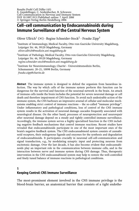
Results Probl Cell Differ (43)E. Gundelfinger, C. Seidenbecher, B. Schraven:Cell Communication in Nervous and Immune SystemDOI 10.1007/015/Published online: 7 April 2006© Springer-Verlag Berlin Heidelberg 2006
Cell–cell communication by Endocannabinoids duringImmune Surveillance of the Central Nervous System
Oliver Ullrich1 (�) · Regine Schneider-Stock2 · Frauke Zipp3
1Institute of Immunology, Medical Faculty, Otto-von-Guericke-University Magdeburg,Leipziger Str. 44, 39120 Magdeburg, [email protected]
2Institute of Pathology, Medical Faculty, Otto-von-Guericke-University Magdeburg,Leipziger Str. 44, 39120 Magdeburg, [email protected]
3Institute for Neuroimmunology, Charité - Universitätsmedizin Berlin,Schumannstr. 20–21, 10098 Berlin, [email protected]
Abstract The immune system is designed to defend the organism from hazardous in-fection. The way by which cells of the immune system perform this function can bedangerous for the survival and function of the neuronal network in the brain. An attackof immune cells inside the brain includes the potential for severe neuronal damage or celldeath and therefore impairment of CNS function. To avoid such undesirable action of theimmune system, the CNS harbours an impressive arsenal of cellular and molecular mech-anisms enabling strict control of immune reactions – the so-called “immune privilege”.Under inflammatory and pathological conditions, loss of control of the CNS immunesystem results in the activation of neuronal damage cascades frequently associated withneurological disease. On the other hand, processes of neuroprotection and neurorepairafter neuronal damage depend on a steady and tightly controlled immune surveillance.Accordingly, the immune system serves a highly specialized function in the CNS includ-ing negative feedback mechanisms that control immune reactions. Recent studies haverevealed that endocannabinoids participate in one of the most important ones of thebrain’s negative feedback system. The CNS endocannabinoid system consists of cannabi-noid receptors, their endogenous ligands and enzymes for the synthesis and degradationof endocannabinoids. It participates crucially in neuronal cell-cell-communication andsignal transduction, e.g., by modulating synaptic input and protecting neurons fromexcitotoxic damage. Over the last decade, it has also become evident that endocannabi-noids play an important role in the communication between immune cells, and in theinteraction between nerve and immune system during CNS damage. Thus, therapeuticintervention in the CNS endocannabinoid system may help to restore the well-controlledand finely tuned balance of immune reactions in pathological conditions.
1Keeping Control: CNS Immune Surveillance
The most-prominent element involved in the CNS immune privilege is theblood-brain barrier, an anatomical barrier that consists of a tight endothe-

282 O. Ullrich et al.
lial layer, a basement membrane, and a network of astrocyte foot processes,which keeps immune cells out of the brain. The blood-brain barrier is notan absolute barrier: proinflammatory cytokins such as IL-1 can induce ex-pression of adhesion molecules on the endothelial cell surface, facilitatebinding of immune cells and induce signals that open the tight junctions,so that immune cells can enter on demand. However, once in the brain,invading immune cells then face a second barrier – the immunological bar-rier of (1) astrocytes and microglial cells that express death ligands such asCD95 (fasL), which bind to death receptors (CD95, fas) on the surface ofinvading T cells, (2) astrocytes that express MCH-II, but no co-stimulatorymolecules, and therefore induce anergy and apoptosis in T cells and (3)an anti-inflammatory cytokine microenvironment that suppresses invadingT cells. Thus, T cell function and survival is strictly controlled and limitedin the brain parenchyma and immune reactions are widely regulated and ex-ecuted by microglial cells, the resident macrophage-like antigen-presentingimmunoeffector cell of the brain.
Microglial cells arrive in the CNS before birth have a very slow turnoverand express MHC-II- and CD80/CD86 in their activated state. In order to sur-vey the CNS environment, microglial cells express a multitude of differentreceptors for cytokines, chemokines, prostaglandins and mediators of neu-ronal injury (Hanisch 2002). In the CNS of a healthy adult, microglial cells aredeactivated, display a ramified morphology, and constantly patrol the braintissue (Nimmerjahn et al. 2005). But to remain deactivated and resting, mi-croglial cells require direct contact with structurally and functionally intactneurons (Hoek et al. 2000) and neuronal electrical activity and neurotrans-mitter release are constantly recognized by surrounding microglia. In the caseof damage, microglia cells become activated in a multi-step-process, which isaccompanied by specific morphological, immunophenotypic and functionalalterations (Kreutzberg et al. 1996). Intracellular signals of microglial ac-tivation include the IL-15-receptor-dependent JAK activity (Hanisch et al.1997), further serin/threonin-kinases and -phosphatases (Lockhart et al.1998), the nuclear CREB-protein (Herdegen et al. 992), NF-kappaB (Lockhartet al. 1998), prostaglandins (Petrova et al. 1999), corticoid-signaling (Drewet al. 2000) and poly-ADP-ribose-polymerase-1 (PARP-1) (Ullrich et al. 2001).After activation, microglia upregulates CNS immune reactions by the re-lease of cytokines, which induce the expression of adhesion molecules onendothelial cells and enhance the permeability of the blood-brain barrier(by TNF-alpha and IL-1), stimulate NK- and TH1-cells (by IL-18 and IL-12)and support B-cell-proliferation and differentiation (by IL-6). Importantly,they produce cytotoxic molecules such as nitrogen and oxygen free radicals,which are capable of damaging neurons directly, but also trigger the releaseof excitatoric amino acids (Banati et al. 1993). Microglia-derived products arealso capable of activating microglial cells themselves, thereby initiating a self-sustaining pathological microglial activation (Takeda et al. 1998). As a result,
Cell–Cell-Communication by Endocannabinoids 283
sustained pathological over-activation of microglia is considered to be a keyevent in many acute and chronic neurodegenerative diseases (McGeer et al.1995, Gebicke–Haerter et al. 1996, Heppner et al. 1998, McGeer et al. 1998,Floyd 1999, Dirnagl et al. 1999). In this situation, inflammatory pathwayscontribute to the onset and progress neurodegeneration, which provides aninteresting opportunity for therapeutic intervention, in particular if the pri-mary pathological event is unknown (e.g., in Alzheimer’s) or irreversible(e.g., after stroke).
Here, a crucial question arises: if the immune response is essential for tis-sue repair in all tissues outside the CNS, does the CNS similarly depend onimmune responses for its maintenance and repair, particularly after injury?Since the pioneering work of Michal Schwarz (Molaem et al. 1999), exten-sive studies have confirmed and described the phenomenon that autoreactiveT cells, as well as activated macrophages, are capable of promoting neuropro-tection and neuroregeneration in experimental models (Schwartz and Kionis2005). In fact this is not a very new idea, since in the animal kingdom it is wellknown that a high degree of immune privilege is associated with a low po-tential of regeneration. Therefore, it is indeed possible that the inability of theCNS to recover after injury is the price it pays for being an immune-privilegedsite. Perhaps this is a necessary price to pay in evolutionary terms, in whichavoiding risk to healthy individuals is more important than helping injuredindividuals to survive. The immune privilege could therefore be considered toresult from both “tactical necessity” and “calculated risk”. Therefore the im-mune system is allowed to keep the CNS under surveillance, but in a strictlycontrolled, limited and well-regulated manner.
2Loosing Control: Inflammatory Escalation during Multiple Sclerosis
During multiple sclerosis (MS), circulating myelin-specific T cells fromthe lymphatic tissue cross the blood-brain barrier and enter the brainparenchyma, where they engage in close contact with microglial cells, theantigen-presenting immunoeffector cell of the CNS. As a consequence ofthis contact, microglial cells become activated, stimulate B cells to producemyelin-specific antibodies, and release oxygen and nitrogen free radicalsand toxic cytokins, which then attack neurons and the myelin sheath. To-gether with inflammation, neuronal pathology involving axonal transectionplays a pivotal role in MS, and the corresponding animal model disease EAE,even during the early phase of inflammation (Ferguson et al. 1997, Meyeret al. 2001, Trapp et al. 1998, Diestel et al. 2003, Aktas et al. 2005). It hasbecome evident that long-term disability in MS correlates better with ax-onal damage than with the degree of demyelination (Bjartmar et al. 2003).Moreover, cortical thinning has been demonstrated using MRI techniques
284 O. Ullrich et al.
in MS patients (Sailer et al. 2003). Here, a distinct distribution of signifi-cant focal thinning of the cerebral cortex in addition to the diffuse corticalatrophy was demonstrated. This focal cortical thinning in the frontal andtemporal brain regions was even observed early in the course of the dis-ease, or in patients with mild disability. Patients with longstanding diseaseor severe disability, however, presented with an additional focal thinningof the motor cortex area. Ongoing axonal degeneration despite blockade ofinflammation, and even early widespread axonal pathology without MRIsigns of inflammation were interpreted as inflammation-independent ax-onal damage (Coles et al. 1999, Filippi et al. 2003). We have, however,recently provided the first demonstration of searching T cells coming intodirect physical contact with the neuronal cell body, and of related pro-cesses in the living brain environment which result in detrimental effectson neurons (Nitsch et al. 2004), whereas unstimulated T cells did not elicitthese effects. In chronic neuroinflammation of the CNS, activated myelin-specific T cells are believed to invade the brain and to be reactivated inthe parenchyma upon recognition of local autoantigens presented by mi-croglial cells (Flügel et al. 2001). This is supported by the fact that microglialcells have been shown to have the same antigen-presenting capacity asmacrophages and dendritic cells (Ulvestad et al. 1994), and can induce an-ergy or T cell activation according to their state of activation (Matyszak et al.1999).
Quantitative [(11)C](R)-PK11195 PET, an imaging technique for activatedmicroglia by the detection of “peripheral benzodiazepine binding sites”(PBBS), demonstrated microglia activation not only in areas of focal pathol-ogy identified by T(1)- and T(2)-weighted MRI, but also in normal-appearinganatomical structures, including cerebral central grey matter (Banati et al.2000). The additional binding frequently delineated neuronal projectionareas, such as the lateral geniculate bodies in patients with a history of op-tic neuritis. In a recent study, eight healthy participants and 22 MS patientswere investigated via PET, where brain atrophy was significantly greater inMS patients compared to age-matched controls, where [11C]PK11195 uptakeincreased with the amount of atrophy (Versijpt et al. 2005). Thus, there arestrong evidences of an involvement of microglial cells in neuronal damagecasacades in EAE as well as in human MS.
Beyond MS, inflammatory processes triggered by microglia are also in-volved in acute and chronic neurodegeneration such as Alzheimer’s disease(Floden and Combs 2005, Giulian et al. 1996, McGeer and McGeer 2001),stroke (Dirnagl et al. 1999, Danton and Dietrich 2003), Huntington’s disease(Sapp et al. 2001, Giorgini et al. 2005, Zhang et al. 2005), amyotrophic lat-eral sclerosis (Turner et al. 2004, Henkel et al. 2004), and also in neuronaldamage during CNS infection (Nau and Bruck 2002) and HIV-encephalopathy(Gonzales-Scarano and Garcia 2005). Moreover, activated microglia may ac-tually promote the growth of certain CNS neoplasia (Graeber et al. 2002) and
Cell–Cell-Communication by Endocannabinoids 285
may detrimental for neurogenesis in the adult brain (Ekdahl et al. 2003, Monjeet al. 2003). Thus, in light of the damage properties of activated microglia, itcan be assumed that intervention in microglia activation is a therapeutic op-tion for the treatment of neurological diseases in which microglial cells areinvolved.
3Maintaining the Balance: The Brain Endocannabinoid System
3.1Endogenous Ligands
The CNS endocannabinoid system consists of cannabinoid receptors andtheir endogenous ligands and enzymes for the synthesis and degradation ofendocannabinoids (Fig. 1). It is well known to participate crucially in neu-ronal cell-cell-communication and signal transduction, to control synapticinput, and to protect neurons from excitotoxic damage.
The medical use of Cannabis in Europe dates back to the 19th century(O’Shaugnessy 1843, Moreau 1845), when Napoleon’s Commission des Sci-ences et des Arts reported the psychotropic, analgesic and antispasmodiceffects after the return of it’s army from Egypt (Rouver 1810, Christison 1848).Hundred years later, the active compound of Cannabis has been identified asthe terpenoid derivative ∆9-tetrahydrocannabinol (THC) (Adams et al. 1941,Gaoni et al. 1964). Discovery of the active compound then facilitated the de-velopment of new potent and selective THC analogues (Melvin et al. 1987)enabled the identification of cannabinoid-sensitive sites in the brain (Devaneet al. 1988), which could be identified as the CB1 (Matsuda et al. 1990) andCB2 (Munro et al. 1993) cannabinoid receptor. But surprisingly, the endoge-nous ligands discovered were lipids, and not peptides as expected. The firstendogenous ligand, the first endocannabinoid, the amide of arachidocnic acidwith ethanolamine, was named anandamide after the Sanskrit word for bliss,ananda (Devane et al. 1992).
Metabolic pathways of anandamide (AEA) are unique, and the releaseof the compound in the living brain is very well regulated (DiMarzo et al.1994, Giuffrida et al. 1999, Walker et al. 1999). A regulatory key enzyme ofanandamide synthesis is the N-acyltransferase (NAT), which catalyses theintermolecular transfer of an arachidonic acid group from the sn-1 positionof phosphatidylcholine to phosphatidylethanolamine (Fig. 1). Cleavage of thereaction product N-arachidonoyl-phosphatidylethanolamine by phospholi-pase D, which exist in two isoforms in mammals (Kodaki et al. 1997), finallyresults in the release of anandamide and phosphatidic acid. The activity ofNAT activity is Ca2+-dependent and moreover regulated by phosphorylationthrough PKA. Consequently, anandamide is produced and released by Ca2+

286 O. Ullrich et al.
Fig. 1 Schematic diagram of endocannabinoid synthesis, signalling and degradation inneuronal and non-neuronal cells
ionophores and neuronal activity, in particular after stimulation of dopamineD2-receptors, muscarinic acetylcholine receptors and metabotropic gluta-mate receptors (Varma et al. 2001, Kim et al. 2002). Dopaminergic neuro-transmission stimulates anandamide release by rho-dependent activation ofPLD (Senogles 2000) or PLCbeta-dependent mobilizing of Ca2+ (Hernández-López 2000).
Interestingly, the production of anandamide represents an ancient plantsignalling system, which is highly conserved in mammalian cells and, there-fore, has to be considered as a very successful evolutionary principle. Inplants, PLD activity produces fatty acid ethanolamides from long-chain fattyacid phosphatidylethanolamines in response to stress or infection, which reg-ulates the expression of plant immunity-related genes (Chapman 2000).
The second endogenous ligand is 2-arachidonoylglycerol (2-AG): Whereas2-AG is produced at central intersections of lipid metabolism (Fig. 1) andfound in high concentrations in brain tissue, anandamide concentration isabout 200-fold lower (Sugiura et al. 1995, Stella et al. 1997) and brain quanti-ties of its precursor N-arachidonoyl-phosphatidylethanolamine are probablytoo little to enable sustained anandamide release. Thus, whereas 2-AG is con-stantly present in CNS tissue, anandamide is made more on demand. 2-AG

Cell–Cell-Communication by Endocannabinoids 287
is formed by hydrolyses of 1,2-diacylglycerol (DAG) by DAG lipase (Farooquiet al. 1989) or, alternatively, by synthesis of 2-arachidonoyl-lysophospholipidby phospholipase A1 (PLA1) and susbseqent hydrolysis by a lyso-PLC activity(Higgs et al. 1994, Pete et al. 1994). Although similar to anandamide, neuronal2-AG is produced in response to intracellular Ca2+ and stimulation of glu-tamate receptors, syntheses of anandamide and 2-AG can be independentlyregulated (Stella et al. 1997, 2001).
Other ligands are noladin ether (Hanus et al. 2001), virodhamine (Porteret al. 2002), and N-arachidonoyldopamine (Huang et al. 2002). Noladin etheris an ether-linked analogue of 2-AG that binds to and activates CB1 receptors(Hanus et al. 2001), while virodhamine is an ester of arachidonic acid andethanolamine, and endogenous CB1 antagonist (Piomelli et al. 2003). Theirphysiological role, however, has yet to be elucidated.
3.2Release, Uptake and Deactivation
In contrast to small and medium-size peptides, endocannabinoids are highlyhydrophobic compounds, and the question how they reach their target struc-tures is not an easy one. One possibility is that endocannabinoids do notleave the cell, but move sideways within the membrane, where they meettheir receptors (Xie et al. 1996). This hypothesis has been supported by theidentification of an intramembranous binding-site of the CB1 receptor foranandamide (Song et al. 1996). But endocannabinoids are capable of leavingthe cells as soon as they are formed and travel as far as 20 µm from its siteof origin (Wilson et al. 2001). Therefore, endocannabinoid binding proteinsare suggested (Beuckmann et al. 2000), analogous to serum albumin, whichfacilitate the transport of anandamids in the blood stream (Bojensen et al.2003).
Although anandamide and 2-AG can diffuse through lipid membranes,a selective carrier system is suggested that accelerate cellular uptake of en-docannabinoids by an energy-independent facilitated diffusion mechanism(Beltramo et al. 1997, Hillard et al. 1997). Anandamide is degraded by fattyacid amide hydrolase (FAAH), an intracellular membrane-bound serine hy-drolase (Schmid et al. 1985, Hillard et al. 1995, Ueda et al. 1995, Cravattet al. 1996). 2-AG ist degraded by monoacylglycerol lipase (MGL), a cy-tosolic serine hydrolase. Both enzymes are widely distributed in the brain.Within the synapse, endocannabinoide degrading enzymes are expressedasymmetrically: Whereas FAAH is predominantly found postsynaptically,MGL is mostly presynaptically (Dinh et al. 2002). Interestingly, in the hip-pocampal CA1 field, MGL-positive axon terminals surround cell bodies ofpyramidal neurons, which could reflect a functional role of presynapticMGL in terminating retrograde signalling mediated by 2-AG (Piomelli et al.2003).

288 O. Ullrich et al.
3.3Signal Transduction
The CB1 receptor is the most abundant G-protein-coupled receptor in themammalian brain and is present in the neocortex, hippocampus, basal gan-glia, cerebellum and brainstem (Herkenham et al. 1990), where it is primarilyfound on glutamatergic terminals and axon terminals of GABA interneurons.CB1 receptors account for most of the behavioural actions of cannabinoiddrugs. In its capacity of standard Gi/o-coupled receptor (Fig. 1), CB1 can ini-tiate signalling events typical of this class of transducing proteins – such asclosure of Ca2+ channels, opening of K+ channels, and inhibition of adeny-lyl cyclase activity – resulting in a decrease in cytosolic cAMP (Howlett et al.1984). Inhibition of N- and P/Q-type voltage-activated Ca2+ channels (Mackieet al. 1992, Caulfield et al. 1992, Twitchell et al. 1997), as well as depres-sion of transmitter release at GABA (gamma-aminobutyric acid) synapsesin the CA1 field of the hippocampus (Hoffmann et al. 2000) and at gluta-matergic synapses in the dorsal striatum (Gerdemann et al. 2001, Huang et al.2001) have all been suggested to result from a direct interaction of Gi/o-protein beta-gamma subunits with the relevant channels. Presynaptic inhibi-tion at GABA and glutamate synapses also involves cannabinoid regulationof voltage-gated K+ currents (Mu et al. 1999). The subsequent regulation ofneuronal gene expression by CB1 depends on the recruitment of complex net-works of intracellular protein kinases such as extracellular signal-regulatedkinase (ERK) and focal adhesion kinase (FAK), which become activated whenhippocampal slices are treated with cannabinoid agonist (Ueda et al. 1995,Cravatt et al. 1996), Derkinderen et al. 1996, 2003). In this context, cannabi-noid receptors are coupled to the activation of ERK (Wartmann et al. 1995,Bouaboula et al. 1995a, 1995b, 1996), of c-jun N-terminal kinase and p38mitogen-activated protein kinase (Liu et al. 2000, Rueda et al. 2000). More-over, CB1 receptor is also coupled to the activation of protein kinase B (PKB)(Gomez del Pilgar et al. 2000). In summary, cannabinoids inhibit the releaseof glutamate and other neurotransmitters in neuronal cells by blunting mem-brane depolarisation and exocytosis (Pertwee 2000, Porter et al. 2001).
Most of the effects of cannabinoids are mediated by their specific receptorsCB1 and CB2, which are coupled to AC through heterotrimeric Gi/o-proteins(Matsuda et al. 1990, Munro et al. 1993). However, in contrast to the CB1receptor, transduction systems responsible for CB2 receptor signalling areless elucidated. CB2 receptors are expressed mainly by cells of the immunesystem such as B cells, NK cells, monocytes, neutrophil granulocytes andT cells (Galiegue et al. 1995). In these cells, inhibition of the cAMP/proteinkinase A (PKA) pathway may be responsible for the immunosuppressive ac-tion of cannabinoids by decreasing the expression of cAMP-responsive genes(Kaminski et al. 1998). CB2 receptor might control immune-cell proliferationby coupling to ERK activation, a process that is dependent on Gi/o-proteins,
Cell–Cell-Communication by Endocannabinoids 289
but independent of cAMP (Bouaboula et al. 1996). Although CB1- and CB2-coupled signalling share some similarities, differences between downstreampathways of both receptors include the ability of CB1 to modulate Ca2+ andK+ channels (Pertwee 2000), to phosphorylate ERK independently of proteinkinase C (Bouaboula et al. 1996), and to activate PKB (Gomez del Pulgar et al.2000).
Recently, it has been discovered that endocannabinoids modulate sphingo-lipid-metabolising pathways by inducing sphingomyelin (SM) breakdownthrough neutral SMase activation (Kaminski 1998), and by acutely increas-ing the levels of the second messenger ceramide (Sanchez et al. 1998, 2001)independently of G-proteins. The resulting peak of ceramide accumulationplays an important role in the induction of apoptosis (Galve-Roperh et al.2000, Guzman et al. 2001). Cannabinoid-induced apoptosis has been inves-tigated in glioma in vitro (Sanchez et al. 1998, Jacobson et al. 2000) and invivo (Sanchez et al. 2001, Galve–Roperh et al. 2000), where it seems to de-pend on the sustained generation of ceramide and raf-1-mediated activationof the ERK cascade (Galve–Roperh et al. 2000). In this context, the relation-ship between activation of the ERK cascade and cell fate has been thoughtto depend on the duration of the stimulus. Apoptotic action of cannabinoidsapparently relies on the long-term peak of ceramide generation and ERK ac-tivation (Galve–Roperh et al. 2000), which presumably results from ceramidede novo synthesis rather than SM hydrolysis (Guzman et al. 2001). In PC-12pheochromocytoma cells, AEA induces superoxide generation, which triggersdownstream signals culminating in caspase-3 activation, and finally resultingin apoptosis (Sarker et al. 2000). In addition, VR1 vanilloid receptor activationby AEA has been reported to trigger apoptosis through a cascade of events(including: cytosolic Ca2+ concentration; cyclo- and lipoxygenase activation;a drop in mitochondrial membrane potential; cytochrome c release; and cas-pase activation) that finally execute apoptotic cell death (Maccarrone et al.2000). However, the possibility that AEA binds to VR1 capsaicin receptors(Zygmunt et al. 1999) is conflictive (Szolcsanyi et al. 2000, Piomelli 2001).
3.4Cell-Cell-Communication
In cells of the immune system, CB2-receptor-stimulation results in inhibitorysignals (Walter et al. 2004) mediated by inhibition of the cAMP/PKA pathwayand of the transcription factors NF-AT und AP-1. Endocannabinoids inhibitthe CB2-mediated release of TNF-α, IL-1β (Klegeris et al. 2003), IL-6 and IL-8(Berdyshev et al. 1997, Gallily et al. 2000) by monocytes/macrophages, andapparently stimulate nitric oxide release (Stefano et al. 1996). In T cells, THCinduces a shift from a Th1- to Th2-type cytokine pattern, such as suppres-sion of IFN-γ and IL-12, and induction of IL-4 release. It has been suggestedthat endocannabinoids use chemotaxis to attract macrophages and microglial

290 O. Ullrich et al.
cells to the site of tissue damage (Walter et al. 2003), where they help tocontrol and limit the local immune response, thus preventing harmful over-activation. Moreover, endocannabinoids are formed by circulating leukocytesand platelets, and induce vascular relaxation by interacting with cannabinoidreceptors on the surface of neighbouring endothelial and smooth muscle cells(Batkai et al. 2001). Thus, outside the brain, endocannabinoids are producedon demand, and act on cells located near their site of synthesis (Piomelli2003).
In the CNS, similar paracrine actions are thought to occur; endocannabi-noids mediate a localized signalling mechanism through which principalneurons modify the strength of incoming synaptic inputs (Piomelli 2003).Here, CB1 receptors are primarily found on axon terminals of GABA-ergicneurons in the neocortex, hippocampal formation, amygdala, basal ganglia,cerebellum, thalamus, hypothalamus, midbrain, medulla, spinal cord and pe-ripheral sensory neurons (Katona et al. 1999, Marsicano et al. 1999, Tsou et al.1998, Katona et al. 2001, McDonald et al. 2001, Gerdeman et al. 2001, Huanget al. 2001, Hohmann et al. 1999, 2000, Herkenham et al. 1991).
In the CA1 field of the hippocampus, membrane depolarization opensvoltage-activated Ca2+ channels in pyramidal neurons, producing an eleva-tion of intracellular Ca2+ concentrations, which stimulate the synthesis of2-AG and of AEA. The endocannabinoids then travel backwards across thesynapse to interact with CB1 receptors on axon terminals of GABA-ergic in-terneurons, leading to the depolarization-induced suppression of inhibition(DSI). Since DSI could facilitate the induction of long-term potentiation inindividual CA1 pyramidal neurons (Carlson et al. 2002), endocannabinoidsare supposed to be involved in modulation of memory. This retrograde sig-nalling by endocannabinoid-mediated DSI occurs in the hippocampus, whereit modulates memory function, but it has also been found outside the hip-pocampus at interneuron–principal cell synapses of the cerebellum (Llanoet al. 1991, Kreitzer et al. 2001).
In the amygdala, anxiogenic stimuli increases anandamide and 2-AG con-centrations (Marsicano et al. 2002), which bind to CB1-positive interneuronsin the basolateral complex that processes emotions (Martin et al. 1999), de-press glutamate release from axon terminals originating in the cortex (Azadet al. 2003) and decrease the activity of the pyramidal neurons in the centralnucleus of the amygdala (Katona et al. 2001). The function of endocannabi-noids in controlling affective states in the amygdala has been demonstratedpharmacologically: FAAH inhibitors exhibit marked anxiolytic-like proper-ties (Kathuria et al. 2003), whereas CB1 inactivation causes anxiety-like andaggressive responses (Navarro et al. 1997, Martin et al. 2002).
The highest densities of CB1 receptors in the brain are in the terminalfields of striatal projections in the basal ganglia, where they inhibit GABArelease and affect motor activity (Romero et al. 2002). In the basal ganglia,anandamide is released by dopamine D2-receptor activation (Giuffrida et al.

Cell–Cell-Communication by Endocannabinoids 291
1999) and act at GABA-ergic interneurons and corticostriatal glutamatergicprojections (Gerdeman et al. 2001, Hohmann et al. 2000, Herkenham et al.1991). Clinically, the major role of endocannabinoid signalling in the basalganglia could be demonstrated by their therapeutic effect in the treatment ofL-DOPA-induced dyskinesias (Sieradzan et al. 2001) and Tourette’s syndrome(Muller-Vahl et al. 2003)
Endocannabinoid signalling interacts also with pain transmission at multi-ple sites: Painful stimuli induce release of anandamide, which in turn binds toCB1 receptors in the periaqueductal grey (Lichtman et al. 1996), rostral ven-tromedial medulla (Meng et al. 1998) and spinal trigeminal nucleus (Jenningset al. 2001) and mediate an analgesic effect (Iversen et al. 2002).
Endocannabinoids interact not only with GABA-ergic signal transmission,but also with glutamate-dependent signal transmission. Principal neurons inthe hippocampus and cerebellum use endocannabinoids to carry out a sig-nalling process that is analogous to DSI, but opposite in sign – a so-calleddepolarization-induced suppression of excitation (DSE) (Alger 2002). DSEtargets glutamatergic rather than GABA axon terminals, and therefore resultsin reduced excitatory input to the affected cell. Interestingly, glutamatergicterminals are less sensitive to endocannabinoid activation (Ohno-Shosakuet al. 2002), which would indicate that a switch from DSI to DSE might occurwhen endocannabinoid concentrations at hippocampal synapses attain a cer-tain threshold value (Piomelli 2003). Moreover, acetylcholine release has alsobeen found to be reduced by cannabinoids both in vitro and in vivo (Schlickeret al. 2001, Gifford et al. 1996, Gessa et al. 1998), as has the release of thebiogenic amines noradrenaline and serotonin (Schlicker et al. 2001). Since en-docannabinoids are clearly involved in striatal LTD (long-term-depression)and hippocampal I-LTD (LTD at adjacent inhibitory synapses), the overall ef-fects of endocannabinoids on hippocampus-dependent learning will surelybe an exciting and central subject of future investigation and discussion (Pi-omelli et al. 2003).
In pathophysiological conditions, endogenous cannabinoids are releasedafter brain injury (Panikashvili et al. 2001, Hansen et al. 2001, Marsicano et al.2003, Mechoulam et al. 2003, Carrier et al. 2004) and are believed to atten-uate neuronal damage by binding to CB1 receptors (Panikashvili et al. 2001,Hansen et al. 2001, Marsicano et al. 2003), thus protecting against excitotoxi-city (Mechoulam et al. 2003, Carrier et al. 2004). Such excitotoxic brain lesionsinitially result in primary destruction of brain parenchyma, which attractsmacrophages and microglia, the major effector cells within the CNS. Theseinflammatory cells produce large amounts of toxic cytokines and oxygen rad-icals, which result subsequently in secondary neuronal damage. Since it isknown that the cannabinoid receptors, CB1 and CB2, are expressed on mi-croglial cells (Facchinetti et al. 2003, Waksman et al. 1999), and that activatedmicroglia are capable of producing larger amounts of endogenous cannabi-noids than neurons (Walter et al. 2003), the function of the endocannabinoid

292 O. Ullrich et al.
system in neuro-immune-communication during neuronal damage needs tobe elucidated in the future.
4Gaining Control: Therapeutic Intervention in CNS Inflammation
4.1Microglia as Therapeutic Target
Very recently, Heppner and colleagues directly targeted microglial activi-ties using CD11b-HSVTK transgenic mice and bone marrow chimeras inwhich a specific lack of microglial activation was achieved (Heppner et al.2005). Using this approach, inflammatory CNS lesions were repressed, re-sulting in a marked reduction of the severity of the animal model of MS.Furthermore, the authors demonstrated that microglial cells are crucial forthe development of EAE, presumably mediated by the release of cytokinesand chemokines as well as reactive oxygen species, as studied in organotypichippocampal slice cultures. Future studies should examine whether classi-cal neurodegenerative diseases may also benefit from a specific microgliatargeting strategy, and how this approach can be translated into clinicalapplication. There are already several therapeutic agents in clinical use ordevelopment, which target microglial activities, among other effects. In par-ticular, some non-steroidal anti-inflammatory drugs (NSAID) seem to havebeneficial effects in neurodegenerative diseases such as Alzeimer’s (McGeerand McGeer 2001). Recently, the nuclear receptor peroxisome proliferator-activated receptor-gamma (PPARgamma) agonist pioglitazone resulted ina reduction in number of activated microglia with reduced expression ofthe proinflammatory enzymes cyclooxygenase 2 (COX2) and inducible ni-tric oxide synthase (iNOS), and a reduction of amyloid deposits in the hip-pocampus and cortex of an Alzheimer model (Heneka et al. 2005). Minocy-cline, which is known to have neuroprotective properties, was recently shownto reduce the release of cytotoxins from activated microglia (Krady et al.2005). However, anti-inflammatory approaches to blocking microglial acti-vation or microglial effector mechanisms are likely non-specific, indiscrim-inate strategies. Moreover, they do not simply reduce inflammatory damageon the one hand, but they also run the risk of inhibiting protective im-munity on the other hand. Therefore, from the clinical point of view, theidentification and therapeutic support of anti-inflammatory negative feed-back loops might be the better choice because these are only active afterimmune activation and therefore harbour a low risk of undesired over-inhibition of immune reactions. This means that the therapeutic supportof endogenous negative regulatory feedback-loops might be a highly effi-cient and clinically safe strategy of neuroprotective intervention. Here, the

Cell–Cell-Communication by Endocannabinoids 293
endocannabinoid system, a major negative regulatory system in the ner-vous and immune system of the CNS, may represent one promising newtherapeutic target in the treatment of acute and chronic neurodegenerativediseases.
4.2Cannabinoid System as Therapeutic Target
Because of its possible involvement in immune control and neuroprotection,cannabinoids are discussed intensively for treatment of inflammatory disease(Croxford et al. 2005) and moreover as an interesting option in the treat-ment of neuroinflammation (Correa et al. 2005). Recently we found that theendocannabinoid system is highly activated in patients with MS and iden-tified the release of AEA by inflamed CNS tissue as a new mechanism ofneuro-immune-communication during CNS injury, which controls and limitsimmune response and protect neurons (Eljaschewitsch et al. 2006). A pre-vious study reported elevated endocannabinoid levels in autoimmune en-cephalomyelitis (EAE), the experimental model of MS, in association withspasticity due to spinal cord pathology (Baker et al. 2001). In EAE, CB1receptor stimulation ameliorated spasticity and tremor (Baker et al. 2000,2004, Arevalo-Martin et al. 2003) and reduced neuronal damage and axonalloss (Pryce et al. 2003), whereas the CB1 receptor antagonist Rimonabant®transiently worsens signs (Baker et al. 2000, 2001, 2004). Evidence for the par-ticipation of endocannabinoid signalling in human MS came from clinicalobservations that a previously undiagnosed MS exacerbated by Rimonabant®in a person being treated for obesity (van Oosten et al. 2004).
Treatment of MS with the cannabis extracts (Cannador) lowered the fre-quency of spasms and improved the mobility in a randomized double-blindcrossover study (Vaney et al. 2004). Bladder dysfunction has been improvedby THC and Sativex in a small-scale open-label study (Brady et al. 2004), aswell as patient perceptions of pain and spasticity (Pertwee et al. 2002, Svend-sen et al. 2004). On the other hand, no positive effects on spasticity has beenreported in patients treated with oral Marinol or Cannador in a blinded trial(Killestein et al. 2002).
In the largest clinical study about the use of cannabinoids for treatingsymptoms related to MS (CAMS), where 667 people were orally administeredover a 15 week period with capsules of THC (Marinol®), capsules of cannabisextract Cannador® or placebo capsules, a subjective improvement of specificsymptoms (pain, spasticity, sleep disturbance, walking time), but no effecton spasticity and tremor has been reported (Zajicek et al. 2003). However,in a one-year follow-up of the CAMS study, overall objective improvementson both spasticity (Ashworth Scale) and general disability indices have beenfound (Zajicek et al. 2004). The results of the long-term CAMS study are par-ticularly encouraging because they indicate that cannabis not only relieves

294 O. Ullrich et al.
symptoms but also is potentially neuroprotective and involved in synapticplasticity; this should be investigated further.
In conclusion, clinical trials ended with both positive and negative re-sults (Pertwee et al. 2002), which could be the consequence of several prob-lems: First, exogenous cannabinoids might interfere locally with endogenouscannabinoids, since they are only partial agonists on cannabinoid receptors.Second, oral delivery of cannabinouid drugs produces a slow and low increasein plasma THC levels (Huestis et al. 1992, Grotenhermen 2003, Wall et al.1983, Ohlsson et al. 1980, Timpone et al. 1997), whereas a correlation of op-timal plasma THC levels with symptom improvement is missing from manystudies. Third, the Ashworth scale to measure spasticity might not be suffi-ciently sensitive to measure small but clinically beneficial effects (Shakespeareet al. 2003). In conclusion, improvements in trial design and outcome meas-urements are important in clarifying the situation. To overcome problems inthe oral delivery of cannabinoids, the sub-lingual spray Sativex® has been de-veloped improvement, which demonstrated beneficial effects on spasticity inpatient-assessed scores (visual analogue scale), but no improvement as meas-ured by the Ashworth scale in a placebo-controlled study in 160 MS patients(Wade et al. 2004).
In conclusion, the endocannabinoid system represents a local messen-ger system within and between the nervous and immune systems, whichis apparently involved in learning and memory, emotions, pain processing,regulation of motor functions, control of immune activation and neuropro-tection. Therefore, elucidating the integration of the endocannabinoid sys-tem in intra- and intercellular signalling networks and their function duringphysiology and pathophysiology might open new avenues of therapeutic in-terventions in the future. A successful therapeutic strategy has to maintain orrestore the well-controlled and finely-tuned balance of immune reactions, andto protect neurons from inflammatory and non-inflammatory damage.
Acknowledgements Minor parts of this chapter (Fig. 1) have also been published by theauthors in Signal Transduction 1–2, 19–27 (2005); these sections appear with kind per-mission of Wiley VCH. We thank Mr. Andrew Mason for his excellent assistance.
References
Aktas O, Smorodchenko A, Brocke S, Infante-Duarte C, Prozorovski T, Meier S, Osman-ova V, Kwidzinski E, Pohl E, Beyer M, Bechmann I, Nitsch R, Zipp F (2005) Neuronaldamage in autoimmune neuroinflammation mediated by the death ligand TRAIL.Neuron 46:421–432
Adams R (1941) Marihuana. Harvey Lect 37:168Alger BE (2002) Retrograde signaling in the regulation of synaptic transmission: focus on
endocannabinoids. Prog Neurobiol 68:247–286

Cell–Cell-Communication by Endocannabinoids 295
Arevalo-Martin A et al. (2003) Therapeutic action of cannabinoids in a murine model ofmultiple sclerosis. J Neurosci 23:2511–2516
Azad SC et al. (2003) Activation of the cannabinoid receptor type 1 decreases glutamater-gic and GABAergic synaptic transmission in the lateral amygdala of the mouse. LearnMem 10:116–128
Baker D et al. (2000) Cannabinoids control spasticity and tremor in a multiple sclerosismodel. Nature 404:84–87
Baker D et al. (2001) Endocannabinoids control spasticity in a multiple sclerosis model.FASEB J 15:300–302
Baker D, Pryce G (2004) The potential role of the endocannabinoid system in the controlof multiple sclerosis. Curr Med Chem 4:195–202
Banati RB, Gehrmann J, Schubert P, Kreutzberg GW (1993) Cytotoxicity of microglia. Glia7:111–118
Banati RB, Newcombe J, Gunn RN, Cagnin A, Turkheimer F, Heppner F, Price G, Wegner F,Giovannoni G, Miller DH, Perkin GD, Smith T, Hewson AK, Bydder G, Kreutzberg GW,Jones T, Cuzner ML, Myers R (2000) The peripheral benzodiazepine binding site in thebrain in multiple sclerosis: quantitative in vivo imaging of microglia as a measure ofdisease activity. Brain 123(Pt 11):2321–2337
Bátkai S et al. (2001) Endocannabinoids acting at vascular CB1 receptors mediate thevasodilated state in advanced liver cirrhosis. Nature Med 7:827–832
Beltramo M et al. (1997) Functional role of high-affinity anandamide transport, as re-vealed by selective inhibition. Science 277:1094–1097
Beltramo M et al. (2000) Reversal of dopamine D2 receptor responses by an anandamidetransport inhibitor. J Neurosci 20:3401–3407
Berdyshev EV, Boichot E, Germain N, Allain N, Anger JP, Lagente V (1997) Influence offatty acid ethanolamides and delta-9-tetrahydrocannabinol on cytokine and arachido-nate release by mononuclear cells. Eur J Pharmacol 330:231–240
Beuckmann CT et al. (2000) Cellular localization of lipocalin-type prostaglandin D syn-thase (β-trace) in the central nervous system of the adult rat. J Comp Neurol 428:62–78
Bjartmar C, Wujek JR, Trapp BD (2003) Axonal loss in the pathology of MS: consequencesfor understanding the pro gressive phase of the disease. J Neurol Sci 206:165–171
Bojensen IN, Hansen HS (2003) Binding of anandamide to bovine serum albumin. J LipidRes 44:1790–1794
Bouaboula M, Bourrie B, Rinaldi-Carmona M, Shire D, Le Fur G, Casellas P (1995) Stimu-lation of cannabinoid receptor CB1 induces krox-24 expression in human astrocytomacells. J Biol Chem 270:13973–13980
Bouaboula M, Poinot Chazel C, Bourrie B, Canat X, Calandra B, Rinaldi Carmona M,Le Fur G, Casellas P (1995) Activation of mitogen-activated protein kinases by stim-ulation of the central cannabinoid receptor CB1. Biochem J 312:637–641
Bouaboula M, Poinot Chazel C, Marchand J, Canat X, Bourrie B, Rinaldi Carmona M, Ca-landra B, Le Fur G, Casellas P (1996) Signaling pathway associated with stimulation ofCB2 peripheral cannabinoid receptor. Involvement of both mitogen-activated proteinkinase and induction of Krox-24 expression. Eur J Biochem 237:704–711
Brady CM et al. (2004) An open-label pilot study of cannabis-based extracts for bladderdysfunction in advanced multiple sclerosis. Mult Scler 10:425–433
Carlson G, Wang Y, Alger BE (2002) Endocannabinoids facilitate the induction of LTP inthe hippocampus. Nature Neurosci 5:723–724
296 O. Ullrich et al.
Carrier EJ, Kearn CS, Barkmeier AJ, Breese NM, Yang W, Nithipatikom K, Pfister SL,Campbell WB, Hillard CJ (2004) Cultured rat microglial cells synthesize the endo-cannabinoid 2-arachidonylglycerol, which increases proliferation via a CB2 receptor-dependent mechanism. Mol Pharmacol 65:999–1007
Caulfield MP, Brown DA (1992) Cannabinoid receptor agonists inhibit Ca current inNG108-15 neuroblastoma cells via a pertussis toxin-sensitive mechanism. Br J Phar-macol 106:231–232
Chapman KD (2002) Emerging physiological roles for N-acylphosphatidylethanolaminemetabolism in plants: signal transduction and membrane protection. Chem PhysLipids 108:221–229
Christison R (1848) In: Lea, Blanchard (eds) A Dispensatory, or Commentary on thePharmacopoeias of Great Britain (and the United States), Philadelphia:971–974
Coles AJ et al. (1999) Monoclonal antibody treatment exposes three mechanisms under-lying the clinical course of multiple sclerosis. Ann Neurol 46:296–304
Correa F, Mestre L, Molina-Holgado E, Arevalo-Martin A, Docagne F, Romero E, Molina-Holgado F, Borrell J, Guaza C (2005) The role of cannabinoid system on immunemodulation: therapeutic implications on CNS inflammation. Mini Rev Med Chem5:671–675
Cravatt BF et al. (1996) Molecular characterization of an enzyme that degrades neuro-modulatory fatty-acid amides. Nature 384:83–87
Croxford JL, Yamamura T (2005) Cannabinoids and the immune system: potential for thetreatment of inflammatory diseases? J Neuroimmunol 166:3–18
Danton GH, Dietrich WD (2003) Inflammatory mechanisms after ischemia and stroke.J Neuropathol Exp Neurol 62:127–136
De Petrocellis L, Bisogno T, Davis JB, Pertwee RG, Di Marzo V (2000) Overlap between theligand recognition properties of the anandamide transporter and the VR1 vanilloid re-ceptor: inhibitors of anandamide uptake with negligible capsaicin-like activity. FEBSLett 483:52–56
Derkinderen P, Toutant M, Burgaya F, Le Bert M, Siciliano JC, de Franciscis V, Gelman M,Girault JA (1996) Regulation of a neuronal form of focal adhesion kinase by anan-damide. Science 273:1719–1722
Derkinderen P, Valjent E, Toutant M, Corvol JC, Enslen H, Ledent C, Trzaskos J, Caboche J,Girault JA (2003) Regulation of extracellular signal-regulated kinase by cannabinoidsin hippocampus. J Neurosci 23:2371–2382
Devane WA, Hanus L, Breuer A, Pertwee RG, Stevenson LA, Griffin G, Gibson D, Mandel-baum A, Etinger A, Mechoulam R (1992) Isolation and structure of a brain constituentthat binds to the cannabinoid receptor. Science 258:1946–1949
Devane WA, Dysarz FA, Johnson MR, Melvin LS, Howlett AC (1988) Determination andcharacterization of a cannabinoid receptor in rat brain. Mol Pharmacol 34:605–613
Di Marzo V, Fontana A, Cadas H, Schinelli S, Cimino G, Schwartz JC, Piomelli D (1994)Formation and inactivation of endogenous cannabinoid anandamide in central neu-rons. Nature 372:686–691
Dinh TP et al. (2002) Brain monoglyceride lipase participating in endocannabinoid inac-tivation. Proc Natl Acad Sci USA 99:10819–10824
Dirnagl U, Iadecola C, Moskowitz MA (1999) Pathobiology of ischaemic stroke: an inte-grated view. Trends Neurosci 22(9):391–397
Drew PD, Chavis JA (2000) Inhibition of microglial cell activation by cortisol. Brain ResBull 52:91–396
Ekdahl CT, Claasen JH, Bonde S, Kokaia Z, Lindvall O (2003) Inflammation is detrimentalfor neurogenesis in adult brain. Proc Natl Acad Sci USA 100(23):13632–13637

Cell–Cell-Communication by Endocannabinoids 297
Eljaschewitsch E, Witting A, Mawrin C, Lee T, Schmidt PM, Wolf S, Hoertnagl H, Raine CS,Schneider-Stock R, Nitsch R, Ullrich O (2006) The endocannabinoid anandamide(AEA) protects neurons during CNS inflammation by induction of MKP-1 in mi-croglial cells. Neuron (in press)
Facchinetti F, Del Giudice E, Furegato S, Passarotto M, Leon A (2003) Cannabinoids ablaterelease of TNFα in rat microglial cells stimulated withlypopolysaccharide. Glia 41:161–168
Farooqui AA, Rammohan KW, Horrocks LA (1989) Isolation, characterization, and regu-lation of diacylglycerol lipases from the bovine brain. Ann NY Acad Sci 559:25–36
Ferguson B et al. (1997) Axonal damage in acute multiple sclerosis lesions. Brain 120:393–399
Filippi M et al. (2003) Evidence for widespread axonal damage at the earliest clinical stageof multiple sclerosis. Brain 126:433–437
Floden AM, Li S, Combs CK (2005) Beta-amyloid-stimulated microglia induce neurondeath via synergistic stimulation of tumor necrosis factor alpha and NMDA receptors.J Neurosci 25(10):2566–2575
Floyd RA (1999) Neuroinflammatory processes are important in neurodegenerative dis-eases: an hypothesis to explain the increased formation of reactive oxygen and nitro-gen species as major factors involved in neurodegenerative disease development. FreeRadic Biol Med 26:1346–1355
Flugel A, Berkowicz T, Ritter T, Labeur M, Jenne DE, Li Z, Ellwart JW, Willem M, Lass-mann H, Wekerle H (2001) Migratory activity and functional changes of green fluor-escent effector cells before and during experimental autoimmune encephalomyelitis.Immunity 14(5):547–560
Franklin A, Parmentier-Batteur S, Walter L, Greenberg DA, Stella N (2003) Palmitoyl-ethanolamide increases after focal cerebral ischemia and potentiates microglial cellmotility. J Neurosci 23:7767–7775
Galiegue S, Mary S, Marchand J, Dussossoy D, Carriere D, Carayon P, Bouaboula M,Shire D, Le Fur G, Casellas P (1995) Expression of central and peripheral cannabi-noid receptors in human immune tissue and leukocyte subpopulations. Eur J Biochem232:54–61
Gallily R, Breuer A, Mechoulam R (2000) 2-Arachidonylglycerol, an endogenous cannabi-noid, inhibits tumor necrosis factor-alpha production in murine macrophages, and inmice. Eur J Pharmacol 406:5–7
Galve-Roperh I, Sanchez C, Cortes L, del Pulgar TG, Izquierdo M, Guzman M (2000) Anti-tumoral action of cannabinoids: involvement of sustained ceramide accumulation andextracellular signal regulated kinase activation. Nat Med 6:313–319
Gaoni Y, Mechoulam R (1964) Isolation, structure and partial synthesis of an active con-stituent of hashish. J Am Chem Soc 86:1646–1647
Garden GA (2002) Microglia in human immunodeficiency virus-associated neurodegen-eration. Glia 40:240–251
Gebicke-Haertner PJ, van Calker D, Norenberg W, Illes P (1996) Molecular mechanisms ofmicroglial activation. Implications for regeneration and neurodegenerative diseases.Neurochem Int 29:1–12
Gerdeman G, Lovinger DM (2001) CB1 cannabinoid receptor inhibits synaptic release ofglutamate in rat dorsolateral striatum. J Neurophysiol 85:468–471
Gessa GL, Casu MA, Carta G, Mascia MS (1998) Cannabinoids decrease acetylcholine re-lease in the medial-prefrontal cortex and hippocampus, reversal by SR 141716A. Eur JPharmacol 355:119–124

298 O. Ullrich et al.
Gifford AN, Ashby CR Jr (1996) Electrically evoked acetlycholine release from hippocam-pal slices is inhibited by the cannabinoid receptor agonist, WIN 55212-2, and ispotentiated by the cannabinoid antagonist, SR 141716A. J Pharmacol Exp Ther277:1431–1436
Giorgini F, Guidetti P, Nguyen Q, Bennett SC, Muchowski PJ (2005) A genomic screen inyeast implicates kynurenine 3-monooxygenase as a therapeutic target for Huntingtondisease. Nat Genet 37(5):526–531
Giuffrida A, Parsone LH, Kerr TM, Rodriguez de Fonseca F, Navarro M, Pomelli D (1999)Dopamine activation of endogenous cannabinoid signaling in dorsal striatum. NatNeurosci 2:358–363
Giulian D, Haverkamp LJ, Yu JH, Karshin W, Tom D, Li J et al. (1996) Specific domainsof beta-amyloid from Alzheimer’s plaque elicit neuron killing in human microglia.J Neurosci 1996:6021–6037
Gomez del Pulgar T, Velasco G, Guzman M (2000) The CB1 cannabinoid receptor iscoupled to the activation of protein kinase B/Akt. Biochem J 347:369–373
Gonzalez-Scarano F, Martin-Garcia J (2005) The neuropathogenesis of AIDS. Nat Rev Im-munol 5(1):69–81
Gorriti MA, Rodríguez de Fonseca F, Navarro M, Palomo T (1999) Chronic (–)-∆9-tetrahydrocannabinol treatment induces sensitization to the psychomotor effects ofamphetamine in rats. Eur J Pharmacol 365:133–142
Graeber MB, Scheithauer BW, Kreutzberg GW (2002) Microglia in brain tumors. Glia40:252–259
Grotenhermen F (2003) Pharmacokinetics and pharmacodynamics of cannabinoids. ClinPharmacokinet 42:327–360
Guzman M, Sanchez C, Galve-Roperh I (2001) Control of the cell survival/death decisionby cannabinoids. J Mol Med 78:613–625
Hampson AJ et al. (2000) Neuroprotective antioxidants from marijuana. Ann New YorkAcad Sci 899:274–282
Hanisch UK (2002) Microglia as a source and target of cytokines. Glia 40(2):140–155Hanisch UK, Lyons S, Nolte C, Prinz M, Weber J, Kettenmann H, Kirchhoff F (1997) Mouse
brain microglia express interleukin-15 and its multimeric receptor complex function-ally coupled to Janus kinase activity. J Biol Chem 272:28853–28860
Hansen HH, Schmid PC, Bittigau P, Lastres-Becker I, Berrendero F, Manzanares J, Ikono-midou C, Schmid HHO, Fernandez-Ruiz JJ, Hansen HS (2001) Anandamide, but not2-arachidonoylglycerol, accumulates during in vivo neurodegeneration. J Neurochem78:1415–1427
Hanus L, Abu-Lafi S, Fride E, Breuer A, Vogel Z, Shalev DE, Kustanovich I, Mechoulam R(2001) 2-Arachidonyl glyceryl ether, an endogenous agonist of the cannabinoid CB1receptor. Proc Natl Acad Sci USA 98:3662–3665
Heneka MT, Sastre M, Dumitrescu-Ozimek L, Hanke A, Dewachter I, Kuiperi C, O’Ban-ion K, Klockgether T, Van Leuven F, Landreth GE (2005) Acute treatment with thePPARgamma agonist pioglitazone and ibuprofen reduces glial inflammation andAbeta1-42 levels in APPV717I transgenic mice. Brain 128(Pt 6):1442–1453
Henkel JS, Engelhardt JI, Siklos L, Simpson EP, Kim SH, Pan T, Goodman JC, Siddique T,Beers DR, Appel SH (2004) Presence of dendritic cells, MCP-1, and activated mi-croglia/macrophages in amyotrophic lateral sclerosis spinal cord tissue. Ann Neurol55:221–235
Heppner F, Skutella T, Hailer NP, Haas D, Nitsch R (1998) Activated microglial cells mi-grate towards site of excitotoxic neuronal injury inside organotypic hippocampal slicecultures. Eur J Neurosci 10:3284–3290

Cell–Cell-Communication by Endocannabinoids 299
Herkenham M, Lynn AB, Little MD, Johnson MR, Melvin LS, de Costa BR, Rice KC (1990)Cannabinoid receptor localization in brain. Proc Natl Acad Sci USA 87:1932–1936
Herkenham M, Lynn AB, de Costa BR, Richfield EK (1991) Neuronal localization ofcannabinoid receptors in the basal ganglia of the rat. Brain Res 547:267–274
Hernández-López S et al. (2000) D2 dopamine receptors in striatal medium spiny neu-rons reduce L-type Ca2+ currents and excitability via a novel PLC1-IP3-calcineurin-signaling cascade. J Neurosci 20:8987–8995
Higgs HN, Glomset JA (1994) Identification of a phosphatidic acid-preferring phospholi-pase A1 from bovine brain and testis. Proc Natl Acad Sci USA 91:9574–9578
Hillard CJ, Wilkison DM, Edgemond WS, Campbell WB (1995) Characterization of the ki-netics and distribution of N-arachidonylethanolamine (anandamide) hydrolysis by ratbrain. Biochim Biophys Acta 1257:249–256
Hillard CJ, Edgemond WS, Jarrahian A, Campbell WB (1997) Accumulation of N-arachidonoylethanolamine (anandamide) into cerebellar granule cells occurs viafacilitated diffusion. J Neurochem 69:631–638
Hoek RM, Ruuls SR, Murphy CA, Wright GJ, Goddard R, Zurawski SM, Blom B, Ho-mola ME, Streit WJ, Brown MH, Barclay AN, Sedgwick JD (2000) Down-regulation ofthe macrophage lineage through interaction with OX2 (CD200). Science 290(5497):1768–1771
Hoffman AF, Lupica CR (2000) Mechanisms of cannabinoid inhibition of GABAA synaptictransmission in the hippocampus. J Neurosci 20:2470–2479
Hohmann AG, Herkenham M (1999) Cannabinoid receptors undergo axonal flow in sen-sory nerves. Neuroscience 92:1171–1175
Hohmann AG, Herkenham M (2000) Localization of cannabinoid CB1 receptor mRNA inneuronal subpopulations of rat striatum: a double-label in situ hybridization study.Synapse 37:71–80
Howlett AC (1984) Inhibition of neuroblastoma adenylate cyclase by cannabinoid andnantradol compounds. Life Sci 35:1803–1813
Huang CC, Lo SW, Hsu KS (2001) Presynaptic mechanisms underlying cannabinoid inhi-bition of excitatory synaptic transmission in rat striatal neurons. J Physiol 532:731–748
Huang SM, Bisogno T, Trevisani M, Al-Hayani A, De Petrocellis L, Fezza F, Tognetto M,Petros TJ, Krey JF, Chu CJ, Miller JD, Davies SN, Geppetti P, Walker JM, Di Marzo V(2002) An endogenous capsaicin-like substance with high potency at recombinant andnative vanilloid VR1 receptors. Proc Natl Acad Sci USA 99:8400–8405
Huestis MA et al. (1992) Blood cannabinoids; I. absorption of THC and formation of11-OH-THC and THC-COOH during and after smoking marijuana. J Anal Toxicol16:228–235
Iversen L, Chapman V (2002) Cannabinoids: a real prospect for pain relief. Curr OpinPharmacol 2:50–55
Jacobsson SO, Rongard E, Stridh M, Tiger G, Fowler CJ (2000) Serum-dependent effectsof tamoxifen and cannabinoids upon C6 glioma cell viability. Biochem Pharmacol60:1807–1813
Jennings EA, Vaughan CW, Christie MJ (2001) Cannabinoid actions on rat superficialmedullary dorsal horn neurons in vitro. J Physiol 534:805–812
Kaminski NE (1998) Regulation of the cAMP cascade, gene expression and immune func-tion by cannabinoid receptors. J Neuroimmunol 83:124–132
Kathuria S et al. (2003) Modulation of anxiety through blockade of anandamide hydro-lysis. Nature Med 9:76–81

300 O. Ullrich et al.
Katona I et al. (1999) Presynaptically located CB1 cannabinoid receptors regulate GABArelease from axon terminals of specific hippocampal interneurons. J Neurosci 19:4544–4558
Katona I et al. (2001) Distribution of CB1 cannabinoid receptors in the amygdala andtheir role in the control of GABAergic transmission. J Neurosci 21:9506–9518
Killestein J et al. (2002) Safety, tolerability and efficacy of orally administered cannabi-noids in MS. Neurology 58:1404–1407
Kim J, Isokawa M, Ledent C, Alger BE (2002) Activation of muscarinic acetylcholine recep-tors enhances the release of endogenous cannabinoids in the hippocampus. J Neurosci22:10182–10191
Klegeris A, Bissonnette CJ, McGeer PL (2003) Reduction of human monocytic cell neuro-toxicity and cytokine secretion by ligands of the cannabinoid-type CB2 receptor. Br JPharamacol 139:775–786
Kodaki T, Yamashita S (1997) Cloning, expression, and characterization of a novel phos-pholipase D complementary DNA from rat brain. J Biol Chem 272:11408–11413
Krady JK, Basu A, Allen CM, Xu Y, LaNoue KF, Gardner TW, Levison SW (2005) Minocy-cline reduces proinflammatory cytokine expression, microglial activation, andcaspase-3 activation in a rodent model of diabetic retinopathy. Diabetes 54(5):1559–1565
Kreitzer AC, Regehr WG (2001) Cerebellar depolarization-induced suppression of inhibi-tion is mediated by endogenous cannabinoids. J Neurosci 21:RC174
Kreutzberg GW (1996) Microglia. A sensor for pathological events in the CNS. TINS19(8):312–318
Lichtman AH, Cook SA, Martin BR (1996) Investigation of brain sites mediatingcannabinoid-induced antinociception in rats: evidence supporting periaquaeductalgray involvement. J Pharmacol Exp Ther 276:585–593
Liu J, Gao B, Mirshahi F, Sanyal AJ, Khanolkar AD, Makriyanis A, Kunos G (2000) Func-tional CB1 cannabinoid receptors in human vascular endothelial cells. Biochem J346:835–840
Llano I, Leresche N, Marty A (1991) Calcium entry increases the sensitivity of cerebellarPurkinje cells to applied GABA and decreases inhibitory synaptic currents. Neuron6:565–574
Lockhart BP, Cressey KC, Lepagnol JM (1998) Supression of nitric oxide formation bytyrosine kinase inhibitors in murine N9 microglia. Br J Pharmacol 123:879–889
Lopez-Rodriguez ML et al. (2001) Design, synthesis and biological evaluation of novelarachidonic acid derivatives as highly potent and selective endocannabinoid trans-porter inhibitors. J Med Chem 44:4505–4508
Maccarrone M, Lorenzon T, Bari M et al. (2000) Anandamide induces apoptosis in humancells via vanilloid receptors. Evidence for a protective role of cannabinoid receptors.J Biol Chem 275:31938–31945
Mackie K, Hille B (1992) Cannabinoids inhibit N-type calcium channels in neuro-blastoma-glioma cells. Proc Natl Acad Sci USA 89:3825–3829
Marsicano G, Lutz B (1999) Expression of the cannabinoid receptor CB1 in distinct neu-ronal subpopulations in the adult mouse forebrain. Eur J Neurosci 11:4213–4225
Marsicano G et al. (2002) The endogenous cannabinoid system controls extinction ofaversive memories. Nature 418:530–534
Marsicano G, Goodenough S, Monory K, Hermann H, Eder M, Cannich A, Azad SC, Cas-cio MG, Gutiérrez SO, van der Stelt M, López-Rodríguez ML, Casanova E, Schütz G,Zieglgänsberger W, Di Marzo V, Behl C, Lutz B (2003) CB1 Cannabinoid Receptors andOn-Demand Defense Against Excitotoxicity. Science 302:84–88

Cell–Cell-Communication by Endocannabinoids 301
Martin WJ et al. (1999) Anatomical basis for cannabinoid-induced antinociception asrevealed by intracerebral microinjections. Brain Res 822:237–242
Martin M, Ledent C, Parmentier M, Maldonado R, Valverde O (2002) Involvement of CB1cannabinoid receptors in emotional behaviour. Psychopharmacology 159:379–387
Matsuda LA, Lolait SJ, Brownstein MJ, Young AC, Bonner TI (1990) Structure of a cannabi-noid receptor and functional expression of the cloned cDNA. Nature 346:561–564
Matyszak MK, Denis-Donini S, Citterio S, Longhi R, Granucci F, Ricciardi-Castagnoli P(1999) Microglia induce myelin basic protein-specific T cell anergy or T cell activation,according to their state of activation. Eur J Immunol 29(10):3063–3076
McDonald AJ, Mascagni F (2001) Localization of the CB1 type cannabinoid receptor in therat basolateral amygdala: high concentrations in a subpopulation of cholecystokinin-containing interneurons. Neuroscience 107:641–652
McGeer PL, McGeer EG (2000) Autotoxicity and Alzheimer disease. Arch Neurol57(6):789–790
McGeer PL, McGeer EG (2001) Inflammation, autotoxicity and Alzheimer disease. Neuro-biology of Aging 22:799–809
McGeer PL, McGeer EG (1998) Glial cell reactions in neurodegenerative diseases: patho-physiology and therapeutic interventions. Alzheimer Dis Assoc Disord 12(Suppl 2):S1–S6
McGeer PL, McGeer EG (1995) The inflammatory response system of brain: implicationsfor therapy of Alzheimer and other neurodegenerative diseases. Brain Res Brain ResRev 21:195–218
Mechoulam R et al. (1995) Identification of an endogenous 2-monoglyceride, present incanine gut, that binds to cannabinoid receptors. Biochem Pharmacol 50:83–90
Mechoulam R, Lichtman AH (2003) Stout Guards of the Central Nervous System. Science302:65–67
Meng ID, Manning BH, Martin WJ, Field HL (1998) An analgesia circuit activated bycannabinoids. Nature 395:381–383
Meyer R, Weissert R, Diem R, Storck MK, de Graaf KL, Kramer B, Bähr M (2001) AcuteNeuronal Apoptosis in a Rat Model of Multiple Sclerosis. J Neurosci 21:6214–6220
Moalem G, Leibowitz-Amit R, Yoles E, Mor F, Cohen IR, Schwartz M (1999) AutoimmuneT cells protect neurons from secondary degeneration after central nervous systemaxotomy. Nat Med 5(1):49–55
Monje ML, Toda H, Palmer TD (2003) Inflammatory blockade restores adult hippocampalneurogenesis. Science 302(5651):1760–1765
Moreau JJ (1845) Du Hachisch et de l’Aliénation Mentale. Fortin, Masson & Co., ParisMu J, Zhuang SY, Kirby MT, Hampson RE, Deadwyler SA (1999) Cannabinoid receptors
differentially modulate potassium A and D currents in hippocampal neurons in cul-ture. J Pharmacol Exp Ther 291:893–902
Muller-Vahl KR, Schneider U, Prevedel H, Theloe K, Kolbe H, Daldrup T, Emrich HM(2003) ∆9-Tetrahydrocannabinol (THC) is effective in the treatment of tics in Tourettesyndrome: a 6-week randomized trial. J Clin Psychiatry 64:459–465
Munro S, Thomas KL, Abu-Shaar M (1993) Molecular characterization of a peripheralreceptor for cannabinoids. Nature 365:61–65
Nau R, Bruck W (2002) Neuronal injury in bacterial meningitis: mechanisms and impli-cations for therapy. Trend Neurosci 25:38–45
Navarro M, Hernandez E, Munoz RM, del Arco I, Villanua MA, Carrera MR, Rodriguezde Fonseca F (1997) Acute administration of the CB1 cannabinoid receptor antagonistSR 141716A induces anxiety-like responses in the rat. Neuroreport 8:491–496
Neumann H (2001) Control of glial immune function by neurons. Glia 36(2):191–199

302 O. Ullrich et al.
Nimmerjahn A, Kirchhoff F, Helmchen F (2005) Resting microglial cells are highly dy-namic surveillants of brain parenchyma in vivo. Science 308(5726):1314–1318
Ohlsson A et al. (1980) Plasma delta-9 tetrahydrcannabinol concentrations and clinicaleffects after oral and intravenous administration and smoking. Clin Pharmacol Ther28:409–416
Ohno-Shosaku T, Tsubokawa H, Mizushima I, Yoneda N, Zimmer A, Kano M (2002) Presy-naptic cannabinoid sensitivity is a major determinant of depolarization-induced retro-grade suppression at hippocampal synapses. J Neurosci 22:3864–3872
van Oosten BW et al. (2004) Multiple sclerosis following treatment with a cannabinoidreceptor-1 antagonist. Mult Scler 10:330–331
O’Shaugnessy WB (1843) On the Cannabis indica or Indian hemp. Pharmacol J Trans 2:594Panikashvili D, Simeonidou C, Ben-Shabat S, Hanus L, Breuer A, Mechoulam R, Shohami E
(2001) An endogenous cannabinoid (2-AG) is neuroprotective after brain injury. Na-ture 413:527–531
Pertwee RG (2000) Cannabinoid receptor ligands: clinical and neuropharmacological con-siderations, relevant to future drug discovery and development. Exp Opin InvestDrugs 9:1–19
Pertwee RG (2002) Cannabinoids and multiple sclerosis. Pharmacol Ther 95:165–174Pete MJ, Ross AH, Exton JH (1994) Purification and properties of phospholipase A1 from
bovine brain. J Biol Chem 269:19494–19500Petrova TV, Akama KT, Van Eldik LJ (1999) Selective modulation of BV-2 microglial ac-
tivation by prostaglandin E(2). Differential effects on endotoxin-stimulated cytokineinduction. J Biol Chem 274:28823–28827
Piomelli D et al. (1999) Structural determinants for recognition and translocation by theanandamide transporter. Proc Natl Acad Sci USA 96:5802–5807
Piomelli D (2001) The ligand that came from within. Trends Pharamcol Sci 22:17–19Piomelli D (2003) The molecular logic of endocannabinoid signalling. Nat Rev Neurosci
4:873–884Piomelli D, Greengard P (1990) Lipoxygenase metabolites of arachidonic acid in neuronal
transmembrane signalling. Trend Pharmacol Sci 11:367–373Porter AC, Felder CC (2001) The endocannabinoid nervous system: unique opportunities
for therapeutic intervention. Pharmacol Ther 90:45–60Porter AC et al. (2002) Characterization of a novel endocannabinoid, virodhamine, with
antagonist activity at the CB1 receptor. J Pharmacol Exp Ther 301:1020–1024Pryce G et al. (2003) Cannabinoids inhibit neurodegeneration in models of multiple scle-
rosis. Brain 126:2191–2202Romero J et al. (2002) The endogenous cannabinoid system and the basal ganglia: bio-
chemical, pharmacological, and therapeutic aspects. Pharmacol Ther 95:137–152Rueda D, Galve-Roperh I, Haro A, Guzman M (2000) The CB1 cannabinoid receptor is
coupled to the action of c-Jun N-terminal kinase. Mol Pharmacol 58:814–820Rouyer M (1810) Sur les medicaments usuels des Egyptiens. Bull Pharmacie 2:25Sailer M et al. (2003) Focal thinning of the cerebral cortex in multiple sclerosis. Brain
126:1734–1744Sanchez C, deCeballos ML, DelPulgar TG, Rueda D, Corbacho C, Velasco G, Galve-
Roperh I, Huffman JW, Ramon y Cajal S, Guzman M (2001) Inhibition of gliomagrowth in vivo by selective activation of the CB(2) cannabinoid receptor. Cancer Res61:5784–5789
Sanchez C, Galve-Roperh I, Canova C, Brachet P, Guzman M (1998) ∆9-tetrahydrocanna-binoid induces apoptosis in C6 glioma cells. FEBS Lett 436:6–10

Cell–Cell-Communication by Endocannabinoids 303
Sanchez C, Galve-Roperh I, Rueda D, Guzman M (1998) Involvement of shingomyelinhydrolysis and the mitogen-activated protein kinase cascade in the ∆9-tetrahydro-cannabinol-induced stimulation of glucose metabolism in primary astrocytes. MolPharmacol 54:834–843
Sapp E, Kegel KB, Aronin N, Hashikawa T, Uchiyama Y, Tohyama K, Bhide PG, Vonsat-tel JP, DiFiglia M (2001) Early and progressive accumulation of reactive microglia inthe Huntington disease brain. J Neuropathol Exp Neurol 60(2):161–72
Sarker KP, Obara S, Nakata M, Kitajima I, Maruyama I (2000) Anandamide induces apop-tosis of PC-12 cells: involvement of superoxide and caspase-3. FEBS Lett 472:34–44
Schlicker E, Kathmann M (2001) Modulation of transmitter release via presynapticcannabinoid receptors. Trends Pharamcol Sci 22:565–572
Schmid PC, Zuzarte-Augustin ML, Schmid HH (1985) Properties of rat liver N-acyl-ethanolamine amidohydrolase. J Biol Chem 260:14145–14149
Schwartz M, Kipnis J (2005) Protective autoimmunity and neuroprotection in inflamma-tory and noninflammatory neurodegenerative diseases. J Neurol Sci 233(1–2):163–166
Shakespeare DT et al. (2003) Anti-spasticity agents for multiple sclerosis. CochraneDatabase Syst Rev 4:CD001332
Sieradzan KA, Fox SH, Hill M, Dick JP, Crossman AR, Brotchie JM (2001) Cannabinoidsreduce levodopa-induced dyskinesia in Parkinson’s disease: a pilot study. Neurology57:2108–2111
Song ZH, Bonner TI (1996) A lysine residue of the cannabinoid receptor is critical forreceptor recognition by several agonists but not WIN55212-2. Mol Pharmacol 49:891–896
Stefano GB, Liu Y, Goligorsky MS (1996) Cannabinoid receptors are coupled to nitricoxide release in invertebrate immunocytes, microglia, and human monocytes. J BiolChem 271:19238–19242
Stella N, Schweitzer P, Piomelli D (1997) A second endogenous cannabinoid that modu-lates long-term potentiation. Nature 388:773–778
Stella N, Piomelli D (2001) Receptor-dependent formation of endogenous cannabinoids incortical neurons. Eur J Pharmacol 425:189–196
Sugiura T, Kondo S, Sukagawa A, Nakane S, Shinoda A, Itoh K, Yamashita A, Waku K(1995)2-Arachidonoylglycerol: a possible endogenous cannabinoid receptor ligand in brain.Biochem Biophys Res Commun 215:89–97
Svendsen KB et al. (2004) Does the cannabinoid dronabinol reduce central pain in mul-tiple sclerosis? Randomised double blind placebo controlled crossover trial. Br Med J329:253
Szolcsanyi J (2000) Anandamide and the question of its functional role for activation ofcapsaicin receptors. Trend Pharmacol Sci 21:203–204
Takeda H, Tomita M, Tanahashi N, Kobari M, Yokoyama M, Takao M, Ito D, Fukuuchi Y(1998) Hydrogen peroxide enhances phagocytic activity of amoeboid microglia. Neu-rosci Lett 240:5–8
Timpone JC et al. (1997) The safety and pharmacokinetics of single-agent and combina-tion therapy with megestrol acetate and dronabinol for the treatment of HIV wastingsyndrome. AIDS Res Hum Retroviruses 13:305–315
Tsou K, Brown S, Sañudo-Peña MC, Mackie K, Walker JM (1998) Immunohistochemicaldistribution of cannabinoid CB1 receptors in the rat central nervous system. Neuro-science 83:393–411
Trapp BD et al. (1998) Axonal transection in the lesions of multiple sclerosis. N EnglJ Med 338:278–285

304 O. Ullrich et al.
Turner MR, Cagnin A, Turkheimer FE, Miller CC, Shaw CE, Brooks DJ, Leigh PN, Ba-nati RB (2004) Evidence of widespread cerebral microglial activation in amyotrophiclateral sclerosis: an [11C](R)-PK11195 positron emission tomography study. NeurobiolDis 15:601–609
Twitchell W, Brown S, Mackie K (1997) Cannabinoids inhibit N- and P/Q-type calciumchannels in cultured rat hippocampal neurons. J Neurophysiol 78:43–50
Ueda N, Kurahashi Y, Yamamoto S, Tokunaga T (1995) Partial purification and character-ization of the porcine brain enzyme hydrolyzing and synthesizing anandamide. J BiolChem 270:23823–23827
Ulvestad E, Williams K, Bjerkvig R, Tiekotter K, Antel J, Matre R (1994) Human mi-croglial cells have phenotypic and functional characteristics in common with bothmacrophages and dendritic antigen-presenting cells. J Leukoc Biol 56(6):732–740
Vaney C et al. (2004) Efficacy, safety and tolerability of an orally administered cannabisextract in the treatment of spasticity in patients with multiple sclerosis: a randomized,double-blind, placebo-controlled, crossover study. Mult Scler 10:417–424
Varma N, Carlson GC, Ledent C, Alger BE (2001) Metabotropic glutamate receptors drivethe endocannabinoid system in hippocampus. J Neurosci 21:RC188
Versijpt J, Debruyne JC, Van Laere KJ, De Vos F, Keppens J, Strijckmans K, Achten E,Slegers G, Dierckx RA, Korf J, De Reuck JL (2005) Microglial imaging with positronemission tomography and atrophy measurements with magnetic resonance imaging inmultiple sclerosis: a correlative study. Mult Scler 11(2):127–134
Wade DT et al. (2004) Do cannabis based medicinal extracts have general or specificeffects on symptoms in multiple sclerosis? A double-blind, randomized, placebo-controlled study on 160 patients. Mult Scler 10:434–441
Waksman Y, Olson JM, Carlisle SJ, Cabral GA (1999) The central cannabinoid receptor(CB1) mediates inhibition of nitric oxide production by rat microglial cells. J Pharma-col Exp Ther 288:1357–1366
Wall ME et al. (1983) Metabolism, disposition and kinetics of delta-9-tetrahydrocanna-binol, in men and women. Clin Pharmacol Ther 34:352–363
Walker JM, Huang SM, Strangman NM, Tsou K, Sañudo-Peña MC (1999) Pain modula-tion by release of the endogenous cannabinoid anandamide. Proc Natl Acad Sci USA96:12198–12203
Walter L, Franklin A, Witting A, Wade C, Xie Y, Kunos G, Mackie K, Stella N (2003)Nonpsychotropic cannabinoid receptors regulate microglial cell migration. J Neurosci23:1398–1405
Walter L, Stella N (2004) Cannabinoids and neuroinflammation. Br J Pharmacol 141:775–785
Wartmann M, Campbell D, Subramanian A, Burstein SH, Davis RJ (1995) The MAP kinasesignal transduction pathway is activated by the endogenous cannabinoid anandamide.FEBS Lett 359:133–136
Wilson RI, Kunos G, Nicoll RA (2001) Presynaptic specificity of endocannabinoid signal-ing in the hippocampus. Neuron 31:1–20
Wilson RI, Nicoll RA (2001) Endogenous cannabinoids mediate retrograde signalling athippocampal synapses. Nature 410:588–592
Wilson RI, Nicoll RA (2002) Endocannabinoid signaling in the brain. Science 296:678–682Xie XQ, Melvin LS, Makriyannis A (1996) The conformational properties of the highly
selective cannabinoid receptor ligand CP-55,940. J Biol Chem 271:10640–10647Zajicek J et al. (2003) Cannabinoids for the treatment of other symptoms related to multi-
ple sclerosis (CAMS study): multicentre, randomized, placebo-controlled trial. Lancet362:1517–1526

Cell–Cell-Communication by Endocannabinoids 305
Zajicek J et al. (2004) The cannabinoids in MS study – final results from 12 months follow-up. Mult Scler 10:S115
Zhang W, Wang T, Pei Z, Miller DS, Wu X, Block ML, Wilson B, Zhang W, Zhou Y, Hong JS,Zhang J (2005) Aggregated alpha-synuclein activates microglia: a process leading todisease progression in Parkinson’s disease. FASEB J 19(6):533–542
Zygmunt PM, Petersson J, Andersson DA, Chuang H, Sorgard M, Di Marzo V, Julius D,Högestätt ED (1999) Vanilloid receptors on sensory nerves mediate the vasodilatoraction of anandamide. Nature 400:452–457

Subject Index
Acetylcholine Receptor (AChR), 69, 71, 88– muscarinic, 286– nicotinic, 26, 72Acetylcholine (see Neurotransmitter)Actin, 2, 3, 5–8, 12, 52, 54, 59, 71, 90, 133,
135, 179, 182, 186, 219, 220, 222–228,230–233, 247, 249, 252, 273
Action potential, 129, 130, 134–137, 139, 141Activated endothelium, 245, 246Active zone, 4, 49–52, 58, 60–64– molecular organization, 50, 52, 58, 61– ultra structure, 51ADAP, 250Adaptive Immune Response, 168, 199Adenosine, 154Adhesion Molecules, 1, 3, 4–13, 52, 62–64,
70, 74–76, 80, 87, 88, 131–135, 141, 155,164–167, 176–178, 206, 229, 230, 245,246, 263, 273, 282
Agrin, 69, 71, 72, 88AKAP, 8, 12Alloantigen, 162, 163, 165Amacrine cell, 115Anandamide (AEA), 285–287, 289–291, 293Ankyrin, 2– AnkyrinG, 132, 133, 138Anoikis, 158Antigen Presenting Cell (see also APC),
175–177, 180, 182, 183, 185, 190–193,199, 200, 202, 203, 205, 207–214, 223,226, 229–231, 272
AP-1, 289Apolipoprotein E receptor 2 (ApoER2), 69Apoptosis, 8, 158, 160, 201, 282, 289Arachidonic acid, 285, 2872-Arachidonoylglycerol (2-AG), 286, 287,
290ARF-GEF, 251, 253ARNO (Cytoohesin-2), 251, 252
Astrocyte, 75–77, 102–104, 134, 138, 264,269, 282
Autoimmunity, 157, 161Axoglial junction, 135, 138–141Axon/axonal, 12, 29, 36, 58, 62, 64, 80, 110,
111, 131–134, 136–144, 283, 284, 287,288, 290, 291, 293
Axonal Degeneration, 284
B7-H1, 159B7RP1 (ICOS-L), 158, 159B Cell Receptor, 202, 203, 213Basal Ganglia, 107, 288, 290, 291Basement Membrane, 153, 282Bassoon, 49, 52, 58–64Bipolar cell, 74Blood Cerebrospinal Fluid (CSF) Barrier,
262Brain Endothelial Cell Lines, 264Brain Injury, 291Brevican, 85–88
C2 domain, 49, 53, 55–60Calcium channel (Ca2+ channel), 50, 52–54,
57, 60, 61, 63, 70, 72, 73, 76, 83, 84, 288,290
– VDCC, 73, 80, 83Cannabinoid Receptors, 285f.– CB1, 285, 287–291– CB2, 285, 289–291Cannador, 293Caspr, 88, 132, 134–136, 138–142CAST/ERC, 49, 52, 58, 59CC Chemokine Receptor (CCR), 269– CCR7, 153, 191, 204, 213– CC2, 269– CxCR-6, 190Cdc42, 5–7, 219, 220, 223, 228, 232Cell adhesion molecule (CAM), 4, 70, 80,

308 Subject Index
87, 133–136, 141Ceramide, 289Cerebellum, 10, 28, 29, 40, 74, 84, 106, 107,
111, 112, 117, 133, 288, 290, 291Chemokine Ligand (CCL)– CCL2, 269– CCL19, 153, 213, 264, 269– CCL21, 153, 165, 191, 213, 264, 269– CXCL12, 153, 227– CXCL13, 153Chemokine Receptor, 190– CC2, 269– CXCR-6, 190Chemokine/chemokines, 153–155, 165, 168,
190–192, 204, 206, 213, 227, 245, 246,248, 263, 264, 269, 282, 292
Chondroitin sulfate proteoglycan (CSPG),69, 84, 85, 87
Cluster of Differentiation (CD)– CD8, 155–158, 160–163, 165, 166,
187–190, 192, 201, 211– CD28, 158, 179, 232, 233– CD31 (PECAM), 164, 248, 273– CD34, 153– CD40, 158, 187– CD80, 158, 159, 179, 186, 233– CD86, 158, 159, 186, 233– CD95, 282– CD98, 248– CD99, 164, 273– CD154, 158Co-Stimulation, 233Collagen, 84, 207, 209, 210Colloid Osmotic Pressure, 152Common Leukocyte Antigen (CLA),
155Complement, 115, 167, 178Connexin, 99–107, 110, 113–119– Cx36, 99, 101, 104, 106–114, 116–119– Cx45, 99, 103–105, 114– Cx57, 99, 115Connexon, 100Contactin, 88, 133–136, 141, 142Cross-Presentation, 156, 160Cyclooxygenase 2 (COX2), 292CYTIP, 252, 253Cytohesin, 251–253Cytomatrix, 49, 50– postsynaptic (see postsynaptic density
(PSD))
– presynaptic (see Cytomatrix at the ActiveZone (CAZ))
cytomatrix at the active zone (CAZ), 49–52,55, 56, 58, 60, 61
– assembly, 49, 50, 58, 62, 64Cytoprotection, 163Cytoskeleton, 3, 8, 59, 71, 76, 81, 90, 101,
133, 135, 219–222, 226, 232, 233, 247,251, 252, 273
Cytotoxic T Lymphocyte (CTL), 157, 162,163, 205, 231, 232
Cytotoxicity, 160, 162, 231, 245, 246
Death neglect, 201Dendrite, 12, 29, 32, 37, 71, 74, 75, 211Dendritic Cells (DC), 155–157, 160, 161,
163, 164, 168, 176, 183, 188, 200, 205,248, 259, 267, 274, 284
Dendritic Spine, 1, 2, 5–13, 75, 78, 79Depolarization-Induced Suppression of
Inhabitation (DSI), 290, 291Diacylglycerol (DAG), 49, 50, 55, 56, 177,
287Diapedesis, 166, 263–266, 269, 271–274Dictyostelium discoideum, 226DNAM, 249Dynein (see Motor Protein)
E-Selectin, 268Electrical synapse (see synapse, electric),
102, 105, 106, 116Electron microscopy, 3, 28, 62, 131, 271Electroretinogram (ERG), 114Electrotonic transmission, 99, 106Ena/VASP, 224, 248, 250Endocannabinoid System, 281, 285,
292–294Endocytosis, 5, 10, 38–40, 51, 59, 153, 156,
158, 224, 225Endoglycan, 153Endomucin, 153Endothelial Cell, 151, 153, 154, 156–158,
160, 162, 163, 165, 166, 252, 264, 266,267, 271, 273, 282
Endothelitis, 157, 162Ephrin, 6, 10, 12Ephrin receptor (EphB), 6, 12Excitatory synapse (see synapse, excitatory)Exocytosis, 40, 49–51, 57, 60, 63, 84, 178,
288

Subject Index 309
Experimental autoimmuneencephalomyelitis (EAE), 140, 191, 259,261, 264, 265, 267–273, 283, 284, 292, 293
Extracellular matrix (ECM), 69–71, 74–77,79–81, 83–85, 87, 88, 90, 153, 245
Extracellular Signal-Regulated Kinase(ERK) (see protein kinase), 7, 8, 288, 289
Extravasation, 192, 245, 246, 261, 272
Fatty Acid Amide Hydrolase (FAAH), 287,290
FERM domain, 247, 249Fibronectin (FN), 69, 75, 76, 83, 88, 250Filopodia, 62, 220Fluorescence resonance electron transfer
(FRET), 247Focal Adhesion Kinase (FAK, see Protein
Kinases), 76, 228, 252, 288Formin, 220Fyn (see protein kinase), 75, 81, 182, 230,
249, 250Fyn binding protein (Fyb), 250
G-Protein, 226, 266, 269, 288, 289G-Protein-Coupled Receptor, 154, 263, 288GABA receptors, 25ff– distribution, 28–30– GABAA receptor, 25–39, 81–83– GABAB receptor, 82, 83– Trafficking, 32ffγ -amino butyric acid (GABA) (see
Neurotransmitter), 25–40, 69, 81, 82, 85,288, 290, 291
– GABAergic, 30–32, 69, 81, 82, 84, 89, 109,111, 112, 118, 290, 291
γ -frequency (gamma-frequency), 110, 111,116
Gap junction, 72, 99–102, 104–107, 109,110, 113, 116–119
Gephyrin, 29–32, 38, 40Glia, 84, 85, 102, 129, 131, 137, 142, 143, 262,
267Gliomedin, 133, 134, 138Glutamate (see Neurotransmitter)– glutamatergic, 60, 84, 288, 291Glutamate receptor, 1ff, 35, 69, 73, 84, 119,
286, 287– AMPA receptor, 5–13, 35, 38, 73, 76, 79,
80, 84, 89– GluR1, 8, 9, 11, 69, 79
– GluR2, 5, 9–12, 84– metabotropic glutamate receptor, 10, 286– NMDA receptor/NR2A/NR2B, 3, 5–11, 70,
73, 75, 76, 78–80, 83, 88, 89, 119Glutamatergic, 60, 84, 288, 291Glycine receptor, 26, 31, 38Golgi apparatus, 33, 35, 37, 59, 63, 64, 178,
229, 231, 232, 251GRASP, 10, 252GRIP, 5, 9–13, 35, 37GRP1, 251, 252GTPase, 2, 7, 52, 56, 223, 225, 247, 248
hDlg (see human Discs large)Heart, 155Helper T Cells, 190Hemichannel, 100Hemisynapse, 175, 177, 180, 182, 183Heparan sulphate proteoglycan (HSPG), 71,
80Heparin-binding growth-associated
molecule (HB-GAM), 69, 80, 81Hepatocytes, 153High Endothelial Venules (HEV), 153, 175,
187–189, 203, 245, 246Hippocampal/Hippocampus, 10, 28, 29, 31,
32, 35, 36, 38, 60, 62, 69, 72, 74–76,78–88, 99, 107, 109–111, 116–118, 287,288, 290–292
– CA1, 28, 57, 74, 76, 78, 80–86, 88, 104,109–111, 287, 288, 290
– CA3, 84, 109, 111HNK-1, 81, 82Horizontal cell, 74, 115Human discs large (hDlg) (see also SAP97),
249Hyaluronic acid (HA), 69, 85
Immune privilege, 259, 260, 274, 281, 283Immunological Synapse (IS), 175–193, 207,
208, 219, 224, 229, 230, 231, 271Immunoreceptor Tyrosine-Based
Activation Motifs (ITAM), 177, 230Inflammation, 158, 161, 164, 165, 167, 168,
176, 183, 186, 245, 246, 267, 269–271,274, 283, 284, 292
Inhibitory synapse (see synapse,inhibitory)
Inner ear hair cell, 61Innexin, 116, 117

310 Subject Index
Inside-out, 77, 246, 250, 251, 263Integrin, 69, 70, 73, 75–77, 167, 177, 182,
219, 230, 232, 245–251, 253, 263, 266,268, 270–272
– α4β7, 263, 270, 271Intercellular Adhesion Molecule-1 (see also
ICAM), 178, 179, 182, 208, 245–248, 250,264, 270, 271, 273
Interferon (IFN), 154, 156, 159, 160, 162,164–166, 168, 289
Interleukin (IL)– IL-1β, 289– IL-12, 158, 159, 164, 249, 282, 289– IL-2, 160, 162, 164, 212, 224, 231, 248, 249– IL-4, 160, 167, 178, 185, 231, 289Interneuron, 28, 79, 81, 99, 108–113, 116,
118, 288, 290, 291Internode, 131, 135Intravital (Video) Microscopy (IVM), 166,
185–187, 189, 190, 265, 266, 268, 272Ion channel, 25, 26, 31, 69, 131, 134, 135,
141, 249
JAB-1, 253JAK, 252, 282Junctional Adhesion Molecules (JAMs),
152, 273Juxtaparanode, 130–132, 135, 136, 138, 142,
143
Kidney, 155, 156Kinase (see protein kinase)Kinesin (see also motor protein), 12, 13, 54,
249
L-Selectin, 188, 270Lamellipodia, 182, 220, 225–227, 249Laminin, 72–75, 84, 89Leading Edge, 138, 180, 193, 206, 221, 227,
248Lecticans, 85, 87Leucocyte Function-Associated Antigen
(LFA)– LFA-1, 154, 165, 178, 182, 183, 192,
229–232, 245, 246, 248–250, 253, 264,266, 270, 271, 273
– LFA-3, 158Linker for activation of T cells (LAT), 177,
230–232Lipopolysaccharide (LPS), 188, 245, 246,
248, 260, 268Liprin, 9, 10, 13, 52, 58, 64Live Cell Imaging, 208, 212Liver, 152, 153, 155–157, 160, 161, 163, 167,
168, 177, 190, 191Liver Sinusoidal Endothelial Cells (LSEC),
152, 153, 157, 163, 168Long-term depression (LTD), 8–10, 83, 87,
88, 291Long-term potentiation (LTP), 8–11, 57, 69,
70, 74–76, 78–83, 85–88, 90, 290Lung Microvascular Endothelial Cells, 152,
163Lymph Node, 153, 156, 176, 184–188,
190–192, 200, 202–205, 208, 210–213,248
Lymphatic Endothelial Cells, 153, 168
Macrophage, 156, 157, 200, 201, 203, 208,224, 228, 274, 282–284, 289, 291
Macropinocytosis, 156Major Histocompatibility Complex (MHC),
155–157, 159, 160, 163–165, 176–186,192, 200–203, 205, 208, 210, 229, 230,259, 282
MAP kinase (see protein kinase/ERK)Marinol, 293Matrix metalloprotease (MMP), 70, 88, 90Membrane-associated guanylate kinase
(MAGUK) (see also SAP97, PSD-95,hDlg), 249
Memory Cells, 206Microenvironment, 87, 90, 151, 153, 158,
259, 264, 282Microglial Cells, 177, 192, 267, 282–285,
290–292Microtubule (see tubulin), 12, 31, 34, 178,
207, 219, 221–223, 226, 229–231, 249Microtubule Organizing Centre (MTOC),
207, 221, 222, 227–232Migration– of neurons, 70, 76, 77– of leucocytes, 164–167, 175, 176, 183–187,
189–191, 200, 202, 203, 206, 210–213,219, 221, 223, 226, 227, 229, 248, 250,253, 259, 261, 263–270, 273, 274
Minocycline, 292Mitogen-activated protein kinase (MAPK)
(see protein kinase), 8, 72, 178Monoacylglycerol Lipase (MGL), 287

Subject Index 311
Monocyte Chemoattractant Proteins(MCP-1), 228
Motor protein, 1, 12, 61, 249– Dynein, 232– Kinesin/KIF, 12, 13,249Multiple Sclerosis, 140, 259, 260, 265, 283Munc13 (UNC13), 50, 52, 53, 55–57, 60, 61,
63, 64, 253Muscle-specific receptor tyrosine kinase
(MUSK), 70Myelin, 102, 103, 129–131, 134, 137,
139–141, 143, 175, 191, 265, 269, 283, 284
N-Acyltransferase (NAT), 285N-WASP, 220, 223–225Narp (pentraxin), 69, 70, 79Natalizumab, 270, 271Natural Killer (T) Cells, 81, 177Naïve CD4 T Cells, 157, 160, 164NCAM, 64, 73, 80, 82, 87Neocortex, 75, 106, 108, 288, 290Neurodegenerative Diseases, 283, 292,
293Neurofascin, 132, 133, 135Neurogenesis, 101, 285Neuroinflammation, 284, 293Neurological disorder/disease, 281Neuromuscular junction (NMJ), 51, 69–72,
74, 77, 88Neuron-Glia, 129, 131–133, 136–144– synapse (glia-neuron), 84Neuroprotection, 281, 283, 293, 294Neurotransmitter, 25, 31, 49–51, 56–58, 60,
62, 69, 73, 81, 82, 85, 87, 99, 114–116,118, 282, 288, 290, 291
– acetylcholine, 26, 69–71, 286, 291– γ -aminobutyric acid (see also GABA),
25–40, 290, 291– dopamine, 10, 115, 118, 119, 286, 290– glutamate, 5, 75, 84, 286–288, 290, 291– release, 50, 51, 56–58, 60–62, 73, 82, 87,
99, 118, 253, 282, 288, 291NF-AT, 289NG2, 84, 85Nitric oxide (NO), 7, 115, 116, 154, 289, 292Nitric Oxide Synthase (NOS), 7, 79, 85, 90,
99, 101, 102, 104–110, 113, 116–118, 129,131, 223, 260, 264, 281–288, 291–293
Node of Ranvier, 129–131, 133, 134, 136,138, 140, 143, 144
Non-Steroidal Anti-Inflammatory Drugs(NSAID), 292
Oligodendrocyte, 84, 102, 103, 129, 130,134, 138, 139, 140, 144, 269
Oral Tolerance, 161, 185
p38 Mitogen-Activated Protein Kinase (seeprotein kinase)
Pain, 291, 293, 294Palmitoylation, 1, 10, 11, 37Pannexin, 99, 100, 106, 116–118Paranode, 131, 134–136, 140–144PDZ domain, 3, 4, 10, 11, 30, 35, 57, 58, 60,
64, 143, 252PDZ-binding motif, 7, 249PECAM (see CD31), 164, 248, 273Peyer’s Patches, 153Phage Display Analysis, 151Phospholipase D, 285Photoreceptor, 49, 59–61, 74Piccolo, 49, 52, 58–64Piccolo-Bassoon transport vesicle (PTV),
49, 62–64Pioglitazone, 292Plasticity (see synaptic plasticity), 1, 5, 7–9,
13, 40, 56, 57, 59, 69, 71, 74–76, 78, 80,81, 83–85, 87, 88, 90, 294
PMA, 248, 249, 252Podocalyxin, 153Podosomes, 223Polysialic acid (see NCAM), 64, 80, 82, 87Postsynaptic density (PSD), 1–7, 9–13,
49–51, 56, 136Potassium (K+) channels, 5, 11, 69, 80, 82,
83, 132, 135, 136, 142–144, 249, 288, 289– Kv, 136, 143Presynaptic bouton terminal, 30, 32, 51, 56,
61, 71, 72, 82, 90, 287, 288, 290, 291Profilin, 248ProSAP (also Shank), 2–4, 6, 7, 12Prostacyclins, 154Proteasome, 11, 35, 36, 157Protein kinase– CaMKII, 7, 8, 11, 72– ERK, 7, 8, 288, 289– FAK, 76, 228, 252, 288– Fyn, 81, 182, 230, 249, 250– p38, 8, 288– PKA/protein kinase A/cAMP-dependent

312 Subject Index
protein kinase, 8, 36, 57, 60, 115, 285, 288– Src, 8, 72, 76, 78, 182, 230, 250– ZAP-70, 177, 179, 181, 182, 230–232PSD-95, 2–12, 30, 32, 37, 73Purkinje cell, 74
Rab3, 49, 52, 56, 57, 59, 63Rac, 2, 220, 225Rap1, 219, 247–249Ras, 2, 7, 8, 10, 177, 219Receptor protein tyrosine phosphatase
(RPTP), 9, 70, 87, 88Reelin, 69, 73, 77–79Regulatory T Cells, 161, 183, 206Reticulo-Endothelial Cell System, 156Retina, 4, 31, 59, 61, 74, 77, 99, 101, 107, 110,
113–115, 119, 139Rho, 5, 7, 88, 133, 219, 220, 223, 225, 226, 286RIAM, 248, 249Ribbon synapse, 49, 59–61RIM, 50, 52, 56–60, 63, 64
SAP97, 2–4, 8, 9, 11, 12 (see also hDlg)Sativex, 293Scaffold, 8, 25, 31, 32, 37, 55, 58, 61, 63, 71,
73, 79, 83, 133, 138, 252Schwann cell, 132–134, 137, 138, 144SDF-1, 227Selectins, 154, 206, 268Selection, 201, 202Serial Encounter Model, 199, 210, 212SH2 Domain-Containing,– Leucocyte Phosphoprotein of 76 kDa
(SLP-76), 181, 230–232, 250Shank, 2–4, 6, 7, 12Shiverer Mouse Mutant, 140Sialomucins, 153Signaling proteins, 1, 3, 11, 245, 246Single Encounter Model, 199, 208siRNA/RNAi, 5, 7–9, 12, 32, 85, 221, 225,
234, 247, 250SKAP-55, 250Skin, 117, 155, 167, 176, 203, 205Sodium (Na+) channels, 129–133, 137–144Spectrin, 133, 135Spectrin/Fodrin, 7, 53, 133, 135Sphingomyelin (SM), 50, 289Spine (dendritic spine), 5–8, 13, 78Stargazin, 5, 9, 11, 73, 79Superantigen, 222
Supra-Molecular Activation Clusters(SMAC), 178, 182, 230–232
Synapse (see also immunological synapse),1ff, 25ff, 49ff, 69ff, 99ff, 129, 175ff, 230,287–291
– chemical, 49, 50, 72, 86, 90, 99, 101, 102,105, 106, 116, 118, 119
– electrical, 99ff, 101, 102, 105–108,110–113, 115, 116, 118, 119
– excitatory, 1ff (1–13), 30–32, 57, 69,75–79, 81, 291
– formation/developmental/assembly, 3, 4,7, 11, 12, 30, 33, 49, 50, 56, 58, 62–64, 69,72, 75, 77, 84, 85, 89, 101
– history, 1– immunological (see immunological
synapse)– inhibitory, 4, 5, 25ff, (25–40), 57, 81, 82,
86, 291– mixed, 119Synaptic plasticity, 5, 7–9, 40, 59, 69, 71,
73–78, 80, 81, 83, 87–89, 294Synaptic transmission, 1, 9, 10, 25ff, 56, 57,
61, 69, 72, 74, 81–83, 85–87, 90, 99, 106,116, 118, 119, 219, 271, 291
Synaptic vesicle (SV), 30, 49–52, 55–57, 59,61–63, 70, 74, 82
Synaptogenesis (see also synapseformation), 30, 35, 49, 50, 58, 64, 69–72,74–77, 79, 80
Syndecan, 7, 80, 81SynGAP, 7, 8
T Cell,– encephalitogenic, 265–271, 273– Receptor, 165, 193, 200, 209, 224, 229, 245,
246– Tolerance, 160, 161, 168, 176, 177,
183–187, 189, 190, 193, 211Talin, 178, 247Tenascin, 69, 70, 81, 83, 134– Tenascin-C (TN-C), 83, 84– Tenascin-R (TN-R), 81–83Tetrahydrocannabinol (THC), 285, 289,
293, 294Th1 cells, 160, 164, 249Thrombospondin (TSP), 70, 77Thymic Organ Cultures, 210Tight Junction (TJ), 101, 152, 260, 272Time-Lapse Microscopy, 199

Subject Index 313
Two Photon Laser Scanning Microscopy(TPLSM), 184, 185, 187
Trafficking,– GABA-receptor, 25, 32–37– glutamate receptor, 1, 6–13– leukocyte, 153, 245, 246, 261, 264, 265,
269–271, 273– membrane, 49–54, 59– proteins, 3Transcytosis, 152, 153Transendothelial migration, 154, 166, 252Transendothelial Migration
(Transmigration), 154, 164–168, 252,263
Transport,– axonal, 13, 49, 54– neurotransmitter receptors, 4, 12, 34, 39– vesicular, 37, 49, 62–64Transporter,– dopamin, 10Tumor necrosis factor (TNF), 154, 156, 159,
160, 167, 176, 260, 268, 282, 289
Ubiquitin/ubiquitinylation, 11, 12, 35,36
Uropod, 180, 183, 191, 206, 227, 228
VASP, 224Vav1, 230–233VCAM-1 (Vascular Cell Adhesion
Molecule-1), 263, 264, 266, 270, 272Very low density lipoprotein (VLDL), 70, 78Vessels, 151, 153, 154, 161, 162, 202, 203,
206, 259, 262, 265–270
WASP, 220, 221, 223–225, 227, 228, 230, 232WASP Interacting Protein WIP (WASPIP),
223, 224WAVE, 225–228Wiskott Aldrich Syndrome (WAS), 223, 228
ZAP-70 (see Protein Kinase), 177, 179, 181,182, 230–232
ZO-1, 49, 56, 152ZO-2, 152