proteins in frizzled-7 receptor signalling - CORE
292
THE ROLE OF PDZ DOMAIN-CONTAINING PROTEINS IN FRIZZLED-7 RECEPTOR SIGNALLING by IZABELA AGNIESZKA BOMBIK A thesis submitted to the University of Birmingham for the degree of DOCTOR OF PHILOSOPHY School of Cancer Sciences College of Medical and Dental Sciences University of Birmingham October 2014
-
Upload
khangminh22 -
Category
Documents
-
view
3 -
download
0
Transcript of proteins in frizzled-7 receptor signalling - CORE
THE ROLE OF PDZ DOMAIN-CONTAINING
PROTEINS IN FRIZZLED-7 RECEPTOR SIGNALLING
by
IZABELA AGNIESZKA BOMBIK
A thesis submitted to the University of Birmingham for the degree of
DOCTOR OF PHILOSOPHY
School of Cancer Sciences
College of Medical and Dental Sciences
University of Birmingham
October 2014
University of Birmingham Research Archive
e-theses repository This unpublished thesis/dissertation is copyright of the author and/or third parties. The intellectual property rights of the author or third parties in respect of this work are as defined by The Copyright Designs and Patents Act 1988 or as modified by any successor legislation. Any use made of information contained in this thesis/dissertation must be in accordance with that legislation and must be properly acknowledged. Further distribution or reproduction in any format is prohibited without the permission of the copyright holder.
ABSTRACT
I
ABSTRACT
Wnt signalling is one the most important pathways involved in embryonic
development. It controls a number of processes including cellular proliferation, stem cell
maintenance, cell fate decisions and establishment of tissue polarity. It is also frequently
deregulated in human cancers. Frizzled-7 (Fz7) is a member of the Frizzled family of
receptors responsible for the signal transduction in Wnt signalling. Frizzled-7 has been
reported to be upregulated in several types of cancer. Furthermore, recent reports suggest
that endocytosis of Frizzled may play a critical role in enhancing role in Wnt signal
transduction, thus facilitating cancer development. We demonstrate here that the C-terminal
PDZ binding motif (PDZ-BM) of Frizzled-7 contributes to signalling triggered by the
receptor. We also explore the interaction between Frizzled-7 and syntenin-1, a PDZ domain
containing protein that controls endocytic trafficking of various transmembrane proteins. We
demonstrate that syntenin-1 regulates Frizzled-7 cell distribution and also modulates
canonical Wnt signalling in epithelial breast cancer cells. Further, we report that the C-
terminal PDZ-BM of Fz7 is indispensable for the receptor interaction with a number of PDZ
proteins that control protein trafficking and cell polarity. Among these PDZ proteins are
LNX1 and LNX2, E3 ubiquitin ligases which are known to control trafficking of
transmembrane proteins. In this study we characterize the interaction between Frizzled-7
and LNX2. We demonstrate that LNX2 influences ubiquitylation of Frizzled-7 and has the
ability to moderate signal transduction within the canonical Wnt pathway in breast cancer
cells.

II
To my parents,
for their love, endless support
and encouragement
ACKNOWLEDGEMENTS
III
ACKNOWLEDGEMENTS
I would like to express my gratitude to my supervisor, Dr Fedor Berditchevski, who
welcomed me in his laboratory and made this thesis possible. I am thankful for all his help
and advice throughout this project. Further, I would like to thank my co-supervisors, Dr Neil
Hotchin and Dr Chris Tselepis, for their constructive advice during my studies and
guidance. I extremely appreciate all your help, support and effort put into my progress. It
was a great privilege to work with all of you. A special thanks to Dr Rajesh Sundaresan for
providing the NMR data, to Dr Douglass Ward for his help in producing and analysing mass
spectrometry data and to Dr Elena Odintsova for her help with confocal microscopy.
Additionally, I would like to extend my gratitude to Cancer Research UK for funding my
research.
I wish to express my profound thanks to all my dear friends and colleagues with
whom I had the pleasure of working. For their help, encouragement and never being too
busy whenever I needed their help. Especially I would like to thank Vera for helping me to
stand on my feet when I first arrived to Birmingham; Ruzica for her patience with my
endless questions; Eva and Michael for their good humour and always being there for me.
Thank you to Tijs, Kamil, Kiren, Anna and everyone else on the fifth floor, for making my
short stay in Biosciences a wonderful experience. It wouldn’t have been the same without all
of you.
I am also thankful to all my friends who made my time in Birmingham unforgettable.
Special thanks to Kasia, for being an amazing housemate and friend for the past three years.
Also thank you to Fiona, Natalie, Glenys and Angela, for their invaluable friendship and
great times we spent together.
Finally, I also would like to thank my family, Mum, Dad, Natalia and Przemek, for
their love, belief and support of all my choices. Without you I wouldn’t be who I am today.
This work would not have been done without all those mentioned above and thus I
extend my deepest appreciation to you all.
TABLE OF CONTENTS
IV
TABLE OF CONTENTS
ABSTRACT ..................................................................................................................................................... I
ACKNOWLEDGEMENTS ..................................................................................................................... III
TABLE OF CONTENTS .......................................................................................................................... IV
LIST OF FIGURES ..................................................................................................................................... IX
LIST OF TABLES ...................................................................................................................................... XII
ABBREVIATIONS ................................................................................................................................. XIII
1. INTRODUCTION ................................................................................................................................ 1
1.1. Wnt signalling overview .................................................................................................. 1
1.1.1. Molecular mechanism of Wnt canonical pathway ...................................................... 4
1.1.2. Molecular mechanism of Wnt noncanonical pathways ............................................ 8
1.1.3. Wnt signalling in diseases ................................................................................................ 10
1.2. Wnt/Frizzled signalling in breast cancer ..................................................................... 15
1.2.1. Frizzled-7 in TNBC ............................................................................................................. 16
1.2.2. Autocrine Wnt signalling ................................................................................................. 17
1.2.3. Crosstalk with other pathways ....................................................................................... 18
1.2.4. Epigenetic modifications................................................................................................... 19
1.3. Frizzled-7: member of Wnt receptor family ................................................................ 20
1.3.1. Frizzled receptors ................................................................................................................ 20
1.3.2. Frizzled-7 ......................................................................................................................... 26
1.3.2.1. Functions of Frizzled-7 ................................................................................................ 26
1.3.2.2. Ligands and binding partners of Frizzled-7 .......................................................... 28
1.3.2.3. Biosynthetic and endocytic trafficking of Frizzled-7 .......................................... 29
1.3.2.4. Major sites of expression of Frizzled-7 .................................................................... 30
1.3.2.5. Frizzled-7 in cancer ............................................................................................................. 31
1.4. Regulation of Frizzled signalling ................................................................................. 33
1.4.1. Endocytosis ......................................................................................................................... 33
1.4.2. Ubiquitin ......................................................................................................................... 38
1.4.2.1. Ubiquitin in Wnt signalling regulation ................................................................... 38
1.4.2.2. Ubiquitin and Frizzled ................................................................................................. 40
1.4.3. PDZ domain containing proteins ................................................................................... 43
1.5. Syntenin-1 ......................................................................................................................... 48
1.5.1. Subcellular localization of syntenin-1 ........................................................................... 48
1.5.2. Molecular structure and binding partners of syntenin-1 ........................................ 49
1.5.3. Functions of syntenin-1 .................................................................................................... 52
TABLE OF CONTENTS
V
1.5.3.1. Syntenin-1 and receptor trafficking ......................................................................... 52
1.5.3.2. Syntenin-1 and metastasis .......................................................................................... 53
1.5.3.3. Syntenin-1 and Frizzled receptors ............................................................................ 55
1.5.3.4. Other functions of syntenin-1 .................................................................................... 56
1.6. LNX family of ubiquitin ligases ................................................................................... 57
1.6.1. RING-type E3 ligases ......................................................................................................... 57
1.6.2. Ligand of Numb Protein-X (LNX) family .................................................................... 59
1.6.3. Molecular structure of LNX1 and LNX2 ...................................................................... 62
1.6.4. Binding partners of LNX1 and LNX2 ........................................................................... 64
1.6.5. Functions of LNX1 and LNX2 ......................................................................................... 68
1.6.5.1. Notch signalling ............................................................................................................. 68
1.6.5.2. Tight junction reorganization .................................................................................... 69
1.6.5.3. Wnt signalling ............................................................................................................... 70
1.6.5.4. Other functions .............................................................................................................. 71
1.6.6. Major sites of expressions of LNX1 and LNX2 .......................................................... 72
1.6.7. Subcellular localization of LNX1 and LNX2 ............................................................... 72
1.6.8. LNX1 and LNX2 in diseases ............................................................................................ 73
1.7. Research objectives .......................................................................................................... 74
2. MATERIALS AND METHODS ................................................................................................... 75
2.1. Materials ............................................................................................................................ 75
2.1.1. Antibodies ......................................................................................................................... 75
2.1.2. DNA oligonucleotides ....................................................................................................... 77
2.1.3. DNA plasmids ..................................................................................................................... 78
2.1.4. siRNA duplexes ................................................................................................................... 80
2.2. Cell culture methods ....................................................................................................... 81
2.2.1. Mammalian cell lines ......................................................................................................... 81
2.2.2. Maintenance of mammalian cell lines........................................................................... 81
2.2.3. Cryopreservation and recovery of cell lines ............................................................... 82
2.2.4. Counting cells ....................................................................................................................... 83
2.2.5. Mycoplasma testing ........................................................................................................... 83
2.2.6. Cell transfection ................................................................................................................... 83
2.2.6.1. Polyethyleimine (PEI) .................................................................................................. 83
2.2.6.2. FuGENE®6 Transfection Reagent ............................................................................ 84
2.2.6.3. Lipofectamine™ RNAiMAX Reagent ..................................................................... 85
2.2.7. Generation of SKBr3 cell lines with stable expression of Frizzled 7 .................... 86
2.2.8. Production of Wnt3a conditioned medium ................................................................ 87
2.2.9. Bacteria strains, media and antibiotics ......................................................................... 88
TABLE OF CONTENTS
VI
2.3. Manipulation of DNA material. ................................................................................... 89
2.3.1. Molecular cloning ............................................................................................................... 89
2.3.2. Amplification of DNA insert by Polymerase Chain Reaction (PCR) .................. 91
2.3.2.1. Myc-Frizzled-7 ............................................................................................................... 91
2.3.2.2. FLAG-Frizzled-7 ............................................................................................................ 92
2.3.2.3. GFP-Frizzled 7 ................................................................................................................ 95
2.3.2.4. Frizzled-7 V574G and V574E ..................................................................................... 97
2.3.3. DNA digestion with endonuclease (restriction) enzymes ...................................... 97
2.3.4. Agarose gel electrophoresis ............................................................................................. 98
2.3.5. DNA extraction from agarose gel (DNA gel purification) ..................................... 98
2.3.6. DNA ligation ........................................................................................................................ 99
2.3.7. Heat-shock transformation of competent bacteria .................................................... 99
2.3.8. Preparation of plasmid DNA from transformed bacteria ..................................... 100
2.3.9. DNA sequencing ............................................................................................................... 100
2.3.10. Preparation of glycerol stocks ....................................................................................... 100
2.4. Protein analysis .............................................................................................................. 101
2.4.1. Preparation of mammalian cell lysates ....................................................................... 101
2.4.2. Protein concentration determination .......................................................................... 102
2.4.3. SDS-PAGE and Western Blot analysis ........................................................................ 102
2.4.4. Quantitative analysis of Western blot images .......................................................... 104
2.4.5. Immunoprecipitation ....................................................................................................... 104
2.4.6. Pull-down with peptides ................................................................................................ 105
2.4.7. Preparation of samples for Mass-Spectrometry ....................................................... 106
2.4.8. Mass spectrometry ............................................................................................................ 107
2.5. Assays on live cells (functional studies) ................................................................... 108
2.5.1. Luciferase TOP-Flash assay ........................................................................................... 108
2.5.2. In vivo ubiquitylation assay ............................................................................................ 110
2.6. Staining and imaging methods ................................................................................... 111
2.6.1. Flow cytometry .................................................................................................................. 111
2.6.2. Fluorescent Activated Cell Sorting (FACS) ............................................................... 112
2.6.3. Immunofluorescence ........................................................................................................ 113
3. RESULTS AND DISCUSSION ................................................................................................... 114
3.1. Establishing a model system to study Fz7 biology ................................................. 114
3.1.1. Characterization of Fz7 expression vectors ............................................................... 115
3.1.2. Assessing functionality of FLAG-Fz7 ......................................................................... 120
3.1.3. Identifying a suitable epithelial breast cancer model ............................................. 125
3.1.4. Fz7 activates canonical Wnt signalling pathway in SKBr3 cells ......................... 128
TABLE OF CONTENTS
VII
3.1.5. Generation and characterization of SKBr3 cell lines stably expressing wild type
and mutant of Fz7. ............................................................................................................ 130
3.1.5.1. Assessing cell surface expression, overall expression and cell localization of
Fz7 in SKBr3 cells. ....................................................................................................... 131
3.1.5.2. Mutation in the C-terminal PDZ binding motif of Fz7 attenuates canonical
Wnt signalling in SKBr3 cells ................................................................................... 134
3.1.6. Discussion ....................................................................................................................... 138
3.2. Investigating the role of the Fz7 interaction with syntenin-1 ............................... 143
3.2.1. Examination of Fz7 binding to syntenin-1................................................................. 144
3.2.1.1. Fz7 interaction with syntenin-1 involves PDZ domains ................................. 144
3.2.1.2. Mapping of syntenin-1 domain that interacts with Fz7 ................................... 149
3.2.2. Syntenin-1 modulates canonical Wnt signalling pathway ................................... 155
3.2.2.1. Syntenin-1 knockdown attenuates canonical Wnt pathway in SKBr3 cell line155
3.2.2.2. Syntenin-1 regulates canonical Wnt signalling through its interaction with Fz7
....................................................................................................................... 158
3.2.3. Syntenin-1 regulates Fz7 distribution in SKBr3 cells ............................................. 161
3.2.4. Syntenin-1 is not involved in the regulation of Fz7 expression .......................... 166
3.2.5. Discussion ....................................................................................................................... 171
3.3. Fz7 interacts with PDZ domain containing proteins .............................................. 176
3.3.1. Pull-down of Fz7 binding partners and their identification by Mass-
Spectrometry ...................................................................................................................... 176
3.3.2. Mutation in the C-terminal PDZ binding motif of Fz7 affects binding of PDZ
domain containing proteins ........................................................................................... 180
3.3.3. Further investigation of Fz7 interaction with LNX1 and LNX2 proteins......... 185
3.3.3.1. Fz7 interaction with proteins from LNX family involves PDZ domains.... 185
3.3.3.2. Mapping of LNX2 domain that interacts with Fz7 ............................................ 194
3.3.3.3. Testing co-localization of Fz7 and LNX2 in SKBr3 cells .................................. 197
3.3.4. Discussion ....................................................................................................................... 200
3.4. Investigating the role of the Fz7 interaction with proteins from LNX family ... 207
3.4.1. LNX1 and LNX2 modulate Fz7 ubiquitylation. ....................................................... 207
3.4.1.1. LNX1 p80, LNX1 p70 and LNX2 overexpression decreases Fz7 ubiquitylation
level ....................................................................................................................... 207
3.4.1.2. LNX2 modulation of Fz7 ubiquitination does not involve proteasomal
degradation of the receptor ...................................................................................... 211
3.4.1.3. Mutations in PDZ-BM of Fz7 lead to increased ubiquitylation of the receptor
....................................................................................................................... 215
3.4.2. LNX2 depletion does not alter Fz7 expression ......................................................... 218
TABLE OF CONTENTS
VIII
3.4.3. LNX2 is involved in regulation of canonical Wnt signalling pathway in
epithelial breast cancer cells ........................................................................................... 223
3.4.3.1. LNX2 depletion attenuates canonical Wnt pathway in SKBr3 cell line ...... 223
3.4.3.2. LNX2 regulates canonical Wnt signalling through its interaction with Fz7227
3.4.4. Discussion ....................................................................................................................... 230
4. GENERAL DISCUSSION AND FUTURE WORK .............................................................. 207
5. APPENDIX: SUPPLEMENTARY FIGURES ........................................................................... 240
6. LIST OF REFERENCES ................................................................................................................. 247
LIST OF FIGURES
IX
LIST OF FIGURES
Figure 1. 1. Wnt signalling pathways. ............................................................................................... 3
Figure 1. 2. Wnt/Frizzled interactions. .............................................................................................. 4
Figure 1. 3. Canonical Wnt pathway. ................................................................................................ 7
Figure 1. 4. Structure of Frizzled receptors ..................................................................................... 24
Figure 1. 5. A model for the regulation of the cell surface level of Frizzled. ............................. 42
Figure 1. 6. 3D structure of a bound PDZ domain. ....................................................................... 44
Figure 1. 7. Schematic domain structure of proteins directly interacting with Frizzled. ......... 47
Figure 1. 8. The domain structure of syntenin-1 ............................................................................ 51
Figure 1. 9. Domain structures of mammalian LNX family members. ....................................... 61
Figure 1. 10. Ligand of Numb Protein X 1 and 2............................................................................ 63
Figure 1. 11. Functional partners of LNX1. ..................................................................................... 67
Figure 2. 1. Cloning process. ............................................................................................................. 90
Figure 2. 2. A two-step PCR protocol utilised to generate FLAG-Frizzled-7. .......................... 94
Figure 2. 3. A two-step PCR protocol utilised to generate GFP-Frizzled-7................................ 96
Figure 3. 1. Characterization of FLAG-Fz7 expression construct. ............................................. 119
Figure 3. 2. Verification of FLAG-Fz7 functionality. ................................................................... 121
Figure 3. 3. Activation of canonical Wnt signalling in Fz7-V5-His expressing cells. .............. 123
Figure 3. 4. Analysis of canonical Wnt signalling pathway activation in epithelial breast cell
lines. ................................................................................................................................................... 127
Figure 3. 5. Fz7 activates the canonical Wnt pathway in SKBr3 cells. ...................................... 129
Figure 3. 6. Characterization of SKBr3 cells stably expressing Fz7 proteins (WT and V574G
mutant)............................................................................................................................................... 133
Figure 3. 7. Mutation in the C-terminal PDZ-binding motif of Fz7 attenuates the canonical
Wnt signalling pathway in SKBr3 cells. ........................................................................................ 137
Figure 3. 8. Fz7 interaction with syntenin-1 involves PDZ domains. ....................................... 146
LIST OF FIGURES
X
Figure 3. 9. Fz7 interacts with endogenous syntenin-1. .............................................................. 148
Figure 3. 10. Interaction with Fz7 involves both PDZ domains of syntenin-1. ....................... 150
Figure 3. 11. Interaction with Fz7 is predominantly mediated by PDZ2 domain of syntenin-1.
............................................................................................................................................................. 154
Figure 3. 12. Syntenin-1 depletion attenuates canonical Wnt signalling in SKBr3 cells
expressing Fz7 WT. .......................................................................................................................... 157
Figure 3. 13. Syntenin-1 regulates canonical Wnt signalling due to its interaction with Fz7. 160
Figure 3. 14. Overexpression of syntenin-1 alters Fz7 cell distribution. ................................... 163
Figure 3. 15. Fz7 co-localizes with syntenin-1 with mutations introduced in PDZ domains .
............................................................................................................................................................. 165
Figure 3. 16. Syntenin-1 depletion does not change Fz7 membrane expression or cell
distribution. ....................................................................................................................................... 170
Figure 3. 17. Pull down of PDZ domain containing binding partners of Fz7. ......................... 178
Figure 3. 18. Western blot confirming pull down of PDZ domain-containing proteins with
Fz7 WT or V574G peptide. .............................................................................................................. 184
Figure 3. 19. Pull down assays demonstrating interactions of LNX1, LNX2 and syntenin-1
proteins with Fz7, Fz3, and Fz8 peptides. ..................................................................................... 188
Figure 3. 20. Fz7 interaction with LNX1 p70 and LNX2 involves PDZ domains. ................... 193
Figure 3. 21. Interaction with Fz7 involves more than one PDZ domain of LNX2. ................ 196
Figure 3. 22. LNX2 co-localizes with Fz7 in SKBr3 cells. ............................................................ 199
Figure 3. 23. LNX1 and LNX2 decrease Fz7 ubiquitylation level. ............................................. 210
Figure 3. 24. LNX2 decrease Fz7 ubiquitylation level in the presence of the proteasome
inhibitor MG132................................................................................................................................ 214
Figure 3. 25. LNX2 decrease ubiquitylation level of Fz7 with mutation introduced in PDZ
binding motif. ................................................................................................................................... 217
Figure 3. 26. LNX2 depletion does not change Fz7 membrane expression or cell distribution.
............................................................................................................................................................. 222
Figure 3. 27. LNX2 depletion attenuates canonical Wnt signalling in SKBr3 cells expressing
Fz7 WT. .............................................................................................................................................. 226
Figure 3. 28. LNX2 regulates canonical Wnt signalling upstream of GSK3. ............................ 229
LIST OF FIGURES
XI
Figure S. 1. Characterization of Myc-Fz7 and GFP-Fz7 expression constructs. ...................... 241
Figure S. 2. Verification of Myc-Fz7 and GFP-Fz7 constructs functionality. .......................... 243
Figure S.3. Mutation in C-terminal PDZ-binding motif of Fz7 attenuates canonical Wnt
signalling pathway in SKBr3 cells. ................................................................................................. 244
Figure S. 4. Fz7 interaction with LNX2 involves PDZ domains (GFP-trap). ........................... 246
LIST OF TABLES
XII
LIST OF TABLES
Table 1. 1. Somatic mutations of Wnt pathway components in cancer. ..................................... 13
Table 1. 2. Wnt signalling proteins are associated with distinct patient outcomes in a cancer-
subtype-specific manner. .................................................................................................................. 14
Table 1. 3. Characteristics of Frizzled receptors ............................................................................ 23
Table 2. 1. Primary antibodies used in this study. ......................................................................... 75
Table 2. 2. Secondary antibodies used in this study. ..................................................................... 76
Table 2. 3. DNA oligonucleotides used for cloning, sequencing or mutagenesis. .................... 77
Table 2. 4. Cloned DNA constructs. ................................................................................................. 78
Table 2. 5. Other plasmids. ................................................................................................................ 79
Table 2. 6. siRNA duplexes. .............................................................................................................. 80
Table 2. 7. Mammalian cell lines used in this study. ..................................................................... 81
Table 2. 8. Transfection guidelines for PEI transfection reagent. ................................................ 84
Table 2. 9. Transfection guidelines for FuGENE®6 transfection reagent. .................................. 85
Table 2. 10. Transfection guidelines for Lipofectamine™ RNAiMAX Reagent. ....................... 86
Table 2. 11. Components of a single mini SDS-PAGE running gel. .......................................... 103
Table 2. 12. Biotinylated peptides used in this study. ................................................................. 106
Table 3. 1. Mass spectrometry results showing potential Fz7-binding proteins. .................... 179
ABBREVIATIONS
XIII
ABBREVIATIONS
AJ adherens junction
ALL acute lymphoblastic leukaemia
APC adenomatosis polyposis coli
aPKC atypical PKC
Arf6 ADP-ribosylation factor 6
AR-JP autosomal recessive juvenile Parkinsonism
BRCA1 breast cancer type 1 susceptibility protein
BSA bovine serum albumin
CAFs cancer associated fibroblasts
CAMKII calmodulin-dependent kinase II
cAMP cyclic adenosine monophosphate
CAR coxsackie and adenovirus receptor
CAST calpastatin
cGMP cyclic guanosine monophosphate
CK2 casein kinase 2
CKIα casein kinase Iα
CLIC/GEEC clathrin-independent carrier/GPI-AP-enriched early endosomal
compartment
CLL chronic lymphocytic leukaemia
CM conditioned medium
CPZ carboxypeptidase Z
CRD cysteine-rich domain
CSKP Peripheral plasma membrane protein CASK
CYLD Ubiquitin carboxyl-terminal hydrolase CYLD
DAAM DVL-associated activator of morphogenesis 1
Dab2 disabled-2
DAPI 4',6-diamidino-2-phenylindole
DDX17 probable ATP-dependent RNA helicase DDX17
DDX5 probable ATP-dependent RNA helicase DDX5
DEP Dishevelled, Egl-10 and Pleckstrin domain
Div diversin
DIX Dishevelled and axin domain
Dkk Dickkopf
DLG1 discs large 1
DLG2 discs large 2
DLG4 discs large 4
DMEM Dulbecco’s modified Eagle’s medium
DMSO dimethyl sulfoxide
DNA deoxyribonucleic acid
ABBREVIATIONS
XIV
dNTP deoxyribonucleotide triphosphate
dPatj Pals-1 associated tight junction protein
DTNA dystrobrevin alpha
DTNB dystrobrevin beta
DTT ditiotreitol
DUB deubiquitylating enzyme
DVL Dishevelled
ECM extracellular matrix
EDTA ethylenediaminetetraacetic acid
EGFR epidermal growth factor receptor
EIF4A eukaryotic translation initiation factor 4A
EMT epithelial-to-mesenchymal transition
ER endoplasmic reticulum
ERK1/2 extracellular signal-regulated kinase 1
ES embryonic stem cells
ESCRT endosomal sorting complex required for transport
FACS fluorescent activated cell sorting
FC flow cytometry
FCS Fetal Calf Serum
FGF fibroblast growth factor
FHA forkhead associated domain
FITC fluorescein-5-isothiocyanate
FRIED Frizzled interaction and ectoderm defects
Fz Frizzled
FZD Frizzled
GFP green fluorescent protein
GOPC Golgi associated PDZ and coiled-coil motif containing
GPC3 Glypican-3
GPCR G protein-coupled receptor
GRIP glutamate receptor-interacting protein
GSK3β glycogen synthase kinase3β
GTP guanosine triphosphate
HA hemagglutinin
HCC hepatocellular carcinoma
HDAC histone deacetylase
HI-BSA heat inactivated BSA
HMEC human mammary epithelial cells
HNRPQ heterogeneous nuclear ribonucleoprotein Q
HRP horseradish peroxidise
HS heparan sulphate
ICAT Inhibitor of β-catenin and TCF
ICD intracellular domain
IF immunofluorescence
IFNγ interferon γ

ABBREVIATIONS
XV
IL-5Rα interleukin-5 receptor α
INADL InaD-like protein
JAM4 junctional adhesion molecule 4
JNK JUN-N-terminal kinase
KCNA4 shaker-type voltage-gated K+ channel
KLHL12 Kelch-like 12
KU70 ATP-dependent DNA helicase 2 subunit 1
KU86 ATP-dependent DNA helicase 2 subunit 2
LB Luria Bertani broth
LGR5 leucine-rich repeat-containing G-protein coupled receptor 5
LGS legless
LNX1 ligand of Numb protein X 1
LNX2 ligand of Numb protein X 2
LRP5/6 low-density lipoprotein receptor-related protein 5/6
LS-MS liquid chromatography and mass spectrometry
mAb monoclonal antibody
MAGE-B18 melanoma-associated antigen B18
MAGI1 membrane-associated guanylate kinase, WW and PDZ domain-
containing protein 1
MAGI3 membrane-associated guanylate kinase, WW and PDZ domain-
containing protein 3
MAPK mitogen-activated protein kinase
MCCA methylcrotonoyl-CoA carboxylase subunit alpha, mitochondrial
MDCK Madin-Darby canine kidney
Mdm2 double minute 2 protein
MMP matrix metalloproteinases
MMTV mouse mammary tumour virus
MPDZ Multiple PDZ domain protein
MPP5 MAGUK p55 subfamily member 5
MPP7 MAGUK p55 subfamily member 7
M-RIP myosin phosphatase Rho interacting protein
MS mass spectrometry
mTOR mammalian target of rapamycin
MUPP1 multiple PDZ domain protein
MuSK muscle skeletal receptor Tyr kinase
MVB multivesicular bodies
MYH9 Myosin-9
NaCl sodium chloride
NaF sodium fluoride
NaN2 sodium azide
NaVO3 sodium orthovanadate
NEDL1 NEDD4-like E3 ubiquitin-protein ligase 1
NES nuclear export signal
ABBREVIATIONS
XVI
NFAT nuclear factor of activated T cells
NF-κB nuclear factor kappa-light-chain-enhancer of activated B cells
NHERF1 Na+/H+ exchange factor 1
Nkd2 Naked2
NLS nuclear localization signal
NMR nuclear magnetic resonance
OCT4 octamer-binding protein 4
pAb polyclonal antibody
PAK6 p21-activated kinase 6
PALS protein associated with Lin-7 1
PBK PDZ-binding kinase
PBS phosphate buffer saline
PCP planar cell polarity
PCR polymerase chain reaction
PDE phosphodiesterase
PDZ Postsynaptic density-95, Disc large, ZO-1
PDZ-BM PDZ binding motif
PDZRN PDZ and RING
PEI polyethyleimine
PIP2 phosphatidylinositol 4,5-bisphosphate
PIPs phosphatidylinositol phosphates
PIST PDZ protein interacting specifically with TC10
PKC protein kinase C
PKG cGMP-dependent kinase G
PLC phospholipase C
PLEKHG5 pleckstrin homology domain-containing family G member 5
PLOD1 procollagen-lysine,2-oxoglutarate 5-dioxygenase 1
PLOD3 procollagen-lysine,2-oxoglutarate 5-dioxygenase 3
PMSF phenylmethylsulfonyl fluoride
PP2 protein phosphatase 2
PSD-93 postsynaptic density protein-93
PSD-95 postsynaptic density protein-95
PTEN phosphatidylinositol 3,4,5-trisphosphate 3-phosphatase and dual-
specificity protein phosphatase
PTHR1R type I parathyroid hormone receptor
PTK7 protein Tyr kinase 7
PX Phox homology domain
PYC Pyruvate carboxylase, mitochondrial
RAB8B Ras-related protein Rab-8B
Rbc3a rabconnectin-3a
RhoA RAS homologue-gene family member A
RhoC RAS homologue-gene family member C
RING Really Interesting New Gene domain
RKIP Raf kinase inhibitor protein
ABBREVIATIONS
XVII
RNF43 ring finger 43
ROCK Rho-associated coiled-coil containing protein kinase
ROR receptor Tyr kinase-like orphan receptor
RPM rates per minute
RYK receptor Tyr kinase
SAP97 synapse associated protein 97
SCC sporadic colorectal cancer
SCRIB Protein scribble homolog
SDCBP syndecan binding protein
SDS-PAGE sodium dodecyl sulphate-poliacrylamide gel electrophoresis
SFRP secreted Frizzled-related protein
SH2 Src homology 2
SH3 Src homology 3
shRNA small hairpin RNA
Sim1 single-minded homolog 1
siRNA Small interfering RNA
SIRT1 NAD-dependent protein deacetylase sirtuin-1
SMO Smoothened
SNX27 Sorting nexin-27
SOST Sclerostin
SSTR5 somatostatin receptor subtype 5
Stbm Strabismus
TCF/LEF T-Cell Factor/Lymphoid Enhancer Factor
TGFα transforming growth factor alpha
TGN trans-Golgi network
TJ tight junction
TMEN46 Transmembrane protein 46
TNBC triple negative breast cancer
TP4A3 Protein tyrosine phosphatase type IVA 3
TSHR thyroid stimulating hormone receptor
Ulk1 Unc-51-like kinase 1
USP8 ubiquitin specific protease 8
Vangl2 van Gogh-like 2
VEGF vascular endothelial growth factor
VSDs ventricular septal defects
WB western blot
WIF Wnt inhibitory factors
WRE Wnt response elements
ZNRF3 zinc and ring finger 3
ZO-1 Tight junction protein ZO-1
ZO-2 Tight junction protein ZO-2
1.INTRODUCTION
1
1. INTRODUCTION
1.1. Wnt signalling overview
Wnt signalling pathways evolved in multicellular organisms to mediate complex cell-
cell communication during development and in adult tissue homeostasis. Wnt signalling
plays an important role in the regulation of many biological processes, such as development,
proliferation, stem cell maintenance, cell movement and establishment of tissue polarity
(Fuerer et al. 2008). Deregulation of Wnt signalling contributes significantly to the
development of cancer and emerges as a central mechanism in cancer biology (Polakis 2012).
Wnt signalling is extremely complex. It involves numerous components regulated in
many steps and involves cross-talk with other pathways. There are three major pathways of
Wnt signalling: the canonical pathway, also known as β-catenin-dependent pathway, and
two noncanonical (β-catenin independent) pathways: the planar cell polarity (PCP) pathway
and the Wnt/Ca2+ pathway. Activation of signal transduction in all of these pathways
requires binding of a protein from the Wnt family to Frizzled receptors and co-receptors
(Rao et al. 2010). A simplified overview of Wnt signalling pathways is presented in the
Figure 1. 1.
Wnt ligands are glycoproteins belonging to a large family of cysteine-rich secreted
growth factors. In humans, there are 19 genes encoding evolutionarily conserved Wnt
proteins (Miller 2002). They can bind to different receptors and activate various downstream
pathways. Several Wnts preferentially activate either canonical (Wnt1, Wnt3a, Wnt8) or non-
1.INTRODUCTION
2
canonical pathways (Wnt5a and Wnt11). Nonetheless, Wnt activity depends on the cellular
context and receptors on the cell surface, so Wnt proteins can not be strictly divided into
subgroups based on the pathway they activate (Niehrs 2012).
There are more than 15 Wnt receptors and co-receptors, and their various
combinations with Wnt proteins determine the nature and extent of downstream pathway
activation. Wnt receptors and co-receptors include 10 Frizzled receptors, low-density
lipoprotein receptor-related protein 5/6 (LRP5/6), receptor Tyr kinase-like orphan receptor
(ROR), protein Tyr kinase 7 (PTK7), receptor Tyr kinase (RYK), muscle skeletal receptor Tyr
kinase (MuSK) and proteoglycan families. This allows for a number of ligand-receptor
combinations (Kikuchi et al. 2007). Additionally, Frizzled can act as a receptor for both,
canonical and non-canonical pathways. The outcome depends on the co-receptor expressed
in the cells; for example, LRP5/6 allows engagement of the canonical pathway, whereas
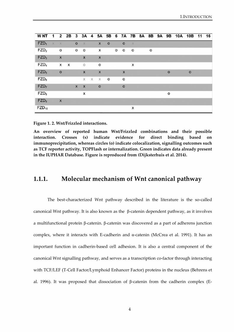
ROR1/2 the non-canonical pathway (Niehrs 2012;van Amerongen R. et al. 2009). Figure 1.2.
summarizes reported Wnt/Frizzled interactions.
1.INTRODUCTION
3
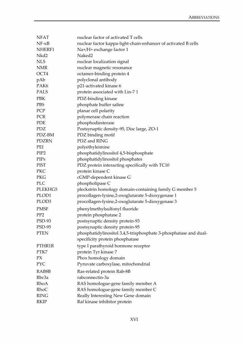
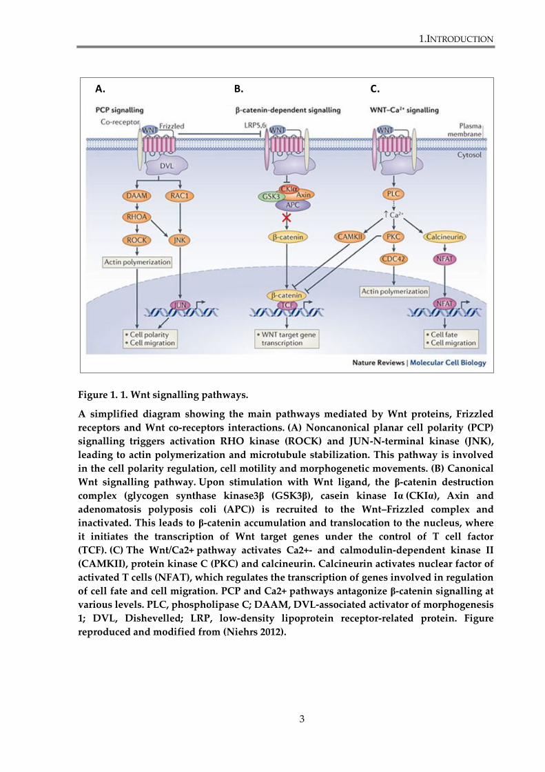
Figure 1. 1. Wnt signalling pathways.
A simplified diagram showing the main pathways mediated by Wnt proteins, Frizzled
receptors and Wnt co-receptors interactions. (A) Noncanonical planar cell polarity (PCP)
signalling triggers activation RHO kinase (ROCK) and JUN-N-terminal kinase (JNK),
leading to actin polymerization and microtubule stabilization. This pathway is involved
in the cell polarity regulation, cell motility and morphogenetic movements. (B) Canonical
Wnt signalling pathway. Upon stimulation with Wnt ligand, the β-catenin destruction
complex (glycogen synthase kinase3β (GSK3β), casein kinase Iα (CKIα), Axin and
adenomatosis polyposis coli (APC)) is recruited to the Wnt–Frizzled complex and
inactivated. This leads to β-catenin accumulation and translocation to the nucleus, where
it initiates the transcription of Wnt target genes under the control of T cell factor
(TCF). (C) The Wnt/Ca2+ pathway activates Ca2+- and calmodulin-dependent kinase II
(CAMKII), protein kinase C (PKC) and calcineurin. Calcineurin activates nuclear factor of
activated T cells (NFAT), which regulates the transcription of genes involved in regulation
of cell fate and cell migration. PCP and Ca2+ pathways antagonize β-catenin signalling at
various levels. PLC, phospholipase C; DAAM, DVL-associated activator of morphogenesis
1; DVL, Dishevelled; LRP, low-density lipoprotein receptor-related protein. Figure
reproduced and modified from (Niehrs 2012).
A. B. C.
1.INTRODUCTION
4
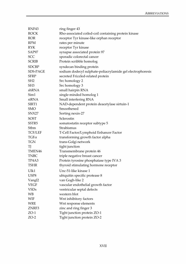
Figure 1. 2. Wnt/Frizzled interactions.
An overview of reported human Wnt/Frizzled combinations and their possible
interaction. Crosses (x) indicate evidence for direct binding based on
immunoprecipitation, whereas circles (o) indicate colocalization, signalling outcomes such
as TCF reporter activity, TOPFlash or internalization. Green indicates data already present
in the IUPHAR Database. Figure is reproduced from (Dijksterhuis et al. 2014).
1.1.1. Molecular mechanism of Wnt canonical pathway
The best-characterized Wnt pathway described in the literature is the so-called
canonical Wnt pathway. It is also known as the β-catenin dependent pathway, as it involves
a multifunctional protein β-catenin. β-catenin was discovered as a part of adherens junction
complex, where it interacts with E-cadherin and α-catenin (McCrea et al. 1991). It has an
important function in cadherin-based cell adhesion. It is also a central component of the
canonical Wnt signalling pathway, and serves as a transcription co-factor through interacting
with TCF/LEF (T-Cell Factor/Lymphoid Enhancer Factor) proteins in the nucleus (Behrens et
al. 1996). It was proposed that dissociation of β-catenin from the cadherin complex (E-
1.INTRODUCTION
5
cadherin-β-catenin-α-catenin) causes loss of cell adhesion and promotes the migration of
cancer cells (Xu et al. 2007).
In the absence of Wnt ligand, the cellular levels of β-catenin are strictly controlled by
a multi-protein destruction complex composed of axin, adenomatous polyposis coli (APC),
glycogen synthase kinase 3β (GSK3β) and casein kinase 1α (CK1α). This complex
phosphorylates β-catenin on the N-terminus, leading to its subsequent ubiquitylation and
degradation by theproteosome. β-catenin can be phosphorylated on Ser33, Ser37, Thr41 and
Ser45. Firstly, Ser45 is phosphorylated by CK1α (Amit et al. 2002). Other sites are
phosphorylated later by GSK3β (Liu et al. 2002). All four residues have to be phosphorylated
to allow ubiquitylation. Mutation in any of these sites leads to β-catenin stabilization in the
cytoplasm. Axin serves as a scaffold protein to bring kinases and β-catenin together.
Phosphorylated β-catenin is recognized by β-TrCP (β-transducin repeat-containing protein),
an E3 ubiquitin ligase, that triggers the ubiquitylation process (Marikawa et al. 1998). Thus,
the intracellular level of β-catenin remains low and TCF/LEF mediated expression of Wnt
response genes is repressed by Grouchos (Rao et al. 2010).
The canonical Wnt pathway is activated by binding of Wnt ligands to Frizzled
receptors and LRP5/6 co-receptors, which leads to the phosporylation of LRP5/6 by CK1γ
and GSK3β (Zeng et al. 2005). The phosphoprotein Dishevelled (Dvl) is recruited to the
plasma membrane, where it interacts with Frizzled via its PDZ domain, self-polymerizes via
DIX domain and serves as a mediator for recruitment of axin-GSK3β to the plasma
membrane (Schwarz-Romond et al. 2007;Wong et al. 2003). Activation of LRP5/6 co-receptors
leads to their redistribution in the plasma membrane to the caveolin-rich areas. This results
in the formation of Wnt-Fz-LRP5/6-Dvl-Axin platform, called LRP5/6 signalosome (Bilic et al.
1.INTRODUCTION
6
2007). Axin recruitment to the plasma membrane leads to disassembly of the destruction
complex and stabilization of β-catenin, which accumulates in the cytoplasm and finally
enters the nucleus. It is suggested that the main route of β-catenin nuclear transport is
independent of the classical Ran-GTPase/importin import mechanism, as it does not possess
a classical nuclear localization signal (NLS) or a nuclear export signal (NES) sequence.
β-catenin is able to directly interact with the nuclear pore complex (nucleoporin Nup358 on
cytoplasmic tail, Nup62 in central channel and Nup98 and Nup153 on the nuclear end).
β-catenin can also self-regulate its own entry into the nucleus (Jamieson et al. 2014). In the
nucleus β-catenin associates with transcription factors from the TCF/LEF family and activates
the transcription of Wnt response genes. Among them are genes that play role in cell
differentiation (siamois and brachyury (protein T), cell signalling (VEGF (vascular
endothelial growth factor), FGF4 (fibroblast growth factor 4) and FGF18), proliferation (cyclin
D1, c-MYC), adhesion (E-cadherin) (Klaus et al. 2008). An overview of the Wnt/β-catenin
pathway is presented in the Figure 1. 3.
The canonical Wnt pathway is regulated by a number of proteins. Secreted Frizzled-
related proteins (SFRPs) and Wnt inhibitory factors (WIFs) can bind Wnt proteins, because
they contain a domain that resembles the cysteine-rich domain (CRD) domain of Frizzled
receptors. SOST and Wise proteins bind LRP5/6 and in consequence inhibit signalling.
Dickkopf (Dkk) triggers LRP5/6 internalization and inactivation, as a result of enhancing
LRP5/6 interaction with Kremen (Staal et al. 2008). Protein phosphatase 2 (PP2) is responsible
for dephosporylation of β-catenin. Inhibitor of β-catenin and TCF (ICAT) and protein Chibby
bind β-catenin inside the nucleus and prevent it from forming a complex with TCF (Klaus et
al. 2008).
1.INTRODUCTION
7
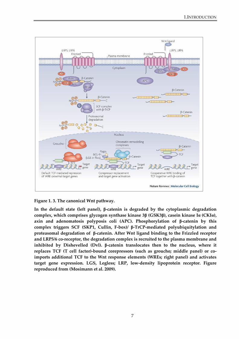
Figure 1. 3. The canonical Wnt pathway.
In the default state (left panel), β-catenin is degraded by the cytoplasmic degradation
complex, which comprises glycogen synthase kinase 3β (GSK3β), casein kinase Iα (CKIα),
axin and adenomatosis polyposis coli (APC). Phosphorylation of β-catenin by this
complex triggers SCF (SKP1, Cullin, F-box)/ β-TrCP-mediated polyubiquitylation and
proteasomal degradation of β-catenin. After Wnt ligand binding to the Frizzled receptor
and LRP5/6 co-receptor, the degradation complex is recruited to the plasma membrane and
inhibited by Dishevelled (Dvl). β-catenin translocates then to the nucleus, where it
replaces TCF (T cell factor)-bound corepressors (such as groucho; middle panel) or co-
imports additional TCF to the Wnt response elements (WREs; right panel) and activates
target gene expression. LGS, Legless; LRP, low-density lipoprotein receptor. Figure
reproduced from (Mosimann et al. 2009).
1.INTRODUCTION
8
1.1.2. Molecular mechanisms involved in non-canonical
Wnt pathway signalling
Non-canonical Wnt pathways are defined as routes independent of β-catenin
transcriptional activity. There are many non-canonical pathways reported. However, due to
a high level of cross-talk between receptors and secondary messengers from different
signalling branches, it is difficult to separate individual pathways completely. For the sake of
simplification, the following branches of the non-canonical Wnt pathways can be
distinguished: PCP, Wnt/Ca2+, Wnt/RAP, Wnt/cAMP, Wnt/GSK3-microtubules, Wnt/aPKC,
Wnt/mTOR, Wnt/ROR and Wnt/RYK (Schulte 2010;Semenov et al. 2007). Of these, the best
described are the PCP and Wnt/Ca2+ pathways.
Planar Cell Polarity (PCP) refers to cell polarization within two-dimensional
epithelium. It occurs during hair orientation and gastrulation (Wu et al. 2009). PCP signalling
is often activated by Wnt5a or Wnt11 through various Frizzled receptors (e.g. Fz6, Fz7 and
Fz8 (Dijksterhuis et al. 2014)) and Dvl, but instead of LRP5/6, ROR and PTK7 are involved,
and signals are transmitted without involving β-catenin. In Drosophila melanogaster, two
complexes relocate to the opposite sites of the cell during the process. Frizzled-Dishevelled-
Diversin (Div) complex localizes distally and Strabismus (Stbm, Vangl in vertebrates)-Prickle
(Pk) localizes proximally, which leads to planar cell polarization (Veeman et al. 2003a). Dvl
together with DAAM (Dishevelled-Associated Activator of Morphogenesis) activates the
small GTPase RhoA (RAS homologue-gene family member A)-ROCK (Rho-associated coiled-
coil containing protein kinase) and JUN-N-terminal kinase (JNK) routes, which results in
actin cytoskeleton reorganization (Endo et al. 2005). PCP and Wnt canonical pathway are
1.INTRODUCTION
9
known to antagonize each other and inhibiting one pathway will usually upregulate the
other (Niehrs 2012).
The Wnt/Ca2+ pathway is activated by interaction of Wnt5a with Frizzled-2 (Sheldahl
et al. 2003). The Frizzled receptor activates phosphodiesterase (PDE) via heterotrimeric G
protein. As a result, the cellular levels of cGMP (cyclic guanosine monophosphate) are
decreased, which inactivates the cGMP-dependent kinase G (PKG) and increases the cellular
concentration of calcium. Wnt/Fz also activates phospholipase C (PLC). It initiates a cascade
of events that results in the generation of diacylglycerol (DAG) and inositol trisphosphate
(IP3), followed by calcium fluxes. This leads to activation of the protein kinase C (PKC),
which induces changes in actin cytoskeleton (Kuhl et al. 2000). Increased Ca2+ concentration
also activates calcium-calmodulin-dependent kinase II (CamKII) and calcium-sensitive
phosphatase calcineurin, which leads to activation of NFAT (nuclear factor of activated T
cells) transcription factor. Wnt/Ca2+ signalling through CamKII inhibits Wnt/β-catenin
pathway (Kohn et al. 2005;Semenov et al. 2007).
1.INTRODUCTION
10
1.1.3. Wnt signalling in diseases
Deregulation of Wnt signalling during development leads to developmental defects
(van Amerongen R. et al. 2009). In adult organisms, Wnt signalling is often associated with
loss of growth control and impaired cell differentiation. Mutations or altered expression of
Wnt signalling pathway components are associated with development of different types of
cancers, such as colon carcinoma. Somatic mutations of Wnt pathway components identified
in cancer are summarized in the table 1.1. Wnt signalling proteins associated with distinct
patient outcomes in a cancer-subtype-specific manner are presented in the table 1.2.
Aberrant Wnt signalling has also been reported in breast cancer, lung cancer, kidney disease,
inflammatory bowel disease, diabetes type II and sarcoidosis (Herr et al. 2012). Furthermore,
Wnt signalling is also involved in maintaining bone mass and is important for bone healing;
findings which may have implications in osteoporosis (Canalis 2013).
Activation of the canonical Wnt pathway, evidenced by stabilization and nuclear
accumulation of β-catenin, has been observed in colorectal, lung, breast, cervical, skin and
liver tumours. β-catenin accumulation is linked with poor differentiation, high proliferative
activity and poor prognosis (Endo et al. 2000). Stabilization of β-catenin may occur as a result
of mutations in components of signalling pathway or due to epigenetic events. Mutations of
APC lead to an inability to downregulate β-catenin (Polakis 2000). Mutations of APC rarely
occur outside of the gastrointestinal tract, but have been found in the majority of sporadic
colorectal cancers (SCC) (Polakis 2007). The acquisition of APC mutations is an initial step in
colorectal carcinogenesis and facilitates and accelerates tumour development (Kinzler et al.
1996). Gain-of-function mutations of the CTNNB1 gene, which encodes β-catenin, occur in
1.INTRODUCTION
11
approximately 1% of SCC and more frequently in sporadic cancers outside of the intestines,
for example in brain tumours, ovarian, prostate and hepatocellular cancers (Camilli et al.
2010;Klaus et al. 2008).
The Wnt pathway can be regulated by epigenetic mechanisms, such as
hypermethylation. Silencing by hypermethylation was reported in different types of tumours
for inhibitory Wnt ligands SFRPs (Caldwell et al. 2006;Jost et al. 2009;Nojima et al.
2007;Suzuki et al. 2008) and DKK (Sato et al. 2007). Overexpression of several Wnt pathway
components in cancer is also common. It was shown for Wnt proteins (Katoh et al. 2001),
Frizzleds (Kirikoshi et al. 2001;Saitoh et al. 2002) and Dishevelled (Okino et al. 2003).
Recent developments showed antagonistic crosstalk between Wnt and Notch
signalling. It was reported that the expansion of intestinal progenitor cells by constitutively
active Notch pathway was contingent to Wnt signalling in the intestine (Fre et al. 2009). The
Notch receptor intracellular domain (ICD) was shown to bind unphosphorylated (active) β-
catenin, making it inaccessible for transcription activity. Notch1 depletion, upregulated Wnt
signalling, whereas expression of non-cleavable Notch1 receptor or inhibition of γ-secretase
led to downregulation of the Wnt pathway. The authors concluded that association of β-
catenin with the Notch receptor leads to its degradation in the lysosomes in a Numb-
dependent fashion (Kwon et al. 2011).
The role of non-canonical pathways in cancer is less well studied, but there is
evidence for their involvement in cancer development. Wnt5a is the main object in studies of
β-catenin independent signalling in cancer. It was shown that, depending on the type of
cancer; it can have either a suppressive or an oncogenic role. The suppressive function
results from antagonizing the β-catenin dependent pathway. Wnt5a expression is
1.INTRODUCTION
12
downregulated in colorectal cancer (Dejmek et al. 2005a;Ying et al. 2008), neuroblastoma
(Blanc et al. 2004), ductal breast cancer (Dejmek et al. 2005b), esophageal squamous cell
carcinoma (Li et al. 2010) and haematological malignancies (Ying et al. 2007). On the other
hand, it is overexpressed in gastric (Kurayoshi et al. 2006), pancreatic (Ripka et al. 2007),
prostate (Wang et al. 2007) and non-small cell lung cancer (Huang et al. 2005). It enhances
cell motility and tumour invasion in breast cancer (Pukrop et al. 2006). It was reported that
Wnt5a-induced migration of melanoma cells depends on signalling through Frizzled 5 and
PKC (Weeraratna et al. 2002). Wnt5a and β-catenin in gastric cancer are expressed in
mutually exclusive manner. Moreover, Wnt5a activates focal adhesion kinase and small
GTP-binding protein Rac. Both play a role in the regulation of cell migration (Kurayoshi et al.
2006).
Recent data have revealed roles for the Wnt/PCP pathway in cancer progression,
invasion, metastasis and in cancer angiogenesis (Wang 2009). It was shown that Frizzled-7
promotes migration of human hepatocellular carcinoma and invasion of colon cancer cells
(Merle et al. 2004;Ueno et al. 2009;Vincan et al. 2007b). There is also evidence that Wnt
signalling plays a significant role in angiogenesis (Masckauchan et al. 2006;Zhang et al. 2006)
and it was reported that Wnt5a mediated signalling regulates endothelial cell growth and
migration (Cheng et al. 2008;Cirone et al. 2008). The latter study by Cirone and colleagues
showed that impaired non-canonical Wnt signalling leads to vascular defects in vertebrate
model.
1.INTRODUCTION
13
Table 1. 1. Somatic mutations of Wnt pathway components in cancer.
Table is reproduced from (Anastas et al. 2013)
1.INTRODUCTION
14
Table 1. 2. Wnt signalling proteins are associated with distinct patient outcomes in a
cancer-subtype-specific manner.
Table is reproduced from (Anastas et al. 2013)

1.INTRODUCTION
15
1.2. Wnt/Frizzled signalling in breast cancer
Breast cancer is the most invasive form of cancer in women and the second leading
cause of death in women in industrialized nations. Evidence shows that Wnt signalling is
essential in the development of normal mammary gland and also plays an important role in
mammary oncogenesis. Mouse mammary carcinoma was the first known example of a Wnt-
related disease. An insertion of the MMTV (mouse mammary tumour virus) was found in
Wnt1 locus, which resultantly was continuously driving Wnt1 expression (van 't Veer et al.
1984). Later, Wnt3 and Wnt10b were also found to carry the insertion of MMTV in breast
cancer (Tekmal et al. 1997).
Numerous reports have shown the deregulation of Wnt signalling in breast cancer.
Mutations in genes encoding components of Wnt signalling pathway, such as APC, β-catenin
or Axin, are rare in breast cancer. Yet, upregulation of the pathway is evident, especially in
basal-like and triple negative breast cancer (TNBC), where nuclear and cytosolic
accumulation of β-catenin is associated with poor outcome (Geyer et al. 2011;Khramtsov et
al. 2010;Lopez-Knowles et al. 2010). Cyclin D1 and c-Myc, well-known targets of canonical
pathway, are also overexpressed in breast tumours (Lin et al. 2000). Recent report by Dey
and colleagues provides additional evidence for preferential activation of Wnt signalling in
TNBC and increased risk for brain and lung metastases (Dey et al. 2013). The mechanisms
underlying activation of Wnt signalling pathways in breast cancer still remain unclear.
However, it seems that it is mainly achieved through upregulation of potentially oncogenic
receptors and co-receptors, such as Frizzled-7 and LRP5/6 (Liu et al. 2010;Yang et al. 2011).
1.INTRODUCTION
16
Promoter hypermethylation of Wnt antagonist genes, like APC, WIF1 and SFRP1 was also
reported (Klarmann et al. 2008;Suzuki et al. 2008).
1.2.1. Frizzled-7 in TNBC
Activation of Wnt signalling pathway is particularly associated with triple-negative
(TNBC) basal-like breast cancer, which is one of the six major breast cancer subtypes. It
represents approximately 10-20% of all breast cancers, and is more frequent in
premenopausal women, especially of African-American and Hispanic descent (Carey et al.
2010). TNBC is characterized by a very aggressive clinical behaviour, demonstrated by lack
or limited expression of estrogen receptor (ER), progesterone receptor (PR) and human
epidermal growth factor receptor 2 (HER2/ERBB2). That makes TNBC cells resistant to
therapeutic protocols based on pharmaceutical targeting of mentioned receptors (Gusterson
2009). TNBC was subdivided into six subtypes. Three of them (basal-like, mesenchymal-like,
mesenchymal stem-like) were characterized by constitutive activation of Wnt signalling.
Even though Herceptin (anti-HER2 antibodies), ER antagonists and aromatase inhibitors
improved treatment of other breast cancer subtypes, the treatment of TNBC still remains
a great challenge (Lehmann et al. 2011).
Frizzled-7 (Fz7) emerges as the most important Frizzled receptor involved in breast
cancer, where it activates the canonical Wnt pathway. Expression of Fz7 is characteristic for
basal-like and triple-negative breast cancer. Fz7 is the only member of Frizzled family of
receptors significantly overexpressed in triple-negative breast cancer (TNBC) and TNBC-
derived cell lines (Yang et al. 2011). Yang and colleagues demonstrated that Fz7 plays
1.INTRODUCTION
17
a crucial role in cell proliferation in TNBC. Its depletion reduces cell growth and expression
of Wnt target genes (Cyclin D1 and c-Myc), inhibits tumourigenesis in vitro, and decreases
the ability of the MDA-MB-231 cell line to form tumours in athymic nude mice. Additionally,
in the same study it has been shown that tumour growth could be reduced by the use of
FZD7shRNA in mice with xenografts of triple negative breast cancer cells (Yang et al. 2011).
These findings indicate that Fz7 is a key factor involved in TNBC cell transformation,
evidenced by changes in cell invasion, motility and clonogenicity.
Recently, it has been shown that SIRT1, a class III histone deacetylase (HDAC)
positively regulates Fz7 mRNA and protein levels in breast cancer cells (Simmons, Jr. et al.
2014)
1.2.2. Autocrine Wnt signalling
Autocrine Wnt signalling has been reported in 25% of human breast and ovarian
cancer cell lines (Bafico et al. 2004) and 50% of human non-small lung carcinoma cell lines
(Akiri et al. 2009). It was suggested that autocrine Wnt signalling contributes to the
proliferation of breast cancer cells in a manner that depends on EGFR activity (Schlange et al.
2007). Recently, Luga and colleagues showed that exosomes derived form human breast
cancer associated fibroblasts (CAFs) are involved in mobilizing autocrine Wnt-PCP
signalling in breast cancer cells and stimulate their invasive behaviour and metastasis in
animal models in tetraspanin CD81 dependent manner (Luga et al. 2012).
1.INTRODUCTION
18
1.2.3. Crosstalk with other pathways
Several reports have shown evidences for crosstalk between Wnt signalling and other
pathways in breast cancer. It was reported that TGFα and Wnt cooperatively induce
mammary tumourigenesis, through the interaction between β-catenin and EGFR/ErbB2
heterodimers (Schroeder et al. 2000;Schroeder et al. 2002). Wnt1 and Wnt5a binding to
Frizzled transactivates ErbB1, which leads to MAPK activation and overexpression of Cyclin
D1 (Civenni et al. 2003). Furthermore, Schlange and colleagues suggested that Wnt1 induces
c-Src-dependent EGFR transactivation, which in turn stimulates ERK1/2 signalling and cell
proliferation (Schlange et al. 2007). Recent report has also shown that Wnt3 expression in
breast cancer cells leads to activation of canonical Wnt signalling, upregulation of EGFR and
promotion of epithelial-to-mesenchymal-like transition. Consequently it results in reduction
of the growth-inhibiting effects of Herceptin in HER2-overexpressing cells. This finding
indicates a role for Wnt3 in the development of Herceptin resistance in patients with breast
cancer tumours overexpressing HER2 receptor (Wu et al. 2012). Interestingly, upregulated
Wnt signalling was also associated with breast cancer resistance to Tamoxifen (Loh et al.
2013). Furthermore, it was reported that increased expression of Wnt1 in human mammary
epithelial cells (HMEC) activates Notch signalling pathway, what leads to tumourigenic
transformation of the HMEC cells (Ayyanan et al. 2006).
1.INTRODUCTION
19
1.2.4. Epigenetic modifications
Gene hypermethylation has been widely documented in several types of cancer
including breast cancer. In relation to the Wnt canonical pathway, DNA methylation of the
WIF1 (Wnt Inhibitory Factor 1) promoter was observed in 60% of breast carcinomas (Ai et al.
2006;Wissmann et al. 2003). Expression of SFRP1, another protein that antagonizes Wnt
signalling, was found to be silenced in breast cancer. 60% of samples from primary breast
cancers displayed methylation of SFRP1 promoter and SFRP1 gene hypermethylation is
associated with shorter survival of patients with invasive breast cancer (Shulewitz et al.
2006;Veeck et al. 2006). It was shown that SFRP1 is methylated in 64%, SFRP2 in 100% and
SFRP5 in 90% of breast cell lines (Suzuki et al. 2008). Furthermore, several studies revealed
that APC gene promoter is hypermethylated in 35-50% of breast cancer tumours and cell
lines (Jin et al. 2001). Downregulation of APC protein leads to activation of β-catenin.
Accumulation of β-catenin can be also a result of E-cadherin downregulation caused by
methylation of CDH1 gene. E-cadherin is a membrane receptor and a main part of adherens
junction complex. It binds β-catenin and decreases its cytoplasmic concentration (Yang et al.
2001).
1.INTRODUCTION
20
1.3. Frizzled-7: member of Wnt receptor family
1.3.1. Frizzled receptors
Frizzled’s were originally identified as Wingless receptors in D. melanogaster (Bhanot
et al. 1996). They are highly conservative in animal kingdom (Schioth et al. 2005). In
mammals, 10 Frizzled isoforms have been identified. The amino acid sequence analysis
revealed that Frizzled share 20 to 40% identity. The level of similarity is higher within
particular clusters: Fz1, Fz2 and Fz7 share 75% amino acid identity, Fz5 and Fz8 share 80%
identity, Fz4, Fz9 and Fz10 share 65% identity, and Fz3 and Fz6 share 50% identity with each
other, as presented in the Figure 1. 4.A. (Fredriksson et al. 2003). The major characteristics of
human Frizzled receptor are summarized in the table 1.3.
Receptors from Frizzled family are seven-transmembrane-spanning proteins.
Although they are G protein-coupled receptors (GPCRs), they differ significantly from other
GPCRs and represent a separate class surface receptors (Foord et al. 2005). Three regions can
be distinguished within Frizzled structure: the extracellular N-terminal portion (224 amino
acids), the transmembrane domains, and intracellular C-terminal cytoplasmic tail (25 amino
acids) (Figure 1.4.B).
The extracellular region consists of a signal sequence, which ensures correct protein
insertion into the membrane, followed by the Cysteine-Rich Domain (CRD), responsible for
binding of Wnt proteins. Ten cysteines present in CRD are highly conserved and form five
disulfide bond (Xu et al. 1998). The three dimensional structure was solved for mFzd8-CRD
(Dann et al. 2001). It revealed a new protein fold, which consists mainly of α-helical structure
1.INTRODUCTION
21
(Figure 1.4.C). Moreover, the existence of CRD dimers was reported. A similar Cysteine-Rich
Domain is also found in SMO (Smoothened), a protein closely related to Frizzled, which does
not bind Wnts (Wu et al. 2002), SFRPs (Secreted Frizzled Related Proteins), collagen
α1(XVIII), carboxypeptidase Z (CPZ), Ror family of receptor tyrosine family kinase and
muscle-specific kinase (MuSK) family (Xu et al. 1998). CRD domain of Frizzled receptors
contains also Asn residues that are predicted to be glycosylated (Schulte 2010). The structure
of X.leavis Wnt8 in complex with mouse CRD of Frizzled8 has been recently resolved (Figure
1.4.D). It revealed a new two-domain Wnt structure, not related to any known protein folds,
with two finger-like domains (N-terminal “lipid thumb” and C-terminal “index finger”)
grasping CRD of Fz8 at two distinct binding sites. The conservation of amino acids in both
Wnt domains facilitates ligand-receptor cross-reactivity, which is important for
understanding functionality of Wnt proteins (Janda et al. 2012).
Transmembrane domain consists of seven membrane-spanning regions, three
intracellular and three extracellular loops. In the extracellular loops 1 and 2 conserved
cysteines are present. They are thought to stabilize an overall structure of the receptors
(Lagerstrom et al. 2008).
The intracellular domain is involved in extensive protein-protein interactions. It also
contains sites for posttranslational modifications, such as phosphorylation, ubiquitylation,
nitrosylation and hydroxylation (Schulte 2010). Cytosolic tails of most Frizzled receptors
have two Postsynaptic density-95, Disc large, ZO-1 (PDZ) binding motifs, PDZ-BM. They are
4-6 amino acid motifs that interact with PDZ domains of partner proteins. Frizzled proximal
PDZ-BM (K-T-xxx-W) interacts with the PDZ domain of the phoshoprotein Dishevelled
(Dvl). This atypical, internal PDZ-BM is placed very close to the plasma membrane and is
1.INTRODUCTION
22
conserved within all Frizzleds (Umbhauer et al. 2000;Wallingford et al. 2005). Dvl is a
cytoplasmic scaffold of which three homologs exist in vertebrates (Dvl1, Dvl2, Dvl3). Their
functional redundancy is evidenced by overlapping expression patterns and high degree of
conserved structures (Gao et al. 2010b). The interaction between Frizzled and Dvl is essential
for different Wnt-induced signalling pathways. Recently, Tauriello and colleagues presented
a model of Frizzled-Dishevelled interaction involving a three-segmented discontinuous
binding surface on the Frizzled receptor. They have shown that the DEP domain and C-
terminal region of Dvl binds cooperatively to the motifs I and II in the third intracellular loop
and to the C-terminal motif III of mouse Frizzled-5 and Xenopus Frizzled-7 (Figure 1.4.D).
That allows forming a stable Fz-Dvl complex at the plasma membrane and initiating
canonical Wnt signalling. Whether Frizzled interaction with Dishevelled requires PDZ and
DEP domains to act cooperatively or sequentially needs to be established (Tauriello et al.
2012).
The distal PDZ-BM (x-S/T-x-φ, where φ represents a hydrophobic residue) is a
standard, terminal class I PDZ ligand (Jelen et al. 2003). It can bind a range of different PDZ
proteins (GOPC, MAGI-3, Syntenin, Kermit, PSD-95) (Wawrzak et al. 2009). So far, the
interactions involving the distal PDZ-BM have been shown for Fz1,-2,-3,-4,-5,-7,-8, but not for
Fz6, -9 and -10 (Schulte 2010).
1.INTRODUCTION
23
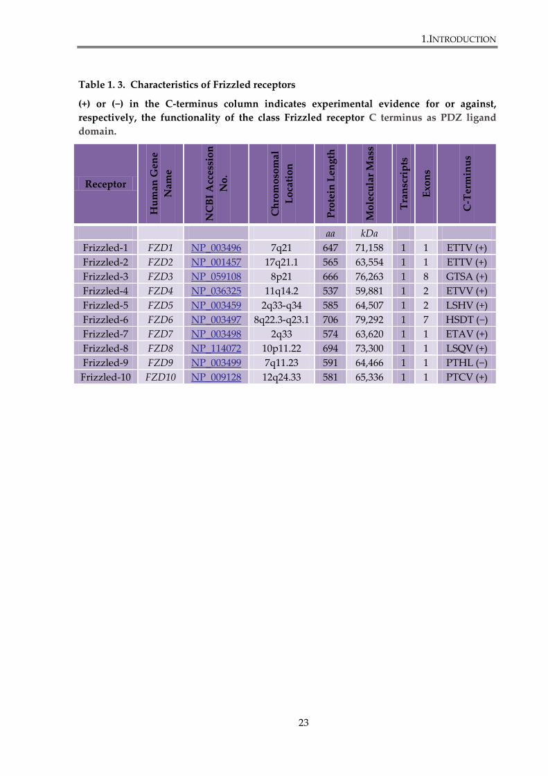
Table 1. 3. Characteristics of Frizzled receptors
(+) or (−) in the C-terminus column indicates experimental evidence for or against,
respectively, the functionality of the class Frizzled receptor C terminus as PDZ ligand
domain.
Receptor
Hu
man
Gen
e
Nam
e
NC
BI
Acc
essi
on
No
.
Ch
rom
oso
mal
Lo
cati
on
Pro
tein
Len
gth
Mo
lecu
lar
Mas
s
Tra
nsc
rip
ts
Exo
ns
C-T
erm
inu
s
aa kDa
Frizzled-1 FZD1 NP_003496 7q21 647 71,158 1 1 ETTV (+)
Frizzled-2 FZD2 NP_001457 17q21.1 565 63,554 1 1 ETTV (+)
Frizzled-3 FZD3 NP_059108 8p21 666 76,263 1 8 GTSA (+)
Frizzled-4 FZD4 NP_036325 11q14.2 537 59,881 1 2 ETVV (+)
Frizzled-5 FZD5 NP_003459 2q33-q34 585 64,507 1 2 LSHV (+)
Frizzled-6 FZD6 NP_003497 8q22.3-q23.1 706 79,292 1 7 HSDT (−)
Frizzled-7 FZD7 NP_003498 2q33 574 63,620 1 1 ETAV (+)
Frizzled-8 FZD8 NP_114072 10p11.22 694 73,300 1 1 LSQV (+)
Frizzled-9 FZD9 NP_003499 7q11.23 591 64,466 1 1 PTHL (−)
Frizzled-10 FZD10 NP_009128 12q24.33 581 65,336 1 1 PTCV (+)
1.INTRODUCTION
24
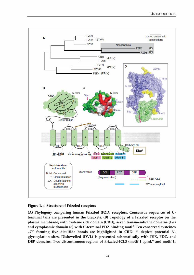
Figure 1. 4. Structure of Frizzled receptors
(A) Phylogeny comparing human Frizzled (FZD) receptors. Consensus sequences of C-
terminal tails are presented in the brackets. (B) Topology of a Frizzled receptor on the
plasma membrane, with cysteine rich domain (CRD), seven transmembrane domains (1-7)
and cytoplasmic domain (8) with C-terminal PDZ binding motif. Ten conserved cysteines
„C” forming five disulfide bonds are highlighted in CRD. Ψ depicts potential N-
glycosylation sites. Dishevelled (DVL) is presented schematically with DIX, PDZ, and
DEP domains. Two discontinuous regions of Frizzled-ICL3 (motif I „pink” and motif II
ETxV
1.INTRODUCTION
25
„green”) bind to the C-terminus of Dvl. The cytoplasmic tail of Frizzled contains the
KTxxxW motif („blue”), which interacts strongly with Dishevelled-PDZ domain. (C)
Crystal structure of Fz8-CRD. Residues in red are suggested to be a Wnt-binding interface.
Residues in green when altered did not affect Wnt activity (PDB:1IJY). (D) Wnt8–Fz8-CRD
co-crystal structure, shown as a ribbon diagram superimposed on surface representation.
Site 1 and site 2, mediate contacts between Wnt8 and Fz8-CRD. Palmitoleic acid adduct
from the Wnt8 thumb at site 1 (red); N-glycans of Wnt8 and Fz8-CRD (yellow). N-term,
amino terminal; C-term, carboxyl terminal; ICL, intracellular loop; ECL, extracellular loop.
Figure reproduced and modified from (MacDonald et al. 2012).
1.INTRODUCTION
26
1.3.2. Frizzled-7
Human FZD7 gene is located on chromosome 2q.3317. Another form of Fz7, FzE3,
was isolated from esophageal carcinoma tissue. FzE3 shares 98% homology with Fz7 and
their sequences differ by 9 amino acids (Tanaka et al. 1998). Frizzled-7 is the only member of
Frizzled family that regulates development of the gastric tract (King et al. 2012b). In recent
years several reports have indicated that Fz7 is an important receptor governing Wnt
signalling in cancer cells.
1.3.2.1. Functions of Frizzled-7
Frizzled-7 is a unique receptor that can activate canonical and noncanonical
pathways. Human Wnt3a, Xenopus Wnt8b and mouse Wnt6 were reported to activate
canonical Wnt signalling through interactions with Fz7 orthologs (Kim et al. 2008b;Linker et
al. 2005;Medina et al. 2000). On the other hand, zebrafish and Xenopus Wnt11, as well as
Xenopus Wnt5a are proposed to bind to Fzd7 to activate noncanonical Wnt pathways (Swain
et al. 2001).
Frizzled-7 activates the canonical signalling upon interaction with Wnt3a, which was
shown in liver cancer cells (Kim et al. 2008b). Frizzled-7 was also found necessary for the
mesenchymal-to-epithelial transition in the human colorectal cancer LIM1863-Mph cell line
(Vincan et al. 2007a)
PCP pathway activation by Frizzled-7 is responsible for regulation of cell movements
via activation of JNK and Rho (Kinoshita et al. 2003;Tanegashima et al. 2008). Frizzled-7 was
also reported to regulate bone morphogenesis through PCP signalling (Li et al. 2009).

1.INTRODUCTION
27
Frizzled-7 interaction with Wnt11 activates PKC-Cdc42 and PKCδ-JNK signalling pathways
that regulate convergent extension movements during gastrulation, as well as migration of
neural crest cells in Xenopus and zebrafish (De Calisto et al. 2005;Djiane et al. 2000;Kinoshita
et al. 2003;Knowlton et al. 2004;Penzo-Mendez et al. 2003). Xenopus Frizzled-7 signalling has
been also shown to activate the PKC and Ca2+/calmodulin kinase signalling during cardiac
development (Sheldahl et al. 2003). Furthermore, Frizzled-7 has been reported in the PCP
pathway-dependent repair of mouse muscle. Receptor interaction with Wnt7a was shown to
activate PCP pathway, in the mechanism involving Vangl2, which resulted in the expansion
of satellite stem cells, and muscle repair (Le Grand et al. 2009).
Recently Frizzled-7 has implicated in the maintenance of embryonic stem cell self-
renewal capacity (Takahashi-Yanaga et al. 2010). Dormeyer and colleagues have reported
that FZD7 mRNA levels are 200-fold higher in human embryonic stem cells, when compared
to differentiated cell types. They have also shown that Fz7 depletion by shRNA in human
embryonic stem cells leads to dramatic changes in the morphology of embryonic stem cell
colonies and loss of expression of the embryonic stem cell-specific transcription factor OCT4
(Dormeyer et al. 2008). Furthermore, Frizzled-7 expression is upregulated in colonic crypt
stem cells, but its expression level decreases when cells differentiate into mature epithelial
cells (Gregorieff et al. 2005).
Frizzled knockout mice (Fz7-/-) display tail truncation and kinking with 100%
penetrance and ventricular septal defects (VSDs) with 15% penetrance. Furthermore, Fz2+/-;
Fz7-/- mice display VSDs with 50% penetrance and Fz2-/-; Fz7-/- mice display convergent
extension defects and mid-gestational lethality with 100% penetrance. It was suggested that
1.INTRODUCTION
28
cross-talk events between different signalling pathways in development are susceptible to
disruptions in Frizzled signalling (Yu et al. 2012).
Taken together, Frizzled-7 is involved in canonical and non-canonical Wnt signalling
during embryogenesis, adult-tissue homeostasis and carcinogenesis.
1.3.2.2. Ligands and binding partners of Frizzled-7
Frizzled-7 has been reported to activate the canonical Wnt pathway upon interaction
with Xenopus Wnt8b, mouse Wnt6, and human Wnt3 (Kim et al. 2008b;Linker et al.
2005;Medina et al. 2000). As direct interaction between Wnt3a and Fz7-CRD has been shown
in L-cells (Kemp et al. 2007). Frizzled-7 is also able to bind Xenopus Wnt11 and mouse Wnt7a.
This, however leads to activation of noncanonical Wnt signalling (Bentzinger et al. 2014;Kim
et al. 2008a).
Frizzled-7 interacts with a number of PDZ (postsynaptic density protein (PSD)-95,
Discs-large and ZO-1) domain-containing proteins. The PDZ domain of DVL family of
proteins binds to the internal PDZ binding motif, KTxxxW, in the C-terminal cytoplasmic
region of Frizzled-7 (Umbhauer et al. 2000;Wong et al. 2003), while other PDZ proteins, such
as discs large 1/synapse associated protein 97 (DLG1/SAP97), DLG2/PSD-93,
DLG4/SAP90/PSD-95, MAGI3 and Syntenin-1 (syndecan binding protein, SDCBP) bind to
the C-terminal PDZ binding motif, ETxV, of the receptor (Hering et al. 2002;Luyten et al.
2008;Schulte et al. 2007;Yao et al. 2004). MAGI3-mediated assembly of Fz7 together with van
Gogh-like 2 (Vangl2) has been shown to activate the Rac–JNK signalling cascade (Yao et al.
2004), while syntenin-dependent assembly of the Fz7–Dishevelled–Syndecan complex was
1.INTRODUCTION
29
reported to activate the Cdc42 signalling pathway (Luyten et al. 2008). Syntenin-1 has been
shown to facilitate interaction between Xenopus Frizzled-7 and Syndecan-2 or Syndecan-4
(Luyten et al. 2008). The interaction between Frizzled-7 and Syndecan-4 leads to the
recruitment of fibronectin in the extracellular matrix and cytoplasmic Dvl to the Fz7–
Syndecan-4 and activation of noncanonical Wnt signalling (Munoz et al. 2006).
There are also proteins involved in regulation of Fz7 trafficking. One of them is
Transmembrane protein 46 (TMEM46; also called Shisa). Immature Fz7 after synthesis in the
endoplasmic reticulum (ER) translocates to the trans-Golgi network (TGN) for further
modification to generate mature Fz7, before being trafficked to the plasma membrane.
TMEM46 is an ER protein with a Cysteine-rich region that associates with Fz7 and mediates
its retention within the ER. It was shown that TMEM46 negatively regulates Fz7 post-
translational maturation and cell-surface delivery (Katoh et al. 2005;Yamamoto et al. 2005).
1.3.2.3. Biosynthetic and endocytic trafficking of Frizzled-7
Synthesized native Fz7 (about 50 kDa) is modified in the endoplasmic reticulum (ER)
to immature Fz7 via Asn-linked glycosylation at two sites (Asn63 and Asn164) within the N-
terminal extracellular region (Katoh et al. 2007;Struewing et al. 2007). Immature Frizzled-7
(about 54 kDa) is then further modified to mature Frizzled-7 (about 64 kDa) with complex
oligosaccharides in the TGN, prior to trafficking to the cell surface. Glycosylation sites on the
extracellular loops of Frizzled-7 could be involved in interactions with ligands. Glycosylation
has also been shown to be important for receptor maturation in the ER, regulated by the
protein Shisa, which mediates Fz7 retention in ER (Yamamoto et al. 2005). Mature Fz7 on the

1.INTRODUCTION
30
plasma membrane is internalized in complex with Dishevelled via clathrin-mediated or
caveolin-mediated endocytosis and can be found in endosomes (Blitzer et al. 2006;Gagliardi
et al. 2008;Yu et al. 2007). Detailed information about endocytic trafficking of Frizzled
receptors is presented in the section 1.4.1 and 1.4.2.2.
1.3.2.4. Major sites of expression of Frizzled-7
Human FZD7 is expressed in the blastocyst and in undifferentiated embryonic stem
(ES) cells. Fz7 mRNA was also detected in ES-derived endodermal and neural progenitors, as
well as in fetal cochlea and regenerating liver (Katoh et al. 2007). Frizzled-7 is relatively
highly expressed in fetal kidney, lung, adult skeletal muscle and heart, and weakly
expressed in adult brain, kidney and placenta (Sagara et al. 1998). It was also found in the
gastrointestinal tract (the esophagus, stomach, duodenum, jejunum, ileum, cecum, colon and
rectum) (Kirikoshi et al. 2001). Boland and colleagues found that Frizzled-7 expression is
downregulated in mesenchymal stem cells during osteogenesis (Boland et al. 2004).
Frizzled-7 mRNA is upregulated in gastric cancer (Kirikoshi et al. 2001;Sagara et al.
1998), esophageal cancer (Tanaka et al. 1998), colorectal cancer (Sagara et al. 1998;Vincan et
al. 2007b) , hepatocellular cancer (Merle et al. 2004;Merle et al. 2005), nasopharyngeal cancer
(Zeng et al. 2007), and in triple negative breast cancer (Yang et al. 2011).
1.INTRODUCTION
31
1.3.2.5. Frizzled-7 in cancer
As already mentioned in the previous section, Frizzled-7 is upregulated in a number
of cancer types, and becomes recognized as a potential therapeutic target for various cancers
(King et al. 2012b).
Frizzled-7 is abundantly expressed in colon cancer tissues and cell lines, also in those
with mutations in genes encoding APC or β-catenin (Ueno et al. 2008). Fz7 is mainly
expressed in well differentiated tumour cells in the centre of cancerous tissue where it is
predominantly restricted to the proliferative regions (Vincan et al. 2007b;Vincan et al. 2010).
Fz7 overexpression in colon cancer cell lines with APC and/or CTNNB1 mutations
upregulates canonical Wnt signalling. Moreover, Fz7 depletion with siRNA downregulates
β-catenin dependent pathway and decreases cell viability, migration and invasion.
Furthermore, high expression of Frizzled-7 is correlated with a significantly shorter survival
time in colon and gastric cancer (Ueno et al. 2009). It was also found that Fz7 is a
downstream target of β-catenin in colorectal cancer cells that may explain its high expression
level in colon cancer. This finding indicates the existence of a feed-forward mechanism that
enhance canonical Wnt signalling in colorectal cancer and facilitate its progression and
metastasis (Vincan et al. 2010). Recently, it was reported that interaction between Fz7 and
Wnt11 might be also involved in progression of colorectal cancer. The authors suggested that
Fz7 might be involved in Wnt11-induced migration of HCT-116 cells. They have also found
a correlation between Wnt11 and Fz7 mRNAs expression in human colon cancer cell lines
and primary tissues of colorectal cancer. The prognostic significance of Wnt11 and Fz7
1.INTRODUCTION
32
expression in primary tissues has been suggested, as Fz7-high and Wnt11-high group had
worse disease free survival than Fz7-low and Wnt11-low group (Nishioka et al. 2013).
Canonical Wnt signalling plays role in normal liver development, and its aberrant
regulation has been reported in hepatocellular carcinoma (HCC) (Fatima et al.
2011;Whittaker et al. 2010). Frizzled-7 mRNA levels has been found elevated in hepatitis B
and C, as well as in non-viral-induced HCC cell lines. Furthermore, Frizzled-7 and/or Wnt3a
are upregulated in 60-90% of human HCCs and in 35-60% of the surrounding pre-neoplastic
liver tissues. Increased expression of Frizzled-7 has been reported to correlate with increased
level of cytosolic/nuclear β-catenin and cell migration (Bengochea et al. 2008;Merle et al.
2004;Merle et al. 2005). A recent report showed that pharmacological inhibition of Fz7 with
small interfering peptides, disrupting its interaction with Dishevelled, displayed anti-tumour
properties against hepatocellular cancer (Nambotin et al. 2011)
Frizzled-7 is also associated with gastric cancer and chronic lymphocytic leukaemia
(CLL). Patients with Fz7-positive gastric cancers have poorer prognosis when compared with
Fz7-negative cancers (Schmuck et al. 2011). Increased Frizzled-7 expression is also associated
with less favourable clinical prognosis in patients with CLL (Kaucka et al. 2013). Increased
Frizzled-7 expression has been also observed in acute lymphoblastic leukaemia (ALL) (Khan
et al. 2007).
Frizzled-7 is the main Wnt receptor implicated in triple-negative breast cancer, where
it plays a crucial role in regulation of cell proliferation (Yang et al. 2011).
1.INTRODUCTION
33
1.4. Regulation of Frizzled signalling
1.4.1. Endocytosis
Endocytosis is a biological process in which cells uptake particles and
macromolecules, such as proteins, into transport vesicles derived from plasma membrane. It
is crucial in development, intercellular communication, signal transduction and cellular
homeostasis. There are different mechanisms of endocytosis. The best studied is the clathrin-
dependent pathway. Beside that, there are several clathrin-independent pathways including
the CLIC/GEEC (clathrin-independent carrier/GPI-AP-enriched early endosomal
compartment) endocytic pathway, arf6-dependent endocytosis, flotillin-dependent
endocytosis, macropinocytosis, circular doral ruffles, phagocytosis, trans-endocytosis and
caveolin-mediated endocytosis (Doherty et al. 2009).
Internalized by varied endocytic pathways, material is directed to different
destinations within mammalian cells. There are two major sorting compartments: the early
and late endosome. The early endosomes (marked by Rab5 GTPase) receive cargo from the
plasma membrane and from the trans-Golgi. They direct the molecules back to the cell
surface by rapid recycling pathway controlled by Rab4 or through recycling endosomes in
Rab11-dependent manner, to the trans-Golgi network by retrograde transport or to the late
endosome/lysosome. The early endosomes also regulate cell signalling, through the
downregulation of internalized receptors, which are packaged into intralumenal vesicles.
These multivesicular regions mature to become the multivesicular body, which transports
cargoes to late endosomes. The transition from early to late endosomes is driven by the
1.INTRODUCTION
34
replacement of Rab5 to Rab7. Rab factors are GTP-binding proteins that play a regulatory
role in vesicle trafficking. The late endosome serves as a compartment gathering molecules
from the endocytic, biosynthetic and autophagic pathways and directing them to the
lysosomes, the Golgi complex or the plasma membrane (Scott et al. 2014). To direct cargoes
to the lysosomes, the ESCRT (endosomal sorting complex required for transport) machinery
assembles on the late endosome membrane. The ESCRT complex is composed of three
subcomplexes, ESCRT I, II and III, recognizing in sequence ubiquitylated proteins and
directing them into inner vesicles of multivesicular bodies and lysosomes. (Gagliardi et al.
2008). Alternatively, some intraluminal vesicles can be released as exosomes into the
extracellular space after fusion of secretory endo-lysosomes with the plasma membrane
(Scott et al. 2014). Recently, some reports have shown that the relationship between
endocytosis and signalling is more complex and it is possible for endocytosis to contribute
positively to signalling.
The inhibition of clathrin mediated endocytosis by different reagents leads to
downregulation of Wnt3a induced canonical signalling in mouse fibroblast L cells (Blitzer et
al. 2006). It was also shown that Dishevelled-1 and Dishevelled-2 proteins bind β-arrestin, a
clathrin adaptor, and these proteins activate synergistically Wnt-dependent transcriptional
activity of TCF factor (Chen et al. 2001). Lack of β-arrestin affects the phosphorylation
process of Dvl-2 and consequently β-catenin fails to be stabilized in response to Wnt (Bryja et
al. 2007).
In addition to these findings, it is proposed that caveolin-dependent route is also
involved in the activation of β-catenin dependent pathway. Wnt3a induces phosphorylation
of LRP6 and subsequently axin is recruited to the Wnt-receptor complex on the plasma
1.INTRODUCTION
35
membrane, which is called LRP6-signalosome, and is highly dynamic. Signalosome is
assembled by Dvl-2 mediated recruitment of axin, GSK3β and β-catenin to the activated
receptors and partially disassembled by endocytosis (Bilic et al. 2007). It was shown that
phosphorylated LRP6 forms intracellular aggregates, which do not co-localize with any
endocytic markers except caveolin. In response to Wnt3a, LRP6 binds to caveolin and is
internalized (Yamamoto et al. 2006). It is suggested that caveolin-mediated endocytosis plays
an important role in inducing internalization of LRP6 and accumulation of β-catenin. The
model of interactions was proposed where the binding of Wnt3a to LRP6 in the lipid rafts
triggers a conformational change of LRP6. This leads to its phosphorylation and association
with axin, Frizzled and Dishevelled. In the next step, the vesicles with phosphorylated LRP6,
in complex with axin and caveolin, are severed from the plasma membrane by dynamin and
transported to the early endosome by Rab5. When forming the vesicle, the complex between
proteins and/or lipids on the vesicle can be altered, what may result in the inhibition of GSK3
and dissociation of β-catenin from axin. This leads to the stabilization of β-catenin (Kikuchi et
al. 2009). It was recently reported that after internalization the Wnt/Fz/LRP6 complex and the
transducer complex (Dvl-2, axin, GSK3β and β-catenin) take separate routes. The first
complex follows a Rab-positive path, whereas the latter forms intracellular aggregates
(Hagemann et al. 2014). Zebrafish Rabconnectin-3a (Rbc3a), a protein associated with the
vacuolar-ATPase (v-ATPase) proton pump control, was recently implied to control
intracellular trafficking events required for Wnt signalling during neural crest migration. It
was shown that loss of Rbc3a in zebrafish embryos disrupts the maturation of endocytic
vesicles which leads to inhibition of Wnt signalling in neural crest cells before migration and
1.INTRODUCTION
36
to increase of the Wnt signalling after the migration, as evidenced by accumulation of Fz7 in
plasma membrane and increased level of β-catenin(Tuttle et al. 2014).
There are several proteins implicated in the regulation of Wnt-induced endocytosis. It
has been shown recently, that the tumour suppressor and endocytic adaptor protein,
disabled-2 (Dab2) selectively recruits LRP6 to the clathrin-mediated endocytic pathway and
sequesters it from the caveolin-dependent route. It was shown that LRP6 phosphorylation by
casein kinase 2 (CK2), upon Wnt stimulation, promotes Dab2 binding and internalization
with clathrin, which has inhibitory effect on canonical Wnt signalling. It is suggested that
Dab2 expression levels influence canonical Wnt signalling by modulating LRP6 endocytic
fate (Jiang et al. 2012). On the other hand, Rab GTPase RAB8B was identified as a positive
regulator of the canonical pathway by promoting LRP6 internalization and rescuing caveolar
inhibition of LRP6 endocytosis. RAB8B was also shown to be involved in regulation of Wnt
signalling during vertebrate development and modulation of Wnt target genes (Demir et al.
2013). Glypican-3 (GPC3) is a membrane bound proteoglycan, associated with hepatocellular
carcinomas. It has been reported to promote canonical Wnt signalling by increasing the
amount of Wnt at the cell surface, but also by direct interaction with Frizzled receptors
through glycosaminoglycan chains. It is suggested that glypican stimulates the formation of
complexes between Wnt and Frizzled. GPC3 was also found in complexes with Frizzled,
Wnt and presumably LRP6, that were internalized after Wnt stimulation (Capurro et al.
2014).
The importance of endocytosis in the non-canonical Wnt pathway is well
documented. As already mentioned, Dishevelled is a part of canonical and PCP pathways.
Dishevelled enhances endocytosis of Frizzled through its interactions with β-arrestin, a
1.INTRODUCTION
37
protein known to recruit clathrin to activated GPCRs and thus to promote receptor
internalization. Frizzled are not able to bind β-arrestin directly, but the interaction can occur
through Dvl. It was shown that is Dvl-2, upon Wnt5a stimulation, recruits β-arrestin to
Frizzled-4 and initiates clathrin-mediated endocytosis (Chen et al. 2003). The importance of
β-arrestin-Dvl-Frizzled interaction was shown in Xenopus, where downregulation of β-
arrestin significantly affected PCP signalling (Kim et al. 2007).
It was also reported that the region of Dishevelled-2 (Dvl-2), that corresponds to the
DEP domain and is required for the PCP signalling, interacts with the μ2-adaptin subunit of
AP-2. This interaction is essential for Frizzled-4 internalization. Moreover, authors suggest
that it is crucial for the PCP signalling, but not for the canonical pathway. When Dvl-2 is
mutated and unable to bind AP-2, there is no noticeable signalling via PCP pathway, but
canonical pathway is not disturbed (Yu et al. 2007).
Wnt5a and Wnt3a can induce the endocytosis of Frizzled-5, when the co-receptor
LRP6 is not expressed ectopically. This process is clathrin dependent. Furthermore, Ryk, co-
receptor that plays role in activation of RhoA and JNK, also interacts with β-arrestin-2 and
promotes internalization of Frizzled-7 and Dishevelled upon Wnt-11 treatment in Xenopus.
Although there is no evidence that clathrin-dependent internalization of Frizzled-4 or
Frizzled-5 is directly linked with activation of PCP or Ca2+ pathways, it is suggested that
Frizzleds are endocytosed in response to Wnts and different endocytic pathways determine
whether activated is canonical or noncanonical signalling (Kikuchi et al. 2009).
Recently, it was reported that Frizzled-3 undergoes endocytosis via filipodia tips
upon Wnt5a stimulation. It was also discovered that in filipodia with active PCP signalling
pathway Dvl2 blocks Dvl1-induced Fz3 hyperphosphorylation and membrane accumulation.
1.INTRODUCTION
38
Furthermore, Fz3 hyperphosphorylation is was found to correlate with loss of PCP signalling
in vivo. It was also reported that small GTPase Arf6 is required for Fz3 endocytosis and
crucial for Wnt-induced outgrowth, indicating the importance of Fz3 recycling in PCP
signalling in growth cone guidance (Onishi et al. 2013)
1.4.2. Ubiquitin
1.4.2.1. Ubiquitin in Wnt signalling regulation
Wnt signalling is regulated by several ubiquitin-mediated mechanisms.
Ubiquitylation is one of the most important posttranslational modification, as it regulates
many biological processes, including proteasome degradation, intracellular trafficking, DNA
repair and signal transduction (Tauriello et al. 2010b). Diverse ubiquitin modifications link to
distinct cellular mechanism. Ubiquitin can be recognized by specific ubiquitin-binding
domains within proteins, thus it can serve as signalling device. Ubiquitylation occurs as a
cascade of reactions catalyzed by three types of enzymes (E1, E2 and E3). The process leads
to the covalent attachment of ubiquitin to the ε-amino group of a lysine residue in the target
protein. Proteins can be mono- or multi-ubiquitylated, i.e. they can have single or multiple
monomers of ubiquitin attached, respectively. Mono-ubiquitylation is associated with
receptor endocytosis, lysosomal targeting, chromatin remodelling and DNA repair (Di Fiore
et al. 2003;Haglund et al. 2003). In addition, very often, protein poliubiquitylation is
observed. It occurs when ubiquitin moieties, added to a protein, form a chain. Ubiquitin has
seven lysines (K6, K11, K27, K29, K33, K48, K63) and each of them can be used to form
various poliubiquitin chains, generating different molecular signals (Acconcia et al.
1.INTRODUCTION
39
2009;Komander et al. 2012). The fate of an ubiquitylated protein depends on the type of
ubiquitin linkage. K48-linked ubiquitin chains target proteins for degradation by
proteosomes, whereas other types of linkages also mediate non-proteolytic functions
(Haglund et al. 2005;Ikeda et al. 2008).
The core components of canonical Wnt pathway, APC, Axin and β-catenin, are
regulated by ubiquitylation. As described in details in the section 1.1.1., phospho-β-catenin
undergoes poliubiquitylation by β-TrCP E3 ligase and subsequent proteosomal degradation
(Marikawa et al. 1998). Furthermore, β-catenin levels are controlled independent of Wnt
signalling by Jade-1, a ubiquitin ligase found primarily in the nucleus, and Siah-1, a p53-
activated ubiquitin ligase, induced in response to DNA damage (Chitalia et al.
2008;Dimitrova et al. 2010). Also, APC and Axin are subjected to ubiquitin-mediated
proteolysis (Tauriello et al. 2010b). Regulation of Dishevelled involves ubiquitylation in at
least two different manners. K48-linked ubiquitylation by the E3 ligases KLHL12 (Kelch-like
12), NEDL1 and inversin targets Dishevelled for degradation and downregulates canonical
Wnt pathway (Angers et al. 2006;Miyazaki et al. 2004;Simons et al. 2005). On the other hand,
K63 poliubiquitylation of Dishevelled-DIX domain positively regulates Wnt signalling.
Inhibition of Dishevelled K63-ubiquitylation by CYLD, tumour suppressor and DUB, was
shown to negatively regulate Wnt signalling (Tauriello et al. 2010a). Recently, Usp14 was
identified as a major DUB of Dishevelled. It was shown that Usp14 regulates Dishevelled
ubiquitylation and subsequent phosphorylation, essential for activation of Wnt pathway
(Jung et al. 2013).
1.INTRODUCTION
40
1.4.2.2. Ubiquitin and Frizzled
Targeting transmembrane proteins to their destinations requires specific mechanisms.
Studies have revealed that ubiquitylation acts as a signal for endocytosis and sorting of
plasma membrane proteins. Addition of ubiquitin changes the properties of target proteins.
It affects their stability, associations with other proteins, enzymatic activity and subcellular
localization.
Removal and recycling of ubiquitin moieties from ubiquitylated substrates is
catalized by specific deubiquitylating enzymes (DUBs). They serve to balance the
ubiquitination reactions within a cell and regulate different processes, such as endosomal
sorting. In mammals deubiquitylation is carried out DUBs enzymes, such as USP8 (ubiquitin
specific protease 8) also called UBPY. It is recruited to endosomes by interaction with ESCRT
III complex and deubiquitylates cargo proteins (Clague et al. 2006). Ubiquitin plays a role in
sorting of internalized receptors to multivesicular bodies and lysosomes. In early endosomes,
proteins without attached ubiquitin are recycled to the plasma membrane or directed to
other intracellular compartments. In contrast, ubiquitylated proteins are directed into late
endosomes and finally lysosomes (Mukhopadhyay et al. 2007).
It was discovered recently that the level of Frizzled receptors on the plasma
membrane is controlled by a cycle of monoubiquitylation and deubiquitylation. The amount
of Frizzled receptors on the cell surface seems to be an important factor that determines cell
responsiveness to Wnt. The information comes from a study on Frizzled and UBPY/USP8
both in mammalian cells and Drosophila. Mukai et al. found that the deubiquitinating enzyme
UBPY/USP8 is a positive regulator of Wnt/β-catenin pathway. While inactivated, it
1.INTRODUCTION
41
downregulates canonical Wnt pathway. Whereas a constitutive active variant of UBPY/USP8
enhances the expression of β-catenin dependent genes. A model was proposed, whereby
Frizzled (Fz8, but also Fz4, Fz5 and Fz6) in the plasma membrane is ubiquitylated by RING-
type ubiquitin ligase ZNRF3 or RNF43 and internalized (Hao et al. 2012;Koo et al. 2012).
Afterwards, there are two possible scenarios. Frizzled receptor can be deubiquitylated by
UBPY/USP8 and recycled to the cell surface via Rab11-positive recycling endosomes, or it
can be trafficked to Rab7-positive multivesicular bodies, and subsequently degraded in a
lysosome (Figure 1.5.). No evidence were found that the cycle of Frizzled
monoubiquitylation – deubiquitylation is influenced by binding of Wnt. It is proposed that
Frizzled recycling is crucial for setting the level of responsiveness of cells to the Wnt ligand
(Cadigan 2010;Mukai et al. 2010).
Recently, two studies have shown that two transmembrane RING finger ubiquitin
ligases, ZNRF3 (zinc and ring finger 3) and RNF43 (ring finger 43), are involved in Wnt
signalling modulation, through their interaction with Wnt receptors. ZNRF3 and RNF43 are
highly homologous Wnt targets (they are both targets of β-catenin – dependent
transcription), detected in LGR5 crypt stem cells and colon cancer (Hao et al. 2012;Koo et al.
2012). Both reports demonstrated that ZNRF3 and RNF43 regulate the stability and cell-
surface levels of Frizzled receptors (Fz5 and Fz8) via multiubiquitylation of lysines in the
cytoplasmic loops and subsequent internalization and lysosomal degradation. A similar
mechanism was recently found for the C.elegans ortholog PLR-1 E3 ligase (Moffat et al. 2014).
Due to the fact that ZNRF3 and RNF43 are Wnt target genes, it is suggested that these
ubiquitin ligases function as a negative feedback regulators of Wnt signalling. However the
structural basis for specific targeting of Frizzled receptors by ZNRF3, RNF43 and PLR-1
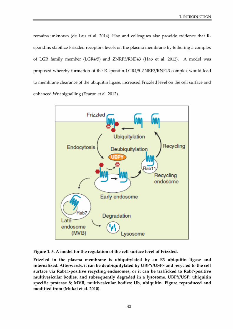
1.INTRODUCTION
42
remains unknown (de Lau et al. 2014). Hao and colleagues also provide evidence that R-
spondins stabilize Frizzled receptors levels on the plasma membrane by tethering a complex
of LGR family member (LGR4/5) and ZNRF3/RNF43 (Hao et al. 2012). A model was
proposed whereby formation of the R-spondin-LGR4/5-ZNRF3/RNF43 complex would lead
to membrane clearance of the ubiquitin ligase, increased Frizzled level on the cell surface and
enhanced Wnt signalling (Fearon et al. 2012).
Figure 1. 5. A model for the regulation of the cell surface level of Frizzled.
Frizzled in the plasma membrane is ubiquitylated by an E3 ubiquitin ligase and
internalized. Afterwards, it can be deubiquitylated by UBPY/USP8 and recycled to the cell
surface via Rab11-positive recycling endosomes, or it can be trafficked to Rab7-positive
multivesicular bodies, and subsequently degraded in a lysosome. UBPY/USP, ubiquitin
specific protease 8; MVB, multivesicular bodies; Ub, ubiquitin. Figure reproduced and
modified from (Mukai et al. 2010).
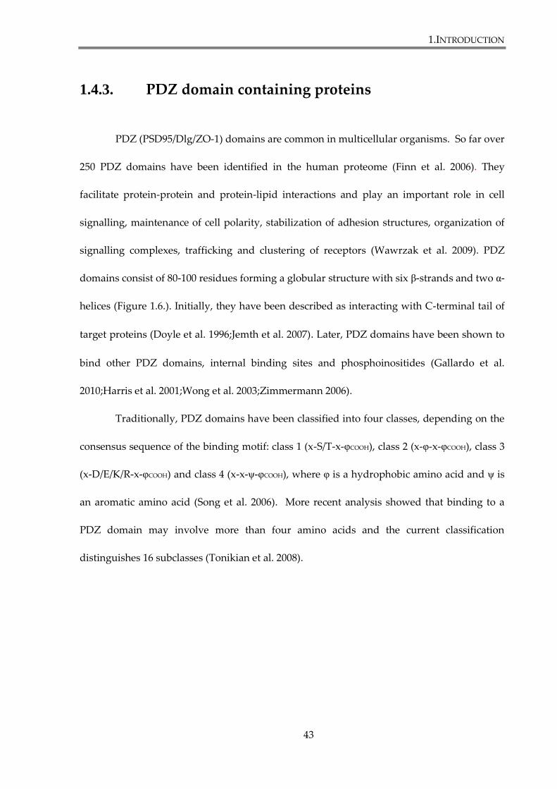
1.INTRODUCTION
43
1.4.3. PDZ domain containing proteins
PDZ (PSD95/Dlg/ZO-1) domains are common in multicellular organisms. So far over
250 PDZ domains have been identified in the human proteome (Finn et al. 2006). They
facilitate protein-protein and protein-lipid interactions and play an important role in cell
signalling, maintenance of cell polarity, stabilization of adhesion structures, organization of
signalling complexes, trafficking and clustering of receptors (Wawrzak et al. 2009). PDZ
domains consist of 80-100 residues forming a globular structure with six β-strands and two α-
helices (Figure 1.6.). Initially, they have been described as interacting with C-terminal tail of
target proteins (Doyle et al. 1996;Jemth et al. 2007). Later, PDZ domains have been shown to
bind other PDZ domains, internal binding sites and phosphoinositides (Gallardo et al.
2010;Harris et al. 2001;Wong et al. 2003;Zimmermann 2006).
Traditionally, PDZ domains have been classified into four classes, depending on the
consensus sequence of the binding motif: class 1 (x-S/T-x-φCOOH), class 2 (x-φ-x-φCOOH), class 3
(x-D/E/K/R-x-φCOOH) and class 4 (x-x-ψ-φCOOH), where φ is a hydrophobic amino acid and ψ is
an aromatic amino acid (Song et al. 2006). More recent analysis showed that binding to a
PDZ domain may involve more than four amino acids and the current classification
distinguishes 16 subclasses (Tonikian et al. 2008).
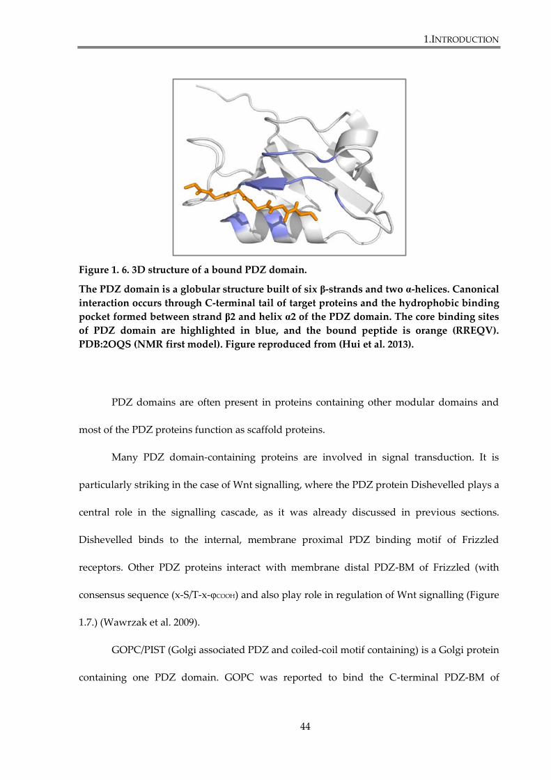
1.INTRODUCTION
44
Figure 1. 6. 3D structure of a bound PDZ domain.
The PDZ domain is a globular structure built of six β-strands and two α-helices. Canonical
interaction occurs through C-terminal tail of target proteins and the hydrophobic binding
pocket formed between strand β2 and helix α2 of the PDZ domain. The core binding sites
of PDZ domain are highlighted in blue, and the bound peptide is orange (RREQV).
PDB:2OQS (NMR first model). Figure reproduced from (Hui et al. 2013).
PDZ domains are often present in proteins containing other modular domains and
most of the PDZ proteins function as scaffold proteins.
Many PDZ domain-containing proteins are involved in signal transduction. It is
particularly striking in the case of Wnt signalling, where the PDZ protein Dishevelled plays a
central role in the signalling cascade, as it was already discussed in previous sections.
Dishevelled binds to the internal, membrane proximal PDZ binding motif of Frizzled
receptors. Other PDZ proteins interact with membrane distal PDZ-BM of Frizzled (with
consensus sequence (x-S/T-x-φCOOH) and also play role in regulation of Wnt signalling (Figure
1.7.) (Wawrzak et al. 2009).
GOPC/PIST (Golgi associated PDZ and coiled-coil motif containing) is a Golgi protein
containing one PDZ domain. GOPC was reported to bind the C-terminal PDZ-BM of
1.INTRODUCTION
45
mammalian Fz5 and Fz8. It was shown that GOPC trafficked together with Frizzled through
Golgi to the plasma membrane, which implies its role in vesicular transport of the receptors
(Yao et al. 2001). GOPC/PIST was implicated in regulation of trafficking of several other
transmembrane proteins. GOPC/PIST overexpression leads to different surface expression
patterns of β1-adrenergic receptor, the somatostatin receptor subtype 5 (SSTR5) and
Cadherin 23, by holding them back in the Golgi (He et al. 2004;Wente et al. 2005;Xu et al.
2010). It also regulates membrane expression of cystic fibrosis transmembrane conductance
regulator (CFTR), by facilitating its targeting to lysosomes (Cheng et al. 2005)
Another PDZ protein involved in Frizzled trafficking is dGRIP (glutamate receptor-
interacting protein). It has been shown to co-localize with DFrizzled-2 in trafficking vesicles
and to play role in transporting the receptor from neuromuscular junctions to the perinuclear
area in Drosophila (Ataman et al. 2006).
MAGI3 (Membrane-associated guanylate kinase, WW and PDZ domain-containing
protein 3) is a protein with six PDZ domains, usually enriched in cell junctions. The second
PDZ domain of the protein has been shown to bind Frizzled-4 and Frizzled-7. MAGI3 co-
localizes with Fz4 and Ltap protein, transmembrane protein supporting noncanonical Wnt
signalling, at cell contact sites in MDCK cells. Complexing of Fz4 with Ltap by MAGI3 has
been reported to enhance noncanonical Wnt/JNK pathway (Yao et al. 2004).
Pals-1 associated tight junction protein (dPatj), also known as InaD-like protein
(INADL), is a protein with four PDZ domains, involved in the establishment of cell polarity.
It forms a subapical complex together with Crumbs and Stardust/Pals-1 proteins. dPatj has
been shown to interact with dFz1 (mediating PCP signalling), but not with DFz2 (mediating
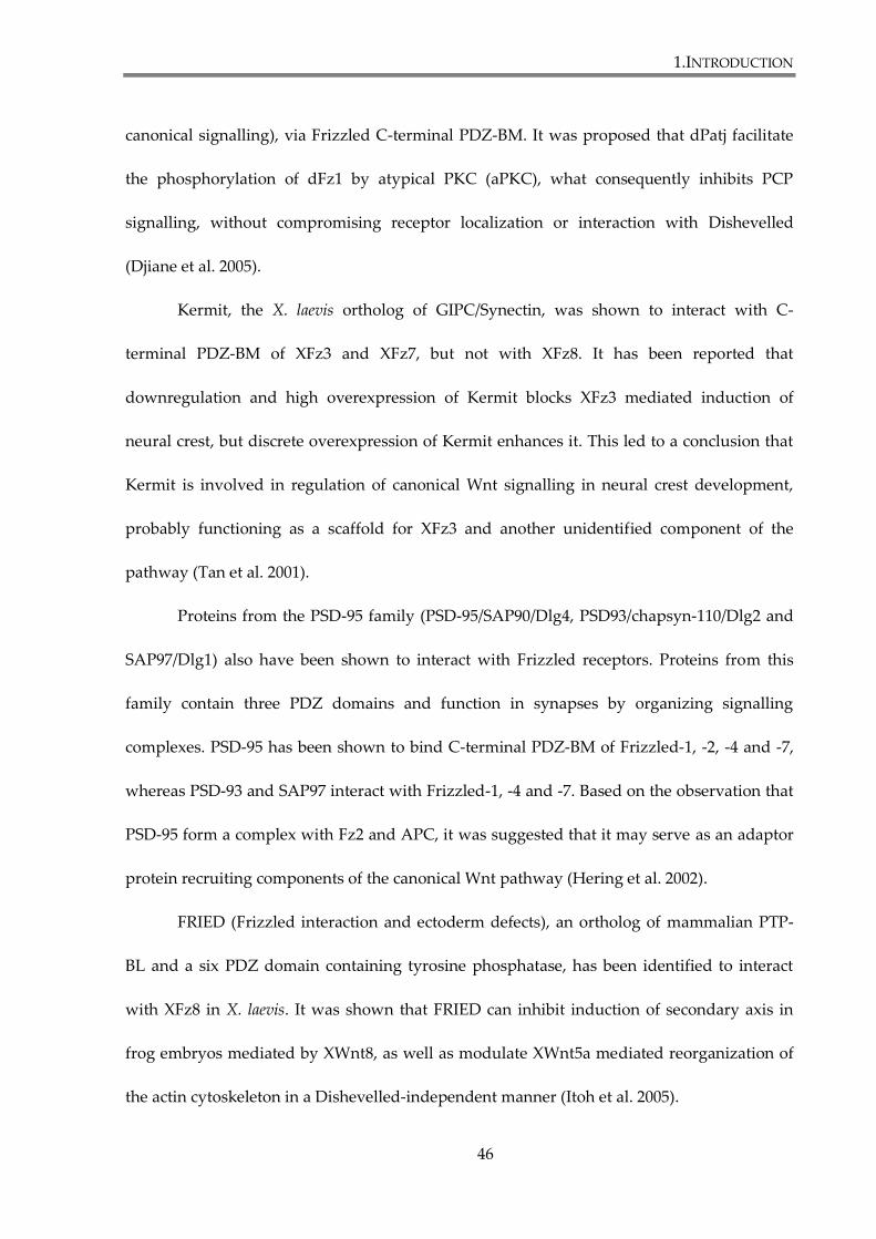
1.INTRODUCTION
46
canonical signalling), via Frizzled C-terminal PDZ-BM. It was proposed that dPatj facilitate
the phosphorylation of dFz1 by atypical PKC (aPKC), what consequently inhibits PCP
signalling, without compromising receptor localization or interaction with Dishevelled
(Djiane et al. 2005).
Kermit, the X. laevis ortholog of GIPC/Synectin, was shown to interact with C-
terminal PDZ-BM of XFz3 and XFz7, but not with XFz8. It has been reported that
downregulation and high overexpression of Kermit blocks XFz3 mediated induction of
neural crest, but discrete overexpression of Kermit enhances it. This led to a conclusion that
Kermit is involved in regulation of canonical Wnt signalling in neural crest development,
probably functioning as a scaffold for XFz3 and another unidentified component of the
pathway (Tan et al. 2001).
Proteins from the PSD-95 family (PSD-95/SAP90/Dlg4, PSD93/chapsyn-110/Dlg2 and
SAP97/Dlg1) also have been shown to interact with Frizzled receptors. Proteins from this
family contain three PDZ domains and function in synapses by organizing signalling
complexes. PSD-95 has been shown to bind C-terminal PDZ-BM of Frizzled-1, -2, -4 and -7,
whereas PSD-93 and SAP97 interact with Frizzled-1, -4 and -7. Based on the observation that
PSD-95 form a complex with Fz2 and APC, it was suggested that it may serve as an adaptor
protein recruiting components of the canonical Wnt pathway (Hering et al. 2002).
FRIED (Frizzled interaction and ectoderm defects), an ortholog of mammalian PTP-
BL and a six PDZ domain containing tyrosine phosphatase, has been identified to interact
with XFz8 in X. laevis. It was shown that FRIED can inhibit induction of secondary axis in
frog embryos mediated by XWnt8, as well as modulate XWnt5a mediated reorganization of
the actin cytoskeleton in a Dishevelled-independent manner (Itoh et al. 2005).
1.INTRODUCTION
47
Finally, NHERF1(Na+/H+ exchange factor 1), adaptor protein highly expressed in
normal mammary epithelium, was reported to regulate canonical Wnt signalling due to a
direct interaction between its second PDZ domain and C-terminal PDZ-BM of Frizzled
receptors (Fz4, Fz2 and Fz7). It was shown that Frizzled binding to NHERF1 modulates
Dishevelled recruitment and Wnt-induced β-catenin activation. Furthermore, the loss of
NHERF1 in breast cancer cell lines enhances Wnt signalling and cell proliferation, which
imply that NHERF1 is involved in breast cancer pathogenesis (Wheeler et al. 2011).
Syntenin-1 is another PDZ domain-containing adaptor protein implicated in the
regulation of Frizzled signalling, and it will be discussed separately in the section 1.6.3.3.
Figure 1. 7. Schematic domain structure of proteins directly interacting with Frizzled.
Figure reproduced from (Schulte et al. 2007).
1.INTRODUCTION
48
1.5. Syntenin-1
Syntenin-1 is a protein containing two PDZ domains, originally identified as a
syndecan binding protein (Grootjans et al. 1997) and as a melanoma differentiation gene
(mda-9), expression of which was induced by interferon γ (IFNγ) (Lin et al. 1998). It is widely
present in all adult tissues (Zimmermann et al. 2001) and especially robustly expressed in
developing skin, spinal cord, heart and lung during development of mouse embryo (Jeon et
al. 2013). Currently, many reports describe numerous interactions between syntenin-1 and
different proteins. It is suggested that syntenin-1 can serve as a scaffold for a number of
intracellular interactions.
1.5.1. Subcellular localization of syntenin-1
Analysis of the syntenin-1 distribution in epithelial cells revealed its localization at
cell-cell contact sites. Syntenin-1 co-localizes with F-actin, syndecan-1, E-cadherin, β-catenin
and α-catenin. In fibroblasts, it was shown that syntenin-1 localizes to focal adhesions and in
stress fibres (Zimmermann et al. 2001). Syntenin-1 also localizes in the early secretory
pathway, e.g. endoplasmic reticulum, intermediate compartment, cis-Golgi and apical
endosomes (Sarkar et al. 2008). Thus, it is implicated that syntenin-1 facilitates trafficking of
transmembrane proteins (Fernandez-Larrea et al. 1999). Furthermore, syntenin-1 interactions
with phosphatidylinositol 4,5-bisphosphate (PIP2) and phospholipase Cδ facilitates its
membrane anchorage (Zimmermann et al. 2002).
1.INTRODUCTION
49
1.5.2. Molecular structure and binding partners of
syntenin-1
Syntenin-1 is a 32 kDa protein. It contains N-terminal domain (NTD), followed by
two PDZ domains (PDZ1 and PDZ2) and C-terminal domain (CTD) (Figure 1.8).
NTD contains 113 amino acids and does not display any structural motifs. However,
it contains three conserved L-Y-P-x-L sequences, where X stands for any amino acid residue.
The first one has a conserved serine residue at the “X” position, and was shown to be
involved in ubiquitin binding to syntenin-1 (Rajesh et al. 2011). Recently, the L-Y-P-x-L
motifs were implicated in syntenin-1 binding to ALIX protein during syndecan-lead cargo
recruitment to exosomes and syntenin-1-syndecan-ALIX complex has been reported to
regulate the biogenesis of exosomes (Baietti et al. 2012). The NTD domain recruits the
transcription factor SOX4 and eukaryotic translation initiation factor 4A (EIF4A) into
signalling complexes (Beekman et al. 2008). It is also important for homo-dimerization and
hetero-dimerization with syntenin-2, protein homologous to syntenin-1 (Koroll et al. 2001). It
was shown that endogenous syntenin-1 is phosphorylated within its NTD domain. Five
conserved tyrosine’s were found (Y4, Y46, Y50, Y56 and Y91), which are potential
phosphorylation sites. Tyrosine phosphorylation within NTD might be an important factor
in the regulation of syntenin-1 functions (Beekman et al. 2008). Indeed, Wawrzyniak and
colleagues showed that phosphorylation of Y56 of syntenin-1 decreases membrane
localization of the protein (Wawrzyniak et al. 2012). It has been also shown that Src-
dependent Tyr4 phosphorylation of syntenin-1 leads to binding of the protein to myosin
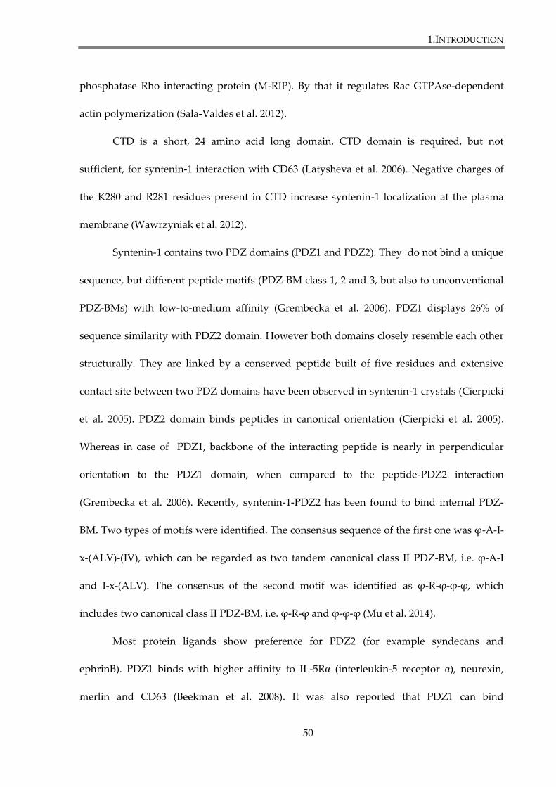
1.INTRODUCTION
50
phosphatase Rho interacting protein (M-RIP). By that it regulates Rac GTPAse-dependent
actin polymerization (Sala-Valdes et al. 2012).
CTD is a short, 24 amino acid long domain. CTD domain is required, but not
sufficient, for syntenin-1 interaction with CD63 (Latysheva et al. 2006). Negative charges of
the K280 and R281 residues present in CTD increase syntenin-1 localization at the plasma
membrane (Wawrzyniak et al. 2012).
Syntenin-1 contains two PDZ domains (PDZ1 and PDZ2). They do not bind a unique
sequence, but different peptide motifs (PDZ-BM class 1, 2 and 3, but also to unconventional
PDZ-BMs) with low-to-medium affinity (Grembecka et al. 2006). PDZ1 displays 26% of
sequence similarity with PDZ2 domain. However both domains closely resemble each other
structurally. They are linked by a conserved peptide built of five residues and extensive
contact site between two PDZ domains have been observed in syntenin-1 crystals (Cierpicki
et al. 2005). PDZ2 domain binds peptides in canonical orientation (Cierpicki et al. 2005).
Whereas in case of PDZ1, backbone of the interacting peptide is nearly in perpendicular
orientation to the PDZ1 domain, when compared to the peptide-PDZ2 interaction
(Grembecka et al. 2006). Recently, syntenin-1-PDZ2 has been found to bind internal PDZ-
BM. Two types of motifs were identified. The consensus sequence of the first one was φ-A-I-
x-(ALV)-(IV), which can be regarded as two tandem canonical class II PDZ-BM, i.e. φ-A-I
and I-x-(ALV). The consensus of the second motif was identified as φ-R-φ-φ-φ, which
includes two canonical class II PDZ-BM, i.e. φ-R-φ and φ-φ-φ (Mu et al. 2014).
Most protein ligands show preference for PDZ2 (for example syndecans and
ephrinB). PDZ1 binds with higher affinity to IL-5Rα (interleukin-5 receptor α), neurexin,
merlin and CD63 (Beekman et al. 2008). It was also reported that PDZ1 can bind
1.INTRODUCTION
51
phosphoinositol lipids in the plasma membrane (Zimmermann 2006). Even though PDZ
domains of syntenin-1 can interact separately with their ligands, most of the interactions
depend on cooperative ligand binding to the PDZ1-PDZ2 tandem (Grembecka et al.
2006;Grootjans et al. 2000). Furthermore, it is suggested that PDZ1 plays a chaperone-like
role in stabilizing of PDZ2 domain folding (Boukerche et al. 2010).
Figure 1. 8. The domain structure of syntenin-1
Syntenin-1 contains N-terminal domain (NTD), followed by two PDZ domains (PDZ1 and
PDZ2) and C-terminal domain (CTD). Five tyrosine residues within the NTD are
conserved, as indicated in the lower part of the figure. LY4PSL motif is implicated in the
ubiquitin binding. Figure is reproduced and modified from (Beekman et al. 2008).
PDZ1 PDZ2NTD CTD
High-affinity binding
Moderate affinity forprotein/PtdIns(4,5)P2
Moderate affinityfor protein
LY4PSL
1.INTRODUCTION
52
1.5.3. Functions of syntenin-1
1.5.3.1. Syntenin-1 and receptor trafficking
Syntenin-1 plays an important role in receptor trafficking. Endocytosis of receptors
leads to signal attenuation, as internalized receptors are targeted for degradation. However it
can also positively influence signalling output, as receptors at endocytic compartments can
interact with different downstream effectors, within the plasma membrane.
The first reported functional interaction of syntenin-1 was with syndecans, membrane
proteins with heparan sulphate (HS) side chains in the extracellular domain. HS chains allow
syndecans to act as co-receptors. They bind and concentrate growth factors at the cell surface
and facilitate their interactions with receptors. It was reported that syntenin-1 mutants,
defective in phosphoinositol lipid PtdIns(4,5)P2 binding, trap syndecans in perinuclear
recycling endosomes, characterized by the presence of Rab11 and Arf6, small GTPases
involved in endosome recycling (Zimmermann et al. 2005). Thus, the interaction with
syntenin-1 plays role in syndecans recycling to the plasma membrane. Furthermore, it has
been shown that membrane receptors associated with syndecans, such as FGF receptor, can
be recycled to the cell surface together with syndecans and syntenin-1 (Zimmermann et al.
2005). Recently, it has been demonstrated that ternary complex syndecan-syntenin-1-ALIX is
able to recruit syndecan associated proteins to the intraluminal vesicles of multivesicular
endosomes and exosomes (Baietti et al. 2012).
Pro-TGFα is membrane bound and functions as a ligand for EGFR (epithelial growth
factor receptor). It was reported that syntenin-1 binds to the C-terminus of the protein and
1.INTRODUCTION
53
both PDZ domains of syntenin-1 are required for optimal interaction. It was shown that
mutations of pro-TGFα, that disrupt its association with syntenin-1, result in retention of the
protein in the endoplasmic reticulum (Fernandez-Larrea et al. 1999).
It was suggested that syntenin-1 plays a role in maintaining Delta1 expression on the
cell surface. Delta1 is a ligand for Notch receptor. Estrach et al. ( 2007) reported that Delta1
interacts with syntenin-1 and its membrane expression is decreased by syntenin-1 depletion.
Delta1 contains PDZ-BM on its C-terminus and a mutation of C-terminal valine leads to
activation of Notch signalling (Estrach et al. 2007).
Syntenin-1 was also reported to regulate endocytosis of tetraspanin CD63.
Tetraspanins regulate maturation and processing of associated transmembrane proteins,
their activity and internalization. CD63 directly binds syntenin-1 and it has been proposed
that the interaction may have a negative effect on AP2-clathrin mediated endocytosis of
CD63, by inhibiting CD63-AP2 association (Latysheva et al. 2006).
1.5.3.2. Syntenin-1 and metastasis
Syntenin-1 was reported to be involved in cancer metastasis. Ability to metastasize is
the major characteristic of malignant tumours. Metastasis is a complex process that requires
attachment of tumour cell to matrix proteins, degradation of extracellular matrix (ECM) by
matrix metalloproteinases (MMP) and migration of cancer cells into surrounding stroma.
Overexpression of syntenin-1 was observed in a number of metastatic breast and gastric cell
lines. Breast cancer cell line MCF7 and gastric cancer cell line Az-521 showed increased
invasion rate and migration properties after transfection with syntenin-1. Moreover,
1.INTRODUCTION
54
syntenin-1 overexpression in MCF7 was associated with pseudopodia formation. Further
analysis showed that PDZ2 domain of syntenin-1 is responsible for the stimulatory effect of
the protein on cell migration (Koo et al. 2002). It was also observed that syntenin-1
expression increases during melanoma progression from primary to metastatic melanoma
(Helmke et al. 2004).
Syntenin-1 displays preferential link with fibronectin signalling. In melanoma cells,
after cell interaction with fibronectin, syntenin-1 facilitates phosphorylation of FAK (focal
adhesion kinase) what leads to activation of p38, JNK (c-jun NH2-terminal kinase) and NF-κB
(nuclear factor κB) activation. NF-κB was shown to induce expression of MMP-2 in
melanoma cells (Boukerche et al. 2005;Boukerche et al. 2007). This pathway can be inhibited
by RKIP (Raf kinase inhibitor protein), which prevents syntenin-1 binding to FAK and c-Src
kinases (Das et al. 2012). Nonetheless, syntenin-1-induced expression of MMP-2 or MMP-9
was not observed in breast cancer cell lines (Koo et al. 2002). Recently, it was reported that
syntenin-1 interaction with c-Src also positively regulates NF-κB activation (Boukerche et al.
2010). Furthermore, activation of c-Src mediated by syntenin-1 induces activation of EGFR
(epidermal growth factor receptor) in uroepithelial cells, and in consequence increases their
proliferation and invasion properties (Dasgupta et al. 2013)
1.INTRODUCTION
55
1.5.3.3. Syntenin-1 and Frizzled receptors
Syntenin-1 was identified as an intracellular ligand for Frizzled receptors. It was
shown that it interacts with the C-terminal PDZ-BM of human Frizzled-3,-7 and -8, but not
with Frizzled-2, -5 and -6. Luyten et al. reported that association with Frizzled-7 requires
PDZ1 domain of syntenin-1, whereas Frizzled-3 and Frizzled-8 prefer the second PDZ
domain. However, the tandem of syntenin-1’s PDZ domains mediates higher affinity
binding than the isolated domains. Syntenin-1 was reported to stimulate noncanonical
Wnt/JNK pathway. Further investigation revealed that in Xenopus leavis Xsyntenin interacts
with XFrizzled-7. Overexpression of Xsyntenin blocks convergent extension in animal cap
assay, while its downregulation affects gastrulation movements and shortens body axis of
embryos, process controlled by noncanonical Wnt signalling. It was also established that
syntenin-1 stimulates Frizzled-7 dependent activation of the PKCα/CDC42 signalling (Luyten
et al. 2008). Syndecans, syntenin-1 binding partners, are Wnt co-receptors and were shown to
stimulate PKCα and CDC42 activities. Xsyndecan-4 functionally and physically interacts
with XFrizzled-7 and XDvl, and a model was proposed where syntenin-1 acts as an
intracellular scaffold protein between Frizzled and syndecans. This function of syntenin-1
would support noncanonical Wnt signalling. In addition, as syntenin-1 was reported to
interact with PIP2 and that this association stimulates syndecan and syndecan cargo
recycling, it is possible that syntenin-1 plays role in joined recycling of Frizzled and
syndecans (Wawrzak et al. 2009).
1.INTRODUCTION
56
1.5.3.4. Other functions of syntenin-1
Syntenin-1 plays several other important roles in cells. It interacts with IL-5Rα
(interleukin-5 receptor α) and the transcription factor Sox4, thus mediates IL-5 dependent
activation of Sox4. IL-5 and SOX4 are implicated in B-cell development and differentiation
and syntenin-1 may also be involved in these processes (Geijsen et al. 2001).
Syntenin-1 interacts with ephrinBs and ephrin receptors (Eph), thus may regulate
ephrin-Eph signalling. It has been shown that syntenin-1 interaction with ephrinB ligands is
important for the pre-synaptic machinery recruitment during development of presynaptic
and postsynaptic compartments in neurons (McClelland et al. 2009). Syntenin also functions
in regulation of axon outgrowth. It interacts with Unc51.1, which is a serine/threonine kinase
important for neurite extension, and Rab5, a marker of early endosomes. This interaction
creates a scaffold for Unc51.1 and endocytic machinery, what is important for the regulation
of axon extension (Tomoda et al. 2004).
Recently syntenin-1 was implicated in ubiquitin-dependent sorting of transmembrane
proteins and facilitating recruitment of ubiquitylated proteins to transmembrane partners.
Syntenin-1 was shown to bind ubiquitin via unconventional pocket in its N-terminal domain,
in a process regulated by Ulk1, a serine/threonine kinase, known to control different
trafficking pathways in cells (Rajesh et al. 2011).
1.INTRODUCTION
57
1.6. LNX family of ubiquitin ligases
1.6.1. RING-type E3 ligases
E3 ubiquitin ligases catalyse the transfer of ubiquitin from an E2 enzyme to the target
protein and also ensure the specificity of ubiquitin modification. E3 ubiquitin ligases
function by one of the two mechanisms. HECT (Homologous to E6-AP Carboxy Terminus)-
type E3s serve as catalytic intermediates in ubiquitination. Ubiquitin is transferred from E2
enzyme to the catalytic cysteine of HECT domain, and later to a lysine in the substrate.
Alternatively, really interesting new gene (RING)-type E3s mediate the transfer of ubiquitin
directly from E2~ubiquitin to the target protein. (Budhidarmo et al. 2012;Metzger et al. 2014).
LNX proteins belong to the RING-type family of E3 ubiquitin ligases, defined by the
presence of RING domain. RING type ubiquitin ligases are conserved from yeast to humans,
and over 400 different ligases were identified in mammalian cells. RING domain binds Zn2+
ion through a cross-braced arrangement of cysteine and histidine amino acids with a
characteristic spacing. The consensus sequence of this domain is C-X2-C-X(9-39)-C-X(1-3)-H-X(2-3)-
C-X2-C-X(4-48)-C-X2-C (where X means any amino acid). Ligases use RING domain to recruit
and activate an E2~ubiquitin enzyme, which in turn transfers ubiquitin to the substrate
(Pickart 2001;Satija et al. 2013).
RING-type E3s were described to function as both tumour suppressors and
oncogenes. At the cellular level they play critical roles in endocytic trafficking, DNA repair
and activation of NF-κB signalling (Metzger et al. 2014). The interaction between E3 ligase
and substrate is often mediated via protein-binding domains on the E3, such as SH2 (Src
1.INTRODUCTION
58
homology 2), SH3 (Src homology 3), FHA (forkhead associated domain), ankyrin repeats or
PDZ domains. Indeed, bioinformatics analysis revealed that 75% of human RING-type E3
ligases contain at least one other domain or binding motif (Deshaies et al. 2009)
Mutations or impaired modulation of RING-type E3s activity are often linked with
human disease. Mutations of BRCA1, an E3 ubiquitin ligase involved in DNA repair, are
associated with familial breast and ovarian cancers (Welcsh et al. 2001). FANC ubiquitin
ligase, which also plays role in DNA repair, is mutated in the multisystemic Fanconi anemia
syndrome, cause of severe developmental defects (Moldovan et al. 2009). Another RING-
type E3, FBXO11 is often mutated or deleted in diffuse large B-cell lymphomas (Duan et al.
2012). The malignant von Hippel–Lindau syndrome is associated with mutations in VHL
protein, the component of the CRL2VHL E3 (Lipkowitz et al. 2011). Mutations in the RING–
IBR (in between RING)–RING (RBR) E3 ubiquitin ligase Parkin were reported in autosomal
recessive juvenile Parkinsonism (AR-JP) (Kitada et al. 1998). Finally, increased expression of
Mdm2 (double minute 2 protein) E3 ligase or loss of its negative modulator, leads to the
upregulation of Mdm2 activity towards the tumour suppressor p53, and is associated with
human cancers, especially those with wild type p53 (Lipkowitz et al. 2011;Metzger et al.
2014).
1.INTRODUCTION
59
1.6.2. Ligand of Numb Protein-X (LNX) family
LNX (Ligand of Numb Protein-X) family of proteins, also known as PDZRN (PDZ
and RING) family, consists of five members (LNX1 – LNX5) as presented in the Figure 1.9.
Proteins belonging to this group are characterized by the presence of RING domain followed
by one to four PDZ domains. Within the family, LNX1 and LNX2 share identical domain
structure, consisting of one RING domain and four PDZ domains. There is a high degree of
similarity between LNX3 and LNX4 (PDZRN3 and PDZRN4), and they are more distantly
related to LNX1 and LNX2. They both contain one RING domain and two PDZ domains.
LNX5 (PDZK4, PDZRN4L) lacks RING domain, but due to its high sequence homology to
LNX3 and LNX4, it was also included in the family. The LNX proteins are unique in
vertebrates as they are the only proteins characterized by the presence of both RING and
PDZ domains. Combination of these two types of domains suggests that PDZ domains might
be involved in substrate recognition and targeting the ubiquitin ligase activity of the proteins
(Flynn et al. 2011).
LNX1 was initially discovered as a protein interacting with Numb, a component of
Notch signalling (Dho et al. 1998). Subsequently, LNX2 was also shown to interact with
Numb and its paralog Numb-like (Rice et al. 2001). Apart from Numb, several other binding
partners of LNX1 and LNX2 were discovered, and they will be further discussed in following
sections.
LNX3 was identified as a binding partner of Semaphorin (Sema4C) in yeast two-
hybrid screen (Wang et al. 1999). Its high expression was detected in heart and muscle. LNX3
was shown to be involved in differentiation of C2C12 cells into myotubes (Ko et al. 2006) and
1.INTRODUCTION
60
into osteoblasts (Honda et al. 2010). It served as a negative regulator of osteoblast
differentiation through inhibition of canonical Wnt signalling. Furthermore, LNX3 was
found to regulate formation of neuromuscular junction, as it interacts and ubiquitylates the
muscle specific tyrosine kinase (MuSK), a key regulator of postsynaptic assembly. LNX3
promotes MuSK endocytosis thus negatively regulates its cell surface expression (Lu et al.
2007).
LNX4 and LNX5 were identified in silico based on homology to LNX3, and they
remain virtually uncharacterized. LNX4 was found to be down regulated in the
hypothalamus of Sim1 (single-minded homolog 1) knockout mice. Whereas LNX5 was
identified as up-regulated in synovial sarcomas. Their physiological functions remain
unknown (Flynn et al. 2011).
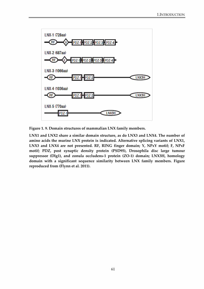
1.INTRODUCTION
61
Figure 1. 9. Domain structures of mammalian LNX family members.
LNX1 and LNX2 share a similar domain structure, as do LNX3 and LNX4. The number of
amino acids the murine LNX protein is indicated. Alternative splicing variants of LNX1,
LNX3 and LNX4 are not presented. RF, RING finger domain; Y, NPxY motif; F, NPxF
motif; PDZ, post synaptic density protein (PSD95), Drosophila disc large tumour
suppressor (Dlg1), and zonula occludens-1 protein (ZO-1) domain; LNX3H, homology
domain with a significant sequence similarity between LNX family members. Figure
reproduced from (Flynn et al. 2011).
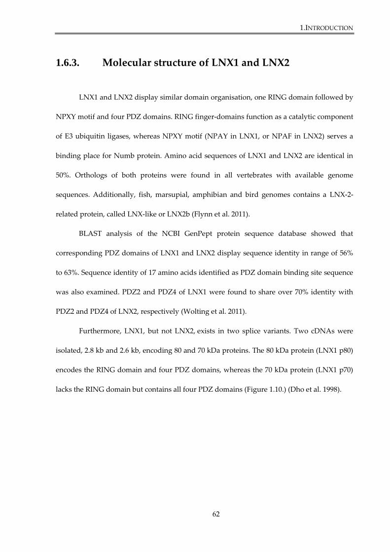
1.INTRODUCTION
62
1.6.3. Molecular structure of LNX1 and LNX2
LNX1 and LNX2 display similar domain organisation, one RING domain followed by
NPXY motif and four PDZ domains. RING finger-domains function as a catalytic component
of E3 ubiquitin ligases, whereas NPXY motif (NPAY in LNX1, or NPAF in LNX2) serves a
binding place for Numb protein. Amino acid sequences of LNX1 and LNX2 are identical in
50%. Orthologs of both proteins were found in all vertebrates with available genome
sequences. Additionally, fish, marsupial, amphibian and bird genomes contains a LNX-2-
related protein, called LNX-like or LNX2b (Flynn et al. 2011).
BLAST analysis of the NCBI GenPept protein sequence database showed that
corresponding PDZ domains of LNX1 and LNX2 display sequence identity in range of 56%
to 63%. Sequence identity of 17 amino acids identified as PDZ domain binding site sequence
was also examined. PDZ2 and PDZ4 of LNX1 were found to share over 70% identity with
PDZ2 and PDZ4 of LNX2, respectively (Wolting et al. 2011).
Furthermore, LNX1, but not LNX2, exists in two splice variants. Two cDNAs were
isolated, 2.8 kb and 2.6 kb, encoding 80 and 70 kDa proteins. The 80 kDa protein (LNX1 p80)
encodes the RING domain and four PDZ domains, whereas the 70 kDa protein (LNX1 p70)
lacks the RING domain but contains all four PDZ domains (Figure 1.10.) (Dho et al. 1998).
1.INTRODUCTION
63
Figure 1. 10. Ligand of Numb Protein X 1 and 2.
LNX1 and LNX2 share a similar domain organisation: one RING domain followed by
NPXY motif and four PDZ domains. RING finger-domains function as a catalytic
component of E3 ubiquitin ligases, whereas NPXY motif (NPAY in LNX1, or NPAF in
LNX2) serves a binding place for Numb protein. LNX1 exists in two splice variants, p80
and p70. Figure based on (Wolting et al. 2011).
RING PDZ4PDZ3PDZ2PDZ1 TFLLNX1 p80
RING PDZ4PDZ3PDZ2PDZ1 SLVLNX2
63% 62% 56% 61%
PDZ4PDZ3PDZ2PDZ1 TFLLNX1 p70 NPAY
NPAF
NPAY
1.INTRODUCTION
64
1.6.4. Binding partners of LNX1 and LNX2
LNX1 interacts with a number of proteins, predominantly those with a PDZ binding
motif. Predicted functional partners of human LNX1, retrieved from the STRING database
(www.string-db.org), are presented in the Figure 1.11. A total of 166 binding partners of
LNX1 have been discovered in different yeast two-hybrids and proteomics studies (Guo et
al. 2012;Rice et al. 2001;Rual et al. 2005;Wolting et al. 2011). LNX1 was initially identified to
interact with the cell fate determinant NUMB in a yeast two-hybrid screen (Dho et al. 1998),
and subsequently it was shown to interact with all four isoforms of human NUMB as the
NUMB PTB domain binds the NPAY motif in LNX1 (Dho et al. 1999). In addition, the first
PDZ domain of LNX1 binds to the PTB domain of NUMB (Nie et al. 2004).
LNX1 interacts with RhoC, a small GTPase playing role in reorganization of the actin
cytoskeleton. Efficient binding of RhoC to LNX1 requires the first PDZ domain of LNX1
(Zheng et al. 2010). KCNA4, a shaker-type voltage-gated K+ channel and PLEKHG5
(pleckstrin homology domain-containing family G member 5) are also recognized by the
PDZ1 of LNX1 via their carboxy terminal tails (Wolting et al. 2011). PLEKH5 can also bind to
PDZ3 of LNX1. Similarly to PLEKH5, LNX1-PDZ1 and PDZ3 can both recognize the carboxy
termini of the proto-oncogene c-Src, a cytoplasmic protein kinase (Weiss et al. 2007). PDZ-
binding kinase (PBK) also binds to LNX1-PDZ1 (Guo et al. 2012).
LNX1-PDZ2 is involved in binding to the pre-synaptic active zone protein CAST
(Higa et al. 2007), junctional adhesion molecule 4 (JAM4) (Kansaku et al. 2006) and to
Coxsackie and adenovirus receptor (CAR) (Sollerbrant et al. 2003) via their C-terminus.
However efficient binding to the latter requires not only C-terminal PDZ binding motif of the
1.INTRODUCTION
65
receptor, but also upstream sequences. Protein kinases Tyk2, PAK6 and PKC-α1 also interact
with the second PDZ domain of LNX1. Additionally PAK6 and PKC-α1 also bind to LNX1-
PDZ4. Tyk2 is a member of Janus kinase (JAK) family of cytoplasmic protein tyrosine
kinases, whereas PAK6 and PKC-α1 are serine/threonine kinases involved in signal
transduction, cell proliferation and apoptosis (Wolting et al. 2011). LNX1-PDZ4 was also
shown to bind numerous PIPs (phosphatidylinositol phosphates). PDZ4 is the only PDZ
domain of LNX1 found to interact with membrane phospholipids and it may regulate LNX1
subcellular localization (Wolting et al. 2011).
Several other ligands have been identified for LNX1, including: CD8α, the T-cell co-
receptor (D'Agostino et al. 2011); Claudin-1, a tight junction protein (Takahashi et al. 2009);
ErbB2, a member of the epidermal growth factor receptor family (Young et al. 2005); SKIP,
the transcription and splicing factor Ski-interacting protein (Chen et al. 2005) and the human
endogenous retrovirus protein NP9 (Armbruester et al. 2004). Additionally, MAGE-B18, a
melanoma antigen family member has been shown to bind to a basic region between the
RING and first PDZ domain of LNX1 via the MAGE homology domain (MHD) (Doyle et al.
2010).
LNX2 has been shown so far to interact only with Numb protein (Nie et al. 2002),
CAR receptor (Sollerbrant et al. 2003) and with CD8α (D'Agostino et al. 2011).
LNX1 and LNX2 contain consensus type I PDZ domain-binding motifs (T-x-L in
LNX1 and S-x-V in LNX2) at their C-termini that can also bind PDZ domains. Indeed, in
yeast two-hybrid assays, LNX1 has been shown to homo-oligomerize and hetero-oligomerize
with LNX2, indicating the possibility that LNX proteins may be able to form large oligomeric
networks in the cell (Rice et al. 2001). In the latest proteomics study Guo and colleagues
1.INTRODUCTION
66
discovered that PDZ domains of LNX can interact with each other and form dimers (Guo et
al. 2012). They have found three pairs of PDZ-PDZ interactions: LNX1-PDZ1 and LNX1-
PDZ4, LNX1-PDZ1 and LNX2-PDZ4, and LNX2-PDZ1 ad LNX2-PDZ4. Using LNX1-PDZ1
and LNX2-PDZ1 as baits in Y2H, they have also discovered that neither of these domains
interact with their own C-termini. However, the interaction of LNX1-PDZ1 with C-terminus
binding motif of LNX2 was detected. Recent study by Mu and colleagues revealed that the
first PDZ domain of LNX2 (LNX2-PDZ1) is able to bind internal PDZ-BM. 17 unique internal
binding motifs were identified, with consensus sequence φ-Tx-φ-Tx- φ, which can be
regarded as two canonical class II C-terminal PDZ-BM in tandem (Mu et al. 2014).

1.INTRODUCTION
67
Figure 1. 11. Functional partners of LNX1.
Network of binding partners of LNX1 based on experimental evidence, retrieved from the
STRING database (www.string-db.org). LNX1, ligand of numb protein X1; LNX2, ligand
of numb protein X2; NUMB, numb homolog; MAGEB18, melanoma antigen family B 18;
IGSF5, immunoglobulin super family member 5; SRC, c-Src sarcoma viral oncogene
homolog; RHOC, Ras homolog gene family member C; CXADR, coxsackie virus and
adenovirus receptor; CD8A, CD8α molecule; INPP5K, inositol polyphospate-5-
phosphatase K.
1.INTRODUCTION
68
1.6.5. Functions of LNX1 and LNX2
The functional significance of many interactions presented in the previous section
remains unclear. Nevertheless LNX1 and LNX2 have been shown to function as both E3
ubiquitin ligases and molecular scaffolds in regulation of the activity, stability, or subcellular
localization of their interacting partners. LNX proteins are involved in several different
biological process, such as Notch signalling, tight junction reorganization and Wnt
signalling.
1.6.5.1. Notch signalling
The E3 ligase activity of LNX1 p80 and LNX2 is possible due to the RING domain,
which has been shown to have the ubiquitin ligase activity in vitro and in vivo. The first
protein identified as a target of LNX1 ubiquitylation was Numb protein, a component of
Notch signalling pathway. Numb is an endocytic adapter, shown to control cell fate during
development by suppressing Notch receptor. LNX1 can ubiquitilate two isoforms of Numb
(p66 and p72), containing the eleven amino acid insert in the PTB domain, and target them
for proteasomal degradation. LNX1 functions as a positive regulator of Notch signalling by
promoting Numb degradation and reducing its ability to antagonize Notch (Nie et al.
2002;Nie et al. 2004). Homologous LNX2 was also shown to interact with Numb and is also
likely to ubiquitilate the protein and thereby modulate Notch signalling (Nie et al. 2002;Rice
et al. 2001). Finally, LNX2 siRNA-mediated silencing leads to downregulation of NOTCH1
expression. Also, following LOF (loss-of-function mutation) of LNX2, the protein level of
1.INTRODUCTION
69
Numb and expression of JAGGED1, a ligand for the receptor Notch1, were significantly
reduced in colorectal cell line SW480 (Camps et al. 2013).
1.6.5.2. Tight junction reorganization
Both LNX1 and LNX2 play a crucial role in tight junction (TJ) reorganization, as they
have been shown to interact with multiple TJ components, namely CAR, JAM4 and claudins.
LNX1 and LNX2 bind to the Coxsackie and adenovirus receptor (CAR) and might function
together at intercellular junctions (Sollerbrant et al. 2003). LNX1 was also reported to interact
with junctional adhesion molecule 4 (JAM4), an immunoglobulin superfamily member
present at TJs. LNX1 is involved in JAM4 endocytosis and redistribution after transforming
growth factor (TGF) β induction. Numb protein was found to be involved in this process and
to form a tripartite complex with JAM4 and LNX1, important for the LNX1-mediated
endocytosis of JAM4. This suggests that LNX1 provides an endocytic scaffold for JAM4
(Kansaku et al. 2006). Finally, claudin-1, claudin-2 and claudin-4 had been reported to be
polyubiquitylated by LNX1, followed by their internalization and degradation in lysosomes.
Overexpression of LNX1 in MDCK (Madin-Darby canine kidney) cells led to downregulation
of claudins from TJs and reduced the number of tight junctions (Takahashi et al. 2009).
Recently, LNX1 has been shown to interact also with claudin-17 (Guo et al. 2012).
1.INTRODUCTION
70
1.6.5.3. Wnt signalling
Recently, several reports have implicated the role of LNX family members in Wnt
signalling. Wnt signalling regulators Naked2 (Nkd2) and Dishevelled-3 (Dvl3) have been
identified to interact with LNX1 PDZ domains (Wolting et al. 2011). Additionally, LNX3 was
found in a high-throughput siRNA screen designed to identify novel Wnt regulators, as
required for Wnt signal transduction (Major et al. 2008). LNX3 has been shown to be an
antagonist of Wnt signalling transmission in osteoblast precursor cells (Honda et al. 2010)
and also to ubiquitylated Dishevelled-3 to promote PCP signalling in endothelial cells
(Sewduth et al. 2014). Furthermore, LNX2b (an LNX zebrafish ortholog) has been implicated
in canonical Wnt signalling in the developing zebrafish embryo. LNX2b was shown to bind
and ubiquitylate the Wnt inhibitor Bozozok, followed by its degradation. Also, altered
dorso-ventral patterning in the LNX2b knockdown embryos coincided with increased
stabilization of Bozozok. As a consequence LNX2b-mediated degradation of Bozozok, the β-
catenin Wnt signalling was upregulated (Ro et al. 2009). Finally, it was reported that siRNA-
mediated silencing of LNX2 downregulated the transcription factor TCF7L2 and its targets,
and reduced Wnt signalling in colorectal cancer cells. TCF7L2 is an important effector of the
Wnt pathway and is responsible for transcriptional regulation of genes involved in Wnt
signalling (Camps et al. 2013).
1.INTRODUCTION
71
1.6.5.4. Other functions
LNX1 p80 and LNX2 are also involved in ubiquitylation, internalization and
degradation in lysosomes of CD8α, a T-cell co-receptor (D'Agostino et al. 2011). Authors also
suggested that LNX2 might be involved in the regulation of CD8α transport to, through, or
from Golgi complex.
LNX1 also ubiquitilates the non-receptor tyrosine kinase c-Src. In addition to acting as
a substrate for LNX1 ubiquitylation, c-Src was shown to phosphorylate LNX1. That suggests
that c-Src may have a role in regulation of LNX1 activity (Weiss et al. 2007).
Guo and colleagues reported that LNX1 mediates ubiquitylation and degradation of
PBK, what leads to inhibition of cell proliferation and enhances cell sensitivity to
doxorubicin-induced apoptosis. It indicates the role of LNX1 in regulation of cell growth and
apoptosis, thus it may act as a tumour suppressor (Guo et al. 2012). Furthermore, a separate
genomic study demonstrated that LNX1 knockdown leads to cell cycle arrest in the G0/G1
phase (Zheng et al. 2011).
1.INTRODUCTION
72
1.6.6. Major sites of expressions of LNX1 and LNX2
Murine LNX1 mRNA is widely expressed. It was found in brain, lung, heart, kidney
and skeletal muscle. LNX1 p80 was specifically reported in heart, skeletal muscle and lunges,
whereas LNX1 p70 expression was restricted to brain and heart (Dho et al. 1998). Human
LNX1 mRNA is similarly expressed in brain, heart, kidney, skeletal muscle and lunges as
well as placenta and pancreas (Xie et al. 2001). Murine LNX2 mRNA was detected in liver,
spleen, lung, heart, kidney, thymus, skeletal muscle and brain. Interestingly, expression
of LNX1 and LNX2 overlaps in some brain regions and differs in others (Rice et al. 2001).
1.6.7. Subcellular localization of LNX1 and LNX2
Several studies have examined the subcellular localization of LNX1 and LNX2 in
different cell lines by fluorescence microscopy and reported its diffuse distribution
throughout the cytoplasm and nucleus. In HEK293 cells LNX1 colocalizes with CAR at cell-
cell contacts (Sollerbrant et al. 2003). In HeLa cells, LNX1 was shown to localize in the
plasma membrane proximity, as well as diffuse in the cytoplasm and nucleus. It also
localizes to the microtubule-like structures (Weiss et al. 2007). Sometimes LNX1 distribution
is altered by co-expression with its interacting partners. Interaction with NP9 leads to LNX1
recruitment to punctuate nuclear structures (Armbruester et al. 2004). Additionally, LNX1
and LNX2 were recruited from the cytoplasm to the plasma membrane by the human
glycoprotein CD8α (D'Agostino et al. 2011). On the other hand, RhoC was redistributed from
the cytoplasm to the nucleus by LNX1 (Zheng et al. 2010). In MDCK cells, endogenous LNX1
1.INTRODUCTION
73
co-localizes with JAM4 to TJs along with JAM4 (Kansaku et al. 2006). However
overexpressed LNX1 localizes to the cytoplasm and to the apical junctional complex, where it
colocalizes with ZO-1 at TJs. Furthermore, LNX1 was reported to colocalize with claudin-2 in
late endosomes and lysosomes of MDCK cells (Takahashi et al. 2009)
1.6.8. LNX1 and LNX2 in diseases
LNX1 has been implicated in brain tumourigenesis. LNX1 mutations were found in a
number of human gliomas (Blom et al. 2008) and the protein was reported to be
downregulated in all gliomas (Chen et al. 2005). Susceptibility to Kawasaki disease, an
infectious inflammatory vasculitis has also been linked to alterations in LNX1 expression
(Burgner et al. 2009). Additionally, interactions of LNX1 with proto-oncogene NP9 and c-Src
indicate its role in tumourigenesis (Armbruester et al. 2004;Weiss et al. 2007). High levels of
LNX1 and LNX2 mRNAs were found in endocarditis, the principal manifestation of chronic
Q fever, but not in acute Q fever (Mehraj et al. 2012). Finally, LNX2 overexpression was
linked with chromosome 13 amplification and activation of the Wnt and Notch signalling
pathways in colorectal cancer (Camps et al. 2013).
1.INTRODUCTION
74
1.7. Research objectives
Frizzled-7, a member of Frizzled family of Wnt receptors, emerges as a key
component involved in activation of Wnt signalling in breast cancer cells. It is a
transmembrane receptor containing two PDZ binding motifs (PDZ-BM) in its cytoplasmic
tail. The membrane proximal PDZ-BM is a binding site for Dishevelled, a crucial component
of Wnt signalling pathways. Whereas the contribution of the C-terminal PDZ-BM of Frizzled
receptors to Wnt signalling remains poorly studied. PDZ domains are very common in
multicellular organisms and present in structures of many scaffolding proteins. They
facilitate protein interactions and are implicated in regulation of cell signalling, maintenance
of cell polarity, as well as in modulation of trafficking and clustering of membrane receptors.
We hypothesized that PDZ proteins interacting with the C-terminal PDZ-BM of Frizzled
receptors are involved in these processes. In this study we focused on Frizzled-7 and the role
of the C-terminal PDZ-BM in the receptor functionality. Specific aims of this study were as
follow:
To determine the importance of Frizzled-7 interactions via the C-terminal PDZ-
BM for transduction of canonical Wnt signalling
To determine the role of syntenin-1, an established Frizzled-7 PDZ partner in
canonical Wnt signalling
To identify novel Frizzled-7 partners interacting with the C-terminal PDZ-BM of
the receptor and examining their role in canonical Wnt signalling
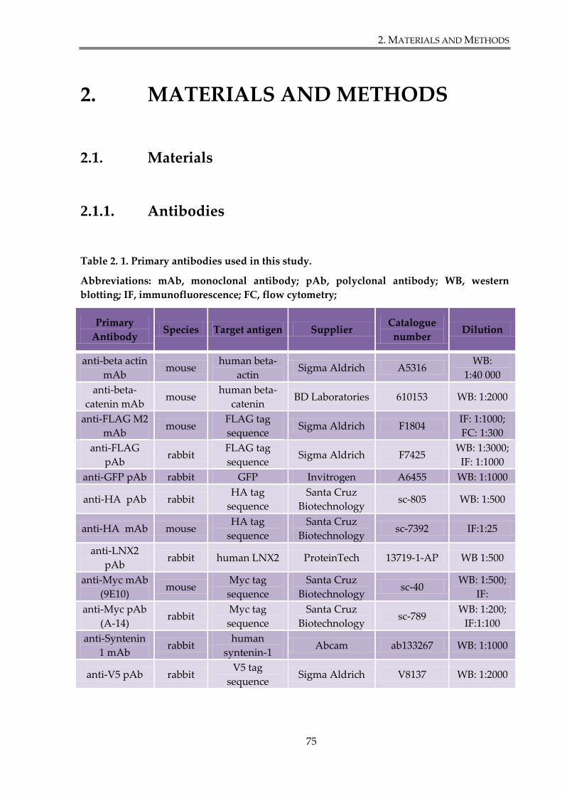
2. MATERIALS AND METHODS
75
2. MATERIALS AND METHODS
2.1. Materials
2.1.1. Antibodies
Table 2. 1. Primary antibodies used in this study.
Abbreviations: mAb, monoclonal antibody; pAb, polyclonal antibody; WB, western
blotting; IF, immunofluorescence; FC, flow cytometry;
Primary
Antibody Species Target antigen Supplier
Catalogue
number Dilution
anti-beta actin
mAb mouse
human beta-
actin Sigma Aldrich A5316
WB:
1:40 000
anti-beta-
catenin mAb mouse
human beta-
catenin BD Laboratories 610153 WB: 1:2000
anti-FLAG M2
mAb mouse
FLAG tag
sequence Sigma Aldrich F1804
IF: 1:1000;
FC: 1:300
anti-FLAG
pAb rabbit
FLAG tag
sequence Sigma Aldrich F7425
WB: 1:3000;
IF: 1:1000
anti-GFP pAb rabbit GFP Invitrogen A6455 WB: 1:1000
anti-HA pAb rabbit HA tag
sequence
Santa Cruz
Biotechnology sc-805 WB: 1:500
anti-HA mAb mouse HA tag
sequence
Santa Cruz
Biotechnology sc-7392 IF:1:25
anti-LNX2
pAb rabbit human LNX2 ProteinTech 13719-1-AP WB 1:500
anti-Myc mAb
(9E10) mouse
Myc tag
sequence
Santa Cruz
Biotechnology sc-40
WB: 1:500;
IF:
anti-Myc pAb
(A-14) rabbit
Myc tag
sequence
Santa Cruz
Biotechnology sc-789
WB: 1:200;
IF:1:100
anti-Syntenin
1 mAb rabbit
human
syntenin-1 Abcam ab133267 WB: 1:1000
anti-V5 pAb rabbit V5 tag
sequence Sigma Aldrich V8137 WB: 1:2000
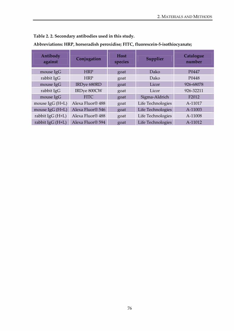
2. MATERIALS AND METHODS
76
Table 2. 2. Secondary antibodies used in this study.
Abbreviations: HRP, horseradish peroxidise; FITC, fluorescein-5-isothiocyanate;
Antibody
against Conjugation
Host
species Supplier
Catalogue
number
mouse IgG HRP goat Dako P0447
rabbit IgG HRP goat Dako P0448
mouse IgG IRDye 680RD goat Licor 926-68078
rabbit IgG IRDye 800CW goat Licor 926-32211
mouse IgG FITC goat Sigma-Aldrich F2012
mouse IgG (H+L) Alexa Fluor® 488 goat Life Technologies A-11017
mouse IgG (H+L) Alexa Fluor® 546 goat Life Technologies A-11003
rabbit IgG (H+L) Alexa Fluor® 488 goat Life Technologies A-11008
rabbit IgG (H+L) Alexa Fluor® 594 goat Life Technologies A-11012
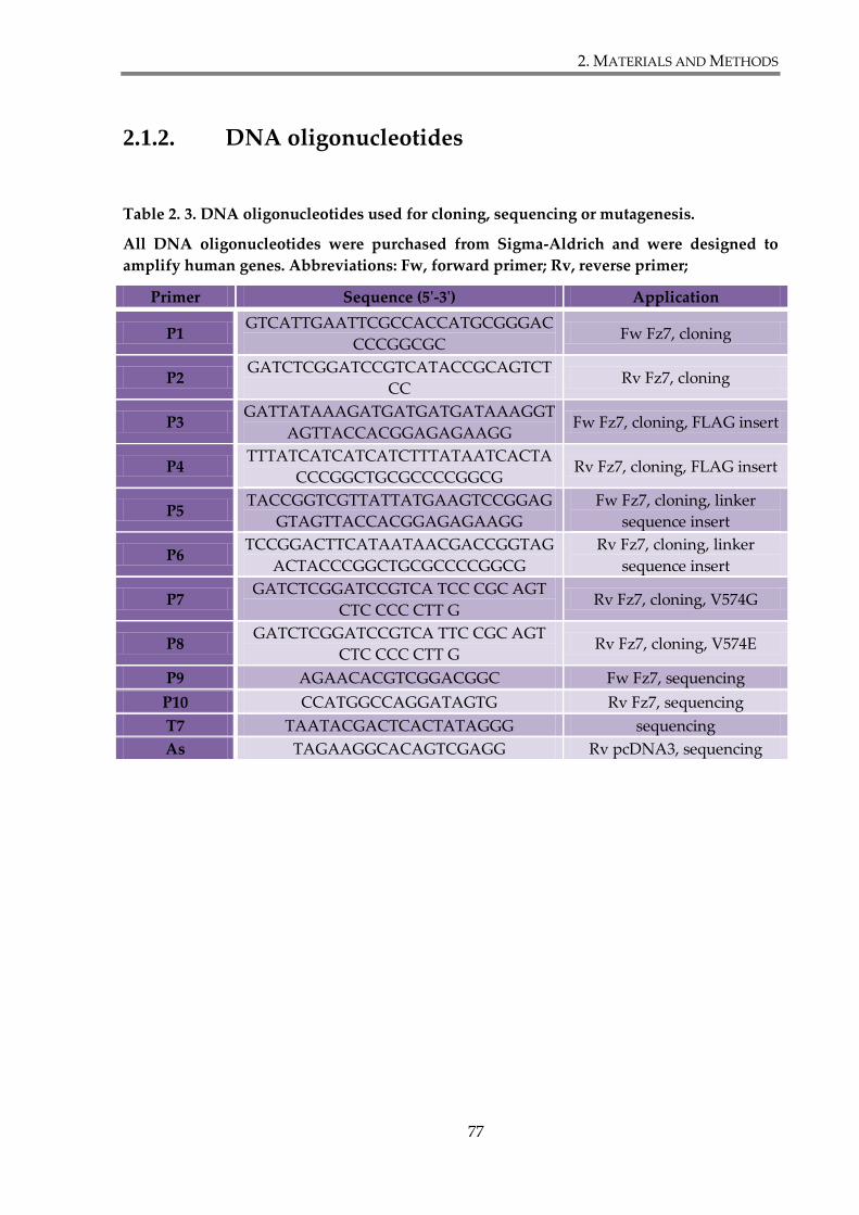
2. MATERIALS AND METHODS
77
2.1.2. DNA oligonucleotides
Table 2. 3. DNA oligonucleotides used for cloning, sequencing or mutagenesis.
All DNA oligonucleotides were purchased from Sigma-Aldrich and were designed to
amplify human genes. Abbreviations: Fw, forward primer; Rv, reverse primer;
Primer Sequence (5'-3') Application
P1 GTCATTGAATTCGCCACCATGCGGGAC
CCCGGCGC Fw Fz7, cloning
P2 GATCTCGGATCCGTCATACCGCAGTCT
CC Rv Fz7, cloning
P3 GATTATAAAGATGATGATGATAAAGGT
AGTTACCACGGAGAGAAGG Fw Fz7, cloning, FLAG insert
P4 TTTATCATCATCATCTTTATAATCACTA
CCCGGCTGCGCCCCGGCG Rv Fz7, cloning, FLAG insert
P5 TACCGGTCGTTATTATGAAGTCCGGAG
GTAGTTACCACGGAGAGAAGG
Fw Fz7, cloning, linker
sequence insert
P6 TCCGGACTTCATAATAACGACCGGTAG
ACTACCCGGCTGCGCCCCGGCG
Rv Fz7, cloning, linker
sequence insert
P7 GATCTCGGATCCGTCA TCC CGC AGT
CTC CCC CTT G Rv Fz7, cloning, V574G
P8 GATCTCGGATCCGTCA TTC CGC AGT
CTC CCC CTT G Rv Fz7, cloning, V574E
P9 AGAACACGTCGGACGGC Fw Fz7, sequencing
P10 CCATGGCCAGGATAGTG Rv Fz7, sequencing
T7 TAATACGACTCACTATAGGG sequencing
As TAGAAGGCACAGTCGAGG Rv pcDNA3, sequencing

2. MATERIALS AND METHODS
78
2.1.3. DNA plasmids
DNA constructs were generated according to cloning procedures, as described in
2.3.2. These are listed in Table 2.4. The DNA constructs provided by other scientists and used
for transfections or as templates for cloning are listed in Table 2.5.
Table 2. 4. Cloned DNA constructs.
Construct Cloning details
pcDNA™3.1-FLAG-Fz7 FLAG epitope was introduced at position 34 of human
Fz7; cloned into pcDNA3.1 plasmid using EcoRI and
BamHI sites
pcDNA™3.1-Myc-Fz7 Myc epitope was introduced at the position 34 of
human Fz7; cloned into pcDNA3.1 plasmid using
EcoRI and BamHI sites
pcDNA™3.1-GFP-Fz7 GFP was introduced at the position 34 of human Fz7;
cloned into pcDNA3.1 plasmid using EcoRI and
BamHI sites
pcDNA™3.1-FLAG-Fz7 V574G Mutagenised Flag-tagged human Fz7 cloned into
pcDNA3.1 plasmid using EcoRI and BamHI sites
pcDNA™3.1-FLAG-Fz7 V574E Mutagenised Flag-tagged human Fz7 cloned into
pcDNA3.1 plasmid using EcoRI and BamHI sites
pcDNA™3.1-Myc-Fz7 V574G Mutagenised Myc-tagged human Fz7 cloned into
pcDNA3.1 plasmid using EcoRI and BamHI sites
pcDNA™3.1-GFP-Fz7 V574G Mutagenised GFP-tagged human Fz7 cloned into
pcDNA3.1 plasmid using EcoRI and BamHI sites
pcDNA™3.1-GFP-Fz7 V574E Mutagenised GFP-tagged human Fz7 cloned into
pcDNA3.1 plasmid using EcoRI and BamHI sites
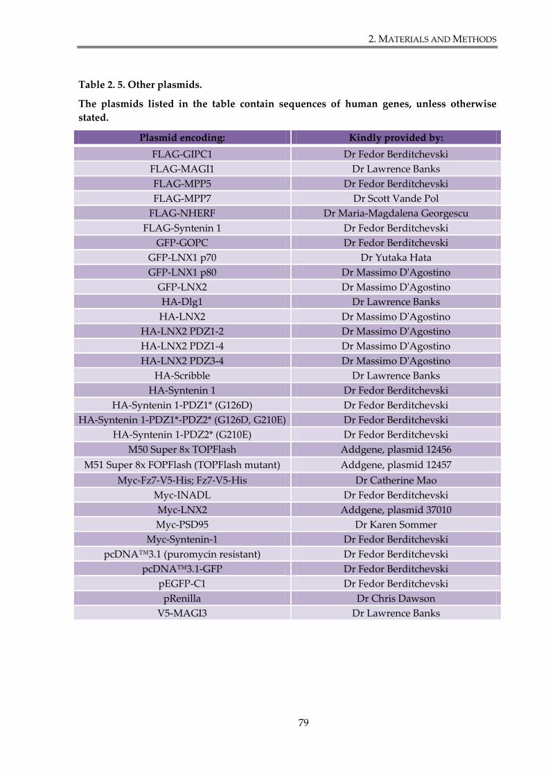
2. MATERIALS AND METHODS
79
Table 2. 5. Other plasmids.
The plasmids listed in the table contain sequences of human genes, unless otherwise
stated.
Plasmid encoding: Kindly provided by:
FLAG-GIPC1 Dr Fedor Berditchevski
FLAG-MAGI1 Dr Lawrence Banks
FLAG-MPP5 Dr Fedor Berditchevski
FLAG-MPP7 Dr Scott Vande Pol
FLAG-NHERF Dr Maria-Magdalena Georgescu
FLAG-Syntenin 1 Dr Fedor Berditchevski
GFP-GOPC Dr Fedor Berditchevski
GFP-LNX1 p70 Dr Yutaka Hata
GFP-LNX1 p80 Dr Massimo D'Agostino
GFP-LNX2 Dr Massimo D'Agostino
HA-Dlg1 Dr Lawrence Banks
HA-LNX2 Dr Massimo D'Agostino
HA-LNX2 PDZ1-2 Dr Massimo D'Agostino
HA-LNX2 PDZ1-4 Dr Massimo D'Agostino
HA-LNX2 PDZ3-4 Dr Massimo D'Agostino
HA-Scribble Dr Lawrence Banks
HA-Syntenin 1 Dr Fedor Berditchevski
HA-Syntenin 1-PDZ1* (G126D) Dr Fedor Berditchevski
HA-Syntenin 1-PDZ1*-PDZ2* (G126D, G210E) Dr Fedor Berditchevski
HA-Syntenin 1-PDZ2* (G210E) Dr Fedor Berditchevski
M50 Super 8x TOPFlash Addgene, plasmid 12456
M51 Super 8x FOPFlash (TOPFlash mutant) Addgene, plasmid 12457
Myc-Fz7-V5-His; Fz7-V5-His Dr Catherine Mao
Myc-INADL Dr Fedor Berditchevski
Myc-LNX2 Addgene, plasmid 37010
Myc-PSD95 Dr Karen Sommer
Myc-Syntenin-1 Dr Fedor Berditchevski
pcDNA™3.1 (puromycin resistant) Dr Fedor Berditchevski
pcDNA™3.1-GFP Dr Fedor Berditchevski
pEGFP-C1 Dr Fedor Berditchevski
pRenilla Dr Chris Dawson
V5-MAGI3 Dr Lawrence Banks
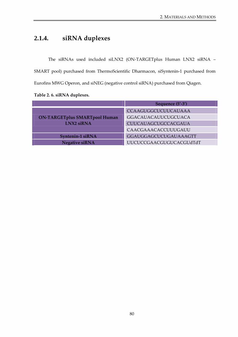
2. MATERIALS AND METHODS
80
2.1.4. siRNA duplexes
The siRNAs used included siLNX2 (ON-TARGETplus Human LNX2 siRNA –
SMART pool) purchased from ThermoScientific Dharmacon, siSyntenin-1 purchased from
Eurofins MWG Operon, and siNEG (negative control siRNA) purchased from Qiagen.
Table 2. 6. siRNA duplexes.
Sequence (5’-3’)
ON-TARGETplus SMARTpool Human
LNX2 siRNA
CCAAGUGGCUCUUCAUAAA
GGACAUACAUUCUGCUACA
CUUCAUAGCUGCCACGAUA
CAACGAAACACCUUUGAUU
Syntenin-1 siRNA GGAUGGAGCUCUGAUAAAGTT
Negative siRNA UUCUCCGAACGUGUCACGUdTdT
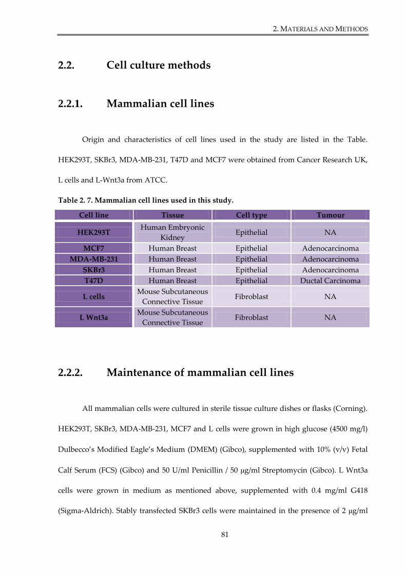
2. MATERIALS AND METHODS
81
2.2. Cell culture methods
2.2.1. Mammalian cell lines
Origin and characteristics of cell lines used in the study are listed in the Table.
HEK293T, SKBr3, MDA-MB-231, T47D and MCF7 were obtained from Cancer Research UK,
L cells and L-Wnt3a from ATCC.
Table 2. 7. Mammalian cell lines used in this study.
Cell line Tissue Cell type Tumour
HEK293T Human Embryonic
Kidney Epithelial NA
MCF7 Human Breast Epithelial Adenocarcinoma
MDA-MB-231 Human Breast Epithelial Adenocarcinoma
SKBr3 Human Breast Epithelial Adenocarcinoma
T47D Human Breast Epithelial Ductal Carcinoma
L cells Mouse Subcutaneous
Connective Tissue Fibroblast NA
L Wnt3a Mouse Subcutaneous
Connective Tissue Fibroblast NA
2.2.2. Maintenance of mammalian cell lines
All mammalian cells were cultured in sterile tissue culture dishes or flasks (Corning).
HEK293T, SKBr3, MDA-MB-231, MCF7 and L cells were grown in high glucose (4500 mg/l)
Dulbecco’s Modified Eagle’s Medium (DMEM) (Gibco), supplemented with 10% (v/v) Fetal
Calf Serum (FCS) (Gibco) and 50 U/ml Penicillin / 50 μg/ml Streptomycin (Gibco). L Wnt3a
cells were grown in medium as mentioned above, supplemented with 0.4 mg/ml G418
(Sigma-Aldrich). Stably transfected SKBr3 cells were maintained in the presence of 2 μg/ml
2. MATERIALS AND METHODS
82
puromycin (Sigma-Aldrich). All cell lines were cultivated at 37°C, in humidified incubator
(Galaxy R CO2 Incubators, RS Biotech) in 5% CO2 atmosphere. When passaging, cells were
split in the ratio 1:5 (SKBr3, MDA-MB-231, MCF7) or 1:10 (HEK293T, L cells, L Wnt3a) twice
a week. Growth medium was removed, and a confluent monolayer of cells was washed with
sterile phosphate buffer saline (PBS) to remove all traces of serum that contains trypsin
inhibitors. To detach cells from a plastic surface, they were incubated with 0.05% Trypsin-
EDTA (Gibco) for 5 min at 37°C to facilitate dispersal. Subsequently, cells were collected
from plate in a fresh growth medium and pelleted by centrifuging at 900 RPM for 3 min.
After resuspension in complete growth medium, cells were plated at required density. Cell
growth and condition were monitored using Nikon TMS microscope.
2.2.3. Cryopreservation and recovery of cell lines
Detached cells, as described in section 2.2.2, were resuspended in freezing solution
(10% (v/v) dimethyl sulfoxide (DMSO) (Sigma-Aldrich) and 90% (v/v) Fetal Calf Serum
(FCS)). The suspension was subsequently frozen in 1 ml CryoTube™ vials (Nunc) at -80°C
for at least 24h before transferring to liquid nitrogen for long term storage.
Cells were recovered by thawing in a water bath for 2 min at 37°C. Thawed cell
suspension was transferred to a tube with 9 ml of pre-warmed, complete growth medium
and pelleted by centrifugation at 900 RPM for 3 min. Pellet was resuspended in fresh
medium and cells were seeded in T25 tissue culture flask. Cells were incubated at 37°C, in
humidified 5% CO2 atmosphere for at least 48h before further manipulation.
2. MATERIALS AND METHODS
83
2.2.4. Counting cells
When a specified number of cells was required for an experiment, cells were
trypsinized (Section 2.2.2), resuspended in fresh complete medium and counted. 10 μl of cell
suspension was used to count cell in Neubauer haemocytometer counting chamber
(Marienfeld) according to the manufacturer’s instructions.
2.2.5. Mycoplasma testing
Cultured cell lines were routinely tested to ensure they were mycoplasma free.
Mycoalert® Mycoplasma Detection Kit (Lonza) was used, according to the manufacturer’s
instructions.
2.2.6. Cell transfection
2.2.6.1. Polyethyleimine (PEI)
HEK293T cells were transfected with DNA plasmids using polyethyleimine (PEI)
(Sigma-Aldrich). PEI working solution (1 mg/ml) was prepared by warming up stock
solution at 55°C and dissolving appropriate amount in dH20. After cooling down to room
temperature, PEI was neutralized with HCl to pH 7.0 and filter sterilized (0.22 μm syringe
filters (Appleton Woods)). Aliquots were stored at -20°C.
To transfect cells, DNA and PEI were mixed in Opti-MEM® I reduced-serum
medium (Gibco) (without FCS), in the amounts indicated in the Table 2.8. Prepared mix was
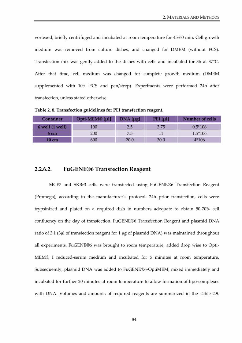
2. MATERIALS AND METHODS
84
vortexed, briefly centrifuged and incubated at room temperature for 45-60 min. Cell growth
medium was removed from culture dishes, and changed for DMEM (without FCS).
Transfection mix was gently added to the dishes with cells and incubated for 3h at 37°C.
After that time, cell medium was changed for complete growth medium (DMEM
supplemented with 10% FCS and pen/strep). Experiments were performed 24h after
transfection, unless stated otherwise.
Table 2. 8. Transfection guidelines for PEI transfection reagent.
Container Opti-MEM® [μl] DNA [μg] PEI [μl] Number of cells
6 well (1 well) 100 2.5 3.75 0.5*106
6 cm 200 7.3 11 1.5*106
10 cm 600 20.0 30.0 4*106
2.2.6.2. FuGENE®6 Transfection Reagent
MCF7 and SKBr3 cells were transfected using FuGENE®6 Transfection Reagent
(Promega), according to the manufacturer’s protocol. 24h prior transfection, cells were
trypsinized and plated on a required dish in numbers adequate to obtain 50-70% cell
confluency on the day of transfection. FuGENE®6 Transfection Reagent and plasmid DNA
ratio of 3:1 (3μl of transfection reagent for 1 μg of plasmid DNA) was maintained throughout
all experiments. FuGENE®6 was brought to room temperature, added drop wise to Opti-
MEM® I reduced-serum medium and incubated for 5 minutes at room temperature.
Subsequently, plasmid DNA was added to FuGENE®6-OptiMEM, mixed immediately and
incubated for further 20 minutes at room temperature to allow formation of lipo-complexes
with DNA. Volumes and amounts of required reagents are summarized in the Table 2.9.
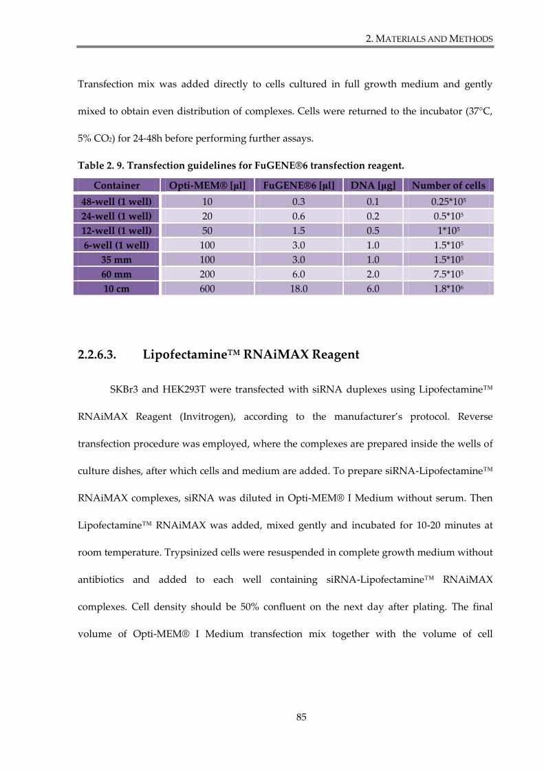
2. MATERIALS AND METHODS
85
Transfection mix was added directly to cells cultured in full growth medium and gently
mixed to obtain even distribution of complexes. Cells were returned to the incubator (37°C,
5% CO2) for 24-48h before performing further assays.
Table 2. 9. Transfection guidelines for FuGENE®6 transfection reagent.
Container Opti-MEM® [μl] FuGENE®6 [μl] DNA [μg] Number of cells
48-well (1 well) 10 0.3 0.1 0.25*105
24-well (1 well) 20 0.6 0.2 0.5*105
12-well (1 well) 50 1.5 0.5 1*105
6-well (1 well) 100 3.0 1.0 1.5*105
35 mm 100 3.0 1.0 1.5*105
60 mm 200 6.0 2.0 7.5*105
10 cm 600 18.0 6.0 1.8*106
2.2.6.3. Lipofectamine™ RNAiMAX Reagent
SKBr3 and HEK293T were transfected with siRNA duplexes using Lipofectamine™
RNAiMAX Reagent (Invitrogen), according to the manufacturer’s protocol. Reverse
transfection procedure was employed, where the complexes are prepared inside the wells of
culture dishes, after which cells and medium are added. To prepare siRNA-Lipofectamine™
RNAiMAX complexes, siRNA was diluted in Opti-MEM® I Medium without serum. Then
Lipofectamine™ RNAiMAX was added, mixed gently and incubated for 10-20 minutes at
room temperature. Trypsinized cells were resuspended in complete growth medium without
antibiotics and added to each well containing siRNA-Lipofectamine™ RNAiMAX
complexes. Cell density should be 50% confluent on the next day after plating. The final
volume of Opti-MEM® I Medium transfection mix together with the volume of cell
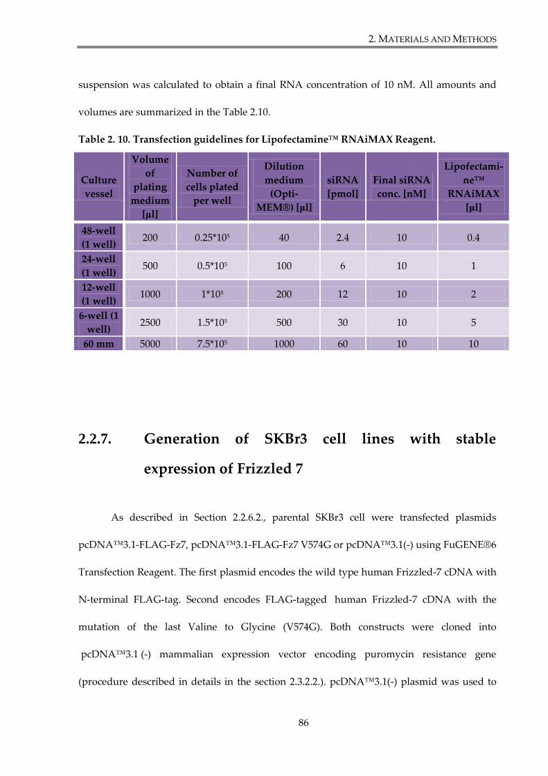
2. MATERIALS AND METHODS
86
suspension was calculated to obtain a final RNA concentration of 10 nM. All amounts and
volumes are summarized in the Table 2.10.
Table 2. 10. Transfection guidelines for Lipofectamine™ RNAiMAX Reagent.
Culture
vessel
Volume
of
plating
medium
[μl]
Number of
cells plated
per well
Dilution
medium
(Opti-
MEM®) [μl]
siRNA
[pmol]
Final siRNA
conc. [nM]
Lipofectami-
ne™
RNAiMAX
[μl]
48-well
(1 well) 200 0.25*105 40 2.4 10 0.4
24-well
(1 well) 500 0.5*105 100 6 10 1
12-well
(1 well) 1000 1*105 200 12 10 2
6-well (1
well) 2500 1.5*105 500 30 10 5
60 mm 5000 7.5*105 1000 60 10 10
2.2.7. Generation of SKBr3 cell lines with stable
expression of Frizzled 7
As described in Section 2.2.6.2., parental SKBr3 cell were transfected plasmids
pcDNA™3.1-FLAG-Fz7, pcDNA™3.1-FLAG-Fz7 V574G or pcDNA™3.1(-) using FuGENE®6
Transfection Reagent. The first plasmid encodes the wild type human Frizzled-7 cDNA with
N-terminal FLAG-tag. Second encodes FLAG-tagged human Frizzled-7 cDNA with the
mutation of the last Valine to Glycine (V574G). Both constructs were cloned into
pcDNA™3.1 (-) mammalian expression vector encoding puromycin resistance gene
(procedure described in details in the section 2.3.2.2.). pcDNA™3.1(-) plasmid was used to
2. MATERIALS AND METHODS
87
generate SKBr3-control cell line. 48h after transfection with DNA plasmids, cells medium
was changed for the regular growth medium containing puromycin (Sigma-Aldrich)
(2μg/ml) to select for stable transfectants. Puromycin containing medium was replaced every
3-4 days, until isolated colonies started to appear. After selection (2-3 weeks) cells were
sorted two times using anti-Flag mouse mAb and fluorescent activated cell sorting (FACS)
(Section 2.6.1.) to obtain SKBr3-FLAG-Fz7 and SKBr3-FLAG-Fz7 V574G stable cell lines.
2.2.8. Production of Wnt3a conditioned medium
Mouse fibroblast, L cells, were transfected for stable expression of Wnt3a (Willert et
al. 2003). L Wnt3a cells are cultivated in DMEM complete growth medium supplemented
with 0.4 mg/ml G418. To prepare Wnt3a conditioned medium (Wnt3a CM) cells were split
and replated into 15 cm culture dish, without G418 if conditioned was to be used with a cell
line sensitive to G418. For 15 cm dish, 4*106 cells were seeded. Cells were grown until almost
confluent, for approximately 48h. Each plate was washed with PBS and 25 ml of complete
growth medium was added. Cells were then incubated for 4 days (37°C, 5% CO2).
Conditioned media were collected into 50 ml tubes and centrifuged at 1000 RPM for 5
minutes. Supernatants were filter sterilized and stored at 4°C. In all experiment conditioned
medium was used in dilution 1:2. Since the conditioned medium contains also other factors
than Wnt3a, it was important to control all experiments with control conditioned medium
from the parental cell line (L cell), prepared in similar way as Wnt3a conditioned medium.
2. MATERIALS AND METHODS
88
2.2.9. Bacteria strains, media and antibiotics
The chemically competent Escherichia Coli (E.Coli) DH5α (NEB) were used for
transformation with various DNA plasmids. Bacteria were cultured in Luria Bertani (LB)
broth or LB agar (Invitrogen) with appropriate antibiotics. Final concentrations of used
antibiotics were as follow: ampicillin (Sigma-Aldrich) 100 μg/ml and kanamycin (Sigma-
Aldrich) 30 μg/ml. Bacteria were grown on LB agar plates at 37°C in incubator (Heraeus) or
in liquid LB media at 37°C in the orbital shaking incubator (Gallenkamp).
2. MATERIALS AND METHODS
89
2.3. Manipulation of DNA material.
2.3.1. Molecular cloning
The DNA cloning was performed as summarised in the figure. 2.1. All the steps are
described in detailed in the next sections. Tagged Fz7 constructs were generated using Myc-
Fz7-V5-His or Fz7-V5-His plasmids as the templates (Struewing et al. 2007). Amino acid
sequences corresponding FLAG and Myc epitopes, and GFP were introduced at the position
34, after the signal peptide, on the N-terminus of the receptor. Tagged Fz7 WT, Fz7 V574G
and Fz7 V574E were generated using a standard PCR technique and cloned into pcDNA™3.1
plasmid using EcoRI and BamHI sites. Sequences of all DNA oligonucleotides (primers) used
in this study are listed in the Table 2.3.
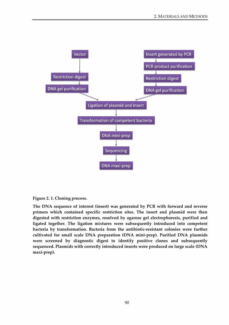
2. MATERIALS AND METHODS
90
Figure 2. 1. Cloning process.
The DNA sequence of interest (insert) was generated by PCR with forward and reverse
primers which contained specific restriction sites. The insert and plasmid were then
digested with restriction enzymes, resolved by agarose gel electrophoresis, purified and
ligated together. The ligation mixtures were subsequently introduced into competent
bacteria by transformation. Bacteria from the antibiotic-resistant colonies were further
cultivated for small scale DNA preparation (DNA mini-prep). Purified DNA plasmids
were screened by diagnostic digest to identify positive clones and subsequently
sequenced. Plasmids with correctly introduced inserts were produced on large scale (DNA
maxi-prep).
2. MATERIALS AND METHODS
91
2.3.2. Amplification of DNA insert by Polymerase Chain
Reaction (PCR)
2.3.2.1. Myc-Frizzled-7
Myc-Frizzled-7 cDNA was amplified in a PCR reaction on the template of Myc-Fz7-
V5-His. For the reaction forward primer P1 containing restriction site for enzyme EcoRI and
reverse primer P2 containing restriction site for enzyme BamHI were used. PCR reactions
were conducted using Expand High Fidelity PCR System (Roche) following manufacturer’s
guidelines. 50 μl reaction mixes were prepared by adding 40.25 μl of water, 5 μl of 10x
concentrated Expand High Fidelity buffer with MgCl2, 1 μl of 10 mM deoxyribonucleotide
triphosphate (dNTP) mix (New England Biolabs), 1 μl of each 100 μM primer (Sigma-
Aldrich), 0,75 μl of Expand High Fidelity Enzyme Mix and 1 μl of DNA template (10 ng). The
PCR reaction involved initial denaturation (94°C, 2 min), then 40 cycles of denaturation
(94°C, 30 sec), annealing (56°C, 1 min) and elongation (72°C, 2 min). A final DNA elongation
step of 72°C for 7 minutes was added when all cycles were completed. Amplified PCR
products were separated and purified using agarose gel electrophoresis.
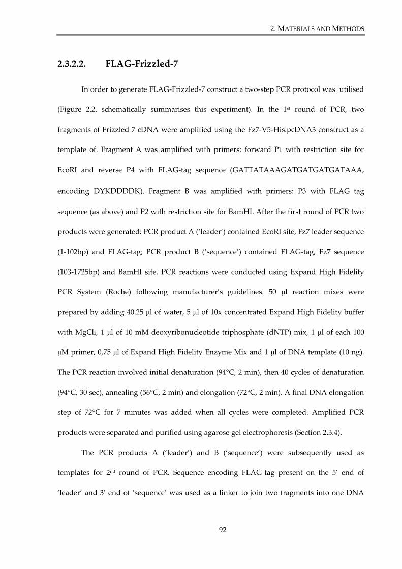
2. MATERIALS AND METHODS
92
2.3.2.2. FLAG-Frizzled-7
In order to generate FLAG-Frizzled-7 construct a two-step PCR protocol was utilised
(Figure 2.2. schematically summarises this experiment). In the 1st round of PCR, two
fragments of Frizzled 7 cDNA were amplified using the Fz7-V5-His:pcDNA3 construct as a
template of. Fragment A was amplified with primers: forward P1 with restriction site for
EcoRI and reverse P4 with FLAG-tag sequence (GATTATAAAGATGATGATGATAAA,
encoding DYKDDDDK). Fragment B was amplified with primers: P3 with FLAG tag
sequence (as above) and P2 with restriction site for BamHI. After the first round of PCR two
products were generated: PCR product A (‘leader’) contained EcoRI site, Fz7 leader sequence
(1-102bp) and FLAG-tag; PCR product B (‘sequence’) contained FLAG-tag, Fz7 sequence
(103-1725bp) and BamHI site. PCR reactions were conducted using Expand High Fidelity
PCR System (Roche) following manufacturer’s guidelines. 50 μl reaction mixes were
prepared by adding 40.25 μl of water, 5 μl of 10x concentrated Expand High Fidelity buffer
with MgCl2, 1 μl of 10 mM deoxyribonucleotide triphosphate (dNTP) mix, 1 μl of each 100
μM primer, 0,75 μl of Expand High Fidelity Enzyme Mix and 1 μl of DNA template (10 ng).
The PCR reaction involved initial denaturation (94°C, 2 min), then 40 cycles of denaturation
(94°C, 30 sec), annealing (56°C, 2 min) and elongation (72°C, 2 min). A final DNA elongation
step of 72°C for 7 minutes was added when all cycles were completed. Amplified PCR
products were separated and purified using agarose gel electrophoresis (Section 2.3.4).
The PCR products A (‘leader’) and B (‘sequence’) were subsequently used as
templates for 2nd round of PCR. Sequence encoding FLAG-tag present on the 5’ end of
‘leader’ and 3’ end of ‘sequence’ was used as a linker to join two fragments into one DNA
2. MATERIALS AND METHODS
93
template during the PCR reaction. To achieve this, Expand High Fidelity PCR System was
used. 50 μl reaction mix was prepared by adding 42 μl of water, 5 μl of 10x concentrated
Expand High Fidelity buffer with MgCl2, 1 μl of 10 mM dNTP mix, 0.75 μl of Expand High
Fidelity Enzyme Mix, 10 ng of template A ‘leader’ (0.5 μl) and 100 ng of template B
‘sequence’ (1 μl). First step of PCR reaction involved initial denaturation (94°C, 2 min), then
15 cycles of denaturation (94°C, 30 sec), annealing (56°C, 1 min) and elongation (72°C, 2 min),
and a final elongation step (72°C, 7 min). Primers were not included in this step to create the
ssDNA. Obtained ssDNA was used as a template for the final amplification with forward
primer P1 and reverse P2. 50 μl reaction mixes were prepared by adding 31.25 μl of water, 5
μl of 10x concentrated Expand High Fidelity buffer with MgCl2, 1 μl of 10 mM dNTP mix, 1
μl of each 100 μM primer (Sigma-Aldrich), 0.75 μl of Expand High Fidelity Enzyme Mix and
10 μl of ssDNA template. The PCR reaction involved initial denaturation (94°C, 2 min), then
35 cycles of denaturation (94°C, 30 sec), annealing (56°C, 1 min) and elongation (72°C, 2 min).
A final DNA elongation step of 72°C for 7 minutes was added when all cycles were
completed. Final PCR product contained EcoRI and BamHI restriction sites, and FLAG-tag
introduced at the position 34, after signal peptide (‘leader’).
2. MATERIALS AND METHODS
94
Figure 2. 2. A two-step PCR protocol utilised to generate FLAG-Frizzled-7.
Details of the protocol are presented in the section 2.3.2.2. „A”, Fz7 leader sequence (1-
102bp); „B”, Fz7 sequence (103-1725bp); P1; forward primer with restriction site for EcoRI;
P4, reverse primer with FLAG-tag sequence; P3, forward primer with FLAG-tag sequence;
P2, reverse primer with restriction site for BamHI; ssDNA, single-stranded DNA;
A B A B
AEcoRI FLAG BFLAG BamHI
AEcoRI FLAG
BFLAG BamHI
AEcoRI FLAG B BamHI
Fz7 template:
P1 P4 P3 P2
P1 P2
Fz7 template:
1st round PCR
2nd round PCR AEcoRI FLAG B BamHI ssDNA template
FLAG-Fz7
2. MATERIALS AND METHODS
95
2.3.2.3. GFP-Frizzled 7
In order to generate GFP- tagged Fz7 construct a similar two-step PCR protocol was
used with slight modifications (summarised in Figure 2.3.). Initially, a linker with two
restriction sites for AgeI and BspEI enzymes was introduced into Fz7 sequence at the
position 34. The experimental PCR conditions were similar to those described in Section
2.3.2.2. As a result Frizzled-7 construct was obtained with the introduced linker sequence,
containing two restrictions, after the leader peptide. These two sites were used for insertion
of GFP into Fz7. GFP sequence was cut out from plasmid pEGFP-C1, using restriction
enzymes AgeI and BspEI. This process is described in details in the section 2.3.3. Digested
Fz7 and GFP fragments were gel purified (Section 2.3.5) and ligated together (Section 2.3.7).
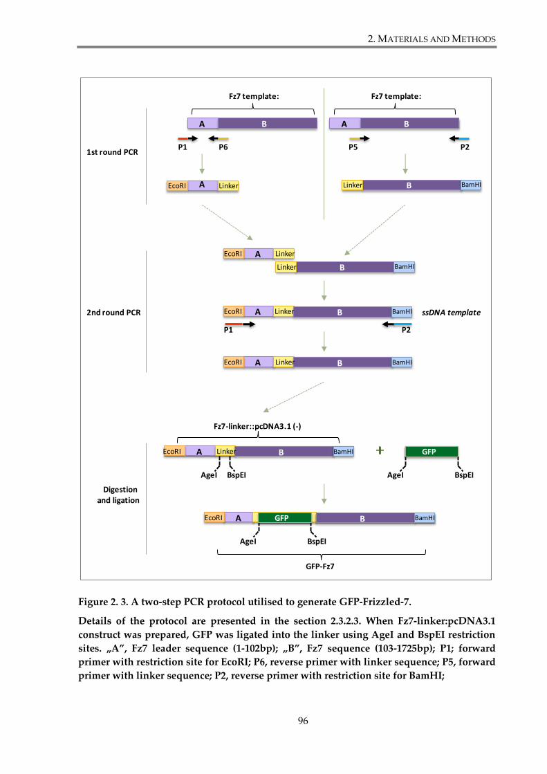
2. MATERIALS AND METHODS
96
Figure 2. 3. A two-step PCR protocol utilised to generate GFP-Frizzled-7.
Details of the protocol are presented in the section 2.3.2.3. When Fz7-linker:pcDNA3.1
construct was prepared, GFP was ligated into the linker using AgeI and BspEI restriction
sites. „A”, Fz7 leader sequence (1-102bp); „B”, Fz7 sequence (103-1725bp); P1; forward
primer with restriction site for EcoRI; P6, reverse primer with linker sequence; P5, forward
primer with linker sequence; P2, reverse primer with restriction site for BamHI;
A B A B
AEcoRI Linker BLinker BamHI
AEcoRI Linker
BLinker BamHI
AEcoRI B BamHI
Fz7 template:
P1 P6 P5 P2
P1 P2
Fz7 template:
1st round PCR
2nd round PCR AEcoRI B BamHI ssDNA template
GFP-Fz7
Linker
Linker
AEcoRI B BamHI
AgeI BspEI
GFP
AgeI BspEI
GFPAEcoRI B BamHILinker
AgeI BspEI
Digestionand ligation
Fz7-linker::pcDNA3.1 (-)
2. MATERIALS AND METHODS
97
2.3.2.4. Frizzled 7 V574G and V574E
In order to generate constructs of Frizzled-7 with mutation of the last amino acid,
V574G or V574E, PCR reactions were set up with forward primer P1 and reverse P7 or P8,
respectively. As templates Myc-Fz7, FLAG-Fz7 and GFP-Fz7 constructs were used.
Amplified PCR products were Fz7 V574G or Fz7 V574 with restrictions sites for EcoRI and
BamHI. Conditions for PCR reaction were the same as in Section 2.3.2.1.
2.3.3. DNA digestion with endonuclease (restriction)
enzymes
Frizzled-7 PCR products and vector pcDNA3.1 were digested with restriction
enzymes EcoRI and BamHI (Roche) according to manufacturer’s guidelines. The digestion
was performed as follows. Purified PCR product (from 50 μl reaction) or vector DNA (3 μg)
were incubated in 50 μl of digestion solution containing dH2O, 1x buffer A (Roche) and 10
units of each enzyme. Digestion was carried out overnight at 37°C.
To generate GFP-Fz7, Fz7-linker construct (Section 2.3.2.3) in pcDNA3.1 and pEGFP-
C1 were sequentially digested with AgeI and BspEI endonucleases. In the first step both
constructs were incubated in 50 μl digestion mixes containing dH2O, 1x buffer 1 (NEB) and
10 units of AgeI enzyme, overnight at 37°C. Subsequently, AgeI digested constructs were
purified and incubated in second 50 μl digestion mix containing dH2O, 1x buffer 3 (NEB)
and 10 units of BspEI endonuclease (NEB), overnight at 37°C.
2. MATERIALS AND METHODS
98
Restriction digest reactions for diagnostic purposes were scaled down appropriately
to the volume of 10 μl, and carried out for 1h at 37°C.
2.3.4. Agarose gel electrophoresis
PCR products or DNA restriction digest were mixed with 6xDNA Loading buffer
(Qiagen) and subjected to horizontal electrophoresis (Geneflow). Gels were prepared by
boiling agarose, 1% (w/v) (Invitrogen), in H2O and subsequently supplementing the solution
with the appropriate volume of 10xTAE buffer and SYBR Safe (Invitrogen). The samples
were resolved by electrophoresis at 100V, and subsequently gels were visualized and
recorded by UV gel documentation system (InGenius® Syngene).
2.3.5. DNA extraction from agarose gel (DNA gel
purification)
DNA bands were cut from the agarose gels and DNA was extracted using the Qiagen
Gel Extraction Kit, following manufacturer’s guidelines. Efficiency of purification was
assessed by loading 1/10 of obtained DNA on 1% agarose gel and visualised as described
above.
2. MATERIALS AND METHODS
99
2.3.6. DNA ligation
Ligation reactions were performed using T4 DNA ligase (NEB) as follows. 1 μl of T4
DNA ligase (400 000 U/ml) and 2 μl of T4 DNA ligase buffer (NEB) were added to the 17 μl of
insert:vector mixture, prepared in ratio 3:1. Before adding T4 ligase, DNA mixture was
preincubated for 2 minutes at 60°C. Ligation was performed for 2h at room temperature.
Reactions were stopped by incubating samples for 10 minutes at 65°C.
2.3.7. Heat-shock transformation of competent bacteria
Chemically competent 5-α Iq E.coli cells (18 μl; NEB) were mixed with 10 μl of ligation
mix or 500 pg of DNA plasmid and incubated for 30 minutes on ice. Afterwards bacteria
were heat-shocked at 42°C for 30 seconds and incubated on ice for further 2 minutes.
Transformation mixed was diluted with 250 μl of antibiotic-free LB medium and bacteria
were allowed to recover for 1h at 37°C in an orbital shaker incubator. Subsequently bacteria
were plated on LB-agar plates with appropriate antibiotic for overnight incubation.
2. MATERIALS AND METHODS
100
2.3.8. Preparation of plasmid DNA from transformed
bacteria
Large scale production of DNA plasmid was performed using Qiagen Plasmid Maxi
Kit (Qiagen) according to the manufacturer’s protocol. For small scale DNA purification
Mini-Prep DNA Purification Kit (Qiagen) was used. Final DNA concentration was measured
with the Nanodrop ND-1000 Spectrophotometer (Labtech).
2.3.9. DNA sequencing
All the constructed plasmids were verified by sequencing at the Functional Genomics
and Proteomics Laboratory (School of Biosciences, University of Birmingham). Briefly, 150
ng of plasmid DNA, 3.2 pmol of an appropriate primer and dH2O were mixed up to the final
volume of 10 μl. Samples were then sent for sequencing analysis.
2.3.10. Preparation of glycerol stocks
All constructs were stored in glycerol stock in -80 °C. Bacterial cultures were mixed
with glycerol (Fisher Scientific) to obtain its final concentration of 30% (v/v).
2. MATERIALS AND METHODS
101
2.4. Protein analysis
2.4.1. Preparation of mammalian cell lysates
Unless specified otherwise, procedure described in this section was used to obtain
mammalian cellular lysates. Proteins were extracted from cells growing in 60 mm dishes, 24h
(HEK293T) or 48h (SKBr3; MCF7) after transfection. Cells were lysed using 500 μl of 1% v/v
Triton X-100 (150 mM NaCl, 50 mM Tris pH 7.4), 0.5% v/v Brij98- 0.5% v/v Triton X-100 (150
mM NaCl, 50 mM Tris pH 7.4) or 0.8% v/v Brij98- 0.2% v/v Triton X-100 (150 mM NaCl, 50
mM Tris pH 7.4), supplemented with 2 mM phenylmethylsulfonyl fluoride (PMSF), 10 μg/ml
aprotinin, 10 μg/ml leupeptin, 2 mM NaVO3 (sodium orthovanadate) and 10 mM NaF
(sodium fluoride). Cells were scraped quickly with a cell scraper and cell lysates were
transferred into a 1,5 ml eppendorf tube. The tubes were subsequently placed on a rotary
wheel for 30 minutes at 4°C. The insoluble material was pelleted at 13,000 rpm for 10 min.
Supernatants were used directly for different assays or stored in -80°C. If needed for SDS-
PAGE (Section 2.4.3.) cell lysates were mixed in ratio 3:1 (v/v) with 4x concentrated Laemmli
loading buffer (0.5M Tris pH 6.8 2.1 ml, 4.4 ml glycerol, 2.2 ml 20% SDS, 1 ml dH2O and
0,05 g bromophenol blue).
2. MATERIALS AND METHODS
102
2.4.2. Protein concentration determination
Protein concentration was determined using Bio-Rad DC Protein Assay Kit according
to the manufacturer’s protocol. In 96-well plate (BD-Falcon) 5 μl of cell lysate was mixed with
25 μl of Bio-Rad mixture (20 μl of reagent S in 1 ml of reagent A) and with 200 μl reagent B.
Samples were incubated at room temperature for 15 minutes and absorbance was measured
at 630 nm wavelength using microplate reader (Bio-Rad). To obtain standard curve, bovine
serum albumin (BSA, Sigma-Aldrich) was serially diluted in an appropriate lysis buffer in a
range of 0.4 – 3.2 mg/ml. Based on the absorbance values for the known concentrations of
BSA standards the protein standard curve was created in MS Excel Software and the protein
concentration in lysate samples was determined. The protein samples and BSA samples were
measured in triplicate to obtain more accurate values.
2.4.3. SDS-PAGE and Western Blot analysis
Lysate samples, with equal amount of proteins, along with prestained protein ladder,
were resolved in SDS-PAGE (sodium dodecyl sulphate-poliacrylamide gel electrophoresis).
If required samples were reduces by adding β-mercaptoethanol in dilution 1:20. Samples
were loaded on 10% polyacrylamide gel (unless stated otherwise) and resolved at 120V using
Bio-Rad electrophoretic equipment and 1x SDS/Tris/Glycine running buffer (GeneFlow).
Table 2.11. shows recipes for preparation 5% stacking and 10% resolving SDS-PAGE: .
The proteins resolved by SDS-PAGE were stained with InstantBlue (Expedeon) or
transferred to a nitrocellulose membrane, using Trans-Blot® Turbo™ Transfer System (Bio-
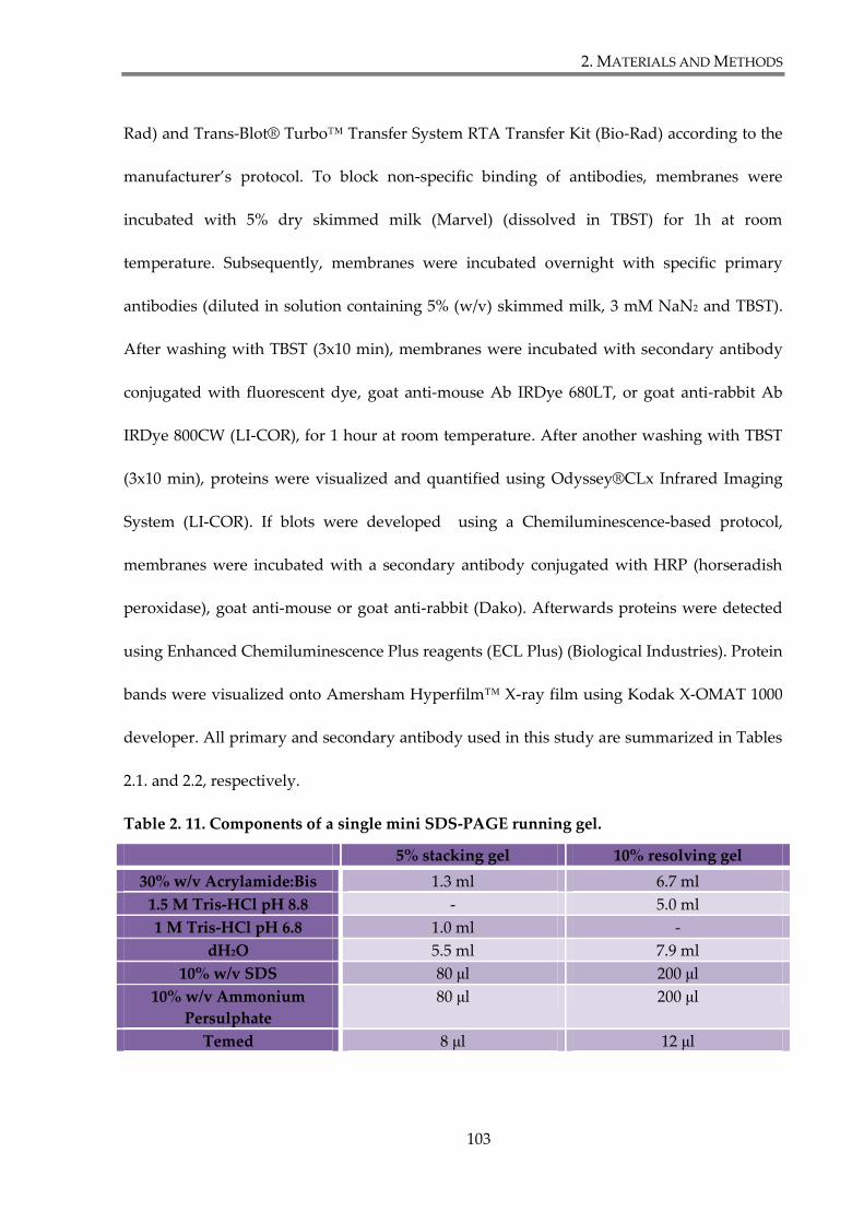
2. MATERIALS AND METHODS
103
Rad) and Trans-Blot® Turbo™ Transfer System RTA Transfer Kit (Bio-Rad) according to the
manufacturer’s protocol. To block non-specific binding of antibodies, membranes were
incubated with 5% dry skimmed milk (Marvel) (dissolved in TBST) for 1h at room
temperature. Subsequently, membranes were incubated overnight with specific primary
antibodies (diluted in solution containing 5% (w/v) skimmed milk, 3 mM NaN2 and TBST).
After washing with TBST (3x10 min), membranes were incubated with secondary antibody
conjugated with fluorescent dye, goat anti-mouse Ab IRDye 680LT, or goat anti-rabbit Ab
IRDye 800CW (LI-COR), for 1 hour at room temperature. After another washing with TBST
(3x10 min), proteins were visualized and quantified using Odyssey®CLx Infrared Imaging
System (LI-COR). If blots were developed using a Chemiluminescence-based protocol,
membranes were incubated with a secondary antibody conjugated with HRP (horseradish
peroxidase), goat anti-mouse or goat anti-rabbit (Dako). Afterwards proteins were detected
using Enhanced Chemiluminescence Plus reagents (ECL Plus) (Biological Industries). Protein
bands were visualized onto Amersham Hyperfilm™ X-ray film using Kodak X-OMAT 1000
developer. All primary and secondary antibody used in this study are summarized in Tables
2.1. and 2.2, respectively.
Table 2. 11. Components of a single mini SDS-PAGE running gel.
5% stacking gel 10% resolving gel
30% w/v Acrylamide:Bis 1.3 ml 6.7 ml
1.5 M Tris-HCl pH 8.8 - 5.0 ml
1 M Tris-HCl pH 6.8 1.0 ml -
dH2O 5.5 ml 7.9 ml
10% w/v SDS 80 μl 200 μl
10% w/v Ammonium
Persulphate
80 μl 200 μl
Temed 8 μl 12 μl
2. MATERIALS AND METHODS
104
2.4.4. Quantitative analysis of Western blot images
Densytometric analysis was carried out on images obtain by Odyssey CLx infrared
imaging system (LI-COR) using Image Studio Lite Software (LI-COR). Rectangular shapes of
equal size were drawn separately around bands of interest and the fluorescent absorbance
within the shapes was calculated. The background absorbance of the blot was subtracted
from the shapes using the software to obtain consistent data. The Median Method was
chosen to subtract the median value of the pixels in the background segment of each shape.
Absorbance values representing the densitometric values were used to quantify protein
expression level in this study.
2.4.5. Immunoprecipitation
For immunoprecipitation experiments, lysates prepared as described in section 2.4.1.,
were incubated for 1h or overnight on a rotary wheel at 4°C with 18 μl of agarose or
magnetic beads conjugated with mouse anti-FLAG M2 mAb (Sigma-Aldrich) or magnetic
GFP-Trap® beads (Chromotek). Subsequently, beads were washed 3 times with 700 μl of ice-
cold TBS –Tween and retained proteins were eluted by adding 15 μl of 2x concentrated
Laemmli sample buffer and boiling for 10 minutes at 60°C. If necessary, beads were pre-
incubated with 0.5% BSA/PBS before adding lysates, to prevent unspecific binding of
proteins to the beads.
2. MATERIALS AND METHODS
105
2.4.6. Pull-down with peptides
For pulldown experiments lysates, prepared as described in section 2.4.1., were
incubated with NeutrAvidin™-conjugated agarose resins (Pierce) with immobilized custom
synthesised biotinylated peptides corresponding to the C-terminal cytoplasmic tails of Fz7,
Fz3 and Fz8 (Alta Bioscience, sequences of peptides are summarized in the Table 2.12.). In
several experiments the Fz7-V574G peptide (Table Table 2.12.) was used alongside of the Fz7
wild-type peptide. NeutrAvidin™ beads (50% slurry solution) were transferred into 1.5 ml
tubes (30 μl/sample for analytical (A) or 50 μl/ sample for preparative (P) purposes) and
washed twice with 500 μl (A) or 1000 μl (P) of ice cold PBS. Beads were incubated with 6 μg
(A) or 20 μg (P) of peptides in ice cold PBS at 4°C on a rotary wheel for at least an hour.
Subsequently, NeutrAvidin™ beads with immobilized peptides were washed twice with ice
cold PBS.
Lysates of mammalian cells were prepared as described in section 2.4.1. (500 μl/6 cm
dish (A), or 3 ml/15 cm dish (P)). An aliquot from each lysate was kept for WB to serve as a
positive control. Lysates were then incubated with NeutrAvidin™ beads for pre-clearance
for 1h at 4°C on a rotary wheel. Subsequently, pre-cleared cell lysates were incubated with
peptide-conjugated NeutrAvidin™ resins over night at 4°C on a rotary wheel.
Beads were washed 3 times with ice-cold TBS–Tween 0.1% and retained proteins
were eluted by adding 15 μl (A) or 25 μl (P) of 2x concentrated Laemmli sample buffer and
boiling for 5 minutes at 95°C.
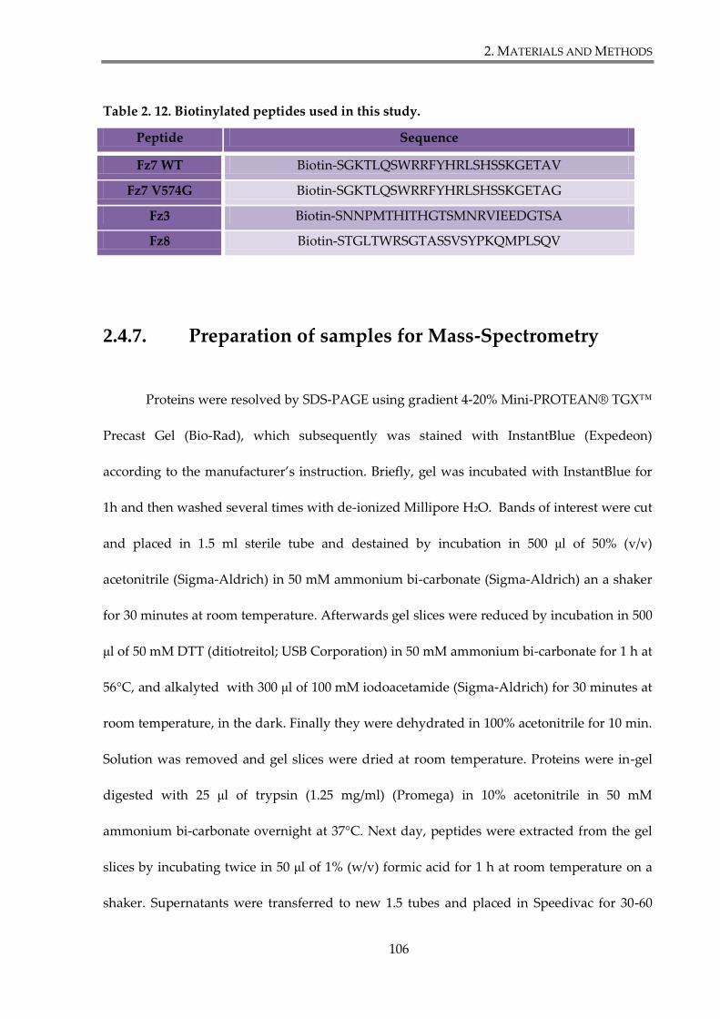
2. MATERIALS AND METHODS
106
Table 2. 12. Biotinylated peptides used in this study.
Peptide Sequence
Fz7 WT Biotin-SGKTLQSWRRFYHRLSHSSKGETAV
Fz7 V574G Biotin-SGKTLQSWRRFYHRLSHSSKGETAG
Fz3 Biotin-SNNPMTHITHGTSMNRVIEEDGTSA
Fz8 Biotin-STGLTWRSGTASSVSYPKQMPLSQV
2.4.7. Preparation of samples for Mass-Spectrometry
Proteins were resolved by SDS-PAGE using gradient 4-20% Mini-PROTEAN® TGX™
Precast Gel (Bio-Rad), which subsequently was stained with InstantBlue (Expedeon)
according to the manufacturer’s instruction. Briefly, gel was incubated with InstantBlue for
1h and then washed several times with de-ionized Millipore H2O. Bands of interest were cut
and placed in 1.5 ml sterile tube and destained by incubation in 500 μl of 50% (v/v)
acetonitrile (Sigma-Aldrich) in 50 mM ammonium bi-carbonate (Sigma-Aldrich) an a shaker
for 30 minutes at room temperature. Afterwards gel slices were reduced by incubation in 500
μl of 50 mM DTT (ditiotreitol; USB Corporation) in 50 mM ammonium bi-carbonate for 1 h at
56°C, and alkalyted with 300 μl of 100 mM iodoacetamide (Sigma-Aldrich) for 30 minutes at
room temperature, in the dark. Finally they were dehydrated in 100% acetonitrile for 10 min.
Solution was removed and gel slices were dried at room temperature. Proteins were in-gel
digested with 25 μl of trypsin (1.25 mg/ml) (Promega) in 10% acetonitrile in 50 mM
ammonium bi-carbonate overnight at 37°C. Next day, peptides were extracted from the gel
slices by incubating twice in 50 μl of 1% (w/v) formic acid for 1 h at room temperature on a
shaker. Supernatants were transferred to new 1.5 tubes and placed in Speedivac for 30-60

2. MATERIALS AND METHODS
107
min at 45°C in order to diminish sample volumes to 40-50 μl. Retained peptides were
analyzed by mass spectrometry.
2.4.8. Mass spectrometry
Proteomic analysis was performed at Proteomics Facility (University of Birmingham)
by Dr Doug Ward. Samples prepared in Section 2.4.7. were used in liquid chromatography
and mass spectrometry analysis, performed on Bruker micrOTOF II (LC-MS). To identify
proteins encompassing detected peptide sequences, data base SwissProtDecoy was used
(Species: Human). During peptide analysis following parameters were applied: Missed
Cleavage: 1; Accurance search: 50 ppm; Fragment searching: 0.1 Da; Peptide score Mascot :
25.

2. MATERIALS AND METHODS
108
2.5. Assays on live cells (functional studies)
2.5.1. Luciferase TOP-Flash assay
TOPFlash luciferase reporter assay was designed to demonstrate activation of
canonical Wnt pathway. It is a read-out for β-catenin transcription activation, the end-point
of canonical Wnt pathway (Korinek et al. 1997).
Cells were plated at the density 2.5×10⁴ per well in 48-well plate. If protein knock-
down was required during the assay, cells were transfected, in triplicates, with 2.4 pmol of
siRNA per well, using Lipofectamine RNAiMAX using reverse protocol during plating cells
(section 2.2.6.3). 24 hours later cells were transfected with DNA plasmids using FuGENE6
(section 2.2.6.2), indicated as a total amount per well in a 48-well plate: SKBr3 with 50 ng
luciferase plasmid (M50 Super 8x TOPFlash or M51 Super 8x FOPFlash; Addgene), 25 ng
pRenilla and 50 ng pcDNA™3.1 plasmids encoding the respective cDNA inserts; HEK293T
with 22.5 ng luciferase plasmid, 2.5 ng pRenilla, 5 ng of pcDNA™3.1 plasmids encoding the
respective cDNA inserts and supplemented with 95 ng pcDNA™3.1. M50 Super 8x
TOPFlash is a β-catenin reporter plasmid, containing TCF/LEF sites upstream of a luciferase
reporter. M51 Super 8x FOPFlash is a control for β-catenin reporter plasmid, containing
mutated TCF/LEF sites upstream of a luciferase reporter. Both constructs were generated at
Randall Moon’s laboratory (Veeman et al. 2003b), based on original TOPFlash constructed at
laboratory of Hans Clevers (Korinek et al. 1997).
At 48h following plating, cells were treated for 24 hours with 50% Wnt3a-conditioned
medium or 50% control medium produced by L-cells (Section 2.2.9). Subsequently, cells were
2. MATERIALS AND METHODS
109
washed with PBS and lysed by incubating with Passive Lysis Buffer (Promega) overnight at -
20°C, 50 μl/well. 20 μl of lysate from each well was transferred to 96-well plate. Firefly
(Photinus pyralis) and Renilla (Renilla reniformis) luminescence signals were measured
sequentially from the same sample, using the Dual-Glo luciferase assay system (Promega).
Briefly, the luminescence signal of the firefly luciferase expressed from TOPFlash or
FOPFlash plasmid was quantitatively measured first by the addition of Luciferase Assay
Reagent II (LARII). Afterwards, the firefly signal was quenched by addition of Stop & Glo®
reagent to the same sample, and Renilla luciferase signal was measured and quantified.
Luminesence signal was measured using Orion L Microplate Luminometer (Berthold
Detection Systems).
Firefly luminescence signal values were normalized according to their corresponding
Renilla signal values. TOP-flash signal was standardized to the background signal (FOP-
flash). Histograms were constructed to show the relative fold change in the levels of
promoter reporter activity. Statistical significance was determined using MS Excel software
using the relevant Student’s t-test after determining whether the sample variance is equal or
unequal by performing an F-test.
2. MATERIALS AND METHODS
110
2.5.2. In vivo ubiquitylation assay
HEK293T cells, plated on 6 cm dish, were co-transfected with HA-Ubiquitin, FLAG-
Fz7 and GFP-LNX2 (GFP-LNX1 p70; GFP-LNX1 p80) or GFP. 48h after transfection cells
were lysed using 100 μl of SDS lysis buffer (containing 0.5% SDS, 50 mM NaF, 1 mM EDTA
in PBS), supplemented with 2 mM PMSF, 10 μg/ml aprotinin, 10 μg/ml leupeptin and 20 mM
iodoacetemide. Samples were incubated at 60°C for 15 minutes, and diluted with 900 μl of
1% v/v Triton X-100 (150 mM NaCl, 50 mM Tris pH 7.4) supplemented with 2 mM PMSF, 10
μg/ml aprotinin, 10 μg/ml leupeptin and 20 mM iodoacetemide. Subsequently samples were
sonicated and the insoluble material was pelleted at 13,000 rpm for 10 min. Supernatants
were incubated for 1h with magnetic beads conjugated with anti-FLAG M2 mAb (Sigma-
Aldrich). Later, beads were washed three times with TBS-Tween. Proteins retained by the
magnetic resins were eluted by incubating with 15 μl of 2x Laemmli buffer for 10 minutes at
60°C. Eluates were analysed by the SDS-PAGE and Western Blotting as described in Section
2.4.3. Frizzled-7 ubiquitylation level was assessed by incubating membranes with rabbit
antibodies against HA-tag, to detect HA-ubiquitin.
2. MATERIALS AND METHODS
111
2.6. Staining and imaging methods
2.6.1. Flow cytometry
Cells growing in monolayer were detached using enzyme-free dissociation buffer
(Gibco) and counted. 3-5 x 10⁵ cells was centrifuged at 1000 RPM for 3 minutes and
resuspended in 50 μl of ice cold 1% HI-BSA/PBS. All the following steps were performed on
ice. To block non-specific binding of antibodies, 1% HI-BSA/PBS was aliquoted into U-
shaped 96-well plate (50 μl/well) and placed on ice for 20 minutes. Then, the blocking
solution was removed and replaced with 150 μl of specific primary antibody diluted in 1%
HI-BSA/PBS or hybridoma supernatant. Subsequently, 50 μl of cell suspension was added to
each well and incubated on ice for 1h. The cells were pelleted by centrifugation at 1000 RPM
for 5 minutes at 4°C and washed twice with 200 μl of ice cold PBS. The last wash was
followed by incubation of cells with secondary FITC- or PE-conjugated antibody (Sigma),
diluted in 1% HI-BSA/PBS for 1h. The incubation with fluorescently labelled secondary
antibodies was carried out under the aluminium foil to prevent sample exposure to light.
Cells were washed again twice with ice cold PBS, as described above. Finally cells were
resuspended in 250 μl of ice cold PBS and transferred into FACS tubes containing 250 μl of
4% paraformaldehyde (PFA). Stained cells were analyzed using Accuri C6 Flow Cytometer
(BD Biosciences) and FlowJo v7.6 software. All antibodies used in this study are listed in
Tables 2.1. and 2.2.
2. MATERIALS AND METHODS
112
2.6.2. Fluorescent Activated Cell Sorting (FACS)
Homogenous population of cells stably expressing Flag-tagged Frizzled proteins
(SKBr3-FLAG-Fz7 and SKBr3-FLAG-Fz7 V574G, section 2.2.7) was established using
Fluorescent Activated Cell Sorting (FACS). The day before cell sorting, cells were trypsinized
and replated at ~50-60% confluency. The sample preparation and all subsequent procedures
were conducted under sterile conditions. On the day of sorting, cells were detached using
enzyme-free dissociation buffer (Gibco) and pelleted by centrifugation at 1200 RPM for 3 min
at 4°C. Subsequently cells were resuspended in 1.5 ml of ice-cold complete growth medium
containing primary antibody, the anti-FLAG M2 mAb (Sigma-Aldrich) was diluted at 1:300
and sterilised by filtration. After incubation for 1 hour, cells were pelleted by centrifugation
at 1200 RPM for 3 min at 4°C, and washed twice with 2 ml of ice-cold complete growth
medium. Then cells were resuspended in 1.5 ml of ice cold complete growth medium
containing secondary, fluorescent labelled antibody (FITC (fluorescein isothiocyanate)-
conjugated goat anti-mouse pAb, (Sigma-Aldrich) in dilution 1:300) and incubated on ice for
one hour in dark. Cells were pelleted, and washed twice with ice-cold complete growth
medium. Finally they were resuspended in 2 ml of ice cold complete growth medium and
passed through Cell Trics filters (Partec) to remove cellular aggregates. Cell suspension was
transfered to sterile FACS tubes (BD Falcon). Cells were sorted using a Beckman Coulter
MoFlo Legacy FACS machine and positive and negative cell populations were separated.
After sorting cells were resuspended in complete growth medium containing double amount
of penicillin/streptomycin to prevent sample contamination. 48h later medium was changed
2. MATERIALS AND METHODS
113
for a regular complete growth medium and cells were cultivated in humidified incubator
(37°C, 5% CO2) until they were ready to use.
2.6.3. Immunofluorescence
Cells grown on glass coverslips or Nunc™ Lab-Tek™ II Chamber Slide™ System
(Thermo Scientific) were transfected with appropriate DNA plasmids using FuGENE6
(Roche). Staining was performed 48h after transfection. Cells were fixed with 4%
paraformaldehyde/PBS for 15 min at room temperature and permeabilized with 0.1% v/v
Triton X-100 for 2 minutes. Coverslips or chambered slides were incubated with blocking
buffer (20% heat inactivated goat serum/PBS) for 1h, followed by 1 h incubation with a
primary antibody, diluted in blocking solution. Cells were washed three times with PBS for
10 minutes, with agitation. Subsequently coverslips or Lab-Tek™ chambered slides were
incubated with appropriate secondary antibodies conjugated with fluorochrome (diluted in
blocking solution), for 1h in darkness. After removing secondary antibody, cells were
incubated with Hoechst33342 (1μg/ml in PBS) for 10 min at room temperature to stain cell
nuclei. Finally cells were washed again with PBS, as described earlier. Coverslips were air
dried and mounted on glass slides, whereas chambers from Lab-Tek™ slides were removed
and slides were mounted with appropriate size coverslips, using anti-fade fluorescence
mounting medium (SlowFade® Gold Antifade Mountant with DAPI, Molecular Probes®).
Coverslips were sealed on the edges using nail polish and stored at -20°C in darkness. The
images were captured using Zeiss LSM510 META confocal system with 63X oil immersion
objective. All antibodies used in this study are summarized in Tables 2.1. and 2.2.
3.RESULTS AND DISCUSSION
114
3. RESULTS AND DISCUSSION
3.1. Establishing a model system to study Fz7 biology
Frizzled-7 (Fz7) is a transmembrane receptor that transduces signals from secreted
glycoproteins known as Wnts. Activation of Wnt signalling pathway frequently occurs in
colorectal cancer, but also in breast cancer. Activation of canonical Wnt signalling is well
studied in colorectal cancer cells, and it is predominantly associated with mutations in APC
or β-catenin proteins (Polakis 2012). However, the mechanism of Wnt pathway activation in
breast cancer remains unclear. Recent reports suggested that Fz7 is involved in breast cancer
malignancy (King et al. 2012a;Simmons, Jr. et al. 2014;Yang et al. 2011)
However, the contribution of the C-terminal PDZ-BM in signalling triggered by Fz7
remains unknown. The work described in this chapter covers characterization of Fz7
expression vectors and identification of a suitable breast cancer cell line to use as a model in
further the study.
3.RESULTS AND DISCUSSION
115
3.1.1. Characterization of Fz7 expression vectors
There is no suitable antibody available to detect endogenous Fz7. Therefore, to be
able to monitor Fz7 expression throughout the functional experiments, a molecular tag had
to be introduced into the Fz7 sequence. The procedure is described in details in the section
2.3. of Materials and Methods. In the vast majority of the following experiments FLAG-Fz7
was used, but Myc- and GFP-Fz7 constructs were also generated.
Fz7 contains a PDZ binding motif on the C-terminal end that enables interactions of
the receptor with PDZ domain containing proteins. Given that the main objective of this
study was to investigate the role of Fz7 interactions with PDZ proteins, the molecular tag
was introduced at the N-terminal extracellular portion of the protein. Employing molecular
biology techniques, FLAG-, Myc- or GFP-sequences were introduced at the position 34, after
the leader peptide, to ensure correct topology of the protein on the plasma membrane.
A schematic representation of Fz7 structure with introduced tag is shown in Figure 3.1.A.
In order to assess FLAG-Fz7 expression in transfected cells, SDS-PAGE and western
blot analysis was used. Samples for the analysis were prepared by mixing cellular lysates
with 4xLaemmli loading buffer containing β-mercaptoethanol, and subsequently boiled at
100°C for 5 minutes. Anti-FLAG pAb were used to detect FLAG-Fz7. While analysing
samples by western blot, it was observed that Fz7 forms aggregates of a high molecular mass
and monomers of the receptor were not detected. Aggregate formation might be a
consequence of Fz7 being a transmembrane receptor with seven hydrophobic regions.
Therefore the procedure of sample preparation for SDS-PAGE-western blot analysis was
modified. In order to do find an alternative protocol several conditions were examined. Cell

3.RESULTS AND DISCUSSION
116
lysates, obtained from HEK293Ts transfected with FLAG-Fz7, were mixed with 4xLaemmli
loading buffer, with or without a reducing agent (β-mercaptoethanol), and incubated at
100°C for 5 minutes or at 60°C for 10 minutes. Results of these experiments are shown in the
Figure 3.1.B. When samples were boiled (100°C), Fz7 formed aggregates and receptor
monomers were impossible to detect (reduced conditions; lane 1) or they were barely
detectable (non-reduced conditions; lane 3). Decreasing the denaturation temperature to
60°C prevented formation of protein aggregates. Separated Fz7 forms included two
monomeric forms with apparent sizes 50 kDa and 60 kDa, (in reduced, lane 5, and non-
reduced conditions, lane 7). These forms are likely to represent a different degree of N-
glycosylation of Fz7 and were observed in both HEK293T and SKBr3 cells (see below). Under
non-reduced conditions (lanes 3 and 7), Fz7 dimers (around 130 kDa) and higher oligomers
with sized exceeding 200 kDa, were also observed.
In order to determine cell localization of Fz7, immunofluorescence analysis was
performed on MCF7s transiently transfected with FLAG-Fz7 expression vector. Cells were
fixed and stained using anti-FLAG antibody. DAPI was used to visualize nuclei. Analysis by
confocal microscopy showed that Fz7 immunoreactivity is mostly detected on the plasma
membrane (Figure 3.1.C). Similar, plasma membrane localization of Fz7 was observed in
SKBr3 cells (see below).
As mentioned at the beginning of this section, in addition to FLAG-Fz7 expression
vector, Myc-Fz7 and GFP-Fz7 expression vectors were generated. Both of them were
subjected to a similar analysis as FLAG-Fz7 construct. Both Myc- and GFP-Fz7 were found to
be expressed in transfected cells and localize on the plasma membrane. Western blot analysis
showed that Myc-Fz7 and GFP-Fz7 are also expressed as two monomers with a different
3.RESULTS AND DISCUSSION
117
degree of N-glycosylation (50 kDa and 60 kDa for Myc-Fz7; 80 kDa and 90 kDa for GFP-Fz7),
but also form dimers and aggregates of higher molecular weight, as seen for FLAG-Fz7.
Summary of this work is presented in the Supplementary Figure S.1.
Taken together, western blot and immunofluorescence experiments showed that
generated construct allows for FLAG-Fz7, Myc-Fz7 and GFP-Fz7 expression on the surface of
transfected cells.
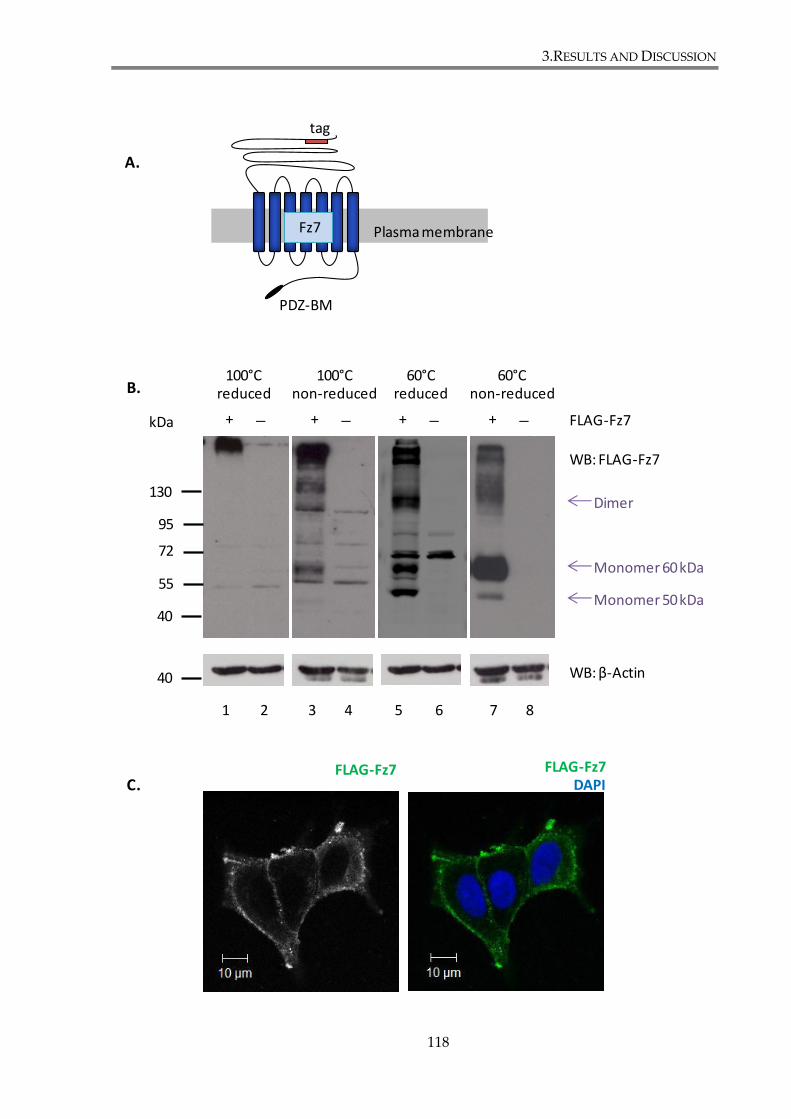
3.RESULTS AND DISCUSSION
118
Plasma membraneFz7
PDZ-BM
tag
95
72
55
100°C reduced
100°C non-reduced
60°C reduced
60°C non-reduced
kDa + + + + FLAG-Fz7
WB: FLAG-Fz7
WB: β-Actin
FLAG-Fz7 FLAG-Fz7DAPI
A.
B.
C.
1 2 3 4 5 6 7 8
130
40
40
Dimer
Monomer 60 kDa
Monomer 50 kDa
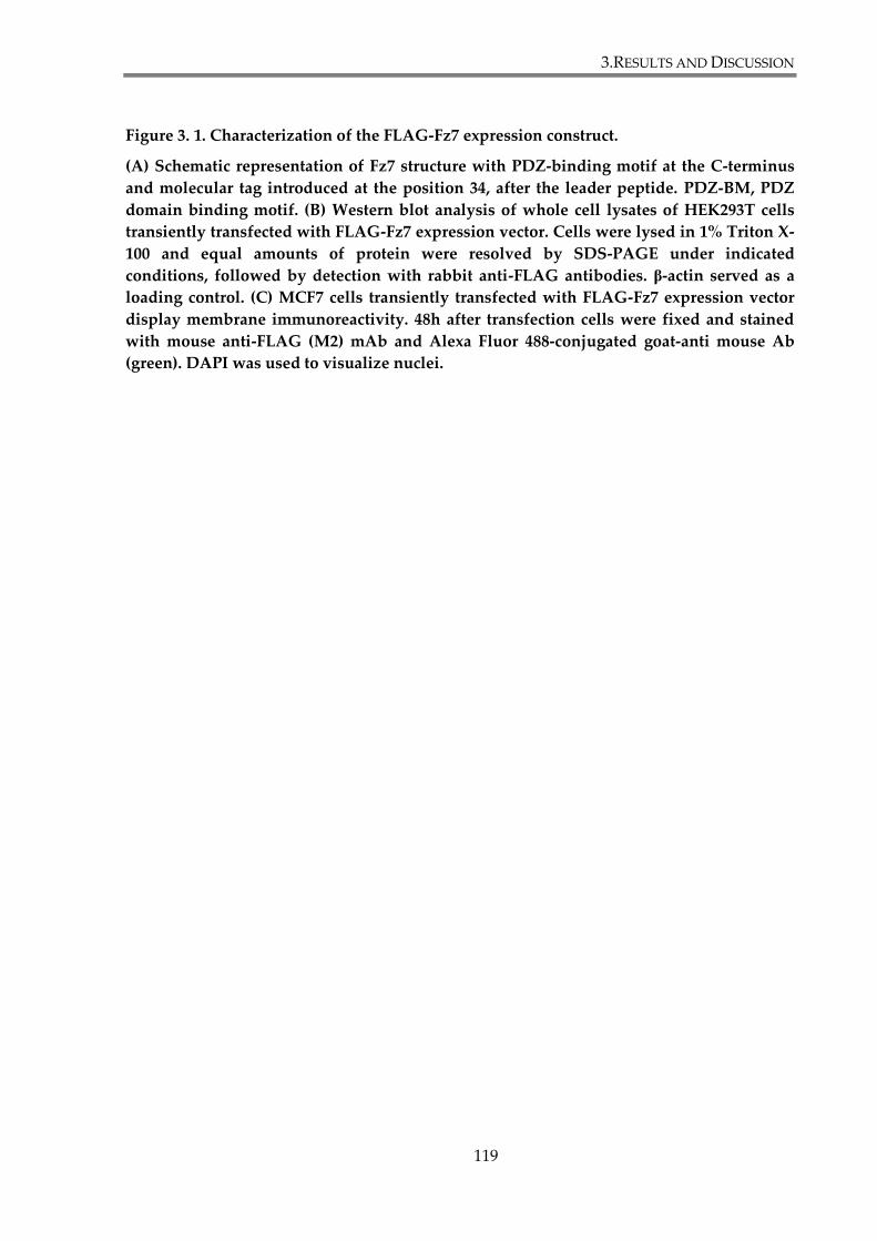
3.RESULTS AND DISCUSSION
119
Figure 3. 1. Characterization of the FLAG-Fz7 expression construct.
(A) Schematic representation of Fz7 structure with PDZ-binding motif at the C-terminus
and molecular tag introduced at the position 34, after the leader peptide. PDZ-BM, PDZ
domain binding motif. (B) Western blot analysis of whole cell lysates of HEK293T cells
transiently transfected with FLAG-Fz7 expression vector. Cells were lysed in 1% Triton X-
100 and equal amounts of protein were resolved by SDS-PAGE under indicated
conditions, followed by detection with rabbit anti-FLAG antibodies. β-actin served as a
loading control. (C) MCF7 cells transiently transfected with FLAG-Fz7 expression vector
display membrane immunoreactivity. 48h after transfection cells were fixed and stained
with mouse anti-FLAG (M2) mAb and Alexa Fluor 488-conjugated goat-anti mouse Ab
(green). DAPI was used to visualize nuclei.
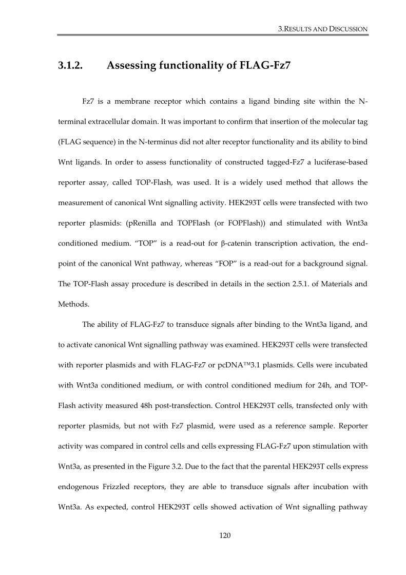
3.RESULTS AND DISCUSSION
120
3.1.2. Assessing functionality of FLAG-Fz7
Fz7 is a membrane receptor which contains a ligand binding site within the N-
terminal extracellular domain. It was important to confirm that insertion of the molecular tag
(FLAG sequence) in the N-terminus did not alter receptor functionality and its ability to bind
Wnt ligands. In order to assess functionality of constructed tagged-Fz7 a luciferase-based
reporter assay, called TOP-Flash, was used. It is a widely used method that allows the
measurement of canonical Wnt signalling activity. HEK293T cells were transfected with two
reporter plasmids: (pRenilla and TOPFlash (or FOPFlash)) and stimulated with Wnt3a
conditioned medium. “TOP” is a read-out for β-catenin transcription activation, the end-
point of the canonical Wnt pathway, whereas “FOP” is a read-out for a background signal.
The TOP-Flash assay procedure is described in details in the section 2.5.1. of Materials and
Methods.
The ability of FLAG-Fz7 to transduce signals after binding to the Wnt3a ligand, and
to activate canonical Wnt signalling pathway was examined. HEK293T cells were transfected
with reporter plasmids and with FLAG-Fz7 or pcDNA™3.1 plasmids. Cells were incubated
with Wnt3a conditioned medium, or with control conditioned medium for 24h, and TOP-
Flash activity measured 48h post-transfection. Control HEK293T cells, transfected only with
reporter plasmids, but not with Fz7 plasmid, were used as a reference sample. Reporter
activity was compared in control cells and cells expressing FLAG-Fz7 upon stimulation with
Wnt3a, as presented in the Figure 3.2. Due to the fact that the parental HEK293T cells express
endogenous Frizzled receptors, they are able to transduce signals after incubation with
Wnt3a. As expected, control HEK293T cells showed activation of Wnt signalling pathway
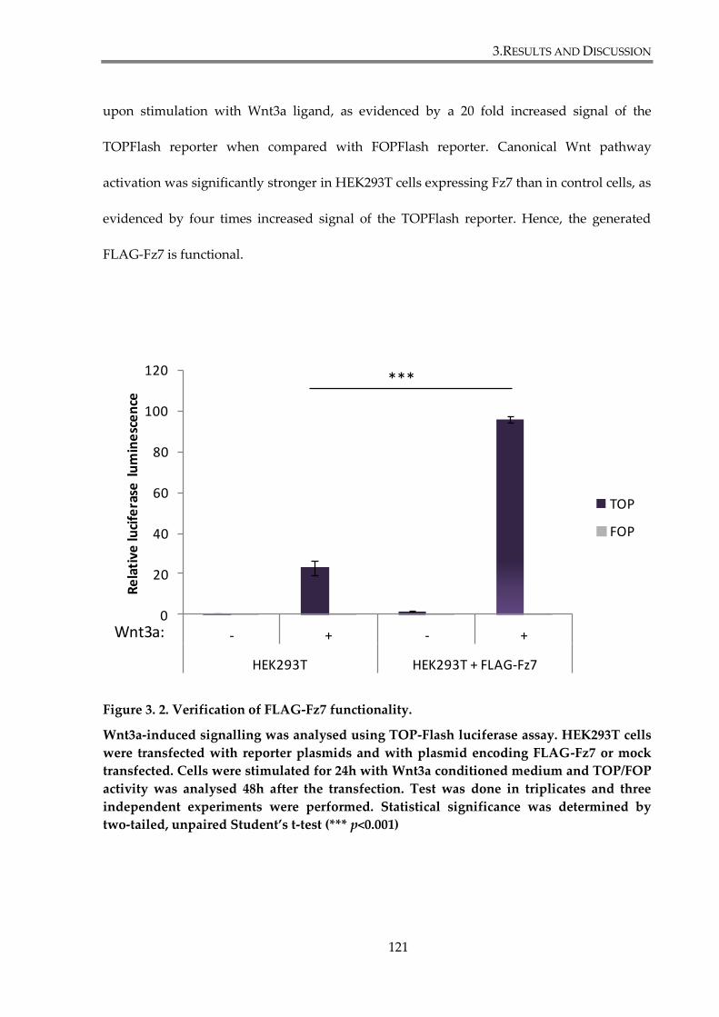
3.RESULTS AND DISCUSSION
121
upon stimulation with Wnt3a ligand, as evidenced by a 20 fold increased signal of the
TOPFlash reporter when compared with FOPFlash reporter. Canonical Wnt pathway
activation was significantly stronger in HEK293T cells expressing Fz7 than in control cells, as
evidenced by four times increased signal of the TOPFlash reporter. Hence, the generated
FLAG-Fz7 is functional.
Figure 3. 2. Verification of FLAG-Fz7 functionality.
Wnt3a-induced signalling was analysed using TOP-Flash luciferase assay. HEK293T cells
were transfected with reporter plasmids and with plasmid encoding FLAG-Fz7 or mock
transfected. Cells were stimulated for 24h with Wnt3a conditioned medium and TOP/FOP
activity was analysed 48h after the transfection. Test was done in triplicates and three
independent experiments were performed. Statistical significance was determined by
two-tailed, unpaired Student’s t-test (*** p<0.001)
0
20
40
60
80
100
120
- + - +
HEK293T HEK293T + FLAG-Fz7
Re
lati
ve lu
cife
rase
lu
min
esc
en
ce
TOP
FOP
Wnt3a:
***
3.RESULTS AND DISCUSSION
122
Functionalities of other generated Fz7 constructs (Myc-Fz7 and GFP-Fz7) were also
assessed in HEK293T cells in similar experiments. The activation level of Wnt canonical
pathway was two fold higher in cells expressing Myc-Fz7 and almost three fold higher in
cells expressing GFP-Fz7, when compared with control samples, as presented in the
Supplementary Figure S.2. Therefore both Myc-Fz7 and GFP-Fz7 appear to be functional.
Wnt pathway activation was also assessed in HEK293T cells expressing Fz7-V5-His.
V5- and His-tag were introduced at the C-terminal end of Fz7 receptor, leaving the N-
terminus intact. Transfection of HEK293T cells with different amounts of DNA encoding
Fz7-V5-His (80 ng, 40 ng, 20 ng, 10ng, 5 ng or 2,5 ng as per one well of 48-well plate) showed
that signal transduction depends on expression level of Fz7. High level of the receptor
expression prevents activation of canonical Wnt signalling pathway by endogenous Frizzled
receptors in HEK293Ts, as was observed in cells transfected with 80 ng to 10 ng of Fz7-V5-
His. The highest activation of Wnt signalling was found in HEK293Ts transfected with 5 ng
of Fz7-V5-His plasmid. Yet, even in the sample with the highest activation of the pathway, it
wasn’t significantly higher then in the control cells, thus demonstrating that insertion of the
molecular tag at the C-terminus of Fz7 receptor attenuates its capability to transduce signals
(Figures 3.2. and 3.3). Furthermore, our results seems to indicate that the Fz7-V5-His
construct have a dominant negative properties.
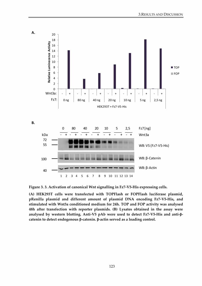
3.RESULTS AND DISCUSSION
123
Figure 3. 3. Activation of canonical Wnt signalling in Fz7-V5-His expressing cells.
(A) HEK293T cells were transfected with TOPFlash or FOPFlash luciferase plasmid,
pRenilla plasmid and different amount of plasmid DNA encoding Fz7-V5-His, and
stimulated with Wnt3a conditioned medium for 24h. TOP and FOP activity was analysed
48h after transfection with reporter plasmids. (B) Lysates obtained in the assay were
analysed by western blotting. Anti-V5 pAb were used to detect Fz7-V5-His and anti-β-
catenin to detect endogenous β-catenin. β-actin served as a loading control.
0
2
4
6
8
10
12
14
16
18
20
- + - + - + - + - + - + - +
0 ng 80 ng 40 ng 20 ng 10 ng 5 ng 2,5 ng
HEK293T + Fz7-V5-His
Re
lati
ve L
um
ine
cen
ce A
ctiv
ity
TOP
FOP
Wnt3a:
Fz7:
WB: β-Catenin
WB: V5 (Fz7-V5-His)
WB: β-Actin
100
72
55
kDa Wnt3a- + - + - + - + - + - + - +
40
0 80 40 20 10 5 2,5 Fz7 [ng]
1 2 3 4 5 6 7 8 9 10 11 12 13 14
A.
B.
3.RESULTS AND DISCUSSION
124
Taken together, insertion of molecular tags within the N-terminus of Fz7 (at the
position 34) does not affect the receptor functionality. However, inserting a tag at the C-
terminus, behind the PDZ binding motif, leads to loss of receptor ability to transduce the
canonical Wnt signalling. This indicates the importance of Fz7 interactions with PDZ
proteins, via the C-terminal binding motif, in the activation of the canonical Wnt signalling.
3.RESULTS AND DISCUSSION
125
3.1.3. Identifying a suitable epithelial breast cancer model
Having generated a functional epitope-tagged Fz7 construct, we proceeded to
identify a suitable breast cancer cell line to use as a model for signalling studies. To
investigate activation of canonical Wnt signalling pathway by Fz7, we have decided to find a
model system in which we could establish a stable expression of the Fz7. For that purpose
several well-characterized epithelial breast cancer cell lines were initially screened using
TOP-Flash reporter assay. The aim of these experiments was to find cells with low
responsiveness to Wnt3a, indicating low expression of endogenous receptors. At the same
time, we were looking for cells in which all the components of the canonical Wnt signalling
pathway downstream of the receptor were intact (i.e. be able to transduce signals upon
activation).
In order to identify a suitable cell line, activation of the canonical Wnt signalling
pathway was analysed in cells stimulated with Wnt3a or when treated with SB216763, an
inhibitor of GSK3,. Wnt3a activates Wnt signalling through the interaction with Frizzled
receptor. TOP-Flash activity, after Wnt3a stimulation, would reflect on expression of the
receptors and an overall functionality of the pathway. SB216763 is a small molecule that
inhibits GSK3 by competing with ATP. Inhibition of GSK3 kinase activates β-catenin
dependent Wnt pathway downstream of Wnt receptor and allows testing functionality of the
pathway in cells deficient in the expression of the receptors. Four epithelial breast cancer cell
lines were tested in these experiments: MDA-MB-231, T47D, MCF7 and SKBr3. HEK293T cell
were used as a control cell line. Cells were transfected with reporter plasmids and incubated
for 24h with Wnt3a conditioned medium or with control conditioned medium (Figure 3.4.A).
3.RESULTS AND DISCUSSION
126
In parallel experiments, cells were treated for 24h with 10 μM SB216763 or vehicle (DMSO)
(Figure 3.4.B). 48h after the transfection activity of TOP-Flash reporter was analysed and
activation of the Wnt pathway was evaluated in samples treated with Wnt3a and GSK3
inhibitor. In order to compare results in different cell lines, obtained TOPFlash reporter
values were standardized to the corresponding FOPFlash reporter values for each of the
tested cell lines. We found that in MDA-MB-231 cells, the canonical Wnt pathway could be
activated by both Wnt3a (two fold increase), and GSK3i (four fold increase), compared to
control samples. These results indicate that canonical Wnt pathway is functional in MDA-
MB-231, but also that these cells express endogenous Frizzled receptors. On the contrary,
T47D and MCF7 cell lines did not display pathway activation upon stimulation with Wnt3a
or GSK3i thus suggesting that the β-catenin-dependent pathway is not functional in these
cells. This may be due to mutations in pathway components downstream of Frizzled, or
interference of other proteins involved in regulation of the pathway (Wheeler et al. 2011).
Finally, analysis of the Wnt signalling pathway in SKBr3 cell line showed that it was not
activated by Wnt3a, but could be induced by GSK3i (more than seven fold stronger when
compared with the control). Whilst these results indicate that SKBr3 cells express no (or low
levels) of endogenous Wnt3a receptors, the β-catenin-dependent pathway remains
functional. That makes the SKBr3 cell line a suitable model to establish stable cell lines and to
study Fz7-dependent signalling.
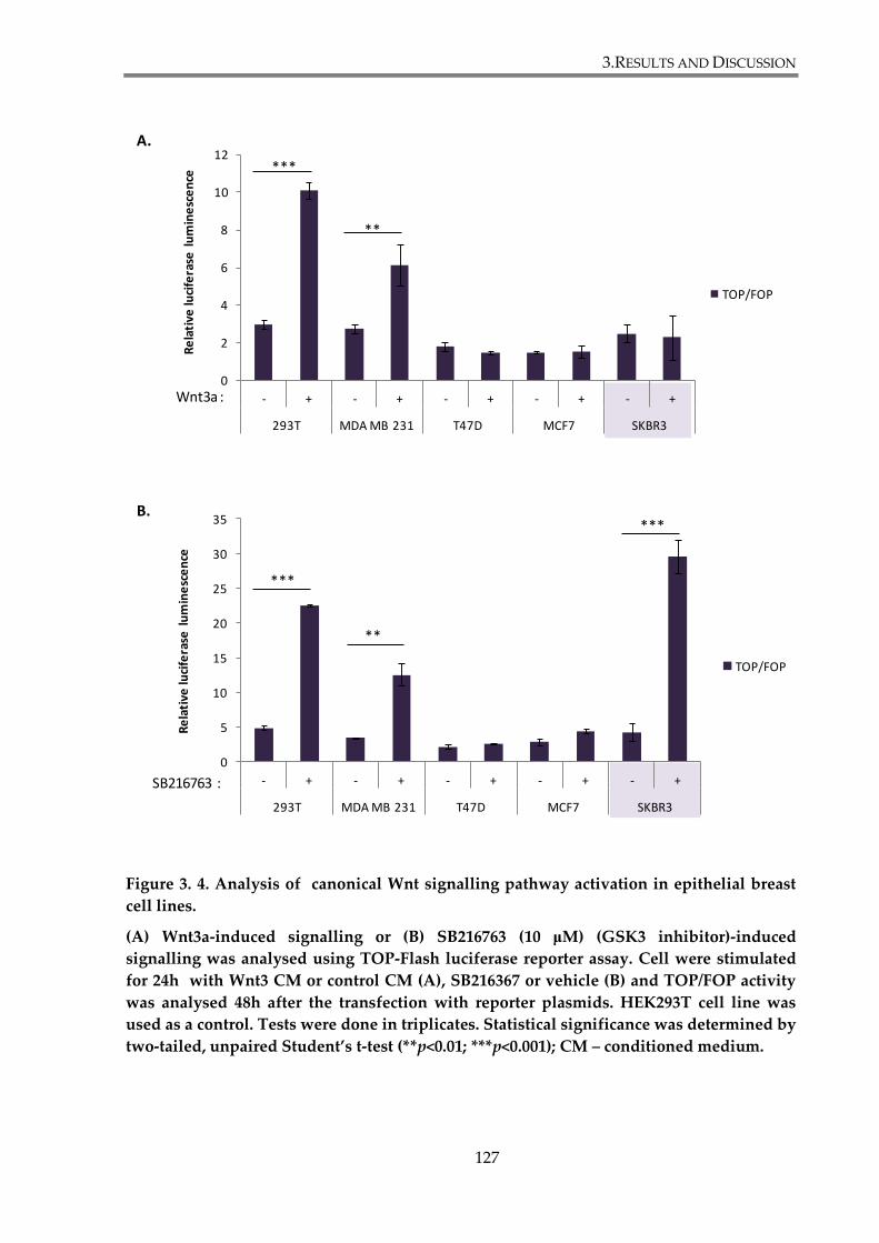
3.RESULTS AND DISCUSSION
127
Figure 3. 4. Analysis of canonical Wnt signalling pathway activation in epithelial breast
cell lines.
(A) Wnt3a-induced signalling or (B) SB216763 (10 μM) (GSK3 inhibitor)-induced
signalling was analysed using TOP-Flash luciferase reporter assay. Cell were stimulated
for 24h with Wnt3 CM or control CM (A), SB216367 or vehicle (B) and TOP/FOP activity
was analysed 48h after the transfection with reporter plasmids. HEK293T cell line was
used as a control. Tests were done in triplicates. Statistical significance was determined by
two-tailed, unpaired Student’s t-test (**p<0.01; ***p<0.001); CM – conditioned medium.
0
2
4
6
8
10
12
- + - + - + - + - +
293T MDA MB 231 T47D MCF7 SKBR3
Re
lati
ve lu
cife
rase
lu
min
esc
en
ce
TOP/FOP
Wnt3a :
***
**
0
5
10
15
20
25
30
35
- + - + - + - + - +
293T MDA MB 231 T47D MCF7 SKBR3
Re
lati
ve lu
cife
rase
lu
min
esc
en
ce
TOP/FOP
SB216763 :
***
**
***
A.
B.
3.RESULTS AND DISCUSSION
128
3.1.4. Fz7 activates canonical Wnt signalling pathway in
SKBr3 cells
In the previous section, SKBr3 epithelial breast cancer cell line was identified as a
suitable model to establish stable cell lines for signalling studies. To verify whether Fz7 is
able to induce activation of canonical Wnt signalling in SKBr3 cells, when stimulated with
Wnt3a, activity of TOPFlash reporter was assessed in cells transiently expressing FLAG-Fz7.
SKBr3 cells were transfected with TOP-Flash reporter plasmids, and with vector expressing
FLAG-Fz7 or mock transfected (pcDNA™3.1). Subsequently cells were incubated with
Wnt3a conditioned medium or with control conditioned medium. Activation of Wnt
signalling upon stimulation with Wnt3a was examined using the TOP-Flash luciferase assay.
Cellular lysates obtained in the assay were also analysed by western blotting. The canonical
Wnt pathway was significantly stronger activated in SKBr3 cells expressing Fz7 cells, as
evidence by more than a six fold increase in signal (Figure 3.5.A). Accordingly, stabilization
of β-catenin was observed in western blot experiments (Figure 3.5.B). Here, control SKBr3
cells (not expressing FLAG-Fz7) exhibit low pathway activation (four fold increase of
TOPFlash activity over the base level). It can be explained by the fact that in this experiment
more sensitive TOPFlash reporter construct was used. Specifically, the reporter construct
contained eight binding sites for TCF/β-catenin, as compared to three sites in the construct
used for the results shown in Figure 3.4. The 8xTOPFlash reporter plasmid was used in all
following experiments.. Taken together, these experiments demonstrated the functionality of
the ectopically expressed Flag-Fz7 construct in SKBr3 cell line.

3.RESULTS AND DISCUSSION
129
Figure 3. 5. Fz7 activates the canonical Wnt pathway in SKBr3 cells.
(A) Wnt-induced signalling was analysed using TOP-Flash luciferase assay. SKBr3 cells
were transiently transfected with plasmid encoding FLAG-Fz7 or mock transfected. Cells
were stimulated for 24h with Wnt3a conditioned medium and TOP/FOP activity was
analysed 48h after transfection with reporter plasmids. Tests were done in triplicates and
three independent experiments were performed. Statistical significance was determined
by two-tailed, unpaired Student’s t-test (* p<0.05). (B) Western blot analysis of Frizzzled-7
expression level in transiently transfected SKBr3 cells. β-actin served as a loading control.
Image shown is a representative of three independent experiments.
0
5
10
15
20
25
30
35
- + - +
SKBr3 SKBr3 + FLAG-Fz7
Re
lati
ve lu
cife
rase
lu
min
esc
en
ce
TOP/FOP
Wnt3a:
*
– + – + Wnt3a
WB: β-Catenin
WB: FLAG -Fz7
WB: β -Actin
100
72
55
kDa
SKB
r3
SKB
r3 +
FLA
G-F
z7
1 2 3 4
A.
B.
40
3.RESULTS AND DISCUSSION
130
3.1.5. Generation and characterization of SKBr3 cell lines
stably expressing wild type and mutant of Fz7.
The SKBr3 cell line expresses endogenous Frizzled receptors at relatively low levels
and canonical Wnt signalling in these cells can only be weakly induced by Wnt ligands.
Nonetheless, introduction of Fz7 into SKBr3 cells leads to strong activation of the pathway in
response to Wnt3a stimulation (sections 3.1.3. and 3.1.4.). These properties made SKBr3 cell
line a suitable model to study Fz7-mediated signalling. SKBr3 cells were stably transfected
with FLAG-Fz7 expression vector as described in section 2.2.7 of Materials and Methods.
Fluorescence-activated Cell Sorting (FACS) using an anti-FLAG mAb was subsequently
performed in order to select and separate FLAG-Fz7 expressing cells from non-expressing
cells, as described in section 2.6.1 of Materials and Methods. FACS-based enrichment was
performed twice to obtain a homogenous population of SKBr3 cells expressing Fz7 on the
cell surface. This cell line will be hereafter referred to as SKBr3-FLAG-Fz7.
A similar approach was used to generate SKBr3 cell line expressing FLAG-Fz7 V574G
mutant of Fz7. In this mutant, the hydrophobic valine at position 574 (Val574, V) was
replaced with neutral amino acid glycine (Gly, G) (Figure 3.5.A). We predicted that such
mutation would prevent binding of PDZ domain containing proteins to Fz7. This cell line
will be referred to as SKBr3-FLAG-Fz7 V574G in the study.

3.RESULTS AND DISCUSSION
131
3.1.5.1. Assessing cell surface expression, overall expression and
cell localization of Fz7 in SKBr3 cells.
After generating the SKBr3-FLAG-Fz7 and SKBr3-FLAG-Fz7 V574G cell lines,
expression of the wild type and V574G mutant Fz7 receptor was verified by WB, flow
cytometry and immunofluorescence staining.
Figure 3.6.B. shows the results western blot experiments: comparable expression
levels were observed for both Fz7 WT (lane 2) and Fz7 V574G (lane 3). However, we have
observed differences in the appearance of antibody-reactive bands in cells expressing Fz7
WT and Fz7 V574G. Specifically, the relative abundance of the 60 kDa protein species was
higher in cells expressing Fz7 V574G mutant, when compared with Fz7 WT. Flow cytometry
analysis also revealed comparable cell surface levels of Fz7 in both cell lines (Figure 3.6.C). In
order to determine localization of Fz7 in SKBr3 cells, an immunofluorescence staining
analysis was carried out. Figure 3.6.D. shows that both, Fz7 WT and Fz7 V574G, were mainly
detected on the plasma membrane. Interestingly, Fz7 V574G expressing cells formed small
Fz7 positive membrane protrusions, which have not been observed in cells expressing wild
type Fz7.
Taken together, WB analysis, flow cytometry, and immunofluorescence staining
demonstrated that Fz7 is expressed in SKBr3-FLAG-Fz7 and SKBr3-FLAG-Fz7 V574G cells
and that it is mainly localized on the cell surface.
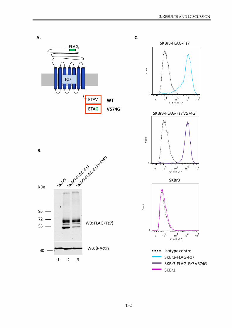
3.RESULTS AND DISCUSSION
132
A.
B.
95
72
55
kDa
WB: FLAG (Fz7)
WB: β-Actin
1 2 3
SKBr3-FLAG-Fz7
SKBr3-FLAG-Fz7 V574G
SKBr3
Isotype control
SKBr3-FLAG-Fz7
SKBr3-FLAG-Fz7 V574G
SKBr3
Fz7
FLAG
ETAV
ETAG V574G
WT
C.
40
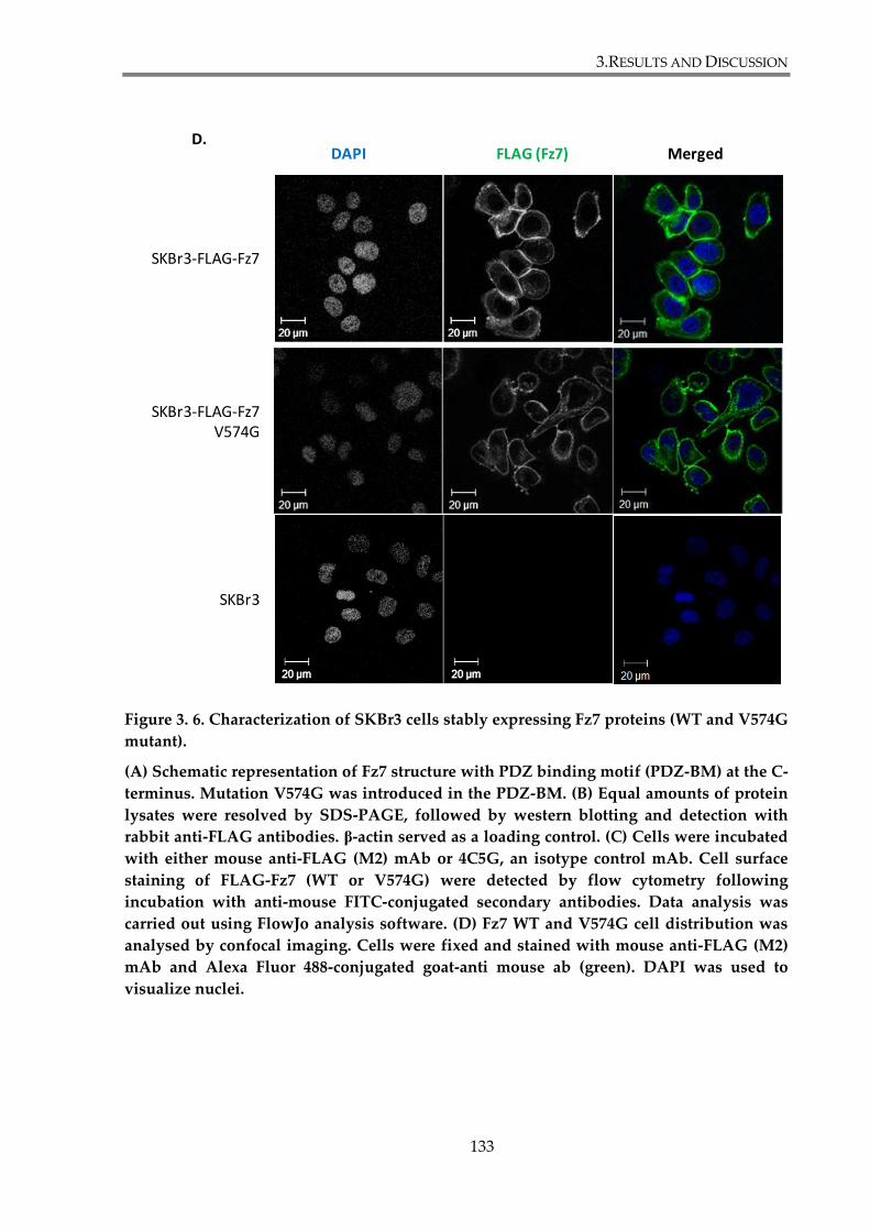
3.RESULTS AND DISCUSSION
133
Figure 3. 6. Characterization of SKBr3 cells stably expressing Fz7 proteins (WT and V574G
mutant).
(A) Schematic representation of Fz7 structure with PDZ binding motif (PDZ-BM) at the C-
terminus. Mutation V574G was introduced in the PDZ-BM. (B) Equal amounts of protein
lysates were resolved by SDS-PAGE, followed by western blotting and detection with
rabbit anti-FLAG antibodies. β-actin served as a loading control. (C) Cells were incubated
with either mouse anti-FLAG (M2) mAb or 4C5G, an isotype control mAb. Cell surface
staining of FLAG-Fz7 (WT or V574G) were detected by flow cytometry following
incubation with anti-mouse FITC-conjugated secondary antibodies. Data analysis was
carried out using FlowJo analysis software. (D) Fz7 WT and V574G cell distribution was
analysed by confocal imaging. Cells were fixed and stained with mouse anti-FLAG (M2)
mAb and Alexa Fluor 488-conjugated goat-anti mouse ab (green). DAPI was used to
visualize nuclei.
FLAG (Fz7) MergedDAPI
SKBr3-FLAG-Fz7
SKBr3
D.
SKBr3-FLAG-Fz7V574G
3.RESULTS AND DISCUSSION
134
3.1.5.2. Mutation in the C-terminal PDZ binding motif of Fz7
attenuates canonical Wnt signalling in SKBr3 cells
Having established and characterized SKBr3 cell lines stably expressing FLAG-Fz7
wild type and FLAG-Fz7 V574G, activation of canonical Wnt signalling was compared in
these two cell lines. SKBr3 parental, SKBr3-FLAG-Fz7 and SKBr3-FLAG-Fz7 V574G cells
were transfected with reporter plasmids and incubated with Wnt3a conditioned medium for
24h. 48h after transfection, the activation of β-catenin-dependent signalling in the cell lines
was assessed by TOP-Flash luciferase reporter assay. Lysates obtained in the assay were
further analysed by WB to confirm Fz7 expression between cell lines and to monitor β-
catenin stabilization within cells after Wnt3a stimulation. As demonstrated in the Figure 3.7.
both, Fz7 WT and Fz7 V574G, activate Wnt signalling pathway over the background level of
parental SKBr3 cell line. This was evident by the increase of TOPFlash reporter signal
(Figure 3.7.A) and β-catenin stabilization detected in western blot analysis (Figure 3.7.B). The
comparison of canonical Wnt pathway activation in cell lines expressing Fz7 WT or Fz7
V574G reveals that Fz7 wild type is more potent signalling receptor when compared with the
Fz7 V574G mutant. In cells expressing Fz7 WT the signal TOPFlash reporter was five times
stronger, when compared to the parental cell line. By comparison, cells expressing Fz7
V574G exhibited only a three fold signal increase. A histogram in the Figure 3.7.A represents
one of three independent experiments. The compilation of the results obtain in three
independent experiments is presented in the Supplementary Figure S.3. In some of the
experiments we also noticed that the level of 60 kDa monomer in the Fz7 WT sample
decreased after stimulation with Wnt3a ligand. However, these changes were not observed
3.RESULTS AND DISCUSSION
135
in all the experiments and, therefore, require further investigation. Our results clearly
demonstrate that the introduction of mutation V574G in the C-terminal PDZ-BM attenuates
activation of the Wnt pathway. These findings provide an evidence for the importance of Fz7
binding to PDZ domain containing proteins in the regulation of the canonical Wnt signalling.
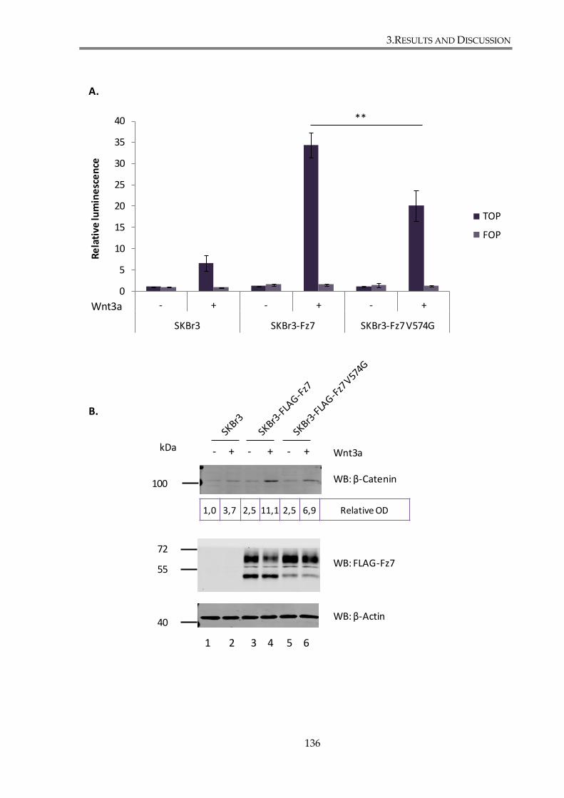
3.RESULTS AND DISCUSSION
136
0
5
10
15
20
25
30
35
40
- + - + - +
SKBr3 SKBr3-Fz7 SKBr3-Fz7 V574G
Re
lati
ve lu
min
esc
en
ce
TOP
FOP
Wnt3a
**
1 2 3 4 5 6
WB: β-Catenin
WB: FLAG-Fz7
WB: β-Actin
100
72
55
kDaWnt3a- + - + - +
A.
B.
1,0 3,7 2,5 11,1 2,5 6,9 Relative OD
40
3.RESULTS AND DISCUSSION
137
Figure 3. 7. Mutation in the C-terminal PDZ-binding motif of Fz7 attenuates the canonical
Wnt signalling in SKBr3 cells.
(A) Wnt-induced signalling was analysed using TOP-FLASH luciferase assay. SKBr3 cells
were stimulated for 24h with Wnt3a conditioned medium and TOP/FOP activities were
analysed 48 hours after the transfection with reporter plasmids. Tests were done in
triplicates. Statistical significance was determined by two-tailed, unpaired Student’s t-test
(**p<0.01). (B) Western blot analysis of Fz7 expression in SKBr3 cells using anti-FLAG
pAb. β-catenin expression level was monitored using anti-β-catenin mAb. Densitometry
values for β-catenin were standardized to the β-actin values of the same sample. β-actin
served a loading control. Image shown is a representative of three independent
experiments.

3.RESULTS AND DISCUSSION
138
3.1.6. Discussion
The ability to monitor the expression level of Fz7 receptor throughout the
experiments was essential. Due to lack of reagents detecting the endogenous receptor in
cells, it was necessary to generate constructs to express tagged forms of the Fz7 receptor.
Protein expression, subcellular localization and the luciferase-based TOP-Flash assays
demonstrated that the insertion of the tag sequences did not affect the functionality of Fz7.
Interestingly, we detected different forms of Fz7 in cells, including two monomers (50 kDa
and 60 kDa) characterized by different degree of N-glycosylation, dimers (around 130 kDa)
and higher oligomers with sized exceeding 200 kDa. Similar forms of Fz7 have been reported
by Struewing and colleagues, who analysed expression of the protein in bovine aortic
endothelial cells (BAEC) (Struewing et al. 2007). They showed that both monomers of Fz7 (54
kDa and 64 kDa) were sensitive to tunicamycin treatment, an inhibitor of N-glycosylation.
Furthermore, the lower molecular weight species were also sensitive to endoglycosidase H
treatment that specifically removes N-linked oligomannose, but not complex
oligosaccharides. Oligomannose chains are adjoined to the receptor during the initial steps of
glycosylation in the ER, whereas more complex oligosaccharides are added in the Golgi.
Hence it was concluded that the 64 kDa monomer is a fully mature receptor, while the 54
kDa monomer is an immature receptor retained in the ER (Struewing et al. 2007). Protein
glycosylation has been shown in the past to be crucial in Wnt signalling. Specifically,
porcupine (Por)/Mom-1 has been shown to be involved in N-glycosylation of Wnt ligands in
the ER (Kadowaki et al. 1996;Tanaka et al. 2002), whereas Boca/Mesd has been implicated to
act as a chaperone for LRP5/6 (Culi et al. 2003). However glycosylation of Frizzled receptors
3.RESULTS AND DISCUSSION
139
haven’t been extensively studied. Yamamoto and colleagues found that Frizzled
glycosylation is regulated by the protein Shisa, which mediates Fz7 retention in the ER
during maturation (Yamamoto et al. 2005). In another study glycosylation was suggested to
facilitate binding of Wnt ligands to mature receptors (Schulte et al. 2007).
We have observed that in HEK293T cells FLAG-tagged Fz7 formed dimers and high
order oligomers. Similarly, the ability of Frizzled receptors to form dimers and oligomers has
been reported previously (Kaykas et al. 2004). These authors have shown that different
Frizzled receptors (Fz1, 2, 4, 7 and 9) form specific homo- and hetero-oligomers. The
heptahelical transmembrane domain of receptors is sufficient for the oligomerization, which
occurs independently of the CRD domain C-terminal tail and associated scaffolding proteins,
and is not influenced by Wnt ligands. Frizzled oligomerization has been also shown to occur
in the ER during or after translation. It was suggested that the process may modulate Wnt
signalling, as different combinations hetero-oligomers may signal in a distinct fashion than
homo-oligomers (Kaykas et al. 2004).
To assess the functionality of the generated tagged-Fz7 constructs we relied on the
luciferase-based reporter assay (called TOP-Flash). It is a widely used test, enabling to
quantify activation level of canonical Wnt pathway in cells, first described in (Korinek et al.
1997). We found that all generated constructs are functional, thus demonstrating the tag
insertion on the N-terminal site did not influence ability of the receptor to transduce signals.
On the other hand, C-terminally tagged Fz7-V5-His was not able to efficiently activate the
canonical Wnt pathway (Figure 3.3.), indicating the importance of the C-terminus in signal
transduction. Importantly, the critical role of the C-terminal PDZ-BM was further confirmed
in experiments involving the Fz7 V574G mutant: ligand-induced activation of the canonical
3.RESULTS AND DISCUSSION
140
Wnt pathway in cells expressing the mutant receptor was ~30% lower when compared to
that observed in the Fz7 wild type expressing cells. To our knowledge this is the first
demonstration of the involvement of the C-terminal PDZ-BM of a Frizzled receptor in the
canonical Wnt pathway. Several studies also shown that the PDZ-BM of a protein is
important for regulation of its signalling. For example, Delta1, a ligand for the Notch
receptor, contains a PDZ-BM within its C-terminus. Mutation of its C-terminal valine leads to
activation of Notch signalling (Estrach et al. 2007). PDZ proteins also affect GPCR signalling.
For example, GABAB receptors interaction with PDZ protein MUPP1 (multi PDZ protein 1)
enhances receptor signalling by affecting G-protein coupling or association of the receptor
with other proteins (Balasubramanian et al. 2007).
Fz7 is the only member of Frizzled family reported to be overexpressed in breast
cancer, especially in basal-like, triple-negative breast cancer (Yang et al. 2011). While
searching for a suitable model breast cancer cell line to study the involvement of the Fz7 C-
terminal PDZ-BM in the canonical Wnt pathway, we confirmed its functionality in MDA-
MB-231 cells, a model triple negative breast cancer cell line (Yang et al. 2011). On the other
hand, we found that the β-catenin-dependent pathway is not functional in T47D and MCF7
cells In this regard, it has been shown that high expression level of NHERF1, protein which
interacts with Fz2, Fz4 and Fz7, attenuates canonical Wnt pathway in MCF7 cells by
preventing Dishevelled recruitment to the receptor. Accordingly, NHERF1 depletion enabled
activation of Wnt signalling in MCF7 cells (Wheeler et al. 2011). In a more recent study
Simmons and colleagues have shown that T47D cell line expresses a set of different Frizzled
receptors, including Fz7, and that activation of Wnt/β-catenin pathway in these cells can be
stimulated by Wnt1 (Simmons, Jr. et al. 2014). One possible explanation for the observed
3.RESULTS AND DISCUSSION
141
discrepancies is differences in ligands (i.e. Wnt1 vs. Wnt3a) and their concentrations. Also,
the outcome of used test depends greatly on the transfection efficiency wherein low
transfection efficiency might explain lack of signal increase after stimulation with Wnt ligand
or GSK3 inhibitor.
Although, mutation of the C-terminal PDZ-BM did not seem to have an apparent
effect on cellular distribution of Fz7, we observed differences in the relative abundance of
two Fz7 molecular species: high- (60kDa) and low-glycosylated (50kDa). The
immunoreactivity for the low-glycosylated form of Fz7 V574G in the western blot was
weaker when compared with corresponding band of the Fz7 WT. These results suggest that
the mutation in the PDZ-BM influences the Fz7 maturation process. Although we have not
explored it further, previous reports have identified a role for PDZ domain proteins in
biosynthetic trafficking of the associated transmembrane receptors. It was shown for
example for cystic fibrosis transmembrane conductance regulator, CTFR, reviewed in
(Pranke et al. 2014).
Immunofluorescence analysis of SKBr3-FLAG-Fz7 and SKBr3-FLAG-Fz7 V574G cell
lines revealed that both Fz7 and Fz7 V574G, localize mainly to the plasma membrane.
Nonetheless, Fz7 V574G expressing cells formed small Fz7 positive membrane protrusions,
which have not been observed in cells expressing wild type Fz7 (Figure 3.6). This indicates
that one or more of PDZ proteins interacting with Fz7 may be involved in regulation of the
trafficking of the receptor. Indeed, many PDZ domain containing proteins have been
implicated in the regulation of receptor trafficking. As an example may serve PDZ proteins
interacting with G protein coupled receptors (GPCRs), another group of heptahelical
membrane receptors structurally similar to Frizzled proteins, e.g. NHERF/EBP50 interacting

3.RESULTS AND DISCUSSION
142
with β2AR, NHERF1 with type I parathyroid hormone receptor (PTHR1R) or Golgi
associated CAL (cystic fibrosis transmembrane conductance regulator-associated ligand)
interacting with somatostatin receptor subtype 5 (SSTR5), all reviewed in (Magalhaes et al.
2012). On the other hand, observed differences may occur due to aberrant signalling on the
actin cytoskeleton, and Fz7 is present in the protrusions simply because the protein is
localised on the plasma membrane.
In summary, work discussed in this chapter included generation of expression
vectors for tagged Fz7 and subsequent characterization of SKBr3-FLAG-Fz7 and SKBr3-
FLAG-Fz7 V574G stable cell lines, as well assessing activation of the canonical Wnt
signalling in those cell lines. We have demonstrated that mutation introduced in the
membrane distal PDZ-BM of Fz7 attenuates activation of the canonical Wnt signalling
pathway in epithelial breast cancer cell line SKBr3. We have established, for the first time,
that canonical Wnt signalling is regulated by Fz7 interactions with PDZ domain containing
proteins through the C-terminal PDZ binding motif of the receptor.
3.RESULTS AND DISCUSSION
143
3.2. Investigating the role of the Fz7 interaction with
syntenin-1
In the previous chapter it was shown that the mutation introduced in a membrane
distal PDZ-BM of Fz7 attenuates activation of the canonical Wnt signalling pathway in the
epithelial breast cancer cell line SKBR3. This finding implies that Fz7 interaction with a
protein or proteins through its C-terminal PDZ binding motif is important for the regulation
of the canonical Wnt pathway. To examine this further, we decided to focus initially on Fz7
interaction with syntenin-1. Syntenin-1 is a protein containing two PDZ domains, which
functions as a scaffolding partner for a number of transmembrane receptors (Beekman et al.
2008), Importantly, it was reported to interact with Fz7 and regulate non-canonical Wnt
signalling (Luyten et al. 2008). The aim of the work described in this chapter was to
investigate whether expression, cell localization and signalling function of Fz7 are regulated
by syntenin-1.

3.RESULTS AND DISCUSSION
144
3.2.1. Examination of Fz7 binding to syntenin-1
3.2.1.1. Fz7 interaction with syntenin-1 involves PDZ domains
To examine the Fz7–syntenin-1 interaction, a co-immunoprecipitation assay was
performed. In these experiments we used wild type and Fz7 V574G expressing cells. We
have also included a Fz7 V574E mutant in which the last C-terminal amino acid Val574 was
replaced with glutamic acid (Glu, E). We predicted that the replacement of hydrophobic
valine with glutamic acid, an amino acid containing negatively charged side chain would
have a more dramatic affect on binding of Fz7 to PDZ proteins. HEK293T cells were co-
transfected with FLAG-syntenin-1 and Myc-Fz7 (WT, or with mutation V574G or V574E).
Cells were lysed and incubated with anti-FLAG agarose beads to pull down syntenin-1
together with associated proteins. Proteins retained by the beads were resolved by SDS-
PAGE and analysed by western blotting. Membranes were incubated with anti-FLAG pAb to
detect syntenin-1 and anti-Myc pAb to detect Fz7. Immunoprecipitation samples were
prepared under non-reduced conditions to facilitate Fz7 detection. WB analysis revealed that
syntenin-1 interacts with the wild type Fz7 (lane 2, Figure 3.8.A). In these experiments we
also observed the association with both mutants of Fz7 V574G and V574E (lanes 4 and 6,
respectively). Densitometry analysis was performed on data obtained in three independent
experiments and presented as a histogram, plotted as a percentage of syntenin-1 binding to
Fz7 mutants relative to the interaction of syntenin-1 with the wild type Fz7 (taken as 100%)
(Figure 3.8.B). Optical density (OD) was measured for both, Fz7 and syntenin-1, and the
ratios between the two proteins were calculated for each sample. This allowed us to compare
3.RESULTS AND DISCUSSION
145
samples with each other. Analysis revealed that syntenin-1 binding to Fz7 V574G was ~40%
lower, and to Fz7 V574E was 60% lower, when compared to the interaction of syntenin-1
with wild type Fz7. Thus, V574E mutation had more substantial impact on the syntenin-1 –
Fz7 interaction.
3.RESULTS AND DISCUSSION
146
Figure 3. 8. Fz7 interaction with syntenin-1 involves PDZ domains.
(A) HEK293T cells were co-transfected with Myc-Fz7 (WT, V574G or V574E) and FLAG-
syntenin-1. 24h after transfection cells were lysed in 0.5% Brij98-0.5% Triton X-100 and
lysates were incubated with Anti-FLAG®M2 Agarose Beads. After immunoprecipitation,
the retained proteins were resolved by SDS-PAGE, followed by western blotting and
protein detection using rabbit anti-Myc pAb and rabbit anti-FLAG pAb. Images depict a
representative of three independent experiments. (B) Densitometry analysis performed on
data obtained in three independent experiments and demonstrated on a histogram,
plotted as percentage of Syntenin-1 binding to Fz7 mutants relative to the interaction of
syntenin-1 with the wild type Fz7 (taken as 100%). The statistical significance was
determined by a two-tailed, unpaired Student’s t-test (*p<0.05).
72
55
35
LysatesIP : FLAG
1 2 3 4 5 6
WB: Myc-Fz7
WB: FLAG-Syntenin-1
WB: β-Actin
7 8 9 10 11 12
Syntenin-1- + - + - + - + - + - +
Fz7
WT
Fz7
V57
4G
Fz7
V57
4E
Fz7
WT
Fz7
V57
4G
Fz7
V57
4E
0
20
40
60
80
100
120
Fz7 WT Fz7 V574G Fz7 V574E
% o
f b
ind
ing
Frizzled 7 binding to Syntenin 1
*
*
A.
B.
kDa
3.RESULTS AND DISCUSSION
147
Reciprocal co-immunoprecipitation experiments were also performed. HEK293T cells
were transiently transfected to express FLAG-Fz7, FLAG-Fz7 V574G or FLAG-Fz7 V574E
and the interaction with the endogenous syntenin-1 was examined after
immunoprecipitation with anti-Flag mAb (Figure.3.9.A). Syntenin-1 was found to interact
with Fz7 WT (lane 2), and also with Fz7 V574G (lane 3) and Fz7 V574E (lane 4), albeit the
interaction with the two mutants was dramatically diminished. Densitometry analysis
(performed as above) showed that syntenin-1 binding to Fz7 V574G was five fold lower, and
to Fz7 V574E was more than ten fold lower, when compared with binding to the wild type
Fz7. These experiments clearly demonstrated that mutations in the PDZ-BM of Fz7
significantly decreases interactions of the protein with syntenin-1. These results also
suggested that the interaction with Fz7 involves at least one of the PDZ domains of syntenin-
1.
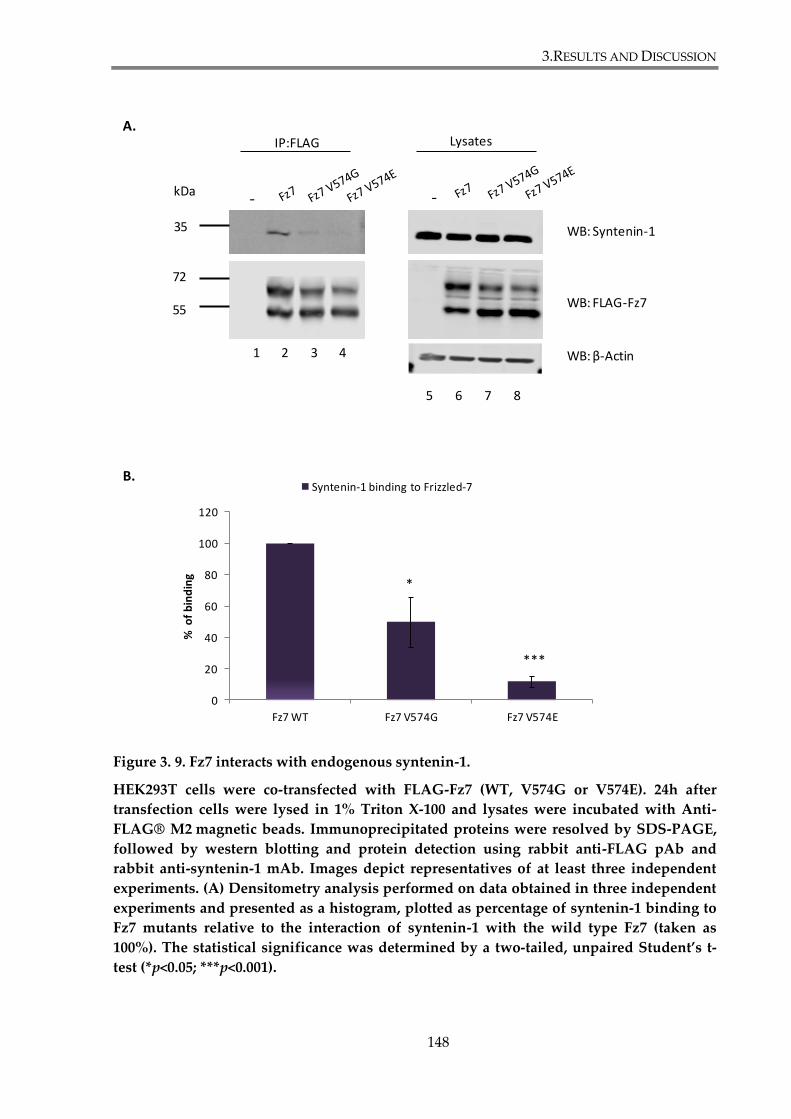
3.RESULTS AND DISCUSSION
148
Figure 3. 9. Fz7 interacts with endogenous syntenin-1.
HEK293T cells were co-transfected with FLAG-Fz7 (WT, V574G or V574E). 24h after
transfection cells were lysed in 1% Triton X-100 and lysates were incubated with Anti-
FLAG® M2 magnetic beads. Immunoprecipitated proteins were resolved by SDS-PAGE,
followed by western blotting and protein detection using rabbit anti-FLAG pAb and
rabbit anti-syntenin-1 mAb. Images depict representatives of at least three independent
experiments. (A) Densitometry analysis performed on data obtained in three independent
experiments and presented as a histogram, plotted as percentage of syntenin-1 binding to
Fz7 mutants relative to the interaction of syntenin-1 with the wild type Fz7 (taken as
100%). The statistical significance was determined by a two-tailed, unpaired Student’s t-
test (*p<0.05; ***p<0.001).
0
20
40
60
80
100
120
Fz7 WT Fz7 V574G Fz7 V574E
% o
f b
ind
ing
Syntenin-1 binding to Frizzled-7
*
***
72
55
35
WB: FLAG-Fz7
WB: Syntenin-1
WB: β-Actin
LysatesIP:FLAG
- -
5 6 7 8
1 2 3 4
kDa
A.
B.

3.RESULTS AND DISCUSSION
149
3.2.1.2. Mapping of syntenin-1 domain that interacts with Fz7
The nature of the syntenin-1 - Fz7 interaction was analysed further using the
following synteinin-1 – encoding constructs: HA-syntenin-1-PDZ1* (G126D), PDZ2* (G210E)
or PDZ1*PDZ2* (G126D, G210E) with introduced mutations in the interacting loops that
prevented binding through the PDZ1, PDZ2 or both, respectively (Latysheva et al. 2006). In
the control experiments we used a construct encoding HA-tagged syntenin-1 (wild type).
HEK293T were co-transfected with FLAG-Fz7 and one of the syntenin-1 – encoding plasmids
and the interaction between the proteins was analysed by immunoprecipitation. These
experiments revealed that both PDZ domains of syntenin-1 are required for its interaction
with Fz7 (Fig.3.10.A). This was evident by a significant decrease in the amount of syntenin-1
mutants detected in Fz7 immunoprecipitates: compare signals in lane 4 (PDZ1*), lane 6
(PDZ2*) and lane 8 (PDZ1*2*) with lane 2 (syntenin-1 WT). Densitometry analysis showed
that both PDZ domains of syntenin-1 contribute to the interaction with Fz7, as syntenin-1
PDZ1* binding to Fz7 was ten fold lower, whereas syntenin-1 PDZ2* and syntenin-1
PDZ1*PDZ2* binding was 20 times lower when compared with the interaction of wild type
syntenin-1 to Fz7 (Figure 3.10.B)
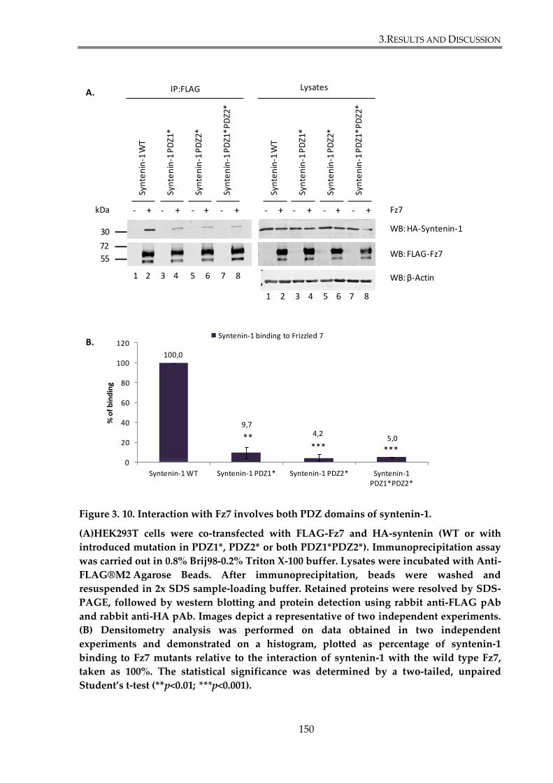
3.RESULTS AND DISCUSSION
150
Figure 3. 10. Interaction with Fz7 involves both PDZ domains of syntenin-1.
(A)HEK293T cells were co-transfected with FLAG-Fz7 and HA-syntenin (WT or with
introduced mutation in PDZ1*, PDZ2* or both PDZ1*PDZ2*). Immunoprecipitation assay
was carried out in 0.8% Brij98-0.2% Triton X-100 buffer. Lysates were incubated with Anti-
FLAG®M2 Agarose Beads. After immunoprecipitation, beads were washed and
resuspended in 2x SDS sample-loading buffer. Retained proteins were resolved by SDS-
PAGE, followed by western blotting and protein detection using rabbit anti-FLAG pAb
and rabbit anti-HA pAb. Images depict a representative of two independent experiments.
(B) Densitometry analysis was performed on data obtained in two independent
experiments and demonstrated on a histogram, plotted as percentage of syntenin-1
binding to Fz7 mutants relative to the interaction of syntenin-1 with the wild type Fz7,
taken as 100%. The statistical significance was determined by a two-tailed, unpaired
Student’s t-test (**p<0.01; ***p<0.001).
LysatesIP:FLAG
WB: FLAG-Fz7
WB: β-Actin
WB: HA-Syntenin-1
Fz7- + - + - + - + - + - + - + - +
Syn
ten
in-1
WT
Syn
ten
in-1
PD
Z1*
Syn
ten
in-1
PD
Z2*
Syn
ten
in-1
PD
Z1*
PD
Z2*
Syn
ten
in-1
WT
Syn
ten
in-1
PD
Z1*
Syn
ten
in-1
PD
Z2*
Syn
ten
in-1
PD
Z1*
PD
Z2*
72
55
30
100,0
9,74,2
5,0
0
20
40
60
80
100
120
Syntenin-1 WT Syntenin-1 PDZ1* Syntenin-1 PDZ2* Syntenin-1 PDZ1*PDZ2*
% o
f b
ind
ing
Syntenin-1 binding to Frizzled 7
***** ***
1 2 3 4 5 6 7 8
1 2 3 4 5 6 7 8
kDa
A.
B.
3.RESULTS AND DISCUSSION
151
To investigate the interaction between Fz7 and syntenin-1 in more detail nuclear
magnetic resonance (NMR) analysis was used in collaboration with Dr Sundaresan Rajesh.
NMR titration analysis of 15N-labeled syntenin-1-PDZ1-2 domains with unlabeled Fz7
peptide, corresponding to the last 8 amino acid of cytoplasmic tail of the receptor, was
carried out. The method was previously described in (Latysheva et al. 2006;Rajesh et al. 2011)
where detailed structural information on the role of syntenin-1’s individual amino acids is
provided. The decrease in signal intensity (chemical shift) for each amino acid of syntenin-1
upon interaction with Fz7 peptide at various concentrations (50 μM – 800 μM) was
calculated and later mapped onto the crystal structure of the syntenin-1-PDZ1-2 monomer,
PDB:1N99 (Kang et al. 2003). The colour code was used to highlight the calculated values:
green represents a decrease in signal intensity of 25-40%; yellow represents a decrease of 40-
60%, orange represents a decrease of 60-80%; and red a decrease of more than 80% (Figure
3.11). The decrease of NMR signal intensity correlates with the proximity of the relevant
amino acid with the interacting peptide. Analysis revealed that syntenin-1-PDZ2 domain is
predominantly involved in the Fz7 peptide binding, although the involvement of PDZ1 was
also observed. When the Fz7 peptide was used at 50 μM (1:0.25 ration toward syntenin-1
monomer concentration) a chemical shift was observed for Ile125 (PDZ1) and Thr219 (PDZ2),
suggesting that the initial step of Fz7 binding involves both PDZ domains (Figure 3.11.A).
However, when the concentration of Fz7 peptide was increased to 100 μM (1:0.5 ratio) a
chemical shift of Gly210 was observed in the carboxylate binding loop of PDZ2, as well as of
several residues in the vicinity of this site Phe211, Phe213, Gly216, and Thr219. In contrast,
chemical shift perturbations in PDZ1 were limited to Ile125 and Gly126 in the carboxylate
binding loop of PDZ1, and to Arg191 (Figure 3.11.B). The chemical shift perturbations,
3.RESULTS AND DISCUSSION
152
obtained when peptide was added in four fold excess (800 μM) (Figure 3.11.C), were used to
map binding sites and revealed a contact surface present predominantly in the syntenin-
PDZ2 domain binding pocket, and to a lesser extent in the PDZ1 (Figure 3.11.D). Namely
residues 199-201, 203-204, 207-211, 213-216, 220, 223-226, 229-235, 239, 242, 246, 255-256, 258-
259 and 265-270 in PDZ2, and 120, 125-127, 130, 141, 147, 179, 186 and 190-191 in PDZ1 were
involved in Fz7 peptide binding. This structural data support our co-immunoprecipitation
assay observations presented in the previous paragraph.
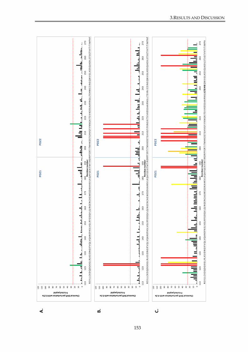
3.RESULTS AND DISCUSSION
153
0
10
20
30
40
50
60
70
80
90
10
0
11
0
12
0
11
31
23
13
31
43
15
31
63
17
31
83
19
32
03
21
32
23
23
32
43
25
32
63
27
3
Chemical Shift perturbation with 0.25x Frizzled peptid
Re
sid
ue
nu
mb
er
PD
Z1
PD
Z2
RE
VL
LC
KD
QD
GK
IGL
RL
KS
IDN
GIF
VQ
LV
QA
NS
PA
SL
VG
LR
FG
DQ
VL
QIN
GE
NC
AG
WS
SD
KA
HK
VL
KQ
AF
GE
KIT
MT
IRD
RP
FE
RT
ITM
HK
DS
TG
HV
GF
IFK
NG
KIT
SIV
KD
SS
AA
RN
GL
LT
EH
NIC
EIN
GQ
NV
IGL
KD
SQ
IAD
ILS
TS
GT
VV
TIT
IMP
AF
0
10
20
30
40
50
60
70
80
90
10
0
11
0
12
0
11
31
23
13
31
43
15
31
63
17
31
83
19
32
03
21
32
23
23
32
43
25
32
63
27
3
Chemical Shift perturbation with 0.5x Frizzled peptid
Re
sid
ue
nu
mb
er
PD
Z1
PD
Z2
RE
VL
LC
KD
QD
GK
IGL
RL
KS
IDN
GIF
VQ
LV
QA
NS
PA
SL
VG
LR
FG
DQ
VL
QIN
GE
NC
AG
WS
SD
KA
HK
VL
KQ
AF
GE
KIT
MT
IRD
RP
FE
RT
ITM
HK
DS
TG
HV
GF
IFK
NG
KIT
SIV
KD
SS
AA
RN
GL
LT
EH
NIC
EIN
GQ
NV
IGL
KD
SQ
IAD
ILS
TS
GT
VV
TIT
IMP
AF
B.
A.
C.
0
10
20
30
40
50
60
70
80
90
10
0
11
0
12
0
11
31
23
13
31
43
15
31
63
17
31
83
19
32
03
21
32
23
24
32
53
26
32
73
Chemical Shift perturbation with 4.0x Frizzled peptid
Re
sid
ue
nu
mb
er
PD
Z1
PD
Z2
RE
VL
LC
KD
QD
GK
IGL
RL
KS
IDN
GIF
VQ
LV
QA
NS
PA
SL
VG
LR
FG
DQ
VL
QIN
GE
NC
AG
WS
SD
KA
HK
VL
KQ
AF
GE
KIT
MT
IRD
RP
FE
RT
ITM
HK
DS
TG
HV
GF
IFK
NG
KIT
SIV
KD
SS
AA
RN
GL
LT
EH
NIC
EIN
GQ
NV
IGL
KD
SQ
IAD
ILS
TS
GT
VV
TIT
IMP
AF
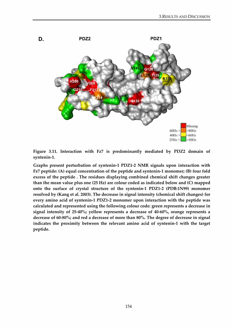
3.RESULTS AND DISCUSSION
154
Figure 3.11. Interaction with Fz7 is predominantly mediated by PDZ2 domain of
syntenin-1.
Graphs present perturbation of syntenin-1 PDZ1-2 NMR signals upon interaction with
Fz7 peptide: (A) equal concentration of the peptide and syntenin-1 monomer; (B) four fold
excess of the peptide . The residues displaying combined chemical shift changes greater
than the mean value plus one (25 Hz) are colour coded as indicated below and (C) mapped
onto the surface of crystal structure of the syntenin-1 PDZ1-2 (PDB:1N99) monomer
resolved by (Kang et al. 2003). The decrease in signal intensity (chemical shift changes) for
every amino acid of syntenin-1 PDZ1-2 monomer upon interaction with the peptide was
calculated and represented using the following colour code: green represents a decrease in
signal intensity of 25-40%; yellow represents a decrease of 40-60%, orange represents a
decrease of 60-80%; and red a decrease of more than 80%. The degree of decrease in signal
indicates the proximity between the relevant amino acid of syntenin-1 with the target
peptide.
D.
3.RESULTS AND DISCUSSION
155
3.2.2. Syntenin-1 modulates canonical Wnt signalling
pathway
3.2.2.1. Syntenin-1 knockdown attenuates canonical Wnt pathway
signalling in the SKBr3 cell line
The role of syntenin-1 in canonical Wnt signalling was examined in the SKBr3
epithelial breast cancer cell line stably expressing FLAG-Fz7 wild type or FLAG-Fz7 V574G
mutant, after syntenin-1 depletion. For this purpose SKBr3 cells were transfected with the
siRNA targeting syntenin-1, or with a control siRNA (siNeg) and with reporter plasmids. On
the following day, cells were stimulated with Wnt3a. 24h after later β-catenin reporter
activity was compared to cells with and without syntenin-1 knock-down (Figure 3.15.A).
Cellular lysates obtained in the experiment were analysed by SDS-PAGE and WB to quantify
the level of syntenin-1 knockdown (Figure 3.15.B). WB analysis confirmed the syntenin-1
knockdown (lanes 3, 4, 7 and 8). The protein expression levels were reduced by 90%,
compared with the control samples. These experiments showed that syntenin-1 depletion
attenuates activation of Wnt signalling in cells expressing wild type Fz7. Reporter activity in
SKBr3-FLAG-Fz7 cells with syntenin-1 knockdown was reduced by 40%, when compared
with control cells. On the other hand, syntenin-1 depletion didn’t alter Wnt signalling in
SKBr3 cells expressing the Fz7 V574G mutant. Taken together, syntenin-1 depletion
attenuates canonical Wnt signalling in SKBr3 cells expressing wild type Fz7, but not the Fz7
V574G mutant.
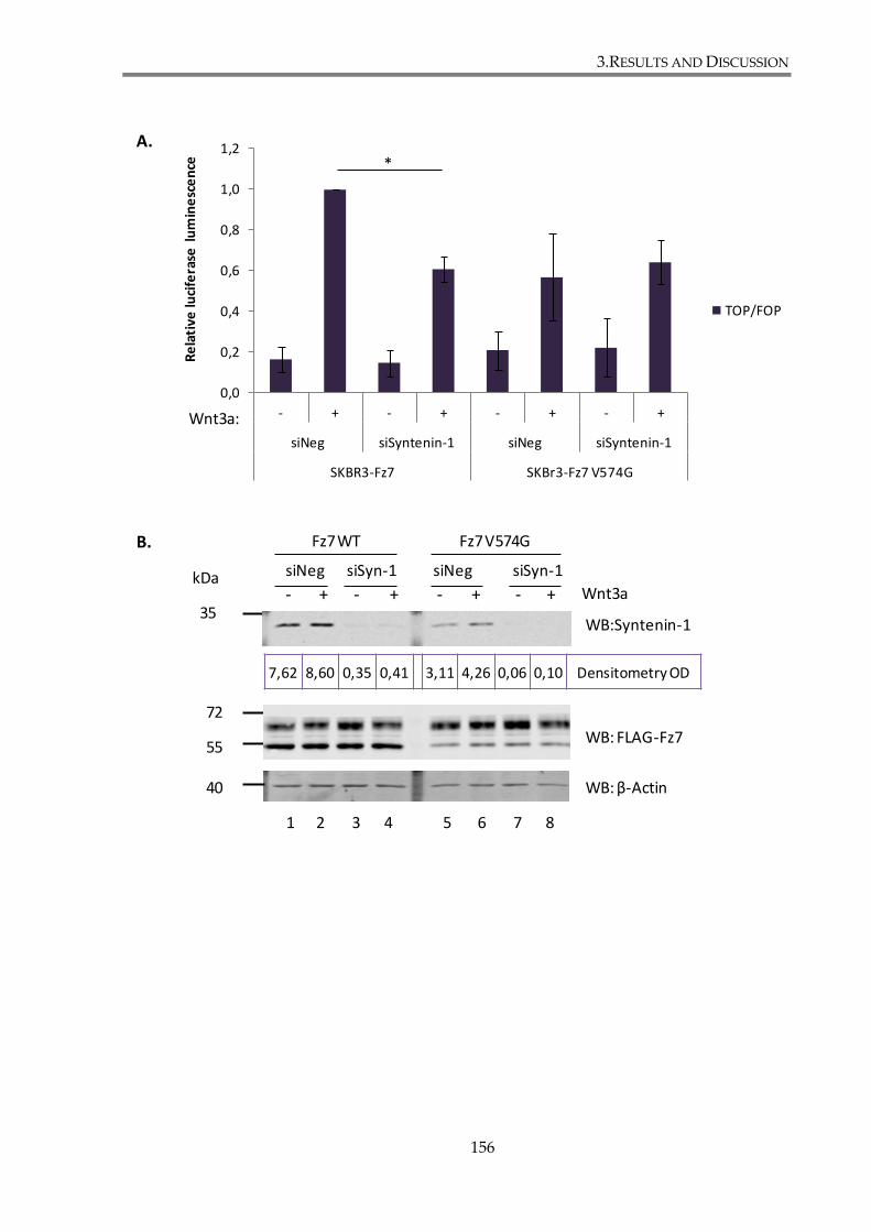
3.RESULTS AND DISCUSSION
156
72
55
35
0,0
0,2
0,4
0,6
0,8
1,0
1,2
- + - + - + - +
siNeg siSyntenin-1 siNeg siSyntenin-1
SKBR3-Fz7 SKBr3-Fz7 V574G
Re
lati
ve lu
cife
rase
lu
min
esc
en
ce
TOP/FOP
Wnt3a:
*
WB:Syntenin-1
WB: β-Actin
WB: FLAG-Fz7
1 2 3 4 5 6 7 8
Wnt3a- + - + - + - +
siNeg siNegsiSyn-1 siSyn-1
Fz7 WT Fz7 V574G
A.
B.
40
kDa
7,62 8,60 0,35 0,41 3,11 4,26 0,06 0,10 Densitometry OD
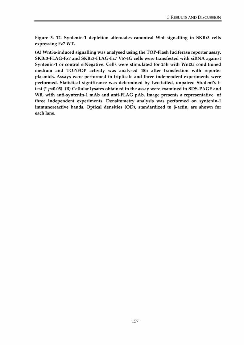
3.RESULTS AND DISCUSSION
157
Figure 3. 12. Syntenin-1 depletion attenuates canonical Wnt signalling in SKBr3 cells
expressing Fz7 WT.
(A) Wnt3a-induced signalling was analysed using the TOP-Flash luciferase reporter assay.
SKBr3-FLAG-Fz7 and SKBr3-FLAG-Fz7 V574G cells were transfected with siRNA against
Syntenin-1 or control siNegative. Cells were stimulated for 24h with Wnt3a conditioned
medium and TOP/FOP activity was analysed 48h after transfection with reporter
plasmids. Assays were performed in triplicate and three independent experiments were
performed. Statistical significance was determined by two-tailed, unpaired Student’s t-
test (* p<0.05). (B) Cellular lysates obtained in the assay were examined in SDS-PAGE and
WB, with anti-syntenin-1 mAb and anti-FLAG pAb. Image presents a representative of
three independent experiments. Densitometry analysis was performed on syntenin-1
immunoreactive bands. Optical densities (OD), standardized to β-actin, are shown for
each lane.

3.RESULTS AND DISCUSSION
158
3.2.2.2. Syntenin-1 regulates canonical Wnt signalling through its
interaction with Fz7
Our results indicate that syntenin-1 regulates canonical Wnt signalling due to its
direct interaction with Fz7. However, one can’t exclude the possibility that syntenin-1 also
interferes with canonical Wnt signalling downstream of the Fz7 receptor, or indirectly by
affecting the activity of alternative pathways (see Introduction). To address this issue, we
compared activation of the canonical Wnt pathway in syntenin-1-depleted cells either
stimulated with Wnt3a, or treated with the GSK3 inhibitor (SB216763), which activates the
catenin – dependent pathway downstream of the receptor. In control experiments, cells
were treated with control siRNA. SKBr3-FLAG-Fz7 cells were transfected with siSyntenin-1
or siNeg and incubated with Wnt3a conditioned medium or control medium. In parallel,
cells transfected with siRNA were treated with 10 μM GSK3i or equivalent amounts of
DMSO. Wnt activation was quantified by TOP-Flash reporter assay 24h later; data is
presented in Figure 3.16.A. Analysis of the reporter assay showed that syntenin-1 depletion
only downregulates Wnt pathway when cells are stimulated with Wnt3a ligand and not with
GSK3i. The level of syntenin-1 knock-down was assessed by analysing cell lysates obtained
the assay by SDS-PAGE and WB with anti-syntenin-1 pAb (Figure 3.16.B), which confirmed
that the protein expression level was more than 90% lower when compared with control
samples. These results provide an additional evidence for syntenin-1 affecting canonical
Wnt signalling upstream of GSK3.
3.RESULTS AND DISCUSSION
159
0
5
10
15
20
25
30
35
Co
ntr
ol C
M
Wn
t3a
Co
ntr
ol C
M
Wn
t3a
DM
SO
SB2
16
76
3
DM
SO
SB2
16
76
3
siNeg siSyntenin siNeg siSyntenin
SKBR3-FLAG-Fz7
Re
lati
ve lu
cife
rase
lu
min
esc
en
ce
TOP/FOP
*
WB: Syntenin-1
WB: Actin
35
Par
en
talC
M
Wn
t3a
CM
DM
SO
SB21
6763
Par
en
talC
M
Wn
t3a
CM
DM
SO
SB21
6763
siNeg siSyntenin-1
1 2 3 4 5 6 7 8
A.
B.
1,44 4,84 3,99 3,80 0,00 0,15 0,16 0,06 DensitometryOD
kDa
40
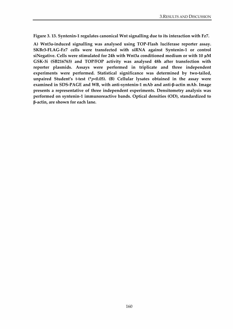
3.RESULTS AND DISCUSSION
160
Figure 3. 13. Syntenin-1 regulates canonical Wnt signalling due to its interaction with Fz7.
A) Wnt3a-induced signalling was analysed using TOP-Flash luciferase reporter assay.
SKBr3-FLAG-Fz7 cells were transfected with siRNA against Syntenin-1 or control
siNegative. Cells were stimulated for 24h with Wnt3a conditioned medium or with 10 μM
GSK-3i (SB216763) and TOP/FOP activity was analysed 48h after transfection with
reporter plasmids. Assays were performed in triplicate and three independent
experiments were performed. Statistical significance was determined by two-tailed,
unpaired Student’s t-test (*p<0.05). (B) Cellular lysates obtained in the assay were
examined in SDS-PAGE and WB, with anti-syntenin-1 mAb and anti-β-actin mAb. Image
presents a representative of three independent experiments. Densitometry analysis was
performed on syntenin-1 immunoreactive bands. Optical densities (OD), standardized to
β-actin, are shown for each lane.

3.RESULTS AND DISCUSSION
161
3.2.3. Syntenin-1 regulates Fz7 distribution in SKBr3 cells
SKBr3 cell lines stably expressing FLAG-Fz7 or FLAG-Fz7 V574G were used to
determine whether syntenin-1 co-localizes with Fz7. Due to the lack of a suitable antibody to
detect endogenous syntenin-1 by immunofluorescence staining, Myc-syntenin-1 was used
for the co-localization studies. SKBr3, SKBr3-FLAG-Fz7 and SKBr3-FLAG-Fz7 V574G cells
were transfected with Myc-syntenin-1 and, 48h later, fixed and stained with rabbit anti-
FLAG and mouse anti-Myc antibodies. DAPI was used to visualize nuclei. Immunoreactivity
for both proteins was analysed by confocal microscopy and presented in the Figure 3.12.
Syntenin-1 has been found to be abundantly present in the cytoplasm of parental SKBr3 cells.
Using a transient transfection approach in which only ~25% cells were transfected with a
syntenin-1 – encoding plasmid allowed us to examine the effect of syntenin-1 overexpression
on localization of Fz7. These experiments revealed that syntenin-1 overexpression changes
Fz7 cell distribution. In both cell lines, SKBr3-FLAG-Fz7 and SKBr3-FLAG-Fz7 V574G,
immunoreactivity for Fz7 was not observed in plasma membrane, in cells overexpressing
syntenin-1. On the other hand, Fz7 immunoreactivity was detected in small puncta
(presumably intracellular vesicles) in the cytoplasm. As expected, no reactivity with anti-Flag
Ab was detected in SKBr3 control cells.
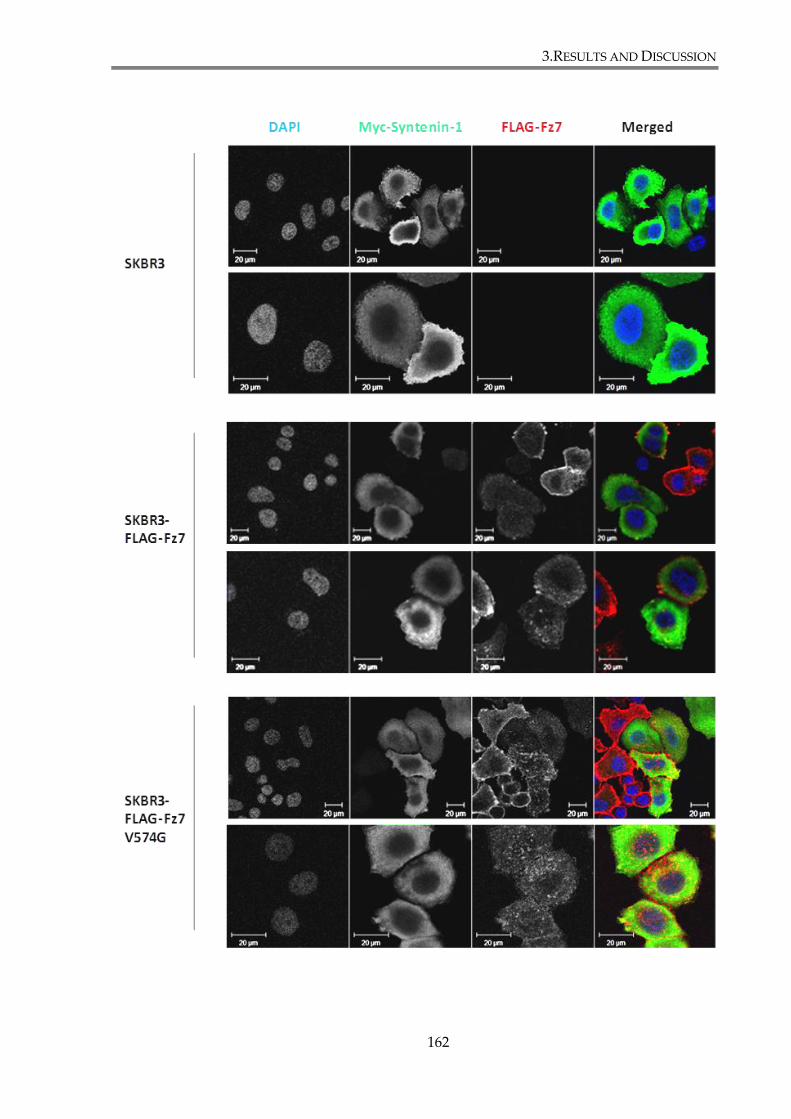
3.RESULTS AND DISCUSSION
162
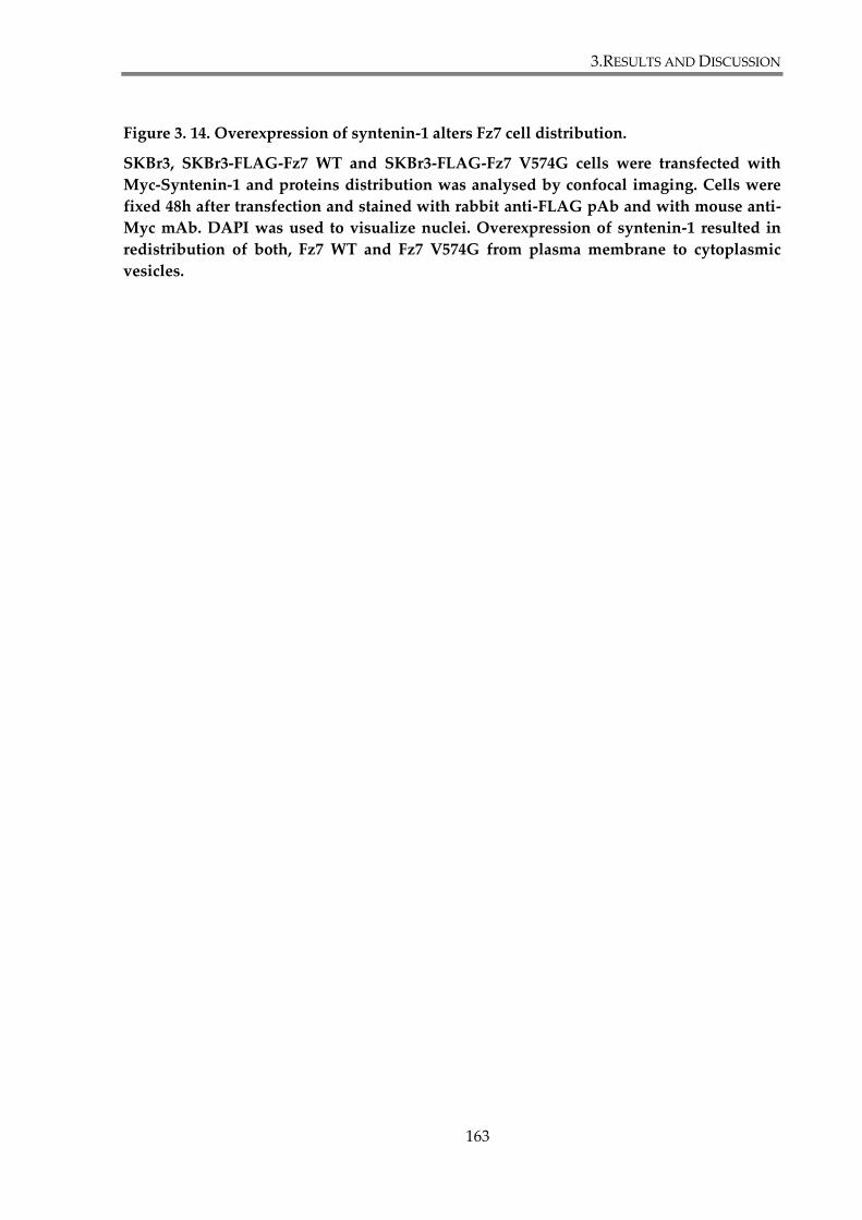
3.RESULTS AND DISCUSSION
163
Figure 3. 14. Overexpression of syntenin-1 alters Fz7 cell distribution.
SKBr3, SKBr3-FLAG-Fz7 WT and SKBr3-FLAG-Fz7 V574G cells were transfected with
Myc-Syntenin-1 and proteins distribution was analysed by confocal imaging. Cells were
fixed 48h after transfection and stained with rabbit anti-FLAG pAb and with mouse anti-
Myc mAb. DAPI was used to visualize nuclei. Overexpression of syntenin-1 resulted in
redistribution of both, Fz7 WT and Fz7 V574G from plasma membrane to cytoplasmic
vesicles.
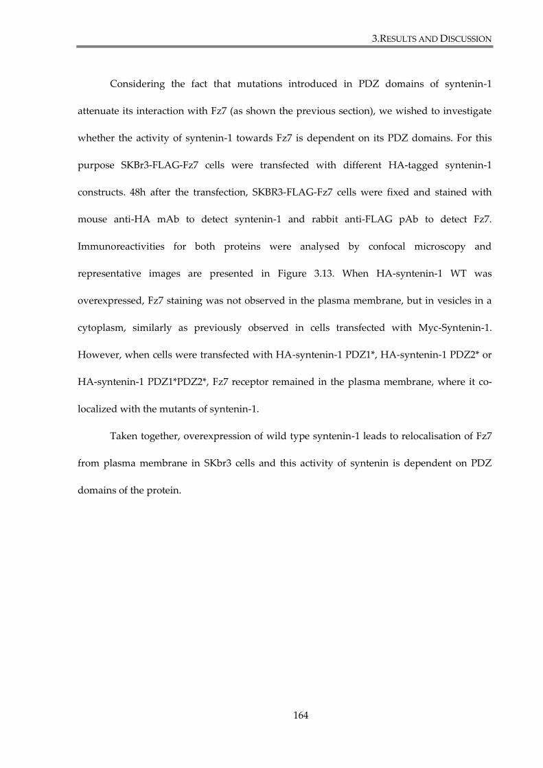
3.RESULTS AND DISCUSSION
164
Considering the fact that mutations introduced in PDZ domains of syntenin-1
attenuate its interaction with Fz7 (as shown the previous section), we wished to investigate
whether the activity of syntenin-1 towards Fz7 is dependent on its PDZ domains. For this
purpose SKBr3-FLAG-Fz7 cells were transfected with different HA-tagged syntenin-1
constructs. 48h after the transfection, SKBR3-FLAG-Fz7 cells were fixed and stained with
mouse anti-HA mAb to detect syntenin-1 and rabbit anti-FLAG pAb to detect Fz7.
Immunoreactivities for both proteins were analysed by confocal microscopy and
representative images are presented in Figure 3.13. When HA-syntenin-1 WT was
overexpressed, Fz7 staining was not observed in the plasma membrane, but in vesicles in a
cytoplasm, similarly as previously observed in cells transfected with Myc-Syntenin-1.
However, when cells were transfected with HA-syntenin-1 PDZ1*, HA-syntenin-1 PDZ2* or
HA-syntenin-1 PDZ1*PDZ2*, Fz7 receptor remained in the plasma membrane, where it co-
localized with the mutants of syntenin-1.
Taken together, overexpression of wild type syntenin-1 leads to relocalisation of Fz7
from plasma membrane in SKbr3 cells and this activity of syntenin is dependent on PDZ
domains of the protein.
3.RESULTS AND DISCUSSION
165
Figure 3.15. Fz7 co-localizes with syntenin-1 with mutations introduced in PDZ domains .
SKBr3, SKBr3-FLAG-Fz7 WT and SKBr3-FLAG-Fz7 V574G cells were co-transfected with
FLAG-Fz7 and HA-syntenin-1 (WT or with introduced mutation in PDZ1*, PDZ2* or both
PDZ1*PDZ2*) and proteins distribution was analysed by confocal imaging. Cells were
fixed 48h after transfection and stained with rabbit anti-FLAG pAb and with mouse anti-
HA mAb. DAPI was used to visualize nuclei. Overexpression of wild type syntenin-1 led
to removal of Fz7 from plasma membrane. All three syntenin-1 PDZ mutants co-localize
with Fz7.
3.RESULTS AND DISCUSSION
166
3.2.4. Syntenin-1 is not involved in the regulation of Fz7
expression
Syntenin-1 overexpression induces changes in Fz7 distribution in SKBr3 cells (Section
3.2.2.). In the reverse experiments we analysed whether depletion of syntenin-1 would also
influence the cellular distribution of Fz7. SKBr3-FLAG-Fz7 and SKBr3-FLAG-Fz7 V574G cells
were transfected with siRNA targeting syntenin-1 (or with control siRNA) and 24h later cells
were stimulated with Wnt3a conditioned medium or with control conditioned medium.
Cells were collected after further 24h and expression levels and distribution were analysed
by WB, flow cytometry and immunofluorescence. Densitometry analysis confirmed
syntenin-1 expression level was 90% lower in cells transfected with siSyntenin-1, than in the
control cells. The analysis of Fz7 expression in whole cell lysates (using anti-FLAG pAb),
revealed no significant changes in total expression level of both, Fz7 WT and Fz7 V574G
(Figure 3.14.A). In parallel, flow cytometry analysis was conducted to assess the surface
expression of Fz7 in collected cells, using anti-FLAG mAb. Fz7 wild type or Fz7 V574G
mutant cell surface expression was not altered in cells with syntenin-1 knock-down (Figure
3.14.B). Finally, cell localization of Fz7 was examined by immunofluorescence. We noticed
that syntenin-1 depletion changed morphology of SKBr3 cells, as they became more
flattened. Nonetheless, no obvious alterations were observed in the receptor distribution in
cells with syntenin-1 knock-down.
Taken together, the analysis of total and membrane expression of Fz7 and cell
distribution by western blot, flow cytometry and immunofluorescence didn’t provide
evidence that syntenin-1 knock-down influences Fz7 expression in SKBr3 cells.
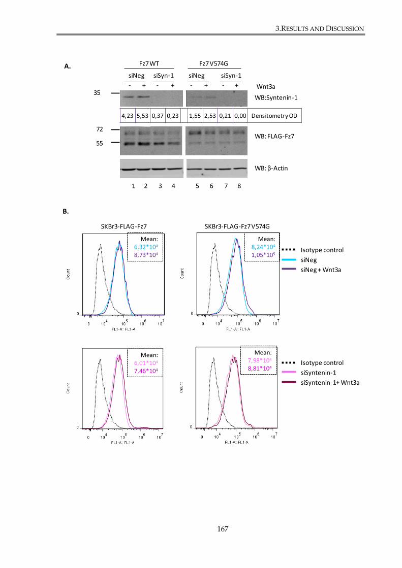
3.RESULTS AND DISCUSSION
167
Isotype control
siNeg
siNeg + Wnt3a
SKBr3-FLAG-Fz7 SKBr3-FLAG-Fz7 V574G
Isotype control
siSyntenin-1
siSyntenin-1+ Wnt3a
72
55
35WB:Syntenin-1
WB: β-Actin
WB: FLAG-Fz7
1 2 3 4 5 6 7 8
Wnt3a- + - + - + - +
siNeg siNegsiSyn-1 siSyn-1
Fz7 WT Fz7 V574G
B.
A.
4,23 5,53 0,37 0,23 1,55 2,53 0,21 0,00 Densitometry OD
Mean:6,32*104
8,73*104
Mean:8,24*104
1,05*105
Mean:6,01*104
7,46*104
Mean:7,98*104
8,81*104
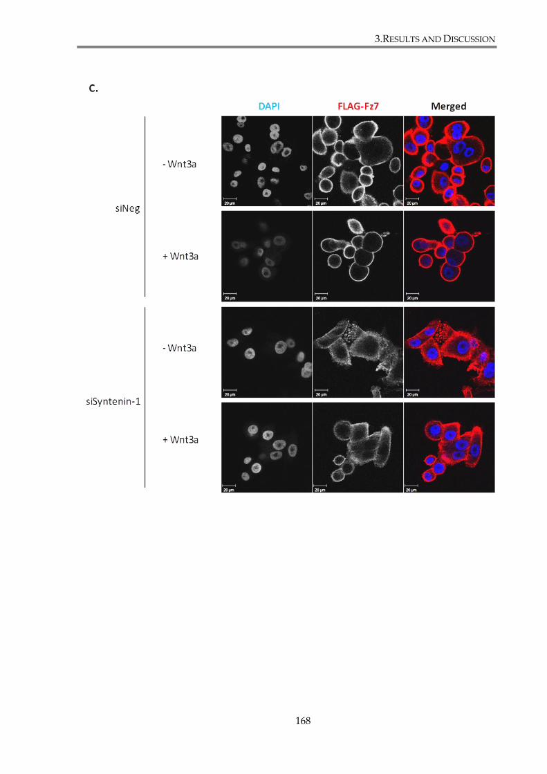
3.RESULTS AND DISCUSSION
168
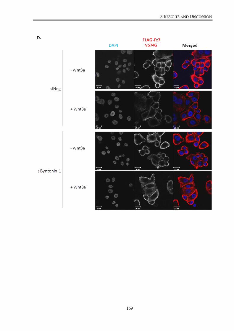
3.RESULTS AND DISCUSSION
169
3.RESULTS AND DISCUSSION
170
Figure 3. 16. Syntenin-1 depletion does not change Fz7 membrane expression or cell
distribution.
SKBr3-FLAG-Fz7 and SKBr3-FLAG-Fz7 V574G cells were transfected with siRNA against
Syntenin-1 or with control siNeg. 24h later cells were stimulated with Wnt3a conditioned
medium or with control conditioned medium. 48h after the transfection cell lysates were
collected and analysed by SDS-PAGE and WB, flow cytometry and immunofluorescence.
(A) Lysates of equal protein concentration were resolved by SDS-PAGE, followed by
western blotting with Anti-FLAG pAb to examined Fz7 expression level, anti-Syntenin-1
mAb to monitor LNX2 knock-down level. β-actin served as a loading control. (B) Flow
cytometry with anti-FLAG mAb was used to examine membrane expression of Fz7. Cells
were incubated on ice for 1h with mouse anti-FLAG (M2) mAb. 4C5G monoclonal
antibody against phosphatidylinositol 4-kinase was used as an isotype control. Cell
surface staining of FLAG-Fz7 (WT or V574G) was detected by flow cytometry following
incubation for 1h on ice with anti-mouse FITC-conjugated secondary antibodies. Data
analysis was carried out using FlowJo analysis software. The values of calculated mean
fluorescence are presented in top-right corner of the plots. (C-D) Fz7 WT (C) and V574G
(D) cell distribution was analysed by confocal imaging. Cells were fixed and stained with
mouse anti-FLAG (M2) mAb and Alexa Fluor 488-conjugated goat-anti mouse ab (green).
DAPI was used to visualize nuclei.
3.RESULTS AND DISCUSSION
171
3.2.5. Discussion
We have found in the proceeding chapter that the canonical Wnt signalling is
regulated by Fz7 interactions through its C-terminal PDZ-BM. Additionally, Fz7 expression
and cell distribution also seem to be dependent on the receptor associations with PDZ
proteins. To examine this issue further, we have initially decided to focus on the interaction
between Fz7 and syntenin-1. Syntenin-1 is a scaffold protein with a tandem of PDZ domains,
previously shown to bind Fz7 (Luyten et al. 2008). The protein was reported to regulate
trafficking of several membrane receptors, such as syndecans, syndecan associated receptors
e.g. FGF, or tetraspanin CD63 (Beekman et al. 2008) (see Introduction). For these reasons, we
have decided to determine whether syntenin-1 regulates Fz7 functions, as well as canonical
Wnt signalling in breast cancer cells.
Our experiments confirmed that syntenin-1 binds to Fz7 in our breast cancer model.
Furthermore, as expected, mutations in both PDZ-BM of Fz7 and PDZ domains in
syntenin-1, significantly decreased the interaction between the two proteins. These results
clearly demonstrate the importance of PDZ domains in Fz7 – syntenin-1 interactions.
However, the fact that none of the mutations analysed completely abolish the association
might indicate a possible contribution of the other part(s) of Fz7 (and syntenin-1) in
stabilization of the complex. If proved in future experiments this would be reminiscent of a
model of Frizzled-Dishevelled interactions, which was recently presented by Tauriello and
colleagues. Specifically, it was demonstrated that this interaction involves a three-segmented
discontinuous binding surface, comprising of the internal PDZ-BM and two additional
motifs in intracellular loops of the Frizzled receptor (Tauriello et al. 2012). Alternatively, one
3.RESULTS AND DISCUSSION
172
can not exclude a possibility that a small proportion of syntenin-1 is recruited to the Fz7
complex indirectly via a network of PDZ-independent interactions.
Our results demonstrated that PDZ2 plays a predominant role in recruiting
syntenin-1 to Fz7. Nevertheless, we have shown that the mutation in PDZ1 also decreased
the interaction of syntenin-1 with Fz7. Luyten and colleagues (2008) reported that syntenin-1
interaction with Fz7 relies on PDZ1. In their study recombinant GST-PDZ domains of
syntenin-1 were used in different type of test (overlay assay), which might explain
differences between our findings and (Luyten et al. 2008). The authors showed single GST-
PDZ1, but not GST-PDZ2, was sufficient to bind to Fz7 peptide. Interestingly, tandem GST-
PDZ2-PDZ2 displayed strong interaction with Fz7. Most protein ligands of syntenin-1 show
preference for PDZ2 (for example syndecans and ephrinB), whereas PDZ1 binds with higher
affinity to IL-5Rα (interleukin-5 receptor α), neurexin, merlin and CD63 (Beekman et al.
2008). Previous reports have also shown that despite preference for one of the PDZ domain,
both of them are required for the interaction of syntenin-1 with different transmembrane
proteins (Sarkar et al. 2004).
Syntenin-1 was reported to be involved in the regulation of non-canonical Wnt
signalling by controlling activation of the PKC/CDC42 pathway (Luyten et al. 2008). This
study also suggested that syntenin-1 does not contribute to the canonical Wnt pathway. By
contrast, our results clearly indicate that depletion of syntenin-1 in breast cancer cells
attenuated -catenin – dependent signalling as tested in the TOPFlash reporter assay. We
based our conclusion on two lines of evidence. Firstly, observed decrease of TOP-Flash
signal following syntenin-1 depletion occurs only in SKBr3 cells expressing Fz7 WT, and not
in cells expressing the Fz7 mutant with reduced capability to bind to syntenin-1 (Figure 3.12).

3.RESULTS AND DISCUSSION
173
Secondly, analysis of the canonical Wnt pathway activation after treatment with an inhibitor
of GSK3 (Figure 3.13) leads to a conclusion that syntenin-1 regulates Wnt signalling
upstream of GSK3 kinase, presumably on Wnt receptor level. One possible explanation for
the discrepancy between our results and conclusions from the previous study is that
different cell models were used. Conclusions by Luyten and colleagues were based on the
experiments performed using MCF7 cells, wherein overexpression of syntenin-1 didn’t have
any significant effects on -catenin – dependent signalling (TOP-Flash experiments).
However, we and others (Wheeler et al. 2011) have shown that canonical Wnt pathway is not
functional in this cell line, due to inhibitory effect of NHERF overexpression.
Further research is required to elucidated the exact mechanism in which syntenin-1
regulates canonical Wnt signalling. However, syntenin-1 was shown to bind syndecans,
ubiquitous transmembrane heparan sulphate proteoglycans. The heparan sulphate chains of
their extracellular domain allow syndecans to act as co-receptors, as they bind and
concentrate growth factors, including Wnts, at the cell surface and facilitate their interactions
with receptors (Bishop et al. 2007;Couchman 2003). It was proposed previously in (Luyten et
al. 2008) that syntenin-1 serves as a scaffold for Fz7-syndecan-4 complex and therefore
regulate noncanonical Wnt signalling, due to the ability of syndecan-4 to bind and activate
PKCα. In this regard, syndecan-1 was recently described as a regulator of β-catenin
dependent Wnt signalling in triple negative subtype of breast cancer. The authors showed
that syndecan-1 depletion correlates with downregulation of LRP6 and reduced
responsiveness to Wnt signalling (Ibrahim et al. 2013). Another possible mechanism may
involve syntenin-1 interaction with another binding partner, such as merlin
(neurofibromatosis 2, NF2). Merlin acts a membrane stabilizing protein and was shown to
3.RESULTS AND DISCUSSION
174
bind and colocalize with syntenin-1 at the plasma membrane (Jannatipour et al. 2001). Merlin
was found to interact with proteins forming adherens junctions (AJs): cadherins and catenins
(Lallemand et al. 2003), with which syntenin-1 was also reported to colocalize (Zimmermann
et al. 2001). Recently, it was shown merlin knockout leads to increased canonical Wnt
signalling in mouse embryonic fibroblasts (Zhou et al. 2011). Thus, interaction between these
two proteins might also be important for regulation of Wnt signalling.
Our results indicate that syntenin-1 may be involved in regulation of receptor
trafficking. Specifically. syntenin-1 overexpression in both SKBr3-FLAG-Fz7 and SKBr3-
FLAG-Fz7 V574G resulted in redistribution of the receptor from the plasma membrane to
small puncta, supposedly cytoplasmic vesicles (Figure 3.14.). Importantly, all three PDZ
mutants of syntenin-1 (PDZ1*, PDZ2* and PDZ1*PDZ2*) were deficient towards Fz7. These
results seem to indicate that syntenin-1 interaction through PDZ domains regulates
subcellular localization of Fz7. Surprisingly, we found that Fz7 V574G mutant was also
redistributed from plasma membrane upon overexpression of the wild type syntenin-1.
It seems that the effect of syntenin-1 on Fz7 distribution is likely to be indirect and involves
another protein (or proteins) whose interaction with syntenin-1 requires PDZ domain.
Syntenin-1 is known to regulate trafficking of a number transmembrane receptors (Beekman
et al. 2008). It has been also reported that complex syndecan-syntenin-1-ALIX is able to
recruit syndecan associated proteins to the intraluminal vesicles of multivesicular endosomes
and exosomes (Baietti et al. 2012). Since syndecan-1 was also implicated in Wnt signalling
(Ibrahim et al. 2013), it is possible that syndecan-1-syntenin-1-ALIX complex recognize Fz7 as
a cargo and traps it in the intraluminal vesicles.
3.RESULTS AND DISCUSSION
175
Finally, we have examined whether syntenin-1 depletion alters Fz7 expression.
SKBr3-FLAG-Fz7 WT and SKBr3-FLAG-Fz7 V574G cells were transfected with siRNA
against syntenin-1 and stimulated with Wnt3a. Subsequently total and membrane
expression, as well as cellular localization of Fz7 was assessed. Analysis by western blot,
flow cytometry and immunofluorescence did not provide evidence that syntenin-1 knock-
down affects Fz7 WT or Fz7 V574G expression (Figure 3.16).
Taken together, syntenin-1 is a scaffold protein binding Fz7 in PDZ dependent
manner. For the first time, we have determined that the interaction between the two proteins
plays a role in regulation of the canonical Wnt signalling in breast cancer cells. We have also
shown that syntenin-1 regulates trafficking and cell localization of Fz7 receptor.
3.RESULTS AND DISCUSSION
176
3.3. Fz7 interacts with PDZ domain containing proteins
Cytoplasmic tails of all Frizzled receptors contain Postsynaptic density-95, Disc large,
ZO-1 (PDZ) binding motifs (PDZ-BMs). The C-terminal PDZ-BM (x-S/T-x-φ, where φ
represents a hydrophobic residue) was reported to bind different PDZ proteins including
GOPC, MAGI-3, Syntenin-1, Kermit, PSD-95 (Wawrzak et al. 2009) (discussed in the
Introduction). Whilst some of these proteins were implicated in noncanonical Wnt pathways,
their roles in transduction of canonical Wnt signalling haven’t been widely addressed in the
past. The aim of work described in this chapter was to identify PDZ proteins that interact
with Fz7 in breast cancer cells, and thus to help elucidate the mechanism by which the C-
terminal PDZ-BM of Fz7 works.
3.3.1. Pull-down of Fz7 binding partners and their
identification by Mass-Spectrometry
In order to find binding partners of Fz7 that interact with WT but not with V574G, a
pull-down assay was performed, described in details in section 2.4.6. of Materials and
Methods. Schematic representation of designed experiments is presented in the Figure
3.17.A. Cellular lysates of epithelial breast cancer cell line MCF7 were incubated either with
biotin-conjugated Fz7 WT or with Fz7 V574G peptide immobilized on agarose resins (Figure
3.17.B). For the control sample, NeutrAvidin agarose resins without peptide were used.
Retained proteins were resolved on SDS-PAGE gel and stained with InstantBlue. Marked
3.RESULTS AND DISCUSSION
177
differences were observed, when “WT” and “V574G” pull-down samples were compared
(Figure 3.17.B), indicating that the C-terminal PDZ binding motif of Fz7 is essential for
interactions with a number of cellular proteins. Proteins specifically pulled down by WT
peptide (marked by red arrows in the Figure 3.17.B) were digested with trypsin and
analyzed by mass spectrometry. Details of samples preparation are described in section 2.4.7.
Protein sequencing was performed by Dr Douglas Ward using liquid chromatography and
mass spectrometry (LS-MS) at the Proteomics Facility of University of Birmingham. Analysis
revealed several PDZ domain-containing proteins that could be Fz7 binding partners.
Identified proteins are listed in the Table 3.1. Among the identified candidates were proteins
involved in receptor trafficking (DLG1, SNX27, LNX1, LNX2, MAGI3, Scribble) and
establishment of cell polarity and tight junction formation (MPP5, MPP7, ZO-2, MAGI3,
INADL, Scribble).
3.RESULTS AND DISCUSSION
178
Figure 3. 17. Pull down of PDZ domain containing binding partners of Fz7.
(A) MCF7 cells were lysed in 1% Triton X-100. The lysates were firstly precleared with
NeutrAvidin agarose resins and then incubated with NeutrAvidin agarose resins with
immobilized biotinylated peptides: Fz7 WT or V574G. For the control sample,
NeutrAvidin agarose resin without peptide was used. After the pull-downs the beads
were washed and resuspended in 2x SDS sample-loading buffer. (B) Samples were
subjected to SDS-PAGE and stained with the Coomassie dye. The protein bands that were
visible in Fz7 WT pull-down lanes, but not in Fz7 V574G pull-down lane or beads alone
pull-down control lane, (marked by red arrows) were excised, processed and analysed by
mass spectrometry
MCF7
MCF7 lysates
Fz7 WT peptide Fz7 V574G peptidePullDown:
SDS -PAGE
Mass Spectrometry Control Fz7WT
Fz7 V574G
PD:
A. B.
1234
567
3.RESULTS AND DISCUSSION
179
Table 3. 1. Mass spectrometry results showing potential Fz7-binding proteins.
The table indicates the gene symbol, name, molecular weight and number of detected
peptides obtained from the digested gel fragments (samples) labelled in the Fig. 3.17.
Accession Protein MW
(kDa)
Peptides Sample
PDZ PROTEINS:
SCRIB Protein scribble homolog 174.8 62 1,2,3,4,5,6,7
INADL InaD-like protein 196.2 37 1,2,3
MPP5 MAGUK p55 subfamily member 5 77.2 36 5,6,7
MAGI3 Membrane-associated guanylate kinase, WW
and PDZ domain-containing protein 3
165.5 35 2,3,4
MPP7 MAGUK p55 subfamily member 7 65.5 27 5,6,7
MAGI1 Membrane-associated guanylate kinase, WW
and PDZ domain-containing protein 1
164.5 27 2,3,4
DLG1 Disks large homolog 1 100.4 11 5,6,7
SNX27 Sorting nexin-27 61.2 11 5,6,7
ZO-2 Tight junction protein ZO-2 133.9 7 4
LNX2 Ligand of Numb protein X 2 76.0 6 5,6
MPDZ Multiple PDZ domain protein 218.5 6 2
LNX1 E3 ubiquitin-protein ligase LNX 80.6 5 5
CSKP Peripheral plasma membrane protein CASK 105.1 4 5,6,7
ZO-1 Tight junction protein ZO-1 193.3 1 1
OTHER MISCALLENOUS HITS:
PLOD3 Procollagen-lysine,2-oxoglutarate
5-dioxygenase 3
84.7 22 5,6
PLOD1 Procollagen-lysine,2-oxoglutarate
5-dioxygenase 1
83.5 18 5,6,7
MYH9 Myosin-9 226.4 18 1,2,3,4,5,6,7
MCCA Methylcrotonoyl-CoA carboxylase subunit
alpha, mitochondrial
80.4 11 6
DDX5 Probable ATP-dependent RNA helicase DDX5 69.1 10 7
TP4A3 Protein tyrosine phosphatase type IVA 3 19.5 9 1,2,3,4,5,6,7
DTNB Dystrobrevin beta 71.3 6 7
KU70 ATP-dependent DNA helicase 2 subunit 1 69.8 6 7
DDX17 Probable ATP-dependent RNA helicase
DDX17
72.3 5 7
HNRPQ Heterogeneous nuclear ribonucleoprotein Q 69.6 5 7
DTNA Dystrobrevin alpha OS=Homo sapiens 83.8 4 6
PYC Pyruvate carboxylase, mitochondrial 129.6 4 5,7
KU86 ATP-dependent DNA helicase 2 subunit 2 82.7 4 5

3.RESULTS AND DISCUSSION
180
3.3.2. Mutation in the C-terminal PDZ binding motif of
Fz7 affects binding of PDZ domain containing
proteins
In order to verify data obtained in mass spectrometry and check whether replacement
of C-terminal valine (Val574) with glycine can disrupt Fz7 binding to identified proteins, a
series of pull-down experiments was conducted. MCF7 cells were transiently transfected
with plasmids encoding PDZ domain containing proteins identified in the section 3.2.1.
(Scribble, INADL, MAGI3, MAGI1, DLG1, LNX2, MPP7, SNX27 and MPP5). In additional
experiments we also examined the interaction of Fz7 peptides with PDZ proteins that have
been previously reported to bind Fz7 (syntenin-1, GIPC1, NHERF, GOBC, and PSD95)
(Wawrzak et al. 2009). The interaction with syntenin-1 was examined using endogenous
protein. Lysates of MCF7 cells expressing PDZ domain-containing proteins were incubated
with NeutrAvidin beads conjugated with biotinylated Fz7 peptides (Fz7 WT or Fz7 V574G).
As a control for unspecific binding of proteins to the beads lysates were incubated with
NeutrAvidin beads without conjugated peptide.
Pull-down assays showed that all of the tested proteins can be pulled down with Fz7
WT peptide (lane 7) and that most of them did not bind to Fz7 V574G peptide (lane 8)
(Figure 3.18.A). However five of the PDZ proteins (syntenin-1, SNX27, MPP5, GOPC and
PSD95) partially retained the ability to bind Fz7 V574G peptide (lane 8), albeit their binding
capacities were markedly diminished (lane 7, Figure 3.18.B).
Taken together, these results revealed that the Val to Gly mutation at the C-terminus
of Fz7 (V574G) affects the ability of the receptor for PDZ-mediated binding to all identified
3.RESULTS AND DISCUSSION
181
PDZ domain-containing partners. Nonetheless, it should be noted that several proteins
posses the ability to bind to Fz7 V574G, however with lower capacity than to Fz7 WT. In the
next chapters, interactions between Fz7 and selected proteins of both types will be further
investigated.
3.RESULTS AND DISCUSSION
182
WB: FLAG-MAGI1
WB: Myc-INADL
WB: FLAG-MPP7
WB: HA-SCRIBBLE
WB: HA-DLG1
Transfection: - + + + - + + + TransfectionPD with Fz7 peptide: - - WT V574G PD with Fz7 peptides
Lysates Pull Down
WB: V5-MAGI3
WB: HA-LNX2
130
170
170
170
170
70
100
kDa
Densitometry OD: 0,19 0,5 14,4 0,19
Densitometry OD: 0,00 0,05 13,9 0,17
Densitometry OD: 0,00 0,00 62 0,07
Densitometry OD: 0,00 0,03 9,42 0,01
Densitometry OD: 0,01 0,00 20,6 0,00
Densitometry OD: 0,00 0,04 0,69 0,00
Densitometry OD: 0,00 0,01 2,69 0,00
1 2 3 4 5 6 7 8
A.
3.RESULTS AND DISCUSSION
183
WB: FLAG-GIPC (Synectin)
WB: FLAG-NHERF
WB: GFP-GOPC
WB: Myc-PSD95
70
70
70
35
70
WB: Syntenin-1
WB: Myc-SNX27
WB: FLAG-MPP5
70
70
Densitometry OD: 0,01 0,19 23,9 1,69
Densitometry OD: 0,00 0,44 16,3 1,69
1 2 3 4 5 6 7 8
Transfection: - + + + - + + + TransfectionPD with Fz7 peptide: - - WT V574G PD with Fz7 peptides
Lysates Pull Down
kDa
Densitometry OD: 0,00 0,00 1,08 0,01
Densitometry OD: 0,18 0,19 1,64 0,00
Densitometry OD: 0,00 0,22 22,1 1,5
Densitometry OD: 0,55 6,85 62,8 12,8
Densitometry OD: 0,00 0,09 2,30 0,63
1 2 3 4 5 6 7 8
Transfection: - + + + - + + + TransfectionPD with Fz7 peptide: - - WT V574G PD with Fz7 peptides
Lysates Pull Down
kDa
B.
3.RESULTS AND DISCUSSION
184
Figure 3. 18. Western blot confirming pull down of PDZ domain-containing proteins with
Fz7 WT or V574G peptide.
Interaction of Fz7 PDZ domain containing partners with Fz7 WT and Fz7 V574G peptides
was assessed by pull down assay. The interaction with Scribble, INADL, MAGI3, MAGI1,
DLG1, LNX2, MPP7, NHERF, GIBC1, SNX27, GOPC, MPP5 and PSD95 was analysed
using cells transfected to express epitope-tagged proteins. The interaction with syntenin-1
was examined using antibodies recognizing an endogenous protein. MCF7 cells were
lysed with 1% Triton X-100 48h after transfection. Lysates were precleared and incubated
with NeutrAvidin agarose resins with immobilized biotinylated peptides. For the control
sample, NeutrAvidin agarose resin without peptide was used. After the pull-downs
retained proteins were resolved by SDS-PAGE, followed by western blotting. Proteins
were detected with epitope-specific antibodies (rabbit anti-HA pAb; rabbit anti-Myc pAb;
rabbit anti-V5 pAb; rabbit anti-FLAG pAb; rabbit anti-GFP pAb) and with rabbit anti-
Syntenin-1 mAb. Densitometry analysis was performed on pull down blots and optical
densities (OD) are shown for each lane. (A) Scribble, INADL, MAGI3, MAGI1, DLG1,
LNX2, MPP7, NHERF and GIPC1 interact with the wild type, but not mutant Fz7 peptide.
(B) Syntenin-1, SNX27, GOPC, MPP5 and PSD95 interact with Fz7 WT peptide and are
able to retain partial binding to Fz7 V574G peptide.
GOPC – Golgi-associated PDZ and coiled-coil motif-containing protein; GIPC1 - PDZ
domain-containing protein GIPC1 ; NHERF – Na(+)/H(+) exchange regulatory cofactor;
PSD95– Postsynaptic density protein 95;
3.RESULTS AND DISCUSSION
185
3.3.3. Further investigation of Fz7 interaction with LNX1
and LNX2 proteins
3.3.3.1. Fz7 interaction with proteins from LNX family involves
PDZ domains
In the mass spectroscopy analysis two members of the LNX family were identified:
LNX1 and LNX2, as potential binding partners of Fz7. These two proteins share an identical
domain structure (RING domain and four PDZ domains), but LNX1 has two isoforms, full
length p80 and p70, which lacks the RING domain (Wolting et al. 2011). To verify whether
different isoforms of LNX can bind to Fz7, a pull-down assay with Fz7 WT and Fz7 V574G
peptides was employed. HEK293T cells were transfected with GFP-LNX1 p80, GFP-LNX1
p70 and GFP-LNX2. Non-transfected cells were used to examine binding of endogenous
LNX2 to peptides. Due to lack of suitable antibody to detect endogenous LNX1 by western
blot, the analysis of this protein was limited to GFP-tagged LNX1 isoforms. Collected cell
lysates were incubated with NeutrAvidin beads with immobilized biotinylated peptides, Fz7
WT and Fz7 V574G, but also with Fz3 and Fz8 peptides. Sequences of the two latter peptides
represented the last 25 C-terminal amino acids of Frizzled-3 or Frizzled-8, respectively, two
Frizzled receptors which may share some of the PDZ partners with Fz7 (Luyten et al. 2008).
In theses experiments we compared the binding specificities of LNX proteins with that of
syntenin-1. Proteins retained by beads were resolved in SDS-PAGE and detected using anti-
GFP, anti-LNX2 or anti-syntenin-1 antibodies. Results presented in the Figure 3.19. show that
both isoforms of LNX1, p80 and p70, bind strongly to the Fz7 WT peptide, and weaker to the
3.RESULTS AND DISCUSSION
186
Fz7 V574G peptide. Specifically, binding capacity of LNX1 p80 to Fz7 V574G peptide is more
than three times lower than to Fz7 WT peptide, whereas of LNX1 p70 is 18 times lower.
Neither isoform binds to Fz3 or Fz8 peptides. LNX2 binds to Fz7 WT peptide, but not to Fz7
V574G peptide. LNX2 also binds to Fz8 with a similar capacity as to Fz7, but not to Fz3
peptide. Finally, we observed that syntenin-1 binds to Fz7 WT, and, to certain extent, to Fz7
V574G (with 10 times lower capacity), as already reported in previous section. Syntenin-1
was also found to bind to Fz3, but not to Fz8 peptide. It’s binding to Fz3 peptide was,
however, twofold lower when compared with Fz7 peptide. Taken together, LNX1, LNX2 and
syntenin-1 have different binding capacities towards tested Frizzled peptides.
3.RESULTS AND DISCUSSION
187
WB: GFP-LNX1 p80
WB: GFP-LNX1 p70
WB: GFP-LNX2
WB: LNX2
Pull down
Lysa
te
Frizzled peptide
130
100
100
100
1 2 3 4 5 6
130
Fz7WT
Fz7V574G
Fz3 Fz8kDa
30 WB: Syntenin-1
38,80 0,25 11,20 1,04 5,06 0,01 Densitometry OD
6,52 0,11 33,70 9,63 0,22 0,32 Densitometry OD
5,19 0,70 38,90 2,15 0,04 0,15 Densitometry OD
14,40 0,81 9,48 0,49 0,16 10,50 Densitometry OD
3,58 0,08 26,40 0,05 0,05 0,27 Densitometry OD
3.RESULTS AND DISCUSSION
188
Figure 3. 19. Pull down assays demonstrating interactions of LNX1, LNX2 and syntenin-1
proteins with Fz7, Fz3, and Fz8 peptides.
The interaction of LNX1, LNX2, syntenin-1 and Fz7, Fz3 and Fz8 was assessed by pull-
down assay. HEK293T cells were lysed 24h after transfection with DNA plasmid encoding
LNX proteins (GFP-LNX1 p80; GFP-LNX1 p70 and GFP-LNX2). Interaction with
endogenous LNX2 and syntenin-1 was also examined. Lysates were precleared and
incubated with NeutrAvidin agarose resins with immobilized biotinylated peptides (Fz7
WT; Fz7 V574G; Fz3; Fz8). Peptide sequences correspond to the last 25 amino acids of the
C-terminal cytoplasmic regions of Frizzled receptors. As a control sample, NeutrAvidin
agarose resin without peptide was used. After the pull-downs retained proteins were
resolved by SDS-PAGE, followed by western blotting and protein detection using rabbit
anti-GFP pAb. rabbit anti-LNX2 pAb or rabbit anti-Syntenin-1 mAb. Densitometry
analysis was performed on pull down blots and optical densities (OD) are shown for each
lane.
3.RESULTS AND DISCUSSION
189
The interaction between Fz7 and LNX proteins was also verified by co-
immunoprecipation using full length FLAG-Fz7 and FLAG-Fz7 V574G mutant. While very
useful, pull-down assays, where only short peptides are used, do not always reflect the
binding dynamics of proteins in cells. The reason for that might be the tertiary structure of
the proteins, their complexes with other binding partners or different compartmentalization
within the cell. In these experiments we also used the FLAG-Fz7 V574E mutant.
HEK293T cells were co-transfected with GFP-LNX1 p80, GFP-LNX1 p70 or GFP-
LNX2 and FLAG-Fz7 WT, FLAG-Fz7 V574G or FLAG-Fz7 V574E. FLAG-Fz7 (WT, V574G
and V574E) complexes were pulled down by the beads and proteins immunoprecipitated
together with Fz7 were analysed by SDS-PAGE and WB. Lysates from cells transfected with
GFP-LNX proteins, but not with Fz7, were used as control samples to visualise possible
unspecific binding of LNX to the beads. To compare binding capacities of LNX proteins to
Fz7 wild type and mutants, densitometry analysis was performed. Optical density (OD) was
measured for both, LNX and Fz7, and the ratio between the two proteins was calculated for
each sample. This allowed us to compare different samples with each other, as the amount of
precipitated proteins may vary between them.
GFP-LNX1 p80 isoform was not immunoprecipitated together with Fz7 (Figure
3.20.A). It is contrary with findings from pull-down experiments. On the other hand, GFP-
LNX1 p70 isoform has been found to interact with Fz7 WT (Figure 3.20.B, lane 2). The signal
was also detected in the immunoprecipitated prepared from cells expressing V574G (lane 3)
and V574E mutants (lane 4), as presented in the Figure 3.20.B. Densitometry analysis
revealed that LNX1 p70 binding to Fz7 V574G was threefold lower, and to Fz7 V574E was
3.RESULTS AND DISCUSSION
190
five fold lower, when compared with binding to the wild type Fz7. Thus, as predicted, the
V574E mutation inhibited the interaction more drastically than V574G.
Similar analysis was performed for the LNX2 interaction with Fz7. As illustrated in
Figure 3.20.D, LNX2 has been found to interact with Fz7 WT (lane 2), with Fz7 V574G (lane
3) and V574E (lane 4). Densitometry analysis was performed on data obtained in three
independent experiments and presented as a histogram: plotted are percentages of LNX2
binding to Fz7 mutants relative to the interaction of LNX2 with the wild type Fz7 (taken as
100%) (Figure 3.20.E). Analysis revealed that LNX2 binding to Fz7 V574G was four fold
lower, and to Fz7 V574E was more than 16 fold lower, when compared with binding to the
wild type Fz7. Thus, as in the case of LNX1 p70, the V574E mutation inhibited the interaction
more drastically than V574G.
Additionally, the interaction between Fz7 and LNX2 was verified using GFP-Trap
system. HEK293T cells were transfected with Myc-LNX2 and GFP or GFP-Fz7 (WT, V574G
or V574E). Lysates from these cells were incubated with GFP-Trap beads to pull down GFP-
tagged proteins. Retained proteins were resolved by the SDS-PAGE and detected by western
blotting using anti-Myc and anti-GFP antibody (Supplementary Figure S.4.A). LNX2 has
been found to interact with Fz7 WT (lane 2), but also with Fz7 V574G (lane 3) and V574E
(lane 4). Densitometry analysis was performed on data obtained in three independent
experiments and presented as a histogram: plotted are percentages of LNX2 binding to Fz7
mutants relative to the interaction of LNX2 with the wild type Fz7 (taken as 100%)
(Supplementary Figure S.4.B). Analysis of this experiment confirmed that LNX2 binding to
Fz7 V574G was four fold lower, and to Fz7 V574E was more than ten fold lower, when
compared with binding to the wild type Fz7.

3.RESULTS AND DISCUSSION
191
Taken together, pull down experiments and immunoprecipitation assays,
demonstrated that Fz7 interacts with proteins from LNX family and that this interaction is
mediated by PDZ domains of LNX and C-terminal PDZ binding motif of Fz7. To start
investigating functional consequences of Fz7-LNX interactions, we decided to focus on
LNX2.
3.RESULTS AND DISCUSSION
192
LysatesIP:FLAG
WB: GFP-LNX1 p80
WB: FLAG-Fz7
WB: β- Actin
- -
LysatesIP:FLAG
WB: GFP-LNX1 p70
WB: FLAG-Fz7
WB: β- Actin
- -
130
100
72
55
72
55
0
20
40
60
80
100
120
Frizzled 7 Frizzled 7 V574G Frizzled 7 V574E
% o
f b
ind
ing
LNX1 p70 binding
*
***
A.
B.
C.
5 6 7 8
1 2 3 4
5 6 7 8
1 2 3 4
kDa
kDa
3.RESULTS AND DISCUSSION
193
Figure 3. 20. Fz7 interaction with LNX1 p70 and LNX2 involves PDZ domains.
HEK293T cells were co-transfected with FLAG-Fz7 (WT, V574G or V574E) and GFP-LNX1
p80 (A), GFP-LNX1 p70 (B) or GFP-LNX2 (D). 24h after transfection cells were lysed in 1%
Triton X-100 and lysates were incubated with Anti-FLAG ® M2 Magnetic Beads.
Immunoprecipitated proteins were resolved by SDS-PAGE, followed by western blotting
and protein detection using rabbit anti-GFP pAb. Shown images represent one of at least
three independent experiments. (C and E) Densitometry analysis was performed on data
obtained in three independent experiments and presented as a histogram: plotted are
percentages of LNX2 binding to Fz7 mutants relative to the interaction of LNX2 with the
wild type Fz7 (taken as 100%). The statistical significance was determined by a two-tailed,
unpaired Student’s t test (*p<0.05; *** p<0.001).
LysatesIP:FLAG
WB: GFP-LNX2
WB: FLAG-Fz7
WB: β- Actin
- -
100
130
72
55
0
20
40
60
80
100
120
Frizzled 7 Frizzled 7 V574G Frizzled 7 V574E
%o
f b
ind
ing
LNX2 binding
***
***
D.
E.
5 6 7 8
1 2 3 4
kDa
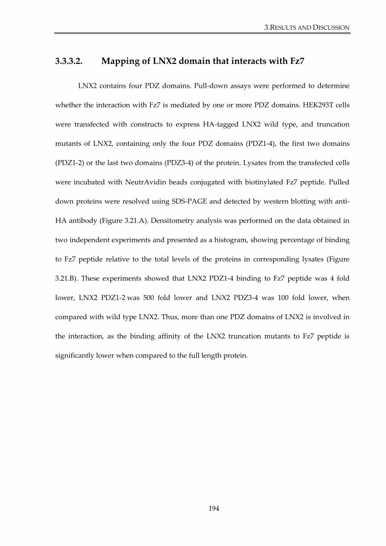
3.RESULTS AND DISCUSSION
194
3.3.3.2. Mapping of LNX2 domain that interacts with Fz7
LNX2 contains four PDZ domains. Pull-down assays were performed to determine
whether the interaction with Fz7 is mediated by one or more PDZ domains. HEK293T cells
were transfected with constructs to express HA-tagged LNX2 wild type, and truncation
mutants of LNX2, containing only the four PDZ domains (PDZ1-4), the first two domains
(PDZ1-2) or the last two domains (PDZ3-4) of the protein. Lysates from the transfected cells
were incubated with NeutrAvidin beads conjugated with biotinylated Fz7 peptide. Pulled
down proteins were resolved using SDS-PAGE and detected by western blotting with anti-
HA antibody (Figure 3.21.A). Densitometry analysis was performed on the data obtained in
two independent experiments and presented as a histogram, showing percentage of binding
to Fz7 peptide relative to the total levels of the proteins in corresponding lysates (Figure
3.21.B). These experiments showed that LNX2 PDZ1-4 binding to Fz7 peptide was 4 fold
lower, LNX2 PDZ1-2 was 500 fold lower and LNX2 PDZ3-4 was 100 fold lower, when
compared with wild type LNX2. Thus, more than one PDZ domains of LNX2 is involved in
the interaction, as the binding affinity of the LNX2 truncation mutants to Fz7 peptide is
significantly lower when compared to the full length protein.
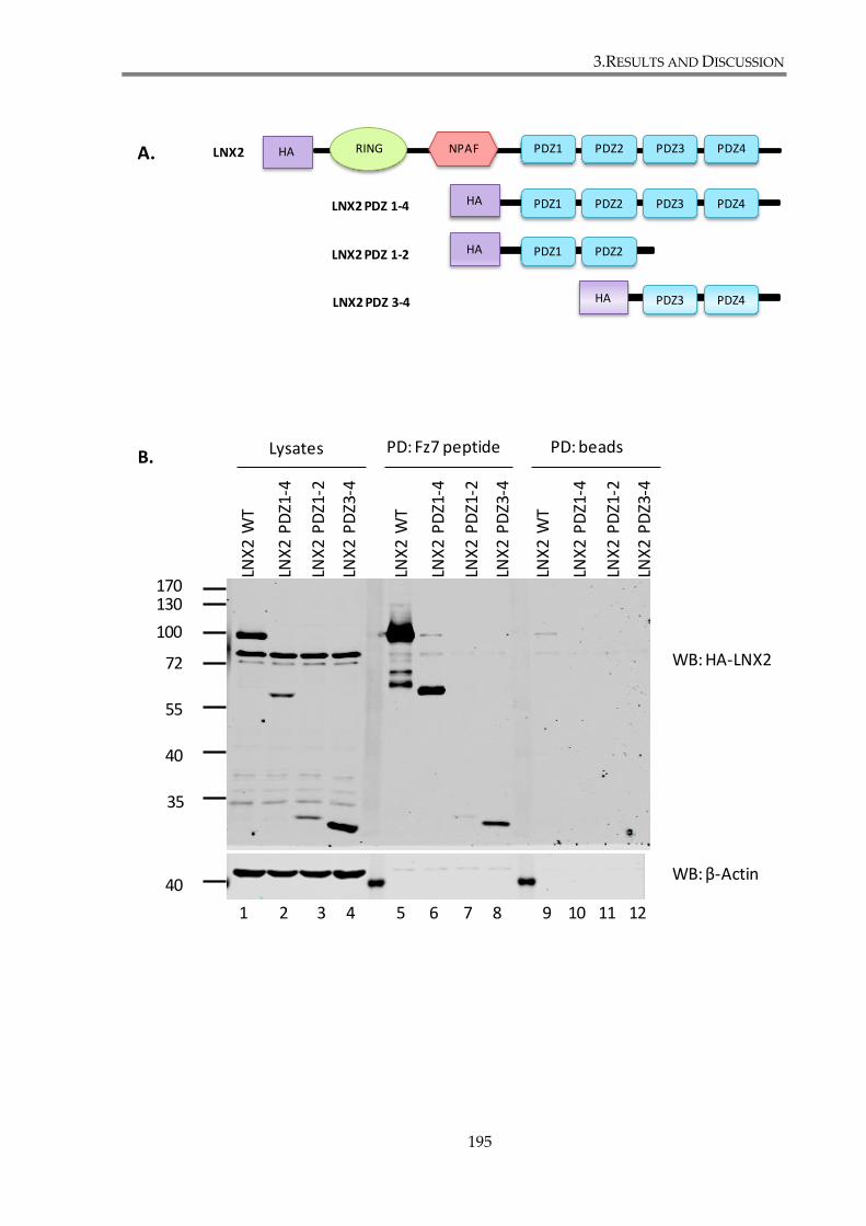
3.RESULTS AND DISCUSSION
195
A. RING PDZ4PDZ3PDZ2PDZ1NPAFLNX2 HA
PDZ4PDZ3PDZ2PDZ1LNX2 PDZ 1-4
PDZ2PDZ1LNX2 PDZ 1-2
PDZ4PDZ3LNX2 PDZ 3-4
HA
HA
HA
Lysates PD: Fz7 peptide PD: beads
WB: HA-LNX2
WB: β-Actin
72
55
35
40
100
130170
LNX
2 W
T
LNX
2 P
DZ1
-4
LNX
2 P
DZ1
-2
LNX
2 P
DZ3
-4
LNX
2 W
T
LNX
2 P
DZ1
-4
LNX
2 P
DZ1
-2
LNX
2 P
DZ3
-4
LNX
2 W
T
LNX
2 P
DZ1
-4
LNX
2 P
DZ1
-2
LNX
2 P
DZ3
-4
40
B.
1 2 3 4 5 6 7 8 9 10 11 12
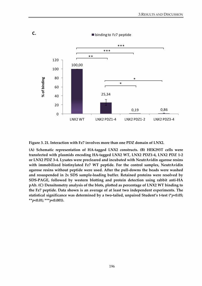
3.RESULTS AND DISCUSSION
196
Figure 3. 21. Interaction with Fz7 involves more than one PDZ domain of LNX2.
(A) Schematic representation of HA-tagged LNX2 constructs. (B) HEK293T cells were
transfected with plasmids encoding HA-tagged LNX2 WT, LNX2 PDZ1-4, LNX2 PDZ 1-2
or LNX2 PDZ 3-4. Lysates were precleared and incubated with NeutrAvidin agarose resins
with immobilized biotinylated Fz7 WT peptide. For the control samples, NeutrAvidin
agarose resins without peptide were used. After the pull-downs the beads were washed
and resuspended in 2x SDS sample-loading buffer. Retained proteins were resolved by
SDS-PAGE, followed by western blotting and protein detection using rabbit anti-HA
pAb. (C) Densitometry analysis of the blots, plotted as percentage of LNX2 WT binding to
the Fz7 peptide. Data shown is an average of at least two independent experiments. The
statistical significance was determined by a two-tailed, unpaired Student’s t-test (*p<0.05;
**p<0.01; ***p<0.001).
C.
100,00
25,34
0,19 0,860
20
40
60
80
100
120
LNX2 WT LNX2 PDZ1-4 LNX2 PDZ1-2 LNX2 PDZ3-4
% o
f b
ind
ing
binding to Fz7 peptide
*****
***
**
3.RESULTS AND DISCUSSION
197
3.3.3.3. Testing co-localization of Fz7 and LNX2 in SKBr3 cells
If LNX2 interacts with Fz7, these two proteins should be present in the same cellular
compartments. To verify whether they co-localize we performed immunofluorescence
experiments on SKBr3 cell lines stably expressing FLAG-Fz7 WT and FLAG-Fz7 V574G. Due
to lack of a suitable antibody to detect endogenous LNX2 by immunofluorescence, Myc-
LNX2 was used for the co-localization study. SKBr3, SKBr3-FLAG-Fz7 and SKBr3-FLAG-Fz7
V574G cells were transfected with Myc-LNX2 and co-distribution of the proteins was
analysed 48h later using rabbit anti-FLAG and mouse anti-Myc antibodies. DAPI was used
to visualize nuclei.
LNX2 was predominantly found in the cytoplasm but was also detected close to the
plasma membrane in parental SKBr3 cells. In both cell lines, SKBr3-FLAG-Fz7 and FLAG-Fz7
V574G, clear co-localization with LNX2 was observed in plasma membrane proximity
(Figure 3.22.). These results provided a supporting evidence for the interaction between
LNX2 and Fz7 in vivo.

3.RESULTS AND DISCUSSION
198
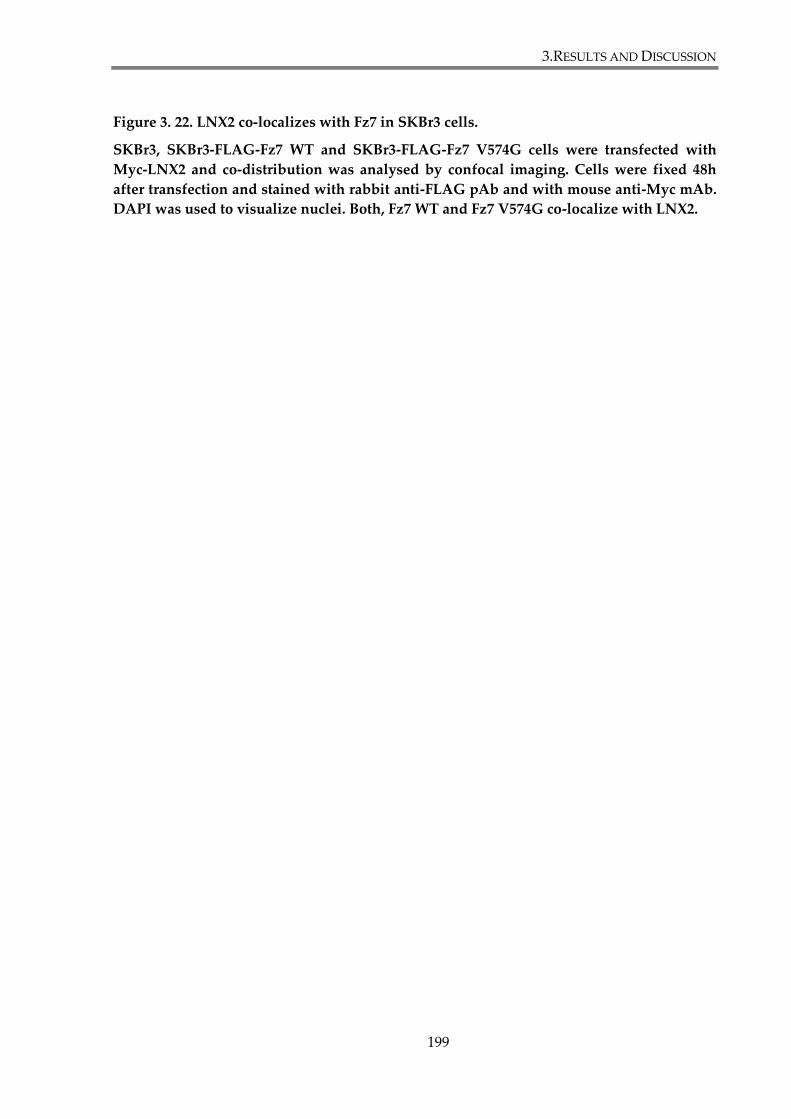
3.RESULTS AND DISCUSSION
199
Figure 3. 22. LNX2 co-localizes with Fz7 in SKBr3 cells.
SKBr3, SKBr3-FLAG-Fz7 WT and SKBr3-FLAG-Fz7 V574G cells were transfected with
Myc-LNX2 and co-distribution was analysed by confocal imaging. Cells were fixed 48h
after transfection and stained with rabbit anti-FLAG pAb and with mouse anti-Myc mAb.
DAPI was used to visualize nuclei. Both, Fz7 WT and Fz7 V574G co-localize with LNX2.
3.RESULTS AND DISCUSSION
200
3.3.4. Discussion
We have found in the proceeding chapters that introduction of a mutation in the C-
terminal PDZ binding motif of Frizzled-7 receptor affects canonical Wnt signalling in breast
cancer cell lines. Subsequently, we have provided evidence that Fz7 interaction with PDZ
protein syntenin-1 regulates functions of the receptor. Nonetheless, one could not exclude
the possibility that other PDZ domain containing proteins are also involved in modulating
Fz7 signalling. To identify cellular proteins that might be responsible for PDZ-BM –
dependent regulation of the canonical Wnt pathway we were searching for novel Fz7
binding partners. We have employed a standard pull-down assay using biotinylated
peptides and found striking differences in an amount of proteins pulled down by Fz7 WT
peptide and Fz7 V574G peptide (Figure 3.17). Proteins pulled down by Fz7 WT peptide, but
not Fz7 V574G peptide, are known to be involved in receptor trafficking, as well as in
establishment of cell polarity and tight junction formation (INADL/PATJ, MPDZ, Scribble,
DLG1, ZO-1, ZO-2, PALS1/MPP5, MPP7, MAGI1, MAGI3, SNX27, LNX1, LNX2; Table 3.1).
Among the identified proteins only MAGI3 and INADL/PATJ were previously reported to
interact with Frizzled receptors. MAGI3 was shown to bind Fz4 and support non-canonical
Wnt/JNK signalling (Yao et al. 2004), whereas INADL/PATJ interacts with dFz1 and inhibits
Wnt/PCP signalling (Djiane et al. 2005).
The remaining proteins are potentially novel binding partners of Fz7. The interactions
with a number of “polarity proteins” suggest a link between Fz7 and basoapical tissue
polarity. Basoapical polarity in the normal epithelium relies on polarity protein complexes
and cell junction components asymmetrically localized between the apex and the base of the

3.RESULTS AND DISCUSSION
201
cell (Bazzoun et al. 2013). Three conserved complexes of protein have been determined:
Crumbs (Crumbs/PALS/PATJ) and Par (Par3/Par6/aPKC/Cdc42) which are apical, and
Scribble (Scribble/Dlg/Lgl) which is basolateral. Polarity proteins control the establishment of
cell junction complexes that consist of transmembrane proteins (occludin, claudin and JAM
for tight junctions; cadherins for adherens junctions; connexins for gap junctions) associated
with cytoplasmic proteins (ZO proteins, catenins and others). Par complex ensures the
localization of tight junctions at the apical domain of cells. The Crumbs complex also
regulates tight junction maintenance through its reciprocal interactions with Par proteins.
The basolateral side of cells is characterized by the presence of both adherence and gap
junctions. Scribble complex associates with adherence junctions to assure their segregation
from tight junctions and to stabilize the coupling of E-cadherins with catenins, but it’s not
certain how Scribble regulates gap junctions (Bazzoun et al. 2013).
One needs to remember that all of the pulled-down proteins contain PDZ domains
and some of them form complexes with each other (see below). Hence at this stage we cannot
be certain which of the proteins is a primary binding partner of Fz7. Among the identified
potential binding partners of Fz7 are PALS1/MPP5 and PATJ/INADL (present in the Crumbs
complex), Scribble and DLG1 (Scribble complex), ZO-1 and ZO-2 (tight and gap junctions),
as well as MPP7 (interacting with DLG1) and MAGI1, protein regulating cell junctions.
PALS1 was reported as an adaptor protein facilitating interaction between Crumbs and
PATJ, which is a scaffolding protein that regulates protein targeting and integrity of tight
junctions (Roh et al. 2002). MPDZ (MUPP1), also identified in our proteomic screen, is a
protein homological to PATJ, and it was shown to bind identical proteins (Assemat et al.
2008). Scribble is a large (175 kDa) cytoplasmic multi-domain scaffold, playing various roles

3.RESULTS AND DISCUSSION
202
in cells. It physically interacts with DLG proteins (Mathew et al. 2002) and with ZO-2 (Metais
et al. 2005). As already mentioned, Scribble co-localizes with adherens junctions, thus
interaction with ZO-2 probably takes place before ZO-2 is segregated to the tight junctions.
Additionally, ZO-2 has been shown to bind to ZO-1, among other components of tight
junction (Itoh et al. 1999). Scribble has been shown to regulate Ras/MAPK and JNK signalling
pathways (Subbaiah et al. 2011), and reported to play an important role in the recycling and
signalling of transmembrane receptor TSHR (thyroid stimulating hormone receptor) (Lahuna
et al. 2005). Similarly to Frizzled receptors, TSHR belongs to GPCR superfamily of proteins.
Thus it is possible that Scribble may be also be involved in Fz7 recycling. The function of
DLG1 protein is not clear, but it associates with APC protein (Matsumine et al. 1996) and it
has been shown to regulate its correct subcellular localization, in a PDZ domain dependent
fashion; however, the functional significance of this interaction remained unknown (Etienne-
Manneville et al. 2005). It also interacts with MPP2, MPP3, MPP7 and Lin7 (Assemat et al.
2008). The interaction between DLG1 and MPP7 regulates formation of tight junction,
whereas Lin7 stabilizes DLG1 and its distribution (Bohl et al. 2007;Stucke et al. 2007). MAGI1
was also implicated in regulation of tight and adherens junction (Subbaiah et al. 2011).
Furthermore it was shown to bind β-catenin (Dobrosotskaya et al. 2000) and to regulate the
tumour suppressor, PTEN (Glaunsinger et al. 2000). Potentially it forms a trimeric β-catenin-
MAGI-PTEN complex (Kotelevets et al. 2005). Further research would be required to explore
which of the identified proteins directly interacts with Fz7 receptor, whether they affect
distribution of Fz7 and whether interaction with any of these proteins affect the canonical
Wnt pathway.
3.RESULTS AND DISCUSSION
203
Fz7 has been reported to be involved in regulation of planar tissue polarity, as it acts
as a receptor in planar cell polarity (PCP) pathway, one of the non-canonical Wnt signalling
pathways (Dijksterhuis et al. 2014). The link between basoapical and planar cell polarity
remains poorly understood. Djiane and colleagues reported apical localization of dFz1 (a
PCP receptor) in Drosophila epithelial cells and its direct interaction with dPatj, a component
of the Crumbs complex. The authors proposed that dFz1-dPatj interaction leads to aPKC
recruitment and subsequent phosphorylation and inhibition of Fz1/PCP signalling (Djiane et
al. 2005). Since Fz7 is also involved in PCP signal transduction, there is possibility that one
the polarity proteins regulates its basoapical localization in cells. Additionally, Scribble has
been implicated in PCP establishment in the Drosophila eye and wing, by interaction with
Stbm/Vang complex (Courbard et al. 2009). Interestingly, Frizzled receptors have also been
implicated to interact with Stbm/Vang during PCP signalling (Wu et al. 2008).
Loss of basoapical cell polarity is a hallmark of the epithelial-to-mesenchymal
transition (EMT), a process in which cells become more spindle-shaped, mobile and acquire
stemlike characteristics. Interestingly, the role of Wnt signalling in promotion of EMT has
been established in various human cancers, e.g. HCC, prostate and breast cancer (Talbot et
al. 2012). Indeed, Scribble was reported to regulate EMT through modulation of the MAPK-
ERK signalling (Elsum et al. 2013). Thus a potential Scribble-Fz7 interaction might play role
in the process.
Another protein identified in the proteomics screen was SNX27 (sorting nexin-27).
It functions as an endosome-associated cargo adaptor. It interacts with transmembrane
proteins containing NPxY motif through its C-terminal FERM-like domain, or PDZ-BM (S/T-
x-φ) through the PDZ domain (Lauffer et al. 2010). SNX27 forms a complex with retromer, a
3.RESULTS AND DISCUSSION
204
heterotrimer composed of VPS26, VPS29 and VPS35, and regulates recycling of cargo-
proteins from endosomes to plasma membrane (Temkin et al. 2011). Interestingly, VPS26A
binding to SNX27 increases the affinity of the PDZ domain of the protein to the ligands,
implying cooperativity in cargo selection (Gallon et al. 2014). SNX27-retromer complex in
involved in regulation of trafficking of β2-adrenergic receptor, ionotrophic glutamate
receptors (NMDA and AMPA receptors) (Loo et al. 2014), potassium channels (Balana et al.
2011), somatostatin receptor (Bauch et al. 2014) and the glucose transporter (Steinberg et al.
2013). Recent proteomics analysis also identified Fz1 and Fz2 among more than 80
transmembrane proteins that can bind to SNX27 (Steinberg et al. 2013). Taking it all into
account, it is possible that SNX27-retromer regulates Fz7 recycling from endosomes to
plasma membrane. Further research is necessary to explore the nature of Fz7-SNX27
interaction.
Finally, we have also identified two proteins from the LNX family. Ligand of Numb
Protein X (LNX) family contains proteins described as E3 ubiquitin ligases, characterized by
the presence a RING domain (see Introduction for more details). Our proteomics analysis
revealed that two members of the LNX family: LNX1 and LNX2, are potential binding
partners of Fz7. These two proteins share an identical domain structure (RING domain and
four PDZ domains), with LNX1 having two isoforms, a full length p80 and p70 which lacks
the RING domain (Wolting et al. 2011). Interestingly, phylogenetic analysis revealed that
LNX1/2 share a common ancestor with two other proteins, also identified in the proteomic
screen: MPDZ/MUPP1 and INADL/PATJ. LNX1/2 have been also shown to associate with
tight junction proteins JAM4, claudin-1 and CAR, and the later two also interact with
MUPP1 (Flynn et al. 2011). Recently, several reports have suggested the role of LNX proteins
3.RESULTS AND DISCUSSION
205
in regulation of Wnt signalling. LNX1 was shown to bind to Wnt signalling regulators
Naked2 (Nkd2) and Dishevelled-3 (Dvl3) (Wolting et al. 2011). LNX2b (an LNX zebrafish
ortholog) has been shown to bind and ubiquitylate the Wnt inhibitor Bozozok and in
consequence upregulate the β-catenin Wnt signalling (Ro et al. 2009). Finally, siRNA-
mediated silencing of LNX2 reduces Wnt signalling in colorectal cancer cells through
downregulation of TCF7L2 (Camps et al. 2013). For that reason we have decided to further
explore the nature of the interaction between Fz7 and LNX.
To verify whether different isoforms of LNX can bind to Fz7, a pull-down assay with
Fz7 peptides was employed (Figure 3.19). Both isoforms of LNX1, p80 and p70, bind strongly
to the Fz7 WT peptide but not to the peptides corresponding to the C-terminal cytoplasmic
tails of Fz3 or Fz8. On the other hand LNX2 binds to both Fz7 WT and Fz8 peptides (but not
to Fz3 peptide). The observed binding specificity of LNX1 and LNX2 supports distinct
binding properties of LNX PDZ domains (Guo et al. 2012) and suggests non-redundant
functions of these enzymes in Frizzled-dependent pathways. In other experiments we found
that syntenin-1 binds to Fz7 and Fz3, but not to Fz8 peptide. While our observation on
syntenin-1 binding to Fz7 and Fz3 is in agreement with the report by Luyten and colleagues
(2008), were unable to confirm their finding on the syntenin-1 – Fz8 interaction. Although the
reasons behind these differences remain unknown, one the possible explanation may be
difference in the experimental protocols used by us (peptide-based pull down from cellular
lysates) and these authors (interaction between recombinant syntenin-1 and the peptide)
(Luyten et al. 2008).
Pull-down assays with peptides do not always reflect the binding dynamics of
proteins in living cells. Accordingly, we have used co-immunoprecipitation assay to further

3.RESULTS AND DISCUSSION
206
verify the interactions. We have established that LNX1 p70 and LNX2 bind to the full length
Fz7, however p80 isoform of LNX1 does not. It might be due to the tertiary structure of the
proteins, their complexes with another binding partners or different compartmentalization
within the cell. Importantly, in the immunoprecipitation experiments we confirmed that
LNX-Fz7 interactions are mediated through PDZ domain interactions. Furthermore, our
results indicate that more than one PDZ domains of LNX2 is involved in these interactions
(Figure 3.21). In this regard, several other proteins has also been shown to bind two PDZ
domain of LNX ligases, e.g. PLEKH5 (PDZ1 and PDZ3), or PAK6 and PKC-α1 (PDZ2 and
PDZ4) (Wolting et al. 2011).
Taken together, work presented in this chapter included identification of novel
binding partners of Fz7 by mass spectrometry. To our knowledge, it’s the first systematic
proteomic screen in search of cytoplasmic proteins interacting with Fz7. Among identified
interactors we have found a number of PDZ proteins forming polarity complexes, thus
suggesting the involvement of Fz7 in establishment or disruption of basoapical cell polarity.
We have also identified several proteins, previously reported to be involved in trafficking of
membrane receptors, to associate with Fz7, such as SNX27, Scribble, DLG1, MAGI3. They
might be novel regulators of Fz7 recycling. Finally, two proteins from LNX family, LNX1 and
LNX2, were also recognized to associate with Fz7 in PDZ domain dependent fashion.
Further analysis confirmed interaction between full length Fz7 and LNX2 in epithelial cells,
as well their colocalization in plasma membrane proximity. The functional significance of
this interaction will be explored in the next section.
3.RESULTS AND DISCUSSION
207
3.4. Investigating the role of the Fz7 interaction with
proteins from LNX family
We have established that Fz7 interacts with LNX1 and LNX2. Whilst previous studies
have reported a functional connection between Wnt signalling and LNX proteins (Camps et
al. 2013;Ro et al. 2009), to date, no investigation has identified a link between LNX and a
specific Frizzled receptor or determined a mechanism explaining how LNX is involved in
regulating Wnt signalling. The aim of the work described in this chapter was to assess
whether Fz7 functions are regulated by LNX proteins.
3.4.1. LNX1 and LNX2 modulate Fz7 ubiquitylation.
3.4.1.1. LNX1 p80, LNX1 p70 and LNX2 overexpression decreases
Fz7 ubiquitylation level
In order to verify whether Fz7 is an ubiquitylation substrate of LNX1 or LNX2, an in
vivo ubiquitylation assay was performed, as described in Materials and Methods (section
2.5.2.). HEK293T cells were transfected with plasmids encoding GFP-LNX1 p80, GFP-LNX1
p70, GFP-LNX2 or GFP, together with FLAG-Fz7 and HA-ubiquitin. 24h post transfection,
cells were lysed and Fz7 was immunoprecipitated using anti-FLAG magnetic beads. Protein
ubiquitylation was analysed by SDS-PAGE and WB using anti-HA pAb (Figure 3.23). Fz7
ubiquitylation was detected as a smear in the anti-FLAG immunoprecipitate by anti-HA
antibodies, indicating protein polyubiquitylation. Interestingly, the amounts of ubiquitylated

3.RESULTS AND DISCUSSION
208
protein species in Fz-immunoprecipitates were consistently lower than in the control
samples. Specifically, densitometry analysis revealed that the levels of ubiquitylated Fz7
were decreased by 50% when both isoforms of LNX1 or LNX2 proteins were overexpressed.
Whilst these experiments involved incubation of samples at 60°C to ease Fz7 detection by
western blotting, additional test with samples incubated at 100°C before SDS-PAGE analysis
confirmed the decreased ubiquitylation level of Fz7 after LNX overexpression (data not
shown).
3.RESULTS AND DISCUSSION
209
72
55
35
40
100
130170
72
55
100
130
35
WB: HA-Ubiquitin
WB: FLAG-Fz7
WB: GFP
WB: β-Actin
WB: GFP (LNX1 and LNX2)
1 2 3 4
5 6 7 8
kDa
GFP
-LN
X1
p80
GFP
-LN
X1
p70
GFP
-LN
X2
GFP
GFP
-LN
X1
p80
GFP
-LN
X1
p70
GFP
-LN
X2
GFP
IP:FLAG LysatesA.
40
+ + + + + + + + Fz7+ + + + + + + + HA-Ubiquitin
3.RESULTS AND DISCUSSION
210
Figure 3. 23. LNX1 and LNX2 decrease the levels of Fz7 ubiquitylation.
(A) HEK293T cells were co-transfected with GFP-LNX1 p80, GFP-LNX1 p70, GFP-LNX2 or
GFP, FLAG-Fz7 WT and HA-Ubiquitin. Subsequently cells were lysed in the lysis buffer
containing 0.5% SDS, incubated at 60°C, diluted with 1% Triton X-100 and sonicated.
Subsequently lysates were incubated with Anti-FLAG®M2 Magnetic Beads and to pull-
down FLAG-Fz7. Ubiquitylated proteins were detected by immunoblotting with anti-HA
antibody. Images depict representatives of at least three independent experiments. (B)
Densitometry analysis of the blots. Data shown is an average of at least three independent
experiments. Ratios between densitometry values (optical density, OD) of
immunoprecipitated HA-ubiquitin and Fz7 were calculated for each sample and presented
in the histogram. This allowed to compare different samples with each other, as the
amount of precipitated proteins may vary between them. Ubiquitylation values of Fz7
were standardized to the value of the control sample (Fz7 co-transfected with GFP, lane 4).
The statistical significance was determined by a two-tailed, unpaired Student’s t-test
(*p<0.05).
0,0
0,2
0,4
0,6
0,8
1,0
1,2
GFP-LNX1 p80 GFP-LNX1 p70 GFP-LNX2 GFP
Re
lati
veO
D
Fz7 ubiquitylation
**
*
B.
3.RESULTS AND DISCUSSION
211
3.4.1.2. LNX2 modulation of Fz7 ubiquitination does not involve
proteasomal degradation of the receptor
In the previous section, decreased levels of Fz7 ubiquitylation were observed in
response to LNX1 and LNX2 overexpression. LNX proteins may regulate Fz7 ubiquitylation
and expression levels through a mechanism involving proteasome dependent protein
degradation. To address this question, we carried out in vivo ubiquitylation experiments in
the presence of MG132, a widely used proteasome inhibitor. HEK293T were co-transfected
with FLAG-Fz7, HA-ubiquitin and GFP-LNX2 or GFP. Cells were then treated with 10 μM
MG132 or the vehicle (DMSO) for 24h. Subsequently, they were lysed, and Fz7 complexes
were immunoprecipitated using anti-FLAG beads (as described in the section 2.5.2. of
Materials and Methods). Protein ubiquitylation was analysed by WB using anti-HA pAb for
detecting HA-ubiquitin (Figure 3.24).
As expected, MG132 treatment increased the overall levels of protein ubiquitylation
in cells. Furthermore, the treatment with MG132 increased the levels of Fz7 ubiquitylation by
70% in control cells. LNX2 expression led to a reduction inFz7 ubiquitylation in the absence
(lane 1) and in the presence (lane 3) of the proteasome inhibitor MG132. Densitometry
analysis revealed that ubiquitylation of Fz7 cells expressing LNX2 was two fold lower
(treated with the vehicle) or three fold lower (treated with MG132), when compared to the
control samples. These findings suggest that the level of Fz7 ubiquitylation is decreased in
cells overexpressing LNX2. However, a decrease in the overall levels of ubiquitylation of
cellular proteins was also observed in cells overexpressing LNX2. The mechanism of
observed alterations do not seem to involve proteasome-dependent degradation. Finally, we
3.RESULTS AND DISCUSSION
212
have also noticed that the total level of LNX2 is increased in MG132-treated cells, suggesting
that LNX2 expression level is controlled by proteasomal degradation.
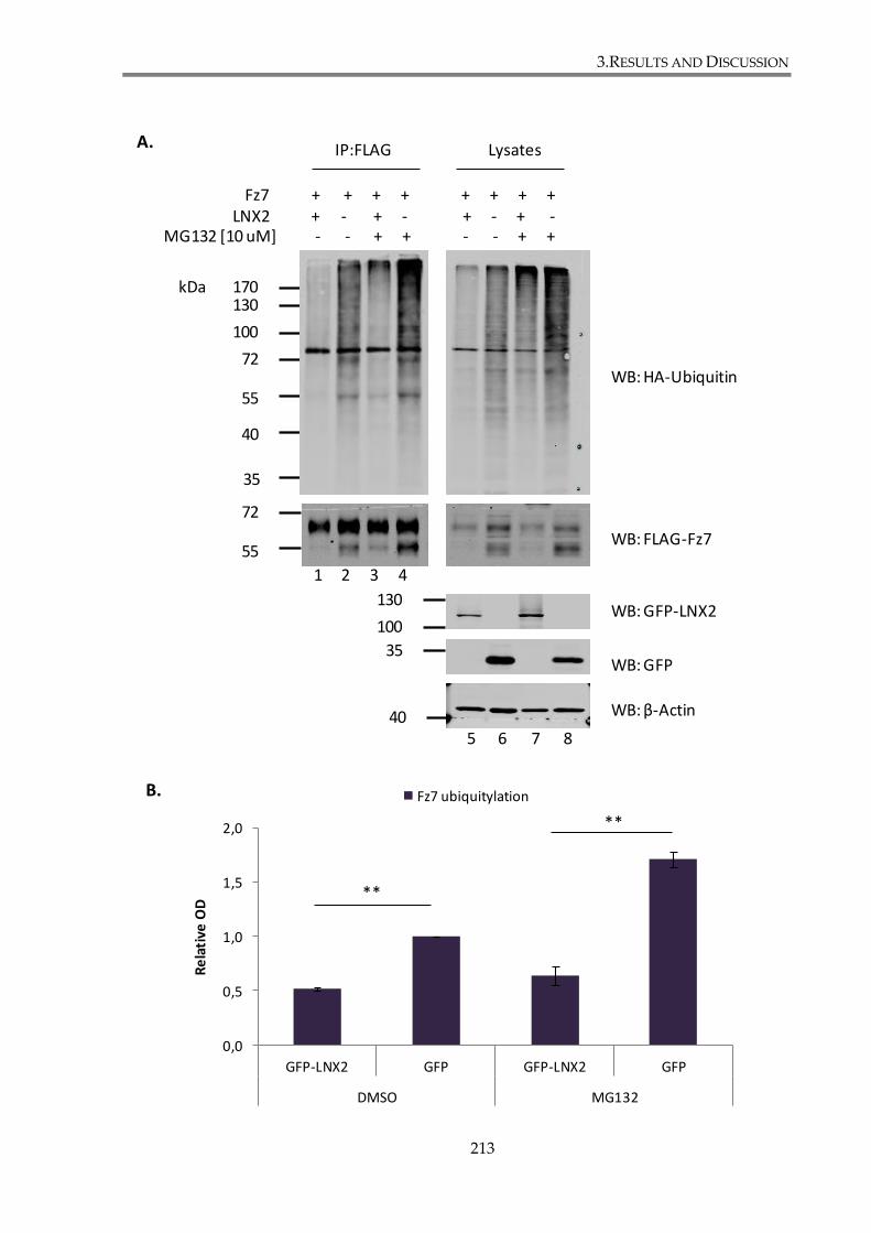
3.RESULTS AND DISCUSSION
213
WB: HA-Ubiquitin
WB: FLAG-Fz7
WB: GFP-LNX2
WB: GFP
WB: β-Actin
LysatesIP:FLAG
5 6 7 8
MG132 [10 uM] - - + + - - + +
72
55
35
40
100
130170
72
55
100
130
35
LNX2 + - + - + - + -
0,0
0,5
1,0
1,5
2,0
GFP-LNX2 GFP GFP-LNX2 GFP
DMSO MG132
Re
lati
ve O
D
Fz7 ubiquitylation
**
**
1 2 3 4
kDa
A.
B.
Fz7 + + + + + + + +
40
3.RESULTS AND DISCUSSION
214
Figure 3.24. LNX2 decrease the level of Fz7 ubiquitylation in the presence of the
proteasome inhibitor MG132.
HEK293T cells were co-transfected with GFP-LNX2 (or GFP), FLAG-Fz7 WT and HA-
Ubiquitin. Cells were subsequently treated with the proteasome inhibitor MG132 (10 μM)
or DMSO for 24h. Subsequently cells were lysed in 0.5% SDS lysis buffer, incubated at
60°C, diluted in 1% Triton X-100 and sonicated. Lysates were subsequently incubated with
Anti-FLAG®M2 Magnetic Beads to pull-down FLAG-Fz7, followed by detection of
ubiquitylation of Fz7 by immunoblotting with anti-HA antibody. Images depict a
representative of three independent experiments. (B) Densitometry analysis of the blots.
Optical density (OD) was measured for both, HA-ubiquitin and Fz7. Ratios between
densitometry values of immunoprecipitated HA-Ubiquitin and Fz7 were calculated for
each sample and presented in the histogram. Ubiquitylation values of Fz7 were
standardized to the value of the control sample (Fz7 co-transfected with GFP and not
treated with MG132, lane 2). Data shown is an average of at least three independent
experiments. The statistical significance was determined by a two-tailed, unpaired
Student’s t-test (**p<0.01).
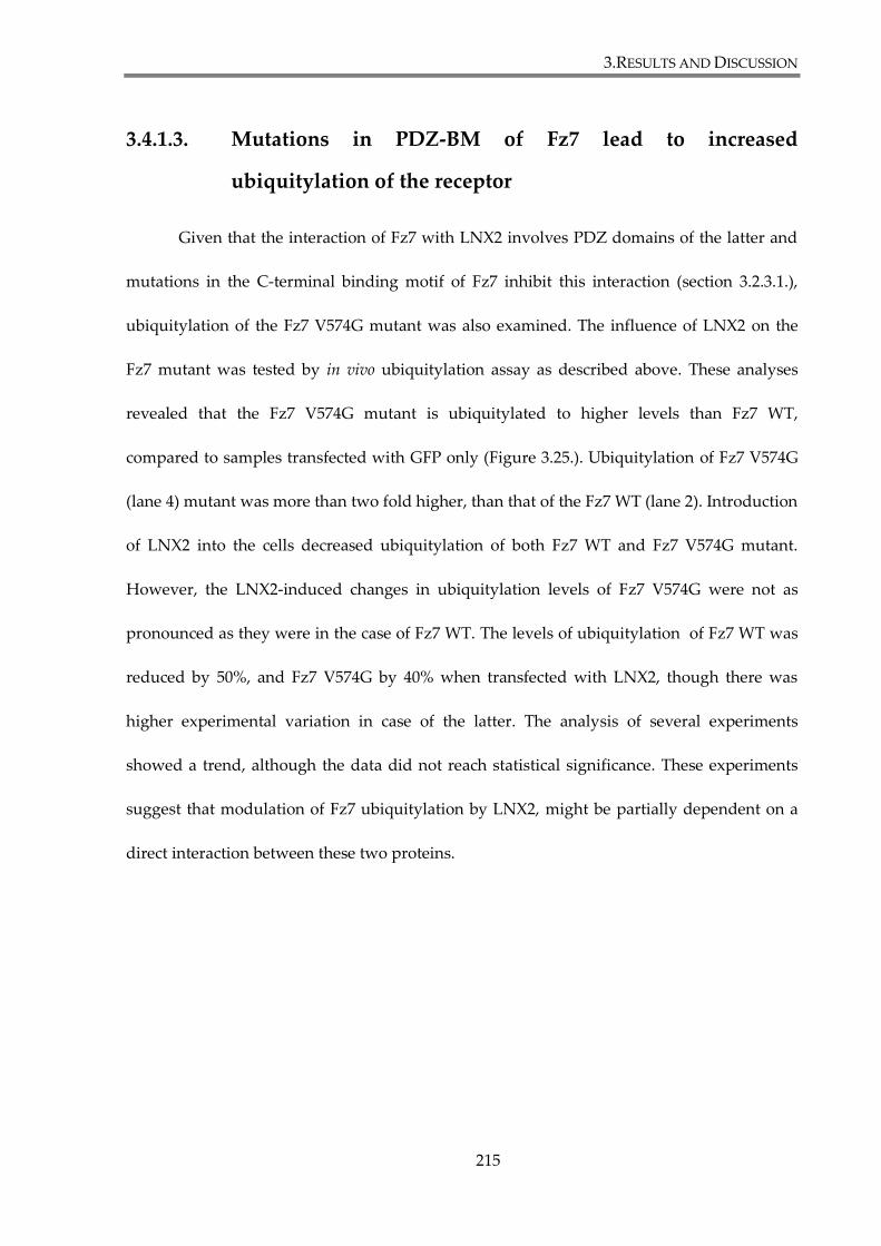
3.RESULTS AND DISCUSSION
215
3.4.1.3. Mutations in PDZ-BM of Fz7 lead to increased
ubiquitylation of the receptor
Given that the interaction of Fz7 with LNX2 involves PDZ domains of the latter and
mutations in the C-terminal binding motif of Fz7 inhibit this interaction (section 3.2.3.1.),
ubiquitylation of the Fz7 V574G mutant was also examined. The influence of LNX2 on the
Fz7 mutant was tested by in vivo ubiquitylation assay as described above. These analyses
revealed that the Fz7 V574G mutant is ubiquitylated to higher levels than Fz7 WT,
compared to samples transfected with GFP only (Figure 3.25.). Ubiquitylation of Fz7 V574G
(lane 4) mutant was more than two fold higher, than that of the Fz7 WT (lane 2). Introduction
of LNX2 into the cells decreased ubiquitylation of both Fz7 WT and Fz7 V574G mutant.
However, the LNX2-induced changes in ubiquitylation levels of Fz7 V574G were not as
pronounced as they were in the case of Fz7 WT. The levels of ubiquitylation of Fz7 WT was
reduced by 50%, and Fz7 V574G by 40% when transfected with LNX2, though there was
higher experimental variation in case of the latter. The analysis of several experiments
showed a trend, although the data did not reach statistical significance. These experiments
suggest that modulation of Fz7 ubiquitylation by LNX2, might be partially dependent on a
direct interaction between these two proteins.
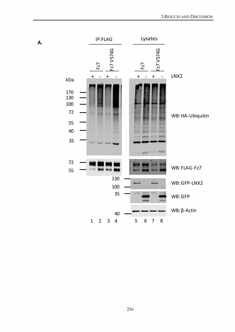
3.RESULTS AND DISCUSSION
216
1 2 3 4
LysatesIP:FLAG
WB: HA-Ubiquitin
WB: FLAG-Fz7
WB: GFP-LNX2
WB: GFP
WB: β-Actin
+ - + - + - + -
5 6 7 8
72
55
35
40
100130170
72
55
100
130
35
kDa
Fz7
Fz7
V57
4G
Fz7
Fz7
V57
4G
LNX2
A.
40
3.RESULTS AND DISCUSSION
217
Figure 3.25. LNX2 decreases the levels of Fz7 ubiquitylation with mutation introduced in
the PDZ binding motif.
(A) HEK293T cells were co-transfected with GFP-LNX2 or GFP, FLAG-Fz7 (WT or V574G)
and HA-Ubiquitin. Subsequently cells were lysed in 0.5% SDS lysis buffer, incubated at
60°C, diluted with 1% Triton X-100 and sonicated. Lysates were incubated with Anti-
FLAG®M2 Magnetic Beads, to pull-down FLAG-Fz7, followed by detection of
ubiquitylation of Fz7 by immunoblotting with anti-HA antibody. Images depict a
representative of three independent experiments. (B) Densitometry analysis of the blots.
Optical density (OD) was measured for both, HA-ubiquitin and Fz7. Ratios between
densitometry values of immunoprecipitated HA-ubiquitin and Fz7 were calculated for
each sample and presented in the histogram. Ubiquitylation values of Fz7 were
standardized to the value of the control sample (Fz7 WT co-transfected with GFP, lane 2).
Data shown is an average of three independent experiments. The statistical significance
was determined by a two-tailed, unpaired Student’s t-test (**p<0.01).
B.
0,0
0,5
1,0
1,5
2,0
2,5
3,0
GFP-LNX2 GFP GFP-LNX2 GFP
Fz7 Fz7 V574G
Re
lati
ve O
D
Fz7 ubiquitylation
**
**

3.RESULTS AND DISCUSSION
218
3.4.2. LNX2 depletion does not alter Fz7 expression
Given the involvement of LNX2 in Fz7 ubiquitylation, it was important to assess
whether knock-down of the enzyme would also influence the levels of expression and the
intracellular distribution of the receptor. SKBr3-FLAG-Fz7 and SKBr3-FLAG-Fz7 V574G cells
were transfected with siRNA against LNX2 or with control siNeg. 24h later cells were
stimulated with Wnt3a conditioned medium or with control conditioned medium.
Subsequent analyses were carried out 48h post transfection. The efficiency of LNX2 knock-
down was shown to be ~90% in cells transfected with siLNX2compared to control cells. The
analysis of Fz7 expression in whole cell lysates (using anti-FLAG pAb), showed an increase
in the total expression level of Fz7 WT in non-stimulated LNX2-depleted cells; the expression
level was decreased in these cells after stimulation with Wnt3a. By contrast, no changes were
observed in Fz7 V574G expression (Figure 3.26.A). In parallel flow cytometry experiments,
we found that depletion of LNX2 did not affect surface expression of surface expression of
either Fz7 WT or Fz7 V574G proteins as assessed by staining with anti-FLAG mAb (Figure
3.26.B). Finally, no apparent alterations in the receptor localization were observed in cells
with transfected with siRNA that target LNX2.
Taken together, apart from the observation that under basal conditions Fz7 WT
expression is increased in LNX2-depleted cells, the analysis of total and membrane of Fz7,
WT and V574G, as well as examination of receptor cell localization by immunofluorescence
didn’t indicate that LNX2 knock-down influence major changes in Fz7 expression in SKBr3
cells.
3.RESULTS AND DISCUSSION
219
Isotype control
siNeg
siNeg + Wnt3a
Isotype control
siLNX2
siLNX2+ Wnt3a
SKBr3-FLAG-Fz7 SKBr3-FLAG-Fz7 V574G
1 2 3 4 5 6 7 8
WB: LNX2
WB: β-Actin
WB: FLAG-Fz7
100
72
55
Wnt3a- + - + - + - +
siNeg siNegsiLNX2 siLNX2
Fz7 WT Fz7 V574G
kDa
40
B.
A.
1,27 1,46 0,10 0,16 0,66 0,64 0,04 0,12 Densitometry OD
Mean:6,32*104
8,73*104
Mean:8,24*104
1,05*105
Mean:6,19*104
6,14*104
Mean:8,28*104
8,36*104
2,41 2,96 5,09 2,53 3,57 2,39 2,60 1,64 Densitometry OD

3.RESULTS AND DISCUSSION
220

3.RESULTS AND DISCUSSION
221

3.RESULTS AND DISCUSSION
222
Figure 3.26. LNX2 depletion does not change Fz7 membrane expression or cell
distribution.
SKBr3-FLAG-Fz7 and SKBr3-FLAG-Fz7 V574G cells were transfected with siRNA against
LNX2 or with control siNeg. 24h later cells were stimulated with Wnt3a conditioned
medium or with control conditioned medium. 48h after the transfection cell lysates were
collected and analysed by SDS-PAGE and WB, flow cytometry and immunofluorescence.
(A) Lysates of equal protein concentrations (2 μg/μl) were resolved on SDS-PAGE,
followed by western blotting with Anti-FLAG pAb to examined Fz7 expression level, anti-
LNX2 pAb to monitor LNX2 knock-down level. β-actin served as a loading control. (B)
Flow cytometry with anti-FLAG mAb was used to examine membrane expression of Fz7.
Cells were incubated on ice for 1h with mouse anti-FLAG (M2) mAb. 4C5G monoclonal
antibody against phosphatidylinositol 4-kinase was used as an isotype control. Cell
surface staining of FLAG-Fz7 (WT or V574G) was revealed by flow cytometry following
incubation for 1h on ice with anti-mouse FITC-conjugated secondary antibodies. Data
analysis was carried out using FlowJo analysis software. The values of calculated mean
fluorescence are presented in top-right corner of the plots. (C-D) Fz7 WT (C) and V574G
(D) cell distribution was analysed by confocal imaging. Cells were fixed and stained with
mouse anti-FLAG (M2) mAb and Alexa Fluor 488-conjugated goat-anti mouse Ab (green).
DAPI was used to visualize nuclei.
3.RESULTS AND DISCUSSION
223
3.4.3. LNX2 is involved in regulation of canonical Wnt
signalling pathway in epithelial breast cancer cells
3.4.3.1. LNX2 depletion attenuates canonical Wnt pathway in
SKBr3 cell line
Fz7 is a membrane receptor involved in Wnt signalling. LNX2 has been shown to
interact with Fz7 in a PDZ dependent manner (section 3.2.3). Given that Fz7 signalling is
regulated by interactions via its C-terminal PDZ-BM (section 3.1.5.2.), the role of LNX2 in
canonical Wnt pathway signalling was examined. Activation of the pathway was assessed in
SKBr3 epithelial breast cancer cell line stably expressing FLAG-Fz7 wild type or FLAG-Fz7
V574G mutant, after LNX2 depletion, using the TOP-Flash reporter assay. SKBr3 cells were
transfected with the siRNA targeting LNX2, or with a control siNegative. On the following
day, cells were stimulated with Fz7 ligand, Wnt3a, and 24h later, TOPFlash activity was
assessed, as described in section 2.5.1 of Materials and Methods. The β-catenin reporter
activity was compared in cells with and without LNX2 knock-down (Figure 3.27.). Western
blot analysis confirmed the efficiency of LNX2 knockdown (lanes 3, 4, 7 and 8), as the protein
expression level was more than 90% lower, when compared with the control samples (lanes
1, 2, 5 and 6). These experiments showed that LNX2 depletion attenuates activation of Wnt
signalling in cells expressing wild type Fz7. Cells with LNX2 knockdown were characterized
by 50% lower reporter activity, when compared with control cells. On the other hand, LNX2
depletion did not alter Wnt signalling in SKBr3 cells expressing the Fz7 V574G mutant.
3.RESULTS AND DISCUSSION
224
Examination of the levels of total β-catenin by western blotting showed that the degree of the
protein stabilization in cells corresponds to the TOP-Flash results.
Taken together, LNX2 depletion attenuates canonical Wnt signalling in SKBr3 cells
expressing wild type Fz7, but not Fz7 V574G mutant.
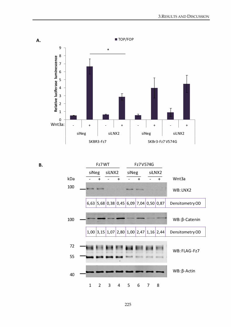
3.RESULTS AND DISCUSSION
225
1 2 3 4 5 6 7 8
WB: β-Catenin
WB: LNX2
WB: β-Actin
WB: FLAG-Fz7
100
72
55
100
Wnt3a- + - + - + - +
siNeg siNegsiLNX2 siLNX2
Fz7 WT Fz7 V574G
0
1
2
3
4
5
6
7
8
9
- + - + - + - +
siNeg siLNX2 siNeg siLNX2
SKBR3-Fz7 SKBr3-Fz7 V574G
Re
lati
ve lu
cife
rase
lu
min
esc
en
ce
TOP/FOP
Wnt3a:
*
kDa
A.
B.
40
6,63 5,68 0,38 0,45 6,09 7,04 0,50 0,87 Densitometry OD
1,00 3,15 1,07 2,80 1,00 2,47 1,16 2,44 Densitometry OD
3.RESULTS AND DISCUSSION
226
Figure 3.27. LNX2 depletion attenuates canonical Wnt signalling in SKBr3 cells expressing
Fz7 WT.
(A) Wnt-induced signalling was analysed using TOP-Flash luciferase assay. SKBr3-FLAG-
Fz7 and SKBr3-FLAG-Fz7 V574G cells were transfected with siRNA against LNX2 or
control siNeg. Cells were stimulated for 24h with Wnt3a conditioned medium and
TOP/FOP activity was analysed 48h after transfection with reporter plasmids. Tests were
done in triplicates and three independent experiments were performed. Statistical
significance was determined by two-tailed, unpaired Student’s t-test (*p<0.05). (B) Cellular
lysates obtained in the assay were examined in SDS-PAGE and WB, with anti-LNX2 pAb,
anti-β-catenin mAb, anti-FLAG pAb and anti-β-actin mAb. Image present a representative
of three independent experiments. Densitometry analysis was performed on LNX2 and β-
catenin immunoreactive bands. Optical densities (OD), standardized to β-actin, are shown
for each lane.
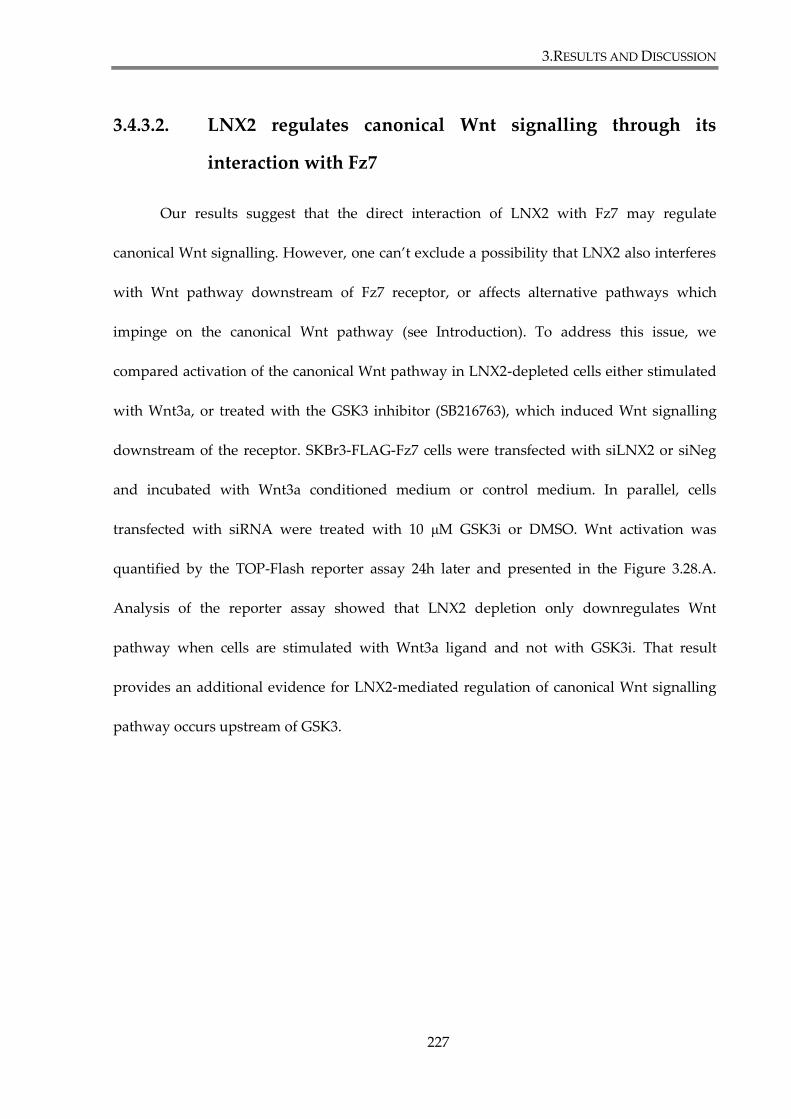
3.RESULTS AND DISCUSSION
227
3.4.3.2. LNX2 regulates canonical Wnt signalling through its
interaction with Fz7
Our results suggest that the direct interaction of LNX2 with Fz7 may regulate
canonical Wnt signalling. However, one can’t exclude a possibility that LNX2 also interferes
with Wnt pathway downstream of Fz7 receptor, or affects alternative pathways which
impinge on the canonical Wnt pathway (see Introduction). To address this issue, we
compared activation of the canonical Wnt pathway in LNX2-depleted cells either stimulated
with Wnt3a, or treated with the GSK3 inhibitor (SB216763), which induced Wnt signalling
downstream of the receptor. SKBr3-FLAG-Fz7 cells were transfected with siLNX2 or siNeg
and incubated with Wnt3a conditioned medium or control medium. In parallel, cells
transfected with siRNA were treated with 10 μM GSK3i or DMSO. Wnt activation was
quantified by the TOP-Flash reporter assay 24h later and presented in the Figure 3.28.A.
Analysis of the reporter assay showed that LNX2 depletion only downregulates Wnt
pathway when cells are stimulated with Wnt3a ligand and not with GSK3i. That result
provides an additional evidence for LNX2-mediated regulation of canonical Wnt signalling
pathway occurs upstream of GSK3.
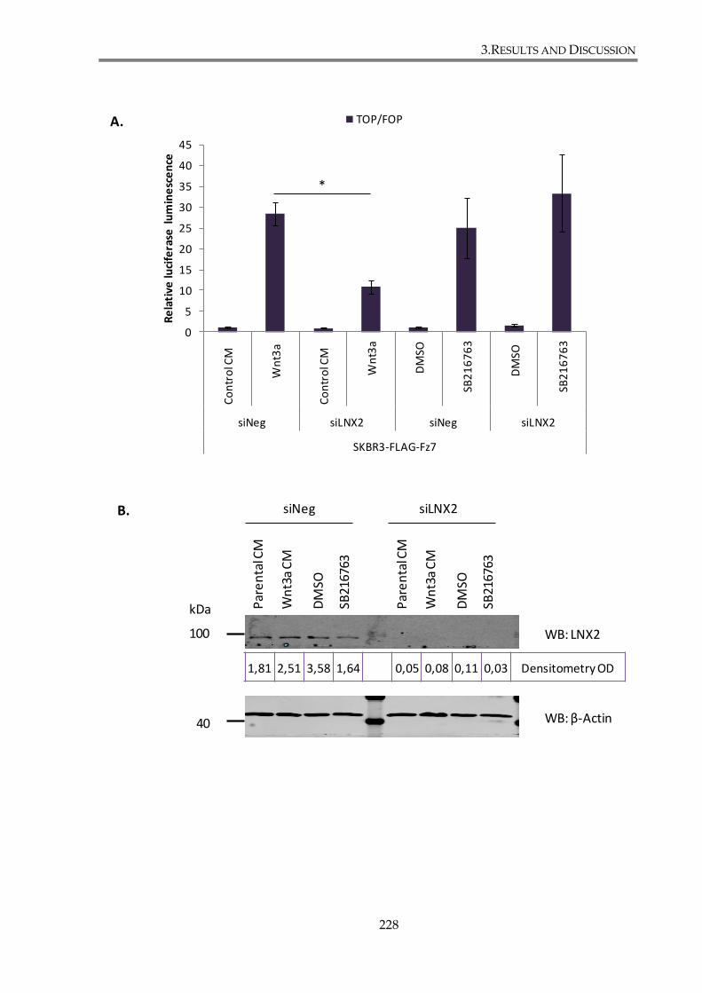
3.RESULTS AND DISCUSSION
228
0
5
10
15
20
25
30
35
40
45
Co
ntr
ol C
M
Wn
t3a
Co
ntr
ol C
M
Wn
t3a
DM
SO
SB2
16
76
3
DM
SO
SB2
16
76
3
siNeg siLNX2 siNeg siLNX2
SKBR3-FLAG-Fz7
Re
lati
ve lu
cife
rase
lu
min
esc
en
ce
TOP/FOP
*
WB: LNX2
WB: β-Actin
100
Par
en
talC
M
Wn
t3a
CM
DM
SO
SB21
6763
Par
en
talC
M
Wn
t3a
CM
DM
SO
SB21
6763
siNeg siLNX2
kDa
A.
B.
40
1,81 2,51 3,58 1,64 0,05 0,08 0,11 0,03 Densitometry OD
3.RESULTS AND DISCUSSION
229
Figure 3. 28. LNX2 regulates canonical Wnt signalling upstream of GSK3.
(A) Wnt-induced signalling was analysed using TOP-Flash luciferase assay. SKBr3-FLAG-
Fz7 cells were transfected with siRNA against LNX2 or control siNegative. Cell were
stimulated for 24h with Wnt3a conditioned medium or with 10 μM GSK3i (SB216763) and
TOP/FOP activity was analysed 48h after transfection with reporter plasmids. Test was
done in triplicates and three independent experiments were performed. Statistical
significance was determined by two-tailed, unpaired Student’s t-test (*p<0.05). (B) Cellular
lysates obtained in the assay were examined in SDS-PAGE and WB, with anti-LNX2 pAb
and anti--actin mAb. Image presents a representative of three independent experiments.
Densitometry analysis was performed on LNX2 and immunoreactive bands. Optical
densities (OD), standardized to -actin, are shown for each lane.
3.RESULTS AND DISCUSSION
230
3.4.4. Discussion
The work described in the proceeding chapter demonstrate that the E3 ubiquitin
ligases: LNX1 and LNX2 are novel binding partners of Fz7. Surprisingly, in vivo
ubiquitylation experiments demonstrated that overexpression of LNX proteins (LNX1p80,
LNX1p70 and LNX2), led to an overall decrease in the levels of ubiquitylatied cellular
proteins (i.e. in total protein lysates), as well asa decrease in the levels of Fz7 ubiquitylation
compared to control samples. It is important to consider that due to the conditions used in
the protocol (incubation of samples for 10 minutes at 60°C before loading on SDS-PAGE gel),
it is possible that the anti-HA signals (HA-ubiquitin) observed in the anti-FLAG
immunoprecipitate may constitute HA-ubiquitin tagged proteins found in a complex with
Fz7. Given that the interaction of Fz7 with LNX2 involves the PDZ domains of the latter, the
ubiquitylation of the Fz7 V574G mutant was also investigated (Figure 3.25). Basal
ubiquitylation of Fz7 V574G was found to be higher than that of Fz7 WT, which identifies a
contribution of the Fz7 PDZ-BM in receptor ubiquitylation. Thus, our data seem to indicate
that Fz7 is not directly ubiquitylated by LNX proteins, which, in fact, function as negative
regulators of ubiquitylation. Although the molecular mechanism(s) underlying this
phenomenon remains to be explored, there are several possibilities which may be
considered. Firstly, LNX proteins might sterically impede interactions of Fz7 with other
ubiquitin ligases. So far, the only identified E3 ubiquitin ligases of Frizzled receptors are two
transmembrane proteins ZNRF3 (zinc and ring finger 3) and RNF43 (ring finger 43), detected
in LGR5 crypt stem cells and colon cancer (Hao et al. 2012;Koo et al. 2012). Both have been
reported to regulate the stability and cell-surface expression of Frizzled receptors (Fz5 and
3.RESULTS AND DISCUSSION
231
Fz8) via multiubiquitylation and subsequent targeting for lysosomal degradation. Whether
Fz7 is also targeted by ZNRF3 and RNF43 and the structural requirements for specific
targeting of Fz receptors by these E3 ligases remain unknown. Another possibility is that
different, as yet unidentified, E3 ubiquitin ligase(s) of Fz7 bind to the receptor indirectly via
another PDZ partner. In this scenario, overexpressed LNX would out compete the alternative
PDZ partners in cells, leading to decreased ubiquitylation of Fz7. Thirdly, one might
hypothesise that LNX itself interacts and regulates the activity of an E3 ubiquitin ligase
targeting Fz7. To elucidate the mechanism of LNX2 dependent regulation of protein
ubiquitylation further experiments are required. Since it is possible that LNX proteins may
target other regulators of the ubiquitylation process in cells, it should be assessed whether
the E3 ligase activity of LNX2 contributes to the observed decrease of protein ubiquitylation.
Introduction of LNX2 with an inactivating mutation in the RING domain in cells expressing
Fz7 might be used for this purpose. Employing the novel CRISPR/Cas9 technology would
allow to replace endogenous LNX2 with the mutant and should be followed by testing the
ubiquitylation of proteins in vivo. Fz7 ubiquitylation should also be assessed in cells with
LNX1 and LNX2 depletion. Additionally mutations can be introduced in PDZ domains of
LNX2 to verify whether such disruption of binding to Fz7 would affect ubiquitylation of the
receptor.
Our data appear to indicate that LNX2 modulates Fz7 expression. We have
consistently observed that in LNX2-overexpressing cells, the total levels of Fz7 is lower than
in control cells. Furthermore, expression of Fz7 V574G mutant, which displays a reduced
capacity to bind to PDZ proteins, seems to be less susceptible to the effects of LNX2. In
agreement with this observation, LNX2 depletion increases the total expression of Fz7 WT,
3.RESULTS AND DISCUSSION
232
but not Fz7 V574G, under basal conditions. At the same time, Fz7 cell surface expression and
subcellular localization remains unchanged. It might be possible that LNX2 regulates
stability of the receptor by affecting its endocytic trafficking. Recently, Sewduth and
colleagues have shown that LNX3/PDZRN3 in endothelial cells ubiquitylates Dvl3 to
promote endocytosis of the Fz4/Dvl3 complex, for activation of PCP signalling pathway
(Sewduth et al. 2014). However, the proposed mechanisms are unlikely to be similar to the
LNX2-dependent regulation of Fz7. The study reports that it is the interaction between LNX3
and PDZ domain of Dvl that is required for recruitment of LNX3 to the Fz4 complex.
Interestingly, Dvl3 has been also identified as a binding partner of LNX1 in a proteomic
screen presented in (Wolting et al. 2011). Performing biotinalytion assay on Fz7 expressing
cells, when LNX2 is overexpressed or depleted, would provide further evidence whether this
protein is (or is not) involved in regulation of Fz7 cell surface expression. Futhermore,
assessing Fz7 expression and trafficking using bafilomycin A1, would allow LNX2 plays role
in lysosome-dependent degradation of the receptor.
Finally, LNX2 role in regulation of the canonical Wnt signalling was examined. We
have established that LNX2 is a novel regulator of Fz7 signalling in breast cancer cells.
Importantly, the contribution of LNX2 to catenin signalling was not observed in cells
expressing Fz7 V574G thus further emphasising the importance of the C-terminal PDZ-BM
in the canonical Wnt pathway. It was previously reported that siRNA-mediated silencing of
LNX2 downregulates Wnt signalling in colorectal cancer cells (Camps et al. 2013). Our data
obtained in breast cancer are in agreement with this report. Whilst, Camps and colleagues
have not determined what is the mechanism of Wnt signalling regulation by LNX2, our
results strongly indicate that the direct interaction of LNX2 with Fz7 receptor is an important
3.RESULTS AND DISCUSSION
233
factor. It is also possible that LNX2 interacts with other proteins that form complex with Fz7,
for example Dishevelled. As already mention, one of the Dishevelled isoforms, Dvl3, has
been found to interact with LNX1 and LNX3 (Sewduth et al. 2014;Wolting et al. 2011).
Although the authors haven’t reported a role of those proteins in canonical Wnt signalling.
In summary, in this chapter we have reported LNX2 as a novel regulator of Fz7-
dependent canonical Wnt signalling in breast cancer cells. We have found out that LNX2
plays role in regulation of Fz7 ubiquitylation and expression level, which might be
responsible for the modulation of the receptor signalling. It was proposed previously that the
expression level of Frizzled receptor correlates with Wnt responsiveness, is controlled by the
interplay between E3 ubiquitin ligase and an deubiquitylating enzyme (Mukai et al. 2010).
However further study is required to elucidated the exact mechanisms underlying the role of
LNX2 in Fz7 functions.
4.GENERAL DISCUSSION AND FUTURE PLANS
234
4. GENERAL DISCUSSION AND
FUTURE WORK
Activation of Wnt signalling, as evidenced by nuclear localisation of β-catenin, has
been previously observed in human breast cancer (Lin at al. 2000; Lopez-Knowles et al.
2010). However, unlike colorectal cancer, this is not a consequence of mutations in APC or
catenin (Candidus et al. 1996;Sorlie et al. 1998), but instead deregulation frequently occurs
through epigenetic modulation of key target genes (Klarmann et al. 2008;Suzuki et al. 2008).
Frizzled-7 seems to be one of the most important Wnt receptors involved in breast cancer. Its
expression is characteristic for triple-negative breast cancer (TNBC) and TNBC-derived cell
lines, where it activates the β-catenin/Wnt pathway and plays a crucial role in regulation of
cell proliferation (Yang et al. 2011). PDZ domain-containing proteins are often involved in
regulation of cell signalling. Indeed, several of them have been implicated in Wnt signalling.
The aim of our study was to determine the role of PDZ domain-containing proteins in
modulating Frizzled-7 signalling and its trafficking pathways, in the context of epithelial
breast cancer cells. Obtained data demonstrate the importance of the C-terminal PDZ-BM of
Frizzled receptors in canonical Wnt signalling. Focusing on syntenin-1 and LNX2, we
reported them as novel regulators of Frizzled-7 and Wnt signalling. Finally, we have also
laid out a strong platform for future investigations by identifying novel binding partners of
Frizzled-7.
4.GENERAL DISCUSSION AND FUTURE PLANS
235
Firstly, we have demonstrated, for the first time, the importance of the C-terminal
PDZ-BM of Frizzled-7 for the correct functioning of the receptor. Our findings illustrate that
Frizzled-7 interactions mediated by the C-terminal PDZ binding motif are crucial for
regulation of trafficking and signalling of the receptor. Investigation of these processes in
more detail and, particularly, the contribution of the newly identified Fz7 partners will
certainly be merited in the future. Our data seem to suggest that mutation introduced in C-
terminal PDZ binding site alters Fz7 maturation process, demonstrated by changes in ratio
between expression levels of two monomers with different degree of glycosylation. The
detailed mechanism of the receptor maturation and the role of Fz7 glycosylation in signalling
have not been addressed widely in the past. Thus it would be important to further explore
the involvement of PDZ proteins in the process. So far, the only known regulator of Fz7
glycosylation is Shisa, protein localized in the ER (Yamamoto et al. 2005). However, since it
does not contain a PDZ domain, its interaction with Frizzled-7 does not explain observed
changes occurring after introduction of mutations into PDZ-BM of the receptor. Thus, it is
likely that other mechanisms are involved.
Secondly, our findings clearly indicate the involvement of syntenin-1 in the
modulation of Fz7 signalling in the canonical Wnt pathway and in the trafficking of the
receptor. Syntenin-1 was reported as an adaptor for a number of transmembrane proteins
(Beekman et al. 2008), thus it may act as a platform for bringing other proteins into the
complex with Fz7. Among those proteins syndecans and tetraspanins are of a particular
interest, given their well-established role as modulators of signalling and trafficking.
Furthermore, syntenin-1, in complex with syndecans and ALIX, was shown to recruit
syndecan-associated proteins to multivesicular endosomes and exosomes (Baietti et al. 2012).
4.GENERAL DISCUSSION AND FUTURE PLANS
236
This might explain Fz7 redistribution from plasma membrane in cells overexpressing
syntenin-1. However syndecans, similarly like Fz7, were shown to bind preferentially to
PDZ2 of syntenin-1, even though both PDZ domains seem to be essential for the association
(Grootjans et al. 2000). Therefore, they might compete with Fz7 for binding to syntenin-1. On
the other hand, tetraspanin CD63 binds syntenin-1 via PDZ1 (Latysheva et al. 2006). CD63 is
a transmembrane protein present in plasma membrane, but also in lysosomes and late
endosomes. It is enriched on the intraluminal vesicles, which are secreted as exosomes.
Additionally CD63 is implicated to function as a regulator of a transport of its interacting
partners (Pols et al. 2009). Thus it may contribute to the receptor trafficking by forming a
tripartite complex with Fz7 and syntenin-1. Another possibility may involve syntenin-1
ability to link transmembrane proteins to ubiquitylated proteins. Ubiquitin binding seems to
be an important factor controlling syntenin-1 relocalization to late endocytic compartments
(Rajesh et al. 2011). To verify whether Fz7 redistribution depends on syntenin-1 interaction
with ubiquitin, one may analyze receptor localization in syntenin-depleted cells and
reconstituted with various syntenin-1 mutants, unable to bind ubiquitin. Interestingly,
ubiquitin binding involves a specific motif in the N-terminal domain of syntenin-1 and is
controlled by Ulk-1 dependent phosphorylation (Rajesh et al. 2011). Ulk-1 is a Ser/Thr kinase
involved in regulation of different trafficking pathways, but also plays a crucial role in
autophagy. Since autophagy was recently reported to attenuate canonical Wnt signalling in
colon cancer, there is possibility for syntenin-1-Ulk-1 involvement in the process (Gao et al.
2010a;Zhang et al. 2011). Considering all possible scenarios, it is important to determine the
fate of Fz7 in cells expressing syntenin-1.
4.GENERAL DISCUSSION AND FUTURE PLANS
237
Thirdly, our data demonstrate, for the first time, that direct interaction of Frizzled-7
with LNX proteins modulates canonical Wnt signalling mediated by the receptor in
epithelial breast cancer cells. Our findings have identified several avenues in need of further
investigation in order to fully understand the contribution of these proteins in regulation of
Frizzled functionality. Proteins from LNX family of molecular scaffolds were reported to act
as E3 ubiquitin ligases (D'Agostino et al. 2011;Nie et al. 2002). However, our data clearly
demonstrate that Fz7 is not a substrate for ubiquitylation by LNX1 or LNX2, two proteins
identified in proteomics analysis. Interestingly, overexpression of these proteins leads to a
significant decrease of Fz7 ubiquitylation level. These findings indicate that after all LNX
proteins are involved in regulation of Fz7 ubiquitylation, nonetheless via an unknown
mechanism. Our results point towards previously unsuspected function of LNX as an
adaptor protein or a competitive inhibitor of ubiquitylation. Therefore, it would be essential
to do more research into a structural aspect of the Fz7-LNX interaction. In this regard our
observation concerning the differential association of Fz7 with LNX1 isoforms is particularly
intriguing. Overexpression of both LNX1 isoforms decreases Fz7 ubiquitylation, even though
LNX1p80 did not bind to Fz7, as shown in the immunoprecipation assay. On the other hand,
LNX1p70, lacking the RING domain, forms a complex with Fz7 and affects the receptor in
a similar way as LNX2. One also needs to identify a relevant Fz7 ubiquitin ligase and
understand the relationship between LNX and this enzyme. A starting point here might
involve establishing a possible link between LNX proteins and ZNRF3 or RNF43, ubiquitin
ligases that were shown to target other Fz receptors (Hao et al. 2012;Koo et al. 2012).
Finally, we have identified a number of novel putative binding partners of Fz7;
namely INADL/PATJ, MPDZ, Scribble, DLG1, ZO-1, ZO-2, PALS1/MPP5, MPP7, MAGI1,
4.GENERAL DISCUSSION AND FUTURE PLANS
238
MAGI3, SNX27, LNX1, LNX2. These PDZ proteins have been previously shown to be
involved in regulation of trafficking of other membrane receptors, as well as in establishment
of cell polarity. One of them is SNX27, sorting nexin-27, an endosome-associated adaptor
controlling recycling of proteins to plasma membrane (Temkin et al. 2011). It is highly
probable that SNX27 regulates Fz7 trafficking between endosomes and plasma membrane.
Thus it would be very interesting to investigate the interaction between these two proteins
and its functional implications. Interestingly, recycling activity of SNX27 requires both, PDZ
domain–dependent binding of the cytoplasmic tail of the ligand and Phox homology (PX)
domain–dependent interaction with the phosphoinositides in endosome membrane (Lauffer
et al. 2010). Hence, one speculate about possible link between SNX27 and syntenin-1 through
phosphoinositides, as the latter also interacts with PtdIns, but via PDZ1 domain
(Zimmermann 2006). Furthermore, SNX27 was shown recently to cooperate with two other
PDZ proteins (GOPC/PIST and NHERF1/3) in regulation of the subcellular localization and
plasma membrane availability of a GPCR protein, somatostatin receptor subtype 5 (SSTR5)
(Bauch et al. 2014). It was reported that those PDZ proteins act sequentially on the
transmembrane ligand during the subcellular trafficking. Accordingly, it is possible that a
similar mechanism regulates Frizzled receptor trafficking. Last but not least, a very
interesting link emerges between Fz7 and basoapical cell polarity, as several of the identified
proteins are components of polarity complexes. Fz7 has been implicated in regulation of
planar cell polarity (Dijksterhuis et al. 2014), yet it wasn’t associated with basoapical polarity.
Basoapical polarity in epithelial cells relies on three conserved complexes, two apical:
Crumbs (Crumbs/PALS/PATJ) and Par (Par3/Par6/aPKC/Cdc42) and one basolateral:
Scribble (Scribble/Dlg/Lgl) (Bazzoun et al. 2013). In the proteomic screen we have identified
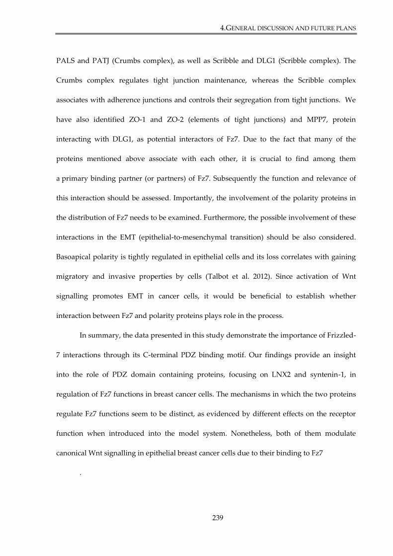
4.GENERAL DISCUSSION AND FUTURE PLANS
239
PALS and PATJ (Crumbs complex), as well as Scribble and DLG1 (Scribble complex). The
Crumbs complex regulates tight junction maintenance, whereas the Scribble complex
associates with adherence junctions and controls their segregation from tight junctions. We
have also identified ZO-1 and ZO-2 (elements of tight junctions) and MPP7, protein
interacting with DLG1, as potential interactors of Fz7. Due to the fact that many of the
proteins mentioned above associate with each other, it is crucial to find among them
a primary binding partner (or partners) of Fz7. Subsequently the function and relevance of
this interaction should be assessed. Importantly, the involvement of the polarity proteins in
the distribution of Fz7 needs to be examined. Furthermore, the possible involvement of these
interactions in the EMT (epithelial-to-mesenchymal transition) should be also considered.
Basoapical polarity is tightly regulated in epithelial cells and its loss correlates with gaining
migratory and invasive properties by cells (Talbot et al. 2012). Since activation of Wnt
signalling promotes EMT in cancer cells, it would be beneficial to establish whether
interaction between Fz7 and polarity proteins plays role in the process.
In summary, the data presented in this study demonstrate the importance of Frizzled-
7 interactions through its C-terminal PDZ binding motif. Our findings provide an insight
into the role of PDZ domain containing proteins, focusing on LNX2 and syntenin-1, in
regulation of Fz7 functions in breast cancer cells. The mechanisms in which the two proteins
regulate Fz7 functions seem to be distinct, as evidenced by different effects on the receptor
function when introduced into the model system. Nonetheless, both of them modulate
canonical Wnt signalling in epithelial breast cancer cells due to their binding to Fz7
.
5. APPENDIX: SUPPLEMENTARY FIGURES
240
5. APPENDIX:
SUPPLEMENTARY FIGURES
Fz7Fz7
DAPI
Myc-Fz7
GFP-Fz7
100
72
55
130
WB: β-Actin
WB: Fz7
kDa
+ GFP-Fz7
+ Myc-Fz7
1 2 3 4
A.
B.
anti-GFP ab anti-Myc ab
40
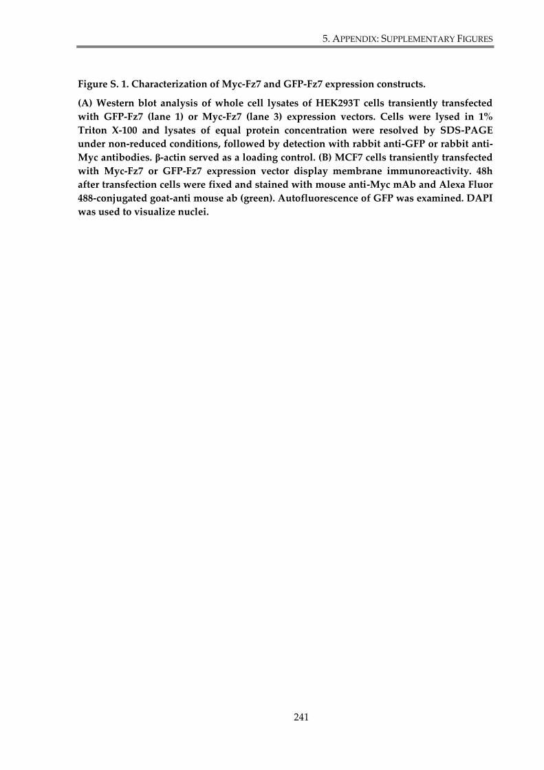
5. APPENDIX: SUPPLEMENTARY FIGURES
241
Figure S. 1. Characterization of Myc-Fz7 and GFP-Fz7 expression constructs.
(A) Western blot analysis of whole cell lysates of HEK293T cells transiently transfected
with GFP-Fz7 (lane 1) or Myc-Fz7 (lane 3) expression vectors. Cells were lysed in 1%
Triton X-100 and lysates of equal protein concentration were resolved by SDS-PAGE
under non-reduced conditions, followed by detection with rabbit anti-GFP or rabbit anti-
Myc antibodies. β-actin served as a loading control. (B) MCF7 cells transiently transfected
with Myc-Fz7 or GFP-Fz7 expression vector display membrane immunoreactivity. 48h
after transfection cells were fixed and stained with mouse anti-Myc mAb and Alexa Fluor
488-conjugated goat-anti mouse ab (green). Autofluorescence of GFP was examined. DAPI
was used to visualize nuclei.
5. APPENDIX: SUPPLEMENTARY FIGURES
242
0
10
20
30
40
50
60
- + - +
HEK293T HEK293T + Myc-Fz7
Re
lati
ve lu
cife
rase
lu
min
esc
en
ce
TOP
FOP
Wnt3a:
**
0
10
20
30
40
50
60
70
80
90
- + - +
HEK293T HEK293T + GFP-Fz7
Re
lati
ve lu
cife
rase
lu
min
esc
en
ce
TOP
FOP
Wnt3a:
*
A.
B.
5. APPENDIX: SUPPLEMENTARY FIGURES
243
Figure S. 2. Verification of Myc-Fz7 and GFP-Fz7 constructs functionality.
Wnt3a-induced signalling was analysed using TOP-Flash luciferase assay. HEK293T cells
were transfected with reporter plasmids and with plasmid encoding Myc-Fz7 (A), GFP-Fz7
(B) or mock transfected. Cells were stimulated for 24h with Wnt3a conditioned medium
and TOP/FOP activity was analysed 48h after the transfection. Test was done in triplicates
and three independent experiments were performed. Statistical significance was
determined by two-tailed, unpaired Student’s t-test (* p<0.05; ** p<0.01)
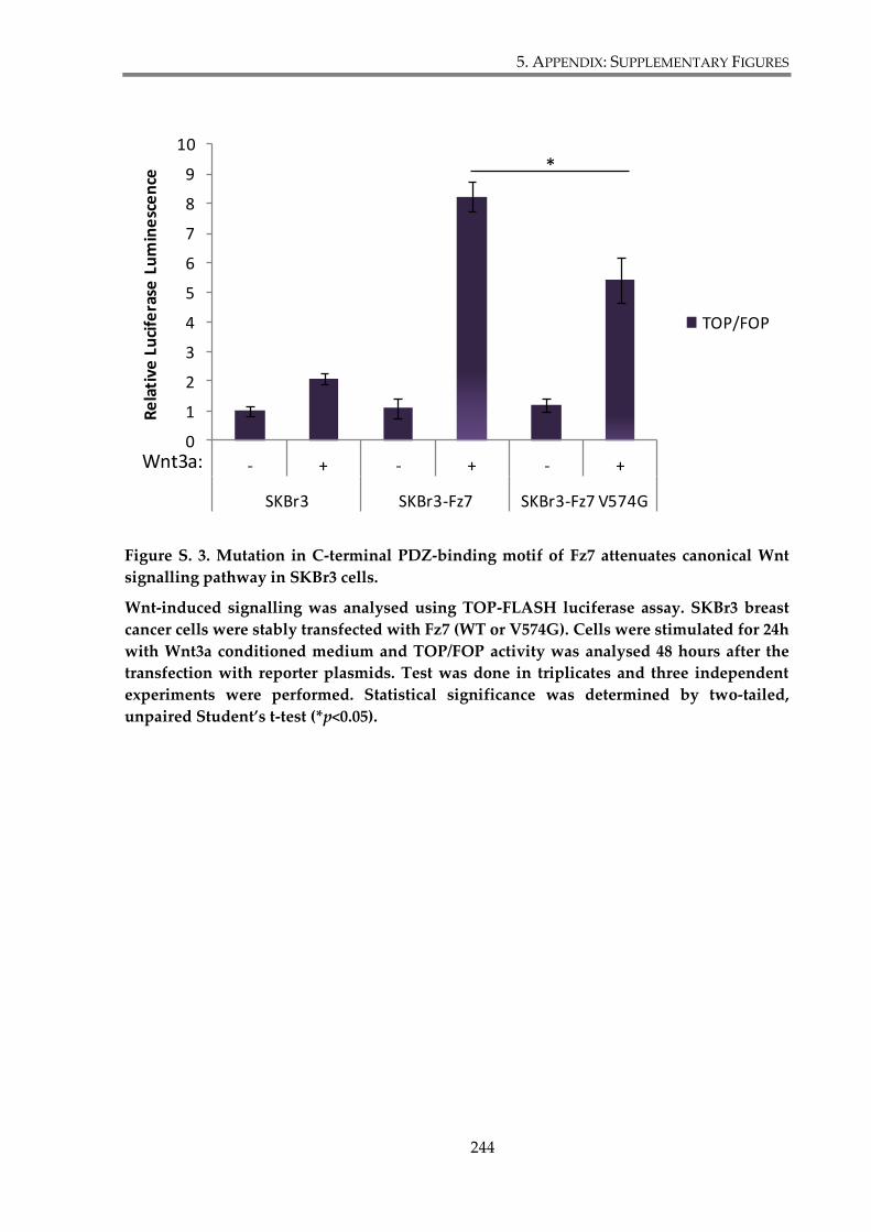
5. APPENDIX: SUPPLEMENTARY FIGURES
244
Figure S. 3. Mutation in C-terminal PDZ-binding motif of Fz7 attenuates canonical Wnt
signalling pathway in SKBr3 cells.
Wnt-induced signalling was analysed using TOP-FLASH luciferase assay. SKBr3 breast
cancer cells were stably transfected with Fz7 (WT or V574G). Cells were stimulated for 24h
with Wnt3a conditioned medium and TOP/FOP activity was analysed 48 hours after the
transfection with reporter plasmids. Test was done in triplicates and three independent
experiments were performed. Statistical significance was determined by two-tailed,
unpaired Student’s t-test (*p<0.05).
0
1
2
3
4
5
6
7
8
9
10
- + - + - +
SKBr3 SKBr3-Fz7 SKBr3-Fz7 V574G
Re
lati
ve L
uci
fera
se L
um
ine
sce
nce
TOP/FOP
Wnt3a:
*
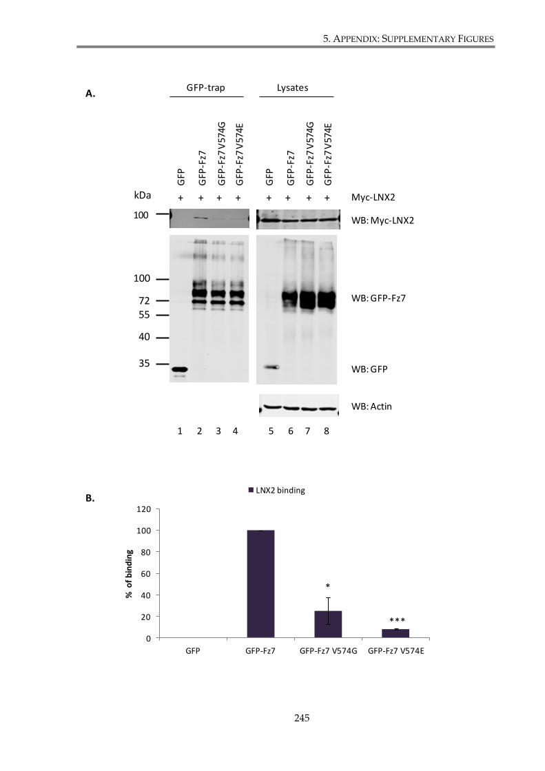
5. APPENDIX: SUPPLEMENTARY FIGURES
245
WB: Myc-LNX2
WB: GFP-Fz7
WB: GFP
WB: Actin
1 2 3 4 5 6 7 8
LysatesGFP-trap
100
100
7255
40
35
GFP
GFP
-Fz7
GFP
-Fz7
V57
4G
GFP
-Fz7
V57
4E
GFP
GFP
-Fz7
GFP
-Fz7
V57
4G
GFP
-Fz7
V57
4E
Myc-LNX2+ + + + + + + + kDa
0
20
40
60
80
100
120
GFP GFP-Fz7 GFP-Fz7 V574G GFP-Fz7 V574E
% o
f b
ind
ing
LNX2 binding
*
***
A.
B.
5. APPENDIX: SUPPLEMENTARY FIGURES
246
Figure S. 4. Fz7 interaction with LNX2 involves PDZ domains (GFP-trap).
(A) HEK293T cells were co-transfected with GFP, GFP-Fz7 (WT, V574G or V574E) and
Myc-LNX2. 24h after transfection cells were lysed in 1% Triton X-100 and lysates were
incubated with GFP-Trap beads. After immunoprecipitation, retained proteins were
resolved by SDS-PAGE, followed by western blotting and protein detection using mouse
anti-Myc mAb and rabbit anti-GFP pAb. Images depict representatives of two
independent experiments. (B) Densitometry analysis performed on data obtained in two
independent experiments and demonstrated on a histogram, plotted as percentage of
LNX2 binding to Fz7 mutants relative to the interaction of LNX2 with the wild type Fz7
(taken as 100%). The statistical significance was determined by a two-tailed, unpaired
Student’s t-test (*p<0.05; *** p<0.001).
6. LIST OF REFERENCES
247
6. LIST OF REFERENCES
Acconcia, F., Sigismund, S., & Polo, S. 2009, "Ubiquitin in trafficking: the network at work", Exp.Cell
Res., vol. 315, no. 9, pp. 1610-1618.
Ai, L., Tao, Q., Zhong, S., Fields, C. R., Kim, W. J., Lee, M. W., Cui, Y., Brown, K. D., & Robertson, K.
D. 2006, "Inactivation of Wnt inhibitory factor-1 (WIF1) expression by epigenetic silencing is a
common event in breast cancer", Carcinogenesis, vol. 27, no. 7, pp. 1341-1348.
Akiri, G., Cherian, M. M., Vijayakumar, S., Liu, G., Bafico, A., & Aaronson, S. A. 2009, "Wnt pathway
aberrations including autocrine Wnt activation occur at high frequency in human non-small-cell lung
carcinoma", Oncogene, vol. 28, no. 21, pp. 2163-2172.
Amit, S., Hatzubai, A., Birman, Y., Andersen, J. S., Ben-Shushan, E., Mann, M., Ben-Neriah, Y., &
Alkalay, I. 2002, "Axin-mediated CKI phosphorylation of beta-catenin at Ser 45: a molecular switch for
the Wnt pathway", Genes Dev., vol. 16, no. 9, pp. 1066-1076.
Anastas, J. N. & Moon, R. T. 2013, "WNT signalling pathways as therapeutic targets in cancer",
Nat.Rev.Cancer, vol. 13, no. 1, pp. 11-26.
Angers, S., Thorpe, C. J., Biechele, T. L., Goldenberg, S. J., Zheng, N., MacCoss, M. J., & Moon, R. T.
2006, "The KLHL12-Cullin-3 ubiquitin ligase negatively regulates the Wnt-beta-catenin pathway by
targeting Dishevelled for degradation", Nat.Cell Biol., vol. 8, no. 4, pp. 348-357.
Armbruester, V., Sauter, M., Roemer, K., Best, B., Hahn, S., Nty, A., Schmid, A., Philipp, S., Mueller,
A., & Mueller-Lantzsch, N. 2004, "Np9 protein of human endogenous retrovirus K interacts with
ligand of numb protein X", J.Virol., vol. 78, no. 19, pp. 10310-10319.
Assemat, E., Bazellieres, E., Pallesi-Pocachard, E., Le, B. A., & Massey-Harroche, D. 2008, "Polarity
complex proteins", Biochim.Biophys.Acta, vol. 1778, no. 3, pp. 614-630.
Ataman, B., Ashley, J., Gorczyca, D., Gorczyca, M., Mathew, D., Wichmann, C., Sigrist, S. J., & Budnik,
V. 2006, "Nuclear trafficking of Drosophila Frizzled-2 during synapse development requires the PDZ
protein dGRIP", Proc.Natl.Acad.Sci.U.S.A, vol. 103, no. 20, pp. 7841-7846.
Ayyanan, A., Civenni, G., Ciarloni, L., Morel, C., Mueller, N., Lefort, K., Mandinova, A., Raffoul, W.,
Fiche, M., Dotto, G. P., & Brisken, C. 2006, "Increased Wnt signaling triggers oncogenic conversion of
human breast epithelial cells by a Notch-dependent mechanism", Proc.Natl.Acad.Sci.U.S.A, vol. 103, no.
10, pp. 3799-3804.
Bafico, A., Liu, G., Goldin, L., Harris, V., & Aaronson, S. A. 2004, "An autocrine mechanism for
constitutive Wnt pathway activation in human cancer cells", Cancer Cell, vol. 6, no. 5, pp. 497-506.
Baietti, M. F., Zhang, Z., Mortier, E., Melchior, A., Degeest, G., Geeraerts, A., Ivarsson, Y., Depoortere,
F., Coomans, C., Vermeiren, E., Zimmermann, P., & David, G. 2012, "Syndecan-syntenin-ALIX
regulates the biogenesis of exosomes", Nat.Cell Biol., vol. 14, no. 7, pp. 677-685.
6. LIST OF REFERENCES
248
Balana, B., Maslennikov, I., Kwiatkowski, W., Stern, K. M., Bahima, L., Choe, S., & Slesinger, P. A.
2011, "Mechanism underlying selective regulation of G protein-gated inwardly rectifying potassium
channels by the psychostimulant-sensitive sorting nexin 27", Proc.Natl.Acad.Sci.U.S.A, vol. 108, no. 14,
pp. 5831-5836.
Balasubramanian, S., Fam, S. R., & Hall, R. A. 2007, "GABAB receptor association with the PDZ
scaffold Mupp1 alters receptor stability and function", J.Biol.Chem., vol. 282, no. 6, pp. 4162-4171.
Bauch, C., Koliwer, J., Buck, F., Honck, H. H., & Kreienkamp, H. J. 2014, "Subcellular sorting of the G-
protein coupled mouse somatostatin receptor 5 by a network of PDZ-domain containing proteins",
PLoS.One., vol. 9, no. 2, p. e88529.
Bazzoun, D., Lelievre, S., & Talhouk, R. 2013, "Polarity proteins as regulators of cell junction
complexes: implications for breast cancer", Pharmacol.Ther., vol. 138, no. 3, pp. 418-427.
Beekman, J. M. & Coffer, P. J. 2008, "The ins and outs of syntenin, a multifunctional intracellular
adaptor protein", J.Cell Sci., vol. 121, no. Pt 9, pp. 1349-1355.
Behrens, J., von Kries, J. P., Kuhl, M., Bruhn, L., Wedlich, D., Grosschedl, R., & Birchmeier, W. 1996,
"Functional interaction of beta-catenin with the transcription factor LEF-1", Nature, vol. 382, no. 6592,
pp. 638-642.
Bengochea, A., de Souza, M. M., Lefrancois, L., Le, R. E., Galy, O., Chemin, I., Kim, M., Wands, J. R.,
Trepo, C., Hainaut, P., Scoazec, J. Y., Vitvitski, L., & Merle, P. 2008, "Common dysregulation of
Wnt/Frizzled receptor elements in human hepatocellular carcinoma", Br.J.Cancer, vol. 99, no. 1, pp.
143-150.
Bentzinger, C. F., von, M. J., Dumont, N. A., Stark, D. A., Wang, Y. X., Nhan, K., Frenette, J.,
Cornelison, D. D., & Rudnicki, M. A. 2014, "Wnt7a stimulates myogenic stem cell motility and
engraftment resulting in improved muscle strength", J.Cell Biol., vol. 205, no. 1, pp. 97-111.
Bhanot, P., Brink, M., Samos, C. H., Hsieh, J. C., Wang, Y., Macke, J. P., Andrew, D., Nathans, J., &
Nusse, R. 1996, "A new member of the frizzled family from Drosophila functions as a Wingless
receptor", Nature, vol. 382, no. 6588, pp. 225-230.
Bilic, J., Huang, Y. L., Davidson, G., Zimmermann, T., Cruciat, C. M., Bienz, M., & Niehrs, C. 2007,
"Wnt induces LRP6 signalosomes and promotes Dishevelled-dependent LRP6 phosphorylation",
Science, vol. 316, no. 5831, pp. 1619-1622.
Bishop, J. R., Schuksz, M., & Esko, J. D. 2007, "Heparan sulphate proteoglycans fine-tune mammalian
physiology", Nature, vol. 446, no. 7139, pp. 1030-1037.
Blanc, E., Roux, G. L., Benard, J., & Raguenez, G. 2004, "Low expression of Wnt-5a gene is associated
with high-risk neuroblastoma", Oncogene, vol. 24, no. 7, pp. 1277-1283.
Blitzer, J. T. & Nusse, R. 2006, "A critical role for endocytosis in Wnt signaling", BMC.Cell Biol., vol. 7,
p. 28.
Blom, T., Roselli, A., Tanner, M., & Nupponen, N. N. 2008, "Mutation and copy number analysis of
LNX1 and Numbl in nervous system tumors", Cancer Genet.Cytogenet., vol. 186, no. 2, pp. 103-109.
6. LIST OF REFERENCES
249
Bohl, J., Brimer, N., Lyons, C., & Vande Pol, S. B. 2007, "The stardust family protein MPP7 forms a
tripartite complex with LIN7 and DLG1 that regulates the stability and localization of DLG1 to cell
junctions", J.Biol.Chem., vol. 282, no. 13, pp. 9392-9400.
Boland, G. M., Perkins, G., Hall, D. J., & Tuan, R. S. 2004, "Wnt 3a promotes proliferation and
suppresses osteogenic differentiation of adult human mesenchymal stem cells", J.Cell Biochem., vol. 93,
no. 6, pp. 1210-1230.
Boukerche, H., Aissaoui, H., Prevost, C., Hirbec, H., Das, S. K., Su, Z. Z., Sarkar, D., & Fisher, P. B.
2010, "Src kinase activation is mandatory for MDA-9/syntenin-mediated activation of nuclear factor-
kappaB", Oncogene, vol. 29, no. 21, pp. 3054-3066.
Boukerche, H., Su, Z. Z., Emdad, L., Baril, P., Balme, B., Thomas, L., Randolph, A., Valerie, K., Sarkar,
D., & Fisher, P. B. 2005, "mda-9/Syntenin: a positive regulator of melanoma metastasis", Cancer Res.,
vol. 65, no. 23, pp. 10901-10911.
Boukerche, H., Su, Z. Z., Emdad, L., Sarkar, D., & Fisher, P. B. 2007, "mda-9/Syntenin regulates the
metastatic phenotype in human melanoma cells by activating nuclear factor-kappaB", Cancer Res., vol.
67, no. 4, pp. 1812-1822.
Bryja, V., Cajanek, L., Grahn, A., & Schulte, G. 2007, "Inhibition of endocytosis blocks Wnt signalling
to beta-catenin by promoting Dishevelled degradation", Acta Physiol (Oxf), vol. 190, no. 1, pp. 55-61.
Budhidarmo, R., Nakatani, Y., & Day, C. L. 2012, "RINGs hold the key to ubiquitin transfer", Trends
Biochem.Sci., vol. 37, no. 2, pp. 58-65.
Burgner, D., Davila, S., Breunis, W. B., Ng, S. B., Li, Y., Bonnard, C., Ling, L., Wright, V. J.,
Thalamuthu, A., Odam, M., Shimizu, C., Burns, J. C., Levin, M., Kuijpers, T. W., & Hibberd, M. L.
2009, "A genome-wide association study identifies novel and functionally related susceptibility Loci
for Kawasaki disease", PLoS.Genet., vol. 5, no. 1, p. e1000319.
Cadigan, K. M. 2010, "Receptor endocytosis: Frizzled joins the ubiquitin club", EMBO J., vol. 29, no. 13,
pp. 2099-2100.
Caldwell, G. M., Jones, C. E., Taniere, P., Warrack, R., Soon, Y., Matthews, G. M., & Morton, D. G.
2006, "The Wnt antagonist sFRP1 is downregulated in premalignant large bowel adenomas",
Br.J.Cancer, vol. 94, no. 6, pp. 922-927.
Camilli, T. C. & Weeraratna, A. T. 2010, "Striking the target in Wnt-y conditions: intervening in Wnt
signaling during cancer progression", Biochem.Pharmacol., vol. 80, no. 5, pp. 702-711.
Camps, J., Pitt, J. J., Emons, G., Hummon, A. B., Case, C. M., Grade, M., Jones, T. L., Nguyen, Q. T.,
Ghadimi, B. M., Beissbarth, T., Difilippantonio, M. J., Caplen, N. J., & Ried, T. 2013, "Genetic
amplification of the NOTCH modulator LNX2 upregulates the WNT/beta-catenin pathway in
colorectal cancer", Cancer Res., vol. 73, no. 6, pp. 2003-2013.
Canalis, E. 2013, "Wnt signalling in osteoporosis: mechanisms and novel therapeutic approaches",
Nat.Rev.Endocrinol., vol. 9, no. 10, pp. 575-583.
Candidus, S., Bischoff, P., Becker, K. F., & Hofler, H. 1996, "No evidence for mutations in the alpha-
and beta-catenin genes in human gastric and breast carcinomas", Cancer Res., vol. 56, no. 1, pp. 49-52.
6. LIST OF REFERENCES
250
Capurro, M., Martin, T., Shi, W., & Filmus, J. 2014, "Glypican-3 binds to Frizzled and plays a direct
role in the stimulation of canonical Wnt signaling", J.Cell Sci., vol. 127, no. Pt 7, pp. 1565-1575.
Carey, L., Winer, E., Viale, G., Cameron, D., & Gianni, L. 2010, "Triple-negative breast cancer: disease
entity or title of convenience?", Nat.Rev.Clin.Oncol., vol. 7, no. 12, pp. 683-692.
Chen, J., Xu, J., Zhao, W., Hu, G., Cheng, H., Kang, Y., Xie, Y., & Lu, Y. 2005, "Characterization of
human LNX, a novel ligand of Numb protein X that is downregulated in human gliomas",
Int.J.Biochem.Cell Biol., vol. 37, no. 11, pp. 2273-2283.
Chen, W., Hu, L. A., Semenov, M. V., Yanagawa, S., Kikuchi, A., Lefkowitz, R. J., & Miller, W. E. 2001,
"beta-Arrestin1 modulates lymphoid enhancer factor transcriptional activity through interaction with
phosphorylated Dishevelled proteins", Proc.Natl.Acad.Sci.U.S.A, vol. 98, no. 26, pp. 14889-14894.
Chen, W., ten, B. D., Brown, J., Ahn, S., Hu, L. A., Miller, W. E., Caron, M. G., Barak, L. S., Nusse, R., &
Lefkowitz, R. J. 2003, "Dishevelled 2 recruits beta-arrestin 2 to mediate Wnt5A-stimulated endocytosis
of Frizzled 4", Science, vol. 301, no. 5638, pp. 1391-1394.
Cheng, C. W., Yeh, J. C., Fan, T. P., Smith, S. K., & Charnock-Jones, D. S. 2008, "Wnt5a-mediated non-
canonical Wnt signalling regulates human endothelial cell proliferation and migration",
Biochem.Biophys.Res.Commun., vol. 365, no. 2, pp. 285-290.
Cheng, J., Wang, H., & Guggino, W. B. 2005, "Regulation of cystic fibrosis transmembrane regulator
trafficking and protein expression by a Rho family small GTPase TC10", J.Biol.Chem., vol. 280, no. 5,
pp. 3731-3739.
Chitalia, V. C., Foy, R. L., Bachschmid, M. M., Zeng, L., Panchenko, M. V., Zhou, M. I., Bharti, A.,
Seldin, D. C., Lecker, S. H., Dominguez, I., & Cohen, H. T. 2008, "Jade-1 inhibits Wnt signalling by
ubiquitylating beta-catenin and mediates Wnt pathway inhibition by pVHL", Nat.Cell Biol., vol. 10, no.
10, pp. 1208-1216.
Cierpicki, T., Bushweller, J. H., & Derewenda, Z. S. 2005, "Probing the supramodular architecture of a
multidomain protein: the structure of syntenin in solution", Structure., vol. 13, no. 2, pp. 319-327.
Cirone, P., Lin, S., Griesbach, H., Zhang, Y., Slusarski, D., & Crews, C. 2008, "A role for planar cell
polarity signaling in angiogenesis", Angiogenesis, vol. 11, no. 4, pp. 347-360.
Civenni, G., Holbro, T., & Hynes, N. E. 2003, "Wnt1 and Wnt5a induce cyclin D1 expression through
ErbB1 transactivation in HC11 mammary epithelial cells", EMBO Rep., vol. 4, no. 2, pp. 166-171.
Clague, M. J. & Urbe, S. 2006, "Endocytosis: the DUB version", Trends Cell Biol., vol. 16, no. 11, pp. 551-
559.
Couchman, J. R. 2003, "Syndecans: proteoglycan regulators of cell-surface microdomains?",
Nat.Rev.Mol.Cell Biol., vol. 4, no. 12, pp. 926-937.
Courbard, J. R., Djiane, A., Wu, J., & Mlodzik, M. 2009, "The apical/basal-polarity determinant Scribble
cooperates with the PCP core factor Stbm/Vang and functions as one of its effectors", Dev.Biol., vol.
333, no. 1, pp. 67-77.
6. LIST OF REFERENCES
251
Culi, J. & Mann, R. S. 2003, "Boca, an endoplasmic reticulum protein required for wingless signaling
and trafficking of LDL receptor family members in Drosophila", Cell, vol. 112, no. 3, pp. 343-354.
D'Agostino, M., Tornillo, G., Caporaso, M. G., Barone, M. V., Ghigo, E., Bonatti, S., & Mottola, G. 2011,
"Ligand of Numb proteins LNX1p80 and LNX2 interact with the human glycoprotein CD8alpha and
promote its ubiquitylation and endocytosis", J.Cell Sci., vol. 124, no. Pt 21, pp. 3545-3556.
Dann, C. E., Hsieh, J. C., Rattner, A., Sharma, D., Nathans, J., & Leahy, D. J. 2001, "Insights into Wnt
binding and signalling from the structures of two Frizzled cysteine-rich domains", Nature, vol. 412, no.
6842, pp. 86-90.
Das, S. K., Bhutia, S. K., Sokhi, U. K., Azab, B., Su, Z. Z., Boukerche, H., Anwar, T., Moen, E. L.,
Chatterjee, D., Pellecchia, M., Sarkar, D., & Fisher, P. B. 2012, "Raf kinase inhibitor RKIP inhibits MDA-
9/syntenin-mediated metastasis in melanoma", Cancer Res., vol. 72, no. 23, pp. 6217-6226.
Dasgupta, S., Menezes, M. E., Das, S. K., Emdad, L., Janjic, A., Bhatia, S., Mukhopadhyay, N. D., Shao,
C., Sarkar, D., & Fisher, P. B. 2013, "Novel role of MDA-9/syntenin in regulating urothelial cell
proliferation by modulating EGFR signaling", Clin.Cancer Res., vol. 19, no. 17, pp. 4621-4633.
De Calisto, J., Araya, C., Marchant, L., Riaz, C. F., & Mayor, R. 2005, "Essential role of non-canonical
Wnt signalling in neural crest migration", Development, vol. 132, no. 11, pp. 2587-2597.
de Lau, W., Peng, W. C., Gros, P., & Clevers, H. 2014, "The R-spondin/Lgr5/Rnf43 module: regulator of
Wnt signal strength", Genes Dev., vol. 28, no. 4, pp. 305-316.
Dejmek, J., Dejmek, A., Safholm, A., Sjolander, A., & Andersson, T. 2005a, "Wnt-5a protein expression
in primary dukes B colon cancers identifies a subgroup of patients with good prognosis", Cancer Res.,
vol. 65, no. 20, pp. 9142-9146.
Dejmek, J., Leandersson, K., Manjer, J., Bjartell, A., Emdin, S. O., Vogel, W. F., Landberg, G., &
Andersson, T. 2005b, "Expression and signaling activity of Wnt-5a/discoidin domain receptor-1 and
Syk plays distinct but decisive roles in breast cancer patient survival", Clin.Cancer Res., vol. 11, no. 2 Pt
1, pp. 520-528.
Demir, K., Kirsch, N., Beretta, C. A., Erdmann, G., Ingelfinger, D., Moro, E., Argenton, F., Carl, M.,
Niehrs, C., & Boutros, M. 2013, "RAB8B is required for activity and caveolar endocytosis of LRP6", Cell
Rep., vol. 4, no. 6, pp. 1224-1234.
Deshaies, R. J. & Joazeiro, C. A. 2009, "RING domain E3 ubiquitin ligases", Annu.Rev.Biochem., vol. 78,
pp. 399-434.
Dey, N., Barwick, B. G., Moreno, C. S., Ordanic-Kodani, M., Chen, Z., Oprea-Ilies, G., Tang, W.,
Catzavelos, C., Kerstann, K. F., Sledge, G. W., Jr., Abramovitz, M., Bouzyk, M., De, P., & Leyland-
Jones, B. R. 2013, "Wnt signaling in triple negative breast cancer is associated with metastasis",
BMC.Cancer, vol. 13, p. 537.
Dho, S. E., French, M. B., Woods, S. A., & McGlade, C. J. 1999, "Characterization of four mammalian
numb protein isoforms. Identification of cytoplasmic and membrane-associated variants of the
phosphotyrosine binding domain", J.Biol.Chem., vol. 274, no. 46, pp. 33097-33104.
6. LIST OF REFERENCES
252
Dho, S. E., Jacob, S., Wolting, C. D., French, M. B., Rohrschneider, L. R., & McGlade, C. J. 1998, "The
mammalian numb phosphotyrosine-binding domain. Characterization of binding specificity and
identification of a novel PDZ domain-containing numb binding protein, LNX", J.Biol.Chem., vol. 273,
no. 15, pp. 9179-9187.
Di Fiore, P. P., Polo, S., & Hofmann, K. 2003, "When ubiquitin meets ubiquitin receptors: a signalling
connection", Nat.Rev.Mol.Cell Biol., vol. 4, no. 6, pp. 491-497.
Dijksterhuis, J. P., Petersen, J., & Schulte, G. 2014, "WNT/Frizzled signalling: receptor-ligand
selectivity with focus on FZD-G protein signalling and its physiological relevance: IUPHAR Review
3", Br.J.Pharmacol., vol. 171, no. 5, pp. 1195-1209.
Dimitrova, Y. N., Li, J., Lee, Y. T., Rios-Esteves, J., Friedman, D. B., Choi, H. J., Weis, W. I., Wang, C. Y.,
& Chazin, W. J. 2010, "Direct ubiquitination of beta-catenin by Siah-1 and regulation by the exchange
factor TBL1", J.Biol.Chem., vol. 285, no. 18, pp. 13507-13516.
Djiane, A., Riou, J., Umbhauer, M., Boucaut, J., & Shi, D. 2000, "Role of frizzled 7 in the regulation of
convergent extension movements during gastrulation in Xenopus laevis", Development, vol. 127, no. 14,
pp. 3091-3100.
Djiane, A., Yogev, S., & Mlodzik, M. 2005, "The apical determinants aPKC and dPatj regulate Frizzled-
dependent planar cell polarity in the Drosophila eye", Cell, vol. 121, no. 4, pp. 621-631.
Dobrosotskaya, I. Y. & James, G. L. 2000, "MAGI-1 interacts with beta-catenin and is associated with
cell-cell adhesion structures", Biochem.Biophys.Res.Commun., vol. 270, no. 3, pp. 903-909.
Doherty, G. J. & McMahon, H. T. 2009, "Mechanisms of endocytosis", Annu.Rev.Biochem., vol. 78, pp.
857-902.
Dormeyer, W., Van, H. D., Braam, S. R., Heck, A. J., Mummery, C. L., & Krijgsveld, J. 2008, "Plasma
membrane proteomics of human embryonic stem cells and human embryonal carcinoma cells",
J.Proteome.Res., vol. 7, no. 7, pp. 2936-2951.
Doyle, D. A., Lee, A., Lewis, J., Kim, E., Sheng, M., & MacKinnon, R. 1996, "Crystal structures of a
complexed and peptide-free membrane protein-binding domain: molecular basis of peptide
recognition by PDZ", Cell, vol. 85, no. 7, pp. 1067-1076.
Doyle, J. M., Gao, J., Wang, J., Yang, M., & Potts, P. R. 2010, "MAGE-RING protein complexes
comprise a family of E3 ubiquitin ligases", Mol.Cell, vol. 39, no. 6, pp. 963-974.
Duan, S., Cermak, L., Pagan, J. K., Rossi, M., Martinengo, C., di Celle, P. F., Chapuy, B., Shipp, M.,
Chiarle, R., & Pagano, M. 2012, "FBXO11 targets BCL6 for degradation and is inactivated in diffuse
large B-cell lymphomas", Nature, vol. 481, no. 7379, pp. 90-93.
Elsum, I. A., Martin, C., & Humbert, P. O. 2013, "Scribble regulates an EMT polarity pathway through
modulation of MAPK-ERK signaling to mediate junction formation", J.Cell Sci., vol. 126, no. Pt 17, pp.
3990-3999.
Endo, K., Ueda, T., Ueyama, J., Ohta, T., & Terada, T. 2000, "Immunoreactive E-cadherin, alpha-
catenin, beta-catenin, and gamma-catenin proteins in hepatocellular carcinoma: relationships with
6. LIST OF REFERENCES
253
tumor grade, clinicopathologic parameters, and patients' survival", Hum.Pathol., vol. 31, no. 5, pp. 558-
565.
Endo, Y., Wolf, V., Muraiso, K., Kamijo, K., Soon, L., Uren, A., Barshishat-Kupper, M., & Rubin, J. S.
2005, "Wnt-3a-dependent cell motility involves RhoA activation and is specifically regulated by
Dishevelled-2", J.Biol.Chem., vol. 280, no. 1, pp. 777-786.
Estrach, S., Legg, J., & Watt, F. M. 2007, "Syntenin mediates Delta1-induced cohesiveness of epidermal
stem cells in culture", J.Cell Sci., vol. 120, no. Pt 16, pp. 2944-2952.
Etienne-Manneville, S., Manneville, J. B., Nicholls, S., Ferenczi, M. A., & Hall, A. 2005, "Cdc42 and
Par6-PKCzeta regulate the spatially localized association of Dlg1 and APC to control cell polarization",
J.Cell Biol., vol. 170, no. 6, pp. 895-901.
Fatima, S., Lee, N. P., & Luk, J. M. 2011, "Dickkopfs and Wnt/beta-catenin signalling in liver cancer",
World J.Clin.Oncol., vol. 2, no. 8, pp. 311-325.
Fearon, E. R. & Spence, J. R. 2012, "Cancer biology: a new RING to Wnt signaling", Curr.Biol., vol. 22,
no. 19, p. R849-R851.
Fernandez-Larrea, J., Merlos-Suarez, A., Urena, J. M., Baselga, J., & Arribas, J. 1999, "A role for a PDZ
protein in the early secretory pathway for the targeting of proTGF-alpha to the cell surface", Mol.Cell,
vol. 3, no. 4, pp. 423-433.
Finn, R. D., Mistry, J., Schuster-Bockler, B., Griffiths-Jones, S., Hollich, V., Lassmann, T., Moxon, S.,
Marshall, M., Khanna, A., Durbin, R., Eddy, S. R., Sonnhammer, E. L., & Bateman, A. 2006, "Pfam:
clans, web tools and services", Nucleic Acids Res., vol. 34, no. Database issue, p. D247-D251.
Flynn, M., Saha, O., & Young, P. 2011, "Molecular evolution of the LNX gene family", BMC.Evol.Biol.,
vol. 11, p. 235.
Foord, S. M., Bonner, T. I., Neubig, R. R., Rosser, E. M., Pin, J. P., Davenport, A. P., Spedding, M., &
Harmar, A. J. 2005, "International Union of Pharmacology. XLVI. G protein-coupled receptor list",
Pharmacol.Rev., vol. 57, no. 2, pp. 279-288.
Fre, S., Pallavi, S. K., Huyghe, M., Lae, M., Janssen, K. P., Robine, S., Artavanis-Tsakonas, S., &
Louvard, D. 2009, "Notch and Wnt signals cooperatively control cell proliferation and tumorigenesis
in the intestine", Proc.Natl.Acad.Sci.U.S.A, vol. 106, no. 15, pp. 6309-6314.
Fredriksson, R., Lagerstrom, M. C., Lundin, L. G., & Schioth, H. B. 2003, "The G-protein-coupled
receptors in the human genome form five main families. Phylogenetic analysis, paralogon groups, and
fingerprints", Mol.Pharmacol., vol. 63, no. 6, pp. 1256-1272.
Fuerer, C., Nusse, R., & ten, B. D. 2008, "Wnt signalling in development and disease. Max Delbruck
Center for Molecular Medicine meeting on Wnt signaling in Development and Disease", EMBO Rep.,
vol. 9, no. 2, pp. 134-138.
Gagliardi, M., Piddini, E., & Vincent, J. P. 2008, "Endocytosis: a positive or a negative influence on Wnt
signalling?", Traffic., vol. 9, no. 1, pp. 1-9.
6. LIST OF REFERENCES
254
Gallardo, R., Ivarsson, Y., Schymkowitz, J., Rousseau, F., & Zimmermann, P. 2010, "Structural
diversity of PDZ-lipid interactions", Chembiochem., vol. 11, no. 4, pp. 456-467.
Gallon, M., Clairfeuille, T., Steinberg, F., Mas, C., Ghai, R., Sessions, R. B., Teasdale, R. D., Collins, B.
M., & Cullen, P. J. 2014, "A unique PDZ domain and arrestin-like fold interaction reveals mechanistic
details of endocytic recycling by SNX27-retromer", Proc.Natl.Acad.Sci.U.S.A, vol. 111, no. 35, p. E3604-
E3613.
Gao, C., Cao, W., Bao, L., Zuo, W., Xie, G., Cai, T., Fu, W., Zhang, J., Wu, W., Zhang, X., & Chen, Y. G.
2010a, "Autophagy negatively regulates Wnt signalling by promoting Dishevelled degradation",
Nat.Cell Biol., vol. 12, no. 8, pp. 781-790.
Gao, C. & Chen, Y. G. 2010b, "Dishevelled: The hub of Wnt signaling", Cell Signal., vol. 22, no. 5, pp.
717-727.
Geijsen, N., Uings, I. J., Pals, C., Armstrong, J., McKinnon, M., Raaijmakers, J. A., Lammers, J. W.,
Koenderman, L., & Coffer, P. J. 2001, "Cytokine-specific transcriptional regulation through an IL-
5Ralpha interacting protein", Science, vol. 293, no. 5532, pp. 1136-1138.
Geyer, F. C., Lacroix-Triki, M., Savage, K., Arnedos, M., Lambros, M. B., Mackay, A., Natrajan, R., &
Reis-Filho, J. S. 2011, "beta-Catenin pathway activation in breast cancer is associated with triple-
negative phenotype but not with CTNNB1 mutation", Mod.Pathol., vol. 24, no. 2, pp. 209-231.
Glaunsinger, B. A., Lee, S. S., Thomas, M., Banks, L., & Javier, R. 2000, "Interactions of the PDZ-protein
MAGI-1 with adenovirus E4-ORF1 and high-risk papillomavirus E6 oncoproteins", Oncogene, vol. 19,
no. 46, pp. 5270-5280.
Gregorieff, A., Pinto, D., Begthel, H., Destree, O., Kielman, M., & Clevers, H. 2005, "Expression pattern
of Wnt signaling components in the adult intestine", Gastroenterology, vol. 129, no. 2, pp. 626-638.
Grembecka, J., Cierpicki, T., Devedjiev, Y., Derewenda, U., Kang, B. S., Bushweller, J. H., &
Derewenda, Z. S. 2006, "The binding of the PDZ tandem of syntenin to target proteins", Biochemistry,
vol. 45, no. 11, pp. 3674-3683.
Grootjans, J. J., Reekmans, G., Ceulemans, H., & David, G. 2000, "Syntenin-syndecan binding requires
syndecan-synteny and the co-operation of both PDZ domains of syntenin", J.Biol.Chem., vol. 275, no.
26, pp. 19933-19941.
Grootjans, J. J., Zimmermann, P., Reekmans, G., Smets, A., Degeest, G., Durr, J., & David, G. 1997,
"Syntenin, a PDZ protein that binds syndecan cytoplasmic domains", Proc.Natl.Acad.Sci.U.S.A, vol. 94,
no. 25, pp. 13683-13688.
Guo, Z., Song, E., Ma, S., Wang, X., Gao, S., Shao, C., Hu, S., Jia, L., Tian, R., Xu, T., & Gao, Y. 2012,
"Proteomics strategy to identify substrates of LNX, a PDZ domain-containing E3 ubiquitin ligase",
J.Proteome.Res., vol. 11, no. 10, pp. 4847-4862.
Gusterson, B. 2009, "Do 'basal-like' breast cancers really exist?", Nat.Rev.Cancer, vol. 9, no. 2, pp. 128-
134.
6. LIST OF REFERENCES
255
Hagemann, A. I., Kurz, J., Kauffeld, S., Chen, Q., Reeves, P. M., Weber, S., Schindler, S., Davidson, G.,
Kirchhausen, T., & Scholpp, S. 2014, "In-vivo analysis of formation and endocytosis of the Wnt/beta-
Catenin signaling complex in zebrafish embryos", J.Cell Sci.
Haglund, K., Di Fiore, P. P., & Dikic, I. 2003, "Distinct monoubiquitin signals in receptor endocytosis",
Trends Biochem.Sci., vol. 28, no. 11, pp. 598-603.
Haglund, K. & Dikic, I. 2005, "Ubiquitylation and cell signaling", EMBO J., vol. 24, no. 19, pp. 3353-
3359.
Hao, H. X., Xie, Y., Zhang, Y., Charlat, O., Oster, E., Avello, M., Lei, H., Mickanin, C., Liu, D., Ruffner,
H., Mao, X., Ma, Q., Zamponi, R., Bouwmeester, T., Finan, P. M., Kirschner, M. W., Porter, J. A.,
Serluca, F. C., & Cong, F. 2012, "ZNRF3 promotes Wnt receptor turnover in an R-spondin-sensitive
manner", Nature, vol. 485, no. 7397, pp. 195-200.
Harris, B. Z., Hillier, B. J., & Lim, W. A. 2001, "Energetic determinants of internal motif recognition by
PDZ domains", Biochemistry, vol. 40, no. 20, pp. 5921-5930.
He, J., Bellini, M., Xu, J., Castleberry, A. M., & Hall, R. A. 2004, "Interaction with cystic fibrosis
transmembrane conductance regulator-associated ligand (CAL) inhibits beta1-adrenergic receptor
surface expression", J.Biol.Chem., vol. 279, no. 48, pp. 50190-50196.
Helmke, B. M., Polychronidis, M., Benner, A., Thome, M., Arribas, J., & Deichmann, M. 2004,
"Melanoma metastasis is associated with enhanced expression of the syntenin gene", Oncol.Rep., vol.
12, no. 2, pp. 221-228.
Hering, H. & Sheng, M. 2002, "Direct interaction of Frizzled-1, -2, -4, and -7 with PDZ domains of PSD-
95", FEBS Lett., vol. 521, no. 1-3, pp. 185-189.
Herr, P., Hausmann, G., & Basler, K. 2012, "WNT secretion and signalling in human disease", Trends
Mol.Med., vol. 18, no. 8, pp. 483-493.
Higa, S., Tokoro, T., Inoue, E., Kitajima, I., & Ohtsuka, T. 2007, "The active zone protein CAST directly
associates with Ligand-of-Numb protein X", Biochem.Biophys.Res.Commun., vol. 354, no. 3, pp. 686-692.
Honda, T., Yamamoto, H., Ishii, A., & Inui, M. 2010, "PDZRN3 negatively regulates BMP-2-induced
osteoblast differentiation through inhibition of Wnt signaling", Mol.Biol.Cell, vol. 21, no. 18, pp. 3269-
3277.
Huang, C. L., Liu, D., Nakano, J., Ishikawa, S., Kontani, K., Yokomise, H., & Ueno, M. 2005, "Wnt5a
expression is associated with the tumor proliferation and the stromal vascular endothelial growth
factor--an expression in non-small-cell lung cancer", J.Clin.Oncol., vol. 23, no. 34, pp. 8765-8773.
Hui, S., Xing, X., & Bader, G. D. 2013, "Predicting PDZ domain mediated protein interactions from
structure", BMC.Bioinformatics., vol. 14, p. 27.
Ibrahim, S. A., Hassan, H., Vilardo, L., Kumar, S. K., Kumar, A. V., Kelsch, R., Schneider, C., Kiesel, L.,
Eich, H. T., Zucchi, I., Reinbold, R., Greve, B., & Gotte, M. 2013, "Syndecan-1 (CD138) modulates
triple-negative breast cancer stem cell properties via regulation of LRP-6 and IL-6-mediated STAT3
signaling", PLoS.One., vol. 8, no. 12, p. e85737.
6. LIST OF REFERENCES
256
Ikeda, F. & Dikic, I. 2008, "Atypical ubiquitin chains: new molecular signals. 'Protein Modifications:
Beyond the Usual Suspects' review series", EMBO Rep., vol. 9, no. 6, pp. 536-542.
Itoh, K., Lisovsky, M., Hikasa, H., & Sokol, S. Y. 2005, "Reorganization of actin cytoskeleton by FRIED,
a Frizzled-8 associated protein tyrosine phosphatase", Dev.Dyn., vol. 234, no. 1, pp. 90-101.
Itoh, M., Furuse, M., Morita, K., Kubota, K., Saitou, M., & Tsukita, S. 1999, "Direct binding of three
tight junction-associated MAGUKs, ZO-1, ZO-2, and ZO-3, with the COOH termini of claudins", J.Cell
Biol., vol. 147, no. 6, pp. 1351-1363.
Jamieson, C., Sharma, M., & Henderson, B. R. 2014, "Targeting the beta-catenin nuclear transport
pathway in cancer", Semin.Cancer Biol.
Janda, C. Y., Waghray, D., Levin, A. M., Thomas, C., & Garcia, K. C. 2012, "Structural basis of Wnt
recognition by Frizzled", Science, vol. 337, no. 6090, pp. 59-64.
Jannatipour, M., Dion, P., Khan, S., Jindal, H., Fan, X., Laganiere, J., Chishti, A. H., & Rouleau, G. A.
2001, "Schwannomin isoform-1 interacts with syntenin via PDZ domains", J.Biol.Chem., vol. 276, no. 35,
pp. 33093-33100.
Jelen, F., Oleksy, A., Smietana, K., & Otlewski, J. 2003, "PDZ domains - common players in the cell
signaling", Acta Biochim.Pol., vol. 50, no. 4, pp. 985-1017.
Jemth, P. & Gianni, S. 2007, "PDZ domains: folding and binding", Biochemistry, vol. 46, no. 30, pp.
8701-8708.
Jeon, H. Y., Das, S. K., Dasgupta, S., Emdad, L., Sarkar, D., Kim, S. H., Lee, S. G., & Fisher, P. B. 2013,
"Expression patterns of MDA-9/syntenin during development of the mouse embryo", J.Mol.Histol., vol.
44, no. 2, pp. 159-166.
Jiang, Y., He, X., & Howe, P. H. 2012, "Disabled-2 (Dab2) inhibits Wnt/beta-catenin signalling by
binding LRP6 and promoting its internalization through clathrin", EMBO J., vol. 31, no. 10, pp. 2336-
2349.
Jin, Z., Tamura, G., Tsuchiya, T., Sakata, K., Kashiwaba, M., Osakabe, M., & Motoyama, T. 2001,
"Adenomatous polyposis coli (APC) gene promoter hypermethylation in primary breast cancers",
Br.J.Cancer, vol. 85, no. 1, pp. 69-73.
Jost, E., Gezer, D., Wilop, S., Suzuki, H., Herman, J. G., Osieka, R., & Galm, O. 2009, "Epigenetic
dysregulation of secreted Frizzled-related proteins in multiple myeloma", Cancer Lett., vol. 281, no. 1,
pp. 24-31.
Jung, H., Kim, B. G., Han, W. H., Lee, J. H., Cho, J. Y., Park, W. S., Maurice, M. M., Han, J. K., Lee, M.
J., Finley, D., & Jho, E. H. 2013, "Deubiquitination of Dishevelled by Usp14 is required for Wnt
signaling", Oncogenesis., vol. 2, p. e64.
Kadowaki, T., Wilder, E., Klingensmith, J., Zachary, K., & Perrimon, N. 1996, "The segment polarity
gene porcupine encodes a putative multitransmembrane protein involved in Wingless processing",
Genes Dev., vol. 10, no. 24, pp. 3116-3128.
6. LIST OF REFERENCES
257
Kang, B. S., Cooper, D. R., Jelen, F., Devedjiev, Y., Derewenda, U., Dauter, Z., Otlewski, J., &
Derewenda, Z. S. 2003, "PDZ tandem of human syntenin: crystal structure and functional properties",
Structure., vol. 11, no. 4, pp. 459-468.
Kansaku, A., Hirabayashi, S., Mori, H., Fujiwara, N., Kawata, A., Ikeda, M., Rokukawa, C., Kurihara,
H., & Hata, Y. 2006, "Ligand-of-Numb protein X is an endocytic scaffold for junctional adhesion
molecule 4", Oncogene, vol. 25, no. 37, pp. 5071-5084.
Katoh, M. & Katoh, M. 2007, "Comparative integromics on FZD7 orthologs: conserved binding sites
for PU.1, SP1, CCAAT-box and TCF/LEF/SOX transcription factors within 5'-promoter region of
mammalian FZD7 orthologs", Int.J.Mol.Med., vol. 19, no. 3, pp. 529-533.
Katoh, M., Kirikoshi, H., Terasaki, H., & Shiokawa, K. 2001, "WNT2B2 mRNA, up-regulated in
primary gastric cancer, is a positive regulator of the WNT- beta-catenin-TCF signaling pathway",
Biochem.Biophys.Res.Commun., vol. 289, no. 5, pp. 1093-1098.
Katoh, Y. & Katoh, M. 2005, "Comparative genomics on Shisa orthologs", Int.J.Mol.Med., vol. 16, no. 1,
pp. 181-185.
Kaucka, M., Plevova, K., Pavlova, S., Janovska, P., Mishra, A., Verner, J., Prochazkova, J., Krejci, P.,
Kotaskova, J., Ovesna, P., Tichy, B., Brychtova, Y., Doubek, M., Kozubik, A., Mayer, J., Pospisilova, S.,
& Bryja, V. 2013, "The planar cell polarity pathway drives pathogenesis of chronic lymphocytic
leukemia by the regulation of B-lymphocyte migration", Cancer Res., vol. 73, no. 5, pp. 1491-1501.
Kaykas, A., Yang-Snyder, J., Heroux, M., Shah, K. V., Bouvier, M., & Moon, R. T. 2004, "Mutant
Frizzled 4 associated with vitreoretinopathy traps wild-type Frizzled in the endoplasmic reticulum by
oligomerization", Nat.Cell Biol., vol. 6, no. 1, pp. 52-58.
Kemp, C. R., Willems, E., Wawrzak, D., Hendrickx, M., Agbor, A. T., & Leyns, L. 2007, "Expression of
Frizzled5, Frizzled7, and Frizzled10 during early mouse development and interactions with canonical
Wnt signaling", Dev.Dyn., vol. 236, no. 7, pp. 2011-2019.
Khan, N. I., Bradstock, K. F., & Bendall, L. J. 2007, "Activation of Wnt/beta-catenin pathway mediates
growth and survival in B-cell progenitor acute lymphoblastic leukaemia", Br.J.Haematol., vol. 138, no.
3, pp. 338-348.
Khramtsov, A. I., Khramtsova, G. F., Tretiakova, M., Huo, D., Olopade, O. I., & Goss, K. H. 2010,
"Wnt/beta-catenin pathway activation is enriched in basal-like breast cancers and predicts poor
outcome", Am.J.Pathol., vol. 176, no. 6, pp. 2911-2920.
Kikuchi, A., Yamamoto, H., & Kishida, S. 2007, "Multiplicity of the interactions of Wnt proteins and
their receptors", Cell Signal., vol. 19, no. 4, pp. 659-671.
Kikuchi, A., Yamamoto, H., & Sato, A. 2009, "Selective activation mechanisms of Wnt signaling
pathways", Trends Cell Biol., vol. 19, no. 3, pp. 119-129.
Kim, G. H. & Han, J. K. 2007, "Essential role for beta-arrestin 2 in the regulation of Xenopus
convergent extension movements", EMBO J., vol. 26, no. 10, pp. 2513-2526.
6. LIST OF REFERENCES
258
Kim, G. H., Her, J. H., & Han, J. K. 2008a, "Ryk cooperates with Frizzled 7 to promote Wnt11-mediated
endocytosis and is essential for Xenopus laevis convergent extension movements", J.Cell Biol., vol. 182,
no. 6, pp. 1073-1082.
Kim, M., Lee, H. C., Tsedensodnom, O., Hartley, R., Lim, Y. S., Yu, E., Merle, P., & Wands, J. R. 2008b,
"Functional interaction between Wnt3 and Frizzled-7 leads to activation of the Wnt/beta-catenin
signaling pathway in hepatocellular carcinoma cells", J.Hepatol., vol. 48, no. 5, pp. 780-791.
King, T. D., Suto, M. J., & Li, Y. 2012a, "The Wnt/beta-catenin signaling pathway: a potential
therapeutic target in the treatment of triple negative breast cancer", J.Cell Biochem., vol. 113, no. 1, pp.
13-18.
King, T. D., Zhang, W., Suto, M. J., & Li, Y. 2012b, "Frizzled7 as an emerging target for cancer therapy",
Cell Signal., vol. 24, no. 4, pp. 846-851.
Kinoshita, N., Iioka, H., Miyakoshi, A., & Ueno, N. 2003, "PKC delta is essential for Dishevelled
function in a noncanonical Wnt pathway that regulates Xenopus convergent extension movements",
Genes Dev., vol. 17, no. 13, pp. 1663-1676.
Kinzler, K. W. & Vogelstein, B. 1996, "Lessons from hereditary colorectal cancer", Cell, vol. 87, no. 2,
pp. 159-170.
Kirikoshi, H., Sekihara, H., & Katoh, M. 2001, "Up-regulation of Frizzled-7 (FZD7) in human gastric
cancer", Int.J.Oncol., vol. 19, no. 1, pp. 111-115.
Kitada, T., Asakawa, S., Hattori, N., Matsumine, H., Yamamura, Y., Minoshima, S., Yokochi, M.,
Mizuno, Y., & Shimizu, N. 1998, "Mutations in the parkin gene cause autosomal recessive juvenile
parkinsonism", Nature, vol. 392, no. 6676, pp. 605-608.
Klarmann, G. J., Decker, A., & Farrar, W. L. 2008, "Epigenetic gene silencing in the Wnt pathway in
breast cancer", Epigenetics., vol. 3, no. 2, pp. 59-63.
Klaus, A. & Birchmeier, W. 2008, "Wnt signalling and its impact on development and cancer",
Nat.Rev.Cancer, vol. 8, no. 5, pp. 387-398.
Knowlton, M. N. & Kelly, G. M. 2004, "Zebrafish Mir antagonizes Frizzled 7-induced gastrulation
defects", Zebrafish., vol. 1, no. 2, pp. 133-144.
Ko, J. A., Kimura, Y., Matsuura, K., Yamamoto, H., Gondo, T., & Inui, M. 2006, "PDZRN3 (LNX3,
SEMCAP3) is required for the differentiation of C2C12 myoblasts into myotubes", J.Cell Sci., vol. 119,
no. Pt 24, pp. 5106-5113.
Kohn, A. D. & Moon, R. T. 2005, "Wnt and calcium signaling: beta-catenin-independent pathways",
Cell Calcium, vol. 38, no. 3-4, pp. 439-446.
Komander, D. & Rape, M. 2012, "The ubiquitin code", Annu.Rev.Biochem., vol. 81, pp. 203-229.
Koo, B. K., Spit, M., Jordens, I., Low, T. Y., Stange, D. E., van de Wetering, M., van Es, J. H.,
Mohammed, S., Heck, A. J., Maurice, M. M., & Clevers, H. 2012, "Tumour suppressor RNF43 is a stem-
cell E3 ligase that induces endocytosis of Wnt receptors", Nature, vol. 488, no. 7413, pp. 665-669.
6. LIST OF REFERENCES
259
Koo, T. H., Lee, J. J., Kim, E. M., Kim, K. W., Kim, H. D., & Lee, J. H. 2002, "Syntenin is overexpressed
and promotes cell migration in metastatic human breast and gastric cancer cell lines", Oncogene, vol.
21, no. 26, pp. 4080-4088.
Korinek, V., Barker, N., Morin, P. J., van, W. D., de, W. R., Kinzler, K. W., Vogelstein, B., & Clevers, H.
1997, "Constitutive transcriptional activation by a beta-catenin-Tcf complex in APC-/- colon carcinoma",
Science, vol. 275, no. 5307, pp. 1784-1787.
Koroll, M., Rathjen, F. G., & Volkmer, H. 2001, "The neural cell recognition molecule neurofascin
interacts with syntenin-1 but not with syntenin-2, both of which reveal self-associating activity",
J.Biol.Chem., vol. 276, no. 14, pp. 10646-10654.
Kotelevets, L., van, H. J., Bruyneel, E., Mareel, M., van, R. F., & Chastre, E. 2005, "Implication of the
MAGI-1b/PTEN signalosome in stabilization of adherens junctions and suppression of invasiveness",
FASEB J., vol. 19, no. 1, pp. 115-117.
Kuhl, M., Sheldahl, L. C., Park, M., Miller, J. R., & Moon, R. T. 2000, "The Wnt/Ca2+ pathway: a new
vertebrate Wnt signaling pathway takes shape", Trends Genet., vol. 16, no. 7, pp. 279-283.
Kurayoshi, M., Oue, N., Yamamoto, H., Kishida, M., Inoue, A., Asahara, T., Yasui, W., & Kikuchi, A.
2006, "Expression of Wnt-5a is correlated with aggressiveness of gastric cancer by stimulating cell
migration and invasion", Cancer Res., vol. 66, no. 21, pp. 10439-10448.
Kwon, C., Cheng, P., King, I. N., Andersen, P., Shenje, L., Nigam, V., & Srivastava, D. 2011, "Notch
post-translationally regulates beta-catenin protein in stem and progenitor cells", Nat.Cell Biol., vol. 13,
no. 10, pp. 1244-1251.
Lagerstrom, M. C. & Schioth, H. B. 2008, "Structural diversity of G protein-coupled receptors and
significance for drug discovery", Nat.Rev.Drug Discov., vol. 7, no. 4, pp. 339-357.
Lahuna, O., Quellari, M., Achard, C., Nola, S., Meduri, G., Navarro, C., Vitale, N., Borg, J. P., &
Misrahi, M. 2005, "Thyrotropin receptor trafficking relies on the hScrib-betaPIX-GIT1-ARF6 pathway",
EMBO J., vol. 24, no. 7, pp. 1364-1374.
Lallemand, D., Curto, M., Saotome, I., Giovannini, M., & McClatchey, A. I. 2003, "NF2 deficiency
promotes tumorigenesis and metastasis by destabilizing adherens junctions", Genes Dev., vol. 17, no. 9,
pp. 1090-1100.
Latysheva, N., Muratov, G., Rajesh, S., Padgett, M., Hotchin, N. A., Overduin, M., & Berditchevski, F.
2006, "Syntenin-1 is a new component of tetraspanin-enriched microdomains: mechanisms and
consequences of the interaction of syntenin-1 with CD63", Mol.Cell Biol., vol. 26, no. 20, pp. 7707-7718.
Lauffer, B. E., Melero, C., Temkin, P., Lei, C., Hong, W., Kortemme, T., & von, Z. M. 2010, "SNX27
mediates PDZ-directed sorting from endosomes to the plasma membrane", J.Cell Biol., vol. 190, no. 4,
pp. 565-574.
Le Grand, F., Jones, A. E., Seale, V., Scime, A., & Rudnicki, M. A. 2009, "Wnt7a activates the planar cell
polarity pathway to drive the symmetric expansion of satellite stem cells", Cell Stem Cell, vol. 4, no. 6,
pp. 535-547.
6. LIST OF REFERENCES
260
Lehmann, B. D., Bauer, J. A., Chen, X., Sanders, M. E., Chakravarthy, A. B., Shyr, Y., & Pietenpol, J. A.
2011, "Identification of human triple-negative breast cancer subtypes and preclinical models for
selection of targeted therapies", J.Clin.Invest, vol. 121, no. 7, pp. 2750-2767.
Li, J., Ying, J., Fan, Y., Wu, L., Ying, Y., Chan, A. T., Srivastava, G., & Tao, Q. 2010, "WNT5A
antagonizes WNT/beta-catenin signaling and is frequently silenced by promoter CpG methylation in
esophageal squamous cell carcinoma", Cancer Biol.Ther., vol. 10, no. 6, pp. 617-624.
Li, Y. & Dudley, A. T. 2009, "Noncanonical frizzled signaling regulates cell polarity of growth plate
chondrocytes", Development, vol. 136, no. 7, pp. 1083-1092.
Lin, J. J., Jiang, H., & Fisher, P. B. 1998, "Melanoma differentiation associated gene-9, mda-9, is a
human gamma interferon responsive gene", Gene, vol. 207, no. 2, pp. 105-110.
Lin, S. Y., Xia, W., Wang, J. C., Kwong, K. Y., Spohn, B., Wen, Y., Pestell, R. G., & Hung, M. C. 2000,
"Beta-catenin, a novel prognostic marker for breast cancer: its roles in cyclin D1 expression and cancer
progression", Proc.Natl.Acad.Sci.U.S.A, vol. 97, no. 8, pp. 4262-4266.
Linker, C., Lesbros, C., Gros, J., Burrus, L. W., Rawls, A., & Marcelle, C. 2005, "beta-Catenin-dependent
Wnt signalling controls the epithelial organisation of somites through the activation of paraxis",
Development, vol. 132, no. 17, pp. 3895-3905.
Lipkowitz, S. & Weissman, A. M. 2011, "RINGs of good and evil: RING finger ubiquitin ligases at the
crossroads of tumour suppression and oncogenesis", Nat.Rev.Cancer, vol. 11, no. 9, pp. 629-643.
Liu, C., Li, Y., Semenov, M., Han, C., Baeg, G. H., Tan, Y., Zhang, Z., Lin, X., & He, X. 2002, "Control of
beta-catenin phosphorylation/degradation by a dual-kinase mechanism", Cell, vol. 108, no. 6, pp. 837-
847.
Liu, C. C., Prior, J., Piwnica-Worms, D., & Bu, G. 2010, "LRP6 overexpression defines a class of breast
cancer subtype and is a target for therapy", Proc.Natl.Acad.Sci.U.S.A, vol. 107, no. 11, pp. 5136-5141.
Loh, Y. N., Hedditch, E. L., Baker, L. A., Jary, E., Ward, R. L., & Ford, C. E. 2013, "The Wnt signalling
pathway is upregulated in an in vitro model of acquired tamoxifen resistant breast cancer",
BMC.Cancer, vol. 13, p. 174.
Loo, L. S., Tang, N., Al-Haddawi, M., Dawe, G. S., & Hong, W. 2014, "A role for sorting nexin 27 in
AMPA receptor trafficking", Nat.Commun., vol. 5, p. 3176.
Lopez-Knowles, E., Zardawi, S. J., McNeil, C. M., Millar, E. K., Crea, P., Musgrove, E. A., Sutherland,
R. L., & O'Toole, S. A. 2010, "Cytoplasmic localization of beta-catenin is a marker of poor outcome in
breast cancer patients", Cancer Epidemiol.Biomarkers Prev., vol. 19, no. 1, pp. 301-309.
Lu, Z., Je, H. S., Young, P., Gross, J., Lu, B., & Feng, G. 2007, "Regulation of synaptic growth and
maturation by a synapse-associated E3 ubiquitin ligase at the neuromuscular junction", J.Cell Biol., vol.
177, no. 6, pp. 1077-1089.
Luga, V., Zhang, L., Viloria-Petit, A. M., Ogunjimi, A. A., Inanlou, M. R., Chiu, E., Buchanan, M.,
Hosein, A. N., Basik, M., & Wrana, J. L. 2012, "Exosomes mediate stromal mobilization of autocrine
Wnt-PCP signaling in breast cancer cell migration", Cell, vol. 151, no. 7, pp. 1542-1556.
6. LIST OF REFERENCES
261
Luyten, A., Mortier, E., Van, C. C., Taelman, V., Degeest, G., Wuytens, G., Lambaerts, K., David, G.,
Bellefroid, E. J., & Zimmermann, P. 2008, "The postsynaptic density 95/disc-large/zona occludens
protein syntenin directly interacts with frizzled 7 and supports noncanonical Wnt signaling",
Mol.Biol.Cell, vol. 19, no. 4, pp. 1594-1604.
MacDonald, B. T. & He, X. 2012, "Frizzled and LRP5/6 receptors for Wnt/beta-catenin signaling", Cold
Spring Harb.Perspect.Biol., vol. 4, no. 12.
Magalhaes, A. C., Dunn, H., & Ferguson, S. S. 2012, "Regulation of GPCR activity, trafficking and
localization by GPCR-interacting proteins", Br.J.Pharmacol., vol. 165, no. 6, pp. 1717-1736.
Major, M. B., Roberts, B. S., Berndt, J. D., Marine, S., Anastas, J., Chung, N., Ferrer, M., Yi, X., Stoick-
Cooper, C. L., von Haller, P. D., Kategaya, L., Chien, A., Angers, S., MacCoss, M., Cleary, M. A.,
Arthur, W. T., & Moon, R. T. 2008, "New regulators of Wnt/beta-catenin signaling revealed by
integrative molecular screening", Sci.Signal., vol. 1, no. 45, p. ra12.
Marikawa, Y. & Elinson, R. P. 1998, "beta-TrCP is a negative regulator of Wnt/beta-catenin signaling
pathway and dorsal axis formation in Xenopus embryos", Mech.Dev., vol. 77, no. 1, pp. 75-80.
Masckauchan, T. N. & Kitajewski, J. 2006, "Wnt/Frizzled signaling in the vasculature: new angiogenic
factors in sight", Physiology.(Bethesda.), vol. 21, pp. 181-188.
Mathew, D., Gramates, L. S., Packard, M., Thomas, U., Bilder, D., Perrimon, N., Gorczyca, M., &
Budnik, V. 2002, "Recruitment of scribble to the synaptic scaffolding complex requires GUK-holder, a
novel DLG binding protein", Curr.Biol., vol. 12, no. 7, pp. 531-539.
Matsumine, A., Ogai, A., Senda, T., Okumura, N., Satoh, K., Baeg, G. H., Kawahara, T., Kobayashi, S.,
Okada, M., Toyoshima, K., & Akiyama, T. 1996, "Binding of APC to the human homolog of the
Drosophila discs large tumor suppressor protein", Science, vol. 272, no. 5264, pp. 1020-1023.
McClelland, A. C., Sheffler-Collins, S. I., Kayser, M. S., & Dalva, M. B. 2009, "Ephrin-B1 and ephrin-B2
mediate EphB-dependent presynaptic development via syntenin-1", Proc.Natl.Acad.Sci.U.S.A, vol. 106,
no. 48, pp. 20487-20492.
McCrea, P. D., Turck, C. W., & Gumbiner, B. 1991, "A homolog of the armadillo protein in Drosophila
(plakoglobin) associated with E-cadherin", Science, vol. 254, no. 5036, pp. 1359-1361.
Medina, A., Reintsch, W., & Steinbeisser, H. 2000, "Xenopus frizzled 7 can act in canonical and non-
canonical Wnt signaling pathways: implications on early patterning and morphogenesis", Mech.Dev.,
vol. 92, no. 2, pp. 227-237.
Mehraj, V., Boucherit, N., Ben, A. A., Capo, C., Bonatti, S., Mege, J. L., Mottola, G., & Ghigo, E. 2012,
"The ligands of Numb proteins X1 and X2 are specific markers for chronic Q fever", FEMS
Immunol.Med.Microbiol., vol. 64, no. 1, pp. 98-100.
Merle, P., de la Monte, S., Kim, M., Herrmann, M., Tanaka, S., Von Dem, B. A., Kew, M. C., Trepo, C.,
& Wands, J. R. 2004, "Functional consequences of frizzled-7 receptor overexpression in human
hepatocellular carcinoma", Gastroenterology, vol. 127, no. 4, pp. 1110-1122.
6. LIST OF REFERENCES
262
Merle, P., Kim, M., Herrmann, M., Gupte, A., Lefrancois, L., Califano, S., Trepo, C., Tanaka, S.,
Vitvitski, L., de la Monte, S., & Wands, J. R. 2005, "Oncogenic role of the frizzled-7/beta-catenin
pathway in hepatocellular carcinoma", J.Hepatol., vol. 43, no. 5, pp. 854-862.
Metais, J. Y., Navarro, C., Santoni, M. J., Audebert, S., & Borg, J. P. 2005, "hScrib interacts with ZO-2 at
the cell-cell junctions of epithelial cells", FEBS Lett., vol. 579, no. 17, pp. 3725-3730.
Metzger, M. B., Pruneda, J. N., Klevit, R. E., & Weissman, A. M. 2014, "RING-type E3 ligases: master
manipulators of E2 ubiquitin-conjugating enzymes and ubiquitination", Biochim.Biophys.Acta, vol.
1843, no. 1, pp. 47-60.
Miller, J. R. 2002, "The Wnts", Genome Biol., vol. 3, no. 1, p. REVIEWS3001.
Miyazaki, K., Fujita, T., Ozaki, T., Kato, C., Kurose, Y., Sakamoto, M., Kato, S., Goto, T., Itoyama, Y.,
Aoki, M., & Nakagawara, A. 2004, "NEDL1, a novel ubiquitin-protein isopeptide ligase for
Dishevelled-1, targets mutant superoxide dismutase-1", J.Biol.Chem., vol. 279, no. 12, pp. 11327-11335.
Moffat, L. L., Robinson, R. E., Bakoulis, A., & Clark, S. G. 2014, "The conserved transmembrane RING
finger protein PLR-1 downregulates Wnt signaling by reducing Frizzled, Ror and Ryk cell-surface
levels in C. elegans", Development, vol. 141, no. 3, pp. 617-628.
Moldovan, G. L. & D'Andrea, A. D. 2009, "How the fanconi anemia pathway guards the genome",
Annu.Rev.Genet., vol. 43, pp. 223-249.
Mosimann, C., Hausmann, G., & Basler, K. 2009, "Beta-catenin hits chromatin: regulation of Wnt target
gene activation", Nat.Rev.Mol.Cell Biol., vol. 10, no. 4, pp. 276-286.
Mu, Y., Cai, P., Hu, S., Ma, S., & Gao, Y. 2014, "Characterization of diverse internal binding
specificities of PDZ domains by yeast two-hybrid screening of a special peptide library", PLoS.One.,
vol. 9, no. 2, p. e88286.
Mukai, A., Yamamoto-Hino, M., Awano, W., Watanabe, W., Komada, M., & Goto, S. 2010, "Balanced
ubiquitylation and deubiquitylation of Frizzled regulate cellular responsiveness to Wg/Wnt", EMBO J.,
vol. 29, no. 13, pp. 2114-2125.
Mukhopadhyay, D. & Riezman, H. 2007, "Proteasome-independent functions of ubiquitin in
endocytosis and signaling", Science, vol. 315, no. 5809, pp. 201-205.
Munoz, R., Moreno, M., Oliva, C., Orbenes, C., & Larrain, J. 2006, "Syndecan-4 regulates non-canonical
Wnt signalling and is essential for convergent and extension movements in Xenopus embryos",
Nat.Cell Biol., vol. 8, no. 5, pp. 492-500.
Nambotin, S. B., Lefrancois, L., Sainsily, X., Berthillon, P., Kim, M., Wands, J. R., Chevallier, M., Jalinot,
P., Scoazec, J. Y., Trepo, C., Zoulim, F., & Merle, P. 2011, "Pharmacological inhibition of Frizzled-7
displays anti-tumor properties in hepatocellular carcinoma", J.Hepatol., vol. 54, no. 2, pp. 288-299.
Nie, J., Li, S. S., & McGlade, C. J. 2004, "A novel PTB-PDZ domain interaction mediates isoform-
specific ubiquitylation of mammalian Numb", J.Biol.Chem., vol. 279, no. 20, pp. 20807-20815.
6. LIST OF REFERENCES
263
Nie, J., McGill, M. A., Dermer, M., Dho, S. E., Wolting, C. D., & McGlade, C. J. 2002, "LNX functions as
a RING type E3 ubiquitin ligase that targets the cell fate determinant Numb for ubiquitin-dependent
degradation", EMBO J., vol. 21, no. 1-2, pp. 93-102.
Niehrs, C. 2012, "The complex world of WNT receptor signalling", Nat.Rev.Mol.Cell Biol., vol. 13, no.
12, pp. 767-779.
Nishioka, M., Ueno, K., Hazama, S., Okada, T., Sakai, K., Suehiro, Y., Okayama, N., Hirata, H., Oka,
M., Imai, K., Dahiya, R., & Hinoda, Y. 2013, "Possible involvement of Wnt11 in colorectal cancer
progression", Mol.Carcinog., vol. 52, no. 3, pp. 207-217.
Nojima, M., Suzuki, H., Toyota, M., Watanabe, Y., Maruyama, R., Sasaki, S., Sasaki, Y., Mita, H.,
Nishikawa, N., Yamaguchi, K., Hirata, K., Itoh, F., Tokino, T., Mori, M., Imai, K., & Shinomura, Y.
2007, "Frequent epigenetic inactivation of SFRP genes and constitutive activation of Wnt signaling in
gastric cancer", Oncogene, vol. 26, no. 32, pp. 4699-4713.
Okino, K., Nagai, H., Hatta, M., Nagahata, T., Yoneyama, K., Ohta, Y., Jin, E., Kawanami, O., Araki, T.,
& Emi, M. 2003, "Up-regulation and overproduction of DVL-1, the human counterpart of the
Drosophila Dishevelled gene, in cervical squamous cell carcinoma", Oncol.Rep., vol. 10, no. 5, pp. 1219-
1223.
Onishi, K., Shafer, B., Lo, C., Tissir, F., Goffinet, A. M., & Zou, Y. 2013, "Antagonistic functions of
Dishevelleds regulate Frizzled3 endocytosis via filopodia tips in Wnt-mediated growth cone
guidance", J.Neurosci., vol. 33, no. 49, pp. 19071-19085.
Penzo-Mendez, A., Umbhauer, M., Djiane, A., Boucaut, J. C., & Riou, J. F. 2003, "Activation of
Gbetagamma signaling downstream of Wnt-11/Xfz7 regulates Cdc42 activity during Xenopus
gastrulation", Dev.Biol., vol. 257, no. 2, pp. 302-314.
Pickart, C. M. 2001, "Mechanisms underlying ubiquitination", Annu.Rev.Biochem., vol. 70, pp. 503-533.
Polakis, P. 2000, "Wnt signaling and cancer", Genes Dev., vol. 14, no. 15, pp. 1837-1851.
Polakis, P. 2007, "The many ways of Wnt in cancer", Curr.Opin.Genet.Dev., vol. 17, no. 1, pp. 45-51.
Polakis, P. 2012, "Wnt signaling in cancer", Cold Spring Harb.Perspect.Biol., vol. 4, no. 5.
Pols, M. S. & Klumperman, J. 2009, "Trafficking and function of the tetraspanin CD63", Exp.Cell Res.,
vol. 315, no. 9, pp. 1584-1592.
Pranke, I. M. & Sermet-Gaudelus, I. 2014, "Biosynthesis of cystic fibrosis transmembrane conductance
regulator", Int.J.Biochem.Cell Biol., vol. 52, pp. 26-38.
Pukrop, T., Klemm, F., Hagemann, T., Gradl, D., Schulz, M., Siemes, S., Trumper, L., & Binder, C.
2006, "Wnt 5a signaling is critical for macrophage-induced invasion of breast cancer cell lines",
Proc.Natl.Acad.Sci.U.S.A, vol. 103, no. 14, pp. 5454-5459.
Rajesh, S., Bago, R., Odintsova, E., Muratov, G., Baldwin, G., Sridhar, P., Rajesh, S., Overduin, M., &
Berditchevski, F. 2011, "Binding to syntenin-1 protein defines a new mode of ubiquitin-based
interactions regulated by phosphorylation", J.Biol.Chem., vol. 286, no. 45, pp. 39606-39614.
6. LIST OF REFERENCES
264
Rao, T. P. & Kuhl, M. 2010, "An updated overview on Wnt signaling pathways: a prelude for more",
Circ.Res., vol. 106, no. 12, pp. 1798-1806.
Rice, D. S., Northcutt, G. M., & Kurschner, C. 2001, "The Lnx family proteins function as molecular
scaffolds for Numb family proteins", Mol.Cell Neurosci., vol. 18, no. 5, pp. 525-540.
Ripka, S., Konig, A., Buchholz, M., Wagner, M., Sipos, B., Kloppel, G., Downward, J., Gress, T., &
Michl, P. 2007, "WNT5A--target of CUTL1 and potent modulator of tumor cell migration and invasion
in pancreatic cancer", Carcinogenesis, vol. 28, no. 6, pp. 1178-1187.
Ro, H. & Dawid, I. B. 2009, "Organizer restriction through modulation of Bozozok stability by the E3
ubiquitin ligase Lnx-like", Nat.Cell Biol., vol. 11, no. 9, pp. 1121-1127.
Roh, M. H., Makarova, O., Liu, C. J., Shin, K., Lee, S., Laurinec, S., Goyal, M., Wiggins, R., & Margolis,
B. 2002, "The Maguk protein, Pals1, functions as an adapter, linking mammalian homologues of
Crumbs and Discs Lost", J.Cell Biol., vol. 157, no. 1, pp. 161-172.
Rual, J. F., Venkatesan, K., Hao, T., Hirozane-Kishikawa, T., Dricot, A., Li, N., Berriz, G. F., Gibbons, F.
D., Dreze, M., Ayivi-Guedehoussou, N., Klitgord, N., Simon, C., Boxem, M., Milstein, S., Rosenberg, J.,
Goldberg, D. S., Zhang, L. V., Wong, S. L., Franklin, G., Li, S., Albala, J. S., Lim, J., Fraughton, C.,
Llamosas, E., Cevik, S., Bex, C., Lamesch, P., Sikorski, R. S., Vandenhaute, J., Zoghbi, H. Y., Smolyar,
A., Bosak, S., Sequerra, R., Doucette-Stamm, L., Cusick, M. E., Hill, D. E., Roth, F. P., & Vidal, M. 2005,
"Towards a proteome-scale map of the human protein-protein interaction network", Nature, vol. 437,
no. 7062, pp. 1173-1178.
Sagara, N., Toda, G., Hirai, M., Terada, M., & Katoh, M. 1998, "Molecular cloning, differential
expression, and chromosomal localization of human frizzled-1, frizzled-2, and frizzled-7",
Biochem.Biophys.Res.Commun., vol. 252, no. 1, pp. 117-122.
Saitoh, T., Mine, T., & Katoh, M. 2002, "Up-regulation of Frizzled-10 (FZD10) by beta-estradiol in
MCF-7 cells and by retinoic acid in NT2 cells", Int.J.Oncol., vol. 20, no. 1, pp. 117-120.
Sala-Valdes, M., Gordon-Alonso, M., Tejera, E., Ibanez, A., Cabrero, J. R., Ursa, A., Mittelbrunn, M.,
Lozano, F., Sanchez-Madrid, F., & Yanez-Mo, M. 2012, "Association of syntenin-1 with M-RIP
polarizes Rac-1 activation during chemotaxis and immune interactions", J.Cell Sci., vol. 125, no. Pt 5,
pp. 1235-1246.
Sarkar, D., Boukerche, H., Su, Z. Z., & Fisher, P. B. 2004, "mda-9/syntenin: recent insights into a novel
cell signaling and metastasis-associated gene", Pharmacol.Ther., vol. 104, no. 2, pp. 101-115.
Sarkar, D., Boukerche, H., Su, Z. Z., & Fisher, P. B. 2008, "mda-9/Syntenin: more than just a simple
adapter protein when it comes to cancer metastasis", Cancer Res., vol. 68, no. 9, pp. 3087-3093.
Satija, Y. K., Bhardwaj, A., & Das, S. 2013, "A portrayal of E3 ubiquitin ligases and deubiquitylases in
cancer", Int.J.Cancer, vol. 133, no. 12, pp. 2759-2768.
Sato, H., Suzuki, H., Toyota, M., Nojima, M., Maruyama, R., Sasaki, S., Takagi, H., Sogabe, Y., Sasaki,
Y., Idogawa, M., Sonoda, T., Mori, M., Imai, K., Tokino, T., & Shinomura, Y. 2007, "Frequent epigenetic
inactivation of DICKKOPF family genes in human gastrointestinal tumors", Carcinogenesis, vol. 28, no.
12, pp. 2459-2466.
6. LIST OF REFERENCES
265
Schioth, H. B. & Fredriksson, R. 2005, "The GRAFS classification system of G-protein coupled
receptors in comparative perspective", Gen.Comp Endocrinol., vol. 142, no. 1-2, pp. 94-101.
Schlange, T., Matsuda, Y., Lienhard, S., Huber, A., & Hynes, N. E. 2007, "Autocrine WNT signaling
contributes to breast cancer cell proliferation via the canonical WNT pathway and EGFR
transactivation", Breast Cancer Res., vol. 9, no. 5, p. R63.
Schmuck, R., Warneke, V., Behrens, H. M., Simon, E., Weichert, W., & Rocken, C. 2011, "Genotypic and
phenotypic characterization of side population of gastric cancer cell lines", Am.J.Pathol., vol. 178, no. 4,
pp. 1792-1804.
Schroeder, J. A., Adriance, M. C., McConnell, E. J., Thompson, M. C., Pockaj, B., & Gendler, S. J. 2002,
"ErbB-beta-catenin complexes are associated with human infiltrating ductal breast and murine
mammary tumor virus (MMTV)-Wnt-1 and MMTV-c-Neu transgenic carcinomas", J.Biol.Chem., vol.
277, no. 25, pp. 22692-22698.
Schroeder, J. A., Troyer, K. L., & Lee, D. C. 2000, "Cooperative induction of mammary tumorigenesis
by TGFalpha and Wnts", Oncogene, vol. 19, no. 28, pp. 3193-3199.
Schulte, G. 2010, "International Union of Basic and Clinical Pharmacology. LXXX. The class Frizzled
receptors", Pharmacol.Rev., vol. 62, no. 4, pp. 632-667.
Schulte, G. & Bryja, V. 2007, "The Frizzled family of unconventional G-protein-coupled receptors",
Trends Pharmacol.Sci., vol. 28, no. 10, pp. 518-525.
Schwarz-Romond, T., Fiedler, M., Shibata, N., Butler, P. J., Kikuchi, A., Higuchi, Y., & Bienz, M. 2007,
"The DIX domain of Dishevelled confers Wnt signaling by dynamic polymerization",
Nat.Struct.Mol.Biol., vol. 14, no. 6, pp. 484-492.
Scott, C. C., Vacca, F., & Gruenberg, J. 2014, "Endosome maturation, transport and functions",
Semin.Cell Dev.Biol., vol. 31, pp. 2-10.
Semenov, M. V., Habas, R., Macdonald, B. T., & He, X. 2007, "SnapShot: Noncanonical Wnt Signaling
Pathways", Cell, vol. 131, no. 7, p. 1378.
Sewduth, R. N., Jaspard-Vinassa, B., Peghaire, C., Guillabert, A., Franzl, N., Larrieu-Lahargue, F.,
Moreau, C., Fruttiger, M., Dufourcq, P., Couffinhal, T., & Duplaa, C. 2014, "The ubiquitin ligase
PDZRN3 is required for vascular morphogenesis through Wnt/planar cell polarity signalling",
Nat.Commun., vol. 5, p. 4832.
Sheldahl, L. C., Slusarski, D. C., Pandur, P., Miller, J. R., Kuhl, M., & Moon, R. T. 2003, "Dishevelled
activates Ca2+ flux, PKC, and CamKII in vertebrate embryos", J.Cell Biol., vol. 161, no. 4, pp. 769-777.
Shulewitz, M., Soloviev, I., Wu, T., Koeppen, H., Polakis, P., & Sakanaka, C. 2006, "Repressor roles for
TCF-4 and Sfrp1 in Wnt signaling in breast cancer", Oncogene, vol. 25, no. 31, pp. 4361-4369.
Simmons, G. E., Jr., Pandey, S., Nedeljkovic-Kurepa, A., Saxena, M., Wang, A., & Pruitt, K. 2014,
"Frizzled 7 expression is positively regulated by SIRT1 and beta-catenin in breast cancer cells",
PLoS.One., vol. 9, no. 6, p. e98861.
6. LIST OF REFERENCES
266
Simons, M., Gloy, J., Ganner, A., Bullerkotte, A., Bashkurov, M., Kronig, C., Schermer, B., Benzing, T.,
Cabello, O. A., Jenny, A., Mlodzik, M., Polok, B., Driever, W., Obara, T., & Walz, G. 2005, "Inversin, the
gene product mutated in nephronophthisis type II, functions as a molecular switch between Wnt
signaling pathways", Nat.Genet., vol. 37, no. 5, pp. 537-543.
Sollerbrant, K., Raschperger, E., Mirza, M., Engstrom, U., Philipson, L., Ljungdahl, P. O., & Pettersson,
R. F. 2003, "The Coxsackievirus and adenovirus receptor (CAR) forms a complex with the PDZ
domain-containing protein ligand-of-numb protein-X (LNX)", J.Biol.Chem., vol. 278, no. 9, pp. 7439-
7444.
Song, E., Gao, S., Tian, R., Ma, S., Huang, H., Guo, J., Li, Y., Zhang, L., & Gao, Y. 2006, "A high
efficiency strategy for binding property characterization of peptide-binding domains", Mol.Cell
Proteomics., vol. 5, no. 8, pp. 1368-1381.
Sorlie, T., Bukholm, I., & Borresen-Dale, A. L. 1998, "Truncating somatic mutation in exon 15 of the
APC gene is a rare event in human breast carcinomas. Mutations in brief no. 179. Online", Hum.Mutat.,
vol. 12, no. 3, p. 215.
Staal, F. J., Luis, T. C., & Tiemessen, M. M. 2008, "WNT signalling in the immune system: WNT is
spreading its wings", Nat.Rev.Immunol., vol. 8, no. 8, pp. 581-593.
Steinberg, F., Gallon, M., Winfield, M., Thomas, E. C., Bell, A. J., Heesom, K. J., Tavare, J. M., & Cullen,
P. J. 2013, "A global analysis of SNX27-retromer assembly and cargo specificity reveals a function in
glucose and metal ion transport", Nat.Cell Biol., vol. 15, no. 5, pp. 461-471.
Struewing, I. T., Barnett, C. D., Zhang, W., Yadav, S., & Mao, C. D. 2007, "Frizzled-7 turnover at the
plasma membrane is regulated by cell density and the Ca(2+) -dependent protease calpain-1", Exp.Cell
Res., vol. 313, no. 16, pp. 3526-3541.
Stucke, V. M., Timmerman, E., Vandekerckhove, J., Gevaert, K., & Hall, A. 2007, "The MAGUK protein
MPP7 binds to the polarity protein hDlg1 and facilitates epithelial tight junction formation",
Mol.Biol.Cell, vol. 18, no. 5, pp. 1744-1755.
Subbaiah, V. K., Kranjec, C., Thomas, M., & Banks, L. 2011, "PDZ domains: the building blocks
regulating tumorigenesis", Biochem.J., vol. 439, no. 2, pp. 195-205.
Suzuki, H., Toyota, M., Carraway, H., Gabrielson, E., Ohmura, T., Fujikane, T., Nishikawa, N., Sogabe,
Y., Nojima, M., Sonoda, T., Mori, M., Hirata, K., Imai, K., Shinomura, Y., Baylin, S. B., & Tokino, T.
2008, "Frequent epigenetic inactivation of Wnt antagonist genes in breast cancer", Br.J.Cancer, vol. 98,
no. 6, pp. 1147-1156.
Swain, R. K., Medina, A., & Steinbeisser, H. 2001, "Functional analysis of the Xenopus frizzled 7
protein domains using chimeric receptors", Int.J.Dev.Biol., vol. 45, no. 1, pp. 259-264.
Takahashi, S., Iwamoto, N., Sasaki, H., Ohashi, M., Oda, Y., Tsukita, S., & Furuse, M. 2009, "The E3
ubiquitin ligase LNX1p80 promotes the removal of claudins from tight junctions in MDCK cells", J.Cell
Sci., vol. 122, no. Pt 7, pp. 985-994.
Takahashi-Yanaga, F. & Kahn, M. 2010, "Targeting Wnt signaling: can we safely eradicate cancer stem
cells?", Clin.Cancer Res., vol. 16, no. 12, pp. 3153-3162.
6. LIST OF REFERENCES
267
Talbot, L. J., Bhattacharya, S. D., & Kuo, P. C. 2012, "Epithelial-mesenchymal transition, the tumor
microenvironment, and metastatic behavior of epithelial malignancies", Int.J.Biochem.Mol.Biol., vol. 3,
no. 2, pp. 117-136.
Tan, C., Deardorff, M. A., Saint-Jeannet, J. P., Yang, J., Arzoumanian, A., & Klein, P. S. 2001, "Kermit, a
frizzled interacting protein, regulates frizzled 3 signaling in neural crest development", Development,
vol. 128, no. 19, pp. 3665-3674.
Tanaka, K., Kitagawa, Y., & Kadowaki, T. 2002, "Drosophila segment polarity gene product porcupine
stimulates the posttranslational N-glycosylation of wingless in the endoplasmic reticulum",
J.Biol.Chem., vol. 277, no. 15, pp. 12816-12823.
Tanaka, S., Akiyoshi, T., Mori, M., Wands, J. R., & Sugimachi, K. 1998, "A novel frizzled gene
identified in human esophageal carcinoma mediates APC/beta-catenin signals",
Proc.Natl.Acad.Sci.U.S.A, vol. 95, no. 17, pp. 10164-10169.
Tanegashima, K., Zhao, H., & Dawid, I. B. 2008, "WGEF activates Rho in the Wnt-PCP pathway and
controls convergent extension in Xenopus gastrulation", EMBO J., vol. 27, no. 4, pp. 606-617.
Tauriello, D. V., Haegebarth, A., Kuper, I., Edelmann, M. J., Henraat, M., Canninga-van Dijk, M. R.,
Kessler, B. M., Clevers, H., & Maurice, M. M. 2010a, "Loss of the tumor suppressor CYLD enhances
Wnt/beta-catenin signaling through K63-linked ubiquitination of Dvl", Mol.Cell, vol. 37, no. 5, pp. 607-
619.
Tauriello, D. V., Jordens, I., Kirchner, K., Slootstra, J. W., Kruitwagen, T., Bouwman, B. A., Noutsou,
M., Rudiger, S. G., Schwamborn, K., Schambony, A., & Maurice, M. M. 2012, "Wnt/beta-catenin
signaling requires interaction of the Dishevelled DEP domain and C terminus with a discontinuous
motif in Frizzled", Proc.Natl.Acad.Sci.U.S.A, vol. 109, no. 14, p. E812-E820.
Tauriello, D. V. & Maurice, M. M. 2010b, "The various roles of ubiquitin in Wnt pathway regulation",
Cell Cycle, vol. 9, no. 18, pp. 3700-3709.
Tekmal, R. R. & Keshava, N. 1997, "Role of MMTV integration locus cellular genes in breast cancer",
Front Biosci., vol. 2, p. d519-d526.
Temkin, P., Lauffer, B., Jager, S., Cimermancic, P., Krogan, N. J., & von, Z. M. 2011, "SNX27 mediates
retromer tubule entry and endosome-to-plasma membrane trafficking of signalling receptors", Nat.Cell
Biol., vol. 13, no. 6, pp. 715-721.
Tomoda, T., Kim, J. H., Zhan, C., & Hatten, M. E. 2004, "Role of Unc51.1 and its binding partners in
CNS axon outgrowth", Genes Dev., vol. 18, no. 5, pp. 541-558.
Tonikian, R., Zhang, Y., Sazinsky, S. L., Currell, B., Yeh, J. H., Reva, B., Held, H. A., Appleton, B. A.,
Evangelista, M., Wu, Y., Xin, X., Chan, A. C., Seshagiri, S., Lasky, L. A., Sander, C., Boone, C., Bader,
G. D., & Sidhu, S. S. 2008, "A specificity map for the PDZ domain family", PLoS.Biol., vol. 6, no. 9, p.
e239.
Tuttle, A. M., Hoffman, T. L., & Schilling, T. F. 2014, "Rabconnectin-3a regulates vesicle endocytosis
and canonical Wnt signaling in zebrafish neural crest migration", PLoS.Biol., vol. 12, no. 5, p. e1001852.
6. LIST OF REFERENCES
268
Ueno, K., Hazama, S., Mitomori, S., Nishioka, M., Suehiro, Y., Hirata, H., Oka, M., Imai, K., Dahiya, R.,
& Hinoda, Y. 2009, "Down-regulation of frizzled-7 expression decreases survival, invasion and
metastatic capabilities of colon cancer cells", Br.J.Cancer, vol. 101, no. 8, pp. 1374-1381.
Ueno, K., Hiura, M., Suehiro, Y., Hazama, S., Hirata, H., Oka, M., Imai, K., Dahiya, R., & Hinoda, Y.
2008, "Frizzled-7 as a potential therapeutic target in colorectal cancer", Neoplasia., vol. 10, no. 7, pp.
697-705.
Umbhauer, M., Djiane, A., Goisset, C., Penzo-Mendez, A., Riou, J. F., Boucaut, J. C., & Shi, D. L. 2000,
"The C-terminal cytoplasmic Lys-thr-X-X-X-Trp motif in frizzled receptors mediates Wnt/beta-catenin
signalling", EMBO J., vol. 19, no. 18, pp. 4944-4954.
van 't Veer, L. J., van Kessel, A. G., van, H. H., van, O. A., & Nusse, R. 1984, "Molecular cloning and
chromosomal assignment of the human homolog of int-1, a mouse gene implicated in mammary
tumorigenesis", Mol.Cell Biol., vol. 4, no. 11, pp. 2532-2534.
van Amerongen R. & Nusse, R. 2009, "Towards an integrated view of Wnt signaling in development",
Development, vol. 136, no. 19, pp. 3205-3214.
Veeck, J., Niederacher, D., An, H., Klopocki, E., Wiesmann, F., Betz, B., Galm, O., Camara, O., Durst,
M., Kristiansen, G., Huszka, C., Knuchel, R., & Dahl, E. 2006, "Aberrant methylation of the Wnt
antagonist SFRP1 in breast cancer is associated with unfavourable prognosis", Oncogene, vol. 25, no.
24, pp. 3479-3488.
Veeman, M. T., Axelrod, J. D., & Moon, R. T. 2003a, "A second canon. Functions and mechanisms of
beta-catenin-independent Wnt signaling", Dev.Cell, vol. 5, no. 3, pp. 367-377.
Veeman, M. T., Slusarski, D. C., Kaykas, A., Louie, S. H., & Moon, R. T. 2003b, "Zebrafish prickle, a
modulator of noncanonical Wnt/Fz signaling, regulates gastrulation movements", Curr.Biol., vol. 13,
no. 8, pp. 680-685.
Vincan, E., Brabletz, T., Faux, M. C., & Ramsay, R. G. 2007a, "A human three-dimensional cell line
model allows the study of dynamic and reversible epithelial-mesenchymal and mesenchymal-
epithelial transition that underpins colorectal carcinogenesis", Cells Tissues.Organs, vol. 185, no. 1-3,
pp. 20-28.
Vincan, E., Darcy, P. K., Farrelly, C. A., Faux, M. C., Brabletz, T., & Ramsay, R. G. 2007b, "Frizzled-7
dictates three-dimensional organization of colorectal cancer cell carcinoids", Oncogene, vol. 26, no. 16,
pp. 2340-2352.
Vincan, E., Flanagan, D. J., Pouliot, N., Brabletz, T., & Spaderna, S. 2010, "Variable FZD7 expression in
colorectal cancers indicates regulation by the tumour microenvironment", Dev.Dyn., vol. 239, no. 1, pp.
311-317.
Wallingford, J. B. & Habas, R. 2005, "The developmental biology of Dishevelled: an enigmatic protein
governing cell fate and cell polarity", Development, vol. 132, no. 20, pp. 4421-4436.
Wang, L. H., Kalb, R. G., & Strittmatter, S. M. 1999, "A PDZ protein regulates the distribution of the
transmembrane semaphorin, M-SemF", J.Biol.Chem., vol. 274, no. 20, pp. 14137-14146.
6. LIST OF REFERENCES
269
Wang, Q., Williamson, M., Bott, S., Brookman-Amissah, N., Freeman, A., Nariculam, J., Hubank, M. J.,
Ahmed, A., & Masters, J. R. 2007, "Hypomethylation of WNT5A, CRIP1 and S100P in prostate cancer",
Oncogene, vol. 26, no. 45, pp. 6560-6565.
Wang, Y. 2009, "Wnt/Planar cell polarity signaling: a new paradigm for cancer therapy", Mol.Cancer
Ther., vol. 8, no. 8, pp. 2103-2109.
Wawrzak, D., Luyten, A., Lambaerts, K., & Zimmermann, P. 2009, "Frizzled-PDZ scaffold interactions
in the control of Wnt signaling", Adv.Enzyme Regul., vol. 49, no. 1, pp. 98-106.
Wawrzyniak, A. M., Vermeiren, E., Zimmermann, P., & Ivarsson, Y. 2012, "Extensions of PSD-95/discs
large/ZO-1 (PDZ) domains influence lipid binding and membrane targeting of syntenin-1", FEBS Lett.,
vol. 586, no. 10, pp. 1445-1451.
Weeraratna, A. T., Jiang, Y., Hostetter, G., Rosenblatt, K., Duray, P., Bittner, M., & Trent, J. M. 2002,
"Wnt5a signaling directly affects cell motility and invasion of metastatic melanoma", Cancer Cell, vol. 1,
no. 3, pp. 279-288.
Weiss, A., Baumgartner, M., Radziwill, G., Dennler, J., & Moelling, K. 2007, "c-Src is a PDZ interaction
partner and substrate of the E3 ubiquitin ligase Ligand-of-Numb protein X1", FEBS Lett., vol. 581, no.
26, pp. 5131-5136.
Welcsh, P. L. & King, M. C. 2001, "BRCA1 and BRCA2 and the genetics of breast and ovarian cancer",
Hum.Mol.Genet., vol. 10, no. 7, pp. 705-713.
Wente, W., Stroh, T., Beaudet, A., Richter, D., & Kreienkamp, H. J. 2005, "Interactions with PDZ
domain proteins PIST/GOPC and PDZK1 regulate intracellular sorting of the somatostatin receptor
subtype 5", J.Biol.Chem., vol. 280, no. 37, pp. 32419-32425.
Wheeler, D. S., Barrick, S. R., Grubisha, M. J., Brufsky, A. M., Friedman, P. A., & Romero, G. 2011,
"Direct interaction between NHERF1 and Frizzled regulates beta-catenin signaling", Oncogene, vol. 30,
no. 1, pp. 32-42.
Whittaker, S., Marais, R., & Zhu, A. X. 2010, "The role of signaling pathways in the development and
treatment of hepatocellular carcinoma", Oncogene, vol. 29, no. 36, pp. 4989-5005.
Willert, K., Brown, J. D., Danenberg, E., Duncan, A. W., Weissman, I. L., Reya, T., Yates, J. R., III, &
Nusse, R. 2003, "Wnt proteins are lipid-modified and can act as stem cell growth factors", Nature, vol.
423, no. 6938, pp. 448-452.
Wissmann, C., Wild, P. J., Kaiser, S., Roepcke, S., Stoehr, R., Woenckhaus, M., Kristiansen, G., Hsieh, J.
C., Hofstaedter, F., Hartmann, A., Knuechel, R., Rosenthal, A., & Pilarsky, C. 2003, "WIF1, a
component of the Wnt pathway, is down-regulated in prostate, breast, lung, and bladder cancer",
J.Pathol., vol. 201, no. 2, pp. 204-212.
Wolting, C. D., Griffiths, E. K., Sarao, R., Prevost, B. C., Wybenga-Groot, L. E., & McGlade, C. J. 2011,
"Biochemical and computational analysis of LNX1 interacting proteins", PLoS.One., vol. 6, no. 11, p.
e26248.
6. LIST OF REFERENCES
270
Wong, H. C., Bourdelas, A., Krauss, A., Lee, H. J., Shao, Y., Wu, D., Mlodzik, M., Shi, D. L., & Zheng, J.
2003, "Direct binding of the PDZ domain of Dishevelled to a conserved internal sequence in the C-
terminal region of Frizzled", Mol.Cell, vol. 12, no. 5, pp. 1251-1260.
Wu, C. H. & Nusse, R. 2002, "Ligand receptor interactions in the Wnt signaling pathway in
Drosophila", J.Biol.Chem., vol. 277, no. 44, pp. 41762-41769.
Wu, J. & Mlodzik, M. 2008, "The frizzled extracellular domain is a ligand for Van Gogh/Stbm during
nonautonomous planar cell polarity signaling", Dev.Cell, vol. 15, no. 3, pp. 462-469.
Wu, J. & Mlodzik, M. 2009, "A quest for the mechanism regulating global planar cell polarity of
tissues", Trends Cell Biol., vol. 19, no. 7, pp. 295-305.
Wu, Y., Ginther, C., Kim, J., Mosher, N., Chung, S., Slamon, D., & Vadgama, J. V. 2012, "Expression of
Wnt3 activates Wnt/beta-catenin pathway and promotes EMT-like phenotype in trastuzumab-
resistant HER2-overexpressing breast cancer cells", Mol.Cancer Res., vol. 10, no. 12, pp. 1597-1606.
Xie, Y., Zhao, W., Wang, W., Zhao, S., Tang, R., Ying, K., Zhou, Z., & Mao, Y. 2001, "Identification of a
human LNX protein containing multiple PDZ domains", Biochem.Genet., vol. 39, no. 3-4, pp. 117-126.
Xu, W. & Kimelman, D. 2007, "Mechanistic insights from structural studies of beta-catenin and its
binding partners", J.Cell Sci., vol. 120, no. Pt 19, pp. 3337-3344.
Xu, Y. K. & Nusse, R. 1998, "The Frizzled CRD domain is conserved in diverse proteins including
several receptor tyrosine kinases", Curr.Biol., vol. 8, no. 12, p. R405-R406.
Xu, Z., Oshima, K., & Heller, S. 2010, "PIST regulates the intracellular trafficking and plasma
membrane expression of cadherin 23", BMC.Cell Biol., vol. 11, p. 80.
Yamamoto, A., Nagano, T., Takehara, S., Hibi, M., & Aizawa, S. 2005, "Shisa promotes head formation
through the inhibition of receptor protein maturation for the caudalizing factors, Wnt and FGF", Cell,
vol. 120, no. 2, pp. 223-235.
Yamamoto, H., Komekado, H., & Kikuchi, A. 2006, "Caveolin is necessary for Wnt-3a-dependent
internalization of LRP6 and accumulation of beta-catenin", Dev.Cell, vol. 11, no. 2, pp. 213-223.
Yang, L., Wu, X., Wang, Y., Zhang, K., Wu, J., Yuan, Y. C., Deng, X., Chen, L., Kim, C. C., Lau, S.,
Somlo, G., & Yen, Y. 2011, "FZD7 has a critical role in cell proliferation in triple negative breast
cancer", Oncogene, vol. 30, no. 43, pp. 4437-4446.
Yang, S. Z., Kohno, N., Yokoyama, A., Kondo, K., Hamada, H., & Hiwada, K. 2001, "Decreased E-
cadherin augments beta-catenin nuclear localization: studies in breast cancer cell lines", Int.J.Oncol.,
vol. 18, no. 3, pp. 541-548.
Yao, R., Maeda, T., Takada, S., & Noda, T. 2001, "Identification of a PDZ domain containing Golgi
protein, GOPC, as an interaction partner of frizzled", Biochem.Biophys.Res.Commun., vol. 286, no. 4, pp.
771-778.
Yao, R., Natsume, Y., & Noda, T. 2004, "MAGI-3 is involved in the regulation of the JNK signaling
pathway as a scaffold protein for frizzled and Ltap", Oncogene, vol. 23, no. 36, pp. 6023-6030.
6. LIST OF REFERENCES
271
Ying, J., Li, H., Chen, Y. W., Srivastava, G., Gao, Z., & Tao, Q. 2007, "WNT5A is epigenetically silenced
in hematologic malignancies and inhibits leukemia cell growth as a tumor suppressor", Blood, vol. 110,
no. 12, pp. 4130-4132.
Ying, J., Li, H., Yu, J., Ng, K. M., Poon, F. F., Wong, S. C., Chan, A. T., Sung, J. J., & Tao, Q. 2008,
"WNT5A exhibits tumor-suppressive activity through antagonizing the Wnt/beta-catenin signaling,
and is frequently methylated in colorectal cancer", Clin.Cancer Res., vol. 14, no. 1, pp. 55-61.
Young, P., Nie, J., Wang, X., McGlade, C. J., Rich, M. M., & Feng, G. 2005, "LNX1 is a perisynaptic
Schwann cell specific E3 ubiquitin ligase that interacts with ErbB2", Mol.Cell Neurosci., vol. 30, no. 2,
pp. 238-248.
Yu, A., Rual, J. F., Tamai, K., Harada, Y., Vidal, M., He, X., & Kirchhausen, T. 2007, "Association of
Dishevelled with the clathrin AP-2 adaptor is required for Frizzled endocytosis and planar cell
polarity signaling", Dev.Cell, vol. 12, no. 1, pp. 129-141.
Yu, H., Ye, X., Guo, N., & Nathans, J. 2012, "Frizzled 2 and frizzled 7 function redundantly in
convergent extension and closure of the ventricular septum and palate: evidence for a network of
interacting genes", Development, vol. 139, no. 23, pp. 4383-4394.
Zeng, X., Tamai, K., Doble, B., Li, S., Huang, H., Habas, R., Okamura, H., Woodgett, J., & He, X. 2005,
"A dual-kinase mechanism for Wnt co-receptor phosphorylation and activation", Nature, vol. 438, no.
7069, pp. 873-877.
Zeng, Z. Y., Zhou, Y. H., Zhang, W. L., Xiong, W., Fan, S. Q., Li, X. L., Luo, X. M., Wu, M. H., Yang, Y.
X., Huang, C., Cao, L., Tang, K., Qian, J., Shen, S. R., & Li, G. Y. 2007, "Gene expression profiling of
nasopharyngeal carcinoma reveals the abnormally regulated Wnt signaling pathway", Hum.Pathol.,
vol. 38, no. 1, pp. 120-133.
Zhang, Y., Wang, F., Han, L., Wu, Y., Li, S., Yang, X., Wang, Y., Ren, F., Zhai, Y., Wang, D., Jia, B., Xia,
Y., & Chang, Z. 2011, "GABARAPL1 negatively regulates Wnt/beta-catenin signaling by mediating
Dvl2 degradation through the autophagy pathway", Cell Physiol Biochem., vol. 27, no. 5, pp. 503-512.
Zhang, Y., Yeh, J. R., Mara, A., Ju, R., Hines, J. F., Cirone, P., Griesbach, H. L., Schneider, I., Slusarski,
D. C., Holley, S. A., & Crews, C. M. 2006, "A chemical and genetic approach to the mode of action of
fumagillin", Chem.Biol., vol. 13, no. 9, pp. 1001-1009.
Zheng, D., Gu, S., Li, Y., Ji, C., Xie, Y., & Mao, Y. 2011, "A global genomic view on LNX siRNA-
mediated cell cycle arrest", Mol.Biol.Rep., vol. 38, no. 4, pp. 2771-2783.
Zheng, D., Sun, Y., Gu, S., Ji, C., Zhao, W., Xie, Y., & Mao, Y. 2010, "LNX (Ligand of Numb-protein X)
interacts with RhoC, both of which regulate AP-1-mediated transcriptional activation", Mol.Biol.Rep.,
vol. 37, no. 5, pp. 2431-2437.
Zhou, L., Ercolano, E., Ammoun, S., Schmid, M. C., Barczyk, M. A., & Hanemann, C. O. 2011, "Merlin-
deficient human tumors show loss of contact inhibition and activation of Wnt/beta-catenin signaling
linked to the PDGFR/Src and Rac/PAK pathways", Neoplasia., vol. 13, no. 12, pp. 1101-1112.
Zimmermann, P. 2006, "The prevalence and significance of PDZ domain-phosphoinositide
interactions", Biochim.Biophys.Acta, vol. 1761, no. 8, pp. 947-956.
6. LIST OF REFERENCES
272
Zimmermann, P., Meerschaert, K., Reekmans, G., Leenaerts, I., Small, J. V., Vandekerckhove, J., David,
G., & Gettemans, J. 2002, "PIP(2)-PDZ domain binding controls the association of syntenin with the
plasma membrane", Mol.Cell, vol. 9, no. 6, pp. 1215-1225.
Zimmermann, P., Tomatis, D., Rosas, M., Grootjans, J., Leenaerts, I., Degeest, G., Reekmans, G.,
Coomans, C., & David, G. 2001, "Characterization of syntenin, a syndecan-binding PDZ protein, as a
component of cell adhesion sites and microfilaments", Mol.Biol.Cell, vol. 12, no. 2, pp. 339-350.
Zimmermann, P., Zhang, Z., Degeest, G., Mortier, E., Leenaerts, I., Coomans, C., Schulz, J., N'Kuli, F.,
Courtoy, P. J., & David, G. 2005, "Syndecan recycling [corrected] is controlled by syntenin-PIP2
interaction and Arf6", Dev.Cell, vol. 9, no. 3, pp. 377-388.

6. LIST OF REFERENCES
273