Sexually dimorphic expression of secreted frizzled-related (SFRP) genes in the developing mouse...
17
Sexually Dimorphic Expression of Secreted Frizzled-Related (SFRP) Genes in the Developing Mouse Müllerian Duct SAM COX, LEE SMITH, DEBORA BOGANI, MICHAEL CHEESEMAN, PAM SIGGERS, and ANDY GREENFIELD * MRC Mammalian Genetics Unit, Harwell, Didcot, United Kingdom Abstract In developing male embryos, the female reproductive tract primordia (Müllerian ducts) regress due to the production of testicular anti-Müllerian hormone (AMH). Because of the association between secreted frizzled-related proteins (SFRPs) and apoptosis, their reported developmental expression patterns and the role of WNT signaling in female reproductive tract development, we examined expression of Sfrp2 and Sfrp5 during development of the Müllerian duct in male (XY) and female (XX) mouse embryos. We show that expression of both Sfrp2 and Sfrp5 is dynamic and sexually dimorphic. In addition, the male-specific expression observed for both genes prior to the onset of regression is absent in mutant male embryos that fail to undergo Müllerian duct regression. We identified ENU-induced point mutations in Sfrp5 and Sfrp2 that are predicted to severely disrupt the function of these genes. Male embryos and adults homozygous for these mutations, both individually and in combination, are viable and apparently fertile with no overt abnormalities of reproductive tract development. Keywords SFRP; Müllerian duct regression; mesonephros; mouse sexual development; ENU mutagenesis INTRODUCTION WNT signaling influences a variety of developmental processes, ranging from specification of cell fate and cell polarity to apoptosis. In the canonical pathway, WNT ligand binds to its target Frizzled (Fz) receptor and initiates an intracellular signal resulting in the nuclear accumulation of nonphosphorylated β-catenin and consequent activation of TCF/LEF- dependent target genes (Wodarz and Nusse, 1998). Secreted frizzled-related proteins (SFRPs) are a family of secreted glycoproteins that contain a cysteine-rich domain (CRD) at their N-terminus that is homologous to the Fz CRD (Jones and Jomary, 2002; Kawano and Kypta, 2003). Several lines of evidence suggest that SFRPs can antagonize WNT-mediated signaling by direct competitive interaction with WNT ligands via the CRD (Leyns et al., 1997; Wang et al., 1997; Dann et al., 2001) or by formation of nonsignaling complexes with Frizzled proteins (Bafico et al., 1999). A possible role for SFRPs in regulating the impact of the WNT signaling pathway in a variety of tissues was postulated soon after their discovery (Moon et al., 1997), though direct evidence supporting a developmental role in vivo is still scant. © 2006 Wiley-Liss, Inc. * Correspondence to: Dr. Andy Greenfield, MRC Mammalian Genetics Unit, Harwell, Oxfordshire OX11 0RD, UK. E-mail: [email protected]. Europe PMC Funders Group Author Manuscript Mol Reprod Dev. Author manuscript; available in PMC 2007 November 19. Published in final edited form as: Mol Reprod Dev. 2006 August ; 73(8): 1008–1016. doi:10.1002/mrd.20507. Europe PMC Funders Author Manuscripts Europe PMC Funders Author Manuscripts
-
Upload
independent -
Category
Documents
-
view
2 -
download
0
Transcript of Sexually dimorphic expression of secreted frizzled-related (SFRP) genes in the developing mouse...

Sexually Dimorphic Expression of Secreted Frizzled-Related(SFRP) Genes in the Developing Mouse Müllerian Duct
SAM COX, LEE SMITH, DEBORA BOGANI, MICHAEL CHEESEMAN, PAM SIGGERS, andANDY GREENFIELD*
MRC Mammalian Genetics Unit, Harwell, Didcot, United Kingdom
AbstractIn developing male embryos, the female reproductive tract primordia (Müllerian ducts) regressdue to the production of testicular anti-Müllerian hormone (AMH). Because of the associationbetween secreted frizzled-related proteins (SFRPs) and apoptosis, their reported developmentalexpression patterns and the role of WNT signaling in female reproductive tract development, weexamined expression of Sfrp2 and Sfrp5 during development of the Müllerian duct in male (XY)and female (XX) mouse embryos. We show that expression of both Sfrp2 and Sfrp5 is dynamicand sexually dimorphic. In addition, the male-specific expression observed for both genes prior tothe onset of regression is absent in mutant male embryos that fail to undergo Müllerian ductregression. We identified ENU-induced point mutations in Sfrp5 and Sfrp2 that are predicted toseverely disrupt the function of these genes. Male embryos and adults homozygous for thesemutations, both individually and in combination, are viable and apparently fertile with no overtabnormalities of reproductive tract development.
KeywordsSFRP; Müllerian duct regression; mesonephros; mouse sexual development; ENU mutagenesis
INTRODUCTIONWNT signaling influences a variety of developmental processes, ranging from specificationof cell fate and cell polarity to apoptosis. In the canonical pathway, WNT ligand binds to itstarget Frizzled (Fz) receptor and initiates an intracellular signal resulting in the nuclearaccumulation of nonphosphorylated β-catenin and consequent activation of TCF/LEF-dependent target genes (Wodarz and Nusse, 1998). Secreted frizzled-related proteins(SFRPs) are a family of secreted glycoproteins that contain a cysteine-rich domain (CRD) attheir N-terminus that is homologous to the Fz CRD (Jones and Jomary, 2002; Kawano andKypta, 2003). Several lines of evidence suggest that SFRPs can antagonize WNT-mediatedsignaling by direct competitive interaction with WNT ligands via the CRD (Leyns et al.,1997; Wang et al., 1997; Dann et al., 2001) or by formation of nonsignaling complexes withFrizzled proteins (Bafico et al., 1999). A possible role for SFRPs in regulating the impact ofthe WNT signaling pathway in a variety of tissues was postulated soon after their discovery(Moon et al., 1997), though direct evidence supporting a developmental role in vivo is stillscant.
© 2006 Wiley-Liss, Inc.* Correspondence to: Dr. Andy Greenfield, MRC Mammalian Genetics Unit, Harwell, Oxfordshire OX11 0RD, UK. E-mail:[email protected].
Europe PMC Funders GroupAuthor ManuscriptMol Reprod Dev. Author manuscript; available in PMC 2007 November 19.
Published in final edited form as:Mol Reprod Dev. 2006 August ; 73(8): 1008–1016. doi:10.1002/mrd.20507.
Europe PM
C Funders A
uthor Manuscripts
Europe PM
C Funders A
uthor Manuscripts

In the mouse, five members of the Sfrp gene family have been identified using a variety ofapproaches including database searches (Rattner et al., 1997), a screen for genes containingsignal sequences (Shirozu et al., 1996), co-purification with HGF/SF (Finch et al., 1997),and a screen for genes regulating apoptosis in cell culture (Melkonyan et al., 1997). Thelatter screen identified SFRP1, SFRP2, and SFRP5, and implicated these in the modulationof cellular responses to cytotoxic signals. Phylogenetic analysis using protein sequencecomparisons also indicates that SFRP1, SFRP2, and SFRP5 are the most closely relatedmembers of the family. An analysis of Sfrp1 and Sfrp2 during mouse embryogenesisrevealed widespread and overlapping expression patterns (Leimeister et al., 1998). Sites ofSfrp2 expression include the comma- and S-shaped bodies of the kidney, the retina of theeye and the forebrain, hindbrain, and neural tube. Sfrp1 was detected in the kidney, eye,brain, teeth, salivary gland, and small intestine. Sfrp5 is expressed in the anterior visceralendoderm and foregut endoderm from 5.5 dpc of mouse development and later in the dorsalneural tube and forebrain (Finley et al., 2003). Sfrp5 is also expressed at high levels in theretinal pigment epithelium of the eye, in a manner complementary to that of Sfrp2, which isrestricted to cells of the inner nuclear layer (Chang et al., 1999). It has been suggested thatthe expression of Sfrp2 and Sfrp5 in this fashion reflects a role in the establishment ofphotoreceptor cell polarity or apoptosis during retinal development. A role for Sfrp2 inmodulation of apoptosis has also been described in the avian hindbrain (Ellies et al., 2000).
Programmed cell death is a common event during mammalian development, where it resultsin the formation and deletion of structures, the control of cell numbers, and the eliminationof abnormal cells (Baehrecke, 2002). An example of developmentally regulated deletion of astructure through apoptosis is the regression of the Müllerian duct in male embryos (Dyche,1979; Roberts et al., 1999; Jamin et al., 2002; Josso and Clemente, 2003; Kobayashi andBehringer, 2003; MacLaughlin and Donahoe, 2004). The Müllerian ducts form adjacent tothe Wolffian ducts from approximately 11.5 dpc in the mouse embryonic mesonephrosthrough invagination of the coelomic epithelium. In females, these ducts differentiate intothe oviducts, uterus, cervix, and upper vagina of the reproductive tract. However, in maleembryos the Müllerian duct regresses due to the action of testicular anti-Müllerian hormone(AMH; also known as Müllerian inhibiting substance, MIS), which signals through its typeII receptor (AMHR2) on the periductal mesenchymal cells to result in epithelial cellregression (Behringer et al., 1994; Mishina et al., 1996). Disruption of the basementmembrane surrounding ductal epithelial cells is an important part of this process and isassociated with the appearance of apoptotic cells and the transformation of healthyneighbors into migratory mesenchymal cells (Allard et al., 2000). One candidate for amolecule activated by AMH signaling and responsible for basement membrane disruption isthe matrix metalloproteinase MMP2 (Roberts et al., 1999). However, it still remains to bedetermined precisely what are the paracrine signals produced by mesenchymal cells andwhat changes these induce in the underlying epithelium that result in regression of theMüllerian duct.
WNT signaling is required for Müllerian duct regression and female reproductive tractdevelopment more generally. Female mice homozygous for a null allele of Wnt4 lack areproductive tract due to the failure of the Müllerian duct to form (Vainio et al., 1999).Similarly, embryonic Müllerian duct extension is blocked in the absence of WNT9B (Carrollet al., 2005), resulting in mutant females that lack a uterus and upper vagina. Male micelacking WNT7A fail to undergo Müllerian duct regression due to the absence of Amhr2expression in ductal mesenchyme (Parr and McMahon, 1998; Parr et al., 1998). Femaleslacking WNT7A also exhibit a variety of abnormalities suggesting that WNT7A signalingmaintains the molecular and morphological boundaries of distinct cellular populations alongthe anteroposterior and radial axes of the female reproductive tract (Miller and Sassoon,1998).
COX et al. Page 2
Mol Reprod Dev. Author manuscript; available in PMC 2007 November 19.
Europe PM
C Funders A
uthor Manuscripts
Europe PM
C Funders A
uthor Manuscripts

Due to the association between SFRPs and apoptosis, their reported developmentalexpression patterns and the broader role of WNT signaling in female reproductive tractdevelopment, we examined expression of Sfrp2 and Sfrp5 during development of theMüllerian duct in male (XY) and female (XX) mouse embryos. Here, we describe theexpression of these genes during Müllerian duct development from the stage immediatelyprior to the onset of male-specific regression and show that Sfrp2 and Sfrp5 are expressed ina sexually dimorphic manner. In addition, we show that the male-specific expressionpatterns observed for these two genes prior to the onset of regression is lost in mutant maleembryos that fail to undergo regression. We have previously identified ENU-induced pointmutations in both Sfrp2 and Sfrp5 that are predicted to disrupt function (Quwailid et al.,2004). Here, we report that male embryos and adults homozygous for these mutations, bothindividually and in combination, are viable and fertile with no overt abnormalities ofreproductive tract development.
MATERIALS AND METHODSExpression Analysis
Wild-type embryos were generated by timed matings of C3H/HeH females and 101 males.Pairs were set up at approximately 3 pm and females were checked for vaginal plugs thefollowing morning. Noon on the day of the plug was counted as 0.5 dpc. Embryos wereharvested at different times of the day and staged accurately based on the number of tailsomites or limb and gonad morphology. Wholemount in situ hybridization (WMISH)analysis of explanted gonad-mesonephros pairs and sample sectioning were performed aspreviously described (Grimmond et al., 2000). A cDNA clone corresponding to Sfrp2(Accession no. AI047549) was identified in the embryonic urogenital ridge library NMUR(Grimmond et al., 2000). An Sfrp5 probe was obtained as an EST (Accession no.AA797570). The Amhr2 (Mishina et al., 1996) and Lim1 (Kobayashi et al., 2004) probesutilized have been described.
Mouse Mutants Utilized and GenotypingThe postaxial hemimelia (px) mutation is caused by a 515 base pair (bp) deletion of theWnt7a gene (Parr et al., 1998). Homozygotes were identified by polymerase chain reaction(PCR) analysis utilizing primers 5′-GGACGAGGGCTGGAAGTG-3′ and 5′-GGTAGATCCTTAGGTTTGCCAGAA-3′. Amplification with these primers gives a 654-bp product for the wild-type allele and a 139 bp-product for the mutant allele.
The identification of the Sfrp2HC50F and Sfrp5HQ27stop mutations has been describedpreviously (Quwailid et al., 2004). Mice heterozygous for these mutations were maintainedon a C3H/HeH background for at least three generations before homozygotes weregenerated by heterozygote intercrosses. Genotyping of mice harboring these mutations wasachieved using an allelic discrimination PCR (AD-PCR) assay. This genotyping wasperformed using an ABI Prism 7000 Sequence Detection System according tomanufacturer's guidelines. Briefly, for each mutation two allele-specific Taqman probes(wild-type and mutant) were designed using the Primer Express program (ABI). Theseprobes each have a distinct fluorophor conjugated to their 5′ end: FAM for the wild-typeprobe and VIC for the mutant probe. The 3′ end of each probe is modified with anonfluorescent quencher (NFQ) and a minor groove binder (MGB) (Applied Biosystems,Foster City, CA). PCR was performed using primers that span the site of the mutation andprobe binding, in combination with the probes, in qPCR MasterMix Plus (Eurogentec,Southampton, UK). The comparative fluorescence of free FAM and VIC was measuredduring and after PCR to determine the genotype of the sample.
COX et al. Page 3
Mol Reprod Dev. Author manuscript; available in PMC 2007 November 19.
Europe PM
C Funders A
uthor Manuscripts
Europe PM
C Funders A
uthor Manuscripts

Mice doubly homozygous for the Sfrp2HC50F and Sfrp5HQ27stop mutations were generatedby crossing individual homozygotes (−/−, +/+) to generate doubly heterozygous mice (−/+,−/+). Males and females of this genotype were crossed to Sfrp5HQ27stop homozygotes togenerate mice of the genotype −/−, −/+ (where the genotype of Sfrp5 is denoted first andthat of Sfrp2 second). Mice of this genotypic class were then inter-crossed and generateddoubly homozygous individuals at the predicted ratio. These mice were identified using theAD-PCR assay described above.
Histological Analysis of Adult Male Reproductive Organs and Pathology PhenotypingA number of Sfrp2HC50F and Sfrp5HQ27stop homozygotes (and littermate controls) generatedto analyze reproductive tract morphology were subsequently investigated in a wider organ(approximately 30) and tissue pathology screen according to a necropsy standard operatingprocedure (see http://www.eumorphia.org/EMPReSS/). Reproductive organs examinedhistologically included the testis and male accessory glands and the ovary, oviduct, uterus,and vagina.
To assess fertility, double homozygotes of both sexes were mated with wild-type mice of theC3H/HeH strain. Three doubly homozygous males were each mated to two C3H/HeHfemales. All of these females became pregnant and produced offspring; the average littersize was 6.5. In addition, three doubly homozygous females were each mated with a C3H/HeH male. All three females became pregnant and produced offspring; the average litter sizewas 9.3.
RESULTSExpression of Sfrp5 and Sfrp2 During Mouse Mesonephros Development
Because morphological changes associated with the onset of Müllerian duct regression arefirst observed at approximately 13.5 dpc in the mouse (Dyche, 1979), we examined Sfrpgene expression in the developing male and female gonad and mesonephros from 12.5 dpc.
The onset of expression of Sfrp5 in the female mesonephros appears to be delayed withrespect to that in the male (Fig. 1). Sfrp5 expression is first observed at approximately 13.0dpc in the male mesonephros (Fig. 1A). All male samples harvested at 13.0 dpc showexpression along the cranial end of the mesonephros, but the caudal limit of this expressionvaries between samples (see Fig. 3C and data not shown). This variation is likely to reflectdifferences in the precise developmental stage of the samples. No expression is observed infemale mesonephroi of the same stage. The initial expression in males, which is somewhatdiffuse and associated with the ductal epithelium and surrounding mesenchyme, becomesrestricted to the epithelium by 13.5 dpc (Fig. 1B,D). Low levels of expression are firstobserved at approximately 13.5 dpc in female mesonephroi (Fig. 1B) and by 14.0 dpc strongSfrp5 expression is visible in the female Müllerian duct (Fig. 1E). This expression isrestricted to the ductal epithelium (data not shown). The spatial distribution of the earliestexpression along the cranial-caudal axis of the female duct is variable, and there appears tobe no consistent association with the cranial end. Expression in the male ductal epithelium isstill observed at 14.0 dpc (Fig. 1E). This expression domain in the male duct is greatlyreduced by 14.5 dpc, due to physical narrowing of the duct (Fig. 1F). This narrowing is amorphological feature of the onset of regression in males (Dyche, 1979). Strong expressionis still observed in female ductal epithelium cells at 14.5 dpc (Fig. 1F). No gonadalexpression was observed in any samples.
Sfrp2 expression in the Wolffian duct and mesonephric tubules of the mouse embryo at 9.5dpc and the metanephric kidney from 12.5 dpc has been described previously (Leimeister etal., 1998; Lescher et al., 1998). As with Sfrp5, we observe sexually dimorphic expression of
COX et al. Page 4
Mol Reprod Dev. Author manuscript; available in PMC 2007 November 19.
Europe PM
C Funders A
uthor Manuscripts
Europe PM
C Funders A
uthor Manuscripts

Sfrp2 in the mesonephros from 13.0 dpc onwards. At 13.0 and 13.5 dpc, a narrow band ofexpression, corresponding to the ductal epithelium, is seen in males (Fig. 2A,B,D). Variationbetween embryos in the extent of the caudal progression of this expression is also observed(data not shown). In contrast, Sfrp2 in the female Müllerian duct is observed in a broaderdomain of expression at these stages (Fig. 2A,B). Sectioning of female samples at 13.5 dpcreveals this domain to correspond to elevated expression in the periductal mesenchyme (Fig.2C). By 14.0 dpc, Sfrp2 is expressed in both the ductal mesenchyme and epithelium offemales (Fig. 2E,F), and by 14.5 dpc, the epithelium is the most prominent site of expression(Fig. 2H). Expression in males, whilst prominent in the ductal epithelium and coelomicepithelium at 14.0 dpc (Fig. 2E,G) is almost lost by 14.5 dpc due to onset of regression ofthe duct (Fig. 2H). Expression in the testis cords is also prominent at 13.5 and 14.0 dpc (Fig.2B,E).
Examination of Sfrp gene Expression in a Mouse Model of Persistent Müllerian DuctSyndrome
We have shown that the Müllerian duct-associated expression profiles of both Sfrp5 andSfrp2 are sexually dimorphic prior to the onset of overt regression in males, data thatsuggest a role for these genes in this process. In order to examine further the relationshipbetween these expression patterns and Müllerian duct regression, we next examinedexpression in mesonephroi from embryos homozygous for the mutation postaxial hemimelia(px) (Searle, 1964). The px mutation arose spontaneously and male adult homozygotes areinfertile due to the persistence of the Müllerian duct. Molecular analysis revealed that the pxmutation results from a deletion of the Wnt7a gene (Parr et al., 1998), and identicalpersistence of the Müllerian duct is observed in male mice homozygous for a targetedWnt7a deletion (Parr and McMahon, 1998). The failure of the Müllerian duct to regress inthese mutants is due to the absence of expression of the Amhr2 gene in the periductalmesenchyme of developing mesonephroi (Parr and McMahon, 1998; Parr et al., 1998). Wefirst confirmed the absence of Amhr2 expression in mesonephroi from 13.0 dpc px/pxembryos (Fig. 3A) and the continued expression of the duct epithelium marker, Lim1 (Fig.3B), indicating the structural integrity of the duct in the mutant embryos. We then examinedthe expression of Sfrp5 in the mutants. In contrast to wild-type embryos at 13.0 dpc, whichshow prominent Sfrp5 expression, there is virtually no expression in the px/px maleembryonic mesonephros (Fig. 3C). In similar fashion, Sfrp2 expression in male px/pxembryonic mesonephroi is almost undetectable (Fig. 3D). Thus, the male-specific expressionof these genes in the Müllerian duct epithelium at approximately 13.0 dpc is lost in maleembryos that are not competent to undergo duct regression.
Identification of Mutant Alleles of Sfrp5 and Sfrp2Given the expression profiles exhibited by the Sfrp genes described above, we wereinterested in determining the phenotypic consequences of eliminating their function bychemical mutagenesis. The use of mouse mutant archives derived from the F1 male progenyof N-ethyl–N-nitrosourea (ENU)-mutagenized male mice is now a viable alternative to genetargeting for functional gene studies in the mouse (Coghill et al., 2002). We have previouslydescribed the screening of a large (>5,000 individuals) archive to identify point mutations inSfrp5 and Sfrp2 (Quwailid et al., 2004). Briefly, we screened exon 1 of each of these genesfor mutations because this exon contains the whole of the CRD, the domain thought to beresponsible for WNT and Fz domain interaction (Bafico et al., 1999; Dann et al., 2001). Ourstudies focused on two mutations detected: Sfrp5HQ27stop and Sfrp2HC50F. TheSfrp5HQ27stop allele is caused by a C to T transition that results in the conversion of aglutamine codon to a stop codon at amino acid position 27. Given this very early chaintermination, the allele is predicted to be a null mutation for Sfrp5. The Sfrp2HC50F allele iscaused by a G to T transversion that results in the replacement of 1 of the 10 canonical
COX et al. Page 5
Mol Reprod Dev. Author manuscript; available in PMC 2007 November 19.
Europe PM
C Funders A
uthor Manuscripts
Europe PM
C Funders A
uthor Manuscripts

cysteine residues of the CRD by a phenylalanine. Given the critical role played by thesecysteines in the formation of disulphide bridges required for the functional conformation ofthe CRD (Chong et al., 2002), it is likely that Sfrp2HC50F represents a loss-of-function alleleof Sfrp2.
Analysis of the Reproductive Tracts of Sfrp5HQ27stop and Sfrp2HC50F HomozygotesFrozen sperm carrying the Sfrp5HQ27stop and Sfrp2HC50F mutations were used for in vitrofertilization in order to generate mice heterozygous for each mutation. Carriers weredistinguished from wild-type sibs by the use of an allelic discrimination assay (Materials andMethods). A further three rounds of backcrossing to the C3H/HeH strain were performed inorder to remove unlinked mutations. Homozygotes were then generated by intercrossing ofheterozygotes. In the case of both mutant alleles, homozygotes were born in the expectedMendelian ratios (data not shown). Male Sfrp5HQ27stop and Sfrp2HC50F homozygotesappeared overtly normal and were fertile. Examination of the reproductive tracts of adultmale homozygotes at between 9 and 14 weeks of age revealed no gross abnormalities and noevidence of persistent Müllerian duct derivatives (Fig. 4A-C). Histological analysis of thetestis in Sfrp5HQ27stop and Sfrp2HC50F homozygous males revealed apparently normalspermatogenesis associated with intraluminal spermatozoa in the epididymis and vasdeferens, and unremarkable prostate, seminal vesiscle, and coagulating glands. Examinationof Müllerian duct regression in homozygous male embryos of both genotypes at 16.5 dpcalso revealed no abnormalities in the temporal progression of this process (data not shown).Female homozygotes of both genotypes also appeared normal and were fertile (data notshown). A general histological screen also revealed no significant abnormalities in male orfemale mice, homozygous or heterozygous, that could be attributed to the Sfrp mutations.
The absence of any overt abnormalities in Sfrp5HQ27stop and Sfrp2HC50F homozygotes raisesthe possibility of functional redundancy between Sfrp5 and Sfrp2. To address this, wegenerated mice doubly homozygous for these mutant alleles, a possibility due to the fertilityof both homozygotes (see Materials and Methods for breeding scheme). Doublehomozygotes were born in the expected Mendelian ratio. Males and females of thisgenotype were apparently fertile and males exhibited no gross morphological abnormalitiesof the adult reproductive tracts (Fig. 4D). A subtle abnormality in the histology of the adulttestis was detected, with double homozygotes exhibiting elevated numbers of residualbodies in some tubules (data not shown). The histology of the ovary, oviduct, uterus, andvagina in two 11-week-old double homozygous females was unremarkable (data notshown). All male and female double homozygotes, however, exhibited tail kinks of varyingseverity (data not shown).
DISCUSSION AND CONCLUSIONSExpression studies described here demonstrate that both Sfrp5 and Sfrp2 show male-specificexpression in the Müllerian duct epithelium between 13.0 and 13.5 dpc, prior to theregression of this embryonic tract. Furthermore, this male-specific Sfrp expression is lost inmesonephroi lacking the competence to undergo Müllerian duct regression. Taken together,these data suggest a role for Sfrp5 and Sfrp2 in the regression of the primitive femalereproductive tract in male embryos. Whilst the alterations in Sfrp2 and Sfrp5 expressionreported here in px homozygotes place them strictly downstream of WNT7A signaling, theirsexually dimorphic pattern at 13.0 dpc is most simply explained on the basis of male-specific AMH signaling, which is also absent in px mutants. Thus, Sfrp5 and Sfrp2 representepithelial markers of the male Müllerian duct primed for regression. The only othermolecule reported to be induced in the Müllerian duct in vivo in a male-specific fashion byAMH, the matrix metalloproteinase gene Mmp2, is expressed in periductal mesenchymalcells (Roberts et al., 2002). The cranial to caudal progression of the expression of these
COX et al. Page 6
Mol Reprod Dev. Author manuscript; available in PMC 2007 November 19.
Europe PM
C Funders A
uthor Manuscripts
Europe PM
C Funders A
uthor Manuscripts

genes between 13.0 and 13.5 dpc may result from the similarly progressive expression ofAMHR2, which is itself responsible for the cranial to caudal wave of apoptosis that resultsin Müllerian duct regression (Allard et al., 2000).
The male-specific expression of Sfrp5 and Sfrp2 at 13.0–13.5 dpc in the ductal epithelium isunlikely to be a result of direct regulation by AMH, because the AMHR2 is expressed on thesurface of periductal mesenchymal cells. Some paracrine signal (Roberts et al., 1999) fromthe mesenchymal cells to the epithelial cells must, therefore, be responsible for upregulationof these genes. The later expression of Sfrp2 and Sfrp5 in the Müllerian duct of femaleembryos suggests a role for these genes in the subsequent development of this tract infemales.
The absence of any gross abnormalities in mice harboring mutant copies of Sfrp5 and Sfrp2can be explained in number of ways. It seems highly likely that the Sfrp5HQ27stop mutationis a null allele. The whole of the CRD of Sfrp5 is encoded by its first exon and so nopossibility for alternative splicing exists that might allow the production of a protein withresidual function. Given this, it is reasonable to conclude that SFRP5 is dispensable fornormal development in the mouse, at least when these are housed in standard, pathogen-freeconditions. Sequence comparison of members of the SFRP gene family suggests that twoclosely related sub-groups exist (Jones and Jomary, 2002). SFRP1, SFRP2, and SFRP5 formone sub-group, whilst SFRP3/FRZB and SFRP4 comprise the second. Thus, the search formolecules that act to compensate for the absence of functional SFRP5 might profitably startwith a study of SFRP2 and SFRP1. For this reason, and due to its striking expression profileduring Müllerian duct development, we identified and rederived a mutation in mouse Sfrp2.The Sfrp2HC50F allele lacks one of the canonical cysteine residues of the CRD of SFRP2,the domain thought to mediate interaction with WNT proteins (Lee et al., 2000). Thismutation is therefore likely to disrupt the structure of the CRD of SFRP2. However, theseverity of the mutation remains unclear, and consequently it is more difficult to interpretthe absence of an abnormal phenotype in Sfrp2HC50F homozygotes. It is worth noting,however, that the presence of the tail-kink phenotype in mice doubly homozygous forSfrp2HC50F and Sfrp5HQ27stop suggests that the Sfrp2HC50F mutation has some functionaldeficit. A recent report describes the generation of mice lacking SFRP1 and SFRP2 usinggene targeting (Satoh et al., 2006). Consistent with data presented here, mice lacking SFRP2are viable and fertile, as are those lacking SFRP1. Interestingly, mice lacking both SFRP1and SFRP2 have defects in anteroposterior axis elongation and somite segmentation duringembryogenesis. These observations suggest that tail-kinks in mice doubly homozygous forSfrp2HC50F and Sfrp5HQ27stop may be due to subtle abnormalities in somitogenesis. Theyalso reveal that SFRP2 and SFRP1 can act in a functionally redundant manner.
The possibility remains that the mutant alleles described here represent null or severelyhypomorphic alleles. If this is the case, it will be important to investigate the role of Sfrp1during Müllerian duct development in the context of disrupted Sfrp5 and Sfrp2. It has beenreported that SFRP1-deficient mice exhibit reduced osteoblast and osteocyte apoptosis(Bodine et al., 2004). However, despite the variety of sites of expression of Sfrp1 inembryonic (Leimeister et al., 1998) and adult tissues (Rattner et al., 1997), no abnormalitiesassociated with these were observed, consistent with the observations of Satoh et al. (2006).These data suggest that functional redundancy between Sfrp family members is common,although the possibility that other, structurally unrelated, molecules may compensate cannotbe excluded.
SFRP genes have been associated with a variety of apoptotic events (Melkonyan et al.,1997; James et al., 2000; Jones et al., 2000; Schumann et al., 2000; Lacher et al., 2003;Bodine et al., 2004). Whilst these potential roles of SFRPs in regulating apoptosis in
COX et al. Page 7
Mol Reprod Dev. Author manuscript; available in PMC 2007 November 19.
Europe PM
C Funders A
uthor Manuscripts
Europe PM
C Funders A
uthor Manuscripts

different contexts are likely to be mediated by their interaction with WNT proteins, in vitrodata suggest that this role may extend beyond the inhibition of canonical WNT signaling(He et al., 2005). The identity of WNT molecules with which SFRPs interact duringMüllerian duct development is unclear. Wnt7a is expressed in the ductal epithelium (Parrand McMahon, 1998), whilst Wnt4 is also expressed in the developing mesonephros (Vainioet al., 1999). Interestingly, it has been proposed that SFRP2 is able to disrupt theantagonistic action of SFRP1 on WNT4 during metanephric kidney development (Yoshinoet al., 2001). Preliminary data indicates that Sfrp1 is expressed in the embryonicmesonephros (SC and AG, unpublished data). No role for WNT4 in Müllerian ductregression has yet been described, however, the role of WNT signaling in duct regression issuggested by the male-specific accumulation of cytoplasmic and nuclear β-catenin in cellsof the periductal mesenchyme (Allard et al., 2000; Xavier and Allard, 2003). It is, therefore,a possibility that SFRP2 and WNT4 may act in concert to regulate Müllerian ductregression.
In summary, data described here suggest a role for Sfrp2 and Sfrp5 in male-specificMüllerian duct regression during mouse mesonephros development. Analysis of ENU-induced mutants for these two genes does not establish these roles, but suggests arequirement for analysis of individuals harboring multiple Sfrp mutant alleles in order toovercome the probable functional redundancy that exists between family members.
AcknowledgmentsWe thank Akio Kobayashi and Richard Behringer for the kind gift of the Lim1 and Amhr2 probes. We also thankMartin Fray and staff of the FESA Core for assistance with in vitro fertilization, Jim Humphreys, Kate Vowell,David Shipston, and Terry Hacker for histology support, Steve Thomas for help with electronic imaging, SaraWells, Elaine Whitehill, Michelle Hammett, and Lucie Vizor for animal husbandry and Ruth Arkell, SeanGrimmond, and Rachel Larder for earlier contributions to this work. Thanks also to Richard Sharpe for advice ontestis histology. This research was supported by the Medical Research Council and animal experiments describedwere covered by UK Home Office Project License PPL 30/1879.
Grant sponsor: Medical Research Council.
REFERENCESAllard S, Adin P, Gouedard L, di Clemente N, Josso N, Orgebin-Crist MC, Picard JY, Xavier F.
Molecular mechanisms of hormone-mediated Müllerian duct regression: Involvement of beta-catenin. Development. 2000; 127:3349–3360. [PubMed: 10887090]
Baehrecke EH. How death shapes life during development. Nat Rev Mol Cell Biol. 2002; 3:779–787.[PubMed: 12360194]
Bafico A, Gazit A, Pramila T, Finch PW, Yaniv A, Aaronson SA. Interaction of frizzled relatedprotein (FRP) with Wnt ligands and the frizzled receptor suggests alternative mechanisms for FRPinhibition of Wnt signaling. J Biol Chem. 1999; 274:16180–16187. [PubMed: 10347172]
Behringer RR, Finegold MJ, Cate RL. Müllerian-inhibiting substance function during mammaliansexual development. Cell. 1994; 79:415–425. [PubMed: 7954809]
Bodine PV, Zhao W, Kharode YP, Bex FJ, Lambert AJ, Goad MB, Gaur T, Stein GS, Lian JB, KommBS. The Wnt antagonist secreted frizzled-related protein-1 is a negative regulator of trabecular boneformation in adult mice. Mol Endocrinol. 2004; 18:1222–1237. [PubMed: 14976225]
Carroll TJ, Park JS, Hayashi S, Majumdar A, McMahon AP. Wnt9b plays a central role in theregulation of mesenchymal to epithelial transitions underlying organogenesis of the mammalianurogenital system. Dev Cell. 2005; 9:283–292. [PubMed: 16054034]
Chang JT, Esumi N, Moore K, Li Y, Zhang S, Chew C, Goodman B, Rattner A, Moody S, Stetten G,Campochiaro PA, Zack DJ. Cloning and characterization of a secreted frizzled-related protein thatis expressed by the retinal pigment epithelium. Hum Mol Genet. 1999; 8:575–583. [PubMed:10072424]
COX et al. Page 8
Mol Reprod Dev. Author manuscript; available in PMC 2007 November 19.
Europe PM
C Funders A
uthor Manuscripts
Europe PM
C Funders A
uthor Manuscripts

Chong JM, Uren A, Rubin JS, Speicher DW. Disulfide bond assignments of secreted Frizzled-relatedprotein-1 provide insights about Frizzled homology and netrin modules. J Biol Chem. 2002;277:5134–5144. [PubMed: 11741940]
Coghill EL, Hugill A, Parkinson N, Davison C, Glenister P, Clements S, Hunter J, Cox RD, BrownSD. A gene-driven approach to the identification of ENU mutants in the mouse. Nat Genet. 2002;30:255–256. [PubMed: 11850622]
Dann CE, Hsieh JC, Rattner A, Sharma D, Nathans J, Leahy DJ. Insights into Wnt binding andsignalling from the structures of two Frizzled cysteine-rich domains. Nature. 2001; 412:86–90.[PubMed: 11452312]
Dyche WJ. A comparative study of the differentiation and involution of the Mullerian duct andWolffian duct in the male and female fetal mouse. J Morphol. 1979; 162:175–209. [PubMed:537099]
Ellies DL, Church V, Francis-West P, Lumsden A. The WNT antagonist cSFRP2 modulatesprogrammed cell death in the developing hindbrain. Development. 2000; 127:5285–5295.[PubMed: 11076751]
Finch PW, He X, Kelley MJ, Uren A, Schaudies RP, Popescu NC, Rudikoff S, Aaronson SA, VarmusHE, Rubin JS. Purification and molecular cloning of a secreted, Frizzled-related antagonist of Wntaction. Proc Natl Acad Sci USA. 1997; 94:6770–6775. [PubMed: 9192640]
Finley KR, Tennessen J, Shawlot W. The mouse secreted frizzled-related protein 5 gene is expressedin the anterior visceral endoderm and foregut endoderm during early post-implantationdevelopment. Gene Expr Patterns. 2003; 3:681–684. [PubMed: 12972006]
Grimmond S, Van Hateren N, Siggers P, Arkell R, Larder R, Soares MB, de Fatima Bonaldo M, SmithL, Tymowska-Lalanne Z, Wells C, Greenfield A. Sexually dimorphic expression of proteasenexin-1 and vanin-1 in the developing mouse gonad prior to overt differentiation suggests a role inmammalian sexual development. Hum Mol Genet. 2000; 9:1553–1560. [PubMed: 10888606]
He B, Lee AY, Dadfarmay S, You L, Xu Z, Reguart N, Mazieres J, Mikami I, McCormick F, JablonsDM. Secreted frizzled-related protein 4 is silenced by hypermethylation and induces apoptosis inbeta-catenin-deficient human mesothelioma cells. Cancer Res. 2005; 65:743–748. [PubMed:15705870]
James IE, Kumar S, Barnes MR, Gress CJ, Hand AT, Dodds RA, Connor JR, Bradley BR, CampbellDA, Grabill SE, Williams K, Blake SM, Gowen M, Lark MW. FrzB-2: A human secreted frizzled-related protein with a potential role in chondrocyte apoptosis. Osteoarthritis Cartilage. 2000;8:452–463. [PubMed: 11069730]
Jamin SP, Arango NA, Mishina Y, Behringer RR. Genetic studies of MIS signalling in sexualdevelopment. Novartis Found Symp. 2002; 244:157–164. discussion 164-158, 203-156, 253-157.[PubMed: 11990789]
Jones SE, Jomary C. Secreted Frizzled-related proteins: Searching for relationships and patterns.Bioessays. 2002; 24:811–820. [PubMed: 12210517]
Jones SE, Jomary C, Grist J, Stewart HJ, Neal MJ. Modulated expression of secreted frizzled-relatedproteins in human retinal degeneration. Neuroreport. 2000; 11:3963–3967. [PubMed: 11192610]
Josso N, Clemente N. Transduction pathway of anti-Mullerian hormone, a sex-specific member of theTGF-beta family. Trends Endocrinol Metab. 2003; 14:91–97. [PubMed: 12591180]
Kawano Y, Kypta R. Secreted antagonists of the Wnt signalling pathway. J Cell Sci. 2003; 116:2627–2634. [PubMed: 12775774]
Kobayashi A, Behringer RR. Developmental genetics of the female reproductive tract in mammals.Nat Rev Genet. 2003; 4:969–980. [PubMed: 14631357]
Kobayashi A, Shawlot W, Kania A, Behringer RR. Requirement of Lim1 for female reproductive tractdevelopment. Development. 2004; 131:539–549. [PubMed: 14695376]
Lacher MD, Siegenthaler A, Jager R, Yan X, Hett S, Xuan L, Saurer S, Lareu RR, Dharmarajan AM,Friis R. Role of DDC-4/sFRP-4, a secreted frizzled-related protein, at the onset of apoptosis inmammary involution. Cell Death Differ. 2003; 10:528–538. [PubMed: 12728251]
Lee CS, Buttitta LA, May NR, Kispert A, Fan CM. SHH-N upregulates Sfrp2 to mediate itscompetitive interaction with WNT1 and WNT4 in the somitic mesoderm. Development. 2000;127:109–118. [PubMed: 10654605]
COX et al. Page 9
Mol Reprod Dev. Author manuscript; available in PMC 2007 November 19.
Europe PM
C Funders A
uthor Manuscripts
Europe PM
C Funders A
uthor Manuscripts

Leimeister C, Bach A, Gessler M. Developmental expression patterns of mouse sFRP genes encodingmembers of the secreted frizzled related protein family. Mech Dev. 1998; 75:29–42. [PubMed:9739103]
Lescher B, Haenig B, Kispert A. sFRP-2 is a target of the Wnt-4 signaling pathway in the developingmetanephric kidney [In Process Citation]. Dev Dyn. 1998; 213:440–451. [PubMed: 9853965]
Leyns L, Bouwmeester T, Kim SH, Piccolo S, De Robertis EM. Frzb-1 is a secreted antagonist of Wntsignaling expressed in the Spemann organizer. Cell. 1997; 88:747–756. [PubMed: 9118218]
MacLaughlin DT, Donahoe PK. Sex determination and differentiation. N Engl J Med. 2004; 350:367–378. [PubMed: 14736929]
Melkonyan HS, Chang WC, Shapiro JP, Mahadevappa M, Fitzpatrick PA, Kiefer MC, Tomei LD,Umansky SR. SARPs: A family of secreted apoptosis-related proteins. Proc Natl Acad Sci USA.1997; 94:13636–13641. [PubMed: 9391078]
Miller C, Sassoon DA. Wnt-7a maintains appropriate uterine patterning during the development of themouse female reproductive tract. Development. 1998; 125:3201–3211. [PubMed: 9671592]
Mishina Y, Rey R, Finegold MJ, Matzuk MM, Josso N, Cate RL, Behringer RR. Genetic analysis ofthe Mullerian-inhibiting substance signal transduction pathway in mammalian sexualdifferentiation. Genes Dev. 1996; 10:2577–2587. [PubMed: 8895659]
Moon RT, Brown JD, Yang-Snyder JA, Miller JR. Structurally related receptors and antagonistscompete for secreted Wnt ligands. Cell. 1997; 88:725–728. [PubMed: 9118212]
Parr BA, McMahon AP. Sexually dimorphic development of the mammalian reproductive tractrequires Wnt-7a. Nature. 1998; 395:707–710. [PubMed: 9790192]
Parr BA, Avery EJ, Cygan JA, McMahon AP. The classical mouse mutant postaxial hemimelia resultsfrom a mutation in the Wnt 7a gene. Dev Biol. 1998; 202:228–234. [PubMed: 9769174]
Quwailid MM, Hugill A, Dear N, Vizor L, Wells S, Horner E, Fuller S, Weedon J, McMath H,Woodman P, Edwards D, Campbell D, Rodger S, Carey J, Roberts A, Glenister P, Lalanne Z,Parkinson N, Coghill EL, McKeone R, Cox S, Willan J, Greenfield A, Keays D, Brady S, Spurr N,Gray I, Hunter J, Brown SD, Cox RD. A gene-driven ENU-based approach to generating an allelicseries in any gene. Mamm Genome. 2004; 15:585–591. [PubMed: 15457338]
Rattner A, Hsieh JC, Smallwood PM, Gilbert DJ, Copeland NG, Jenkins NA, Nathans J. A family ofsecreted proteins contains homology to the cysteine-rich ligand-binding domain of frizzledreceptors. Proc Natl Acad Sci U S A. 1997; 94:2859–2863. [PubMed: 9096311]
Roberts LM, Hirokawa Y, Nachtigal MW, Ingraham HA. Paracrine-mediated apoptosis inreproductive tract development. Dev Biol. 1999; 208:110–122. [PubMed: 10075845]
Roberts LM, Visser JA, Ingraham HA. Involvement of a matrix metalloproteinase in MIS-induced celldeath during urogenital development. Development. 2002; 129:1487–1496. [PubMed: 11880357]
Satoh W, Gotoh T, Tsunematsu Y, Aizawa S, Shimono A. Sfrp1 and Sfrp2 regulate anteroposterioraxis elongation and somite segmentation during mouse embryogenesis. Development. 2006;133:989–999. [PubMed: 16467359]
Schumann H, Holtz J, Zerkowski HR, Hatzfeld M. Expression of secreted frizzled related proteins 3and 4 in human ventricular myocardium correlates with apoptosis related gene expression.Cardiovasc Res. 2000; 45:720–728. [PubMed: 10728394]
Searle AG. The genetics and morphology of two ‘luxoid’ mutants in the house mouse. Genet Res,Camb. 1964; 5:171–197.
Shirozu M, Tada H, Tashiro K, Nakamura T, Lopez ND, Nazarea M, Hamada T, Sato T, Nakano T,Honjo T. Characterization of novel secreted and membrane proteins isolated by the signalsequence trap method. Genomics. 1996; 37:273–280. [PubMed: 8938438]
Vainio S, Heikkila M, Kispert A, Chin N, McMahon AP. Female development in mammals isregulated by Wnt-4 signalling [In Process Citation]. Nature. 1999; 397:405–409. [PubMed:9989404]
Wang S, Krinks M, Lin K, Luyten FP, Moos M Jr. Frzb, a secreted protein expressed in the Spemannorganizer, binds and inhibits Wnt-8. Cell. 1997; 88:757–766. [PubMed: 9118219]
Wodarz A, Nusse R. Mechanisms of Wnt signaling in development. Annu Rev Cell Dev Biol. 1998;14:59–88. [PubMed: 9891778]
COX et al. Page 10
Mol Reprod Dev. Author manuscript; available in PMC 2007 November 19.
Europe PM
C Funders A
uthor Manuscripts
Europe PM
C Funders A
uthor Manuscripts

Xavier F, Allard S. Anti-Mullerian hormone, beta-catenin and Mullerian duct regression. Mol CellEndocrinol. 2003; 211:115–121. [PubMed: 14656484]
Yoshino K, Rubin JS, Higinbotham KG, Uren A, Anest V, Plisov SY, Perantoni AO. SecretedFrizzled-related proteins can regulate metanephric development. Mech Dev. 2001; 102:45–55.[PubMed: 11287180]
COX et al. Page 11
Mol Reprod Dev. Author manuscript; available in PMC 2007 November 19.
Europe PM
C Funders A
uthor Manuscripts
Europe PM
C Funders A
uthor Manuscripts

Fig. 1.Sexually dimorphic expression of Sfrp5 in the developing mesonephros. Wholemount in situhybridization analysis of Sfrp5 expression in male and female mesonephros and gonadsamples at 13.0 (A), 13.5 (B, C, D), 14.0 dpc (E), and 14.5 dpc (F). In A, B, E, and F, thefemale sample is on the left, the male on the right. The mesonephros (m) and gonad (g) arelabeled in the female sample in A. A: At 13 dpc, Sfrp5 demonstrates diffuse expression atthe lateral edge of the male mesonephros (arrow), but is not expressed in the femalemesonephros. Examination of wholemount samples indicates that Sfrp5 expression in malesat this stage is detectable in the Müllerian duct (MD) epithelium at highest levels, but also inthe surrounding mesenchyme and the coelomic epithelium. This localization was confirmedby transverse sections (data not shown). B: By 13.5 dpc, male-specific Sfrp5 expression isrestricted to the MD epithelium (arrowhead), whilst expression in females is detected only atlow levels (arrow). Transverse sections of the mesonephric region in 13.5 dpc samplesconfirms this epithelial expression in males (MD arrowhead in D). Expression in females isoften undetectable at this stage after sectioning (MD arrowhead in C). By 14.0 dpc (E),prominent expression is detectable in the MD epithelium of females (arrow). Expression inthe MD epithelium of males (arrowhead) is reduced at this stage, due to the onset of MDregression. By 14.5 dpc (F), expression has increased in the female MD epithelium (arrow)
COX et al. Page 12
Mol Reprod Dev. Author manuscript; available in PMC 2007 November 19.
Europe PM
C Funders A
uthor Manuscripts
Europe PM
C Funders A
uthor Manuscripts

whilst the narrowing and fragmentation of expression in the male duct (arrowhead) is evenmore pronounced. WD, Wolffian duct.
COX et al. Page 13
Mol Reprod Dev. Author manuscript; available in PMC 2007 November 19.
Europe PM
C Funders A
uthor Manuscripts
Europe PM
C Funders A
uthor Manuscripts
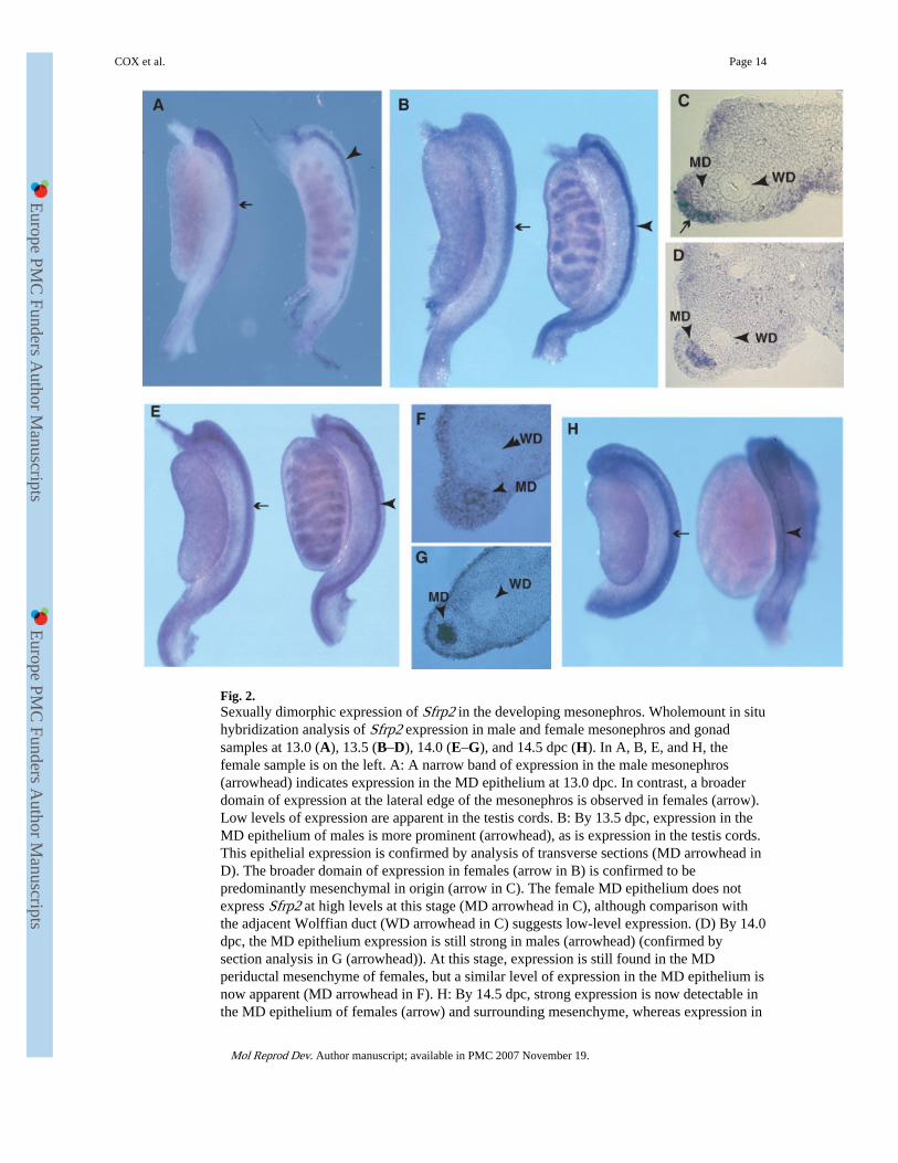
Fig. 2.Sexually dimorphic expression of Sfrp2 in the developing mesonephros. Wholemount in situhybridization analysis of Sfrp2 expression in male and female mesonephros and gonadsamples at 13.0 (A), 13.5 (B–D), 14.0 (E–G), and 14.5 dpc (H). In A, B, E, and H, thefemale sample is on the left. A: A narrow band of expression in the male mesonephros(arrowhead) indicates expression in the MD epithelium at 13.0 dpc. In contrast, a broaderdomain of expression at the lateral edge of the mesonephros is observed in females (arrow).Low levels of expression are apparent in the testis cords. B: By 13.5 dpc, expression in theMD epithelium of males is more prominent (arrowhead), as is expression in the testis cords.This epithelial expression is confirmed by analysis of transverse sections (MD arrowhead inD). The broader domain of expression in females (arrow in B) is confirmed to bepredominantly mesenchymal in origin (arrow in C). The female MD epithelium does notexpress Sfrp2 at high levels at this stage (MD arrowhead in C), although comparison withthe adjacent Wolffian duct (WD arrowhead in C) suggests low-level expression. (D) By 14.0dpc, the MD epithelium expression is still strong in males (arrowhead) (confirmed bysection analysis in G (arrowhead)). At this stage, expression is still found in the MDperiductal mesenchyme of females, but a similar level of expression in the MD epithelium isnow apparent (MD arrowhead in F). H: By 14.5 dpc, strong expression is now detectable inthe MD epithelium of females (arrow) and surrounding mesenchyme, whereas expression in
COX et al. Page 14
Mol Reprod Dev. Author manuscript; available in PMC 2007 November 19.
Europe PM
C Funders A
uthor Manuscripts
Europe PM
C Funders A
uthor Manuscripts
the male MD epithelium is greatly diminished (arrowhead) due to the onset of regression. Inall samples examined there appears to be low-level expression in most regions of themesonephros and gonad, including the surrounding coelomic epithelium.
COX et al. Page 15
Mol Reprod Dev. Author manuscript; available in PMC 2007 November 19.
Europe PM
C Funders A
uthor Manuscripts
Europe PM
C Funders A
uthor Manuscripts

Fig. 3.Expression of Sfrp5 and Sfrp2 in developing mesonephroi from px/px homozygotes.Expression of Amhr2 (A), Lim1 (B), Sfrp5 (C), and Sfrp2 (D) was examined by WMISHanalysis of mesonephroi dissected from male embryos (13.0–13.5 dpc) homozygous for thepostaxial hemimelia mutation (px/px). Mutant homozygous males fail to undergo MDregression due to the absence of AMH type 2 receptor (Amhr2) expression in the MDperiductal mesenchyme. A: Amhr2 is a marker of the developing MD mesenchyme atapproximately 13.5 dpc and exhibits a broad domain of expression at the lateral edge of themesonephros in wild-type male embryos (arrowhead). In contrast, no expression is seen inmesonephroi from embryonic px/px homozygotes at the same stage. Amhr2 is alsoexpressed in the testis cords of both wild-type and mutant samples. B: Lim1 marks the MDepithelium at approximately 13.0 dpc. This expression is clearly visible as narrow regions ofexpression (arrows) in both mutant and wild-type mesonephroi. This indicates that the MDepithelium is still present in px/px homozygotes at 13.0 dpc. C: At approximately 13.0 dpc,Sfrp5 expression is observed in the wild-type embryonic mesonephros as described inFigure 1. Note the relatively broad domain of expression at the cranial end of themesonephros (arrowhead) and the absence of expression in the most caudal regions of theduct (arrow). In male mesonephroi from px/px homozygotes Sfrp5 expression is negligibleat the same stage. Expression is apparent at low levels (asterisk), but does not exhibit thecranial to caudal progression apparent in wild-type males. D: At approximately 13.5 dpc,Sfrp2 is expressed in the MD epithelium of wild-type males (arrowhead) as described inFigure 2. In contrast, this expression is absent in the px/px male mesonephros. Testis cordexpression is undiminished in the mutant sample.
COX et al. Page 16
Mol Reprod Dev. Author manuscript; available in PMC 2007 November 19.
Europe PM
C Funders A
uthor Manuscripts
Europe PM
C Funders A
uthor Manuscripts

Fig. 4.Morphological analysis of adult male reproductive organs in wild-type, Sfrp5HQ27stop
homozygotes, Sfrp2HC50F homozygotes and doubly homozygous mutant mice. (A)Wholemount image of 9-week-old wild-type male reproductive organs indicating the testis(t), cauda epididymis (e), vas deferens (vd), seminal vesicle (sv), and bladder (bl). There areno observable abnormalities of the reproductive tracts in adult males of the followinggenotypes: Sfrp5HQ27stop/Sfrp5HQ27stop (B, 9-week-old male); Sfrp2HC50F/Sfrp2HC50F (C,14-week-old male); Sfrp5HQ27stop/Sfrp5HQ27stop, Sfrp2HC50F/Sfrp2HC50F (doublehomozygote) (D, 13-week-old male).
COX et al. Page 17
Mol Reprod Dev. Author manuscript; available in PMC 2007 November 19.
Europe PM
C Funders A
uthor Manuscripts
Europe PM
C Funders A
uthor Manuscripts