Mouse Cristin/R-spondin Family Proteins Are Novel Ligands for the Frizzled 8 and LRP6 Receptors and...
12
Mouse Cristin/R-spondin Family Proteins Are Novel Ligands for the Frizzled 8 and LRP6 Receptors and Activate -Catenin-dependent Gene Expression * Received for publication, July 29, 2005, and in revised form, March 2, 2006 Published, JBC Papers in Press, March 16, 2006, DOI 10.1074/jbc.M508324200 Ju-Suk Nam ‡ , Taryn J. Turcotte ‡ , Peter F. Smith ‡ , Sangdun Choi § , and Jeong Kyo Yoon ‡1 From the ‡ Center for Molecular Medicine, Maine Medical Center Research Institute, Scarborough, Maine 04074 and the § Division of Biology, California Institute of Technology, Pasadena, California 91125 Wnt signaling plays critical biological roles during normal embryonic development and homeostasis in adults. In the canonical pathway, binding of Wnt ligands to the Frizzled (Fzd) receptor and the low density lipoprotein-related receptor (LRP) 5 or LRP6 core- ceptor initiates downstream signaling events leading to gene activa- tion by -catenin and the T-cell factor (TCF)-lymphoid enhancer factor (LEF) family transcription factor complex. In this study, we provide several lines of evidence that the mouse Cristin/R-spondin family proteins function as Fzd8 and LRP6 receptor ligands and induce the canonical Wnt/-catenin signaling pathway, leading to TCF-dependent gene activation. First, conditioned medium con- taining Cristin/R-spondin proteins effectively induced reporter activity in a TCF-binding site-dependent manner. Second, stimula- tion of cells with Cristin/R-spondin was accompanied by stabiliza- tion of endogenous -catenin proteins and induction of canonical Wnt target genes. Third, Cristin/R-spondin proteins physically interacted with the extracellular domains of the LRP6 and Fzd8 receptors in vivo and in vitro. Interestingly, unlike canonical Wnt ligands, Cristin/R-spondin failed to form a ternary complex with both LRP6 and Fzd8 receptors, suggesting that R-spondin may acti- vate the canonical Wnt signaling pathway by different mechanisms. Furthermore, Cristin/R-spondin proteins possess an intriguing positive modulatory activity on Wnt ligands, possibly through a direct interaction. Our findings expand the repertoire of ligands that induce -catenin/TCF-dependent gene activation and impli- cate the presence of active -catenin-dependent gene activation in a Wnt-free biological context. Wnt signaling is one of the key signaling pathways controlling cell proliferation, differentiation, and morphogenesis during embryogenesis and in adults (1, 2). Wnt ligands are secreted glycoproteins and induce multiple intracellular signaling pathways (3–7). In the canonical path- way, Wnt ligands bind to the Frizzled (Fzd) 2 family receptors and their low density lipoprotein receptor-related protein (LRP) 5 or LRP6 core- ceptor, leading to recruitment of Dishevelled (Dvl) and Axin proteins from the cytoplasm to the receptors on the plasma membrane (6). Sub- sequently, the activities of glycogen synthase kinase-3 and casein kinase I are inhibited. As a result, -catenin proteins, which are nor- mally phosphorylated by these kinases and degraded in the absence of Wnt stimulation, become underphosphorylated and escape from the degradation pathway. Accumulated -catenin proteins in the cyto- plasm are eventually translocated into the nucleus, where they are engaged in gene activation as a complex with T-cell factor (TCF) family transcription factors. Additionally, Wnt ligands also induce -catenin- independent intracellular signaling pathways, including the Wnt/Ca 2 , Wnt/cGMP, and planar cell polarity pathways, in which only the Fzd receptors, not the LRP5 or LRP6 coreceptor, are required (4, 7). Recent discoveries changed the conventional view of Wnt ligands and Fzd/LRP receptors (8 –11). Norrin, a secreted protein unrelated to the Wnt proteins, is encoded by the Norrie disease gene, which is respon- sible for human hereditary Norrie disease, which is associated with blindness, deafness, and mental retardation (12, 13). Recently, Norrin was discovered as a high affinity ligand specific for the Fzd4LRP6 recep- tor complex, which induces -catenin-dependent signaling (8). The RYK, Derailed, and LIN-18 proteins, receptor tyrosine kinases with unknown function, were determined to be novel Wnt receptors in mouse, Drosophila, and Caenorhabditis elegans, respectively (9 –11). These discoveries expanded the repertoire of ligands and receptors involved in Wnt/Fzd/LRP signaling and suggest the possibility of unknown ligands unrelated to the Wnt proteins and receptors unrelated to Fzd and LRP5 or LRP6. We provide evidence strongly suggesting that a multigene family of novel secreted proteins, mouse Cristin/R-spondin proteins, may work at the receptor level to activate downstream -catenin-dependent gene activation and may function as a class of ligands unrelated to Wnt pro- teins. Moreover, Cristin/R-spondin proteins exhibited intriguing syner- gistic activity on Wnt ligands, possibly through direct physical interac- tion. Xenopus R-spondin2 and human R-spondin1 protein activity on canonical Wnt signaling was demonstrated recently (14, 15), consistent with our findings. These results suggest that the function of Cristin/R- spondin family proteins is conserved in vertebrates. MATERIALS AND METHODS Molecular Biology—Mouse expressed sequence tag cDNA clones encoding full-length Cristin/R-spondin proteins were obtained from the I.M.A.G.E. Consortium. The coding regions of all Cristin/R-spondin genes and the deletion mutants of Cristin1/R-spondin3 were PCR-am- plified and cloned into the pcDNA3 plasmid carrying a C-terminal hemagglutinin (HA) tag (pcDNA3-HA) or Myc/His epitope tags (pcDNA3.1-Myc/His). The coding sequence of mouse Wnt1 was PCR- amplified and cloned into pcDNA3-HA and the CS2-MT vectors to create HA- and Myc-tagged constructs, respectively. DNA sequence * This work was supported by National Institutes of Health Grant P20 RR018789 (to J. K. Y.) and by the State of Maine (to J. K. Y.). The costs of publication of this article were defrayed in part by the payment of page charges. This article must therefore be hereby marked “advertisement” in accordance with 18 U.S.C. Section 1734 solely to indicate this fact. The nucleotide sequence(s) reported in this paper has been submitted to the GenBank TM /EBI Data Bank with accession number(s) AY864332. 1 To whom correspondence should be addressed: Center for Molecular Medicine, Maine Medical Center Research Inst., 81 Research Dr., Scarborough, ME 04074. Tel.: 207-885- 8196; Fax: 207-885-9179; E-mail: [email protected]. 2 The abbreviations used are: Fzd, Frizzled; LRP, low density lipoprotein receptor-related protein; Dvl, Dishevelled; TCF, T-cell factor; HA, hemagglutinin; CRD, cysteine-rich domain; CM, conditioned medium/media; RT, reverse transcription; TSR, throm- bospondin type I repeat; HSPGs, heparan sulfate proteoglycans. THE JOURNAL OF BIOLOGICAL CHEMISTRY VOL. 281, NO. 19, pp. 13247–13257, May 12, 2006 © 2006 by The American Society for Biochemistry and Molecular Biology, Inc. Printed in the U.S.A. MAY 12, 2006 • VOLUME 281 • NUMBER 19 JOURNAL OF BIOLOGICAL CHEMISTRY 13247 by guest on September 25, 2016 http://www.jbc.org/ Downloaded from
Transcript of Mouse Cristin/R-spondin Family Proteins Are Novel Ligands for the Frizzled 8 and LRP6 Receptors and...

Mouse Cristin/R-spondin Family Proteins AreNovel Ligands for the Frizzled 8 and LRP6 Receptors andActivate �-Catenin-dependent Gene Expression*
Received for publication, July 29, 2005, and in revised form, March 2, 2006 Published, JBC Papers in Press, March 16, 2006, DOI 10.1074/jbc.M508324200
Ju-Suk Nam‡, Taryn J. Turcotte‡, Peter F. Smith‡, Sangdun Choi§, and Jeong Kyo Yoon‡1
From the ‡Center for Molecular Medicine, Maine Medical Center Research Institute, Scarborough, Maine 04074 and the §Division ofBiology, California Institute of Technology, Pasadena, California 91125
Wnt signaling plays critical biological roles during normalembryonic development andhomeostasis in adults. In the canonicalpathway, binding of Wnt ligands to the Frizzled (Fzd) receptor andthe low density lipoprotein-related receptor (LRP) 5 or LRP6 core-ceptor initiates downstream signaling events leading to gene activa-tion by �-catenin and the T-cell factor (TCF)-lymphoid enhancerfactor (LEF) family transcription factor complex. In this study, weprovide several lines of evidence that the mouse Cristin/R-spondinfamily proteins function as Fzd8 and LRP6 receptor ligands andinduce the canonical Wnt/�-catenin signaling pathway, leading toTCF-dependent gene activation. First, conditioned medium con-taining Cristin/R-spondin proteins effectively induced reporteractivity in a TCF-binding site-dependent manner. Second, stimula-tion of cells with Cristin/R-spondin was accompanied by stabiliza-tion of endogenous �-catenin proteins and induction of canonicalWnt target genes. Third, Cristin/R-spondin proteins physicallyinteracted with the extracellular domains of the LRP6 and Fzd8receptors in vivo and in vitro. Interestingly, unlike canonical Wntligands, Cristin/R-spondin failed to form a ternary complex withboth LRP6 and Fzd8 receptors, suggesting that R-spondinmay acti-vate the canonicalWnt signaling pathway by differentmechanisms.Furthermore, Cristin/R-spondin proteins possess an intriguingpositive modulatory activity on Wnt ligands, possibly through adirect interaction. Our findings expand the repertoire of ligandsthat induce �-catenin/TCF-dependent gene activation and impli-cate the presence of active�-catenin-dependent gene activation in aWnt-free biological context.
Wnt signaling is one of the key signaling pathways controlling cellproliferation, differentiation, andmorphogenesis during embryogenesisand in adults (1, 2). Wnt ligands are secreted glycoproteins and inducemultiple intracellular signaling pathways (3–7). In the canonical path-way, Wnt ligands bind to the Frizzled (Fzd)2 family receptors and theirlow density lipoprotein receptor-related protein (LRP) 5 or LRP6 core-ceptor, leading to recruitment of Dishevelled (Dvl) and Axin proteins
from the cytoplasm to the receptors on the plasma membrane (6). Sub-sequently, the activities of glycogen synthase kinase-3� and caseinkinase I are inhibited. As a result, �-catenin proteins, which are nor-mally phosphorylated by these kinases and degraded in the absence ofWnt stimulation, become underphosphorylated and escape from thedegradation pathway. Accumulated �-catenin proteins in the cyto-plasm are eventually translocated into the nucleus, where they areengaged in gene activation as a complex with T-cell factor (TCF) familytranscription factors. Additionally, Wnt ligands also induce �-catenin-independent intracellular signaling pathways, including theWnt/Ca2�,Wnt/cGMP, and planar cell polarity pathways, in which only the Fzdreceptors, not the LRP5 or LRP6 coreceptor, are required (4, 7).Recent discoveries changed the conventional view ofWnt ligands and
Fzd/LRP receptors (8–11). Norrin, a secreted protein unrelated to theWnt proteins, is encoded by the Norrie disease gene, which is respon-sible for human hereditary Norrie disease, which is associated withblindness, deafness, and mental retardation (12, 13). Recently, Norrinwas discovered as a high affinity ligand specific for the Fzd4�LRP6 recep-tor complex, which induces �-catenin-dependent signaling (8). TheRYK, Derailed, and LIN-18 proteins, receptor tyrosine kinases withunknown function, were determined to be novel Wnt receptors inmouse, Drosophila, and Caenorhabditis elegans, respectively (9–11).These discoveries expanded the repertoire of ligands and receptorsinvolved in Wnt/Fzd/LRP signaling and suggest the possibility ofunknown ligands unrelated to theWnt proteins and receptors unrelatedto Fzd and LRP5 or LRP6.We provide evidence strongly suggesting that a multigene family of
novel secreted proteins, mouse Cristin/R-spondin proteins, may workat the receptor level to activate downstream �-catenin-dependent geneactivation and may function as a class of ligands unrelated to Wnt pro-teins.Moreover, Cristin/R-spondin proteins exhibited intriguing syner-gistic activity on Wnt ligands, possibly through direct physical interac-tion. Xenopus R-spondin2 and human R-spondin1 protein activity oncanonicalWnt signaling was demonstrated recently (14, 15), consistentwith our findings. These results suggest that the function of Cristin/R-spondin family proteins is conserved in vertebrates.
MATERIALS AND METHODS
Molecular Biology—Mouse expressed sequence tag cDNA clonesencoding full-length Cristin/R-spondin proteins were obtained fromthe I.M.A.G.E. Consortium. The coding regions of allCristin/R-spondingenes and the deletion mutants of Cristin1/R-spondin3 were PCR-am-plified and cloned into the pcDNA3 plasmid carrying a C-terminalhemagglutinin (HA) tag (pcDNA3-HA) or Myc/His epitope tags(pcDNA3.1-Myc/His). The coding sequence of mouseWnt1 was PCR-amplified and cloned into pcDNA3-HA and the CS2-MT vectors tocreate HA- and Myc-tagged constructs, respectively. DNA sequence
* This work was supported by National Institutes of Health Grant P20 RR018789 (toJ. K. Y.) and by the State of Maine (to J. K. Y.). The costs of publication of this articlewere defrayed in part by the payment of page charges. This article must therefore behereby marked “advertisement” in accordance with 18 U.S.C. Section 1734 solely toindicate this fact.
The nucleotide sequence(s) reported in this paper has been submitted to the GenBankTM/EBIData Bank with accession number(s) AY864332.
1 To whom correspondence should be addressed: Center for Molecular Medicine, MaineMedical Center Research Inst., 81 Research Dr., Scarborough, ME 04074. Tel.: 207-885-8196; Fax: 207-885-9179; E-mail: [email protected].
2 The abbreviations used are: Fzd, Frizzled; LRP, low density lipoprotein receptor-relatedprotein; Dvl, Dishevelled; TCF, T-cell factor; HA, hemagglutinin; CRD, cysteine-richdomain; CM, conditioned medium/media; RT, reverse transcription; TSR, throm-bospondin type I repeat; HSPGs, heparan sulfate proteoglycans.
THE JOURNAL OF BIOLOGICAL CHEMISTRY VOL. 281, NO. 19, pp. 13247–13257, May 12, 2006© 2006 by The American Society for Biochemistry and Molecular Biology, Inc. Printed in the U.S.A.
MAY 12, 2006 • VOLUME 281 • NUMBER 19 JOURNAL OF BIOLOGICAL CHEMISTRY 13247
by guest on September 25, 2016
http://ww
w.jbc.org/
Dow
nloaded from

encoding a cysteine-rich domain (CRD) of mouse Fzd8 was excisedfrom the Fzd8CRD-IgG plasmid (16) and cloned into pcDNA3-HA tocreate a C-terminally HA-tagged Fzd8CRD construct (Fzd8CRD-HA).
Cell Culture, DNA Transfection, and Luciferase Assay—Humanembryonic kidney 293T cells and mouse L-cells were routinely main-tained in Dulbecco’s modified Eagle’s medium containing 10% fetalbovine serum in 5% CO2 at 37 °C. P19 cells were maintained in �-min-imal essential medium supplemented with 7.5% calf serum and 2.5%fetal bovine serum. 293T cells were transfected using FuGENE6 reagent(Roche Applied Science) according to the manufacturer’s protocol. Forluciferase assay, 3 � 104 cells were seeded in each well of 24-well plates.The TOPflash or FOPflash reporter (20 ng) and a Renilla luciferase-thymidine kinase construct (10 ng) were used along with variousamounts of expression plasmids as indicated in the figure legends. Bothluciferase activities were measured using a Dual-Luciferase assay kit(Promega) according to the manufacturer’s protocol.
Preparation of ConditionedMedium (CM) and Isolation of Cristin/R-spondin Proteins—Wnt3a CM was prepared from the mouse Wnt3aL-cell line (obtained from American Type Culture Collection) asdescribed previously (17). CM containing Cristin/R-spondin proteinswas obtained from 293T cells transiently transfected with Cristin/R-spondin expression plasmids. For biochemical assay, serum-free Dul-becco’s modified Eagle’s medium/nutrient mixture F-12 (1:1) was usedto obtain the CM. Soluble heparin (50 �g/ml) was added to the culturemedium. Dulbecco’s modified Eagle’s medium containing 10% fetalbovine serumwas used to prepare the CM for the luciferase and �-cate-nin stabilization assays. For purification of histidine-tagged Cristin/R-spondin proteins, total lysates of 293T cells transiently transfected withhistidine-tagged Cristin/R-spondin expression constructs were pre-pared with lysis buffer (50 mM Tris (pH 8.0), 150 mM NaCl, and 0.1%Nonidet P-40) containing protease inhibitor mixture V (Calbiochem).The cell lysates were incubated with nickel-nitrilotriacetic acid-agarosebeads (Qiagen Inc.) for 2 h at 4 °C and washed with 10 mM imidazolebuffer three times. The Cristin/R-spondin proteins were eluted withbuffer containing 250 mM imidazole. The HA-tagged mouse Fzd8 CRDand human IgG fusions of the mouse Fzd8 CRD and the human LRP6extracellular domain (LRP6N) were prepared as serum-free CM for-mats from transiently transfected 293T cells as described previously(18).
Heparin Binding and Cristin/R-spondin Binding Assay—Total celllysates of 293T cells transfected with Cristin-HA constructs were pre-pared using lysis buffer containing protease inhibitors and incubatedovernight at 4 °C with heparin-Sepharose beads (Sigma). The beadswere washed with lysis buffer three times at room temperature, and theCristin/R-spondin proteinswere elutedwith bufferswith differentNaClconcentrations. The presence of Cristin/R-spondin proteins was exam-ined by Western blot analysis.CM containing IgG, Fzd8CRD-IgG, and LRP6N-IgG were first incu-
bated with protein A-Sepharose beads to conjugate IgG fusion proteinsto the beads. Histidine-tagged Cristin/R-spondin proteins were incu-bated overnight at 4 °C with the beads. After three washings with 0.3 M
NaCl/phosphate buffer, the associated Cristin/R-spondin proteins wereanalyzed byWestern blot analysis using anti-His, anti-HA, or anti-Mycprimary antibody. The filters were reprobed with anti-human IgGantibody.
RNA Isolation and Reverse Transcription (RT)-PCR Analysis—TotalRNA was isolated from cultured cells and Xenopus animal cap explantsusing TRIzol reagent (Invitrogen) according to the manufacturer’s pro-tocol and digested with RNase-free DNase I to remove genomic DNAcontamination. The first-strand cDNA was synthesized with Super-
Script II (Invitrogen), and one-tenth of the cDNA was used for eachPCR. The sequences of the PCR primers were as follows: mouse glycer-aldehyde-3-phosphate dehydrogenase (GADPH), 5�-GTGGCAAAGT-GGAGATTGTTGCC-3� (sense) and 5�-GATGATGACCCGTTTG-GCTCC-3� (antisense); mouse Cristin1/R-spondin3, 5�-GTACACTG-TGAGGCCAGTGAA-3� (sense) and 5�-ATGGCTAGAACACCTGT-CCTG-3� (antisense); mouse BrachyuryT, 5�-TGCTGCCTGTGAGT-CATAC-3� (sense) and 5�-ACAAGAGGCTGTAGAACATG-3� (anti-sense); mouse Cdx1, 5�-GAACCAAGGACAAGTACCGTG-3� (sense)and 5�-GGTAGAAACTCCTCCTTGACG-3 (antisense); XenopusSiamois, 5�-AAGGAACCCCACCAGGATAA-3� (sense) and 5�-TAC-TGGTGGCTGGAGAAATA-3� (antisense); Xenopus Xnr3, 5�-TCCA-CTTGTGCAGTTCCACAG-3� (sense) and 5�-ATCTCTTCATGGT-GCCTCAGG-3� (antisense); and Xenopus Xmax2, 5�-GTGGAAAGC-GACGAAGACTC-3� (sense) and 5�-CCGAGCTCGAGTAGTTGGA-C-3� (antisense).
Western Blot Analysis, Immunoprecipitation, and Immunofluores-cence Staining—For Western blotting, anti-HA (clone 12CA5), anti-Myc (clone 9E10), anti-His (Rockland Immunochemicals, Inc.), andanti-human IgG (Jackson ImmunoResearch Laboratories, Inc.) anti-bodies were used at 1:2000, 1:5000, 1:3000, and 1:4000 dilutions, respec-tively. Anti-�-catenin (Pharmingen) and anti-�-actin (Sigma) antibod-ieswere used at 1:500 and 1:5000 dilutions, respectively. The presence oftarget proteins was detected by the chemiluminescencemethod (Amer-sham Biosciences). For immunoprecipitation, protein A-Sepharose andanti-HA or anti-Myc antibody conjugated to agarose (Sigma) or Sepha-rose (Santa Cruz Biotechnology, Inc.) beads were used to purify theprotein complex. Subcellular localization of Cristin/R-spondin proteinsin 293T cells was determined by immunofluorescence staining usingmouse anti-HA primary antibody (1:1000 dilution) and Alexa 488-con-jugated goat anti-mouse IgG secondary antibody (1:500 dilution;Molecular Probe). Images were acquired by confocal microscopy.
Mouse Embryo Collection andWhole-mount in Situ Hybridization—Wild-type embryos at different stages were collected from the timedmatings of ICR mice. The pMesogenin1 gene mutant embryos werecollected from the mating between the heterozygous animals, and thegenotypes of collected embryoswere determined by genomicDNAPCRanalysis with yolk sac DNA as described (19). The collected embryoswere immediately fixed in freshly prepared 4% paraformaldehyde/phos-phate-buffered saline solution overnight at 4 °C and kept in 100%meth-anol at �20 °C until used. Digoxigenin-labeled antisense Cristin1/R-spondin3 RNA probes were synthesized in vitro from a linearized DNAtemplate utilizing appropriate RNA polymerases in the presence ofdigoxigenin-labeled CTP. Whole-mount in situ hybridization was per-formed as described previously (19). Stained embryos were photo-graphed with a Zeiss AxioCam digital camera.
Xenopus Embryos and Animal Cap Explants—Xenopus embryoswere prepared by in vitro fertilization of oocytes collected from hor-monally induced females by a standard protocol. Capped RNAwas syn-thesized using an Ambion Message Machine kit according to the rec-ommended protocol. Various amounts of RNA were injected into theanimal pole of two-cell stage embryos. Animal caps were explanted atthe blastula stage and cultured in 1� modified Bart’s saline until thecompanion embryos reached the gastrulation stage.
RESULTS
Multigene Family of Novel Secreted Mouse Cristin/R-spondinProteins—Wepreviously demonstrated thatmousemutants lacking thepMesogenin1 gene, which encodes a presomitic mesoderm-specificbasic helix-loop-helix transcription factor (20), display severe defects in
Cristin/R-spondin Ligands in Wnt Signaling
13248 JOURNAL OF BIOLOGICAL CHEMISTRY VOLUME 281 • NUMBER 19 • MAY 12, 2006
by guest on September 25, 2016
http://ww
w.jbc.org/
Dow
nloaded from

FIGURE 1. Cristin/R-spondin multigene family proteins are novel secreted proteins. A and B, mouse (m) Cristin1/R-spondin3 RNA expression in the tail buds of pMesogenin1-nullmutant embryos was analyzed by both RT-PCR and whole-mount in situ hybridization, respectively. For total RNA isolation, the trunk regions below the hind limb levels of normal (�,wild-type and heterozygous embryos combined) and homozygous null mutant (�/�) embryos at embryonic day 9.5 (E9.5) were dissected in ice-cold phosphate-buffered saline.More than 40 individuals were combined to isolate RNA. Both heterozygous (�/�) and homozygous (�/�) pMesogenin1 mutant embryonic day 9.5 embryos were collected and usedfor whole-mount in situ hybridization after genotyping of yolk sac genomic DNA. GAPDH, glyceraldehyde-3-phosphate dehydrogenase. C, shown is a comparison of mouseCristin/R-spondin family protein sequences. The predicted protein sequences were aligned using the ClustalW program of the Lasergene package (DNAStar, Inc., Madison, WI).Identical amino acids are shown in black boxes. Hyphens indicate a sequence gap. The GenBankTM accession numbers for mouse R-spondin proteins are as follows: mouse Cristin3/R-spondin1, NP_619624; mouse Cristin2/R-spondin2, NP_766403; mouse Cristin1/R-spondin3, AY864332; and mouse Cristin4/R-spondin4, XP_130619. D, shown is a phylogeneticanalysis of vertebrate R-spondin family proteins. The discovery of human and Xenopus R-spondins was reported while this manuscript was in preparation. The GenBankTM accessionnumbers as follows: human R-spondin1, NP_775911; human R-spondin2, NP_116173; human R-spondin3, AAQ88533; human R-spondin4, XP_297816; Xenopus R-spondin2,AY753198; and Xenopus R-spondin3, AY753199. Mm, Mus muscularis; Hs, Homo sapiens; Xl, Xenopus laevis. Xt, Xenopus tropicalis. E, the syntenic relationship between human (H) andmouse (M) R-spondin gene loci is shown. The predicted protein sequences of human R-spondin1 do not contain any notable signal sequences. It is unclear whether this particular DNAsequence represents an alternatively spliced form or an artifact of cDNA cloning. Therefore, the number of exons of the human R-spondin1 gene is provisional. Chr., chromosome.
Cristin/R-spondin Ligands in Wnt Signaling
MAY 12, 2006 • VOLUME 281 • NUMBER 19 JOURNAL OF BIOLOGICAL CHEMISTRY 13249
by guest on September 25, 2016
http://ww
w.jbc.org/
Dow
nloaded from
posterior paraxial mesoderm development (19). In an attempt to under-stand the molecular mechanisms by which pMesogenin1 regulatesparaxial mesoderm development, potential target genes for pMesoge-nin1 were screened by analyzing gene expression profiles in the pre-somitic mesoderm of pMesogenin1-null and wild-type embryos. Onegene whose RNA expression was increased by �4-fold in the pMesoge-nin1-null mutant samples encoded a novel secreted protein (Fig. 1A).Based on the protein structure derived from the predicted peptidesequences, we named this gene Cristin1 (for cysteine-rich and singlethrombospondin domain-containing protein-1) (Fig. 1C). Whole-mount in situ hybridization analysis showed prominent RNA expres-sion of Cristin1 in the tail buds of wild-type mouse embryos (Fig. 1B).Cristin1 expression within the tail buds of homozygous pMesogenin1-null mutants was significantly expanded, increased, and tightly associ-ated with the tail bud defects, consistent with RT-PCR data. Thus, aber-rant Cristin1 expression may be related to the pMesogenin1-null
phenotype.Cristin1 expressionwas also detected in the primitive streak,dorsal neural tube, forebrain, and migrating neural crests of mouseembryos (Fig. 1B). In searching sequence data bases, we identified threegenes homologous toCristin1 in themouse genome (designated asCris-tin2, Cristin3, and Cristin4) (Fig. 1C). We also identified four homolo-gous genes in the human genome as well as expressed sequence tagclones with high sequence homology among other vertebrates, includ-ing chicken, Xenopus, and zebrafish. Phylogenetic analysis of predictedprotein sequences, syntenic relationships of gene loci in human andmouse chromosomes, and comparisons of genomic structures deter-mined the orthologous relationships among the human and mousegenes (Fig. 1, D and E). Interestingly, searches of the Drosophila andC. elegans genome data bases failed to identify Cristin homologs. Thus,Cristin genes may be unique to vertebrates, although functionalhomologs may exist in invertebrate species. Recently, the isolation ofmouse R-spondin, which is identical to mouse Cristin3, and two Xeno-
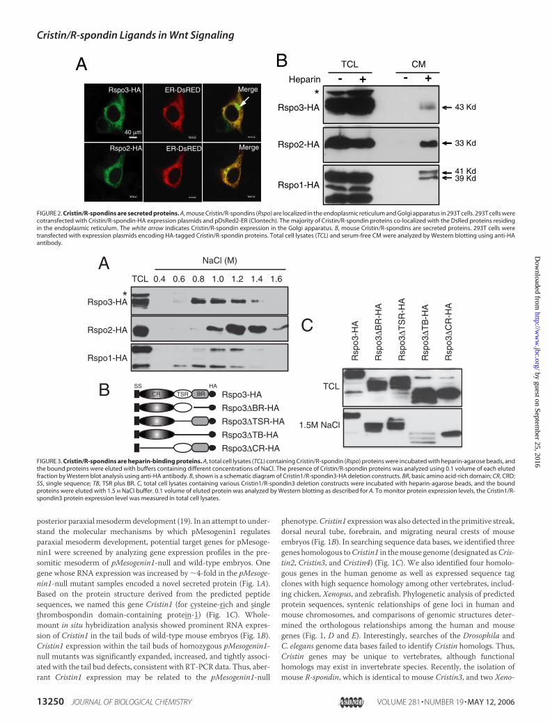
FIGURE 2. Cristin/R-spondins are secreted proteins. A, mouse Cristin/R-spondins (Rspo) are localized in the endoplasmic reticulum and Golgi apparatus in 293T cells. 293T cells werecotransfected with Cristin/R-spondin-HA expression plasmids and pDsRed2-ER (Clontech). The majority of Cristin/R-spondin proteins co-localized with the DsRed proteins residingin the endoplasmic reticulum. The white arrow indicates Cristin/R-spondin expression in the Golgi apparatus. B, mouse Cristin/R-spondins are secreted proteins. 293T cells weretransfected with expression plasmids encoding HA-tagged Cristin/R-spondin proteins. Total cell lysates (TCL) and serum-free CM were analyzed by Western blotting using anti-HAantibody.
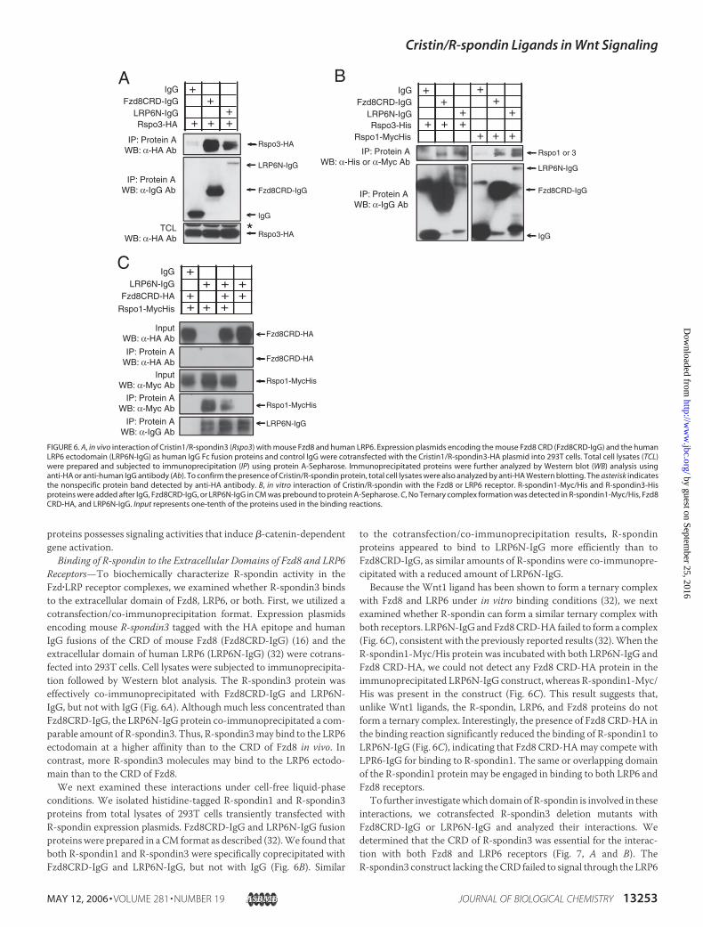
FIGURE 3. Cristin/R-spondins are heparin-binding proteins. A, total cell lysates (TCL) containing Cristin/R-spondin (Rspo) proteins were incubated with heparin-agarose beads, andthe bound proteins were eluted with buffers containing different concentrations of NaCl. The presence of Cristin/R-spondin proteins was analyzed using 0.1 volume of each elutedfraction by Western blot analysis using anti-HA antibody. B, shown is a schematic diagram of Cristin1/R-spondin3-HA deletion constructs. BR, basic amino acid-rich domain; CR, CRD;SS, single sequence; TB, TSR plus BR. C, total cell lysates containing various Cristin1/R-spondin3 deletion constructs were incubated with heparin-agarose beads, and the boundproteins were eluted with 1.5 M NaCl buffer. 0.1 volume of eluted protein was analyzed by Western blotting as described for A. To monitor protein expression levels, the Cristin1/R-spondin3 protein expression level was measured in total cell lysates.
Cristin/R-spondin Ligands in Wnt Signaling
13250 JOURNAL OF BIOLOGICAL CHEMISTRY VOLUME 281 • NUMBER 19 • MAY 12, 2006
by guest on September 25, 2016
http://ww
w.jbc.org/
Dow
nloaded from

pus homologs of the mouse R-spondin genes was reported (14, 21).Thus, for consistency in nomenclature, we renamed theCristin genes toR-spondin as indicated in Fig. 1D.
Comparisons of the predicted mouse R-spondin amino acidsequences revealed significant homologies (Fig. 1C). First, all R-spondinproteins contain anN-terminal 20–25-amino acid hydrophobic region,which likely serves as a signal sequence for secretion. Three additionalconserved protein domains are evident: (i) a CRDwith homology to theCRDs of furin and the insulin-like growth factor receptor, (ii) a throm-bospondin type I repeat (TSR) (22), and (iii) a C-terminal basic aminoacid-rich domain (Fig. 1C).The presence of a putative signal sequence and the lack of a notable
transmembrane domain suggest that R-spondins may be secreted pro-teins. To test this possibility, we transfected expression plasmids encod-ing HA-tagged R-spondins into human embryonic kidney 293T cellsand examined the subcellular localization of R-spondins by immunoflu-orescence staining. R-spondin proteins were detected mainly in theendoplasmic reticulum and the Golgi apparatus (Fig. 2A), indicatingthat R-spondin proteins are in the secretory pathway. In similarly trans-fected cells, the expression of R-spondin proteins and their secretioninto the CM were monitored by Western blot analysis. The majority ofthe R-spondin proteins were associated with total cell lysates and were
barely detected, if at all, in theCM(Fig. 2B). Interestingly, the addition ofsoluble heparin to the medium in culture significantly enhanced thepresence of R-spondins in the CM and confirmed their nature assecreted proteins (Fig. 2B). The addition of sodium chlorate, an inhibi-tor of sulfation, increased the level of R-spondin proteins in the CM.3
These results suggest that secreted R-spondins may be associated, inpart, with the heparan sulfate proteoglycans (HSPGs) of the plasmamembrane and extracellular matrix.
R-spondin Binds to Heparin with High Affinity—We next examinedwhether R-spondin can bind to heparin.We prepared lysates from293Tcells transfected with R-spondin-HA expression plasmids. The celllysates were incubated with heparin-Sepharose beads, and the boundproteins were eluted with a series of buffers with different salt concen-trations. All R-spondins tested efficiently bound to the heparin-Sepha-rose beads and were eluted from the heparin beads between 0.8 and 1.2MNaCl (Fig. 3A). Thus, all R-spondins bind to heparin with high affinitycomparable with fibroblast growth factors (23).To determine which domains within R-spondin3 are required for
heparin binding, we generated a set of R-spondin3 constructs contain-
3 J.-S. Nam, T. J. Turcotte, and J. K. Yoon, unpublished data.
FIGURE 4. Cristin/R-spondin proteins activatecanonical Wnt/�-catenin signaling. A, mouseCristin1/R-spondin3 (Rspo3) induced �-catenin-dependent gene activation in a dose-dependentmanner. 20 ng of TOPflash or FOPflash reporterplasmid was cotransfected with the indicatedamounts of Cristin1/R-spondin3 expression plas-mid into 293T cells grown in 24-well plates. 10 ngof Renilla luciferase driven by the thymidine kinasegene promoter was included as a transfection con-trol. B, Cristin/R-spondin functioned at the ligand/receptor level. 293T cells were transfected withmouse Fzd8 (25 ng) and human LRP6 (10 ng)expression plasmids and stimulated with CM con-taining Cristin1/R-spondin3 for 24 h. The TOPflashreporter activities were measured. CMV, cytomeg-alovirus. C, Dkk1 and Kremen1 reversed the activa-tion of the TOPflash reporter that was induced byboth Cristin1/R-spondin3 and LRP6. 293T cells in24-well plates were transfected with various com-binations of Cristin1/R-spondin3 (25 ng), LRP6 (10ng), Dkk1 (10 ng), and Kremen1 (10 ng) expressionplasmids with the TOPflash reporter (20 ng). D, nosynergy between Cristin1/R-spondin3 and intra-cellular components of the Wnt signaling pathwayin the activation of the TOPflash reporter wasobserved. Expression plasmids encoding human�-catenin(S33A) (�-cat(S33A); 80 ng), human TCF1(10 ng), and human Dvl1 (50 ng) were cotrans-fected into 293T cells for the TOPflash reporterassay as indicated. E, Cristin1/R-spondin3 failed topotentiate the TOPflash reporter activationinduced in 293T cells by treatment with LiCl, aninhibitor of glycogen synthase kinase-3�. 293Tcells were transfected with the R-spondin1 expres-sion plasmid and the TOPflash reporter. At 24 hafter transfection, the cells were treated witheither LiCl (40 mM) or NaCl (40 mM) for 8 h.
Cristin/R-spondin Ligands in Wnt Signaling
MAY 12, 2006 • VOLUME 281 • NUMBER 19 JOURNAL OF BIOLOGICAL CHEMISTRY 13251
by guest on September 25, 2016
http://ww
w.jbc.org/
Dow
nloaded from

ing various deletions of the identified domains (Fig. 3B). Total lysates of293T cells transfected with these constructs were prepared, and theheparin binding of each protein construct was determined. It appearsthat both the basic amino acid-rich and TSR domains contain themajority of heparin binding capability, as deletion of both domains sig-nificantly decreased R-spondin3 binding to heparin (Fig. 3C). In con-trast, R-spondin3 with a CRD deletion showed efficient binding to hep-arin comparable with wild-type R-spondin3, indicating that the CRDmay not be necessary for heparin binding. We concluded thatR-spondin3 is a heparin-binding protein and that both the TSR andbasic amino acid-rich domains are necessary for binding to heparin.
R-spondin Activates Canonical Wnt/�-Catenin Signaling—Twoobservations led us to evaluate the possible involvement of R-spondin inWnt signaling. First, embryonic expression of R-spondin genes highlyoverlaps with known wnt gene expression domains (24). Second, CCN(CYR61/connective tissue growth factor/NOV) family proteins, whichcontain domains structurally similar to those of R-spondins such as thecysteine-rich and TSR domains (25), were recently demonstrated toeither antagonize or agonize Wnt signaling in a context-dependentmanner (26, 27). The steady-state level of �-catenin protein and theactivity of reporter constructs such as the TOPflash reporter carryingTCF1-binding sites in their upstream regulatory regions were exten-sively used to analyze canonical Wnt signaling. In 293T cells, overex-pression of R-spondin3 strongly induced TOPflash reporter activity in adose-dependent manner (Fig. 4A). Activation was dependent on theTCF1-binding site because a reporter carrying nonfunctional TCF1-binding sites (FOPflash) was not activated by R-spondin3. In addition,other mouse R-spondin family members and Xenopus R-spondin2showed similar activities (14).3 Interestingly, CM collected from theculture of 293T cells transfected with R-spondins effectively inducedTOPflash reporter activity, suggesting that R-spondin may act in theextracellular environment (Fig. 4B).To determine the target position for R-spondin activity in the Wnt
signaling axis, we examined reporter activity under conditions ofR-spondin3 coexpression with various Wnt signaling components.R-spondin3 activity was synergistically potentiated by the LRP6 recep-tor (Fig. 4B). However, unlike Wnt ligands, R-spondin3 failed to showany significant synergy with Fzd8 (Fig. 4B). Fzd8 failed to furtherenhance the reporter activity co-induced by R-spondin3 and LRP6 (Fig.4B). Therefore, Fzd8 does not appear to actively contribute to the intra-cellular transmission of R-spondin signals. Furthermore, the reporteractivity induced by both LRP6 and R-spondin3 was sensitive to thepresence of both Dickkopf1 (Dkk1) (28) and Kremen1 (29), a conditionthat presumably enhanced LRP6 receptor endocytosis and inhibitedintracellular signaling through the LRP6 receptor (Fig. 4C) (29). How-ever, Dkk1 or Kremen1 alone marginally affected or had no effect onreporter activity induced by R-spondin3 and LRP6.In contrast, R-spondin3 failed to show any synergistic activation of
the reporter when coexpressed with other intracellular components ofWnt signaling, including Dvl1 or�-catenin/TCF1 (Fig. 4D). In addition,when cells were treated with LiCl, which mimics Wnt activation byinhibiting glycogen synthase kinase-3�, no synergistic activation withR-spondin3 of reporter activity was observed (Fig. 4E).
R-spondin Stabilizes Endogenous �-Catenin and Induces the KnownCanonical Wnt/�-Catenin Target Genes—We examined R-spondinactivity on stabilization of �-catenin, a landmark intracellular eventupon activation of canonical Wnt signaling. R-spondin1 andR-spondin3 CM effectively increased steady-state �-catenin levels in293T cells compared with Wnt3a CM (Fig. 5A), consistent with thereporter assay results. This result clearly shows that degradation of cyto-
plasmic �-catenin was prevented by R-spondins and that accumulated�-cateninmay lead to the activation of reporter activity as shown in Fig.4A. Both Xenopus R-spondin2 and human R-spondin1 also induce thestabilization of �-catenin (14, 15), consistent with our results.
We further investigated whether R-spondin can induce the expres-sion of genes known as canonical Wnt signaling targets. Mouse embry-onic carcinoma P19 cells were stimulated with Wnt3a or R-spondin3CM for 2 and 3 days, respectively, and expression of BrachyuryT andCdx1, two known Wnt3a target genes, was examined by semiquantita-tive RT-PCR analysis. Robust induction of both genes was observed incells incubated with R-spondin3 andWnt3a CM, respectively (Fig. 5B).Because both genes are expressed in the tail bud regions of mouseembryos (30, 31), where R-spondin3 is also expressed, it is likely thatboth BrachyuryT and Cdx1 are direct downstream targets ofR-spondin3 signaling.Axis duplication assay inXenopus embryos is a signature assay to test
Wnt/�-catenin activation in vivo. We next examined whether ectopicR-spondin expression in Xenopus embryos induces axis duplication.Injection of up to 5 ng of R-spondin1 and R-spondin3 RNAs into one ofthe ventral blastomeres in four-cell stage embryos did not induce anysignificant axis duplication phenotype, whereas similarly injected wnt1RNA in a picogram quantity produced a clear duplication of axis (datanot shown). Our results are consistent with those recently reported forthe Xenopus R-spondin2 gene (14). The majority of embryos injectedwith a high concentration of R-spondin3 RNA displayed severe gastru-lation failure. Additionally, we occasionally observed tail duplication ina small number of theR-spondin3RNA-injected embryos. Interestingly,unlike whole embryo injection, transcription of two knownWnt targetgenes, Siamois andXnr3, was significantly enhanced in Xenopus animalcap explants injected with mouse R-spondin3 RNA compared withuninjected and control enhanced green fluorescent protein RNA-in-jected cap explants (Fig. 5C).We concluded that the R-spondin family of
FIGURE 5. A, stabilization of �-catenin proteins by R-spondins. 293T cells were treatedwith CM containing R-spondin1 (Rspo1), R-spondin3 (Rspo3), or Wnt3a for 12 h. Thesteady-state level of endogenous �-catenin protein was analyzed by Western blotting.�-Actin expression was measured by Western blotting to provide a control for totalsample quantity. B, induction of Wnt3a target genes by Cristin1/R-spondin3 in embry-onic carcinoma P19 cells. Monolayer cultured P19 cells were stimulated with Cristin1/R-spondin3 CM (1:2) or Wnt3a CM (1:10) for 1 (D1) and 2 (D2) days, respectively. RNAexpression of the BrachyuryT and Cdx1 genes was analyzed by semiquantitative RT-PCR.Glyceraldehyde-3-phosphate dehydrogenase (GAPDH) expression was also measured toensure the quality and quantity of cDNA samples. C, induction of Wnt target genes inXenopus animal cap explants injected with mouse R-spondin1 RNA. Injection of mouse(m) Cristin1/R-spondin3 RNA (5 ng) or mouse wnt1 RNA (10 pg) induced both Siamois andXnr3 RNA expression effectively. Co-injection of Cristin1/R-spondin3 (1 ng) and wnt1 (0.5pg) RNAs at suboptimal concentrations synergistically activated target gene expression.To ensure the quantity and quality of cDNA samples, elongation factor-1� (EF1�) geneexpression was measured. EGFP, enhanced green fluorescent protein.
Cristin/R-spondin Ligands in Wnt Signaling
13252 JOURNAL OF BIOLOGICAL CHEMISTRY VOLUME 281 • NUMBER 19 • MAY 12, 2006
by guest on September 25, 2016
http://ww
w.jbc.org/
Dow
nloaded from
proteins possesses signaling activities that induce �-catenin-dependentgene activation.
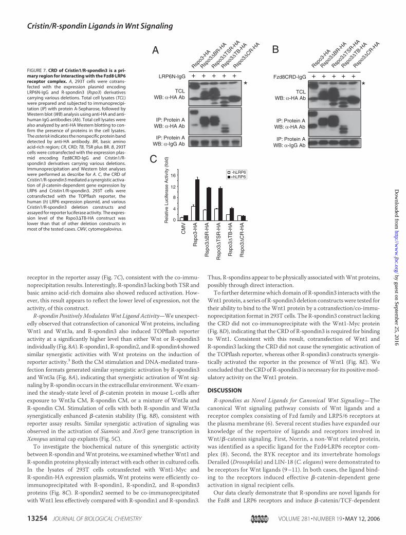
Binding of R-spondin to the Extracellular Domains of Fzd8 and LRP6Receptors—To biochemically characterize R-spondin activity in theFzd�LRP receptor complexes, we examined whether R-spondin3 bindsto the extracellular domain of Fzd8, LRP6, or both. First, we utilized acotransfection/co-immunoprecipitation format. Expression plasmidsencoding mouse R-spondin3 tagged with the HA epitope and humanIgG fusions of the CRD of mouse Fzd8 (Fzd8CRD-IgG) (16) and theextracellular domain of human LRP6 (LRP6N-IgG) (32) were cotrans-fected into 293T cells. Cell lysates were subjected to immunoprecipita-tion followed by Western blot analysis. The R-spondin3 protein waseffectively co-immunoprecipitated with Fzd8CRD-IgG and LRP6N-IgG, but not with IgG (Fig. 6A). Although much less concentrated thanFzd8CRD-IgG, the LRP6N-IgG protein co-immunoprecipitated a com-parable amount of R-spondin3. Thus, R-spondin3may bind to the LRP6ectodomain at a higher affinity than to the CRD of Fzd8 in vivo. Incontrast, more R-spondin3 molecules may bind to the LRP6 ectodo-main than to the CRD of Fzd8.We next examined these interactions under cell-free liquid-phase
conditions. We isolated histidine-tagged R-spondin1 and R-spondin3proteins from total lysates of 293T cells transiently transfected withR-spondin expression plasmids. Fzd8CRD-IgG and LRP6N-IgG fusionproteinswere prepared in aCM format as described (32).We found thatboth R-spondin1 and R-spondin3 were specifically coprecipitated withFzd8CRD-IgG and LRP6N-IgG, but not with IgG (Fig. 6B). Similar
to the cotransfection/co-immunoprecipitation results, R-spondinproteins appeared to bind to LRP6N-IgG more efficiently than toFzd8CRD-IgG, as similar amounts of R-spondins were co-immunopre-cipitated with a reduced amount of LRP6N-IgG.Because the Wnt1 ligand has been shown to form a ternary complex
with Fzd8 and LRP6 under in vitro binding conditions (32), we nextexamined whether R-spondin can form a similar ternary complex withboth receptors. LRP6N-IgG andFzd8CRD-HA failed to forma complex(Fig. 6C), consistent with the previously reported results (32).When theR-spondin1-Myc/His protein was incubated with both LRP6N-IgG andFzd8 CRD-HA, we could not detect any Fzd8 CRD-HA protein in theimmunoprecipitated LRP6N-IgG construct, whereas R-spondin1-Myc/His was present in the construct (Fig. 6C). This result suggests that,unlike Wnt1 ligands, the R-spondin, LRP6, and Fzd8 proteins do notform a ternary complex. Interestingly, the presence of Fzd8 CRD-HA inthe binding reaction significantly reduced the binding of R-spondin1 toLRP6N-IgG (Fig. 6C), indicating that Fzd8 CRD-HAmay compete withLPR6-IgG for binding to R-spondin1. The same or overlapping domainof the R-spondin1 protein may be engaged in binding to both LRP6 andFzd8 receptors.To further investigatewhich domain of R-spondin is involved in these
interactions, we cotransfected R-spondin3 deletion mutants withFzd8CRD-IgG or LRP6N-IgG and analyzed their interactions. Wedetermined that the CRD of R-spondin3 was essential for the interac-tion with both Fzd8 and LRP6 receptors (Fig. 7, A and B). TheR-spondin3 construct lacking theCRD failed to signal through the LRP6
FIGURE 6. A, in vivo interaction of Cristin1/R-spondin3 (Rspo3) with mouse Fzd8 and human LRP6. Expression plasmids encoding the mouse Fzd8 CRD (Fzd8CRD-IgG) and the humanLRP6 ectodomain (LRP6N-IgG) as human IgG Fc fusion proteins and control IgG were cotransfected with the Cristin1/R-spondin3-HA plasmid into 293T cells. Total cell lysates (TCL)were prepared and subjected to immunoprecipitation (IP) using protein A-Sepharose. Immunoprecipitated proteins were further analyzed by Western blot (WB) analysis usinganti-HA or anti-human IgG antibody (Ab). To confirm the presence of Cristin/R-spondin protein, total cell lysates were also analyzed by anti-HA Western blotting. The asterisk indicatesthe nonspecific protein band detected by anti-HA antibody. B, in vitro interaction of Cristin/R-spondin with the Fzd8 or LRP6 receptor. R-spondin1-Myc/His and R-spondin3-Hisproteins were added after IgG, Fzd8CRD-IgG, or LRP6N-IgG in CM was prebound to protein A-Sepharose. C, No Ternary complex formation was detected in R-spondin1-Myc/His, Fzd8CRD-HA, and LRP6N-IgG. Input represents one-tenth of the proteins used in the binding reactions.
Cristin/R-spondin Ligands in Wnt Signaling
MAY 12, 2006 • VOLUME 281 • NUMBER 19 JOURNAL OF BIOLOGICAL CHEMISTRY 13253
by guest on September 25, 2016
http://ww
w.jbc.org/
Dow
nloaded from
receptor in the reporter assay (Fig. 7C), consistent with the co-immu-noprecipitation results. Interestingly, R-spondin3 lacking both TSR andbasic amino acid-rich domains also showed reduced activation. How-ever, this result appears to reflect the lower level of expression, not theactivity, of this construct.
R-spondin PositivelyModulatesWnt Ligand Activity—We unexpect-edly observed that cotransfection of canonical Wnt proteins, includingWnt1 and Wnt3a, and R-spondin3 also induced TOPflash reporteractivity at a significantly higher level than either Wnt or R-spondin3individually (Fig. 8A). R-spondin1, R-spondin2, andR-spondin4 showedsimilar synergistic activities with Wnt proteins on the induction ofreporter activity.3 Both the CM stimulation and DNA-mediated trans-fection formats generated similar synergistic activation by R-spondin3and Wnt3a (Fig. 8A), indicating that synergistic activation of Wnt sig-naling by R-spondin occurs in the extracellular environment.We exam-ined the steady-state level of �-catenin protein in mouse L-cells afterexposure to Wnt3a CM, R-spondin CM, or a mixture of Wnt3a andR-spondin CM. Stimulation of cells with both R-spondin and Wnt3asynergistically enhanced �-catenin stability (Fig. 8B), consistent withreporter assay results. Similar synergistic activation of signaling wasobserved in the activation of Siamois and Xnr3 gene transcription inXenopus animal cap explants (Fig. 5C).
To investigate the biochemical nature of this synergistic activitybetween R-spondin andWnt proteins, we examined whetherWnt1 andR-spondin proteins physically interact with each other in cultured cells.In the lysates of 293T cells cotransfected with Wnt1-Myc andR-spondin-HA expression plasmids, Wnt proteins were efficiently co-immunoprecipitated with R-spondin1, R-spondin2, and R-spondin3proteins (Fig. 8C). R-spondin2 seemed to be co-immunoprecipitatedwith Wnt1 less effectively compared with R-spondin1 and R-spondin3.
Thus, R-spondins appear to be physically associated withWnt proteins,possibly through direct interaction.To further determine which domain of R-spondin3 interacts with the
Wnt1 protein, a series of R-spondin3 deletion constructs were tested fortheir ability to bind to the Wnt1 protein by a cotransfection/co-immu-noprecipitation format in 293T cells. The R-spondin3 construct lackingthe CRD did not co-immunoprecipitate with the Wnt1-Myc protein(Fig. 8D), indicating that the CRD of R-spondin3 is required for bindingto Wnt1. Consistent with this result, cotransfection of Wnt1 andR-spondin3 lacking the CRD did not cause the synergistic activation ofthe TOPflash reporter, whereas other R-spondin3 constructs synergis-tically activated the reporter in the presence of Wnt1 (Fig. 8E). Weconcluded that theCRDof R-spondin3 is necessary for its positivemod-ulatory activity on the Wnt1 protein.
DISCUSSION
R-spondins as Novel Ligands for Canonical Wnt Signaling—Thecanonical Wnt signaling pathway consists of Wnt ligands and areceptor complex consisting of Fzd family and LRP5/6 receptors atthe plasma membrane (6). Several recent studies have expanded ourknowledge of the repertoire of ligands and receptors involved inWnt/�-catenin signaling. First, Norrin, a non-Wnt related protein,was identified as a specific ligand for the Fzd4�LRP6 receptor com-plex (8). Second, the RYK receptor and its invertebrate homologsDerailed (Drosophila) and LIN-18 (C. elegans) were demonstrated tobe receptors for Wnt ligands (9–11). In both cases, the ligand bind-ing to the receptors induced effective �-catenin-dependent geneactivation in signal recipient cells.Our data clearly demonstrate that R-spondins are novel ligands for
the Fzd8 and LRP6 receptors and induce �-catenin/TCF-dependent
FIGURE 7. CRD of Cristin1/R-spondin3 is a pri-mary region for interacting with the Fzd8�LRP6receptor complex. A, 293T cells were cotrans-fected with the expression plasmid encodingLRP6N-IgG and R-spondin3 (Rspo3) derivativescarrying various deletions. Total cell lysates (TCL)were prepared and subjected to immunoprecipi-tation (IP) with protein A-Sepharose, followed byWestern blot (WB) analysis using anti-HA and anti-human IgG antibodies (Ab). Total cell lysates werealso analyzed by anti-HA Western blotting to con-firm the presence of proteins in the cell lysates.The asterisk indicates the nonspecific protein banddetected by anti-HA antibody. BR, basic aminoacid-rich region; CR, CRD; TB, TSR plus BR. B, 293Tcells were cotransfected with the expression plas-mid encoding Fzd8CRD-IgG and Cristin1/R-spondin3 derivatives carrying various deletions.Immunoprecipitation and Western blot analyseswere performed as describe for A. C, the CRD ofCristin1/R-spondin3 mediated a synergistic activa-tion of �-catenin-dependent gene expression byLRP6 and Cristin1/R-spondin3. 293T cells werecotransfected with the TOPflash reporter, thehuman (h) LRP6 expression plasmid, and variousCristin1/R-spondin3 deletion constructs andassayed for reporter luciferase activity. The expres-sion level of the Rspo3�TB-HA construct waslower than that of other deletion constructs inmost of the tested cases. CMV, cytomegalovirus.
Cristin/R-spondin Ligands in Wnt Signaling
13254 JOURNAL OF BIOLOGICAL CHEMISTRY VOLUME 281 • NUMBER 19 • MAY 12, 2006
by guest on September 25, 2016
http://ww
w.jbc.org/
Dow
nloaded from

gene activation. Recently, the discovery of Xenopus R-spondin2 andR-spondin3 genes was reported (14). Xenopus R-spondin2 and humanR-spondin1 exhibit a positive signaling activity on canonical Wnt sig-naling (14, 15), consistentwith our results.However, both studies lacked
any evidence showing the interactions between R-spondin and the Fzdand LRP5/6 receptors. In their report on Xenopus R-spondin2, Kazan-skaya et al. (14) suggested that their attempt to determine bindingbetween R-spondin and the Fzd or LRP6 receptor failed. However, we
FIGURE 8. Cristin/R-spondin proteins function as a positive modulator of Wnt ligands. A, Cristin1/R-spondin3 (Rspo3) CM and Wnt3a CM synergistically induce TOPflash reporteractivity. 293T cells were transfected with the TOPflash reporter and stimulated with Wnt3a CM (1:2), Cristin1/R-spondin3 CM (1:2), or both for 8 h. Control CM was used for diluting theCM or as a background. In a parallel experiment, expression plasmids encoding Wnt3a and Cristin1/R-spondin3 were cotransfected into 293T cells and tested for synergism. B,synergistic increase in the steady-state level of �-catenin protein in mouse L-cells stimulated with various concentrations of Wnt3a (1:2–50), Cristin1/R-spondin3 CM (1:2), or both for12 h. The �-catenin protein level was determined by Western blot analysis. The �-actin level was also measured to confirm an equivalent amount of sample loading. C, Wnt1 andCristin/R-spondin proteins are associated with each other in 293T cells. 293T cells were cotransfected with Wnt1-Myc and different Cristin/R-spondin-HA expression plasmids. As anegative control, the enhanced green fluorescent protein (EGFP)-HA expression plasmid was cotransfected with the Wnt1-Myc plasmid. Total cell lysates (TCL) were prepared andsubjected to immunoprecipitation (IP) using anti-HA antibody (Ab)-agarose beads. The immune complexes were analyzed by Western blotting (WB) using anti-Myc antibodies. Todetermine protein expression, total cell lysates were also analyzed by Western blotting using anti-Myc and anti-HA antibodies. D, the CRD of R-spondin3 is required for Wnt1 binding.R-spondin1 deletion constructs tagged with the HA epitope were cotransfected with the Wnt1-Myc plasmid into 293T cells. Anti-Myc antibody-agarose beads were used toimmunoprecipitate the Wnt1-Myc protein from cell lysates. Anti-HA and anti-Myc antibodies were used to detect R-spondin1 derivatives and Wnt1 protein, respectively, within theimmune complex by Western blot analysis. Expression of the R-spondin1 derivatives was also determined by Western blot analysis of total cell lysates using anti-HA antibody. BR,basic amino acid-rich domain; CR, CRD. E, the CRD of R-spondin3 is necessary for the synergistic regulation of TOPflash reporter activity induced by Wnt1. Wnt1-Myc (25 ng) andR-spondin1-HA deletion constructs (25 ng) were cotransfected with the TOPflash reporter (20 ng) into 293T cells.
Cristin/R-spondin Ligands in Wnt Signaling
MAY 12, 2006 • VOLUME 281 • NUMBER 19 JOURNAL OF BIOLOGICAL CHEMISTRY 13255
by guest on September 25, 2016
http://ww
w.jbc.org/
Dow
nloaded from

convincingly demonstrated that R-spondin interacts with the extracel-lular domains of the Fzd8 and LRP6 receptors both in vivo and in vitro(Fig. 6). It is difficult to understand the reason for the different results. Itis possible that Xenopus R-spondin may have a significantly weakerbinding activity for the receptors, which made it difficult to detect theinteraction in their assay. Nonetheless, our results confirm that mouseR-spondin is indeed a novel ligand for both Fzd and LRP receptors.Interestingly, the Fzd8 receptor was incompetent tomediate a down-
stream signaling event leading to �-catenin-dependent gene activationdespite the efficient binding to the R-spondin protein (Figs. 4B, 6, and7B). Two explanations are possible for these puzzling data. First, anincreasing body of evidence indicates that signaling activities throughthe LRP6 receptor may be dissociated from those through the Fzdreceptors (33–36). Thus, preferential R-spondin signaling through theLRP6 receptor may be another example of dissociation of intracellularsignaling events at the receptor level. Therefore, R-spondin binding tothe LRP6 receptor may be sufficient to induce �-catenin-dependentsignaling without any contribution from Fzd (Fig. 9, Model 3). Ourresult from the in vitro binding assay supports this possibility. Unlikecanonical Wnt ligands that could induce ternary complex formationwith the Fzd8 and LRP6 receptors (Fig. 9, Model 1), the R-spondin1protein failed to form a ternary complex (Fig. 6C). Because the sameCRD of R-spondin was required for binding to the Fzd and LRP6 recep-tors (Fig. 7), it is likely that R-spondin binding to both receptors may becompetitive. Consistent with this notion, Fzd8 CRD-HA significantlyinhibited the binding of R-spondin1 to the LRP6N-IgGprotein (Fig. 6C).Because the association of Fzd and LRP6 mediated by canonical Wntligands appears to be critical for activating downstream events (18), thisresult suggests that R-spondin induces �-catenin-dependent gene acti-vation by mechanisms distinct from Wnt signaling. Furthermore, it ispossible that the R-spondin and Fzd interaction may induce only non-canonical signaling independent of �-catenin (Fig. 9,Model 4).
Second, R-spondinmay preferentially bind to and transmit the signalthrough other Fzd receptors alongwith the LRP6 receptor (Fig. 9,Model2). Although it is currently unknownwhether other Fzd receptors trans-duce R-spondin signals, this is a very plausible scenario given that theNorrin ligand transduces the signal exclusively through the Fzd4�LRP6receptor complex (8).
Positive Modulation of Wnt Ligand Activity by R-spondin—Surpris-ingly, we discovered that R-spondin plays a positive modulatory role inWnt ligand activity presumably by directly interacting withWnt ligands(Fig. 8). It appears that the CRD of R-spondin3 is necessary for thisactivity (Fig. 8, D and E). How can R-spondins modulate Wnt activity?First, it is plausible that the Wnt�R-spondin complex is a higher affinityligand than Wnt or R-spondin individually. Second, R-spondin maystabilize the Wnt ligand/receptor interaction without increasing thebinding affinity of the Wnt ligand for the receptor. Third, Wnt may beprotected from degradation or turnover when it is associated withR-spondin. Themolecular mechanisms underlying this activity will be afocus of future studies.A large number of Wnt antagonists are known. Some Wnt antago-
nists, including secreted Frizzled-related proteins (37) andWnt inhibi-tory factor (38), likely inhibitWnt signaling activity by direct interactionwith Wnt proteins to prevent their receptor binding. In contrast, Dkk1may not directly interact with Wnt proteins, but may regulate LRP5/6receptor endocytosis or availability (29). Because R-spondin seems todirectly interact withWnt proteins, how R-spondin activity is regulatedby otherWnt-binding antagonists will be a focus of future investigation.To this end, theCCN family proteins, which primarily act asWnt antag-onists (26, 27), are of particular interest. The presence of similar protein
motifs such as TSRs and CRDs in both R-spondins and CCN proteins(25) strongly suggests that they may belong to the ligand superfamilyand have opposing activities for modulating Wnt signaling activity.
Possible Regulatory Role of HSPGs in R-spondin and Wnt Signal-ing—The majority of R-spondin proteins expressed in the cells appearto reside within the endoplasmic reticulum and Golgi apparatus,whereas a relatively small portion is secreted from cells (Fig. 2). Thesecreted R-spondin seems to be associated with the plasma membraneand extracellular matrix. We found that R-spondins are heparin-bind-ing proteins with high affinity and that both the TSR and basic domainsof R-spondin3 are required for this binding (Fig. 3). Therefore, it ishighly likely that secreted R-spondin is associated with HSPGs on theplasma membrane and extracellular matrix.Heparin or HSPGs play important regulatory roles in growth factor/
cytokine signaling (39, 40). The role of HSPGs in Wnt signaling wasrecently demonstrated (41–43). Two models have been proposed for aHSPG regulatory role inWnt signaling based primarily on studies on theDrosophila Wnt homolog Wingless (44). First, HSPGs control eitherdegradation or diffusion of Wingless to regulate its availability. Second,HSPGs serve as low affinity receptors for the Wingless ligand and mayfacilitate its binding to the Fzd/LRP receptors. It was demonstratedrecently that heparin binding to Wnt ligands inhibits Wnt signalingactivity in mammalian cells (41). Furthermore, Chinese hamster ovarycell mutants for heparan sulfate biosynthesis are defective in the activa-tion of Fzd receptor signaling (41). It would be intriguing to determinewhether HSPGs regulate R-spondin signaling activity and, if so, whatthe underlying mechanisms are. In our preliminary studies, we foundthat soluble heparin exhibited an inhibitory activity on R-spondin sig-naling, similar to Wnt signaling.3
Fine Control of Canonical Wnt Signaling Activity—Our study hasprovided evidence that R-spondin family proteins are novel ligands forthe Fzd8 and LRP6 receptors. Furthermore, R-spondin proteins caninduce �-catenin-dependent gene activation likely through the LRP6receptor by mechanisms different from those in canonical Wnt signal-ing. Interestingly, under R-spondin and Wnt coexistence conditions,the signaling activity of the Wnt�R-spondin ligand complex is signifi-cantly higher than that of either Wnt or R-spondin alone. Thus, com-binatorial expression of R-spondin andWnt proteins may produce dif-ferential signaling activities that lead to differential activation of abattery of genes, which eventually govern the outcome of cellular pro-cesses. Future studies will be focused on determining the molecularmechanisms by which the differential signaling activity is regulated. Inaddition, the co-presence of different secretedWnt antagonists (37) will
FIGURE 9. Proposed models for Cristin/R-spondin and Wnt signaling through theFzd and LRP6 receptors. Model 1 is a current view of canonical Wnt signaling. In Model2, Cristin/R-spondin proteins generate �-catenin-dependent signaling through the Fzd/LRP6 receptors, similar to canonical Wnt signaling. In contrast, Cristin/R-spondin maytransduce its signal through the LRP6 receptor (Model 3). Cristin/R-spondin also inducesnon-canonical Wnt signaling through the Fzd receptor (Model 4). Wnt and Cristin/R-spondin ligands are indicated by white and gray ovals, respectively.
Cristin/R-spondin Ligands in Wnt Signaling
13256 JOURNAL OF BIOLOGICAL CHEMISTRY VOLUME 281 • NUMBER 19 • MAY 12, 2006
by guest on September 25, 2016
http://ww
w.jbc.org/
Dow
nloaded from

further define the level of signaling activity, although whether anyknownWnt antagonists affect R-spondin activity is unknown.
Acknowledgments—We thanks Drs. Robert Friesel, Lucy Liaw, Leif Oxburgh,Doug Spicer, and DonWojchowski for critical reading of and comments on themanuscript and Norma Albrecht for proofreading. We are indebted to Drs.Rudolf Grosschedl, Xi He, Jen-Chih Hsieh, Nobue Itasaki, Randall Moon,Christof Niehrs, and Roel Nusse for reagents.
REFERENCES1. Wodarz, A., and Nusse, R. (1998) Annu. Rev. Cell Dev. Biol. 14, 59–882. Polakis, P. (2000) Genes Dev. 14, 1837–18513. Malbon, C. C. (2004) Front. Biosci. 9, 1048–10584. Wang, H. Y. (2004) Front. Biosci. 9, 1043–10475. Moon, R. T., Bowerman, B., Boutros, M., and Perrimon, N. (2002) Science 296,
1644–16466. He, X., Semenov, M., Tamai, K., and Zeng, X. (2004) Development (Camb.) 131,
1663–16777. Kuhl, M. (2004) Front. Biosci. 9, 967–9748. Xu, Q., Wang, Y., Dabdoub, A., Smallwood, P. M., Williams, J., Woods, C., Kelley,
M. W., Jiang, L., Tasman, W., Zhang, K., and Nathans, J. (2004) Cell 116, 883–8959. Inoue, T., Oz, H. S., Wiland, D., Gharib, S., Deshpande, R., Hill, R. J., Katz, W. S., and
Sternberg, P. W. (2004) Cell 118, 795–80610. Lu, W., Yamamoto, V., Ortega, B., and Baltimore, D. (2004) Cell 119, 97–10811. Yoshikawa, S., McKinnon, R. D., Kokel, M., and Thomas, J. B. (2003) Nature 422,
583–58812. Berger, W., Meindl, A., van de Pol, T. J., Cremers, F. P., Ropers, H. H., Doerner, C.,
Monaco, A., Bergen, A. A., Lebo, R., Warburg, M., Zergollern, L., Lorenz, B., Gal, A.,Bleeker-Wagemakers, E. M., and Meltinger, T. (1992) Nat. Genet. 1, 199–203
13. Chen, Z. Y., Hendriks, R.W., Jobling,M.A., Powell, J. F., Breakefield, X.O., Sims, K. B.,and Craig, I. W. (1992) Nat. Genet. 1, 204–208
14. Kazanskaya,O., Glinka, A., del BarcoBarrantes, I., Stannek, P., Niehrs, C., andWu,W.(2004) Dev. Cell 7, 525–534
15. Kim, K. A., Kakitani,M., Zhao, J., Oshima, T., Tang, T., Binnerts,M., Liu, Y., Boyle, B.,Park, E., Emtage, P., Funk, W. D., and Tomizuka, K. (2005) Science 309, 1256–1259
16. Hsieh, J.-C., Rattner, A., Smallwood, P. M., and Nathans, J. (1999) Proc. Natl. Acad.Sci. U. S. A. 96, 3546–3551
17. Willert, K., Brown, J. D., Danenberg, E., Duncan, A. W., Weissman, I. L., Reya, T.,Yates, J. R., III, and Nusse, R. (2003) Nature 423, 448–452
18. Semenov, M. V., Tamai, K., Brott, B. K., Kuhl, M., Sokol, S., and He, X. (2001) Curr.
Biol. 11, 951–96119. Yoon, J. K., and Wold, B. (2000) Genes Dev. 14, 3204–321420. Yoon, J. K., Moon, R. T., and Wold, B. (2000) Dev. Biol. 222, 376–39121. Kamata, T., Katsube, K., Michikawa, M., Yamada, M., Takada, S., and Mizusawa, H.
(2004) Biochim. Biophys. Acta 1676, 51–6222. Adams, J. C., and Tucker, R. P. (2000) Dev. Dyn. 218, 280–29923. Fernig, D. G., and Gallagher, J. T. (1994) Prog. Growth Factor Res. 5, 353–37724. Takada, S., Stark, K. L., Shea, M. J., Vassileva, G., McMahon, J. A., and McMahon,
A. P. (1994) Genes Dev. 8, 174–18925. Brigstock, D. R. (2003) J. Endocrinol. 178, 169–17526. Mercurio, S., Latinkic, B., Itasaki, N., Krumlauf, R., and Smith, J. C. (2004) Develop-
ment (Camb.) 131, 2137–214727. Latinkic, B. V., Mercurio, S., Bennett, B., Hirst, E. M., Xu, Q., Lau, L. F., Mohun, T. J.,
and Smith, J. C. (2003) Development (Camb.) 130, 2429–244128. Glinka, A., Wu, W., Delius, H., Monaghan, A. P., Blumenstock, C., and Niehrs, C.
(1998) Nature 391, 357–36229. Mao, B.,Wu,W., Davidson, G.,Marhold, J., Li, M.,Mechler, B.M., Delius, H., Hoppe,
D., Stannek, P., Walter, C., Glinka, A., and Niehrs, C. (2002) Nature 417, 664–66730. Subramanian, V., Meyer, B. I., and Gruss, P. (1995) Cell 83, 641–65331. Wilkinson, D. G., Bhatt, S., and Herrmann, B. G. (1990) Nature 343, 657–65932. Tamai, K., Semenov, M., Kato, Y., Spokony, R., Liu, C., Katsuyama, Y., Hess, F.,
Saint-Jeannet, J. P., and He, X. (2000) Nature 407, 530–53533. Brennan, K., Gonzalez-Sancho, J. M., Castelo-Soccio, L. A., Howe, L. R., and Brown,
A. M. (2004) Oncogene 23, 4873–488434. Tolwinski, N. S., Wehrli, M., Rives, A., Erdeniz, N., DiNardo, S., and Wieschaus, E.
(2003) Dev. Cell 4, 407–41835. Gonzalez-Sancho, J. M., Brennan, K. R., Castelo-Soccio, L. A., and Brown, A. M.
(2004)Mol. Cell. Biol. 24, 4757–476836. Li, L., Mao, J., Sun, L., Liu, W., and Wu, D. (2002) J. Biol. Chem. 277, 5977–598137. Kawano, Y., and Kypta, R. (2003) J. Cell Sci. 116, 2627–263438. Hsieh, J.-C., Kodjabachian, L., Rebbert, M. L., Rattner, A., Smallwood, P. M., Samos,
C. H., Nusse, R., Dawid, I. B., and Nathans, J. (1999) Nature 398, 431–43639. Lin, X., and Perrimon, N. (2000)Matrix Biol. 19, 303–30740. Wiedlocha, A., and Sorensen, V. (2004) Curr. Top. Microbiol. Immunol. 286, 45–7941. Ai, X., Do, A. T., Lozynska, O., Kusche-Gullberg, M., Lindahl, U., and Emerson, C. P.,
Jr. (2003) J. Cell Biol. 162, 341–35142. Dhoot,G. K., Gustafsson,M.K., Ai, X., Sun,W., Standiford,D.M., andEmerson, C. P.,
Jr. (2001) Science 293, 1663–166643. Tsuda,M., Kamimura, K., Nakato, H., Archer, M., Staatz,W., Fox, B., Humphrey,M.,
Olson, S., Futch, T., Kaluza, V., Siegfried, E., Stam, L., and Selleck, S. B. (1999)Nature400, 276–280
44. Lin, X. (2004) Development (Camb.) 131, 6009–6021
Cristin/R-spondin Ligands in Wnt Signaling
MAY 12, 2006 • VOLUME 281 • NUMBER 19 JOURNAL OF BIOLOGICAL CHEMISTRY 13257
by guest on September 25, 2016
http://ww
w.jbc.org/
Dow
nloaded from

Ju-Suk Nam, Taryn J. Turcotte, Peter F. Smith, Sangdun Choi and Jeong Kyo Yoon-Catenin-dependent Gene Expressionβand LRP6 Receptors and Activate
Mouse Cristin/R-spondin Family Proteins Are Novel Ligands for the Frizzled 8
doi: 10.1074/jbc.M508324200 originally published online March 16, 20062006, 281:13247-13257.J. Biol. Chem.
10.1074/jbc.M508324200Access the most updated version of this article at doi:
Alerts:
When a correction for this article is posted•
When this article is cited•
to choose from all of JBC's e-mail alertsClick here
http://www.jbc.org/content/281/19/13247.full.html#ref-list-1
This article cites 44 references, 16 of which can be accessed free at
by guest on September 25, 2016
http://ww
w.jbc.org/
Dow
nloaded from