p38 MAPK Regulates IL‐1β Induced IL‐6 Expression Through mRNA Stability in Osteoblasts
A
Owcmmmapb©
KP
1
kftsa(iaakaa
0d
Neurobiology of Aging 32 (2011) 2266–2278
Ethanol withdrawal acts as an age-specific stressor to activatecerebellar P38 kinase
Marianna E. Jung ∗, Xiaohua Ju, James W. Simpkins, Daniel B. Metzger,Liang-Jun Yan, Yi Wen
Department of Pharmacology and Neuroscience, Institute for Aging and Alzheimer’s disease, University of NorthTexas Health Science Center at Fort Worth, 3500 Camp Bowie Blvd., Fort Worth, TX 76107-2699, USA
Received 20 May 2009; received in revised form 17 December 2009; accepted 7 January 2010Available online 1 February 2010
bstract
We investigated whether protein kinase P38 plays a role in the brain-aging changes associated with repeated ethanol withdrawal (EW).variectomized young, middle-age and older rats, with or without 17�-estradiol (E2) implantation, received a 90-day ethanol with repeatedithdrawal. They were tested for active pP38 expression in cerebellar Purkinje neurons and whole-cerebellar lysates using immunohisto-
hemistry and enzyme-linked immunosorbent assay, respectively. They were also tested for the Rotarod task to determine the behavioralanifestation of cerebellar neuronal stress and for reactive oxygen species (ROS) and mitochondrial protein carbonyls to determine oxidativeechanisms. Middle-age EW rats showed higher levels of pP38-positive Purkinje neurons/cerebellar lysates, which coincided with increaseditochondrial protein oxidation than other diet/age groups. Exacerbated motor deficit due to age–EW combination also began at the middle-
ge. In comparison, ROS contents peaked in older EW rats. E2 treatment mitigated each of the EW effects to a different extent. Collectively,
P38 may mediate the brain-aging changes associated with pro-oxidant EW at vulnerable ages and in vulnerable neurons in a manner protectedy estrogen.2010 Elsevier Inc. All rights reserved.
eywords: Age; Brain-aging-changes; Cerebellum; 17�-estradiol; Ethanol withdrawal; Female rats; Mitochondrial protein carbonyls; Phosphor-P38 (pP38);
PaPPb2
da2ee
urkinje neurons; Reactive oxygen species
. Introduction
P38 belongs to the family of mitogen-activated proteininases that mediate signaling cascades and regulate cellate in response to cellular stress (Arimoto et al., 2008). Aransient, moderate activation of P38 is associated with cellurvival or differentiation, whereas the sustained or excessctivation generally correlates with pathological conditionsBarca et al., 2008; Du et al., 2008; Giordano et al., 2008). P38s activated upon phosphorylation (Moriguchi et al., 1996)nd thus, pP38 is often measured as an indicator of P38ctivation. P38 is also known as a stress-activated protein
inase because P38 is phosphorylated by stress signals suchs inflammatory cytokines, heat shock or ischemia (Aydin etl., 2005). The known members of the P38 family include∗ Corresponding author. Tel.: +1 817 735 0132; fax: +1 817 735 2091.E-mail address: [email protected] (M.E. Jung).
miFia(
197-4580/$ – see front matter © 2010 Elsevier Inc. All rights reserved.oi:10.1016/j.neurobiolaging.2010.01.005
38� (Lee et al., 1994), P38� (Jiang et al., 1996; Stein etl., 1997), P38� (Lechner et al., 1996; Li et al., 1996) and38� (Jiang et al., 1997). Among these isozymes, P38� and38� are highly expressed in brain areas that are vulnera-le to ethanol/EW, such as cerebellum and cortex (Lee et al.,000; Nonaka et al., 2008; Xiong et al., 2006).
The signaling role of P38 in the effects of ethanol has beenemonstrated in a study in which a P38 inhibitor SB203580ttenuated ethanol-induced cell death in HT22 cells (Ku et al.,007). Acute ethanol treatment led to P38 activation (Norkinat al., 2007) and augmented endotoxin-induced pP38 lev-ls in a manner attenuated by a P38 inhibitor in humanonocytes (Drechsler et al., 2006). P38 is also implicated
n age-associated physiological or pathological conditions.
or instance, while old men had a higher level of basal pP38n skeletal muscles than young men, the opposite trend inge-associated pP38 levels occurred after strenuous exerciseWilliamson et al., 2003). Pathological activation of P38 was
ogy of A
seHaiiopaEtEa
vattlpImRwFaoawciiFdiaoipaatEtg
2
2
t(Ag
o8sasoggftb
odsiacaTbolT2r((ieiresndo
2
wTwticcrmot
M.E. Jung et al. / Neurobiol
hown in the brains of Alzheimer’s disease patients (Hensleyt al., 1999) and in the livers of aged rats after challenged with2O2 (Vereker et al., 2000). These studies suggest that hyper-
ctivation of P38 may reflect an aging process. Because agings associated with estrogen depletion in females, we exam-ned the protective role of estrogen in the homeostatic statusf P38 at different ages. Previously, E2 attenuated the phos-horylation of P38 induced by angiotensin II in cells (Wu etl., 2009), cardiac hypertrophy in ovariectomized mice (vanickels et al., 2001) and myocardial inflammation in ovariec-
omized rats (Wang et al., 2006). These studies suggest that2 may attenuate P38 activation, especially when neuronsre challenged by the stress of aging or EW.
In the current study, we used cerebellum, one of the mostulnerable brain areas to ethanol/EW insults (Ramadoss etl., 2007; Santucci et al., 2008). Although cerebellum con-ains multiple types of neurons, Purkinje neurons constitutehe single output of all motor coordination in the cerebel-ar cortex. Therefore, damage to these neurons inevitablyrovokes impairment of motor behavior and coordination.n fact, a substantial loss of these neurons coincided withotor deficit in ethanol-withdrawn rats (Jung et al., 2002;ewal et al., 2003). Using this brain area, we investigatedhether EW provokes the age-specific activation of P38.irst, we examined the effects of ethanol vs. EW on P38ctivation by measuring protein levels of pP38 (I). Sec-nd, we assessed the effects of age–EW interaction on P38ctivation by assessing pP38 in Purkinje neurons and inhole-cerebellar lysates. The behavioral manifestation of
erebellar neuronal stress was assessed by a Rotarod testn which a shorter latency to fall from an accelerating rodndicated poorer cerebellar-related motor performance (II).inally, the potential oxidative mechanisms underlying age-ependent activation of P38 and estrogen protection werenvestigated by measuring reactive oxygen species (ROS)nd mitochondrial protein oxidation (III). In the initial partf this study (I), we used young male rats before superimpos-ng age and the female hormone estrogen. In the subsequentart of this study (II and III), we used female rats at theges of 5–8 months (young), 12–15 months (middle-age)nd 16–19 months (older) in a model of ovariectomy. Usinghis model, we provide empirical evidence that repeatedW provokes the hyperactivation of P38 in a manner that
argets vulnerable ages and neurons and is protected by estro-en.
. Materials and methods
.1. Subjects and experimental groups
All rats were housed individually in a room with con-◦
rolled temperature (22–25 C), humidity (55%) and lights7 AM–7 PM). Body weights were recorded twice a week.ll housing and procedures were in accordance with theuidelines of the Institutional Care and Use Committee
mohw
ging 32 (2011) 2266–2278 2267
f the National Research Council (NIH publication no.5–23, revised 1996) and were approved by the Univer-ity of North Texas Health Science Center Animal Carend Use Committee. All ethanol-withdrawn animals wereacrificed 14 days after the last ethanol-dose (14th dayf EW). Ethanol-consuming rats (named ethanol-exposureroup) continuously received an ethanol diet (6.5%) and wereradually withdrawn from an ethanol diet (5% for 3 days, 3%or 2 days and 2% for 2 days) to avoid EW stress. They werehen sacrificed on the next morning. The ethanol diet (2%)ottle was available until they were sacrificed.
For the initial test of pP38, we used male rats (5 monthsld, Sprague–Dawley) and administered a short-term ethanoliet (6.5%, 5 weeks) to them. The usage of male rats and ahort-term diet was to characterize the expression of pP38n the absence of the influence of estrogen, age or agingssociated with a lengthy diet. They were divided into theontrol dextrin, the ethanol-exposure or the EW group. Forging and estrogen studies, we used female rats (Fisher 344).hey were approximately 8, 15 or 19 months old when testsegan after a 90-day diet program. All female rats werevariectomized, implanted with an E2 or a control oil pel-et and allowed to recover for 2 weeks after the surgery.hey were then divided into five groups, and we began a5-day diet and a 5-day abrupt withdrawal cycle which wasepeated three times. The five groups were (1) dextrin + oil,2) dextrin + E2, (3) ethanol-exposure + oil, (4) EW + oil and5) EW + E2 groups. Because we were interested in EWnsults rather than ethanol insults, we did not include anthanol-exposure + E2 group. Separately, ovariectomized ratsmplanted with oil pellets were subjected to an identical dietegimen and assigned exclusively for the analysis of bloodthanol concentrations (BEC). The dextrin diet was used as aole control diet because we have repeatedly observed no sig-ificant difference between a dextrin diet vs. a regular chowiet or intermittent vs. continuous dextrin diets in vital signsr in experimental outcomes.
.2. Ovariectomy and E2 implantation
Under isoflurane (2% v/v) anesthesia, a small incisionas made in the abdominal cavity directly above the ovary.he ovaries were removed bilaterally and the incisionsere closed with stainless steel wound clips. Immediately
hereafter, Silastic pellets (s.c.) containing E2 or oil weremplanted on the dorsal part of the rat, and incisions werelosed with clips. The E2 pellet releases physiological con-entrations of E2 (29–34 pg/ml) for 3 weeks and thus waseplaced every 3 weeks. According to this schedule, ani-als had two episodes of E2 implantation that took place
n the 4th day of EW. At this point of EW, animals areypically recovered from the hyperexcitatory signs that are
ost severe during the first 24 h of EW. Thus, the proceduresf ovariectomy and E2 implantation unlikely influenced theyperexcitatory stress of EW. Furthermore, rats implantedith a control pellet did not differ from non-surgery rats in
2 ogy of A
vpe
2w
b(wtpNddEttt
2
(cFwpims
2
wbrwiSp2aErs
2
Cpec
atdosdeltr
2
b(clvwfaC1mfTbpbcBnIa
2i
MePi4wfaaiaa
268 M.E. Jung et al. / Neurobiol
ital signs, physical activities, or appearance. The method ofreparing the hormone pellet s was previously reported (Yangt al., 2001).
.3. Chronic ethanol administration in a liquid diet andithdrawal
The amount of dextrin and ethanol was calculated in com-ination to adjust the concentration of ethanol to 6.5% v/vJung et al., 2002). Control animals were fed a liquid dietith dextrin isocalorically substituted for ethanol. The nutri-
ionally balanced fresh diet (100 ml/rat) was prepared daily,oured into an l-shaped Plexiglass (Bio-Serv Frenchtown,J) and placed in each home cage every morning. On the lastay of each diet cycle, a small aliquot (50 ml) of ethanol orextrin diet was administered (7 AM) to ensure that all of theW rats finished the same amount of ethanol by the time of
he ethanol diet removal. Twelve hours later (7 PM), the dietubes were removed, and regular chow pellets were placed inhe home cage.
.4. Collection of brain tissues
Rats were humanely sacrificed under anesthesia [xylazine20 mg/kg, i.p.) and ketamine (100 mg/kg, i.p.)]. Cerebelliontaining the vermis area were used for biochemical assays.or immunohistochemical assays, a separate set of ratsas perfused intracardially with saline followed by 4%araformaldehyde in a phosphate buffer. Brains were dividednto halves by a midsagittal cut. A parasagittal cut was then
ade 3 mm to the left of the cerebellar midline, and thisample was used for the immunohistochemistry.
.5. Blood ethanol concentrations (BEC)
On the last day of each diet cycle, whole blood (100 �l)as collected from the tail vein 3 h after placing fresh dietottles. In brief, the rats were restrained in a Plexiglas rodentestraint device, and a syringe fitted with a 25-gauge needleas inserted toward the tail vein. Blood was collected and
mmediately mixed with 90 �l of ice-cold, 0.55 M HClO4.amples were centrifuged at 1500 × g for 10 min to sedimentrotein precipitate. Supernatants were adjusted to pH 5 with00 �l of a solution containing 0.6 M KOH and 50 mM aceticcid, and then centrifuged to sediment KClO4 precipitate.thanol in the supernatant was measured by a colorimet-
ic assay (Smolen et al., 1986) using a Beckman DU 640pectrophotometer.
.6. Accelerating Rotarod
This motor driven treadmill (Omnitech Electronics,
olumbus, OH) measures running coordination and motorerformance such that a shorter latency to fall from an accel-rating rod indicates poorer motor performance. The rotoronsists of four cylinders that are mounted 35.5 cm abovercta
ging 32 (2011) 2266–2278
padded surface. Rats were placed on the cylinder and aimer switch was simultaneously activated to rotate the cylin-ers. Acceleration continued until 44 rpm for maximum 90 sr animals fell to the padded surface, which simultaneouslytopped the timer. Rats were tested for 3 sessions/day for 5ays with a 20-min resting period between sessions (Rewalt al., 2004). The tests were not initiated until 7 days after theast ethanol diet to avoid the possibility that acutely occurringhe hyperexcitatory signs of EW would obscure cerebellar-elated motor deficit.
.7. Immunoblotting
The hemi-cerebellum was weighed and added to lysisuffer containing ice-cold PBS, a protease inhibitor cocktailEMD bioscience, CA), PMSF, and a phosphatase inhibitorocktail. An aliquot was combined with an equal volume ofysis buffer [0.5 M Tris-HCl (pH 6.8), 4.4% w/v SDS, 20%/v glycerol, 2% v/v 2-mercaptoethanol]. After the samplesere heated (95 ◦C, 5 min) and sonicated (10 s), Brad-
ord assay was conducted to analyze protein concentrationsccording to manufacture’s instructions (Biorad, Hercules,A). A 30 �g of sample protein was electrophoresed on a0% SDS-PAGE and then transferred onto a nitrocelluloseembrane. Nonspecific binding sites were blocked with 5%
at free milk. The blot was washed in PBS containing 0.05%ween-20 and probed overnight with a rabbit polyclonal anti-ody against P38 or a rabbit monoclonal antibody againstP38 (Thr180/Tyr182) (Cell Signaling, Danvers, MA). Thelot was then incubated with horseradish peroxidase (HRP)-onjugated secondary antibodies for 1 h at room temperature.ands were detected using the UVP western blotting lumi-escence system and quantified by an image densitometer.mmunoblottings for �-actin were carried out as a positivend a loading control.
.8. pP38 (Thr180/Tyr182) ELISA (enzyme-linkedmmunosorbent assay)
pP38 (Thr180/Tyr182) ELISA (Cell Signaling, Danvers,A) was used to quantitate P38 activation in cerebellar
xtracts by measuring the protein level of an active form of38 (pP38). Briefly, the coating antibody was diluted (1:100)
n PBS, added to a microplate, and incubated overnight at◦C. The plate contents were then discarded. The plate wasashed with PBS (×3) and then blocked with blocking buffer
or 2 h. Cerebellar tissue lysates were then added to the wellnd incubated again for 2 h. A detecting antibody (1:100) andsecondary antibody (1:1000) were sequentially added and
ncubated. Finally the plate was incubated at 37 ◦C for 30 minnd mixed with TMB substrates. STOP solution (100 �l) wasdded to each well to terminate the reaction. The plate was
ead using a microplate reader at an absorbance 450 nm. HeLaells treated with vehicle or anisomycin were used to validatehe ELISA analysis as positive controls. Lysis buffer was useds a blank condition.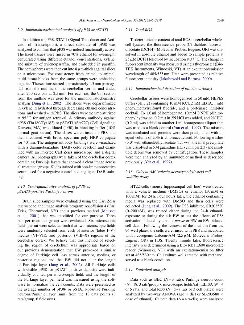
ogy of A
2
vaTdaTomtiafaitapDntfwiccasi
2p
mZerfiwmciodpowvtwtnr
2
cds2flTwfl
2
bpcp(wwe(wuwp
2v
w1mc(eac9wEirss
2
M.E. Jung et al. / Neurobiol
.9. Immunohistochemical analysis of pP38 or pSTAT1
In addition to pP38, STAT1 (Signal Transducer and Acti-ator of Transcription), a direct substrate of pP38 wasnalyzed to confirm that pP38 was indeed functionally active.he fixed tissues were rinsed in 70% ethanol for overnight,ehydrated using different ethanol concentrations, xylene,nd mixture of xylene/paraffin, and embedded in paraffin.he hemispheres were then cut into 8 �m-thick sagittal slicesn a microtome. For consistency from animal to animal,ulti-tissue blocks from the same groups were embedded
ogether. The sections started approximately 1.5 mm parasag-tal from the midline of the cerebellar vermis and endedfter 250 sections at 2.5 mm. For each rat, the 9th sectionrom the midline was used for the immunohistochemistrynalysis (Jung et al., 2002). The slides were deparaffinizedn xylene, rehydrated through decreasing ethanol concentra-ions, and washed with PBS. The slices were then moisturizedt 95 ◦C for antigen retrieval. A primary antibody againstP38 (Thr180/Tyr182) or pSTAT1 (Ser727) (Cell signaling,anvers, MA) was diluted (1:50) in blocking buffer (10%ormal goat serum). The slices were rinsed in PBS andhen incubated with broad spectrum poly HRP conjugateor 40 min. The antigen-antibody bindings were visualizedith a diaminobenzidine (DAB) color reaction and exam-
ned with an inverted Carl Zeiss microscope and a digitalamera. All photographs were taken of the cerebellar cortexontaining Purkinje layers that showed a clear image acrossll treatment groups. Slides stained with non-immunoreactiveerum used for a negative control had negligent DAB stain-ng.
.10. Semi-quantitative analysis of pP38- orSTAT1-positive Purkinje neurons
Brain slice samples were evaluated using the Carl Zeissicroscope, the image analysis program AxioVision 4 (Carleiss, Thornwood, NY) and a previous method (Maussett al., 2001) that was modified for our purpose. Threeats per treatment group were evaluated. Six microscopicelds per rat were selected such that two microscopic fieldsere randomly selected from each of anterior (lobes I–V),edius (VI–VII), and posterior (VIII–X) regions of the
erebellar cortex. We believe that this method of select-ng the region of cerebellum was appropriate based onur previous demonstration that EW provoked a similaregree of Purkinje cell loss across anterior, medius, orosterior regions and that EW did not alter the lengthf Purkinje layer (Jung et al., 2002). All Purkinje cellsith visible pP38- or pSTAT1-positive deposits were indi-idually counted per microscopic field, and the length ofhe Purkinje layer per field was measured using the soft-
are to normalize the cell counts. Data were presented ashe average number of pP38- or pSTAT1-positive Purkinjeeurons/Purkinje layer (mm) from the 18 data points (3ats/group, 6 fields/rat).
(oad
ging 32 (2011) 2266–2278 2269
.11. Total ROS
To determine the content of total ROS in cerebellar whole-ell lysates, the fluorescence probe 2,7-dichlorofluoresciniacetate (DCFH) (Molecular Probes, Eugene, OR) was dis-olved in absolute ethanol and added to sample proteins at5 �M DCFH followed by incubation at 37 ◦C. The change inuorescent intensity was measured using a fluorometer (Bio-EK Instruments, Winooski, VT) at an excitation/emissionavelength of 485/535 nm. Data were presented as relativeuorescent intensity (Jakubowski and Bartosz, 2000).
.12. Immunochemical detection of protein carbonyl
Cerebellar tissues were homogenized in 50 mM HEPESuffer (pH 7.2) containing 10 mM KCl, 2 mM EDTA, 1 mMhenylmethylsulfonyl fluoride, and a proteinase inhibitorocktail. To 1.0 ml of homogenate, 10 mM DNPH (dinitro-henylhydrazine, 0.2 ml) in 2N HCl was added, and 2N HCl0.2 ml) was added to another 1 ml homogenate aliquot thatas used as a blank control (Yan et al., 1997). The mixtureas incubated and proteins were then precipitated with an
qual volume of 20% trichloroacetic acid. Following washes×3) with ethanol/ethyl acetate (1:1 v/v), the final precipitateas dissolved in 6 M guanidine HCl (2 ml, pH 2.3) and insol-ble debris was removed by centrifugation. These samplesere then analyzed by an immunoblot method as describedreviously (Yan et al., 1997).
.13. Calcein-AM (calcein-acetoxymethylester) celliability assay
HT22 cells (mouse hippocampal cell line) were treatedith a vehicle medium (DMSO) or ethanol (50 mM or00 mM) for 24 h. Four hours later, the ethanol containingedia was replaced with DMSO and then cells were
ollected (Jung et al., 2009). The P38 inhibitor, SB2035800–200 nM), was treated either during the 24-h ethanol-xposure or during the 4-h EW to test the effects of P38ctivation induced by ethanol per se or EW on EW-inducedell death. Following the removal of the medium from the6-well plates, the cells were rinsed with PBS and incubatedith fluorogenic Calcein-AM (2.5 �M, Molecular Probes,ugene, OR) in PBS. Twenty minute later, fluorescence
ntensity was determined using a Bio-Tek FL600 microplateeader (Winooski, VT) with an excitation/emission filteret at 485/530 nm. Cell culture wells treated with methanolerved as a blank condition.
.14. Statistical analysis
Data such as BEC (N = 3 rats), Purkinje neuron count
N = 18, 3 rats/group, 6 microscopic fields/rat), ELISA (N = 4r 5 rats) and total ROS (N = 5–7 rats or 3 cell plates) werenalyzed by two-way ANOVA (age × diet or SB203580 ×ose of ethanol); Calcein data (N = 4 wells) were analyzed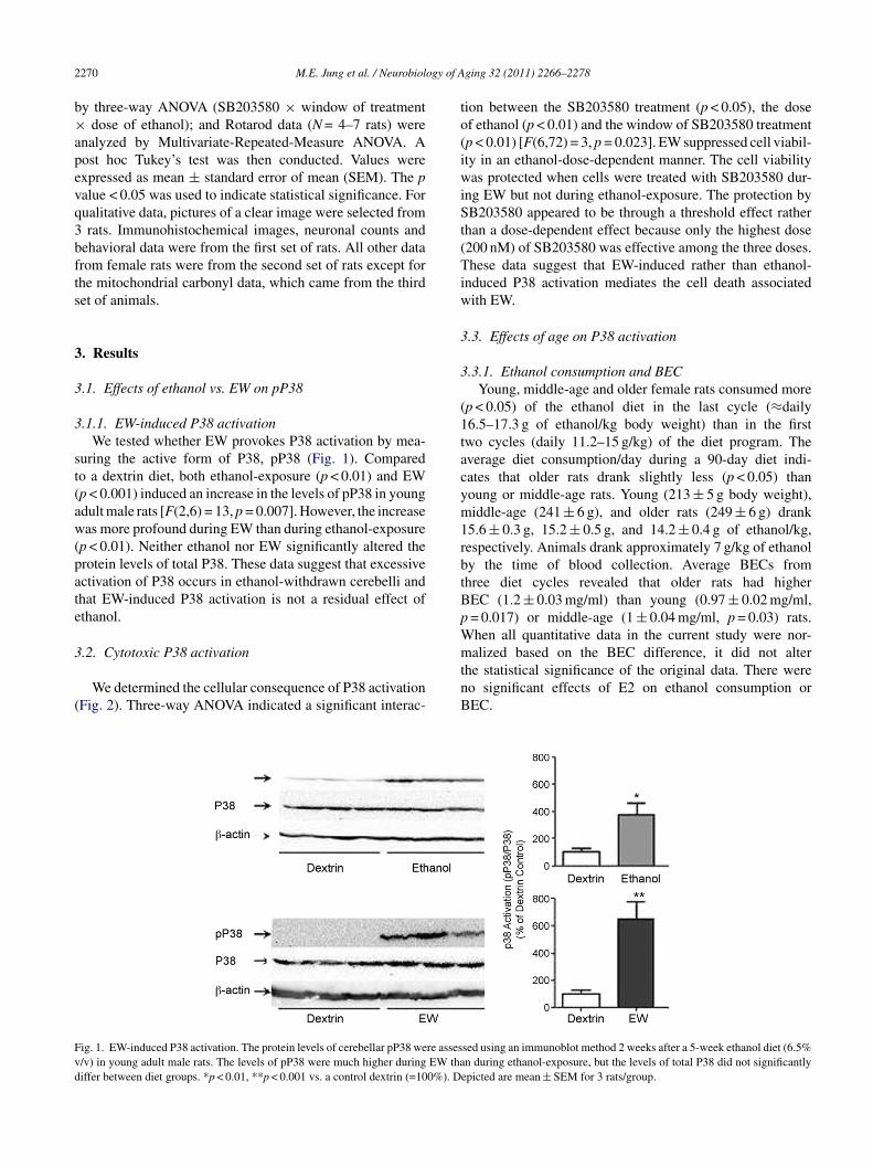
2 ogy of A
b×apevq3bfts
3
3
3
st(aw(pate
3
(
to(iwiSt(Tiw
3
3
(1tacym1rbtBpW
Fvd
270 M.E. Jung et al. / Neurobiol
y three-way ANOVA (SB203580 × window of treatmentdose of ethanol); and Rotarod data (N = 4–7 rats) were
nalyzed by Multivariate-Repeated-Measure ANOVA. Aost hoc Tukey’s test was then conducted. Values werexpressed as mean ± standard error of mean (SEM). The palue < 0.05 was used to indicate statistical significance. Forualitative data, pictures of a clear image were selected fromrats. Immunohistochemical images, neuronal counts and
ehavioral data were from the first set of rats. All other datarom female rats were from the second set of rats except forhe mitochondrial carbonyl data, which came from the thirdet of animals.
. Results
.1. Effects of ethanol vs. EW on pP38
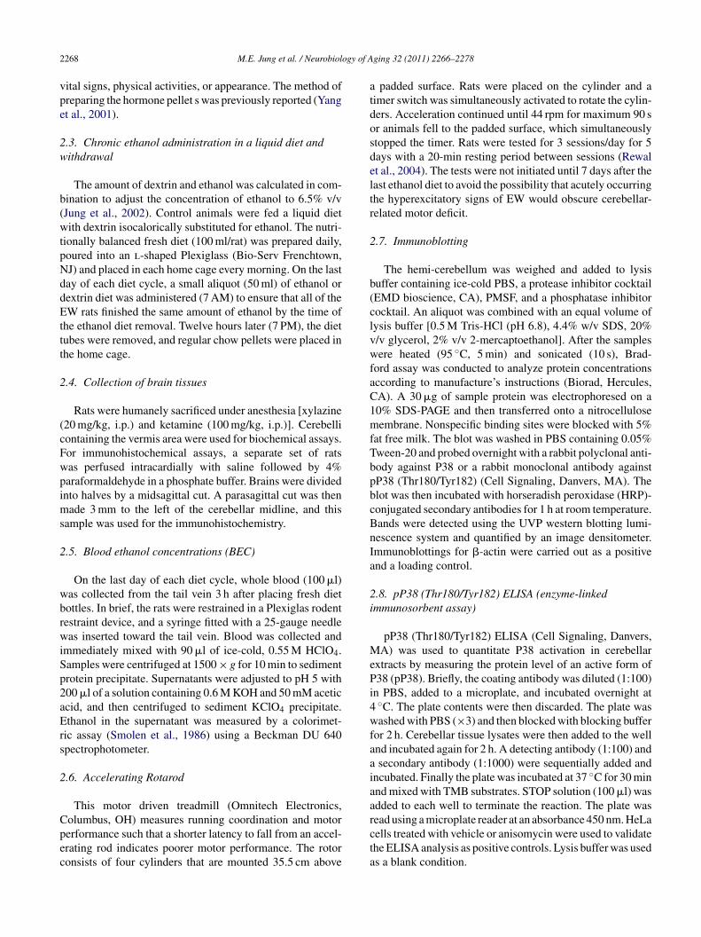
.1.1. EW-induced P38 activationWe tested whether EW provokes P38 activation by mea-
uring the active form of P38, pP38 (Fig. 1). Comparedo a dextrin diet, both ethanol-exposure (p < 0.01) and EWp < 0.001) induced an increase in the levels of pP38 in youngdult male rats [F(2,6) = 13, p = 0.007]. However, the increaseas more profound during EW than during ethanol-exposure
p < 0.01). Neither ethanol nor EW significantly altered therotein levels of total P38. These data suggest that excessivectivation of P38 occurs in ethanol-withdrawn cerebelli andhat EW-induced P38 activation is not a residual effect ofthanol.
.2. Cytotoxic P38 activation
We determined the cellular consequence of P38 activationFig. 2). Three-way ANOVA indicated a significant interac-
mtnB
ig. 1. EW-induced P38 activation. The protein levels of cerebellar pP38 were asses/v) in young adult male rats. The levels of pP38 were much higher during EW thaiffer between diet groups. *p < 0.01, **p < 0.001 vs. a control dextrin (=100%). D
ging 32 (2011) 2266–2278
ion between the SB203580 treatment (p < 0.05), the dosef ethanol (p < 0.01) and the window of SB203580 treatmentp < 0.01) [F(6,72) = 3, p = 0.023]. EW suppressed cell viabil-ty in an ethanol-dose-dependent manner. The cell viabilityas protected when cells were treated with SB203580 dur-
ng EW but not during ethanol-exposure. The protection byB203580 appeared to be through a threshold effect rather
han a dose-dependent effect because only the highest dose200 nM) of SB203580 was effective among the three doses.hese data suggest that EW-induced rather than ethanol-
nduced P38 activation mediates the cell death associatedith EW.
.3. Effects of age on P38 activation
.3.1. Ethanol consumption and BECYoung, middle-age and older female rats consumed more
p < 0.05) of the ethanol diet in the last cycle (≈daily6.5–17.3 g of ethanol/kg body weight) than in the firstwo cycles (daily 11.2–15 g/kg) of the diet program. Theverage diet consumption/day during a 90-day diet indi-ates that older rats drank slightly less (p < 0.05) thanoung or middle-age rats. Young (213 ± 5 g body weight),iddle-age (241 ± 6 g), and older rats (249 ± 6 g) drank
5.6 ± 0.3 g, 15.2 ± 0.5 g, and 14.2 ± 0.4 g of ethanol/kg,espectively. Animals drank approximately 7 g/kg of ethanoly the time of blood collection. Average BECs fromhree diet cycles revealed that older rats had higherEC (1.2 ± 0.03 mg/ml) than young (0.97 ± 0.02 mg/ml,= 0.017) or middle-age (1 ± 0.04 mg/ml, p = 0.03) rats.hen all quantitative data in the current study were nor-
alized based on the BEC difference, it did not alterhe statistical significance of the original data. There wereo significant effects of E2 on ethanol consumption orEC.
sed using an immunoblot method 2 weeks after a 5-week ethanol diet (6.5%n during ethanol-exposure, but the levels of total P38 did not significantly
epicted are mean ± SEM for 3 rats/group.
M.E. Jung et al. / Neurobiology of Aging 32 (2011) 2266–2278 2271
F as meat y figuree *p = 0.0
3e
ad(wEeepddecp
Ftrdtddottv
TrliEsmprbt
3
ig. 2. Cytotoxic P38 activation. The effect of EW on HT22 cell viability wo control cell viability at 100% (statistical symbols were omitted to simplifthanol-exposure phase (left), mitigated EW-induced cell death. *p = 0.039, *
.3.2. Age-dependent motor deficit inthanol-withdrawn rats
Behavioral manifestation of cerebellar neuronal stress wasssessed using a Rotarod test in four diet groups (dextrin,extrin + E2, EW, and EW + E2) at the ages of 8 (young), 15middle-age) or 19 (older) months. Ethanol-exposure groupsere not tested because they were intoxicated due to gradualW. EW (+oil pellet) rats had the shortest latencies (poor-st) among the diet groups on most of the 5-day-test days,specially in the middle-age and older groups [F(3,56) = 3,= 0.038 by diet; F(2,56) = 5, p = 0.011 by age]. Only young,extrin rats performed better as they repeated the task for 5ays (day-effect, p < 0.01). Because there was no significant
ffect of repeating tasks, except for the young, dextrin rats, weomputed an average latency across the 5 days [F(3,57) = 3.5,= 0.021 by diet; F(2,57) = 5.4, p = 0.007 by age] (Fig. 3).ig. 3. Age-dependent motor deficit in ethanol-withdrawn rats. Ovariec-omized rats were implanted with oil or E2 pellets, received a 90-day dietegimen, a 25-day ethanol (6.5% v/v) or a control dextrin diet followed by 5-ay abrupt withdrawal, and the cycle was repeated three times. Beginning athe 7th day of EW, the rats were tested for the Rotarod task (3 sessions/day, 5ays). Data were presented as an average latency from the 5-day data. Motoreficit (shorter latency) was found in young EW rats (p < 0.01) compared tother diet groups, became more severe (p < 0.01) in middle-age EW rats, andhen remained at a similar level in older EW rats. *p < 0.01 vs. dextrin, dex-rin + E2 or EW + E2 at each age. ‡p < 0.05 vs. 8-month-old dextrin. †p < 0.01s. 8-month-old EW. Depicted are mean ± SEM for 4–7 rats/group.
cigba
3
oiompdwinbpp(etn
sured 4 h after 24-h ethanol exposure. EW decreased cell viability relatives). SB203580 (200 nM) treatment during the EW phase (right), but not the09 vs. vehicle (non SB203580). Depicted are mean ± SEM for 4/treatment.
he average latency for 5 days was also shorter in EWats than in other young, diet groups (p < 0.01). The shorteratency in young, EW rats was further shortened (p < 0.01)n middle-age EW rats. However, the latency in middle-ageW rats did not significantly differ from that in older EW rats,uggesting that the onset of age–EW interaction occurs at aiddle-age (15 months old). A latency decrease was more
rofound in EW middle-age rats than in dextrin middle-ageats (p < 0.05), indicating an adverse effect of age–EW com-ination. While E2 per se did not alter the latency, it increasedhe latency of EW rats at all three ages.
.3.3. P38 activation in whole-cerebellar extractsWhen the level of pP38 was determined in the whole-
ellular extraction of cerebellum, the level was increased onlyn the middle-age EW rats among all of the diet and ageroups [F(3,72) = 6, p < 0.01 by diet; F(2,72) = 10, p < 0.01y age] (Fig. 4). Neither age per se nor E2 per se significantlyltered the total cerebellar level of pP38.
.3.4. P38 activation in Purkinje neuronsAlthough pP38 immunoreactivity was found in other types
f neurons, such as granular neurons, we focused on Purk-nje neurons because Purkinje neurons are the sole outputf the cerebellar cortex and play a major role in cerebellarotor function (Watanabe, 2008). The immunohistochemical
hotographs revealed that the pP38 mmunoreactivity (darkeposits) was more distinctively visualized in the ethanol-ithdrawn Purkinje neurons along the Purkinje layer than that
n the other diet groups across all three ages (Fig. 5A–D). Theumber of pP38-positive Purkinje neurons did indeed differy diet [F(3,204) = 57, p < 0.001] and by age [F(2,204) = 22,< 0.001]; middle-age EW rats had a higher number of pP38-ositive Purkinje neurons than young EW or older EW rats
p < 0.01) (Fig. 5E). E2 treatment protected against the EWffects across all three ages (p < 0.01). Among dextrin groups,he middle-age rats also had a higher number of pP38-positiveeurons than young rats (p < 0.05), but the magnitude of the
2272 M.E. Jung et al. / Neurobiology of Aging 32 (2011) 2266–2278
Fig. 4. P38 activation in whole-cerebellar extracts. Ovariectomized rats underwent repeated EW as described in the Fig. 3 legend. At the 14th day of EW, thep g ELISAo dextrinf
daPatimattoradpd
3
3
igt(aReasneEitEprp
[degd
3
toisgswtpcd
4
tmdtEitTnEo
rotein level of pP38 was measured in the cerebellar whole-cell lysates usinr age groups (left). E2 per se did not significantly alter the level of pP38 inor 4 or 5 rats/group.
ifference was smaller than the difference between youngnd middle-age EW rats (p < 0.05). These data indicate that38 activation targets Purkinje neurons upon EW insults inmanner that is exacerbated at a vulnerable age and is pro-
ected by estrogen. In order to confirm that the pP38 identifieds indeed functionally active (phosphorylates a substrate), we
easured the level of phospho-STAT1 (pSTAT1). STAT1 isdirect substrate of pP38 (Kovarik et al., 1999). Please note
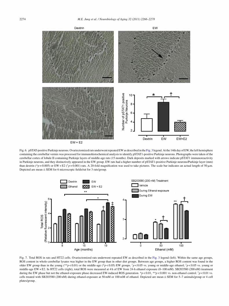
hat the sole purpose for this STAT1 test is to confirm func-ionally active pP38, not to test the mechanistic involvementf STAT1. This test employed only middle-age groups as aepresentative case. Similar to the pP38 results, EW rats hadhigher number of pSTAT1-positive Purkinje neurons thanextrin (p = 0.005) or EW + E2 (p = 0.001) rats [F(2,51) = 29,= 0.001] (Fig. 6), suggesting functional activation of pP38uring EW.
.4. Oxidative mechanisms
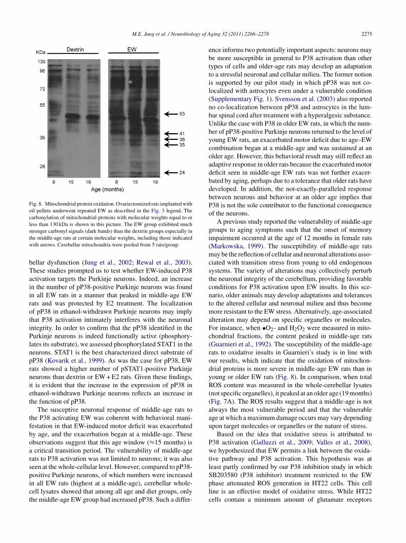
.4.1. Total ROS in rats and HT22 cellsWe determined whether an oxidative pathway is
nvolved in EW-induced P38 activation. In all three ageroups, EW rats had a higher ROS content than dex-rin (p < 0.001), ethanol-exposure (p < 0.01) or EW + E2p < 0.01) rats [F(3,72) = 456, p < 0.001] (Fig. 7A). Betweenges [F(2,72) = 9, p < 0.01], older rats had moderately higherOS contents than young or middle-age rats (p < 0.05),xcept for dextrin rats which showed no difference betweenge groups. Neither age per se nor E2 per se (data not shown)ignificantly altered the ROS content. Although EW predomi-antly provoked the generation of ROS, both the exacerbatingffect of age–EW combination and the protective effects of2 appeared to be moderate. To determine P38 and redox
nteraction during EW, we inhibited EW-induced P38 activa-ion and measured ROS in HT22 cells. As in the in vivo case,
W significantly increased the ROS content [F(2,18) = 996,< 0.001 by ethanol-dose] (Fig. 7B). SB203580 treatmentestricted to the EW phase, but not the ethanol-exposurehase, protected against the EW-induced ROS generation
bEda
assay. The middle-age EW rats had a higher level of pP38 than other dietdiet rats (right). *p < 0.01 vs. all other groups. Depicted are mean ± SEM
F(2,18) = 212, p < 0.001 by the SB203580 treatment win-ow; F(4,18) = 78, p < 0.001 by the interaction between thethanol-dose and the SB203580 window]. These results sug-est that EW-induced P38 activation perturbs a redox balanceuring EW.
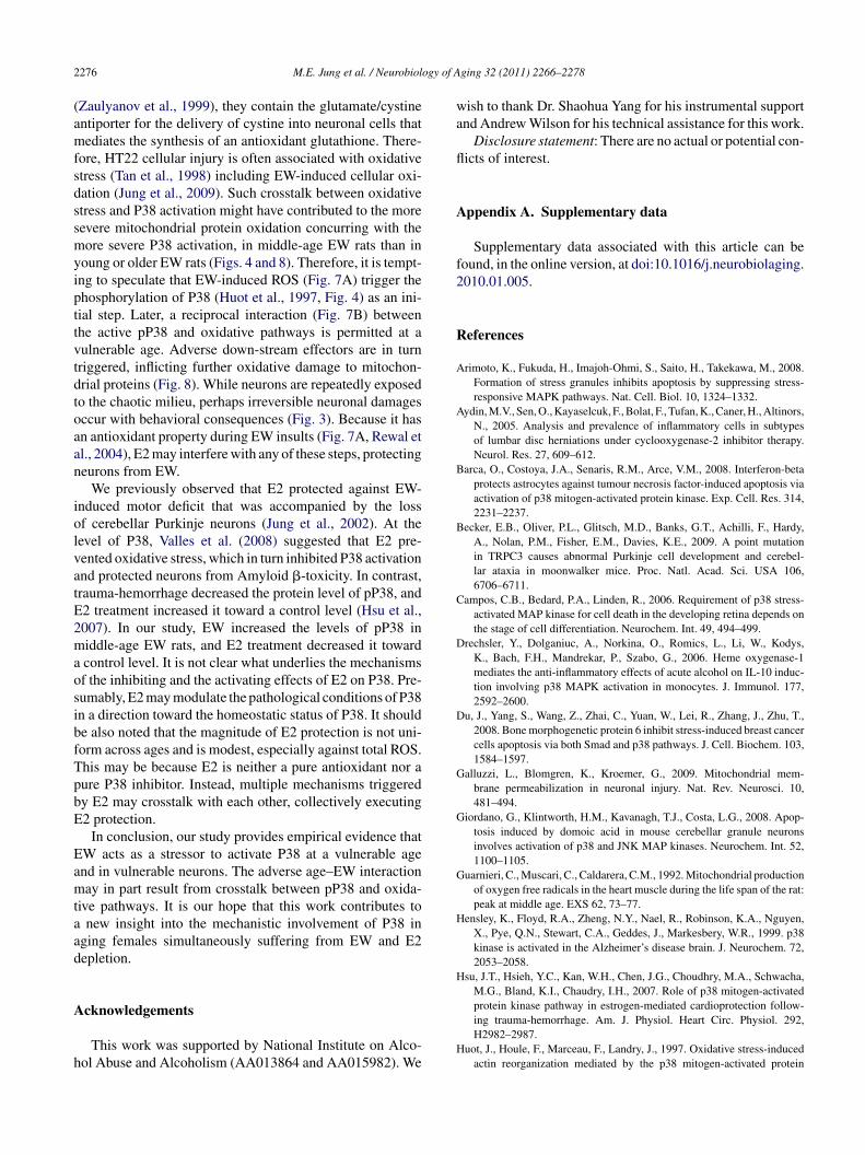
.4.2. Mitochondrial protein oxidationSince mitochondria are believed to be vulnerable to oxida-
ive stress and aging, we tested whether mitochondrial proteinxidation age-dependently coincides with P38 activation dur-ng EW (Fig. 8). EW rats showed protein carbonylation moreeverely than dextrin rats at all three ages. Between ageroups, the middle-age EW rats exhibited stronger carbonylignals than young or older EW rats at certain moleculareights of proteins. In all three age groups, EW did not alter
he levels of proteins stained with Coomassie blue staininger se (Jung et al., 2008). Given these findings, an age–EWombination appears to exacerbate mitochondrial protein oxi-ation at a vulnerable age.
. Discussion
We have demonstrated that EW provokes the hyperac-ivation of protein kinase P38. The activation of P38 was
ore severe in middle-age (15 months) EW rats, who alsoisplayed more severe oxidation of mitochondrial proteinshan other diet (control dextrin, ethanol-exposure, and/orW + E2) and age groups (8 and 19 months). Furthermore,
t was the middle-age at which the adverse age–EW interac-ion began to affect cerebellar neuron-related motor deficit.hese results suggest that the activation of P38 targets a vul-erable age and neurons challenged with EW. Such effects ofW do not appear to be due to a difference in body weightsr ethanol kinetics because there was no correlation between
ody weights, BEC, and the degree of damage associated withW. P38 activation has both beneficial (Peart et al., 2007) andeleterious (Campos et al., 2006) effects and thus, we testedcellular consequence of P38 activation. As expected, exces-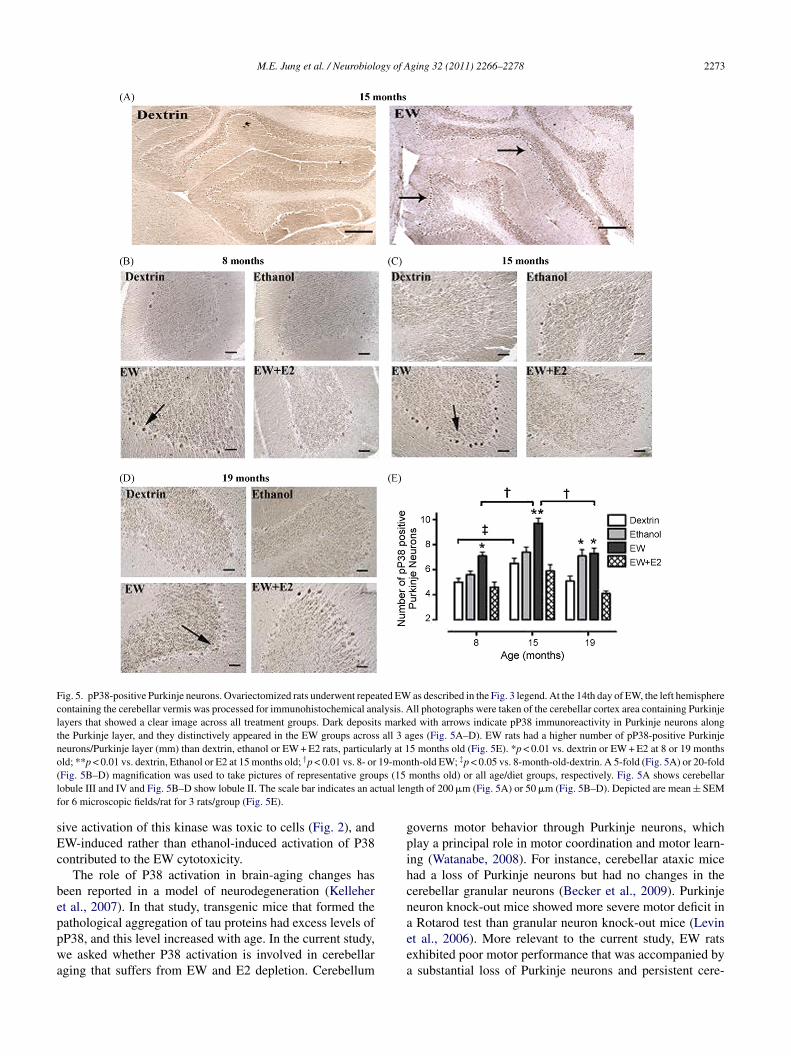
M.E. Jung et al. / Neurobiology of Aging 32 (2011) 2266–2278 2273
Fig. 5. pP38-positive Purkinje neurons. Ovariectomized rats underwent repeated EW as described in the Fig. 3 legend. At the 14th day of EW, the left hemispherecontaining the cerebellar vermis was processed for immunohistochemical analysis. All photographs were taken of the cerebellar cortex area containing Purkinjelayers that showed a clear image across all treatment groups. Dark deposits marked with arrows indicate pP38 immunoreactivity in Purkinje neurons alongthe Purkinje layer, and they distinctively appeared in the EW groups across all 3 ages (Fig. 5A–D). EW rats had a higher number of pP38-positive Purkinjeneurons/Purkinje layer (mm) than dextrin, ethanol or EW + E2 rats, particularly at 15 months old (Fig. 5E). *p < 0.01 vs. dextrin or EW + E2 at 8 or 19 monthsold; **p < 0.01 vs. dextrin, Ethanol or E2 at 15 months old; †p < 0.01 vs. 8- or 19-month-old EW; ‡p < 0.05 vs. 8-month-old-dextrin. A 5-fold (Fig. 5A) or 20-fold( ps (15l tual lenf
sEc
beppwa
gpihcna
Fig. 5B–D) magnification was used to take pictures of representative grouobule III and IV and Fig. 5B–D show lobule II. The scale bar indicates an acor 6 microscopic fields/rat for 3 rats/group (Fig. 5E).
ive activation of this kinase was toxic to cells (Fig. 2), andW-induced rather than ethanol-induced activation of P38ontributed to the EW cytotoxicity.
The role of P38 activation in brain-aging changes haseen reported in a model of neurodegeneration (Kellehert al., 2007). In that study, transgenic mice that formed theathological aggregation of tau proteins had excess levels of
P38, and this level increased with age. In the current study,e asked whether P38 activation is involved in cerebellarging that suffers from EW and E2 depletion. Cerebellum
eea
months old) or all age/diet groups, respectively. Fig. 5A shows cerebellargth of 200 �m (Fig. 5A) or 50 �m (Fig. 5B–D). Depicted are mean ± SEM
overns motor behavior through Purkinje neurons, whichlay a principal role in motor coordination and motor learn-ng (Watanabe, 2008). For instance, cerebellar ataxic micead a loss of Purkinje neurons but had no changes in theerebellar granular neurons (Becker et al., 2009). Purkinjeeuron knock-out mice showed more severe motor deficit inRotarod test than granular neuron knock-out mice (Levin
t al., 2006). More relevant to the current study, EW ratsxhibited poor motor performance that was accompanied bysubstantial loss of Purkinje neurons and persistent cere-
2274 M.E. Jung et al. / Neurobiology of Aging 32 (2011) 2266–2278
Fig. 6. pSTAT-positive Purkinje neurons. Ovariectomized rats underwent repeated EW as described in the Fig. 3 legend. At the 14th day of EW, the left hemispherecontaining the cerebellar vermis was processed for immunohistochemical analysis to identify pSTAT1-positive Purkinje neurons. Photographs were taken of thecerebellar cortex of lobule II containing Purkinje layers of middle-age rats (15 months). Dark deposits marked with arrows indicate pSTAT1 immunoreactivityin Purkinje neurons, and they distinctively appeared in the EW group. EW rats had a higher number of pSTAT1-positive Purkinje neurons/Purkinje layer (mm)than dextrin (*p = 0.005) or EW + E2 (†p = 0.001) rats. A 20-fold magnification was used to take pictures. The scale bar indicates an actual length of 50 �m.Depicted are mean ± SEM for 6 microscopic fields/rat for 3 rats/group.
Fig. 7. Total ROS in rats and HT22 cells. Ovariectomized rats underwent repeated EW as described in the Fig. 3 legend (left). Within the same age groups,ROS content in whole-cerebellar lysates was higher in the EW group than in other diet groups. Between age groups, a higher ROS content was found in theolder EW group than in the young (**p < 0.01) or the middle-age (*p < 0.05) EW groups. †p < 0.05 vs. young or middle-age ethanol; ‡p < 0.05 vs. young ormiddle-age EW + E2. In HT22 cells (right), total ROS were measured at 4 h of EW from 24-h ethanol exposure (0–100 mM). SB203580 (200 nM) treatmentduring the EW phase but not the ethanol-exposure phase decreased EW-induced ROS generation. *p < 0.01, **p < 0.001 vs. non-ethanol control. †p < 0.01 vs.cells treated with SB203580 (200 nM) during ethanol-exposure at 50 mM or 100 mM of ethanol. Depicted are mean ± SEM for 5–7 animals/group or 4 cellplates/group.
M.E. Jung et al. / Neurobiology of A
Fig. 8. Mitochondrial protein oxidation. Ovariectomized rats implanted withoil pellets underwent repeated EW as described in the Fig. 3 legend. Thecarbonylation of mitochondrial proteins with molecular weights equal to orless than 130 kDa is shown in this picture. The EW group exhibited muchstronger carbonyl signals (dark bands) than the dextrin groups especially intw
bTaiirotiPlnprniet
tfboarspict
ebttil(nbUbycoadbdbPo
gi(mcstcntmaFc(rodyR((aau
PwtlSB203580 (P38 inhibitor) treatment restricted to the EW
he middle-age rats at certain molecular weights, including those indicatedith arrows. Cerebellar mitochondria were pooled from 5 rats/group.
ellar dysfunction (Jung et al., 2002; Rewal et al., 2003).hese studies prompted us to test whether EW-induced P38ctivation targets the Purkinje neurons. Indeed, an increasen the number of pP38-positive Purkinje neurons was foundn all EW rats in a manner that peaked in middle-age EWats and was protected by E2 treatment. The localizationf pP38 in ethanol-withdrawn Purkinje neurons may implyhat P38 activation intimately interferes with the neuronalntegrity. In order to confirm that the pP38 identified in theurkinje neurons is indeed functionally active (phosphory-
ates its substrate), we assessed phosphorylated STAT1 in theeurons. STAT1 is the best characterized direct substrate ofP38 (Kovarik et al., 1999). As was the case for pP38, EWats showed a higher number of pSTAT1-positive Purkinjeeurons than dextrin or EW + E2 rats. Given these findings,t is evident that the increase in the expression of pP38 inthanol-withdrawn Purkinje neurons reflects an increase inhe function of pP38.
The susceptive neuronal response of middle-age rats tohe P38 activating EW was coherent with behavioral mani-estation in that EW-induced motor deficit was exacerbatedy age, and the exacerbation began at a middle-age. Thesebservations suggest that this age window (≈15 months) iscritical transition period. The vulnerability of middle-age
ats to P38 activation was not limited to neurons; it was alsoeen at the whole-cellular level. However, compared to pP38-ositive Purkinje neurons, of which numbers were increased
n all EW rats (highest at a middle-age), cerebellar whole-ell lysates showed that among all age and diet groups, onlyhe middle-age EW group had increased pP38. Such a differ-plc
ging 32 (2011) 2266–2278 2275
nce informs two potentially important aspects: neurons maye more susceptible in general to P38 activation than otherypes of cells and older-age rats may develop an adaptationo a stressful neuronal and cellular milieu. The former notions supported by our pilot study in which pP38 was not co-ocalized with astrocytes even under a vulnerable conditionSupplementary Fig. 1). Svensson et al. (2003) also reportedo co-localization between pP38 and astrocytes in the lum-ar spinal cord after treatment with a hyperalgesic substance.nlike the case with P38 in older EW rats, in which the num-er of pP38-positive Purkinje neurons returned to the level ofoung EW rats, an exacerbated motor deficit due to age–EWombination began at a middle-age and was sustained at anlder age. However, this behavioral result may still reflect andaptive response in older rats because the exacerbated motoreficit seen in middle-age EW rats was not further exacer-ated by aging, perhaps due to a tolerance that older rats haveeveloped. In addition, the not-exactly-paralleled responseetween neurons and behavior at an older age implies that38 is not the sole contributor to the functional consequencef the neurons.
A previous study reported the vulnerability of middle-ageroups to aging symptoms such that the onset of memorympairment occurred at the age of 12 months in female ratsMarkowska, 1999). The susceptibility of middle-age ratsay be the reflection of cellular and neuronal alterations asso-
iated with transition stress from young to old endogenousystems. The variety of alterations may collectively perturbhe neuronal integrity of the cerebellum, providing favorableonditions for P38 activation upon EW insults. In this sce-ario, older animals may develop adaptations and toleranceso the altered cellular and neuronal milieu and thus become
ore resistant to the EW stress. Alternatively, age-associatedlteration may depend on specific organelles or molecules.or instance, when •O2- and H2O2 were measured in mito-hondrial fractions, the content peaked in middle-age ratsGuarnieri et al., 1992). The susceptibility of the middle-ageats to oxidative insults in Guarnieri’s study is in line withur results, which indicate that the oxidation of mitochon-rial proteins is more severe in middle-age EW rats than inoung or older EW rats (Fig. 8). In comparison, when totalOS content was measured in the whole-cerebellar lysates
not specific organelles), it peaked at an older age (19 months)Fig. 7A). The ROS results suggest that a middle-age is notlways the most vulnerable period and that the vulnerablege at which a maximum damage occurs may vary dependingpon target molecules or organelles or the nature of stress.
Based on the idea that oxidative stress is attributed to38 activation (Galluzzi et al., 2009; Valles et al., 2008),e hypothesized that EW permits a link between the oxida-
ive pathway and P38 activation. This hypothesis was ateast partly confirmed by our P38 inhibition study in which
hase attenuated ROS generation in HT22 cells. This celline is an effective model of oxidative stress. While HT22ells contain a minimum amount of glutamate receptors
2 ogy of A
(amfsdssmyipttvtdtoaan
iolvatE2maosibfTpbE
Eamtaad
A
h
wa
fl
A
f2
R
A
A
B
B
C
D
D
G
G
G
H
H
276 M.E. Jung et al. / Neurobiol
Zaulyanov et al., 1999), they contain the glutamate/cystinentiporter for the delivery of cystine into neuronal cells thatediates the synthesis of an antioxidant glutathione. There-
ore, HT22 cellular injury is often associated with oxidativetress (Tan et al., 1998) including EW-induced cellular oxi-ation (Jung et al., 2009). Such crosstalk between oxidativetress and P38 activation might have contributed to the moreevere mitochondrial protein oxidation concurring with theore severe P38 activation, in middle-age EW rats than in
oung or older EW rats (Figs. 4 and 8). Therefore, it is tempt-ng to speculate that EW-induced ROS (Fig. 7A) trigger thehosphorylation of P38 (Huot et al., 1997, Fig. 4) as an ini-ial step. Later, a reciprocal interaction (Fig. 7B) betweenhe active pP38 and oxidative pathways is permitted at aulnerable age. Adverse down-stream effectors are in turnriggered, inflicting further oxidative damage to mitochon-rial proteins (Fig. 8). While neurons are repeatedly exposedo the chaotic milieu, perhaps irreversible neuronal damagesccur with behavioral consequences (Fig. 3). Because it hasn antioxidant property during EW insults (Fig. 7A, Rewal etl., 2004), E2 may interfere with any of these steps, protectingeurons from EW.
We previously observed that E2 protected against EW-nduced motor deficit that was accompanied by the lossf cerebellar Purkinje neurons (Jung et al., 2002). At theevel of P38, Valles et al. (2008) suggested that E2 pre-ented oxidative stress, which in turn inhibited P38 activationnd protected neurons from Amyloid �-toxicity. In contrast,rauma-hemorrhage decreased the protein level of pP38, and2 treatment increased it toward a control level (Hsu et al.,007). In our study, EW increased the levels of pP38 iniddle-age EW rats, and E2 treatment decreased it towardcontrol level. It is not clear what underlies the mechanismsf the inhibiting and the activating effects of E2 on P38. Pre-umably, E2 may modulate the pathological conditions of P38n a direction toward the homeostatic status of P38. It shoulde also noted that the magnitude of E2 protection is not uni-orm across ages and is modest, especially against total ROS.his may be because E2 is neither a pure antioxidant nor aure P38 inhibitor. Instead, multiple mechanisms triggeredy E2 may crosstalk with each other, collectively executing2 protection.
In conclusion, our study provides empirical evidence thatW acts as a stressor to activate P38 at a vulnerable agend in vulnerable neurons. The adverse age–EW interactionay in part result from crosstalk between pP38 and oxida-
ive pathways. It is our hope that this work contributes tonew insight into the mechanistic involvement of P38 in
ging females simultaneously suffering from EW and E2epletion.
cknowledgements
This work was supported by National Institute on Alco-ol Abuse and Alcoholism (AA013864 and AA015982). We
H
ging 32 (2011) 2266–2278
ish to thank Dr. Shaohua Yang for his instrumental supportnd Andrew Wilson for his technical assistance for this work.
Disclosure statement: There are no actual or potential con-icts of interest.
ppendix A. Supplementary data
Supplementary data associated with this article can beound, in the online version, at doi:10.1016/j.neurobiolaging.010.01.005.
eferences
rimoto, K., Fukuda, H., Imajoh-Ohmi, S., Saito, H., Takekawa, M., 2008.Formation of stress granules inhibits apoptosis by suppressing stress-responsive MAPK pathways. Nat. Cell. Biol. 10, 1324–1332.
ydin, M.V., Sen, O., Kayaselcuk, F., Bolat, F., Tufan, K., Caner, H., Altinors,N., 2005. Analysis and prevalence of inflammatory cells in subtypesof lumbar disc herniations under cyclooxygenase-2 inhibitor therapy.Neurol. Res. 27, 609–612.
arca, O., Costoya, J.A., Senaris, R.M., Arce, V.M., 2008. Interferon-betaprotects astrocytes against tumour necrosis factor-induced apoptosis viaactivation of p38 mitogen-activated protein kinase. Exp. Cell. Res. 314,2231–2237.
ecker, E.B., Oliver, P.L., Glitsch, M.D., Banks, G.T., Achilli, F., Hardy,A., Nolan, P.M., Fisher, E.M., Davies, K.E., 2009. A point mutationin TRPC3 causes abnormal Purkinje cell development and cerebel-lar ataxia in moonwalker mice. Proc. Natl. Acad. Sci. USA 106,6706–6711.
ampos, C.B., Bedard, P.A., Linden, R., 2006. Requirement of p38 stress-activated MAP kinase for cell death in the developing retina depends onthe stage of cell differentiation. Neurochem. Int. 49, 494–499.
rechsler, Y., Dolganiuc, A., Norkina, O., Romics, L., Li, W., Kodys,K., Bach, F.H., Mandrekar, P., Szabo, G., 2006. Heme oxygenase-1mediates the anti-inflammatory effects of acute alcohol on IL-10 induc-tion involving p38 MAPK activation in monocytes. J. Immunol. 177,2592–2600.
u, J., Yang, S., Wang, Z., Zhai, C., Yuan, W., Lei, R., Zhang, J., Zhu, T.,2008. Bone morphogenetic protein 6 inhibit stress-induced breast cancercells apoptosis via both Smad and p38 pathways. J. Cell. Biochem. 103,1584–1597.
alluzzi, L., Blomgren, K., Kroemer, G., 2009. Mitochondrial mem-brane permeabilization in neuronal injury. Nat. Rev. Neurosci. 10,481–494.
iordano, G., Klintworth, H.M., Kavanagh, T.J., Costa, L.G., 2008. Apop-tosis induced by domoic acid in mouse cerebellar granule neuronsinvolves activation of p38 and JNK MAP kinases. Neurochem. Int. 52,1100–1105.
uarnieri, C., Muscari, C., Caldarera, C.M., 1992. Mitochondrial productionof oxygen free radicals in the heart muscle during the life span of the rat:peak at middle age. EXS 62, 73–77.
ensley, K., Floyd, R.A., Zheng, N.Y., Nael, R., Robinson, K.A., Nguyen,X., Pye, Q.N., Stewart, C.A., Geddes, J., Markesbery, W.R., 1999. p38kinase is activated in the Alzheimer’s disease brain. J. Neurochem. 72,2053–2058.
su, J.T., Hsieh, Y.C., Kan, W.H., Chen, J.G., Choudhry, M.A., Schwacha,M.G., Bland, K.I., Chaudry, I.H., 2007. Role of p38 mitogen-activatedprotein kinase pathway in estrogen-mediated cardioprotection follow-
ing trauma-hemorrhage. Am. J. Physiol. Heart Circ. Physiol. 292,H2982–2987.uot, J., Houle, F., Marceau, F., Landry, J., 1997. Oxidative stress-inducedactin reorganization mediated by the p38 mitogen-activated protein
ogy of A
J
J
J
J
J
J
K
K
K
L
L
L
L
L
M
M
M
N
N
P
R
R
R
S
S
S
S
T
V
v
V
W
W
W
W
X
M.E. Jung et al. / Neurobiol
kinase/heat shock protein 27 pathway in vascular endothelial cells. Circ.Res. 80, 383–392.
akubowski, W., Bartosz, G., 2000. 2,7-dichlorofluorescin oxidation andreactive oxygen species: what does it measure? Cell. Biol. Int. 24,757–760.
iang, Y., Chen, C., Li, Z., Guo, W., Gegner, J.A., Lin, S., Han, J., 1996.Characterization of the structure and function of a new mitogen-activatedprotein kinase (p38beta). J. Biol. Chem. 271, 17920–17926.
iang, Y., Gram, H., Zhao, M., New, L., Gu, J., Feng, L., Di Padova, F., Ule-vitch, R.J., Han, J., 1997. Characterization of the structure and functionof the fourth member of p38 group mitogen-activated protein kinases,p38delta. J. Biol. Chem. 272, 30122–30128.
ung, M.E., Yan, L.J., Forster, M.J., Simpkins, J.W., 2008. Ethanol with-drawal provokes mitochondrial injury in an estrogen preventable manner.J. Bioenerg. Biomembr. 40, 35–44.
ung, M.E., Wilson, A.M., Ju, X., Wen, Y., Metzger, D.B., Simpkins, J.W.,2009. Ethanol withdrawal provokes opening of the mitochondrial mem-brane permeability transition pore in an estrogen-preventable manner. J.Pharmacol. Exp. Ther. 328, 692–698.
ung, M.E., Yang, S.H., Brun-Zinkernagel, A.M., Simpkins, J.W., 2002.Estradiol protects against cerebellar damage and motor deficit in ethanol-withdrawn rats. Alcohol 26, 83–93.
elleher, I., Garwood, C., Hanger, D.P., Anderton, B.H., Noble, W., 2007.Kinase activities increase during the development of tauopathy in htaumice. J. Neurochem. 103, 2256–2267.
ovarik, P., Stoiber, D., Eyers, P.A., Menghini, R., Neininger, A., Gaes-tel, M., Cohen, P., Decker, T., 1999. Stress-induced phosphorylation ofSTAT1 at Ser727 requires p38 mitogen-activated protein kinase whereasIFN-gamma uses a different signaling pathway. Proc. Natl. Acad. Sci.USA 96, 13956–13961.
u, B.M., Lee, Y.K., Jeong, J.Y., Mun, J., Han, J.Y., Roh, G.S., Kim, H.J.,Cho, G.J., Choi, W.S., Yi, G.S., Kang, S.S., 2007. Ethanol-induced oxida-tive stress is mediated by p38 MAPK pathway in mouse hippocampalcells. Neurosci. Lett. 419, 64–67.
echner, C., Zahalka, M.A., Giot, J.F., Moller, N.P., Ullrich, A., 1996. ERK6,a mitogen-activated protein kinase involved in C2C12 myoblast differ-entiation. Proc. Natl. Acad. Sci. USA 93, 4355–4359.
ee, J.C., Laydon, J.T., McDonnell, P.C., Gallagher, T.F., Kumar, S., Green,D., McNulty, D., Blumenthal, M.J., Heys, J.R., Landvatter, S.W., 1994.A protein kinase involved in the regulation of inflammatory cytokinebiosynthesis. Nature 372, 739–746.
ee, S.H., Park, J., Che, Y., Han, P.L., Lee, J.K., 2000. Constitutive activityand differential localization of p38alpha and p38beta MAPKs in adultmouse brain. J. Neurosci. Res. 60, 623–631.
evin, S.I., Khaliq, Z.M., Aman, T.K., Grieco, T.M., Kearney, J.A.,Raman, I.M., Meisler, M.H., 2006. Impaired motor function in micewith cell-specific knockout of sodium channel Scn8a (NaV1.6) incerebellar purkinje neurons and granule cells. J. Neurophysiol. 96,785–793.
i, H.L., Wu, S., Rottenberg, H., 1996. Alcohol inhibits the depolarization-induced stimulation of oxidative phosphorylation in synaptosomes. J.Neurochem. 66, 1691–1697.
arkowska, A.L., 1999. Sex dimorphisms in the rate of age-related decline inspatial memory: relevance to alterations in the estrous cycle. J. Neurosci.19, 8122–8133.
ausset, A.L., de Seze, R., Montpeyroux, F., Privat, A., 2001. Effects ofradiofrequency exposure on the GABAergic system in the rat cerebel-lum: clues from semi-quantitative immunohistochemistry. Brain Res.912, 33–46.
origuchi, T., Toyoshima, F., Gotoh, Y., Iwamatsu, A., Irie, K., Mori, E.,Kuroyanagi, N., Hagiwara, M., Matsumoto, K., Nishida, E., 1996. Purifi-cation and identification of a major activator for p38 from osmoticallyshocked cells. Activation of mitogen-activated protein kinase kinase 6
by osmotic shock, tumor necrosis factor-alpha, and H2O2. J. Biol. Chem.271, 26981–26988.onaka, Y., Miyajima, M., Ogino, I., Nakajima, M., Arai, H., 2008.Analysis of neuronal cell death in the cerebral cortex of H-
Y
ging 32 (2011) 2266–2278 2277
Tx rats with compensated hydrocephalus. J. Neurosurg. Pediatr. 1,68–74.
orkina, O., Dolganiuc, A., Shapiro, T., Kodys, K., Mandrekar, P., Szabo,G., 2007. Acute alcohol activates STAT3, AP-1, and Sp-1 transcriptionfactors via the family of Src kinases to promote IL-10 production inhuman monocytes. J. Leukoc. Biol. 82, 752–762.
eart, J.N., Gross, E.R., Headrick, J.P., Gross, G.J., 2007. Impairedp38 MAPK/HSP27 signaling underlies aging-related failure in opioid-mediated cardioprotection. J. Mol. Cell. Cardiol. 42, 972–980.
amadoss, J., Lunde, E.R., Chen, W.J., West, J.R., Cudd, T.A., 2007.Temporal vulnerability of fetal cerebellar Purkinje cells to chronicbinge alcohol exposure: ovine model. Alcohol Clin. Exp. Res. 31,1738–1745.
ewal, M., Jung, M.E., Simpkins, J.W., 2004. Role of the GABA-A system inestrogen-induced protection against brain lipid peroxidation in ethanol-withdrawn rats. Alcohol Clin. Exp. Res. 28, 1907–1915.
ewal, M., Jung, M.E., Wen, Y., Brun-Zinkernagel, A.M., Simpkins, J.W.,2003. Role of the GABAA system in behavioral, motoric, and cere-bellar protection by estrogen during ethanol withdrawal. Alcohol 31,49–61.
antucci, A.C., Cortes, C., Bettica, A., Cortes, F., 2008. Chronic ethanolconsumption in rats produces residual increases in anxiety 4 monthsafter withdrawal. Behav. Brain Res. 188, 24–31.
molen, A., Marks, M.J., Smolen, T.N., Collins, A.C., 1986. Dose and routeof administration alter the relative elimination of ethanol by long-sleepand short-sleep mice. Alcohol. Clin. Exp. Res. 10, 198–204.
tein, B., Yang, M.X., Young, D.B., Janknecht, R., Hunter, T., Murray, B.W.,Barbosa, M.S., 1997. p38-2, a novel mitogen-activated protein kinasewith distinct properties. J. Biol. Chem. 272, 19509–19517.
vensson, C.I., Marsala, M., Westerlund, A., Calcutt, N.A., Campana, W.M.,Freshwater, J.D., Catalano, R., Feng, Y., Protter, A.A., Scott, B., Yaksh,T.L., 2003. Activation of p38 mitogen-activated protein kinase in spinalmicroglia is a critical link in inflammation-induced spinal pain process-ing. J. Neurochem. 86, 1534–1544.
an, S., Wood, M., Maher, P., 1998. Oxidative stress induces a form of pro-grammed cell death with characteristics of both apoptosis and necrosisin neuronal cells. J. Neurochem. 71, 95–105.
alles, S.L., Borras, C., Gambini, J., Furriol, J., Ortega, A., Sastre, J., Pal-lardo, F.V., Vina, J., 2008. Oestradiol or genistein rescues neurons fromamyloid beta-induced cell death by inhibiting activation of p38. AgingCell. 7, 112–118.
an Eickels, M., Grohe, C., Cleutjens, J.P., Janssen, B.J., Wellens, H.J.,Doevendans, P.A., 2001. 17beta-estradiol attenuates the development ofpressure-overload hypertrophy. Circulation 104, 1419–1423.
ereker, E., O’Donnell, E., Lynch, M.A., 2000. The inhibitory effectof interleukin-1beta on long-term potentiation is coupled withincreased activity of stress-activated protein kinases. J. Neurosci. 20,6811–6819.
ang, M., Tsai, B.M., Reiger, K.M., Brown, J.W., Meldrum, D.R., 2006. 17-beta-Estradiol decreases p38 MAPK-mediated myocardial inflammationand dysfunction following acute ischemia. J. Mol. Cell. Cardiol. 40,205–212.
atanabe, M., 2008. Molecular mechanisms governing competitive synapticwiring in cerebellar Purkinje cells. Tohoku J. Exp. Med. 214, 175–190.
illiamson, D., Gallagher, P., Harber, M., Hollon, C., Trappe, S., 2003.Mitogen-activated protein kinase (MAPK) pathway activation: effectsof age and acute exercise on human skeletal muscle. J. Physiol. 547,977–987.
u, M., Han, M., Li, J., Xu, X., Li, T., Que, L., Ha, T., Li, C., Chen,Q., Li, Y., 2009. 17beta-estradiol inhibits angiotensin II-induced cardiacmyofibroblast differentiation. Eur. J. Pharmacol. 616, 155–159.
iong, W., Kojic, L.Z., Zhang, L., Prasad, S.S., Douglas, R., Wang, Y.,Cynader, M.S., 2006. Anisomycin activates p38 MAP kinase to induce
LTD in mouse primary visual cortex. Brain Res. 1085, 68–76.an, L.J., Levine, R.L., Sohal, R.S., 1997. Oxidative damage duringaging targets mitochondrial aconitase. Proc. Natl. Acad. Sci. USA 94,11168–11172.

2 ogy of A
Y
278 M.E. Jung et al. / Neurobiol
ang, S.H., He, Z., Wu, S.S., He, Y.J., Cutright, J., Millard, W.J., Day, A.L.,Simpkins, J.W., 2001. 17-beta estradiol can reduce secondary ischemicdamage and mortality of subarachnoid hemorrhage. J. Cereb. Blood FlowMetab. 21, 174–181.
Z
ging 32 (2011) 2266–2278
aulyanov, L.L., Green, P.S., Simpkins, J.W., 1999. Glutamate receptorrequirement for neuronal death from anoxia-reoxygenation: an in Vitromodel for assessment of the neuroprotective effects of estrogens. CellMol. Neurobiol. 19, 705–718.
Copyright © 2022 FDOKUMEN