The p38 mitogen-activated protein kinases modulate endothelial cell survival and tissue repair
Upload
independentCategory
view
2download
0
A new p38 MAP kinase-regulated transcriptionalcoactivator that stimulates p53-dependentapoptosis
Ana Cuadrado1,*, Vanesa Lafarga1,Peter CF Cheung2,3, Ignacio Dolado1,Susana Llanos1, Philip Cohen2 andAngel R Nebreda1,*1CNIO (Spanish National Cancer Center), Madrid, Spain and 2MRCProtein Phosphorylation Unit, University of Dundee, Dundee, UK
The p38 mitogen-activated protein kinase (MAPK)
signaling pathway plays an important role in stress-induced
cell-fate decisions by orchestrating responses that go from
cell-cycle arrest to apoptosis. We have identified a new p38
MAPK-regulated protein that we named p18Hamlet, which
becomes stabilized and accumulates in response to certain
genotoxic stresses such as UV or cisplatin treatment.
Overexpression of p18Hamlet is sufficient to induce apopto-
sis, whereas its downregulation reduces the apoptotic
response to these DNA damage-inducing agents. We show
that p18Hamlet interacts with p53 and stimulates the trans-
cription of several proapoptotic p53 target genes such as
PUMA and NOXA. This correlates with enhanced p18Hamlet-
induced recruitment of p53 to the promoters. In proliferat-
ing cells, low steady-state levels of p18Hamlet are probably
maintained by a p53-dependent negative feedback loop.
Therefore, p18Hamlet is a new cell-fate regulator that links
the p38 MAPK and p53 pathways and contributes to the
establishment of p53-regulated stress responses.
The EMBO Journal (2007) 26, 2115–2126. doi:10.1038/
sj.emboj.7601657; Published online 22 March 2007
Subject Categories: differentiation & death
Keywords: apoptosis; p38 MAP kinase; p53; stress response;
transcriptional coactivator
Introduction
Cells are continuously exposed to a variety of environmental
stresses and, as a consequence, sometimes have to take the
important decision whether to live or not to live. Several
signaling pathways are involved in the stress-induced cell-
fate decisions. One of these pathways leads to activation of
the p38 mitogen-activated protein kinase (MAPK) cascade
that coordinates cell responses to many types of stresses
including UV, chemotherapeutic agents and oncogenes.
Four p38 MAPKs have been identified in higher eukar-
yotes. The most widely expressed and studied family member
is p38a, which can be activated by the MAPK kinases MKK6,
MKK3 and MKK4. Many proteins can be phosphorylated by
p38 MAPKs, including protein kinases and a growing list of
transcription factors. The set of substrates targeted by p38
MAPKs in each particular case is thought to be an important
determinant for the specificity of the cellular responses,
which can be as diverse as cytokine production, cell differ-
entiation, cell-cycle arrest or apoptosis (Nebreda and Porras,
2000; Ono and Han, 2000; Bulavin and Fornace, 2004).
Activation of p38a in response to several anticancer agents
is necessary and, in some cases, sufficient, to induce apop-
tosis in a variety of cancer cell lines (Sanchez-Prieto et al,
2000; Deacon et al, 2003; Poizat et al, 2005; Coltella et al,
2006). These results, together with the ability of p38a to
positively regulate several tumor suppressor pathways and to
attenuate oncogenic signals, have led to the proposal that this
protein may function as a tumor suppressor (Bulavin and
Fornace, 2004).
There is good evidence supporting a role for p38a in the
regulation of the tumor suppressor protein p53, mainly
through the phosphorylation of p53 induced by several
types of stress (Bulavin et al, 1999; She et al, 2000). p53 is
one of the most commonly mutated genes in human cancers
and its loss of function is believed to result in increased
genomic instability, with the subsequent acquisition of addi-
tional oncogenic mutations (Vousden and Prives, 2005). The
protein level and transcriptional activity of p53 are upregu-
lated in response to many stresses, including DNA damage.
Upon activation, p53 coordinates a complex cellular re-
sponse, which can lead to reversible cell-cycle arrest, an
irreversible senescence-like state or apoptosis (Vousden and
Lu, 2002). The role of p53 in the maintenance of genome
integrity involves multiple control mechanisms, including
various post-translational modifications, such as phospho-
rylation, acetylation, ubiquitination and sumoylation (Bode
and Dong, 2004). These modifications may increase the half-
life of the p53 protein, which results in a rapid rise in
intracellular p53 levels and also enhances its ability to bind
to specific promoter DNA sequences.
An additional and attractive mechanism of p53 regulation
has emerged in the last years as a collection of transcriptional
coactivators that influence p53 activity, usually without
modifying the p53 protein (Coutts and La Thangue, 2005).
These coactivators confer specificity to the p53 response as
they are upregulated in response to certain types of stresses
and, in some cases, enhance the ability of p53 to activate the
transcription of genes involved in a particular response. This
is the case of the ASPP (apoptotis-stimulating proteins of p53)
family of proteins, which can interact with p53 and specifi-
cally stimulate the expression of the proapoptotic genes BAX
and PIG3 (Samuels-Lev et al, 2001). Another example is
hnRNP K (heterogeneous nuclear ribonucleoprotein K), re-
cently identified as a transcriptional cofactor for p53 that hasReceived: 15 August 2006; accepted: 26 February 2007; publishedonline: 22 March 2007
*Corresponding authors. AR Nebreda or A Cuadrado, MolecularOncology Programme, CNIO (Spanish National Cancer Center),Melchor Fernandez Almagro 3, Madrid -28029 Spain.Tel.: þ 34 91 2246900; Fax: þ 34 91 7328033;E-mail: [email protected] or [email protected] address: Nanyang Technological University, Singapore
The EMBO Journal (2007) 26, 2115–2126 | & 2007 European Molecular Biology Organization |All Rights Reserved 0261-4189/07
www.embojournal.org
&2007 European Molecular Biology Organization The EMBO Journal VOL 26 | NO 8 | 2007
EMBO
THE
EMBOJOURNAL
THE
EMBOJOURNAL
2115

a crucial role in DNA damage-induced cell-cycle arrest
(Moumen et al, 2005).
We report here a new p38a-regulated protein, which we
named p18Hamlet, based on its ability to control life-or-death
cell-fate decisions. p18Hamlet accumulates in response to
genotoxic stresses and induces the transcriptional activation
of several p53 target genes such as NOXA and PUMA.
Results
Identification of a new p38 MAPK substrate
We performed yeast two-hybrid screenings to identify new
proteins that could mediate the biological responses of p38a(Cheung et al, 2003). In these experiments, we found that
human p38a specifically interacted with a poorly character-
ized protein of 18 kDa (NP_006340). We named this protein
p18Hamlet, based on its function, which is related to the
regulation of cell-fate decisions, as described below. Two
interesting features of p18Hamlet were a C-terminal zinc-
finger-HIT1-type domain, which has been described as a
protein–protein interaction domain in the Trip3 coactivator
of hepatocyte nuclear factor-4a (Iwahashi et al, 2002), and a
bipartite nuclear localization signal (Figure 1A). p18Hamlet
was conserved along the evolutionary scale from yeast to
human (Figure 1B).
We first investigated whether p18Hamlet was able to bind to
different members of the p38 MAPK family. For this analysis,
we performed in vitro pull-down assays with 35S-labelled p38
MAPKs and recombinant GST-fused p18Hamlet protein. As
expected from the yeast two-hybrid results, p18Hamlet was
able to interact with p38a and also with p38b but not with
p38g and p38d, or with the p38 activator MKK6 (Figure 1C
and Supplementary Figure 1). The interaction between
p18Hamlet and p38a was also observed in transfected HEK-
293T cells, and was independent of the activation loop
phosphorylation of p38a (Figure 1D). We also detected inter-
action of endogenous p18Hamlet with Myc-p38a transfected in
HEK-293Tcells (Figure 1E), but we failed to detect interaction
of both endogenous proteins. Of note, we could not detect
interaction between the transfected proteins using p18Hamlet
or p38a antibodies for immunoprecipitation (IP) (not shown),
suggesting that the antibodies might sterically interfere with
or somehow affect complex formation. We also confirmed that
both p38a and p38b phosphorylated p18Hamlet in vitro with
similar efficiencies (Figure 1F), whereas p38g or p38d did not
phosphorylate p18Hamlet (Supplementary Figure 1).
The sequence of p18Hamlet contains only one consensus
MAPK phosphorylation site (Ser/Thr–Pro) at Ser124.
However, mutation of this Ser to Ala did not affect in vitro
phosphorylation of p18Hamlet by p38a (not shown). Using
generic phospho antibodies, we found that p18Hamlet was
phosphorylated by p38a in vitro on Thr residues
(Figure 2A). Based on this result, we individually mutated
the nine Thr residues present in human p18Hamlet and found
four (Thr6, Thr64, Thr71 and Thr103) that could be poten-
tially phosphorylated by p38a. However, the quadruple mu-
tant T6A, T64A, T71A and T103A (4�T/A) was still partially
phosphorylated by p38a (Supplementary Figure 2), suggest-
ing that additional residues might be involved.
We found that p38 MAPK activation by UV treatment
also correlated with Thr phosphorylation of p18Hamlet, and
this phosphorylation was significantly reduced when the cells
were pretreated with the p38 MAPK inhibitor SB203580
(Figure 2B). This suggests that p38 is involved in UV-induced
p18Hamlet phosphorylation. The high conservation of Thr103
as a phosphorylation site in p18Hamlet proteins from different
species (Figure 1A), together with the in vitro phosphoryla-
tion experiments, suggested that this residue could be an
important target for p38 MAPK. Indeed, mutation of Thr103
impaired the p38 MAPK-mediated phosphorylation of
p18Hamlet in cells, as determined by the reduced signal
observed with the phospho-Thr antibody, when both wild-
type (wt) and the T103A p18Hamlet proteins were coexpressed
with the p38 activator MKK6DD (Figure 2C). We developed
an antibody that specifically recognized phospho-Thr103
(Figure 2D) and confirmed that p18Hamlet was phosphorylated
on this residue in UV-treated cells (Figure 2E). It is important
to note that both the generic phospho-Thr and the specific
phospho-Thr103 antibodies recognize p18Hamlet phospho-
rylated on Thr103. However, in vitro kinase assays indicate
that this was not the only p38a-dependent phosphorylationresidue present in p18Hamlet (Supplementary Figure 2).
p18Hamlet protein levels are regulated by p38 MAPK
The mRNA levels of p18Hamlet varied significantly among
different human tissues and cell lines in contrast with the
p18Hamlet protein, which was usually difficult to detect
(Supplementary Figure 3 and data not shown). We therefore
investigated the possibility that p18Hamlet protein stability
could be regulated. As shown in Figure 3A, incubation with
the proteasome inhibitor MG132 resulted in the accumulation
of endogenous p18Hamlet protein in both mouse embryonic
fibroblasts (MEFs) and human osteosarcoma U2OS cells. To
demonstrate that p18Hamlet was a target of the ubiquitin–
proteasome system, we transfected U2OS cells with Myc-
p18Hamlet alone or in combination with HA-ubiquitin and
then analyzed the Myc-p18Hamlet immunoprecipitates by im-
munoblotting with anti-p18Hamlet antibody. In this experi-
ment, we detected a smear of slowly migrating p18Hamlet
forms that were not observed in extracts from cells trans-
fected with either HA-ubiquitin or p18Hamlet alone, suggesting
that they probably correspond to p18Hamlet–ubiquitin conju-
gates. This was further supported by the recognition of the
slowly migrating forms of p18Hamlet with an anti-HA antibody
(Figure 3B). MG132 concentrations that inhibit the protea-
some have been also reported to activate p38 MAPK (Wu
et al, 2004). We confirmed that MG132-induced p18Hamlet
accumulation correlated with the phosphorylation of p38
MAPK in MEFs, but this effect was abolished when the
MG132 treatment was performed in the presence of the p38
MAPK inhibitor SB203580 (Supplementary Figure 4). These
results strongly suggest that p18Hamlet accumulation requires
the activation of p38 MAPK.
Next, we investigated the effect of genotoxic stresses that
activate the p38 MAPK pathway, such as UV (Kyriakis and
Avruch, 1996), on the endogenous p18Hamlet protein levels. In
agreement with the above results, UV-induced p38 MAPK
phosphorylation correlated with a small (about two-fold) but
reproducible increase in endogenous p18Hamlet protein levels
in different cell lines (Figure 3C). The amount of p18Hamlet
protein typically peaked between 1 and 6 h after the treatment
and later decreased to levels lower than those in untreated
cells. Importantly, the UV-induced accumulation of p18Hamlet
was prevented by pretreatment with SB203580 (Figure 3D).
A new p38-regulated coactivator of p53A Cuadrado et al
The EMBO Journal VOL 26 | NO 8 | 2007 &2007 European Molecular Biology Organization2116

We then investigated whether p18Hamlet accumulation was
specific for UV-induced p38 MAPK activation or if it was a
more general response to stress. Treatment of cells with the
chemical DNA damage-inducing agent cisplatin induced p38
MAPK activation, which was accompanied by a significant
increase in endogenous p18Hamlet levels (Figure 3E).
To further strengthen the connection between p38
MAPK activation and accumulation of p18Hamlet, we trans-
fected U2OS cells with a low amount of p18Hamlet (to limit
apoptosis induction, see below) together with increasing
amounts of p38a and a constant amount of its activator
MKK6DD (Figure 3F). In this experiment, we observed a
direct correlation between p38a activation levels and the
amount of p18Hamlet protein expressed. We also detected
maximal p18Hamlet phosphorylation on Thr103 with the
lowest concentration of p38a used, suggesting that this
residue might be efficiently phosphorylated in situations of
poor p38 MAPK activation. However, the T103A mutant
protein was still able to accumulate upon p38a activation
(Supplementary Figure 5), suggesting that Thr103 phosphor-
ylation may contribute to, but is not essential for, p18Hamlet
accumulation.
Finally, we confirmed that p18Hamlet was an unstable
protein with a half-life of less than 3 h in cycloheximide-
treated U2OS cells (Figure 4A). Interestingly, specific activa-
tion of p38a was sufficient to significantly increase the half-
life of p18Hamlet (Figure 4B and C). In contrast, we could
detect no changes in p18Hamlet mRNA levels when cells were
treated with cisplatin or UV (Figure 4D). These results
indicated that stress-induced accumulation of p18Hamlet was
Figure 1 p18Hamlet is an evolutionarily conserved substrate of the p38a and p38b MAPKs. (A) Amino-acid sequence alignment of p18Hamlet
proteins. Identical and similar residues are indicated by asterisks and two dots, respectively. The domains corresponding to the nuclearlocalization signal (NLS) and the zinc-finger HIT type I domain (Znf-HIT) are boxed. The arrowhead indicates a Thr residue that was selected togenerate specific phospho antibodies. Sequence alignment was performed using the ClustalW program. (B) Phylogenetic tree of p18Hamlet
proteins from Homo sapiens (NM_006349), Mus musculus (BC026751), Xenopus (NM_001017056), Drosophila (NP_608895), Danio rerio(AAH67648), Caenorhabditis elegans (NP_504477), Schizosaccharomyces pombe (CAB60106) and Saccharomyces cerevisiae (NP_013671).(C) GST pull-down assays were performed with the indicated GST-fused proteins and 35S-labelled p18Hamlet. (D) HEK-293 cells were transfectedwith 5 mg of Myc-p38a, MKK6DD and p18Hamlet, as indicated. Forty-eight hours after transfection, total cell lysates were prepared and analyzedby Western blotting, together with Myc IP. (E) HEK-293 cells were transfected with Myc-p38a, and 48h after transfection, p38a wasimmunoprecipitated with Myc and HA (as a negative control) antibodies. (F) Kinase assays were performed using activated p38a and p38bMAPKs (200 ng) and GST, GST-p18Hamlet and GST-ATF-2 (1mg) in the presence of 32P-g-ATP. Coomassie staining shows the proteins used.
A new p38-regulated coactivator of p53A Cuadrado et al
&2007 European Molecular Biology Organization The EMBO Journal VOL 26 | NO 8 | 2007 2117

mainly regulated at the level of protein stability. To determine
the importance of p38 MAPK-mediated phosphorylation in
p18Hamlet protein stability, we performed cycloheximide-
chase experiments in U2OS cells transfected with p18Hamlet
wt, T103A or the quadruple mutant 4�T/A and then UV
irradiated the cells (Figure 4E). Whereas the T103A and wt
proteins behaved similarly and were both significantly accu-
mulated in response to UV, 4�T/A mutant expression levels
were not affected by this treatment. Moreover, the 4�T/A
mutant was expressed at levels lower than wt p18Hamlet,
supporting the idea that phosphorylation of these sites
could be important for the regulation of p18Hamlet protein
stability.
Accumulation of p18Hamlet induces apoptosis
Once we established that p18Hamlet levels increased in re-
sponse to DNA damage, we investigated the biological sig-
nificance of this accumulation. U2OS cells were transfected
Figure 2 p38a phosphorylates several Thr residues in p18Hamlet. (A) GST-p18Hamlet or GST proteins (500 ng) were phosphorylated with p38ain vitro and then analyzed by Western blotting with phospho-Thr antibodies. (B) HEK-293 cells were transfected with Myc-p18Hamlet and 48h laterwere treated with UValone or in the presence of 10mM SB203580. Three hours after irradiation, total cell lysates were prepared and analyzed byWestern blotting, together with Myc immunoprecipitates. (C) HEK-293 cells were transfected with Myc-p18Hamlet wt and T103A either alone ortogether with MKK6DD, as indicated, and p18Hamlet phosphorylation was analyzed by Myc IP followed by Western blotting with phospho-Thrantibody. Total cell lysates were also analyzed by Western. (D) GST-p18Hamlet wt, T103A and T127A proteins were incubated with p38a andMKK6DD or with MKK6DD alone and then analyzed by Western blotting with both phospho-Thr103-p18Hamlet and generic phospho-Thrantibodies. (E) HeLa cells overexpressing p18Hamlet were UV irradiated, lysed at the indicated times after irradiation and analyzed by Westernblotting with the phospho-Thr103-p18Hamlet antibody.
Figure 3 Accumulation of p18Hamlet protein in response to DNA damage-inducing agents. (A) MEFs and U2OS cells were treated with theproteasome inhibitor MG132 (25mM) for 2 h and then lysed. Expression of endogenous p18Hamlet was analyzed by Western blotting. (B) U2OScells were transfected with HA-ubiquitin and Myc-p18Hamlet, as indicated, and 16 h after transfection, were treated with MG132 (25mM) for 5 h.Myc immunoprecipitates were analyzed by Western blotting using p18Hamlet and HA antibodies. (C) SK-Mel-103 and U2OS cells were treatedwith UV and cell lysates were analyzed by Western blotting using the indicated antibodies. (D) SK-Mel-103 cell lysates were prepared 3 h afterUV treatment, either in the presence or absence of SB203580 (SB, 10mM), and analyzed by Western blotting. (E) SK-Mel-103 cells were treatedwith cisplatin for the indicated times and p18Hamlet accumulation was analyzed by Western blotting. (F) U2OS cells were cotransfected withMKK6DD (600 ng), p18Hamlet (1mg) and increasing amounts of p38a, as indicated. Twenty-four hours after transfection, lysates were preparedfrom both attached and floating cells (that express higher levels of p18Hamlet) and analyzed by Western blotting.
A new p38-regulated coactivator of p53A Cuadrado et al
The EMBO Journal VOL 26 | NO 8 | 2007 &2007 European Molecular Biology Organization2118

with either GFP-p18Hamlet or GFP alone and then sorted to
analyze the cell-cycle profile in the fluorescent cell popula-
tion. We found that around 6% of the GFP-expressing cells
were apoptotic (sub-G0/G1 population), whereas this
amount increased to 18% in the GFP-p18Hamlet-positive
cells, indicating that p18Hamlet overexpression was sufficient
to induce apoptosis. Interestingly, GFP-p18Hamlet overexpres-
sion did not affect apoptosis levels in SAOS cells, a p53-
deficient human osteosarcoma cell line (Figure 5A).
Given the key role of p53 in the apoptotic response induced
by DNA damage, we investigated if p18Hamlet could regulate
p53. In U2OS cells, GFP-p18Hamlet overexpression did not
affect the total levels of p53 (Figure 5B), but resulted in
higher levels of the proapoptotic p53 target gene NOXA,
whereas it had no effect on other p53-dependent proapoptotic
genes such as PUMA or Bax. In contrast, NOXA levels were
not affected by GFP-p18Hamlet overexpression in SAOS cells.
To study the induction of apoptosis by p18Hamlet in more
detail, we generated a tetracycline-inducible system in which
p18Hamlet protein expression peaked at about 16 h after addi-
tion of tetracycline to U2OS cells (Supplementary Figure 6).
Consistent with the above results, tetracycline-induced
p18Hamlet expression was accompanied by an increase in
both NOXA mRNA and protein levels (Figure 5C), whereas
we could detect no significant changes in the expression of
other p53-dependent targets, such as PUMA, Bax, p21 or
Hdm2 (Figure 5C and data not shown). We also confirmed
that p18Hamlet induction was sufficient on its own to signifi-
cantly increase the early and late apoptotic populations. In
addition, p18Hamlet cooperates with cisplatin treatment in
apoptosis induction (Figure 5D). To confirm the proapoptotic
function of p18Hamlet in a more physiological system, we
overexpressed p18Hamlet in primary MEFs. As shown in
Figure 5E, p18Hamlet overexpression was sufficient to increase
apoptosis levels in non-stressed cells, and it also strongly
promoted apoptosis induced by cisplatin or UV. Taken to-
gether, these results support a role for p18Hamlet in p53-
mediated apoptosis induction.
To confirm the role of endogenous p18Hamlet as an apop-
tosis mediator, we designed siRNA oligonucleotides that
efficiently downregulated p18Hamlet (Figure 6A and
Supplementary Figures 7 and 8). Previous work has shown
that p53 contributes to apoptosis induced by UV or cisplatin
in both U2OS and MCF7 cells (Bergamaschi et al, 2003).
Using a highly sensitive, quantitative method that detects
apoptotic nucleosomes (see Materials and methods), we
found that p18Hamlet downregulation did not affect basal
apoptosis levels, but significantly impaired apoptosis induced
by either UV or cisplatin in U2OS cells (Figure 6A). The same
effect was observed in MCF7 cells treated with cisplatin
(Supplementary Figure 7). We then analyzed the effect of
p18Hamlet downregulation on the expression of p53-depen-
dent target genes upon cisplatin and UV treatment.
Interestingly, only certain p53 target genes were induced in
response to these two types of stress. In particular, NOXA and
Hdm2 responded to cisplatin, whereas p21, Bax and PUMA
remained unaffected. In the case of UV, only NOXA, and also
slightly PUMA, protein levels were upregulated, whereas p21,
Hdm2 and Bax levels did not increase (Figure 6B). This result
suggests that specific programs of gene expression account
Figure 4 Stabilization of p18Hamlet protein in response to p38a activation. (A) U2OS cells were cotransfected with p18Hamlet (1mg) and GFP(500ng) and incubated with cycloheximide (CHX, 30 mg/ml) for the indicated times. Total cell lysates were analyzed byWestern blotting. (B, C)U2OS cells were transfected with p18Hamlet, either alone or together with p38a and MKK6DD, and 24 h later were incubated with CHX for up to4 h. Expression of p18Hamlet protein was determined by Western blotting. The blots corresponding to cells untreated or treated with CHX for 4 h,in the presence or absence of active p38a, are shown in (C). (D) Total RNAs were obtained from UV- or cisplatin-treated SK-Mel-103 cells andwere analyzed by Northern blotting with a p18Hamlet probe. GAPDH was used to confirm equal RNA loading. (E) U2OS cells were transfectedwith 6mg of p18Hamlet wt, T103A or 4�T/A, as indicated. Twenty-four hours after transfection, cells were treated with CHX alone or incombination with MG132 for 4 h. Where indicated, cells were also UV irradiated 1 h before collection. A GFP expression vector (200ng) wascotransfected to ensure equal efficiency of transfection.
A new p38-regulated coactivator of p53A Cuadrado et al
&2007 European Molecular Biology Organization The EMBO Journal VOL 26 | NO 8 | 2007 2119

for the p53-dependent apoptosis in response to each particu-
lar type of stress. Downregulation of p18Hamlet prevented
NOXA and, in the case of UV irradiation, also PUMA protein
accumulation, but had no effect on Hdm2 protein induction.
Taken together, the results support an important role for
p18Hamlet in stress-induced apoptosis.
p18Hamlet activates p53-dependent gene promoters
The ability of p18Hamlet to upregulate p53-dependent proa-
poptotic genes prompted us to investigate whether both
proteins could interact. We found that tetracycline-induced
p18Hamlet co-immunoprecipitated with endogenous p53 from
U2OS cells (Figure 7A, upper panel), and we also managed to
co-immunoprecipitate the two endogenous proteins
(Figure 7A, lower panel). The interaction was also observed
in pull-down assays using recombinant GST-p53 and 35S-
labelled p18Hamlet and it involves the zinc-finger domain of
p18Hamlet and the C-terminal region of p53 (see below,
Figure 9D and Supplementary Figure 9). In contrast, over-
expression of p18Hamlet did not affect phosphorylation of p53
on Ser15 and Ser46, two common p53 phosphorylation
events in response to stress (Figure 7B).
We then analyzed whether p18Hamlet could regulate the
transactivation function of p53. For these experiments, we
expressed the p53-regulated promoters of Hdm2, Bax, NOXA
and PUMA in U2OS cells, which contain wt p53 protein. As
shown in Figure 7B, p18Hamlet stimulated the transcription of
the three p53-regulated proapoptotic genes (only a minor
effect was observed in the case of Bax), but had no effect on
the Hdm2 promoter. We confirmed that both Bax and Hdm2
Figure 5 Induction of apoptosis by p18Hamlet overexpression. (A) U2OS and SAOS cells were transfected with GFP or GFP-p18Hamlet andanalyzed by flow cytometry. The percentage of cells with a sub-G0/G1 DNA content in a representative experiment is shown. (B) U2OS andSAOS cells were transfected as indicated in (A) and the expression of the indicated proteins was analyzed by Western blotting. (C) U2OS cellsexpressing inducible p18Hamlet were treated with tetracycline for 24 h before RNA and protein extraction. Samples were analyzed byquantitative RT–PCRs (left) and Western blotting (right). (D) U2OS cells expressing tetracycline-inducible p18Hamlet were incubated with tetfor 24 h and then treated with cisplatin for another 24 h before analyzing apoptosis by annexin V staining. Numbers on top of the bars indicatetotal percentage of early and late apoptotic events as well as dead cells. The lower panel shows the protein levels of overexpressed Myc-p18Hamlet, endogenous (End.) p18Hamlet (marked with an asterisk) and phospho-Ser15-p53. (E) Primary MEFs were infected with pBABE puroor p18Hamlet-expressing pBABE puro retroviruses. After puromycin selection, cells were treated for 24 h with cisplatin or UV as indicated, andthe percentage of sub-G0/G1 cell population, as a measure of apoptosis levels, was analyzed by FACS.
A new p38-regulated coactivator of p53A Cuadrado et al
The EMBO Journal VOL 26 | NO 8 | 2007 &2007 European Molecular Biology Organization2120

promoters were indeed able to respond to p53
(Supplementary Figure 10). Importantly, stimulation of p53
transcriptional activity by p18Hamlet was dependent upon the
integrity of its C-terminal zinc-finger HIT domain, as a
p18Hamlet derivative lacking the last 37 amino acids
(p18Hamlet(1–117)) had no significant effect on any of the p53-
regulated promoters (Figure 7C). The C-terminally truncated
p18Hamlet(1–117) protein was still able to bind to p38a and
localized to the nucleus, as the full-length p18Hamlet, but
failed to interact with p53 (Figure 9D and Supplementary
Figure 9). It therefore appears that p38a and p53 interact with
different parts of the p18Hamlet protein and that the C-terminal
domain of p18Hamlet is required for p53 transactivation.
To confirm that the effect of p18Hamlet was indeed p53
dependent, we used a reporter plasmid with the PUMA
minimal promoter (PUMA 4�BS2) consisting of four tandem
repeats of the p53 binding site or a mutant version of this
reporter, which does not bind to p53 (Yu et al, 2001). Full-
length p18Hamlet increased PUMA 4�BS2 transcription by
about four-fold, whereas the mutant p18Hamlet(1–117) had no
effect. In contrast, neither full-length nor truncated p18Hamlet
proteins were able to stimulate transcription of the mutated
PUMA 4�BS2 promoter (Figure 7D). Based on these results,
we conclude that p18Hamlet can activate, through its C-term-
inal domain, the transcription of several p53-dependent
genes.
Consistent with the results shown in Figure 6B, cisplatin
was able to differentially transactivate several p53-dependent
promoters, being more effective in the cases of NOXA
and PIG-3 (Figure 7E) when compared with other genes
such as Bax or PUMA. The downregulation of p18Hamlet
inhibited by about 50% the ability of cisplatin to transactivate
the NOXA and PUMA promoters, whereas it had a more
moderate effect on p21 and Bax promoters (Figure 7F).
In addition, when p18Hamlet overexpression was combined
with cisplatin treatment, we observed an additive effect on
the NOXA promoter, without affecting the Hdm2 promoter,
leading to an imbalance that was clearly favorable to the
transcription of the proapoptotic gene NOXA (Figure 7G). Of
note, overexpression of p18Hamlet in p53-deficient SAOS
cells had no effect on the PUMA 4�BS2-reporter construct,
unless the cells were cotransfected with p53 (Supplementary
Figure 11).
To determine the importance of p38 MAPK-mediated
phosphorylation in p18Hamlet function, we analyzed the abil-
ity of different p18Hamlet mutants to transactivate the PUMA
4�BS2 promoter and to induce apoptosis. The three
p18Hamlet mutants were able to bind to p38a to a similar
extent, but phosphorylation was significantly reduced in
p18Hamlet-T103A and specially in the quadruple mutant
p18Hamlet-4�T/A (Supplementary Figure 2). However, only
the p18Hamlet-T103A mutant recapitulated the activity of the
wt protein regarding PUMA promoter transactivation and
apoptosis induction, consistent with the idea that phosphor-
ylation of this residue was not essential for p18Hamlet function
and accumulation (Figure 7H). In contrast, truncated
p18Hamlet(1–117) was not able to induce cell death, supporting
the importance of p53 interaction for the induction of apop-
tosis by p18Hamlet. Finally, p18Hamlet-4�T/A showed a signifi-
cant decrease in both PUMA transactivation and apoptosis
induction, suggesting that p18Hamlet phosphorylation is
essential for its activity (Figure 7H).
Our results indicated that the ability of p18Hamlet to stimu-
late p53-induced transcription was not related to p53 phos-
phorylation or stabilization. We therefore performed
chromatin IP (ChIP) experiments to investigate the recruit-
ment of p53 to the promoter of its target genes. We found that
p18Hamlet overexpression was sufficient to enhance p53 bind-
ing to both the PUMA and NOXA promoters, whereas it had
no effect on the ability of p53 to bind to the Hdm2 promoter
(Figure 8A). In addition, p18Hamlet itself was also bound to
the NOXA promoter, even in unstimulated U2OS cells, and
the binding was increased in response to cisplatin treatment
(Figure 8B). Interestingly, p18Hamlet knockdown had a pro-
found effect on p53 loading onto the NOXA promoter
(Figure 8C), supporting a key role for p18Hamlet in the
recruitment of p53 to certain target promoters.
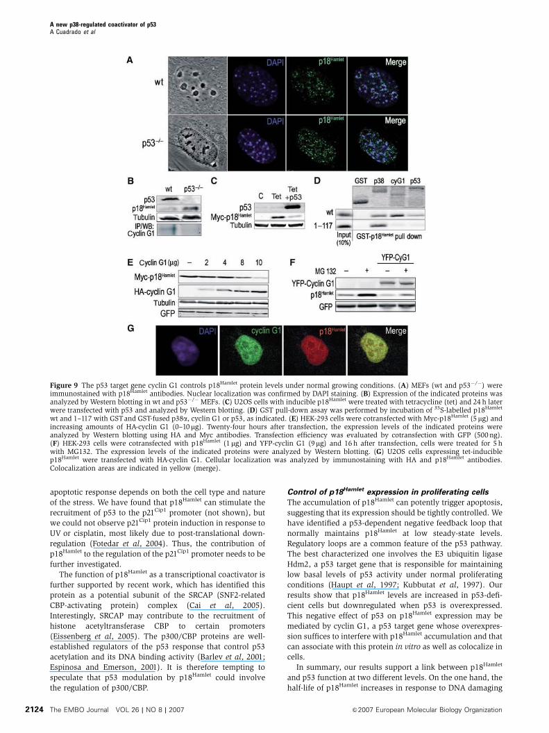
p18Hamlet levels can be regulated by cyclin G1 in
normally proliferating cells
The ability of p18Hamlet to induce apoptosis suggests that the
levels of this protein should be strictly regulated under
Figure 6 p18Hamlet is required for apoptosis induction in response to DNA damage. (A) U2OS cells were transfected with p18Hamlet or controlsiRNAs and the levels of endogenous p18Hamlet were analyzed by Western blotting (upper). Forty-eight hours after transfection, cells weretreated with UV or cisplatin for 24 h and apoptosis was quantified by measuring DNA fragmentation in a colorimetric assay. Means7standarddeviations of three independent experiments are represented. Statistical significance was evaluated with the Student’s t-test (P-values areshown). (B) U2OS cells were treated with siRNAs and UVor cisplatin as described in (A). Expression of the indicated proteins was detected byWestern blotting.
A new p38-regulated coactivator of p53A Cuadrado et al
&2007 European Molecular Biology Organization The EMBO Journal VOL 26 | NO 8 | 2007 2121

normal growing conditions in order to avoid improper biolo-
gical responses. We analyzed the subcellular localization of
endogenous p18Hamlet protein by immunofluorescence and
found a nuclear pattern with a clear and well-defined peri-
nucleolar distribution (Figure 9A, upper panels). The same
pattern was observed for transfected Myc-p18Hamlet using
either Myc or p18Hamlet antibodies (Supplementary Figure
13). Interestingly, p18Hamlet was expressed at higher levels in
p53-deficient MEFs than in their wt counterparts, whereas
cyclin G1 was downregulated (Figure 9B) and p18Hamlet
expression was more disorganized in the absence of
p53, being uniformly distributed all over the nuclear com-
partment (Figure 9A, lower panels). This suggested that
p53 downstream effectors could be normally required to
maintain p18Hamlet protein at low levels and in the right
subcellular localization. In fact, p53 overexpression resulted
Figure 7 p18Hamlet stimulates some p53-regulated genes. (A) Upper: U2OS cells with inducible p18Hamlet were treated with tetracycline (tet) for24 h and total cell lysates were immunoprecipitated with p53 or control antibodies and then blotted with p18Hamlet antibodies. Lower: totallysates of MG132-treated U2OS cells were immunoprecipitated and blotted as above to detect interaction between to endogenous p18Hamlet andp53. (B) U2OS cells expressing tet-inducible p18Hamlet were treated with tet or cisplatin and then analyzed by Western blotting. (C) U2OS cellswere transfected with empty vector (control) and either p18Hamlet wt or p18Hamlet(1–117), together with reporter constructs containing differentp53-responsive promoters upstream of the luciferase gene, as indicated. Luciferase activity was analyzed 16h later and transfection efficiencywas normalized to Renilla activity. Means7s.d. of three independent experiments are represented. (D) Transfections and luciferase assays wereperformed exactly as in (C) to test the wt and mutant PUMA 4�BS2 reporters. (E) U2OS cells were transfected with the indicated reporterconstructs. Twenty-four hours later, cells were treated with cisplatin and after 16 h, luciferase activity was measured and normalized to Renilla.(F) U2OS cells were transfected with p18Hamlet or control siRNAs and 24h later cotransfected with the indicated luciferase reporters. Twenty-four hours after transfection, cells were incubated with cisplatin for 16 h and luciferase activity was measured. (G) U2OS cells were transfectedwith p18Hamlet or empty vector (control) in combination with Hdm2 and NOXA promoter reporters. Twenty four hours after transfection, cellswere mock treated or treated with cisplatin and luciferase activity was measured 10 h later. Means7s.d. of three independent experiments arerepresented. (H) U2OS cells were transfected with GFP alone (control) or the indicated GFP-tagged p18Hamlet proteins and 48h later the sub G0/G1 percentage in the fluorescent population was determined by FACS (white bars). U2OS cells were transfected with empty vector (control) orMyc-tagged wt and mutant p18Hamlet proteins, together with the PUMA 4�BS2 reporter, and luciferase activity was measured 16 h later (blackbars). Expression levels of Myc- and GFP-tagged p18Hamlet proteins are shown in Supplementary Figure 12.
A new p38-regulated coactivator of p53A Cuadrado et al
The EMBO Journal VOL 26 | NO 8 | 2007 &2007 European Molecular Biology Organization2122

in a decreased accumulation of p18Hamlet protein in U2OS
cells (Figure 9C).
A potential candidate regulator of p18Hamlet was the p53
target gene cyclin G1, which has been reported to interact
in vitro with p18Hamlet (Xu et al, 2000). It has been reported
that p53�/� MEFs express lower levels of cyclin G1 protein
than wt MEFs (Reimer et al, 1999). In contrast, as mentioned
above, p18Hamlet was expressed at higher levels in p53-
deficient than in wt MEFs (Figure 9B), suggesting that
both proteins could be subjected to opposite regulation.
We confirmed that cyclin G1 and p18Hamlet proteins
interacted in vitro (Figure 9D) and colocalized in vivo
(Figure 9G). Interestingly, cotransfection of increasing
amounts of cyclin G1 with a constant amount of p18Hamlet
efficiently decreased the expression of p18Hamlet (Figure 9E),
and this effect could be mediated by the ubiquitin–protea-
some system (Figure 9F). In contrast, the subcellular locali-
zation of p18Hamlet was not affected by cyclin G1
overexpression (data not shown). Thus, cyclin G1 induces
degradation of p18Hamlet and can potentially control its ex-
pression levels in normally growing cells.
Discussion
Mammalian cells have evolved a complex network of DNA
damage responses to ensure the integrity of their genomes.
These mechanisms enable injured cells either to arrest the
cell cycle and establish a DNA repair program or to undergo
cell death by apoptosis, depending on the severity of the
damage. We have identified p18Hamlet as a new protein
regulated by the stress-activated p38 MAPK pathway
and have established its implication in p53-induced apopto-
sis. Specifically, p18Hamlet protein accumulates in response to
genotoxic agents and behaves as a p53 transcriptional coactivator
that promotes the expression of genes such as NOXA and
PUMA, helping cells to undergo apoptosis.
p18Hamlet links the p38 MAPK and p53 pathways
Our results indicate that p38 MAPK plays an important role in
the regulation of p18Hamlet half-life. In particular, p38 MAPK
activation is required for the accumulation of p18Hamlet
induced by DNA damage-inducing agents such as UV. We
also showed that several sites that are phosphorylated by
p38a in vitro are also important for p18Hamlet protein stability
in cells. However, the exact contribution of specific phos-
phorylation sites to p18Hamlet protein stability remains to be
elucidated. It is also possible that p38 MAPK might regulate
the stabilization of the p18Hamlet protein by other mechan-
isms, in addition to direct phosphorylation.
Previous studies have documented that activation of the
p38 MAPK pathway may lead to p53-induced apoptosis
(Bulavin and Fornace, 2004). The connection between p38
MAPK activation and increased transcription from p53-regu-
lated promoters has been classically attributed to the ability
of p38 MAPK to directly phosphorylate p53 on Ser33 and
Ser46 (Bulavin et al, 1999; Sanchez-Prieto et al, 2000). Our
findings provide a new mechanism by which p38 MAPK can
contribute to p53-induced apoptosis, namely by contributing
to the stabilization of p18Hamlet, a p53 coactivator that
stimulates p53-dependent transcription.
We have shown that the stimulatory effect of p18Hamlet on
p53-regulated genes is mediated by the p53 binding sites in
the promoters. Nevertheless, we cannot rule out that
p18Hamlet could also have p53-independent functions. The
biological significance of p18Hamlet homologues in yeast,
which lack p53-related proteins, is as yet unclear but might
support this possibility.
p18Hamlet as a determinant for p53 response specificity
There are several mechanisms by which p18Hamlet could
provide specificity to p53-regulated stress responses. First,
p18Hamlet does not seem to be a ubiquitously expressed
protein, in contrast with p38 MAPK and p53, and might
therefore provide tissue-specific responses. Second,
p18Hamlet accumulates only in response to certain types of
stresses such as UV and cisplatin but not in response to girradiation (not shown). Finally, p18Hamlet can specifically
stimulate the transcription of certain p53-dependent promo-
ters. In particular, in response to cisplatin, p18Hamlet contri-
butes to the p53-dependent transcriptional activation of
NOXA but not Hdm2, suggesting that the effects of
p18Hamlet on p53-mediated transcription are promoter speci-
fic. This property of p18Hamlet is shared by other p53 coacti-
vators. For example, ASPP proteins can stimulate
transcription of the proapoptotic genes Bax and PIG3, but
not Mdm2 or p21Cip1 (Samuels-Lev et al, 2001), whereas
hDaxx specifically represses p53-mediated induction of
genes involved in cell-cycle arrest such as p21Cip1 (Gostissa
et al, 2004). Other p53 coactivators, such as the p300/CBP
cofactor JMY, can efficiently upregulate Bax but not p21Cip1
(Shikama et al, 1999).
It has been clearly established that p21Cip1 has a key role in
p53-induced cell-cycle arrest, but the molecular pathways
involved in p53-mediated apoptosis are not fully understood.
Several proapoptotic molecules can be transcriptionally in-
duced by p53, but the contribution of each factor to the
Figure 8 Recruitment of p53 and p18Hamlet to p53-regulated pro-moters. (A) U2OS cells expressing tetracycline (tet)-induciblep18Hamlet were treated with tet for 24 h or UV irradiated for 8 hand then subjected to ChIP analysis. The DNA associated with thep53 immunoprecipitates was subjected to PCR with primers specificfor the Hdm2, PUMA and NOXA promoters. (B) U2OS cells weretreated with cisplatin for 6 h and then subjected to ChIP analysisusing both p53 and p18Hamlet antibodies and NOXA primers. (C) p53recruitment to NOXA promoter was analyzed by ChIP assay in U2OScells 72 h after incubation with control and p18Hamlet siRNAs.
A new p38-regulated coactivator of p53A Cuadrado et al
&2007 European Molecular Biology Organization The EMBO Journal VOL 26 | NO 8 | 2007 2123
apoptotic response depends on both the cell type and nature
of the stress. We have found that p18Hamlet can stimulate the
recruitment of p53 to the p21Cip1 promoter (not shown), but
we could not observe p21Cip1 protein induction in response to
UV or cisplatin, most likely due to post-translational down-
regulation (Fotedar et al, 2004). Thus, the contribution of
p18Hamlet to the regulation of the p21Cip1 promoter needs to be
further investigated.
The function of p18Hamlet as a transcriptional coactivator is
further supported by recent work, which has identified this
protein as a potential subunit of the SRCAP (SNF2-related
CBP-activating protein) complex (Cai et al, 2005).
Interestingly, SRCAP may contribute to the recruitment of
histone acetyltransferase CBP to certain promoters
(Eissenberg et al, 2005). The p300/CBP proteins are well-
established regulators of the p53 response that control p53
acetylation and its DNA binding activity (Barlev et al, 2001;
Espinosa and Emerson, 2001). It is therefore tempting to
speculate that p53 modulation by p18Hamlet could involve
the regulation of p300/CBP.
Control of p18Hamlet expression in proliferating cells
The accumulation of p18Hamlet can potently trigger apoptosis,
suggesting that its expression should be tightly controlled. We
have identified a p53-dependent negative feedback loop that
normally maintains p18Hamlet at low steady-state levels.
Regulatory loops are a common feature of the p53 pathway.
The best characterized one involves the E3 ubiquitin ligase
Hdm2, a p53 target gene that is responsible for maintaining
low basal levels of p53 activity under normal proliferating
conditions (Haupt et al, 1997; Kubbutat et al, 1997). Our
results show that p18Hamlet levels are increased in p53-defi-
cient cells but downregulated when p53 is overexpressed.
This negative effect of p53 on p18Hamlet expression may be
mediated by cyclin G1, a p53 target gene whose overexpres-
sion suffices to interfere with p18Hamlet accumulation and that
can associate with this protein in vitro as well as colocalize in
cells.
In summary, our results support a link between p18Hamlet
and p53 function at two different levels. On the one hand, the
half-life of p18Hamlet increases in response to DNA damaging
Figure 9 The p53 target gene cyclin G1 controls p18Hamlet protein levels under normal growing conditions. (A) MEFs (wt and p53�/�) wereimmunostained with p18Hamlet antibodies. Nuclear localization was confirmed by DAPI staining. (B) Expression of the indicated proteins wasanalyzed by Western blotting in wt and p53�/� MEFs. (C) U2OS cells with inducible p18Hamlet were treated with tetracycline (tet) and 24 h laterwere transfected with p53 and analyzed by Western blotting. (D) GST pull-down assay was performed by incubation of 35S-labelled p18Hamlet
wt and 1–117 with GSTand GST-fused p38a, cyclin G1 or p53, as indicated. (E) HEK-293 cells were cotransfected with Myc-p18Hamlet (5mg) andincreasing amounts of HA-cyclin G1 (0–10mg). Twenty-four hours after transfection, the expression levels of the indicated proteins wereanalyzed by Western blotting using HA and Myc antibodies. Transfection efficiency was evaluated by cotransfection with GFP (500ng).(F) HEK-293 cells were cotransfected with p18Hamlet (1mg) and YFP-cyclin G1 (9mg) and 16 h after transfection, cells were treated for 5 hwith MG132. The expression levels of the indicated proteins were analyzed by Western blotting. (G) U2OS cells expressing tet-induciblep18Hamlet were transfected with HA-cyclin G1. Cellular localization was analyzed by immunostaining with HA and p18Hamlet antibodies.Colocalization areas are indicated in yellow (merge).
A new p38-regulated coactivator of p53A Cuadrado et al
The EMBO Journal VOL 26 | NO 8 | 2007 &2007 European Molecular Biology Organization2124
agents and this is mediated at least in part by p38 MAPK.
Accumulation of p18Hamlet leads to apoptosis, by increasing
the ability of p53 to bind to specific promoters such as the
proapoptotic genes NOXA and PUMA. In addition, low stea-
dy-state levels of p18Hamlet are maintained by a p53-depen-
dent mechanism, probably mediated by cyclin G1. Therefore,
p18Hamlet functions as a new cell-fate regulator, which con-
tributes to the implementation of p53-regulated cellular re-
sponses.
Materials and methods
DNA cloning and mutagenesisThe human p18Hamlet cDNA was obtained from a Gal4 fusion-expressing clone identified in yeast two-hybrid screenings usingp38a as bait (Cheung et al, 2003). Expression constructs forEscherichia coli and mammalian cells are described in Supplemen-tary data. All p18Hamlet mutants were prepared using the Quick-Changes site-directed mutagenesis kit (Stratagene) and wereverified by DNA sequencing.
Cell cultureHEK-293, HeLa, SAOS, MCF7, U2OS and melanoma SK-Mel-103cells as well as wt and p53�/� MEFs were maintained in Dulbecco’smodified Eagle’s medium supplemented with 10% fetal bovineserum. FuGene reagent (Roche Applied Science) was used for celltransfection according to the manufacturer’s protocol. Cells weretreated with UV (50–100 J/m2) and cisplatin (5–10mg/ml), asindicated.
The generation of stable cell lines expressing inducible p18Hamlet
and the retroviral infections were performed as indicated inSupplementary data.
Transfection of siRNA and apoptosis measurementThe siRNA oligonucleotide for p18Hamlet (UGCGGACACUGGAAAGAAAUU) was obtained from Dharmacon (Lafayette, CO). U2OSand MCF7 cells were grown to 50% of confluency and transfectedwith Dharmafect reagent 1 (Dharmacon) according to the manu-facturer’s protocol. Cells were treated with UVor cisplatin 48 h aftersiRNA transfection. Human Lamin A siRNA (siGLOTM, Dharmacon)was used as a control. Apoptosis was analyzed using the cell deathdetection ELISAPLUS kit (Roche Applied Science).
Flow cytometry analysisCells were trypsinized, washed with PBS, fixed with chilled 70%ethanol for 30min at 41C and then incubated in PBS containing30mg/ml of RNAse and stained for 30min at 371C with propidium
iodide (25mg/ml). Apoptotic cells were determined by theirhypochromic subdiploid staining profiles. To estimate earlyapoptotic cells, Alexa 488-conjugated annexin V was used togetherwith propidium iodide counterstain following the manufacturer’srecommendations (Molecular Probes Inc.).
Luciferase expression analysisU2OS and SAOS cells (2�105) were plated 24h before transfectionin six multiwell dishes. Transactivation assays contained 30ng ofthe Renilla expression construct phRL-TK (Promega), as a transfec-tion control, 10 ng of p53, 300 ng of promoter reporter and 700 ng offull-length or mutated p18Hamlet, as indicated. Cells were lysed inreporter lysis buffer 24 h after transfection. In the case of cisplatintreatments, cells were treated with the drug 24h after transfectionand collected 10–16h later. Luciferase and Renilla activities weremeasured using the Dual-Luciferases Reporter kit (Promega).
Antibodies, Western blotting, IP, pull-down and kinase assaysWestern blot analysis was performed using 40–60mg of cell lysatesprepared in ice-cold IP lysis buffer. Buffers and antibodies aredescribed in Supplementary data.
For IPs, 20 ml of anti-Myc or anti-HA agarose conjugates wereincubated with 250–500 mg of protein lysates for 14 h at 41C. Thebeads were then washed three times in IP buffer and analyzed byimmunoblotting or further washed in kinase buffer and used forkinase assays (Alonso et al, 2000).
GST pull-downs and in vitro kinase assays were performed asdescribed in Supplementary data.
Quantitative RT–PCR, Northern blot, ChIP analysis,ubiquitination assays, immunofluorescence and confocalmicroscopyThese protocols are described in Supplementary data.
Supplementary dataSupplementary data are available at The EMBO Journal Online(http://www.embojournal.org).
Acknowledgements
This work was initiated while AC, ID and ARN were working at theEMBL (Heidelberg, Germany). We thank Manuel Serrano, MarisolSoengas, Ivan Dikic and Pedro Lazo for providing advice andreagents and Esther Seco and Sara Sanchez for technical support.AC acknowledges a post-doctoral fellowship from FundacionRamon Areces (Spain). ARN is supported by grants from MECand the Fundacion Cientıfica de la AECC (Spain) and PC by theUK MRC and the Royal Society.
References
Alonso G, Ambrosino C, Jones M, Nebreda AR (2000) Differe-ntial activation of p38 mitogen-activated protein kinaseisoforms depending on signal strength. J Biol Chem 275:40641–40648
Barlev NA, Liu L, Chehab NH, Mansfield K, Harris KG, HalazonetisTD, Berger SL (2001) Acetylation of p53 activates transcriptionthrough recruitment of coactivators/histone acetyltransferases.Mol Cell 8: 1243–1254
Bergamaschi D, Samuels Y, O’Neil NJ, Trigiante G, Crook T, HsiehJK, O’Connor DJ, Zhong S, Campargue I, Tomlinson ML,Kuwabara PE, Lu X (2003) iASPP oncoprotein is a key inhibitorof p53 conserved from worm to human. Nat Genet 33: 162–167
Bode AM, Dong Z (2004) Post-translational modification of p53 intumorigenesis. Nat Rev Cancer 4: 793–805
Bulavin DV, Fornace Jr AJ (2004) p38 MAP kinase’s emerging roleas a tumor suppressor. Adv Cancer Res 92: 95–118
Bulavin DV, Saito S, Hollander MC, Sakaguchi K, Anderson CW,Appella E, Fornace Jr AJ (1999) Phosphorylation of human p53by p38 kinase coordinates N-terminal phosphorylation and apop-tosis in response to UV radiation. EMBO J 18: 6845–6854
Cai Y, Jin J, Florens L, Swanson SK, Kusch T, Li B, Workman JL,Washburn MP, Conaway RC, Conaway JW (2005) The mamma-lian YL1 protein is a shared subunit of the TRRAP/TIP60
histone acetyltransferase and SRCAP complexes. J Biol Chem280: 13665–13670
Cheung PC, Campbell DG, Nebreda AR, Cohen P (2003) Feedbackcontrol of the protein kinase TAK1 by SAPK2a/p38alpha. EMBO J22: 5793–5805
Coltella N, Rasola A, Nano E, Bardella C, Fassetta M, Filigheddu N,Graziani A, Comoglio PM, Di Renzo MF (2006) p38 MAPK turnshepatocyte growth factor to a death signal that commits ovariancancer cells to chemotherapy-induced apoptosis. Int J Cancer 118:2981–2990
Coutts AS, La Thangue NB (2005) The p53 response: emerginglevels of co-factor complexity. Biochem Biophys Res Commun 331:778–785
Deacon K, Mistry P, Chernoff J, Blank JL, Patel R (2003) p38Mitogen-activated protein kinase mediates cell death and p21-activated kinase mediates cell survival during chemotherapeuticdrug-induced mitotic arrest. Mol Biol Cell 14: 2071–2087
Eissenberg JC, Wong M, Chrivia JC (2005) Human SRCAP andDrosophila melanogaster DOM are homologs that function inthe notch signaling pathway. Mol Cell Biol 25: 6559–6569
Espinosa JM, Emerson BM (2001) Transcriptional regulation by p53through intrinsic DNA/chromatin binding and site-directed co-factor recruitment. Mol Cell 8: 57–69
A new p38-regulated coactivator of p53A Cuadrado et al
&2007 European Molecular Biology Organization The EMBO Journal VOL 26 | NO 8 | 2007 2125

Fotedar R, Bendjennat M, Fotedar A (2004) Role of p21WAF1 in thecellular response to UV. Cell Cycle 3: 134–137
Gostissa M, Morelli M, Mantovani F, Guida E, Piazza S, Collavin L,Brancolini C, Schneider C, Del Sal G (2004) The transcriptionalrepressor hDaxx potentiates p53-dependent apoptosis. J BiolChem 279: 48013–48023
Haupt Y, Maya R, Kazaz A, Oren M (1997) Mdm2 promotes therapid degradation of p53. Nature 387: 296–299
Iwahashi H, Yamagata K, Yoshiuchi I, Terasaki J, Yang Q, Fukui K,Ihara A, Zhu Q, Asakura T, Cao Y, Imagawa A, Namba M,Hanafusa T, Miyagawa J, Matsuzawa Y (2002) Thyroid hormonereceptor interacting protein 3 (trip3) is a novel coactivator ofhepatocyte nuclear factor-4alpha. Diabetes 51: 910–914
Kubbutat MH, Jones SN, Vousden KH (1997) Regulation of p53stability by Mdm2. Nature 387: 299–303
Kyriakis JM, Avruch J (1996) Sounding the alarm: protein kinasecascades activated by stress and inflammation. J Biol Chem 271:24313–24316
Moumen A, Masterson P, O’Connor MJ, Jackson SP (2005) hnRNPK: an HDM2 target and transcriptional coactivator of p53 inresponse to DNA damage. Cell 123: 1065–1078
Nebreda AR, Porras A (2000) p38 MAP kinases: beyond the stressresponse. Trends Biochem Sci 25: 257–260
Ono K, Han J (2000) The p38 signal transduction pathway: activa-tion and function. Cell Signal 12: 1–13
Poizat C, Puri PL, Bai Y, Kedes L (2005) Phosphorylation-dependentdegradation of p300 by doxorubicin-activated p38 mitogen-activated protein kinase in cardiac cells. Mol Cell Biol 25:2673–2687
Reimer CL, Borras AM, Kurdistani SK, Garreau JR, Chung M,Aaronson SA, Lee SW (1999) Altered regulation of cyclin G in
human breast cancer and its specific localization at replicationfoci in response to DNA damage in p53+/+ cells. J Biol Chem274: 11022–11029
Samuels-Lev Y, O’Connor DJ, Bergamaschi D, Trigiante G, Hsieh JK,Zhong S, Campargue I, Naumovski L, Crook T, Lu X (2001) ASPPproteins specifically stimulate the apoptotic function of p53. MolCell 8: 781–794
Sanchez-Prieto R, Rojas JM, Taya Y, Gutkind JS (2000) A role for thep38 mitogen-acitvated protein kinase pathway in the transcrip-tional activation of p53 on genotoxic stress by chemotherapeuticagents. Cancer Res 60: 2464–2472
She QB, Chen N, Dong Z (2000) ERKs and p38 kinase phosphorylatep53 protein at serine 15 in response to UV radiation. J Biol Chem275: 20444–20449
Shikama N, Lee CW, France S, Delavaine L, Lyon J, Krstic-Demonacos M, La Thangue NB (1999) A novel cofactor forp300 that regulates the p53 response. Mol Cell 4: 365–376
Vousden KH, Lu X (2002) Live or let die: the cell’s response to p53.Nat Rev Cancer 2: 594–604
Vousden KH, Prives C (2005) P53 and prognosis: new insights andfurther complexity. Cell 120: 7–10
Wu WT, Chi KH, Ho FM, Tsao WC, Lin WW (2004) Proteasomeinhibitors up-regulate haem oxygenase-1 gene expression: re-quirement of p38 MAPK (mitogen-activated protein kinase) acti-vation but not of NF-kappaB (nuclear factor kappaB) inhibition.Biochem J 379: 587–593
Xu F, Wang Y, Hall FL (2000) Molecular cloning and characteriza-tion of FX3, a novel zinc-finger protein. Oncol Rep 7: 995–1001
Yu J, Zhang L, Hwang PM, Kinzler KW, Vogelstein B (2001) PUMAinduces the rapid apoptosis of colorectal cancer cells. Mol Cell 7:673–682
A new p38-regulated coactivator of p53A Cuadrado et al
The EMBO Journal VOL 26 | NO 8 | 2007 &2007 European Molecular Biology Organization2126
Copyright © 2022 FDOKUMEN