Coactivator Function Defines the Active Estrogen Receptor Alpha Cistrome
11
MOLECULAR AND CELLULAR BIOLOGY, June 2009, p. 3413–3423 Vol. 29, No. 12 0270-7306/09/$08.000 doi:10.1128/MCB.00020-09 Copyright © 2009, American Society for Microbiology. All Rights Reserved. Coactivator Function Defines the Active Estrogen Receptor Alpha Cistrome † Mathieu Lupien, 1 ‡ Je ´ro ˆme Eeckhoute, 1 § Clifford A. Meyer, 2 Susan A. Krum, 1 Daniel R. Rhodes, 3 X. Shirley Liu, 2 and Myles Brown 1 * Division of Molecular and Cellular Oncology, Department of Medical Oncology, Dana-Farber Cancer Institute, and Department of Medicine, Brigham and Women’s Hospital and Harvard Medical School, Boston, Massachusetts 02115 1 ; Department of Biostatistics and Computational Biology, Dana-Farber Cancer Institute and Harvard School of Public Health, Boston, Massachusetts 02115 2 ; and Compendia Bioscience, Inc., Ann Arbor, Michigan 48104 3 Received 6 January 2009/Returned for modification 16 February 2009/Accepted 29 March 2009 Proper activation of transcriptional networks in complex organisms is central to the response to stimuli. We demonstrate that the selective activation of a subset of the estrogen receptor alpha (ER) cistrome in MCF7 breast cancer cells provides specificity to the estradiol (E2) response. ER-specific enhancers that are subject to E2-induced coactivator-associated arginine methyltransferase 1 (CARM1) action are critical to E2-stimu- lated gene expression. This is true for both FoxA1-dependent and independent enhancers. In contrast, a subset of E2-suppressed genes are controlled by FoxA1-independent ER binding sites. Nonetheless, these are sites of E2-induced CARM1 activity. In addition, the MCF7 RNA polymerase II cistrome reveals preferential occupancy of E2-regulated promoters prior to stimulation. Interestingly, E2-suppressed genes tend to lie in otherwise silent genomic regions. Together, our results suggest that the transcriptional response to E2 in breast cancer cells is dependent on the interplay between polymerase II pre-occupied promoters and the subset of the ER cistrome associated with coactivation. The transcriptional response to estrogen in numerous tis- sues, including mammary gland, bone, and uterine tissues, and in diseases such as breast cancer is dependent on estrogen receptor alpha (ER). Genome-wide positional analyses de- fining the set of cis-regulatory elements recruiting ER, known as its cistrome, in breast cancer cells have revealed its predom- inant recruitment to enhancers as opposed to promoter re- gions (6, 7, 37, 39). As for many other transcription factors, genomic recruitment of ER is restricted to a small proportion of its putative binding sites (4.4%) offering a primary means of defining the response to estradiol (E2) (5, 7, 37). Similarly, the promoter predominant Pol II recruitment in breast cancer cells is restricted to a subset of promoters upon E2 stimulation (7, 32, 33, 35). Epigenetic modifications are central to the lineage-specific recruitment at enhancers and promoter re- gions. Indeed, promoters of activated genes harbor trimethyl- ated histone H3 lysine 4 (H3K4me3) favoring the recruitment of chromatin remodeling enzymes and histone acetylases (1, 18, 42, 52, 56, 58). In contrast, promoters associated with tran- scriptional repression harbor trimethylated H3K27 (H3K27me3) (1, 3, 36, 42). Similarly, functional enhancers are associated with mono- and dimethylation of H3K4 (H3K4me1, me2) restricting the recruitment and the chromatin remodeling activity of the pioneer factor FoxA1, required for ER binding, in a lineage- dependent manner, while levels of H3K9me2 are elevated on nonfunctional enhancers (15, 25, 40). Despite these epigenetic constraints, RNA polymerase II (Pol II) and ER together are recruited to more than 9,000 independent high-confidence (false discovery rate [FDR], 1%) sites across the genome of breast cancer cells upon E2 stimu- lation (7). Studies limited to a small number of ER target sites have implicated coactivators, such as the coactivator as- sociated arginine methyltransferase 1 (CARM1), in the E2 response (22). As they are recruited to ER binding sites, coactivators allow for a series of posttranslational modifica- tions on histones and other coactivator proteins in order to facilitate chromatin remodeling and cycling of the transcrip- tional unit essential for the E2 response (41, 57). In the case of CARM1, this involves dimethylation of arginine residues on histone H3 as well as on the coactivator AIB1 (8, 48). In addition, recent studies in Drosophila have revealed the dom- inant presence of poised Pol II at promoters of genes involved in the response to stimuli and developmental signals (47, 69). In the present study, we investigated the impact of CARM1 coactivator’s activity on ER binding sites and of Pol II at promoters in the transcriptional response to E2 through ge- nome-wide positional analyses in human breast cancer cells. MATERIALS AND METHODS ChIP-microarray preparation. Cells were hormone deprived for 3 days in phenol red-free medium (Invitrogen) supplemented with 10% charcoal dextran- treated fetal bovine serum. Cells were stimulated with the estrogen 17-estradiol (10 8 M) for 45 min and cross-linked by using 1% formaldehyde. Samples were sonicated (Fisher Sonic Desmembrator, model 500) and immunoprecipitated, as previously described (40), using an antibody against histone H3 arginine 17 * Corresponding author. Mailing address: Dana-Farber Cancer In- stitute, Division of Molecular and Cellular Oncology, 44 Binney St., Boston, MA 02115. Phone: (617) 632-3948. Fax: (617) 632-5417. E-mail: [email protected]. † Supplemental material for this article may be found at http://mcb .asm.org/. ‡ Present address: Norris Cotton Cancer Center, Dartmouth Med- ical School, Lebanon, NH 03756. § Present address: UMR CNRS 6026, Universite ´ de Rennes 1, Cam- pus de Beaulieu, 35042 Rennes, France. Present address: Biomedical Sciences Research Building, UCLA, Los Angeles, CA 90095. Published ahead of print on 13 April 2009. 3413 on March 20, 2016 by guest http://mcb.asm.org/ Downloaded from
-
Upload
hms-harvard -
Category
Documents
-
view
2 -
download
0
Transcript of Coactivator Function Defines the Active Estrogen Receptor Alpha Cistrome

MOLECULAR AND CELLULAR BIOLOGY, June 2009, p. 3413–3423 Vol. 29, No. 120270-7306/09/$08.00�0 doi:10.1128/MCB.00020-09Copyright © 2009, American Society for Microbiology. All Rights Reserved.
Coactivator Function Defines the Active Estrogen ReceptorAlpha Cistrome�†
Mathieu Lupien,1‡ Jerome Eeckhoute,1§ Clifford A. Meyer,2 Susan A. Krum,1�Daniel R. Rhodes,3 X. Shirley Liu,2 and Myles Brown1*
Division of Molecular and Cellular Oncology, Department of Medical Oncology, Dana-Farber Cancer Institute, and Department ofMedicine, Brigham and Women’s Hospital and Harvard Medical School, Boston, Massachusetts 021151; Department of
Biostatistics and Computational Biology, Dana-Farber Cancer Institute and Harvard School of Public Health,Boston, Massachusetts 021152; and Compendia Bioscience, Inc., Ann Arbor, Michigan 481043
Received 6 January 2009/Returned for modification 16 February 2009/Accepted 29 March 2009
Proper activation of transcriptional networks in complex organisms is central to the response to stimuli. Wedemonstrate that the selective activation of a subset of the estrogen receptor alpha (ER�) cistrome in MCF7breast cancer cells provides specificity to the estradiol (E2) response. ER�-specific enhancers that are subjectto E2-induced coactivator-associated arginine methyltransferase 1 (CARM1) action are critical to E2-stimu-lated gene expression. This is true for both FoxA1-dependent and independent enhancers. In contrast, a subsetof E2-suppressed genes are controlled by FoxA1-independent ER� binding sites. Nonetheless, these are sitesof E2-induced CARM1 activity. In addition, the MCF7 RNA polymerase II cistrome reveals preferentialoccupancy of E2-regulated promoters prior to stimulation. Interestingly, E2-suppressed genes tend to lie inotherwise silent genomic regions. Together, our results suggest that the transcriptional response to E2 inbreast cancer cells is dependent on the interplay between polymerase II pre-occupied promoters and the subsetof the ER� cistrome associated with coactivation.
The transcriptional response to estrogen in numerous tis-sues, including mammary gland, bone, and uterine tissues, andin diseases such as breast cancer is dependent on estrogenreceptor alpha (ER�). Genome-wide positional analyses de-fining the set of cis-regulatory elements recruiting ER�, knownas its cistrome, in breast cancer cells have revealed its predom-inant recruitment to enhancers as opposed to promoter re-gions (6, 7, 37, 39). As for many other transcription factors,genomic recruitment of ER� is restricted to a small proportionof its putative binding sites (�4.4%) offering a primary meansof defining the response to estradiol (E2) (5, 7, 37). Similarly,the promoter predominant Pol II recruitment in breast cancercells is restricted to a subset of promoters upon E2 stimulation(7, 32, 33, 35). Epigenetic modifications are central to thelineage-specific recruitment at enhancers and promoter re-gions. Indeed, promoters of activated genes harbor trimethyl-ated histone H3 lysine 4 (H3K4me3) favoring the recruitmentof chromatin remodeling enzymes and histone acetylases (1,18, 42, 52, 56, 58). In contrast, promoters associated with tran-scriptional repression harbor trimethylated H3K27 (H3K27me3)(1, 3, 36, 42). Similarly, functional enhancers are associated with
mono- and dimethylation of H3K4 (H3K4me1, me2) restrictingthe recruitment and the chromatin remodeling activity of thepioneer factor FoxA1, required for ER� binding, in a lineage-dependent manner, while levels of H3K9me2 are elevated onnonfunctional enhancers (15, 25, 40).
Despite these epigenetic constraints, RNA polymerase II(Pol II) and ER� together are recruited to more than 9,000independent high-confidence (false discovery rate [FDR], 1%)sites across the genome of breast cancer cells upon E2 stimu-lation (7). Studies limited to a small number of ER� targetsites have implicated coactivators, such as the coactivator as-sociated arginine methyltransferase 1 (CARM1), in the E2response (22). As they are recruited to ER� binding sites,coactivators allow for a series of posttranslational modifica-tions on histones and other coactivator proteins in order tofacilitate chromatin remodeling and cycling of the transcrip-tional unit essential for the E2 response (41, 57). In the case ofCARM1, this involves dimethylation of arginine residues onhistone H3 as well as on the coactivator AIB1 (8, 48). Inaddition, recent studies in Drosophila have revealed the dom-inant presence of poised Pol II at promoters of genes involvedin the response to stimuli and developmental signals (47, 69).In the present study, we investigated the impact of CARM1coactivator’s activity on ER� binding sites and of Pol II atpromoters in the transcriptional response to E2 through ge-nome-wide positional analyses in human breast cancer cells.
MATERIALS AND METHODS
ChIP-microarray preparation. Cells were hormone deprived for 3 days inphenol red-free medium (Invitrogen) supplemented with 10% charcoal dextran-treated fetal bovine serum. Cells were stimulated with the estrogen 17�-estradiol(10�8 M) for 45 min and cross-linked by using 1% formaldehyde. Samples weresonicated (Fisher Sonic Desmembrator, model 500) and immunoprecipitated, aspreviously described (40), using an antibody against histone H3 arginine 17
* Corresponding author. Mailing address: Dana-Farber Cancer In-stitute, Division of Molecular and Cellular Oncology, 44 Binney St.,Boston, MA 02115. Phone: (617) 632-3948. Fax: (617) 632-5417.E-mail: [email protected].
† Supplemental material for this article may be found at http://mcb.asm.org/.
‡ Present address: Norris Cotton Cancer Center, Dartmouth Med-ical School, Lebanon, NH 03756.
§ Present address: UMR CNRS 6026, Universite de Rennes 1, Cam-pus de Beaulieu, 35042 Rennes, France.
� Present address: Biomedical Sciences Research Building, UCLA,Los Angeles, CA 90095.
� Published ahead of print on 13 April 2009.
3413
on March 20, 2016 by guest
http://mcb.asm
.org/D
ownloaded from
dimethylated (H3R17me2; Upstate Biotechnology, 07-214) and Pol II (Abcam,4H8; Santa Cruz Biotechnology, H-224). Purified samples were labeled as pre-viously described (6). The microarrays used were Affymetrix GeneChip HumanTiling 2.0R Array Sets. Genome-wide chromatin immunoprecipitation (ChIP)-on-ChIP analysis was conducted by using a model-based analysis of tiling-arrays
program (MAT) (30). All ChIP-on-ChIP data used in the present study can beaccessed at http://research.dfci.harvard.edu/brownlab/datasets/.
Cluster analysis. We generated a set of genomic intervals derived from theunion of all high-confidence sites associated with either ER�, FoxA1, or CARM1activity or Pol II. Next, we assigned the score to each interval for each factor as
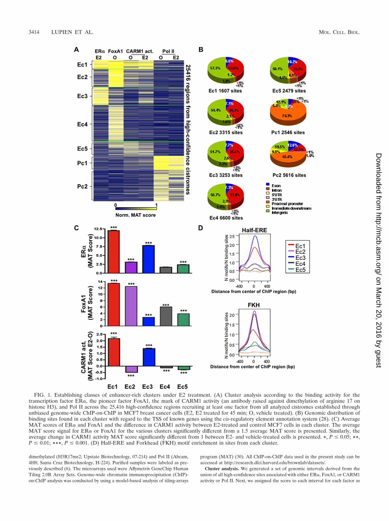
FIG. 1. Establishing classes of enhancer-rich clusters under E2 treatment. (A) Cluster analysis according to the binding activity for thetranscription factor ER�, the pioneer factor FoxA1, the mark of CARM1 activity (an antibody raised against dimethylation of arginine 17 onhistone H3), and Pol II across the 25,416 high-confidence regions recruiting at least one factor from all analyzed cistromes established throughunbiased genome-wide ChIP-on-ChIP in MCF7 breast cancer cells (E2, E2 treated for 45 min; O, vehicle treated). (B) Genomic distribution ofbinding sites found in each cluster with regard to the TSS of known genes using the cis-regulatory element annotation system (28). (C) AverageMAT scores of ER� and FoxA1 and the difference in CARM1 activity between E2-treated and control MCF7 cells in each cluster. The averageMAT score signal for ER� or FoxA1 for the various clusters significantly different from a 1.5 average MAT score is presented. Similarly, theaverage change in CARM1 activity MAT score significantly different from 1 between E2- and vehicle-treated cells is presented. *, P � 0.05; **,P � 0.01; ***, P � 0.001. (D) Half-ERE and Forkhead (FKH) motif enrichment in sites from each cluster.
3414 LUPIEN ET AL. MOL. CELL. BIOL.
on March 20, 2016 by guest
http://mcb.asm
.org/D
ownloaded from

the maximum MAT score falling within the interval for the given factor. For eachfactor MAT scores were trimmed at the 2.5 and 97.5 percentiles and scaled to liebetween 0 and 1. Genomic regions were clustered by using k-means clustering.
Sequence analysis. Genome-wide distribution as well as sequence conserva-tion analysis of H3R17me2 chip-on-chip was determined by using cis-elementannotation systems (28). Enriched motifs within clusters as well as the associa-tions with gene expression were analyzed as described previously (40).
ChIP assays. At 2 to 3 days before induction, MCF-7 cells were seeded inphenol red-free Dulbecco modified Eagle medium supplemented with 10% char-coal-dextran-treated fetal bovine serum (Omega Scientific, Inc., Tarzana, CA), 2mM L-glutamine, and 100 U of penicillin-streptomycin/ml at a density of 5 � 106
cells per 150-mm plates. Cells were subsequently induced with 10�8 M E2 for 45min. ChIP experiments were then performed as described previously (16). An-tibodies to ER� (Lab Vision, Ab-10; Santa Cruz Biotechnology, sc-543),H3R17me2 (Upstate Biotechnology, 07-214), H3K18ac (Upstate Biotechnology,07-354), H3K27ac (Upstate Biotechnology, 07-360), H4K12ac (Upstate Biotech-nology, 07-595), H3 (Abcam, ab1791), p300 (Santa Cruz Biotechnology, sc-585),and SRC1 (Santa Cruz Biotechnology, sc-8995) were used for this assay. PurifiedDNA was used in quantitative PCR (qPCR) analysis. The primers used in thisanalysis are listed in Table S1 in the supplemental material. Immunoprecipitated
DNA amounts were normalized to inputs and are expressed as the relativeenrichment.
FAIRE analysis. Formaldehyde-assisted isolation of regulatory elements(FAIRE) was performed as described in reference 21. The primers used in thisanalysis are listed in Table S1 in the supplemental material.
RESULTS
Distinct enhancer-rich clusters characterize genomic ER�recruitment. In order to better characterize the impact of co-activator action on the ER� cistrome upon E2 stimulation, wehave established the relative level of CARM1 activity acrossthe genome of MCF7 breast cancer cells. This was achievedthrough ChIP studies combined with whole-genome tiling-pathmicroarrays (ChIP-on-ChIP) using an antibody that recognizesexclusively sites of CARM1-dependent arginine methylation,including histone H3 dimethylated on arginine 17 (H3R17me2)
FIG. 2. E2-induced CARM1 activity at ER� sites associates with activating events. (A) Level of recruitment for the coactivators p300 and SRC1under vehicle (O) or E2 treatment established by ChIP-qPCR on eight ER� sites associated and eight not associated with CARM1 activation inMCF7 breast cancer cells. (B) Levels of histone modifications, namely, H3K18ac, H3K27ac, and H4K12ac, were determined as in panel A.(C) Impact of E2 treatment on nucleosome density. The changes in occupancy of the core histone H3 were determined by ChIP-qPCR as in panelA. Alterations to the DNA accessibility were determined by using FAIRE (21). The results are derived from a minimum of two independentexperiments. *, P � 0.05; **, P � 0.01; ***, P � 0.001.
VOL. 29, 2009 COACTIVATION OF ER�-BOUND REGULATORY ELEMENTS 3415
on March 20, 2016 by guest
http://mcb.asm
.org/D
ownloaded from

and the CARM1-dependent arginine methylation of AIB1 (seeFig. S1 and S2 in the supplemental material) (9, 48, 67). Morethan 4,088 and 4,461 high-confidence sites were identified be-fore and after E2 stimulation, respectively (FDR, 6%) (see Fig.
S1A and B in the supplemental material). Interestingly,CARM1 activity was found predominantly (94.1%) at regionsfar from known promoters (see Fig. S1C in the supplementalmaterial). The Pol II cistrome was also determined in the
FIG. 3. Clusters associated with CARM1 activation drive the response under E2 treatment. (A) Proportion of E2 upregulated genes comparedto nonregulated genes with at least one binding site from a specific cluster within increasing window distances from their TSS in MCF7 cells. *,P � 0.05; **, P � 0.01; ***, P � 0.001. (B) GPER and TESK2 expression after CARM1 silencing in MCF7 was determined by reversetranscription-qPCR and revealed the requirement for CARM1 in the E2-induced repression of GPER and TESK2. siLUC was used as a control.*, P � 0.05; **, P � 0.01; ***, P � 0.001. (C) Enrichment of Ec3 cluster sites (blue blocks) near GPER and TESK2 E2-downregulated targetgenes (red blocks).
3416 LUPIEN ET AL. MOL. CELL. BIOL.
on March 20, 2016 by guest
http://mcb.asm
.org/D
ownloaded from

absence of E2 to address the role of promoter-associated fac-tors in this system (see Fig. S3 in the supplemental material).As anticipated, of the 7,420 high-confidence sites (FDR, 5%)recruiting Pol II, 55.4% were recruited within 1 kb upstream ofannotated transcription start sites (TSS) (see Fig. S3A to C inthe supplemental material).
To establish the contribution of CARM1 activation and PolII recruitment to E2 signaling, we combined our newly derivedcistromes with previously published cistromes for FoxA1 in thepresence or absence of E2, as well as ER� and Pol II inE2-treated MCF7 cells (7, 40). We first established the bindingactivity as determined by MAT score (29) for all factors acrossthe 25,416 high-confidence regions recruiting at least one ofthese factors in MCF7 cells. The use of k-means clusteringrevealed five enhancer-rich clusters and two promoter-richclusters (Fig. 1A to C). Interestingly, each cluster consisted ofsites demonstrating high sequence conservation across verte-brate species (see Fig. S4 in the supplemental material). ER�was most significantly recruited after E2 stimulation to clustersEc1 (23% of the 5782 ER� high-confidence sites) and Ec3(47% of the 5782 ER� high-confidence sites), with �2.6% ofthe high-confidence sites found at promoters (Fig. 1A and C).The previously reported sites of FoxA1 recruitment favoringER� binding were found predominantly in cluster Ec1 but notEc3 (Fig. 1A and C). Correspondingly, both cluster Ec1 andEc3 were highly enriched for the ERE half-site motif, while theForkhead motif was only enriched in cluster Ec1 (Fig. 1D). Inaddition, both clusters demonstrated E2-induced CARM1 ac-tivity as measured by the MAT score (Fig. 1A and C and seeFig. S5 in the supplemental material). Cluster Ec2 consisted ofsites found at �2.1% of promoters where FoxA1 was stronglyrecruited but where ER� had low binding activity (Fig. 1A toC and see Fig. S5 in the supplemental material). Accordingly,the Forkhead motif was enriched in this cluster, while the EREhalf-site motif was not significantly enriched (Fig. 1D). In ad-dition, CARM1 activity was not induced on sites from thiscluster following E2 stimulation (Fig. 1A and C and see Fig. S5in the supplemental material). Finally, sites from the enhancer-
rich clusters Ec4 and Ec5, with �6.1% of sites at promoters,did not demonstrate strong ER� recruitment. However, sitesfrom cluster Ec4 but not Ec5 associated with FoxA1 binding.In addition, ligand-independent CARM1 activity was associ-ated with cluster Ec5 independently of E2 stimulation (Fig. 1Aand C and see Fig. S5 in the supplemental material). Globally,these data reveal that various classes of regulatory elementsare established under E2 stimulation, and those associatedwith ER� and FoxA1 recruitment, as well as CARM1 activity,are found predominantly in enhancer regions across the ge-nome.
E2-induced CARM1 activity associates with coactivator re-cruitment, histone modifications, and chromatin opening. Acommon feature of sites from clusters Ec1 and Ec3 predomi-nantly involved in the E2-mediated regulation of gene expres-sion is their association with the induction of CARM1 activityafter E2 treatment (Fig. 1A and C). In order to better char-acterize the active state of these enhancer regions, we investi-gated the level of coactivator recruitment and histone modifi-cations after E2 treatment. Sites recruiting ER� and associatedwith CARM1 activity significantly recruited other coactivators,such as p300 and SRC1, under E2 stimulation (Fig. 2A and seeFig. S6 in the supplemental material). Similarly, histone mod-ifications, such as acetylation of lysine 18 or 27 on histone H3(H3K18ac, H3K27ac), as well as on lysine 12 of histone H4(H4K12ac), were significantly induced by E2 on these samesites (Fig. 2B). ER� binding sites not associated with the in-duction of CARM1 activity did not demonstrate any significantinduction of coactivator recruitment or histone modificationunder E2 treatment (Fig. 2A and B). It is noteworthy that ER�binding sites undergoing coactivator recruitment and histonemodifications after E2 treatment also associated with E2-in-duced chromatin opening measured by histone H3 density orextractability by FAIRE (21) (Fig. 2C). Considering that 30%of ER� binding sites are not associated with clusters Ec1 orEc3 typified by E2-inducted CARM1 activity, our results revealthat the specific transcriptional response to E2 is in part de-
FIG. 4. ER�-positive primary breast tumor expression profile relates to clusters associated with CARM1 activation. Relationship betweencluster-associated gene list (genes with a binding site from a given cluster within 20 kb of their TSS) and genes overexpressed in ER�-positiveprimary breast tumors (the top 1, 5, or 10% overexpressed genes from primary breast tumors were included in the analysis). Twenty independentlydefined ER�-positive primary breast tumor overexpressing gene signatures (blue) were compared using an Oncomine Concepts Map to the fiveenhancer clusters (red) derived gene lists. Odds ratios (OR) are presented when clusters are significantly associated with independent primarybreast cancer overexpression gene signatures (P � 6e�6).
VOL. 29, 2009 COACTIVATION OF ER�-BOUND REGULATORY ELEMENTS 3417
on March 20, 2016 by guest
http://mcb.asm
.org/D
ownloaded from

pendent on the selective activation of a fraction of sites re-cruiting ER�.
CARM1 activation on ER� binding sites drives the tran-scriptional response to E2. In order to address the role of thevarious enhancer-rich clusters in gene regulation, we estab-lished the proportion of genes regulated after 3 h of E2 treat-ment versus nonregulated genes with at least one binding sitefrom a particular cluster within increasing window distances inkilobases from the TSS. This revealed a significant enrichmentof E2 upregulated genes over nonregulated genes with regardto sites from clusters Ec1, Ec2, and Ec3 from various windowdistances from the TSS, as far as 160 to 320 kb for both Ec1and Ec3 (Fig. 3A). Hence, our results suggest that the subset ofthe ER� cistrome subject to CARM1 activation upon E2 treat-ment, whether strongly or weakly associated with FoxA1 bind-ing, is responsible for E2-mediated gene induction. Thus, thepreviously suggested role for CARM1 in mediating the E2response (19, 67) is due to its activity at enhancer regionsdefined by a specific subset of the ER� cistrome. Interestingly,
genes downregulated after E2 stimulation were significantlyenriched over nonregulated genes near sites primarily fromcluster Ec3 that could be as far away as 160 to 320 kb (Fig. 3A).Accordingly, silencing CARM1 (see Fig. S2A in the supple-mental material) significantly prevented the E2-mediated re-pression of GPER and TESK2 (Fig. 3B and C). Hence, thisfinding suggests a predominant role for ER� sites associatedweakly or not at all with FoxA1 and undergoing ligand-depen-dent CARM1 activity in E2-mediated gene downregulation.
In order to address the physiological relevance of the differ-ent clusters, we compared the list of genes with a binding sitefrom a particular cluster within 20 kb of their TSS to the topgenes coexpressed with ER� in primary breast tumors from 20independent studies (4, 10, 13, 20, 23, 26, 27, 43, 44, 50, 51, 54,55, 60–62, 64–66, 68, 70). Remarkably, genes coexpressed withER� defined in 19 out of the 20 independent studies werehighly associated (odds ratio � 3) with sites from Ec1 within 20kb of their TSS (Fig. 4). Less significant association (odds ratiobetween 2 and 3) between ER� coexpressed genes and sites
FIG. 5. Cell-type specific coactivation of ER� binding sites associates with the transcriptional response. (A) Relative expression of PDK4 andFasL genes after E2 treatment for 3 h in MCF7 breast cancer and U2OS/ER� cells. (B) Relative enrichment of ER� and CARM1 activityestablished by ChIP in both MCF7 and U2OS/ER� cells after E2 treatment at the PDK4 and FasL enhancers.
3418 LUPIEN ET AL. MOL. CELL. BIOL.
on March 20, 2016 by guest
http://mcb.asm
.org/D
ownloaded from

from Ec2 and Ec3 was also detected in 2 out of the 20 inde-pendent expression profiles from primary breast tumors (Fig.4). Hence, these results further support the predominant reg-ulatory role of sites from cluster Ec1 and less significantly fromclusters Ec2 and Ec3 in the establishment of the phenotype ofER�-positive breast cancers.
Further evidence for the association between sites of ER�recruitment and their activation to mediate transcriptionalprogram originates from the comparison of the MCF7 breastcancer and U2OS osteosarcoma cell lines. Indeed, E2 treat-ment in both cell lines allows for the recruitment of ER� to anumber of common sites (34). Interestingly, the transcriptional
FIG. 6. Pol II occupied promoter of E2 target genes prior to stimulation. (A) Cluster analysis performed as described for Fig. 1A across the25,903 promoter regions associated with the RefSeq genes. (B) Proportions of all, E2-upregulated, and downregulated genes with a promotertypical of clusters Pc1prom, Pc2prom, or PcNullprom. (C) Enrichment of E2-downregulated versus nonregulated genes with at least one promoter ofthe Pc1prom, Pc2prom, or PcNullprom cluster within increasing window distances from the genes’ TSS. (D) Specific examples of E2 downregulatedgenes surrounded by gene with Pol II deprived promoters. Sites from cluster Pc1prom (orange), Pc2prom (pink), or PcNullprom (dark blue) arepresented with respect to E2-downregulated genes (red block). (E) Relative chromatin accessibilities of promoters from cluster Pc1prom, Pc2prom,or PcNullprom measured by FAIRE-ChIP. *, P � 0.05; **, P � 0.01; ***, P � 0.001.
VOL. 29, 2009 COACTIVATION OF ER�-BOUND REGULATORY ELEMENTS 3419
on March 20, 2016 by guest
http://mcb.asm
.org/D
ownloaded from

program is cell line specific (34). For instance, PDK4 and FasLare two U2OS-specific E2-induced genes (Fig. 5A). AlthoughER� gets recruited to the PDK4 and FasL enhancers in bothcell lines, they are coactivated only in U2OS cells (Fig. 5B).These results suggest that ER� recruitment associates withcoactivation in order to mediate gene expression.
Pol II occupies the promoter of E2 regulated genes prior tostimulation. To address how different components of the tran-scriptional response to E2 signaling impact promoter activity inbreast cancer cells, we performed k-means clustering on the25,903 RefSeq gene promoters as described for Fig. 1A. Threedistinct clusters could be defined (Fig. 6A). The first promotercluster (Pc1prom) consisting of 4,846 sites revealed strong Pol IIrecruitment both prior to and after E2 stimulation and nosignificant recruitment of ER�, FoxA1 nor evidence ofCARM1 activity (Fig. 6A). Similarly, the 7,316 promotersfound in the second cluster (Pc2prom) were specifically en-riched for Pol II both prior to and after E2 stimulation, albeitat lower levels than on sites from Pc1prom (Fig. 6A). Finally,13,741 promoters (PcNullprom) in MCF7 cells were not signif-icantly associated with the recruitment of Pol II, ER�, FoxA1,or CARM1 activity (Fig. 6A). Interestingly, more than 47% ofpromoters were associated with sites from either Pc1prom orPc2prom; hence, with Pol II occupied promoters prior to E2stimulation (Fig. 6A and B and see Fig. S7A in the supple-mental material). Strikingly, more than 85% of the E2 upregu-lated and 74% of E2 downregulated genes had a promotertypical of either cluster Pc1prom or Pc2prom (Fig. 6B and seeFig. S7A in the supplemental material). Furthermore, down-
regulated genes were typically found in the regions of Pol IIunoccupied promoters (Fig. 6C and D). In agreement with this,the promoters of genes surrounding downregulated genes werefound in condensed chromatin measured by FAIRE (Fig. 6E)(17). This suggests that stimulus-dependent gene regulation ispredominantly dependent on receptive promoters as definedby the presence of Pol II prior to stimulation and on the stateof promoter occupancy in neighboring genes.
DISCUSSION
The selective utilization of enhancer regions and promotersis central to the establishment of lineage-specific transcrip-tional programs and stimuli specific responses. Exploiting theE2 signaling pathway, we have combined the cistromes fromdifferent components of a transcriptional response, namely,the pioneer factor FoxA1, the transcription factor ER�, amarker of the activity of the coactivator CARM1, and Pol II.Our results reveal that FoxA1-dependent and -independentER� sites coactivated upon E2 treatment are predominantlydriving the response to E2 in breast cancer cells (Fig. 7).Hence, the specific transcriptional program associated with E2stimulation is not only dependent on the restricted genomicrecruitment of ER� but also on the activation of a selectednumber of binding sites associated with coactivator recruit-ment and histone modifications. This is in agreement with thecentral role of coactivators in the response to E2 in both celllines and mouse models (19, 46, 59, 67). Interestingly, ourresults also reveal an association between ER� binding sites
FIG. 7. Model for the selection of functional and active enhancer sites in response to estrogen stimulation in breast cancer cells. A schematicrepresentation of the transcriptional response to E2 stimulation in breast cancer cells is shown. The functional association between ER� recruitingsites undergoing coactivator (CoA) recruitment/activation and histone modifications with transcriptional regulation of the gene harboring Pol IIat their promoters both prior to and after E2 stimulation is depicted. Sites of ER� recruitment not associated with these secondary events do notsignificantly impact E2-induced regulation of gene expression.
3420 LUPIEN ET AL. MOL. CELL. BIOL.
on March 20, 2016 by guest
http://mcb.asm
.org/D
ownloaded from

displaying E2-induced CARM1 activity and gene repression.As we previously suggested, squelching and/or displacement oftranscriptional units with greater regulatory capacities couldaccount for this association (7). In addition, we demonstrate ona genome-wide scale that enhancers located as far as 160 to 320kb from the TSS of the E2 target genes mediate the transcrip-tional response. This is in accordance with previous studiesrevealing the intrachromosomal interactions required for op-timal transcriptional response upon E2 stimulation in MCF7cells (6, 12). Considering the commonality of such long-rangeinteractions between promoters and enhancers (38, 49, 63),defining how these are established on a genome-wide scale isof fundamental importance.
Furthermore, we show that Pol II promoter occupancy istypical of E2 responsive genes in MCF7 breast cancer cells thatare both up- and downregulated. This is reminiscent of studiesin Drosophila revealing the contribution of stalled or poisedPol II at the promoter of genes involved in the response tostimuli and developmental signals (47, 69). It also parallelsprevious work revealing Pol II at the promoter of unexpressedgenes (2, 24, 31–33, 53). It is consistent with the concept of PolII foci in the nucleus known as transcription factories thatremain intact in the absence of transcription (45). In fact,postrecruitment regulation of Pol II was recently revealed tobe central for the rapid signaling response to estrogen (33).Interestingly, we identified a difference between the genomicenvironments of E2 up- versus downregulated genes. Indeed,although Pol II typically occupies the promoter of E2 down-regulated genes, the promoters of surrounding genes tend tobe deprived of Pol II. Hence, the transcriptional response isdependent on the presence of a receptive promoter typified byPol II occupancy prior to stimulation.
Globally, our results reveal that the specificity of the tran-scriptional response to E2 stimulation is dependent on theinterplay between receptive promoters occupied by Pol II priorto stimulation and subclasses of ER� enhancers associatedwith E2-induced coactivator activity. Considering the uniqueexpression profiles associated with ER� activation under dis-tinct stimuli (11, 14), it remains to be established whetherdistinct subclasses of the ER� cistrome will be involved inthese responses as well.
ACKNOWLEDGMENTS
We thank Shannon T. Bailey for helpful discussion and review of themanuscript.
This study was supported by grants from the NIDDK (R01DK074967to M.B.), the NCI (P01 CA8011105) and a DF/HCC Breast CancerSPORE Grant (to M.B.), the DFCI Women’s Cancers Program, andthe U.S. Department of Defense Breast Cancer Research ProgramAwards (W81XWH-08-1-0214 to M.L.).
REFERENCES
1. Azuara, V., P. Perry, S. Sauer, M. Spivakov, H. F. Jorgensen, R. M. John, M.Gouti, M. Casanova, G. Warnes, M. Merkenschlager, and A. G. Fisher. 2006.Chromatin signatures of pluripotent cell lines. Nat. Cell Biol. 8:532–538.
2. Barski, A., S. Cuddapah, K. Cui, T. Y. Roh, D. E. Schones, Z. Wang, G. Wei,I. Chepelev, and K. Zhao. 2007. High-resolution profiling of histone methyl-ations in the human genome. Cell 129:823–837.
3. Bernstein, B. E., T. S. Mikkelsen, X. Xie, M. Kamal, D. J. Huebert, J. Cuff,B. Fry, A. Meissner, M. Wernig, K. Plath, R. Jaenisch, A. Wagschal, R. Feil,S. L. Schreiber, and E. S. Lander. 2006. A bivalent chromatin structuremarks key developmental genes in embryonic stem cells. Cell 125:315–326.
4. Bild, A. H., G. Yao, J. T. Chang, Q. Wang, A. Potti, D. Chasse, M. B. Joshi,D. Harpole, J. M. Lancaster, A. Berchuck, J. A. Olson, Jr., J. R. Marks, H. K.
Dressman, M. West, and J. R. Nevins. 2006. Oncogenic pathway signaturesin human cancers as a guide to targeted therapies. Nature 439:353–357.
5. Bourdeau, V., J. Deschenes, R. Metivier, Y. Nagai, D. Nguyen, N. Bretschnei-der, F. Gannon, J. H. White, and S. Mader. 2004. Genome-wide identifica-tion of high-affinity estrogen response elements in human and mouse. Mol.Endocrinol. 18:1411–1427.
6. Carroll, J. S., X. S. Liu, A. S. Brodsky, W. Li, C. A. Meyer, A. J. Szary, J.Eeckhoute, W. Shao, E. V. Hestermann, T. R. Geistlinger, E. A. Fox, P. A.Silver, and M. Brown. 2005. Chromosome-wide mapping of estrogen recep-tor binding reveals long-range regulation requiring the Forkhead proteinFoxA1. Cell 122:33–43.
7. Carroll, J. S., C. A. Meyer, J. Song, W. Li, T. R. Geistlinger, J. Eeckhoute,A. S. Brodsky, E. K. Keeton, K. C. Fertuck, G. F. Hall, Q. Wang, S.Bekiranov, V. Sementchenko, E. A. Fox, P. A. Silver, T. R. Gingeras, X. S.Liu, and M. Brown. 2006. Genome-wide analysis of estrogen receptor bind-ing sites. Nat. Genet. 38:1289–1297.
8. Chen, D., H. Ma, H. Hong, S. S. Koh, S. M. Huang, B. T. Schurter, D. W.Aswad, and M. R. Stallcup. 1999. Regulation of transcription by a proteinmethyltransferase. Science 284:2174–2177.
9. Cheng, D., J. Cote, S. Shaaban, and M. T. Bedford. 2007. The argininemethyltransferase CARM1 regulates the coupling of transcription andmRNA processing. Mol. Cell 25:71–83.
10. Chin, K., S. DeVries, J. Fridlyand, P. T. Spellman, R. Roydasgupta, W. L.Kuo, A. Lapuk, R. M. Neve, Z. Qian, T. Ryder, F. Chen, H. Feiler, T.Tokuyasu, C. Kingsley, S. Dairkee, Z. Meng, K. Chew, D. Pinkel, A. Jain,B. M. Ljung, L. Esserman, D. G. Albertson, F. M. Waldman, and J. W. Gray.2006. Genomic and transcriptional aberrations linked to breast cancer patho-physiologies. Cancer Cell 10:529–541.
11. Cunliffe, H. E., M. Ringner, S. Bilke, R. L. Walker, J. M. Cheung, Y. Chen,and P. S. Meltzer. 2003. The gene expression response of breast cancer togrowth regulators: patterns and correlation with tumor expression profiles.Cancer Res. 63:7158–7166.
12. Deschenes, J., V. Bourdeau, J. H. White, and S. Mader. 2007. Regulation ofGREB1 transcription by estrogen receptor alpha through a multipartiteenhancer spread over 20 kb of upstream flanking sequences. J. Biol. Chem.282:17335–17339.
13. Desmedt, C., F. Piette, S. Loi, Y. Wang, F. Lallemand, B. Haibe-Kains, G.Viale, M. Delorenzi, Y. Zhang, M. S. d’Assignies, J. Bergh, R. Lidereau, P.Ellis, A. L. Harris, J. G. Klijn, J. A. Foekens, F. Cardoso, M. J. Piccart, M.Buyse, and C. Sotiriou. 2007. Strong time dependence of the 76-gene prog-nostic signature for node-negative breast cancer patients in the TRANSBIGmulticenter independent validation series. Clin. Cancer Res. 13:3207–3214.
14. Dudek, P., and D. Picard. 2008. Genomics of signaling crosstalk of estrogenreceptor alpha in breast cancer cells. PLoS ONE 3:e1859.
15. Eeckhoute, J., J. S. Carroll, T. R. Geistlinger, M. I. Torres-Arzayus, and M.Brown. 2006. A cell-type-specific transcriptional network required for estro-gen regulation of cyclin D1 and cell cycle progression in breast cancer. GenesDev. 20:2513–2526.
16. Eeckhoute, J., E. K. Keeton, M. Lupien, S. A. Krum, J. S. Carroll, and M.Brown. 2007. Positive cross-regulatory loop ties GATA-3 to estrogen recep-tor alpha expression in breast cancer. Cancer Res. 67:6477–6483.
17. Eeckhoute, J., M. Lupien, C. A. Meyer, M. P. Verzi, R. A. Shivdasani, X. S.Liu, and M. Brown. 2008. Cell-type selective remodeling defines the activesubset of FOXA1-bound enhancers. Genome Res. 19:372–380.
18. Flanagan, J. F., L. Z. Mi, M. Chruszcz, M. Cymborowski, K. L. Clines, Y.Kim, W. Minor, F. Rastinejad, and S. Khorasanizadeh. 2005. Double chro-modomains cooperate to recognize the methylated histone H3 tail. Nature438:1181–1185.
19. Frietze, S., M. Lupien, P. A. Silver, and M. Brown. 2008. CARM1 regulatesestrogen-stimulated breast cancer growth through up-regulation of E2F1.Cancer Res. 68:301–306.
20. Ginestier, C., N. Cervera, P. Finetti, S. Esteyries, B. Esterni, J. Adelaide, L.Xerri, P. Viens, J. Jacquemier, E. Charafe-Jauffret, M. Chaffanet, D. Birn-baum, and F. Bertucci. 2006. Prognosis and gene expression profiling of20q13-amplified breast cancers. Clin. Cancer Res. 12:4533–4544.
21. Giresi, P. G., J. Kim, R. M. McDaniell, V. R. Iyer, and J. D. Lieb. 2007.FAIRE (formaldehyde-assisted isolation of regulatory elements) isolatesactive regulatory elements from human chromatin. Genome Res. 17:877–885.
22. Green, K. A., and J. S. Carroll. 2007. Oestrogen-receptor-mediated tran-scription and the influence of cofactors and chromatin state. Nat. Rev.Cancer 7:713–722.
23. Gruvberger, S., M. Ringner, Y. Chen, S. Panavally, L. H. Saal, A. Borg, M.Ferno, C. Peterson, and P. S. Meltzer. 2001. Estrogen receptor status inbreast cancer is associated with remarkably distinct gene expression patterns.Cancer Res. 61:5979–5984.
24. Guenther, M. G., S. S. Levine, L. A. Boyer, R. Jaenisch, and R. A. Young.2007. A chromatin landmark and transcription initiation at most promotersin human cells. Cell 130:77–88.
25. Heintzman, N. D., R. K. Stuart, G. Hon, Y. Fu, C. W. Ching, R. D. Hawkins,L. O. Barrera, S. Van Calcar, C. Qu, K. A. Ching, W. Wang, Z. Weng, R. D.Green, G. E. Crawford, and B. Ren. 2007. Distinct and predictive chromatin
VOL. 29, 2009 COACTIVATION OF ER�-BOUND REGULATORY ELEMENTS 3421
on March 20, 2016 by guest
http://mcb.asm
.org/D
ownloaded from

signatures of transcriptional promoters and enhancers in the human genome.Nat. Genet. 39:311–318.
26. Hess, K. R., K. Anderson, W. F. Symmans, V. Valero, N. Ibrahim, J. A.Mejia, D. Booser, R. L. Theriault, A. U. Buzdar, P. J. Dempsey, R. Rouzier,N. Sneige, J. S. Ross, T. Vidaurre, H. L. Gomez, G. N. Hortobagyi, and L.Pusztai. 2006. Pharmacogenomic predictor of sensitivity to preoperativechemotherapy with paclitaxel and fluorouracil, doxorubicin, and cyclophos-phamide in breast cancer. J. Clin. Oncol. 24:4236–4244.
27. Ivshina, A. V., J. George, O. Senko, B. Mow, T. C. Putti, J. Smeds, T.Lindahl, Y. Pawitan, P. Hall, H. Nordgren, J. E. Wong, E. T. Liu, J. Bergh,V. A. Kuznetsov, and L. D. Miller. 2006. Genetic reclassification of histologicgrade delineates new clinical subtypes of breast cancer. Cancer Res. 66:10292–10301.
28. Ji, X., W. Li, J. Song, L. Wei, and X. S. Liu. 2006. CEAS: cis-regulatoryelement annotation system. Nucleic Acids Res. 34:W551–W554.
29. Johnson, D. S., W. Li, D. B. Gordon, A. Bhattacharjee, B. Curry, J. Ghosh,L. Brizuela, J. S. Carroll, M. Brown, P. Flicek, C. M. Koch, I. Dunham, M.Bieda, X. Xu, P. J. Farnham, P. Kapranov, D. A. Nix, T. R. Gingeras, X.Zhang, H. Holster, N. Jiang, R. Green, J. S. Song, S. A. McCuine, E. Anton,L. Nguyen, N. D. Trinklein, Z. Ye, K. Ching, D. Hawkins, B. Ren, P. C.Scacheri, J. Rozowsky, A. Karpikov, G. Euskirchen, S. Weissman, M. Ger-stein, M. Snyder, A. Yang, Z. Moqtaderi, H. Hirsch, H. P. Shulha, Y. Fu, Z.Weng, K. Struhl, R. M. Myers, J. D. Lieb, and X. S. Liu. 2008. Systematicevaluation of variability in ChIP-chip experiments using predefined DNAtargets. Genome Res. 18:393–403.
30. Johnson, W. E., W. Li, C. A. Meyer, R. Gottardo, J. S. Carroll, M. Brown,and X. S. Liu. 2006. Model-based analysis of tiling-arrays for ChIP-chip.Proc. Natl. Acad. Sci. USA 103:12457–12462.
31. Kim, T. H., L. O. Barrera, M. Zheng, C. Qu, M. A. Singer, T. A. Richmond,Y. Wu, R. D. Green, and B. Ren. 2005. A high-resolution map of activepromoters in the human genome. Nature 436:876–880.
32. Kininis, M., B. S. Chen, A. G. Diehl, G. D. Isaacs, T. Zhang, A. C. Siepel,A. G. Clark, and W. L. Kraus. 2007. Genomic analyses of transcription factorbinding, histone acetylation, and gene expression reveal mechanistically dis-tinct classes of estrogen-regulated promoters. Mol. Cell. Biol. 27:5090–5104.
33. Kininis, M., G. D. Isaacs, L. J. Core, N. Hah, and W. L. Kraus. 2009.Postrecruitment regulation of RNA polymerase II directs rapid signalingresponses at the promoters of estrogen target genes. Mol. Cell. Biol. 29:1123–1133.
34. Krum, S. A., G. A. Miranda-Carboni, M. Lupien, J. Eeckhoute, J. S. Carroll,and M. Brown. 2008. Unique ER� cistromes control cell type-specific generegulation. Mol. Endocrinol. 22:2393–2406.
35. Kwon, Y. S., I. Garcia-Bassets, K. R. Hutt, C. S. Cheng, M. Jin, D. Liu, C.Benner, D. Wang, Z. Ye, M. Bibikova, J. B. Fan, L. Duan, C. K. Glass, M. G.Rosenfeld, and X. D. Fu. 2007. Sensitive ChIP-DSL technology reveals anextensive estrogen receptor alpha-binding program on human gene promot-ers. Proc. Natl. Acad. Sci. USA 104:4852–4857.
36. Lee, T. I., R. G. Jenner, L. A. Boyer, M. G. Guenther, S. S. Levine, R. M.Kumar, B. Chevalier, S. E. Johnstone, M. F. Cole, K. Isono, H. Koseki, T.Fuchikami, K. Abe, H. L. Murray, J. P. Zucker, B. Yuan, G. W. Bell, E.Herbolsheimer, N. M. Hannett, K. Sun, D. T. Odom, A. P. Otte, T. L.Volkert, D. P. Bartel, D. A. Melton, D. K. Gifford, R. Jaenisch, and R. A.Young. 2006. Control of developmental regulators by Polycomb in humanembryonic stem cells. Cell 125:301–313.
37. Lin, C. Y., V. B. Vega, J. S. Thomsen, T. Zhang, S. L. Kong, M. Xie, K. P.Chiu, L. Lipovich, D. H. Barnett, F. Stossi, A. Yeo, J. George, V. A.Kuznetsov, Y. K. Lee, T. H. Charn, N. Palanisamy, L. D. Miller, E. Cheung,B. S. Katzenellenbogen, Y. Ruan, G. Bourque, C. L. Wei, and E. T. Liu. 2007.Whole-genome cartography of estrogen receptor alpha binding sites. PLoSGenet. 3:e87.
38. Ling, J. Q., T. Li, J. F. Hu, T. H. Vu, H. L. Chen, X. W. Qiu, A. M. Cherry,and A. R. Hoffman. 2006. CTCF mediates interchromosomal colocalizationbetween Igf2/H19 and Wsb1/Nf1. Science 312:269–272.
39. Liu, Y., H. Gao, T. T. Marstrand, A. Strom, E. Valen, A. Sandelin, J. A.Gustafsson, and K. Dahlman-Wright. 2008. The genome landscape of ER�-and ER�-binding DNA regions. Proc. Natl. Acad. Sci. USA 105:2604–2609.
40. Lupien, M., J. Eeckhoute, C. Meyer, Q. Wang, Y. Zhang, W. Li, J. Carroll,X. Liu, and M. Brown. 2008. FoxA1 translates epigenetic signatures intoenhancer driven lineage-specific transcription. Cell 132:958–970.
41. Metivier, R., G. Penot, M. R. Hubner, G. Reid, H. Brand, M. Kos, and F.Gannon. 2003. Estrogen receptor-alpha directs ordered, cyclical, and com-binatorial recruitment of cofactors on a natural target promoter. Cell 115:751–763.
42. Mikkelsen, T. S., M. Ku, D. B. Jaffe, B. Issac, E. Lieberman, G. Giannoukos,P. Alvarez, W. Brockman, T. K. Kim, R. P. Koche, W. Lee, E. Mendenhall,A. O’Donovan, A. Presser, C. Russ, X. Xie, A. Meissner, M. Wernig, R.Jaenisch, C. Nusbaum, E. S. Lander, and B. E. Bernstein. 2007. Genome-wide maps of chromatin state in pluripotent and lineage-committed cells.Nature 448:553–560.
43. Miller, L. D., J. Smeds, J. George, V. B. Vega, L. Vergara, A. Ploner, Y.Pawitan, P. Hall, S. Klaar, E. T. Liu, and J. Bergh. 2005. An expressionsignature for p53 status in human breast cancer predicts mutation status,
transcriptional effects, and patient survival. Proc. Natl. Acad. Sci. USA102:13550–13555.
44. Minn, A. J., G. P. Gupta, P. M. Siegel, P. D. Bos, W. Shu, D. D. Giri, A. Viale,A. B. Olshen, W. L. Gerald, and J. Massague. 2005. Genes that mediatebreast cancer metastasis to lung. Nature 436:518–524.
45. Mitchell, J. A., and P. Fraser. 2008. Transcription factories are nuclearsubcompartments that remain in the absence of transcription. Genes Dev.22:20–25.
46. Modder, U. I., A. Sanyal, J. Xu, B. W. O’Malley, T. C. Spelsberg, and S.Khosla. 2008. The skeletal response to estrogen is impaired in female but notin male steroid receptor coactivator (SRC)-1 knockout mice. Bone 42:414–421.
47. Muse, G. W., D. A. Gilchrist, S. Nechaev, R. Shah, J. S. Parker, S. F.Grissom, J. Zeitlinger, and K. Adelman. 2007. RNA polymerase is poised foractivation across the genome. Nat. Genet. 39:1507–1511.
48. Naeem, H., D. Cheng, Q. Zhao, C. Underhill, M. Tini, M. T. Bedford, and J.Torchia. 2007. The activity and stability of the transcriptional coactivatorp/CIP/SRC-3 are regulated by CARM1-dependent methylation. Mol. Cell.Biol. 27:120–134.
49. Osborne, C. S., L. Chakalova, K. E. Brown, D. Carter, A. Horton, E. De-brand, B. Goyenechea, J. A. Mitchell, S. Lopes, W. Reik, and P. Fraser. 2004.Active genes dynamically colocalize to shared sites of ongoing transcription.Nat. Genet. 36:1065–1071.
50. Perou, C. M., S. S. Jeffrey, M. van de Rijn, C. A. Rees, M. B. Eisen, D. T.Ross, A. Pergamenschikov, C. F. Williams, S. X. Zhu, J. C. Lee, D. Lashkari,D. Shalon, P. O. Brown, and D. Botstein. 1999. Distinctive gene expressionpatterns in human mammary epithelial cells and breast cancers. Proc. Natl.Acad. Sci. USA 96:9212–9217.
51. Pollack, J. R., T. Sorlie, C. M. Perou, C. A. Rees, S. S. Jeffrey, P. E. Lonning,R. Tibshirani, D. Botstein, A. L. Borresen-Dale, and P. O. Brown. 2002.Microarray analysis reveals a major direct role of DNA copy number alter-ation in the transcriptional program of human breast tumors. Proc. Natl.Acad. Sci. USA 99:12963–12968.
52. Pray-Grant, M. G., J. A. Daniel, D. Schieltz, J. R. Yates III, and P. A. Grant.2005. Chd1 chromodomain links histone H3 methylation with SAGA- andSLIK-dependent acetylation. Nature 433:434–438.
53. Radonjic, M., J. C. Andrau, P. Lijnzaad, P. Kemmeren, T. T. Kockelkorn, D.van Leenen, N. L. van Berkum, and F. C. Holstege. 2005. Genome-wideanalyses reveal RNA polymerase II located upstream of genes poised forrapid response upon Saccharomyces cerevisiae stationary phase exit. Mol.Cell 18:171–183.
54. Richardson, A. L., Z. C. Wang, A. De Nicolo, X. Lu, M. Brown, A. Miron, X.Liao, J. D. Iglehart, D. M. Livingston, and S. Ganesan. 2006. X chromo-somal abnormalities in basal-like human breast cancer. Cancer Cell 9:121–132.
55. Saal, L. H., P. Johansson, K. Holm, S. K. Gruvberger-Saal, Q. B. She, M.Maurer, S. Koujak, A. A. Ferrando, P. Malmstrom, L. Memeo, J. Isola, P. O.Bendahl, N. Rosen, H. Hibshoosh, M. Ringner, A. Borg, and R. Parsons.2007. Poor prognosis in carcinoma is associated with a gene expressionsignature of aberrant PTEN tumor suppressor pathway activity. Proc. Natl.Acad. Sci. USA 104:7564–7569.
56. Santos-Rosa, H., R. Schneider, B. E. Bernstein, N. Karabetsou, A. Morillon,C. Weise, S. L. Schreiber, J. Mellor, and T. Kouzarides. 2003. Methylationof histone H3 K4 mediates association of the Isw1p ATPase with chromatin.Mol. Cell 12:1325–1332.
57. Shang, Y., X. Hu, J. DiRenzo, M. A. Lazar, and M. Brown. 2000. Cofactordynamics and sufficiency in estrogen receptor-regulated transcription. Cell103:843–852.
58. Sims, R. J., III, C. F. Chen, H. Santos-Rosa, T. Kouzarides, S. S. Patel, andD. Reinberg. 2005. Human but not yeast CHD1 binds directly and selectivelyto histone H3 methylated at lysine 4 via its tandem chromodomains. J. Biol.Chem. 280:41789–41792.
59. Smith, C. L., D. G. DeVera, D. J. Lamb, Z. Nawaz, Y. H. Jiang, A. L. Beaudet,and B. W. O’Malley. 2002. Genetic ablation of the steroid receptor coacti-vator-ubiquitin ligase, E6-AP, results in tissue-selective steroid hormoneresistance and defects in reproduction. Mol. Cell. Biol. 22:525–535.
60. Sorlie, T., C. M. Perou, R. Tibshirani, T. Aas, S. Geisler, H. Johnsen, T.Hastie, M. B. Eisen, M. van de Rijn, S. S. Jeffrey, T. Thorsen, H. Quist, J. C.Matese, P. O. Brown, D. Botstein, P. Eystein Lonning, and A. L. Borresen-Dale. 2001. Gene expression patterns of breast carcinomas distinguish tumorsubclasses with clinical implications. Proc. Natl. Acad. Sci. USA 98:10869–10874.
61. Sotiriou, C., S. Y. Neo, L. M. McShane, E. L. Korn, P. M. Long, A. Jazaeri,P. Martiat, S. B. Fox, A. L. Harris, and E. T. Liu. 2003. Breast cancerclassification and prognosis based on gene expression profiles from a popu-lation-based study. Proc. Natl. Acad. Sci. USA 100:10393–10398.
62. Sotiriou, C., P. Wirapati, S. Loi, A. Harris, S. Fox, J. Smeds, H. Nordgren,P. Farmer, V. Praz, B. Haibe-Kains, C. Desmedt, D. Larsimont, F. Cardoso,H. Peterse, D. Nuyten, M. Buyse, M. J. Van de Vijver, J. Bergh, M. Piccart,and M. Delorenzi. 2006. Gene expression profiling in breast cancer: under-standing the molecular basis of histologic grade to improve prognosis.J. Natl. Cancer Inst. 98:262–272.
3422 LUPIEN ET AL. MOL. CELL. BIOL.
on March 20, 2016 by guest
http://mcb.asm
.org/D
ownloaded from

63. Spilianakis, C. G., M. D. Lalioti, T. Town, G. R. Lee, and R. A. Flavell. 2005.Interchromosomal associations between alternatively expressed loci. Nature435:637–645.
64. van de Vijver, M. J., Y. D. He, L. J. van’t Veer, H. Dai, A. A. Hart, D. W.Voskuil, G. J. Schreiber, J. L. Peterse, C. Roberts, M. J. Marton, M. Parrish,D. Atsma, A. Witteveen, A. Glas, L. Delahaye, T. van der Velde, H. Bartelink,S. Rodenhuis, E. T. Rutgers, S. H. Friend, and R. Bernards. 2002. A gene-expression signature as a predictor of survival in breast cancer. N. Engl.J. Med. 347:1999–2009.
65. Wang, Y., J. G. Klijn, Y. Zhang, A. M. Sieuwerts, M. P. Look, F. Yang, D.Talantov, M. Timmermans, M. E. Meijer-van Gelder, J. Yu, T. Jatkoe, E. M.Berns, D. Atkins, and J. A. Foekens. 2005. Gene-expression profiles topredict distant metastasis of lymph-node-negative primary breast cancer.Lancet 365:671–679.
66. West, M., C. Blanchette, H. Dressman, E. Huang, S. Ishida, R. Spang, H.Zuzan, J. A. Olson, Jr., J. R. Marks, and J. R. Nevins. 2001. Predicting the
clinical status of human breast cancer by using gene expression profiles. Proc.Natl. Acad. Sci. USA 98:11462–11467.
67. Yadav, N., J. Lee, J. Kim, J. Shen, M. C. Hu, C. M. Aldaz, and M. T. Bedford.2003. Specific protein methylation defects and gene expression perturbationsin coactivator-associated arginine methyltransferase 1-deficient mice. Proc.Natl. Acad. Sci. USA 100:6464–6468.
68. Yu, K., K. Ganesan, L. D. Miller, and P. Tan. 2006. A modular analysis ofbreast cancer reveals a novel low-grade molecular signature in estrogenreceptor-positive tumors. Clin. Cancer Res. 12:3288–3296.
69. Zeitlinger, J., A. Stark, M. Kellis, J. W. Hong, S. Nechaev, K. Adelman, M.Levine, and R. A. Young. 2007. RNA polymerase stalling at developmentalcontrol genes in the Drosophila melanogaster embryo. Nat. Genet. 39:1512–1516.
70. Zhao, H., A. Langerod, Y. Ji, K. W. Nowels, J. M. Nesland, R. Tibshirani,I. K. Bukholm, R. Karesen, D. Botstein, A. L. Borresen-Dale, and S. S.Jeffrey. 2004. Different gene expression patterns in invasive lobular andductal carcinomas of the breast. Mol. Biol. Cell 15:2523–2536.
VOL. 29, 2009 COACTIVATION OF ER�-BOUND REGULATORY ELEMENTS 3423
on March 20, 2016 by guest
http://mcb.asm
.org/D
ownloaded from