Estrogen-like effects of diet-derived cadmium differ from those of orally administered CdCl 2 in the...
10
Toxicology Letters 202 (2011) 75–84 Contents lists available at ScienceDirect Toxicology Letters journal homepage: www.elsevier.com/locate/toxlet Estrogen-like effects of diet-derived cadmium differ from those of orally administered CdCl 2 in the ERE-luc estrogen reporter mouse model Balaji Ramachandran a , Sari Mäkelä b , Jean-Pierre Cravedi c , Marika Berglund d , Helen Håkansson d , Pauliina Damdimopoulou b,1,2 , Adriana Maggi a,∗,1 a Center of Excellence on Neurodegenerative Diseases and Department of Pharmacological Sciences, University of Milan, Via Balzaretti, 9 I-20123 Milan, Italy b Functional Foods Forum and Institute of Biomedicine, University of Turku, FI-20014 University of Turku, Finland c INRA, UMR1089 Xénobiotiques, BP 93173, F31300 Toulouse, France d Institute of Environmental Medicine, Karolinska Institutet, SE-171 77 Stockholm, Sweden article info Article history: Received 9 November 2010 Received in revised form 21 December 2010 Accepted 23 December 2010 Available online 30 December 2010 Keywords: Heavy metals Cadmium Dietary endocrine modulators Estrogen signaling Estrogen reporter mouse Bioavailability abstract Cadmium (Cd), an environmental and dietary contaminant, has been described to mimic the effects of 17-estradiol (E 2 ) in selected model systems when studied as an inorganic salt. However, inorganic Cd salts do not represent the main form of Cd exposure in general human populations. The aims of this study were to compare the estrogen-like effects and the bioavailability of dietary Cd to inorganic CdCl 2 . Adult ovariectomized ERE-luc reporter mice were administered two bread based diets containing different con- centrations of Cd (17.57 and 49.22 g/kg, corresponding to oral intakes of 1.8 and 5.1 g/kg body weight (bw) per day, respectively), inorganic CdCl 2 (1 g/kg bw per day by gavage) or E 2 (5 g/kg bw per day pellet) for 21 days. The effects on estrogen signaling were investigated by studying the uterine weights, luciferase activation, and expression of endogenous estrogen target genes. The uterine weight was sig- nificantly increased by both CdCl 2 and E 2 but not by the Cd containing diets. All treatments modulated the expression of luciferase and the endogenous estrogen target genes; however, there was no consistent overlap between the responses triggered by the bread diets and the responses stimulated by CdCl 2 or E 2 . Oral exposure to Cd was calculated and the concentrations in liver and kidneys quantified to estimate the amount of absorbed Cd retained in tissues. The results suggest significantly lower absorption and/or tissue retention of dietary Cd compared to CdCl 2 following oral exposure. Altogether, our results support previous reports on in vivo estrogenicity of CdCl 2 but do not suggest the same activity for diet bound Cd. This study calls for caution when extrapolating results from pure compound studies (e.g. estrogenicity of CdCl 2 ) to dietary exposure scenarios (e.g. estrogenicity of diet bound Cd). Further basic research is needed on the mechanisms of interaction between Cd and the estrogen signaling, biologically active species of Cd, and biomarkers of estrogen-like effects of Cd in vivo before human health risk assessment on the hormone disruptive effects of Cd can be carried out. © 2010 Elsevier Ireland Ltd. All rights reserved. Abbreviations: BLI, bioluminescence imaging; ATP, adenosine 5 -triphosphate; E2, 17-estradiol; s.c., subcutaneous; i.p, intraperitoneal; WAT, white adipose tissue; ER, estrogen receptor; ERE, estrogen responsive element; KPO4, potassium phosphate; RLU, relative light unit; OVX, ovariectomized; PTWI, provisional tolerable weekly intake; IARC, International Agency for Research on Cancer; TWI, Tolerable weekly intake; EFSA, European Food Safety Authority; WHO, world health organization; FAO, Food and Agriculture Organization; JECFA, Joint FAO/WHO Expert Committee on Food Additives; ICPMS, Inductively coupled plasma mass spectrometry; bw, body weight; MT, metallothionein; PTMA, prothymosin. ∗ Corresponding author. Tel.: +39 02 50318287; fax: +39 02 50318290. E-mail addresses: [email protected] (B. Ramachandran), sarmak@utu.fi (S. Mäkelä), [email protected] (J.-P. Cravedi), [email protected] (M. Berglund), [email protected] (H. Håkansson), [email protected] (P. Damdimopoulou), [email protected] (A. Maggi). 1 These two authors have equally contributed to the preparation of this manuscript. 2 Current address: Department of Biosciences and Nutrition, Karolinska Institutet, SE-14157 Huddinge, Sweden. 0378-4274/$ – see front matter © 2010 Elsevier Ireland Ltd. All rights reserved. doi:10.1016/j.toxlet.2010.12.022
-
Upload
independent -
Category
Documents
-
view
2 -
download
0
Transcript of Estrogen-like effects of diet-derived cadmium differ from those of orally administered CdCl 2 in the...
Toxicology Letters 202 (2011) 75–84
Contents lists available at ScienceDirect
Toxicology Letters
journa l homepage: www.e lsev ier .com/ locate / tox le t
Estrogen-like effects of diet-derived cadmium differ from those of orallyadministered CdCl2 in the ERE-luc estrogen reporter mouse model
Balaji Ramachandrana, Sari Mäkeläb, Jean-Pierre Cravedic, Marika Berglundd, Helen Håkanssond,Pauliina Damdimopouloub,1,2, Adriana Maggia,∗,1
a Center of Excellence on Neurodegenerative Diseases and Department of Pharmacological Sciences, University of Milan, Via Balzaretti, 9 I-20123 Milan, Italyb Functional Foods Forum and Institute of Biomedicine, University of Turku, FI-20014 University of Turku, Finlandc INRA, UMR1089 Xénobiotiques, BP 93173, F31300 Toulouse, Franced Institute of Environmental Medicine, Karolinska Institutet, SE-171 77 Stockholm, Sweden
a r t i c l e i n f o
Article history:Received 9 November 2010Received in revised form21 December 2010Accepted 23 December 2010Available online 30 December 2010
Keywords:Heavy metalsCadmiumDietary endocrine modulatorsEstrogen signalingEstrogen reporter mouseBioavailability
a b s t r a c t
Cadmium (Cd), an environmental and dietary contaminant, has been described to mimic the effects of17�-estradiol (E2) in selected model systems when studied as an inorganic salt. However, inorganic Cdsalts do not represent the main form of Cd exposure in general human populations. The aims of this studywere to compare the estrogen-like effects and the bioavailability of dietary Cd to inorganic CdCl2. Adultovariectomized ERE-luc reporter mice were administered two bread based diets containing different con-centrations of Cd (17.57 and 49.22 �g/kg, corresponding to oral intakes of 1.8 and 5.1 �g/kg body weight(bw) per day, respectively), inorganic CdCl2 (1 �g/kg bw per day by gavage) or E2 (5 �g/kg bw per daypellet) for 21 days. The effects on estrogen signaling were investigated by studying the uterine weights,luciferase activation, and expression of endogenous estrogen target genes. The uterine weight was sig-nificantly increased by both CdCl2 and E2 but not by the Cd containing diets. All treatments modulatedthe expression of luciferase and the endogenous estrogen target genes; however, there was no consistentoverlap between the responses triggered by the bread diets and the responses stimulated by CdCl2 or E2.Oral exposure to Cd was calculated and the concentrations in liver and kidneys quantified to estimatethe amount of absorbed Cd retained in tissues. The results suggest significantly lower absorption and/ortissue retention of dietary Cd compared to CdCl2 following oral exposure. Altogether, our results supportprevious reports on in vivo estrogenicity of CdCl2 but do not suggest the same activity for diet bound Cd.This study calls for caution when extrapolating results from pure compound studies (e.g. estrogenicity ofCdCl2) to dietary exposure scenarios (e.g. estrogenicity of diet bound Cd). Further basic research is neededon the mechanisms of interaction between Cd and the estrogen signaling, biologically active species of
Cd, and biomarkers of estrogen-like effects of Cd in vivo before human health risk assessment on thehormone disruptive effects of Cd can be carried out.© 2010 Elsevier Ireland Ltd. All rights reserved.
Abbreviations: BLI, bioluminescence imaging; ATP, adenosine 5′-triphosphate; E2, 17�-estradiol; s.c., subcutaneous; i.p, intraperitoneal; WAT, white adipose tissue;ER, estrogen receptor; ERE, estrogen responsive element; KPO4, potassium phosphate; RLU, relative light unit; OVX, ovariectomized; PTWI, provisional tolerable weeklyintake; IARC, International Agency for Research on Cancer; TWI, Tolerable weekly intake; EFSA, European Food Safety Authority; WHO, world health organization; FAO, Foodand Agriculture Organization; JECFA, Joint FAO/WHO Expert Committee on Food Additives; ICPMS, Inductively coupled plasma mass spectrometry; bw, body weight; MT,metallothionein; PTMA, prothymosin.
∗ Corresponding author. Tel.: +39 02 50318287; fax: +39 02 50318290.E-mail addresses: [email protected] (B. Ramachandran), [email protected] (S. Mäkelä), [email protected] (J.-P. Cravedi), [email protected] (M. Berglund),
[email protected] (H. Håkansson), [email protected] (P. Damdimopoulou), [email protected] (A. Maggi).1 These two authors have equally contributed to the preparation of this manuscript.2 Current address: Department of Biosciences and Nutrition, Karolinska Institutet, SE-14157 Huddinge, Sweden.
0378-4274/$ – see front matter © 2010 Elsevier Ireland Ltd. All rights reserved.doi:10.1016/j.toxlet.2010.12.022
7 icology Letters 202 (2011) 75–84
1
tafiT1ecoaasvc2ociaos
7t(eE3ieic
itcCCob3llA
e2tiEiceeeeoPugvdee
Table 1Analyzed composition of the custom-made breads per fresh weight.
Component Wheat bread Flax bread Unit
Carbohydrates 48.9 38.1 g/100 gGlucose 0.10 0.16 g/100 gFructose 0.33 0.33 g/100 gSaccharose 0.09 0.22 g/100 gLactose 0.04 <0.04 g/100 gProtein 10.0 10.2 g/100 gFat 3.0 5.4 g/100 gSaturated fatty acids 26.2 12.0 %Monounsaturated fatty acids 33.2 37.1 %Polyunsaturated fatty acids 39.3 50.1 %Energy content 263 242 kcal/100 gTotal dietary fibre 3.1 6.9 g/100 g
6 B. Ramachandran et al. / Tox
. Introduction
Cadmium (Cd) is ubiquitously present in the environment, dueo its natural occurrence and as a pollutant from industrial andgricultural sources. Animal and epidemiological studies have con-rmed that Cd is toxic to the gonads (Henson and Chedrese, 2004;hompson and Bannigan, 2008), kidney (Lind et al., 1997; Min et al.,996), liver (Rikans and Yamano, 2000.) and bone (Bhattacharyyat al., 1988). In 1993 the International Agency for Research on Can-er (IARC) classified Cd as a human carcinogen (Group 1) basedn cancer incidence data derived from occupationally exposeddults (IARC, 1993). Atmospheric pollution, phosphate fertilizersnd sewage sludge appear to be the major contributors to Cd depo-ition in agricultural soils. Cd is present in virtually all foods atariable concentration depending on the type of food and con-entration of Cd in the soil (Mor and Ceylan, 2008; Satarug et al.,003). Diet represents the main source of Cd exposure in non-ccupationally exposed humans. Primarily because of their highonsumption, the food groups that contribute to the daily Cd intaken humans the most are cereal products, rice, vegetables, nuts, rootsnd tuber (FAO/WHO, 2005; EFSA, 2009). Bread, accounting for 36%f the weekly Cd intake, has been identified as the largest singleource of Cd in diet in Sweden (Olsson et al., 2002).
The provisional tolerable weekly intake (PTWI) for Cd is set to�g/kg bw by the Joint FAO/WHO Expert Committee on Food Addi-
ives (JECFA) in 1988, and re-evaluated in 2005 without a changeFAO/WHO, 1988, 2005). European Food Safety Authority (EFSA)stimated in 2009 that the median of average intake values acrossuropean countries is 2.3 �g/kg bw per week (range from 1.9 to.0 �g/kg bw per week) (EFSA, 2009), and set the tolerable weekly
ntake (TWI) as 2.5 �g/kg bw. The current population exposure lev-ls are very close to the limits set by JECFA and EFSA. Specific groupsn risk of consuming higher dietary Cd than recognized as safe arehildren and vegetarians (EFSA, 2009).
Dietary Cd is mainly absorbed in the proximal part of the smallntestine (Reeves and Chaney, 2008). It enters the systemic circula-ion complexed to proteins, such as albumin, metallothionein andysteine, and is taken up by tissues (Zalups and Ahmad, 2003).d accumulates in particular to liver and kidneys (Henson andhedrese, 2004; Thompson and Bannigan, 2008), and the amountf Cd in these organs is estimated to account for over 70% of totalody burden (Andersen, 1989). The half-life of Cd varies from 6 to8 years in the human kidney and from 4 to 19 years in the human
iver (Kostial et al., 1979; Kjellstrom and Nordberg, 1985). This veryong biological half-life makes Cd a cumulative toxin (Järup andkesson, 2009).
Several independent reports suggest that Cd interacts with thestrogen receptor alpha (ER�) (Stoica et al., 2000; Martin et al.,003). ERs are ligand-inducible transcription factors that mediatehe responses to the female sex steroids estrogens for instancen reproductive, skeletal and nervous systems. Dysregulation ofR activity by environmental chemicals has been suggested to benvolved in the development of reproductive disorders and can-er. CdCl2 was reported to bind ER� with high affinity (Stoicat al., 2000), activate it in transient transfection assays, induce thexpression of estrogen target genes and promote proliferation ofstrogen dependent cells in vitro (Stoica et al., 2000; Garcia-Moralest al., 1994; Martínez-Campa et al., 2006; Wilson et al., 2004). Invariectomized (OVX) rats CdCl2 at a concentration similar to theTWI (single i.p. injection 5 �g/kg bw) induces uterine growth andp-regulates estrogen target genes in the uterus and mammary
land (Johnson et al., 2003). In some studies the in vitro and inivo effects of CdCl2 were blocked by antiestrogens suggesting airect involvement of the ER� pathway (Stoica et al., 2000; Johnsont al., 2003; Brama et al., 2007). In most in vivo studies to date, thestrogen effects of Cd were estimated after exposure to inorganicAsh 2.0 2.1 g/100 gMoisture 33.0 37.3 g/100 gNatrium 540 490 mg/100 gCadmium 20.5 85.5 �g/kg
Cd salts dissolved in a buffer or saline (Johnson et al., 2003; Höferet al., 2009; Ali et al., 2010). It is not known whether diet-bound Cdwould induce similar effects.
The aim of the present study was to investigate the effects ofwhole food items that intrinsically contain varying concentrationsof Cd on estrogen signaling and tissue Cd concentrations in vivo,comparing the results to those of CdCl2 and in parallel to E2 as anestrogen control. Since cereal products are among the most signif-icant contributors to the daily Cd intake in humans (EFSA, 2009;Olsson et al., 2002), and bread has been identified as an importantcereal contributor (Olsson et al., 2002), we designed two bread-based diets with differing intrinsic concentrations of Cd. The effectswere studied in estrogen reporter ERE-luc mice as changes in theuterine weight and expression of luciferase and endogenous estro-gen target genes and as Cd concentration in intestine, liver andkidneys after 21 days of exposure.
2. Materials and methods
2.1. Chemicals
17�-Estradiol (E2) was administered to the mice as a long-term release pel-let (Innovative research of America) that can release 5–6 �g/kg bw per day, CdCl2was purchased from Sigma–Aldrich (Pomezia, Italy), ketamine (Imalgene 500) fromMerial (Tolouse, France), xilazine (Rompun) from Bayer (Shawnee Mission, Kansas,USA), and Beetle luciferin potassium salt from Promega (Milan, Italy).
2.2. Experimental animals
Animal colonies were housed according to the Guidelines for Care and Use ofExperimental Animals. The Italian Ministry of Research (DM124/2003-A) and MilanUniversity approved all animal studies, after approval by the expert committee atthe Department of Pharmacological Sciences, University of Milan. The studies werecarried out in heterozygous 4–6 months adult female ERE-Luc mice (Ciana et al.,2001) in the C57BL/6 genetic background. The mice were ovariectomized (OVX) 15days before the experiment to eliminate the production of ovarian estrogens. Theanimal room was maintained within a temperature range of 22–25 ◦C and relativehumidity of 50 ± 10% with a 12-h light–dark cycle (lights on at 07:00 a.m.). In eachexperimental group, mice were matched for age, weight and background luciferaseexpression.
2.3. Preparation of the breads-based diets
To obtain bread with differing Cd concentrations, white wheat toast bread withand without flaxseed supplement were custom made. Flaxseed is an oilseed thatefficiently accumulates Cd from the soil, and flaxseeds are a common ingredientin multigrain breads. The flaxseed-supplemented bread was based on the samerecipe as the white wheat toast but it was modified to contain 10% milled flaxseed.The breads were analyzed for macronutrient composition by Lantmännen Ana-
lyzen (Tampere, Finland) with accredited methods (Table 1). Cd concentration inthe breads was analyzed in duplicates with grafite furnace atomic absorption spec-trometry using standard addition quantification method as described before (Khanet al., 2007).The rodent diets were designed and manufactured by Special Diet Services(Whitham, Essex, UK) so that highest feasible amount of bread (42.92%) was added
B. Ramachandran et al. / Toxicology Letters 202 (2011) 75–84 77
Table 2Calculated macronutrient composition of the rodent diets.
AIN93G Wheat bread diet Flax bread diet
Moisture 10.0 10.0 10.0Crude oil 6.9 6.7 6.7Crude protein 16.4 16.0 16.6Crude fibre 3.3 3.3 3.3Ash 3.2 3.9 4.0
tpaed
2
tt
eweW
ewlfietst
drd(ae
a2adtwii
somN
2
nbotctok
2
(
Control
CdClCdCl2
25
30
35**
(RL
U/ µ
g p
rot.
)
10
15
20
*
0
5
UterusSk.MuscleWATThymusLungKidneyLiver
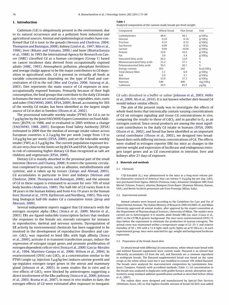
Fig. 1. Acute effects of CdCl2 on luciferase activity in adult cycling ERE-luc mice.Luciferase activity in different tissues ex vivo 6 h after a single oral dose of 1 �g/kg bw
wheat bread, flax bread and E2 groups, but by the second week of
Carbohydrates 59.6 59.6 58.5Energy (kcal/g) 3.7 3.6 3.6
o the purified AIN93G rodent diet without compromising the macronutrient com-osition or pellet quality of the diets. The Cd concentration in the rodent diets wasnalyzed by inductively coupled plasma mass spectrometry as described before (Alit al., 2010). The final calculated macronutrient compositions of the experimentaliets are shown in Table 2.
.4. Study design
To lower the background exposure to dietary estrogens the mice were switchedo purified AIN93G diet at the time of the ovariectomy, 2 weeks prior to the start ofhe experimental period.
Initially, we tested whether the selected animal model is applicable to studystrogenic activity of inorganic Cd by treating adult intact ERE-Luc female miceith a single dose of CdCl2 (1 �g/kg bw) by gavage. Tissues were dissected 6 h post
xposure and luciferase activity quantified in uterus, liver, kidney, lung, thymus,AT and skeletal muscle as described before (Rando et al., 2010).
For the 21-day study, the baseline luciferase activity before the start of thexperimental period was measured in all animals by in vivo imaging and the miceere allocated to the different experimental groups so that the average background
uciferase activity at start was comparable in all groups. Each group consisted ofve animals and all animals had free access to feed and tap water throughout thexperiment. Food, water and body weights were recorded once a week throughouthe experiment. Since animals were maintained in cages of five mice each, we mea-ured the entire amount of food and water used daily by the group and normalizedo single animal.
The five different experimental groups were: (1) control group (purified AIN93Giet); (2) CdCl2 group (1 �g/kg bw per day by gavage); (3) E2 group (s.c. long termelease pellet 5 �g/kg bw per day to resemble the physiological concentration atiestrus); (4) wheat bread diet (low Cd content 17.57 �g/kg); and (5) flax bread diethigh Cd content 49.22 �g/kg). In the CdCl2 group, CdCl2 dissolved in saline wasdministered as a single 100 �l bolus every 24 h. The mice were maintained in thexperimental groups for 21 days.
Bioluminescent imaging (BLI) sessions were carried out on days 0, 1, 7, 14 and 21t 2:00 p.m. before the daily CdCl2 administration, as described before (Ciana et al.,003). For quantification, photon emission was counted in the regions of interestnd the signals obtained were integrated from each anatomical area as previouslyescribed (Biserni et al., 2008; Rando et al., 2010). Photon emission is defined ashe number of counts per second per square centimeter (cts/cm2s). Quantificationsere done using WinLight32 imaging software (Berthold Technologies). Normal-
zation was performed using an external source of photons enabling to measure thenstrumental efficiency of photon counting (Glowell, Luxbiotech, Edinburgh, UK).
On the last day of the experiment (day 21) the animals were sacrificed and tis-ues were dissected and equally divided for the quantitative analysis of the contentf Cd, luciferase and specific mRNAs. All tissues were handled with acid washedaterial to avoid Cd contamination and the small intestine was washed with 0.9%aCl containing 5 mM EDTA (pH 7) to remove any undigested food.
.5. Cd in tissues
We quantified the concentration of Cd in the major storage organs, liver and kid-eys, and in the small intestine (duodenum and jejunum) with the method describedefore by Ali et al. (2010). For the bioavailability estimation, the absolute quantityf Cd in these organs was calculated based on the tissue weights. To account forhe background Cd concentration at the start of the experiment, we subtracted theontrol group values from the flax bread, wheat bread, and CdCl2 group values. Theissue Cd values were then compared to the oral intake to estimate the absorptionf the orally consumed Cd into the gut epithelium, and deposition into the liver andidneys.
.6. Luciferase enzymatic assay
Luciferase extraction from frozen tissues was carried out as previously describedRando et al., 2010).
CdCl2. The luciferase activity was related to the protein concentration in each sampleand expressed as RLU/�g protein. Values represent mean ± SEM (n = 5). The data wasanalyzed by one-way ANOVA and Bonferroni test. *p < 0.05, **p < 0.01, treatmentversus control. RLU, relative light units.
2.7. Gene expression analysis
Real time PCR experiments were performed using RNA extracted with RNeasy®
Mini kit (Qiagen, Milan, Italy). The preparation of cDNA was carried out as described(Biserni et al., 2008) using TaqMan technology with pre-made primers from Taq-Man Gene Expression assays (Applied Biosystems, Milan, Italy) for the endogenousgene Esr1 (Mm00433149 m1), progesterone receptor (Mm00435625 m1) prothymo-sine (Mm02342432 g1), and as a reference gene assay 18S rRNA VIC-MGB-PDAR.The amplification reaction was carried out according to the manufacturer’s protocolusing 7900HT fast real-time PCR system (Applied Biosystems, Milan, Italy) and datawere analyzed using the 2−��Ct method (Livak and Schmittgen, 2001).
2.8. Statistical analysis
Normally distributed data was analyzed with one-way or two-way ANOVAand p < 0.05 was considered as statistically significant difference. The differencesbetween the groups were then analyzed with Bonferroni post hoc test, comparingthe treatments to the control group. All analyses were carried out with GraphPadPrism version 5.01 for Windows, GraphPad Software (San Diego California USA). Intables and figures, data is presented as mean ± SEM unless stated otherwise, and sta-tistical differences are marked as ‘*’ for p < 0.05, ‘**’ for p < 0.01 and ‘***’ for p < 0.001.All probabilities are two-tailed.
3. Results
3.1. Reporter gene activity in the ERE-Luc mouse model afteracute exposure to CdCl2
The sensitivity of the selected animal model was tested by treat-ing adult cycling females with a single oral dose of 1 �g/kg/bw ofCdCl2. Significant luciferase induction was detected 6 h later in theuterus and thymus (Fig. 1) demonstrating that the reporter genesystem in the ERE-Luc mouse model is sensitive to oral Cd treat-ment in low exposure concentrations.
3.2. Food and water intake and organ weights
OVX adult female mice were maintained on the experimentaldiets for 21 days and the food and water consumption was recordedonce a week. The average daily food intake was 2.5 g/mouse andwater consumption 5.5–6 g/mouse in all treatments except theE2 group (7.6 g/mouse). All groups consumed similar amounts offood, and the food and water consumption did not significantlychange during the 21-day experimental period within each respec-tive group. Treatments did not affect the body weight of the control,
the study in the mice treated with CdCl2 we observed a significantweight loss (Table 3).
The weights of livers and kidneys did not differ between con-trol, wheat and flax bread groups (Fig. 2). CdCl2 administration
78 B. Ramachandran et al. / Toxicology Letters 202 (2011) 75–84
Table 3Body weights of the ERE-Luc mice during the experiment.
Treatment Day of treatment % difference at d21(treatment versus control)
1 7 14 21
Control 23.8 ± 0.6 24.0 ± 0.4 24.2 ± 0.4 25.0 ± 0.5 –CdCl2 23.2 ± 0.3 22.6 ± 0.4 22.3 ± 0.7* 22.7 ± 0.5* −9Wheat bread 23.5 ± 0.2 24.0 ± 0.3 24.2 ± 0.3 24.4 ± 0.3 −3Flax bread 23.9 ± 0.9 24.3 ± 0.9 24.6 ± 0.9 25.7 ± 1.0 +317�-Estradiol 23.0 ± 0.6 24.0 ± 0.8 25.1 ± 0.8 25.7 ± 0.6 +3
The weights are expressed in grams as mean ± SEM (n = 5).* p < 0.05 control versus treatment and time by two-way ANOVA and Bonferroni post hoc test.
F re reco
ciw
otceww
3
aflte
btdg1Cbfln
TC
f
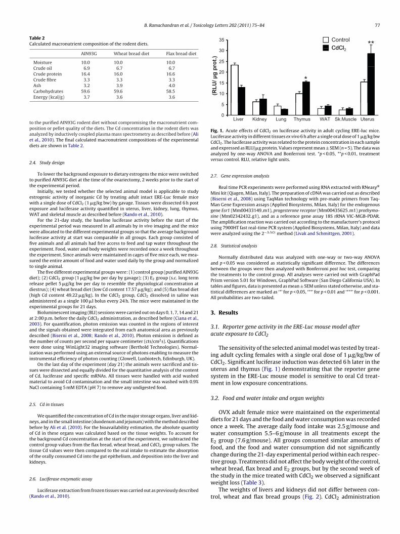
ig. 2. Organ weights at sacrifice. The wet weights of uterus, liver and kidneys wene-way ANOVA and Bonferroni test. *p < 0.05, **p < 0.01, treatment versus control.
aused a significant decrease in the liver weight and E2 significantlyncreased liver weight, but these treatments did not change kidney
eights (Fig. 2).The wet weight of the uterus has long been utilized as a hallmark
f estrogen action, and the OECD uterine growth assay has becomehe standard guideline in the assessment of estrogenic activities ofhemicals. In our study, E2 significantly increased uterine weight, asxpected. A smaller but significant increase in the uterine weightsas observed in the CdCl2 group. No modulation of uterine weightas observed in the wheat and flax diet groups (Fig. 2).
.3. Cd intake and concentration in gut, liver and kidneys
The wheat bread diet was analyzed to contain 17.57 �g/kg Cd,nd the diet based on the same bread supplemented with 10%axseed contained 49.22 �g/kg. The Cd concentration in the con-rol diet (AIN93G) was 9.22 �g/kg. All diets were comparable toach other in terms of macronutrient composition (Table 2).
The total oral intake of Cd per kg bw was calculated in each groupased on the recorded body weights (Table 3) and food consump-ion data. During the 21-day study, the mice consuming controliet ingested a total of 483 ng Cd (≈0.95 �g/kg bw per day); theroup treated with CdCl2 966 ng (≈2.0 �g/kg per day, consisting of
.0 �g/kg bw per day as CdCl2 and 1.0 �g/kg bw per day as dietaryd originating from the control diet); the mice consuming wheatread diet 924 ng (≈1.8 �g/kg bw per day) and the mice consumingax bread diet 2580 ng (≈5.1 �g/kg bw per day) (Table 4). Sig-ificant increase in Cd concentration was observed in the smallable 4d intake and tissue accumulations in the 21-day exposure.
Oral intake(ng)
Exposure(�g/kg/day)
Control 483 0.95Wheat bread group 924 1.8Flax bread group 2580 5.1CdCl2 group 966 (483 ng from the CdCl2 and
483 ng from the AIN93G diet)2.0 (1.0 from the CdCl2 and1.0 from the AIN93G diet)
a The average background concentration (i.e. average concentration in the control grouprom the values.
b Intestine, liver and kidneys.c % intake value is based on the intake of CdCl2.
orded at sacrifice. Values represent mean ± SEM (n = 5). The data was analyzed by
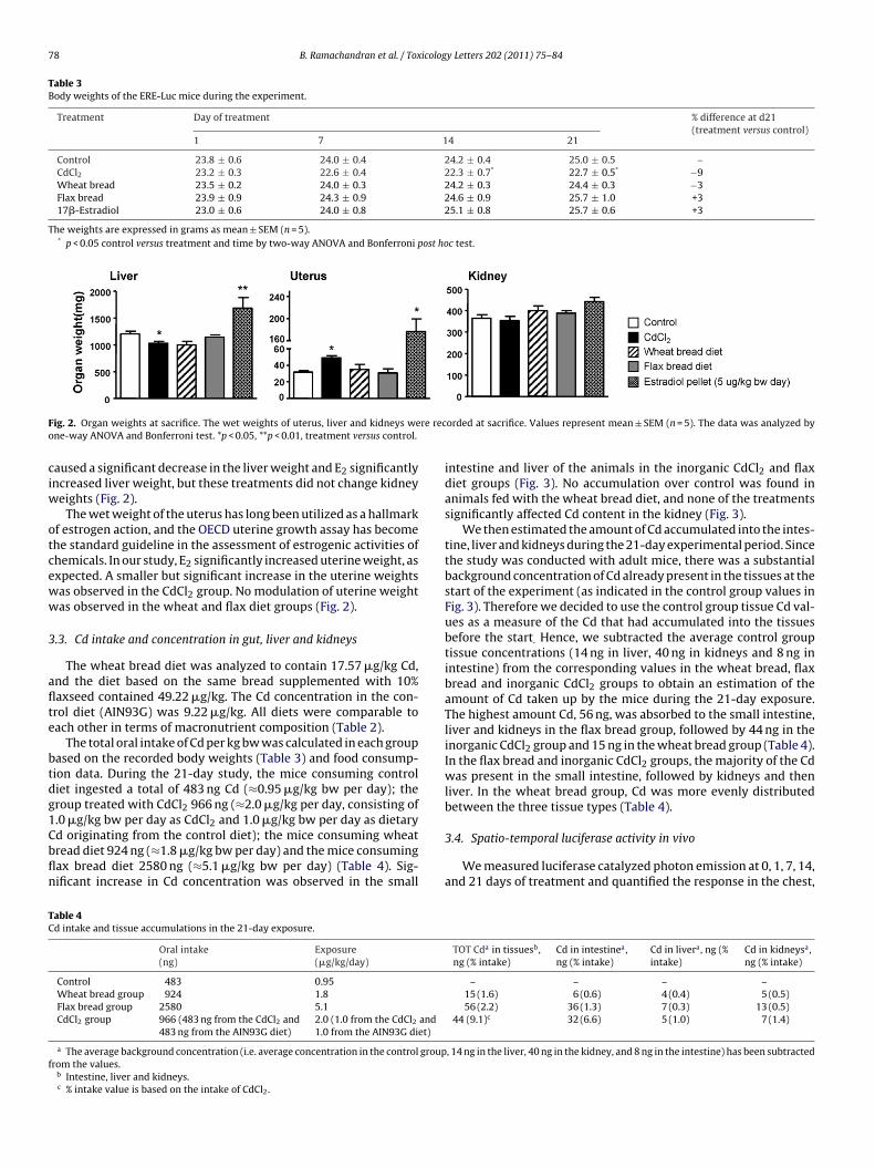
intestine and liver of the animals in the inorganic CdCl2 and flaxdiet groups (Fig. 3). No accumulation over control was found inanimals fed with the wheat bread diet, and none of the treatmentssignificantly affected Cd content in the kidney (Fig. 3).
We then estimated the amount of Cd accumulated into the intes-tine, liver and kidneys during the 21-day experimental period. Sincethe study was conducted with adult mice, there was a substantialbackground concentration of Cd already present in the tissues at thestart of the experiment (as indicated in the control group values inFig. 3). Therefore we decided to use the control group tissue Cd val-ues as a measure of the Cd that had accumulated into the tissuesbefore the start. Hence, we subtracted the average control grouptissue concentrations (14 ng in liver, 40 ng in kidneys and 8 ng inintestine) from the corresponding values in the wheat bread, flaxbread and inorganic CdCl2 groups to obtain an estimation of theamount of Cd taken up by the mice during the 21-day exposure.The highest amount Cd, 56 ng, was absorbed to the small intestine,liver and kidneys in the flax bread group, followed by 44 ng in theinorganic CdCl2 group and 15 ng in the wheat bread group (Table 4).In the flax bread and inorganic CdCl2 groups, the majority of the Cdwas present in the small intestine, followed by kidneys and thenliver. In the wheat bread group, Cd was more evenly distributedbetween the three tissue types (Table 4).
3.4. Spatio-temporal luciferase activity in vivo
We measured luciferase catalyzed photon emission at 0, 1, 7, 14,and 21 days of treatment and quantified the response in the chest,
TOT Cda in tissuesb,ng (% intake)
Cd in intestinea,ng (% intake)
Cd in livera, ng (%intake)
Cd in kidneysa,ng (% intake)
– – – –15 (1.6) 6 (0.6) 4 (0.4) 5 (0.5)56 (2.2) 36 (1.3) 7 (0.3) 13 (0.5)
44 (9.1)c 32 (6.6) 5 (1.0) 7 (1.4)
, 14 ng in the liver, 40 ng in the kidney, and 8 ng in the intestine) has been subtracted
B. Ramachandran et al. / Toxicology Letters 202 (2011) 75–84 79
F in sms ntrol 4a ata wav
gt2csal
3
sryIsusbnaecdtagn
3
eAdegeebiiltald
ig. 3. Concentration of Cd in liver, kidneys and intestine. The concentration of Cdpectrometry. Exposure to Cd during the 21-day period in the different groups: cos CdCl2 and 483 ng from control diet). Values represent mean ± SEM (n = 5). The dersus control.
enital area, abdomen and limbs separately to reflect the activi-ies in the liver, reproductive tract, gut and bone (Biserni et al.,008; Rando et al., 2010). The luciferase activity did not signifi-antly vary in time in the control group (Fig. 4) In the E2 groupignificantly increased activity was measured in the chest, genitalrea and abdomen (Fig. 4c–e). CdCl2, wheat and flax diets affecteduciferase expression only in the chest area (Fig. 4a).
.5. Luciferase activity ex vivo in tissue homogenates
Although the in vivo imaging is a good starting point whentudying estrogen activity, it cannot give detailed information onesponses in inner organs. Therefore we carried out luciferase anal-sis in tissues dissected on the last day of the experiment (day 21).n E2 group, significant luciferase induction was detected in themall intestine (jejunum), kidney, brain, pituitary gland, WAT, andterus, and significant repression in thymus (Fig. 5). Inorganic CdCl2ignificantly affected luciferase activity only in WAT (Fig. 5). Theread diets modulated luciferase activity in a wider range of tissues,amely in liver, pituitary gland, thymus and WAT. Both inhibitorynd inducing effects were observed with the bread diets, but theffects did not systematically mimic those of CdCl2 or E2 (Fig. 5). Toontrol that the lack of similarity to E2 effects in the CdCl2 and breadiet groups was not dependent on repression of ER� expression byhe treatments, we quantified the expression of ER� in uterus, WATnd thymus. The expression did not differ from the baseline, sug-esting that the lack of similarities in the estrogen responses wereot due to altered receptor expression (Supplementary Fig. 1).
.6. Expression of endogenous estrogen target genes
We quantified the expression of two endogenous markers ofstrogen signaling, progesterone receptor (PR) and prothymosine(PTMA), in uterus, WAT and thymus. As predicted by the luciferaseata, E2 induced both PR and PTMA expression in the classicalstrogen target tissues, uterus and WAT (Fig. 6). Similarly, inor-anic CdCl2 displayed estrogen activity in the WAT by inducing PRxpression, as predicted by the luciferase response (Fig. 6). How-ver, the responses to the bread diets were more difficult to predictased on the luciferase response: wheat bread induced luciferase
n WAT but none of the endogenous genes, and both bread dietsnduced PTMA in uterus without affecting luciferase. The corre-
ation between luciferase and endogenous gene responses in thehymus was even more complicated. Inorganic CdCl2 did not affectny measured parameters in this organ, while bread diets increaseduciferase and PTMA, flax bread in addition decreased PR, and E2ecreased luciferase and PR but increased PTMA (Fig. 6).all intestine, liver and kidneys was measured by inductively coupled plasma mass83 ng, wheat bread diet 924 ng, flax bread diet 2580 ng, and CdCl2 966 ng (483 ngs analyzed by one-way ANOVA and Bonferroni test. *p < 0.05, **p < 0.01, treatment
4. Discussion
The present study investigated the effect of a 21-day oraladministration of Cd in different forms on markers of estrogenic-ity and tissue deposition of Cd in ERE-Luc reporter mice. Ourresults confirm previous reports showing that CdCl2 may possessestrogen-like activity in vivo (Johnson et al., 2003; Höfer et al.,2009; Ali et al., 2010), and we identify WAT as a potential noveltarget tissue. However, bread bound Cd did not mirror the effectsof CdCl2. Our data suggests that caution should be taken when purecompound results are extrapolated to dietary exposure scenarios.
4.1. Effects of the treatments on estrogen signaling
We used three different marker genes to asses genomic estrogensignaling after Cd exposure: luciferase, PR and PTMA. The luciferasegene is driven by a dimerized consensus estrogen responsive ele-ment and functions as a sensitive indicator of estrogen activity(Ciana et al., 2003). Both chosen endogenous genes harbor estro-gen responsive elements in their promoter regions and respond toestrogens in different tissues (Garnier et al., 1997; Tsitsilonis et al.,1998; Martini and Katzenellenbogen, 2001; Bianco and Montano,2002). We first measured the expression of luciferase in a widevariety of tissues, and then completed the data with the twoendogenous markers in uterus, a classical estrogen target tissue;in WAT, an estrogen target tissue where CdCl2 increased luciferaseactivity; and in thymus, a non-classical estrogen target tissue whereboth bread diets modulated luciferase activity.
In the E2 treated mice, luciferase, PTMA and PR expression wereall significantly up-regulated in typical estrogen target tissues,uterus and WAT (Figs. 5 and 6). In thymus, however, E2 repressedluciferase and PR while promoted PTMA expression. Although allthree marker genes are regulated directly by E2, PTMA also affectsER� signaling on another level: it binds to co-factors involved inestrogen signaling affecting their availability for ER� (Bianco andMontano, 2002). Perhaps in thymus, where the biological functionand genomic targets of estrogens might differ from the classicaltarget tissues such as uterus and WAT, also the regulation of thereceptor activity in terms of co-factors is different, leading to thediscrepancy in the expression between PTMA and the two othermarkers in our study.
The mice receiving inorganic CdCl2 displayed a narrower rangeof effects than the mice receiving E2. For instance, none of thegenes was significantly regulated in the uterus. Previously, inor-ganic Cd has been shown to up-regulate C3 and PR in the rat
uterus (Johnson et al., 2003; Höfer et al., 2009). In another reportermouse model, CdCl2 did not affect luciferase expression; insteadit promoted markers of membrane-bound estrogen signaling (Aliet al., 2010). Strains differ in their capacity to produce metalloth-ionein to neutralize Cd, which could lead to different biological
80 B. Ramachandran et al. / Toxicology Letters 202 (2011) 75–84
F dy. Lug osureb e exprb rol.
orwJamagmfotats
ig. 4. Spatio-temporal luciferase expression in the treatment groups during the stuenital area, abdomen and limbs on days 0, 1, 7, 14 and 21 of treatment. Daily expread diet 5.1 and CdCl2 2.0 (1.0 from CdCl2 and 1.0 from control diet). The results ary two-way ANOVA and Bonferroni test. *p < 0.05, **p < 0.01, treatment versus cont
utcomes (Shaikh et al., 1993). For example, in Sprague-Dawleyats up-regulation of C3 is obtained after a single 5 �g/kg bw dose,hile in Wistar rats 2 mg/kg bw dose is required (Höfer et al., 2009;
ohnson et al., 2003). Our study was conducted in C57BL6 mice,s was the study of Ali et al. Although the length of their treat-ent period was only 3 days compared to our 21 days, the results
re similar: no significant regulation of genomic markers of estro-en signaling was observed in the uterus, suggesting that C57Blice differ from rats. Interestingly, when we treated intact cycling
emale mice with CdCl2 in our sensitivity test, significant induction
f luciferase in the uterus was observed (Fig. 1). These observa-ions suggest that both the strain and the hormonal milieu of thenimals might affect the bioactivity of Cd. More studies are neededo define the circumstances under which Cd can promote estrogenignaling.ciferase activity was measured by in vivo imaging as photon emission in the chest,to Cd in the different groups as �g/kg bw: control 0.95, wheat bread diet 1.8, flaxessed as cts/cm2 and the bars represent mean ± SEM (n = 5). The data was analyzed
Interestingly, CdCl2 promoted the expression of PR andluciferase in WAT. Moreover, the body weight of the CdCl2 treatedmice was significantly lower than control by the end of the exper-iment, despite similar food consumption in all groups. Recently,CdCl2 was shown to reduce both body weight and WAT weight inadult male mice (Kawakami et al., 2010). Although in that study rel-atively high doses were used (0.5–2 mg/kg bw), the results togetherwith ours suggest that CdCl2 might be active in the adipose tissue,and this activity could involve estrogen signaling.
The bread diets had diverse effects, modulating at least one of
the markers in all three tissues (WAT, thymus and uterus). In theuterus, the bread diets did not regulate luciferase or PR, which isin agreement with the literature (Penttinen-Damdimopoulou et al.,2009). PTMA expression in the uterus, however, was up-regulatedby the diets. As discussed above, the more complex involvement of
B. Ramachandran et al. / Toxicology Letters 202 (2011) 75–84 81
F activita RLU/�w contrA l. RLU
Pm
CbCvtbfattHhags
vIiaTegsottrplw
ig. 5. Effects of the treatments on tissue specific luciferase expression. Luciferasectivity was related to the protein concentration in each sample and expressed asheat bread diet 1.8, flax bread diet 5.1, and CdCl2 2.0 (1.0 from CdCl2 and 1.0 fromNOVA and Bonferroni test. *p < 0.05, **p < 0.01, ***p < 0.001 treatment versus contro
TMA in estrogen signaling might make this gene a more generalarker of estrogen modulation than luciferase and PR.In WAT, where both E2 and CdCl2 induced ER signaling, the high
d flax bread diet was devoid of such activity but the low Cd wheatread promoted luciferase expression. Although the oral intake ofd was similar in wheat and CdCl2 groups(1.8 �g/kg bw from dietersus 1.0 �g/kg as CdCl2 + 1.0 �g/kg from diet, respectively), theotal tissue deposition of Cd was more comparable between the flaxread and CdCl2 groups (56 ng versus 44 ng, respectively). It is there-ore difficult to understand why the flax bread diet did not affect ERctivity in WAT while wheat bread diet did. Similarly, in thymus,he effects induced by the bread diets did not systematically mirrorhose of E2 or CdCl2 and there was no apparent dose-dependency.ere, the presence of other bioactive substances in the bread matrixas to be considered. For instance wheat contains alkylresorcinolsnd flaxseed lignans. The influence of these compounds on estro-en signaling in the presence of Cd could be addressed in futuretudies.
Luciferase activity was also measured in other tissue types by exivo analysis as well as in different body areas by in vivo imaging.n prolonged exposure to ER modulators, the reporter gene activityn our mouse model oscillates with compound/treatment specificmplitudes and frequencies (Rando et al., 2009; Rando et al., 2010).he functional consequences of such oscillation are not clear, how-ver, to reliably detect ER modulation in long exposure the reporterene activity should be measured at different time points. If weummarize the tissue targets of the treatment in our study basedn both in vivo and ex vivo analysis, it seems that the bread dietsarget liver, pituitary gland, thymus and WAT. Inorganic CdCl2, on
he other hand, only targets WAT and liver. E2 is active in a widerange of tissues (liver, reproductive tissues, intestine, kidney, brain,ituitary gland, thymus, and WAT), as expected based on our ear-ier studies (Rando et al., 2010; Ciana et al., 2003). In our study, liveras the only tissue type where modulation of estrogen signaling
y in different tissues ex vivo after the 21-day long treatment period. The luciferaseg protein. Daily exposure to Cd in the different groups as �g/kg bw: control 0.95,ol diet). The bars represent mean ± SEM (n = 5). The data was analyzed by one-way, relative light units.
was observed after all treatments (CdCl2 and E2 in vivo, bread dietsin vivo and ex vitro). Since many absorbed dietary compounds arefirst transported to the liver, and this organ responds to estrogenmodulators, biomarkers of estrogen signaling in the liver could beused as a quick screen for estrogen modulation by dietary factors.
The traditional screen for estrogen-like activity in vivo is therodent uterine growth test. We therefore measured the uterineweights in our study, and as expected, E2 significantly increased theweight. Interestingly, also CdCl2 increased uterine weight althoughnone of the markers of genomic signaling were regulated. Inresponse to estrogens, the expression of hundreds of genes in theuterus is regulated in preparation to embryo implantation andpregnancy, and both genomic and non-genomic signaling path-ways are involved. In a recent study in another estrogen reportermouse model, a 3-day s.c. exposure to 5–500 �g/kg bw CdCl2activated markers of non-genomic signaling in the absence of ERE-response or uterine growth effects (Ali et al., 2010). In the presentstudy, membrane associated signaling was not measured, but itis possible that this pathway was stimulated by CdCl2 throughoutthe 21-day experimental period, finally leading to increased uter-ine weight. It is noteworthy that the increase we measured wassignificantly lower than in the E2 group, suggesting only partialestrogen effects. The bread diets did not modulate uterine weight.Similar results were obtained with wheat bran and flaxseed diets inanother reporter mouse model (Penttinen-Damdimopoulou et al.,2009) and with flaxseed diet in an immature rat uterotrophic test(Khan et al., 2007).
4.2. Bioaccumulation of dietary Cd
The mice in all groups, including control, ingested Cd in amountsexceeding the new EFSA recommendation (2.5 �g/kg bw in aweek): the control mice ingested 2.7-fold, wheat bread mice 5.0-fold, CdCl2 group 5.6-fold (both dietary background and gavaged

82 B. Ramachandran et al. / Toxicology Letters 202 (2011) 75–84
F ion of1 bw: coa lyzedv
Csc(oamaeCCmbtgwbo
a1mewCc
ig. 6. Effects of the treatments on endogenous estrogen target genes. The express8S RNA in selected tissues. Daily exposure to Cd in the different groups as �g/kgnd 1.0 from control diet). The bars represent mean ± SEM (n = 5). The data was anaersus control.
dCl2 counted), and flax bread group 14.3-fold more than con-idered safe by EFSA. These intake values are not exaggeratedompared to humans in that they reflect exposures in polluted areasWHO, 1992; Järup et al., 1998). As planned, a clear gradient in theral intake of Cd was achieved between the groups: the wheat breadnd CdCl2 mice ingested 2-fold and the flax bread mice over 5-foldore Cd than the controls. When Cd deposition to intestine, liver
nd kidneys were compared between the groups, a pattern differ-nt from the oral intake was observed. Although wheat bread anddCl2 mice had similar oral intakes of Cd (924 ng versus 483 ng asdCl2 + 483 ng from the diet, respectively), the CdCl2 mice accu-ulated markedly more Cd into the tissues (44 ng) than the wheat
read mice (15 ng) (Table 4). Indeed, the total tissue accumula-ion in the CdCl2 group was more comparable to the flax breadroup (56 ng), although the oral intake in the flax group (2580 ng)as over twice as high as in the CdCl2 group. This suggests higher
ioavailability of gavaged CdCl2 compared to diet bound Cd, asbserved in previous studies (Chan et al., 2000; Lind et al., 1998).
Studies conducted with 109Cd have shown that over 70% ofbsorbed dietary Cd is deposited into liver and kidneys (Andersen,989). However, in our study majority of the Cd seemed to accu-
ulate to the intestine (Table 4). The absorption of Cd has beenxtensively studied, and the metal is known to rapidly accumulateithin the intestinal mucosa (Zalups and Ahmad, 2003; Reeves andhaney, 2008). In contrast, the transport of Cd into the systemicirculation occurs with a low rate (Zalups and Ahmad, 2003). It is
progesterone receptor and prothymosine mRNA was quantified against ribosomalntrol 0.95, wheat bread diet 1.8, flax bread diet 5.1, and CdCl2 2.0 (1.0 from CdCl2by one-way ANOVA and Bonferroni test. *p < 0.05, **p < 0.01, ***p < 0.001 treatment
therefore possible that the Cd bound to mucosa sheds away withthe renewing enterocytes before further absorption. Some supportfor this hypothesis comes from a study by Phillpotts (1979). Heorally administered adult rats a low dose of 109Cd and studied theretention in various tissues. The majority of 109Cd was detected inthe duodenum within the 24 h post challenge, but as the concen-tration in the duodenum rapidly fell between 24 and 48 h post Cdadministration, the concentration in the inner organs did not rise(Phillpotts, 1979). Similarly, House et al. (2003) has shown thatthe whole body retention of 109Cd declines dramatically withinthe first week after ingestion, suggesting an initial shorter termretention for this metal than the long term storage in liver andkidneys (House et al., 2003). In agreement with Andersen (1989),also Phillpotts detected the majority of absorbed Cd (i.e. Cd presentin inner organs) in the kidneys and liver. Hence, we can use theamount of Cd in liver and kidneys as a proxy of the absorbed Cdwhen estimating the bioavailability in our study.
When the quantity of Cd in liver and kidneys is compared to theoral intake, the highest retention was observed in the CdCl2 group,where a quantity corresponding to 2.4% of the oral CdCl2 intakewas detected. In the bread groups, markedly less Cd accumulated to
these organs; in both wheat and flax bread groups the quantity of Cdaccumulated to the liver and kidneys corresponded to less than 1%of the orally ingested dose. These values are in agreement with theliterature, showing that the presence of wheat in diet decreases Cdaccumulation in tissues (Chan et al., 2000; House et al., 2003). The
icolog
fa2mHiectppd
agrifWadpiaCtbtctfibfflsbtm(obSe
4
bgccrp
ahsglsaottc
B. Ramachandran et al. / Tox
actors believed to contribute to the Cd uptake from the gut mostre trace element deficiencies and dietary fibre (Reeves and Chaney,008). In all our diets, the same standard amount of AIN93G mineralix was added (Reeves et al., 1993), so deficiencies are not likely.owever, there are differences in the makeup of the dietary fibre
n the diets. All diets contain 3.3% fibre. In the control diet, this wasntirely composed of cellulose. In the bread diets the fibre fractionontained both fibre naturally present in the breads and cellulosehat was added to balance the fibre fraction to 3.3% in all diets. Theresence of complex plant fibre, known to inhibit Cd absorption,robably partially explains the lower bioavailability from the breadiets (Andersen et al., 2004).
In spite of similar oral intakes in the wheat and CdCl2 groups,nd similar absolute tissue levels of Cd in flax bread and CdCl2roups, we did not observe any systematic similarities in estrogenesponses in these groups. This suggests that the form in which Cd isngested affects the absorption as discussed above, but also that theorm in which Cd is absorbed affects subsequent biological activity.
hen CdCl2 is administered by oral gavage, Cd is probably rapidlyvailable for absorption in ionic form at the low pH of proximal duo-enum. In the flax bread diet, Cd is mainly bound to the flaxseedrotein fraction (Lei et al., 2003), that requires digestion before Cd
s released. Interestingly, mercury can be absorbed through aminocid transporters in complex with cysteines (Cannon et al., 2001).d can also complex to cysteines, but the involvement of amino acidransporters in the absorption of Cd from intestine lumen has noteen studied to our knowledge. Intriguingly, the form of Cd is knowo affect its toxicity: in the case of kidney damage, metallothioneinomplexed Cd causes nephrotoxicity at 8-fold lower doses than cys-eine complexed Cd (Min et al., 1986). We hypothesize that theorm of absorbed Cd may also affect subsequent estrogenic activ-ty, which could help to explain the differences between CdCl2 andread diets in the present study. Another explanation to the dif-erences could be additional bioactive substances present in theax bread diet. For instance, flaxseed is the richest known dietaryource of lignans, plant secondary metabolites that are convertedy gut microbes to enterolactone. We have shown that enterolac-one selectively regulates estrogen signaling, and this compound
ight thereby contribute to the net results measured in this studyPenttinen et al., 2007; Adlercreutz, 2007). Furthermore, the effectf food matrix on the in vivo activity of estrogenic compounds haseen demonstrated in several other studies (Rando et al., 2009;irtori et al., 2005; Sirtori et al., 2009; Penttinen-Damdimopoulout al., 2009).
.3. Cd as an endocrine disruptor: critical issues and conclusions
The current classification of Cd as type I human carcinogen isased on data from occupationally exposed adults, and the healthuidance values on biomarkers of nephrotoxicity. The recent dis-overy of the endocrine effects of Cd in vivo in experimental animalsalls for re-evaluation of human health effects, and data supportingisk assessment, such as identification of biomarkers, should be ariority of future Cd studies.
To date the estrogenicity of Cd in vivo has been studied bydministering inorganic Cd salts by injections or gavage, althoughuman populations are exposed to Cd mainly through diet. Ourtudy is the first one to compare the estrogen-like activity of inor-anic CdCl2 to the activity of a major dietary source of Cd, bread. Theack of overlap in effects of the bread diets, CdCl2 and E2 on estrogenignaling in the various different parameters measured is intriguing
nd illustrates the challenges in extrapolating the observed effectsf a pure compound, such as the E2-like activity of inorganic CdCl2,o the effect of a Cd-containing bread diet. It seems that whenhe biological activity of dietary substances is investigated, pureompound studies should be accompanied by approaches, whichy Letters 202 (2011) 75–84 83
take into consideration the natural food context of the substancein question to account for possible matrix effects.
The quantification of Cd in tissues revealed that the diet-boundCd is poorly absorbed compared to inorganic Cd. Furthermore, evenin the presence of similar tissue Cd concentrations, diet-derivedCd does not possess the same estrogenic activity as CdCl2. Takentogether, our results support previous findings of estrogen-likeactivity of inorganic CdCl2 in vivo, but do not suggest that diet-bound Cd would possess the identical activity. The future researchefforts should be focused on understanding the mechanism of inter-action between Cd and estrogen signaling; the identification ofbiologically active Cd species in vivo; and the characterization ofbiomarkers of the estrogenic effects of Cd in vivo.
Conflict of interest statement
The authors declare that there are no conflicts of interest.
Acknowledgements and funding
The authors wish to thank Paola Assi, Clara Meda, MonicaRebecchi, Sandrine Bruel and Anne Hillenweck for their excellenttechnical assistance; Paolo Sparaciari for his critical suggestions onanimal handling and Sara Della Torre for her extensive help in theanalysis of RT-PCR data. This work was supported by the Networkof Excellence (FOOD-CT-2004-506319), CASCADE NoE to AM, SM,PD, JC, MB and HH; and EXERA (LSHB-CT-2006-037168) to AM.
Appendix A. Supplementary data
Supplementary data associated with this article can be found, inthe online version, at doi:10.1016/j.toxlet.2010.12.022.
References
Adlercreutz, H., 2007. Lignans and human health. Crit. Rev. Clin. Lab. Sci. 44 (5–6),483–525.
Ali, I., Penttinen-Damdimopoulou, P.E., Mäkelä, S.I., Berglund, M., Stenius, U.,Akesson, A., Håkansson, H., Halldin, K., 2010. Estrogen-like effects of cadmiumin vivo do not appear to be mediated via the classical estrogen receptor tran-scriptional pathway. Environ Health Perspect 118 (10), 1389–1394.
Andersen, O., 1989. Oral cadmium exposure in mice: toxicokinetics and efficiencyof chelating agents. Crit. Rev. Toxicol. 20 (2), 83–112.
Andersen, O., Nielsen, J.B., Nordberg, G.F., 2004. Nutritional interactions in intestinalcadmium uptake possibilities for risk reduction. Biometals 17 (5), 543–547.
Bhattacharyya, M.H., Whelton, B.D., Stern, P.H., Peterson, D.P., 1988. Cadmium accel-erates bone loss in ovariectomized mice and fetal rat limb bones in culture. Proc.Natl. Acad. Sci. U.S.A. 85, 8761–8765.
Bianco, N.R., Montano, M.M., 2002. Regulation of prothymosin alpha by estrogenreceptor alpha: molecular mechanisms and relevance in estrogen-mediatedbreast cell growth. Oncogene 21 (34), 5233–5244.
Biserni, A., Giannessi, F., Sciarroni, A.F., Milazzo, F.M., Maggi, A., Ciana, P., 2008.In vivo imaging reveals selective peroxisome proliferator activated receptormodulator activity of the synthetic ligand 3-(1-(4-chlorobenzyl)-3-t-butylthio-5-isopropylindol-2-yl)-2,2-dimethylpropanoic acid (MK886). Mol. Pharmacol.73, 1434–1443.
Brama, M., Gnessi, L., Basciani, S., Cerulli, N., Politi, L., Spera, G., Mariani, S., Cherubini,S., d’Abusco, A.S., Scandurra, R., Migliaccio, S., 2007. Cadmium induces mitogenicsignaling in breast cancer cell by an ERalpha-dependent mechanism. Mol. CellEndocrinol. 264, 102–108.
Cannon, V.T., Zalups, R.K., Barfuss, D.W., 2001. Amino acid transporters involved inluminal transport of mercuric conjugates of cysteine in rabbit proximal tubule.J. Pharmacol. Exp. Ther. 298 (2), 780–789.
Chan, D.Y., Black, W., Hale, B., 2000. Bioaccumulation of cadmium from durum wheatdiets in the livers and kidneys of mice. Bull. Environ. Contam. Toxicol. 64 (4),526–533.
Ciana, P., Luccio, G.D., Belcredito, S., Pollio, G., Vegeto, E., Tatangelo, L., Tiveron, C.,Maggi, A., 2001. Engineering of a mouse for the in vivo profiling of estrogen
receptor activity. Mol. Endocrinol. 15, 1104–1113.Ciana, P., Raviscioni, M., Mussi, P., Vegeto, E., Que, I., Parker, M.G., Lowik, C., Maggi, A.,2003. In vivo imaging of transcriptionally active estrogen receptors. Nat. Med. 9(1), 82–86.
EFSA, 2009. Scientific opinion of the Panel on Contaminants in the Food Chain on arequest from the European Commission on cadmium in food. EFSA J. 980, 1–144.
8 icolog
F
F
G
G
H
H
H
I
J
J
J
K
K
K
K
L
L
L
L
M
M
Wilson, V.S., Bobseine, K., Gray, L.E., 2004. Development and characterization of
4 B. Ramachandran et al. / Tox
AO/WHO (Food and Agriculture Organization/World Health Organization),1988. Evaluation of certain food additives and contaminants (Thirty-ThirdReport of the Joint FAO/WHO Expert Committee on Food Additives).WHO Technical Report Series, No. 776, 1989 [1988, TRS 776-JECFA 33].http://www.inchem.org/documents/jecfa/jecmono/v024je09.htm.
AO/WHO (Food and Agriculture Organization/World Health Organization),2005. Summary and conclusions of the sixty-four meeting of the JointFAO/WHO Expert Committee on Food Additives (JECFA). Available at:ftp://ftp.fao.org/es/esn/jecfa/jecfa64 summary.pdf.
arcia-Morales, P., Saceda, M., Kenney, N., Kim, N., Salomon, D.S., Gottardis, M.M.,Solomon, H.B., Sholler, P.F., Jordan, V.C., Martin, M.B., 1994. Effect of cadmiumon estrogen receptor levels and estrogen-induced responses in human breastcancer cells. J. Biol. Chem. 269, 16896–16901.
arnier, M., Di Lorenzo, D., Albertini, A., Maggi, A., 1997. Identification of estrogen-responsive genes in neuroblastoma SK-ER3 cells. J. Neurosci. 17, 4591–4599.
enson, M.C., Chedrese, P.J., 2004. Endocrine disruption by cadmium, a commonenvironmental toxicant with paradoxical effects on reproduction. Exp. Biol. Med.(Maywood) 229, 383–392.
öfer, N., Diel, P., Wittsiepe, J., Wilhelm, M., Degen, G.H., 2009. Dose- and route-dependent hormonal activity of the metalloestrogen cadmium in the rat uterus.Toxicol. Lett. 191, 123–131.
ouse, W.A., Hart, J.J., Norvell, W.A., Welch, R.M., 2003. Cadmium absorption andretention by rats fed durum wheat (Triticum turgidum L. var. durum) grain. Br. J.Nutr. 89 (4), 499–508.
ARC (International agency for research on cancer), 1993. Beryllium, Cadmium,Mercury and Exposures in the Glass Manufacturing Industry. IARC Mono-graphs on the Evaluation of Carcinogenic Risk of Chemicals to Humans,vol. 58. International Agency for Research on Cancer, Lyon, France, 444 pp.http://monographs.iarc.fr/ENG/Monographs/vol58/volume58.pdf.
ärup, L., Akesson, A., 2009. Current status of cadmium as an environmental healthproblem. Toxicol. Appl. Pharmacol. 238, 201–208.
ärup, L., Berglund, M., Elinder, C.G., Nordberg, G., Vahter, M., 1998. Health effectsof cadmium exposure – a review of the literature and a risk estimate. Scand. J.Work Environ. Health 24 (Suppl. 1), 1–51.
ohnson, M.D., Kenney, N., Stoica, A., Hilakivi-Clarke, L., Singh, B., Chepko, G., Clarke,R., Sholler, P.F., Lirio, A.A., Foss, C., Reiter, R., Trock, B., Paik, S., Martin, M.B., 2003.Cadmium mimics the in vivo effects of estrogen in the uterus and mammarygland. Nat. Med. 9, 1081–1084.
awakami, T., Sugimoto, H., Furuichi, R., Kadota, Y., Inoue, M., Setsu, K., Suzuki,S., Sato, M., 2010. Cadmium reduces adipocyte size and expression levels ofadiponectin and Peg1/Mest in adipose tissue. Toxicology 267 (1–3), 20–26.
han, G., Penttinen, P., Cabanes, A., Foxworth, A., Chezek, A., Mastropole, K., Yu, B.,Smeds, A., Halttunen, T., Good, C., Mäkelä, S., Hilakivi-Clarke, L., 2007. Maternalflaxseed diet during pregnancy or lactation increases female rat offspring’s sus-ceptibility to carcinogen-induced mammary tumorigenesis. Reprod. Toxicol. 23,397–406.
jellstrom, T., Nordberg, G.F., 1985. Kinetic model of cadmium metabolism. In Cad-mium and health: a toxicological and epidemiological appraisal. In: Friberg,L., Elinder, C.-G., Kjellstrom, T.K., Nordberg, G.F. (Eds.), Exposure, Dose andMetabolism, vol. 1. CRC Press, Boca Raton, Florida, pp. 180–197.
ostial, K., Kello, D., Blanusa, M., Maljkovi, T., Rabar, I., 1979. Influence of some factorson cadmium pharmacokinetics and toxicity. Environ. Health Perspect. 28, 89–95.
ei, B., Li-Chan, E.C.Y., Oomah, B.D., Mazza, G., 2003. Distribution of cadmium-binding components in flax (Linum usitatissimum L.) seed. J. Agric. Food Chem.51 (3), 814–821.
ind, Y., Engman, J., Jorhem, L., Glynn, A.W., 1997. Cadmium accumulation in liverand kidney of mice exposed to the same weekly cadmium dose continuously oronce a week. Food Chem. Toxicol. 35, 891–895.
ind, Y., Engman, J., Jorhem, L., Glynn, A.W., 1998. Accumulation of cadmium fromwheat bran, sugar-beet fibre, carrots and cadmium chloride in the liver andkidneys of mice. Br. J. Nutr. 80 (2), 205–211.
ivak, K.J., Schmittgen, T.D., 2001. Analysis of relative gene expression datausing real-time quantitative PCR and the 2−��Ct method. Methods 25, 402–408.
artin, M.B., Reiter, R., Pham, T., Avellanet, Y.R., Camara, J., Lahm, M., Pentecost,E., Pratap, K., Gilmore, B.A., Divekar, S., Dagata, R.S., Bull, J.L., Stoica, A., 2003.
Estrogen-like activity of metals in MCF-7 breast cancer cells. Endocrinology 144(6), 2425–2436.artínez-Campa, C., Alonso-González, C., Mediavilla, M.D., Cos, S., González, A.,Ramos, S., Sánchez-Barceló, E.J., 2006. Melatonin inhibits both ER alpha activa-tion and breast cancer cell proliferation induced by a metalloestrogen, cadmium.J. Pineal Res. 40 (4), 291–296.
y Letters 202 (2011) 75–84
Martini, P.G., Katzenellenbogen, B.S., 2001. Regulation of prothymosin alpha geneexpression by estrogen in estrogen receptor-containing breast cancer cells viaupstream half-palindromic estrogen response element motifs. Endocrinology142 (8), 3493–3501.
Min, K.S., Kobayashi, K., Onosaka, S., Ohta, N., Okada, Y., Tanaka, K., 1986. Tissuedistribution of cadmium and nephropathy after administration of cadmium inseveral chemical forms. Toxicol. Appl. Pharmacol. 86, 262–270.
Min, K.S., Onosaka, S., Tanaka, K., 1996. Renal accumulation of cadmium andnephropathy following long-term administration of cadmium-metallothionein.Toxicol. Appl. Pharmacol. 141, 102–109.
Mor, F., Ceylan, S., 2008. Cadmium and lead contamination in vegetables collectedfrom industrial, traffic and rural areas in Bursa Province, Turkey. Food Addit.Contam. Part A: Chem. Anal. Control Exp. Risk Assess. 25, 611–615.
OECD, 2007. Guidelines for the Testing of Chemicals Test No. G 440: UterotrophicBioassay in Rodents (adopted 16 October 2007).
Olsson, I.M., Bensryd, I., Lundh, T., Ottosson, H., Skerfving, S., Oskarsson, A., 2002.Cadmium in blood and urine impact of sex, age, dietary intake, iron status, andformer smoking – association of renal effects. Environ. Health Perspect. 110,1185–1190.
Penttinen, P., Jaehrling, J., Damdimopoulos, A.E., Inzunza, J., Lemmen, J.G., van derSaag, P., Pettersson, K., Gauglitz, G., Mäkelä, S., Pongratz, I., 2007. Diet-derivedpolyphenol metabolite enterolactone is a tissue-specific estrogen receptor acti-vator. Endocrinology 148 (10), 4875–4886.
Penttinen-Damdimopoulou, P.E., Power, K.A., Hurmerinta, T.T., Nurmi, T., van derSaag, P.T., Mäkelä, S.I., 2009. Dietary sources of lignans and isoflavones modulateresponses to estradiol in estrogen reporter mice. Mol. Nutr. Food Res. 53 (8),996–1006.
Phillpotts, C.J., 1979. Retention of cadmium in the duodenum of the rat followingoral administration. Toxicology 14 (3), 245–253.
Rando, G., Ramachandran, B., Rebecchi, M., Ciana, P., Maggi, A., 2009. Differentialeffect of pure isoflavones and soymilk on estrogen receptor activity in mice.Toxicol. Appl. Pharmacol. 237 (3), 288–297.
Rando, G., Horner, D., Biserni, A., Ramachandran, B., Caruso, D., Ciana, P., Komm, B.,Maggi, A., 2010. An innovative method to classify SERMs based on the dynamicsof estrogen receptor transcriptional activity in living animals. Mol. Endocrinol.24 (4), 735–744.
Reeves, P.G., Chaney, R.L., 2008. Bioavailability as an issue in risk assessment andmanagement of food cadmium: a review. Sci. Total Environ. 398 (1–3), 13–19.
Reeves, P.G., Nielsen, F.H., Fahey, G.C., 1993. AIN-93 purified diets for laboratoryrodents: final report of the American Institute of Nutrition ad hoc writingcommittee on the reformulation of the AIN-76A rodent diet. J. Nutr. 123 (11),1939–1951.
Rikans, L.E., Yamano, T., 2000. Mechanisms of cadmium-mediated acute hepatotox-icity. J. Biochem. Mol. Toxicol. 14, 110–117.
Satarug, S., Baker, J.R., Urbenjapol, S., Haswell-Elkins, M., Reilly, P.E.B., Williams, D.J.,Moore, M.R., 2003. A global perspective on cadmium pollution and toxicity innon-occupationally exposed population. Toxicol. Lett. 137, 65–83.
Shaikh, Z.A., Jordan, S.A., Tewari, P.C., 1993. Cadmium disposition and metalloth-ionein induction in mice: strain-, sex-, age- and dose-dependent differences.Toxicology 80 (1), 51–70.
Sirtori, C.R., Arnoldi, A., Johnson, S.K., 2005. Phytoestrogens: end of a tale? Ann. Med.37, 423–438.
Sirtori, C.R., Galli, C., W., Anderson, J., Arnoldi, A., 2009. Nutritional and nutraceuticalapproaches to dyslipidemia and atherosclerosis prevention: focus on dietaryproteins. Atherosclerosis 203, 8–17.
Stoica, A., Katzenellenbogen, B.S., Martin, M.B., 2000. Activation of estrogenreceptor-alpha by the heavy metal cadmium. Mol. Endocrinol. 14, 545–553.
Thompson, J., Bannigan, J., 2008. Cadmium: toxic effects on the reproductive systemand the embryo. Reprod. Toxicol. 25, 304–315.
Tsitsilonis, O.E., Bekris, E., Voutsas, I.F., Baxevanis, C.N., Markopoulos, C.,Papadopoulou, S.A., Kontzoglou, K., Stoeva, S., Gogas, J., Voelter, W., Papamichail,M., 1998. The prognostic value of alpha-thymosins in breast cancer. AnticancerRes. 18, 1501–1508.
WHO, 1992. Cadmium. Environmental Health Criteria, vol. 134. World Health Orga-nization, Geneva.
a cell line that stably expresses an estrogen-responsive luciferase reporter forthe detection of estrogen receptor agonist and antagonists. Toxicol. Sci. 81 (1),69–77.
Zalups, R.K., Ahmad, S., 2003. Molecular handling of cadmium in transporting epithe-lia. Toxicol. Appl. Pharmacol. 186 (3), 163–188.