Scrapie infectivity is quickly cleared in tissues of orally-infected farmed fish
7
BioMed Central Page 1 of 7 (page number not for citation purposes) BMC Veterinary Research Open Access Research article Scrapie infectivity is quickly cleared in tissues of orally-infected farmed fish Loredana Ingrosso 1 , Beatriz Novoa 2 , Andrea Z Dalla Valle 3 , Franco Cardone 1 , Raquel Aranguren 2 , Marco Sbriccoli 1 , Simona Bevivino 1 , Marcello Iriti 4 , Quanguo Liu 1 , Vito Vetrugno 1 , Mei Lu 1 , Franco Faoro 4 , Salvatore Ciappellano 3 , Antonio Figueras 2 and Maurizio Pocchiari* 1 Address: 1 Istituto Superiore di Sanità, Department of Cellular Biology and Neuroscience, viale Regina Elena,299,00161 Rome, Italy, 2 Instituto Investigaciones Marinas, CSIC, Eduardo Cabello 6, 36208 Vigo, Spain, 3 Section of Human Nutrition, Department of Food Science and Microbiology, DiSTAM, University of Milan, via Celoria 2, 20133 Milano, Italy and 4 Institute of Plant Pathology, University of Milan and Institute of Plant Virology, CNR, Milano, Italy Email: Loredana Ingrosso - [email protected]; Beatriz Novoa - [email protected]; Andrea Z Dalla Valle - [email protected]; Franco Cardone - [email protected]; Raquel Aranguren - [email protected]; Marco Sbriccoli - [email protected]; Simona Bevivino - [email protected]; Marcello Iriti - [email protected]; Quanguo Liu - [email protected]; Vito Vetrugno - [email protected]; Mei Lu - [email protected]; Franco Faoro - [email protected]; Salvatore Ciappellano - [email protected]; Antonio Figueras - [email protected]; Maurizio Pocchiari* - [email protected] * Corresponding author Abstract Background: Scrapie and bovine spongiform encephalopathy (BSE) belongs to the group of animal transmissible spongiform encephalopathy (TSE). BSE epidemic in the UK and elsewhere in Europe has been linked to the use of bovine meat and bone meals (MBM) in the feeding of cattle. There is concern that pigs, poultry and fish bred for human consumption and fed with infected MBM would eventually develop BSE or carry residual infectivity without disease. Although there has been no evidence of infection in these species, experimental data on the susceptibility to the BSE agent of farm animals other than sheep and cow are limited only to pigs and domestic chicken. In the framework of a EU-granted project we have challenged two species of fish largely used in human food consumption, rainbow trout (Oncorhynchus mykiss) and turbot (Scophthalmus maximus), with a mouse- adapted TSE strain (scrapie 139A), to assess the risk related to oral consumption of TSE contaminated food. In trout, we also checked the "in vitro" ability of the pathological isoform of the mouse prion protein (PrP Sc ) to cross the intestinal epithelium when added to the mucosal side of everted intestine. Results: Fish challenged with a large amount of scrapie mouse brain homogenate by either oral or parenteral routes, showed the ability to clear the majority of infectivity load. None of the fish tissues taken at different time points after oral or parenteral inoculation was able to provoke scrapie disease after intracerebral inoculation in recipient mice. However, a few recipient mice were positive for PrP Sc and spongiform lesions in the brain. We also showed a specific binding of PrP Sc to the mucosal side of fish intestine in the absence of an active uptake of the prion protein through the intestinal wall. Conclusion: These results indicate that scrapie 139A, and possibly BSE, is quickly removed from fish tissues despite evidence of a prion like protein in fish and of a specific binding of PrP Sc to the mucosal side of fish intestine. Published: 15 June 2006 BMC Veterinary Research 2006, 2:21 doi:10.1186/1746-6148-2-21 Received: 28 March 2006 Accepted: 15 June 2006 This article is available from: http://www.biomedcentral.com/1746-6148/2/21 © 2006 Ingrosso et al; licensee BioMed Central Ltd. This is an Open Access article distributed under the terms of the Creative Commons Attribution License (http://creativecommons.org/licenses/by/2.0 ), which permits unrestricted use, distribution, and reproduction in any medium, provided the original work is properly cited.
-
Upload
independent -
Category
Documents
-
view
6 -
download
0
Transcript of Scrapie infectivity is quickly cleared in tissues of orally-infected farmed fish

BioMed CentralBMC Veterinary Research
ss
Open AcceResearch articleScrapie infectivity is quickly cleared in tissues of orally-infected farmed fishLoredana Ingrosso1, Beatriz Novoa2, Andrea Z Dalla Valle3, Franco Cardone1, Raquel Aranguren2, Marco Sbriccoli1, Simona Bevivino1, Marcello Iriti4, Quanguo Liu1, Vito Vetrugno1, Mei Lu1, Franco Faoro4, Salvatore Ciappellano3, Antonio Figueras2 and Maurizio Pocchiari*1Address: 1Istituto Superiore di Sanità, Department of Cellular Biology and Neuroscience, viale Regina Elena,299,00161 Rome, Italy, 2Instituto Investigaciones Marinas, CSIC, Eduardo Cabello 6, 36208 Vigo, Spain, 3Section of Human Nutrition, Department of Food Science and Microbiology, DiSTAM, University of Milan, via Celoria 2, 20133 Milano, Italy and 4Institute of Plant Pathology, University of Milan and Institute of Plant Virology, CNR, Milano, Italy
Email: Loredana Ingrosso - [email protected]; Beatriz Novoa - [email protected]; Andrea Z Dalla Valle - [email protected]; Franco Cardone - [email protected]; Raquel Aranguren - [email protected]; Marco Sbriccoli - [email protected]; Simona Bevivino - [email protected]; Marcello Iriti - [email protected]; Quanguo Liu - [email protected]; Vito Vetrugno - [email protected]; Mei Lu - [email protected]; Franco Faoro - [email protected]; Salvatore Ciappellano - [email protected]; Antonio Figueras - [email protected]; Maurizio Pocchiari* - [email protected]
* Corresponding author
AbstractBackground: Scrapie and bovine spongiform encephalopathy (BSE) belongs to the group of animal transmissiblespongiform encephalopathy (TSE). BSE epidemic in the UK and elsewhere in Europe has been linked to the useof bovine meat and bone meals (MBM) in the feeding of cattle. There is concern that pigs, poultry and fish bredfor human consumption and fed with infected MBM would eventually develop BSE or carry residual infectivitywithout disease. Although there has been no evidence of infection in these species, experimental data on thesusceptibility to the BSE agent of farm animals other than sheep and cow are limited only to pigs and domesticchicken. In the framework of a EU-granted project we have challenged two species of fish largely used in humanfood consumption, rainbow trout (Oncorhynchus mykiss) and turbot (Scophthalmus maximus), with a mouse-adapted TSE strain (scrapie 139A), to assess the risk related to oral consumption of TSE contaminated food. Introut, we also checked the "in vitro" ability of the pathological isoform of the mouse prion protein (PrPSc) to crossthe intestinal epithelium when added to the mucosal side of everted intestine.
Results: Fish challenged with a large amount of scrapie mouse brain homogenate by either oral or parenteralroutes, showed the ability to clear the majority of infectivity load. None of the fish tissues taken at different timepoints after oral or parenteral inoculation was able to provoke scrapie disease after intracerebral inoculation inrecipient mice. However, a few recipient mice were positive for PrPSc and spongiform lesions in the brain. Wealso showed a specific binding of PrPSc to the mucosal side of fish intestine in the absence of an active uptake ofthe prion protein through the intestinal wall.
Conclusion: These results indicate that scrapie 139A, and possibly BSE, is quickly removed from fish tissuesdespite evidence of a prion like protein in fish and of a specific binding of PrPSc to the mucosal side of fish intestine.
Published: 15 June 2006
BMC Veterinary Research 2006, 2:21 doi:10.1186/1746-6148-2-21
Received: 28 March 2006Accepted: 15 June 2006
This article is available from: http://www.biomedcentral.com/1746-6148/2/21
© 2006 Ingrosso et al; licensee BioMed Central Ltd.This is an Open Access article distributed under the terms of the Creative Commons Attribution License (http://creativecommons.org/licenses/by/2.0), which permits unrestricted use, distribution, and reproduction in any medium, provided the original work is properly cited.
Page 1 of 7(page number not for citation purposes)

BMC Veterinary Research 2006, 2:21 http://www.biomedcentral.com/1746-6148/2/21
BackgroundTransmissible spongiform encephalopathy (TSE) or priondiseases are fatal human and animal neurological disor-ders with a worldwide distribution. Human TSE diseasesinclude sporadic, genetic, iatrogenic and variant Creut-zfeldt-Jakob disease (CJD), Gerstmann-Sträussler-Scheinker disease and sporadic or familial fatal insomnia.Animal counterparts are scrapie in sheep and goats,bovine spongiform encephalopathy (BSE), transmissiblemink encephalopathy, and chronic wasting disease ofmule deer and elk. There are strong evidences that amonghuman TSE diseases only variant CJD is caused by theconsumption of BSE-contaminated meat products [1,2].The critical pathogenetic event in TSE diseases is the con-formational change of the physiological host prion pro-tein (PrPc) into an insoluble form (PrPSc) able to provokethe pathognomonic brain lesions and death. Transgenicmice devoid of PrPc are unable to sustain TSE infectionafter experimental inoculation demonstrating the key roleof PrPc in the pathogenesis of these diseases [3].
The PrP gene is highly preserved among mammals [4],and sequences of prion-like cDNAs have been describedin other vertebrate classes including birds [5-7], reptiles[8,9], amphibians [10], and fish [11-13]. The presence ofproteins "similar to" PrPc (stPrP, [12]) in fish has raisedconcern about a possible transmission of TSE agents tohumans through consumption of farmed fish since mam-malian MBM (meat and bone meal) and other mamma-lian products were historically fed to farmed fish [14]. Thedistribution of stPrP in trout organism was also studiedthrough the use of newly described monoclonal antibod-ies which show that the protein is preferentially distrib-uted in brain, optic nerve and spinal cord in contrast to itsabsence (or presence at undetectable level) outside thenervous system, including the intestine [15].
The passage of TSE agents between animals of differentspecies is usually impaired by what is called the speciesbarrier, i.e. the difficulty to establish clinical disease intothe new host even after a prolonged incubation period.Infectivity, however, might be present without clinicalpresentation of disease, and tissues from first attemptedtransmission might be infectious when re-inoculated insusceptible animals [16].
The need to give an answer to public concern about safetyof food possibly contaminated with TSE agents promptedus to set up an experiment that uses fish as recipient of ascrapie agent (mouse-adapted 139A strain). Both "invitro" and "in vivo" approaches were devised in anattempt to draw a pattern of risk related to human con-sumption of fish products. The 139A mouse-adapted TSEscrapie strain was chosen because of its ability to cross thespecies barrier in different species of rodents [17], while
trout (Oncorhynchus mykiss) and turbot (Scophthalmus max-imus) for their large use in aquaculture food industry. Wealso challenged the "in vitro" ability of PrPSc to cross theintestinal tissues of rainbow trout when 139A was addedto the mucosal side of everted or statically perfused intes-tine.
Results and discussionOur major aim was to investigate whether tissues of orallyinfected fish would retain some residual infectivity (i.e.,not due to replication of TSE agents). Thus, turbot andtrout were forced-fed with the 139A strain of scrapie andsacrificed from 1 to 90 days after challenge. Muscle, intes-tine, and brain were taken at different time points (1, 15,30, 60 and 90 days post inoculation) and inoculated intorecipient mice for the measurement of residual infectivity(Table 1A).
No behavioral or swimming abnormalities were observedin orally inoculated trout and turbot and histologicalexamination of tissues from inoculated fish did not showany pathological mark nor prion protein was detected byimmunohistochemical examination (data not shown).None of the recipient mice developed scrapie disease. NoPrPSc was detected in the brain of the mice injected withturbot tissues (Table 1A). However, PrPSc was detected inthe brain of one from the 8 mice inoculated with the intes-tine of trout taken one day after force-feeding inoculation,suggesting that the trout intestine contained some resid-ual infectivity.
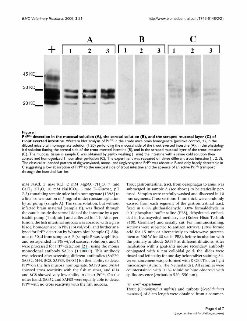
We therefore investigated whether trout intestine bindsPrPSc and transfers it to the serosal side where it mayspread to the lymphoid tissues and, eventually into theCNS. Using everted trout intestine immersed in a solutioncontaining 139A we observed that PrPSc slightly absorbs tothe mucosal intestinal layer as shown by the low, yetdetectable signal at the western blot (Figure 1C). Whenstatically perfused intestine was used in order to study thepossible role of pyloric caeca in PrPSc absorption, it waspossible to detect the presence of PrPSc by immunohisto-chemistry in the stratum compactum both in the trout intes-tine and in the pyloric caeca (Figure 2c, 2f). On the otherhand, the absence of signal for PrPSc at the western blot inthe solution fluxed into the serosal side of the evertedintestine (Figure 1B) excludes, at least in this experimentalsetting, an active secretion of PrPSc from one side to theother side of the intestinal tract.
Such data are in agreement with the recent finding thatstPrP is not detectable in the intestine of Rainbow trout[15], which renders unlikely an active uptake of exoge-nous PrPSc, but does not exclude an unspecific binding ofPrPSc to the fish intestinal mucosa.
Page 2 of 7(page number not for citation purposes)

BMC Veterinary Research 2006, 2:21 http://www.biomedcentral.com/1746-6148/2/21
In a second experiment, we inoculated trout and turbotwith the same strain of scrapie as above by parenteralroutes to increase the possibility of a successful infection(Table 1B). Brain and spleen, as the most likely tissues forPrPSc accumulation, were taken 15 and 90 days (the long-est survival time of fish in a close circuit water tanks) afterinoculation and residual infectivity was bio-assayed inmice. None of the recipient mice developed scrapie dis-ease during their lifespan. However, PrPSc was observed atthe western blot in the brain of a few recipient mice inoc-ulated with tissues taken from turbot or trout (Table 1B).These data show that 15 days after inoculation only aboutone LD50 of scrapie-infectivity is present in the spleen ofturbot, and much less in the brain of turbot or in thespleen of trout. Almost no infectivity (only 1 in 37 recipi-ent mice were PrPSc positive) was however detected 90days after inoculation of fish tissues, suggesting that theinfectivity measured at 15 days post inoculation was likelyrelated to the residual inoculum. As the fishes were notkept more than 90 days, it is not possible to exclude thatinfectivity might develop at a later time point, after an ini-tial clearance phase of the inoculum, though this possibil-ity is not likely.
In our work, we did not evaluate whether mammalian TSEagents may establish infection in fish. Nonetheless, the"lesion profile" curves based upon the severity of spongi-form changes in brain areas of mice injected with 139Abefore and after passage in fish (Figure 3), show a similarpattern suggesting that the 139A strain did not adapt tofish in the 3 months period examined. A statistical com-parison of scores between the two curves, done area byarea by the Mann-Whitney's test, showed a significant dif-ference only in the dorsal medulla area (p = 0.045). No
statistically significant difference was observed in thesuperior colliculus, hypothalamus, cingulate, and adja-cent cortices. Permutation tests performed to overcomeproblems due to the small sample size, confirmed thispattern. The overall lower scores observed in the brain ofmice inoculated with fish tissues could merely representthe fact that these mice were not symptomatic when sacri-ficed while control scrapie infected mice showed evidentclinical signs of disease.
ConclusionThese data show that about 4 million LD50 of 139A givenby forced feeding were readily removed from fish intestine(both trout and turbot) in the first 24 hours after infectionand that infection never reached the brain, the spleen orthe muscles. This suggests that scrapie is quickly removedfrom fish tissues despite the presence of a cellular prion-like protein [15,18] and a prion protein-like gene in fish[11-13]. With all the cautions due to the differencebetween the 139A and the BSE strains, and that in thisexperiment fishes were observed for no more than 90 daysafter infection, it is tentatively possible to assume that theconsumption of fish fed with BSE-infected MBM shouldnot pose any substantial threat to public health.
Methods"In vitro" experimentsRainbow trout (Oncorhynchus mykiss) from 50 to 200 g ofbody weight were kept at 15°C for one week and starved24 hours before the experiments. After sacrifice with alethal dose of MS 222 (200 mg/l) the whole intestine wasextracted, carefully everted, and canulated according topublished procedures [19,20]. Briefly, canulated intestinewas submerged in 25 ml of physiological solution (145
Table 1: Measurement of scrapie infectivity in fish tissues by mouse bioassay. Brains from recipient mice inoculated with turbot or trout tissues from either oral or multiple parenteral routes were assayed for PrPSc by Western blot. Whenever samples were available, histology and/or immunohistochemistry was performed. Asterisks indicate mice contributing to the lesion profiles curve of the fish-passaged 139A (Figure 3)
A. Tissues from infected fish via the oral route
Turbot Trout
Days after infection Muscle Intestine Brain Spleen Muscle Intestine Brain Spleen
1 0/10 0/8 0/8 - 0/10 1/8 0/5 -15 0/7 0/8 0/6 - 0/10 0/8 0/13 -30 0/10 0/8 0/10 - 0/8 0/8 0/9 -60 0/9 0/6 0/8 - 0/7 0/6 0/6 -90 0/7 0/8 0/9 - 0/10 0/11 0/10 -
B. Tissues from infected fish via multiple parenteral routes
15 - - 2/8 4/7* - - 0/9 1/9*
90 - - 1/7* 0/8 - - 0/11 0/11
Page 3 of 7(page number not for citation purposes)
BMC Veterinary Research 2006, 2:21 http://www.biomedcentral.com/1746-6148/2/21
mM NaCl, 5 mM KCl, 2 mM MgSO4·7H2O, 7 mMCaCl2·2H2O, 10 mM NaHCO3, 5 mM D-Glucose, pH7.2) containing scrapie mice brain homogenate (139A) toa final concentration of 5 mg/ml under constant agitationby air pump (sample A). The same solution, but withoutinfected brain material (sample B), was fluxed throughthe canula inside the serosal side of the intestine by a per-istaltic pump (1 ml/min) and collected for 1 h. After per-fusion, the fish intestinal mucosa was abraded with a glassblade, homogenized in PBS (1:4 vol/vol), and further ana-lysed for PrPSc detection by Western blot (sample C). Aliq-uots of 30 µl from samples A, B (sample B was lyophilisedand resuspended in 1% wt/vol sarcosyl solution), and Cwere processed for PrPSc detection [21], using the mousemonoclonal antibody SAF83 (1:10000). This antibodywas selected after screening different antibodies (SAF70,SAF32, 6H4, 8G8, SAF83, SAF84) for their ability to detectPrPSc on the fish mucosa homogenate. SAF70 and SAF84showed cross reactivity with the fish mucosa, and 6H4and 8G8 showed very low ability to detect PrPSc. On theother hand, SAF32 and SAF83 were equally able to detectPrPSc with no cross reactivity with the fish mucosa.
Trout gastrointestinal tract, from oesophagus to anus, wassubmerged in sample A (see above) to be statically per-fused. Samples were carefully washed and dissected in 10mm segments. Cross sections, 1 mm thick, were randomlyexcised from each segment of the gastrointestinal tract,fixed in 0.8% glutharaldehyde, 3.0% formaldehyde in0.01 phosphate buffer saline (PBS), dehydrated, embed-ded in hydroxyethyl-methacrylate (Kulzer Histo-Technik8100, Germany) and serially cut. For immunostaining,sections were subjected to antigen retrieval (98% formicacid for 15 min or alternatively to microwave pretreat-ment at 600 W for 60 sec in PBS), before incubation withthe primary antibody SAF83 at different dilutions. Afterincubation with a goat-anti mouse secondary antibodyconjugated with 6 nm colloidal gold, the slides wererinsed and left to dry for one day before silver staining. Sil-ver enhancement was performed with R-GENT kit for lightmicroscopy (Aurion, The Netherlands). All samples werecounterstained with 0.1% toluidine blue observed withepifluorescence (excitation 520–550 nm).
"In vivo" experimentTrout (Oncorhynchus mykiss) and turbots (Scophthalmusmaximus) of 8 cm length were obtained from a commer-
PrPSc detection in the mucosal solution (A), the serosal solution (B), and the scraped mucosal layer (C) of trout everted intes-tineFigure 1PrPSc detection in the mucosal solution (A), the serosal solution (B), and the scraped mucosal layer (C) of trout everted intestine. Western blot analysis of PrPSc in the crude mice brain homogenate (positive control, +), in the diluted mice brain homogenate solution (1:20) perfonding the mucosal side of the trout everted intestine (A), in the physiolog-ical solution fluxing the serosal side of the trout everted intestine (B), and in the scraped mucosal layer of the trout intestine (C). The mucosal tissue in sample C was obtained by gently washing (1 min) the intestine with a saline cold solution then ablated and homogenized 1 hour after perfusion (C). The experiment was repeated on three different trout intestine (1, 2, 3). The classical tri-banded pattern of diglycosylated, mono- and unglycosylated PrPSc was absent in B and only barely detectable in C suggesting a low absorption of PrPSc to the mucosal side of trout intestine and the absence of an active PrPSc transport through the intestinal barrier.
Page 4 of 7(page number not for citation purposes)

BMC Veterinary Research 2006, 2:21 http://www.biomedcentral.com/1746-6148/2/21
cial farm and acclimatized to laboratory conditions for 3weeks before oral or parenteral infection with the mouse-adapted 139A strain of scrapie. The inoculum was pre-pared from a pool of 70 g of 139A scrapie-infected mouse(C57/BL) brains. Brain tissue was homogenized in sterilePBS to obtain a 10% (wt/vol) suspension, centrifuged at913 × g (4°C per 15 minutes), and supernatant collected.
Infectivity titre of this suspension was calculated by end-point titration as previously described [22], using groupsof 6–10 animals for each dilution. The amount of infectiv-ity, calculated by the Reed and Muench method, was 108.9
LD50 per gram of tissue. Control homogenate was pre-pared with the same procedure using brain tissue fromhealthy mice. One group of turbots (n = 20) or trout (n =
PrPSc accumulation in the pyloric caeca (a, b, c) and non-everted intestine (d, e, f) of trout intestine statically perfusedFigure 2PrPSc accumulation in the pyloric caeca (a, b, c) and non-everted intestine (d, e, f) of trout intestine statically perfused. Immunohistochemistry on trout sections of pyloric caeca (a, b, c) and non-everted intestines (d, e, f). The static per-fusion was performed for 1 hour at 15°C, in a PBS solution containing 50 µl/ml of either 10% uninfected mice brain homoge-nate (a, d) or 10% mice scrapie brain homogenate (139A) (b, c, e, f). Immunolabelling was performed with the monoclonal antibody SAF83 (a, c, d, f), or, as control, the monoclonal anti-HA against influenza virus (anti-HA, clone 12CA5, subtype IgG2b, k), (b, e). All the immunogold-labelled sections were silver enhanced and counterstained with 0.1% toluidine blue (see materials and methods for details). PrPSc localisation (arrows) occurred in the stratum compactum (S) of distal intestine and pyloric caecum incubated with SAF83 (c, f). No immunolabelling was present in control tissues incubated with the same anti-body (a, d), except for a low unspecific background. The background was slightly higher, though unspecific, in anti-HA labelled sections (b, e) due to the high reactivity of this antibody. All micrographs were taken at the same magnifications; V = villo, T = tunica muscolaris; asterisks point to unspecific labelling present in all samples and due to micro-fractures between the outer specimen surface and the resin.
Page 5 of 7(page number not for citation purposes)

BMC Veterinary Research 2006, 2:21 http://www.biomedcentral.com/1746-6148/2/21
20) was force-fed with 0.05 ml of 139A scrapie-infected(106.6 LD50) or control mouse brain homogenates.Another group of turbots (n = 30) or trout (n = 30) wassimultaneously inoculated by the intracerebral (0.03 ml,106.4 LD50), intraperitoneal (0.1 ml, 106.9 LD50), and intra-muscular (0.1 ml, 106.9 LD50) routes with either 139A orcontrol brain homogenates. After inoculation, fish weremaintained in close circuit water tanks under bio-securityconditions to avoid water contamination with the scrapieagent.
Samples from brain, muscle, and intestine were takenfrom 3 orally-infected and 1 control fish for each speciesat 1, 15, 30, 60 and 90 days post-infection. Moreover,brain and spleen were taken from parenterally infectedfish at 15 and 90 days post-infection. Collected sampleswere divided in two parts: one was immediately frozen at-80°C for measuring scrapie infectivity by mouse bio-assay, the other was fixed for histological or immunohis-tochemical examination. Histopathological examinationof fish tissues was routinely performed on every samplecollected. Briefly, samples were fixed in Davidson andprocessed in an automatic tissue processor, embedded in
paraffin and cut in 5 µm sections, deparaffinized, rehy-dratated and then stained with haematoxylin-eosin. Theimmunohistochemical detection of the prion protein infish tissue sections was performed as described [23].
For the bioassay of fish tissues in mice, each group of fishsamples, belonging to the same time point, was homoge-nized using separate sets of instruments to avoid acciden-tal cross-contamination. Homogenisers were thenaccurately decontaminated (2 cycles of autoclave at134°C for 1 hour followed by extensive washing and athird cycle of autoclave at 120°C for 20 minutes) beforere-use on another group of samples. Twenty µl of 10% tis-sue homogenate were injected intracerebrally in groups of6–11 C57/BL male mice for infectivity bioassay. Animalsshowing clinical signs (not necessarily related to scrapie)or moribund were sacrificed, their brains removed anddivided in two halves. One half was immediately frozen at-70°C for PrPSc analysis, the other half was fixed in forma-lin for histological examination. PrPSc was purified asdescribed [24] and detected by western blotting usingmonoclonal antibody SAF84 (SpiBio) diluted 1:5000.Mice were housed at the animal facility of the IstitutoSuperiore di Sanità (ISS) under the supervision of theService for Biotechnology and Animal Welfare of the ISSwho warrants the adherence to the national and interna-tional regulations on animal welfare.
Histological analysis and lesion profiles of mice brainsFormalin-fixed brains were decontaminated with 98%formic acid for 1 h, stained with haematoxylin-eosin andscored for the intensity of vacuolar degeneration (rangingfrom 0, no vacuolar change, to 5, severe vacuolar change)in nine standard grey matter areas for building up the'lesion profiles' [25]. Sections were coded and blind exam-ined. In Figure 3, the lesion profile of 139A after passagein fish was a mean of 4 animals when brains for histologywere available (2 mice inoculated with turbot spleen at 15days, 1 mouse inoculated with trout spleen at 15 days,and 1 turbot brain taken 90 days after parenteral inocula-tion). We pooled these data together because none ofthese mice showed clinical signs and were sacrificed atabout the same time after inoculation (221, 204, 215, and221 days, respectively). The reference curve of 139A notpassaged in fish was obtained pooling data from 6 mice.Immunohistochemistry was performed as described [23].
AbbreviationsPrP: prion protein
PrPc: cellular prion protein
PrPSc: pathological prion protein
stPrP: "similar to" PrP
Lesion profiles for mice following inoculation with 139A before (circle) and after (square) passage in fishFigure 3Lesion profiles for mice following inoculation with 139A before (circle) and after (square) passage in fish. The lesion profiles for the fish-passaged 139A (square) was a mean of 4 mice: 2 mice inoculated with turbot spleen at 15 days, 1 mouse inoculated with trout spleen at 15 days, and 1 turbot brain taken 90 days after parenteral inoculation. The reference curve (circle) was a mean of 6 mice inoculated with the 139A non passaged in fish. Vacuolation was evalu-ated in nine standard areas: 1, dorsal medulla; 2, cerebellar cortex; 3, superior colliculus; 4, hypothalamus; 5, thalamus; 6, hippocampus; 7, septum; 8, retrosplenial and adjacent motor cortex; 9, cingulate and adjacent motor cortex. Data are mean ± SE.
�
�
�
�
��
��
�
�
� �
�
�
�
��
�
1 2 3 4 5 6 7 8 90
1
2
3
4
Le
sio
n in
ten
sit
y s
co
re
Scoring areas
Page 6 of 7(page number not for citation purposes)

BMC Veterinary Research 2006, 2:21 http://www.biomedcentral.com/1746-6148/2/21
TSE: Transmissible Spongiform Encephalopathy
CJD: Creutzfeldt-Jakob disease
GSS: Gerstmann-Sträussler-Scheinker disease
BSE: bovine Spongiform encephalopathy
MBM: bovine meat and bone meals
anti-HA: anti-haemagglutinin
Authors' contributionsLI contributed to the design of the study, did mice inocu-lation, analysis of data and drafted the manuscript. BN,RA established the protocol for fish infection and sam-pling of fish tissues, they also performed it. AZDV, FF, andMI contributed to the "in vitro" experimental work and tothe protocols setting. MS did the neuropathology of themice brains and contributed to analysis of data. SB, ML,QL, and VV did western blot analysis of mouse brains andcontributed to monitoring of mice, collection and analy-sis of data. FC did coordination and collection of clinical,biochemical and histological data from the mouse bio-assay. SC contributed to the design of the study set up theprotocol for the "in vitro" experiments. AF contributed tothe design of the "in vivo " experimental work and the his-tological studies on fish tissues. MP participated in thedesign and coordination of the study, interpretation ofdata and drafting the manuscript. All the authors read andapproved the final manuscript.
AcknowledgementsThis study was funded through the EU grant FAIR CT97 3308 and the EU grant QLK5-2002-00866. Authors thank Maurizio Bonanno and Nicola Bel-lizzi for their skilful assistance in animal care and collection of samples, Dr. Dario Maffi for lending histological instruments and for technical help, Dr Maria Puopolo for statistical analysis, and Dr Alessandra Garozzo for administrative support. Special thanks to prof. Carla Liana Bolis and prof. Severino Ronchi for their endless effort in the coordination and completion of the project.
References1. Will RG, Ironside JW, Zeidler M, Cousens SN, Estibeiro K, Alpero-
vitch A, Poser S, Pocchiari M, Hofman A, Smith PG: A new variantof Creutzfeldt-Jakob disease in the UK. Lancet 1996,347:921-925.
2. Bruce ME: 'New variant' Creutzfeldt-Jakob disease and bovinespongiform encephalopathy. Nat Med 2006, 6:258-259.
3. Collinge J: Molecular neurology of prion disease. J Neurol Neuro-surg Psychiatry 2005, 76:906-919.
4. Lysek DA, Schorn C, Nivon LG, Esteve-Moya V, Christen B, CalzolaiL, von Schroetter C, Fiorito F, Herrmann T, Guntert P, Wuthrich K:Prion protein NMR structures of cats, dogs, pigs, and sheep.Proc Natl Acad Sci USA 2005, 102:640-645.
5. Harris DA, Falls DL, Johnson FA, Fischbach GD: A prion-like pro-tein from chicken brain copurifies with an acetylcholinereceptor-inducing activity. Proc Natl Acad Sci USA 1991,88:7664-7668.
6. Lysek DA, Calzolai L, Wuthrich K: NMR assignment of thechicken prion protein fragments chPrP(128–242) andchPrP(25–242). J Biomol NMR 2004, 30:97.
7. Gabriel JM, Oesch B, Kretzschmar H, Scott M, Prusiner SB: Molecu-lar cloning of a candidate chicken prion protein. Proc Natl AcadSci USA 1992, 89:9097-9101.
8. Simonic T, Duga S, Strumbo B, Asselta R, Ceciliani F, Ronchi S: cDNAcloning of turtle prion protein. FEBS Lett 2002, 469:33-38.
9. Calzolai L, Lysek DA, Perez DR, Guntert P, Wuthrich K: Prion pro-tein NMR structures of chickens, turtles, and frogs. Proc NatlAcad Sci USA 2005, 102:651-655.
10. Strumbo B, Ronchi S, Bolis LC, Simonic T: Molecular cloning of thecDNA coding for Xenopus laevis prion protein. FEBS Lett 2001,508:170-174.
11. Suzuki T, Kurokawa T, Hashimoto H, Sugiyama M: cDNA sequenceand tissue expression of Fugu rubripes prion protein-like: acandidate for the teleost orthologue of tetrapod PrPs. Bio-chem Biophy Res Commun 2002, 294:912-917.
12. Oidtmann B, Simon D, Holtkamp N, Hoffmann R, Baier M: Identifi-cation of cDNAs from Japanese pufferfish (Fugu rubripes)and Atlantic salmon (Salmo salar) coding for homologues totetrapod prion proteins. FEBS Lett 2003, 538:96-100.
13. Rivera-Milla E, Stuermer CA, Malaga-Trillo E: An evolutionarybasis for scrapie disease:identification of a fish prion mRNA.Trends Genet 2003, 19:72-75.
14. Matthews D, Cooke BC: The potential for transmissible spong-iform encephalopathies in non-ruminant livestock and fish.Rev Sc Tech 2003, 22:283-296.
15. Maddison BC, Patel S, James RF, Conlon HE, Oidtmann B, Baier M,Whitelam GC, Gough KC: Generation and characterisation ofmonoclonal antibodies to Rainbow trout (Oncorhynchusmykiss) prion protein. J Immunol Methods 2005, 306:202-210.
16. Race R, Chesebro B: Scrapie infectivity found in resistant spe-cies. Nature 1998, 392:770.
17. Kimberlin RH, Cole S, Walker CA: Temporary and permanentmodifications to a single strain of mouse scrapie on trans-mission to rats and hamsters. J Gen Virol 1987, 68:1875-1881.
18. Gibbs CJ Jr, Bolis CL: Normal isoform of amyloid protein (PrP)in brains of spawning salmon. Mol Psychiatry 1997, 2:146-147.
19. Erba D, Ciappellano S, Testolin G: Effect of caseinphosphopep-tides on inhibition of calcium intestinal absorption due tophosphate. Nutr Res 2001, 21:649-656.
20. Erba D, Ciappellano S, Testolin G: Effect of the ratio of caseinphosphopeptides to calcium (w/w) on passive calcium trans-port in the distal small intestine of rats. Nutrition 2002,18:743-746.
21. Cardone F, Liu QG, Petraroli R, Ladogana A, D'Alessandro M, ArpinoC, Di Bari M, Macchi G, Pocchiari M: Prion protein glycotypeanalysis in familial and sporadic Creutzfeldt-Jakob diseasepatients. Brain Res Bull 1999, 49:429-433.
22. Pocchiari M, Casaccia P, Ladogana A: Amphotericin B: a novelclass of antiscrapie drugs. J Infect Dis 1989, 160:795-802.
23. Bell JE, Gentleman SM, Ironside JW, McCardle L, Lantos PL, Doey L,Lowe J, Fergusson J, Luther P, McQuaid S, Allen IV: Prion Proteinimmunocytochemistry-UK five centre consensus report.Neuropath Appl Neuro 1997, 23:26-35.
24. Xi YG, Cardone F, Pocchiari M: Detection of proteinase-resist-ant protein (PrP) in small brain tissue samples from Creut-zfeldt-Jakob disease patients. J Neurol Sci 1994, 124:171-173.
25. Fraser H, Dickinson AG: The sequential development of thebrain lesions of scrapie in three strains of mice. J Comp Pathol1968, 78:301-311.
Page 7 of 7(page number not for citation purposes)
http://www.ncbi.nlm.nih.gov/entrez/query.fcgi?cmd=Retrieve&db=PubMed&dopt=Abstract&list_uids=8598754
http://www.ncbi.nlm.nih.gov/entrez/query.fcgi?cmd=Retrieve&db=PubMed&dopt=Abstract&list_uids=8598754
http://www.ncbi.nlm.nih.gov/entrez/query.fcgi?cmd=Retrieve&db=PubMed&dopt=Abstract&list_uids=1715573
http://www.ncbi.nlm.nih.gov/entrez/query.fcgi?cmd=Retrieve&db=PubMed&dopt=Abstract&list_uids=1715573
http://www.ncbi.nlm.nih.gov/entrez/query.fcgi?cmd=Retrieve&db=PubMed&dopt=Abstract&list_uids=1715573
http://www.ncbi.nlm.nih.gov/entrez/query.fcgi?cmd=Retrieve&db=PubMed&dopt=Abstract&list_uids=1409608
http://www.ncbi.nlm.nih.gov/entrez/query.fcgi?cmd=Retrieve&db=PubMed&dopt=Abstract&list_uids=1409608
http://www.ncbi.nlm.nih.gov/entrez/query.fcgi?cmd=Retrieve&db=PubMed&dopt=Abstract&list_uids=9572135
http://www.ncbi.nlm.nih.gov/entrez/query.fcgi?cmd=Retrieve&db=PubMed&dopt=Abstract&list_uids=9572135
http://www.ncbi.nlm.nih.gov/entrez/query.fcgi?cmd=Retrieve&db=PubMed&dopt=Abstract&list_uids=3110370
http://www.ncbi.nlm.nih.gov/entrez/query.fcgi?cmd=Retrieve&db=PubMed&dopt=Abstract&list_uids=3110370
http://www.ncbi.nlm.nih.gov/entrez/query.fcgi?cmd=Retrieve&db=PubMed&dopt=Abstract&list_uids=3110370
http://www.ncbi.nlm.nih.gov/entrez/query.fcgi?cmd=Retrieve&db=PubMed&dopt=Abstract&list_uids=9106239
http://www.ncbi.nlm.nih.gov/entrez/query.fcgi?cmd=Retrieve&db=PubMed&dopt=Abstract&list_uids=9106239
http://www.ncbi.nlm.nih.gov/entrez/query.fcgi?cmd=Retrieve&db=PubMed&dopt=Abstract&list_uids=2509571
http://www.ncbi.nlm.nih.gov/entrez/query.fcgi?cmd=Retrieve&db=PubMed&dopt=Abstract&list_uids=2509571
http://www.ncbi.nlm.nih.gov/entrez/query.fcgi?cmd=Retrieve&db=PubMed&dopt=Abstract&list_uids=7964868
http://www.ncbi.nlm.nih.gov/entrez/query.fcgi?cmd=Retrieve&db=PubMed&dopt=Abstract&list_uids=7964868
http://www.ncbi.nlm.nih.gov/entrez/query.fcgi?cmd=Retrieve&db=PubMed&dopt=Abstract&list_uids=7964868
http://www.ncbi.nlm.nih.gov/entrez/query.fcgi?cmd=Retrieve&db=PubMed&dopt=Abstract&list_uids=4970192