
Features of follicular dendritic cells in ovine pharyngeal tonsil: An in vivo and in vitro study in...
7
Veterinary Immunology and Immunopathology 141 (2011) 26–32 Contents lists available at ScienceDirect Veterinary Immunology and Immunopathology journal homepage: www.elsevier.com/locate/vetimm Research paper Features of follicular dendritic cells in ovine pharyngeal tonsil: An in vivo and in vitro study in the context of scrapie pathogenesis Vinciane Toppets a,∗ , Valerie Defaweux b , Joelle Piret a , Nathalie Kirschvink c , Luc Grobet a , Nadine Antoine a a Department of Morphology and Pathology, Laboratory of Animal Histology and Embryology, Faculty of Veterinary Medicine, University of Liege, Belgium b Laboratory of Human Histology, Faculty of Medicine, University of Liege, Belgium c Animal Physiology, Veterinary Department, Faculty of Sciences, University of Namur FUNDP, Belgium article info Article history: Received 23 July 2010 Received in revised form 24 January 2011 Accepted 31 January 2011 Keywords: Sheep Scrapie Pharyngeal tonsil Follicular dendritic cell Anti-FDC antibodies abstract Although the alimentary tract has been suggested as the most likely portal of entry in natural scrapie, a growing amount of data indicates that the respiratory system and more specifically the pharyngeal tonsils serve as a natural portal of entry for scrapie. This study describes for the first time the broad cell populations in the lymphoid com- partment of pharyngeal tonsils and more specifically inside the lymphoid follicles where the scrapie agent accumulates during the period of latency. Follicular dendritic cells (FDCs), stromal cells located in the light zone of the germinal centre of lymphoid follicles, seem to be the principal causal factor in the accumulation of the infectious agent in transmissible spongiform encephalopathy (TSE) diseases. Knowing that efficient lymphoreticular prion propagation requires PrPc expression, we analysed the expression of PrPc with the mouse monoclonal antibody Pri 909 both in situ and on FDC-cluster-enriched cell suspensions. In situ, a positive staining was observed in the germinal centre of pharyngeal lymph follicles. The germinal centre labelling was due to the presence of a follicular dendritic network as revealed after immunogold staining of isolated FDC clusters. Our results suggest that the pharyngeal lymphoreticular system and more specifically PrPc expressing follicular dendritic cells could serve as a prion “reservoir” during the latency phase, thus playing a key role during the scrapie lymphoinvasion. © 2011 Elsevier B.V. All rights reserved. 1. Introduction In humans and in many animal species, the tonsils are a part of the pharyngeal mucosal immune system (Ogra, 2000) and form a ring of lymphoid tissue in the pharyn- geal wall, known as the Waldeyer’s ring (Perry and Whyte, 1998). ∗ Corresponding author. Present address: Department of Morphology and Pathology, Laboratory of Animal Histology and Embryology, Faculty of Veterinary Medicine, University of Liege, Boulevard de Colonster, 20 Bat. B43a, 4000 Liege, Belgium. Tel.: +32 4 366 40 85; fax: +32 4 366 40 86. E-mail address: [email protected] (V. Toppets). In sheep, the palatine (tonsilla palatina), the lingual (ton- silla lingualis) and the tonsil of the soft palate (tonsilla veli palatini) are located in the oropharynx; the pharyn- geal (tonsilla pharyngea) and tubal tonsils (tonsilla tubaria) in the nasopharynx, and the paraepiglottic tonsil (ton- silla paraepiglottica) in the laryngopharynx (Barone, 1997). Defined as tonsils with or without crypts based upon their relationship with the surface epithelium, they con- sist mainly of secondary lymph follicles separated by T-dependent zones, areas through which antigens can pass and where effector cells accumulate. In sheep, the palatine and the pharyngeal tonsils contain the largest volume of lymphoid tissues and the great- est epithelial surface area of the oropharyngeal and the 0165-2427/$ – see front matter © 2011 Elsevier B.V. All rights reserved. doi:10.1016/j.vetimm.2011.01.014
-
Upload
independent -
Category
Documents
-
view
1 -
download
0
Transcript of Features of follicular dendritic cells in ovine pharyngeal tonsil: An in vivo and in vitro study in...

R
FA
VLa
Ub
c
a
ARRA
KSSPFA
1
a2g1
aoBf
0d
Veterinary Immunology and Immunopathology 141 (2011) 26–32
Contents lists available at ScienceDirect
Veterinary Immunology and Immunopathology
journa l homepage: www.e lsev ier .com/ locate /vet imm
esearch paper
eatures of follicular dendritic cells in ovine pharyngeal tonsil:n in vivo and in vitro study in the context of scrapie pathogenesis
inciane Toppetsa,∗, Valerie Defaweuxb, Joelle Pireta, Nathalie Kirschvinkc,uc Grobeta, Nadine Antoinea
Department of Morphology and Pathology, Laboratory of Animal Histology and Embryology, Faculty of Veterinary Medicine,niversity of Liege, BelgiumLaboratory of Human Histology, Faculty of Medicine, University of Liege, BelgiumAnimal Physiology, Veterinary Department, Faculty of Sciences, University of Namur FUNDP, Belgium
r t i c l e i n f o
rticle history:eceived 23 July 2010eceived in revised form 24 January 2011ccepted 31 January 2011
eywords:heepcrapieharyngeal tonsilollicular dendritic cellnti-FDC antibodies
a b s t r a c t
Although the alimentary tract has been suggested as the most likely portal of entry innatural scrapie, a growing amount of data indicates that the respiratory system and morespecifically the pharyngeal tonsils serve as a natural portal of entry for scrapie.
This study describes for the first time the broad cell populations in the lymphoid com-partment of pharyngeal tonsils and more specifically inside the lymphoid follicles wherethe scrapie agent accumulates during the period of latency.
Follicular dendritic cells (FDCs), stromal cells located in the light zone of the germinalcentre of lymphoid follicles, seem to be the principal causal factor in the accumulation ofthe infectious agent in transmissible spongiform encephalopathy (TSE) diseases. Knowingthat efficient lymphoreticular prion propagation requires PrPc expression, we analysedthe expression of PrPc with the mouse monoclonal antibody Pri 909 both in situ and onFDC-cluster-enriched cell suspensions.
In situ, a positive staining was observed in the germinal centre of pharyngeal lymphfollicles. The germinal centre labelling was due to the presence of a follicular dendriticnetwork as revealed after immunogold staining of isolated FDC clusters. Our results suggestthat the pharyngeal lymphoreticular system and more specifically PrPc expressing follicular
ld servscrapi
dendritic cells coukey role during the
. Introduction
In humans and in many animal species, the tonsils are
part of the pharyngeal mucosal immune system (Ogra,000) and form a ring of lymphoid tissue in the pharyn-eal wall, known as the Waldeyer’s ring (Perry and Whyte,998).
∗ Corresponding author. Present address: Department of Morphologynd Pathology, Laboratory of Animal Histology and Embryology, Facultyf Veterinary Medicine, University of Liege, Boulevard de Colonster, 20at. B43a, 4000 Liege, Belgium. Tel.: +32 4 366 40 85;
ax: +32 4 366 40 86.E-mail address: [email protected] (V. Toppets).
165-2427/$ – see front matter © 2011 Elsevier B.V. All rights reserved.oi:10.1016/j.vetimm.2011.01.014
e as a prion “reservoir” during the latency phase, thus playing ae lymphoinvasion.
© 2011 Elsevier B.V. All rights reserved.
In sheep, the palatine (tonsilla palatina), the lingual (ton-silla lingualis) and the tonsil of the soft palate (tonsillaveli palatini) are located in the oropharynx; the pharyn-geal (tonsilla pharyngea) and tubal tonsils (tonsilla tubaria)in the nasopharynx, and the paraepiglottic tonsil (ton-silla paraepiglottica) in the laryngopharynx (Barone, 1997).Defined as tonsils with or without crypts based upontheir relationship with the surface epithelium, they con-sist mainly of secondary lymph follicles separated by
T-dependent zones, areas through which antigens can passand where effector cells accumulate.In sheep, the palatine and the pharyngeal tonsils containthe largest volume of lymphoid tissues and the great-est epithelial surface area of the oropharyngeal and the

ogy and
V. Toppets et al. / Veterinary Immunolnasopharyngeal parts of the Waldeyer’s ring, respectively(Casteleyn et al., 2007, 2008). Palatine tonsils are bilateral,are the size of a hazelnut and present one to three cryptscovered by the buccal epidermoid epithelium (Gabriel andvan den Broeck, 2003; Cocquyt et al., 2005). The non-crypticpharyngeal tonsil is a unique and circumvoluated eleva-tion located on the caudal part of the pharyngeal septum.The lymphoid compartment consists of secondary folli-cles lying underneath the pseudostratified columnar ciliaryepithelium.
Massive intraepithelial lymphocyte infiltration suggeststhat these tonsils are perfectly adapted to sample foreignantigens.
Relatively easily accessible, palatine and pharyngealtonsils thus appear to be of strong interest in the diagno-sis of several diseases. It is well known that animal tonsilsare the sites of entry and replication of several pathogens.Studies on the foot and mouth disease virus (FMDV) insheep confirm that the tonsils are the main predilectionsites for the persistence of this aphtovirus (Kitching andHughes, 2002; Horsington and Zhang, 2007). High levels ofPrPd have been detected in palatine tonsil follicles of sheepnaturally infected with the scrapie agent (Jeffrey et al.,2001). Tonsillar biopsies can thus be useful in guarantee-ing food safety (Thuring et al., 2002). Infection via the nasalroute may also be possible in scrapie infected flocks. Recentexperiments postulate that the upper respiratory tract, andmore specifically the pharyngeal tonsil, serves as a naturalportal of entry for the scrapie agent (Hamir et al., 2008). Thishypothesis is sustained by the presence of microfold M cellsin the ovine nasopharyngeal mucosal epithelium (Stanleyet al., 2001; Casteleyn et al., 2010), since these cells havebeen shown to efficiently transcytose prion infectivity inan in vitro co-culture system (Heppner et al., 2001).
The mucosa-associated lymphoid tissues play an impor-tant role in scrapie pathogenesis. PrPd accumulates insidethe germinal centres (McBride et al., 1992) before spread-ing to the sites of neuroinvasion. The cells implicatedin prion pathogenesis are follicular dendritic cells (FDCs)which reside in the germinal centre (Jeffrey et al., 2000;Mabbott and Bruce, 2001). Numerous studies using chi-maeric mice indicate that mature follicular dendritic cellsare necessary for prion propagation within the lym-phoreticular system (Brown et al., 1999). In the absenceof mature FDCs, neuroinvasion following peripheral chal-lenge is significantly impaired in scrapie pathogenesis(Mabbott et al., 2000; Montrasio et al., 2000). The abil-ity to recognise ovine FDCs is limited. In fact, among thevarious anti-FDC antibodies commercially available, DRC1(Naiem et al., 1983; Johnson et al., 1986), FDC-M1 (Koscoet al., 1992) and CNA.42 (Raymond et al., 1997; Lezmi et al.,2001) all fail to recognise specifically FDCs from all thesecondary ruminants lymph organs. FDC-B1, a new mousemonoclonal antibody, has recently been produced andcharacterised by Melot et al. (2004). The antigen detectedby FDC-B1 is expressed exclusively on the surface of FDCs
in all ruminant lymphoid organs.The aims of our study were (i) the identification of theFDCs inside the pharyngeal tonsil; (ii) the study of theirmorphological characteristics and (iii) the analysis, in vivoand in vitro, of the PrPc expression on their cell membrane.
Immunopathology 141 (2011) 26–32 27
This approach will be useful for future comparative studieson the implication of the FDC network in the replication ofthe scrapie agent in the nasopharyngeal mucosa of sheepwith different polymorphisms in the host PrP gene.
2. Materials and methods
2.1. Sample collection
Pharyngeal and palatine tonsils were obtained at a localabattoir from clinically healthy sheep aged from 4 to 6months. These animals showed the ARR/ARR genotyperesistant toward scrapie.
2.2. Immunohistochemistry
Some specimens were immersed in Tissue-TekOCT embedding medium (SAKURA, Zouterwoude, theNetherlands), snap-frozen and stored at −20 ◦C untilprocessing for immunohistochemistry. Sections were cutat −15 ◦C with a microtome (MICRON HM 500 OM) anddeposited on glass slides coated with poly-l-lysine (Sigma,St. Louis, USA), air-dried, fixed in acetone for 10 min at4 ◦C, and stored at −80 ◦C until use.
After rehydration, the cryosections were incubatedfor 1 h at room temperature with antibodies directedagainst the selected lymphoid cell populations or withthose which react with ovine PrPc. Immune cells werestained with the following antibodies: follicular dendriticcells (FDCs) identified with two mouse monoclonal anti-bodies: FDC-B1 (undiluted hybridoma supernatant) kindlyprovided by Melot et al. (2004) and CNA.42 (1/100) (Dako,Glostrup, Denmark), B cells revealed with a mouse anti-human CD79 �cy mAb (clone HM 57) (1/1000) (Dako,Glostrup, Denmark) and T cells with a rabbit anti-CD3polyclonal antibody (1/600) (Dako, Glostrup, Denmark).Cellular prion protein expression was analysed on tissuesections and on FDC clusters with a mouse anti-humanmonoclonal antibody Pri 909 (SPIbio, Montigny le Breton-neux, France) raised against an epitope located at aminoacid sequence 214–230 in the protein and diluted at1/100 in PBS. The samples were rinsed three times inthe same buffer. They were then incubated with a con-jugated goat anti-mouse (for FDC-B1, CNA.42, CD79 andanti-PrPc mAb) or goat anti-rabbit (for CD3) immunoglob-ulin peroxidase labelled polymer (Amplification EnVision®
System-HRP, Dako, Glostrup, Denmark) for 30 min atroom temperature. Peroxidase activity was revealed with9ethyl-3-aminocarbazole (AEC, Zymed, San Francisco, USA)combined to H2O2 as substrate for 12 min. The specificity ofthe antibodies was tested using an isotype control antibodyand negative controls were obtained by omitting the pri-mary antibody. The specimens were counterstained withMayer’s haematoxylin.
2.3. Isolation of FDC clusters
Three protocols were tested. The first two compared theefficiency of two enzymatic cocktails in terms of FDC clusternumbers and of PrPc expression by freshly isolated FDCs.Pharyngeal tonsils were cut into 1 mm thick slices. The
2 ogy and
ltNde
t1poitgctsws
tisR5(ctirm
2l
cPwbpfiatwiti
Numerous secondary lymphoid follicles surrounded byan interfollicular area formed the sub-epithelial laminapropria mucosae. Lymphoid follicles consisted of a germi-nal centre (GC) and a mantle zone (MZ) which generallyfaced the epithelium. CD79 positive B cells were located in
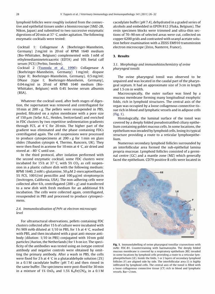
Fig. 1. Immunolabelling of ovine pharyngeal tonsillar cryosections withmAb: FDC-B1. Counterstaining with haematoxylin. The deeply foldedmucous membrane is covered by a respiratory epithelium (RE) invaded
8 V. Toppets et al. / Veterinary Immunol
ymphoid follicles were roughly isolated from the connec-ive and epithelial tissues under a biomicroscope (SMZ-2B,ikon, Japan) and submitted to two successive enzymaticigestions of 20 min at 37 ◦C, under agitation. The followingnzymatic cocktails were tested:
Cocktail 1: Collagenase A (Boehringer-Mannheim,Germany) 2 mg/ml in 20 ml of RPMI 1640 medium(Bio-Whittaker, Belgium) complemented with 1 mM ofethylenediaminetetraacetic (EDTA) and 10% foetal calfserum (FCS) (Perbio, Sweden).Cocktail 2 (Tsunoda et al., 1990): Collagenase A(Boehringer-Mannheim, Germany) 1 mg/ml; dispase(type II; Boehringer-Mannheim, Germany), 0.5 mg/ml;DNase (type I; Boehringer-Mannheim, Germany),0.04 mg/ml in 20 ml of RPMI 1640 medium (Bio-Whittaker, Belgium) with 0.4% bovine serum albumin(BSA).
Whatever the cocktail used, after both stages of diges-ion, the supernatant was removed and centrifugated for0 min at 200 × g. The pellets were resuspended in PBS,ooled, filtrated on a nylon membrane with a pore sizef 150 �m (Sefar A.G., Heiden, Switzerland) and enrichedn FDC clusters by two repetitive sedimentation gradientshrough FCS, at 4 ◦C for 20 min. The higher part of theradient was eliminated and the phase containing FDCsentrifugated again. The cell suspensions were processedo produce cytospecimens at 200 × g for 1 min on glasslides (Shandon cytospin 4, Thermo, Runcorn, UK). Theyere then fixed in acetone for 10 min at 4 ◦C, air dried and
tored at −80 ◦C until use.In the third protocol, after isolation performed with
he second enzymatic cocktail, some FDC clusters werencubated for 15 h at 37 ◦C, with 5% CO2 as cell suspen-ion in a plastic culture dish with the following medium:PMI 1640, 2 mM l-glutamine, 50 �M 2-mercaptoethanol,% FCS, 100 U/ml penicillin and 100 �g/ml streptomycinInvitrogen, California, USA). The non-adhering cells wereollected after 6 h, centrifugated (200 × g) and transferredo a new dish with fresh medium for an additional 9 hncubation. The cells were collected again, centrifugated,esuspended in PBS and processed to produce cytospeci-ens.
.4. Immunolocalisation of PrPc at electron microscopicevel
For ultrastuctural observations, pellets containing FDClusters collected after 15 h of culture were incubated withri 909 mAb diluted at 1/10 in PBS, for 1 h at 4 ◦C, washedith PBS, and then incubated with a goat anti-mouse anti-
ody (dilution: 1/10 in PBS) conjugated with 10 nm goldarticles (Aurion, the Netherlands) for 1 h on ice. The speci-city of the antibodies was tested using an isotype controlntibody and negative controls were obtained by omit-
ing the primary antibody. After a wash in PBS, the cellsere fixed for 2 h at 4 ◦C in a glutaraldehyde solution (2%)n a 0.1 M cacodylate buffer (pH 7.4) and then rinsed inhe same buffer. The specimens were post-fixed for 30 minn a mixture of 1% OsO4 and 1.5% K4Fe(CN)6 in a 0.1 M
Immunopathology 141 (2011) 26–32
cacodylate buffer (pH 7.4), dehydrated in a graded series ofalcohols and embedded in EPON 812 (Fluka, Belgium). Theresin specimen blocks were trimmed and ultra-thin sec-tions of 70–90 nm of selected areas were cut, collected oncopper 6200 grids and contrasted with uranyl acetate solu-tion before examination with a ZEISS EM910 transmissionelectron microscope (Zeiss, Nanterre, France).
3. Results
3.1. Morphology and immunohistochemistry of ovinepharyngeal tonsils
The ovine pharyngeal tonsil was observed to beunpaired and was located in the caudal part of the pharyn-geal septum. It had an approximate size of 3 cm in lengthand 1.5 cm in width.
Macroscopically, the outer surface was lined by amucous membrane forming many longitudinal exophyticfolds, rich in lymphoid structures. The central axis of theorgan was occupied by a loose collagenous connective tis-sue rich in blood and lymphatic vessels and in adipose cells(Fig. 1).
Histologically, the luminal surface of the tonsil wascovered by a deeply folded pseudostratified ciliary epithe-lium containing goblet mucous cells. In some locations, theepithelium was invaded by lymphoid cells, losing its typicalstructure providing a route to a reticular lymphoepithe-lium.
in some locations by lymphoid cells providing a route to a reticular lym-phoepithelium (LE). Inside the folds, 1 or 2 layers of secondary lymphoidfollicles (F) are aligned side by side. The interfollicular area (I) is highlyinfiltrated by lymphoid cells. The central axis of the tonsil is filled witha loose collagenous connective tissue (CT) rich in blood and lymphaticvessels. Bar = 2 mm.
V. Toppets et al. / Veterinary Immunology and Immunopathology 141 (2011) 26–32 29
nd d) totres ofl tonsil
The FDC clusters isolated with the second enzymaticcocktail were then submitted to an additional stage, last-ing 15 h, of culture in cell suspension conditions and thisallowed the re-expression of PrPc at the cell surface of theFDCs.
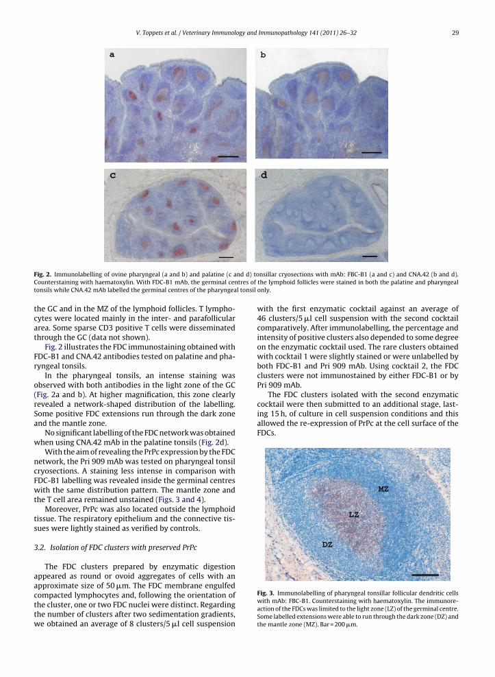
Fig. 2. Immunolabelling of ovine pharyngeal (a and b) and palatine (c aCounterstaining with haematoxylin. With FDC-B1 mAb, the germinal centonsils while CNA.42 mAb labelled the germinal centres of the pharyngea
the GC and in the MZ of the lymphoid follicles. T lympho-cytes were located mainly in the inter- and parafolliculararea. Some sparse CD3 positive T cells were disseminatedthrough the GC (data not shown).
Fig. 2 illustrates the FDC immunostaining obtained withFDC-B1 and CNA.42 antibodies tested on palatine and pha-ryngeal tonsils.
In the pharyngeal tonsils, an intense staining wasobserved with both antibodies in the light zone of the GC(Fig. 2a and b). At higher magnification, this zone clearlyrevealed a network-shaped distribution of the labelling.Some positive FDC extensions run through the dark zoneand the mantle zone.
No significant labelling of the FDC network was obtainedwhen using CNA.42 mAb in the palatine tonsils (Fig. 2d).
With the aim of revealing the PrPc expression by the FDCnetwork, the Pri 909 mAb was tested on pharyngeal tonsilcryosections. A staining less intense in comparison withFDC-B1 labelling was revealed inside the germinal centreswith the same distribution pattern. The mantle zone andthe T cell area remained unstained (Figs. 3 and 4).
Moreover, PrPc was also located outside the lymphoidtissue. The respiratory epithelium and the connective tis-sues were lightly stained as verified by controls.
3.2. Isolation of FDC clusters with preserved PrPc
The FDC clusters prepared by enzymatic digestionappeared as round or ovoid aggregates of cells with an
approximate size of 50 �m. The FDC membrane engulfedcompacted lymphocytes and, following the orientation ofthe cluster, one or two FDC nuclei were distinct. Regardingthe number of clusters after two sedimentation gradients,we obtained an average of 8 clusters/5 �l cell suspensionnsillar cryosections with mAb: FBC-B1 (a and c) and CNA.42 (b and d).the lymphoid follicles were stained in both the palatine and pharyngealonly.
with the first enzymatic cocktail against an average of46 clusters/5 �l cell suspension with the second cocktailcomparatively. After immunolabelling, the percentage andintensity of positive clusters also depended to some degreeon the enzymatic cocktail used. The rare clusters obtainedwith cocktail 1 were slightly stained or were unlabelled byboth FDC-B1 and Pri 909 mAb. Using cocktail 2, the FDCclusters were not immunostained by either FDC-B1 or byPri 909 mAb.
Fig. 3. Immunolabelling of pharyngeal tonsillar follicular dendritic cellswith mAb: FBC-B1. Counterstaining with haematoxylin. The immunore-action of the FDCs was limited to the light zone (LZ) of the germinal centre.Some labelled extensions were able to run through the dark zone (DZ) andthe mantle zone (MZ). Bar = 200 �m.

30 V. Toppets et al. / Veterinary Immunology and Immunopathology 141 (2011) 26–32
F9t(
w
es
3
fieiw
Fg9p
Fig. 6. Transmission electron micrograph of an FDC cluster isolatedfrom ovine pharyngeal tonsils. The cluster appears as a round aggregatewhere the membrane of the FDCs (*) has ensheathed 12 lymphocytes (L).Bar = 1.5 �m.
ig. 4. Immunolabelling of a secondary lymphoid follicle with mAb: Pri09. Counterstaining with haematoxylin. Pri 909 mAb allowed the detec-ion of PrPc in both the light and the dark zones of the germinal centreGC). The mantle zone (MZ) remained unstained. Bar = 200 �m.
The staining was located on the membrane extensionsrapped around lymphoid cells (Fig. 5).
Contaminant cells remained unstained except for thepithelial cells, which expressed a slight labelling (data nothown).
.3. Ultrastructural location of PrPc on FDC clusters
The precise location of Pri 909 mAb labelling was con-rmed on ultrathin sections after immunogold analysis bylectron microscopy. A low magnification of an FDC clustern Fig. 6 illustrates an FDC containing two nuclear sections
ith abundant euchromatin surrounded by a thin rim of
ig. 5. Immunolabelling of an FDC cluster isolated from ovine pharyn-eal tonsils with mAb: Pri 909. Counterstaining with haematoxylin. Pri09 labelled the FDC cytoplasmic extensions surrounding several lym-hocytes. Isolated lymphocytes were negative. Bar = 10 �m.
Fig. 7. Immunogold labelling with mAb: Pri 909 of an FDC cluster isolatedfrom ovine pharyngeal tonsils. Gold particles (arrowheads) line up alongthe cell surface of FDC cytoplasmic extensions. Bar = 200 nm.
heterochromatin. Cytoplasmic extensions of the FDC wereshown to have engulfed some lymphocytes.
At higher magnification (Fig. 7), gold particles werelocalised on the extensions of the FDC cytoplasmic mem-brane. Macrophages and lymphocytes did not bear any goldparticles. No signal occurred in the absence of Pri 909 orwhen it was replaced by isotype control antibody.
4. Discussion
It is generally accepted that in natural scrapie, sheepbecome infected by the pathogen protein early in life andthat the route of infection is likely to be oral in the majorityof cases. Dissemination of prions into the environment canoccur from several types of biological material, e.g. infec-tious placenta, amniotic fluid, and sheep saliva. Moreover,
infectivity persists in contaminated clay components of soiland aqueous soil extracts for over 29 months, leading todisease transmission by feeding or drinking (Johnson et al.,2006).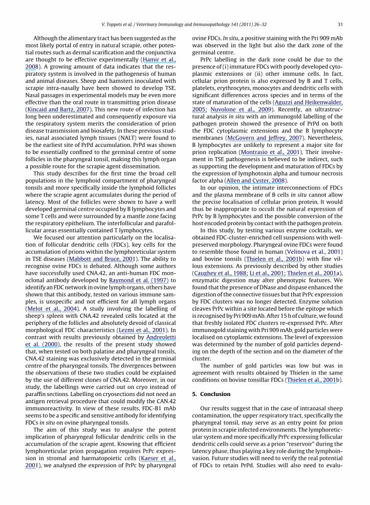
ogy and
V. Toppets et al. / Veterinary ImmunolAlthough the alimentary tract has been suggested as themost likely portal of entry in natural scrapie, other poten-tial routes such as dermal scarification and the conjunctivaare thought to be effective experimentally (Hamir et al.,2008). A growing amount of data indicates that the res-piratory system is involved in the pathogenesis of humanand animal diseases. Sheep and hamsters inoculated withscrapie intra-nasally have been showed to develop TSE.Nasal passages in experimental models may be even moreeffective than the oral route in transmitting prion disease(Kincaid and Bartz, 2007). This new route of infection haslong been underestimated and consequently exposure viathe respiratory system merits the consideration of priondisease transmission and biosafety. In these previous stud-ies, nasal associated lymph tissues (NALT) were found tobe the earliest site of PrPd accumulation. PrPd was shownto be essentially confined to the germinal centre of somefollicles in the pharyngeal tonsil, making this lymph organa possible route for the scrapie agent dissemination.
This study describes for the first time the broad cellpopulations in the lymphoid compartment of pharyngealtonsils and more specifically inside the lymphoid follicleswhere the scrapie agent accumulates during the period oflatency. Most of the follicles were shown to have a welldeveloped germinal centre occupied by B lymphocytes andsome T cells and were surrounded by a mantle zone facingthe respiratory epithelium. The interfollicular and parafol-licular areas essentially contained T lymphocytes.
We focused our attention particularly on the localisa-tion of follicular dendritic cells (FDCs), key cells for theaccumulation of prions within the lymphoreticular systemin TSE diseases (Mabbott and Bruce, 2001). The ability torecognise ovine FDCs is debated. Although some authorshave successfully used CNA.42, an anti-human FDC mon-oclonal antibody developed by Raymond et al. (1997) toidentify an FDC network in ovine lymph organs, others haveshown that this antibody, tested on various immune sam-ples, is unspecific and not efficient for all lymph organs(Melot et al., 2004). A study involving the labelling ofsheep’s spleen with CNA.42 revealed cells located at theperiphery of the follicles and absolutely devoid of classicalmorphological FDC characteristics (Lezmi et al., 2001). Incontrast with results previously obtained by Andreolettiet al. (2000), the results of the present study showedthat, when tested on both palatine and pharyngeal tonsils,CNA.42 staining was exclusively detected in the germinalcentre of the pharyngeal tonsils. The divergences betweenthe observations of these two studies could be explainedby the use of different clones of CNA.42. Moreover, in ourstudy, the labellings were carried out on cryo instead ofparaffin sections. Labelling on cryosections did not need anantigen retrieval procedure that could modify the CAN.42immunoreactivity. In view of these results, FDC-B1 mAbseems to be a specific and sensitive antibody for identifyingFDCs in situ on ovine pharyngeal tonsils.
The aim of this study was to analyse the potent
implication of pharyngeal follicular dendritic cells in theaccumulation of the scrapie agent. Knowing that efficientlymphoreticular prion propagation requires PrPc expres-sion in stromal and haematopoietic cells (Kaeser et al.,2001), we analysed the expression of PrPc by pharyngealImmunopathology 141 (2011) 26–32 31
ovine FDCs. In situ, a positive staining with the Pri 909 mAbwas observed in the light but also the dark zone of thegerminal centre.
PrPc labelling in the dark zone could be due to thepresence of (i) immature FDCs with poorly developed cyto-plasmic extensions or (ii) other immune cells. In fact,cellular prion protein is also expressed by B and T cells,platelets, erythrocytes, monocytes and dendritic cells withsignificant differences across species and in terms of thestate of maturation of the cells (Aguzzi and Heikenwalder,2005; Nuvolone et al., 2009). Recently, an ultrastruc-tural analysis in situ with an immunogold labelling of thepathogen protein showed the presence of PrPd on boththe FDC cytoplasmic extensions and the B lymphocytemembranes (McGovern and Jeffrey, 2007). Nevertheless,B lymphocytes are unlikely to represent a major site forprion replication (Montrasio et al., 2001). Their involve-ment in TSE pathogenesis is believed to be indirect, suchas supporting the development and maturation of FDCs bythe expression of lymphotoxin alpha and tumour necrosisfactor alpha (Allen and Cyster, 2008).
In our opinion, the intimate interconnections of FDCsand the plasma membrane of B cells in situ cannot allowthe precise localisation of cellular prion protein. It wouldthus be inappropriate to occult the natural expression ofPrPc by B lymphocytes and the possible conversion of thehost encoded protein by contact with the pathogen protein.
In this study, by testing various enzyme cocktails, weobtained FDC-cluster-enriched cell suspensions with well-preserved morphology. Pharyngeal ovine FDCs were foundto resemble those found in human (Velinova et al., 2001)and bovine tonsils (Thielen et al., 2001b) with fine vil-lous extensions. As previously described by other studies(Caughey et al., 1988; Li et al., 2001; Thielen et al., 2001a),enzymatic digestion may alter phenotypic features. Wefound that the presence of DNase and dispase enhanced thedigestion of the connective tissues but that PrPc expressionby FDC clusters was no longer detected. Enzyme solutioncleaves PrPc within a site located before the epitope whichis recognised by Pri 909 mAb. After 15 h of culture, we foundthat freshly isolated FDC clusters re-expressed PrPc. Afterimmunogold staining with Pri 909 mAb, gold particles werelocalised on cytoplamic extensions. The level of expressionwas determined by the number of gold particles depend-ing on the depth of the section and on the diameter of thecluster.
The number of gold particles was low but was inagreement with results obtained by Thielen in the sameconditions on bovine tonsillar FDCs (Thielen et al., 2001b).
5. Conclusion
Our results suggest that in the case of intranasal sheepcontamination, the upper respiratory tract, specifically thepharyngeal tonsil, may serve as an entry point for prionprotein in scrapie infected environments. The lymphoretic-
ular system and more specifically PrPc expressing folliculardendritic cells could serve as a prion “reservoir” during thelatency phase, thus playing a key role during the lymphoin-vasion. Future studies will need to verify the real potentialof FDCs to retain PrPd. Studies will also need to evalu-
3 ogy and
aatf
A
aHU
R
A
A
A
B
B
C
C
C
C
C
G
H
H
H
J
J
J
J
2 V. Toppets et al. / Veterinary Immunol
te the mechanisms of propagation of the disease-causinggent from these cells, resident in the lymphoid folliclesowards the central nervous system, the target organ foratal lesions.
cknowledgements
We acknowledge the technical assistance of Drs. C. Thillnd P. Hubert. We thank Professor E. Heinen from theuman Histology Unit of The Faculty of Medicine of theniversity of Liege who kindly provided the FDC-B1 mAb.
eferences
guzzi, A., Heikenwalder, M., 2005. Prions, cytokines, and chemokines: ameeting in lymphoid organs. Immunity 22, 145–154.
llen, C.D., Cyster, J.G., 2008. Follicular dendritic cell networks of pri-mary follicles and germinal centers: phenotype and function. Semin.Immunol. 20, 14–25.
ndreoletti, O., Berthon, P., Marc, D., Sarradin, P., Grosclaude, J., vanKeulen, L., Schelcher, F., Elsen, J.M., Lantier, F., 2000. Early accumu-lation of PrP(Sc) in gut-associated lymphoid and nervous tissues ofsusceptible sheep from a Romanov flock with natural scrapie. J. Gen.Virol. 81, 3115–3126.
arone, R., 1997. Pharynx et Oesophage. In: Barone, R. (Ed.), AnatomieComparée des Mammifères Domestiques, Troisième Tome: Splanch-nologie. Editions Vigot, Paris, pp. 249–290.
rown, K.L., Stewart, K., Ritchie, D.L., Mabbott, N.A., Williams, A., Fraser,H., Morrison, W.I., Bruce, M.E., 1999. Scrapie replication in lymphoidtissues depends on prion protein-expressing follicular dendritic cells.Nat. Med. 5, 1308–1312.
asteleyn, C., Cornelissen, M., Simoens, P., Van den Broeck, W., 2010. Ultra-microscopic examination of the ovine tonsillar epithelia. Anat. Rec.(Hoboken) 293, 879–889.
asteleyn, C., Cornillie, P., Simoens, P., Van Den Broeck, W., 2008. Stereo-logical assessment of the epithelial surface area of the ovine palatineand pharyngeal tonsils. Anat. Histol. Embryol. 37, 366–368.
asteleyn, C., Van den Broeck, W., Simoens, P., 2007. Histological charac-teristics and stereological volume assessment of the ovine tonsils. Vet.Immunol. Immunopathol. 120, 124–135.
aughey, B., Race, R.E., Vogel, M., Buchmeier, M.J., Chesebro, B., 1988.In vitro expression in eukaryotic cells of a prion protein gene clonedfrom scrapie-infected mouse brain. Proc. Natl. Acad. Sci. U.S.A. 85,4657–4661.
ocquyt, G., Baten, T., Simoens, P., Van Den Broeck, W., 2005. Anatom-ical localisation and histology of the ovine tonsils. Vet. Immunol.Immunopathol. 107, 79–86.
abriel, A.C.G., van den Broeck, W., 2003. Aspects anatomique et his-tologique des tonsilles du mouton. Ann. Med. Vet. 147, 251–258.
amir, A.N., Kunkle, R.A., Richt, J.A., Miller, J.M., Greenlee, J.J., 2008. Exper-imental transmission of US scrapie agent by nasal, peritoneal, andconjunctival routes to genetically susceptible sheep. Vet. Pathol. 45,7–11.
eppner, F.L., Christ, A.D., Klein, M.A., Prinz, M., Fried, M., Kraehenbuhl,J.P., Aguzzi, A., 2001. Transepithelial prion transport by M cells. Nat.Med. 7, 976–977.
orsington, J., Zhang, Z., 2007. Analysis of foot-and-mouth disease virusreplication using strand-specific quantitative RT-PCR. J. Virol. Meth-ods 144, 149–155.
effrey, M., McGovern, G., Goodsir, C.M., Brown, K.L., Bruce, M.E., 2000.Sites of prion protein accumulation in scrapie-infected mouse spleenrevealed by immuno-electron microscopy. J. Pathol. 191, 323–332.
effrey, M., Martin, S., Thomson, J.R., Dingwall, W.S., Begara-McGorum,I., Gonzalez, L., 2001. Onset and distribution of tissue prp accumula-tion in scrapie-affected suffolk sheep as demonstrated by sequentialnecropsies and tonsillar biopsies. J. Comp. Pathol. 125, 48–57.
ohnson, C.J., Phillips, K.E., Schramm, P.T., McKenzie, D., Aiken, J.M., Ped-ersen, J.A., 2006. Prions adhere to soil minerals and remain infectious.PLoS Pathog. 2, e32.
ohnson, G.D., Hardie, D.L., Ling, N.R., Maclennan, I.C., 1986. Human follicu-lar dendritic cells (FDC): a study with monoclonal antibodies (MoAb).Clin. Exp. Immunol. 64, 205–213.
Immunopathology 141 (2011) 26–32
Kaeser, P.S., Klein, M.A., Schwarz, P., Aguzzi, A., 2001. Efficient lym-phoreticular prion propagation requires PrP(c) in stromal andhematopoietic cells. J. Virol. 75, 7097–7106.
Kincaid, A.E., Bartz, J.C., 2007. The nasal cavity is a route for prion infectionin hamsters. J. Virol. 81, 4482–4491.
Kitching, R.P., Hughes, G.J., 2002. Clinical variation in foot and mouthdisease: sheep and goats. Rev. Sci. Tech. 21, 505–512.
Kosco, M.H., Pflugfelder, E., Gray, D., 1992. Follicular dendritic cell-dependent adhesion and proliferation of B cells in vitro. J. Immunol.148, 2331–2339.
Lezmi, S., Bencsik, A., Baron, T., 2001. CNA42 monoclonal antibody iden-tifies FDC as PrPsc accumulating cells in the spleen of scrapie affectedsheep. Vet. Immunol. Immunopathol. 82, 1–8.
Li, R., Liu, D., Zanusso, G., Liu, T., Fayen, J.D., Huang, J.H., Petersen, R.B.,Gambetti, P., Sy, M.S., 2001. The expression and potential functionof cellular prion protein in human lymphocytes. Cell. Immunol. 207,49–58.
Mabbott, N.A., Bruce, M.E., 2001. The immunobiology of TSE diseases. J.Gen. Virol. 82, 2307–2318.
Mabbott, N.A., Mackay, F., Minns, F., Bruce, M.E., 2000. Temporary inacti-vation of follicular dendritic cells delays neuroinvasion of scrapie. Nat.Med. 6, 719–720.
McBride, P.A., Eikelenboom, P., Kraal, G., Fraser, H., Bruce, M.E., 1992.PrP protein is associated with follicular dendritic cells of spleens andlymph nodes in uninfected and scrapie-infected mice. J. Pathol. 168,413–418.
McGovern, G., Jeffrey, M., 2007. Scrapie-specific pathology of sheep lym-phoid tissues. PLoS One 2, e1304.
Melot, F., Defaweux, V., Jolois, O., Collard, A., Robert, B., Heinen, E., Antoine,N., 2004. FDC-B1: a new monoclonal antibody directed againstbovine follicular dendritic cells. Vet. Immunol. Immunopathol. 97,1–9.
Montrasio, F., Frigg, R., Glatzel, M., Klein, M.A., Mackay, F., Aguzzi,A., Weissmann, C., 2000. Impaired prion replication in spleensof mice lacking functional follicular dendritic cells. Science 288,1257–1259.
Montrasio, F., Cozzio, A., Flechsig, E., Rossi, D., Klein, M.A., Rulicke, T., Rae-ber, A.J., Vosshenrich, C.A., Proft, J., Aguzzi, A., Weissmann, C., 2001.B lymphocyte-restricted expression of prion protein does not enableprion replication in prion protein knockout mice. Proc. Natl. Acad. Sci.U.S.A. 98, 4034–4037.
Naiem, M., Gerdes, J., Abdulaziz, Z., Stein, H., Mason, D.Y., 1983. Productionof a monoclonal antibody reactive with human dendritic reticulumcells and its use in the immunohistological analysis of lymphoid tissue.J. Clin. Pathol. 36, 167–175.
Nuvolone, M., Aguzzi, A., Heikenwalder, M., 2009. Cells and prions: alicense to replicate. FEBS Lett. 583, 2674–2684.
Ogra, P.L., 2000. Mucosal immune response in the ear, nose and throat.Pediatr. Infect. Dis. J. 19, S4–8.
Perry, M., Whyte, A., 1998. Immunology of the tonsils. Immunol. Today19, 414–421.
Raymond, I., Al Saati, T., Tkaczuk, J., Chittal, S., Delsol, G., 1997. CNA.42,a new monoclonal antibody directed against a fixative-resistantantigen of follicular dendritic reticulum cells. Am. J. Pathol. 151,1577–1585.
Stanley, A.C., Huntley, J.F., Jeffrey, M., Buxton, D., 2001. Characterization ofovine nasal-associated lymphoid tissue and identification of M cellsin the overlying follicle-associated epithelium. J. Comp. Pathol. 125,262–270.
Thielen, C., Antoine, N., Melot, F., Cesbron, J.Y., Heinen, E., Tsunoda, R.,2001a. Human FDC express PrPc in vivo and in vitro. Dev. Immunol.8, 259–266.
Thielen, C., Melot, F., Jolois, O., Leclercq, F., Tsunoda, R., Frobert, Y., Heinen,E., Antoine, N., 2001b. Isolation of bovine follicular dendritic cellsallows the demonstration of a particular cellular prion protein. CellTissue Res. 306, 49–55.
Thuring, C.M., Crowe, M.A., McAllister, H., Earley, B., Roche, J.F., Sweeney,T., 2002. Evaluation of peripheral lymphoreticular biopsy techniquesand their clinical side effects in sheep. Vet. Rec. 150, 97–102.
Tsunoda, R., Nakayama, M., Onozaki, K., Heinen, E., Cormann, N., Kinet-
Denoel, C., Kojima, M., 1990. Isolation and long-term cultivation ofhuman tonsil follicular dendritic cells. Virchows Arch. B Cell. Pathol.Incl. Mol. Pathol. 59, 95–105.Velinova, M., Thielen, C., Melot, F., Donga, J., Eicher, S., Heinen, E., Antoine,N., 2001. New histochemical and ultrastructural observations on nor-mal bovine tonsils. Vet. Rec. 149, 613–617.


![4* • Pharingeal grooves/cleft : 4 • [Pharyngeal membrane]](https://static.fdokumen.com/doc/165x107/6334ea00b9085e0bf5093ec7/4-pharingeal-groovescleft-4-pharyngeal-membrane.jpg)