New semionotiform (Actinopterygii: Neopterygii) from the Late Jurassic of southern Germany
Evolutionary Trends of the Pharyngeal Dentition inCypriniformes (Actinopterygii: Ostariophysi)Emmanuel Pasco-Viel1, Cyril Charles3¤, Pascale Chevret2, Marie Semon2, Paul Tafforeau4,
Laurent Viriot1,3*., Vincent Laudet2*.
1 Evo-devo of Vertebrate Dentition, Institut de Genomique Fonctionnelle de Lyon, Universite de Lyon, CNRS, INRA, Ecole Normale Superieure de Lyon, Lyon, France,
2 Molecular Zoology, Institut de Genomique Fonctionnelle de Lyon, Universite de Lyon, CNRS, INRA, Ecole Normale Superieure de Lyon, Lyon, France, 3 iPHEP, CNRS UMR
6046, Universite de Poitiers, Poitiers, France, 4 European Synchrotron Radiation Facility, Grenoble, France
Abstract
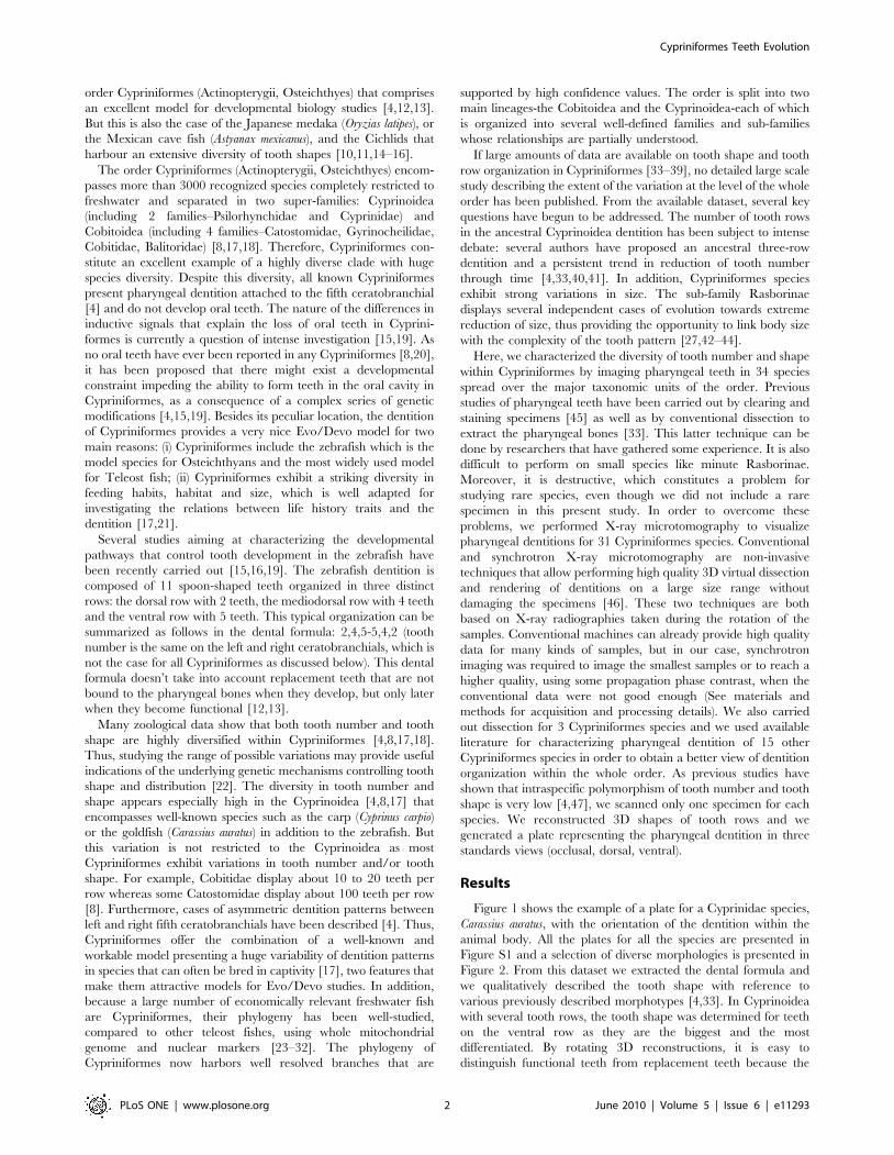
Background: The fish order Cypriniformes is one of the most diverse ray-finned fish groups in the world with more than3000 recognized species. Cypriniformes are characterized by a striking distribution of their dentition: namely the absence oforal teeth and presence of pharyngeal teeth on the last gill arch (fifth ceratobranchial). Despite this limited localisation, thediversity of tooth patterns in Cypriniformes is astonishing. Here we provide a further description of this diversity using X-raymicrotomography and we map the resulting dental characters on a phylogenetic tree to explore evolutionary trends.
Results: We performed a pilot survey of dental formulae and individual tooth shapes in 34 adult species of Cypriniformes byX-ray microtomography (using either conventional X-ray machine, or synchrotron microtomography when necessary) or bydissecting. By mapping morphological results in a phylogenetic tree, it emerges that the two super-families Cobitoidea andCyprinoidea have followed two distinct evolutionary pathways. Furthermore, our analysis supports the hypothesis of athree-row dentition as ancestral for Cyprinoidea and a general trend in tooth row reduction in most derived lineages. Yet,this general scheme must be considered with caution as several events of tooth row gain and loss have occurred duringevolutionary history of Cyprinoidea.
Significance: Dentition diversity in Cypriniformes constitutes an excellent model to study the evolution of complexmorphological structures. This morphological survey clearly advocates for extending the use of X-ray microtomography tostudy tooth morphology in Cypriniformes. Yet, our survey also underlines that improved knowledge of Cypriniformes lifetraits, such as feeding habits, is required as current knowledge is not sufficient to conclude on the link between diet anddental morphology.
Citation: Pasco-Viel E, Charles C, Chevret P, Semon M, Tafforeau P, et al. (2010) Evolutionary Trends of the Pharyngeal Dentition in Cypriniformes (Actinopterygii:Ostariophysi). PLoS ONE 5(6): e11293. doi:10.1371/journal.pone.0011293
Editor: Johannes Jaeger, Centre for Genomic Regulation (CRG), Universitat Pompeu Fabra, Spain
Received October 28, 2009; Accepted May 31, 2010; Published June 24, 2010
Copyright: � 2010 Pasco-Viel et al. This is an open-access article distributed under the terms of the Creative Commons Attribution License, which permitsunrestricted use, distribution, and reproduction in any medium, provided the original author and source are credited.
Funding: This work has been funded by the Ministere de l’Education Nationale de la Recherche et de Technologie (MENRT, www.education.gouv.fr), the CentreNational de la Recherche Scientifique (CNRS, www.cnrs.fr), the European Synchrotron Radiation Facility (ESRF, www.esrf.eu) and Ecole Normale Superieure (ENS) ofLyon (www.ens-lyon.fr) as well as by Agence Nationale pour la Recherche (ANR) grant Bouillabaisse (www.agence-nationale-recherche.fr/ANR-09-BLAN-0127-02).The funders had no role in study design, data collection and analysis, decision to publish, or preparation of the manuscript.
Competing Interests: Author Vincent Laudet is on the editorial board.
* E-mail: [email protected] (VL); [email protected] (LV)
. These authors contributed equally to this work.
¤ Current address: Department of Orofacial Sciences, University of California San Francisco, San Francisco, California, United States of America
Introduction
Many biological structures such as somites, segments or limbs,
exhibit repeated patterns and these structures are often variable
between related species. Within these serially homologous
structures, teeth were relatively neglected by Evo/Devo studies
despite their promise for describing and understanding both the
diversity and evolution of complex adaptive structures. Teeth can
be used for integrated studies and results have begun to
accumulate on the genomic and/or developmental basis of their
wide diversity [1,2]. Up to now, the evolution and development of
teeth have been more intensively investigated in the mouse model
in which mechanisms controlling tooth crown shape, tooth
identity, dental row segmentation, or occurrence of toothless
areas are under intense investigation [1–3]. However, teeth of
Actinopterygians are becoming more and more intensively studied
[4,5]. When compared to Mammals, Actinopterygian fish display
two dental traits that are reminiscent of the basal condition of
Vertebrates: (i) their teeth are widely distributed within the oral
and pharyngeal cavities, and (ii) their teeth are constantly replaced
throughout the duration of the animal’s life (polyphyodonty) [6,7].
Interestingly, Actinopterygian fish display a great diversity in tooth
number and location [4,8]: they display rows of hundreds of teeth
which can be located on the lower and upper jaws, the floor of the
mouth (basihyal, basibranchials), the roof of the mouth (e.g.
vomer, palatine), the upper and lower pharyngeal regions. In
addition to this diversity, Actinopterygian fish include several well-
studied experimental models that allow for the study of tooth
development from a mechanistic perspective [9–11]. This is for
example the case of the zebrafish, Danio rerio, a member of the
PLoS ONE | www.plosone.org 1 June 2010 | Volume 5 | Issue 6 | e11293
order Cypriniformes (Actinopterygii, Osteichthyes) that comprises
an excellent model for developmental biology studies [4,12,13].
But this is also the case of the Japanese medaka (Oryzias latipes), or
the Mexican cave fish (Astyanax mexicanus), and the Cichlids that
harbour an extensive diversity of tooth shapes [10,11,14–16].
The order Cypriniformes (Actinopterygii, Osteichthyes) encom-
passes more than 3000 recognized species completely restricted to
freshwater and separated in two super-families: Cyprinoidea
(including 2 families–Psilorhynchidae and Cyprinidae) and
Cobitoidea (including 4 families–Catostomidae, Gyrinocheilidae,
Cobitidae, Balitoridae) [8,17,18]. Therefore, Cypriniformes con-
stitute an excellent example of a highly diverse clade with huge
species diversity. Despite this diversity, all known Cypriniformes
present pharyngeal dentition attached to the fifth ceratobranchial
[4] and do not develop oral teeth. The nature of the differences in
inductive signals that explain the loss of oral teeth in Cyprini-
formes is currently a question of intense investigation [15,19]. As
no oral teeth have ever been reported in any Cypriniformes [8,20],
it has been proposed that there might exist a developmental
constraint impeding the ability to form teeth in the oral cavity in
Cypriniformes, as a consequence of a complex series of genetic
modifications [4,15,19]. Besides its peculiar location, the dentition
of Cypriniformes provides a very nice Evo/Devo model for two
main reasons: (i) Cypriniformes include the zebrafish which is the
model species for Osteichthyans and the most widely used model
for Teleost fish; (ii) Cypriniformes exhibit a striking diversity in
feeding habits, habitat and size, which is well adapted for
investigating the relations between life history traits and the
dentition [17,21].
Several studies aiming at characterizing the developmental
pathways that control tooth development in the zebrafish have
been recently carried out [15,16,19]. The zebrafish dentition is
composed of 11 spoon-shaped teeth organized in three distinct
rows: the dorsal row with 2 teeth, the mediodorsal row with 4 teeth
and the ventral row with 5 teeth. This typical organization can be
summarized as follows in the dental formula: 2,4,5-5,4,2 (tooth
number is the same on the left and right ceratobranchials, which is
not the case for all Cypriniformes as discussed below). This dental
formula doesn’t take into account replacement teeth that are not
bound to the pharyngeal bones when they develop, but only later
when they become functional [12,13].
Many zoological data show that both tooth number and tooth
shape are highly diversified within Cypriniformes [4,8,17,18].
Thus, studying the range of possible variations may provide useful
indications of the underlying genetic mechanisms controlling tooth
shape and distribution [22]. The diversity in tooth number and
shape appears especially high in the Cyprinoidea [4,8,17] that
encompasses well-known species such as the carp (Cyprinus carpio)
or the goldfish (Carassius auratus) in addition to the zebrafish. But
this variation is not restricted to the Cyprinoidea as most
Cypriniformes exhibit variations in tooth number and/or tooth
shape. For example, Cobitidae display about 10 to 20 teeth per
row whereas some Catostomidae display about 100 teeth per row
[8]. Furthermore, cases of asymmetric dentition patterns between
left and right fifth ceratobranchials have been described [4]. Thus,
Cypriniformes offer the combination of a well-known and
workable model presenting a huge variability of dentition patterns
in species that can often be bred in captivity [17], two features that
make them attractive models for Evo/Devo studies. In addition,
because a large number of economically relevant freshwater fish
are Cypriniformes, their phylogeny has been well-studied,
compared to other teleost fishes, using whole mitochondrial
genome and nuclear markers [23–32]. The phylogeny of
Cypriniformes now harbors well resolved branches that are
supported by high confidence values. The order is split into two
main lineages-the Cobitoidea and the Cyprinoidea-each of which
is organized into several well-defined families and sub-families
whose relationships are partially understood.
If large amounts of data are available on tooth shape and tooth
row organization in Cypriniformes [33–39], no detailed large scale
study describing the extent of the variation at the level of the whole
order has been published. From the available dataset, several key
questions have begun to be addressed. The number of tooth rows
in the ancestral Cyprinoidea dentition has been subject to intense
debate: several authors have proposed an ancestral three-row
dentition and a persistent trend in reduction of tooth number
through time [4,33,40,41]. In addition, Cypriniformes species
exhibit strong variations in size. The sub-family Rasborinae
displays several independent cases of evolution towards extreme
reduction of size, thus providing the opportunity to link body size
with the complexity of the tooth pattern [27,42–44].
Here, we characterized the diversity of tooth number and shape
within Cypriniformes by imaging pharyngeal teeth in 34 species
spread over the major taxonomic units of the order. Previous
studies of pharyngeal teeth have been carried out by clearing and
staining specimens [45] as well as by conventional dissection to
extract the pharyngeal bones [33]. This latter technique can be
done by researchers that have gathered some experience. It is also
difficult to perform on small species like minute Rasborinae.
Moreover, it is destructive, which constitutes a problem for
studying rare species, even though we did not include a rare
specimen in this present study. In order to overcome these
problems, we performed X-ray microtomography to visualize
pharyngeal dentitions for 31 Cypriniformes species. Conventional
and synchrotron X-ray microtomography are non-invasive
techniques that allow performing high quality 3D virtual dissection
and rendering of dentitions on a large size range without
damaging the specimens [46]. These two techniques are both
based on X-ray radiographies taken during the rotation of the
samples. Conventional machines can already provide high quality
data for many kinds of samples, but in our case, synchrotron
imaging was required to image the smallest samples or to reach a
higher quality, using some propagation phase contrast, when the
conventional data were not good enough (See materials and
methods for acquisition and processing details). We also carried
out dissection for 3 Cypriniformes species and we used available
literature for characterizing pharyngeal dentition of 15 other
Cypriniformes species in order to obtain a better view of dentition
organization within the whole order. As previous studies have
shown that intraspecific polymorphism of tooth number and tooth
shape is very low [4,47], we scanned only one specimen for each
species. We reconstructed 3D shapes of tooth rows and we
generated a plate representing the pharyngeal dentition in three
standards views (occlusal, dorsal, ventral).
Results
Figure 1 shows the example of a plate for a Cyprinidae species,
Carassius auratus, with the orientation of the dentition within the
animal body. All the plates for all the species are presented in
Figure S1 and a selection of diverse morphologies is presented in
Figure 2. From this dataset we extracted the dental formula and
we qualitatively described the tooth shape with reference to
various previously described morphotypes [4,33]. In Cyprinoidea
with several tooth rows, the tooth shape was determined for teeth
on the ventral row as they are the biggest and the most
differentiated. By rotating 3D reconstructions, it is easy to
distinguish functional teeth from replacement teeth because the
Cypriniformes Teeth Evolution
PLoS ONE | www.plosone.org 2 June 2010 | Volume 5 | Issue 6 | e11293
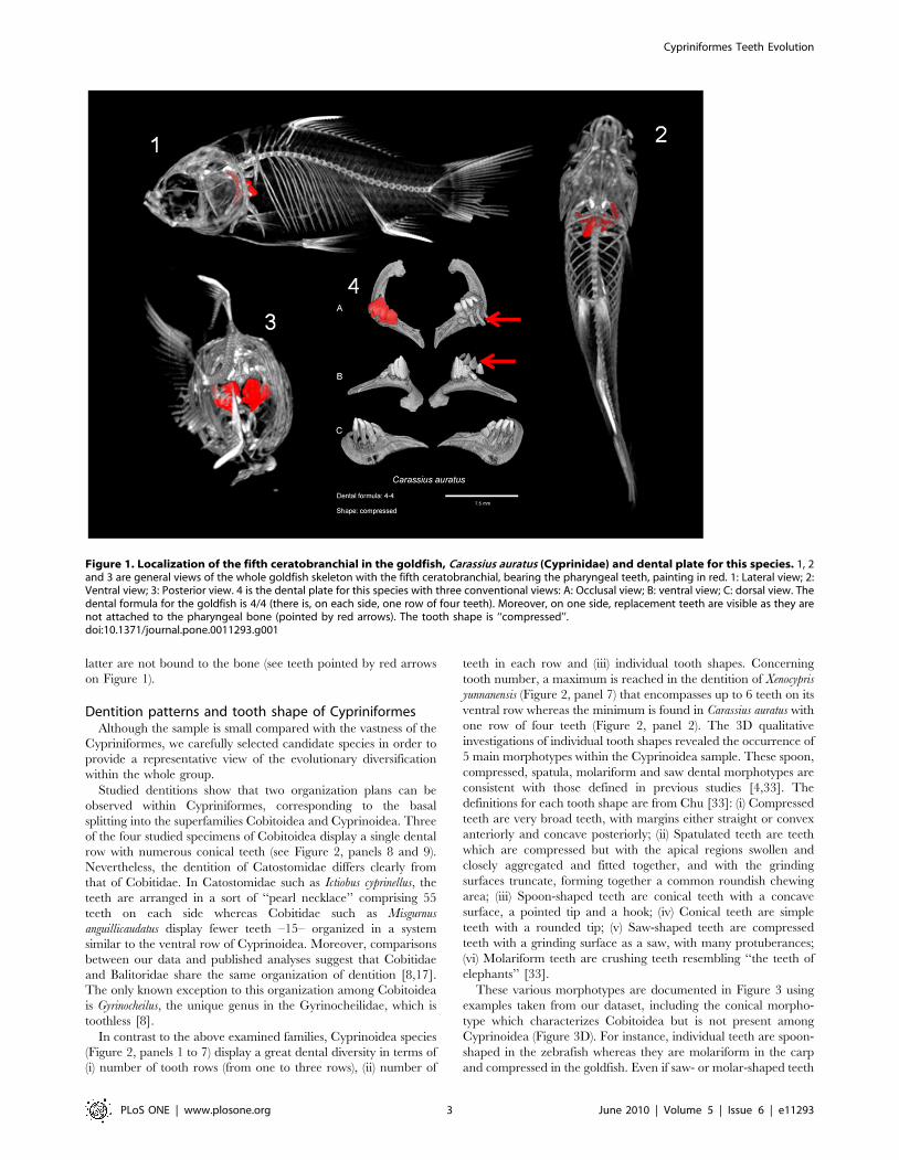
latter are not bound to the bone (see teeth pointed by red arrows
on Figure 1).
Dentition patterns and tooth shape of CypriniformesAlthough the sample is small compared with the vastness of the
Cypriniformes, we carefully selected candidate species in order to
provide a representative view of the evolutionary diversification
within the whole group.
Studied dentitions show that two organization plans can be
observed within Cypriniformes, corresponding to the basal
splitting into the superfamilies Cobitoidea and Cyprinoidea. Three
of the four studied specimens of Cobitoidea display a single dental
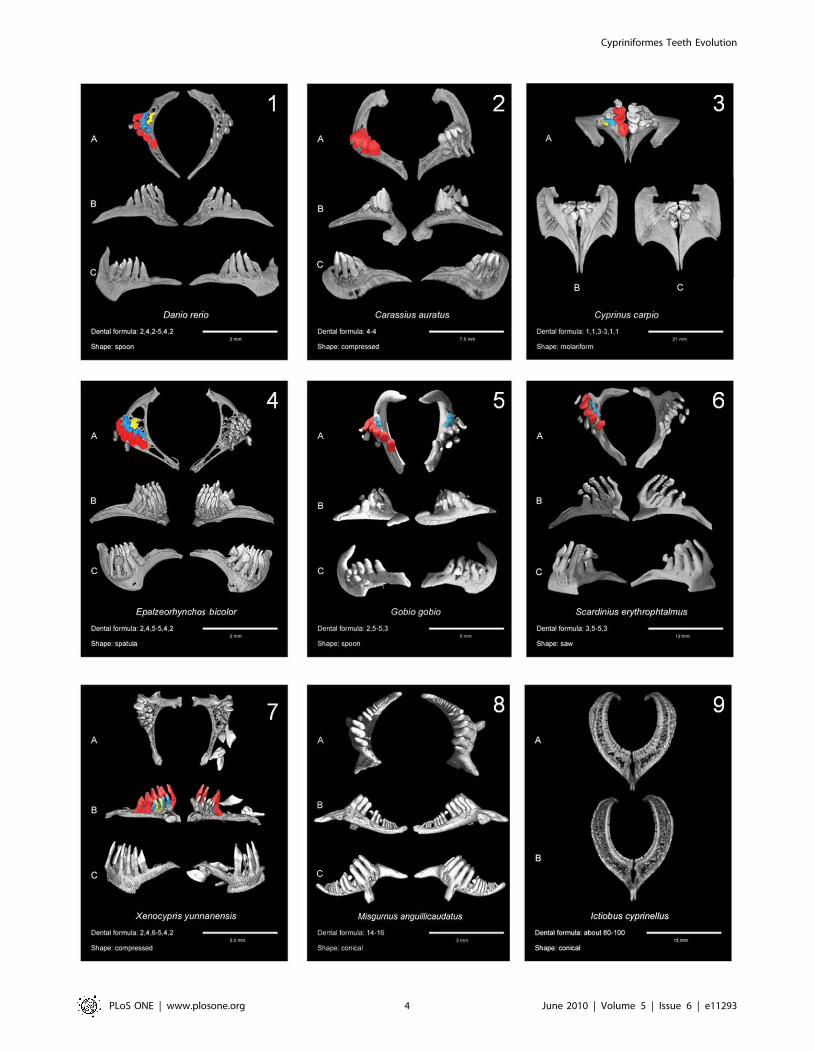
row with numerous conical teeth (see Figure 2, panels 8 and 9).
Nevertheless, the dentition of Catostomidae differs clearly from
that of Cobitidae. In Catostomidae such as Ictiobus cyprinellus, the
teeth are arranged in a sort of ‘‘pearl necklace’’ comprising 55
teeth on each side whereas Cobitidae such as Misgurnus
anguillicaudatus display fewer teeth –15– organized in a system
similar to the ventral row of Cyprinoidea. Moreover, comparisons
between our data and published analyses suggest that Cobitidae
and Balitoridae share the same organization of dentition [8,17].
The only known exception to this organization among Cobitoidea
is Gyrinocheilus, the unique genus in the Gyrinocheilidae, which is
toothless [8].
In contrast to the above examined families, Cyprinoidea species
(Figure 2, panels 1 to 7) display a great dental diversity in terms of
(i) number of tooth rows (from one to three rows), (ii) number of
teeth in each row and (iii) individual tooth shapes. Concerning
tooth number, a maximum is reached in the dentition of Xenocypris
yunnanensis (Figure 2, panel 7) that encompasses up to 6 teeth on its
ventral row whereas the minimum is found in Carassius auratus with
one row of four teeth (Figure 2, panel 2). The 3D qualitative
investigations of individual tooth shapes revealed the occurrence of
5 main morphotypes within the Cyprinoidea sample. These spoon,
compressed, spatula, molariform and saw dental morphotypes are
consistent with those defined in previous studies [4,33]. The
definitions for each tooth shape are from Chu [33]: (i) Compressed
teeth are very broad teeth, with margins either straight or convex
anteriorly and concave posteriorly; (ii) Spatulated teeth are teeth
which are compressed but with the apical regions swollen and
closely aggregated and fitted together, and with the grinding
surfaces truncate, forming together a common roundish chewing
area; (iii) Spoon-shaped teeth are conical teeth with a concave
surface, a pointed tip and a hook; (iv) Conical teeth are simple
teeth with a rounded tip; (v) Saw-shaped teeth are compressed
teeth with a grinding surface as a saw, with many protuberances;
(vi) Molariform teeth are crushing teeth resembling ‘‘the teeth of
elephants’’ [33].
These various morphotypes are documented in Figure 3 using
examples taken from our dataset, including the conical morpho-
type which characterizes Cobitoidea but is not present among
Cyprinoidea (Figure 3D). For instance, individual teeth are spoon-
shaped in the zebrafish whereas they are molariform in the carp
and compressed in the goldfish. Even if saw- or molar-shaped teeth
Figure 1. Localization of the fifth ceratobranchial in the goldfish, Carassius auratus (Cyprinidae) and dental plate for this species. 1, 2and 3 are general views of the whole goldfish skeleton with the fifth ceratobranchial, bearing the pharyngeal teeth, painting in red. 1: Lateral view; 2:Ventral view; 3: Posterior view. 4 is the dental plate for this species with three conventional views: A: Occlusal view; B: ventral view; C: dorsal view. Thedental formula for the goldfish is 4/4 (there is, on each side, one row of four teeth). Moreover, on one side, replacement teeth are visible as they arenot attached to the pharyngeal bone (pointed by red arrows). The tooth shape is ‘‘compressed’’.doi:10.1371/journal.pone.0011293.g001
Cypriniformes Teeth Evolution
PLoS ONE | www.plosone.org 3 June 2010 | Volume 5 | Issue 6 | e11293
Cypriniformes Teeth Evolution
PLoS ONE | www.plosone.org 4 June 2010 | Volume 5 | Issue 6 | e11293

clearly differ from one another (Figure 3E–F), spoon-, compressed-
and spatula-shaped teeth appear to be more similar in shape, the
spatula morphotype displaying an intermediary morphology
between spoon and compressed morphotypes (Figure 3A–C).
Asymmetry is a frequent feature of Cyprinoidea pharyngeal
dentition [4]. Asymmetric dentition has been documented for
Gobio gobio, which has respectively two and three teeth on its left
and right dorsal rows (Figure 2 panel 5). A similar situation is
found in Xenocypris yunnanensis, which displays a ventral dental row
with 6 teeth on the left side and 5 teeth on the right side (Figure 2
panel 7). Overall, 14 of the 42 studied species of Cyprinoidea with
a determined tooth row number exhibit asymmetrical features,
which is one third. This is undoubtedly not linked to replacement
transient asymmetries (as a missing tooth on the ceratobranchial
can be matched by a gap in the bone–see the example of Xenocypris
yunnanensis on Figure 2 panel 7). Asymmetry is especially frequent
Figure 3. Various morphotypes of Cypriniformes teeth illustrated with examples from our dataset. A: spoon tooth shape of Puntiussemifasciolatus (Cyprininae) with an incurved depressed region and a hook at the end of each tooth; B: spatula tooth shape of Epalzeorhynchos bicolor(Cyprininae), an intermediary form between the spoon and compressed morphologies, with teeth forming a grinding surface; C: compressed toothshape of Carassius carassius (Cyprininae), with an elongated depressed region; D: conical tooth shape of Catostomus commersonii (Catostomidae); E:saw tooth shape of Scardinius erythrophtalmus (Leuciscinae); F: molariform tooth shape of Cyprinus carpio (Cyprininae).doi:10.1371/journal.pone.0011293.g003
Figure 2. Examples of Cypriniformes dentitions obtained by microtomography and analysis of 3D reconstructions. In each case, thedental formula and the tooth shape are indicated. 1: Danio rerio (Rasborinae); 2,4,5; spoon. 2: Carassius auratus (Cyprininae); 4; compressed. 3:Cyprinus carpio (Cyprininae); 1,1,3; molariform. 4: Epalzeorhynchos bicolor (Cyprininae); 2,4,5; spatula. 5: Gobio gobio (Gobioninae); 2,5(25,3); spoon. 6:Scardinius erythrophtalmus (Leuciscinae); 3,5; saw. 7: Xenocypris yunnanensis (Cultrinae); 2,3,5(26,3,2); compressed. 8: Misgurnus anguillicaudatus(Cobitidae); 14–16; conical. 9: Ictiobus cyprinellus (Catostomidae); 55; conical. A: occlusal view; B: ventral view; C: dorsal view. Except for 8 and 9. A:anterior view; B: posterior view. For Cyprinoidea plates, teeth are painted according to the following: (i) teeth on the left side are all painted accordingto their row: red for ventral row, blue for mediodorsal row and yellow for dorsal row; (ii) teeth on the right side are painted only if their row displays atooth number different from the left side (so that species displaying asymmetry are easily visible because of colours on the right half-bone).doi:10.1371/journal.pone.0011293.g002
Cypriniformes Teeth Evolution
PLoS ONE | www.plosone.org 5 June 2010 | Volume 5 | Issue 6 | e11293
within Leuciscinae and Rasborinae. This raises the question of the
developmental mechanisms of tooth row organization that favour
asymmetry inside these families. Nevertheless, asymmetric patterns
can emerge within species located in clades in which most of the
species exhibit a symmetrical pattern (e.g. Puntius semifasciolatus in
Cyprininae).
Cypriniformes also frequently display miniaturization, especially
in Rasborinae, a subfamily that encompasses Paedocypris progenetica,
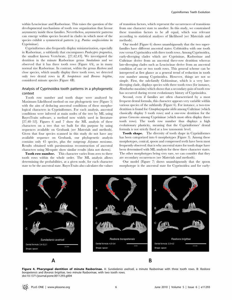
the smallest known Vertebrate, [27,42,43]. We investigated the
dentition in the minute Rasborinae genus Sundadanio and we
observed that it has three tooth rows (Figure 4A), as in many
normal size Rasborinae. In contrast, within the genus Rasbora and
close species, which usually display three tooth rows, we detected
only two dental rows in R. borapetensis and Boraras brigittae,
considered minute species (Figure 4B).
Analysis of Cyprinoidea tooth patterns in a phylogeneticcontext
Tooth row number and tooth shape were analyzed by
Maximum Likelihood method on our phylogenetic tree (Figure 5)
with the aim of deducing ancestral conditions of these morpho-
logical characters in Cypriniformes. For each analysis, ancestral
conditions were inferred at main nodes of the tree by ML using
BayesTraits software, a method now widely used in literature
[27,48–52]. Figures 6 and 7 show the ML analysis of these
characters on a tree that we built for this purpose by using
sequences available on Genbank (see Materials and methods).
Given that four species scanned in this study do not have any
available sequence on Genbank, our phylogenetic analysis
contains only 45 species, plus the outgroup Astyanax mexicanus.
Results obtained with parsimonious reconstruction of ancestral
characters using Mesquite show similar results (data not shown).
Tooth row number. This character varies from zero to three
tooth rows within the whole order. The ML analysis allows
determining the probabilities, at a given node, for each character
state to be the ancestral state. BayesTraits also calculates the values
of transition factors, which represent the occurrences of transitions
from one character state to another. In this study, we constrained
these transition factors to be all equal, which was relevant
according to statistical analyses of likelihood (see Materials and
methods).
Our model (Figure 6) shows unambiguously that the two super-
families have different ancestral states: Cobitoidea with one tooth
row versus Cyprinoidea with three tooth rows. Among Cyprinoidea,
early-diverging clades which are Cyprininae, Rasborinae and
Cultrinae derive from an ancestral three-row dentition whereas
late-diverging clades such as Leuciscinae derive from an ancestral
condition of one or two tooth rows. This general scheme can be
interpreted at first glance as a general trend of reduction in tooth
row number among Cyprinoidea. However, things are not so
simple. First, the sub-family Gobioninae, which is a very late-
diverging clade, displays species with three tooth rows (for instance,
Hemibarbus maculatus) which shows that a secondary gain of tooth row
has occurred during recent evolutionary history of Cyprinoidea.
Second, even if families are often characterized by a most
frequent dental formula, this character appears very variable within
various species of the subfamily (Figure 6). For instance, a two-row
dentition is found for Ctenopharyngodon idella among Cultrinae (which
classically display 3 tooth rows) and a one-row dentition for the
genus Carassius among Cyprininae (which most often display three
tooth rows). The tooth row number thus displays a high
evolutionary plasticity, meaning that the Cypriniformes’ dental
formula is not strictly fixed at a low taxonomic level.
Tooth shape. The diversity of tooth shape in Cypriniformes
has been categorized into 6 morphotypes (Figure 3). Among these
morphotypes, conical, spoon and compressed teeth have been most
frequently observed: that is why ancestral states for tooth shape have
been determined with ML analysis for these three character states.
The other morphotypes being very rare, we can consider that they
are secondary occurrences (see Materials and methods).
Our model (Figure 7) shows unambiguously that the spoon
morphotype is the ancestral state for Cyprinoidea and for early-
Figure 4. Pharyngeal dentition of minute Rasborinae. A: Sundadanio axelrodi, a minute Rasborinae with three tooth rows. B: Rasboraborapetensis and Boraras brigittae, two minute Rasborinae, with two tooth rows.doi:10.1371/journal.pone.0011293.g004
Cypriniformes Teeth Evolution
PLoS ONE | www.plosone.org 6 June 2010 | Volume 5 | Issue 6 | e11293
diverging clades such as Cyprininae, Rasborinae and Cultrinae.
For late-diverging clades such as Leuciscinae, the compressed
morphotype is the ancestral tooth shape. Yet, as for tooth row
number, this scheme is far too general. The compressed
morphotype can be found in early-diverging clades (Carassius
among Cyprininae and Esomus among Rasborinae) and the spoon
morphotype in late-diverging clades (Gobio among Gobioninae).
This implies that tooth shape has been frequently and conver-
gently remodelled over the course of evolution. As for tooth row
number, divergences of tooth morphotypes occur within the
various families. This is the case of the dental morphotype in
Cyprinus carpio, which is molariform, whereas the most frequently
reported morphotype of Cyprininae is spoon-shaped. In the same
way, Scardinius erythrophtalmus is the only observed Leuciscinae with
saw-shaped teeth whereas most Leuciscinae display compressed
teeth. It is also interesting to note that the genera Esomus and
Carassius have evolved the same way with regard to both tooth row
number and tooth shape. Esomus and Carassius both display a one-
row dentition with compressed teeth and these genera belong to
two different families in which the predominating dentition is
made up of three rows of spoon-shaped teeth. This may suggest a
functional coupling of these two characters in these genera.
In conclusion, more generally within Cyprinoidea, early-
diverging clades are mostly species with three rows of spoon-
shaped teeth whereas late-diverging clades are mostly species with
one or two row(s) of compressed teeth.
Discussion
This preliminary morphological survey of the pharyngeal
dentition in 49 species selected as representative of the major
clades of Cypriniformes allows us to draw several conclusions: (i)
microtomography is an adequate tool to perform non-invasive
studies of the pharyngeal dentition in Cypriniformes; (ii) from a
primitive pattern, various strategies were adopted within the two
super-families: Cobitoidea display a single row with a high number
of conical teeth while Cyprinoidea display several tooth rows with
few teeth in each row and a high diversity in shape; (iii) the fact
that tooth number and tooth shape have been frequently
remodeled during evolution reveals a strong plasticity of the
pharyngeal dentition in Cypriniformes.
3D microtomography is a powerful tool to characterizepharyngeal tooth morphology
This study reveals the power of 3D microtomography in order
to perform non invasive studies of Cypriniformes pharyngeal teeth.
These teeth are located inside the body, therefore analysing them
is destructive for the specimen. This is a main issue for museum
collections as it is necessary to save those collections and find out
techniques which allow studies without any damage, especially for
rare specimens.
Moreover, tooth shape cannot be accurately characterized by
simple observation of pharyngeal teeth (see panels of dissected
specimens in this study, in Figure S1), especially for small
specimens. Thus, in this latter case, tooth extraction must be
followed by scanning electron microscopy which is a demanding
technique. On the contrary, microtomography, as long as
resolution is sufficient, lets determine tooth morphology by a
simple observation of unprepared specimens.
Another positive aspect of microtomography will be the
possibility to carry out a highly extensive sampling, thanks to the
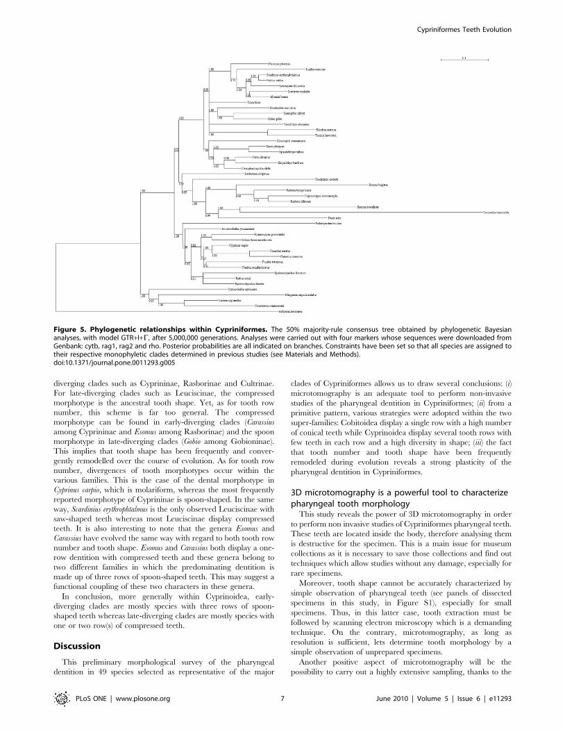
Figure 5. Phylogenetic relationships within Cypriniformes. The 50% majority-rule consensus tree obtained by phylogenetic Bayesiananalyses, with model GTR+I+C, after 5,000,000 generations. Analyses were carried out with four markers whose sequences were downloaded fromGenbank: cytb, rag1, rag2 and rho. Posterior probabilities are all indicated on branches. Constraints have been set so that all species are assigned totheir respective monophyletic clades determined in previous studies (see Materials and Methods).doi:10.1371/journal.pone.0011293.g005
Cypriniformes Teeth Evolution
PLoS ONE | www.plosone.org 7 June 2010 | Volume 5 | Issue 6 | e11293

little time it takes (about five hours for one specimen from the scan
to the virtual extraction of the bones).
The ancestral dental formula of Cypriniformes andCyprinoidea
Our results show that the ancestral condition of Cypriniformes
cannot be determined with our limited sample of Cypriniformes
species. This absence of conclusion is due to the fact that tooth row
number and tooth shape are very different between the two super-
families. Considering the ancestral dental formula of Cyprinoidea,
the common view of a three-row dentition [33] is supported by this
analysis.
Our analysis of the basal condition in Cypriniformes can be
confronted to paleontological data. The oldest known fossil
Cypriniformes were unearthed from the Early Palaeocene deposits
of Northern America dated at about 60 Ma [53]. In Asia and
Europe, the oldest Cypriniformes date from the Early Eocene at
about 50 Ma [54–57]. The earliest North-American fossils were
unambiguously identified as Catostomidae whereas the earliest
Asian fossils include Catostomidae and Cyprinoidea. [54–57]. A
frequent problem with fossil vertebrates is that teeth are the best-
preserved feature during the fossilization process as dental tissues
are the hardest part of the body. As a consequence, fossil
Cypriniformes are frequently only represented by isolated
pharyngeal teeth or fragments of pharyngeal bones and the
identification of the various taxa is based quite exclusively on
dental characters [58]. No other characters, independent of the
dentition, can be used to assign fossil specimens to the various
groups of Cyprinoidea. Thus, fossils cannot be characterized as
primitive or derived forms. In certain sites where the taphonomic
conditions allowed for the fossilization of the whole skeleton,
complete fossils display many characters independent of the
dentition. This is the case for the nearly complete skeleton of the
earliest known Cyprinoidea found in China that has been assigned
to the Gobioninae [56]. This suggests that some families of
Cyprinoidea were already individualized at that time, which
should encourage paleontologists to search for older Cyprini-
formes fossils.
Taken together, paleontological data indicate that, up to now,
the earliest known Cypriniformes is a Catostomidae, which had a
unique tooth row. But it is clear that the fossil record concerning
the early evolution of Cypriniformes has to be highly improved so
that a more thorough comparison of extant and fossil forms can be
carried out. Notwithstanding, available data on odontogenesis of
Danio rerio also provides interesting information. The five teeth of
the ventral dental row appear completely prior to the development
of the two other rows [12]. This observation indicates that the
occurrence of the two other rows is a secondary process over the
course of odontogenesis. All this favours the hypothesis of an
ancestral pharyngeal dentition of Cypriniformes comprising a
single dental row. According to our analysis, this hypothesis is as
plausible as the one based on three dental rows. Thus, following
Figure 6. Evolution of the character ‘‘number of tooth rows’’ by performing ML analyses on our phylogenetic tree usingBayesTraits. The variants of this character are coded with different colours: each circle in front of species’ names is coloured according to thenumber of tooth rows either determined in our study or found in literature. The ancestral characters inferred by the ML method at different nodes ofthe tree are given by coloured pies. The proportion of each colour in pies represents the proportion of likelihood for each character state. For toothrow number, the state ‘‘0’’ was not taken into account for the ML analysis (see Materials and Methods).doi:10.1371/journal.pone.0011293.g006
Cypriniformes Teeth Evolution
PLoS ONE | www.plosone.org 8 June 2010 | Volume 5 | Issue 6 | e11293

that hypothesis, supplementary rows of pharyngeal teeth would
have appeared quickly during the early evolution of Cyprinoidea.
One argument that could support this hypothesis would be the
existence of extinct or extant very-early diverging Cyprinoidea
with one tooth row. According to recent phylogenetic analyses, the
species Psilorhynchus balitora could be such a candidate but
phylogenetic relationships of this species are still very unclear
[26,29]. In our study, this species is determined to be at the basis of
Cyprinidae, thus displaying a basal location in the Cyprinoidea
tree.
Is there a simplification of the dentition pattern duringevolution of Cyprinoidea?
Previous studies have suggested that a reduction of tooth
number occurred during evolution of Cyprinoidea and that this
trend was the one governing all the Cyprinoidea tooth row
number evolution [4,33]. Our ML analysis shows that there is
indeed a general scheme of tooth row reduction from early-
diverging clades with three tooth rows to late-diverging clades with
one or two tooth row(s). Yet, as stated in the Results section, this
general trend does not account for all the evolution of tooth row
number in Cyprinoidea. Results showed here from morphological
data clearly demonstrate that there have been multiple gains and
losses of tooth rows over the course of evolution of Cyprinoidea.
We found several clades close to Leuciscinae, such as Gobioninae,
which can display three tooth rows. On the contrary, there are
basal Cyprinoidea that display less than three tooth rows, such as
Esomus or Carassius, with one tooth row.
The reduction hypothesis was also based on the general view
that loss is more likely to happen during evolution than gain,
which is true when several gains of complex homologous structures
are considered [59]. In the case of Cypriniformes dentition,
homology has never been unambiguously proved between the
teeth of the different rows and among different species. This means
that several appearances of two or three rows could be an example
of evolutionary convergence of analogous structures, which is a
frequent phenomenon during evolution [60]. Indeed, if distinct
genetic modules control the development of the individual tooth
and the patterning of the dental row, as it is the case in Mammals
[61,62], one can imagine independent gains and losses of tooth
rows without major changes in the structure or shape of individual
teeth.
Further to this, the occurrence of specimens with four tooth
rows does not argue in favour of the reduction hypothesis. Several
specimens have been found with four tooth rows, but they have
been considered as abnormal specimens because all the other
specimens of the species displayed three tooth rows [41]. The
numerous reported cases of a four-row dentition occurring in
Cyprinoidea demonstrate that adding a supplementary tooth row
is neither a complex nor a rare event. This fits perfectly with the
notion that loss is not necessarily favoured above gain, and that
both processes can effectively take place. Nevertheless, previous
Figure 7. Evolution of the character ‘‘tooth shape’’ by performing ML analyses on our phylogenetic tree using BayesTraits. Thevariants of this character are coded with different colours: each circle in front of species’ names is coloured according to the tooth shape determinedin our study or found in literature. The ancestral characters inferred by this method at different nodes of the tree are given by coloured pies. Theproportion of each colour in pies represents the proportion of likelihood for each character state. For tooth shape, the states ‘‘spatula’’, ‘‘molariform’’and ‘‘saw’’ morphotypes were not taken into account for the ML analysis (see Materials&Methods).doi:10.1371/journal.pone.0011293.g007
Cypriniformes Teeth Evolution
PLoS ONE | www.plosone.org 9 June 2010 | Volume 5 | Issue 6 | e11293

studies favouring the reduction hypothesis considered that this
four-row mutation was an atavism and thus concluded that the
ancestral character could be a four-row dentition [41]. Our data
reject this latter hypothesis because no species displays four tooth
rows under normal development. Thus it’s unlikely that no other
species would have retained the four-row dentition if it had been
the ancestral condition. Moreover, and it is a crucial point, the
fossil record for Cyprinoidea does not indicate any species with
more than three tooth rows. Therefore we state that the existence
of some cases of Cyprinoidea with four tooth rows demonstrates
that a supplementary dental row could easily emerge during
evolution and that the atavistic interpretation is invalid.
Functional importance of the differences in toothnumber and shape
Finally, with the aim of better understanding the processes that
generate the wide diversity of tooth patterns observed here, we
compared our data to known Cypriniformes life history traits such
as size parameters of the fishes and their feeding habits. As
mentioned above, Cypriniformes include the smallest known
vertebrate, the South-East Asian species Paedocypris progenetica, in
which the adult measures less than one centimetre [27,42–43].
Moreover, several other Rasborinae are minute fishes and there
were several independent evolutionary trends towards miniatur-
ization within this group [27]. Cypriniformes thus provide an
excellent opportunity to evaluate if there is a correlation between
the size of the fish and its dental row number. According to this
hypothesis, tiny species should exhibit a reduction of their
dentition. Our observations nevertheless do not support this
hypothesis because we found no simple correlation between the
size of the adult fish and the number of tooth rows. For instance,
Microrasbora and Sundadanio that are two genera of minute
Rasborinae, display three tooth rows (Figure 4A and [34]). In
contrast, Rasbora and close species usually display three tooth rows
and two minute species from this group (R. borapetensis and Boraras
brigittae) only display two tooth rows (Figure 4B). Thus, no clear
correlation exists between the number of tooth rows and the size of
the fish. More generally, it is difficult to conclude on the existence
of a link between dental row number and size parameters such as
standard length. Indeed, Rasborinae species often display three
tooth rows and they are among the smallest Cyprinoidea (less than
ten centimetres) whereas Leuciscinae species commonly display
one or two tooth row(s) and they are generally big fishes (more
than thirty centimetres). It would nevertheless be interesting to
more extensively document Cypriniformes size parameters and
compare tooth organization with more relevant parameters such
as head length or ceratobranchial length, as the standard length is
probably not the most appropriate parameter to be considered.
Concerning feeding habits, few precise data are available for
Cypriniformes [63]. In fact, the only useful data for such a
comparison should come from fishes taken from their natural
habitat since fishes kept in fisheries or in ponds do not necessarily
have the same behaviour as their natural congeners. Most
available data from wild specimens come from simple observations
of species from common European rivers that are mainly
considered as omnivorous [35]. Highly specialized tooth shapes,
such as the saw morphotype in Scardinius eryhtrophtalmus, obviously
suggests an adaptation to a special diet but no precise data allow us
to substantiate this assumption. There are also close genera with
divergent tooth shapes, such as Carassius with compressed teeth
and Cyprinus with molariform teeth, which suggests that feeding
habits could be a highly diversifying evolutionary factor. Once
again, available data on wild specimens are scarce. It would thus
be of great interest to document more precisely the diet of such
species by analyzing stomach contents of wild specimens.
PerspectivesWith respect to the techniques used, investigation of dentition
using microtomography was by far more revealing than micro-
scopic examination of dissected or cleared specimens. Further-
more, X-ray image acquisitions are not invasive, a fundamental
consideration for material borrowed from museum collections.
Laboratory microtomographs proved to be able to image in 3D
the tooth number and tooth shape in small to large size
Cypriniformes (Figure 2 and Figure S1). For high quality scan,
minute specimens or larvae, it is preferable to use synchrotron
sources. Microtomography also makes it easier to image and
distinguish functional teeth from replacement teeth because
replacement teeth are not attached to the pharyngeal bones and
these teeth could be lost or omitted in case of dissection.
The present study clearly shows that a systematic survey of
pharyngeal dentition patterns in Cypriniformes using 3D micro-
tomography is feasible and will allow for a better evaluation of the
factors that could have played a role in the stunning diversity
observed in these fishes. Indeed, our ML analyses of Cyprini-
formes’ ancestral dental characters is based on a small data set
with regards to the size of the order Cypriniformes and certainly
requires a more complete analysis from a wider sampling. We
believe that the Cypriniformes’ pharyngeal dentition offers a very
good model to study the factors implicated in the evolutionary
diversification of complex morphological structures. Indeed,
Cypriniformes include both a very strong diversity of patterns
coupled with the existence of an excellent functional model for
developmental biology and genomics, the zebrafish Danio rerio.
Cypriniformes tooth morphology will certainly benefit from a
better understanding of the genetic processes underlying the
diversity of pharyngeal teeth among Cypriniformes. For this, it will
be useful to study developmental mechanisms underlying tooth
development of other Cyprinoidea for comparisons with Danio
rerio, especially species with a different number of tooth rows. It has
already been shown in mouse mutants that it is possible to
determine genes involved in the number of tooth rows [64]. It
would also be interesting to produce mutants of Danio rerio with
abnormal numbers of tooth rows for which the mutation could be
characterized, in order to determine a set of genes that could be
important for tooth row organization.
Another interesting prospect of this work would be to perform a
quantitative analysis of tooth shape based on 3D data, instead of
the relatively simple qualitative description that we have
performed in this paper. Such an approach has already been
carried out with Danio rerio [65] in order to test differences in tooth
size and shape during replacement tooth cycles. Quantitative
analyses of tooth size and shape have also been carried out to infer
evolutionary trends, for instance with rodents [66]. This would
allow for better understanding of the various directions in the
morphospace in which the changes occurred, the constraints that
are shaping these variations and to better understand the processes
of evolutionary convergence of tooth shape [67]. This could
contribute to bridge a gap between developmental studies and
classical morphological analysis and to establish Cypriniformes as
a useful integrated model for dentition Evo/Devo studies.
Materials and Methods
Cypriniformes samplingThe Cypriniformes specimens analyzed here belong to 49
different species: (i) 31 species have been scanned especially for the
Cypriniformes Teeth Evolution
PLoS ONE | www.plosone.org 10 June 2010 | Volume 5 | Issue 6 | e11293
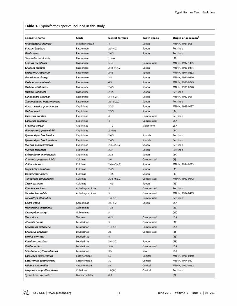
Table 1. Cypriniformes species included in this study.
Scientific name Clade Dental formula Tooth shape Origin of specimen1
Psilorhynchus balitora Psilorhynchidae 4 Spoon MNHN, 1931-056
Boraras brigittae Rasborinae 2,5-(4,2) Spoon Pet shop
Danio rerio Rasborinae 2,4,5 Spoon Pet shop
Danionella translucida Rasborinae 1 row [38]
Esomus metallicus Rasborinae 5-(4) Compressed MNHN, 1987-1355
Laubuca laubuca Rasborinae 2,4,5-(4,4,2) Spoon MNHN, 1983-0214
Luciosoma setigerum Rasborinae 2,4,5 Spoon MNHN, 1994-0232
Opsaridium christyi Rasborinae 3,5 Spoon MNHN, 1986-0416
Rasbora borapetensis Rasborinae 4,5 Spoon MNHN, 1983-0249
Rasbora einthoveni Rasborinae 2,4,5 Spoon MNHN, 1986-0228
Rasbora trilineata Rasborinae 2,4,5 Spoon Pet shop
Sundadanio axelrodi Rasborinae 2,4-(5,2,1) Spoon MNHN, 1982-0681
Trigonostigma heteromorpha Rasborinae 2,5-(5,2,2) Spoon Pet shop
Acrossocheilus yunnanensis Cyprininae 2,3,5 Spoon MNHN, 1949-0037
Barbus reinii Cyprininae 2,3,5 Spoon [34]
Carassius auratus Cyprininae 4 Compressed Pet shop
Carassius carassius Cyprininae 4 Compressed LSA
Cyprinus carpio Cyprininae 1,1,3 Molariform LSA
Gymnocypris przewalskii Cyprininae 2 rows [34]
Epalzeorhynchos bicolor Cyprininae 2,4,5 Spatula Pet shop
Epalzeorhynchos frenatum Cyprininae 2,4,5 Spatula Pet shop
Puntius semifasciolatus Cyprininae 2,3,4-(5,3,2) Spoon Pet shop
Puntius tetrazona Cyprininae 2,3,4 Spoon Pet shop
Schizothorax meridionalis Cyprininae 2,3,5 Spoon [34]
Ctenopharyngodon idella Cultrinae 2,4 Compressed [4]
Culter alburnus Cultrinae 2,4,4-(5,4,2) Spoon MNHN, 1934-0213
Elopichthys bambusa Cultrinae 2,4,5 Spoon [33]
Opsariicthys bidens Cultrinae 1,4,5 Spoon [33]
Xenocypris yunnanensis Cultrinae 2,3,5-(6,3,2) Compressed MNHN, 1949-0042
Zacco platypus Cultrinae 1,4,5 Spoon [33]
Rhodeus sericeus Acheilognathinae 5 Compressed Pet shop
Tanakia lanceolata Acheilognathinae 5 Compressed MNHN, 1984-0419
Tanichthys albonubes 1,4-(5,1) Compressed Pet shop
Gobio gobio Gobioninae 3,5-(5,2) Spoon LSA
Hemibarbus maculatus Gobioninae 1,3,5 [33]
Saurogobio dabryi Gobioninae 5 [33]
Tinca tinca Tincinae 4-(5) Compressed LSA
Abramis brama Leuciscinae 5 Compressed [37]
Leucaspius delineatus Leuciscinae 1,4-(5,1) Compressed LSA
Leuciscus cephalus Leuciscinae 2,5 Compressed [35]
Luxilus cornutus Leuciscinae 4 [35]
Phoxinus phoxinus Leuciscinae 2,4-(5,2) Spoon [39]
Rutilus rutilus Leuciscinae 5-(6) Compressed LSA
Scardinius erythrophtalmus Leuciscinae 3,5 Saw LSA
Carpiodes microstomus Catostomidae 50 Conical MNHN, 1905-0340
Catostomus commersonii Catostomidae 30 Conical MNHN, 1994-0301
Ictiobus cyprinellus Catostomidae 55 Conical MNHN, 2002-0352
Misgurnus anguillicaudatus Cobitidae 14-(16) Conical Pet shop
Gyrinocheilus aymonieri Gyrinocheilidae 0-0 [8]
Cypriniformes Teeth Evolution
PLoS ONE | www.plosone.org 11 June 2010 | Volume 5 | Issue 6 | e11293

present study; (ii) 3 species have been dissected and dentition has
been imaged by traditional microscopy; (iii) the dentition of the 15
other species has been taken from the literature. All Cypriniformes
analyzed in this study are listed in Table 1. We chose specimens
belonging to the two large superfamilies: Cobitoidea and
Cyprinoidea. For Cobitoidea we sampled 3 of the 4 families:
Catostomidae, Cobitidae, Gyrinocheilidae. Given the current
questions regarding the evolution of dentition in Cyprinidae, we
focused our analysis on this super-family, and sampled specimens
from each of its sub-families: Rasborinae, Cyprininae, Cultrinae,
Acheilognathinae, Gobioninae, Leuciscinae and Tincinae. The
sampling was also focused on minute Rasborinae. Several
Cypriniformes came from different pet shops as they comprise
many species that are very common among exotic fish breeders.
As indicated in Table 1, many Cypriniformes came from the
National Museum of Natural History of Paris (MNHN),
the Natural History Museum of London (NHM) as well as from
the Laboratory of Animal Sciences of Nancy (LSA). Reference
numbers are indicated in Table 1 for specimens loaned from
museums of Paris and London.
Phylogenetic analysesAs mentioned in the Results section, we built our own
phylogenetic tree in this study, not with the intention to publish
a new Cypriniformes phylogeny, but as a necessary step to
reconstruct ancestral characters with BayesTraits [48], a method
which requires branch lengths. That is why we did not carry out
any sequencing effort but we used available sequences. We worked
with four neutral markers: cytochrome b (cytb), recombination
activating gene 1 (rag1), recombination activating gene 2 (rag2)
and rhodopsin (rho). Four species that we scanned in this study
were not used for the phylogenetic analysis as no sequence is
available on Genbank: Carpiodes microstomus, Rasbora einthoveni,
Laubuca laubuca and Opsaridium christyi. Thus, 45 species were
included in our phylogenetic analysis, plus the outgroup Astyanax
mexicanus. All sequences used for our phylogeny were downloaded
from Genbank. Accession numbers are indicated in Table 2.
Sequences were aligned using Seaview [68]: in order to limit the
number of gaps, we kept 1142 base pairs for cytb, 1497 for rag1,
1251 for rag2 and 485 for rho. All sequences were concatenated
for each species. Phylogenetic analysis was carried out with Mr
Bayes [69]. The parameters were as followed: lset nst = 6,
rates = invgamma. Sequences were partitioned with respect to
the four genes and the three codon positions. The command
‘‘unlink’’ was used to set different model parameters for the
different partitions. Four MCMC chains were performed with
5,000,000 generations, sampling one tree per 100 replicates. We
discarded the 10,000 first sampled trees as burn-in after diagnostic
of convergent likelihood. These trees were used to construct a 50%
majority rule consensus tree with posterior probabilities for each
node. Our phylogenetic tree was then compared to previously
published ones in order to determine any incongruence. Three
taxa were not assigned to their usual clades in our analysis:
Luciosoma setigerum was determined as an outgroup of the
Rasborinae, Xenocypris yunnanensis was determined as an outgroup
of the Cultrinae and Gyrinocheilus aymonieri was determined as an
outgroup of the order Cypriniformes. As previous papers have
shown that the two first taxa are respectively members of
Rasborinae and Cultrinae [70,71] and as several papers have
proved the monophyly of the majors clades within Cypriniformes
[23–29], we decided to set constraints in our Bayesian analysis so
that all species in our sample are correctly assigned to their
monophyletic clade (Cobitoidea, Cyprininae, Rasborinae, Cultri-
nae, Gobioninae, Leuciscinae, Acheilognathinae).
X-ray microtomographyFishes were scanned either using a conventional microtomo-
graph or at the European Synchrotron Radiation Facility (ESRF,
Grenoble, France), which is a third generation synchrotron [46].
The conventional microtomograph was a Viscom X8050-16 high
resolution (voxel size used from 5 mm to 20 mm) at the iPHEP
(International Institute of Paleoprimatology, Human Evolution
and Paleoenvironments) in Poitiers. Cypriniformes were scanned
with the following settings: 200 mA and from 80 to 120 kV
depending on the size of the individual (the bigger the fish, the
higher the voltage due to the increased thickness of the element the
X-rays must traverse). The tomographic reconstruction was
performed using the software attached to the machine. Small
Cypriniformes (less than 50 mm) were scanned on the beam line
BM05 at the ESRF (European Synchrotron Radiation Facility) of
Grenoble. We used isotropic voxel sizes of respectively 5.4 mm and
0.691 mm (depending on the size of the specimens). We used a
monochromatic beam at energy of 20 keV for the largest samples
(monochromatization done with a double Si111 Bragg system) and
18 keV for the smallest ones (monochromatization done with a
double multilayer monochoromator). Ring artefacts and tomo-
graphic reconstructions were performed using ESRF inhouse
tools.
Morphological analysesReconstructed volumes were analyzed with VGStudioxMax
1.2.1 (Volume Graphics, Heidelberg, Germany) and the fifth
ceratobranchial was extracted for each sample using the 3D magic
wand growing region tool and manual adjustments. Ancestral
states of tooth row number and tooth shape were reconstructed for
different nodes of our phylogenetic tree by Maximum Likelihood
method with BayesTraits [48]. BayesTraits reconstructs the
ancestral characters at each node of the tree by finding the lowest
likelihood value for the whole data, and gives the transition factors
corresponding to this likelihood value. Those transition factors
represent the values for each transition from one character state to
Dental formulas and tooth shapes in various Cypriniformes determined in this study by microtomography and/or clearing (species in bold letters) or found in literature.The dental formula of one half-bone is written when the species displays a symmetrical dental formula whereas in the case of asymmetrical formulas, the formula of theright half-bone is in parentheses. Dental formulas were determined as explained in the introduction, i.e. by counting the number of teeth on each row from the mostventral to the most dorsal row (and the inverse for the right bone). For data coming from the literature, tooth shape was sometimes not available. For species scannedor dissected in this study, origin of the specimen is written (petshop, MNHN for the National Museum of Natural History of Paris, NHM for the National History Museumof London, LSA for the Laboratory of Animal Sciences of Nancy) as well as reference numbers for museum specimens. For data coming from literature, the reference inwhich data were found is given.1The reference for loaned specimens from museums is provided or bibliographical references for species that were not scanned in this study but used in theparsimonious analysis.
doi:10.1371/journal.pone.0011293.t001
Table 1. Cont.
Cypriniformes Teeth Evolution
PLoS ONE | www.plosone.org 12 June 2010 | Volume 5 | Issue 6 | e11293
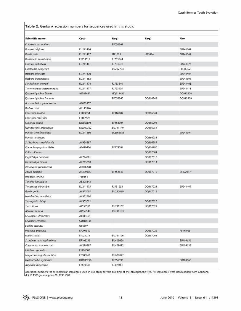
Table 2. Genbank accession numbers for sequences used in this study.
Scientific name Cytb Rag1 Rag2 Rho
Psilorhynchus balitora EF056369
Boraras brigittae EU241414 EU241347
Danio rerio EU241427 U71093 U71094 EU241362
Danionella translucida FJ753515 FJ753544
Esomus metallicus EU241441 FJ753531 EU241376
Luciosoma setigerum EU292704 FJ531352
Rasbora trilineata EU241470 EU241404
Rasbora borapetensis EU241463 EU241398
Sundadanio axelrodi EU241474 FJ753540 EU241408
Trigonostigma heteromorpha EU241477 FJ753530 EU241411
Epalzeorhynchos bicolor AJ388457 GQ913456 GQ913508
Epalzeorhynchos frenatus EF056360 DQ366943 GQ913509
Acrossocheilus yunnanensis AF051857
Barbus reinii AF145946
Carassius auratus FJ169954 EF186007 DQ366941
Carassius carassius FJ167428
Cyprinus carpio DQ868875 EF458304 DQ366994
Gymnocypris przewalskii DQ309362 EU711149 DQ366954
Puntius semifasciolatus EU241460 DQ366951 EU241394
Puntius tetrazona DQ366938
Schizothorax meridionalis AY954287 DQ366989
Ctenopharyngodon idella AF420424 EF178284 DQ366996
Culter alburnus DQ367004
Elopichthys bambusa AY744501 DQ367016
Opsariicthys bidens AY245090 DQ367014
Xenocypris yunnanensis AF036208
Zacco platypus AF309085 EF452848 DQ367010 EF452917
Rhodeus sericeus Y10454
Tanakia lanceolata AB208543
Tanichthys albonubes EU241475 FJ531253 DQ367023 EU241409
Gobio gobio AY953007 EU292689 DQ367015
Hemibarbus maculatus AY952990
Saurogobio dabryi AY953011 DQ367020
Tinca tinca AJ555551 EU711162 DQ367029
Abramis brama AJ555548 EU711103
Leucaspius delineatus AJ388459
Leuciscus cephalus GU182336
Luxilus cornutus U66597
Phoxinus phoxinus EF094550 DQ367022 FJ197065
Rutilus rutilus FJ025074 EU711126 DQ367003
Scardinius erythrophtalmus EF105295 EU409628 EU409656
Catostomus commersoni AY279397 EU409612 EU409638
Ictiobus cyprinellus FJ226308
Misgurnus anguillicaudatus EF088651 EU670842
Gyrinocheilus aymonieri DQ105256 EF056390 EU409663
Astyanax mexicanus FJ439346 FJ439461
Accession numbers for all molecular sequences used in our study for the building of the phylogenetic tree. All sequences were downloaded from Genbank.doi:10.1371/journal.pone.0011293.t002
Cypriniformes Teeth Evolution
PLoS ONE | www.plosone.org 13 June 2010 | Volume 5 | Issue 6 | e11293
another in the whole tree. Then, it is possible to build models in
which transition factors can be constrained. For instance,
transition factors can be constrained to be all equal or one
transition factor can be constrained to be zero. BayesTraits gives
the likelihood value associated with the model. Statistical tests can
be carried out to compare likelihood values between different
models, in order to know if likelihood values are statistically
different or not. For this study, there was no statistical difference
between the complex model (no constraint) and a simpler model,
which is that all transition factors are constrained to be equal.
Once a model has been chosen, it is possible to extract, for each
node of the tree, the likelihood value associated with each
character state at a given node. These values were plotted in our
phylogenetic tree, using pies. Moreover, because of a small
number of species compared to the number of character states for
both tooth row number and tooth shape, rare states (which are ‘‘0’’
for tooth row number and ‘‘spatula’’, ‘‘molariform’’ and
‘‘compressed’’ morphotypes for tooth shape) tend to be deter-
mined as ancestral states by BayesTraits, no matter which model is
used, which is very unlikely as these character states are almost all
restricted to only one species of our dataset. Consequently, based
on the parsimonious hypothesis that these rare character states
cannot be considered as ancestral, they were discarded from the
ML analysis of ancestral characters with BayesTraits but they are
represented for convenience on Figures 6 and 7.
Fish clearing and stainingCypriniformes were cleared and stained according to Taylor
and van Dyke [45]. For relatively big individuals (more than 5 cm)
that are long to clear entirely, they were stained with Alizarin red
(which colours bones) and the fifth ceratobranchial bearing
pharyngeal teeth was extracted by dissecting the samples. The
5th ceratobranchial was then observed with binoculars and
pictures were taken with a photograph included in the binoculars.
Supporting Information
Figure S1 Plates of the 34 Cypriniformes scanned or dissected in
this study. 31 species were surveyed by 3D microtomography. The
corresponding plates present the pharyngeal dentition in three
views (A: occlusal view; B: dorsal view; C: ventral view), except for
Catostomidae (A: anterior view; B: posterior view) and Psilor-
hynchus (only occlusal view). For the four dissected specimens, one
view is given for each plate (the type of view is written); the view
was chosen in order to best determine the dental formula. For all
plates, the name of the species and its dental formulas are written,
as well as a scale giving the size of the fifth ceratobranchial arch.
Tooth shape is only available for species surveyed by microtomo-
graphy. In each plate, the different tooth rows are differentiated
using colours (from the most ventral to the most dorsal rows: red,
blue, yellow, green) on the left side. Asymmetrical patterns are
shown by the use of colours on the right side, for the rows which
display a different number of teeth.
Found at: doi:10.1371/journal.pone.0011293.s001 (4.71 MB TIF)
Acknowledgments
We thank Thomas Iwema for help at the ESRF; Monette Veran for her
knowledge of Cypriniformes; Romain Causse and Patrice Pruvost (from
MNHN), Patrick Campbell and Oliver Crimmen (from NHM) for loans of
Cypriniformes; Fabrice Teletchea (from LSA) for generous gifts of
Cypriniformes; Fabienne Schall for bibliographic links on Cypriniformes;
Olga Otero for data on fossils; Laure Bernard for the fish facility, members
of the iPHEP of Poitiers and especially Ghislaine Florent, Carine Noel and
Patrick Vignaud for welcoming and facilitating the accommodation of EPV
during his training in the iPHEP. We thank the ESRF for having providing
beamtime on the beamline BM05 and to have hosted EPV for his basic
training in microtomographic data processing. We thank Joanne Burden
for editing the manuscript.
Author Contributions
Conceived and designed the experiments: LV VL. Performed the
experiments: EPV CC PT LV. Analyzed the data: EPV PC MS. Wrote
the paper: EPV LV VL.
References
1. Tucker A, Sharpe P (2004) The cutting-edge of mammalian development; how
the embryo makes teeth. Nat Rev Genet 5(7): 499–508.
2. Tummers M, Thesleff I (2009) The importance of signal pathway modulation inall aspects of tooth development. J Exp Zoolog B Mol Dev Evol 312B(4):
309–19.
3. Caton J, Tucker AS (2009) Current knowledge of tooth development: patterningand mineralization of the murine dentition. J Anat 214(4): 502–15.
4. Stock DW (2007) Zebrafish dentition in comparative context. J Exp Zool B Mol
Dev Evol 308B: 523–49.
5. Streelman JT, Peichel CL, Parichy DM (2007) Developmental genetics ofadaptation in fishes: The case for novelty. Ann Rev Ecol Evol Syst 38: 655–681.
6. Huysseune A, Thesleff I (2004) Continuous tooth replacement: the possible
involvement of epithelial stem cells. Bioessays 26(6): 665–71.
7. Huysseune A, Sire JY, Witten PE (2009) Evolutionary and developmental originsof the vertebrate dentition. J Anat 214(4): 465–76.
8. Nelson JS (2006) Fishes of the world, 4th edition. Hoboken, NJ: Wiley. 624 p.
9. Huysseune A, Witten PE (2008) An evolutionary view on tooth development and
replacement in wild Atlantic salmon (Salmo salar L.). Evol Devol 10(1): 6–14.
10. Debiais-Thibaud M, Borday-Birraux V, Germon I, Bourrat F, Metcalfe CJ,et al. (2007) Development of oral and pharyngeal teeth in the medaka (Oryzias
latipes): comparison of morphology and expression of eve1 gene. J Exp Zool B MolDev Evol 308(6): 693–708.
11. Fraser GJ, Hulsey CD, Bloomquist RF, Uyesugi K, Manley NR, et al. (2009) An
ancient gene network is co-opted for teeth on old and new jaws. PLoS Biol 7(2):e31.
12. Van Der Heyden C, Huysseune A (2000) Dynamics of tooth formation and
replacement in the zebrafish (Danio rerio) (Teleostei, Cyprinidae). Dev Dyn 219:486–96.
13. Van Der Heyden C, Wautier K, Huysseune A (2001) Tooth succession in the
zebrafish (Danio rerio). Arch Oral Biol 46: 1051–58.
14. Debiais-Thibaud M, Germon I, Laurenti P, Casane D, Borday-Birraux V (2008)Low divergence in Dlx gene expression between dentitions of the medaka
(Oryzias latipes) versus high level of expression shuffling in Osteichtyans. EvolDev 10(4): 464–76.
15. Stock DW, Jackman WR, Trapani J (2006) Developmental genetic mechanisms
of evolutionary tooth loss in cypriniform fishes. Development 133(16): 3127–37.
16. Jackman WR, Stock DW (2006) Transgenic analysis of Dlx regulation in fishtooth development reveals evolutionary retention of enhancer function despite
organ loss. Proc Natl Acad Sci U S A 103(51): 19390–5.
17. Winfield IJ, Nelson JS (1991) Cyprinid Fishes Systematics Biology andExplotation. London: Chapman and Hall.
18. FishBase (2010) http://www.fishbase.org/search.php.
19. Borday-Birraux V, Van der Heyden C, Debiais-Thibaud M, Verreijdt L,
Stock DW, et al. (2006) Expression of Dlx genes during the development of thezebrafish pharyngeal dentition: evolutionary implications. Evol Dev 8(2):
130–41.
20. Britz R, Conway KW, Ruber L (2009) Spectacular morphological novelty in aminiature cyprinid fish, Danionella dracula n. sp. Proc Biol Sci 276(1665):
2179–86.
21. Gosline WA (1973) Considerations regarding the phylogeny of cypriniformfishes, with special reference to structures associated with feeding. Copeia 1973:
761–776.
22. Honeycutt RL (2008) Small changes, big results: evolution of morphologicaldiscontinuity in mammals. J Biol 7(3): 9.
23. Saitoh K, Sado T, Mayden RL, Hanzawa N, Nakamura K, et al. (2006)
Mitogenomic evolution and interrelationships of the Cypriniformes (Actinopter-ygii: Ostariophysi): the first evidence toward resolution of higher-level
relationships of the world’s largest freshawater fish clade based on 59 wholemitogenome sequences. J Mol Evol 63: 826–41.
24. He S, Mayden RL, Wang X, Wang W, Tang KL, et al. (2008) Molecular
phylogenetics of the family Cyprinidae (Actinopterygii: Cypriniformes) asevidenced by sequence variation in the first intron of S7 ribosomal protein-
coding gene: further evidence from a nuclear gene of the systematic chaos in the
family. Mol Phylogenet Evol 46(3): 818–29.
Cypriniformes Teeth Evolution
PLoS ONE | www.plosone.org 14 June 2010 | Volume 5 | Issue 6 | e11293
25. Mayden RL, Chen WJ, Bart HL, Doosey MH, Simons AM, et al. (2009)
Reconstructing the phylogenetic relationships of the earth’s most diverse clade offreshwater fishes—order Cypriniformes (Actinopterygii: Ostariophysi): a case
study using multiple nuclear loci and the mitochondrial genome. Mol
Phylogenet Evol 51(3): 500–14.26. Chen WJ, Mayden RL (2009) Molecular systematics of the Cyprinoidea
(Teleostei: Cypriniformes), the world’s largest clade of freshwater fishes: furtherevidence from six nuclear genes. Mol Phylogenet Evol 52(2): 544–9.
27. Ruber L, Kottelat M, Tan HH, Ng PK, Britz R (2007) Evolution of
miniaturization and the phylogenetic position of Paedocypris, comprising theworld’s smallest vertebrate. BMC Evol Biol 7: 38.
28. Wang X, Li J, He S (2007) Molecular evidence for the monophyly of East Asiangroups of Cyprinidae (Teleostei: Cypriniformes) derived from the nuclear
recombination activating gene 2 sequences. Mol Phylogenet Evol 42(1): 157–70.29. Slechtova V, Bohlen J, Heok Hui T (2007) Families of Cobitoidea (Teleostei;
Cypriniformes) as revealed from nuclear genetic data and the position of the
mysterious genera Barbucca, Psilorhynchus, Serpenticobitis and Vaillantella. MolPhylogenet Evol 44: 1358–65.
30. Tang Q, Liu H, Mayden RL, Xiong B (2006) Comparison of evolutionary ratesin the mitochondrial DNA cytochrome b gene and control region and their
implications for phylogeny of the Cobitoidea (Teleostei: Cypriniformes). Mol
Phylogenet Evol 39: 347–57.31. Mayden RL, Tang KL, Conway KW, Freyhof J, Chamberlain S, et al. (2007)
Phylogenetic relationships of Danio within the order Cypriniformes: a frameworkfor comparative and evolutionary studies of a model species. J Exp Zool B Mol
Dev Evol 308: 642–654.32. Zardoya R, Doadrio I (1999) Molecular evidence on the evolutionary and
biogeographical patterns of European Cyprinids. J Mol Evol. 49: 227–37.
33. Chu YT (1935) Comparative studies on the scales and on the pharyngeals andtheir teeth in Chinese cyprinids, with particular reference to taxonomy and
evolution. Biol Bull St Johns Univ 2: 1–225.34. Talwar PK, Jhingran AG (1992) Inland fishes of India and adjacent countries,
Vol I. Rotterdam: A.A. Balkema, 1158 p.
35. Regan C (1911) The freshwater fishes of the British isles. London: Methuen.287 p.
36. Boulenger GA (1909) Catalogue of the freshwater fishes of Africa in the BritishMuseum (Natural History), Vols I and II. London: British Museum (Natural
History).37. Jordan DS, Evermann BW (1896) The fishes of North and Middle America, Vol
I. Washington: Govt Print Off.
38. Jayaram KC (1981) The freshwater fishes of India, Pakistan, Bangladesh, Burmaand Sri Lanka – a handbook. Calcutta: The survey. 475 p.
39. Heckel JJ, Kner R (1858) Die Susswasserfische der Osterreichen Monarchie mit Rucksicht
auf der angrenzenden Lander. Leipzig: Engelmann. 388 p.
40. Nakajima T (1987) Development of pharyngeal dentition in the cobitid fishes,
Misgurnus anguillicaudatus and Cobitis biwae, with a consideration of evolution ofCypriniform dentitions. Copeia 1987: 208–213.
41. Golubtsov AS, Dzerjinskii KF, Profokiev AM (2005) Four rows of pharyngealteeth in an aberrant specimen of the small African barb Barbus paludinosus
(Cyprinidae): novelty or atavistic alteration? J Fish Biol 67: 286–91.42. Kottelat M, Britz R, Tan HH, Witte KE (2006) Paedocypris, a new genus of
South-East Asian cyprinid fish with a remarkable sexual dimorphism, comprises
the world’s smallest vertebrate. Proc R Soc B 273: 895–99.43. Britz R, Conway KW (2009) Osteology of Paedocypris, a miniature and highly
developmentally truncated fish (Teleostei: Ostariophysi: Cyprinidae). J Morphol270(4): 389–412.
44. Hanken J, Wake DB (1993) Miniaturization of body-size: organismal
consequences and evolutionary significance. Ann Rev Ecol Syst 24: 501–519.45. Taylor WR, Van Dyke GC (1985) Revised procedures for staining and clearing
small fishes and other vertebrates for bone and cartilage study. Cybium 9:107–119.
46. Tafforeau P, Boistel R, Boller E, Bravin, Brunet M, et al. (2006) Applications of
X-ray synchrotron microtomography for non destructive 3D studies ofpaleontological specimens. Applied Physics A, Materials Science & Processing
83: 195–202.
47. Eastman JT, Underhill JC (1973) Intraspecific variation in the pharyngeal tooth
formulae of some cyprinid fishes. Copeia 1973: 45–53.
48. Pagel M (1999) The maximum likelihood approach to reconstructing ancestral
character states of discrete characters on phylogenies. Systematic Biology 48:612–622. www.evolution.rdg.ac.uk.
49. Organ CL, Shedlock AM, Meade A, Pagel M, Edwards SV (2007) Origin of
avian genome size and structure in non-avian dinosaurs. Nature 446: 180–184.
50. Barker D, Meade A, Pagel M (2007) Constrained models of evolution lead to
improved prediction of functional linkage from correlated gain and loss of genes.
Bioinformatics 23: 14–20.
51. Organ CL, Janes DE, Meade A, Pagel M (2009) Genotypic sex determinationenabled adaptive radiations of extinct marine reptiles. Nature 461: 389–392.
52. Scott JL, Kawahara AY, Skevington JH, Yen SH, Sami A, et al. (2010) Theevolutionary origins of ritualized acoustic signals in caterpillars. Nature
communications 1: 4.
53. Patterson C (1993) Osteichthyes: Teleostei. In: Benton MJ, ed. The fossil record
2. London: Chapman & Hall. pp 622–656.
54. Cavender TM (1991) The fossil record of the Cyprinidae. In: Winfield IJ,
Nelson JS, eds. Cyprinid fishes: systematics, biology and exploitation. New York:Chapman & Hall. pp 34–54.
55. Gayet M (1987) Lower vertebrates from the early-middle Eocene KuldanaFormation of Kohat (Pakistan): Holostei and Teleostei. Contribution of the
Museum of Paleontology, university of Michigan 27: 151–68.
56. Liu J, Chang M (2009) A new Eocene catostomid (Teleostei: Cypriniformes)
from northeastern China and early divergence of Catostomidae. Science inChina Series D: Earth Sciences 52(2): 189–202.
57. Chang M, Chen G (2008) Fossil Cypriniformes from China and its adjacentareas and their palaeobiogeographical implications. In: Cavin L, Longbottom A,
Richter M, eds. Fishes and the break-up of Pangea. London: Geological Society,Special publications. 295, 337–350. pp 337–350.
58. Otero O (2001) The oldest-known cyprinid fish of the Afro-Arabic plate and itspaleobiogeographical implications. J Vertebrate Paleontology 21: 386–88.
59. Collin R, Miglietta MP (2008) Reversing opinions on Dollo’s Law. Trends EcolEvol 23(11): 602–9.
60. Conway-Morris S (1998) The Crucible of Creation: The Burgess Shale and the
Rise of the Animals. Oxford: Oxford University Press. 242 p.
61. Kuratani S (2009) Modularity, comparative embryology and evo-devo:
developmental dissection of evolving body plans. Dev Biol 332(1): 61–9.
62. Davidson E (2006) The Regulatory Genome: Gene Regulatory Networks in
Development And Evolution. Academic Press.
63. Lammens EHRR, Hoogenboezem W (1991) Diets and feeding behavior. In:
Winfield IJ, Nelson JS, eds. Cyprinid fishes: systematics, biology andexploitation. New York: Chapman & Hall. pp 353–376.
64. Zhang Z, Lan Y, Chai Y, Jiang R (2009) Antagonistic actions of Msx1 and Osr2pattern mammalian teeth into a single row. Science 323(5918): 1232–4.
65. Wautier K, Van der Heyden C, Huysseune A (2001) A quantitative analysis ofpharyngeal tooth shape in the zebrafish (Danio rerio, Teleostei, Cyprinidae). Arch
Oral Biol 46: 67–75.
66. Renaud S, Michaux J, Schmidt DN, Aguilar JP, Mein P, et al. (2005)
Morphological evolution, ecological diversification and climate change inrodents. Proc Biol Soc 272(1563): 609–17.
67. Lazzari V, Charles C, Tafforeau P, Vianey-Liaud M, Aguilar JP, et al. (2008)Mosaic convergence of rodent dentitions. PLoS One 3(10): e3607.
68. Galtier N, Gouy M, Gautier C (1996) SEAVIEW and PHYLO_WIN: two
graphic tools for sequence alignment and molecular phylogeny. Comput Appl
Biosci 12: 543–548.
69. Ronquist F, Huelsenbeck JP (2003) MRBAYES 3: Bayesian phylogenetic
inference under mixed models. Bioinformatics 19: 1572–1574.
70. Conway K, Chen WJ, Mayden RL (2008) The ‘‘Celestial Pearl Danio’’ is aminiature Danio (s.s) (Ostariophysi: Cyprinidae): evidence from morphology and
molecules. Zootaxa 1686: 1–28.
71. Xiao W, Zhang Y, Liu H (2001) Molecular systematics of Xenocyprinae
(teleostei: cyprinidae): taxonomy, biogeography and coevolution of a specialgroup restricted in East Asia. Mol Phylogenet Evol 18(2): 163–173.
Cypriniformes Teeth Evolution
PLoS ONE | www.plosone.org 15 June 2010 | Volume 5 | Issue 6 | e11293
Copyright © 2022 FDOKUMEN