New semionotiform (Actinopterygii: Neopterygii) from the Late Jurassic of southern Germany
20
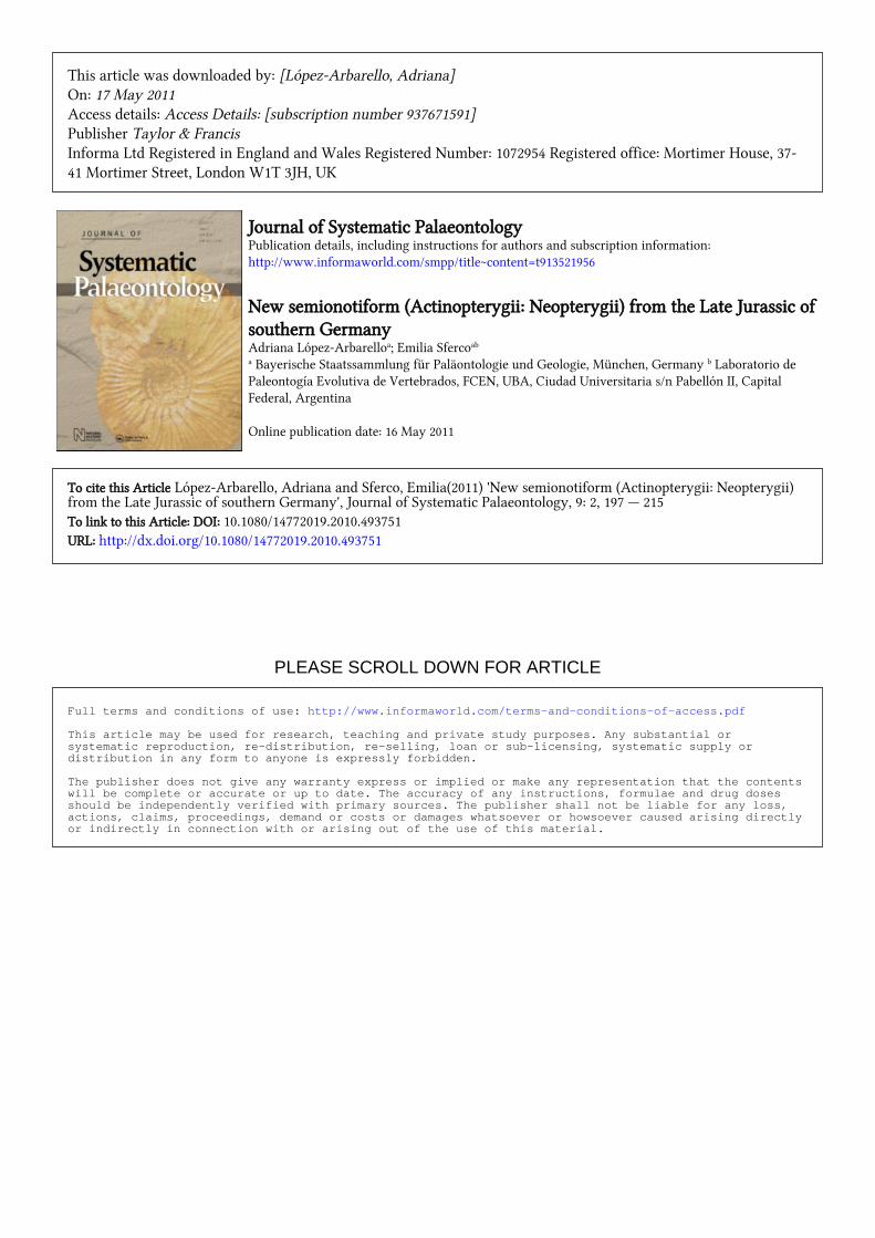
PLEASE SCROLL DOWN FOR ARTICLE This article was downloaded by: [López-Arbarello, Adriana] On: 17 May 2011 Access details: Access Details: [subscription number 937671591] Publisher Taylor & Francis Informa Ltd Registered in England and Wales Registered Number: 1072954 Registered office: Mortimer House, 37- 41 Mortimer Street, London W1T 3JH, UK Journal of Systematic Palaeontology Publication details, including instructions for authors and subscription information: http://www.informaworld.com/smpp/title~content=t913521956 New semionotiform (Actinopterygii: Neopterygii) from the Late Jurassic of southern Germany Adriana López-Arbarello a ; Emilia Sferco ab a Bayerische Staatssammlung für Paläontologie und Geologie, München, Germany b Laboratorio de Paleontogía Evolutiva de Vertebrados, FCEN, UBA, Ciudad Universitaria s/n Pabellón II, Capital Federal, Argentina Online publication date: 16 May 2011 To cite this Article López-Arbarello, Adriana and Sferco, Emilia(2011) 'New semionotiform (Actinopterygii: Neopterygii) from the Late Jurassic of southern Germany', Journal of Systematic Palaeontology, 9: 2, 197 — 215 To link to this Article: DOI: 10.1080/14772019.2010.493751 URL: http://dx.doi.org/10.1080/14772019.2010.493751 Full terms and conditions of use: http://www.informaworld.com/terms-and-conditions-of-access.pdf This article may be used for research, teaching and private study purposes. Any substantial or systematic reproduction, re-distribution, re-selling, loan or sub-licensing, systematic supply or distribution in any form to anyone is expressly forbidden. The publisher does not give any warranty express or implied or make any representation that the contents will be complete or accurate or up to date. The accuracy of any instructions, formulae and drug doses should be independently verified with primary sources. The publisher shall not be liable for any loss, actions, claims, proceedings, demand or costs or damages whatsoever or howsoever caused arising directly or indirectly in connection with or arising out of the use of this material.
Transcript of New semionotiform (Actinopterygii: Neopterygii) from the Late Jurassic of southern Germany
PLEASE SCROLL DOWN FOR ARTICLE
This article was downloaded by: [López-Arbarello, Adriana]On: 17 May 2011Access details: Access Details: [subscription number 937671591]Publisher Taylor & FrancisInforma Ltd Registered in England and Wales Registered Number: 1072954 Registered office: Mortimer House, 37-41 Mortimer Street, London W1T 3JH, UK
Journal of Systematic PalaeontologyPublication details, including instructions for authors and subscription information:http://www.informaworld.com/smpp/title~content=t913521956
New semionotiform (Actinopterygii: Neopterygii) from the Late Jurassic ofsouthern GermanyAdriana López-Arbarelloa; Emilia Sfercoab
a Bayerische Staatssammlung für Paläontologie und Geologie, München, Germany b Laboratorio dePaleontogía Evolutiva de Vertebrados, FCEN, UBA, Ciudad Universitaria s/n Pabellón II, CapitalFederal, Argentina
Online publication date: 16 May 2011
To cite this Article López-Arbarello, Adriana and Sferco, Emilia(2011) 'New semionotiform (Actinopterygii: Neopterygii)from the Late Jurassic of southern Germany', Journal of Systematic Palaeontology, 9: 2, 197 — 215To link to this Article: DOI: 10.1080/14772019.2010.493751URL: http://dx.doi.org/10.1080/14772019.2010.493751
Full terms and conditions of use: http://www.informaworld.com/terms-and-conditions-of-access.pdf
This article may be used for research, teaching and private study purposes. Any substantial orsystematic reproduction, re-distribution, re-selling, loan or sub-licensing, systematic supply ordistribution in any form to anyone is expressly forbidden.
The publisher does not give any warranty express or implied or make any representation that the contentswill be complete or accurate or up to date. The accuracy of any instructions, formulae and drug dosesshould be independently verified with primary sources. The publisher shall not be liable for any loss,actions, claims, proceedings, demand or costs or damages whatsoever or howsoever caused arising directlyor indirectly in connection with or arising out of the use of this material.
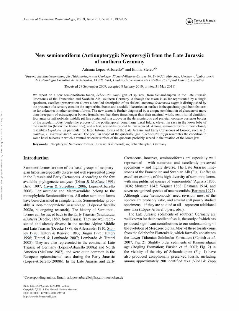
Journal of Systematic Palaeontology, Vol. 9, Issue 2, June 2011, 197–215
New semionotiform (Actinopterygii: Neopterygii) from the Late Jurassicof southern Germany
Adriana Lopez-Arbarelloa∗ and Emilia Sfercoa,b
aBayerische Staatssammlung fur Palaontologie und Geologie, Richard-Wagner-Strasse 10, D-80333 Munchen, Germany; bLaboratoriode Paleontogıa Evolutiva de Vertebrados, FCEN, UBA, Ciudad Universitaria s/n Pabellon II, Capital Federal, Argentina
(Received 29 September 2009; accepted 8 January 2010; printed 31 May 2011)
We report on a new semionotiform taxon, Scheenstia zappi gen. et sp. nov., from Schamhaupten in the Late Jurassiclimestones of the Franconian and Swabian Alb, southern Germany. Although the taxon is so far represented by a singlespecimen, excellent preservation allows a detailed description of its skeletal anatomy. Scheenstia zappi is distinguished bythe presence of a sensory canal in the supraorbital bones and a saddle-like articular surface in the quadratojugal, both featuresso far unknown in other semionotiforms. The new taxon is further diagnosed by a unique combination of characters: morethan three pairs of extrascapular bones; frontals less than three times longer than their maximal width; semitritorial dentition;four anterior infraorbitals; middle pit line contained in a groove in the dermopterotic and parietal; concave posterior borderof the angular, robust bugle-like process of the posttemporal bone; large basal fulcra; eleven fin rays in the lower lobe ofthe caudal fin (below the lateral line); and a first, scale-like caudal fin ray reduced. Among semionotiforms it most closelyresembles Lepidotes, in particular the large tritorial forms of the Late Jurassic and Early Cretaceous of Europe, such as L.mantelli, L. maximus and L. laevis. The peculiar shape of the quadratojugal in Scheenstia zappi resembles the condition insome basal teleosts in which a ventral articular surface of the quadrate probably served in the rotation of the lower jaw.
Keywords: Neopterygii; Semionotiformes; Jurassic; Kimmeridgian; Schamhaupten; Germany
Introduction
Semionotiformes are one of the basal groups of neoptery-gian fishes, an especially diverse and well represented groupin the Jurassic and Early Cretaceous. According to the fewavailable phylogenetic analyses (Olsen & McCune 1991;Brito 1997; Cavin & Suteethorn 2006; Lopez-Arbarello2006), Lepisosteidae and Macrosemiidae belong to themonophyletic Semionotiformes. All other semionotiformshave been classified in a single family, Semionotidae, prob-ably a non-monophyletic assemblage (Lopez-Arbarello2008a, b; ongoing research). The history of Semionoti-formes can be traced back to the Early Triassic (Semionotusalsaticus Deecke, 1889, from Elsass). They are well repre-sented and already diverse in the marine Alpine Middleand Late Triassic (Deecke 1889; de Allessandri 1910; Stol-ley 1920; Tintori & Renesto 1983; Burgin 1995; Tintori1996; Tintori & Lombardo 2007; Lombardo & Tintori2008). They are also represented in the continental LateTriassic of Germany (Lopez-Arbarello 2008a) and NorthAmerica (McCune 1987), and were quite common in theEuropean epicontinental seas during the Early Jurassic(Lopez-Arbarello 2008b). In the Late Jurassic and Early
∗Corresponding author. Email: [email protected]
Cretaceous, however, semionotiforms are especially wellrepresented – with numerous and excellently preservedspecimens – and highly diverse. The Late Jurassic lime-stones of the Franconian and Swabian Alb (Fig. 1) offer anexcellent example of this high diversity of semionotiforms,with nine published species of ‘semionotids’ (Agassiz 1833,1836; Munster 1842; Wagner 1863; Eastman 1914) andseven recognized species of macrosemiids (Bartram 1977).Although these ‘semionotids’ need revision, most of thespecies are probably valid, and several still poorly studiedspecimens – if they are studied at all – represent additionalnew taxa (Lopez-Arbarello pers. obs.).
The Late Jurassic sediments of southern Germany arewell known for their excellent fossils, the study of which hasproduced significant contributions to our understanding ofthe evolution of Mesozoic biotas. Most of these fossils comefrom the Solnhofen Plattenkalk, which formally constitutesthe Lower Tithonian Solnhofen Formation (Fursich et al.2007; Fig. 2). Slightly older sediments of Kimmeridgianage (Rogling Formation; Fursich et al. 2007; Fig. 2) inthe vicinity of the city of Schamhaupten (Fig. 1) havealso produced exceptionally preserved fossils, includingamong approximately 200 identified taxa (Viohl & Zapp
ISSN 1477-2019 print / 1478-0941 onlineCopyright C© 2011 The Natural History MuseumDOI: 10.1080/14772019.2010.493751http://www.informaworld.com
Downloaded By: [López-Arbarello, Adriana] At: 07:22 17 May 2011

198 A. Lopez-Arbarello and E. Sferco
Figure 1. Geographic location of the main localities in the Late Jurassic limestones of the Franconian and Swabian Alb, Bavaria, Germany:Solnhofen /Langenaltheim Basin (1); Schernfeld Basin (2); Eichstatt Basin (3); Gungolding/Palzpainten Basin (4); Denkendorf/BohmfelderBasin (5); Schamhaupten/Zandt Basin (6); Hartheim Basin (7); Hienheim Basin (8); Kelheim Basin (9); Painten Basin (10); Brunn Basin(11); and Daiting Basin (12). Modified from Meyer & Schmidt-Kaler (1989), Selden & Nudds (2004) and Burnham (2007).
Figure 2. Stratigraphical chart of the Upper Jurassic sequence in the Franconian Alb showing the chronostratigraphical relationships ofthe Kimmeridgian sediments at Schamhaupten (Rogling Formation), Bavaria, Germany. Modified from Schmid et al. (2005) and Fursichet al. (2007).
Downloaded By: [López-Arbarello, Adriana] At: 07:22 17 May 2011
New semionotiform (Actinopterygii: Neopterygii) 199
2007), remarkable vertebrates such as the teleost Siemen-sichthys siemensi Arratia, 2000, and the juvenile thero-pod Juravenator starki Gohlich & Chiappe, 2006. Anotherexcellently preserved vertebrate from the same sequenceis a fish already described and referred to as Lepidotes sp.(Semionotiformes) by Thies & Zapp (1997).
According to Viohl & Zapp (2007, pp. 138–139), inSchamhaupten: “the deposition of silicified plattenkalkoccurred during a ‘transgressive systems tract’, whichhad started with the spongiolithic biostrome facies ofthe Treuchtlingen marble in the early late Kimmeridgianand reached the ‘maximum flooding surface’ probablyduring the deposition of the Solnhofen Plattenkalk in theearly Tithonian (Keupp & Matyszkiewicz 1997)”. In Viohl& Zapp’s (2007) palaeonvironmental reconstruction, as aconsequence of a halocline in the Schamhaupten Basin, thebottom zone was dysaerobic and thus hostile to benthic life,with the exception of microbial mats, which were respon-sible for the high quality of fossil preservation. Unlikethe bottom zone, the higher water column was rich inpelagic organisms, indicating good connections with theTethys. Benthic, demersal and terrestrial organisms areallochthonous to the Schamhaupten Basin and were proba-bly washed in during storm events.
This article includes a detailed description of a newsemionotiform taxon represented by the fish previouslyreferred to as Lepidotes sp. by Thies & Zapp (1997). Wefurther discuss its systematic relations and the implicationsof several of its anatomical features for current ideas ofhomologies and character evolution in semionotiform andother neopterygian fishes. The specimen described hereinwas collected in Stark Quarry, north-west of Schamhauptenin the Southern Franconian Alb, Bavaria, Germany. Thegeological setting of the quarry is explained by Joyce (2000)and detailed geological information, including local profilesand description of the bearing sediments is provided byRoper (1992), Thies & Zapp (1997) and Viohl & Zapp(2007).
Material and methods
The specimen was chemically and mechanically preparedby M. Zapp. Detailed information on the preparationmethodology is included in Thies & Zapp (1997). Thespecimen was studied under a Leica Wild M32 stereomi-croscope equipped with a camera lucida Wild 308700. Alldrawings were made directly from the fossil.
Skull bones are named according to the use of mostauthors studying actinopterygians. The bones carrying theinfraorbital sensory canal anterior to the orbit are referred toas ‘anterior infraorbitals’ following Wenz (1999, 2003) andLopez-Arbarello & Codorniu (2007). Fringing fulcra arenamed according to Patterson (1982). Scutes and unpairedand paired basal fulcra are identified according to Lopez-
Arbarello & Codorniu (2007). It is possible to differentiatethese structures because of their general shape and orienta-tion, and particularly because of their position: the scuteslie on the caudal peduncle, but unpaired basal fulcra lie onthe body lobe of the tail. The relative position of the fins andscale counts are expressed in a pterygial formula in whichD, P, A and C indicate the number of scale rows betweenthe first complete row behind the pectoral girdle and theinsertion of the dorsal, pelvic, anal and caudal fins respec-tively, and T is the total number of scale rows between thepectoral girdle and the caudal inversion (Westoll 1944).
Institutional abbreviationsBSPG: Bayerische Staatssammlung fur Palaontologie undGeologie, Munchen, Germany; GSM: British GeologicalSurvey, Nottingham, UK; JME: Jura Museum, Naturwis-senschaftliche Sammlungen Eichstatt, Germany; MNHN:Museum National d’Histoire Naturelle, Paris, France.
Anatomical abbreviationsa.io: anterior infraorbital bone; ag: angular bone; ao:antorbital bone; a.pl: anterior pit line; ap-dpt: auto-dermopalatine ossification; cl: cleithrum; co: coronoidbone; cor: coracoid bone; d: dentary; dpt: dermopteroticbone; dsph: dermosphenotic bone; enpt: endopterygoidbone; ex: extrascapular bone; io: infraorbital bone; io.c:infraorbital sensory canal; fr: frontal bone; fr.fu: fring-ing fulcrum; iop: interoperculum; l.et: lateral ethmoid;m.c: mandibular sensory canal; m.pl: middle pit line; mx:maxilla; na: nasal bone; o.c: orbital sensory canal; oc.c:occipital sensory canal; op: operculum; pa: parietal bone;p.b.fu: paired basal fulcrum; pcl: postcleithrum; p.d.c.fu:paired dorsal caudal fulcrum; pmx: premaxilla; pop: preop-erculum; pop.c: preopercular sensory canal; ptt: posttem-poral bone; q: quadrate bone; qj: quadratojugal bone;rar: retroarticular bone; ro: rostral bone; sag: surangularbone; s: scale; sc: scapula; scl: supracleithrum; sc.r: scale-like fin ray; sct: scute; so.c: supraorbital sensory canal;so: supraorbital bone; sop: suboperculum; suo: subor-bital bone; t.c: temporal sensory canal; u.b.fu: unpairedbasal fulcrum; u.d.c.fu: paired dorsal caudal fulcrum. An‘l’ between brackets after the abbreviations indicates leftelements.
Systematic description
Neopterygii Regan, 1923Semionotiformes Arambourg & Bertin, 1958 sensu Olsen
and McCune, 1991Semionotiformes incertae family
Genus Scheenstia gen. nov.
Type species. Scheenstia zappi sp. nov.
Downloaded By: [López-Arbarello, Adriana] At: 07:22 17 May 2011

200 A. Lopez-Arbarello and E. Sferco
Etymology. The name Scheenstia is a compound word inBavarian dialect composed of ‘schee’ ( = beautiful), whichbecomes ‘scheens’ after appropriate conjugation in genitivemasculine, and ‘Tia’ ( = animal).
Diagnosis. As for the type and only species.
Scheenstia zappi sp. nov.(Figs 3–9)
1997 Lepidotes sp. Thies & Zapp: 18, pl. 4–7.
Holotype. JME-Sch 80, complete specimen preserved inright lateral view (Fig. 3).
Type locality. Stark Quarry, 0.5 km north-westof Schamhaupten in the Southern Franconian Alb,Bavaria, Germany. Gauss-Kruger Coordinates: R4469650/H5420160 (Thies & Zapp, 1997; Fig. 1).
Type horizon. Rogling Formation (Upper Kimmeridgian;Fursich et al. 2007; Fig. 2). Malm Epsilon 2 Level 5 in theProfile of Thies & Zapp (1997).
Etymology. The species name zappi honours Mr ManfredZapp, who not only discovered the specimen but also carriedout excellent preparation and published the specimen for thefirst time.
Diagnosis. The following diagnosis is based on a uniquecombination of characters, among which possible autapo-morphies are indicated with asterisks (∗): more than threepairs of extrascapular bones; frontals less than three timeslonger than their maximal width; orbital sensory canalin the supraorbital bones∗; saddle-like articular surfacein the quadratojugal∗; semitritorial dentition; four ante-rior infraorbitals; middle pit line contained in a groove inthe dermopterotic and parietal; concave posterior border ofthe angular; robust bugle-like process of the posttemporalbone; large basal fulcra; 11 fin rays in the lower lobe of thecaudal fin (below the lateral line); first, scale-like caudalfin ray reduced; anterior flank scales with serrated posteriorborders. Pterygial formula:
D26
P8 A20 C32T37
Description
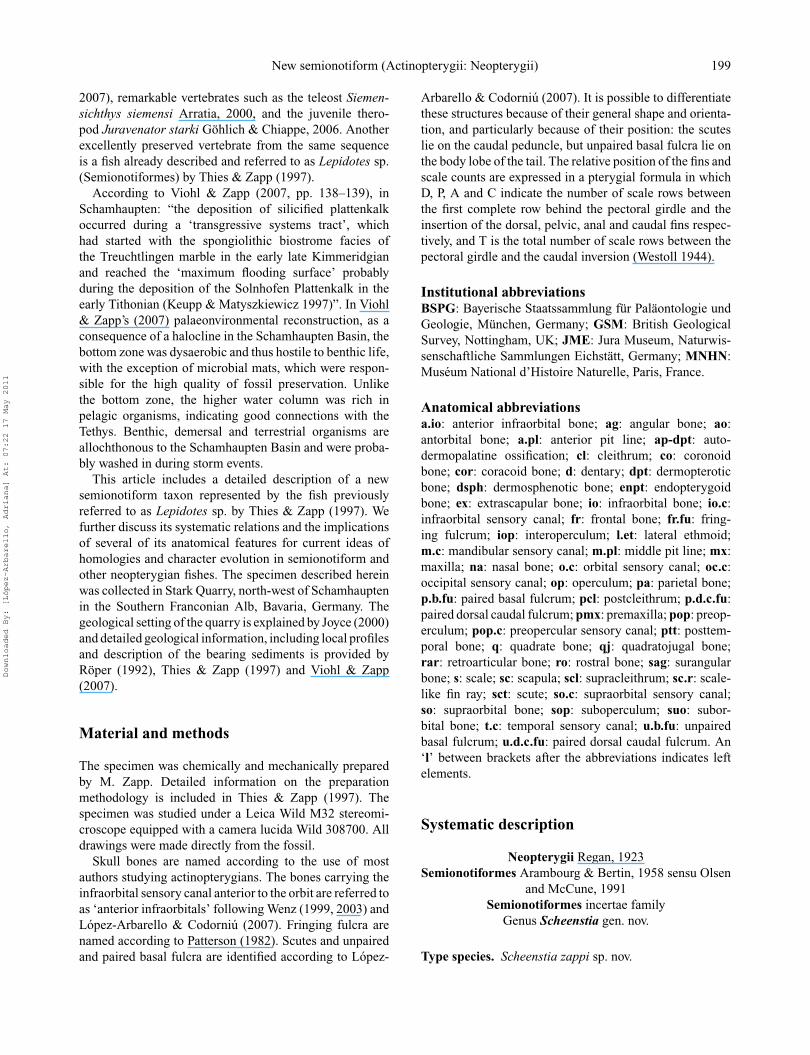
Scheenstia zappi is a medium size semionotiform with arelatively deep body (Fig. 3). As noted by Thies & Zapp(1997), the total length of the fossil is 34.5 cm. The fishhas a standard length (SL: the length from the tip of thesnout to the base of the caudal fin at the hinge line) of 30.2
cm, and the body depth is approximately 0.45 of the SL.The head is slightly longer than deep, head length beingabout 0.3 of total length. Although the bones in the mostanterior region of the skull are generally disarticulated andslightly displaced, the antorbital portion of the skull is rela-tively short in comparison with other semionotiforms (Figs4, 5). The preorbital length is only about 0.4 of the headlength and approximately two times the longitudinal diam-eter of the eye. The depth of caudal peduncle is about 0.2of the SL. The insertion of the pelvic fins is approximatelymidway between the pectoral and anal fins. The dorsal finoriginates approximately midway between the pelvic andanal fins. Basal and fringing fulcra are preserved in allthe fins, with the exception of the pectoral fins, which aremore poorly preserved than the other ones. The body iscompletely covered with thick and unornamented rhom-boid scales, among which most of the flank scales haveserrated posterior borders. The dermal bones of the skulllack a ganoin cover, except for some ganoin patches andtubercles on the parietal, frontal, suborbitals, infraorbitals,dermopterotic, preoperculum and operculum. The patternof ornamentation of the bones in the skull and pectoralgirdle was described in detail by Thies & Zapp (1997).
Skull roofConsistent with the observations of Thies & Zapp (1997),four small, approximately rectangular extrascapular bonesare preserved on the right side of the fish (Figs 4–6).However, a fifth element is apparently missing medially.The preserved series of four extrascapulars does not reachthe dorsal midline and there is no indication of the supraoc-cipital commissure in the parietal. Also, there is a slightthinness of the posterodorsal corner of the parietal, onwhich the putative fifth extrascapular would have been artic-ulated. Therefore the complete extrascapular series proba-bly included five paired elements. Preserved extrascapulars1 to 4, from lateral to medial, articulate with the pari-etal and dermopterotic bones anteriorly, with the opercu-lum ventrally, and with the posttemporal posteriorly. Thefirst extrascapular articulates with the dermopterotic, thesecond extrascapular articulates with the dermopteroticand parietal, and the extrascapulars 3 and 4, the small-est in the series, articulate with the parietal only. Except forthe extrascapular 4, which posteriorly contacts the dorsal-most scale of the first row, all extrascapulars articulate withthe posttemporal posteriorly. The supraoccipital commis-sure is represented by relatively large pores in each of theextrascapular bones.
The parietal bone is rectangular, approximately 1.5 timeslonger than wide (Figs 4–6). The length of the parietal isabout 0.3 of the length of the frontal. The parietal sutureswith the frontal anteriorly, laterally with the dermopteroticand posteriorly with the three last extrascapular bones. Themost dorsal scale of the first vertical series has a strong,broad anterodorsal articular process, which contacts the
Downloaded By: [López-Arbarello, Adriana] At: 07:22 17 May 2011
New semionotiform (Actinopterygii: Neopterygii) 201
Figure 3. Scheenstia zappi gen. et sp. nov. Photograph of the holotype and only known specimen, JME-Sch 80, preserved in right lateralview. Scale bar = 5 cm.
posterior border of the parietal (Fig. 6). The medial borderof the parietal is smooth with a single excavation posteri-orly, indicating a mostly smooth inter-parietal suture and aposterior medial expansion of the left parietal. The supraor-bital sensory canal enters the parietal very close to itslateral margin and bends posteriorly towards the midline.A dorsoventral groove in the posterior half of the parietal,which is pierced by three pores, is interpreted, in agreementwith Thies & Zapp (1997), as the middle pit line. Similarly aseries of three pores aligned in an antero-posterior directionimmediately in front of the middle pit line and medial to thesupraorbital canal is interpreted as the anterior pit line. Aposterior pit line is absent. Additionally, several very smallpores intercalate between the supraorbital sensory canal andthe anterior and middle pit lines. These additional poresmight represent ramifications or even connections betweenthese sensory lines. The parietal is ornamented with tuber-cles of variable size, most of which are covered with ganoin.
The frontal is disarticulated, a little displaced anterodor-sally and slightly rotated in clockwise direction. It is along, approximately rectangular bone (Figs 4, 7). Anteri-orly smoothly narrowed, it presents a supraorbital constric-tion and slightly widens posteriorly. The frontal is approx-imately 2.6 times longer than its maximal width. As notedby Thies & Zapp (1997), the inter-frontal suture is smooth,as evidenced by the smooth medial border of the preserved
right frontal. The posterior border of the frontal is alsosmooth, excepting a strong triangular posterior expansionclose to its lateral border which articulates between the pari-etal and dermopterotic (Figs 7–8). Additionally, the frontalpresents a medioventrally directed process, overlapped bythe dermosphenotic. Therefore, the frontal articulates withthe dermopterotic posterolaterally, and with the dermosphe-notic and supraorbitals laterally. The supraorbital sensorycanal is represented by a main line of pores, containedin a groove which is especially deep in the anterior thirdof the bone (Fig. 7). The canal runs at about one third fromthe lateral margin of the frontal in the anterior two thirds ofthe bone, before curving laterally to closely approximate thelateral margin of the frontal. Medial to the area where themain supraorbital canal curves laterally, there is an accumu-lation of numerous pores, which might represent branchesof this main canal or pit lines. Except for the most anteriorportion, which is smooth, the frontal is ornamented withscattered tubercles, most of which are covered with ganoin.
Both nasal bones are preserved, though it should be notedthat the right element was misinterpreted by Thies & Zapp(1997, fig. 6; cf. Figs 4–5, 9 herein). The right nasal ispreserved more or less in place, in external view. The leftnasal is displaced anteriorly and dorsally, lying anterior tothe right nasal and showing its internal view. The bonesare small and approximately crescent-shaped. The sensory
Downloaded By: [López-Arbarello, Adriana] At: 07:22 17 May 2011
202 A. Lopez-Arbarello and E. Sferco
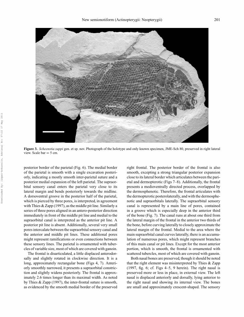
Figure 4. Scheenstia zappi gen. et sp. nov. Photograph of the skull of the holotype, JME-Sch 80. Scale bar = 2 cm.
canal runs into a groove close to the medial margin andbends laterally in the anterior portion towards the connec-tion with the infraorbital canal in the antorbital bone, thoughsuch a connection is not directly observable. Lateral to thecanal, the bone turns into a very thin, laminar membrane.
The dermopterotic bone is subrectangular in shape andsmoothly narrowed in its anterior part (Figs 4–6). It is1.4 times longer than deep and 1.2 times longer thanthe parietal. The dermopterotic contacts posteriorly withthe anterodorsal corner of the operculum and with thetwo lateral-most extrascapular bones, and sutures withthe parietal medially, with the frontal anteromedially, with
the dermosphenotic anterolaterally, and laterally with thedorsal-most suborbital and the preoperculum. The tempo-ral canal is represented by relatively large pores alignedvery close to the ventral margins of the dermopterotic andlateral-most extrascapular bones (Fig. 6). A dorsoventralgroove in the posterior portion of the dermopterotic, whichis pierced by four pores and is in continuity with a dorsoven-tral groove in the parietal, is interpreted as the ventralportion of the middle pit line. Additional pores, which mayrepresent isolated pits, are placed anterior (three) and poste-rior (one) to the middle pit line. The ornamentation of thedermopterotic is very similar of that of the parietal.
Downloaded By: [López-Arbarello, Adriana] At: 07:22 17 May 2011

New semionotiform (Actinopterygii: Neopterygii) 203
Figure 5. Scheenstia zappi gen. et sp. nov. Stereo photographs of the skull of the holotype, JME-Sch 80. Scale bar = 2 cm.
BraincaseThe braincase is almost completely hidden, but a well-ossified lateral ethmoid protrudes at the level of the secondanterior infraorbital (Figs 4–5, 9). The lateral ethmoid hasa large, longitudinally elongated lateral process which isventrally concave, forming a trough. This ventral troughresembles the condition in Amia (Grande & Bemis 1998)and Pholidophorus germanicus (Patterson 1975) in whichsuch a trough forms an articular surface for the largeautopalatine. Lateral ethmoids are rarely known in fossilbasal neopterygians (Patterson 1975). Among semionoti-forms, right lateral ethmoids are only known in one spec-imen of Araripelepidotes temnurus (MNHN MSE 635)where they are also well ossified, but in this specimenthe lateral process is incompletely preserved and cannotbe compared (Wenz & Brito 1996). Lateral ethmoids weredoubtfully identified, but not described and only roughlyillustrated in Lepidotes semiserratus (Stensio 1932; Holm-gren & Stensio 1936; Rayner 1948).
Orbital seriesThe circumorbital series is complete and comprises twosupraorbitals, a dermosphenotic, six infraorbitals, four ante-rior infraorbitals, and an antorbital (Figs 4–5, 8). All bonesin this series are ornamented with scattered tubercles, whichare covered with ganoin only in the supraorbitals, dermo-sphenotic and infraorbitals at the posterior border of theorbit. The supraorbital bones are rectangular and relativelylarge. The anterior supraorbital is the largest, as deep asthe posterior supraorbital but about 1.2 times longer. It is
approximately two times longer than deep, and articulateswith the first infraorbital anteriorly closing the circumor-bital ring. The posterior supraorbital is about 1.3 timeslonger than deep. Both supraorbital bones are longitudi-nally traversed by a smooth groove perforated by smallpores. This groove and pores are interpreted as a sensorycanal, here named the orbital canal (Fig. 8).
The dermosphenotic is irregularly hexagonal, located atthe posterodorsal corner of the orbit (Figs 4–6). It consti-tutes the largest bone located around the orbit. The boneoverlaps the medioventrally directed process of the frontaland articulates with the dermopterotic dorsoposteriorly,with the dorsal-most suborbital posteriorly, with the lastinfraorbital ventrally, and the posterior supraorbital anteri-orly. The sensory canals are shown by several pores (Fig.6). In the anterodorsal portion, the dermosphenotic makesan anterodorsally-directed step where two relatively largepores face two large pores of the supraorbital canal closeto the posteroventral margin of the frontal. Posterior to thisstep, two other pores open very close to the dorsal margin ofthe dermosphenotic and probably represent the connectionwith the temporal sensory canal. Close to the posteroventralmargin of the dermosphenotic, a wide groove indicates theinitial path of the infraorbital canal in the dermosphenotic.Additionally, a series of five smaller pores in a groove in thecentral portion of the bone represents the infraorbital canal.Close to the centre of the bone, this groove bifurcates severaltimes, indicating intense branching of the infraorbital canal.However, two larger branches indicate a connection with thetemporal canal on the one hand, and a connection with the
Downloaded By: [López-Arbarello, Adriana] At: 07:22 17 May 2011
204 A. Lopez-Arbarello and E. Sferco
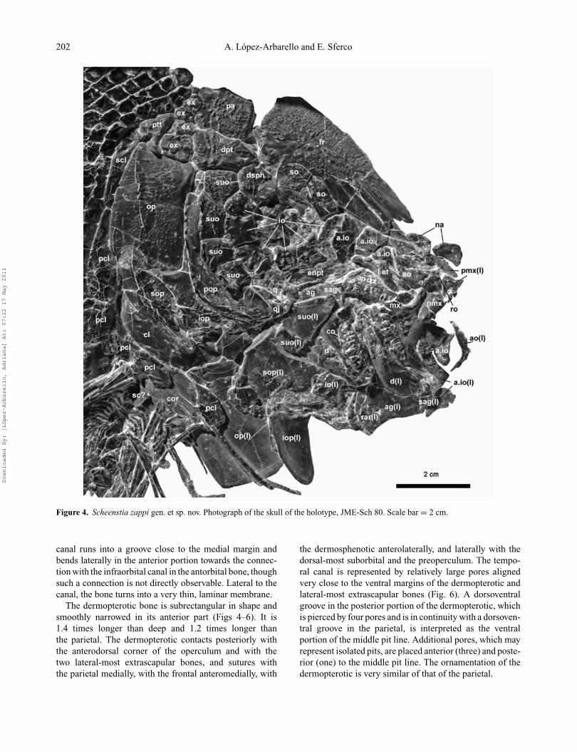
Figure 6. Scheenstia zappi gen. et sp. nov. Line drawing of the temporal and occipital regions of the skull of the holotype, JME-Sch 80.Scale bar = 1 cm.
orbital canal in the supraorbital bones on the other hand(Fig. 8).
There are six infraorbital bones (io1–io6 in anterior toposterior direction) and four anterior infraorbital bones(a.io1–a.io4 in anterior to posterior direction) (Figs 4–5, 8).Thies & Zapp (1997) did not distinguish between infraor-bitals and anterior infraorbitals and reported nine infraor-bitals in total, including a fragment of the third infraorbitalas a separate element: “Das kleinste Infraorbitale befindetsich innerhalb des Ringes in einer ventrocaudalen Posi-tion und scheint durch Fragmentation aus einem der beidenbenachbarten, großeren Infraorbitalia entstanden zu sein”(Thies & Zapp 1997, p. 19). The first and second ante-rior infraorbitals were also omitted in their count, butit should be noted that the first anterior infraorbital iscompletely disarticulated and displaced, while the secondanterior infraorbital is approximately in place, but brokenand badly preserved (see below). The last three infraorbitals(io4–io6) are quite similar in size and shape, all approxi-mately quadrangular, narrowing smoothly towards the orbit.The posterior margins of these infraorbitals, io4–io6, arepartially covered with patches of ganoin; io5 and io6 formthe posterior margin of the orbit, and io4 is located at theposteroventral corner of the orbit. The trajectory of theinfraorbital sensory canal in these three bones is indicatedby a smooth groove running almost parallel to the margin
of the orbit, perforated by several small pores. Branches ofthe canal are not evident in infraorbitals 5 and 6. Instead,in io4, branching is indicated by four superficial groovesradiating from the centre of the main groove. The follow-ing two infraorbitals (io3–io2) are a little smaller than thepreceding ones and slightly dorsally displaced. They formthe ventral border of the orbit. The posteroventral portionof io3 is broken, still in articulation with io4; as mentionedabove the rest of the bone is slightly displaced. Infraor-bitals io3 and io2 are somewhat trapezoidal in shape, io2being deepest anteriorly and only slightly larger than io3.In infraorbitals 2 and 3 the trajectory of the infraorbitalcanal is indicated by a groove, pierced by several smallpores, running in the dorsal halves of these bones. Severallarger ventrally directed openings indicate branches of themain sensory canal. The first infraorbital closes the circu-morbital ring. It is a rectangular bone, approximately 2.2times deeper than long, and articulates dorsally with theanterior-most supraorbital. There are four anterior infraor-bitals (a.io4–a.io1) between the first infraorbital bone andthe antorbital bone. The anterior infraorbitals are disar-ticulated and a little displaced, except for a.io1, which iscompletely displaced, and preserved isolated anterior to theskull (Figs 4–5, 9). However, their natural topographic rela-tionships can be reconstructed (Fig. 8). All of the anteriorinfraorbitals have free dorsal margins, i.e. do not contact
Downloaded By: [López-Arbarello, Adriana] At: 07:22 17 May 2011

New semionotiform (Actinopterygii: Neopterygii) 205
other bones dorsally; a.io4–a.io2 are lateroventral to theanterior portion of the frontal and a.io1 is placed anteriorto the frontal. The third and fourth anterior infraorbitals(a.io3–a.io4) are approximately rectangular; a.io4 is about1.8 times deeper than long and a.io3 is 1.4 times deeperthan long. The anterior infraorbital a.io3 forms a triangu-lar anterodorsal expansion; the a.io2 is preserved in place,though broken, lying on the massive lateral ethmoid (Fig.9). However, its shape can be reconstructed, also consid-ering the shape of the disarticulated left anterior infraor-bitals. The first two anterior infraorbitals (a.io1–a.io2) areapproximately triangular, tapering dorsally. The infraorbitalsensory canal runs through the centre of the first infraor-bital (io1) and last anterior infraorbital (a.io4), and bendsventrally to approximate the ventral border of the otherthree anterior infraorbitals (a.io3–a.io1) (Fig. 8).
The right antorbital is preserved in approximately itsnatural position and the left antorbital is completelydisplaced, preserved anterior to the skull (Figs 4, 5). Thies& Zapp (1997) misinterpreted the left antorbital as the rightelement and did not recognize the other antorbital. The boneis slender and typically boomerang-shaped, with taperingends. The horizontal arm is 1.35 (right element) to 1.4 (leftelement) times longer than the vertical arm. Little evidenceof the trajectory of the sensory canal is preserved withthe exception of two small openings on the most anteriorportion of the lateral surface of the horizontal arm of theright antorbital (this surface is completely broken posteri-orly) (Fig. 9). A small rostral bone is preserved almost inplace, anterior to the antorbital (Fig. 9). The rostral has aright-angled triangle shape with its opposite side oriented tothe midline, indicating that there were two paired rostrals,of which the right element is preserved. The ethmoidalcommissure is revealed by a pore in the central part of therostral.
Four suborbital bones are preserved articulated on theright side of the skull (Figs 4, 5) but this series iscertainly incomplete. Two semiarticulated additional subor-bital bones are separately preserved lateral to the right lowerjaw (Figs 4, 5). They are preserved in lateral view and,considering their shape and bringing them back to theirnatural position (Fig. 8), we conclude that they correspondto the left side of the skull (in contrast to Thies & Zapp1997). Consequently, the complete series of six suborbitalbones in Scheenstia zappi is arranged in one row betweenthe preoperculum and the circumborbital bones (Fig. 8). Wethus disagree with Thies & Zapp (1997) who concludedthat additional suborbital bones might have been presentbut not preserved. The dorsal-most suborbital is approxi-mately trapezoidal, acuminating ventrally, and articulateswith the last infraorbital and the dermosphenotic anteriorly,the dermopterotic dorsally and the dorsal tip of the preop-erculum posteriorly (Figs 4–6). The following suborbital isthe largest of the series, approximately rectangular, with ananterior narrowed portion, posterodorsally acuminated and
Figure 7. Scheenstia zappi gen. et sp. nov. Line drawing of thefrontal bone in the holotype, JME-Sch 80. Scale bar = 1 cm.
about 1.5 times deeper than wide (Figs 4–6). The followingtwo suborbitals are quadrangular, approximately the samewidth as the second (Figs 4, 5). As mentioned above, thesecond suborbital bone (numbering the bones in anteriorto posterior direction) is disarticulated and displaced, andcorresponds to the left side of the skull. It is the smallest inthe series and approximately rectangular. The most anteriorsuborbital, which is also disarticulated and displaced fromthe left side of the skull, is subtriangular and completes thedermal cover of the cheek, which completely hides the ossi-fications of the palatoquadrate. Reconstructing the subor-bital series in its natural position, suborbital 1 is placedventral to the orbit, suborbitals 2 and 3 articulate followingthe posteroventral corner of the orbit, and suborbitals 4–6are placed posterior to the orbit.
Hyopalatine bonesThe quadratojugal is well exposed at the anterior end of thepreoperculum (Figs 4, 5, 9, 10). The exact shape of the boneis unknown because it is partially covered by the preop-erculum and dorsally broken. The exposed portion formsa strongly sinuous anterodorsal border and an expanded,anteroventrally rounded anterior portion. The quadratojugalis an independent bone articulating with the quadrate medi-ally and with the preoperculum laterally. The ventrolateralsurface of the expanded anterior portion is free and formsa saddle-shaped surface directed anteriorly, which proba-bly articulated with the lower jaw (Fig. 10). The quadrateis incomplete and rather poorly preserved. The dorsal andposterior borders of the bone are broken, but the ventraland articular portions are relatively well preserved. Thearticular portion is relatively large and strongly ossified(Fig. 10). The posteroventral margin of the quadrate islaterally expanded forming a flat surface, slightly concaveanteriorly, which articulates with the anterior expandedportion of the quadratojugal anteromedially. The quadrato-mandibular articulation is not preserved. However, consid-ering the location of the quadrate, it probably took placehalfway between the centre and the anterior border of theorbit.
The metapterygoid lamina is only partially exposed andpoorly preserved dorsal and posterior to the quadrate.The pterygoid bones are very difficult to interpret. The
Downloaded By: [López-Arbarello, Adriana] At: 07:22 17 May 2011
206 A. Lopez-Arbarello and E. Sferco
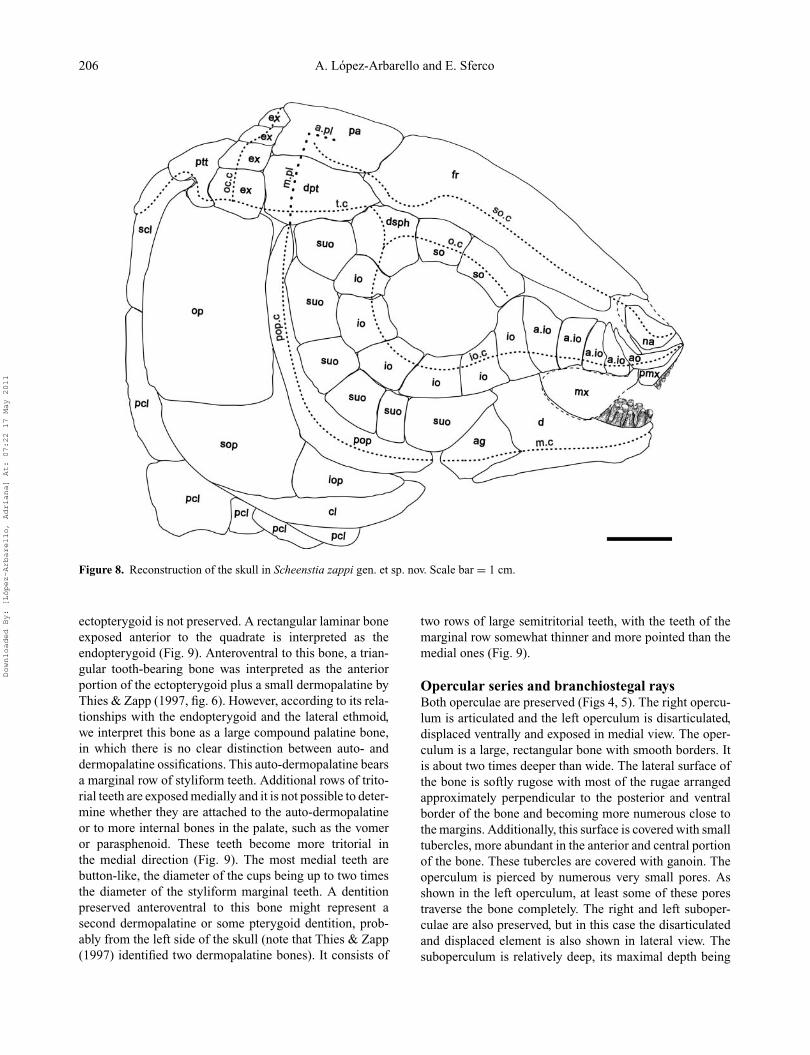
Figure 8. Reconstruction of the skull in Scheenstia zappi gen. et sp. nov. Scale bar = 1 cm.
ectopterygoid is not preserved. A rectangular laminar boneexposed anterior to the quadrate is interpreted as theendopterygoid (Fig. 9). Anteroventral to this bone, a trian-gular tooth-bearing bone was interpreted as the anteriorportion of the ectopterygoid plus a small dermopalatine byThies & Zapp (1997, fig. 6). However, according to its rela-tionships with the endopterygoid and the lateral ethmoid,we interpret this bone as a large compound palatine bone,in which there is no clear distinction between auto- anddermopalatine ossifications. This auto-dermopalatine bearsa marginal row of styliform teeth. Additional rows of trito-rial teeth are exposed medially and it is not possible to deter-mine whether they are attached to the auto-dermopalatineor to more internal bones in the palate, such as the vomeror parasphenoid. These teeth become more tritorial inthe medial direction (Fig. 9). The most medial teeth arebutton-like, the diameter of the cups being up to two timesthe diameter of the styliform marginal teeth. A dentitionpreserved anteroventral to this bone might represent asecond dermopalatine or some pterygoid dentition, prob-ably from the left side of the skull (note that Thies & Zapp(1997) identified two dermopalatine bones). It consists of
two rows of large semitritorial teeth, with the teeth of themarginal row somewhat thinner and more pointed than themedial ones (Fig. 9).
Opercular series and branchiostegal raysBoth operculae are preserved (Figs 4, 5). The right opercu-lum is articulated and the left operculum is disarticulated,displaced ventrally and exposed in medial view. The oper-culum is a large, rectangular bone with smooth borders. Itis about two times deeper than wide. The lateral surface ofthe bone is softly rugose with most of the rugae arrangedapproximately perpendicular to the posterior and ventralborder of the bone and becoming more numerous close tothe margins. Additionally, this surface is covered with smalltubercles, more abundant in the anterior and central portionof the bone. These tubercles are covered with ganoin. Theoperculum is pierced by numerous very small pores. Asshown in the left operculum, at least some of these porestraverse the bone completely. The right and left suboper-culae are also preserved, but in this case the disarticulatedand displaced element is also shown in lateral view. Thesuboperculum is relatively deep, its maximal depth being
Downloaded By: [López-Arbarello, Adriana] At: 07:22 17 May 2011

New semionotiform (Actinopterygii: Neopterygii) 207
Figure 9. Scheenstia zappi gen. et sp. nov. Detailed photograph of the ossifications of the palatoquadrate and snout in the holotype,JME-Sch 80. Scale bar = 1 cm.
0.5 of the depth of the operculum. The suboperculum hasa well developed anterodorsal process, which is about halfthe length of the suboperculum and reaches up to approx-imately 0.25 of the total depth of the operculum. The freeborders are smooth and it shows a similarly rugose andtuberculous surface to that of the operculum, though thetubercles are less abundant. The suboperculum is pierced byseveral small pores. Both interoperculae are also preserved.The right interoperculum is articulated and, thus, partiallyhidden by the preoperculum. The left interoperculum isdisarticulated, displaced ventrally and preserved in medialview. It is two times longer than its maximal depth and doesnot reach the anterior end of the preoperculum. It is alsopierced by numerous small pores. As noticed by Thies &Zapp (1997), branchiostegal rays are very badly, if at all,preserved.
The strongly ossified preoperculum is crescent-shaped,concave anteriorly, forming an angle of about 110◦. Itreaches the dermopterotic dorsally and articulates with thesuborbital series anteriorly. The preopercular sensory canalruns enclosed in the thickened anterior and anterodorsalmargin of the preoperculum and exits the bone throughsix large openings. The most anterior three openings areanteroventrally directed, while the posterior three open-ings are posteroventrally directed. The preopercular sensorycanal enters the dermopterotic close to its posterior borderand is in continuity with the median pit line (Fig. 6). Small
pores are also present in the preoperculum. The posteriorand ventral borders of the preoperculum are straight, and thesurface is smooth, with only a thin layer of ganoin coveringthe posterior margin of the bone (Figs 4, 5).
JawsThe upper jaw is completely disarticulated (Figs 4, 5, 9).Both premaxillae are only partially preserved. As noted byThies & Zapp (1997), the nasal processes of the premaxillaeare large, though badly preserved. A relatively small fora-men for the olfactory nerve is placed at the base of the nasalprocess of the right premaxilla (Fig. 9). Some premaxillaryteeth are shown, but their exact number and arrangement isunknown. The premaxillary teeth are strong and styliform.Thies & Zapp (1997) noticed that the maxilla is broken andpoorly preserved between the bones of the palatoquadrateand the displaced left lower jaw. It has a long, cylindrical,anteromedially directed articular process. The main body ofthe maxilla is badly broken, thus it is not possible to recon-struct its shape and extent. A supramaxilla is not preservedor absent.
Both lower jaws are displaced and preserved in lateralview (Figs 4, 5). The dentary is a large, robust bonewhich deepens posteriorly in the coronoid process. Asusual in semionotiforms, the dentary has a well devel-oped posteroventral process, which in this case does notreach the posterior border of the lower jaw. The dentaries
Downloaded By: [López-Arbarello, Adriana] At: 07:22 17 May 2011

208 A. Lopez-Arbarello and E. Sferco
Figure 10. Scheenstia zappi gen. et sp. nov. Detailed stereo photographs of the holotype (JME-Sch 80) showing the peculiar shape of thequadratojugal. Scale bar = 0.5 cm.
have a row of five marginal, robust styliform teeth. Asdescribed by Thies & Zapp (1997), one coronoid boneis preserved in place, articulating with the dentary poste-rior to its toothed portion. This coronoid has a marginalrow of at least five styliform teeth, which are only slightlysmaller than the teeth on the dentary. Additional rows (atleast four additional rows are evident in the right mandible)of gradually more tritorial teeth are preserved medially.The most internal teeth are button-like and as large as theobserved palatopterygoid teeth. Two completely disarticu-lated button-like teeth are even larger than the largest teethobserved in the lower jaw or the palatopterygoid region.The coronoid process is very high, representing approxi-mately 0.55 of the total length of the lower jaw. The angularbone is large, with a slightly concave posterior border. Theposteroventral corner of the angular is well preserved andforms a triangular projection beyond the retroarticular. Thesurangular bone crowns the coronoid process, articulatingwith the most dorsal borders of the dentary and angularbones. The retroarticular bone was not recognized by Thies& Zapp (1997), but is partially preserved ventral to theangular and posterior to the posteroventral process of thedentary. The passage of the mandibular sensory canal isrevealed by a series of pores close to the ventral marginof the angular bone. It continues in the dentary dorsal tothe posteroventral process and follows forwards through thecentre of the bone. Additional smaller pores are distributeddorsal and ventral to the mandibular canal in the dentaryand only dorsally in the angular. Thies & Zapp (1997) inter-preted some of these pores as an additional canal, but we
find no evidence to confirm that the pores would have beenrelated to a series of additional neuromasts.
Pectoral girdleThe cleithrum, supracleithrum, posttemporal, five postclei-thra and two coracoids are preserved (Figs 4, 5). With theexception of the two most dorsal postcleithra, all the dermalbones in the pectoral girdle are smooth and with smoothposterior borders. The cleithrum is a large crescent-shapedbone with narrowing anterior and posterior ends. The dorsalportion of the cleithrum is hidden by the operculum andsuboperculum. After reconstructing its natural position,the ventral portion of the cleithrum reaches the level ofthe posterior border of the orbit anteriorly (Fig. 8). Theedge between the branchial and lateral surfaces of the clei-thrum forms a robust ridge, along which rest at least threeseries of tiny denticles, also noted by Thies & Zapp (1997)(Figs 4, 5).
The supracleithrum is almost completely exposed, deepand narrow, about four or five times deeper than wide, withthickened anterior border and laminar posterior portion.Anterodorsally, the supracleithrum is laterally expandedforming an approximately circular concave articular surfacefor the posttemporal (Fig. 6). The supracleithrum is notcovered with ganoin. Three large pores indicate that thelateral line traverses the central portion in the dorsal thirdof the supracleithrum in anterodorsal to posteroventraldirection. The posttemporal is relatively small, but robust,and triangular, tapering dorsally, reaching the level of thearticulation between the second and third extrascapulars
Downloaded By: [López-Arbarello, Adriana] At: 07:22 17 May 2011
New semionotiform (Actinopterygii: Neopterygii) 209
(Figs 6, 8). The posterior portion of the posttemporalis ornamented with scarce tubercles, some of which arecovered with ganoin. The posttemporal has a very robustcylindrical anteroventral process with a slender shaft andan expanded distal end (Fig. 6). The distal surface of thisbugle-like process (only the edge of which is exposed) isslightly convex and oriented ventro-medially. Despite theorientation of the bugle-like process, it does not seem tohave reached the braincase. The posteroventral portion ofthe posttemporal is deeply convex and articulates with theexpanded concave surface of the supracleithrum. It is notpossible to recognize a trajectory of the sensory canal inthe posttemporal with certainty. A single pore is placed atthe base of the bugle-like process, in the deep concavitybetween this process and the posterior rounded portion ofthe bone.
There are five postcleithra aligned along the distal marginof the cleithrum (Figs 4, 5). Unlike Thies & Zapp (1997), wefind no evidence for additional elements in this series. Allpostcleithra have ganoin on their distal margins. Close to thedistal border of the dorsal-most postcleithra (postcleithra 5and 4 if the bones are numbered in anterior to posteriordirection) the ganoin cover forms posteroventral to poste-riorly directed ridges. The dorsal-most postcleithrum is thelargest, about two times deeper than the following post-cleithrum, which is approximately as deep as long, with theanterior border slightly sinuous. On the dorsal portion of thedistal margin of the most dorsal postcleithrum, the ganoinridges end before reaching the distal border of the bone,which is smooth. Ventrally, the ganoin ridges on the fifth andfourth postcleithra gradually approximate the distal borderof these bones. The ventral portion of the distal border ofthe fifth postcleithrum and the distal border of the fourthpostcleithrum are broken, but according to the extension ofthe ganoin ridges these borders were probably serrated, asis the case of the adjacent scales. The third postcleithrumis the smallest in the series and is approximately quadran-gular in shape. The second postcleithrum is oblong and thefirst postcleithrum, displaced anteroventrally, is petal-like.According to the extension of the ganoin cover and pres-ence of small tubercles, the three anterior-most postcleithrapartially overlapped each other (Fig. 8).
The endochondral elements of the pectoral girdle arepartially exposed. There is a very large and laminar cora-coid ossification, exposed ventral to the cleithrum (Figs 4,5). A fragment of bone preserved at the base of the pectoralfin might represent the scapula. The propterygium is poorlyexposed anterior to the basis of the right pectoral fin rays.Two elongate radials are poorly preserved below the scapu-lar ossification.
Paired finsBoth pectoral fins are partially preserved (Fig. 3). Althoughthe right pectoral fin is partially disarticulated and the finrays are distally broken, it is clear that only one pair of
basal fulcra was present. The presence of fringing fulcrais doubtful. Although no fringing fulcrum is preserved, thequality of preservation is not good enough to be certainabout their absence. The first fin ray is broken, but wasprobably not segmented or branched. The next few raysonly branched very distally, if at all. The exact number ofpectoral rays is dubious, but the bases of 13 lepidotrichiaare articulated in the right pectoral fin; this is probably thetotal number. When the bases of the pectoral rays are wellpreserved a small spinous process is evident. In the fewarticulated rays this process is directed dorsally.
Only the right pelvic fin is preserved (Fig. 3). This pelvicfin is placed at about 0.5 of the standard length, approxi-mately midway between the pectoral and anal fins, imme-diately behind the eighth vertical row of scales. The pelvicfin inserts at about 87.5% of the body depth, where a welldeveloped unpaired basal fulcrum is followed by one pair ofbasal fulcra and at least four long and slender paired fringingfulcra. The two most distal fringing fulcra are segmented.The number of pelvic fin rays is unknown because the finis completely folded.
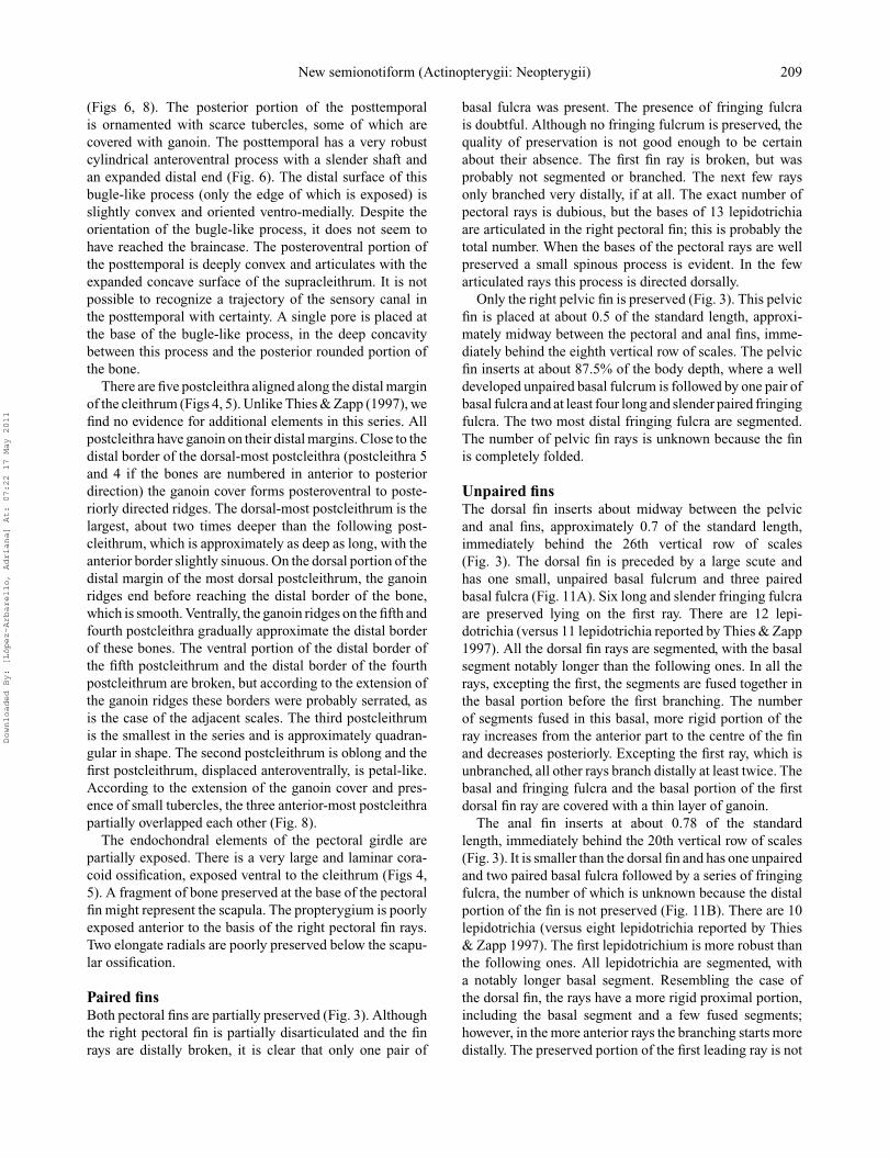
Unpaired finsThe dorsal fin inserts about midway between the pelvicand anal fins, approximately 0.7 of the standard length,immediately behind the 26th vertical row of scales(Fig. 3). The dorsal fin is preceded by a large scute andhas one small, unpaired basal fulcrum and three pairedbasal fulcra (Fig. 11A). Six long and slender fringing fulcraare preserved lying on the first ray. There are 12 lepi-dotrichia (versus 11 lepidotrichia reported by Thies & Zapp1997). All the dorsal fin rays are segmented, with the basalsegment notably longer than the following ones. In all therays, excepting the first, the segments are fused together inthe basal portion before the first branching. The numberof segments fused in this basal, more rigid portion of theray increases from the anterior part to the centre of the finand decreases posteriorly. Excepting the first ray, which isunbranched, all other rays branch distally at least twice. Thebasal and fringing fulcra and the basal portion of the firstdorsal fin ray are covered with a thin layer of ganoin.
The anal fin inserts at about 0.78 of the standardlength, immediately behind the 20th vertical row of scales(Fig. 3). It is smaller than the dorsal fin and has one unpairedand two paired basal fulcra followed by a series of fringingfulcra, the number of which is unknown because the distalportion of the fin is not preserved (Fig. 11B). There are 10lepidotrichia (versus eight lepidotrichia reported by Thies& Zapp 1997). The first lepidotrichium is more robust thanthe following ones. All lepidotrichia are segmented, witha notably longer basal segment. Resembling the case ofthe dorsal fin, the rays have a more rigid proximal portion,including the basal segment and a few fused segments;however, in the more anterior rays the branching starts moredistally. The preserved portion of the first leading ray is not
Downloaded By: [López-Arbarello, Adriana] At: 07:22 17 May 2011
210 A. Lopez-Arbarello and E. Sferco
Figure 11. Scheenstia zappi gen. et sp. nov., holotype, JME-Sch 80. A, photograph of the dorsal fin; B, photograph of the anal fin. Scalebar = 2 cm.
branched and the following ray branches at least once. Thebasal and fringing fulcra and the basal segment of the firstray are covered with ganoin. Tiny patches of ganoin lie onthe centre of several segments in the first ray.
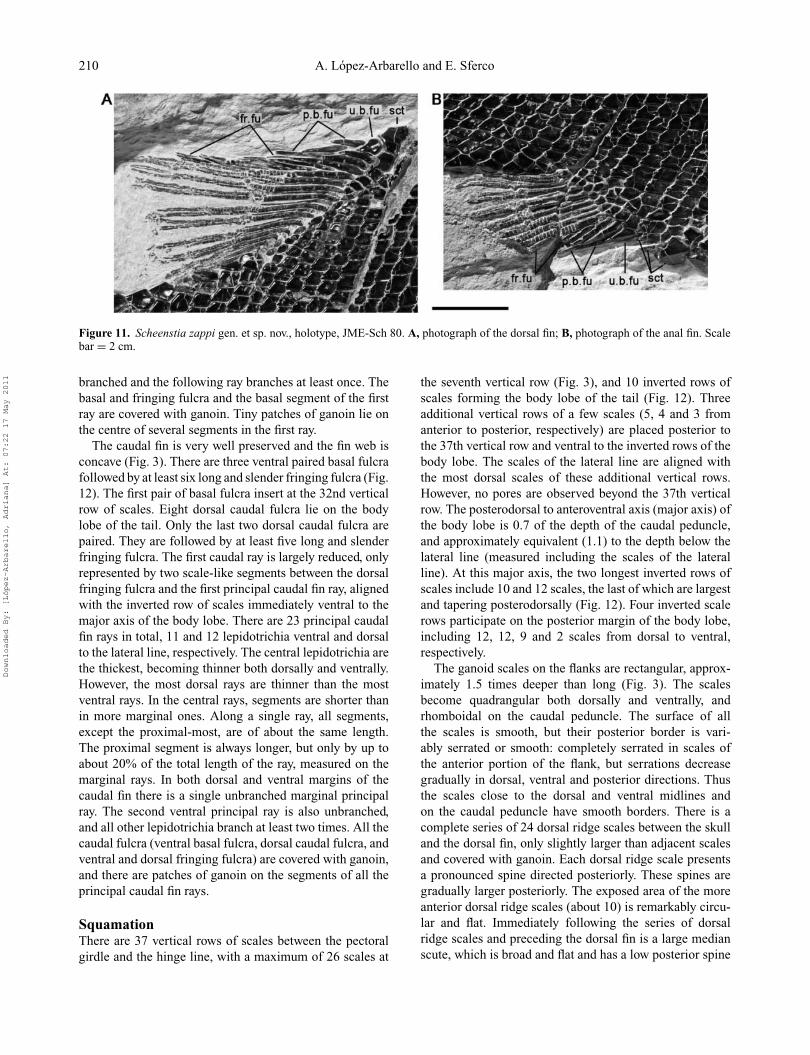
The caudal fin is very well preserved and the fin web isconcave (Fig. 3). There are three ventral paired basal fulcrafollowed by at least six long and slender fringing fulcra (Fig.12). The first pair of basal fulcra insert at the 32nd verticalrow of scales. Eight dorsal caudal fulcra lie on the bodylobe of the tail. Only the last two dorsal caudal fulcra arepaired. They are followed by at least five long and slenderfringing fulcra. The first caudal ray is largely reduced, onlyrepresented by two scale-like segments between the dorsalfringing fulcra and the first principal caudal fin ray, alignedwith the inverted row of scales immediately ventral to themajor axis of the body lobe. There are 23 principal caudalfin rays in total, 11 and 12 lepidotrichia ventral and dorsalto the lateral line, respectively. The central lepidotrichia arethe thickest, becoming thinner both dorsally and ventrally.However, the most dorsal rays are thinner than the mostventral rays. In the central rays, segments are shorter thanin more marginal ones. Along a single ray, all segments,except the proximal-most, are of about the same length.The proximal segment is always longer, but only by up toabout 20% of the total length of the ray, measured on themarginal rays. In both dorsal and ventral margins of thecaudal fin there is a single unbranched marginal principalray. The second ventral principal ray is also unbranched,and all other lepidotrichia branch at least two times. All thecaudal fulcra (ventral basal fulcra, dorsal caudal fulcra, andventral and dorsal fringing fulcra) are covered with ganoin,and there are patches of ganoin on the segments of all theprincipal caudal fin rays.
SquamationThere are 37 vertical rows of scales between the pectoralgirdle and the hinge line, with a maximum of 26 scales at
the seventh vertical row (Fig. 3), and 10 inverted rows ofscales forming the body lobe of the tail (Fig. 12). Threeadditional vertical rows of a few scales (5, 4 and 3 fromanterior to posterior, respectively) are placed posterior tothe 37th vertical row and ventral to the inverted rows of thebody lobe. The scales of the lateral line are aligned withthe most dorsal scales of these additional vertical rows.However, no pores are observed beyond the 37th verticalrow. The posterodorsal to anteroventral axis (major axis) ofthe body lobe is 0.7 of the depth of the caudal peduncle,and approximately equivalent (1.1) to the depth below thelateral line (measured including the scales of the lateralline). At this major axis, the two longest inverted rows ofscales include 10 and 12 scales, the last of which are largestand tapering posterodorsally (Fig. 12). Four inverted scalerows participate on the posterior margin of the body lobe,including 12, 12, 9 and 2 scales from dorsal to ventral,respectively.
The ganoid scales on the flanks are rectangular, approx-imately 1.5 times deeper than long (Fig. 3). The scalesbecome quadrangular both dorsally and ventrally, andrhomboidal on the caudal peduncle. The surface of allthe scales is smooth, but their posterior border is vari-ably serrated or smooth: completely serrated in scales ofthe anterior portion of the flank, but serrations decreasegradually in dorsal, ventral and posterior directions. Thusthe scales close to the dorsal and ventral midlines andon the caudal peduncle have smooth borders. There is acomplete series of 24 dorsal ridge scales between the skulland the dorsal fin, only slightly larger than adjacent scalesand covered with ganoin. Each dorsal ridge scale presentsa pronounced spine directed posteriorly. These spines aregradually larger posteriorly. The exposed area of the moreanterior dorsal ridge scales (about 10) is remarkably circu-lar and flat. Immediately following the series of dorsalridge scales and preceding the dorsal fin is a large medianscute, which is broad and flat and has a low posterior spine
Downloaded By: [López-Arbarello, Adriana] At: 07:22 17 May 2011

New semionotiform (Actinopterygii: Neopterygii) 211
Figure 12. Scheenstia zappi gen. et sp. nov. Photograph of caudal fin in the holotype, JME-Sch 80. Scale bar = 2 cm.
(Fig. 11A). This large scute is aligned with a series offour additional rhomboid scales, which are distinct, notaligned with the vertical rows of scales, but surroundingthe bases of the dorsal basal fulcra and first and seconddorsal fin rays. Otherwise the scales at the base of thedorsal fin are well aligned with vertical rows of scales,but notably smaller. About six hexagonal, unornamentedmedian scales, which increase in size posteriorly, garnishthe dorsal midline between the dorsal fin and the hinge line(Fig. 12). Only the most posterior of these dorsal scales isnotably larger than the adjacent scales and can be distin-guished as a scute. A series of five median scales also lieon the ventral midline between the anal and caudal fins(Fig. 12). As the dorsal equivalents, these ventral medianscales are distinct in shape, but only the two most posteriorare larger than adjacent scales and thus distinguished asscutes. There are three large scutes preceding the anal fin,a median element followed by a pair of lateroventral scutes(Fig. 11B). These scutes probably surrounded the vent. Theleft lateroventral scute is preserved in sagital view, but theother two scutes are well exposed and show smooth surfacesand strongly serrated posterior borders.
Discussion
As mentioned in the Introduction, although ‘semionotids’from the Late Jurassic limestones of the Franconian andSwabian Alb are in need of revision, not only do mostof the species seem to be valid but additional new taxaare being discovered (Lopez-Arbarello pers. obs.). Amongthese the specimen described in this article, previouslypublished by Thies & Zapp (1997) as Lepidotes sp., isshown to represent a new taxon, Scheenstia zappi gen. etsp. nov. The presence of a series of anterior infraorbitalbones and the absence of gular plates in S. zappi supportits referral to the Semionotiformes (Olsen & McCune1991, Cavin & Suteethorn 2006, Lopez-Arbarello 2009).Among semionotiforms, S. zappi lacks the synapomor-phies of Lepisosteidae (Wiley 1976) or Macrosemiidae(Bartram 1977). As mentioned in the Introduction, allsemionotiforms that are not lepisosteids or macrosemiidsare currently grouped in a single Family Semionotidae, butsince such an assemblage is probably polyphyletic (Lopez-Arbarello 2008b) and no other family is properly definedwithin this order, we leave Scheenstia as Semionotiformes
Downloaded By: [López-Arbarello, Adriana] At: 07:22 17 May 2011

212 A. Lopez-Arbarello and E. Sferco
incertae family, pending the completion of an ongoing anal-ysis of semionotiform phylogenetic relationships (Lopez-Arbarello in press.).
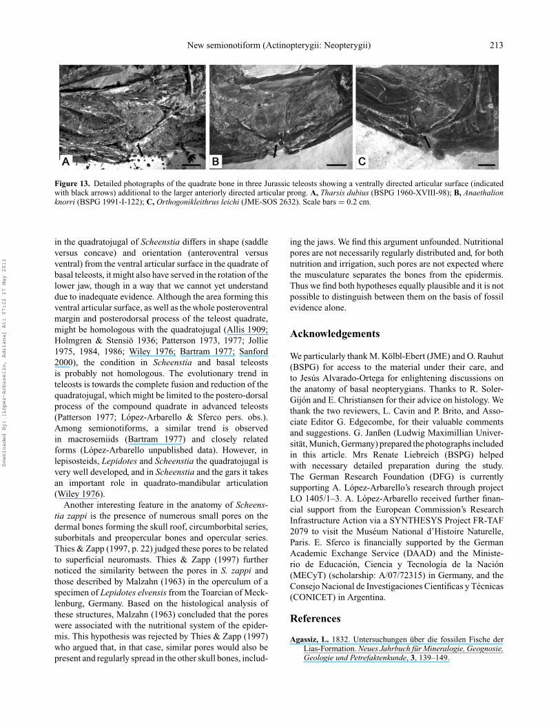
Due to the lack of phylogenetic analyses of interrela-tionships between species currently grouped in Semionoti-dae, autapomorphic features in S. zappi cannot be prop-erly established. However, Scheenstia differs from all othersemionotiforms represented by comparable material, in thepresence of a sensory canal in the supraorbital bone, herecalled the orbital canal, and a saddle-like articular surfacein the quadratojugal. This saddle shape in the ventrolateralsurface of the expanded anterior portion of the quadra-tojugal probably articulated with the lower jaw, posteriorto the articulation of the quadrate. The right lower jaw inthe single specimen of S. zappi is disarticulated, and thedisplaced left lower jaw is preserved in lateral view. There-fore, no direct evidence for such articulation is observed.However, we have observed a comparable condition in thequadrato-mandibular articulation of some Jurassic basalteleosts. In teleosts the lower jaw articulates only withthe quadrate, but in several taxa this bone has a ventrallydirected articular surface additional to the larger anteri-orly directed articular prong (Fig. 13). Such a conditionis present at least in the Jurassic Tharsis dubius (spec-imens BSPG 1960-XVIII-98 in Fig. 13A, BSPG 1964-XXIII-280, BSPG 1961-III-23, and BSPG 1964-XXIII-160among others; also Nybelin 1974, pl. 14, fig. 2) (see Nybe-lin 1974, Leptolepis normandica, pl. 3, fig. 3, pl. 4, fig. 2,and Leptolepides sprattiformis, pl. 25, fig. 3), Anaethalionknorri (specimens BSPG 1991-I-122 in Fig. 13B; BSPG1960-XVIII-93, BSPG 1969-XVI-39, BSPG 1969-XVI-31,BSPG 1964-XXIII-394, BSPG 1953-I-97 among others;see also Nybelin 1967, pl. 2, fig. 1 (Anaethalion knorri)and fig. 2 of (Anaethalion angustus)) and Orthogoniklei-thrus leichi (JME-SOS 2632; Fig. 13C). In these fishes, theventral articular surface is placed at the level at which itwould be the ossification centre of the quadrate (Arratia& Schultze 1991, Arratia 1999), and apparently enablesthe vertical rotation of the lower jaw. The saddle-shapedsurface in the quadratojugal of Scheenstia takes a similarposition to that of the ventral articular surface of the teleostquadrate, and might have played a similar role (see discus-sion below).
Apart from two possible autapomorphic features, thecombination of characters in the diagnosis is uniquelyfound in Scheenstia zappi. Among semionotiforms outsideLepisosteidae and Macrosemiidae, this new taxon mostclosely resembles the species of Lepidotes Agassiz, 1832and, unsurprisingly, the new fish from Schamhaupten wasinitially identified as a species of this genus (Thies & Zapp1997). Scheenstia resembles Lepidotes in several morpho-logical features, such as the series of suborbital bonesarranged in one row and extending anteriorly, ventral tothe orbit, and the presence of a reduced scale-like firstcaudal fin ray. The first of these features represents a
condition otherwise only present in Isanichthys amongsemionotiforms. The second feature is only found in fisheswithin Semionotiformes, but might represent a plesiomor-phic condition because a reduced, scale-like first caudal finray is also present in Dapedium (Wenz 1968) and somebasal teleosts like Pholidophorus bechei (Patterson 1968).Among semionotiforms the most common – though proba-bly derived – condition is the presence of a fully developed,scale-like first caudal fin ray. Such a feature is observed inthe lepisosteids Lepisosteus and Atractosteus (Wiley 1976),macrosemiids Macrosemius (Bartram 1977), Propterus(Bartram 1977; BSPG 1986-XV-121, BSPG 1964-XXIII-145b) and also in Paralepidotus (BPG 2003-XXIX-632,BSPG 2007-I-62), Semiolepis (Lombardo & Tintori 2008)and Semionotus minor (lectotype GSM 27975).
The large tritorial forms of the Late Jurassic and EarlyCretaceous of Europe, currently referred to Lepidotes (i.e.L. mantelli Agassiz, 1833, L. fittoni Agassiz, 1833, L. laevisAgassiz, 1837, L. maximus Wagner, 1863, L. decoratusWagner, 1863, L. degenhardti Branco, 1885) differ from thenew taxon in the presence of an extremely tritorial denti-tion (see Jain 1983 for a definition of this kind of denti-tion) and a very deep mandibular symphysis, features thatare absent in Scheenstia. Furthermore, Scheenstia can bedistinguished by the shape of the infraorbital bones placedat the posterior border of the orbit, which are expandedposteriorly (thus being longer than deep) in those tritorialspecies of Lepidotes. The latter feature is also present inthe primitive gar Masillosteous Micklich & Klappert, 2001,Araripelepidotes Santos, 1990, Pliodetes Wenz, 1999, andIsanichthys Cavin & Suteethorn, 2006. Scheenstia furtherhas high basal fulcra, a condition that resembles the case inSemionotus Agassiz, 1832, but is very different from thatin Lepidotes, in which the basal fulcra are low and small(Lopez-Arbarello 2008a).
In addition to the taxonomically significant morpholog-ical features discussed above, other anatomical charactersin Scheenstia deserve discussion due to their importanceconcerning character evolution of in neopterygian fishes.The saddle-like articular surface of the quadratojugal isa very interesting feature of Scheenstia. As mentionedabove, the additional ventral articular surface observed inthe quadrate of some basal teleosts (see species indicatedabove), might enable the rotation of the lower jaw. Inthese fishes, when the mouth is closed, the ventral articularsurface of the quadrate is free (Fig. 13), but when themouth is open it articulates with the postarticular processof the lower jaw (Tharsis dubius, BSPG 1964-XXIII-280).Due to the development of the postarticular process of thelower jaw in teleosts (Nelson 1973), such a ventral articularsurface might have developed in the quadrate of thesebasal teleosts to allow the rotation of the lower jaw. Simul-taneously, the articular surface of the quadrate might haveserved as a stop, preventing an extreme downward rotationof the lower jaw. Although the saddle-like articular surface
Downloaded By: [López-Arbarello, Adriana] At: 07:22 17 May 2011
New semionotiform (Actinopterygii: Neopterygii) 213
Figure 13. Detailed photographs of the quadrate bone in three Jurassic teleosts showing a ventrally directed articular surface (indicatedwith black arrows) additional to the larger anteriorly directed articular prong. A, Tharsis dubius (BSPG 1960-XVIII-98); B, Anaethalionknorri (BSPG 1991-I-122); C, Orthogonikleithrus leichi (JME-SOS 2632). Scale bars = 0.2 cm.
in the quadratojugal of Scheenstia differs in shape (saddleversus concave) and orientation (anteroventral versusventral) from the ventral articular surface in the quadrate ofbasal teleosts, it might also have served in the rotation of thelower jaw, though in a way that we cannot yet understanddue to inadequate evidence. Although the area forming thisventral articular surface, as well as the whole posteroventralmargin and posterodorsal process of the teleost quadrate,might be homologous with the quadratojugal (Allis 1909;Holmgren & Stensio 1936; Patterson 1973, 1977; Jollie1975, 1984, 1986; Wiley 1976; Bartram 1977; Sanford2000), the condition in Scheenstia and basal teleostsis probably not homologous. The evolutionary trend inteleosts is towards the complete fusion and reduction of thequadratojugal, which might be limited to the postero-dorsalprocess of the compound quadrate in advanced teleosts(Patterson 1977; Lopez-Arbarello & Sferco pers. obs.).Among semionotiforms, a similar trend is observedin macrosemiids (Bartram 1977) and closely relatedforms (Lopez-Arbarello unpublished data). However, inlepisosteids, Lepidotes and Scheenstia the quadratojugal isvery well developed, and in Scheenstia and the gars it takesan important role in quadrato-mandibular articulation(Wiley 1976).
Another interesting feature in the anatomy of Scheens-tia zappi is the presence of numerous small pores on thedermal bones forming the skull roof, circumborbital series,suborbitals and preopercular bones and opercular series.Thies & Zapp (1997, p. 22) judged these pores to be relatedto superficial neuromasts. Thies & Zapp (1997) furthernoticed the similarity between the pores in S. zappi andthose described by Malzahn (1963) in the operculum of aspecimen of Lepidotes elvensis from the Toarcian of Meck-lenburg, Germany. Based on the histological analysis ofthese structures, Malzahn (1963) concluded that the poreswere associated with the nutritional system of the epider-mis. This hypothesis was rejected by Thies & Zapp (1997)who argued that, in that case, similar pores would also bepresent and regularly spread in the other skull bones, includ-
ing the jaws. We find this argument unfounded. Nutritionalpores are not necessarily regularly distributed and, for bothnutrition and irrigation, such pores are not expected wherethe musculature separates the bones from the epidermis.Thus we find both hypotheses equally plausible and it is notpossible to distinguish between them on the basis of fossilevidence alone.
Acknowledgements
We particularly thank M. Kolbl-Ebert (JME) and O. Rauhut(BSPG) for access to the material under their care, andto Jesus Alvarado-Ortega for enlightening discussions onthe anatomy of basal neopterygians. Thanks to R. Soler-Gijon and E. Christiansen for their advice on histology. Wethank the two reviewers, L. Cavin and P. Brito, and Asso-ciate Editor G. Edgecombe, for their valuable commentsand suggestions. G. Janßen (Ludwig Maximillian Univer-sitat, Munich, Germany) prepared the photographs includedin this article. Mrs Renate Liebreich (BSPG) helpedwith necessary detailed preparation during the study.The German Research Foundation (DFG) is currentlysupporting A. Lopez-Arbarello’s research through projectLO 1405/1–3. A. Lopez-Arbarello received further finan-cial support from the European Commission’s ResearchInfrastructure Action via a SYNTHESYS Project FR-TAF2079 to visit the Museum National d’Histoire Naturelle,Paris. E. Sferco is financially supported by the GermanAcademic Exchange Service (DAAD) and the Ministe-rio de Educacion, Ciencia y Tecnologıa de la Nacion(MECyT) (scholarship: A/07/72315) in Germany, and theConsejo Nacional de Investigaciones Cientıficas y Tecnicas(CONICET) in Argentina.
References
Agassiz, L. 1832. Untersuchungen uber die fossilen Fische derLias-Formation. Neues Jahrbuch fur Mineralogie, Geognosie,Geologie und Petrefaktenkunde, 3, 139–149.
Downloaded By: [López-Arbarello, Adriana] At: 07:22 17 May 2011

214 A. Lopez-Arbarello and E. Sferco
Agassiz, L. 1833–1844. Recherches sur les Poissons Fossiles.Petitpierre, Neuchatel et Soleure, 1420 pp.
Allis, E. P., Jr 1909. The cranial anatomy of the mailcheekedfishes. Zoologica, 22, 1–219, 5 pls.
Arambourg, C. & Bertin, L. 1958. On the fossil fishes found byMr. Gardner in the Province of Ceara, in the North of Brazil.Edinburgh New Philosophical Journal, 30, 82–84.
Arratia, G. 1999. The monophyly of Teleostei and stem-groupteleosts. Consensus and disagreements. Pp. 265–334 in G.Arratia & H.-P. Schultze (eds) Mesozoic Fishes 2. Systematicsand Fossil Record. Verlag Dr. Friedrich Pfeil, Munchen.
Arratia, G. 2000. New teleostean fishes from the Jurassic ofsouthern Germany and the systematic problems concern-ing the ‘pholidophoriforms’. Palaontologische Zeitschrift, 74,113–143.
Arratia, G. & Schultze, H.-P. 1991. Palatoquadrate and its ossi-fications: Development and homology within osteichthyans.Journal of Morphology, 208, 1–81.
Bartram, A. W. H. 1977. The Macrosemiidae, a Mesozoic familyof holostean fishes. Bulletin of the British Museum (NaturalHistory), Geology Series, 29, 137–234.
Branco, W. 1885. Ueber eine neue Lepidotus – Art aus demWealden. Jahrbuch der Koniglich Preussischen geologischenLandesanstalt, 1884, 181–200.
Brito, P. M. 1997. Revision des Aspidorhynchidae (Pisces,Actinopterygii) du Mesozoıque: osteologie, rela-tions phylogenetiques, donnees environnmentales etbiogeographiques. Geodiversitas, 19, 681–772.
Burnham, D. A. 2007. Archaeopteryx – a re-evaluation suggest-ing an arboreal habitat and an intermediate stage in trees downorigin of flight. Neues Jahrbuch fur Geologie und Palaontolo-gie, Abhandlung, 245, 33–44.
Burgin, T. 1995. Actinopterygian fishes (Osteichthyes,Actinopterygii) from the Kalkschieferzone (uppermostLadinian) near Meride (Canton Ticino, Southern Switzer-land). Eclogae geologicae Helvetiae, 88, 803–826.
Cavin, L. & Suteethorn, V. 2006. A new Semionotiform(Actinopterygii, Neopterygii) from Upper Jurassic-LowerCretaceous Deposits of North-East Thailand, with commentson the relationships of semionotiforms. Palaeontology, 49,339–353.
de Alessandri, G. 1910. Studii sui Pesci Triasici della Lombardia.Memorie della Societa Italiana di Scienze Naturali e MuseoCivico di Storia Naturale di Milano, 7, 1–174.
Deecke, W. 1889. Ueber Fische aus verschiedenen Horizonten derTrias. Palaeontographica, 35, 13–138.
Eastman, C. R. 1914. Catalog of the fossil fishes in theCarnegie Museum. Memoirs of the Carnegie Museum, 6,389–423.
Fursich, F. T., Werner, W., Schneider, S. & Mauser, M. 2007.Sedimentology, taphonomy, and palaeoecology of a laminatedplattenkalk from the Kimmeridgian of the northern Franco-nian Alb (southern Germany). Palaeogeography, Palaeocli-matology, Palaeoecology, 243, 92–117.
Gohlich, U. B. & Chiappe, L. M. 2006. A new carnivo-rous dinosaur from the Late Jurassic Solnhofen archipelago.Nature, 440, 329–332.
Grande, L. & Bemis, W. E. 1998. A comprehensive phylogeneticstudy of amiid fishes (Amiidae) based on comparative skeletalanatomy. An empirical search for interconnected patterns ofnatural history. Society of Vertebrate Paleontology Memoir,4, 1–690.
Holmgren, N. & Stensio, E. 1936. Kranium und visceralskelet derAkranier und Fische, Handbuch der vergleichenden Anatomieder Wirbeltiere, 4, 1–233.
Jain, S. L. 1983. A review of the genus Lepidotes (Actinoptery-gii: Semionotiformes) with special reference to the speciesfrom Kota Formation (Lower Jurassic), India. Journal of thePalaeontological Society of India, 28, 7–42.
Jollie, M. 1975. Development of the head skeleton and pectoralgirdle in Esox. Journal of Morphology, 147, 61–88.
Jollie, M. 1984. Development of cranial and pectoral girdle bonesof Lepisosteus with a note on scales. Copeia, 1984, 476–502.
Jollie, M. 1986. A primer of bone names for the understanding ofthe actinopterygian head and pectoral girdle skeletons. Cana-dian Journal of Zoology, 64, 365–379.
Joyce, W. G. 2000. The first complete skeleton of Solnhofiaparsonsi (Cryptodira, Eurysternidae) from the Upper Juras-sic of Germany and its taxonomic implications. Journal ofPalaeontology, 74, 684–700.
Keupp, H. & Matyszkiewicz, J. 1997. Zur Faziesrelevanz vonSaccocoma-Resten (Schwebcrinoiden) in Oberjura-Kalkendes nordlichen Tethys-Schelfs. Geologische Blatter furNordost-Bayern, 47, 53–70.
Lombardo, C. & Tintori, A. 2008. A new semionotid fish(Actinopterygii, Osteichthyes) from the Late Triassic of north-ern Italy. Pp. 129–142 in G. Arratia, H.-P. Schultze & M. V.H. Wilson (eds) Mesozoic Fishes 4. Verlag Dr. Friedrich Pfeil,Munchen.
Lopez-Arbarello, A. 2006. Ordering the semionotids 1: incorpo-rating the recently described and revised taxa in a cladisticstudy. Journal of Vertebrate Paleontology, 26(3), 91A.
Lopez-Arbarello, A. 2008a. Revision of Semionotus bergeriAgassiz, 1833 (Upper Triassic, Germany), with commentson the taxonomic status of Semionotus (Actinopterygii,Semionotiformes). Palaontologische Zeitschrift, 82, 40–54.
Lopez-Arbarello, A. 2008b. What, if anything, is Lepidotes?Erlanger geologische Abhandlungen Sonderband, 6, 41.
Lopez-Arbarello, A. (in press). The homology of the infraorbitalbones and the monophyly of Semionotiformes. Journal ofVertebrate Paleontology, Supplement to, 3.
Lopez-Arbarello, A. & Codorniu, L. 2007. Semionotids(Neopterygii, Semionotiformes) from the Lower CretaceousLagarcito Formation, San Luis Province, Argentina. Journalof Vertebrate Paleontology, 27, 811–826.
Malzahn, E. 1963. Beitrage zur Geschiebeforschung (4. Beitrag).Lepidotus elvensis Blainville aus dem Posidonienschieferder Dobbertiner Liasscholle, mit speziellen Untersuchungenzur Histologie des Operculums. Geologisches Jahrbuch, 80,539–560.
McCune, A. R. 1987. Toward the phylogeny of a fossil speciesflock: semionotid fishes from a lake deposit in the Early Juras-sic Towaco Formation, Newark Basin. Bulletin of the PeabodyMuseum of Natural History, 43, 1–108.
Meyer, R. & Schmidt-Kaler, H. 1989. PalaeogeographischerAtlas des Sueddeutschen Oberjura (Malm). GeologischesJahrbuch, A 115, 3–77.
Micklich, N. & Klappert, G. 2001. Masillosteus kelleri, a newgar (Actinopterygii, Lepisosteidae) from the Middle Eoceneof Grube Messel (Hessen, Germany). Kaupia, 11, 73–81.
Munster, G. Graf zu. 1842. Briefliche Mitteilung. NeuesJahrbuch fur Mineralogie, Geognosie, Geologie und Petre-faktenkunde, 12, 97–98.
Nelson, G. 1973. Notes on the structure and relationships ofcertain Cretaceous and Eocene teleostean fishes. AmericanMuseum Novitates, 2524, 1–31.
Nybelin, O. 1967. Versuch einer taxonomischen Revision derAnaethalion-Arten des Weissjura Deutschlands. Acta RegiaeSocietatis Scientiarum et Litterarum Gothoburgensis, Zoolog-ica, 2, 1–52.
Downloaded By: [López-Arbarello, Adriana] At: 07:22 17 May 2011

New semionotiform (Actinopterygii: Neopterygii) 215
Nybelin, O. 1974. A revision of the leptolepid fishes. Acta RegiaeSocietatis Scientiarum et Litterarum Gothoburgensis, Zoolog-ica, 9, 1–202.
Olsen, P. E. & McCune, A. R. 1991. Morphology of the Semiono-tus elegans species group from the Early Jurassic part of theNewark Supergroup of eastern North America with commentson the Family Semionotidae (Neopterygii). Journal of Verte-brate Paleontology, 11(3), 269–292.
Patterson, C. 1968. The caudal skeleton in Lower Liassic pholi-dophorid fishes. Bulletin of the British Museum (NaturalHistory), Geology Series, 16, 201–239.
Patterson, C. 1973. Interrelationships of holosteans. Pp. 233–305in P. H. Greenwood, R. S. Miles & C. Patterson (eds) Interre-lationships of fishes. Academic Press, London.
Patterson, C. 1975. The braincase of pholidophorid andleptolepid fishes, with a review of the actinopterygian brain-case. Philosophical Transactions of the Royal Society ofLondon, Series B, 269, 275–579.
Patterson, C. 1977. Cartilage bones, dermal bones and membranebones, or the exoskeleton versus the endoskeleton. Pp. 1–121in S. Mahala-Andrews, R. S. Miles & A. D. Walker (eds)Problems in Vertebrate Evolution. British Museum (NaturalHistory), London.
Patterson, C. 1982. Morphology and interrelationships ofprimitive actinopterygian fishes. American Zoology, 22(2),241–259.
Rayner, D. H. 1948. The structure of certain Jurassic holosteanfishes with special reference to their neurocrania. Philosoph-ical Transactions of the Royal Society London, Series B, 233,287–345.
Regan, C. T. 1923. The skeleton of Lepidosteus, with remarks onthe origin and evolution of the lower neopterygian fishes.Proceedings of the Zoological Society of London, 1923,445–461.
Roper, M. 1992. Beitrag zur Deutung des Lebensraumes der Plat-tenkalke der Altmuhlalb (Malm Epsilon 2 bis Malm Zeta 3).Doctoral thesis, Rheinische Friedrich Wilhelms-UniversitatBonn.
Sanford, C. P. J. 2000. Salmonoid fish osteology and phylogeny(Teleostei: Salmonoidei). Theses Zoologicae. Koeltz Scien-tific Books, Konigstein, 264 pp.
Santos, R. S. 1990. Nova conceituacao generica de Lepidotestemnurus Agassiz, 1841 (Pisces Semionotiformes). Anais daAcademia Brasileira de Ciencias, 62(3), 239–249.
Schmid, D., Leinfelder, R. R. & Schweigert, G. 2005. Stratigra-phy and palaeoenvironments of the Upper Jurassic of southernGermany. Zitteliana B, 26, 31–41.
Selden, P. & Nudds, J. 2004. Evolution of Fossil Ecosystems.University of Chicago Press, Chicago, 160 pp.
Stensio, E. 1932. Triassic fishes from east Greenland collectedby the Danish Expeditions in 1929–1931. Meddelelser omGrønland, 83(3), 1–305.
Stolley, E. 1920. Beitrage zur Kenntnis der Ganoiden desdeutschen Muschelkalks. Palaeontographica Abteilung A, 63,25–96.
Thies, D. & Zapp, M. 1997. Ein Lepidotes (Actinoptery-gii, Semionotiformes) aus den Plattenkalken (Oberjura,oberes Kimmeridgium) bei Schamhaupten (Sueddeutsch-land). Archaeopteryx, 15, 11–26.
Tintori, A. 1996. Paralepidotes ornatus (Agassiz, 1933–43): Asemionotid from the Norian (Late Triassic) of Europe. Pp.167–179 in G. Arratia & G. Viohl (eds) Mesozoic Fishes– Systematic and Paleoecology. Verlag Dr. Friedrich Pfeil,Munchen.
Tintori, A. & Lombardo, C. 2007. A new early Semionotidae(Semionotiformes, Actinopterygii) from the Upper Ladinianof Monte San Giorgio area (Southern Switzerland and North-ern Italy). Rivista Italiana di Paleontologia e Stratigrafia, 113,369–381.
Tintori, A. & Renesto, S. 1983. The Macrosemiidae (Pisces,Actinopterygii) from the Upper Triassic of Lombardy (N.Italy). Rivista Italiana di Paleontologia e Stratigrafia, 89,209–222.
Viohl, G. & Zapp, M. 2007. Schamhaupten, an outstand-ing Fossil-Lagerstatte in a silicified Plattenkalk aroundthe Kimmeridgian-Tithonian boundary (Southern FranconianAlb, Bavaria). Neues Jahrbuch fur Geologie und Palaontolo-gie Abhandlungen, 245, 127–142.
Wagner, A. 1863. Monographie der fossilen Fische ausden lithographischen Schiefern Bayern’s. Abhandlungender Koniglich Bayerische Akademie der Wissenschaften,Mathematisch-Physikalischen Klasse, 9(3), 611–748.
Wenz, S. 1968. Complements a l’etude des poissonsactinopterygiens du Jurassique francais, CahiersPaleontologie. Centre National de la Recherche Scien-tifique, Paris, 276 pp.
Wenz, S. 1999. †Pliodetes nigeriensis, gen. nov. et. sp. nov., a newsemionotid fish from the Lower Cretaceous of Gadoufaoua(Niger Republic): phylogenetic comments. Pp. 107–120 inG. Arratia & H.-P. Schultze (eds) Mesozoic Fishes 2 –Systematics and Fossil Record. Verlag Dr. Friederich Pfeil,Munchen.
Wenz, S. 2003. Les Lepidotes (Actinopterygii, Semiono-tiformes) du Cretace inferieur (Barremien) de LasHoyas (Province de Cuenca, Espagne). Geodiversitas, 25,481–499.
Wenz, S. & Brito, P. M. 1996. New data about the lepisosteidsand semionotids from the Early Cretaceous of Chapada doAraripe (NE Brazil): Phylogenetic implications. Pp. 153–165in G. Arratia & H.-P. Schultze (eds) Mesozoic Fishes 2 –Systematics and Fossil Record. Verlag Dr. Friedrich Pfeil,Munchen.
Westoll, T. S. 1944. The Haplolepidae, a new family of LateCarboniferous bony fishes. A study in Taxonomy and Evolu-tion. Bulletin of the American Museum of Natural History, 83,1–121.
Wiley, E. O. 1976. The phylogeny and biogeography offossil and Recent gars (Actinopterygii: Lepisosteidae).University of Kansas, Miscellaneous Publications, 64, 1–111.
Downloaded By: [López-Arbarello, Adriana] At: 07:22 17 May 2011