Anatomy and phylogenetic analysis of the neotropical callichthyid catfishes (Ostariophysi,...
64
Zoological Journal of the Linnean Society (1998), 124: 105–168. With 37 figures Article ID: zj970127 Anatomy and phylogenetic analysis of the neotropical callichthyid catfishes (Ostariophysi, Siluriformes) ROBERTO E. REIS Laborato ´rio de Ictiologia, Museu de Cie ˆncias e Tecnologia – PUCRS, Av. Ipiranga, 6681, Caixa Postal 1429, 90619-900 Porto Alegre, RS, Brasil Received March 1996; accepted for publication July 1997 Based mainly on morphological characters, the phylogenetic relationships among genera and some species groups of the neotropical family Callichthyidae were examined. A study of the osteology of a generalized callichthyid, Callichthys callichthys (Linnaeus), with detailed comparisons among representatives of the remaining genera in the family, is presented and used as a basis for the phylogenetic analysis. A single most parsimonious tree supported the monophyly of the family Callichthyidae based on 28 derived features and the division of the family in the subfamilies Corydoradinae and Callichthyinae. In the subfamily Corydoradinae, the genus Aspidoras is the sister-group of the clade formed by Corydoras plus Brochis. Five derived features support the monophyly of this clade and four support the monophyly of Brochis. No characters, however, were found to support the genus Corydoras. In the subfamily Callichthyinae, Dianema and Hoplosternum are sister-taxa. Megalechis represents the sister-group of Dianema plus Hoplosternum and Lepthoplosternum represents the sister-group to Megalechis plus Dianema plus Hoplosternum. Finally, Callichthys is considered the least derived member of the subfamily, and is hypothesized as the sister-group of the remaining species. A key to all callichthyid genera is provided. 1998 The Linnean Society of London ADDITIONAL KEY WORDS:—South America – biogeography – Loricarioidei – Cal- lichthyidae – phylogeny – Teleostei. CONTENTS Introduction . . . . . . . . . . . . . . . . . . . . . . . 106 Material and methods . . . . . . . . . . . . . . . . . . . 109 Abbreviations . . . . . . . . . . . . . . . . . . . . . . 110 Osteology of Callichthys callichthys . . . . . . . . . . . . . . . . 111 Description . . . . . . . . . . . . . . . . . . . . . . . 112 Neurocranium . . . . . . . . . . . . . . . . . . . . 112 Latero-sensory canals . . . . . . . . . . . . . . . . . . 119 Infraorbital series . . . . . . . . . . . . . . . . . . . 122 Suspensorium and mandibular arch . . . . . . . . . . . . . 122 Opercular series . . . . . . . . . . . . . . . . . . . . 125 Hyoid arch . . . . . . . . . . . . . . . . . . . . . 126 Branchial arches . . . . . . . . . . . . . . . . . . . . 127 Weberian apparatus and axial skeleton . . . . . . . . . . . . 128 105 0024–4082/98/100105+64 $30.00/0 1998 The Linnean Society of London
Transcript of Anatomy and phylogenetic analysis of the neotropical callichthyid catfishes (Ostariophysi,...

Zoological Journal of the Linnean Society (1998), 124: 105–168. With 37 figures
Article ID: zj970127
Anatomy and phylogenetic analysis ofthe neotropical callichthyid catfishes(Ostariophysi, Siluriformes)
ROBERTO E. REIS
Laboratorio de Ictiologia, Museu de Ciencias e Tecnologia – PUCRS, Av. Ipiranga, 6681,Caixa Postal 1429, 90619-900 Porto Alegre, RS, Brasil
Received March 1996; accepted for publication July 1997
Based mainly on morphological characters, the phylogenetic relationships among genera andsome species groups of the neotropical family Callichthyidae were examined. A study ofthe osteology of a generalized callichthyid, Callichthys callichthys (Linnaeus), with detailedcomparisons among representatives of the remaining genera in the family, is presented andused as a basis for the phylogenetic analysis. A single most parsimonious tree supported themonophyly of the family Callichthyidae based on 28 derived features and the division of thefamily in the subfamilies Corydoradinae and Callichthyinae. In the subfamily Corydoradinae,the genus Aspidoras is the sister-group of the clade formed by Corydoras plus Brochis. Fivederived features support the monophyly of this clade and four support the monophyly ofBrochis. No characters, however, were found to support the genus Corydoras. In the subfamilyCallichthyinae, Dianema and Hoplosternum are sister-taxa. Megalechis represents the sister-groupof Dianema plus Hoplosternum and Lepthoplosternum represents the sister-group to Megalechis plusDianema plus Hoplosternum. Finally, Callichthys is considered the least derived member of thesubfamily, and is hypothesized as the sister-group of the remaining species. A key to allcallichthyid genera is provided.
1998 The Linnean Society of London
ADDITIONAL KEY WORDS:—South America – biogeography – Loricarioidei – Cal-lichthyidae – phylogeny – Teleostei.
CONTENTS
Introduction . . . . . . . . . . . . . . . . . . . . . . . 106Material and methods . . . . . . . . . . . . . . . . . . . 109Abbreviations . . . . . . . . . . . . . . . . . . . . . . 110Osteology of Callichthys callichthys . . . . . . . . . . . . . . . . 111Description . . . . . . . . . . . . . . . . . . . . . . . 112
Neurocranium . . . . . . . . . . . . . . . . . . . . 112Latero-sensory canals . . . . . . . . . . . . . . . . . . 119Infraorbital series . . . . . . . . . . . . . . . . . . . 122Suspensorium and mandibular arch . . . . . . . . . . . . . 122Opercular series . . . . . . . . . . . . . . . . . . . . 125Hyoid arch . . . . . . . . . . . . . . . . . . . . . 126Branchial arches . . . . . . . . . . . . . . . . . . . . 127Weberian apparatus and axial skeleton . . . . . . . . . . . . 128
1050024–4082/98/100105+64 $30.00/0 1998 The Linnean Society of London

R. E. REIS106
Unpaired fins . . . . . . . . . . . . . . . . . . . . . 131Pectoral fin and girdle . . . . . . . . . . . . . . . . . . 134Pelvic fin and girdle . . . . . . . . . . . . . . . . . . . 135
Phylogenetic analysis of the Callichthyidae . . . . . . . . . . . . . 136Analysis of characters . . . . . . . . . . . . . . . . . . . . 136
Neurocranium . . . . . . . . . . . . . . . . . . . . 136Latero-sensory canals . . . . . . . . . . . . . . . . . . 139Infraorbital series . . . . . . . . . . . . . . . . . . . 141Suspensorium and mandibular arch . . . . . . . . . . . . . 143Opercular series . . . . . . . . . . . . . . . . . . . . 145Hyoid arch . . . . . . . . . . . . . . . . . . . . . 147Branchial arches . . . . . . . . . . . . . . . . . . . . 147Weberian apparatus . . . . . . . . . . . . . . . . . . . 148Unpaired fins . . . . . . . . . . . . . . . . . . . . . 149Pectoral fin and girdle . . . . . . . . . . . . . . . . . . 150Pelvic fin and girdle . . . . . . . . . . . . . . . . . . . 152Other characters . . . . . . . . . . . . . . . . . . . . 154
Cladistic Analysis . . . . . . . . . . . . . . . . . . . . . 157Cladogram topology . . . . . . . . . . . . . . . . . . 157Zero branch-lengths . . . . . . . . . . . . . . . . . . 159
Comments on the Corydoras species groups of Nijssen (1970) and Nijssen &Isbrucker (1980) . . . . . . . . . . . . . . . . . . . . . 160
Key for genera of Callichthyidae . . . . . . . . . . . . . . . . 161Acknowledgements . . . . . . . . . . . . . . . . . . . . 162References . . . . . . . . . . . . . . . . . . . . . . . 162Appendix . . . . . . . . . . . . . . . . . . . . . . . . 166
INTRODUCTION
The Siluriformes represent an advanced order of the Ostariophysi, a group thatincludes 25% of all Teleostei species and about three fourths of all freshwater fishesof the world (Fink & Fink, 1981). The Siluriformes are currently divided in 32families, distributed in Asia, Africa, the Americas and Europe. Two families, Ariidaeand Plotosidae, are secondarily marine. There are about 400 genera and over 2200recent species, approximately 1300 of which are in the neotropical region (Nelson,1984).
The diversity of form, body size and habitats in the South American siluriforms,especially in the superfamily Loricarioidea, is remarkable. The Loricarioidea includesthree of the most diverse families of neotropical Siluriformes: Loricariidae (600species), Trichomycteridae (175 species) and Callichthyidae (158 species), contributingto more than two thirds of siluriform species in South America.
The Loricarioidea is one of the best studied groups of Siluriformes (Lundberg &Baskin 1969; Howes, 1983; Schaefer & Lauder, 1986; Schaefer, 1988, 1990; Pinna,1992). It is composed of six families and currently diagnosed by the derived presenceof a reduced swimbladder encapsulated in expansions of the parapophysis of thecomplex vertebrae, and of odontodes. The Loricariidae are more closely related tothe Astroblepidae, which has already been included as a subfamily of the former.Together, they represent the sister-group of the Scoloplacidae, a family comprisingfour miniature species. This clade has as its sister-group the family Callichthyidae,which is the least known group in terms of phylogenetic interrelationships. The fourfamilies above form the ‘advanced Loricarioidea’ and represent the sister-group ofa clade formed by Nematogeneidae, represented by one single species in southern

ANATOMY AND PHYLOGENY OF CALLICHTHYIDAE 107
Diplom
ystes
+ Hyp
sidor
is
other
Silu
rifor
mes
Nemat
ogen
eidae
Copion
odon
tinae
Trichog
enes
other
Tric
homyc
terid
ae
Lorica
riida
e
Astrob
lepid
ae
Scolop
lacid
ae
Callic
hthyid
ae
Figure 1. Phylogenetic relationships of the Siluriformes, modified from Fink & Fink (1981), Grande(1987), Howes (19830, Schaefer (1990) and Pinna (1992).
Chile, plus Trichomycteridae (Fig. 1). The interrelationships of the Callichthyidaeare unknown and its monophyly has never been properly demonstrated. Until now,only the reduced premaxilla with loss of dentition (Schaefer & Lauder, 1986) andthe presence of a divided adductor mandibulae muscle (Schaefer, 1990), have beenformally proposed as synapomorphic for the family.
The fishes of the family Callichthyidae are easily recognized by having the bodyalmost completely protected by a bony armour, composed of two longitudinal seriesof dermal plates. The approximately 161 species are currently grouped in sevengenera. Of these, approximately 130 belong to Corydoras Lacepede, 1803, the largestsiluriform genus. The Callichthyidae inhabit a variety of habitats in the neotropicalregion, from small, swift and oxygen-rich creeks to big rivers and flooded areas,including swampy habitats where dissolved oxygen may be virtually absent. Theirhighest diversity is in the headwaters of the Amazonas drainage and rivers drainingthe Guiana Shield, and they are distributed from the west coast of Panama, westof the Andes, to the Rio de La Plata drainage, in Argentina (Fig. 2).
Significant modifications have occurred to the classification of the Callichthyidaesince they where first recognized by Bonaparte (1838) as the subfamily ‘Callichtini’of the family Siluridae. Bleeker (1862) separated the ‘Callichthyoidei’ as a family,recognizing Callichthys Scopoli, 1777, Hoplosternum Gill, 1858 and Corydoras. Gunther(1864) placed the Callichthyidae with the Loricariidae in the same subtribe [‘Lo-ricarina’], which along with the subtribe ‘Argiina’ (=Astroblepidae) and ‘Sisorina’(=Sisoridae), composed the tribe ‘Hypostomatina’ of the family Siluridae. Gill (1872)finally separated the Callichthyidae from the remaining ‘Hypostomatina’ of Guntherin a family of itself, in his order Nematognathi.

R. E. REIS108
Callichthyidae
Figure 2. Geographic distribution of the family Callichthyidae.
As proposed by Gunther (1864) and Gill (1872), the family Callichthyidae includedonly the genus Callichthys, the remaining genera available at the time regarded assynonyms. Eigenmann & Eigenmann (1890) presented a new classification for thefamily, recognizing Callichthys, Scleromystax Gunther, 1864, Hoplosternum, DecapogonEigenmann & Eigenmann, 1888, Dianema Cope, 1871, Brochis Cope, 1871 andCorydoras. Eigenmann (1910) recognized ten genera in the family Callichthyidae,adding Chaenothorax Cope, 1878, Aspidoras Ihering, 1907 and Osteogaster Cope, 1894,to his 1890 list. Ellis (1913), in a review of the family, recognized the same tengenera of Eigenmann (1910) plus a new one, Cascadura Ellis, 1913, and listed 51species regarded as valid, assigning 29 to Corydoras. The last taxonomic revision ofthe entire family was Gosline (1940), who recognized 44 species, assigning 33 toCorydoras. Gosline recognized eight genera, including Cataphractops Fowler, 1915,described after Ellis’s revision, and synonymizing Decapogon under Dianema, Scleromystaxand Osteogaster under Corydoras, and Chaenothorax under Brochis. Twenty years laterHoedeman (1960e) synonymized Cascadura under Hoplosternum. Reis (1997) made themost recent modification to the generic arrangement of the family Callichthyidae,

ANATOMY AND PHYLOGENY OF CALLICHTHYIDAE 109
describing Lepthoplosternum Reis, 1997 and Megalechis Reis, 1997. The former includesthe species known as ‘Hoplosternum’ pectorale and three other undescribed species,while Megalechis comprises the species known as ‘Hoplosternum’ thoracatum and Callichthyspersonatus. Discussions concerning the phylogenetic interrelationships of the Cal-lichthyidae are comparatively more recent and started with Gosline (1940).
This paper has three objectives: (1) to describe the skeletal anatomy of theCallichthyidae, providing a basis for the analysis of morphological characters; (2) tostudy the phylogenetic interrelationships among species and species-groups; and (3)to test the monophyly of the family and its genera.
MATERIAL AND METHODS
This study is based on specimens of Callichthyidae and other Teleostei depositedin the following institutions: Academy of Natural Sciences, Philadelphia (ANSP),American Museum of Natural History, New York (AMNH), California Academyof Sciences, San Francisco (CAS), Field Museum of Natural History, Chicago(FMNH), Institute of Taxonomic Zoology (Zoologische Museum), Amsterdam(ZMA), Museo Nacional de Historia Natural, La Paz (MNHN), Museu de Cienciase Tecnologia da Pontifıcia Universidade Catolica do Rio Grande do Sul, PortoAlegre (MCP), Museu Nacional, Rio de Janeiro (MNRJ), Museu de Zoologia daUniversidade de Sao Paulo, Sao Paulo (MZUSP), Museum d’Histoire Naturelle,Geneve (MHNG), Stanford University (specimens currently at CAS) (SU), NationalMuseum of Natural History, Washington (USNM), Naturhistorisches Museum, Wien(NMW), Royal Ontario Museum, Toronto (ROM), Swedish Museum of NaturalHistory, Stockholm (NRM), Universidade Federal do Rio Grande do Sul, PortoAlegre (UFRGS), University of Michigan Museum of Zoology, Ann Arbor (UMMZ)and Museum fur Naturkunde der Humboldt Universitat, Berlin (ZMB). A list ofspecimens examined is provided in the Appendix.
All morphological observations were made under a Zeiss SV8 microscope andillustrations were prepared with a camera lucida. The nomenclature used for skeletalmorphology follows Weitzman (1962), Fink & Fink (1981), and Schaefer (1987);Winterbottom (1974) for muscles.
The osteology of Callichthys callichths from the rio Jacuı drainage is based mainlyon the specimen MCP 7026 (55.4 mm standard length). Comparisons to otherspecimens of Callichthys and representatives of all other genera of Callichthyidaewere made and the relevant differences are described. The specimens studied,including those used as outgroups, were prepared using the Taylor & Van Dyke(1985) method for demonstration of bones and cartilages. Connective tissues and afew nerves were examined by the immersion of previously cleared and stainedspecimens in 70% ethanol. Muscles were examined by dissecting specimens slightlystained but not cleared or macerated. Examination of the latero-sensory canals wasdone by injection with black india ink.
Cladistic methodology was employed for the reconstruction of the phylogenetichypothesis (Hennig, 1966; Eldredge & Cracraft, 1980; Wiley, 1981; Nelson &Platnick, 1981). Accordingly, the establishment of monophyletic groups in theconstruction of hypothesis of relationships is based on the sharing of derivedcharacters.

R. E. REIS110
As many species of each callichthyid genus as possible, always including the type-species of each genus, were studied for the proposition of the phylogenetic hypothesis.The characters used in this analysis were initially selected by a literature search andby the study of callichthyid osteology. The complete skeletal anatomy of this groupwas examined and the selection of osteological characters was based on the presenceof intergeneric variation. No morphological or behavioral characters were a prioriweighted differentially.
The computer program PAUP (Phylogenetic Analysis Using Parsimony) version3.1.1 by David L. Swofford was used to find the most parsimonious tree (Swofford,1993). Polarity decisions were made for characters using the ‘outgroup algorithm’presented by Maddison et al. (1984) for outgroups with resolved relationships. Thisprocedure was used to construct a hypothetical ancestor for the callichthyids, basedon the loricarioid relationships as demonstrated by Schaefer & Lauder (1986) andSchaefer (1990). Characters were coded additively (matrix presented on Table 1).To a few multi-state characters the direction of the transformation series was definedaccording to an a priori progression of similarity. For example, in some species ofHoplosternum and Megalechis the pectoral-fin spine becomes very elongated during thereproductive season. In one species, H. littorale, after elongation, the distal tip of thespine suffers an upward torsion, forming a right angle hook. The hypothesis oftransformation is that the pectoral-fin spine first got elongated (state 1) and then, ina single species, the distal hook evolved (state 2). Multi-state characters where alogical progression is not evident were regarded as ‘unordered’ in the numericalanalysis. The algorithm ‘Branch-and-bound’ was used for the analysis. This algorithmmakes an exhaustive search and guarantees finding the shorter tree(s) (Swofford,1993). To test the different possibilities of character distribution on the mostparsimonious tree, derived from alternative optimizations, all options have beentested. Autapomorphic characters were included in the Analysis of Characters sectionand in the numerical analysis, as they are useful in diagnosing taxa. As the consistencyindex of a tree is artificially affected by the autapomorphies present in a tree (Brookset al., 1986; Carpenter, 1988), all autapomorphic characters were deleted from thedata matrix (Table 1) prior to the calculation of the consistency index.
ABBREVIATIONS
aa angulo-articularabf fosssa of the abductor muscles of
pectoral fina-hio area of articulation with the
hyomandibulabb basibranchial 1–3bn swimbladderboc basioccipitalbp basipterygiumcab branchiostegal cartilagecb ceratobranchial 1–5cha anterior ceratohyalchp posterior ceratohyal
ciol crest for articulation with firstinfraorbital
cl cleithrumclap crest for insertion of levator arcus
palatini
cm Meckelian cartilageco coracoidcv vertabral centrumd dentaryea adipose-fin spineeb epibranchial 1–4el lateral ethmoidep palatal splint

ANATOMY AND PHYLOGENY OF CALLICHTHYIDAE 111
epu epuralesf sphenoticexo exoccipitalf frontalfb pharyngobranchial 2–3fdp foramen of the pneumatic ductfha hyo-symplectic fenestrafhnf foramen for hyomandibular ramus
of the facial nerveftgf trigenino-facial foramenftgf+o trigemino-facial plus optic foramenhb hypobranchial 1–4hd dorsal hypohyalhio hyomandibulahip hypuralhpp hypuropophysishv ventral hypophyalih interhyalim intermandibularis muscleio infraorbitaliop interoperclelb ossified Baudelot’s ligmentlhm hyoid-mandibular ligmentlim interopercle-mandibular ligmentme mesethmoidmpt metapterygoidmx maxillan nasaloe orbitosphenoidoll lateral line ossicleop operclepa parasphenoidpae anterior external process of
basipterygiumpai anterior internal process of
basipterygium
pal palatinepar parhypuralpc4 cartilagenous process of fourth
archpc5 cartilagenous process of fifth archpd dentigerous platepe pterosphenoidpi ischiatic processpm premaxillapn nuchal platepop preoperclepro prooticpt-sc pterotic-superacleithrumptpd transverse process of dorsal
pterygiophoreptvc transverse process of complex
vertebrapvvc ventral process of complex
vertabrapzvc postzygapophysis of complex
vertebraq quadrateqlo crest for insertion of levator operculi
rb branchiostegal raysrd distal radialrpr proximal radialscc suture of posterior processes of cl
and cosn supraneuralso supraoccipitaltr tripusuh urohyalun uroneuralvo vomer
OSTEOLOGY OF CALLICHTHYS CALLICHTHYS (LINNAEUS)
The family Callichthyidae is one of the largest among the neotropical Siluriformes,comprising more than 7% of all Siluriformes of the world. Probably due to its broadgeographic distribution, the limited collections in South America, and lack of detailedinformation on morphologic and geographic interspecific variation, few taxonomicrevisions have been attempted. By the same reason and also due to the scarcity ofcomparative anatomic studies, no hypothesis of phylogenetic relationships has beenpresented since Gosline (1940). Most of the systematic literature on callichthyidsincludes checklists, descriptions of new species, and regional reviews (Nijssen &Isbrucker, 1979, 1980, 1983, 1986). Limited studies of morphology and developmentwere made by Hoedeman (1960a–d) with Callichthys and Hoplosternum. Additionally,

R. E. REIS112
Alexander 1964, 1965), Chardon (1968), Lundberg & Baskin (1969), Schaefer &Lauder (1986), Schaefer (1988), and Howes & Teugels (1989) described parts of theanatomy of some representatives of the callichthyids in comparative studies, butno hypothesis of phylogenetic relationships was formally proposed using cladisticmethods.
This section contains a comparative anatomic description of a generalized rep-resentative of the callichthyid family, Callichthys callichthys. The purpose of thisdescription is to provide a discussion of the variation of the osteological charactersamong the callichthyids, clearing conflicts in literature on homology and terminology.The genus Callichthys includes 13 nominal species. The taxonomy of this group,however, is very confused and specimens of this genus are usually identified as C.callichthys regardless of it collecting locality.
DESCRIPTION
Neurocranium
The neurocranium of Callichthys callichthys (Figs 3, 4) is subtriangular, roundedanteriorly in dorsal view, broadening abruptly at the lateral ethmoids and then moregradually towards the pterotic-supracleithrum, the widest portion. On the dorsalsurface the lateral ethmoid, frontal, sphenotic, pterotic-supracleithrum and supra-occipital are partially exposed and support odontodes in some species. The homologyand terminology of certain bones in siluriform neurocranium have been debated inthe literature (e.g. Hoedeman, 1960b, c; Alexander, 1964, 1965; Chardon, 1968;Lundberg, 1975; Fink & Fink, 1981; Arratia, 1987). The frontal bones (Fig. 3, f )are nearly triangular, forming part of the orbital margin with the lateral ethmoidanteriorly and the pterotic-supracleithum posteriorly. In the middorsal line betweenfrontals there is an oval fontanel, transversally divided by the epiphyseal bar intotwo smaller, oval openings. In Hoplosternum, Megalechis, Lepthoplosternum and Dianema,Corydoras and Brochis the fontanel is elongated posteriorly, entering the supraoccipitalbone (Fig. 5). In young Aspidoras the frontal fontanel is as described above. Duringgrowth, however, it is severely reduced to two small openings before and after theepiphyseal bar. There is a pore of the supraorbital sensorial canal in the middleportion of the frontal, which represents the parietal branch of that canal in otherSiluriformes (Lundberg, 1975; Arratia, 1987; Schaefer, 1987). Ventrally (Fig. 4) thefrontal forms the roof of the orbital cavity.
The sphenotic bones (Figs 3, 4) are broad, squarish to ovoid in dorsal view andform the postero-dorsal portion of the orbits. These bones are crossed by thetemporal branch of the sensorial canal but lack pores. The sphenotic laterallycontacts the second infraorbital, where the first pore of the infraorbital sensorialcanal is located. The sphenotic is squarish ventrally and has a large synchondraljoint with the hyomandibula (Fig. 4, a-hio). In all genera except Callichthys theprootic also participates in this synchondral articulation with the hyomandibula (Fig.6).
Among the Loricarioidea, the supracleithrum, pterotic and posttemporal (andpossibly also the ossified Baudelot’s ligament) are fused together in the Callichthyidae,Scoloplacidae, Astroblepidae and Loricariidae, into a compound bone called pterotic-supracleithrum (Lundberg, 1975; Bailey & Baskin, 1976; Howes, 1983; Schaefer,

ANATOMY AND PHYLOGENY OF CALLICHTHYIDAE 113
T
1.M
atri
xof
char
acte
rst
ates
for
Cal
licht
hyid
ae,
asin
put
toth
enu
mer
ical
anal
ysis
ofPA
UP.
NT
SAL
stan
dsfo
rN
emat
ogen
eida
e,T
rich
omyc
teri
dae,
Scol
opla
cida
e,A
stro
blep
idae
and
Lor
icar
iidae
Cha
ract
ers
01
23
45
67
Tax
on12
3456
7890
1234
5678
9012
3456
7890
1234
5678
9012
3456
7890
1234
5678
9012
3456
7890
12
NT
SAL
0000
0000
0000
0000
0000
0000
0000
0000
0000
0000
0000
0000
0000
0000
0000
0000
0000
0000
Cal
lich
thys
0011
1110
0111
1111
0101
0100
0011
0101
1011
0100
1101
1011
0000
0010
0010
1111
1101
0110
Hop
lost
ernu
mlittor
ale
0011
0011
1211
1111
1101
1120
0010
0101
1011
0000
1101
1011
1000
0111
2011
1111
1101
1110
Hop
lost
ernu
mm
agda
lena
0011
0011
1111
1111
1101
1110
0010
0101
1011
0010
1101
1011
1000
0112
0011
1111
1101
1110
Hep
lost
ernu
mpu
ncta
tus
0011
0011
1111
1111
1101
1110
0010
0101
1011
0000
1101
1011
1000
0112
0011
1111
1101
9110
Meg
alec
his
0011
1010
0111
1111
1101
1100
0010
0101
1011
0000
1101
1011
1000
0012
1011
1111
1101
1110
Lep
thop
lost
ernu
m00
1110
1001
1111
1111
0111
0000
1001
0110
1100
0011
0110
1110
0010
1100
1011
1111
3111
10D
iane
ma
0011
0011
1111
1111
1201
1100
0010
1101
1011
0000
1101
1011
1000
0112
0011
1111
1121
9111
Asp
idor
as11
1000
1021
0011
1111
1111
0111
0000
1111
1110
0101
1101
1011
0001
1001
0011
1111
1001
00C
oryd
oras
0010
0010
2100
1111
1112
1111
1100
0011
1111
1001
0111
0110
1110
0111
0000
1111
0110
0100
Bro
chis
0000
0010
2100
1111
1112
2111
1100
0011
1111
1001
0111
0110
1111
0112
0000
1111
0110
0100

R. E. REIS114
Figure 3. Cranium and complex centrum of Callichthys callichthys (MCP 7026), dorsal view, right sidedissected. Scale bar=1 mm.
1984, 1988, 1990). In Callichthys callichthys the pterotic-supracleithrum forms most ofthe postero-lateral portion of the cranium, and has three latero-sensory pores. Themost posterior portion of this bone, which is crossed by the sensorial canal, partiallycloses the lateral opening of the swimbladder capsule (Figs 3 and 4, pt-sc).
The supraoccipital (Fig. 3, so) forms the central, dorsal portion of neurocranium.The posterior process of the supraoccipital, variably present in different siluriformgroups, is very reduced in C. callichthys, and does not participate in the support ofthe dorsal-fin skeleton. In Aspidoras, Corydoras and Brochis the posterior process ismore developed, and articulates with the nuchal plate in the latter two (Fig. 7). Thespecies of Aspidoras have a small supraoccipital fossa (fontanel) (Figs 5, 7), completelyindependent of the frontal fontanel. This opening is wide and distinct in youngerspecimens, but sometimes closes in large individuals.
The mesethmoid (Figs 3 and 4, me) has a large synchondral articulation with thelateral ethmoids ventrally. There is a large articular facet for the palatine in thiscartilaginous area. Posteriorly the mesethmoid has a V-shaped suture with thevomer. In the dorsal portion, the mesethmoid sutures medially with the frontals andlaterally with the lateral ethmoids. The lateral projections of the mesethmoid (cornua),common in most of the Siluriformes, are lacking in all callichthyid genera exceptBrochis (Fig. 8), in which they are reduced but present. The mesethmoid cornua are

ANATOMY AND PHYLOGENY OF CALLICHTHYIDAE 115
Figure 4. Neurocranium and complex centrum of Callichthys callichthys 7026), ventral view. Scale bar=1 mm.
Figure 5. Postero-dorsal portion of cranium. A, Aspidoras fuscogutattus (MZUSP 35833). B, Corydorasornatus (MCP 14259). Scale bar=1 mm.
also very reduced in Astroblepidae, Loricariidae (Schaefer, 1987) and Scoloplacidae(Schaefer, 1990).
The paired lateral ethmoid bones (Figs 3, 4) are extremely developed in Cal-lichthyidae and bear a large antorbital process postero-laterally directed, which

R. E. REIS116
Figure 6. Postero-ventral portion of neurocranium of Lepthoplosternum tordilho (UFRGS 0966), showingparticipation of prootic in the articulation with hyomandibula. Scale bar=1 mm.
Figure 7. Predorsal region showing nature of contact between supraoccipital and nuchal plate. A,Aspidoras rochai (MZUSP 24634). B, Corydoras ellisae (MCP 15517). C, Brochis splendens (MCP 14261).Scale bar=1 mm.
articulates with the first infraorbital and forms the anterior portion of the orbit. InCallichthys, Lepthoplosternum, Megalechis, Hoplosternum and Dianema the lateral ethmoidhas a large, roundish, dorso-lateral depression that forms the totality of the nasalcapsule. This situation is very unusual among the Ostariophysi, but is also presentin the Loricariidae (Schaefer, 1987). Additionally, in these three genera the lateralethmoid bears a segment of the supraorbital sensorial canal, making the connectionbetween the frontal and the nasal bones. In Aspidoras, Corydoras and Brochis the nasalcapsule is formed laterally by the lateral ethmoid and medially by the frontal. The
ANATOMY AND PHYLOGENY OF CALLICHTHYIDAE 117
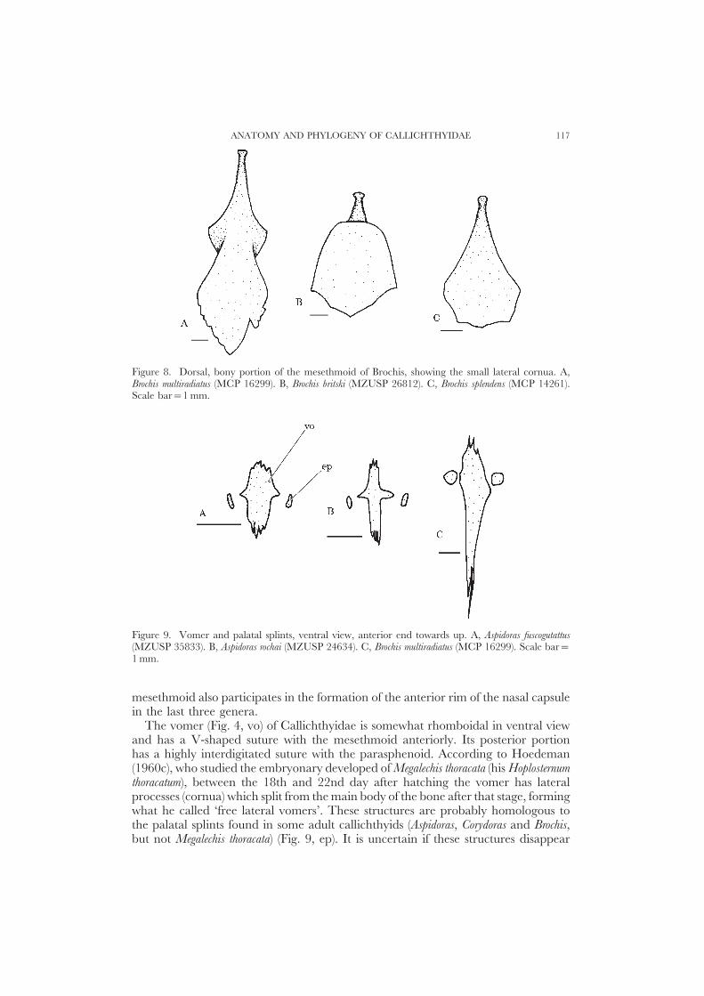
Figure 8. Dorsal, bony portion of the mesethmoid of Brochis, showing the small lateral cornua. A,Brochis multiradiatus (MCP 16299). B, Brochis britski (MZUSP 26812). C, Brochis splendens (MCP 14261).Scale bar=1 mm.
Figure 9. Vomer and palatal splints, ventral view, anterior end towards up. A, Aspidoras fuscogutattus(MZUSP 35833). B, Aspidoras rochai (MZUSP 24634). C, Brochis multiradiatus (MCP 16299). Scale bar=1 mm.
mesethmoid also participates in the formation of the anterior rim of the nasal capsulein the last three genera.
The vomer (Fig. 4, vo) of Callichthyidae is somewhat rhomboidal in ventral viewand has a V-shaped suture with the mesethmoid anteriorly. Its posterior portionhas a highly interdigitated suture with the parasphenoid. According to Hoedeman(1960c), who studied the embryonary developed of Megalechis thoracata (his Hoplosternumthoracatum), between the 18th and 22nd day after hatching the vomer has lateralprocesses (cornua) which split from the main body of the bone after that stage, formingwhat he called ‘free lateral vomers’. These structures are probably homologous tothe palatal splints found in some adult callichthyids (Aspidoras, Corydoras and Brochis,but not Megalechis thoracata) (Fig. 9, ep). It is uncertain if these structures disappear

R. E. REIS118
Figure 10. Parasphenoid of Callichthyidae, ventral view, anterior end towards up. A, Callichthyscallichthys (MCP 7026). B, Lepthoplosternum tordilho (UFRGS 0966). C, Megalechis thoracata (ANSP 165468).D, Dianema urostriata (MZUSP 35573). E, Hoplosternum littorale (SU 59110). F, Aspidoras rochai (MZUSP24634). G, Corydoras ornatus (MCP 14259). H, Brochis multiradiatus (MCP 16299). Scale bar=1 mm.
subsequently in the ontogenetic development or if they are never present in Callichthys,Dianema, Lepthoplosternum, Hoplosternum or Megalechis personata. Lateral cornua of thevomer are present in Diplomystes (Arratia, 1987), †Hypsidoris (Grande, 1987),Ictaluridae (Lundberg, 1982) and in most Siluriformes. The homology of the lateralcornua of the vomer of Siluriformes and the palatal splints of some Callichthyidae,however, is not properly demonstrated.
The parasphenoid (Fig. 4, pa) has deep interdigitated sutures with the vomeranteriorly and with the basioccipital posteriorly. It separates the lateral ethmoids,orbitosphenoids and prootics at the midline. In the posterior half the parasphenoidhas triangular lateral wings, which articulate with the prootics. In Callichthys theparasphenoid is flat (Fig. 4); in Lepthoplosternum, Megalechis, Hoplosternum and Dianemait has a low ridge posteriorly, near the basioccipital. In the remaining genera,however, the parasphenoid is very narrow and shows a strong midline ridge anteriorly(Fig. 10).
The paired orbitosphenoids (Fig. 4, oe) are nearly triangular and slightly concave

ANATOMY AND PHYLOGENY OF CALLICHTHYIDAE 119
in ventral view. There is a suture to the parasphenoid and, apparenty, a medial suturearticulating both orbitosphenoids dorsal to the parasphenoid. The orbitosphenoid hasa partially synchondral articulation with the lateral ethmoid, frontal, pterosphenoidand prootic. In Callichthys the orbitosphenoid is comparatively small and does notform part of the margin of the trigemino-facial and optic foramina, which areseparate in this genus and in Lepthoplosternum and Megalechis. In remaining genera,the pterosphenoid is more laterally situated and the orbitosphenoid forms part ofthe antero-medial edge of the compound trigemino-facial+optic foramen.
The pterosphenoids (Fig. 4, pe) are flat and squarish, forming the antero-dorsal edge of the compound trigemino-facial +optic foramen. Together with theorbitosphenoid, it constitutes most of the orbital medial face.
The paired prootics (Fig. 4, pro) are approximately squarish and form most ofthe floor of the braincase. Anteriorly, the prootic forms the postero-ventral edge ofthe compound trigemino-facial+optic foramen. In all Callichthyidae except Cal-lichthys, the prootic participates in a synchondral articulation with the sphenotic andthe hyomandibula. In Callichthys, the prootic is separated from the hyomandibulararticulation, but possesses a laminar lateral process which contacts the hyomandibula.Posterior to the compound foramen, the prootic possesses a second large foramen,which is absent in Astroblepidae and Trichomycteridae (but is present in Trichomycterussp. MCP 14369) and Scoloplacidae (Schaefer, 1987, 1990). In Loricariidae thereare two foramina in the same position which, according to Schaefer (1987), arepossibly not homologous with the auditory foramen of Characiformes (Weitzman,1962) and recent Clupeomorpha (Fink & Fink, 1981). A small foramen in the lateralportion of the prootic, near the articulation with the hyomandibula, is the passagefor the hyomandibular branch of the facial nerve (Fig. 4, fhnf ).
The exoccipitals (Fig. 4, exo) and the basioccipital are fused in the Callichthyidae,and there is no apparent limit between these bones. The region corresponding tothe exoccipital is sutured to the parasphenoid and synchondrally united to theprootic and pterotic-supracleithrum. A large foramen for the vagous nerve is presentin the midventral portion corresponding to the exoccipital.
The region of the basioccipital (Fig. 4, boc) is anteriorly sutured to the parasphenoidand to the complex vertebral centrum. There is a laminar expansion in the mainbody of the basioccipital, which forms the antero-ventral portion of the swimbladdercapsule. This bony shelf is laterally expanded and joins the dorsal process of thecleithrum (Fig. 4, lb). It is probably homologous to Baudelot’s ligament found inother Siluriformes (Fink & Fink, 1981).
The epioccipitals are relatively small in Callichthyidae, forming the posteriorportion of the braincase. They are synchondrally united to the pterotic-supracleithrumand supraoccipital. It is mostly concealed between the braincase and the swimbladdercapsule and forms part of its anterior wall.
Latero-sensory canals
The branching pattern of the cephalic latero-sensory canals in Callichthyidae issimilar to the general pattern of the Siluriformes, as described by Lundberg(1975) and Schaefer (1988). In most siluriforms the lateral line canal enters thesupracleithrum passing anteriorly to the pterotic, where it splits in the postero-lateralbranch, a small ramification that ends in a pore in the pterotic itself, and in the

R. E. REIS120
Figure 11. Mandible, suspensorium and opercle of Callichthys callichthys (MCP 7026), right side, lateralview, bones of face removed. Scale bar=1 mm.
preopercle-mandibular branch, before proceeding further ahead to form the temporalbranch in the sphenotic. The preopercle-mandibular branch primitively has fivepores in the preopercle (Schaefer, 1988), before entering the mandible. Entering thesphenotic, the temporal canal gives rise to a lateral branch, the infraorbital canal,which varies in extension and number of pores in the many siluriform groups. Insidethe frontal, the temporal canal gives rise to a postero-medial branch, the parietalbranch, with a terminal pore close to the supraoccipital. In addition to thesebranches, the temporal canal has two more openings, one in the mid portion offrontal and other in its anterior portion, where it enters the nasal.
In Callichthys (Fig. 3), Lepthoplosternum, Megalechis, Hoplosternum and Dianema thelateral line canal enters the pterotic-supracleithrum and splits in three branchesbefore entering the sphenotic: the postero-lateral branch, the preopercular branch(this branch never enters the mandible in advanced Loricarioidea [Schaefer, 1988]),and an additional ramification, the ‘antero-lateral branch’. The small antero-lateralbranch is located between the two other, primitive ramifications of the pterotic, andis restricted to the lateral portion of the pterotic-supracleithrum, with a single porenear the lateral edge of the bone. In Corydoras, Aspidoras and Brochis the antero-lateralbranch is absent. Entering the sphenotic, the temporal canal has a lateral branchwith a pore in the bone edge, originating the infraorbital canal. Continuing ontothe frontal, the temporal canal has a single pore and the postero-medial branch islacking. Exiting the frontal, the canal enters the lateral ethmoid and opens in asingle pore near the nasal cavity, entering the nasal bone. In Corydoras, Aspidoras andBrochis the canal does not cross the lateral ethmoid, passing directly from the frontalto the nasal.
In Callichthyidae (Fig. 11) the preopercular sensory canal is represented by acanal segment with three pores in the preopercle. This segment, however, is not

ANATOMY AND PHYLOGENY OF CALLICHTHYIDAE 121
Figure 12. Infraorbital bone series of Callichthyidae, right side, lateral view, anterior portion towardsright. A, Callichthys callichthys (MCP 7026). B, Lepthoplosternum tordilho (UFRGS 0962). C, Megalechisthoracata (NRM 15422). D, Dianema urostriata (MZUSP 35573). E, Hoplosternum littorale (SU 59110). F,Aspidoras fuscogutattus (MZUSP 35833). G, Corydoras barbatus (MCP 10604). H, Brochis multiradiatus (MCP16299). Scale bar=1 mm.
connected to the preopercular branch in the pterotic-supracleithrum. According toSchaefer (1988) the three pores present in Callichthyidae are homologous with pores3, 4 and 5 of primitive Siluriformes.
The infraorbital canal is significantly reduced in callichthyids. In Callichthys (Fig.12), Lepthoplosternum, Megalechis, Hoplosternum and Dianema it is restricted to the secondinfraorbital bone with one single pore in its latero-ventral margin. In Aspidoras,Corydoras and Brochis, the first infraorbital also bears part of the sensory canal andhas two pores.

R. E. REIS122
Infraorbital series
The infraorbital series of Callichthyidae is strongly reduced comparatively toother Siluriformes. Diplomystes, Nematogenys, Copionodon, Trichogenes, as well as mostsiluriforms, possess a well developed infraorbital series and associated sensory canal,ending anteriorly in the lachrimal-antorbital (lachrimal and antorbital are supposedlyfused in Siluriformes [Lundberg, 1970; Schaefer, 1990]). In Trichomycteridae,Scoloplacidae, Astroblepidae and Loricariidae, but not in Callichthyidae, there is asmall bone of uncertain homology associated with the nasal capsule and withoutsensory canal. Such structure is probably homologous with either the lachrimal ormore likely the antorbital of other Ostariophysii. The Callichthyidae has only twoinfraorbital bones and whether these bones are homologous with the two lastinfraorbitals of primitive catfishes (infraorbitals 4 and 5), or represent fused elements,is an uncertain question, and they will be termed first and second infraorbital bonesin this paper.
In Scoloplacidae the infraorbital bones and associated sensory canal are completelylacking. In Astroblepidae and Loricariidae the infraorbital sensory canal is presentand crosses five or six infraorbitals without, however, entering the lachrimal. InCallichthyidae there are only two infraorbital bones and a lachrimal-antorbital isabsent. In some Callichthyidae there are dermal plates on the face, anterior to thefirst infraorbital bone, but these lack a sensorial canal.
In Aspidoras, Corydoras and Brochis the first infraorbital has an anterior laminarprojection which is sutured to the latero-ventral portion of the lateral ethmoid. Inthe remaining genera such a projection is lacking and the first infraorbital articulateswith the lateral ethmoid in its postero-ventral projection, in the orbital rim. InCallichthys, Lepthoplosternum, Megalechis, Hoplosternum, Dianema, Aspidoras and in somespecies of Corydoras (e.g. Corydoras barbatus) the first infraorbital possesses a medial,horizontal laminar projection, variable in size, which forms the floor of the ocularcavity. In Lepthoplosternum, Megalechis, Hoplosternum, Dianema, Aspidoras and in someCorydoras, the second infraorbital also possesses an inner, laminar projection, whichforms the posterior wall of the ocular cavity.
Suspensorium and mandibular arch
The suspensorium of Teleostei is formed by seven bones: hyomandibula, pre-opercle, symplectic, quadrate, metapterygoid, mesopterygoid (=entopterygoid) andectopterygoid (=pterygoid). In Callichthyidae it includes the hyomandibula, quad-rate, preopercle and metapterygoid (the symplectic is absent in all Siluriformes [Fink& Fink, 1981]).
The hyomandibula of Callichthys callichthys (Fig. 11, hio) is nearly rectangular inlateral view, synchondrally articulated with the metapterygoid and quadrate antero-ventrally and strongly sutured to the preopercle postero-ventrally. It is connectedto the neurocranium through a synchondral articulation with the sphenotic and, toa smaller extent, to the pterotic-supracleithrum (Fig. 4, a-hio). In the remaininggenera the prootic is wider than in Callichthys and participates in the synchondralarticulation with the hyomandibula (Fig. 6, a-hio). Antero-medially to the ne-urocranium articulation, the hyomandibula has a small laminar process whichcontacts (but does not articulate with) the prootic. Just above the suture with the

ANATOMY AND PHYLOGENY OF CALLICHTHYIDAE 123
Figure 13. Mandible, suspensorium and opercle of Corydoras ornatus (MCP 14259), right side, lateralview, bones of face removed. Scale bar=1 mm.
preopercle, in the lateral surface, the hyomandibula has a distinct crest for theinsertion of the levator arcus palatini (Fig. 11, clap), which extends dorsally from theopercle articular condyle. The posterior margin of the hyomandibula is almoststraight and continues ventrally to the large opercle articulation condyle. The facialnerve crosses the hyomandibula through a small foramen in its central portion (Fig.11, fhnf ). In Aspidoras, Corydoras and Brochis the hyomandibula is more elongate (Fig.13) and has an interdigitating suture with the metapterygoid anteriorly.
The preopercle of Callichthys (Fig. 11, pop) is elongated in lateral view and stronglysutured to the quadrate and hyomandibula. It extends from the quadrate-mandiblearticulation condyle, passing below the hyosymplectic cartilage and bordering thehyosymplectic fenestra, to near the opercle articulation condyle of the hyomandibula.In the porterodorsal portion the preopercle bears a segment of the latero-sensorycanal with pores showing positional homology with pores 3, 4 and 5 of the preopercle-mandibular sensory canal of primitive Siluriformes (Schaefer, 1988). Contrary tothe condition found in Callichthys, Lepthoplosternum, Megalechis, Hoplosternum and Dianema,where the preopercle is covered with skin, the preopercle is partially exposed in theremaining genera and even bears odontodes in some species. In addition, in Aspidoras,Corydoras and Brochis the preopercle is deeper and meets the hyosymplectic cartilage,closing the hyosymplectic fenestra.
The quadrate of Callichthys (Fig. 11, q) is roughly triangular in lateral view, as inmost Siluriformes (Grande, 1987; Schaefer, 1987). In its postero-dorsal portion thereis a synchondral joint with the metapterygoid and the hyomandibula, and it issutured to the preopercle ventrally. Anteriorly the quadrate bears a large condylefor articulation with the mandible.
Various Siluriformes have lost one or more bones of the pterygoid series and thevariation in size of the remaining bones is usually significant, making statements ofhomology difficult. Loricariidae (Schaefer, 1987), Astroblepidae, Scoloplacidae andCallichthyidae apparently lost the ectopterygoid and mesopterygoid, which are

R. E. REIS124
variably present in other Siluriformes. In Callichthys the metapterygoid (Fig. 11, mpt)is nearly triangular with a posterior projection towards the hyomandibula and ananterior bifurcated projection, ending near the palatine and lateral ethmoid. Postero-ventrally the metapterygoid has a synchondral joint with the hyomandibula andquadrate, besides a small area of contact or even interdigitating suture to thequadrate. In the remaining genera (Fig. 13) there is no posterior projection contactingthe hyomandibula. In Aspidoras, Corydoras and Brochis the metapterygoid articulateswith the hyomandibula by means of an interdigitating suture only. Its anteriorprojection is simple, not approaching the lateral ethmoid, and expanded forward,widely contacting the palatine (especially in Corydoras and Brochis). In one specimenof Aspidoras rochai (MZUSP 24634), however, the anterior projection of the me-tapterygoid is separated from the posterior portion of the bone.
The palatine (Figs 3 and 4, pal) is substantially variable in Callichthyidae. InCallichthys the palatine is small and the process posterior to the lateral ethmoidcondyle is reduced to a small area for the insertion of the extensor tentaculi. Thepalatine has a large cartilagenous condyle anteriorly for the articulation with themaxilla. In the remaining genera the posterior process is small if compared toprimitive Siluriformes (Grande, 1987; Arratia, 1990), but not as reduced as inCallichthys. The palatal splint, present in Loricariidae (Schaefer, 1987), Astroblepidae,and some Trichomycteridae, is variably present in Callichthyidae (see discussion onvomer above).
The maxilla (Figs 3 and 4, mx) is approximately L-shaped. Its medial portion isshorter and provided with a condyle for articulation with the palatine cartilage. Thelateral portion is long and laminar. In Corydoras, Aspidoras and Brochis the maxillarylaminar portion is not as developed as in the remaining genera and there is a smallprocess in the postero-lateral face for insertion of the ratractor tentaculi.
The premaxilla (Figs 3 and 4, pm) is remarkably reduced in size and nearlytriangular in ventral view, with a small dorsal process. As well as in Scoloplacidae,Astroblepidae and Loricariidae (Howes, 1983; Schaefer & Lauder, 1986; Schaefer,1987, 1990) the premaxilla of Callichthyidae is extremely movable due to aligamentous junction with the mesethmoid, contrary to the situation commonlyfound in other Siluriformes. The two premaxillae are united medially by means ofa synchondral joint and have a small postero-ventral crest for the insertion of theabductor muscles. The premaxilla of adult callichthyids has no teeth, but some canbe present in young specimens. One 55.4 mm specimen of C. callichthys has onetooth in each premaxillary bone.
The mandible of Siluriformes is comprised by the dentary (Figs 11 and 13, d)and the angulo-articular bones (Figs 11 and 13, aa). The coronomeckelian bone isalso present in some basal Siluriformes. In Callichthyidae the dentary is elongatedand has a distinct antero-dorsal surface provided with minute conic teeth. In Aspidoras,Corydoras and Brochis the dentary completely lacks teeth. Antero-medially the dentarybears a small condyle for the insertion of the intermandibularis muscle (Figs 11 and13, im). Dorsally the angulo-articular bone has, just behind the junction with dentary,a small coronoid laminar process for the insertion of the adductor mandibulae. Accordingto Schaefer & Lauder (1986) the adductor mandibulae inserts in that process laterallyas well as medially. In the posterior portion the angulo-articular possesses an articularfacet which articulates with the quadrate condyle and, postero-medially, a smallprocess for insertion of the interopercle-mandibular (Fig. 14, lim) and hyoid-mandibular Fig. 14, lhm) ligaments. Medially the mandible has a well developed

ANATOMY AND PHYLOGENY OF CALLICHTHYIDAE 125
Figure 14. Mandible, right side, medial view. A, Corydoras ornatus (MCP 14259). B, Callichthys callichthys(MCP 7026). Scale bar=1 mm.
Meckelian cartilage in the mandibular fossa between the dentary and the angulo-articular.
Opercular series
The opercular series of Siluriformes is composed of the opercle, usually triangularand not squarish as in most other Ostariophysii and basal Teleostei, and by theinteropercle, which is also short in the longitudinal axis and nearly triangular (Fink& Fink, 1981).
The opercle of Callichthys (Fig. 11, op) is ovoid in lateral view and bears welldeveloped odontodes in its posterior margin. In the remaining genera the opercleis variably triangular or ovoid (Fig. 13) and bears no odontodes in its posteriormargin. Antero-dorsally there is the opercular condyle for articulation with thehyomandibular. Above that condyle, in the outer side, there is a small, laminarprocess for the insertion of the dilatator operculi. In the inner surface of the operclethere is a strong crest for insertion of the levator operculi, beginning in the opercularcondyle and progressing backwards. In Corydoras, Aspidoras and Brochis the levatoroperculi crest starts at the opercular condyle and runs postero-ventrally. Antero-ventrally the opercle contacts the interopercle (Figs 11 and 13, iop) which istriangular. The interopercle-mandibular ligament originates at the anterior projectionof the interopercle and inserts in the angulo-articular, medially to its articulationwith the quadrate. In Corydoras, Aspidoras and Brochis the interopercle is much shorter

R. E. REIS126
Figure 15. Hyoid and branchial arches of Callichthys callichthys (MCP 7026), dorsal view, anteriorportion at the foot of the diagram. Branchiostegal rays removed. Scale bar=1 mm.
in its longitudinal axis and the interoperculo-mandibular ligament is consequentlylonger.
Hyoid arch
The hyoid arch in primitive Teleostei and in Diplomystes comprises the pairedinterhyal, posterior ceratohyal, anterior ceratohyal, dorsal hypohyal, ventral hypohyaland the impaired urohyal. Among the Siluriformes many groups have independentlylost the dorsal hypohyal (Weitzman, 1962; Lundberg, 1982), including Tri-chomycteridae, Scoloplacidae, Astroblepidae and Loricariidae. In Callichthyidaethe hyoid arch is composed in the same way but the dorsal hypohyal is present inCorydoras, Aspidoras and Brochis.
The interhyal (Figs 15 and 16, ih) is small and rectangular in Callichthyidae,associated with the medioventral portion of the hyo-symplectic cartilage. Theposterior ceratohyal (Figs 15 and 16, chp) is triangular in lateral view and concavemedially. Laterally it has a small crest for insertion of the hyoid-mandibular ligament.Its articulation with the anterior ceratohyal is synchondral only.
The anterior ceratohyal of Callichthyidae (Figs 15 and 16, cha) is flat antero-dorsally and possesses a large, laminar ventral projection, which is produced in anexpanded cartilage to the union with the posterior ceratohyal. Three cartilagenous

ANATOMY AND PHYLOGENY OF CALLICHTHYIDAE 127
Figure 16. Hyoid arch. A, Callichthys callichthys (MCP 7026), right side,ventral view. B, Corydoras ornatus(MCP 14259), right side, dorsal view, branchiostegal rays removed. Scale bar=1 mm.
projections depart from this cartilagenous expansion and support the four branch-iostegial rays—the two inner and smaller originating from the same projection. Thedistal end of the three outer branchiostegal rays (two outer in Corydoras, Aspidorasand Brochis) are connected to a branchiostegal cartilage (Fig. 16, cab) which isattached to the upper, medial surface of the opercle. This branchiostegal cartilageis present in all Callichthyidae examined. In the antero-dorsal surface of the anteriorceratohyal there is a small foramen for the passage of the afferent mandibularyartery (Bertmar, 1962; Nelson, 1967). The junction between the anterior ceratohyaland the hypohyal is synchondral only.
The ventral hypohyal (Figs 15 and 16, hv) is roughly squarish and synchondrallyconnected to its pair. There is a posterior depression for insertion of the ligamentsoriginating in the anterior projection of the urohyal. In Corydoras, Aspidoras andBrochis there is also a small dorsal hypohyal (Fig. 16, hd) synchondrally united withthe ventral hypohyal and with the anterior ceratohyal.
The urohyal is an unpaired bone, flat and laterally expanded, with a large crestin the ventral surface where the sternohyoideus inserts. Anteriorly the urohyal has twoprocesses which are ligamentously connected with the ventral hypohyals.
Branchial arches
The branchial arches are composed by the basibranchial, hypobranchial, cer-atobranchial, epibranchial, pharyngobranchial and tooth plates. Small gill-rakersare present on the anterior surface of the first two arches, on the posterior surface

R. E. REIS128
of third arch, on both surfaces of fourth arch and on the anterior border or thefifth arch. The second and third arches are more separated between themselvesthan the remaining arches.
In all Callichthyidae there are three basibranchials (Fig. 15, bb), but only theanterior two are ossified.
Five pairs of hypobranchials (Fig. 15, hb) are present. Of these, only the first pairis entirely ossified and possesses an anterior projection which contacts the postero-medial face of the anterior ceratohyal. The second pair of hypobranchials is ossifiedonly anteriorly and forms an anterior projection which contacts the first arch in thearticulation between the basi- and ceratobranchial.
There are five pairs of ceratobranchials (Fig. 15, cb), all entirely ossified, andprogressively more robust posteriorly. The fifth pair of ceratobranchials is expandedto support a lower tooth plate, with small conic teeth on the middorsal surface. Inthe upper end of the fifth ceratobranchial there is a small cartilagenous process (Fig.15, pc5) which contacts the upper extremity of the fourth ceratobranchial.
Four fully ossified epibranchial bones (Fig. 15, eb) occur in Callichthyidae. Theepibranchials in the three first arches have laminar projections in the postero-dorsal surface, variable in shape, which support branchial filaments. The secondepibranchial has a triangular projection on the antero-ventral surface which contactsthe first epibranchial. The first epibranchial of Corydoras, Aspidoras and Brochis alsopossesses a laminar projection of variable size on the anterior face. A smallcartilagenous process attaches on the epibranchial-ceratobranchial articulation ofthe fourth arch (Fig. 15, pc4). It runs posteriorly, parallel to the cartilagenous processof the fifth arch.
In Callichthyidae there are two pair of completely ossified pharyngobranchialbones (Fig. 15, fb). The pharyngobranchial of the third arch is thin and elongatedwith a laminar triangular process directed anteriorly. The pharyngobranchial in thefourth arch is smaller but robust, triangular in dorsal view. In Callichthys, Lep-thoplosternum, Megalechis, Hoplosternum and Dianema there is a small triangular cartilageassociated with the dorsal end of the first two epibranchials (Fig. 15). The natureand positional relationships to the surrounding structures suggest homology ofthis cartilage with the second pharyngobranchial of Diplomystes and other basalOstariophysi.
The upper tooth plate is oval to triangular in dorsal view and bears small conicteeth on its postero-ventral surface. The dorsal portion of this plate is cartilaginousand possesses a pair of shallow depressions for articulation of third and fourthepibranchias.
Weberian apparatus and axial skeleton
The anatomy and function of the Weberian apparatus of the Siluriformes hasreceived a large amount of attention from ichthyologists (Reissner, 1859; Sørensen,1890; Bridge & Haddon, 1894; Chranilov, 1929; Tilak, 1963, 1965; Alexander,1964, 1965; Chardon, 1968; Fink & Fink, 1981). Some features of the Weberianapparatus of the Siluriformes, such as the loss of the intercalarium articular processas vertebral centra 2, 3 and 4 fused into a complex centrum, are specializations ofthese groups (Fink & Fink, 1981). In Loricarioidea, however, the Weberian apparatusis deeply modified compared to the condition found in more basal Siluriformes. In

ANATOMY AND PHYLOGENY OF CALLICHTHYIDAE 129
Figure 17. Exoccipitals, basioccipital, complex centrum and sixth vertebral centrum of Corydoras ornatus(MCP 14259), ventral view, anterior portion uppermost. Scale bar=1 mm.
this group, the gas bladder is reduced in the anterior chamber, as in other Siluriformes(Alexander, 1964) and is divided into two small, laterally symmetrical chambers,encapsulated by expansions of the transversal process of the complex centrum (Figs3 and 4, ptvc). In Callichthyidae there is a delicate duct connecting both gas bladderchambers which originates just behind the os suspensorium (lacking in the Loricarioideaaccording to Arratia [1987], but apparently present in Callichthyidae) and leavingthe gas bladder capsule through a foramen on the medial portion of the complexcentrum transverse process (Fig. 4, fdp), exactly behind the ventral process of thatcentrum (Fig. 4, pvvc). This connecting duct crosses the ventral portion of thecomplex centrum protected by a transversal, double, bony ‘bridge’ (Figs 4, 17). InCorydoras, Aspidoras and Brochis there are two parallel bony laminae on the ventralsurface of the complex centrum, anterior to the transversal bony bridge, that formlateral walls for the aortic canal. In all callichthyids the aortic canal is displacedtowards the left side of the complex centrum, leaving a second passage in the rightside for the right posterior cardinal vein (Figs 4, 17) (Alexander, 1964). The firstvertebral centrum, usually free from the complex centrum as well as from thebasioccipital in most Siluriformes, is fused and incorporated in the WeberianApparatus in the Loricarioidea. The fifth vertebral centrum is separated from thecomplex verebra in most Otophysi (Fink & Fink, 1981) including Diplomystes (Arratia,1987), but is enclosed in the complex centrum in the remaining Siluriformes including+Hypsidoris (Grande, 1987).
Besides the encapsulated gas bladder, the Loricarioidea show additional spe-cializations in the ossicles of the Weberian apparatus. Trichomycteridae, Cal-lichthyidae, Astroblepidae and Loricariidae (Scoloplacidae not seen) have lost theintercalarium, claustrum and the transformator process of the tripus (Baskin, 1972).

R. E. REIS130
Figure 18. Compound tripus of Hoplosternum littorale (MCP 11507). Right side, ventral view. Anteriorportion uppermost. Scale bar=1 mm.
Figure 19. Lateral opening of swim-bladder capsule, right side, lateral view. Lateral body platesremoved. A, Dianema urostriata (MZUSP 35573). B, Corydoras ornatus (MCP 14259). Scale bar=1 mm.
Another interpretation (Schaefer, 1990) suggests that the tripus, intercalarium andscaphium are fused with the interossicular ligaments into a compound tripus, andthat the claustrum disappeared in Scoloplacidae and Callichthyidae. Accordingly,the single ossicle connecting the gas bladder to the inner ear in Callichthyidaeis the fused tripus-intercalarium-scaphium-interossicular ligament, herein calledcompound tripus (Fig. 18, tr). Similarly to the Scoloplacidae (Schaefer, 1990), theposterior branch of the compound tripus projects laterally through a small foramenin the medial wall of the gas bladder capsule and contacts its tunica externa. Theanterior end of the compound tripus, originated from the scaphium, is circular andconcave, and contacts medially the sinus impar. The sinus impar is heavily ossified inCallichthyidae.
Gas bladder contact with the external medium is variable in the Loricarioidea.In Nematogenys, Trichomycteridae and Scoloplacidae the gas bladder capsule isopened laterally. In Astroblepidae and most Loricariidae the lateral portion of thecapsule is formed by the pterotic-supracleithrum, and the hydrostatic communicationis made through perforations in that bone. In Callichthyidae, however, there is ahollow posterior expansion of the pterotic-supracleithrum bearing the latero-sensorycanal, which partially covers the aperture of the gas bladder capsule (Fig. 19).

ANATOMY AND PHYLOGENY OF CALLICHTHYIDAE 131
The sixth centrum of the Callichthyidae is large and bears well developed,triangular-shaped parapophyses. The anterior portion of each parapophysis ar-ticulates with a small posterior process of the complex centrum transversal process,providing additional strength to the vertebral centrum articulation. Laterally, theparapophysis has a large area for rib articulation. The sixth centrum rib is veryrobust and projects postero-ventrally making contact with the inner face of a plateof the inferior lateral series.
In Callichthys callichthys there are 31 preural vertebrae, inclusive of the five vertebraeincorporated into the Weberian complex, and exclusive of the single centrumincorporated into the ural complex. Centra 7 to 14 possess parapophyses, graduallylarger towards caudal fin, for articulation of thin ribs. Centra 15 and 16 have welldeveloped parapophyses but have no associated ribs. In centrum 17 the parapophysesare fused together medially, forming a lamina directed postero-ventrally, whichcontacts the hemal spine of the following centrum. Centra 18 to 31 bear well-developed hemal spines. A neural spine is present on centra 6 to 31, becominggradually thinner caudally. The neural spines associated with the dorsal-fin pter-ygiophores are sometimes slightly bifid distally. Small differences in numbers ofvertebrae and ribs, as well as in the shape of parapophysis, occur in the remaininggenera of the family.
The caudal skeleton of the Siluriformes was studied in detail by Lundberg &Baskin (1969), who identified tendencies of fusion of hypurals and hypurapophysesand reduction of caudal-fin principal and procurrent rays in a variety of groups. Inthe primitive siluriform condition, found in Diplomystes, the parhypural and sixhypurals are free among themselves and from the urostyle, the hypurapophysis isof type ‘A’, and the caudal rays number 9+9. In the Loricarioidea there areprogressive modifications of this pattern (Lundberg & Baskin, 1969). In Callichthyidae(Fig. 20) the parhypural is fused to the hypurals 1 and 2, and the hypurals 3 to 5are fused together with the epural, only a notch between hypurals 2 and 3remaining. The hypurapophysis is of type ‘C’, more common in the Siluriformes.The hypurapophysis is the place of origin of muscles that move the caudal-fin rays,the hypochordal longitudinalis in its dorsal face, and the flexor ventralis in the ventral face(Winterbottom, 1974). Seven dorsal and seven ventral principal rays are present inthe caudal fin of all Callichthyidae. Three dorsal and three ventral procurrent raysoccur in Callichthys callichthys.
Unpaired fins
The dorsal fin of Callichthys comprises a vestigial anterior spinelet, two unbranchedand seven branched rays (Fig. 21). All rays can bear odontodes. The vestigial spineletis supported internally by a pterygiophore composed of a proximal radial only. Thispterygiophore has a small transverse process contacting the second pterygiophore.The two unbranched rays are supported by one single, large pterygiophore, probablyderived from the fusion of pterygiophores 2 and 3, bearing large transverse processes(Fig. 21, ptpd). A strong ligament originates in this process and inserts laterally inthe distal portion of the rib of the sixth centrum. The distal radial of this largepterygiophore is cartilaginous. Each subsequent branched ray is supported by apterygiophore composed of a bony proximal and a cartilaginous distal radial (medialradials are absent in Siluriformes [Fink & Fink, 1981]). The last two branched rays

R. E. REIS132
Figure 20. Caudal-fin skeleton and adipose-fin spine of Callichthys callichthys (MCP 7026), left side,lateral view. Scale bar=1 mm.
Figure 21. Dorsal-fin skeleton of Callichthys callichthys (MCP 7026), right side, lateral view. Scale bar=1 mm.

ANATOMY AND PHYLOGENY OF CALLICHTHYIDAE 133
Figure 22. Anal-fin skeleton of Callichthys callichthys (MCP 7026), right side, lateral view. Scale bar=1 mm.
are supported by one single pterygiophore. The pterygiophores 1 to 5 contact theneural spines of vertebral centra 6 to 10, which are slightly bifid distally. Additionally,there is a small supraneural anterior to the first pterygiophore.
A considerable amount of transformation occurred in the evolution of the dorsal-fin skeleton in the Callichthyidae. Except for Callichthys, all callichthyids have thefirst and second pterygiophores fused dorsally and all remaining pterygiophores(except the last one, usually) have small transverse processes where the dorsal tip ofthe upper lateral plates are attached. In all callichthyids but Callichthys, the fusionof the two first pterygiophores forms a nuchal plate. In Lepthoplosternum, Megalechisand Hoplosternum the nuchal plate is small and covered with skin; in Dianema thenuchal plate is larger, but still covered with skin; in Aspidoras it is big, exposed andarticulated with a plate of the upper lateral series. In Corydoras and Brochis, besidesthe characters present in Aspidoras, the nuchal plate contacts the supraoccipitalprocess anteriorly. In Dianema, Aspidoras, Corydoras and Brochis there is one singleunbranched ray which is transformed into a strong and pungent spine, and theanterior vestigial element forms a locking mechanism for the defensive spine. Usuallyeight branched rays are present in the dorsal fin, except for Brochis which has 10–18branched rays. In Brochis and some species of Corydoras the dorsal spine is serratedin the posterior adge. A supraneural is absent in Aspidoras, Corydoras and Brochis.
The anal fin of Callichthys is comprised of two unbranched and five branched rays(Fig. 22), and all can have odontodes. The two unbranched rays are internallysupported by one single, robust pterygiophore, possibly originated from the fusionof pterygiophores 1 and 2. Each subsequent branched ray is supported by apterygiophore composed of a bony proximal and a cartilaginous distal radial. Thelast two branched rays are supported by a single pterygiophore.
In other genera there are variably five or six branched rays, and the lastpterygiophore supports the last two rays. Additionally, the frst ray is more robustand thickened at the base. In Aspidoras, Corydoras and Brochis, similar to the conditionfound in Callichthys, the first pterygiophore has an anterior laminar expansion. InBrochis the first and sometimes the second unbranched ray is sufficiently thick androbust that it can become pungent.
The adipose fin of the Callichthyidae is formed by one single, spinous bony ray,preceded by a few preadipose plates (Fig. 20, ea). The adipose-fin ray is bifurcatedin its proximal portion and is embedded in the flesh dorsal to the anal fin. In Brochis

R. E. REIS134
Figure 23. Scapular girdle of Callichthys callichthys (MCP 7026). A, right side, ventral view. B, rightside, dorsal view, odontodes and hooks not represented. Scale bar=1 mm.
and in some species of Corydoras its proximal portion is strongly expanded. In thesespecies the adipose-fin spine is seemingly immovable, contrary to the conditionfound in remaining callichthyids, where the adipose-fin spine is depressible. Themovement of the spine is accomplished by two muscular bands (Hoedeman, 1960a).
Pectoral fin and girdle
In the Callichthyidae, similarly to all other Siluriformes, the pectoral girdle consistsof the fused scapula, coracoid and mesocoracoid (scapulocoracoid of Lundberg[1970], here termed coracoid), tightly associated with the cleithrum (Schaefer, 1987).The two halves of the pectoral girdle are articulated ventrally. In the anterior,cleithral portion the articulation is simple, juxtaposing the cleithra. In the posteriorportion, the coracoids are articulated by means of an interdigitated, medial suture.Lateral to this medial articulation there is a large, ovoid fossa (Fig. 23, abf ) for thepassage of the pectoral-fin abductor muscles. The cleithrum (Fig. 23, cl) possesses awell developed vertical lamina laterally, which is exposed above the pectoral-finbase and anteriorly forms the posterior wall of the branchial chamber. In the dorso-lateral portion of this lamina there is a strong vertical process that penetrates aforamen between the ventral face of the pterotic-supracleithrum and the transversalprocess of the complex centrum. The extent of exposition of the coracoid is quite

ANATOMY AND PHYLOGENY OF CALLICHTHYIDAE 135
variable in the Callichthyidae. In Callichthys and Aspidoras the ventral face of thecoracoid is weakly developed and exposed only laterally. In Lepthoplosternum andHoplosternum, the ventral lamina of the coracoid is further developed and is partiallyexposed in the ventral surface. In Dianema, Megalechis and Brochis, the coracoid isextremely developed and broadly exposed ventrally, sometimes leaving only a minutearea of skin on the abdomen. There is a large amount of variation in size andexposition of the coracoid among the species of Corydoras. Unlikely remainingSiluriformes, the posterior processes of the cleithrum and coracoid are suturedbehind the insertion of the pectoral fin in the Callichthyidae, forming a bony shieldaround the entire base of that fin.
The pectoral fin consists of the first unbranched and strongly ossified elementand seven to 10 branched rays. In Callichthys seven branched rays are present andthe unbranched ray is spinous but not pungent, covered with large odontodes. InLepthoplosternum, Megalechis and Hoplosternum the first element is also covered withodontodes and not pungent, but eight to 10 branched rays are present. Theremaining genera also have eight to 10 branched rays (seven in Aspidoras pauciradiatus),but the pectoral spine is covered with minute odontodes and is always pungent,despite some unossified segments being usually present in its distal tip. In Aspidorasthe ossified portion of the pectoral-fin spine is short, attaining about half the lengthof the fin, with an unossified, well developed segmental distal tip. The pectoral-finspine possesses a locking mechanism by friction of its base inside a groove in thepectoral girdle, as described and illustrated by Hoedeman (1960a).
The pectoral-fin rays are supported by two elongated, ossified proximal radialsand four to six distal radials. The first distal radial has a medial cartilaginous portionand a lateral ossified expansion that help supporting the spine. The remaining distalradials are spherical and completely cartilaginous.
Pelvic fin and girdle
The pelvic girdle, deeply modified from the basal pattern of Siluriformes, consistsof two contiguous basipterygia, sutured in the midline, and six pelvic-fin rays.Primitively in Siluriformes the basipterygium is bifid anteriorly (Fink & Fink, 1981;Arratia, 1987; Grande, 1987) with an inner process along the midline and an outerprocess, antero-laterally directed. This is also the condition found in most of theLoricarioidea (modified in Scoloplacidae and in some Loricariidae). Importantmodifications are found in Callichthyidae. The inner anterior process, along themidline, is well developed and extended anteriorly, and has a small dorsal laminarprocess posteriorly directed. The external anterior process of the basipterigium (Fig.24, pae) is strongly reduced and expanded into a lateral lamina, which is connectedto one plate of the lower lateral series of body plates by means of connective tissues.
The ischiac process of the basipterygium is also significantly modified in theCallichthyidae, and is divided in dorsal and ventral portions. The ventral portionis bent antero-ventrally, below the main body of the basipterygium, which is verydeep medially in Callichthyidae. This ventral portion, which is reduced in Brochis,provides a bony anterior wall for the anal papilla. The dorsal portion of the ischiacprocess is bent dorso-laterally, forming a laminar process that connects to the distaltip of a rib (except in Corydoras and Brochis) and to the ventral tip of a plate of thelower lateral series. The dorsal portion of the ischiac process also possesses a large

R. E. REIS136
Figure 24. Pelvic girdle of Callichthys callichthys (MCP 7026), dorsal view, anterior portion uppermost.Scale bar=1 mm.
foramen near its base for the passage of the retractor muscle of the pelvic fin(Hoedeman, 1960a). All pelvic-fin rays bear odontodes. There are no radials in thepelvic fin of Siluriformes (Lundberg, 1970; Schaefer, 1987), but Arratia (1987)describes a small cartilagenous radial in Diplomystes.
The extensive amount of structural changes in the pelvic girdle of the Callichthyidaemay be related to reproductive behaviour. During the spawning, females carry theireggs into a basket formed by the apposition of both anteriorly-turned pelvic fins, asextensively described in the aquarium literature (see Burgess, 1989: 671, 679 forphotographs).
PHYLOGENETIC ANALYSIS OF THE CALLICHTHYIDAE
Efforts to unravel the interrelationships among the subgroups of Callichthyidaeare scarce, and started with Gosline (1940) who regarded five characters of Cascaduraas primitive, and considered this genus to be the ancestor of the whole family.Hoedeman (1952) presented a new classification for the family, arranging its speciesin subfamilies and tribes. Ribeiro (1959) modified the classification of Hoedeman,isolating the genus Callichthys in its own, supposedly primitive, subfamily.
A preliminary analysis of the interrelationships of the Callichthyidae demonstratedthat the genus Hoplosternum, as defined prior to Reis (1997), is not monophyletic.For this reason, all distinct species of Hoplosternum were individually included in thesubsequent analysis, resulting in three monophyletic assemblages of species. Theseassemblages are the present genera Hoplosternum, Megalechis and Lepthoplosternum (Reis,1997).
ANALYSIS OF CHARACTERS
Neurocranium
1. Supraoccipital – fossa. The supraoccipital is usually a strong and compact bone inthe Siluriformes, presenting no openings in its dorsal surface, except for the extended

ANATOMY AND PHYLOGENY OF CALLICHTHYIDAE 137
Figure 25. Mesethmoid of some Siluriformes, dorsal view. A, Diplomystes chilensis, modified from Arratia(1987:22). B, †Hypsidoris farsonensis, modified from Grande (1987:33). C, Nematogenys inermis (USNM259095). D, Trichomycterus sp. (MCP 14369). E, Trichogenes longipinnis (MZUSP 40238). Scale bar=1 mm.
frontal fontanel when present (state 0). In the species of Aspidoras, however, there isa small circular fossa in the posterior portion of the supraoccipital. This fossa islarge and conspicuous in younger individuals but can be strongly reduced or rarelyclosed in large adults, remaining, however, a small depression covered with thickskin (Fig. 7, state 1). Weitzman & Balph (1979) suggested that this fossa may representremains of an elongated frontal fontanel that splits during ontogenetic development.Although there are no indications of an elongated fontanel in very young specimens,development of these features has not been thoroughly studied. A fossa similar tothat in Aspidoras was found in Pimelodus blochii (MZUSP 38236).
2. Frontals – reduction of fontanel. In the species of Aspidoras the frontal fontanel ismarkedly reduced, remaining as small openings before and after the epiphyseal bar(Fig. 5, state 1). The frontal fontanel in the remaining Callichthyidae and otherSiluriformes, when present, is comparatively larger (Fig. 5 state 0).
3. Mesethmoid – lateral expansions. In Siluriformes the mesethmoid bears a pair ofanterior lateral cornua above the premaxillae. Diplomystes (Arratia, 1987), †Hypsidoris(Grande, 1987), Nematogenys and Trichomycteridae (Fig. 25) share this state. Amongthe advanced Loricarioidea, the mesethmoid lateral cornua are exceptionally reducedbut present in Scoloplacidae, Astroblepidae, Loricariidae and in the callichthyidgenus Brochis (Fig. 8, state 0). In the remaining callichthyid genera the cornua areabsent and the mesethmoid ends anteriorly as a sharp tip (Figs 3 and 4, state 1).

R. E. REIS138
Figure 26. Trigemino-facial and optic nerves foramina and surrounding bones, right side, ventralview. A, Dianema urostriata (MZUSP 35573). B, Megalechis thoracata (ANSP 165468). Scale bar=1 mm.
4. Lateral ethmoid – nasal capsule. In Siluriformes the olfatory organ is usually en-capsulated in a cavity variably formed by the frontal, mesethmoid, lateral ethmoid,lachrimal-antorbital and palatine. This condition is found in most Siluriformes,including Nematogenys, Trichomycteridae, Scoloplacidae, Astroblepidae and in thecallichthyid genera Aspidoras, Corydoras and Brochis (state 0). In Callichthys, Lep-thoplosternum, Megalechis, Hoplosternum and Dianema, as well as in the Loricariidae(Schaefer, 1987), the nasal capsule is entirely formed by an expansion of the lateralethmoid (Fig. 3, state 1). The presence of state 1 in callichthyids and loricariids ismost parsimoniously interpreted as convergent.
5. Orbitosphenoid – participation on the trigemino-facial foramen. In Siluriformes the foraminafor the trigemino-facial and the optic nerves are either separate or united in alarge compound foramen called trigemino-facial+optic (Howes, 1983). This largecompound foramen is located between the parasphenoid, orbitosphenoid, ptero-sphenoid and prootic. The participation of the parasphenoid on the margin of thecompound foramen is variable among the Trichomycteridae and does not occur inthe advanced Loricarioidea. Such condition is shared by Loricariidae, Astroblepidaeand most Callichthyidae (state 0). In Callichthys (Fig. 4) the prootic advances ventrallyto meet the pterosphenoid, forming a bony bridge across the compound foramenand separating the trigemino-facial nerve from the optic nerve (Fig. 26, state 1). InScoloplacidae the two foramina are also apparently separated, and the trigemino-facial foramen opens in the prootic (Schaefer, 1990).
6. Prootic – articulation with hyomandibula. Among the Siluriformes the prootic variablyparticipates in the hyomandibular articulation with the neurocranium. Presumably

ANATOMY AND PHYLOGENY OF CALLICHTHYIDAE 139
the most basal condition is the hyomandibula articulating with the synchondral jointbetween sphenotic and the prootic, with small participation of the pterotic and,sometimes, of the pterosphenoid (e.g. Diplomystes [Arratia, 1987]). This condition isshared by Diplomystes, Nematogenys, most Trichomycteridae, Callichthyidae except forCallichthys, Scoloplacidae, Astroblepidae and Loricariidae (Fig. 6, state 0). In Callichthysthe hyomandibular articulation is displaced more laterally and it contacts thesphenotic and the pterotic-supracleithrum only (Fig. 4, state 1). A similar conditionis found in Ictaluridae (Lundberg, 1982) and Trichogenes.
7. Exoccipital – fusion with basioccipital. Diplomystes, Ictaluridae, and most Siluriformespossess free exoccipitals on each side of the basioccipital. This condition is alsoshared by Nematogenys, Copionodon, Trichogenes, Astroblepidae and Loricariidae (state0). In all Callichthyidae, however, the exoccipitals are fused to the basioccipital andthe limits of these bones are not visible (Fig. 4, state 1). A similar condition is foundin Scoloplacidae and was regarded as derived for that family by Schaefer (1990).The resolution of this character in the Loricarioidea phylogeny is ambiguous. Theevolution of state 1 in the ancestor of the advanced loricarioids with subsequentreversal to state 0 in Astroblepidae+Loricariidae is equally parsimonious to theconvergent evolution of state 1 in Callichthyidae and Scoloplacidae.
Latero-sensory canals
8. First lateral-line ossicle – laminar expansion. The lateral-line canal is frequently enclosedby small, tubular ossicles in Siluriformes, including Diplomystes (Arratia, 1987),Nematogenys, Trichogenes and other Trichomycteridae, and Astroblepidae. The lateral-line canal is absent posterior to the pterotic-supracleithrum in Scoloplacidae (Schaeferet al., 1989). In Callichthyidae the first ossicle of the lateral line posterior to thepterotic-supracleithrum is tubular in Callichthys, Aspidoras, Corydoras, Brochis, Megalechisand Lepthoplosternum (Fig. 27, state 0). In Hoplosternum and Dianema the first lateral-line ossicle has laminar expansions around the ossified canal (Fig. 27, state 1).
9. Second lateral-line ossicle – laminar expansion. Similarly to the first, the second lateral-line ossicle of Siluriformes is usually also tubular. With the exception of Callichthysand Lepthoplosternum which share the condition present in most Siluriformes (Fig. 27,state 0), all Callichthyidae possess laminar expansions in the second lateral-lineossicle (Fig. 27, state 1). In Aspidoras, Corydoras and Brochis these expansions areparticularly developed (Fig. 27, state 2). This character was coded as unordered inthe numerical analysis. Laminar expansions in the anterior lateral-line ossiclesare also present in Loricariidae. This similarity, however, is most parsimoniouslyinterpreted as convergent.
10. Lateral line – reduction. In most Siluriformes, as in Nematogenys, Copionodontinae(Pinna, 1992), Trichogenes, Astroblepidae and Loricariidae, the lateral line sensorycanal is variably developed, frequently reaching the caudal peduncle (state 0). InScoloplacidae the lateral-line canal is lacking posterior to the pterotic-supracleithrum,probably due to the general miniaturization that occurred in this group (Schaeferet al., 1989). In Callichthyidae the lateral-line sensory canal is typically reduced toone or two plates of the upper lateral series, posterior to the two ossicles describedabove (state 1). In Hoplosternum littorale, four to six plates bear latero-sensory canals

R. E. REIS140
Figure 27. Two first ossicles in the lateral line of Callichthyidae, left side, lateral view. A, Callichthyscallichthys (MCP 7026). B, Lepthoplosternum pectorale (MHNG 2166.99). C, Megalechis thoracata (USNM264035). D, Dianema urostriata (MZUSP 35573). E, Hoplosternum littorale (MCP 7027). F, Aspidorasfuscogutattus. G, Corydoras ellisae (MCP 15517). H, Brochis multiradiatus (MCP 16299). Scale bar=1 mm.
(state 2). Three or fewer plates with sensory canal were variably found in the speciesof Corydoras. This character was coded as unordered in the numerical analysis.
11. Pterotic-supracleithrum – additional branch of sensory canal. In Aspidoras, Corydoras,Brochis and other Loricarioidea families, the lateral line canal enters the pterotic-supracleithrum and gives rise to two branches before passing onto the sphenotic:the postero-lateral and the preopercle-mandibular branches (Figs 5 and 19, state 0).In Callichthys (Fig. 3), Lepthoplosternum, Megalechis, Hoplosternum and Dianema (Fig. 19)there is a small additional ramification, the antero-lateral branch, between the othertwo and restricted to the lateral portion of the pterotic-supracleithrum, opening inone single pore near the margin of the bone (state 1).
12. Lateral ethmoid – presence of sensory canal. In most Siluriformes the supraorbital canal

ANATOMY AND PHYLOGENY OF CALLICHTHYIDAE 141
crosses the frontal passing directly into the nasal bone, usually with a pore betweenthese bones (state 0). In Callichthys, Lepthoplosternum, Megalechis, Hoplosternum andDianema, however, the frontal is separated from the nasal by a portion of the lateralethmoid. In these genera the supraorbital sensory canal enters the lateral ethmoid(Fig. 4) before continuing to the nasal (state 1).
13. Preopercle – interruption of sensory canal. As a generalized condition in Siluriformesthe preopercular sensory canal originates from a ramification in the pterotic andadvances uninterrupted through the preopercle, with usually five pores beforeentering the mandible (state 0). The preopercular sensory canal in Callichthyidae(Fig. 11) is represented by a segment of canal with three pores showing positionalhomology with pores 3, 4 and 5 of primitive Siluriformes (Schaefer, 1988). Inaddition, this segment is not connected with the preopercle-mandibular ramificationin the pterotic-supracleithrum (state 1).
Infraorbital series
14. Lachrimal-antorbital – absence. In Diplomystes (Arratia, 1987), †Hypsidoris (Grande,1987), Ictaluridae, Nematogenys, Copionodontinae (Pinna, 1992), Trichogenes, otherTrichomycteridae and most Siluriformes, the lachrimal-antorbital is present andusually bears the anterior end of the infraorbital sensory canal. In Loricariidae,Astroblepidae and Scoloplacidae there is a small bone homologous with either thelachrimal or the antorbital of other Ostariophysii with, however, no sensory canal(state 0). In Callichthyidae the lachrimal-antorbital is absent (state 1).
15. Infraorbital bones – reduction. In Diplomystes (Arratia, 1987), †Hypsidoris (Grande,1987), Nematogenys, Trichomycteridae, Astroblepidae and Loricariidae the infraorbitalsensory canal usually crosses five or more infraorbital bones before entering thelachrimal-antorbital, when present (state 0). In Callichthyidae the infraorbital seriesis reduced to two bones only (state 1). Scoloplacidae has no infraorbital sensorycanal.
16. Infraorbital bones – laminar expansion. In most Siluriformes as Diplomystes (Arratia,1987), †Hypsidoris (Grande, 1987), Nematogenys, Trichomycteridae and Astroblepidae,the infraorbital bones are reduced to a bony tube bearing the sensory canal (Fink& Fink, 1981 - state 0). In Scoloplacidae there are no infraobital bones. In allCallichthyidae, however, the bones of the infraorbital series are laminarly expandedaround the tubular portion (state 1). Laminar infraorbital bones are also present inLoricariidae, and an interpretation of independent origin in these two families ismore parsimonious than assuming reversal in Scoloplacidae and Astroblepidae.
17. Infraorbital bones – exposition. In Diplomystes, Nematogenys and in most Siluriformesthe infraorbital bones, when present, are covered by skin and only the sensory canalpores are visible externally. Among the Callichthyidae this condition is shared byCallichthys only (state 0). In the remaining genera the infraorbital bones are exposedon the face and can even bear small odontodes (state 1). Among the advancedLoricarioidea, however, the Loricariidae also has exposed infraorbital bones, butthis similarity is most parsimoniously interpreted as a convergence. The resolutionof this character is ambiguous in the most parsimonious tree. The evolution of state1 in the ancestor of the whole family and subsequent reversion to state 0 in Callichthys

R. E. REIS142
is equally parsimonious with the convergent evolution of the state 1 in Corydoradinaeand in the sister-group of Callichthys.
18. First infraorbital – articulation with lateral ethmoid. In Diplomystes (Arratia, 1987),†Hypsidoris (Grande, 1987), Nematogenys, Trichomycteridae and Astroblepidae thebones of the infraorbital series do not articulate with the lateral ethmoid (state 0).In Callichthyidae the first infraorbital is firmly attached to the latero-posteriorprojection of the lateral ethmoid through a small articular facet in its inner portion(Fig. 12, state 1). In Dianema the first infraorbital has the small articular facet but isconnected to the lateral ethmoid by means of connective tissue only (state 2). Thischaracter was coded as unordered in the numerical analysis.
19. First infraorbital – anterior expansion. In the species of Corydoras, Aspidoras and Brochisthe first infraorbital possesses an anterior expansion forming a large articular facetwith the external border of the lateral ethmoid (Fig. 12, state 1). In the remainingCallichthyidae such expansion is not present and the articulation of the firstinfraorbital with the lateral ethmoid is only via the inner articular facet. Except forthe Loricariidae, the remaining Loricarioidea do not have an articulation betweeninfraorbital bones and the lateral ethmoid (state 0).
20. First infraorbital – inner expansion. The infraorbital bones of Siluriformes, whenpresent, are usually restricted to a bony tube bearing the sensorial canal, or areexpanded in a superficial lamina, as in Callichthyidae and Loricariidae (state 0). InCallichthyidae the first infraorbital is in deep association with the eye and has anaccessory, inner, horizontal laminar expansion (Fig. 12), which forms the floor ofthe ocular cavity (state 1). In Corydoras and Brochis this laminar expansion is smalland restricted to the anterior portion of the first infraorbital (state 2). This characterwas coded as unordered in the numerical analysis.
21. Second infraorbital – inner expansion. Similarly to the first infraorbital, the secondinfraorbital of Loricarioidea is usually tubular or externally flat, without internalexpansion. Among the Callichthyidae, only Callichthys shares this condition (Fig. 12,state 0). In the remaining genera the second infraorbital also possess an accessory,inner, vertical laminar expansion, variably articulated with the hyomandibula,forming the posterior wall of the ocular cavity (Fig. 12, state 1). In the species ofBrochis, however, this inner expansion is small (Fig. 12, state 2). This character wascoded as unordered in the numerical analysis.
22. Second infraorbital – articulation with the sphenotic. In Diplomystes (Arratia, 1987),Nematogenys and remaining Loricarioidea, the last infraorbital, when present, is veryclose to the sphenotic but does not articulate with it (state 0). In Callichthyidae thesecond infraorbital articulates with the sphenotic by means of a small articular facetin its dorsal portion (state 1).
23. Second infraorbital – contact with the pterotic-supracleithrum. In most Siluriformes thebones of the infraorbital series make no contact with the pterotic, a situation sharedby almost all Loricarioidea (state 0). In large specimens of Hoplosternum, Brochis andmost but not all species of Corydoras, however, the second infraorbital possess atriangular posterior expansion contacting the antero-lateral portion of the pterotic-supracleithrum (state 1). In Hoplosternum littorale such expansion is very large andcovers a portion of skin between the infraorbital, the opercle and the pterotic-supracleithrum usually exposed in other Callichthyidae (Fig. 12, state 2). A small

ANATOMY AND PHYLOGENY OF CALLICHTHYIDAE 143
articulation between the last infraorbital and the pterotic-supracleithrum is alsopresent in some representatives of the Loricariidae (e.g. Hypostomus regani), but thenature and position of such articulation is different and it is most parsimoniouslyinterpreted as homoplastic.
Suspensorium and mandibular arch
24. Hyomandibula – suture with the metapterygoid. In Nematogenys, Trichogenes and otherTrichomycteridae (but not all), Scoloplacidae, Callichthys, Lepthoplosternum, Megalechis,Hoplosternum and Dianema, the hyomandibula and the metapterygoid are synchondrallyunited but not sutured (Fig. 11, state 0). Aspidoras, Corydoras and Brochis, however,share a sutural contact between these bones, reinforcing their connection (Fig. 13, state1). As discussed by Schaefer (1990), other apparently unrelated, basal Siluriformes asDiplomystes (Arratia, 1987) and †Hypsidoris (Grande, 1987), among others, have asutural joint between the hyomandibula and the metapterygoid. It is possible thatsuch a joint evolved many times in the Siluriformes but it is certainly derived withinthe Loricarioidea.
25. Hyomandibula – articulation with the infraorbital bones. Among the Loricarioidea andother Siluriformes the hyomandibula variably forms the inner portion of the postero-ventral wall of the orbital cavity. The hyomandibula, however, never articulateswith the infraorbital bone series, a condition shared by Callichthys, Lepthoplosternum,Megalechis, Hoplosternum and Dianema (state 0). In Aspidoras, Corydoras and Brochis thehyomandibula has a small crest in the outer face for articulation with the infraorbitalbones (Fig. 13, aio, state 1).
26. Preopercle – exposition. In Diplomystes, Nematogenys, Copionodontinae (Pinna, 1992),Trichogenes and other Trichomycteridae, Scoloplacidae, Astroblepidae as well as inmost Siluriformes, the preopercle is covered with skin. The same condition occursin Callichthys, Lepthoplosternum, Megalechis, Hoplosternum and Dianema (Fig. 11, state 0).Contrastingly, in Aspidoras, Corydoras and Brochis, the preopercle is partially exposedin the cheek surface (Fig. 13), and even bears small odontodes in some species (state1). In Loricariidae the preopercle may also be partially exposed in the cheek surface.This similarity, however, is most parsimoniously interpreted as a convergence.
27. Hyo-symplectic fenestra – presence. In Diplomystes (Fink & Fink, 1981; Arratia, 1987),and most other Siluriformes, including Nematogenys, Trichomycteridae, Scoloplacidae,Astroblepidae and Loricariidae, the quadrate, hyomandibula and preopercle arecompletely united by a hyo-symplectic cartilage, a condition also shared by Aspidoras,Corydoras and Brochis (Fig. 13, state 0). In Callichthys (Fig. 11), Lepthoplosternum, Megalechis,Hoplosternum and Dianema there is a space between those three bones, delimited bythe hyo-symplectic cartilage, forming a hyo-symplectic fenestra (state 1). In otherunrelated Teleostei (e.g. Platanichthys [Clupeidae], Gymnogeophagus [Cichlidae],Gonorhynchiformes (Chanos Fink & Fink, 1981), Characiformes and Gymnotiformes,a similar fenestra may be present. The fenestra can be plesiomorphic for Siluriformesbut is most parsimoniously interpreted as derived among the Loricarioidea.
28. Metapterygoid – posterior projection. The metapterygoid of Callichthys (Fig. 11) has adorsal projection, extending posteriorly towards the hyomandibula (state 1), whichis not shared by any other Callichthyidae. In callichthyid groups with sutural joint

R. E. REIS144
Figure 28. Portion of suspensorium and mandible of Dianema urostriata (MZUSP 35573), right side,medial view, anterior portion at left. Scale bar=1 mm.
between the metapterygoid and the hyomandibula, Aspidoras, Corydoras and Brochis,as well as in remaining Loricarioidea, the metapterygoid-hyomandibular suture isin the postero-ventral portion of metapterygoid and never in its dorsal portion (Fig.13), and the dorsal posterior projection is absent (state 0).
29. Metapterygoid – suture with the quadrate. In Diplomystes (Fink & Fink, 1981; Arratia,1987), Nematogenys, Trichogenes and other Trichomycteridae and Scoloplacidae, themetapterygoid has a synchondral joint, variable in length, with the dorsal face ofthe quadrate (state 0), a condition shared by all Callichthyidae (Fig. 11) exceptDianema. In Dianema, besides the synchondral joint occupying about three-fourths ofthe posterior articular area, there is a deeply interdigitating suture anteriorly (Fig.28, state 1). The suspensorium of Astroblepidae and Loricariidae is strongly modifiedand the metapterygoid and quadrate are articulated in the entire area of contact.In these two families, however, the profound interdigitation seen in Dianema isabsent. Some other unrelated Siluriformes such as Pimelodus and Parapimelodus (bothPimelodidae) have sutures similar to those found in Dianema, but these are mostparsimoniously interpreted as homoplastic.
30. Metapterygoid – anterior projection. In Callichthys, Lepthoplosternum, Megalechis, Hoplo-sternum and Dianema the metapterygoid has an antero-dorsal laminar projection,directed towards the lateral ethmoid (Fig. 11, state 1). This projection is notarticulated with the lateral ethmoid as in Loricariidae (Schaefer, 1987). In Aspidoras,Corydoras and Brochis, as in other Loricarioidea and other groups of Siluriformes, theantero-dorsal projection is absent (Fig. 13, state 0).
31. Maxilla – process for muscle insertion. Primitively in Siluriformes the retractor tentaculiinserts on the postero-lateral surface of the maxilla (Winterbottom, 1974), near thebase of its distal portion. This condition is shared by Callichthys, Lepthoplosternum,Megalechis, Hoplosternum and Dianema (state 0). In Aspidoras, Corydoras and Brochis there

ANATOMY AND PHYLOGENY OF CALLICHTHYIDAE 145
is a small process in the postero-lateral surface of the maxilla for the insertion ofthe retractor tentaculi (state 1). This muscle seems to be absent in Nematogenys andTrichomycteridae (Schaefer & Lauder, 1986).
32. Premaxilla – reduction. In most Siluriformes the premaxilla is tightly united to themesethmoid, restricting the mobility of the maxillae. The advanced Loricarioideashare highly mobile premaxillae, due to a weak ligamentous junction to themesethmoid (Howes, 1983; Schaefer & Lauder, 1986; Schaefer, 1987, 1990).Additionally, in Callichthyidae the premaxilla is extremely reduced in size (Schaefer& Lauder, 1986), and is usually smaller than the articular proximal portion of themaxilla (Figs 3 and 4, state 1). The premaxilla is never as reduced in otherLoricarioidea and most other Siluriformes (state 0).
33. Premaxilla – absence of teeth. Adult Diplomystes, Nematogenys, other Loricarioidea andmost other Siluriformes have teeth on premaxilla (state 0). In Callichthyidae,premaxillary teeth may be present in young (Machado-Allison, 1986) but never inadults (state 1). Some Loricariidae (e.g. Planiloricaria, Hemiodontichthys) are also devoidof premaxillary teeth, but this similarity is regarded as convergent.
34. Dentary – absence of teeth. Adult Diplomystes, Nematogenys, other Loricarioidea andmost remaining Siluriformes have teeth on dentary, a condition shared by Callichthys,Lepthoplosternum, Megalechis, Hoplosternum and Dianema (state 0). In Aspidoras, Corydorasand Brochis the dentary is edentulous in adults (state 1).
35. Dentary – lateral projection. The dentary of Siluriformes usually bears teeth on thedorsal surface of its distal half, a condition shared with most Loricarioidea (state 0).In Callichthyidae, however, the dentary bears an antero-lateral projection bearingteeth and directed posteriorly, approximately in the middle of the mandible, whichconfers a particular shape to the dentary (Figs 11 and 13, state 1). Even thoseCallichthyidae species without dentary teeth have the projection.
36. Dentary – process for muscle insertion. The intermandibularis is a muscle that extendsbetween the two hemimandibles, invariably inserted on the dentaries (Winterbottom,1974). The dentary of Loricarioidea is usually smooth in its ventral surface, wherethe intermandibularis is inserted (state 0). In Callichthyidae the ventral face of thedentary has a small process, directed medially, for the intermandibularis insertion (Figs11 and 13, state 1).
Opercular series
37. Opercle – crest for muscle insertion. In most Siluriformes, Nematogenys, Trichomycteridae,Scoloplacidae, Astroblepidae and Loricariidae there is a strong crest for insertionof the levator operculi on the inner surface of the opercle, beginning near the condylefor articulation with the hyomandibula and extending postero-dorsally. Among theCallichthyidae this condition is shared by Callichthys, Lepthoplosternum, Megalechis,Hoplosternum and Dianema (state 0). In Aspidoras, Corydoras and Brochis the crest for

R. E. REIS146
Figure 29. Opercle of Callichthyidae, right side, medial view, anterior portion towards left. A,Callichthys callichthys (MCP 7026). B, Lepthoplosternum tordilho (UFRGS 0966). C, Megalechis thoracata (NRM15422). D, Dianema urostriata (MZUSP 35573). E, Hoplosternum littorale (SU 59110). F, Brochis multiradiatus(MCP 16299). G, Aspidoras fuscogutattus (MZUSP 35833). H, Corydoras barbatus (MCP 10604). Scalebar=1 mm.
insertion of the levator operculi is directed postero-ventrally, leaving a larger areadorsally for muscle insertion (Fig. 29, state 1).
38. Opercle – modification in shape. The opercle of Siluriformes is generally triangularin lateral view, with a crest for the insertion of the levator operculi in the inner surface.In Loricarioidea the crest is distant from the dorsal edge of the opercle (see character37, Fig. 29, state 0). In Callichthys the portion of the opercle dorsal to the crest islacking, leaving the crest close to its antero-dorsal margin and conferring an ovoidaspect to the opercle (Fig. 29, state 1).
39. Interopercle – exposition. The interopercle is covered by skin in most Siluriformes,

ANATOMY AND PHYLOGENY OF CALLICHTHYIDAE 147
including Nematogenys. In Trichomycteridae the interopercle bear hypertrophied,exposed odontodes, but remains itself covered by skin. This state is shared by mostCallichthyidae (state 0). In Hoplosternum magdalenae, however, part of the interopercleis exposed and appears on the surface as a small roundish plate ventral to theopercle (state 1).
Hyoid arch
40. Dorsal hypohyal – presence. Teleostei primitively have dorsal and ventral hypohyalbones. In Siluriformes, Diplomystes (Arratia, 1987) has dorsal and ventral hypohyalbones, a condition quite variable among the Siluriformes. Arratia (1987) describeda rudimentary dorsal hypohyal in Nematogenys (referred to as ventral by Arratia), butI was unable to find this bone in the specimen I examined. In Copionodontinae(Pinna, 1992) and remaining Trichomycteridae, Scoloplacidae, Astroblepidae andLoricariidae only the ventral hypohyal is present, a condition shared by Callichthys,Lepthoplosternum, Megalechis, Hoplosternum and Dianema (Figs 15 and 16, state 0). InAspidoras, Corydoras and Brochis there is also a small dorsal hypohyal (Fig. 16, state1). The presence of a dorsal hypohyal is probably a primitive condition for theSiluriformes, but is interpreted as derived within Loricarioidea.
41. Branchiostegal rays – articulation. Siluriformes usually have five or more (about tenin Diplomystes) branchiostegal rays articulating with the posterior surface of anteriorand posterior ceratohyals. In the advanced Loricarioidea only four or fewer branch-iostegal rays are present (Schaefer, 1990), and there is a wide cartilagenous expansionbetween the anterior and posterior ceratohyal bones where the branchiostegal raysare attached via connective tissue (state 0). In Callichthys, Lepthoplosternum, Megalechis,Hoplosternum and Dianema, the branchiostegal rays are attached to three cartilagenousrods articulated with the cartilaginous expansion (Fig. 16, state 1), the most internalrod supporting two branchiostegal rays.
42. Branchiostegal cartilage – presence. Primitively in Ostariophysii the distal extremityof the branchiostegal rays lay close to the dorsal, internal portion of the opercle,inside the branchiostegal membrane. This condition is shared by all Siluriformesexamined (state 0). In Callichthyidae, however, there is a ‘branchiostegal cartilage’attached to the inner, dorsal portion of the opercle. The dorsal tip of the threeexternal branchiostegal rays of Callichthys, Lepthoplosternum, Megalechis, Hoplosternumand Dianema (two in the remaining genera) are attached to this cartilage via connectivetissue (Fig. 16, state 1). In males of Scoloplacidae the distal end of the two externalbranchiostegal rays are expanded and attached to a middorsal process in the opercle(Schaefer, 1990). This similarity, however, is not homologous because it is a bonyprocess of the opercle and not a cartilage in the branchiostegal membrane that isinvolved in the articulation of branchiostegal rays.
Branchial arches
43. First epibranchial – anterior projection. The anterior face of the first epibranchial ofLoricarioidea, Diplomystes (Arratia, 1987) and most Siluriformes if smooth andwithout bony expansion, a condition shared by Callichthys, Lepthoplosternum, Megalechis,

R. E. REIS148
Figure 30. Epibranchials 1 and 2 of Corydoras barbatus (MCP 10604). Scale bar=1 mm.
Hoplosternum and Dianema (state 0). The first epibranchial of Aspidoras, Corydoras andBrochis has a small laminar expansion on its anterior face, near the articulation withthe ceratobranchial, which seems to help supporting the gill rackers (Fig. 30, state1). The first epibranchial of some Loricariidae also has an anterior laminar expansion.The position and orientation of such expansion, however, are distinct in the twofamilies, suggesting non-homology.
44. Second epibranchial – anterior projection. The second epibranchial of Loricarioidea,Diplomystes (Arratia, 1987) and most Siluriformes is usually cylindrical or with laminarexpansions in its postero-dorsal surface for support of branchial filaments (state 0).In Callichthyidae, in addition to these posterior expansions, there is a triangularexpansion in the anterior face of the second epibranchial which approaches andsometimes contacts the posterior face of the first epibranchial (Figs 15 and 30, state1).
45. Second pharyngobranchial – presence. In most basal Ostariophysi, Gymnotoidei andDiplomystes (Arratia, 1987) there are four pairs of pharyngobranchial bones. Inmost Siluriformes and most Loricarioidea, however, only the third and fourthpharyngobranchials are present, a condition shared by Aspidoras, Corydoras and Brochis(state 0). In Callichthys, Lepthoplosternum, Megalechis, Hoplosternum and Dianema there isa small triangular cartilage associated with the dorsal extremity of the first andsecond epibranchial bones. The position and nature of such a structure indicatesprobable homology with the second pharyngobranchial of Diplomystes and otherOstariophysi (Fig. 15, state 1).
Weberian apparatus
46. Aortic canal – bony walls. The aortic canal leaves the gills and runs ventrally tothe complex vertebra, passing through a bony ‘bridge’ formed by the ventral processesof the complex centrum in Nematogenys, Trichomycteridae and Callichthyidae. Thisbridge apparently disappeared or is modified in the Scoloplacidae+ Astroblepidae+ Loricariidae. In Aspidoras, Corydoras and Brochis, there are two longitudinal bony

ANATOMY AND PHYLOGENY OF CALLICHTHYIDAE 149
shafts anterior to the ventral processes of the complex centrum, forming lateral wallsfor the aortic canal (Fig. 17, state 1).
47. Gas bladder capsule – lateral opening. In Loricarioidea the gas bladder is divided intwo small lateral chambers, encapsulated by expansions of the transverse process ofthe complex centrum. In Nematogenys, Copionodontinae (Pinna, 1992) and otherTrichomycteridae, and Scoloplacidae the gas bladder capsule is open laterally (state0). In Callichthyidae, however, there is a hollow posterior expansion of the pterotic-supracleithrum bearing the latero-sensory canal, which partially covers the apertureof the gas bladder capsule (Fig. 19, state 1). In Astroblepidae and most Loricariidaethe lateral aperture of the capsule is completely covered by the pterotic-supra-cleithrum, and the hydrostatic communication is made through perforations in thatbone. The condition found in Astroblepidae and Loricariidae is possibly homologouswith the one found in Callichthyidae, each clade showing its own subsequentmodification of the structure.
Unpaired fins
48. Supraneural – presence. A small free supraneural, probably homologous with thefifth supraneural of basal Teleostei, since supraneurals 1–4 are included in theWeberian apparatus or disappeared in Ostariophysi (Fink & Fink, 1981; Arratia,1987), is present in Diplomystes and various other Siluriformes. Among the Lo-ricarioidea, a free supraneural is absent in Nematogenys, Trichomycteridae, Sco-loplacidae, Aspidoras, Corydoras and Brochis (state 0). In Callichthys, Lepthoplosternum,Megalechis, Hoplosternum and Dianema there is a free laminar supraneural in front ofthe first dorsal pterygiophore (Fig. 21, state 1). One supraneural is also presentin Astroblepidae and Loricariidae (secondarily disappeared or fused to the firstpterygiophore in some groups, e.g. Hypostomus [Schaefer, 1987]). Accordingly, thepresence of a supraneural is probably primitive for Siluriformes, but is consideredderived within the Loricarioidea.
49. Dorsal radials – transversal processes. Except for Callichthys, where the upper lateralplates are distant from the base of the dorsal-fin rays (state 0), all remainingCallichthyidae have small transversal processes on the proximal radial bones ofdorsal fin. These small transversal processes provide an articular area for the dorsalend of the upper lateral plates (state 1). Similar processes are present in Loricariidae(Schaefer, 1991), and articulate with the dermal plates adjacent to the dorsalfin. Since these processes are absent in Scoloplacidae, Astroblepidae and basalLoricarioidea, their evolution must have occurred independently in Callichthyidaeand Loricariidae. The resolution of this character is ambiguous in the shortestcladogram found in the present study. The derived loss of the transversal processesin Callichthys (reversal to state 0) after its evolution in the ancestor of the entirefamily (node A), is equally parsimonious with the convergent evolution of theprocesses in Corydoradinae and in the Scoloplacidae+Astroblepidae+Loricariidae.
50. Nuchal plate – exposition. The presence of a nuchal plate formed by expansions ofthe first dorsal pterygiophores (sometimes also of the supraneural) is quite variableamong the Siluriformes (e.g. Doradidae, Ictaluridae, Pimelodidae, Auchenipteridae).In Loricarioidea, a nuchal plate formed by the two first pterygiophores is present

R. E. REIS150
in Callichthyidae, Scoloplacidae and Loricariidae. This nuchal plate, however, issometimes covered by the skin and predorsal scutes, as in Scoloplacidae, Callichthys,Lepthoplosternum, Megalechis, Hoplosternum and Dianema (state 0), or exposed right infront of the dorsal fin, as in Aspidoras, Corydoras, Brochis and Loricariidae (state 1).The exposition of the nuchal plate is most parsimoniously interpreted as convergentin Callichthyidae and Loricariidae.
51. Nuchal plate – contact with supraoccipital bone. Among the Loricarioidea onlyCallichthyidae, Scoloplacidae and Loricariidae have a nuchal plate, usually smalland not extended anteriorly (state 0). The nuchal plate of Corydoras and Brochis,however, is markedly extended anteriorly, and contacts the posteriorly extendedsupraoccipital bone (Fig. 7, state 1).
52. Dorsal-fin rays – number. Six through eight branched dorsal-fin rays seems to bethe plesiomorphic condition in Siluriformes, a count seen in Diplomystes, Nematogenys,most Trichomycteridae, Callichthyidae, Astroblepidae and Loricariidae (state 0). InScoloplacidae there are only four branched dorsal-fin rays. Among the Callichthyidae,however, the species of Brochis have 10 through 18 branched dorsal-fin rays (state1).
53. Anal-fin rays – number. Diplomystes (Arratia, 1987), Nematogenys, Trichogenes and otherTrichomycteridae, and most Callichthyidae share the plesiomorphic presence oftwo (sometimes more) unbranched rays preceding the branched anal-fin rays (state0). The species of Lepthoplosternum possess only one simple ray preceding the branchedones in the anal fin (state 1). The presence of one single unbranched ray in the analfin also occurs in some species of Corydoras. In the group formed by Scoloplacidae,Astroblepidae and Loricariidae there is also a single unbranched ray in the leadingedge of the anal fin, but it is typically large and bears odontodes. The reductionfrom two to one unbranched ray in the anal fin inScoloplacidae+Astroblepidae+Loricariidae and in Lepthoplosternum is most par-simoniously interpreted as convergent.
54. Caudal fin – shape. The shape of the caudal fin is extremely variable amongSiluriformes. Diplomystes, †Hypsidoris (Grande, 1987) and most of the remainingSiluriformes have a forked or bilobed caudal fin. Among the Loricarioidea, Nematogenysand various other Trichomycteridae have a rounded caudal fin, but Trichogenes andother Trichomycteridae present a bilobed caudal fin. In Copionodontinae (Pinna,1992) the caudal fin is slightly bilobed. Among the advanced Loricarioidea, thecaudal fin is rounded in Scoloplacidae and truncated or emarginated in Astroblepidaeand Loricariidae. Because of the high degree of variation among the Loricarioidea,it is problematic to estimate an ancestral state for the outgroup node, but therounded shape can be regarded as plesiomorphic for the ancestor of all Callichthyidae.Callichthys, Lepthoplosternum and Megalechis have a rounded caudal fin (state 0). Dianema,Hoplosternum, Aspidoras, Corydoras and Brochis have a bilobed caudal fin shape (state1).
Pectoral fin and girdle
55. Cleithrum/coracoid – articulation of posterior processes. The development and degreeof exposition of the bones in the pectoral girdle of Siluriformes show wide variation

ANATOMY AND PHYLOGENY OF CALLICHTHYIDAE 151
Figure 31. Pectoral-fin spines of mature males, right side. A, Hoplosternum littorale (ANSP 133888). B,Megalechi thoracata (NMW 46261). Scale bar=1 mm.
among various families. The posterior processes of the cleithrum and coracoid bonesare variably exposed in the advanced Loricarioidea (absent in Astroblepidae) and,except for the Callichthyidae, they are never sutured laterally behind the insertionof the pectoral fin (state 0). In all Callichthyidae the posterior processes of thecleithrum and coracoid bones are extremely developed and sutured to each otherlateroventrally, posterior to the pectoral-fin base (Fig. 23, state 1).
56. Coracoid — development. The degree of development of the posterior process ofthe coracoid is largely variable among the Siluriformes. The process is not developedin basal Loricarioidea, contrarily to the condition found in the advanced Lo-ricarioidea, where only the Astroblepidae lack such processes. In Scoloplacidae andLoricariidae the posterior processes are developed but without medial expansionscovering part of the abdomen (state 0), a condition shared also with Callichthys andAspidoras. In remaining Callichthyidae there are medial expansions of the posteriorprocess of the coracoid, exposed ventrally, and partially covering the abdomen (state1). In Brochis, Dianema, Megalechis, H. magdalenae and H. punctatum, however, the medialexpansions are especially developed, covering most of the skin between the pectoralfins and sometimes overlapping in the midline in fully developed mature males (state2).
57. Pectoral-fin spine – sexual dimorphism. Secondary sexual dimorphism on the extentof development of pectoral-fin spine is not known in males of Nematogenys, Co-pionodontinae, Trichogenes, Scoloplacidae or Astroblepidae. Some Loricariidae showa special development of pectoral-fin spines of males involving overall size andhypertrophied odontodes. In the Callichthyidae, males generally have the pectoral-fin spine slightly longer or stronger than females (state 0). In Megalechis and H.littorale, however, the pectoral spine undergoes drastic elongation during reproductiveseason, usually reaching beyond the middle of the pelvic fin (Fig. 31, state 1). InHoplosternum littorale the tip of the fully developed pectoral-fin spine has a dorsaltwist, forming a distinctive hook (state 2). The unusual development of the pectoral-fin spine in mature males of a few species of Corydoras (C. octocirrus, p.e.) is bestinterpreted as a convergence.
58. Pectoral-fin spine – shortening. In most Siluriformes and Loricarioidea the pectoral-fin spine is approximately as long as or even longer than the subsequent branched

R. E. REIS152
rays, a condition shared by most Callichthyidae (state 0). In Aspidoras the ossifiedportion of the pectoral-fin spine is short though strong and pungent, and has a welldeveloped segmentary, unossified distal tip. The pungent, ossified portion of thespine is about half the length of the first branched ray (state 1).
59. Pectoral fin – adipose body. Contrary to the remaining Loricarioidea and otherCallichthyidae, mature males of Callichthys, Lepthoplosternum, Megalechis, Hoplosternumand Dianema have a thick adipose body on the ventral surface of the pectoral fin(Fig. 32, state 1). This soft, whitish, adipose tissue appears only during the reproductiveseason on the ventral face of each branched ray. The aquarium literature refers to‘adipose’ tissue, but histologic cuts of pectoral fins of mature males of Hoplosternumlittorale and Dianema longibarbis revealed the presence of two kinds of tissues: adipose andglandular. The function of this structure is unknown but production of pheromone forthe reproductive activity is a possibility.
Pelvic fin and girdle
60. Pelvic fins – skin fold. The first unbranched pelvic-fin ray of the Loricarioidea,including most Callichthyidae, has no skin folds besides the interradial membrane(state 0). Mature females of Hoplosternum, Megalechis and Dianema have a small foldof skin on the external face of the leading pelvic-fin ray. The function of this skinfold is uncertain but it is probably used during the process of carrying eggs with thepelvic fins during spawning (Fig. 33, state 1).
61. Basipterygium – deepening. Diplomystes (Arratia, 1987), †Hypsidoris (Grande, 1987)and most Siluriformes, as well as Nematogenys, Trichomycteridae, Scoloplacidae,Astroblepidae and Loricariidae share flat basipterygia (state 0). The basipterygiumof Callichthyidae is strongly deepened medially, forming a large ventral cavity (Fig.24, state 1). Scoloplax dolicholophia also has a ventral cavity in the pelvic girdle(Schaefer, 1990) but with distinct position and morphology from that found in theCallichthyidae, evidence of non-homology.
62. Basipterygium – external anterior process. Primitively the basipterygium of Siluriformesif bifid anteriorly (Fink & Fink, 1981; Arratia, 1987; Grande, 1987), bearing twodiverging anterior processes. Two anterior processes in the basipterygium is thecondition found in most Loricarioidea (secondarily modified in Scoloplacidae andsome Loricariidae) (state 0). In Callichthyidae, the external process is very reducedand modified into a lateral lamina, not extended anteriorly, and connected to theinner surface of a plate of the lower lateral series via connective tissue (Fig. 24, state1).
63. Basipterygium – internal anterior process. In Diplomystes (Arratia, 1987), most Siluriformesand Loricarioidea except Callichthyidae, the internal anterior process of the ba-sipterygium is approximately cylindrical or horizontally laminar (state 0). In Cal-lichthyidae the well developed internal processes of basipterygia are very close inthe midline and have a small dorsal, laminar process directed backward (Fig. 24,state 1).
64. Basipterygium – ischiac process. The ischiac process of the basipterygium is usuallyflat and variably elongated posteriorly in Siluriformes and most Loricarioidea (state

ANATOMY AND PHYLOGENY OF CALLICHTHYIDAE 153
Figure 32. Pectoral-fin adipose body of a reproductive male of Hoplosternum littorale (MCP 10541). A,scale bar=100 lm, arrow indicates a branched ray transverally cut. B, scale bar=100 lm. C, scalebar=10 lm.
0). In Callichthyidae the ischiac process is divided in two portions. The dorsalportion is bent dorso-laterally and the ventral one antero-ventrally, ventral to themain body of the basipterygium and delimiting the anterior wall of the anal papilla(Fig. 24, state 1).

R. E. REIS154
Figure 33. Pelvic fin of a female of Hoplosternum magdalenae (NRM 15409), right side, ventral view.Arrow indicates the skin fold present in nuptial females. Scale bar=1 mm.
65. Basipterygium – connection with a rib. The pelvic girdle of Teleostei is primitivelynot connected to the axial skeleton, a condition shared with all Ostariophysiexamined (state 0). In Callichthyidae, however, the dorsal portion of the ischiacprocess is physically associated with the distal portion of a rib, establishing aconnection between the pelvic girdle and the axial skeleton (state 1). In Corydorasand Brochis the ribs are comparatively shorter and the contact with the basipterygiumis absent. The resolution of this character in the most parsimonious tree is ambiguous,because the derivation of the state 1 in the ancestor of all Callichthyidae and thesubsequent reversal to state 0 in the clade Corydoras plus Brochis is as parsimoniousas the convergent evolution of state 1 in Aspidoras and in Callichthyinae.
Other characters
66. Dermal plates – pattern. All Callichthyidae have the body almost completelyenclosed in a bony armour comprising two longitudinal series of plates. Since amongadvanced Loricarioidea only Astroblepidae lacks dermal plates, it is possible thatthese plates evolved in their common ancestor, having subsequently disappeared inAstroblepidae and thus being a synapomorphy for all advanced Loricarioidea. Theinterpretation of the presence of dermal plates as a homologous character is moreparsimonious than the independent evolution in Callichthyidae, Scoloplacidae andLoricariidae. The arrangement of these plates, however, is unique for each familyand two longitudinal series are found only in Callichthyidae (state 1).
67. Lower lip – shape. The shape of the lower lip is highly variable among Loricarioideadue to their many feeding specializations. The more generalized shape, however, isthat found in Nematogenys, Copionodontinae (Pinna, 1992), Trichogenes and Scolo-placidae, which consists of a skin fold directed backwards and sometimes notchedmedially (Fig. 34, state 0). Among the Callichthyidae, this state is shared by Callichthys,

ANATOMY AND PHYLOGENY OF CALLICHTHYIDAE 155
Figure 34. Lower lip of some Loricarioidea, ventral view. A, Trichogenes longipinnis (MZUSP 40238).B, Lethoplosternum beni (USM 304582). C, Dianema urostriata (MZUSP 14260). D, Corydoras paleatus (MCP14270). Scale bar=1 mm.
Hoplosternum and Megalechis. Three distinct modifications of this plesiomorphic con-dition appeared in the Callichthyidae. Aspidoras, Corydoras and Brochis have a smallbarbel of variable size in each lobe of the lower lip (Fig. 34, state 1). The species ofDianema have two small barbels in each lobe of the lower lip (Fig. 34, state 2).Finally, Lepthoplosternum has an additional lateral notch, forming a small fleshyprojection turned laterally, near the medial notch (Fig. 34, state 3). This characterwas coded as unordered in the numerical analysis.
68. Maxillary barbels – length. In Diplomystes and most Loricarioidea the maxillarybarbels are short and rarely reach beyond the gill openings (state 0). In Callichthys,Lepthoplosternum, Megalechis, Hoplosternum and Dianema, however, the maxillary barbelsreach the origin of the pelvic fins (pectoral fins in Dianema) (state 1).
69. Coloration – pattern in young. Young individuals of Callichthyidae, as in many otherSiluriformes groups, frequently exhibit a colour pattern distinct than that found inthe adults. Among the Callichthyinae, young specimens (about 15–20 mm SL) of

R. E. REIS156
Figure 35. Characteristic colour pattern of young Hoplosternum species (MZUSP 23423). Scale bar=1 mm.
Hoplosternum, Megalechis and Lepthoplosternum have a light-brown ground with four orfive dark transverse stripes between the dorsal-fin origin and the caudal fin (Fig. 35,state 1). Young individuals of Callichthys, Aspidoras, Corydoras and Brochis have a colourpattern similar to that found in the adult or a particular pattern different than theone described above (state 0). The colour pattern of young Dianema and Hoplosternumpunctatum were not examined and this character is coded as ‘missing’ for these taxain the numerical matrix.
70. Hydrostatic balance – maintenance via air breathing. With the exceptions of Clariidaeand Heteropneustidae, the Siluriformes are usually incapable of breathing at-mospheric air. There are no records of air breathing in Nematogenys, Astroblepidaeand Scoloplacidae (state 0). The Callichthyidae, on the other hand, are air-breathersand the air is collected in the water surface and swallowed. Their ‘accessoryrespiratory organ’ is the intestine, and the air is eventually expelled through theanus. In this family, however, the air swallowed plays a more important role in themaintenance of hydrostatic balance than in the respiration itself, contributing toabout 75% of the necessary air to attain neutral buoyancy (Gee, 1976; Gee & Graham,1978; state 1). Some representatives of the Loricariidae and Trichomycteridae (Gee,1976; pers. obs.), also disply air-breathing capabilities. In such cases, however, therespiration takes place in the stomach and the air is later released through themouth and gill openings. Additionally, contrary to the Loricariidae and Tri-chomycteridae that breathe air only in case of hypoxia, the Callichthyidae breathair under all water conditions.
71. Nest for spawning – construction. Most Siluriformes lay their eggs on substrates likerocks, logs, leaves (some Loricariidae), breed them into the mouth cavity (Ariidae),or even in nests on muddy bottom (some Pimelodidae). Among the Callichthyidae,Aspidoras, Corydoras and Brochis are substrate brooders (state 0). Callichthys, Leptho-plosternum, Megalechis, Hoplosternum and Dianema, on the other hand, build a floatingnest composed of foam and vegetal debris for spawning (Burgess, 1987, 1989;Machado-Allison & Zaret, 1984; Winemiller, 1987) (state 1).
72. Association with the bottom – absence. Diplomystes, Nematogenys and most Tri-chomycteridae, as well as the Scoloplacidae, Astroblepidae and Loricariidae typicallydwell in the bottom of water bodies, where they usually feed. This condition isshared by most Callichthyidae (state 0) except for the species of Dianema and for a

ANATOMY AND PHYLOGENY OF CALLICHTHYIDAE 157
few Corydoras (C. hastatus and C. pygmaeus), which spend most of the time swimmingin midwater, distant from the bottom (Burgess, 1987, 1989) (state 1). This behaviouris most parsimoniously interpreted as independently derived in those two Corydoras(which are sister-species [Schaefer et al., 1989]) and in Dianema.
In addition to the characters above, Schaefer (1990) suggested one more syn-apomorphy for the Callichthyidae:
In most siluroids, Nematogenys, and trichomycterids the adductor mandibulae is littledifferentiated, no part of which inserts directly or indirectly on the premaxillae(state 0). In callichthyids the adductor is slightly subdivided. A ventro-lateral portionlies lateral to the ramus mandibularis nerve trunk and inserts on the lower jaw,while the more massive medial portion lies medial to the ramus mandibularis andinserts onto the complex ligamentum primordium. This ligament has an anteriorportion which involves the maxilla, premaxilla and palatine. Antero-dorsal fibersof the adductor approach the premaxilla, but make no direct insertion (state 1). Inscoloplacids, astroblepids and loricariids the fibers insert directly onto the premaxilla. . . (state 2).
Schaefer (1990), however, examined only representatives of Callichthys, Corydoras,Dianema, Megalechis and Hoplosternum. Since I was unable to examine this characterin additional species, it was not included in my numerical analysis, but it may bean additional synapomorphy for the entire family Callichthyidae.
CLADISTIC ANALYSIS
Cladogram topology
One single most parsimonious tree was found by the PAUP ‘Branch and bound’algorithm, with 94 steps and consistency index of 0.861 (Fig. 36). The character-state changes can be included in three categories, all interpreted as evolutionarynovelties: state changes that occurred only once, changes involving reversals to a moreprimitive condition and changes including independent evolution (convergences). Inthe cladograms (Figs 36, 37) changes uniquely derived among the Loricarioidea areindicated by a solid bar, and changes involving reversals or convergence within theLoricarioidea are represented by an open bar. Convergence and reversal within thefamily Callichthyidae are indicated in the cladograms by the letters ‘C’ and ‘R’. Ofthe 72 characters analysed only ten (3, 5, 9, 17, 21, 23, 49, 54, 56 and 65) showedhomoplastic state distributions (Fig. 36).
The preferred optimization option was ACCTRAN, which seems to be the-oretically superior since it maximizes the homoplastic character changes that arerepresented as reversals, rather than as parallelisms, thus minimizing the falsificationof the initial hypothesis of homology (primary homologies), and better fitting thefundamental relationship between homology and synapomorphy (Pinna, 1991). Theresult of the optimization using ACCTRAN is shown in the cladogram of Figure36. Three characters show ambiguous distributions in the most parsimonious tree(17, 49 and 65). The alternative distribution of these characters was mapped on theshortest cladogram and is shown in Figure 37.

R. E. REIS158
49.0 R
Callic
hthys
38.128.121.0 R17.0 R6.1
67.353.1
69.156.1 C
60.156.2 C
57.1
54.1 C9.1 C8.15.0 R
23.1 C
72.167.229.118.2
57.256.1 R23.210.2 39.1
58.12.11.1
65.0 R56.1 C51.123.1 C20.2
56.2 C52.121.23.0 R
71.168.159.148.145.141.130.127.112.111.15.14.1
67.154.1 C50.146.143.140.137.134.131.126.125.124.119.19.2 C
70.166.165.164.163.162.161.155.149.147.144.142.136.135.133.132.122.121.120.118.117.116.115.114.113.110.17.13.1
Lepth
oplos
ternum
Meg
alec
his
Dianem
a
Hoplos
ternum
litto
rale
Aspid
oras
Coryd
oras
Broch
is
Hoplos
ternum
mag
dalen
ae
Hoplos
ternum
puncta
tum
B
C
D
E
F
G
H
A
NTSAL
Figure 36. Relationships among genera and species-groups of Callichthyidae (consistency index=0.861; length=94 steps). NTSAL are the initials of the remaining families of Loricarioidea. Charactersderived only once within the Loricarioidea are mapped on the cladogram as black rectangles; openrectangles indicate characters derived more than once within the superfamily. Homoplastic states areindicated by ‘C’ (convergences or parallelisms) and ‘R’ (reversals). The numbers of characters andtheir states (separated by a decimal), correspond to the sequence presented in the text.

ANATOMY AND PHYLOGENY OF CALLICHTHYIDAE 159
Callic
hthys
49.1 C
Lepth
oplos
ternum
Meg
alec
his
Dianem
a
Hoplos
ternum
litto
rale
Aspid
oras
Coryd
oras
Broch
is
Hoplos
ternum
mag
dalen
ae
Hoplos
ternum
puncta
tum
NTSAL
17.1 C
65.1 C
49.1 C17.1 C
65.1 C
G
HF
E
D
C
B
A
Figure 37. Equally parsimonious alternative resolution of characters, mapped on the shortest treetopology. See Fig. 36 for a description of symbols and text for description of the characters.
Zero branch-lengths
A zero branch-length is assigned by PAUP to any branch where no charactertransformation is detected for that particular taxon. The implications of zero branch-lengths for phylogenetic analysis were revised by Coddington & Scharff (1994).There are two branches with length zero in the shortest tree: Hoplosternum punctatum,which could not be diagnosed by an autapomorphic feature, and the genus Corydoras.

R. E. REIS160
In the first case, there are no implications for the validity of the assemblages withinthe family, but a zero branch-length for Corydoras has further implications. The factthat no synapomorphies were found to diagnose Corydoras, as traditionally defined(Gosline, 1940), suggests that this genus may be paraphyletic. The possible para-phyletic nature of Corydoras, however, is a consequence of recognizing Brochis as aseparate genus. When apomorphic members of a monophyletic group are placedin a separate higher taxon of coordinate rank with the non-apomorphic members,the result is usually a paraphyletic group (Schaefer, 1987). Fink & Fink (1986) andSchaefer (1987) described two similar situations among the Stomiiformes andLoricariidae, respectively. The conservative approach to treat this situation is torefute the genus Brochis, synonymizing it under Corydoras. If this approach is chosen,however, the information about the monophyletic nature Brochis will be lost and thegenus Corydoras will be even more complex. Additional phylogenetic studies areneeded within Corydoras in order to uncover its monophyletic subgroups and possiblysplit Corydoras in more than one monophyletic genus. The genus Brochis is likely tosurvive this procedure.
Comments on the Corydoras species groups of Nijssen (1970) and Nijssen & Isbrucker(1980)
The species of Corydoras were arranged by Nijssen (1970) into nine morphologicalgroups based mainly on coloration and biometric features, although four speciescould not be assigned to any of the nine groups:
(1) Corydoras punctatus group includes 34 species with dark punctuations on bodyand head;
(2) C. barbatus group includes 16 species with a colour pattern formed by brownishspots on the dorso-lateral scutes alternating with similar spots on the ventro-lateralscutes;
(3) C. aeneus group includes six species with greyish-blue plain coloration on dorsalportion of body;
(4) C. nattereri group includes eight species showing a dark stripe on junction ofdorsal and ventral lateral scutes;
(5) C. eques group comprises seven species with a lateral dark stripe on upperportion of flanks;
(6) C. acutus group includes six species with greyish coloration, serrated pectoral-fin spine and elongated snout;
(7) C. elegans group, with three species, shows various longitudinal brown stripeson the lateral scutes;
(8) C. caudimaculatus group includes only two species with a large dark blotch onthe caudal peduncle;
(9) C. hastatus group comprises three species sharing a distinctive small body size.Nijssen & Isbrucker (1980) reviewed the genus Corydoras and rearranged the 94
then known species into five species-groups: C. punctatus Group (31 species), C.barbatus Group (11 species), C. aeneus Group (25 species), C. elegans Group (8 species),and C. acutus Group (19 species).
It is possible that some of these groups, or even subgroups, with evident mor-phological uniformity, represent monophyletic assemblages of species, but no explicitattempt to objectively investigate their monophyly has been made to date and no

ANATOMY AND PHYLOGENY OF CALLICHTHYIDAE 161
evidence for monophyly of any of the species groups in presently known. Thegeographic distribution of some of the smaller groups of Nijssen (1970)—especiallythe C. eques group (upper Amazonas and Orinoco drainages), C. hastatus group(Madeira and Paraguay River drainages), and C. caudimaculatus group (MadeiraRiver)—is coincident with recently documented distribution patterns of other fishes.Some of these include: Steindachnerina guentheri (Vari, 1991) and Cyphocharax festivus(Vari, 1992) in the upper Amazonas and Orinoco River drainages; Gymnogeophagusbalzanii (Reis & Malabarba, 1988) and Psectrogaster curviventris (Vari, 1989) in theMadeira and Paraguay River drainages; besides a series of species endemic to theMadeira River (e.g. Lepthoplosternum beni [Reis, 1997], Brachychalcinus copei [Reis,1989], Xenurobrycon polyancistrus [Weitzman, 1987]). This superposition could indicatethe existence of general patterns of geographic distribution, suggesting a startingpoint to the objective analysis of the monophyly of the Corydoras subgroups.
As discussed before, the genus Corydoras is possibly not monophyletic and thepresent knowledge of its species interrelationships (including those presently assignedto Brochis) is far from ideal. For this reason, the Corydoras species-groups discussedabove should not be taken for granted in phylogenetic studies, despite their utilityfor species identification.
KEY FOR GENERA OF CALLICHTHYIDAE
1. Dorsal face of lateral ethmoid bearing a segment of the supraorbital latero-sensorycanal; preopercle covered with skin; dentary with teeth; nuchal plate covered withskin; snout depressed; maxillary barbel long, usually extending beyond gill opening(Callichthyinae) ...........................................................................................................21′. Dorsal face of lateral ethmoid without latero-sensory canal; preopercle exposed;dentary without teeth; nuchal plate exposed; snout compressed; maxillary barbelshort, usually not extending beyond eye Corydoradinae) ........................................62. Infraorbital bones covered with skin; head very depressed, its depth less than75% of cleithral width; coracoids not exposed ventrally ........................... Callichthys2′. Infraorbital bones exposed; head moderately depressed, depth more than 75%of cleithral width; coracoids exposed ventrally .........................................................33. Caudal fin bifurcated or concave .........................................................................43′. Caudal fin truncate or convex .............................................................................54. First infraobital bone weakly connected to the lateral ethmoid; dorsal-fin spineas long as the first branched rays .................................................................. Dianema4′. First infraorbital bone articulated to the lateral ethmoid; dorsal-fin spine abouthalf the length of the first branched rays ............................................... Hoplosternum5. Dorsal fin with one spine, one unbranched ray and 7–8 branched rays; anal finwith two unbranched and 5–6 branched rays; mature males with very elongatedpectoral-fin spine ......................................................................................... Megalechis5′. Dorsal fin with one spine and 7 branched rays; anal fin with one unbranchedand 5 branched rays; mature males with pectoral-fin spine thickened but notparticularly elongated ..........................................................................Lepthoplosternum6. Roundish supraoccipital fossa present; ossified portion of pectoral- and dorsal-finspines short, about half length of first branched rays; nuchal plate not in contactwith the posterior process of the supraoccipital ........................................... Aspidoras

R. E. REIS162
6′. Supraoccipital fossa absent; ossified portion of pectoral- and dorsal-fin spinesabout as long as the subsequent branched rays; nuchal plate in contact with theposterior process of the supraoccipital ......................................................................77. Dorsal fin with 7–9 branched rays ...........................................................Corydoras7′. Dorsal fin with 10–18 branched rays ......................................................... Brochis
ACKNOWLEDGEMENTS
I thank Naercio A. Menezes for his guidance and support for this research, whichformed a portion of a Ph.D. dissertation at the Universidade de Sao Paulo. I amvery grateful to the many people and institutions that made specimens available forthis study: Barbara Herzig (NMW), Barry Chernoff (FMNH), Brian Dyer (UMMZ),Carl Ferraris, Jr. (AMNH), Claude Weber (MHNG), David Catania (CAS), DouglasNelson (UMMZ), Eugenia Bohlke (ANSP), Gabriela Piacentino (MACN), HanNijssen (ZMA), Hans-Joachim Paepke (ZMB), Heraldo A. Britski (MZUSP), HoracioHiguchi (MCZ), Isaac Isbrucker (ZMA), Jaime Sarmiento (MNHN, Bolivia), JoseFigueiredo (MZUSP), Karsten Hartel (MCZ), Mario de Pinna (AMNH), Mary AnneRogers (FMNH), Naercio Menezes (MZUSP), Richard Vari (USNM), RichardWinterbottom (ROM), Scott Schaefer (ANSP), Sonia Muller (MHNG), StanleyWeitzman (USNM), Susan Jewett (USNM), Sven Kullander (NRM), Tomio Iwamoto(CAS), Ulisses Caramaschi (MNRJ), William and Sara Fink (UMMZ), WilliamEschmeyer (CAS) and William Saul (ANSP). Mario de Pinna and Scott Schaeferreviewed the manuscript and provided many insightful suggestions. I am also thankfulto Claude Weber, Claudia de Melo, Dione Seripierri, Flavio Bockmann, GordonHowes, Han Nijssen, Isaac Isbrucker, Jose Carlos Oliveira, Lee Finley, MartaZamana, Martine Desoutter, Mary Anne Rogers and Richard Vari for providingliterature and other information.
For their hospitality during my stay in many cities in research connected withthis study I am very grateful to Barry Chernoff (Chicago), Carl Ferraris Jr. (NewYork), Francisco L. Franco and Maria Ines Sani Franco (Sao Paulo), KarstenHartel (Cambridge), Paulo Buckup and Marcia Velho (Ann Arbor), Richard Vari(Washington), Scott Schaefer (Philadelphia), Stanley and Marilyn Weitzman (Wash-ington), and Tomio Iwamoto (San Francisco). Technical assistance and support wasprovided by Carla Fontana (MCP), Edson Pereira (MCP), Edson Vidal (MCP),Gizelani Guazzelli (MCP), Jose Pezzi da Silva (MCP) and Osvaldo Oyakawa(MZUSP).
Portions of this research were supported by grants from CAPES – FundacaoCoordenacao do Aperfeicoamento de Pessoal de Nıvel Superior (1990–1993); CNPq– Conselho Nacional de Desenvolvimento Cientıfico e tecnologico; FAPERGS –Fundacao de Amparo a Pesquisa do Rio Grande do Sul; and Pontifıcia UniversidadeCatolica do Rio Grande do Sul, Pro-Reitoria de Pesquisa e Pos-Graduacao.
REFERENCES
Alexander, R.McN. 1964. The structure of the Weberian apparatus in the Siluri. Proceedings of theZoological Society of London 142: 419–440.

ANATOMY AND PHYLOGENY OF CALLICHTHYIDAE 163
Alexander R.McN. 1965. Structure and function in the catfish. Journal of Zoology 148: 88–152.Arratia G. 1987. Description of the primitive family Diplomystidae (Siluriformes, Teleostei, Pisces):
morphology, taxonomy and phylogenetic implications. Bonner Zoologische Monographien 24: 1–120.Arratia G. 1990. Development and diversity of the suspensorium of trichomycterids and comparison
with loricarioids (Teleostei: Siluriformes). Journal of Morphology 205: 193–218.Arratia G, Menu-Marque S. 1984. New catfish of the genus Trichomycterus from the high Andes of
South America (Pisces, Siluriformes) with remarks on distribution and ecology. Zoologische JahrbuchSystematics 111: 493–520.
Bailey RM, Baskin JN. 1976. Scoloplax dicra, a new armored catfish from the bolivian amazon.Occasional Papers of the Museum of Zoology University of Michigan 674: 1–14.
Baskin JN. 1972. Structure and relationships of the Trichomycteridae. Ph.D. Thesis, The CityUniversity of New York, 389 pp.
Bertmar G. 1962. On the ontogeny and evolution of the arterial vascular system in the head of theafrican characidean fish Hepsetus odoe. Acta Zoologica 43: 255–294.
Bleeker MP. 1862. Atlas ichthyologique des Indes Orientales Neerlandaises. Tome II. Siluroıdes, Characoıdes etHeterobranchoıdes. Amsterdam: Frederic Muller.
Bonaparte CL. 1838. A new systematic arrangement of vertebrated animals. Transactions of the LinneanSociety of London 18(3): 247–304.
Bridge TW, Haddon AC. 1894. Contributions to the anatomy of fishes. II. The air-bladder andWeberian ossicles in the Siluroid fishes. Philosophical Transactions of the Royal Society of London 184(B):65–434.
Brooks DR, O’Grady RT, Wiley EO. 1986. A measure of the information content of phylogenetictrees, and its use as an optimality criterion. Systematic Zoology 35: 571–581.
Burgess WE. 1987. Corydoras and raleted catfishes – a complete introduction. Neptune City, NJ: TFHPublications
Burgess WE. 1989. An atlas of freshwater and marine catfishes. A preliminary survey of the Siluriformes. NeptuneCity, NJ: TFH Publications.
Carpenter JM. 1988. Choosing among multiple equally parsimonious cladograms. Cladistics 4:291–296.
Chardon M. 1968. Anatomie comparee de l’appareil de Weber et des structures connexes chez les Siluriformes.Musee Royal de l’Afrique Centrale, Annales, Serie 8, Sciences Zoologiques, n. 169, 277p.
Chranilov NS. 1929. Beitrage zur Kenntnis des Weber’schen Apparates der Ostariophysi. 2. DerWebersche Apparat bei Siluriformes. Zoologische Jahrbucher. Abteilung fur Anatomie und Ontogenie der Tiere51: 323–462.
Coddington J, Scharff N. 1994. Problems with zero-length branches. Cladistics 10: 415–423.Ellis MD. 1913. The plated nematognths. Annals of the Carnegie Museum 8(3–4): 384–413.Eigenmann CH. 1910. Catalogue of the fresh-water fishes of tropical and south temperate America.
Reports of Princeton University Expeditions to Patagonia 1896–1899. 3(4): 375–484.Eigenmann CH, Eigenmann RS. 1890. A revision of the south american Nematognathi or catfishes.
Occasional Papers of the California Academy of Sciences 1: 1–508.Eldredge N. Cracraft J. 1980. Phylogenetic patterns and the evolutionary process. New York: Columbia
Univ. Press.Fink SV, Fink WL. 1981. Interrelationships of the ostariophysan fishes (Teleostei). Zoological Journal
of the Linnean Society 72: 297–353.Fink WL, Fink SV. 1986. A phylogenetic analysis of the genus Stomias, including synonymization of
Macrostomias. Copeia 1986: 494–503.Gee JH. 1976. Buoyancy and aerial respiration: factors influencing the evolution of reduced swim-
bladder volume of some Central American catfishes (Trichomycteridae, Callichthyidae, Loricariidae,Astroblepidae). Canadian Journal of Zoology 54(7): 1030–1037.
Gee JH, Graham JB. 1978. Respiratory and hydrostatic functions of the intestine of the catfishesHoplosternum thoracatum and Brochis splendens (Callichthyidae). Journal of Experimental Biology 74: 1–16.
Gill T. 1872. Arrangement of the families of fishes, or Classes Pisces, Marsipobranchii, and Leptocardii. Washington:Smithsonian Institution
Gosline WA. 1940. A revision of the Neotropical catfishes of the family Callichthyidae. StanfordIchthyological Bulletin 2(1): 1–36.
Grande L. 1987. Redescription of+Hypsidoris farsonensis (Teleostei: Siluriformes), with a reassessmentof its phylogenetic relationships. Journal of Vertebrate Paleontology 7: 24–54.
Gunther ACLG. 1864. Catalogue of the fishes in the British Museum. Catalogue of the Physostomi, containing

R. E. REIS164
the families Siluridae, Characinidae, Haplochitonidae, Sternoptychidae, Scopelidae, Stomiatidae in the collection ofthe British Museum. v.5 i–xxii+1–455p.
Hennig W. 1966. Phylogenetic Systematics. Urbana: Univ. Illinois Press.Hoedeman JJ. 1952. Notes on the ichthyology of Surinam (Dutch Guiana). The catfish genera
Hoplosternum and Callichthys, with key to the genera and groups of the family Callichthyidae. Beaufortia12: 1–12.
Hoedeman JJ. 1960a. Studies on callichthyid fishes. 3. Notes on the development of Callichthys (1)(Pisces, Siluriformes). Bulletin of Aquatic Biology 1(9): 53–72.
Hoedeman JJ. 1960b. Studies on callichthyid fishes. 4. Development of the skull of Callichthys andHoplosternum (1) (Pisces, Siluriformes). Bulletin of Aquatic Biology 1(10): 73–84.
Hoedeman JJ. 1960c. Studies on callichthyid fishes. 5. Development of the skull of Callichthys andHoplosternum (2) (Pisces, Siluriformes). Bulletin of Aquatic Biology 2(13): 214–36.
Hoedeman JJ. 1960d. Studies on callichthyid fishes. 6. The axial squeleton of Callichthys andHoplosternum (Pisces, Siluriformes). Bulletin of Aquatic Biology 2(13): 21–36.
Hoedeman JJ. 1960e. The status of the Callichthyid genus Cascadura Ellis, 1913 (Pisces, Siluriformes,Callichthyidae). Bulletin of Aquatic Biology 1(2): 15–19.
Howes GJ. 1983. The cranial muscles of loricarioid catfishes, their homologies and value as taxonomiccharacters (Teleostei: Siluriformes). Bulletin of the British Museum of Natural History (Zoology) 45(6):309–345.
Howes GJ, Teugels GG. 1989. Observations on the ontogeny and homology of the pterygoid bonesin Corydoras paleatus and some other catfish. Journal of Zoology 219: 441–456.
Lundberg JG. 1970. The evolutionary history of North American catfishes, family Ictaluridae. Ph.D.dissertation, Univ. of Michigan.
Lundberg JG. 1975. Homologies of the upper shoulder girdle and temporal region bones in catfishes(order Siluriformes), with comments on the skull of the Hologeneidae. Copeia 1975: 66–74.
Lundberg JG. 1982. The comparative anatomy of the toothless lindcat, Trogloglanis pattersoni Eigenmann,with a phylogenetic analysis of the ictalurid catfishes. Miscelaneous Publications of the Museum of ZoologyUniversity Michigan 163: 1–85.
Lundberg JG, Baskin JN. 1969. The caudal skeleton of the catfishes, order Siluriformes. AmericanMuseum Novitates 2398: 1–49.
Machado-Allison A. 1986. Aspectos sobre la historia natural del ‘currito’ Hoplosternum littorale(Hancock, 1828) (Siluriformes, Callichthyidae) en el bajo llano de Venezuela: desarrollo, alimentaciony distribucion espacial. Acta Cientıfica Venezolana 37: 72–78.
Machado-Allison A. Zaret TM. 1984. Datos sobre la biologia reproductiva de Hoplosternum littorale(Siluriformes, Callichthyidae) de Venezuela. Acta Cientıfica Venezolana 35: 142–146
Maddison W.P, Donoghue MJ, Maddison DR. 1984. Out-group analysis and parsimony. SystematicZoology 33: 83–103.
Nelson GJ. 1967. Branchial arteries on the teleostean fish Elops hawaiiensis. Zoologisch Beitrage 13:137–148.
Nelson JS. 1984. Fishes of the world. New York: John Wiley.Nelson GJ, Platnick N. 1981. Systematics and biogeography: cladistics and vicariance. New York: Columbia
Univ. Press.Nijssen H. 1970. Revision of the Surinam catfishes of the genus Corydoras Lacepede, 1803 (Pisces,
Siluriformes, Callichthyidae). Beaufortia 18(230): 1–75.Nijssen H, Isbrucker IJH. 1979. Chronological enumeration of nominal species and subspecies of
Corydoras (Pisces, Siluriformes, Callichthyidae). Bulletin of Zoology Museum University Amsterdam 6(17):129–135.
Nijssen H. Isbrucker IJH. 1980. A review of the genus Corydoras Lacepede, 1803 (Pisces, Siluriformes,Callichthyidae). Bijdrague Dierkunde 50(1): 190–220.
Nijssen H. Isbrucker IJH. 1983. A review of the genus Corydoras from Colombia, with descriptionsof two new species (Pisces, Siluriformes, Callichthyidae). Beaufortia 33(5): 53–71.
Nijssen H, Isbrucker IJH. 1986. A review of the genus Corydoras from Peru and Ecuador (Pisces,Siluriformes, Callichthyidae). Studies of the Neotropical Fauna & Environment 21(1/2): 1–68.
Pinna MCC. 1991. Concepts and tests of homology in the cladistic paradigm. Cladistics 7: 367–394.Pinna MCC. 1992. A new subfamily of Trichomycteridae (Teleostei, Siluriformes), lower Loricarioid
relationships, and a discussion on the impact of additional taxa for phylogenetic analysis. ZoologicalJournal of the Linnean Society 106:
Reis RE. 1989. Systematic revision of the Neotropical characid subfamily Stethaprioninae (Pisces:Characiformes). Comunicacoes do Museu de Ciencias da PUC-RS, serie Zoologia 2(6): 3–86.

ANATOMY AND PHYLOGENY OF CALLICHTHYIDAE 165
Reis RE. 1997. Revision of the neotropical catfish genus Hoplosternum (Ostariophysi, Siluriformes,Callichthyidae), with the description of two new genera. Ichthyological Explorations of Freshwaters 7(4):299–326.
Reis RE, Malabarba LR. 1988. Revision of the Neotropical cichlid genus Gymnogeophagus Ribeiro,1918, with descriptions of two new species (Pisces, Perciformes). Revista Brasileira de Zoologia 4(4):259–305.
Reissner E. 1859. Ueber die Schwimmblase und den Gehorapparat einiger Siluroiden. ArchivesAnatomie und Physiologie 1859: 421–438.
Ribeiro PM. 1959. Consideracoes sobre Callichthyidae Gill, 1872 (Nematognathi). Boletim do MuseuNacional, nova serie 206: 1–9.
Schaefer SA. 1984. Mechanical strength of the pectoral spine/girdle complex in Pterygoplichthys(Siluriformes: Loricariidae). Copeia 1984: 1004–1008.
Schaefer SA. 1987. Osteology of Hypostomus plecostomus (Linnaeus), with a phylogenetic analysis of theloricariid subfamilies (Pisces: Siluriformes). Contributions in Sciences, Natural History Museum of Los AngelesCounty 394: 1–31.
Schaefer SA. 1988. Homology and evolution of the opercular series in the loricarioid catfishes (Pisces:Siluriformes). Journal of Zoology 214: 81–93.
Schaefer SA. 1990. Anatomy and relationships of the scoloplacid catfishes. Proceedings of the Academyof Natural Sciences of Philadelphia 142: 167–210.
Schaefer SA. 1991. Phylogenetic analysis of the loricariid subfamily Hypoptopomatinae (Pisces:Siluriformes: Loricariidae), with comments on generic diagnoses and geographic distribution.Zoological Journal of the Linnean Society 102: 1–41.
Schaefer SA, Lauder GV. 1986. Historical transformation of functional design: evolutionarymorphology of feeding mechanisms in loricarioid catfishes. Systematic Zoology 35(4): 489–508.
Schaefer SA, Weitzman SH, Britski HA. 1989. Review of the Neotropical catfish genus Scoloplax(Pisces: Loricarioidea: Scoloplacidae) with comments on reductive characters in phylogeneticanalysis. Proceedings of the Academy of Natural Sciences of Philadelphia 141: 181–211.
Sørensen W. 1890. Om Forbeninger i Svommeblaeren, Pleura og Aortas Vaeg og Sammensmeltningderaf med Hvirvelsajlen saerlig hos Siluroiderne samt de saakaldte Weberske Knoglers Morfologi.Koninklijl Danske Videnskabernes 6: 65–152.
Swofford DL. 1993. PAUP – Phylogenetic Analysis Using Parsimony – version 3.1. Computer programdistributed by the Illinois Natural History Survey, Champaign Illinois.
Taylor WR, Van Dyke GC. 1985. Revised procedures for staining and clearing small fishes andother vertebrates for bone and cartilage study. Cybium 9(2): 107–119.
Tilak R. 1963. The osteocranium and the Weberian apparatus of the fishes of the family Schilbeidae(Pisces: Siluriformes). Proceedings of the Zoological Society of London 143: 1–36.
Tilak R. 1965. The comparative morphology of the osteocranium and the Weberian apparatus ofTachysuridae (Pisces: Siluriformes). Journal of Zoology 146: 150–174.
Vari RP. 1989. Systematics of the Neotropical characiform genus Psectrogaster Eigenmann & Eigenmann(Pisces: Characiformes). Smithsonian Contributions to Zoology 481: 1–43.
Vari RP. 1991. Systematics of the Neotropical characiform genus Steindachnerina Fowler (Pisces:Ostariophysi). Smithsonian Contributions to Zoology 507: 1–118.
Vari RP. 1992. Systematics of the Neotropical characiform genus Cyphocharax Fowler (Pisces: Os-tariophysi). Smithsonian Contributions to Zoology 529: 1–137.
Weitzman SH. 1962. The osteology of Brycon meeki, a generalized characid fish, with an osteologicaldefinition of the family. Stanford Ichthyological Bulletin 8: 1–77.
Weitzman SH. 1987. A new species of Xenurobrycon (Teleostei: Characidae) from the Rıo Mamorebasin of Bolivia. Proceedings of the Biological Society of Washington 100(1): 112–120.
Weitzman SH. Balph MH. 1979. Some phylogenetic implications of a discovery of Aspidoraspauciradiatus (Pisces: Siluriformes: Callichthyidae) from the rio Negro in Brazil. Proceedings of theBiological Society of Washington 92(1): 10–22.
Wiley EO. 1981. Phylogenetics – the theory and practice of phylogenetic systematics. New York: John Wiley.Winemiller KO. 1987. Feeding and reproductive biology of the currito, Hoplosternum littorale, in the
Venezuelan llanos with comments onthe possible function of the enlarged male pectoral spines.Environmental Biololgy of Fishes 20(3): 219–227.
Winterbottom R. 1974. A descriptive synonymy of the striated muscles of the Teleostei. Proceedingsof the Academy of Natural Sciences of Philadelphia 125: 225–317.

R. E. REIS166
APPENDIX
Specimens examined for comparative purposes and used as outgroups. Abbreviations are: c&s, clearedand counterstained for bone and cartilage; d, dearticulated skeleton; a, dissected in alcohol; e, examinedfor external characters only; ∗, histological cut of the pectoral fin of a mature male.
ClupeiformesClupeidae
Platanichthys platana MCP 14488 (2 c&s).Cypriniformes
CyprinidaeNotropis hudsonius MCP 14730 (2 c&s).Notemigonus crysoleucas MCP 14731 (2 c&s).Pimephales promelas MCP 10975 (2 c&s).
CharaciformesCharacidae
Charax stenopterus MCP 9013 (1 c&s).Poptella brevispina MCP l0969 (2 c&s).
SiluriformesGymnotoidei
GymnotidaeGymnotus carapo MCP 8550 (1 c&s).
SternopygidaeHypopomus sp. MCP 13643 (1 c&s).
RhamphichthyidaeEigenmannia virescens MCP 14918 (2 c&s).
SiluriformesDiplomystidae
Diplomystes viedmensis MCP 14069 (1 e).Ictaluridae
Noturus insignis MCP 14727 (1 c&s).Pylodictis olivaris MCP 14736 (1 c&s).
AriidaeNetuma barba MCP 07910 (2 c&s).
BagridaeChandramara chandramara MZUSP 41322 (1 c&s).
AspredinidaeBunocephalus iheringii MCP 14169 (1 c&s).
AuchenipteridaeAuchenipterus sp. MCP 13556 (1 c&s).Tatia boemia MCP 12949 (2 c&s).
PimelodidaeHeptapterus mustelinus MCP 12723 (2 c&s).Iheringichthys labrosus MCP 13203 (1 c&s).Pimelodus blochii MZUSP 38236 (1 c&s).Parapimelodus valenciennis MCP 10246 (1 c&s), MCP 10545 (1 c&s).Parapimelodus nigribarbis MCP 7053 (11 c&s).
NematogenyidaeNematogenys inermis USNM 259095 (1 c&s).
TrichomycteridaeAcanthopoma sp. (grupo bondi) MZUSP 30421 (1 c&s).Homodiaetus vazferreirai MCP 14181 (2 c&s).Ochmacanthus sp. MZUSP 30477 (1 c&s).Paracanthopoma sp. MZUSP 30404 (2 c&s).Pareiodon sp. MZUSP 23336 (2 c&s).Pseudostegophilus maculatus MZUSP (1 c&s).Pseudostegophilus nemurus MZUSP 30427 (2 c&s).Scleronema sp. MCP 14139 (1 c&s).

ANATOMY AND PHYLOGENY OF CALLICHTHYIDAE 167
Trichogenes longipinnis MZUSP 40238 (2 c&s).Trichomycterus sp. MCP 14369 (2 c&s).
CallichthyidaeAspidoras albater MCP 15974 (1 c&s).Aspidoras fuscoguttatus MZUSP 35833 (3 c&s).Aspidoras menezesi [paratype] UMMZ 195951 (1 c&s).Aspidoras pauciradiatus AMNH 58399 (3 d).Aspidoras cf. poecilus MCP 16301 (3 c&s), MZUSP 40650 (4 c&s).Aspidoras cf. rochai MZUSP 24634 (4 c&s).Aspidoras virgulatus USNM 279266 (1 d).Brochis britskii [paratype] MZUSP 26812 (1 c&s).Brochis multiradiatus MCP 16299 (1 e), MCP 16302 (1 c&s, 4 e).Brochis splendens MCP 14261 (1 c&s), USNM 284627 (5 e).Callichthys callichthys MCP 7026 (1 c&s), MCP 10193 (1 c&s), MCP 10282 (5 c&s).Callichthys personatus ANSP 165468 (2 c&s).Corydoras barbatus MCP 10604 (4 c&s).Corydoras ehrardti MCP 14354 (2 c&s).Corydoras elegans MCP 14248 (2 c&s).Corydoras ellisae MCP 15517 (2 c&s).Corydoras hastatus MCP 15521 (4 c&s).Corydoras leucomelas MCP 14249 (1 c&s).Corydoras nattereri MCP 14980 (1 c&s), MCP 14259 (2 c&s).Corydoras paleatus MCP 14835 (2 c&s).Corydoras panda MCP 14257 (2 c&s).Corydoras punctatus MCP 16138 (2 c&s).Corydoras rabauti MCP 14258 (1 c&s).Corydoras undulatus MCP 13954 (1 c&s e 1 d).Corydoras cf. xinguensis MCP 15633 (3 c&s).Corydoras sp. ‘A’ MCP 15699 (3 c&s).Corydoras sp. ‘B’ MCP 15632 (3 c&s).Corydoras sp. ‘C’ MCP 15530 1 c&s).Corydoras sp. ‘E’ MCP 15516 (1 c&s).Corydoras sp. ‘G’ MCP 16303 (1 c&s).Corydoras sp. ‘I’ MCP 15697 (1 c&s).Corydoras sp. MNRJ 11715 (2 c&s).Dianema longibarbis MZUSP 35558 (3 c&s), MZUSP 27593 (2 c&s), MZUSP 30850 (1 ∗).Dianema urostriata MZUSP 41671 (2 c&s), MZUSP 35573 (2 c&s).Hoplosternum littorale MCP 11507 (2 c&s), MCP 14601 (1 c&s), MCP 7027 (1 c&s), MCP 10541
(1 ∗). Specimens previously identified as Cataphractops melampterus: ANSP 16458 (2 e), ANSP8318 (lectotype) and ANSP 8319 (1 d), CAS 60754 (1 e), SU 59110 (1 c&s, 1 e), CAS 29315(1 e), ANSP 128726 (3 e), USNM 181623 (1 e). Specimens previously identified as Cascaduramaculocephala: FMNH 54878 (holotype), CAS 60755 (1 d), CAS 60752 (1 e), CAS 60751 (1e), SU 58797 (1 e).
Hoplosternum magdalenae CAS 76516 (4 e), USNM 88324 (2 e).Hoplosternum punctatus AMNH 11580 (4 c&s).Lepthoplosternum pectorale MHNG 2166.99 (1 c&s).Lepthoplosternum altamazonicum MHNG 2388.94 (2 c&s).Lepthoplosternum beni USNM 305858 (2 c&s).Lepthoplosternum tordilho UFRGS 0966 (2 c&s).Megalechis thoracata NRM 15422 (1 c&s), MZUSP 25451 (3 c&s), USNM 264035 (2 c&s).
ScoloplacidaeScoloplax dicra MZUSP 9680 (1 c&s).Scoloplax dolicholophia MZUSP 6834 (1 c&s).Scoloplax distolothrix MZUSP 4884 (1 c&s).Scoloplax empousa MCP 15686 (2 c&s).
AstroblepidaeAstroblepus longiceps MZUSP 27842 (2 c&s).Astroblepus taczanowskyi MZUSP 26025 (3 e).

R. E. REIS168
LoricariidaeAncistrus taunayi MCP 12626 (2 c&s).Hemiancistrus sp. MCP 11199 (2 c&s).Hemipsilichthys vestigipinnis [paratypes] MCP 14346 (2 c&s).Hypoptopoma guentheri MCP 15744 (2 c&s).Hypostomus regani MZUSP 24438 (1 c&s).Kronichthys heylandi MCP 12586 (1 c&s).Neoplecostomus microps MCP 12199 (2 c&s).Parotocinclus maculicauda MCP 10990 (2 c&s).Peckoltia sp. MCP (1 c&s).Rineloricaria cadeae MCP 11108 (2 c&s).
PerciformesPercichthyidae
Percichthys sp. MCP 11628 (1 c&s).Cichlidae
Gymnogeophagus setequedas MCP 14705 (2 c&s).