FURTHER RESULTS FROM THE GALACTIC O-STAR SPECTROSCOPIC SURVEY: RAPIDLY ROTATING LATE ON GIANTS
Upload
college-de-franceCategory
view
0download
0
PLEASE SCROLL DOWN FOR ARTICLE
This article was downloaded by: [Pinton, Aurelie]On: 9 February 2011Access details: Access Details: [subscription number 933278867]Publisher Taylor & FrancisInforma Ltd Registered in England and Wales Registered Number: 1072954 Registered office: Mortimer House, 37-41 Mortimer Street, London W1T 3JH, UK
Journal of Vertebrate PaleontologyPublication details, including instructions for authors and subscription information:http://www.informaworld.com/smpp/title~content=t917000010
Giants in a minute catfish genus: first description of fossil Mochokus(Siluriformes, Mochokidae) in the Late Miocene of Chad, including M.Gigas, sp. nov.Aurelie Pintona; Olga Oteroa; Andossa Likiusb; Hassane Taïsso Mackayeb; Patrick Vignauda; MichelBrunetac
a Faculté des Sciences Fondamentales et Appliquées, Université de Poitiers, Institut International dePaléoprimatologie, Paléontologie Humaine: Evolution et Paléoenvironnements—CNRS UMR 6046,Poitiers cedex, France b Département de Paléontologie, Université de N'Djaména, N'Djaména, Tchad c
Chaire de Paléontologie humaine, Collège de France, Paris Cedex 05, France
Online publication date: 08 February 2011
To cite this Article Pinton, Aurelie , Otero, Olga , Likius, Andossa , Mackaye, Hassane Taïsso , Vignaud, Patrick andBrunet, Michel(2011) 'Giants in a minute catfish genus: first description of fossil Mochokus (Siluriformes, Mochokidae)in the Late Miocene of Chad, including M. Gigas, sp. nov.', Journal of Vertebrate Paleontology, 31: 1, 22 — 31To link to this Article: DOI: 10.1080/02724634.2011.539648URL: http://dx.doi.org/10.1080/02724634.2011.539648
Full terms and conditions of use: http://www.informaworld.com/terms-and-conditions-of-access.pdf
This article may be used for research, teaching and private study purposes. Any substantial orsystematic reproduction, re-distribution, re-selling, loan or sub-licensing, systematic supply ordistribution in any form to anyone is expressly forbidden.
The publisher does not give any warranty express or implied or make any representation that the contentswill be complete or accurate or up to date. The accuracy of any instructions, formulae and drug dosesshould be independently verified with primary sources. The publisher shall not be liable for any loss,actions, claims, proceedings, demand or costs or damages whatsoever or howsoever caused arising directlyor indirectly in connection with or arising out of the use of this material.

Journal of Vertebrate Paleontology 31(1):22–31, January 2011© 2011 by the Society of Vertebrate Paleontology
ARTICLE
GIANTS IN A MINUTE CATFISH GENUS: FIRST DESCRIPTION OF FOSSIL MOCHOKUS(SILURIFORMES, MOCHOKIDAE) IN THE LATE MIOCENE OF CHAD, INCLUDING M. GIGAS,
SP. NOV.
AURELIE PINTON,*,1 OLGA OTERO,1 ANDOSSA LIKIUS,2 HASSANE TAISSO MACKAYE,2 PATRICK VIGNAUD,1
and MICHEL BRUNET1,3
1Institut International de Paleoprimatologie, Paleontologie Humaine: Evolution et Paleoenvironnements—CNRS UMR 6046,Faculte des Sciences Fondamentales et Appliquees, Universite de Poitiers, 40 av. du Recteur Pineau, F-86 022 Poitiers cedex,
France, [email protected]; [email protected]; [email protected];2Departement de Paleontologie, Universite de N’Djamena, BP 1117, N’Djamena, Tchad, likius [email protected];
[email protected];3College de France, Chaire de Paleontologie humaine, 3 Rue d’Ulm, 75231 Paris Cedex 05, France,
ABSTRACT—The first Mochokus (Siluriformes, Mochokidae) remains of the fossil record are described, including a newspecies based on a sub-complete neurocranium discovered in the Late Miocene deposits of Toros-Menalla, Chad, CentralAfrica, dated to 7.0 Ma. Mochokus gigas, sp. nov., is the first fossil species described for the family Mochokidae, which isthe largest family of African catfishes. The description is based on the comparison with the two living species of the genus,M. niloticus and M. brevis. Mochokus gigas, sp. nov., is a very large species presenting original features, notably on thepectoral spines and on the anterior part of the neurocranium. One of the apomorphies characterizing the modern genusMochokus is found on the fossil: the root of the mesocoracoid arch is located on the posterior side of the vertical laminaof the coracoid and lacks any dorsally directed prominence. Several other Mochokus fossil remains, including nuchal plates,cleithra, and dorsal spines, are described and attributed to Mochokus, sp. indet., or Mochokus sp. Finally, and on the basisof preliminary observations of the fossil fish assemblages from different sites of Toros-Menalla, the lack of Mochokus fossilremains is discussed.
INTRODUCTION
The family Mochokidae (Ostariophysi, Siluriformes) is a fresh-water catfish group endemic to Africa. With nine genera ex-tant and nearly 200 species, the family is distributed throughoutAfrica excluding the Maghreb and Cape ichthyoprovince (Paugyand Roberts, 2004). Some mochokid genera overlap in smallbasins of a restricted geographical area, whereas others exhibit analmost pan-African distribution. For instance, Mochokiella areknown from the Bagru and the Jong River in Sierra Leone only(Ferraris, 2007), whereas Chiloglanis species are present from thenorthern part of the Nile River, up to the Limpopo River, inSouthern Africa (Skelton, 2000).
This rich modern diversity is somewhat reflected in the fos-sil record. Together with Clariidae, Bagridae, and Claroteidae,Mochokidae are among the most well-representated families ofthe African catfish fossil record. Until now, the mochokid fos-sil record was only represented by Synodontis remains; how-ever, no fossil species were described in the genus. Their ear-liest occurrence is a supraoccipital aged ca. 18 Ma, and col-lected in Moghara, Egypt (Priem, 1920). This supraoccipital istruncated posteriorly and exhibits the typical posterior notchallowing insertion of the anterior nuchal plate, which assuresthe presence of a Synodontis fish. Next, the Synodontis fos-sil record extends from the Early Miocene to the Pleistocenewith fossils reported in Eastern and Central Africa, and alsoin Maghreb (see Pinton 2008, for details). So far, the absenceof fossil remains attributed to other mochokid genera could
*Corresponding author.
be explained either by the lack of fossil sites in the modernarea of distribution for Atopochilus, Euchilichthys, and Acan-thocleithron (Fig. 1), or by taphonomic bias that may limit therecord of the small and fragile remains of the minute species,such as Mochokiella for instance (Otero, 2010; Otero et al.,2010).
In recent years, the paleontological expeditions conducted bythe MPFT (Mission Paleoanthropologique Franco-Tchadienne)focused on the late Miocene deposits of the Anthracotheriid Unitin Toros-Menalla (TM), biochronologically and radiochronolog-ically dated to 7 Ma (Vignaud et al., 2002; Lebatard et al., 2008).They have yielded a rich vertebrate fauna (Vignaud et al., 2002)including the remains of the oldest known hominid species, Sahe-lanthropus tchadensis (Brunet et al., 2002, 2005). The diverse fishfauna includes new taxa such as Polypterus faraou (Otero et al.,2006) and the catfish Auchenoglanis soye, which is the first-knownfossil auchenoglanidinid species (Otero et al., 2007). In one site(TM266), we identified 19 families including Mochokidae (Oteroet al., 2010), almost all present in the modern Chad Basin. Amongthe hundreds of sites that yielded fish fossils, five yielded mate-rial are attributed to Mochokus: TM82, TM90, TM92, TM256,and TM337. All the specimens are characterized by their largesize when compared to the modern species of Mochokus. In thisstudy, we present and discuss the attribution of these first Mo-chokus remains from the fossil record. Most of them can be re-lated to a new species that is described herein.
Institutional Abbreviations—CNAR, Centre Nationald’Appui a la Recherche (Chad, N’Djamena); CU, CornellUniversity Museum of Vertebrates; MNHN, Museum Nationald’Histoire Naturelle (Paris); TM, Toros-Menalla.
22
Downloaded By: [Pinton, Aurelie] At: 09:58 9 February 2011

PINTON ET AL.—THE CATFISH MOCHOKUS FROM THE LATE MIOCENE OF CHAD 23
FIGURE 1. Geographic distribution of some genera of the family Mo-chokidae and location of the Toros-Menalla area. Modern data from Fau-nafri (http://www.ird.fr/poissons-afrique/faunafri/index.svg).
MATERIALS AND METHODS
The sites TM82, TM90, TM92, TM256, and TM337 that yieldedfossil Mochokus material all belong to the Anthracotheriid Unit(AU) in the fossiliferous area called Toros-Menalla and locatedto the west of the Djurab Erg in Chad (Vignaud et al., 2002).The fossiliferous layers in the AU are root-rich perilacustrine de-posits characterized by the mixing of both inherited Aeolian sandand other grains resulting from the lacustrine reworking, and areoften matrix supported (mainly pelites and diatomites). It corre-sponds to a shallow perilacustrine environment that was subjectto frequent inundation due to recurrent lateral variations of theshoreline. Cosmogenic nuclide dating was carried out on the de-posits of the AU. This gave an age of 7.04 ± 0.18 Ma by interpo-lation of the data (Lebatard et al., 2008).
Most of the modern African catfish families are represented bygenera in which the neurocranium is strongly ossified. Thus, thecatfish genera Clarotes and Chrysichthys (Claroteidae), Bagrus(Bagridae), Synodontis (Mochokidae), and Clarias and Hetero-branchus (Clariidae) are well represented in the fossil record.Their neurocranium anatomy as well as their bony ornamenta-tion is characteristic enough to identify taxa even at a first glancein the field. In continental Africa, the family Mochokidae itselfis easily identified by, for instance, the posterior margin of thesupraoccipital, which is truncated with a median notch. Withinthe family Mochokidae, the Mochokus skull distinguishes itselfnotably from Synodontis, by several characters, including an L-shape of the posttemporal, a contact between the posttemporaland the nuchal shield, nuchal shield with three nuchal plates, anda large pterotic bone without any pterotic spine. Thus, the Mo-chokus fossil material is easily identified, which allows us to de-scribe Mochokus fossil remains based on the comparison with theextant species of the genus only, i.e., M. niloticus and M. brevis.
Most of the Mochokus remains from Toros-Menalla is at-tributed or referred to Mochokus gigas, sp. nov. The holotypematerial (TM92-06-070) is a near-complete neurocranium withthe partial pectoral girdle, the anterior part of the Weberian ap-paratus, and the pectoral spines (Fig. 2). Nine additional speci-mens are morphologically similar to the new species, but mightalso belong to another species (two middle nuchal plates, a post-temporal, a cleithrum, and a posttemporal associated with a clei-thrum and three pectoral spines from TM82, TM90, TM256 and
TM337). Moreover, a few other Mochokus fossils differ from theholotype morphology (one middle nuchal plate and the associ-ated posterior nuchal plates from TM256). They might belongto a fossil species but they are not named due to the scarcity ofthe available data. Finally, three dorsal spines from TM90 are at-tributed to Mochokus sp. The specimen number refers to the fos-siliferous site and to the year of collection: e.g., TM92–06–070 isthe 70th specimen collected in 2006, at site 92 of Toros-Menalla.After having been studied, all the fossils collected by the MPFTwere housed in the collection of the CNAR.
The comparison material is composed of specimens of the twoliving species of the genus: one alcoholic specimen of Mochokusbrevis (MNHN1959–0393; 25 mm SL), and three Mochokusniloticus including one fresh specimen (MNHN1919–2078; 30mm SL), one alizarin stained specimen (CU91386; 35 mm SL),and one prepared dry-skeleton (personal collection O. Otero;25 mm SL). To discuss the systematic attribution of the holo-type TM92–06–070, we explore the morphological variability andthe phylogenetic relationships within Mochokidae. First, 10 dry-skeletons of three species of Synodontis are used: S. courteti,S. membranaceus, and S. nigrita. Second, we will discuss the phy-logenetic position of M. gigas based on the hypothesis proposedby Vigliotta (2008), which includes 60 recent taxa (among which19 are outgroup taxa) and 93 characters (Supplementary Data1; available online at www.vertpaleo.org/jvp/JVPcontents.html).The phylogenetic analyses were performed with PAUP (Phyloge-netic Analysis Using Parsimony) version 4.0b10 (Swofford, 2002)using heuristic search with the following parameters: the branch-swapping option by tree-bisection-reconnection is selected, thestarting tree was obtained using stepwise addition, with a randomaddition sequence of 1000 replicates and ACCTRAN option onlywas applied.
SYSTEMATIC PALEONTOLOGY
SILURIFORMES Cuvier, 1817 (sensu Rosen et al., 1981)MOCHOKIDAE Jordan, 1923
Genus MOCHOKUS Joannis, 1835MOCHOKUS GIGAS, sp. nov.
(Figs. 2–5)
Holotype—TM92-06-070 is a near-complete cranium in con-nection with the nuchal plates, the weberian apparatus, and a partof the pectoral girdle (Fig. 2). The well-preserved left cleithrumand coracoid are in connection with the neurocranium, and thenear-complete left pectoral spine is articulated with the girdle(Fig. 2C). The fragmentary right coracoid, cleithrum, and pec-toral spine were found articulated, near to the rest of the skull.In dorsal view, the lateral bones of the holotype are slightly dis-placed to a more dorsal position (Fig. 2A). Thus, both posttem-poral bones mask the supraoccipital laterally. In ventral view, aslight displacement of the pterotic is also noticed.
Type Locality—TM92, which belongs to the AnthracotheriidUnit, in the fossiliferous area of Toros-Menalla, Late Miocene,Western Djurab, Chad (Vignaud et al., 2002), aged ca. 7 Ma(Lebatard et al. 2008).
Diagnosis—The cranial length of the fossil holotype is about80 mm and exceeds the maximum length of the whole body re-ported for the two other modern species of Mochokus. Moreover,Mochokus gigas differs from the other species of Mochokus bythe lateral ethmoids longer than wide; the anterior fontanel pecu-liarly narrow and not reaching the posterior part of the meseth-moid; the prootic flat; the posterior nuchal plates quadrangularwith a free ventral edge, the length of which is smaller than thelength of the suture line with the middle nuchal plate; the thirddorsal fin proximal radial is joined to the posterior margin of theposterior nuchal plate; the vomer with lateral wings and an orig-inal cross-like shape. The ventrolateral process of the pectoral
Downloaded By: [Pinton, Aurelie] At: 09:58 9 February 2011

24 JOURNAL OF VERTEBRATE PALEONTOLOGY, VOL. 31, NO. 1, 2011
FIGURE 2. Photos and drawings of the holotype of the new species Mochokus gigas, sp. nov. A, dorsal view; B, ventral view; C, lateral view.Hatch-markings correspond to lacunae let by bones displacement. Dotted lines when the bony edge is broken. Scale bar equals 5 mm.
Downloaded By: [Pinton, Aurelie] At: 09:58 9 February 2011

PINTON ET AL.—THE CATFISH MOCHOKUS FROM THE LATE MIOCENE OF CHAD 25
FIGURE 3. The neurocranium and the nuchal complex of the modern species of Mochokus. A, M. brevis in dorsal view; B, M. niloticus in dorsalview (MNHN1919-2078); C, detail of the anterior part of the neurocranium of M. niloticus in dorsal view (CU91386); D, M. niloticus in ventral view(CU91386). Hatch-markings correspond to skin cover. Scale bars equal 5 mm.
Downloaded By: [Pinton, Aurelie] At: 09:58 9 February 2011
26 JOURNAL OF VERTEBRATE PALEONTOLOGY, VOL. 31, NO. 1, 2011
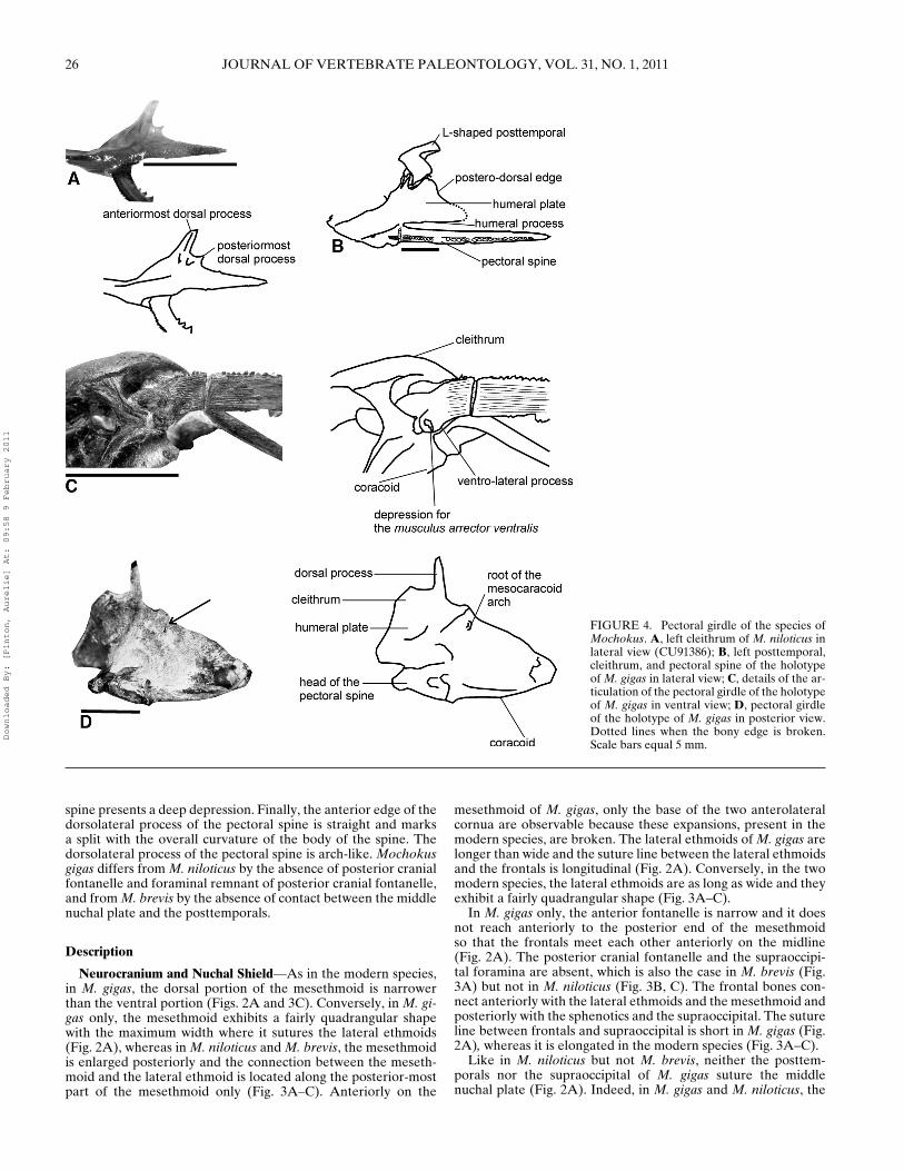
FIGURE 4. Pectoral girdle of the species ofMochokus. A, left cleithrum of M. niloticus inlateral view (CU91386); B, left posttemporal,cleithrum, and pectoral spine of the holotypeof M. gigas in lateral view; C, details of the ar-ticulation of the pectoral girdle of the holotypeof M. gigas in ventral view; D, pectoral girdleof the holotype of M. gigas in posterior view.Dotted lines when the bony edge is broken.Scale bars equal 5 mm.
spine presents a deep depression. Finally, the anterior edge of thedorsolateral process of the pectoral spine is straight and marksa split with the overall curvature of the body of the spine. Thedorsolateral process of the pectoral spine is arch-like. Mochokusgigas differs from M. niloticus by the absence of posterior cranialfontanelle and foraminal remnant of posterior cranial fontanelle,and from M. brevis by the absence of contact between the middlenuchal plate and the posttemporals.
Description
Neurocranium and Nuchal Shield—As in the modern species,in M. gigas, the dorsal portion of the mesethmoid is narrowerthan the ventral portion (Figs. 2A and 3C). Conversely, in M. gi-gas only, the mesethmoid exhibits a fairly quadrangular shapewith the maximum width where it sutures the lateral ethmoids(Fig. 2A), whereas in M. niloticus and M. brevis, the mesethmoidis enlarged posteriorly and the connection between the meseth-moid and the lateral ethmoid is located along the posterior-mostpart of the mesethmoid only (Fig. 3A–C). Anteriorly on the
mesethmoid of M. gigas, only the base of the two anterolateralcornua are observable because these expansions, present in themodern species, are broken. The lateral ethmoids of M. gigas arelonger than wide and the suture line between the lateral ethmoidsand the frontals is longitudinal (Fig. 2A). Conversely, in the twomodern species, the lateral ethmoids are as long as wide and theyexhibit a fairly quadrangular shape (Fig. 3A–C).
In M. gigas only, the anterior fontanelle is narrow and it doesnot reach anteriorly to the posterior end of the mesethmoidso that the frontals meet each other anteriorly on the midline(Fig. 2A). The posterior cranial fontanelle and the supraoccipi-tal foramina are absent, which is also the case in M. brevis (Fig.3A) but not in M. niloticus (Fig. 3B, C). The frontal bones con-nect anteriorly with the lateral ethmoids and the mesethmoid andposteriorly with the sphenotics and the supraoccipital. The sutureline between frontals and supraoccipital is short in M. gigas (Fig.2A), whereas it is elongated in the modern species (Fig. 3A–C).
Like in M. niloticus but not M. brevis, neither the posttem-porals nor the supraoccipital of M. gigas suture the middlenuchal plate (Fig. 2A). Indeed, in M. gigas and M. niloticus, the
Downloaded By: [Pinton, Aurelie] At: 09:58 9 February 2011

PINTON ET AL.—THE CATFISH MOCHOKUS FROM THE LATE MIOCENE OF CHAD 27
anterior nuchal plate is broad (Figs. 2A and 3B) and not reducedto a minute plate that inserts between the supraoccipital and themiddle nuchal plate (Fig. 3A). Thus, the anterior edge of the mid-dle nuchal plate differs in shape between M. gigas where it is con-cave, whereas it presents a notch for the insertion of the anteriornuchal plate in M. brevis. In M. gigas, the posterior nuchal platesexhibit a square shape with the length of the ventral edge as longas the length of the suture line with the middle nuchal plate (Fig.2A), whereas in the two modern species, they are triangular (Fig.3A, B). Moreover, in M. gigas only the third dorsal fin proximalradial contacts the posterior margin of the posterior nuchal plate(Fig. 2A; data from Vigliotta, 2008).
Ventrally, the basioccipital of M. gigas (Fig. 2B) extends ontothe parasphenoid further than in M. niloticus (Fig. 3D). More-over, the suture line with the exoccipital is straight in lateral view,whereas it is interdigitated in the modern species. Finally, the lat-eral limbs of the basioccipital project posteriorly (Fig. 2B), andnot laterally as in M. niloticus (Fig. 3D).
The prootic is large in the three species. In M. gigas, we as-sume that the utricular otolith cavity is markedly smaller thanin the other species because the surface of the bone is notinflated. In the three species, the pterotic bone is large, withthe length of its free lateral edge clearly exceeding that of thesphenotic. This feature is marked in M. brevis (Fig. 3A) butless pronounced in M. niloticus (Fig. 3B) and M. gigas (Fig.2A). In M. gigas, the large pore for the pterotic branch ofthe otic canal opens in the lateral wall of the pterotic andis not associated with any pterotic spine, which is also thecase in the modern species (Vigliotta, 2008). Most of the hy-omandibular facet is supported by the sphenotic and extends ontothe pterosphenoid, but entirely excludes the prootic (Fig. 2B),like in M. brevis and M. niloticus (Vigliotta, 2008; A.P., pers.observ.).
The sphenoid region is similar in the three species. Thepterosphenoid contributes to the structure of the lateral wall ofthe braincase. From front to back, it sutures with the orbitosphe-noid, the frontal, the sphenotic, the prootic, and the parasphe-noid. The optic foramen opens in front of the connection be-tween the orbitosphenoid and the parasphenoid; the trigeminofacialis foramen opens posteriorly. Dorsal to this foramen, thepterosphenoid gets thicker and exhibits a small process that ac-commodates the anterior spur of the hyomandibula. The or-bitosphenoid presents an overall Y-shape due to lateral flangesdeveloped all along the bone (Fig. 2B).
In M. gigas, the snout is overall elongate, which is particularlymarked in the vomer when compared with M. niloticus. In M. gi-gas, the vomer presents a cross-like shape (Fig. 2B), whereas it isreduced to a small pad in M. niloticus (Fig. 3D).
Weberian Apparatus—The holotype of M. gigas has a well-preserved Weberian apparatus: the elastic spring apparatus, theparapophyses of the fifth centrum, and the posterior parapophy-ses of the complex centrum are observed, together with a frag-ment of the tripus (Fig. 2B). As in the modern species of Mo-chokus, the apparatus of M. gigas has the anterior pair of para-pophyses of the compound centrum, which is autogenous, pos-terodorsally directed, and deeply separated from the followingparapophysis.
Pectoral Girdle and Spines—As in the modern species, theposttemporal of M. gigas exhibits a L-shaped outline (Fig. 4B)and the ventral process of the ossified transcapular ligament issimple, without any medial expansion (present elsewhere in thefamily, i.e., in Synodontis). The anterior-most dorsal process ofthe cleithrum lies in front of the cavity of the posttemporal me-dial limb. Between the two dorsal processes of the cleithrum, adeep horseshoe-shaped notch accommodates the lower limb ofthe posttemporal (Fig. 2C). The humeral plate is partially seenon the holotype, where it extends between the humeral and theposterior-most dorsal processes. The outline of the posterodorsal
FIGURE 5. Right pectoral spines of species of Mochokus in ventralview. A, details of the articular head of M. niloticus (personal collection);B, complete view of a pectoral spine of M. gigas (TM90–04–27); C, de-tails on the articular head of M. gigas (TM90–04–27); D, details on thearticular head of M. gigas (TM90–99–55). Hatch-markings correspond tobroken surface. Scale bars equal 2 mm (A) or 5 mm (B–D).
edge of the cleithrum is concave (Fig. 2C). In the modern Mo-chokus species, the humeral plate seems more acute than in thefossil (Fig. 4A). It is also concave in its anterior-most part andstraight and parallel to the ventral edge of the cleithrum posteri-orly (Fig. 4A). On the coracoid of M. gigas, we observe the rootof the mesocoracoid arch placed on the posterior side of the verti-cal lamina. The root of this mesocoracoid arch lacks any dorsallydirected prominence (Fig. 4D). The pectoral locking foramen ispresent and classically located ventrally onto the area of the ar-ticulation for the pectoral spine.
Smooth serrations develop along the anterior and posteriormargins of the pectoral spines, as in modern Mochokus (Fig.5A; Vigliotta, 2008; A.P., pers. observ.). The overall articularhead morphology also looks similar among the three species. Thepectoral spines of M. gigas are characterized by the depressionfor the musculus arrector ventralis on the ventrolateral process,which is deeper and clearly limited posteriorly by a wall (Fig.4C). The anterior edge of the dorsolateral process of the spine isstraight and marks a split with the overall curvature of the bodyof the spine and the dorsolateral process of the spine is arch-like.Finally, the inner fossa of the spine is reduced.
MOCHOKUS cf. M. GIGAS(Figs. 5, 6)
Material—TM82-01-25 consists of a cleithrum and a posttem-poral (Fig. 6A); TM90-99-51 is a posttemporal with a part ofthe cleithrum; TM90-99-54 is an almost-complete middle nuchalplate (Fig. 6C); TM90-04-62 is a partial cleithrum with the prox-imal half of the pectoral spine; TM90-04-27 is a complete pec-toral spine (Fig. 5C, D); TM90-99-55 (Fig. 5D) and TM90-99-56are proximal fragments of pectoral spines; TM256-01-05 is an iso-lated cleithrum (Fig. 6B); TM337-03-19 is a well-preserved mid-dle nuchal plate (Fig. 6D).
Occurrence—TM82, TM90, TM256, and TM337, which be-long to the Anthracotheriid Unit in the fossiliferous area of
Downloaded By: [Pinton, Aurelie] At: 09:58 9 February 2011

28 JOURNAL OF VERTEBRATE PALEONTOLOGY, VOL. 31, NO. 1, 2011
FIGURE 6. Right cleithra and middle nuchal plates attributed to Mo-chokus cf. gigas. A, cleithrum and posttemporal in lateral view (TM82-01-25); B, cleithrum in lateral view (TM256-01-05); C, middle nuchal plate indorsal view (TM90-99-54); D, middle nuchal plate in dorsal view (TM337-03-19). The arrow shows the front of the fossil. Dotted lines when thebony edge is broken. Scale bars equal 5 mm.
Toros-Menalla, Late Miocene, Western Djurab, Chad (Vignaudet al., 2002), aged ca. 7 Ma (Lebatard et al., 2008).
Description—The posttemporal TM82-01-25 presents the typ-ical L-shape observed in Mochokus and the humeral plate showsthe same concavity of the posterodorsal border as observed inthe holotype of M. gigas (Fig. 6A). Moreover, the dimensionsof the bones fall in the same range as M. gigas and thus ex-clude that it from being attributed to a modern species. Thecleithrum TM256-01-05 also shows the features described on theholotype of M. gigas (Fig. 6B). The posttemporal TM90-99-51 issimilar to the posttemporal of M. gigas in all the details preserved.The middle nuchal plates TM90-99-54 and TM337-03-19 presentan anterior suture line concave in outline without any depression,as observed in M. gigas or M. niloticus (Fig. 6C, D). Once again,based on the dimensions of the specimens, they are attributed toMochokus cf. gigas. Finally, the specimens TM90-99-55, TM90-99-56, and TM90-04-27 present the specific characters observedon M. gigas: the depression for the musculus arrector ventralis onthe ventrolateral process, the anterior edge of the dorsolateralprocess of the spine straight and marking a split with the overallcurvature of the body, and an arch-like dorsolateral process (Fig.5B-D). Moreover, the inner fossa of those specimens is reduced.They do not show any other complementary features than thoseobserved on the holotype.
MOCHOKUS, sp. indet.(Fig. 7)
FIGURE 7. Dorsal view of the partial middle nuchal plate and posteriornuchal plates of Mochokus sp. indet. (TM256-01-04). The arrow showsthe front of the fossil. Scale bar equals 5 mm.
Material—TM256-01-04 consists of a partial median nuchalplate with the pair of posterior nuchal plates (Fig. 7).
Occurrence—TM256, which belongs to the AnthracotheriidUnit of the fossiliferous area of Toros-Menalla, Late Miocene,Western Djurab, Chad (Vignaud et al., 2002), aged ca. 7 Ma(Lebatard et al., 2008).
Description—Only one half of the middle nuchal plate ofTM256-01-04 is preserved (Fig. 7). The anterior suture line isconcave without any anterior depression and thus differs fromM. brevis but resembles both M. gigas and M. niloticus. The pos-terior nuchal plates of TM256-01-04 are triangular in outline andtheir ventral free edges are longer than the sutures establishedwith the middle nuchal plate (Fig. 7). It thus differs from M. gi-gas. Finally, the dimensions of TM256-01-04 largely surpass thoseof the modern species. Because of this singular combination ofcharacters, we suspect that a second new species is present in TM.However, we consider that the material is insufficiently diagnos-tic to support the erection of a new species. Indeed, the shape ofthe posterior nuchal plate is known to be variable in at least oneother mochokid genus, i.e., Synodontis (Pinton and Otero, 2010).
MOCHOKUS sp.(Fig. 8)
Material—TM90-99-57, TM90-99-58, and TM90-99-59 are dor-sal spines (Fig. 8B, C). The first and the second are near complete,and the third presents a well-preserved head but only part of thebody is preserved.
Occurrence—TM90, which belongs to the AnthracotheriidUnit of the fossiliferous area of Toros-Menalla, Late Miocene,Western Djurab, Chad (Vignaud et al., 2002), aged ca. 7 Ma(Lebatard et al., 2008).
Description—In Mochokus, the head of the spines are trian-gular and lateral wings are well developed when compared to thewidth of the body of the spines (Fig. 8). The foramen is small (Fig.8B, C). The anterior edge exhibits serrae with preserved bases,except in M. brevis (Fig. 8). In posterior view, the body of thespine is smooth without any tubercles (Fig. 8). In lateral view, thespines TM90-99-57 and TM90-99-58 are straight and do not showany curvature (Fig. 8B, C). As in the modern species, the fol-lowing combination of characters: presence of tubercles on theanterior edge, their absence on the posterior edge, and absenceof curvature, is unique among African catfishes belonging only toMochokus (Gayet and Van Neer, 1990; Vigliotta, 2008; A.P. andO.O., pers. observ.).
Downloaded By: [Pinton, Aurelie] At: 09:58 9 February 2011

PINTON ET AL.—THE CATFISH MOCHOKUS FROM THE LATE MIOCENE OF CHAD 29
FIGURE 8. Dorsal spines of Mochokus. A, M. niloticus in dorsal andlateral views (CU91386); B, Mochokus sp. (TM90-99-57) in anterior, pos-terior, and lateral views; C, Mochokus sp. (TM90-99-59) in anterior, pos-terior, and lateral views. Scale bars equal 5 mm.
SYSTEMATIC DISCUSSION
The family Mochokidae includes nine genera. So far, only onephylogenetic study of the intrafamilial relationships has been pro-posed (Vigliotta, 2008), on the basis of osteological characters. Inthis study, the genus Mochokus is recovered as a monophyleticgroup based on five synapomorphies. It occupies a basal positionwithin the Mochokidae because the species of Mochokus show animportant number of traits considered as plesiomorphic amongcatfishes (Vigliotta, 2008). One of the synapomorphies proposed
by Vigliotta (2008) for the genus is observed on the holotype ofM. gigas: the root of the mesocoracoid arch is located on theposterior side of the vertical lamina of the coracoid and lacksany dorsally directed prominence. According to Vigliotta (2008),this character has been independently acquired by chiloglanidins.The other synapomorphic character states cannot be evaluatedon the fossil. However, the holotype of M. gigas shows a combi-nation of characters that is proper for the genus Mochokus: (1)the mesethmoid is clearly narrower in ventral view than in dor-sal view, and thus differs from Acanthocleithron, Synodontis, andMicrosynodontis (Vigliotta, 2008); (2) the posttemporals extendposteriorly and contact the anterior nuchal plate; this characteris present in Mochokus and Mochokiella only (Vigliotta, 2008);(3) the hyomandibular facet on the neurocranium is excludedfrom the prootic; this feature is also observed in Acanthocleithron(Vigliotta, 2008); (4) as in two Chiloglanis species (Chiloglanismacropterus and Chiloglanis sp. ‘burundi’) only, the ventral faceof the pterotic is free of spine, which is the catfish plesiomorphicstate according to Vigliotta (2008). All other mochokids exhibit aspinous projection on the ventral face of the pterotic.
Lastly, we have to discuss the new characters observed on theholotype and the defining of the new species of Mochokus. No-tably, the holotype TM92-06-070 is easily diagnosed as a newtaxon and we want to exclude the possibility that it merits place-ment in a new genus. We also further explore the phylogeneticrelationships and the morphological variability within Mochoki-dae.
Considering the matrix established by Vigliotta (2008), only 2of the 28 characters coded on M. gigas differ from the two mod-ern species of Mochokus: (1) the shape of the lateral ethmoid iscoded as short and wide in the modern species whereas elongatedand narrow in M. gigas; (2) the third dorsal-fin proximal-middleradial is joined to the posterior nuchal plate in M. gigas whereasit is free in the two modern species (Supplementary Data 2, char-acter 22). The tree bisection-reconnection heuristic search algo-rithm resulted in 33 equally parsimonious trees of 409 steps (CI= 0.3619 and RI = 0.8489). The recovered 33 trees differ in therelative placement of the three species of Mochokus. However,the Mochokus clade is always recovered as a monophyletic clade(decay index: 1; branch length: 8) with each species alternativelyplaced basally with respect to the two others. In the strict con-sensus tree (423 steps, CI = 0.3499, RI = 8408), the relationshipsbetween the three species of Mochokus remain unresolved. Inthis phylogenetic analysis, nothing supports the distinction of aseparate new genus for this fossil species.
Second, we checked that the range of variation inferred by theinclusion of M. gigas into the genus Mochokus exists elsewherein mochokids. We use here the results of the only extensive studyon Synodontis (Pinton and Otero, 2010). The main striking differ-ences between species of Mochokus are located in the ethmoidianregion. First, in M. gigas, the lateral ethmoids are wide, elongatedanteroposteriorly, and they suture the mesethmoid all along theirposterior half (Fig. 9E). On the contrary, in other species of
FIGURE 9. Dorsal view of the anterior partof the neurocranium of some species of Syn-odontis and Mochokus. A, S. courteti; B,S. membranaceus; C, S. nigrita; D, M. niloticus(CU91386); E, holotype of M. gigas. Scale barsequal 5 mm.
Downloaded By: [Pinton, Aurelie] At: 09:58 9 February 2011

30 JOURNAL OF VERTEBRATE PALEONTOLOGY, VOL. 31, NO. 1, 2011
Mochokus, the lateral ethmoids are located more posteriorly andthe suture between the lateral ethmoids and the mesethmoidis short (Fig. 9D). Such differences are also observed betweenspecies of Synodontis. For instance, in S. nigrita (Fig. 9C), thelateral ethmoids are clearly directed more posteriorly than inS. membranaceus and S. courteti (Fig. 9A, B). Second, differ-ences are observed in the width of the cranial fontanelle (Fig.9). Indeed, in M. gigas, the cranial fontanelle is narrow, whereasit is wide in the modern species of Mochokus. This characteris also variable among the species of Synodontis with the samerange (Fig. 9A–C; Pinton and Otero, 2010) and, to a lesser ex-tent, within each species according to the size of the fish.
CONCLUSION
Several fossil remains attributed to Mochokus fishes have beencollected in Toros-Menalla (TM82, TM90, TM92, TM256, andTM337). These are the only fossils attributed to this genus so far.Among them, a near-complete skull, isolated cleithra, and post-temporal from the late Miocene of Western Djourab (Chad) areattributed to a new species, Mochokus gigas, the first fossil taxondescribed for the family Mochokidae. The Chadian fossil occur-rence of the genus is not surprising when looking at the distribu-tion of the two modern species: they inhabit the freshwaters ofthe Chad basin and overlap the Nile basin. Conversely, the largedimensions of the fossils are amazing. Indeed, the size of the fos-sil neurocranium is about 10 times larger than the modern ones.Body size has shown to be correlated with many life history andecological traits. Notably, body size seems important to resourceuse and ecological interactions among species. In that respect, it iscorrelated with the trophic position in a given food web and here,we can suspect that the ecological traits of M. gigas fish were dif-ferent from the modern species.
Among the dozens of sites of Toros-Menalla where fishes havebeen actively collected, only five have yielded Mochokus re-mains. This limited distribution may be related with their lowabundance in the sites concerned. For instance, in TM90 wherehalf of the Mochokus remains have been collected, hundredsof catfish fossils were sampled. Mochokus fishes are rare andpoorly represented in the fish fossil association of Toros-Menalla.When comparing Mochokus abundance with Synodontis abun-dance (the only other mochokid genus known at this fossil site),we notice that Synodontis remains are always more abundant.One exception is site TM90. Based on preliminary observationson the fish assemblages in different sites of Toros-Menalla, it hasbeen postulated that the relative abundance of fish taxa may de-pend on the environment (Otero et al., 2007). TM90 is largelydominated by clariids (Clarias/Heterobranchus) and polypterids(Otero et al., 2006), which can stand poorly oxygenated waterand are physiologically adapted to marshy or swampy conditions.This is supported by other faunic elements, notably the presenceof the bird Heliopais cf. personata (Gruiformes) whose ecologicalpreference is still or slow-moving waters (Louchard et al., 2005).Thus, the scarcity of M. gigas in Toros-Menalla could be ex-plained by a low ecological tolerance. The remains of Mochokus,sp. indet., suspected to belong to another species have been foundin TM256, a site where they represent, with an isolated cleithrum,the only material of Mochokus found. This raises the possibilitythat two species of Mochokus could have cohabitated in Toros-Menalla, with different ecological preferences.
ACKNOWLEDGMENTS
We thank the Chadian authorities (MENR: Univ. N’Djamena,CNAR) and the French authorities (MESR: CNRS; Univ.Poitiers; MAE: DCSUR, Paris; FSP de la cooperation francaise,Ambassade de France, N’Djamena). The MPFT fieldwork andresearch is also supported by ANR (Projet ANR 05-BLAN-0235)and NSF (RHOI). We extend our gratitude to all members of the
MPFT. For their support, we extend our gratitude to the FrenchMinistere de l’Enseignement superieur, de la Recherche et de laTechnologie (CNRS; Universite de Poitiers) and the Ministeredes Affaires Etrangeres (DCSUR, Paris; Cooperation SCAC andFSP, N’Djamena), to the Region Poitou-Charentes. Great thanksto S. Lavoue (National Museum of Natural History, London) andJ. Friel (Cornell University, Ithaca, New York) for providing ma-terial of comparison as well as to P. Pruvost (Museum d’HistoireNaturelle, Paris) for an access to the collections. We greatly thankthe reviewers for all comments and helpful advises. Drawings andphotographs by A. Pinton.
LITERATURE CITED
Brunet, M., F. Guy, D. Pilbeam, D. E. Lieberman, A. Likius, H. T. Mack-aye, M. Ponce de Leon, C. P. E. Zollikofer, and P. Vignaud. 2005.New material of the Earliest Hominid from the Upper Miocene ofChad. Nature 434:753–755.
Brunet, M., F. Guy, D. Pilbeam, H. T. Mackaye, A. Likius, D. Ahounta,A. Beauvilain, C. Blondel, H. Bocherens, J.-R. Boisserie, L. de Bo-nis, Y. Coppens, J. Dejax, C. Denys, P. Duringer, V. Eisenmann,G. Fanone, P. Fronty, D. Geraads, T. Lehmann, F. Lihoreau, A.Louchart, A. Mahamat, G. Merceron, G. Mouchelin, O. Otero, P.Pelaez Campomanes, M. Ponce De Leon, J.-C. Rage, M. Sapanet,M. Schuster, J. Sudre, P. Tassy, X. Valentin, P. Vignaud, L. Viriot,A. Zazzo, and C. Zollikofer. 2002. A new hominid from the upperMiocene of Chad, Central Africa. Nature 418:145–151.
Cuvier, G. 1817. Le regne animal distribue d’apres son organisation, pourservir de base a l’histoire naturelle des animaux et d’introductiona l’anatomie comparee. Avec figures, dessinees d’apres nature.Tome II, contenant les reptiles, les poissons, les mollusques et lesannelides. Paris:1–532.
Ferraris, C. J., Jr. 2007. Checklist of catfishes, recent and fossil (Oste-ichthyes: Siluriformes), and catalogue of siluriform primary types.Zootaxa 1418:1–628.
Gayet, M., and W. Van Neer. 1990. Caracteres diagnostiques desepines de quelques silures africains. Journal of African Zoology104:241–252.
de Joannis, L. 1835. Observations sur les poissons du Nil, et descriptionde plusieurs especes nouvelles. Magasin de Zoologie 5:1–53.
Jordan, D. S. 1923. A Classification of Fishes Including Families and Gen-era As Far As Known. Stanford University Publication, UniversitySeries, Biological Sciences. Stanford University, Palo Alto, Califor-nia, 166. pp.
Louchard, A, P. Vignaud, A. Likius, H. T. Mackaye, and M. Brunet. 2005.A new swan (Aves: Anatidae), from the latest Miocene of Chad andLibya. Journal of Vertebrate Paleontology 25:384–392.
Lebatard, A. E., D. L Bourles, P. Duringer, M. Jolivet, R. Braucher, J.Carcaillet, M. Schuster, N. Arnaud, P. Monie, F. Lihoreau, A. Lik-ius, H.T. Mackaye, P. Vignaud, and M. Brunet. 2008. Cosmogenicnuclide dating of Sahelanthropus tchadensis and Australopithecusbahrelghazali: Mio-Pliocene hominids from Chad. Proceedings ofthe National Academy of Sciences of the United States of America105:3226–3231.
Otero, O. 2010. What controls the freshwater fish fossil record? A fo-cus on the Late Cretaceous and Tertiary Afro-Arabia. Cybium34:93–113.
Otero, O., A. Likius, P. Vignaud, and M. Brunet. 2006. A new polypteridfish: Polypterus faraou sp. nov. (Cladistia, Polypteridae) from theLate Miocene, Toros-Menalla, Chad. Zoological Journal of the Lin-nean Society 146:227–237.
Otero, O., A. Likius, P. Vignaud, and M. Brunet. 2007. A new claroteidcatfish (Siluriformes) from the Upper Miocene of Toros-Menalla,Chad: Auchenoglanis soye, sp. nov. Journal of Vertebrate Paleon-tology 27:285–294.
Otero, O., A. Pinton, H. T. Mackaye, A. Likius, P. Vignaud, andM. Brunet. 2010. The fish assemblage associated with the LateMiocene Chadian hominid (Toros-Menalla, Western Djurab)and its palaeoenvironmental significance. Paleontographica Acta292:21–51.
Paugy, D., and T. R. Roberts. 2004. Mochokidae; pp. 500–563 in C.Leveque, D. Paugy, and G. G. Teugels (eds.), Faunes des poissonsd’eau douce et saumatres de l’Afrique de l’Ouest. Orstom/IRD Pub-lications, Paris.
Downloaded By: [Pinton, Aurelie] At: 09:58 9 February 2011

PINTON ET AL.—THE CATFISH MOCHOKUS FROM THE LATE MIOCENE OF CHAD 31
Pinton, A. 2008. Anatomy, systematic and phylogeny of modern andfossil Synodontis (Siluriformes, Mochokidae): paleobiogeographicalimplications in the Neogene of Africa. PhD dissertation, Universitede Poitiers, Poitiers, France, 280. pp.
Pinton, A., and O. Otero. 2010. The bony anatomy of Chadian Synodontis(Osteichthyes, Teleostei, Siluriformes, Mochokidae): interspecificvariations and specific characters. Zoosystema 32:173–231.
Rosen, D. E., P. L. Forey, B. G. Gardiner, and C. Patterson. 1981. Lung-fishes, tetrapods, paleontology and plesiomorphy. Bulletin of theAmerican museum of Natural History 167:159–276.
Skelton, P. H. (ed.). 2000. A Complete Guide to the Freshwater Fishes ofSouthern Africa. Struik Publishers, Cape Town, 395. pp.
Swofford, D. L. 2002. PAUP∗. Sinauer Associates, Sunderland, Mas-sachusetts.
Priem, R. 1920. Poissons fossiles du Miocene d’Egypte (Burdigalien deMoghara, “Desert lybique”); pp 8–15 in R. Fourtau (ed.), Contri-
bution a l’etude des vertebres miocenes de l’Egypte. GovernmentPress, Cairo.
Vigliotta, T. R. 2008. A phylogenetic study of the African catfish fam-ily Mochokidae (Osteichthyes, Ostariophysi, Siluriformes), with akey to genera. Proceedings of the Academy of Natural Sciences ofPhiladelphia 157:73–136.
Vignaud, P., P. Duringer, H. T. Mackaye, A. Likius, C. Blondel, J.-R. Boisserie, L. de Bonis, V. Eisenmann, M.-E. Etienne, D. Ger-aads, F. Guy, T. Lehmann, F. Lihoreau, N. Lopez Martinez, C.Mourer- Chauvire, O. Otero, J.-C. Rage, M. Schuster, L. Viriot,A. Zazzo, and M. Brunet. 2002. Geology and paleontology ofthe Upper Miocene Toros-Menalla hominid locality, Chad. Nature418:152–155.
Submitted December 2, 2009; received September 23, 2010Handling editor: Terry Grande.
Downloaded By: [Pinton, Aurelie] At: 09:58 9 February 2011
Copyright © 2022 FDOKUMEN


![GÉOPOLITIQUE ET POPULATIONS AU TCHAD [Geopolitics and populations in Chad]](https://static.fdokumen.com/doc/165x107/631378e5fc260b71020f1c3f/geopolitique-et-populations-au-tchad-geopolitics-and-populations-in-chad.jpg)