Structural and functional analysis of the ovine laminin receptor gene (RPSA): Possible involvement...
14
Structural and functional analysis of the ovine laminin receptor gene (RPSA): Possible involvement of the LRP/LR protein in scrapie response Ane Marcos-Carcavilla Jorge H. Calvo Carmen Gonza ´lez Carmen Serrano Katayoun Moazami-Goudarzi Pascal Laurent Maud Bertaud He ´le `ne Hayes Anne E. Beattie Jaber Lyahyai Inmaculada Martı ´n-Burriel Juan Marı ´a Torres Magdalena Serrano Received: 2 August 2007 / Accepted: 23 November 2007 / Published online: 18 January 2008 Ó Springer Science+Business Media, LLC 2007 Abstract Scrapie is a prion disease affecting sheep and goats. Susceptibility to this neurodegenerative disease shows polygenic variance. The involvement of the laminin receptor (LRP/LR) in the metabolism and propagation of prions has previously been demonstrated. In the present work, the ovine laminin receptor gene (RPSA) was isolated, characterized, and mapped to ovine chromosome OAR19q13. Real-time RT-PCR revealed a significant decrease in RPSA mRNA in cerebellum after scrapie infection. Conversely, no differ- ences were detected in other brain regions such as diencephalon and medulla oblongata. Association analysis showed that a polymorphism reflecting the presence of a RPSA pseudogene was overrepresented in a group of sheep resistant to scrapie infection. No amino acid change in the LRP/LR protein was found in the 126 sheep analyzed. However, interesting amino acid positions (241, 272, and 290), which could participate in the species barrier to scrapie and maybe to other transmissible spongiform encephalopa- thies, were identified by comparing LRP/LR sequences from various mammals with variable levels of resistance to scrapie. Introduction Scrapie (SC) is a transmissible spongiform encephalopathy (TSE) of sheep and goats. TSEs are neurodegenerative diseases caused by transmissible proteinaceous particles (prions) devoid of nucleic acid that affect various animal species, including man. The key event in the pathogenesis of prion diseases is the conversion of the host-encoded and naturally expressed cellular prion protein (PrP c ) into its aberrant counterpart (PrP sc ). During the conversion pro- cess, PrP sc acquires new biophysical and biochemical characteristics which cause its accumulation in the form of amyloid plaques in nervous and lymphoid tissues (Prusiner 1998). In SC-infected cells, PrP sc seems to originate from a posttranslational refolding of PrP c . Although the exact subcellular location of this process in unknown, rafts of the plasma membrane or the endocytic pathway have been invoked (Ben-Zaken et al. 2003). Transgenic studies on the species barrier have indicated that accessory molecules of the host are probably involved in this process. Thus, during A. Marcos-Carcavilla (&) C. Gonza ´lez M. Serrano Departamento de Mejora Gene ´tica Animal, INIA, Ctra La Corun ˜a Km 7.5, Madrid 28040, Spain e-mail: [email protected] J. H. Calvo Unidad de Tecnologı ´a en Produccio ´n Animal, Avda. Montan ˜ana 930, CITA, Zaragoza 50059, Spain K. Moazami-Goudarzi P. Laurent M. Bertaud H. Hayes Laboratoire de Ge ´ne ´tique biochimique et de Cytoge ´ne ´tique, De ´partement de Ge ´ne ´tique Animale, INRA, Centre de Recherche de Jouy, Jouy-en-Josas, Cedex 78352, France A. E. Beattie AgResearch, Invermay Agricultural Centre, Private Bag 50034, Mosgiel, New Zealand C. Serrano J. Lyahyai I. Martı ´n-Burriel Laboratorio de Gene ´tica Bioquı ´mica, Facultad de Veterinaria, Universidad de Zaragoza, Miguel Servet 177, Zaragoza 50013, Spain J. M. Torres CISA-INIA, Carretera de Algete a El Casar s/n, Valdeolmos, Madrid 28130, Spain 123 Mamm Genome (2008) 19:92–105 DOI 10.1007/s00335-007-9085-6
-
Upload
independent -
Category
Documents
-
view
0 -
download
0
Transcript of Structural and functional analysis of the ovine laminin receptor gene (RPSA): Possible involvement...

Structural and functional analysis of the ovine laminin receptorgene (RPSA): Possible involvement of the LRP/LR proteinin scrapie response
Ane Marcos-Carcavilla Æ Jorge H. Calvo Æ Carmen Gonzalez ÆCarmen Serrano Æ Katayoun Moazami-Goudarzi Æ Pascal Laurent ÆMaud Bertaud Æ Helene Hayes Æ Anne E. Beattie Æ Jaber Lyahyai ÆInmaculada Martın-Burriel Æ Juan Marıa Torres Æ Magdalena Serrano
Received: 2 August 2007 / Accepted: 23 November 2007 / Published online: 18 January 2008
� Springer Science+Business Media, LLC 2007
Abstract Scrapie is a prion disease affecting sheep and
goats. Susceptibility to this neurodegenerative disease shows
polygenic variance. The involvement of the laminin receptor
(LRP/LR) in the metabolism and propagation of prions has
previously been demonstrated. In the present work, the ovine
laminin receptor gene (RPSA) was isolated, characterized,
and mapped to ovine chromosome OAR19q13. Real-time
RT-PCR revealed a significant decrease in RPSA mRNA in
cerebellum after scrapie infection. Conversely, no differ-
ences were detected in other brain regions such as
diencephalon and medulla oblongata. Association analysis
showed that a polymorphism reflecting the presence of a
RPSA pseudogene was overrepresented in a group of sheep
resistant to scrapie infection. No amino acid change in the
LRP/LR protein was found in the 126 sheep analyzed.
However, interesting amino acid positions (241, 272, and
290), which could participate in the species barrier to scrapie
and maybe to other transmissible spongiform encephalopa-
thies, were identified by comparing LRP/LR sequences from
various mammals with variable levels of resistance to
scrapie.
Introduction
Scrapie (SC) is a transmissible spongiform encephalopathy
(TSE) of sheep and goats. TSEs are neurodegenerative
diseases caused by transmissible proteinaceous particles
(prions) devoid of nucleic acid that affect various animal
species, including man. The key event in the pathogenesis
of prion diseases is the conversion of the host-encoded and
naturally expressed cellular prion protein (PrPc) into its
aberrant counterpart (PrPsc). During the conversion pro-
cess, PrPsc acquires new biophysical and biochemical
characteristics which cause its accumulation in the form of
amyloid plaques in nervous and lymphoid tissues (Prusiner
1998).
In SC-infected cells, PrPsc seems to originate from a
posttranslational refolding of PrPc. Although the exact
subcellular location of this process in unknown, rafts of the
plasma membrane or the endocytic pathway have been
invoked (Ben-Zaken et al. 2003). Transgenic studies on the
species barrier have indicated that accessory molecules of
the host are probably involved in this process. Thus, during
A. Marcos-Carcavilla (&) � C. Gonzalez � M. Serrano
Departamento de Mejora Genetica Animal, INIA,
Ctra La Coruna Km 7.5, Madrid 28040, Spain
e-mail: [email protected]
J. H. Calvo
Unidad de Tecnologıa en Produccion Animal,
Avda. Montanana 930, CITA, Zaragoza 50059, Spain
K. Moazami-Goudarzi � P. Laurent � M. Bertaud � H. Hayes
Laboratoire de Genetique biochimique et de Cytogenetique,
Departement de Genetique Animale,
INRA, Centre de Recherche de Jouy,
Jouy-en-Josas, Cedex 78352, France
A. E. Beattie
AgResearch, Invermay Agricultural Centre,
Private Bag 50034, Mosgiel, New Zealand
C. Serrano � J. Lyahyai � I. Martın-Burriel
Laboratorio de Genetica Bioquımica,
Facultad de Veterinaria, Universidad de Zaragoza,
Miguel Servet 177, Zaragoza 50013, Spain
J. M. Torres
CISA-INIA, Carretera de Algete a El Casar s/n,
Valdeolmos, Madrid 28130, Spain
123
Mamm Genome (2008) 19:92–105
DOI 10.1007/s00335-007-9085-6
recent years, the involvement of LRP/LR together with
heparan sulfate proteoglycanes (HSPGs) as cofactors/co-
receptors has been demonstrated, not only in PrPc
metabolism but also in prion propagation (Adjou et al.
2003; Gauczynski et al. 2001, 2006; Horonchik et al. 2005;
Hundt et al. 2001; Leucht et al. 2003; Morel et al. 2005;
Rieger et al. 1997; Vana and Weiss 2006).
Various isoforms corresponding to different maturation
states of the nonintegrin laminin receptor LRP/LR (44, 60,
67, and 220 kDa) have been isolated from mouse brains
and found to bind to PrP (Simoneau et al. 2003). The high-
affinity 67-kDa LR is often referred to as the 32-67-kDa
LR (Kim et al. 1998) or the 37-67-kDa LR (Rao et al.
1994). This discrepancy in terminology is due to the fact
that cDNA clones of the 67-kDa LR contain a sequence
potentially coding for only the 32-kDa polypeptide, while
the translation product of the corresponding mRNA has a
molecular mass of 37 kDa and acts as a precursor (LRP) of
the 67-kDa LR (Shmakov et al. 2000). The process by
which the 67-kDa LR is assembled from the precursor is
not yet understood. Several hypotheses have been proposed
to explain this polymorphism (Castronovo et al. 1991;
Hundt et al. 2001; Landowski et al. 1995); the most reli-
able one involves association of the 37-kDa LPR with
HSPGs (Hundt et al. 2001).
The 37-kDa LRP protein is a component of the
translational machinery because it is specifically associ-
ated with the 40S ribosomal subunit (Auth and
Brawerman 1992), and it also seems to be involved in
maintaining nuclear architecture by stabilizing chromatin
via multiple associations with proteic and nucleic acid
components of the nucleosome (Kinoshita et al. 1998).
Thus, this isoform is localized mainly in the cytoplasm
(Yenofsky et al. 1982) and in the nucleus (Yow et al.
1988). In addition, the 67-kDa LR can be present as a
laminin-binding protein on the cell surface (Rabacchi
et al. 1990) or in a free form in the extracellular matrix
(Karpatova et al. 1996).
Susceptibility to SC is associated with polymorphisms
in the amino acid sequence of the PrP protein. To date, 25
polymorphic codons and 40 haplotypes have been descri-
bed in the open reading frame of the gene encoding the PrP
(PRNP) (Goldmann et al. 2005). In a slightly simplified
assessment of the genetic susceptibility of a sheep to
develop SC, polymorphisms are classified according to
amino acid changes at positions V136A, R154H, and
QH171R (Baylis et al. 2004). However, additional evi-
dence for other genomic regions containing genes that
influence the incubation period for SC in mice (Lloyd et al.
2001, 2002; Manolakou et al. 2001; Moreno et al. 2003b;
Stephenson et al. 2000) and sheep (Moreno et al. 2003a)
have been reported. Thus, a reasonable hypothesis is that a
major gene (PRNP) controlling resistance/susceptibility to
SC coexists with a number of other genes that modulate its
effect.
Furthermore, transmission of prions from one species to
another is usually inefficient and accompanied by a pro-
longed incubation time. Among the few animal species that
appear to be resistant to oral infection by the TSE agent are
rabbits, dogs, and pigs (Knorr et al. 2007; Lysek et al. 2005;
Vorberg et al. 2003). The resistance to prion infection is a
characteristic of the host species, and the PrP amino acid
sequence of both the donor and the recipient animal plays an
important role (Castilla et al. 2004; Vorberg et al. 2003).
Nevertheless, the involvement of additional factors, other
than PrP, is also possible. In several model systems, both the
species-specific formation of PrPsc and the transmission of
the TSE agent across species barriers have been mapped to
the central region of the PrP molecule comprising residues
108-171 (Kocisko et al. 1995; Priola et al. 1994; Scott et al.
1993). This region includes the direct binding domain (aa
144–179) to LRP/LR (Hundt et al. 2001), highlighting a
putative role of the LRP/LR on TSE species barrier.
This information has led us to study the gene encoding
the LRP/LR (RPSA, derived from the approved name at
HUGO Gene Nomenclature Committee (HGNC), ribo-
somal protein SA) as a possible candidate contributing to
ovine SC. The first objective of this work was to determine
the position of the RPSA gene on the ovine genome in order
to verify if its localization was in concordance with pre-
vious reports describing QTLs associated with resistance/
susceptibility and the incubation period for different TSEs
in ovine (Moreno et al. 2003a) and murine models (Lloyd
et al. 2001, 2002; Manolakou et al. 2001; Moreno et al.
2003b; Stephenson et al. 2000). The next step was to verify
if there was any difference in the expression of this gene
between SC-infected and uninfected sheep presenting the
same genotype for PrP (ARQ/ARQ). This comparison was
performed using real-time RT-PCR in various brain
regions. Finally, the RPSA polymorphism was studied in
order to perform association analyses to identify putative
mutations in the ovine RPSA locus that could explain, at
least in part, the polygenic response to SC in sheep. The
possible role of LRP/LR in SC and other TSEs species
barrier is also discussed.
Material and methods
Isolation of sheep-specific DNA fragments
Genomic DNA was extracted from ovine lymphocytes
according to the salting-out procedure (Miller et al. 1988).
BAC DNA was purified by a Maxipreparation with the
Nucleobond PC 100 Kit (Macherey-Nagel) according to
the manufacturer’s instructions.
A. Marcos-Carcavilla et al.: Ovine RPSA gene and scrapie 93
123

It is widely recognized that, in mammals, a single gene,
RPSA, encodes the LRP/LR protein. Nevertheless, the
existence of numerous RPSA pseudogenes has greatly hin-
dered the sequencing and mapping of this gene in all the
mammalian species studied. Thus, to isolate and sequence
the ovine RPSA gene, 20 heterologous primers derived from
the human (GenBank HSU43901 and BC071968) and
bovine sequences (GenBank NW_930073 and BC102490)
and 21 specific primers based on the sequence of the ovine
fragments previously isolated with the primers described
above were synthesized. Appendix 1 shows only the primer
pairs that were useful to isolate RPSA ovine sequences. The
correct identity of amplified fragments from genomic DNA
was confirmed by sequencing a BAC containing the ovine
RPSA gene, which was isolated from the ovine BAC library
at INRA (Jouy-en-Josas, France) (Vaiman et al. 1999).
Genomic (60–100 ng) and BAC DNA (100 ng) were
amplified in a final volume of 25 ll containing 0.5 lM of
each primer, 200 lM of dNTPs, 1.5-2 mM MgCl2, 2.5 ll of
109 buffer MgCl2 free (Biotools), and 1 U Taq polymerase
(Biotools). The following PCR conditions were used:
denaturation at 94�C for 5 min, 30 amplification cycles of
denaturation at 94�C for 45 sec, annealing at temperatures
between 50�C and 66�C for 30 sec, and extension at 72�C for
30 sec to 1 min 45 sec, followed by a final 5–10 min
extension at 72�C. Primer pairs and their amplification
conditions are shown in Appendix 1. The resulting PCR
fragments were purified with the GFX PCR DNA and Gel
Band Purification Kit (Amersham Bioscience) and bidirec-
tionally sequenced with the PCR primers. The identity of
the fragments was confirmed by BLAST analysis
(http://www.ncbi.nlm.nih.gov/BLAST/). Additional infor-
matics programs such as ‘‘Neural Network Promoter
Prediction’’ (http://www.fruitfly.org/seq_tools/promoter.
html), ‘‘CISTER’’ (http://www.zlab.bu.edu/%7Emfrith/
cister.shtml), ‘‘Signal Scan’’ (http://www.bimas.dcrt.nih.
gov/molbio/signal/), ‘‘TFSEARCH’’ (http://www.cbrc.jp/
research/db/TFSEARCH.html), and ‘‘TargetScan’’ (http://
www.targetscan.org/), were used to study the promoter and
look for possible transcriptional and translational regulatory
elements.
Chromosomal location
Ovine RPSA gene was localized on the ovine genome by
two approaches: cytogenetic and genetic mapping.
Cytogenetic mapping
To avoid possible coamplifications with pseudogenes, a pair
of primers hybridizing with the RPSA intron 4 (see Appendix
1) was used to screen the ovine BAC library mentioned
above (Vaiman et al. 1999). DNA from the identified BAC
clone was labeled by nick-translation in the presence of
biotin-14-dATP (BioNick TM 18247–015 labeling system)
and used as a probe for in situ hybridization on RBP-banded
ovine chromosome preparations according to the protocol
described by Hayes et al. (1991).
Genetic mapping
The cytogenetic localization was confirmed by linkage
mapping in nine sheep families (129 animals). RPSA gene
was mapped against markers on the sheep framework map
(Maddox et al. 2001). Multipoint linkage analysis of the
International Mapping Flock (IMF) pedigrees (Crawford
et al. 1995) was performed using CRI-MAP (Lander and
Green 1987). Thus, genomic DNA from five domestic sheep
breeds [Latxa (n = 3), Manchega (n = 4), Awassi (n = 2),
Assaf (n = 2), and Rasa Aragonesa (n = 2)] was amplified
and sequenced as previously described and analyzed using
CHROMAS 1.43 and ClustalW (http://www.ebi.ac.uk/
clustalw/) software to detect polymorphisms along the iso-
lated sequences of the ovine RPSA gene. In this case, neither
their PRNP genotype nor their pattern of SC resistance/
susceptibility was taken into account. Animals were chosen
from different breeds to avoid any problem due to allele
fixation because of the selective status of these breeds in our
country (Spain), and to identify a representative polymor-
phism at the species level.
To follow the distribution of the gene within the pedi-
grees of the AgResearch IMF, the C/T transversion located
at position 69 in the RPSA exon 6 was analyzed by PCR-
RFLP using primers that hybridized with RPSA exon 5 and
exon 6 (see Appendix 1). The reaction product (8 ll) was
digested with 0.2 U of AvaII at 37�C for 4 h in a final
volume of 18 ll and then electrophoresed on a 3.5% aga-
rose gel for visualization.
Gene expression
Thirteen Rasa Aragonesa female sheep (3–5 years old) with
the same PRNP genotype (ARQ/ARQ) were included in this
study. Eight animals, which exhibited clinical signs of SC in
a terminal state, came from a closed regulatory monitored
flock and from the Spanish Scrapie surveillance program
and were kindly provided by the Prion Research Centre of
the University of Zaragoza. Control animals (n = 5) were
selected from a different flock of the same breed where no
SC has been reported to date. The aim of this sample design
was to determine if SC disease might cause any change in the
expression pattern of RPSA in sheep.
94 A. Marcos-Carcavilla et al.: Ovine RPSA gene and scrapie
123

After sacrificing the animals by intravenous injection of
sodium pentobarbital and exsanguination, small fragments
of medulla oblongata, cerebellum, diencephalon, and pre-
frontal cortex were included in RNAlater (Ambion). Total
mRNA was purified with the RNeasy Lipid Tissue Mini Kit
(Qiagen). Complementary DNA (cDNA) was synthesized
from 1 lg of each RNA preparation using random hexamer
primers with the SuperScript First-Strand Synthesis System
for RT-PCR (Invitrogen). Gene expression levels were
subsequently determined by real-time RT-PCR using
primers hybridizing with ovine RPSA exons 5 and 6. All
real-time RT-PCR reactions were run in triplicate. Three
tissue-specific housekeeping genes were used to normalize
each set of results. Thus, following the suggestions of
Vandesompele et al. (2002), data were normalized for
hexose-6-phosphate dehydrogenase (G6PDH), glyceralde-
hyde-3-phosphate dehydrogenase (GAPDH), and b-actin
(ACTB). Primers and probes used for gene expression
analysis, their concentrations, and amplicon sizes are
shown in Appendix 2. Amplifications were carried out in a
final volume of 10 ll containing SYBRGreen PCR Master
Mix (Applied Biosystems) or TaqMan Universal PCR
Master Mix, No AmpErase UNG (Applied Biosystems),
depending on the gene analyzed (see Appendix 2). After
preheating the mix at 95�C for 10 min, 40 cycles at 95�C
for 15 sec and 60�C for 30 sec were carried out.
The threshold cycle values were transformed into raw
quantity values and normalized according to the procedure
described by Garcia-Crespo et al. (2005). Finally, a paired-
sample comparison was performed to contrast the mean of
the differences in RPSA mRNA expression among the four
tissues analyzed in each individual. In addition, the means
from SC infected and control animals were compared with
a t test.
Polymorphism detection and association analysis
It is well established that there are significant differences
among breeds in which PRNP genotypes are attacked by
SC. For instance, the ARQ/ARQ genotype shows different
levels of resistance to SC infection depending on the breed
studied (Baylis et al. 2004). However, although most of the
additional amino acid polymorphisms, located between aa
101 and aa 241 in the PrP, have been found on the ARQ
haplotype, there is no evidence of any correlation between
the occurrence of a specific ARQ haplotype and the SC
disease status of a flock (Goldmann et al. 2005).
The existence of two binding sites for PrP on the LRP/
LR has been reported: a direct one located between amino
acid 161–179, and a putative second HSPG-dependent PrP
binding domain between amino acid 180–285 of the LRP/
LR. It is conceivable that one of the major sources of
variability in SC resistance/susceptibility and/or incubation
period originating from LRP/LR might be due to differ-
ences in the amino acids involved in the LRP/LR-PrPc/
PrPsc interaction.
Because the genomic region encoding amino acids
161–285 of the LRP/LR expands from the last 18
nucleotides of the RPSA exon 4 to the end of the coding
region in exon 7, two PCR reactions were carried out
using primers hybridizing with exon 4 and intron 4, and
intron 4 and exon 7 (see Appendix 1). Thus, genomic
DNA from 84 adult ARQ/ARQ Rasa Aragonesa sheep was
examined and used to perform association analyses. All
these animals belonged to seven flocks of sheep, where
natural SC was detected, and conserved for research
purposes by the Prion Research Centre of the University
of Zaragoza. Thirty-three of them (infected), which were
selected from six different flocks, exhibited clinical and
immunohistochemical signs of SC, while the remaining
51 (healthy), despite pertaining to a flock where SC was
detected, did not. No PrPsc or clinical signs were detected
in these 51 animals; nevertheless, the possible appearance
of infection signs with longer incubation periods cannot
be ruled out. To determine if the polymorphism pattern
found in these 84 animals was exclusive of the Rasa
Aragonesa group, an additional genotyping of 42 ARQ/
ARQ sheep belonging to the Manchega (n = 35) and
Assaf (n = 7) breeds, which had no previous contact with
SC, was carried out.
Finally, association analysis was performed with the
CATMOD procedure of the SAS statistical package (SAS
1998), which is a procedure for categorical data modeling.
In this case, since there were only two response levels,
the standard response function (generalized logits) was
adopted using the maximum likelihood estimation
method. The analysis took into account responses (pres-
ence/absence of clinical signs) to SC as the dependent
variable and the genotypes for the SNPs tested as the
independent factor.
Interspecies amino acid sequence comparison
To check if there was any important variation that could
explain the different levels of resistance to prion infection,
which depends on both host species and prion origin,
LRP/LR amino acid sequences from several mammals
displaying different responses to SC were compared.
Thus, mRNA from the spleen of a white New Zealand
rabbit was retrotranscribed, amplified, and bidirectionally
sequenced with the aim of predicting the protein sequence
encoded by the mRNA. The heterologous primer pairs
used hybridized with RPSA exons 2 and 7. Rabbit RNA
(200 ng) was retrotranscribed and amplified in a final
A. Marcos-Carcavilla et al.: Ovine RPSA gene and scrapie 95
123

volume of 50 ll containing 0.5 lM of each primer,
200 lM of dNTPs, 2 mM MgCl2, 5 ll of 10 9 buffer
MgCl2 free (Biotools), 4 U SuperScript II Reverse
Transcriptase (Invitrogen), 2 U RNasin Plus RNase
Inhibitor (Promega) and 1 U Taq polymerase (Biotools).
Retrotranscription (RT) and touchdown PCR (TD-PCR)
were performed as follows: an initial step at 42�C for
45 min, followed by 95�C for 10 min for the RT. Next,
the TD-PCR was carried out with nine cycles of dena-
turation at 95�C for 1 min, annealing at variable
temperatures, and extension at 72�C for 1 min 10 sec. In
the first cycle, the annealing temperature was set to 60�C,
and for each of the eight subsequent cycles the annealing
temperature was decreased by 1�C. Then, after 5 min at
95�C, 34 cycles at 95�C for 45 sec, 50�C for 30 sec, and
72�C for 1 min 10 sec were performed, followed by a
final step at 72�C for 10 min.
Finally, the LRP/LR amino acid sequences from mouse
(P14206 and NP_035159), rat (NP_058834.1), hamster
(P38982), man (P08865), cattle (P26452), pig
(NP_001032223.1), cat (Scaffold_214833), and dog
(NW_876276), deposited in GenBank, together with the
ovine (EF394773 and EF222474) and rabbit (EF222437)
ones inferred in the present work, were compared with the
ClustalW program (http://www.ebi.ac.uk/clustalw/).
Results
Isolation of the ovine RPSA gene and polymorphism
detection
Due to the presence of RPSA pseudogenes, big efforts were
necessary to decipher the ovine RPSA gene sequence.
Finally, with the exception of intron 3, which covers 9 kb
in cattle, the sequence of the ovine RPSA gene was
determined, including part of both the 50 and 30 flanking
regions (Fig. 1 and GenBank accession numbers EF222474
and EF394773).
The structure of the ovine RPSA gene was inferred by
comparison between the genomic and cDNA sequences
from sheep, man, and cattle (EF394773 and EF222474,
U43901, and BC102490, respectively). Thus, we could
confirm that the ovine gene is split into seven exons
ranging in size from around 50 (exon 1) to 246 nucleotides
(exon 4). The presence of two small nucleolar RNAs
(snoRNA) within ovine RPSA exons 2 and 4 (ACA6 and
E2, respectively) was also corroborated. The precise length
and full sequence of each of them are shown in Fig. 1 and
GenBank accession number EF394773. The entire exon 1
and the beginning of exon 2 (33 bases) are untranslated
sequences. The ATG codon, described as the translational
start, is located 133 bases upstream from the end of exon 2.
The splicing donor and acceptor sites follow the classical
rule with AG/GT nucleotides, respectively, ending and
starting successive introns. However, in two cases, obedi-
ence to this rule implies the existence of split codons: the
first one between exons 2 and 3 encoding a glycine and the
second one between exons 6 and 7 corresponding to a
glutamic acid.
The existence of various transcription start points has
been described in other species (Clausse et al. 1996;
Jackers et al. 1996; and GenBank accession numbers
NM_001012321 and NM_002295). To determine the
transcription initiation site in sheep, the ovine sequence
(EF394773) was compared with previously reported
sequences from cattle and man (BC102490 and U43901,
respectively). Furthermore, the ‘‘Neural Network Promoter
Prediction’’ program was used. As a result, four possible
transcription start points were identified; thus, ovine exon 1
might cover 52 bp (Figs. 1B, 2A).
In addition to the coding region, a sequence of 350 bp
corresponding to the 50 flanking region of the ovine
RPSA gene was also analyzed. Possible cis-regulatory
elements were inferred in the region between positions
-350 and + 1398 (Fig. 2). Potential SP-1, AP-1, and
AP-2 sites were predicted with the CISTER, Signal
Scan, and TFSEARCH programs. In addition, a metal
responding element (Simons and Toomre 2000), previ-
ously described in the chicken and human RPSA genes
(Clausse et al. 1996; Jackers et al. 1996), was also
identified. Contrary to these previous works, possible
TATA and CCAAT boxes were predicted in silico. The
TargetScan program was used in an attempt to find
possible targets for microRNAs that could be involved in
the posttranscriptional regulation of ovine RPSA gene.
Several putative targets were detected along the mRNA
sequence (Fig. 1).
By comparing sequences from Latxa, Manchega, Aw-
assi, Asaff, and Rasa Aragonesa sheep breeds, 27
polymorphic sites were identified within the isolated frag-
ments (see GenBank accession numbers EF222474 and
EF394773). Although nine of them were located within
exons 2, 3, 4, 6, and 7, none of these alternatives produced
any change in the amino acid sequence. The remaining 18
were spread along ovine RPSA introns 1, 2, 4, 5, and 6.
Interestingly, as shown in Fig. 1, the A/G transitions at
positions 198 and 96 in exons 4 and 7, respectively, could
affect possible microRNA targets.
Chromosomal localization of ovine RPSA gene
The ovine RPSA gene was mapped to OAR19q13 by FISH
using the ovine BAC library (Vaiman et al. 1999) at INRA-
CRJ (Jouy-en Josas, France). These results were confirmed
96 A. Marcos-Carcavilla et al.: Ovine RPSA gene and scrapie
123
by linkage mapping using the IMF pedigrees. The best
supported linkage map of the relevant region of chromo-
some 19 that includes the RPSA gene is shown in Fig. 3
and was obtained by CRI-MAP multipoint analysis after
performing the two-point and flips options. The ovine
RPSA gene was located, with the most likely position being
between CSSM06 (r = 0.04, two-point LOD score =
18.97) and -2HF3B (r = 0.10, two-point LOD score =
13.46) markers.
Gene expression analyses
The expression of RPSA was studied by real-time
RT-PCR in several brain regions, including medulla
oblongata, cerebellum, diencephalon, and prefrontal cor-
tex, from five control and eight SC-infected ARQ/ARQ
Rasa Aragonesa breed sheep (Fig. 4). The paired-sample
comparison among tissues revealed significant differ-
ences between prefrontal cortex and medulla oblongata
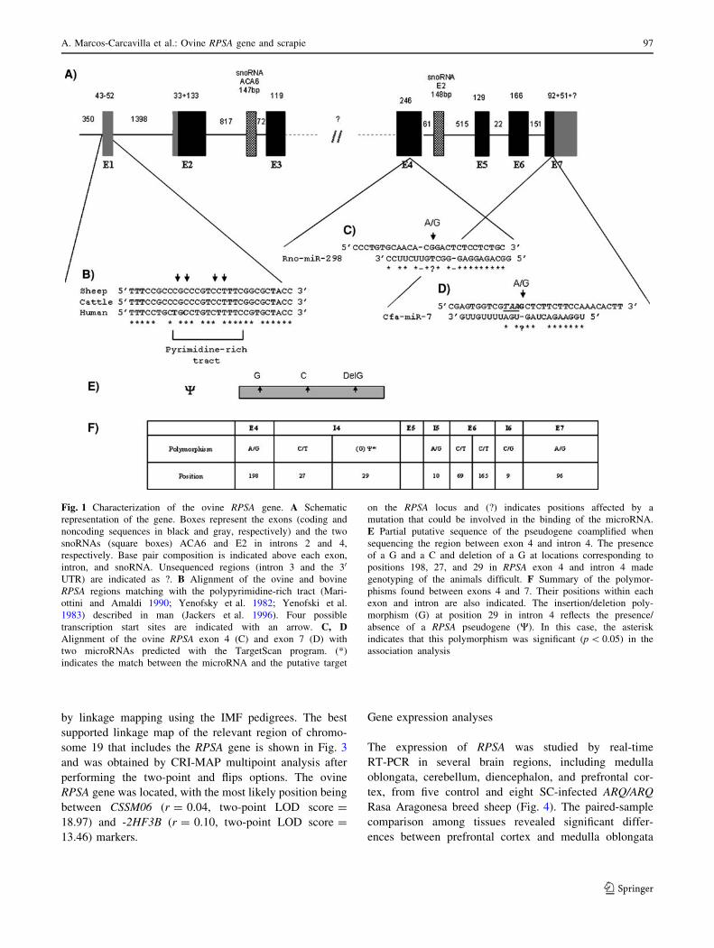
Fig. 1 Characterization of the ovine RPSA gene. A Schematic
representation of the gene. Boxes represent the exons (coding and
noncoding sequences in black and gray, respectively) and the two
snoRNAs (square boxes) ACA6 and E2 in introns 2 and 4,
respectively. Base pair composition is indicated above each exon,
intron, and snoRNA. Unsequenced regions (intron 3 and the 30
UTR) are indicated as ?. B Alignment of the ovine and bovine
RPSA regions matching with the polypyrimidine-rich tract (Mari-
ottini and Amaldi 1990; Yenofsky et al. 1982; Yenofski et al.
1983) described in man (Jackers et al. 1996). Four possible
transcription start sites are indicated with an arrow. C, DAlignment of the ovine RPSA exon 4 (C) and exon 7 (D) with
two microRNAs predicted with the TargetScan program. (*)
indicates the match between the microRNA and the putative target
on the RPSA locus and (?) indicates positions affected by a
mutation that could be involved in the binding of the microRNA.
E Partial putative sequence of the pseudogene coamplified when
sequencing the region between exon 4 and intron 4. The presence
of a G and a C and deletion of a G at locations corresponding to
positions 198, 27, and 29 in RPSA exon 4 and intron 4 made
genotyping of the animals difficult. F Summary of the polymor-
phisms found between exons 4 and 7. Their positions within each
exon and intron are also indicated. The insertion/deletion poly-
morphism (G) at position 29 in intron 4 reflects the presence/
absence of a RPSA pseudogene (W). In this case, the asterisk
indicates that this polymorphism was significant (p \ 0.05) in the
association analysis
A. Marcos-Carcavilla et al.: Ovine RPSA gene and scrapie 97
123

Fig. 2 Partial sequence of the ovine RPSA gene. A The DNA
sequence of the ovine RPSA gene between -350 bp from the first
possible transcription start point (+1) and exon 2. Exons 1 and 2 are
shown in capital bold letters. Four possible transcription start points
are indicated with aˇ. The first three were previously described in
chicken (Clausse et al. 1996) and man (Clausse et al. 1996; Jackers
et al. 1996), while the last one was predicted in silico. The translation
start codon (ATG) at exon 2 is in italics and underlined. Cis-
regulatory elements predicted within this segment are underlined. BSummary of the cis-regulatory elements identified with their sequence
and their exact position. Elements like the MRE or the hexamer motif
CTTCCG were previously described in human and chicken RPSAgenes (Clausse et al. 1996; Jackers et al. 1996). The remaining
elements were inferred in silico
Fig. 3 Cytogenetic (left) and genetic (right) maps showing localiza-
tion of the RPSA gene on sheep chromosome 19
Fig. 4 Relative expression level (arbitrary units) in the analyzed
brain regions of five control (white) and eight SC-infected (black/
gray) sheep. The mean (l) and the standard deviation (bars) for
normalized data are shown. **indicates significant differences
(p \ 0.01) in mRNA concentration between affected and healthy
animals
98 A. Marcos-Carcavilla et al.: Ovine RPSA gene and scrapie
123

(p = 0.00248), prefrontal cortex and diencephalon
(p = 0.00005), cerebellum and medulla oblongata
(p = 0.02745), cerebellum and prefrontal cortex
(p = 0.00109), and cerebellum and diencephalon
(p = 0.00104). In contrast, no differences were observed
between diencephalon and medulla oblongata (p [ 0.05).
On the other hand, significant differences in the
expression of RPSA between SC-infected and control
animals were found in cerebellum (p = 0.00585). Con-
versely, no differences were detected (p [ 0.05) in the
rest of the nervous tissues studied.
Polymorphism detection and association analysis
Eight polymorphic sites were identified within the 84 Rasa
Aragonesa breed animals in the RPSA region encoding the
two binding domains for PrP, at positions 198 in exon 4, 27
and 29 in intron 4, 10 in intron 5, 69 and 165 in exon 6, 9 in
intron 6, and 96 in exon 7 (Fig. 1 and GenBank sequence
EF394773). Sequencing of the region between exon 4 and
intron 4 of various animals provided confusing data. After
many assays using different combinations of primer pairs,
the existence of a nonprocessed pseudogene coamplifying
with the ovine RPSA gene and lacking the G at position 29
in intron 4 of the ovine gene was confirmed (see an
extended explanation in Appendix 3). In addition, the SNPs
located in exons 4, 6, and 7 were silent, and the SNP in
intron 5 was poorly represented (0.6%). Thus, only the
SNPs at positions 29 and 9 in introns 4 and 6, respectively,
were tested in the association analysis. The v2 tests among
maximum-likelihood estimations of the genotypes for the
polymorphisms in introns 4 and 6 (positions 29 and 9,
respectively) were significant only for the insertion/dele-
tion variation (p = 0.0177). Thus, 59.5% of the Rasa
Aragonesa breed sheep had a heterozygous genotype (+/-)
for the insertion/deletion polymorphism at position 29 in
the ovine RPSA intron 4, indicative of the presence of the
pseudogene. The distribution of this polymorphism among
the 84 Rasa Aragonesa breed sheep analyzed in the asso-
ciation test is given in Table 1. The v2 test (4.465)
for the contingency Table 1 indicates a significant
association (p = 0.035) between the presence/absence of
the pseudogene and the resistance/susceptibility to SC.
Thus, the pseudogene is present, at least in one copy, in
68.6% and 45.4% of the healthy and infected Rasa Ara-
gonesa breed sheep, respectively.
Interspecies amino acid sequence comparison
We decided to compare the LRP/LR amino acid
sequences of various mammals presenting different sus-
ceptibilities to prion infection (Knorr et al. 2007; Lysek
et al. 2005; Vorberg et al. 2003) to check if amino acid
changes could account for this variable response. Thus,
rabbit RPSA cDNA was isolated and sequenced (Gen-
Bank accession number EF222473). This rabbit cDNA
fragment included the region encoding both the direct
(161–179) and the putative indirect (180–285) binding
domains of the LRP/LR to the PrP as suggested by
Hundt et al. (2001). The rest of the LRP/LR sequences
that are represented in Fig. 5A are derived from nucle-
otide and amino acid sequences that were previously
deposited in the GenBank database. As shown in Fig. 5,
little variation exists within all the sequences. Positions
241, 272, and 290 seem to be the only locations that
vary considerably among the species examined (A241T,
A272TS, and A290T). In particular, positions 241 and
272 are of special interest because they affect the indi-
rect binding domain to PrP. The different amino acid
combinations at these positions in the LRP/LR sequence
for the species analyzed are summarized in Fig. 5B. In
this regard, Creighton (1992) determined that along a
peptide sequence, alanine is most commonly located in
the alpha-helix, whereas threonine seems to have pref-
erence for the beta-helix and serine for the coiled
regions. The possible role of these changes is discussed
in the section below.
Discussion
Our first efforts to isolate the ovine RPSA sequence from
genomic DNA were unsuccessful, but when ovine cDNA
samples and a BAC containing the ovine gene became
available, we could characterize the entire sequence of the
ovine RPSA gene with the exception of intron 3 (see Fig. 1
and GenBank accession numbers EF222474 and
EF394773). Four possible translational start sites were
identified (Figs. 1B, 2A). Nevertheless, additional experi-
ments need to be performed to determine the exact
transcription start point. Besides, several transcriptional
and posttranscriptional regulatory sequences were also
predicted (Figs. 1C, D, and 2). Furthermore, as previously
Table 1 Genotypes detected for the insertion/deletion polymorphism
(G) at position 29 in the ovine RPSA intron 4 which reflects the
presence of a RPSA pseudogene
(G) SC infected Healthy Total
(+/-) W 15 35 50
(+/+) no W 18 16 34
Total 33 51 84
v2 = 4.465 for the contingency table and p = 0.035 (p \ 0.05)
A. Marcos-Carcavilla et al.: Ovine RPSA gene and scrapie 99
123

reported in man (Jackers et al. 1996; Selvamurugan and
Eliceiri 1995; and GenBank accession numbers HUM-
SNE21X, AF375472, AJ609429, and HSU43901), the
localization of the ACA6 and E2 snoRNAs, required for
processing ribosomal RNA precursors (Kiss et al. 2004;
Nag et al. 1993; Selvamurugan and Eliceiri 1995), within
ovine RPSA introns 2 and 4, respectively, was also deter-
mined. Although ovine intron 3 was not sequenced in the
present work, it is known that its human and bovine
orthologs cover 2 and 9 kb, respectively. The possible
importance of the two Alu sequences contained within the
RPSA intron 3 has been discussed by Jackers et al. (1996).
The localization of the ovine RPSA gene on OAR19q13,
between CSSM06 and -2HF3B loci (Fig. 2), is consistent
with comparative mapping information because this gene
maps to human chromosome 3 and mouse chromosome 9.
It should be noted that mouse chromosome 9 has already
been reported as a chromosome that contains a QTL region
involved in the incubation period of SC (Moreno et al.
2003b), but the RPSA locus is outside the significant
interval related with this character.
Despite all the sheep analyzed in the expression
analysis belonging to the same breed (Rasa Aragonesa)
and PRNP genotype (ARQ/ARQ), real-time RT-PCR
results reflected a high individual variability (Fig. 4) that
could be explained by the presence of different strains or
a combination of different dose, route, duration, and time
of infection. These factors could not be controlled
because the animals studied here were all field cases.
Expression results revealed that independent of the dis-
ease status, medulla oblongata and diencephalon showed
between three and six times less mRNA than cerebellum
and prefrontal cortex, respectively. When comparing SC-
infected and control sheep, a significant decrease in
RPSA mRNA was observed in the cerebellum of infected
animals (p \ 0.01) (Fig. 4). Previous studies reported
that medulla oblongata and diencephalon were the brain
areas where more PrPsc deposition and neuronal damage
was observed in ARQ/ARQ SC-infected sheep (Caplazi
et al. 2004; Ligios et al. 2002; Vidal et al. 2006) This
finding could be interpreted as depending on the differ-
ent rate of LRP/LR production in the different brain
regions, which could be related to the possible involve-
ment of LRP/LR, in combination with PrPc and laminin,
in the neurodegeneration process as proposed Baloui
et al. (2004). Neverthelss, the existence of RPSA pseu-
dogenes that could be transcribed has to be considered.
Therefore, further expression analysis would be neces-
sary to determine if the overexpression of RPSA detected
in the present work is the result of the regulation of
either the RPSA functional gene or one or more RPSA
pseudogene(s).
Fig. 5 Comparison of the LRP/LR amino acid sequence among
several mammalian species showing different levels of resistance to
SC (Knorr et al. 2007; Lysek et al. 2005; Morel et al. 2005). AAlignment of LRP/LR amino acid sequences. Amino acid differences
among species are indicated by a ^. Arrows show amino acids 243
and 249 that vary in mouse. Alanine, serine, and threonine, which are
variable at 241, 272, and 290 positions, are highlighted in gray, black,
and square boxes, respectively. Other amino acids that also vary in rat
(aa 4), hamster (aa 183), and man (aa 293) are shown in bold letters.
Highlighted in black and in a square are the direct (aa161 to 179) and
the putative indirect (aa 180 to 285) binding domains of PrP on the
LRP/LR (Hundt et al. 2001). B A table summarizing the different
species compared, the accession numbers for the sequences used in
the alignment and the origin of these sequences (cDNA, genomic
DNA, or protein sequence), the amino acid composition at positions
241, 272, and 290 of the PrP, and the response of the different species
to SC infection
100 A. Marcos-Carcavilla et al.: Ovine RPSA gene and scrapie
123
On the other hand, interindividual differences in the
production, accumulation at the cell surface, and/or shed-
ding to the extracellular space of LRP/LR, as well as
differences at the LRP/LR amino acid sequence that affect
the interaction with the PrPsc, could be responsible for the
variable responses to SC infection. Thus, we decided to
look for polymorphisms in the regions involved in the
LRP/LR-PrPc/PrPsc interaction. Because all the mutations
found in this region were silent, they are not supposed to
have any effect on the binding domains to PrPc/PrPsc.
However, the genotyping of the segment between exon 4
and intron 4 revealed the presence of a RPSA nonprocessed
pseudogene in almost 60% of the sheep analyzed. Associa-
tion analysis revealed that the polymorphism at position 29
in intron 4 was overrepresented (p \ 0.05) in the group of
sheep that did not develop the disease despite being in con-
tact with the SC agent (Table 1). Nevertheless, due to the
reduced sample size, these results should be considered with
caution. In this way, the existence of pseudogenes with
biological roles has been described (Asano et al. 2004;
Balakirev and Ayala 2003; Hirotsune et al. 2003). Animals
bearing the pseudogene may have some advantage when
they are exposed to SC because, for instance, the pseudogene
might help in the regulation of the functional gene. This fact
would explain why the RPSA locus is outside the QTL
intervals related to the SC incubation period described to
date. Thus, it would be interesting to map this pseudogene in
sheep to check if its location corresponds to any previously
described QTL region related to incubation period and/or
resistance/susceptibility to SC (Moreno et al. 2003a).
On the other hand, the transmission of the TSE agent
from one species to another appears to depend on the
amino acid sequence homology between the host PrPc and
the exogenous PrPsc (Vorberg et al. 2003). Nonetheless, as
the involvement of additional factors cannot be ruled out,
we have compared the LRP/LR amino acid sequences from
various mammals, including dog, pig, and rabbit. The aim
of this comparison was to check if there was any important
variation along the LRP/LR sequence that could explain
the absence of a report in the literature about natural prion
infection in any of these three species.
As shown in Fig. 5, little variation exists among these
sequences. Although substitutions at positions 241, 272, and
290 are within or close to the putative indirect binding
domain to PrPc/PrPsc (aa 180–285), alteration of the global
structure of the interaction site is not expected (Knorr et al.
2007). However, it is possible that the different combinations
of amino acids through the LRP/LR sequence may modify its
specificity with other proteins affecting the interaction
between the LRP/LR from one species and prions from dif-
ferent origins, strengthening or weakening the species
barrier. For instance, Fig. 5B suggests that the presence of a
threonine at amino acid 241 of the LRP/LR protein confers
resistance to SC infection. Nevertheless, it is known that the
critical determinant in a protein function and interactions
with its ligands is its overall tertiary structure. Unfortunately,
the only way to check how these amino acid changes could
affect the interaction between LRP/LR with different prion
strains is by performing structural investigations that apply
crystallographic and/or nuclear magnetic resonance tech-
niques that are outside the scope of this study.
In the present work, the ovine RPSA gene was iso-
lated and characterized. Its organization and localization
agree with the data available in other species. Real-time
RT-PCR results revealed changes in the expression of
RPSA after natural SC infection in specific central ner-
vous system regions, indicating its possible involvement
in TSE-related neurodegeneration. We suggest that it
may be the presence of a RPSA nonprocessed pseudo-
gene, and not the variation in the ovine LRP/LR amino
acid sequence, that could explain the differences in
susceptibility to SC among sheep with the same PRNP
genotype. However, the possible involvement of this
receptor in the species barrier to SC and other TSEs
should not be discarded. In any case, further studies are
required to confirm the results presented here and to
examine the existence of polymorphisms at regions that
affect the gene and protein regulation and processing,
and the possible involvement of ovine RPSA pseudo-
genes in the incubation period of SC in sheep.
Acknowledgments The authors thank the CERSYRA-Valdepenas
and AGRAMA breeders association, CSIC-Leon, CITA-Aragon,
Prion Research Centre of the University of Zaragoza, INIA-Madrid,
and ETSIA-Polytechnique University of Madrid for kindly providing
Manchega, Awassi, Assaf, Rasa Aragonesa, and rabbit samples. The
authors are very grateful to Dr. C. Mansilla and Dr. F. Ponz for
helping improve the RT-PCR, to Dr. K.G. Dodds for correcting the
English of the manuscript, to Dr. M.E.F. Alves for her continuous
help, and to Dr. E.P. Cribiu and Dr. P. Zaragoza for allowing us to
perform the cytogenetic mapping and the expression analysis in
their respective laboratories. This work was supported by the
RTA2006–00104 INIA project and a Predoctoral Grant from the
INIA.
A. Marcos-Carcavilla et al.: Ovine RPSA gene and scrapie 101
123

Appendix
Appendix 1 List of primers used to amplify and sequence the ovine RPSA gene
Primer
localization
Foreward
primer
Reverse
primer
Amplicon
size
Ta Ext.
time
(-420)-Intron1R 50-TAACTTCTCCAGTTTTTGTT-30 50-CCTATGCAACACCTTGGAAAATCA-30 1600 55 10 450 0
Exon1-Intron2 50-GGGGTCCATACGGCGTTGT-30 50-TAGCTGGAATGACCAAGCAAAGAG-30 1700 61 10 450 0
Intron1 50-TTACCGGAAAGATCAAACTTCACG-30 50-CCTATGCAACACCTTGGAAAATCA-30 800 55 10
Intron1F-Intron2R 50-TTACCGGAAAGATCAAACTTCACG-30 50-TAGCTGGAATGACCAAGCAAAGAG-30 1200 60 10 300 0
Exon2-Exon3 50-TTACGACCACTCGGTGGTGGTTC-30 50-GAAGCTTCTCCCACGTCCTCTTG -30 1200 50 450 0
Exon2-Exon3 50-GCAGCAGGAACCCACTTAGG-30 50-GACATCAGCCGGGTTTTCAAT-30 200 60 300 0
Intron2 50-AGAGGGCTCTCACTAAGTAACTGA-30 50-TTCCATGCAGAATCCAAGAAG-30 700 55 300 0
Intron2-Exon3 50-AGAGGGCTCTCACTAAGTAACTGA-30 50-GACATCAGCCGGGTTTTCAAT-30 1000 57 10 300 0
Intron2-Exon3 50-AGCCTTAGAGGGCTCTCACT-30 50-GACATCAGCCGGGTTTTCAAT-30 1000 60 10
Exon4-Intron4 50-CGAGCTGTGCTGAAGTTTGC-30 50-CAGCAGACTGTTAATTTGAAAGTG-30 800 60 10
Exon4-Intron4 50-CTCCGGGAACCTTCACTAACCA-30 50-CAGCAGACTGTTAATTTGAAAGTG-30 775 65 10
Exon4-Intron4 50-CGAGCTGTGCTGAAGTTTGC-30 50-GGCCTCAACTCCAAGCTCTA-30 350 60 450 0
Exon4-Intron6 50-CGAGCTGTGCTGAAGTTTGC-30 50-CCCCGTCCCACCAATTTGC-30 1500 51 10 300 0
Intron4 50-CTGGCCCAATGAGTGGAG-30 50-CAGCAGACTGTTAATTTGAAAGTG-30 475 66 300 0
Intron4-Exon7 50-TGGGGGTGCGTATTACTGTT-30 50-GTTTATTTCCATCAACCA-30 750 52 10
Exon5-Exon6 50-TACAGGGACCCCGAGGAG-30 50-CACCTGCACGCCTTCAGA-30
Appendix 2 List of primers and probes used for performing RT-PCR
Gene Primer and probes sequences Concentration (nM) Size
RPSA F: 50-CAGGGACCCCGAGGAGATT-30 300
R: 50-CACCTGCACGCCTTCAGA-30 300 151 pb
G6PDH F: 50-TGACCTATGGCAACCGATACAA-30 300
(Garcıa-Crespo et al. 2005) R: 50-CCGCAAAAGACATCCAGGAT-30 300 76 pb
GAPDH F: 50-TCCATGACCACTTTGGCATCGT-30 300
(Lyahyai et al. 2006) R: 50-GTCTTCTGGGTCGCAGTGA-30 300
P: FAM- AGGGACTTATGACCACTGTCCACGCC -TAMRA 150 70 pb
ACTB F: 50-ATGCCTCCTGCACCACCA-30 300
(Garcıa-Crespo et al. 2005) R: 50-GCATTTGCGGTGGACGAT-30 300 125 pb
102 A. Marcos-Carcavilla et al.: Ovine RPSA gene and scrapie
123

Table A1
PCR1 PCR2
Amplified F1-R1 F2-R2
Sequenced with F1 Sequence code secR Sequence code
Polymorphism position E4198 I427 I429 E4198 I427 I429
Animal
A AG CT II 02P7Y AG CT II 033ZQ
B GG CT ID 02P7N GG CT II 036IM
C AG CT ID 02P7U AG TT II 036I6
A. Marcos-Carcavilla et al.: Ovine RPSA gene and scrapie 103
123

Appendix 3
Figure A1 is a representation of the PCRs that confirm the
presence of a RPSA pseudogene coamplifying with the
RPSA functional gene when performing the amplification
reactions to test the polymorphisms located in exon 4
(represented in capital bold letters) and intron 4 (repre-
sented in lower-case) for the association analysis.
Underlined and enclosed between square brackets is the
snoRNA E2. Primers used are in gray. The polymorphisms
found among this region are indicated in bold and under-
lined letters. Table A1 summarizes the primers used in the
amplification and sequencing reactions as well as several
animals whose genotypes for the polymorphisms at posi-
tions 198 in exon 4 and 27 and 29 in intron 4 allowed us to
infer the presence of a RPSA pseudogene which was not
present in all the animals analyzed. The code of each
sequence is also indicated. Thus, animal A did not bear the
pseudogene, so results from both PCRs are consistent.
Conversely, animals B and C bore the pseudogene, which
was inferred by the insertion/deletion of a G (highlighted in
gray) when performing PCR1 but not PCR2. In addition,
animal B was heterozygous for the SNP at position 29 in
intron 4, while animal C was homozygous (TT) for the same
position. However, in the last case, the TT genotype was not
possible to determine from the 02P7U sequence, which
included the sequence of both the gene and the pseudogene.
References
Adjou KT, Simoneau S, Sales N, Lamoury F, Dormont D et al. (2003)
A novel generation of heparan sulfate mimetics for the treatment
of prion diseases. J Gen Virol 84:2595–2603
Asano Y, Takashima S, Asakura M, Shintani Y, Liao Y et al. (2004)
Lamr1 functional retroposon causes right ventricular dysplasia in
mice. Nat Genet 36:123–130
Auth D, Brawerman G (1992) A 33-kDa polypeptide with homology
to the laminin receptor: component of translation machinery.
Proc Natl Acad Sci USA 89:4368–4372
Balakirev ES, Ayala FJ (2003) Pseudogenes: are they ‘‘junk’’ or
functional DNA? Annu Rev Genet 37:123–151
Baloui H, von Boxberg Y, Vinh J, Weiss S, Rossier J et al. (2004)
Cellular prion protein/laminin receptor: distribution in adult
central nervous system and characterization of an isoform
associated with a subtype of cortical neurons. Eur J Neurosci
20:2605–2616
Baylis M, Chihota C, Stevenson E, Goldmann W, Smith A et al.
(2004) Risk of scrapie in British sheep of different prion protein
genotype. J Gen Virol 85:2735–2740
Ben-Zaken O, Tzaban S, Tal Y, Horonchik L, Esko JD et al. (2003)
Cellular heparan sulfate participates in the metabolism of prions.
J Biol Chem 278:40041–40049
Caplazi P, O’Rourke K, Wolf C, Shaw D, Baszler TV (2004) Biology
of PrPsc accumulation in two natural scrapie-infected sheep
flocks. J Vet Diagn Invest 16:489–496
Castilla J, Gutierrez-Adan A, Brun A, Doyle D, Pintado B et al.
(2004) Subclinical bovine spongiform encephalopathy infection
in transgenic mice expressing porcine prion protein. J Neurosci
24:5063–5069
Castronovo V, Claysmith AP, Barker KT, Cioce V, Krutzsch HC
et al. (1991) Biosynthesis of the 67 kDa high affinity laminin
receptor. Biochem Biophys Res Commun 177:177–183
Clausse N, Jackers P, Jares P, Joris B, Sobel ME et al. (1996)
Identification of the active gene coding for the metastasis-
associated 37LRP/p40 multifunctional protein. DNA Cell Biol
15:1009–1023
Crawford AM, Dodds KG, Ede AJ, Pierson CA, Montgomery GW,
et al. (1995) An autosomal genetic linkage map of the sheep
genome. Genetics 140:703–724
Creighton TE (1992) Protein folding pathways determined using
disulphide bonds. Bioessays 14:195–199
Garcia-Crespo D, Juste R, Hurtado A (2005) Selection of ovine
housekeeping genes for normalisation by real-time RT-PCR;
analysis of PrP gene expression and genetic susceptibility to
scrapie. BMC Vet Res 1:3
Gauczynski S, Peyrin JM, Haik S, Leucht C, Hundt C et al. (2001)
The 37-kDa/67-kDa laminin receptor acts as the cell-surface
receptor for the cellular prion protein. EMBO J 20:5863–5875
Gauczynski S, Nikles D, El-Gogo S, Papy-Garcia D, Rey C et al.
(2006) The 37-kDa/67-kDa laminin receptor acts as a receptor
for infectious prions and is inhibited by polysulfated glycanes. J
Infect Dis 194:702–709
Goldmann W, Baylis M, Chihota C, Stevenson E, Hunter N (2005)
Frequencies of PrP gene haplotypes in British sheep flocks and
the implications for breeding programmes. J Appl Microbiol
98:1294–1302
Hayes H, Petit E, Dutrillaux B (1991) Comparison of RBG-banded
karyotypes of cattle, sheep and goats. Cytogenet Cell Genet
57:51–55
Hirotsune S, Yoshida N, Chen A, Garrett L, Sugiyama F et al. (2003)
An expressed pseudogene regulates the messenger-RNA stability
of its homologous coding gene. Nature 423:91–96
Horonchik L, Tzaban S, Ben-Zaken O, Yedidia Y, Rouvinski A et al.
(2005) Heparan sulfate is a cellular receptor for purified
infectious prions. J Biol Chem 280:17062–17067
Hundt C, Peyrin JM, Haik S, Gauczynski S, Leucht C et al. (2001)
Identification of interaction domains of the prion protein with its
37-kDa/67-kDa laminin receptor. EMBO J 20:5876–5886
Jackers P, Clausse N, Fernandez M, Berti A, Princen F et al. (1996)
Seventeen copies of the human 37 kDa laminin receptor
precursor/p40 ribosome-associated protein gene are processed
pseudogenes arisen from retropositional events. Biochim Bio-
phys Acta 1305:98–104
Karpatova M, Tagliabue E, Castronovo V, Magnifico A, Ardini E
et al. (1996) Shedding of the 67-kD laminin receptor by human
cancer cells. J Cell Biochem 60:226–234
Kim WH, Lee BL, Jun SH, Song SY, Kleinman HK (1998)
Expression of 32/67-kDa laminin receptor in laminin adhesion-
selected human colon cancer cell lines. Br J Cancer 77:15–20
Kinoshita K, Kaneda Y, Sato M, Saeki Y, Wataya-Kaneda M et al.
(1998) LBP-p40 binds DNA tightly through associations with
histones H2A, H2B, and H4. Biochem Biophys Res Commun
253:277–282
Kiss AM, Jady BE, Bertrand E, Kiss T (2004) Human box H/ACA
pseudouridylation guide RNA machinery. Mol Cell Biol
24:5797–5807
Knorr C, Beuermann C, Beck J, Brenig B (2007) Characterization of
the porcine multicopy ribosomal protein SA/37-kDa laminin
receptor gene family. Gene 395:135–143
Kocisko DA, Priola SA, Raymond GJ, Chesebro B, Lansbury PT Jr
et al. (1995) Species specificity in the cell-free conversion of
prion protein to protease-resistant forms: a model for the scrapie
species barrier. Proc Natl Acad Sci USA 92:3923–3927
104 A. Marcos-Carcavilla et al.: Ovine RPSA gene and scrapie
123

Lander ES, Green P (1987) Construction of multilocus genetic
linkage maps in humans. Proc Natl Acad Sci USA 84:2363–2367
Landowski TH, Dratz EA, Starkey JR (1995) Studies of the structure
of the metastasis-associated 67 kDa laminin binding protein:
fatty acid acylation and evidence supporting dimerization of the
32 kDa gene product to form the mature protein. Biochemistry
34:11276–11287
Leucht C, Simoneau S, Rey C, Vana K, Rieger R et al. (2003) The
37 kDa/67 kDa laminin receptor is required for PrP(Sc) prop-
agation in scrapie-infected neuronal cells. EMBO Rep 4:290–
295
Ligios C, Jeffrey M, Ryder SJ, Bellworthy SJ, Simmons MM (2002)
Distinction of scrapie phenotypes in sheep by lesion profiling. J
Comp Pathol 127:45–57
Lyahyai J, Bolea R, Serrano C, Monleon E, Moreno C et al. (2006)
Correlation between Bax overexpression and prion deposition in
medulla oblongata from natural scrapie without evidence of
apoptosis. Acta Neuropathol (Berl) 112:451–460
Lysek DA, Schorn C, Nivon LG, Esteve-Moya V, Christen B et al.
(2005) Prion protein NMR structures of cats, dogs, pigs, and
sheep. Proc Natl Acad Sci USA 102:640–645
Lloyd SE, Onwuazor ON, Beck JA, Mallinson G, Farrall M et al.
(2001) Identification of multiple quantitative trait loci linked to
prion disease incubation period in mice. Proc Natl Acad Sci USA
98:6279–6283
Lloyd SE, Uphill JB, Targonski PV, Fisher EM, Collinge J (2002)
Identification of genetic loci affecting mouse-adapted bovine
spongiform encephalopathy incubation time in mice. Neuroge-
netics 4:77–81
Maddox JF, Davies KP, Crawford AM, Hulme DJ, Vaiman D et al.
(2001) An enhanced linkage map of the sheep genome
comprising more than 1000 loci. Genome Res 11:1275–1289
Manolakou K, Beaton J, McConnell I, Farquar C, Manson J et al.
(2001) Genetic and environmental factors modify bovine
spongiform encephalopathy incubation period in mice. Proc
Natl Acad Sci USA 98:7402–7407
Mariottini P, Amaldi F (1990) The 50 untranslated region of mRNA
for ribosomal protein S19 is involved in its translational
regulation during Xenopus development. Mol Cell Biol
10:816–822
Miller S, Dykes D, Polesky H (1988) A simple salting out procedure
for extracting DNA from human nucleated cells. Nucleic Acids
Res 16:1215
Morel E, Andrieu T, Casagrande F, Gauczynski S, Weiss S et al.
(2005) Bovine prion is endocytosed by human enterocytes via
the 37 kDa/67 kDa laminin receptor. Am J Pathol 167:1033–
1042
Moreno CR, Cosseddu GM, Andreoletti IO, Schibler L, Roig A et al
(2003a) Identification of quantitative trait loci (QTL) modulating
prion incubation period in sheep. Proceedings of the Interna-
tional Workshop on Major Genes and QTL in Sheep and Goat,
Toulouse
Moreno CR, Lantier F, Lantier I, Sarradin P, Elsen JM (2003b)
Detection of new quantitative trait loci for susceptibility to
transmissible spongiform encephalopathies in mice. Genetics
165:2085–2091
Nag MK, Thai TT, Ruff EA, Selvamurugan N, Kunnimalaiyaan M
et al. (1993) Genes for E1, E2, and E3 small nucleolar RNAs.
Proc Natl Acad Sci USA 90:9001–9005
Priola SA, Caughey B, Race RE, Chesebro B (1994) Heterologous
PrP molecules interfere with accumulation of protease-resistant
PrP in scrapie-infected murine neuroblastoma cells. J Virol
68:4873–4878
Prusiner SB (1998) Prions. Proc Natl Acad Sci USA 95:13363–13383
Rabacchi SA, Neve RL, Drager UC (1990) A positional marker for
the dorsal embryonic retina is homologous to the high-affinity
laminin receptor. Development 109:521–531
Rao M, Manishen WJ, Maheshwari Y, Sykes DE, Siyanova EY et al.
(1994) Laminin receptor expression in rat intestine and liver
during development and differentiation. Gastroenterology
107:764–772
Rieger R, Edenhofer F, Lasmezas CI, Weiss S (1997) The human 37-
kDa laminin receptor precursor interacts with the prion protein in
eukaryotic cells. Nat Med 3:1383–1388
SAS Institute Inc. (1989) SAS/STAT� Users’s Guide, version 6, 4th
ed., vol. 1 (Cary, NC: SAS Institute, Inc.)
Scott M, Groth D, Foster D, Torchia M, Yang SL et al. (1993)
Propagation of prions with artificial properties in transgenic mice
expressing chimeric PrP genes. Cell 73:979–988
Selvamurugan N, Eliceiri GL (1995) The gene for human E2 small
nucleolar RNA resides in an intron of a laminin-binding protein
gene. Genomics 30:400–401
Shmakov AN, Bode J, Kilshaw PJ, Ghosh S (2000) Diverse patterns
of expression of the 67-kD laminin receptor in human small
intestinal mucosa: potential binding sites for prion proteins? J
Pathol 191:318–322
Simoneau S, Haik S, Leucht C, Dormont D, Deslys JP et al. (2003)
Different isoforms of the non-integrin laminin receptor are
present in mouse brain and bind PrP. Biol Chem 384:243–246
Simons K, Toomre D (2000) Lipid rafts and signal transduction. Nat
Rev Mol Cell Biol 1:31–39
Stephenson DA, Chiotti K, Ebeling C, Groth D, DeArmond SJ et al.
(2000) Quantitative trait loci affecting prion incubation time in
mice. Genomics 69:47–53
Vaiman D, Billault A, Tabet-Aoul K, Schibler L, Vilette D et al.
(1999) Construction and characterization of a sheep BAC library
of three genome equivalents. Mamm Genome 10:585–587
Vana K, Weiss S (2006) A trans-dominant negative 37 kDa/67 kDa
laminin receptor mutant impairs PrP(Sc) propagation in scrapie-
infected neuronal cells. J Mol Biol 358:57–66
Vandesompele J, De Preter K, Pattyn F, Poppe B, Van Roy N et al.
(2002) Accurate normalization of real-time quantitative RT-PCR
data by geometric averaging of multiple internal control genes.
Genome Biol 3:RESEARCH0034
Vidal E, Bolea R, Tortosa R, Costa C, Domenech A et al. (2006)
Assessment of calcium-binding proteins (Parvalbumin and
Calbindin D-28 K) and perineuronal nets in normal and
scrapie-affected adult sheep brains. J Virol Meth 36:137–146
Vorberg I, Groschup MH, Pfaff E, Priola SA (2003) Multiple amino
acid residues within the rabbit prion protein inhibit formation of
its abnormal isoform. J Virol 77:2003–2009
Yenofsky R, Bergmann I, Brawerman G (1982) Messenger RNA
species partially in a repressed state in mouse sarcoma ascites
cells. Proc Natl Acad Sci USA 79:5876–5880
Yenofsky R, Cereghini S, Krowczynska A, Brawerman G (1983)
Regulation of mRNA utilization in mouse erythroleukemia cells
induced to differentiate by exposure to dimethyl sulfoxide. Mol
Cell Biol 3:1197–1203
Yow H, Wong JM, Chen HS, Lee C, Steele GD et al. (1988) Increased
mRNA expression of a laminin-binding protein in human colon
carcinoma: complete sequence of a full-length cDNA encoding
the protein. Proc Natl Acad Sci USA 85:6394–6398
A. Marcos-Carcavilla et al.: Ovine RPSA gene and scrapie 105
123