Polypyrrole doped with 2 peptide sequences from laminin
9
Biomaterials 27 (2006) 2405–2413 Polypyrrole doped with 2 peptide sequences from laminin William R. Stauffer a,b , Xinyan T. Cui a,b, a Department of Bioengineering, University of Pittsburgh, Pittsburgh, PA 15261, USA b McGowan Institute for Regenerative Medicine, University of Pittsburgh, Pittsburgh, PA 15203, USA Received 19 July 2005; accepted 29 October 2005 Abstract In the field of neural tissue engineering, electrically conducting, biocompatible surfaces are of great interest. Over the past several decades conducting polymers have been studied as candidate surfaces because they fit these criteria. Several attempts have been made to combine the conductivity and biocompatibility of conducting polymers with biomolecules that could promote specific cell attachment and growth. In this report the laminin fragments CDPGYIGSR (p31) and RNIAEIIKDI (p20) are used as dopants in electropolymerization of the conducting polymer polypyrrole (PPy). The electrical properties of the resulting films are analyzed by impedance spectroscopy and cyclic voltammetry and compared to gold. PPy/p20 surfaces consistently demonstrate the lowest impedance and largest charge capacity for a given deposition charge. Next, in vitro studies using primary neurons cultured in a defined media and primary astrocytes in a serum containing media were performed; neuron density and neurite length, as well as astrocyte density, were quantified. Surfaces doped with a combination of the two peptides (PPy/p20-p31) consistently supported the highest neuronal density. It is shown that surfaces doped with the laminin fragment p20 had significantly longer primary neurites than either the p31 doped or poly(styrenesulfonate) doped PPy surfaces. Finally, the astrocyte studies demonstrate that PPy surfaces have significantly less astrocyte adhesion in culture than the common electrode material, gold. r 2005 Elsevier Ltd. All rights reserved. Keywords: Biocompatibility; Neural cell; Neural prosthesis; Laminin; Cell adhesion; Brain; Electroactive polymer; Electrochemistry 1. Introduction Polypyrrole (PPy) is a biocompatible, electrically active, and conductive material [1–9]. This makes it an ideal candidate for electrodes used in neural prostheses and guidance channels for nerve regeneration where electrical signal conduction is the implicit function. Previous research has examined neurite outgrowth on PPy films both with and without electrical stimulation and found that neurite extension was enhanced on PPy films when an electrical current was passed through the films [3]. Using the Michigan recording probes, Cui et al. [4,6,7] showed that PPy coatings on the surface of the electrodes dramatically reduced electrical impedance and improved signal conduction. Along with the electrical properties exhibited by PPy, another feature that can be exploited for regenerative medicine purposes is the dopant selection. Electropolymer- ization of conducting polymers incorporates counter ions in solution, referred to as dopants, to balance charge [9].A common non-bioactive dopant used in many studies is the anionic poly(styrenesulfonate) (PSS) [2–4,7–12]. However, of great interest is the use of biological agents as dopants. These biological agents are immobilized on the PPy surface and are able to interact locally with cells. In one study, the proteoglycan, hyaluronic acid, was used as a dopant during electropolymerization. The resulting surfaces promoted vascularization in vivo but did not promote PC-12 cell attachment in vitro [8]. In a separate study, the laminin fragment CDPGYIGSR was used as a dopant in the surface modification of Michigan recording electrodes. In vitro studies showed that neuroblastoma cells grew preferentially on and around electrodes coated with PPy/CDPGYIGSR compared to PPy/CH 3 COO films [7]. Furthermore, in vivo studies demonstrated that electrodes surface modified with PPy/CDPGYIGSR showed a significant increase in local neurofilament staining 1 week after implantation [6]. ARTICLE IN PRESS www.elsevier.com/locate/biomaterials 0142-9612/$ - see front matter r 2005 Elsevier Ltd. All rights reserved. doi:10.1016/j.biomaterials.2005.10.024 Corresponding author. Department of Bioengineering, University of Pittsburgh, Pittsburgh, PA 15261, USA. Tel.: +1 412 624 1079; fax: +1 412 383 8788. E-mail address: [email protected] (X.T. Cui).
Transcript of Polypyrrole doped with 2 peptide sequences from laminin

ARTICLE IN PRESS
0142-9612/$ - se
doi:10.1016/j.bi
�CorrespondPittsburgh, Pit
+1 412 383 878
E-mail addr
Biomaterials 27 (2006) 2405–2413
www.elsevier.com/locate/biomaterials
Polypyrrole doped with 2 peptide sequences from laminin
William R. Stauffera,b, Xinyan T. Cuia,b,�
aDepartment of Bioengineering, University of Pittsburgh, Pittsburgh, PA 15261, USAbMcGowan Institute for Regenerative Medicine, University of Pittsburgh, Pittsburgh, PA 15203, USA
Received 19 July 2005; accepted 29 October 2005
Abstract
In the field of neural tissue engineering, electrically conducting, biocompatible surfaces are of great interest. Over the past several
decades conducting polymers have been studied as candidate surfaces because they fit these criteria. Several attempts have been made to
combine the conductivity and biocompatibility of conducting polymers with biomolecules that could promote specific cell attachment
and growth. In this report the laminin fragments CDPGYIGSR (p31) and RNIAEIIKDI (p20) are used as dopants in
electropolymerization of the conducting polymer polypyrrole (PPy). The electrical properties of the resulting films are analyzed by
impedance spectroscopy and cyclic voltammetry and compared to gold. PPy/p20 surfaces consistently demonstrate the lowest impedance
and largest charge capacity for a given deposition charge. Next, in vitro studies using primary neurons cultured in a defined media and
primary astrocytes in a serum containing media were performed; neuron density and neurite length, as well as astrocyte density, were
quantified. Surfaces doped with a combination of the two peptides (PPy/p20-p31) consistently supported the highest neuronal density. It
is shown that surfaces doped with the laminin fragment p20 had significantly longer primary neurites than either the p31 doped or
poly(styrenesulfonate) doped PPy surfaces. Finally, the astrocyte studies demonstrate that PPy surfaces have significantly less astrocyte
adhesion in culture than the common electrode material, gold.
r 2005 Elsevier Ltd. All rights reserved.
Keywords: Biocompatibility; Neural cell; Neural prosthesis; Laminin; Cell adhesion; Brain; Electroactive polymer; Electrochemistry
1. Introduction
Polypyrrole (PPy) is a biocompatible, electrically active,and conductive material [1–9]. This makes it an idealcandidate for electrodes used in neural prostheses andguidance channels for nerve regeneration where electricalsignal conduction is the implicit function. Previous researchhas examined neurite outgrowth on PPy films both with andwithout electrical stimulation and found that neuriteextension was enhanced on PPy films when an electricalcurrent was passed through the films [3]. Using the Michiganrecording probes, Cui et al. [4,6,7] showed that PPy coatingson the surface of the electrodes dramatically reducedelectrical impedance and improved signal conduction.
Along with the electrical properties exhibited by PPy,another feature that can be exploited for regenerative
e front matter r 2005 Elsevier Ltd. All rights reserved.
omaterials.2005.10.024
ing author. Department of Bioengineering, University of
tsburgh, PA 15261, USA. Tel.: +1 412 624 1079; fax:
8.
ess: [email protected] (X.T. Cui).
medicine purposes is the dopant selection. Electropolymer-ization of conducting polymers incorporates counter ions insolution, referred to as dopants, to balance charge [9]. Acommon non-bioactive dopant used in many studies is theanionic poly(styrenesulfonate) (PSS) [2–4,7–12]. However,of great interest is the use of biological agents as dopants.These biological agents are immobilized on the PPy surfaceand are able to interact locally with cells. In one study, theproteoglycan, hyaluronic acid, was used as a dopant duringelectropolymerization. The resulting surfaces promotedvascularization in vivo but did not promote PC-12 cellattachment in vitro [8]. In a separate study, the lamininfragment CDPGYIGSR was used as a dopant in the surfacemodification of Michigan recording electrodes. In vitrostudies showed that neuroblastoma cells grew preferentiallyon and around electrodes coated with PPy/CDPGYIGSRcompared to PPy/CH3COO� films [7]. Furthermore, in vivostudies demonstrated that electrodes surface modified withPPy/CDPGYIGSR showed a significant increase in localneurofilament staining 1 week after implantation [6].

ARTICLE IN PRESSW.R. Stauffer, X.T. Cui / Biomaterials 27 (2006) 2405–24132406
The goal of this work was to design an electricallyconductive surface that is not merely biocompatible butactively promotes the growth and differentiation of desiredcells types in vitro. Previous in vitro studies have usedneuroblastoma or PC-12 cells cultured in a serum basedmedia with or without nerve growth factor (NGF)[2,3,7,8,11] or primary neurons in a serum based media[12]. Serum based media contains an unknown mixture ofproteins which adsorb onto the surface to facilitate cellattachment and growth, thereby diminishing the effect ofany surface immobilized biomolecule. Furthermore, neu-ron-like cell lines such as PC-12 and neuroblastoma cellsare excellent means of establishing preliminary data.However, these cell lines lack key neuronal features suchas polarity of markers, and the ability to form synapticconnections (neuroblastoma cells can be forced to formconnections by over expression of synapsin IIB) that makesthem fundamentally different from primary neurons [13].In this work we examine the effects of PPy surfaces dopedwith laminin fragments on primary neuronal culture in adefined media with no added growth factors.
Laminin is a multifunctional extracellular matrix (ECM)protein and basement membrane component associatedwith several cell types. As such, different peptide sequenceswithin laminin are responsible for its various functions[14–16]. Two well characterized cell binding domains arep31 (CDPGYIGSR) and p20 (RNIAEIIKDI). It has beenshown that p31 plays a role in the binding of manydifferent cell types including neurons and astrocytes [15].Furthermore, as indicated earlier, p31 has been utilized as adopant in electropolymerization of PPy and the growth ofglial and neuroblastoma cells were quantified on thesurface. The in vivo neuronal tissue response to PPy/p31coated electrodes has also been assessed [6,7]. Conversely,previous literature has suggested that p20 is a neuriteoutgrowth promoting domain of laminin [14]. To the bestof our knowledge, p20 has never been utilized as a dopantin the electropolymerization of conducting polymers.
In this work, the electrical properties of the different PPysurfaces were tested and compared for enhanced transmis-sion of electrical signals. It is demonstrated that the chosenbiomolecular dopants have a significant effect on neuronattachment and neurite extension. The work shows that theeffect on neuron growth occurs specifically through thebioactive species immobilized on the surface. Finally,astrocyte growth on the different surfaces was examinedand it is shown that fewer cells attached and grew on PPysurfaces than on gold after 3 days in culture.
2. Materials and methods
2.1. Substrate preparation
Gold-coated plastic coverslips (Fisher) were used as a model system for
the electrochemical deposition of polymer and as cell culture substrates. In
order to ensure constant current density and provide suitable sized
surfaces to fit in tissue culture wells, the coverslips were cut to a uniform
dimension of 7mm� 22mm. The coverslips were cleaned with 8 N HNO3
for 30min followed by two washes in deionized H2O and stored in ethanol
until gold deposition.
To create a conductive surface for electrochemical deposition, the cut,
cleaned coverslips were sputter coated with a layer of gold using a
Cressington Sputter Coater (Cressington Scientific Instruments, Inc.) with
thickness control. The thickness of the gold layer was approximately
40 nm. The coated coverslips were stored in a dessicator until used.
2.2. Electrochemical deposition
The peptides DCDPGYIGSR (p31) and DRNIAEIIKDIC (p20) were
synthesized at the Peptide Synthesis Facility at the University of
Pittsburgh. An aspartic acid (D) was added on the N terminal of each
peptide to increase the negative charge for more efficient dopant
incorporation. A cysteine (C) was added to the C terminal of p20 for
additional coupling capability. Two types of electrodes were used for
deposition of PPy/peptide film, gold electrode sites (1.25� 10�5 cm2) of a
Michigan Probe (NeuroNexus), and gold-coated coverslips (0.7 cm2).
The films on the electrode sites of the Michigan probe were grown
under an anodic current density of 0.5mA/cm2. The electrochemical cell
was a 2ml glass cuvette with a platinum plate as the counter electrode. The
monomer solution contained 0.5M pyrrole (Sigma) and 5mg/ml of
peptide. Thickness of the films was controlled by time of deposition. On
gold coated coverslips, PPy/peptide films were grown galvanostatically
under varying anodic current density for electrochemical studies and for
use as cell culture substrates. The dopants used were the laminin fragments
p31 and p20, both at 5mg/ml, a mixture (50:50) of the two peptides at a
total peptide concentration of 5mg/ml, and the non-bioactive dopant PSS
at 0.1M. To analyze the electrochemical properties and to optimize the
thickness for cell growth, five different deposition charges (1.25–25mC)were used during electropolymerization.
2.3. Surface characterization
2.3.1. Thickness measurement
The PPy film thickness was measured with a Gaertner L-117 Null
ellipsometer (Gaertner Scientific Corporation). A linearly polarized
He–Ne laser with wavelength 632.8 nm was used in the measurement at
incident angle 701. The analyzer (A) can detect the change in the phase
angle (D) and the amplitude change (C) of the light after reflection from
the sample. Refractive indices of n1 ¼ 1 for air, n2 ¼ 1:45 for the organic
molecular monolayer, and n3 ¼ 0:166þ 3:22i for gold were used to
calculate the thickness of films. The measurements were performed four
times at different locations for each sample, and the results are reported as
an average of the measurements.
2.3.2. Scanning electron microscopy
Surface morphology was analyzed by Scanning Electron Microscopy
(SEM). Specimens were synthesized according to the method described
above, and then a 3.5 nm coating of Au/Pd was sputtered on the surface.
Imaging was done in a JEOL 9335 Field Emission Gun SEM (Jeol USA)
at 5KV. Cells growing on the different surfaces were analyzed by SEM
also. Biological samples were prepared with 2.5% gluteraldehyde and 1%
osmium-tetroxide, both for 1 h, followed by dehydration and critical point
drying.
2.3.3. Fourier transformed infrared spectroscopy
To characterize the chemical composition of the PPy/biomolecule
coatings, reflective Fourier transform infrared (FTIR) spectroscopy was
performed using a Thermo-Nicolette Magna-IR 560 Spectrometer
attached to a Continuum microscope equipped with a liquid nitrogen-
cooled MCT detector (Thermo Electron Corporation). The microscope
accessory enabled the focusing of the IR beam on a small area of the
sample. The microscope aperture was used to adjust the infrared light spot
area. The background signal was collected from a gold mirror. The

ARTICLE IN PRESS
Fig. 1. Bode plot of impedance spectroscopy of PPy/p20 coatings on gold
coverslips deposited at different charge densities (current density of 36 mA/
cm2 for 50, 125, 250, 500, and 1000 s).
W.R. Stauffer, X.T. Cui / Biomaterials 27 (2006) 2405–2413 2407
samples were scanned 100 times in the range of 4000–650 cm–1. OMNIC
software was used to generate the FTIR spectrum.
2.4. Electrochemical impedance spectroscopy and cyclic
voltammetry
Electrochemical impedance spectroscopy was performed using a
Gamry Potentiostat, FAS2/Femostat (Gamry Instruments), under the
control of the Gamry Framework software. A solution of 0.1M PBS buffer
(pH ¼ 7.0) was used as the electrolyte in a three-electrode cell. A platinum
foil was used as the counter electrode, a saturated calomel electrode (SCE)
was employed as the reference electrode, and the electrode (gold coated
coverslip or microelectrode sites on the Michigan probe) with or without
polymer coatings, was used as the working electrode. An AC sinusoid of
5mV amplitude was applied as the input signal with the DC potential set
to 0V. The value of the impedance was determined at four discrete
frequencies per decade over the range of 10–105Hz.
Cyclic voltammetry (CV) was performed using the same electrochemi-
cal setup as described above. The voltage was swept from �0.6V to
+0.8V versus SCE and the scan rate was set to 50mV/s for four cycles.
2.5. Cell culture
Surface modified plastic coverslips were affixed to the bottom of cell
culture wells in 24 well tissue culture plates (Corning Costar) using Kwik-
Sil (World Precision Instruments, Inc.). Following placement of the
coverslips in the tissue culture plates, the entire plate was exposed to UV
light under a laminar flow hood for 30min to sterilize the samples.
Following sterilization, the culture wells containing the samples were
washed 3 times with sterile PBS.
E18 Sprague Dawley (SD) rat cortices were purchased from BrainBits,
LLC. Cell culture was performed according to Brewer et al. [17]. Briefly,
rat cortices were triturated with a 1ml pipette and removed from
Hibernate MediaTM by centrifugation at 800g. The cells were re-
suspended in 1ml Neurobasal media (Gibco, 21103-049) containing B27
(Gibco, 17504-044), 0.5mM glutamine (Sigma), and 25mM glutamate
(Sigma). Cell counts were performed using trypan blue and cells were
plated at a density of 1.5� 105 cells/cm2. Cells were grown in culture for
three and eight days at 371 in 5% CO2. Media changes were performed
every four days as needed.
Astrocytes were isolated by enzymatic digestion with trypsin of a
murine cortex. The resulting suspension was plated on uncoated TCPS
and grown in DMEM with 10% fetal calf serum (FCS) (Hyclone,
SH30071.04). Astrocytes were passaged once a week, up to 4 weeks. For
experiments, astrocytes were digested with 0.25% trypsin, and plated at
1.5� 105 cells/cm2 in DMEM /10% FCS on the PPy surfaces. They were
allowed to grow for 3 days.
2.6. Immunohistochemistry
Cells were fixed in 4% paraformaldehyde for 10min. Immunochemical
labeling was done using antibodies against a neuronal marker, class III
b-tubulin (Covance), and the astrocyte-specific marker glial fibrillary
acidic protein (GFAP; Dako). Nonspecific binding was blocked by
incubation with 2% goat serum. Primary antibodies were diluted 1:500 in
PBS containing 2% goat serum and incubation was performed for 1 h at
room temperature. Following PBS washes, an Alexa 488 goat anti-mouse
secondary (Molecular Probes) was diluted 1:1000 in PBS containing 2%
goat serum and incubated for 1 h at room temperature. Cells were
counterstained using the nuclear dye Hoechst 33258 (Sigma) at a
concentration of 2mg/ml in PBS.
2.7. Data collection and statistical analysis
Cell counts on the b-tubulin and GFAP images were obtained manually
by looking for co-localization of the respective marker with a DAPI
stained nuclei. Ten random images from each coverslip were counted in
this manner and the total number summed. The summed values were then
averaged across all the coverslips in a particular group. This value was
then used to statistically evaluate the different conditions. Neurite
measurement was performed with the program ImageJ and the plug-in
NeuronJ. On ten random images from each condition, the primary axon
of each cell was measured with the tracing algorithm contained in Neuron
J. Statistical Analysis was performed in SPSS. For experiments that
involved the comparison of two groups (such as the inhibition studies) the
standard Student’s t-test was performed. For comparisons involving
multiple groups, ANOVA followed by Bonferroni’s post-hoc analysis was
used.
3. Results
3.1. Chemical and electrochemical characterization
The deposited film was characterized qualitatively byFTIR spectroscopy. From 1800 to 4000 cm�1, the spectrumshowed a continuous increase in absorption. This is the tailof the �1 eV (�8066 cm�1) bipolaron absorption band,which is the signature for electrical conductivity ofconducting polymers [6,18]. The strong Amide I and IIpeaks near 1640 and 1520 cm�1, and the amino band at3200 cm�1, can be clearly seen, which confirms that thepeptide was incorporated in the PPy film.The first requirement of a biomedical surface intended to
communicate with the nervous system is that it beelectrically conductive. We used electrochemical impedancespectroscopy and CV to assess the conductive nature of thesurfaces.Fig. 1 shows the impedance spectrum of a gold-coated
coverslip and five PPy/p20 coatings of different thicknesseson gold coverslips. As can be seen from the graph,impedance drops as the coating time increases, this isprobably due to the increase in surface area as the coatinggets thicker and rougher. Fig. 2 compares the impedancebehavior of the three different bioactive coatings on goldcoverslips using a nyquist plot, a graph of the imaginaryversus real parts of the impedance at different frequencies.In order to further analyze the differences in impedance
ARTICLE IN PRESSW.R. Stauffer, X.T. Cui / Biomaterials 27 (2006) 2405–24132408
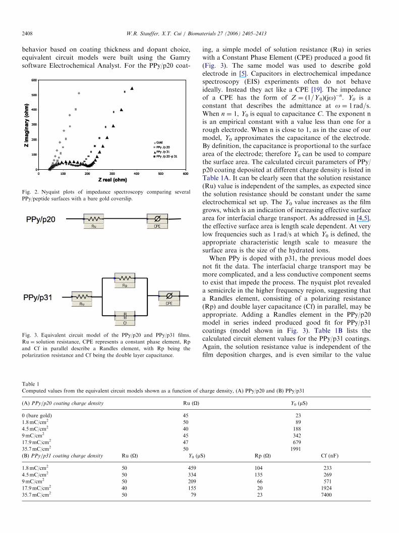
behavior based on coating thickness and dopant choice,equivalent circuit models were built using the Gamrysoftware Electrochemical Analyst. For the PPy/p20 coat-
Fig. 2. Nyquist plots of impedance spectroscopy comparing several
PPy/peptide surfaces with a bare gold coverslip.
Fig. 3. Equivalent circuit model of the PPy/p20 and PPy/p31 films.
Ru ¼ solution resistance, CPE represents a constant phase element, Rp
and Cf in parallel describe a Randles element, with Rp being the
polarization resistance and Cf being the double layer capacitance.
Table 1
Computed values from the equivalent circuit models shown as a function of c
(A) PPy/p20 coating charge density Ru (O)
0 (bare gold) 45
1.8mC/cm2 50
4.5mC/cm2 40
9mC/cm2 45
17.9mC/cm2 47
35.7mC/cm2 50
(B) PPy/p31 coating charge density Ru (O) Y0 (mS
1.8mC/cm2 50 459
4.5mC/cm2 50 334
9mC/cm2 50 209
17.9mC/cm2 40 155
35.7mC/cm2 50 79
ing, a simple model of solution resistance (Ru) in serieswith a Constant Phase Element (CPE) produced a good fit(Fig. 3). The same model was used to describe goldelectrode in [5]. Capacitors in electrochemical impedancespectroscopy (EIS) experiments often do not behaveideally. Instead they act like a CPE [19]. The impedanceof a CPE has the form of Z ¼ ð1=Y 0ÞðjoÞ
�n. Y0 is aconstant that describes the admittance at o ¼ 1 rad=s.When n ¼ 1, Y0 is equal to capacitance C. The exponent nis an empirical constant with a value less than one for arough electrode. When n is close to 1, as in the case of ourmodel, Y0 approximates the capacitance of the electrode.By definition, the capacitance is proportional to the surfacearea of the electrode; therefore Y0 can be used to comparethe surface area. The calculated circuit parameters of PPy/p20 coating deposited at different charge density is listed inTable 1A. It can be clearly seen that the solution resistance(Ru) value is independent of the samples, as expected sincethe solution resistance should be constant under the sameelectrochemical set up. The Y0 value increases as the filmgrows, which is an indication of increasing effective surfacearea for interfacial charge transport. As addressed in [4,5],the effective surface area is length scale dependent. At verylow frequencies such as 1 rad/s at which Y0 is defined, theappropriate characteristic length scale to measure thesurface area is the size of the hydrated ions.When PPy is doped with p31, the previous model does
not fit the data. The interfacial charge transport may bemore complicated, and a less conductive component seemsto exist that impede the process. The nyquist plot revealeda semicircle in the higher frequency region, suggesting thata Randles element, consisting of a polarizing resistance(Rp) and double layer capacitance (Cf) in parallel, may beappropriate. Adding a Randles element in the PPy/p20model in series indeed produced good fit for PPy/p31coatings (model shown in Fig. 3). Table 1B lists thecalculated circuit element values for the PPy/p31 coatings.Again, the solution resistance value is independent of thefilm deposition charges, and is even similar to the value
harge density, (A) PPy/p20 and (B) PPy/p31
Y0 (mS)
23
89
188
342
679
1991
) Rp (O) Cf (nF)
104 233
135 269
66 571
20 1924
23 7400

ARTICLE IN PRESS
Fig. 4. Cyclic voltammetry scans of three different PPy/peptide surfaces.
The scan rate is 50mV/s.
Table 2
Charge capacity (Q) of PPy/peptide films as a function of charge density
and peptide
Charge density
(mC/cm2)
PPy/p31
(mC)PPy/p31-p20
(mC)PPy/p20
(mC)
1.8 422 482 614
4.5 374 457 621
9 417 458 621
17.9 387 545 1089
35.7 443 763 1528
Fig. 5. Impedance spectra of coated and uncoated Michigan probe
recording electrodes.
W.R. Stauffer, X.T. Cui / Biomaterials 27 (2006) 2405–2413 2409
obtained from the PPy/p20 circuit. This further validatesboth circuit models.
A second assessment of the electronic properties of thePPy coatings is through CV. In this procedure the potentialis swept through a defined range and the resulting currentis recorded. Fig. 4 shows the CV curves for PPy/p20, PPy/p31, and PPy/p31-p20. The charge capacity, Q, can beobtained from integrating the current passed for half acycle. The charge capacities for different thickness filmswith different dopants can be found in Table 2. Note thatfor PPy/p20 coatings, the charge capacity increases as thedeposition charge increases, the same trend found in Y0.This is not surprising given the fact that charge capacity isrelated to the electrode capacitance.
To test the electrical property of the coatings on actualneural electrodes, PPy/p31 and PPy/p20 coatings weregrown galvanostatically on the electrodes of MichiganProbes. Fig. 5 shows the impedance spectrum of a baregold electrode, an electrode with a PPy/p31 coating, and anelectrode with a PPy/p20 coating. At the biological relevantfrequency of 1 kHz [20], both PPy/p31 and PPy/p20showed lower impedance than the bare gold electrode.Lower impedance is beneficial in recording minute extra-cellular neural signal. In this respect, PPy/p20 showedsuperior electrical property than PPy/p31.
3.2. Bioactivity test
As indicated earlier, laminin is a multifunctional ECMprotein that associates with several cell types. As such,different peptide sequences within Laminin are responsiblefor its different functions [14,15]. Research has demon-strated that p31 is an effective dopant for PPy andproduces a rough surface with low impedance thatenhances the attachment of neuroblastoma cells relativeto PPy/CH3COO� films, as well as promoting neuronalbinding in vivo [6,7]. One of our goals was to extend thisresearch by using primary neurons instead of neuroblasto-ma or PC-12 cells, and a defined culture media to betterisolate surface effects. A second goal was to investigateother short peptide fragments that could be utilized asdopants and have positive effects on neuronal growth.Given the previous literature showing that p20 plays animportant role in neurite extension [14], it was an idealcandidate for investigation.Primary neurons obtained from the cortices of E18 SD
rats were cultured in a defined media containing Neuro-basal, B27, glutamine, and glutamate. Early observationsof primary neuronal growth on relatively thick PPy/p31surfaces (thickness was set with charge density of 17.9mC/cm2) showed a very heterogeneous cell density. Assumingthat film thickness variation within a relatively thicksample could account for differences in cell growth, filmswith different thickness as controlled by total chargedensity and verified by ellipsometry, were synthesized assubstrates for cell growth. Films with charge density of 1.8,4.5, 9.0, 17.9, and 35.7mC/cm2 were synthesized. Fig. 6shows the resulting thickness versus charge density curve.For the PPy/p31 coated coverslips, optimal cell growth(highest cell density) tended to occur on thinner, ratherthan thicker samples. Surfaces having charge densities bet-ween 4.5 and 9.0mC/cm2 were found to have a significantlyhigher average cell density than for surfaces with chargedensity of 17.9mC/cm2 and higher (po0:02), see Fig. 6.Coatings with charge density of 4.5mC/cm2 are thereforeused as the optimized surface for the bioactivity test.PPy doped with PSS was used as the control PPy
surfaces and the positive control was laminin adsorbed

ARTICLE IN PRESS
Fig. 6. Neuron density as a function of charge density. Thickness
increased with increasing charge density as seen on the Y2 axis. A
significantly higher neuron density was found on surfaces with charge
density between 4.5 and 9mC/cm2 (po0:02).
Fig. 7. Neuron density on PPy surfaces. (#po0:002) (*po0:03).
W.R. Stauffer, X.T. Cui / Biomaterials 27 (2006) 2405–24132410
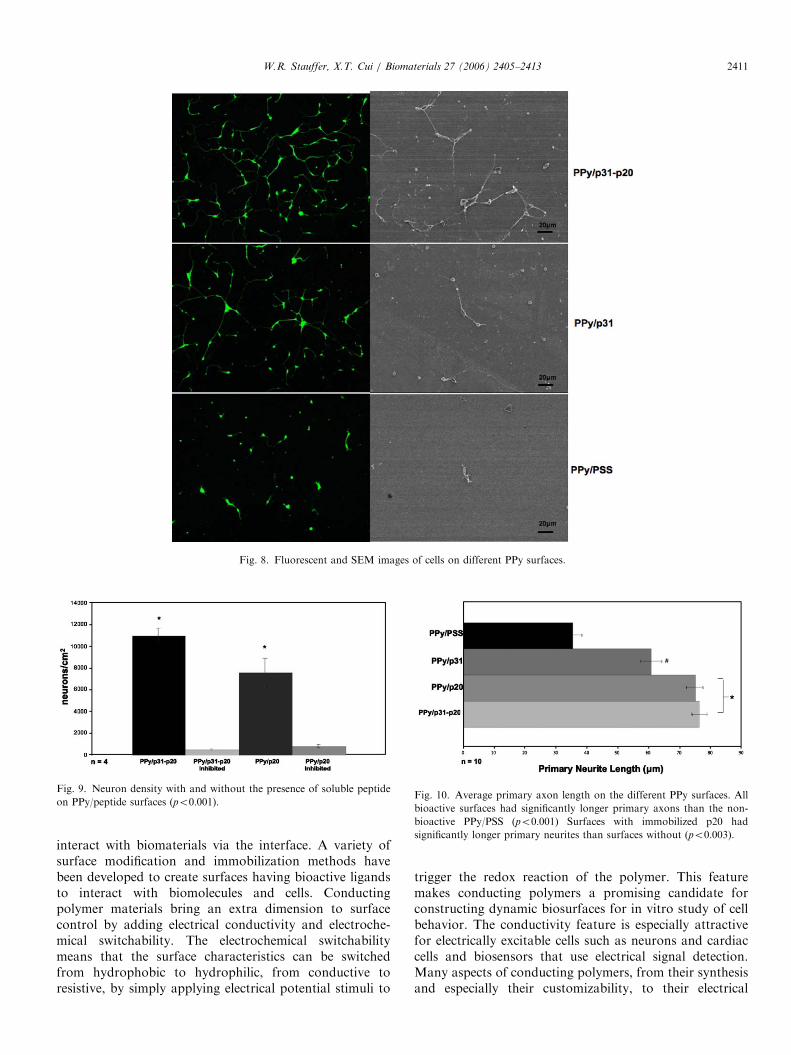
surfaces. The bioactive surfaces that we tested were PPy/p31, PPy/p20, and PPy/p31-p20. Significantly higher celldensities were found using Bonferroni post-hoc analysis onall bioactive surfaces relative to the PPy/PSS surface.Furthermore, the PPy/p20 and PPy/p31-p20 showedsignificantly higher neuron densities than the PPy/p31(po0:03). Fig. 7 shows a graph of cell density versuspeptide after 3 DIV. Neuron density on the laminin controlwas found to be approximately three-fold greater than thebest PPy/peptide surface. Cellular growth can be qualita-tively appreciated by referring to Fig. 8 which showsfluorescent and SEM images of neurons on PPy/p31-p20,PPy/p31, and PPy/PSS after 3 DIV.
During electropolymerization, the choice of the dopantcan have a significant affect on the final morphology of thePPy film [6,7]. Since different peptides were used as thedopants to create the different surfaces, it was important toverify that the differences in cell density were duespecifically to the immobilized peptide and not an effectof a secondary consequence such as differences in filmmorphology, i.e. roughness. This was accomplishedthrough inhibition studies. In the inhibition studies, priorto plating the cells on the surface, p20 was dissolved into
the cell suspension (0.2mM) and then added into theculture plates containing PPy/p20 coated coverslips.Similarly p31 was added into the plates containing PPy/p31 surfaces and 1:1 mixture of p20 and p31 was addedinto the plates of PPy/p20-p31. The rational for theseinhibition studies is that if the cell attachment is beingmediated by the surface immobilized peptides, then cellattachment should be inhibited when peptide receptors areblocked by the corresponding peptides in soluble form.Fig. 9 shows the cell density on PPy/p20 and PPy/p20-p31surfaces with and without soluble peptide after 3 DIV. Inboth cases a near total inhibition of cell growth wasobserved (po0:001). As a control, cells with solubilizedpeptide in suspension were plated on polylysine coatedTCPS dishes and no significant difference in cell growthwas observed (not shown). This result clearly indicates thatcell attachment is being mediated through the peptidesimmobilized in the PPy surface.Finally, primary neurite (axon) length was examined to
see if the different surfaces would affect neurite outgrowth.Neurons on all three bioactive surfaces had significantlylonger axons than those on the non-bioactive surface(po0:001). In Fig. 10, the differential effect on primaryneurite outgrowth found among the bioactive surfaces isshown. Surfaces that have p20 immobilized (both the PPy/p20 and PPy/p31–p20) had neurons with significantlylonger primary neurites than neurons on the PPy/p31sur-faces after 3 DIV (po0:005). As stated above, previousliterature has shown that p20 plays an important role inpromoting neurite outgrowth while p31 only has effect oncell attachment [14]. The results of this study show similarfindings indicating the bioactivity of the peptides were wellpreserved in the PPy film.While one of the main objectives of this study was to
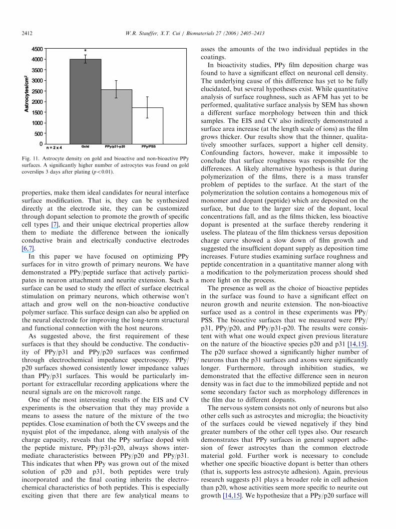
optimize neuronal growth and attachment on a materialthat could be used to modify brain recording electrodes, itwas recognized that the brain consists not only of neuronsbut other cell types as well and the immobilized peptidescould have a deleterious effect if it were to promote othercellular in-growth, such as that of astrocytes. To accountfor this possible phenomenon, the same surfaces, as well asbare gold coverslips, were used as a substrate for astrocyteculture. A significantly higher number (po0:01) of astro-cytes were found on the gold surfaces than on any PPysurface (Fig. 11). There was a trend towards PPy/p31having significantly higher astrocyte attachment than PPy/p20. If a less stringent post-hoc analysis than Bonferroni’scorrection is used, such as a least-square difference method,then po0:05. While it would be unsafe to draw conclusionsfrom this, the result highlights the need for further study ofthe effect of biomolecules on astrocyte attachment to PPysurfaces.
4. Discussion
In the field of biomaterials, the design of bioactivesurfaces is of particular interest since biological systems
ARTICLE IN PRESS
Fig. 8. Fluorescent and SEM images of cells on different PPy surfaces.
Fig. 9. Neuron density with and without the presence of soluble peptide
on PPy/peptide surfaces (po0:001).Fig. 10. Average primary axon length on the different PPy surfaces. All
bioactive surfaces had significantly longer primary axons than the non-
bioactive PPy/PSS (po0:001) Surfaces with immobilized p20 had
significantly longer primary neurites than surfaces without (po0:003).
W.R. Stauffer, X.T. Cui / Biomaterials 27 (2006) 2405–2413 2411
interact with biomaterials via the interface. A variety ofsurface modification and immobilization methods havebeen developed to create surfaces having bioactive ligandsto interact with biomolecules and cells. Conductingpolymer materials bring an extra dimension to surfacecontrol by adding electrical conductivity and electroche-mical switchability. The electrochemical switchabilitymeans that the surface characteristics can be switchedfrom hydrophobic to hydrophilic, from conductive toresistive, by simply applying electrical potential stimuli to
trigger the redox reaction of the polymer. This featuremakes conducting polymers a promising candidate forconstructing dynamic biosurfaces for in vitro study of cellbehavior. The conductivity feature is especially attractivefor electrically excitable cells such as neurons and cardiaccells and biosensors that use electrical signal detection.Many aspects of conducting polymers, from their synthesisand especially their customizability, to their electrical
ARTICLE IN PRESS
Fig. 11. Astrocyte density on gold and bioactive and non-bioactive PPy
surfaces. A significantly higher number of astrocytes was found on gold
coverslips 3 days after plating (po0:01).
W.R. Stauffer, X.T. Cui / Biomaterials 27 (2006) 2405–24132412
properties, make them ideal candidates for neural interfacesurface modification. That is, they can be synthesizeddirectly at the electrode site, they can be customizedthrough dopant selection to promote the growth of specificcell types [7], and their unique electrical properties allowthem to mediate the difference between the ionicallyconductive brain and electrically conductive electrodes[6,7].
In this paper we have focused on optimizing PPysurfaces for in vitro growth of primary neurons. We havedemonstrated a PPy/peptide surface that actively partici-pates in neuron attachment and neurite extension. Such asurface can be used to study the effect of surface electricalstimulation on primary neurons, which otherwise won’tattach and grow well on the non-bioactive conductivepolymer surface. This surface design can also be applied onthe neural electrode for improving the long-term structuraland functional connection with the host neurons.
As suggested above, the first requirement of thesesurfaces is that they should be conductive. The conductiv-ity of PPy/p31 and PPy/p20 surfaces was confirmedthrough electrochemical impedance spectroscopy. PPy/p20 surfaces showed consistently lower impedance valuesthan PPy/p31 surfaces. This would be particularly im-portant for extracellular recording applications where theneural signals are on the microvolt range.
One of the most interesting results of the EIS and CVexperiments is the observation that they may provide ameans to assess the nature of the mixture of the twopeptides. Close examination of both the CV sweeps and thenyquist plot of the impedance, along with analysis of thecharge capacity, reveals that the PPy surface doped withthe peptide mixture, PPy/p31-p20, always shows inter-mediate characteristics between PPy/p20 and PPy/p31.This indicates that when PPy was grown out of the mixedsolution of p20 and p31, both peptides were trulyincorporated and the final coating inherits the electro-chemical characteristics of both peptides. This is especiallyexciting given that there are few analytical means to
asses the amounts of the two individual peptides in thecoatings.In bioactivity studies, PPy film deposition charge was
found to have a significant effect on neuronal cell density.The underlying cause of this difference has yet to be fullyelucidated, but several hypotheses exist. While quantitativeanalysis of surface roughness, such as AFM has yet to beperformed, qualitative surface analysis by SEM has showna different surface morphology between thin and thicksamples. The EIS and CV also indirectly demonstrated asurface area increase (at the length scale of ions) as the filmgrows thicker. Our results show that the thinner, qualita-tively smoother surfaces, support a higher cell density.Confounding factors, however, make it impossible toconclude that surface roughness was responsible for thedifferences. A likely alternative hypothesis is that duringpolymerization of the films, there is a mass transferproblem of peptides to the surface. At the start of thepolymerization the solution contains a homogenous mix ofmonomer and dopant (peptide) which are deposited on thesurface, but due to the larger size of the dopant, localconcentrations fall, and as the films thicken, less bioactivedopant is presented at the surface thereby rendering ituseless. The plateau of the film thickness versus depositioncharge curve showed a slow down of film growth andsuggested the insufficient dopant supply as deposition timeincreases. Future studies examining surface roughness andpeptide concentration in a quantitative manner along witha modification to the polymerization process should shedmore light on the process.The presence as well as the choice of bioactive peptides
in the surface was found to have a significant effect onneuron growth and neurite extension. The non-bioactivesurface used as a control in these experiments was PPy/PSS. The bioactive surfaces that we measured were PPy/p31, PPy/p20, and PPy/p31-p20. The results were consis-tent with what one would expect given previous literatureon the nature of the bioactive species p20 and p31 [14,15].The p20 surface showed a significantly higher number ofneurons than the p31 surfaces and axons were significantlylonger. Furthermore, through inhibition studies, wedemonstrated that the effective difference seen in neurondensity was in fact due to the immobilized peptide and notsome secondary factor such as morphology differences inthe film due to different dopants.The nervous system consists not only of neurons but also
other cells such as astrocytes and microglia; the bioactivityof the surfaces could be viewed negatively if they bindgreater numbers of the other cell types also. Our researchdemonstrates that PPy surfaces in general support adhe-sion of fewer astrocytes than the common electrodematerial gold. Further work is necessary to concludewhether one specific bioactive dopant is better than others(that is, supports less astrocyte adhesion). Again, previousresearch suggests p31 plays a broader role in cell adhesionthan p20, whose activities seem more specific to neurite outgrowth [14,15]. We hypothesize that a PPy/p20 surface will

ARTICLE IN PRESSW.R. Stauffer, X.T. Cui / Biomaterials 27 (2006) 2405–2413 2413
support fewer astrocytes than a PPy/p31. If this hypothesiswere supported then it would point to PPy/p20 being anoptimal surface for applications where direct neuronattachment and intimate connections are vital to thedesired function. The results in this paper however showedno significant difference between any of the PPy surfaces(including the non-bioactive PPy/PSS) when astrocytes arecultured on them. Nevertheless, the experiments were donein a serum-based media; therefore, the difference in surfacecomposition could be masked by the adsorbed serumproteins. It has been shown that when cultured in serumcontaining media, astrocytes are relatively insensitive todifferences in surface chemistries as long as the proteinsnecessary for cellular attachment are capable of adsorbingto the material to some extent [21]. Ongoing work is beingconducted to establish a serum free astrocyte culturesystem to better isolate the effects of surface biomolecules.
This work compares the efficacy of two different lamininfragments as dopants in conducting polymer surfaces. Theelectrical as well as the biological properties of the resultingsurfaces are reported. It is found that PPy surfacescontaining bioactive peptides are better at supportingneuronal growth and neurite extension than non-bioactivesurfaces. Furthermore, it is shown that the choice ofbioactive molecule has a significant effect on both theelectrical and biological properties of the resulting surface.PPy surfaces using p20 as the dopant were found tosupport a higher density of neurons and promote longeraxon growth than surfaces doped with p31. A surface withthe combination of the two laminin fragment dopants wasdiscovered to support a higher neuronal density thansurfaces containing either of the dopants individually.Finally, it was observed that astrocytes were less prone toattachment on PPy surfaces compared to the commonelectrode material gold. These surfaces exhibit lowelectrical impedance and high charge capacity relative toconventional conductive substrates, such as metals. Such amaterial has the potential to make a positive impact on thefield of neural tissue engineering and neural prostheticdevices.
Acknowledgments
We acknowledge the financial support provided by theWhitaker Foundation, the Department of Bioengineering(University of Pittsburgh), McGowan Institute for Regen-erative Medicine, University of Pittsburgh—CMRF (Com-petitive Medical Research Fund), and CRDF (CentralResearch Development Fund). We also thank Dr. AndrewSchwartz and Chance Spalding, Dr. Carl Lagenaur, Dr.Simon Watkins, Dr. Donna Stolz, and Marc Rubin(Center for Biological Imaging at U. Pitt), Dr. JianjunWei, Michael Bartkovsky, Erdrin Azemi Charley, andReecha Wadhwa for their technical help during the courseof this project.
References
[1] Diaz AF, Castillo JI, Logan JA, Lee W- Y. Electrochemistry
of conducting polypyrrole films. J Electroanal Chem 1981;128:
115–32.
[2] Wong JY, Langer R, Ingber DE. Electrically conducting polymers
can noninvasively control the shape and growth of mammalian cells.
Proc Natl Acad Sci USA 1994;91(8):3201–4.
[3] Schmidt CE, Shastri VR, Vacanti JP, Langer R. Stimulation of
neurite outgrowth using an electrically conducting polymer. Proc
Natl Acad Sci USA 1997;94(17):8948–53.
[4] Cui XY, Martin DC. Electrochemical deposition and characteriza-
tion of poly (3,4-ethylenedioxythiophene) on neural microelectrode
arrays. Sensors Actuators B—Chem 2003;89(1–2):92–102.
[5] Cui XY, Martin DC. Fuzzy gold electrodes for lowering impedance
and improving adhesion with electrodeposited conducting polymer
films. Sensors Actuators A—Phys 2003;103(3):384–94.
[6] Cui X, Wiler J, Dzaman M, Altschuler RA, Martin DC. In vivo
studies of polypyrrole/peptide coated neural probes. Biomaterials
2003;24(5):777–87.
[7] Cui X, Lee VA, Raphael Y, Wiler JA, Hetke JF, Anderson DJ, et al.
Surface modification of neural recording electrodes with conduct-
ing polymer/biomolecule blends. J Biomed Mater Res 2001;56(2):
261–72.
[8] Collier JH, Camp JP, Hudson TW, Schmidt CE. Synthesis and
characterization of polypyrrole-hyaluronic acid composite biomater-
ials for tissue engineering applications. J Biomed Mater Res 2000;
50(4):574–84.
[9] Wallace GG. Conductive electroactive polymers: intelligent materials
systems. 2nd ed. Boca Raton, Fla.: CRC Press; 2003.
[10] Webb K, Hlady V, Tresco PA. Relationships among cell attachment,
spreading, cytoskeletal organization, and migration rate for ancho-
rage-dependent cells on model surfaces. J Biomed Mater Res
2000;49(3):362–8.
[11] Kotwal A, Schmidt CE. Electrical stimulation alters protein
adsorption and nerve cell interactions with electrically conducting
biomaterials. Biomaterials 2001;22(10):1055–64.
[12] George PM, Lyckman AW, Lavan DA, Hegde A, Leung Y, Avasare
R, et al. Fabrication and biocompatibility of polypyrrole implants
suitable for neural prosthetics. Biomaterials 2005;26(17):3511–9.
[13] Craig AM, Banker G. Neuronal polarity. Annu Rev Neurosci 1994;
17:267–310.
[14] Liesi P, Narvanen A, Soos J, Sariola H, Snounou G. Identification of
a neurite outgrowth-promoting domain of laminin using synthetic
peptides. FEBS Lett 1989;244(1):141–8.
[15] Graf J, Iwamoto Y, Sasaki M, Martin GR, Kleinman HK, Robey
FA, et al. Identification of an amino acid sequence in laminin
mediating cell attachment, chemotaxis, and receptor binding. Cell
1987;48(6):989–96.
[16] Ard MD, Bunge RP. Heparan sulfate proteoglycan and laminin
immunoreactivity on cultured astrocytes: relationship to differentia-
tion and neurite growth. J Neurosci 1988;8(8):2844–58.
[17] Brewer GJ, Price PJ. Viable cultured neurons in ambient carbon
dioxide and hibernation storage for a month. Neuroreport 1996;7(9):
1509–12.
[18] Allen NS, Murray KS, Fleming RJ, Saunders BR. Physical proper-
ties of polypyrrole films containing trisoxalatometallate anions
and prepared from aqueous solution. Synth Metals 1997;87(3):
237–47.
[19] MacDonald JR. Impedance spectroscopy: emphasizing solid materi-
als and systems. New York: Wiley; 1987.
[20] Kuffler SW. from neuron to brain: a cellular approach to the function
of the nervous system. Sunerland, MA: Sinauer Associates; 1976.
[21] Biran R, Noble MD, Tresco PA. Characterization of cortical
astrocytes on materials of differing surface chemistry. J Biomed
Mater Res 1999;46(2):150–9.