Reactivation of covalently immobilized lipase from Thermomyces lanuginosus
Upload
khangminh22Category
view
5download
0
INST
ITU
TO
DE C
IÊN
CIA
S B
IOM
ÉD
ICA
S A
BEL S
ALA
ZA
R
FA
CU
LD
AD
E D
E F
AR
MÁ
CIA
Danie
la F
ilipa d
os S
anto
s B
arros. E
ngin
eere
d p
latfo
rms w
ith site
-specific
imm
obiliz
ed la
min
in fo
r neura
l applic
atio
ns
Engin
eered p
latfo
rm
s w
ith s
ite-s
pecific
imm
obiliz
ed la
min
in fo
r n
eural a
pplic
atio
ns
Danie
la F
ilipa d
os S
anto
s Barro
s
Engineered platforms with site-specific
immobilized laminin for neural applications
Daniela Filipa dos Santos Barros
D 2019
D.IC
BA
S 2019
SED
E A
DM
INIS
TR
AT
IVA
DOUTORAMENTO
BIOTECNOLOGIA MOLECULAR E CELULAR APLICADA ÀS CIÊNCIAS DA SAÚDE
Daniela Filipa dos Santos Barros
Engineered platforms with site-specific immobilized laminin for neural applications
Tese de Candidatura ao grau de Doutor em
Biotecnologia Molecular e Celular Aplicada às
Ciências da Saúde;
Programa Doutoral da Universidade do Porto
(Instituto de Ciências Biomédicas de Abel
Salazar e Faculdade de Farmácia)
Orientadora – Professora Doutora Ana Paula
Gomes Moreira Pêgo
Categoria – Investigadora Principal, Professora
Associada Convidada
Afiliação – Instituto de Investigação e Inovação
em Saúde (i3S), Universidade do Porto (UP);
Instituto Nacional de Engenharia Biomédica
(INEB), UP; Faculdade de Engenharia (FEUP),
UP; Instituto de Ciências Biomédicas Abel
Salazar (ICBAS), UP.
Co-orientadora – Doutora Isabel Maria Santana
Ramos de Freitas Amaral
Categoria – Investigadora Auxiliar
Afiliação – Instituto de Investigação e Inovação
em Saúde, Universidade do Porto (i3S, UP);
Instituto Nacional de Engenharia Biomédica
(INEB), UP; Faculdade de Engenharia (FEUP),
UP.

The work presented in this thesis was developed at:
nBTT - nanoBiomaterials for Targeted Therapies Group
i3S – Instituto de Investigação e Inovação em Saúde
INEB – Instituto Nacional de Engenharia Biomédica
Universidade do Porto, Portugal
García Lab
Parker H. Petit Institute for Bioengineering and Bioscience
George W. Woodruff School of Mechanical Engineering
Georgia Institute of Technology, Atlanta, GA, USA
The work presented in this thesis was funded by projects NORTE-01-0145-FEDER-000008
and NORTE-01-0145-FEDER-000012, supported by Norte Portugal Regional Operational
Programme (NORTE 2020), under the PORTUGAL 2020 Partnership Agreement, through
the European Regional Development Fund (ERDF) and FEDER - Fundo Europeu de
Desenvolvimento Regional funds through the COMPETE 2020 - Operacional Programme
for Competitiveness and Internationalisation (POCI), Portugal 2020; by Portuguese funds
through FCT/MCTES in the framework of the project "Institute for Research and Innovation
in Health Sciences" (POCI-01-0145-FEDER-007274) and Santa Casa da Misericórdia de
Lisboa through project COMBINE (Prémio Neurociências Melo e Castro-MC-1068-2015).
D. Barros was supported by FCT PhD Programs (PD/BD/105953/2014) and Programa
Operacional Potencial Humano (POCH), in the scope of the BiotechHealth Program
(Doctoral Program on Cellular and Molecular Biotechnology Applied to Health Sciences),
Programa FLAD Healthcare 2020 and the project PARES (Prémio Albino Aroso).

Remember what they say There’s no shortcut to a dream
It’s all about blood and sweat And life is what you manage in-between
October, Broken Bells

i
ACKNOWLEDGMENTS
E 5 anos depois, eis que esta longa e desafiante jornada está prestes a terminar! O
doutoramento foi sem dúvida uma etapa durante a qual vivi algumas das melhores e
também piores experiências da minha vida, e que me fez crescer não só cientificamente,
mas também a nível pessoal. Felizmente não fiz este caminho sozinha, tive pessoas
espetaculares a apoiar-me e a fazer-me querer ir sempre um bocadinho mais além, e é a
todas elas que quero agradecer e também dedicar esta tese.
Às minhas orientadoras Ana Paula e Isabel, obrigada por me terem recebido e me terem
dado a oportunidade de explorar uma área que estava totalmente fora da minha zona de
conforto...aprendi imenso nestes últimos anos. Uma das coisas mais importantes que
aprendi foi que muitas vezes (ou quase sempre) temos de desligar o “descomplicómetro”
e olhar para aquilo que vemos como perdido/mau de forma mais positiva. Obrigada pelo
vosso entusiasmo, apoio e pelas palavras de incentivo. A vossa orientação e apoio foi
fundamental para o meu crescimento científico nestes últimos anos.
To Andrés, thank you for the opportunity to work in your laboratory and for being such an
enthusiastic and positive mentor. I had a really great time in your lab. Thank you also, to all
the members from García Lab who welcomed me so well, and specially to Woojin and
Ricardo, my coffee buddies, for the friendship and support, making my journey at
GeorgiaTech, a really happy journey.
Ao Professor Mário Barbosa, pela oportunidade de frequentar o BiotechHealth e pelo
esforço em tornar este, um programa doutoral de excelência. À Helena Martins, Catarina
Pereira, Daniel Vasconcelos e João Cortez por estarem sempre disponíveis para ajudar e
tornar a nossa vida um bocadinho mais fácil. Aos meus colegas do BiotechHealth que
fizeram este caminho comigo, foi muito bom contar com o vosso apoio e companheirismo.
À Paula Parreira e Cristina Martins, pela ajuda e apoio numa fase crucial desta tese.
Obrigada pela vossa persistência e pelas palavras de incentivo...a vossa ajuda foi
fundamental para o meu trabalho.
A todas as pessoas dos serviços do i3S, Frederico, Fátima, Maria, Dalila, Ricardo, Eduardo,
Tânia, Paula, Hugo e André, muito obrigada por partilharem o vosso conhecimento e me
ajudarem nos momentos mais críticos. Um obrigada especial à Joana, que foi essencial na
fase inicial do meu doutoramento. A tua paciência e apoio fizeram toda a diferença.
Ao grupo nBTT/Lab214.S3, obrigada pela amizade, apoio, por todos os momentos de
diversão, pelas palavras de incentivo e acima de tudo por aguentarem tão bem todo o meu
ii
drama (#dramaqueen)...vocês são espetaculares e vou guardar-vos para sempre no
coração.
Um muito obrigada a todos os colegas e amigos do INEB/IBMC/i3S...sinto-me uma sortuda
pela quantidade enorme de pessoas que tive oportunidade de conhecer e manter na minha
vida ao longo destes 5 anos (e não só). Foi muito bom poder contar com o vosso apoio e
amizade!!
À Catarina e ao Philippe, os novos “bosses”, por todo o apoio e compreensão nesta fase
de transição. A todos os colegas do ICB Lab e do iBiMED por me acolherem tão bem no
laboratório e em Aveiro.
À minha família do coração, àqueles que são as “minhas pessoas” Beta, Jorginho, Pedro
e Tânia, e ao mais recente elemento, Tiago, obrigada pelo apoio, pelo carinho, pelos serões
passados a estupidificar (sempre com o belo do copo de vinho a acompanhar)...obrigada
por fazerem parte da minha vida e me deixarem fazer parte da vossa.
À pessoa que durante muitos anos partilhou comigo as angústias, alegrias e que nunca me
impediu de sonhar... apesar dos nossos caminhos já não se cruzarem, obrigada Ricardo
por nunca teres desistido de mim!
Aos meus pais, não há palavras para lhes agradecer o apoio e amor incondicional. Muito
do que sou hoje devo-o a vocês, que me ensinaram a não baixar os braços e a nunca
desistir de ir atrás dos meus sonhos. OBRIGADA POR TUDO!!!
À minha irmã, pelo apoio, pelo amor, por mostrar tanto entusiasmo e orgulho pelas minhas
conquistas, por nunca me deixar desistir, pelas palavras certas nos momentos mais
complicados,...POR TUDO!!! Obrigada ao André pelos conselhos e apoio. E obrigada ao
meu Guigas, que é uma das pessoas que eu mais amo neste mundo e que nos seus
pequenos gestos me faz sentir tão amada e feliz.

iii
LIST OF MAIN OUTPUTS
The main outputs of the work conducted during the period of this thesis are listed
below:
Papers in peer-reviewed scientific journals
D. Barros, P. Parreira, J. Furtado, F. Ferreira-da-Silva, E. Conde-Sousa, A.J. Garcia, M.C.L.
Martins, I.F. Amaral, A.P. Pêgo, An affinity-based approach to engineer laminin-presenting
cell instructive microenvironments, Biomaterials 192 (2019) 601-611
W. M. Han, S. E. Anderson, M. Mohiuddin, D. Barros, S. A. Nakhai, E. Shin, I. F. Amaral,
A. P. Pêgo, A. J. García, Y. C. Jang, Synthetic matrix enhances transplanted satellite cell
engraftment in dystrophic and aged skeletal muscle with comorbid trauma, Science
Advances, 4 (2018).
J. Silva, A. R. Bento, D. Barros, T. L. Laundos, S. R. Sousa, P. Quelhas, M. M. Sousa, A.
P. Pêgo, I. F. Amaral, Fibrin functionalization with synthetic adhesive ligands interacting
with a6b1 integrin receptor enhances neurite outgrowth of embryonic stem cell-derived
neural stem/progenitors, Acta Biomaterialia, 59 (2017) 243-256.
D. Barros, I. F. Amaral, A. P. Pêgo, Biomimetic Synthetic Self-Assembled Hydrogels for Cell
Transplantation. Current Topics in Medicinal Chemistry, 15(13) (2015) 1209-26.
Manuscripts in preparation for submission
D. Barros, I. F. Amaral, A. P. Pêgo, Laminin-inspired cell instructive microenvironments for
neural tissue engineering. (Literature Review).
D. Barros, A. J. García, I. F. Amaral, A. P. Pêgo, Engineering hydrogels with affinity-bound
laminin as 3D neural stem cell culture systems. (Original Research).
Book Chapter
J. Caldeira, F. Sousa, D. Sousa, D. Barros (2018). Extracelular matrix constitution and
function for tissue regeneration and repair, in Peptides and Proteins as Biomaterials for
Tissue Regeneration and Repair, M.A. Barbosa and M.C.L. Martins, Editors. Woodhead
Publishing – Elsevier.
Patents
D. Barros, I.F. Amaral, A.P. Pêgo, Laminin immobilization, methods and uses thereof,
Portuguese Provisional Patent Application no. 20181000074673, filled 2018/11/30.
Published articles were herein reproduced with permission from editors.

iv
v
ABSTRACT
Neural stem cells (NSCs) hold great potential for application in the treatment of central
nervous system (CNS) disorders. These multipotent cells reside within a dynamic and
complex microenvironment, the NSC niche, where cell-cell interactions and local
microenvironmental cues are key to regulate stem cell behavior. Therefore, the
development of new platforms allowing a better understanding of niche properties and
how they regulate NSC behavior and function is highly desirable. These are expected to
provide valuable insight into cell physiology and interactions with the surrounding
microenvironment, and ultimately be used for the development of more effective NSC-
based regenerative therapies.
Laminin has a key role in the modulation of NSC function and fate within neurogenic
niches. Indeed, different studies already explored the immobilization of full-length laminin
and its peptide analogues into diverse biological and synthetic matrices, to recapitulate
the microenvironment of stem cell niches and/or gain insight into the role of ECM on
stem cell behavior. However, the strategies currently explored for laminin immobilization
within three-dimensional (3D) matrices do not address a critical aspect influencing
laminin bioactivity, which is the control over the exposure of laminin crucial bioactive
epitopes.
In this thesis, we propose the design of biologically relevant matrices able to recapitulate
the laminin-rich microenvironment of NSC niches. For that, we explored the modification
of a degradable synthetic hydrogel with full-length laminin or with a laminin-derived
peptide interacting with syndecans - AG73.
One of the specific aims of this thesis was the establishment of an affinity-based
approach that takes advantage of the high affinity interaction between laminin and the
human N-terminal agrin (hNtA) domain. A recombinant human NtA (rhNtA) domain was
produced and successfully conjugated at its N-terminal with a thiol-terminated
poly(ethylene glycol) (PEG) – mono-PEGylated rhNtA. Mono-PEGylated rhNtA showed
ability to act as an effective natural binding partner for the site-selective immobilization
of laminin, while preserving the exposure of laminin key bioactive domains. This
translated into an enhanced ability of laminin to polymerize and mediate hNSC adhesion
and spreading. Aiming at the establishment of a chemically-defined cell-instructive
microenvironment, the proposed affinity-based approach was explored to promote the
site-selective immobilization of different laminin isoforms (in the context of this thesis
assessed for mouse laminin-111 and recombinant human laminin-521) into a degradable
synthetic hydrogel. A four-arm maleimide-end functionalized poly(ethylene glycol) (PEG-
vi
4MAL) macromer based hydrogel was selected as the base material, due to its modular
nature, good biocompatibility and well characterized biochemical and biophysical
properties. The PEG-4MAL macromer was functionalized with the thiol-terminated
mono-PEGylated rhNtA domain to mediate the site-selective incorporation of laminin into
PEG-4MAL. The developed hydrogels revealed mechanical properties (complex
modulus (G*) = 251 Pa) within the range of values preferred for NSC proliferation (100 –
1000 Pa) and neurite branching and extension (200 – 400 Pa). Moreover, the affinity-
bound laminin PEG-4MAL hydrogels showed an enhanced ability to mediate hNSC
proliferation and neurite outgrowth, when compared to unmodified hydrogels, and most
importantly, in contrast to hydrogels containing equal amount of physically entrapped
laminin.
In alternative to the incorporation of full-length laminin, a laminin-derived peptide
interacting with syndecan receptors (AG73) was explored to confer bioactivity to 3D
matrices. The modular nature of the synthetic matrix used, allowed us to explore the
effect of AG73 peptide density on cell behavior, independently of other hydrogel
biophysical and biochemical properties. The tethering of AG73 peptide to PEG-4MAL
hydrogels led to a significant improvement of cell viability after 7 days in culture,
independently of the input peptide concentration. In addition, we demonstrated that a
higher input AG73 concentration (1 mM) better supports hNSC proliferation and neurite
outgrowth, as well as the establishment of a neuronal network.
Overall, the work presented in this thesis contributed to: (i) the development of an affinity-
based strategy for site-selective immobilization of laminin, suitable for use as an
alternative to conventional immobilization approaches (physical entrapment and non-
selective covalent immobilization) in a wide range of applications (e.g. engineered
coatings for neuroelectrodes, 2D substrates for cell culture, and biofunctionalization of
3D matrices); (ii) development of engineered matrices for use as defined 3D platforms
for the establishment of artificial NSC niches and as temporary mimetic
microenvironments to support hNSC transplantation in the context of nervous system
regeneration/disorders.
In summary, we strongly believe this work opens new avenues in the design of more
efficient hydrogel matrices for application in NSC-based regenerative approaches for the
treatment of traumatic CNS disorders or neurodegenerative diseases.

vii
RESUMO
As células estaminais neurais (CENs) apresentam um grande potencial para aplicação
no tratamento de doenças do sistema nervoso central (SNC). Estas células
multipotentes residem num microambiente dinâmico e complexo, o nicho das CEN, onde
as interações célula-célula e os sinais do microambiente local são essenciais para
regular o comportamento das células estaminais. Deste modo, o desenvolvimento de
novas plataformas que permitam compreender melhor as propriedades do nicho e a
forma como estas regulam o comportamento e a função das CENs é altamente
vantajoso. É expectável que estas plataformas permitam uma melhor compreensão da
fisiologia celular das CENs e das suas interações com o ambiente circundante, e que
contribuam para o desenvolvimento de terapias regenerativas baseadas no transplante
de CENs mais efetivas.
A laminina tem um papel crucial na modulação da função e diferenciação das CENs nos
nichos neurogénicos. De facto, já vários estudos exploraram a imobilização da molécula
da laminina e de péptidos derivados da laminina em matrizes biológicas e sintéticas, de
forma a recapitular o microambiente do nicho e/ou a compreender melhor o papel da
matriz extracelular no comportamento das células estaminais. Contudo, as estratégias
exploradas atualmente para a imobilização da laminina em matrizes tridimensionais (3D)
não contemplam um aspeto crítico para a bioatividade da laminina, e que é o controlo
sobre a exposição dos epítopos bioativos mais importantes.
Nesta tese, propomos o desenvolvimento de matrizes biologicamente relevantes
capazes de recapitular o microambiente rico em laminina dos nichos neurogénicos. Para
isso, explorámos a modificação de um hidrogel sintético degradável com a molécula da
laminina ou com um péptido derivado da laminina capaz de interagir com recetores
sindecano – AG73.
Um dos objetivos específicos desta tese foi o desenvolvimento de uma estratégia de
imobilização por afinidade, baseada na interação de elevada afinidade entre a laminina
e o domínio N-terminal da agrina (NtA). O domínio recombinate humano NtA (rhNtA) foi
produzido e conjugado eficazmente no seu N-terminal com um polietilenoglicol (PEG)
contendo um grupo tiol numa das suas extremidades – rhNtA mono-PEGilado. O rhNtA
mono-PEGilado demonstrou capacidade para atuar como um ligando natural para a
imobilização seletiva da laminina, sem comprometer a exposição dos domínios bioativos
desta proteína. A eficiência do ligando proposto traduziu-se numa maior capacidade da
laminina para polimerizar e para mediar a adesão das CENs humanas, assim como a
organização do seu citoesqueleto. Com o objetivo de estabelecer um microambiente
viii
quimicamente definido e capaz de modular o comportamento celular, explorámos a
estratégia de imobilização por afinidade proposta para a imobilização controlada de
diferentes isoformas da laminina (no contexto desta tese avaliámos a laminina-111
derivada de ratinho e a laminina-521 recombinante humana) num hidrogel sintético e
degradável. Para o efeito selecionámos um hidrogel de PEG com quatro braços
funcionalizados nas extremidades com maleimida (PEG-4MAL) devido à sua natureza
modular, biocompatibilidade e propriedades bioquímicas e biofísicas bem
caracterizadas. O PEG-4MAL foi inicialmente funcionalizado com o domínio rhNtA
mono-PEGilado para lhe permitir mediar a incorporação selectiva da laminina. Os
hidrogéis desenvolvidos apresentaram propriedades mecânicas (módulo complexo (G*)
= 251 Pa) no gama de valores preferidos para a proliferação das CEN (100 – 1000 Pa)
e extensão de neurites (200 – 400 Pa). Os hidrogéis com laminina imobilizada
seletivamente revelaram modular de forma mais eficaz a proliferação das CENs e a
extensão de neurites, quando comparados com hidrogéis não modificados, e sobretudo
em contraste com hidrogéis sem o domínio rhNtA mono-PEGilado, mas contendo a
mesma quantidade de laminina fisicamente incorporada.
Alternativamente à imobilização da molécula da laminina, também explorámos um
péptido derivado da laminina que interage com os recetores sindecano (AG73), para
conferir bioatividade às matrizes 3D. A natureza modular da matriz sintética utilizada,
permitiu-nos explorar o efeito da densidade do péptido AG73 no comportamento celular,
independentemente de outras propriedades bioquímicas e biofísicas do hidrogel. Os
hidrogéis de PEG-4MAL modificados com o péptido levaram a um aumento significativo
da viabilidade celular após 7 dias em cultura, independentemente da concentração de
péptido. Além disso, a concentração máxima de AG73 testada (1 mM) revelou suportar
melhor a proliferação das CENs e a extensão de neurites, tal como favorecer o
estabelecimento de uma rede neuronal.
No geral, o trabalho apresentado nesta tese contribuiu para: (i) o desenvolvimento de
uma estratégia de afinidade para a imobilização seletiva da laminina, com potencial para
ser utilizada em alternativa às estratégias de imobilização convencionais (incorporação
física e imobilização covalente não seletiva) em diversas aplicações (p.e. revestimentos
para neuro-elétrodos, substratos para cultura celular, e biofuncionalização de matrizes
3D); (ii) desenvolvimento de plataformas 3D definidas com potencial de utilização no
estabelecimento de nichos neurogénicos artificiais e como microambientes temporários
biomiméticos, para transplante de CENs no contexto da regeneração do sistema
nervoso.

ix
Em resumo, acreditamos que este trabalho abre novas perspetivas para o
desenvolvimento de matrizes mais eficazes para aplicação em terapias regenerativas
baseadas no transplante de CENs para o tratamento de doenças traumáticas ou
degenerativas do SNC.

x

xi
TABLE OF CONTENTS
ACKOWLEDGMENTS .......................................................................................... i
LIST OF MAIN OUTPUTS ................................................................................... iii
ABSTRACT ......................................................................................................... v
RESUMO ............................................................................................................ vii
TABLE OF CONTENTS ...................................................................................... xi
LIST OF ABBREVIATIONS ................................................................................ xv
PART 1
CHAPTER I | General Introduction ..................................................................... 3
CHAPTER II | Aim and Structure of the Thesis ................................................ 23
Aim of the Thesis .................................................................................................... 25
Structure of the Thesis ............................................................................................ 26
CHAPTER III | Laminin-Inspired Cell-Instructive Microenvironments for Neural Tissue Engineering .......................................................................................... 31
Abstract .................................................................................................................. 34
1.Laminin ................................................................................................................ 35
1.1. Structure and domain architecture .......................................................... 35
1.2. Main Functions ....................................................................................... 36
1.2.1. Laminin polymerization .............................................................. 38
1.2.2. Laminin role in the assembly and stability of basement membranes
............................................................................................................ 38
1.2.3. Cell adhesion-promoting activity ................................................ 39
2.Laminin in the central nervous system ................................................................. 40
3.Laminin in neurogenic niches ............................................................................... 42
4.Laminin inspired hydrogels to recreate the microenvironment of neurogenic niches
............................................................................................................................... 45
xii
4.1. Hydrogels functionalized with full-length laminin ..................................... 45
4.2. Hydrogels functionalized with laminin-derived peptides .......................... 52
5.Engineering laminin-inspired hydrogels: progress and future challenges ............. 58
References ............................................................................................................. 60
CHAPTER IV | Biomimetic Synthetic Self-Assembled Hydrogels for Cell Transplantation ................................................................................................ 77
Abstract .................................................................................................................. 80
Cell Transplantation in the Context of Regenerative Medicine ................................ 81
Hydrogels as Attractive Vehicles for Cell Transplantation ....................................... 82
Self-Assembled Hydrogels ...................................................................................... 84
Engineered protein-based hydrogels ............................................................. 84
Elastin-like polypeptides ...................................................................... 85
Leucine zipper coiled-coil-based polypeptides ..................................... 86
Amphiphilic block co-polypeptides ....................................................... 87
Mixing-induced two-component hydrogel ............................................. 88
Peptide-based synthetic hydrogels ................................................................ 90
Self-assembling peptides ..................................................................... 90
Self-assembling peptide amphiphiles ................................................... 92
DNA-based hydrogels ................................................................................... 96
Hybrid hydrogels ........................................................................................... 98
In vivo Biological Performance of Synthetic Self-Assembled Hydrogels ................ 101
Translation of Synthetic Self-Assembled Hydrogels into the Clinic: Progress and
Future Challenges................................................................................................. 106
Ackowledgments ................................................................................................... 108
References ........................................................................................................... 109
PART 2
CHAPTER V | An Affinity-Based Approach to Engineer Laminin Presenting Cell Instructive Microenvironments ............................................................... 123

xiii
Abstract ................................................................................................................ 126
1. Introduction ..................................................................................................... 127
2. Materials and Methods .................................................................................... 129
3. Results and Discussion ................................................................................... 133
4. Conclusion ...................................................................................................... 146
5. Acknowledgments ........................................................................................... 147
Supplementary Data ............................................................................................. 148
Supplementary Materials and Methods ....................................................... 148
Supplementary Figures and Tables ............................................................. 152
References ........................................................................................................... 161
CHAPTER VI | Engineering Hydrogels with Affinity-Bound Laminin as 3D Neural Stem Cell Culture Systems ................................................................. 167
Abstract ................................................................................................................ 170
1. Introduction ..................................................................................................... 171
2. Materials and Methods .................................................................................... 174
3. Results and Discussion ................................................................................... 177
4. Conclusion ...................................................................................................... 185
Acknowledgments ................................................................................................. 186
Supplementary Data ............................................................................................. 187
Supplementary Materials and Methods ....................................................... 187
Supplementary Figures and Tables ............................................................. 188
References ........................................................................................................... 190
CHAPTER VII | An Alternative Approach to Engineer Synthetic Hydrogels with Affinity-Bound Laminin .................................................................................. 195
Abstract ................................................................................................................ 198
Materials and Methods .......................................................................................... 206
Acknowledgments ................................................................................................. 209
xiv
Supplementary Data ............................................................................................. 211
Supplementary Materials and Methods ....................................................... 211
Supplementary Figures and Tables ............................................................. 212
References ........................................................................................................... 214
CHAPTER VIII | AG73-Functionalized Synthetic Hydrogels Support Neural Stem Cell Survival, Proliferation and Neurite Outgrowth .............................. 219
Abstract ................................................................................................................ 222
1. Introduction ..................................................................................................... 223
2. Materials and Methods .................................................................................... 226
3. Results and Discussion ................................................................................... 229
4. Conclusion ...................................................................................................... 234
Acknowledgments ................................................................................................. 234
References ........................................................................................................... 235
CHAPTER IX | Concluding Remarks and Future Perspectives ..................... 241
APPENDIX I | Laminin Immobilization, methods and uses thereof ............... 251
xv
LIST OF ABBREVIATIONS
A A – Avogadro’s constant
Ac-PEG-NHS – Acrylate - Poly(ethylene
glycol) - N-hydroxysuccinimide
ACN – Acetonitrile
ATDPC – Adipose tissue-derived
progenitor cell(s)
B BDNF – Brain-derived neurotrophic
factor
bFGF – Basic fibroblast growth factor
BM – Basement membrane
BMEC – Brain microvascular endothelial
cell(s)
BMP - Bone morphogenic protein
BMHP - Bone-marrow homing peptide
BMSC - Bone marrow stromal cell(s)
BSA - Bovine serum albumin
C CBD - Collagen-binding domain
CHCA - alpha-Cyano-4-
hydroxycinnamic acid
CLSM - Confocal laser scanning
microscopy
CNS - Central Nervous System
CS – Chondroitin sulfate
D DAPI - 4’,6-diamidino-2-phenylindole
DGR - Osteopontin cell-adhesion motif
DIBO – Dibenzocyclooctyne
Dibutyril cAMP - N6, 2’-O-
Dibutyryladenosine 3’, 5’-cyclic
monophosphate sodium salt
DRG - Dorsal root ganglia
ΔD - Dissipation shift
E E - Elastic moduli
ECM - Extracellular matrix
EDC - 3-(N,N-dimethylamino) propyl-N-
ethylcarbodiimide
EGF – Epidermal growth factor
EG4 - (11-mercaptoundecyl)
tetraethylene glycol
ELISA - Enzyme-linked immunosorbent
assay
ELP - Elastin-like polypeptide
ESC - Embryonic stem cell(s)
F Δf - Frequency shift
FBS – Fetal bovine serum
FI - Fluorescence intensity
Fmoc - Fluoren-9-ylmethyloxycarbonyl
Fmoc-FF - Fluoren-9-
ylmethyloxycarbonyl-diphenylalanine
FOV – Field of view
G G’ – Storage modulus
G’’ – Loss modulus
G* - Complex modulus
GAG – Glycosaminoglycan
GMEM - Glasgow Minimum Essential
Medium
H HA - Hyaluronic acid
xvi
hECFC-EC – Human endothelial
colony-forming cell-derived endothelial
cell(s)
HEPES - 4-(2-hydroxyethyl)-1-
piperazineethanesulfonic acid
hiPSC-EC – Human induced pluripotent
stem cell-derived endothelial cell(s)
HRP - Horseradish peroxidase
HS - Heparan sulfate
HSPG - Heparan sulfate proteoglycan
HUVEC - Human umbilical vein
endothelial cell(s)
I IPN - Interpenetrating polymer network
iPSC - Induced pluripotent stem cell(s)
IPTG - Isopropyl b-D-1-
thiogalactopyranoside
IRRAS - Infrared reflection absorption
spectroscopy
K ka - Association rate constant
KD - Dissociation constant
kd - Dissociation rate constant
L LE – Laminin-type epidermal growth
factor like
LG - Laminin globular
LN - Laminin N-terminal
LVR – Linear viscoelastic region
M MC - Methylcellulose
MMP - Matrix metalloproteinase
mPEG - mono-PEGylated
MS - Mass spectrometry
MSC - Mesenchymal stem cell(s)
msLn-111 - Laminin-111 from mouse
Engelbreth-Holm-Swarm sarcoma
MWCO - Molecular weight cut-off
N Nap - Naphthalene
NHS - N-hydroxysuccinimide
NSC - Neural stem cell
NPC – Neural progenitor cell
NSPC – Neural stem progenitor cell
NT - Neurotrophin
NtA - N-terminal agrin
O ON - Overnight
OXMC - Oxidized methylcellulose
P d - Phase angle PA - Peptide amphiphile
PBS - Phosphate buffered saline
PCLM - Poly(caprolactone
methacryloyloxyethyl ester)
PCR - Polymerase chain reaction
PDL – poly(D-lysine)
PEG - Poly(ethylene glycol)
PEG-4Ac - Four-arm acrylate-end
functionalized poly(ethylene glycol)
PEG-4MAL - Four-arm maleimide-end
functionalized poly(ethylene glycol)
PEG-DA - Poly(ethylene glycol)
diacrylate
PEGDVS - Poly(ethylene glycol) divinyl
sulfone
PFA - Paraformaldeyde
xvii
PGR - 2-unit RGD motifs
pHPMA-poly(N-(2-hydroxypropyl)
methacrylamide)
PI – Propidium iodide
PMF - Peptide mass fingerprint
Q QCM-D - Quartz crystal microbalance
with dissipation monitoring
R R – Gas constant
rhLn-521 - Recombinant human
Laminin-521
RIA - Radioimmunoassay
RT - Room temperature
S SAM - Self-assembled monolayer
SANPAH - sulfosuccinimidyl 6-(4’-
azido-2’-nitrophenylamino)hexanoate
SC – Schwann cell
Sca-1 – Stem cells antigen-1
SCI - Spinal cord injury
SD - Standard deviation
SDS-PAGE - Sodium dodecyl sulfate–
polyacrylamide gel electrophoresis
SDF-1a - Stromal cell-derived factor-1a
SELP - Silk-elastin-like polypeptide
SEM - Standard error of the mean
SFM - StemProÒ NSC serum-free
medium
SGZ - Subgranular zone
SH-PEG-SGA - thiol-poly(ethylene
glycol) -succinimidyl glutaramide
SH-PEG-SH – poly(ethylene glycol)-
dithiol
SLIC - Sequence and ligation
independent cloning
SPR - Surface Plasmon Resonance
SVZ - Subventricular zone
T 3D - Three-dimensional
2D - Two-dimensional
T - Temperature
Tt - Transition temperature
TBS-T - Tris-buffered saline – 0.1%
Tween® 20
TCPS - Tissue culture polystyrene
TFA - Trifluoroacetic acid
TMB - 3,3’,5,5’ tetramethyl benzidine
Trx-His6 - Thioredoxin-poly-His6 (6x
Histidine residues)
U uPA - urokinase plasminogen activator
UV - Ultraviolet
V VEGF - Vascular endothelial growth
factor
VZ - Ventricular zone

xviii

1
PART 1

2

3
CHAPTER I _________________________________________________________________________
General Introduction

4
CHAPTER I | General Introduction
5
Central nervous system (CNS) neurological disorders, which may be induced by physical
trauma (e.g. spinal cord or traumatic brain injury), or by chronic neural degeneration in the
case of neurodegenerative diseases (e.g. Parkinson’s and Alzheimer’s disease and
Amyotrophic Lateral Sclerosis)), affect millions worldwide and are commonly characterized by
the loss of neurons and glial cells [1, 2]. After an injury/trauma, a physical and chemical barrier
to axonal regeneration is created within CNS. This leads to the destruction of neural and
vasculature structures; recruitment of inflammatory cells and reactive astrocytes, which favor
the formation of a glial scar; and to the release of inhibitory molecules associated with myelin,
fibrotic tissue or glial scar that will contribute to the failure of axonal regrowth [3, 4]. Ultimately,
the complexity and hostility of the CNS microenvironment established after injury/trauma,
limits its ability to repair and regenerate [5].
The treatments currently available for CNS disorders, which include physical therapy,
pharmacological intervention and functional electrical stimulation [6, 7], although able to
alleviate the symptoms of the disease or injury, do not evidence ability to efficiently promote
the regrowth and restoration of damaged central nerve cells, neither the creation of new
neurons. Therefore, continuous research to better understand the pathophysiology of CNS
disorders and provide new therapeutic strategies is highly needed. In this context, stem cell
therapies are being extensively explored, as they allow the targeting of multiple therapeutic
mechanisms in a controlled fashion [4, 5, 8]. These therapies rely upon the neuroprotective,
trophic and replacement potential of stem cells [4, 9], which may contribute to create a
permissive microenvironment for the processes of repair and regeneration to occur. Among
the different stem cells being currently investigated in the context of neurologic disorders,
neural stem cells (NSCs) constitute one of the most attractive choices [4, 9, 10].
NSCs are multipotent stem cells that differentiate into the main cell phenotypes of the CNS -
neurons, oligodendrocytes and astrocytes - and can thus, be used for the replacement of lost
neurons or oligodendrocytes [9]. Moreover, through the production and secretion of
neurotrophic and immunomodulatory factors, either naturally or through genetic modification,
transplanted NSCs have shown to have a neuroprotective effect on endogenous neural cells
[9] and are able to promote the regrowth of disrupted axons in vivo [11]. Ultimately, NSC
transplantation is expected to support the regeneration of the damaged CNS, through the
reestablishment of a relay neuronal circuitry and restoration of the neurological function. NSCs
can be isolated from developing or adult CNS, where they reside within distinct and specific
microenvironments; differentiated from pluripotent stem cells, including embryonic stem (ES)
or induced pluripotent stem (iPS) cells; or directly converted from somatic cells, such as skin
fibroblasts, urine cells and blood cells, easily harvested in the clinic [9].
CHAPTER I | General Introduction
6
Different CNS disorders have already shown promise as targets for NSC transplantation, both
in pre-clinical and clinical studies [9, 12, 13]. Nevertheless, and despite the significant
advances made in the last few years, towards the clinical implementation of NSC-based
therapies [9, 14], these still present limited success. This is mainly related to the rapid
clearance and minimal engraftment observed within the host tissue after transplantation.
Indeed, different studies showed that less than 5% of transplanted cells remain at the site of
injection within days of transplantation [15]. The lack of stable engraftment is thought to be
mainly related with the complexity and hostility of the CNS microenvironment established after
injury, which adversely impacts stem cell fate and function. In this regard, and to achieve long-
term functional integration of transplanted NSCs into the host CNS and better support their
survival and differentiation along the neuronal lineage, a better understanding of the
surrounding microenvironment, both under physiological and pathological conditions, as well
as the underlying mechanisms controlling the NSC fate is highly desirable.
NSCs reside within a dynamic and complex microenvironment, the stem cell niche, where cell-
cell interactions and local microenvironmental cues, including those from neighboring cells,
humoral factors and extracellular matrix (ECM), are key to regulate stem cell behavior [16-18].
However, the intrinsic regulatory mechanisms allowing NSCs to integrate this complex array
of signals remain poorly understood, as the traditional culture systems are unable to
recapitulate several important features of these microenvironments [19]. Consequently, in the
last few years, emphasis was put on the development of well-defined and tunable three-
dimensional (3D) platforms that replicate the physical and chemical elements of the native
neurogenic niches. These are expected to provide more accurate insights into in vivo cell
physiological function and interactions with the surrounding microenvironment, and ultimately,
contribute to the design of more effective NSC-based therapies. Different standard
approaches for the 3D culture of cells, including spheroids, porous scaffolds and hydrogels,
have been explored in this regard [20, 21]. Among these, hydrogels still constitute the most
widespread option, as they are well-defined and tunable matrices that share many key
physical properties with native ECM [19]. These include high-water content, good permeability
and elasticity, resembling the nature of soft tissue microenvironments [22, 23]. Moreover, the
structural, mechanical and chemical properties of hydrogels can be easily tuned to mimic the
tissue-specific ECM microenvironment [22, 23]. Natural and synthetic polymer-based
hydrogels have been already explored in different studies to mimic critical aspects of the NSC
niches [24-27]. Natural polymers constitute an attractive option for the design of biomimetic
hydrogels, due to their inherent bioactivity and mechanical properties similar to those of the
native ECM. Nevertheless, their use is very often hindered by batch-to-batch variability and
limited range of mechanical properties [28, 29]. Synthetic polymers, in turn, have been
CHAPTER I | General Introduction
7
receiving more attention for such applications, as they are able to overcome many of the
inherent limitations of natural-based hydrogels. These are chemically-defined materials, which
can be easily tuned to present the desired mechanical properties [23]. Moreover, although
synthetic polymers, usually, may not intrinsically offer biological information (e.g. adhesive
cues, growth factors and protease-sensitive sequences), they can be easily engineered to
include biological signals [23].
Poly(ethylene glycol) (PEG), the starting material explored in this thesis, is a synthetic
hydrophilic polymer widely used in clinic, with low risk of immunogenicity, which has been
extensively explored for the development of cell-instructive microenvironments, with
application in the framework of regenerative medicine and tissue engineering [30-32]. PEG
has linear and branched (multi-arm or star) basic structure that can be easily modified with
different functional groups, including acrylate, maleimide, vinyl sulfone, among others [33], to
allow hydrogel formation or conjugation with biomolecules. Different crosslinking methods can
be explored for PEG hydrogel formation, including free radical polymerization and covalent
reaction of PEG macromers with reactive chain ends (e.g. Michael-type addition, click
chemistry, enzymatic reaction, etc; for a more comprehensive review see [30]). Michael-type
addition, the polymerization approach used in this thesis, is one of the most commonly
explored, as it allows the easy and fast incorporation of cysteine-containing peptides within
appropriately functionalized PEG macromers, at physiological pH conditions [34, 35]. As
previously mentioned, the chemical and mechanical properties of synthetic hydrogels can be
easily tuned and optimized to create a favorable cell microenvironment to maximize the
survival and function of encapsulated cells and promote interaction with the host
microenvironment. Matrix adhesiveness and degradability are two critical factors to take into
consideration, when designing cell-instructive microenvironments, as they will be key for the
modulation of NSC function (e.g. viability, proliferation, outgrowth and differentiation) and
matrix remodeling [27, 36, 37]. Therefore, and despite the bioinert nature of PEG, this polymer
can be synthesized to include reactive functional groups enabling the tethering of bioactive
cues, such as cell adhesive domains and protease-sensitive sequences. The structural and
viscoelastic properties of these hydrogels, can also be tailored by varying the polymer
concentration, chain length, chain configuration (e.g. linear, multi-arm, etc.) and cross-linking
density [38, 39]. The modulation of these properties will ultimately impact different cellular
functions, including NSC survival, proliferation and differentiation [34, 36, 37]. In addition, the
fine control over hydrogel mechanical and structural properties will be crucial to direct neural
stem cell fate [40-43].
Within the neurogenic niche, laminin constitutes one of the most important and best described
ECM factors [44, 45]. This heterotrimeric glycoprotein comprises several bioactive domains
CHAPTER I | General Introduction
8
contributing for basement membrane assembly and stability, and involved on the modulation
of NSC behavior (e.g. cell adhesion, viability and neuronal outgrowth and migration) [46-49].
Indeed, different studies have already explored the immobilization of full-length laminin and
their peptide analogues, for the design of 3D NSC niche microenvironments (for a more
comprehensive review see [50]). The developed 3D matrices have shown ability to create a
permissive microenvironment for cell growth and differentiation in vitro, and some of these
matrices evidenced promising in vivo biological performance, when explored as vehicles for
cell transplantation in the context of neurological disorders.
The tethering of bioactive factors, such as proteins and peptides, onto hydrogels requires
appropriate synthetic techniques for the preservation of biological function. Nevertheless, the
selection of the most appropriate chemistry is not trivial, as it should assure control over
peptide/ protein orientation and conformation, which in turn will impact their bioactivity and
ability to modulate cellular behavior [51-53]. Specifically, strategies explored, to date, for full-
length laminin immobilization, have relied either on its transient non-covalent incorporation or
physical entrapment or, alternatively, on its non-selective covalent immobilization by taking
advantage of functional groups present in multiple sites of the laminin structure, such as
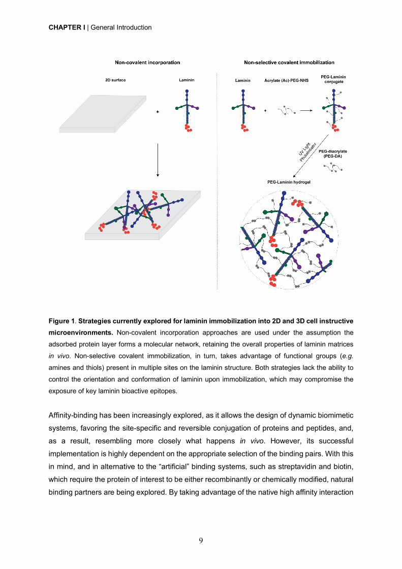
amines and thiols [50] (Fig. 1). Despite widely used, one of the main limitations presented by
these strategies is the inability to control the conformation and orientation of bioactive
molecules upon immobilization. In this regard, in recent years, immobilization strategies have
shifted towards site-specific conjugation, with special focus on biorthogonal chemical
reactions (click chemistry), enzymatic ligation and affinity binding, using either unnatural
amino acids or engineered site-selective amino acid sequences (for a more comprehensive
review see [54]). These strategies are expected to provide a higher retention of bioactivity, by
favoring the access to the active sites of immobilized proteins.
CHAPTER I | General Introduction
9
Figure 1. Strategies currently explored for laminin immobilization into 2D and 3D cell instructive microenvironments. Non-covalent incorporation approaches are used under the assumption the
adsorbed protein layer forms a molecular network, retaining the overall properties of laminin matrices
in vivo. Non-selective covalent immobilization, in turn, takes advantage of functional groups (e.g.
amines and thiols) present in multiple sites on the laminin structure. Both strategies lack the ability to
control the orientation and conformation of laminin upon immobilization, which may compromise the
exposure of key laminin bioactive epitopes.
Affinity-binding has been increasingly explored, as it allows the design of dynamic biomimetic
systems, favoring the site-specific and reversible conjugation of proteins and peptides, and,
as a result, resembling more closely what happens in vivo. However, its successful
implementation is highly dependent on the appropriate selection of the binding pairs. With this
in mind, and in alternative to the “artificial” binding systems, such as streptavidin and biotin,
which require the protein of interest to be either recombinantly or chemically modified, natural
binding partners are being explored. By taking advantage of the native high affinity interaction
CHAPTER I | General Introduction
10
between proteins and peptides, strong non-covalent interactions can be established without
the need for protein modification.
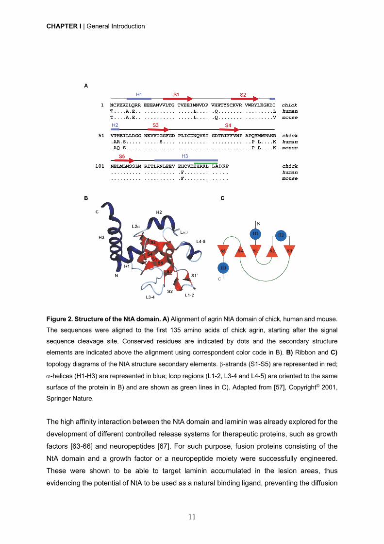
The N-terminal agrin (NtA) domain, which comprises the first 135 amino acids of agrin,
mediates a high affinity interaction with laminin (dissociation constant KD @ 5nM) [55], required
for the integration of agrin into synaptic basal lamina and other basement membranes [56].
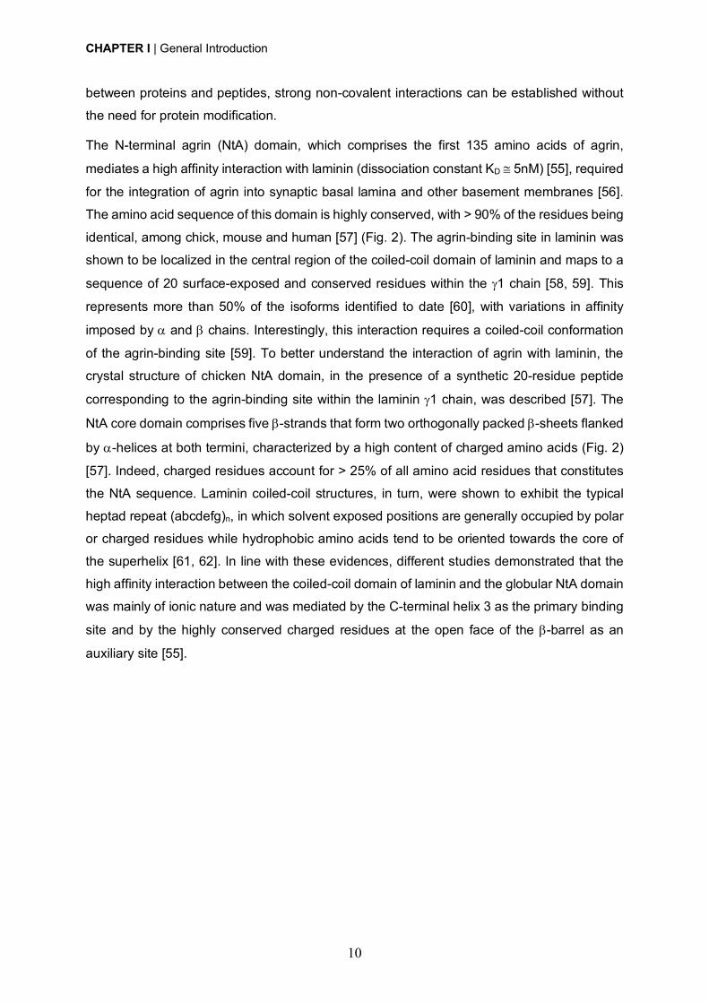
The amino acid sequence of this domain is highly conserved, with > 90% of the residues being
identical, among chick, mouse and human [57] (Fig. 2). The agrin-binding site in laminin was
shown to be localized in the central region of the coiled-coil domain of laminin and maps to a
sequence of 20 surface-exposed and conserved residues within the g1 chain [58, 59]. This
represents more than 50% of the isoforms identified to date [60], with variations in affinity
imposed by a and b chains. Interestingly, this interaction requires a coiled-coil conformation
of the agrin-binding site [59]. To better understand the interaction of agrin with laminin, the
crystal structure of chicken NtA domain, in the presence of a synthetic 20-residue peptide
corresponding to the agrin-binding site within the laminin g1 chain, was described [57]. The
NtA core domain comprises five b-strands that form two orthogonally packed b-sheets flanked
by a-helices at both termini, characterized by a high content of charged amino acids (Fig. 2)
[57]. Indeed, charged residues account for > 25% of all amino acid residues that constitutes
the NtA sequence. Laminin coiled-coil structures, in turn, were shown to exhibit the typical
heptad repeat (abcdefg)n, in which solvent exposed positions are generally occupied by polar
or charged residues while hydrophobic amino acids tend to be oriented towards the core of
the superhelix [61, 62]. In line with these evidences, different studies demonstrated that the
high affinity interaction between the coiled-coil domain of laminin and the globular NtA domain
was mainly of ionic nature and was mediated by the C-terminal helix 3 as the primary binding
site and by the highly conserved charged residues at the open face of the b-barrel as an
auxiliary site [55].
CHAPTER I | General Introduction
11
Figure 2. Structure of the NtA domain. A) Alignment of agrin NtA domain of chick, human and mouse.
The sequences were aligned to the first 135 amino acids of chick agrin, starting after the signal
sequence cleavage site. Conserved residues are indicated by dots and the secondary structure
elements are indicated above the alignment using correspondent color code in B). B) Ribbon and C) topology diagrams of the NtA structure secondary elements. b-strands (S1-S5) are represented in red;
a-helices (H1-H3) are represented in blue; loop regions (L1-2, L3-4 and L4-5) are oriented to the same
surface of the protein in B) and are shown as green lines in C). Adapted from [57], Copyright© 2001,
Springer Nature.
The high affinity interaction between the NtA domain and laminin was already explored for the
development of different controlled release systems for therapeutic proteins, such as growth
factors [63-66] and neuropeptides [67]. For such purpose, fusion proteins consisting of the
NtA domain and a growth factor or a neuropeptide moiety were successfully engineered.
These were shown to be able to target laminin accumulated in the lesion areas, thus
evidencing the potential of NtA to be used as a natural binding ligand, preventing the diffusion

CHAPTER I | General Introduction
12
of the growth factors or neuropeptides and hence increase their timely presence at a specific
site.
Laminin-derived small adhesive sequences have been increasingly explored, in alternative to
the incorporation of the full-length protein, to confer bioactivity to 3D matrices [50]. Most of the
works published to date, have explored synthetic adhesive peptides, which mediate cell
interaction via integrin receptors [50]. Alternatively, cell binding ligands interacting with
transmembrane heparan sulfate proteoglycans (HSPGs), and more specifically syndecans,
have gained more attention in the last few years, as attractive alternative to engineer
biomimetic matrices, as result of their key role in regulating NSC stemness [68-70]. Syndecans
are transmembrane HSPGs, with a key role on the modulation of different biological
processes, including neural patterning, angiogenesis, inflammation and wound healing [71].
These cell adhesion receptors are composed by an extracellular domain, comprising
glycosaminoglycan (GAG) chains, including heparan sulfate (HS) and chondroitin sulfate (CS)
chains, which mediate the interaction with growth factors, ECM proteins, as well as with other
receptors (Fig. 3) [71, 72]. Syndecan receptors also present a single transmembrane domain
that regulates homo- and hetero-dimerization, and a short cytoplasmic domain, which interact
directly with intracellular signaling molecules (Fig. 3) [71, 72]. The extracellular domain of
these receptors varies between the four members of the syndecan family identified in
mammals (Syndecan-1, -2, -3 and -4), while the transmembrane and cytoplasmic domains,,
are highly conserved [71].
Figure 3. Schematic representation of mammalian syndecan receptors. Syndecan receptors are
composed by an extracellular domain comprising several glycosaminoglycan (GAG) chains – Heparan
CHAPTER I | General Introduction
13
sulfate (HS) and Chondroitin sulfate (CS) chains, which mediate several cell-cell and cell-matrix
interactions. They also have a single transmembrane domain responsible to promote self-association
between core proteins, and a cytoplasmic domain that will mediate downstream signaling. Adapted
from [73], Copyright® 2014, John Wiley & Sons.
The four members of the syndecan family (Syndecan-1, -2, -3 and -4) present distinct cell-
and tissue-specific expression patterns [74]. More specifically, within the NSC niche,
syndecans have a key role on the modulation of all stages of stem cell maintenance and
neurogenesis (e.g. proliferation, self-renewal, differentiation, migration and maturation), either
through independent signaling or by working alongside with other receptors, such as integrins
[68-70]. Syndecan-1 is highly expressed during neurogenesis and is key to support NSC
proliferation and progenitor cell maintenance [75, 76], while syndecan-4 constitutes a key
marker of NSCs differentiation [77]. These evidences are in agreement with previous studies
from our lab, showing that human NSCs (hNSCs) derived from the H9 embryonic stem cell
line, cultured under basal conditions, express high levels of syndecan-1 (98.6%), whereas
syndecan-4 levels were found in much lower amounts (2.62%) [78]. Syndecan-3 is key for the
modulation of axon guidance [79, 80] and neurite outgrowth [81, 82], and together with
syndecan-4 [83] controls neuronal migration. Lastly, syndecan-2 exerts a remarkable effect in
dendritic spine morphogenesis [84].
AG73 (RKRLQVQLSIRT) is a synthetic peptide derived from the laminin globular 4 (LG4)
domain of laminin a1 chain, which interacts with syndecan-1 [85, 86] and syndecan-4 [87].
This peptide was reported to promote cell adhesion with membrane ruffling [88-90], and
showed great potential to enhance neurite outgrowth of PC12 neuronal cells when tethered
onto natural-based hydrogels [91-93]. A previous study from our lab, evidenced that
functionalization of fibrin hydrogels with AG73 improves cell outgrowth of hNSCs [78]. Despite
the promising results, the intrinsic bioactivity presented by natural-based polymers makes
difficult to deconvolute the contribution of the syndecan-binding peptide to the final biological
outcome. Synthetic-based polymers, in turn, constitute an attractive alternative in this regard,
as they are chemically-defined materials that allow a precise and independent control over the
biochemical and biophysical properties of the matrix [23]. Additionally, to the best of our
knowledge, to date, no study evaluating the functionalization of synthetic hydrogels with the
AG73 peptide and assessing its effect on the modulation of NSC fate and function was
conducted.
In summary, the design of biologically relevant matrices able to recapitulate the
microenvironment of the stem cell niches and modulate the stem cell function and fate, hold
CHAPTER I | General Introduction
14
great potential when trying to improve the efficacy of cell-based therapeutic strategies
currently under investigation and development.
CHAPTER I | General Introduction
15
References
[1] R.Y. Tam, T. Fuehrmann, N. Mitrousis, M.S. Shoichet, Regenerative therapies for central
nervous system diseases: a biomaterials approach, Neuropsychopharmacology 39(1) (2014)
169-88.
[2] L. Binan, A. Ajji, G. De Crescenzo, M. Jolicoeur, Approaches for neural tissue regeneration,
Stem Cell Rev 10(1) (2014) 44-59.
[3] G. Yiu, Z. He, Glial inhibition of CNS axon regeneration, Nat Rev Neurosci 7(8) (2006) 617-
27.
[4] A.P. Pego, S. Kubinova, D. Cizkova, I. Vanicky, F.M. Mar, M.M. Sousa, E. Sykova,
Regenerative medicine for the treatment of spinal cord injury: more than just promises?, J Cell
Mol Med 16(11) (2012) 2564-82.
[5] S.A. Goldman, Stem and Progenitor Cell-Based Therapy of the Central Nervous System:
Hopes, Hype, and Wishful Thinking, Cell Stem Cell 18(2) (2016) 174-88.
[6] C.H. Ho, R.J. Triolo, A.L. Elias, K.L. Kilgore, A.F. DiMarco, K. Bogie, A.H. Vette, M.L. Audu,
R. Kobetic, S.R. Chang, K.M. Chan, S. Dukelow, D.J. Bourbeau, S.W. Brose, K.J. Gustafson,
Z.H. Kiss, V.K. Mushahwar, Functional electrical stimulation and spinal cord injury, Phys Med
Rehabil Clin N Am 25(3) (2014) 631-54, ix.
[7] I. Vismara, S. Papa, F. Rossi, G. Forloni, P. Veglianese, Current Options for Cell Therapy
in Spinal Cord Injury, Trends Mol Med 23(9) (2017) 831-849.
[8] O. Lindvall, Z. Kokaia, Stem cells in human neurodegenerative disorders--time for clinical
translation?, J Clin Invest 120(1) (2010) 29-40.
[9] Y. Tang, P. Yu, L. Cheng, Current progress in the derivation and therapeutic application of
neural stem cells, Cell Death Dis 8(10) (2017) e3108.
[10] J.A. Steinbeck, L. Studer, Moving stem cells to the clinic: potential and limitations for brain
repair, Neuron 86(1) (2015) 187-206.
[11] P. Lu, L.L. Jones, E.Y. Snyder, M.H. Tuszynski, Neural stem cells constitutively secrete
neurotrophic factors and promote extensive host axonal growth after spinal cord injury, Exp
Neurol 181(2) (2003) 115-29.
[12] K. Reekmans, J. Praet, J. Daans, V. Reumers, P. Pauwels, A. Van der Linden, Z.N.
Berneman, P. Ponsaerts, Current challenges for the advancement of neural stem cell biology
and transplantation research, Stem Cell Rev 8(1) (2012) 262-78.
[13] D. McLauchlan, N.P. Robertson, Stem cells in the treatment of central nervous system
disease, J Neurol 265(4) (2018) 984-986.
[14] A. Trounson, C. McDonald, Stem Cell Therapies in Clinical Trials: Progress and
Challenges, Cell Stem Cell 17(1) (2015) 11-22.
CHAPTER I | General Introduction
16
[15] M.H. Amer, F. Rose, K.M. Shakesheff, M. Modo, L.J. White, Translational considerations
in injectable cell-based therapeutics for neurological applications: concepts, progress and
challenges, NPJ Regen Med 2 (2017) 23.
[16] F. Gattazzo, A. Urciuolo, P. Bonaldo, Extracellular matrix: a dynamic microenvironment
for stem cell niche, Biochim Biophys Acta 1840(8) (2014) 2506-19.
[17] S.W. Lane, D.A. Williams, F.M. Watt, Modulating the stem cell niche for tissue
regeneration, Nat Biotechnol 32(8) (2014) 795-803.
[18] A. Vishwakarma, J. Rouwkema, P.A. Jones, J.M. Karp, The Need to Study, Mimic and
Target Stem Cell Niches, in: A. Vishwakarma, J.M. Karp (Eds.), Biology and Engineering of
Stem Cell Niches, Elsevier2017.
[19] J.L. Wilson, T.C. McDevitt, Biofunctional Hydrogels for Three-Dimensional Stem Cell
Culture, in: A. Vishwakarma, J.M. Karp (Eds.), Biology and Engineering of Stem Cell Niches,
Elsevier2017.
[20] F. Ruedinger, A. Lavrentieva, C. Blume, I. Pepelanova, T. Scheper, Hydrogels for 3D
mammalian cell culture: a starting guide for laboratory practice, Appl Microbiol Biotechnol
99(2) (2015) 623-36.
[21] Technology Platforms for 3D Cell Culture: A User's Guide, John Wiley & Sons Ltd.2017.
[22] M.W. Tibbitt, K.S. Anseth, Hydrogels as Extracellular Matrix Mimics for 3D Cell Culture,
Biotechnol. Bioeng. 103(4) (2009) 655-663.
[23] D. Barros, I.F. Amaral, A.P. Pego, Biomimetic synthetic self-assembled hydrogels for cell
transplantation, Curr Top Med Chem 15(13) (2015) 1209-26.
[24] K.J. Lampe, S.C. Heilshorn, Building stem cell niches from the molecule up through
engineered peptide materials, Neurosci Lett 519(2) (2012) 138-46.
[25] J. Lam, S.T. Carmichael, W.E. Lowry, T. Segura, Hydrogel design of experiments
methodology to optimize hydrogel for iPSC-NPC culture, Adv Healthc Mater 4(4) (2015) 534-
9.
[26] C. Regalado-Santiago, E. Juarez-Aguilar, J.D. Olivares-Hernandez, E. Tamariz,
Mimicking Neural Stem Cell Niche by Biocompatible Substrates, Stem Cells Int 2016 (2016)
1513285.
[27] C.M. Madl, B.L. LeSavage, R.E. Dewi, C.B. Dinh, R.S. Stowers, M. Khariton, K.J. Lampe,
D. Nguyen, O. Chaudhuri, A. Enejder, S.C. Heilshorn, Maintenance of neural progenitor cell
stemness in 3D hydrogels requires matrix remodelling, Nat Mater 16(12) (2017) 1233-1242.
[28] J. Zhu, R.E. Marchant, Design properties of hydrogel tissue-engineering scaffolds, Expert
Rev. Med. Devices 8(5) (2011) 607-26.
[29] G.A. Saracino, D. Cigognini, D. Silva, A. Caprini, F. Gelain, Nanomaterials design and
tests for neural tissue engineering, Chem. Soc. Rev. 42(1) (2013) 225-62.
CHAPTER I | General Introduction
17
[30] J. Zhu, Bioactive modification of poly(ethylene glycol) hydrogels for tissue engineering,
Biomaterials 31(17) (2010) 4639-56.
[31] Y.H. Tsou, J. Khoneisser, P.C. Huang, X. Xu, Hydrogel as a bioactive material to regulate
stem cell fate, Bioact Mater 1(1) (2016) 39-55.
[32] X. Lu, T.H. Perera, A.B. Aria, L.A.S. Callahan, Polyethylene glycol in spinal cord injury
repair: a critical review, J Exp Pharmacol 10 (2018) 37-49.
[33] N.A. Peppas, K.B. Keys, M. Torres-Lugo, A.M. Lowman, Poly(ethylene glycol)-containing
hydrogels in drug delivery, J Control Release 62(1-2) (1999) 81-7.
[34] E.A. Phelps, N.O. Enemchukwu, V.F. Fiore, J.C. Sy, N. Murthy, T.A. Sulchek, T.H. Barker,
A.J. Garcia, Maleimide cross-linked bioactive PEG hydrogel exhibits improved reaction
kinetics and cross-linking for cell encapsulation and in situ delivery, Adv Mater 24(1) (2012)
64-70, 2.
[35] J. Yu, F. Chen, X. Wang, N. Dong, C. Lu, G. Yang, Z. Chen, Synthesis and
characterization of MMP degradable and maleimide cross-linked PEG hydrogels for tissue
engineering scaffolds, Polymer Degradation and Stability 133 (2016) 312-320.
[36] N.O. Enemchukwu, R. Cruz-Acuna, T. Bongiorno, C.T. Johnson, J.R. Garcia, T. Sulchek,
A.J. Garcia, Synthetic matrices reveal contributions of ECM biophysical and biochemical
properties to epithelial morphogenesis, J Cell Biol 212(1) (2016) 113-24.
[37] W.M. Han, S.E. Anderson, M. Mohiuddin, D. Barros, S.A. Nakhai, E. Shin, I.F. Amaral,
A.P. Pego, A.J. Garcia, Y.C. Jang, Synthetic matrix enhances transplanted satellite cell
engraftment in dystrophic and aged skeletal muscle with comorbid trauma, Sci Adv 4(8) (2018)
eaar4008.
[38] S. Lin, N. Sangaj, T. Razafiarison, C. Zhang, S. Varghese, Influence of physical properties
of biomaterials on cellular behavior, Pharm Res 28(6) (2011) 1422-30.
[39] J. Kim, Y.P. Kong, S.M. Niedzielski, R.K. Singh, A.J. Putnam, A. Shikanov,
Characterization of the crosslinking kinetics of multi-arm poly(ethylene glycol) hydrogels
formed via Michael-type addition, Soft Matter 12(7) (2016) 2076-85.
[40] L.A. Flanagan, Y.E. Ju, B. Marg, M. Osterfield, P.A. Janmey, Neurite branching on
deformable substrates, Neuroreport 13(18) (2002) 2411-5.
[41] K. Saha, A.J. Keung, E.F. Irwin, Y. Li, L. Little, D.V. Schaffer, K.E. Healy, Substrate
modulus directs neural stem cell behavior, Biophys. J. 95(9) (2008) 4426-38.
[42] A. Banerjee, M. Arha, S. Choudhary, R.S. Ashton, S.R. Bhatia, D.V. Schaffer, R.S. Kane,
The influence of hydrogel modulus on the proliferation and differentiation of encapsulated
neural stem cells, Biomaterials 30(27) (2009) 4695-9.
[43] S.R. Hynes, M.F. Rauch, J.P. Bertram, E.B. Lavik, A library of tunable poly(ethylene
glycol)/poly(L-lysine) hydrogels to investigate the material cues that influence neural stem cell
differentiation, J Biomed Mater Res A 89(2) (2009) 499-509.
CHAPTER I | General Introduction
18
[44] A. Kerever, J. Schnack, D. Vellinga, N. Ichikawa, C. Moon, E. Arikawa-Hirasawa, J.T.
Efird, F. Mercier, Novel extracellular matrix structures in the neural stem cell niche capture the
neurogenic factor fibroblast growth factor 2 from the extracellular milieu, Stem Cells 25(9)
(2007) 2146-57.
[45] I. Kazanis, J.D. Lathia, T.J. Vadakkan, E. Raborn, R. Wan, M.R. Mughal, D.M. Eckley, T.
Sasaki, B. Patton, M.P. Mattson, K.K. Hirschi, M.E. Dickinson, C. ffrench-Constant,
Quiescence and activation of stem and precursor cell populations in the subependymal zone
of the mammalian brain are associated with distinct cellular and extracellular matrix signals, J
Neurosci 30(29) (2010) 9771-81.
[46] A. Hyysalo, M. Ristola, M.E. Makinen, S. Hayrynen, M. Nykter, S. Narkilahti, Laminin
alpha5 substrates promote survival, network formation and functional development of human
pluripotent stem cell-derived neurons in vitro, Stem Cell Res 24 (2017) 118-127.
[47] L. Luckenbill-Edds, Laminin and the mechanism of neuronal outgrowth, Brain Res Brain
Res Rev 23(1-2) (1997) 1-27.
[48] S.K. Powell, H.K. Kleinman, Neuronal laminins and their cellular receptors, Int J Biochem
Cell Biol 29(3) (1997) 401-14.
[49] S. Plantman, M. Patarroyo, K. Fried, A. Domogatskaya, K. Tryggvason, H. Hammarberg,
S. Cullheim, Integrin-laminin interactions controlling neurite outgrowth from adult DRG
neurons in vitro, Mol Cell Neurosci 39(1) (2008) 50-62.
[50] D. Barros, I.F. Amaral, A.P. Pego, Laminin inspired cell instructive microenvironments for
neural tissue engineering applications, Manuscript in preparation (2019).
[51] B.G. Keselowsky, D.M. Collard, A.J. Garcia, Surface chemistry modulates fibronectin
conformation and directs integrin binding and specificity to control cell adhesion, J Biomed
Mater Res A 66(2) (2003) 247-59.
[52] J.C. Rodriguez Hernandez, M. Salmeron Sanchez, J.M. Soria, J.L. Gomez Ribelles, M.
Monleon Pradas, Substrate chemistry-dependent conformations of single laminin molecules
on polymer surfaces are revealed by the phase signal of atomic force microscopy, Biophys J
93(1) (2007) 202-7.
[53] O.M. Ba, M. Hindie, P. Marmey, O. Gallet, K. Anselme, A. Ponche, A.C. Duncan, Protein
covalent immobilization via its scarce thiol versus abundant amine groups: Effect on
orientation, cell binding domain exposure and conformational lability, Colloids Surf B
Biointerfaces 134 (2015) 73-80.
[54] S.A. Fisher, A.E.G. Baker, M.S. Shoichet, Designing Peptide and Protein Modified
Hydrogels: Selecting the Optimal Conjugation Strategy, J Am Chem Soc 139(22) (2017) 7416-
7427.
[55] J.B. Mascarenhas, M.A. Ruegg, U. Winzen, W. Halfter, J. Engel, J. Stetefeld, Mapping of
the laminin-binding site of the N-terminal agrin domain (NtA), EMBO J 22(3) (2003) 529-36.
CHAPTER I | General Introduction
19
[56] M.A. Ruegg, J.L. Bixby, Agrin orchestrates synaptic differentiation at the vertebrate
neuromuscular junction, Trends Neurosci 21(1) (1998) 22-7.
[57] J. Stetefeld, M. Jenny, T. Schulthess, R. Landwehr, B. Schumacher, S. Frank, M.A.
Ruegg, J. Engel, R.A. Kammerer, The laminin-binding domain of agrin is structurally related
to N-TIMP-1, Nat Struct Biol 8(8) (2001) 705-9.
[58] A.J. Denzer, T. Schulthess, C. Fauser, B. Schumacher, R.A. Kammerer, J. Engel, M.A.
Ruegg, Electron microscopic structure of agrin and mapping of its binding site in laminin-1,
EMBO J 17(2) (1998) 335-43.
[59] R.A. Kammerer, T. Schulthess, R. Landwehr, B. Schumacher, A. Lustig, P.D. Yurchenco,
M.A. Ruegg, J. Engel, A.J. Denzer, Interaction of agrin with laminin requires a coiled-coil
conformation of the agrin-binding site within the laminin gamma1 chain, EMBO J 18(23) (1999)
6762-70.
[60] M. Aumailley, L. Bruckner-Tuderman, W.G. Carter, R. Deutzmann, D. Edgar, P. Ekblom,
J. Engel, E. Engvall, E. Hohenester, J.C. Jones, H.K. Kleinman, M.P. Marinkovich, G.R.
Martin, U. Mayer, G. Meneguzzi, J.H. Miner, K. Miyazaki, M. Patarroyo, M. Paulsson, V.
Quaranta, J.R. Sanes, T. Sasaki, K. Sekiguchi, L.M. Sorokin, J.F. Talts, K. Tryggvason, J.
Uitto, I. Virtanen, K. von der Mark, U.M. Wewer, Y. Yamada, P.D. Yurchenco, A simplified
laminin nomenclature, Matrix Biol 24(5) (2005) 326-32.
[61] D.A. Parry, R.D. Fraser, J.M. Squire, Fifty years of coiled-coils and alpha-helical bundles:
a close relationship between sequence and structure, J Struct Biol 163(3) (2008) 258-69.
[62] G. Armony, E. Jacob, T. Moran, Y. Levin, T. Mehlman, Y. Levy, D. Fass, Cross-linking
reveals laminin coiled-coil architecture, Proc Natl Acad Sci U S A 113(47) (2016) 13384-
13389.
[63] W. Sun, C. Sun, H. Zhao, H. Lin, Q. Han, J. Wang, H. Ma, B. Chen, Z. Xiao, J. Dai,
Improvement of sciatic nerve regeneration using laminin-binding human NGF-beta, PLoS One
4(7) (2009) e6180.
[64] Q. Han, B. Li, H. Feng, Z. Xiao, B. Chen, Y. Zhao, J. Huang, J. Dai, The promotion of
cerebral ischemia recovery in rats by laminin-binding BDNF, Biomaterials 32(22) (2011) 5077-
85.
[65] J. Xie, B. Jin, D.W. Li, B. Shen, N. Gong, T.Z. Zhang, P. Dong, Effect of laminin-binding
BDNF on induction of recurrent laryngeal nerve regeneration by miR-222 activation of mTOR
signal pathway, Am J Transl Res 7(6) (2015) 1071-80.
[66] B. Wang, J. Yuan, X. Chen, J. Xu, Y. Li, P. Dong, Functional regeneration of the
transected recurrent laryngeal nerve using a collagen scaffold loaded with laminin and laminin-
binding BDNF and GDNF, Sci Rep 6 (2016) 32292.
CHAPTER I | General Introduction
20
[67] L. Wu, J. Wang, X. Chen, A. Hong, Expression, identification and biological effects of the
novel recombination protein, PACAP38-NtA, with high bioactivity, Int J Mol Med 35(2) (2015)
376-82.
[68] M. Ford-Perriss, K. Turner, S. Guimond, A. Apedaile, H.D. Haubeck, J. Turnbull, M.
Murphy, Localisation of specific heparan sulfate proteoglycans during the proliferative phase
of brain development, Dev Dyn 227(2) (2003) 170-84.
[69] Y. Choi, H. Chung, H. Jung, J.R. Couchman, E.S. Oh, Syndecans as cell surface
receptors: Unique structure equates with functional diversity, Matrix Biol 30(2) (2011) 93-9.
[70] F.E. Poulain, H.J. Yost, Heparan sulfate proteoglycans: a sugar code for vertebrate
development?, Development 142(20) (2015) 3456-67.
[71] N.A. Afratis, D. Nikitovic, H.A. Multhaupt, A.D. Theocharis, J.R. Couchman, N.K.
Karamanos, Syndecans - key regulators of cell signaling and biological functions, FEBS J
284(1) (2017) 27-41.
[72] H. Chung, H.A. Multhaupt, E.S. Oh, J.R. Couchman, Minireview: Syndecans and their
crucial roles during tissue regeneration, FEBS Lett 590(15) (2016) 2408-17.
[73] J.R. Couchman, S. Gopal, H.C. Lim, S. Norgaard, H.A. Multhaupt, Fell-Muir Lecture:
Syndecans: from peripheral coreceptors to mainstream regulators of cell behaviour, Int J Exp
Pathol 96(1) (2015) 1-10.
[74] X. Xian, S. Gopal, J.R. Couchman, Syndecans as receptors and organizers of the
extracellular matrix, Cell Tissue Res 339(1) (2010) 31-46.
[75] Q. Wang, L. Yang, C. Alexander, S. Temple, The niche factor syndecan-1 regulates the
maintenance and proliferation of neural progenitor cells during mammalian cortical
development, PLoS One 7(8) (2012) e42883.
[76] L. Morizur, A. Chicheportiche, L.R. Gauthier, M. Daynac, F.D. Boussin, M.A. Mouthon,
Distinct Molecular Signatures of Quiescent and Activated Adult Neural Stem Cells Reveal
Specific Interactions with Their Microenvironment, Stem Cell Reports (2018).
[77] L.E. Oikari, R.K. Okolicsanyi, A. Qin, C. Yu, L.R. Griffiths, L.M. Haupt, Cell surface
heparan sulfate proteoglycans as novel markers of human neural stem cell fate determination,
Stem Cell Res 16(1) (2016) 92-104.
[78] A.R. Bento, Improving neurite outgrowth in 3D hydrogel matrices by mimicking cell
receptor-ECM interactions occurring in neurogenic niches: an engineering approach to
develop more efficient neural stem cell hydrogel carriers, Faculdade de Engenharia,
Universidade do Porto, 2018.
[79] A. Kinnunen, T. Kinnunen, M. Kaksonen, R. Nolo, P. Panula, H. Rauvala, N-syndecan
and HB-GAM (heparin-binding growth-associated molecule) associate with early axonal tracts
in the rat brain, Eur J Neurosci 10(2) (1998) 635-48.
CHAPTER I | General Introduction
21
[80] A. Kinnunen, M. Niemi, T. Kinnunen, M. Kaksonen, R. Nolo, H. Rauvala, Heparan
sulphate and HB-GAM (heparin-binding growth-associated molecule) in the development of
the thalamocortical pathway of rat brain, Eur J Neurosci 11(2) (1999) 491-502.
[81] E. Raulo, M.A. Chernousov, D.J. Carey, R. Nolo, H. Rauvala, Isolation of a neuronal cell
surface receptor of heparin binding growth-associated molecule (HB-GAM). Identification as
N-syndecan (syndecan-3), J Biol Chem 269(17) (1994) 12999-3004.
[82] T. Kinnunen, E. Raulo, R. Nolo, M. Maccarana, U. Lindahl, H. Rauvala, Neurite outgrowth
in brain neurons induced by heparin-binding growth-associated molecule (HB-GAM) depends
on the specific interaction of HB-GAM with heparan sulfate at the cell surface, J Biol Chem
271(4) (1996) 2243-8.
[83] H.K. Matthews, L. Marchant, C. Carmona-Fontaine, S. Kuriyama, J. Larrain, M.R. Holt,
M. Parsons, R. Mayor, Directional migration of neural crest cells in vivo is regulated by
Syndecan-4/Rac1 and non-canonical Wnt signaling/RhoA, Development 135(10) (2008)
1771-80.
[84] I.M. Ethell, Y. Yamaguchi, Cell surface heparan sulfate proteoglycan syndecan-2 induces
the maturation of dendritic spines in rat hippocampal neurons, J Cell Biol 144(3) (1999) 575-
86.
[85] M.P. Hoffman, M. Nomizu, E. Roque, S. Lee, D.W. Jung, Y. Yamada, H.K. Kleinman,
Laminin-1 and laminin-2 G-domain synthetic peptides bind syndecan-1 and are involved in
acinar formation of a human submandibular gland cell line, J Biol Chem 273(44) (1998) 28633-
41.
[86] L.N. Gama-de-Souza, E. Cyreno-Oliveira, V.M. Freitas, E.S. Melo, V.F. Vilas-Boas, A.S.
Moriscot, R.G. Jaeger, Adhesion and protease activity in cell lines from human salivary gland
tumors are regulated by the laminin-derived peptide AG73, syndecan-1 and beta1 integrin,
Matrix Biol 27(5) (2008) 402-19.
[87] N. Suzuki, N. Ichikawa, S. Kasai, M. Yamada, N. Nishi, H. Morioka, H. Yamashita, Y.
Kitagawa, A. Utani, M.P. Hoffman, M. Nomizu, Syndecan binding sites in the laminin alpha1
chain G domain, Biochemistry 42(43) (2003) 12625-33.
[88] M. Nomizu, W.H. Kim, K. Yamamura, A. Utani, S.Y. Song, A. Otaka, P.P. Roller, H.K.
Kleinman, Y. Yamada, Identification of cell binding sites in the laminin alpha 1 chain carboxyl-
terminal globular domain by systematic screening of synthetic peptides, J Biol Chem 270(35)
(1995) 20583-90.
[89] K. Hozumi, N. Suzuki, P.K. Nielsen, M. Nomizu, Y. Yamada, Laminin alpha1 chain LG4
module promotes cell attachment through syndecans and cell spreading through integrin
alpha2beta1, J Biol Chem 281(43) (2006) 32929-40.
CHAPTER I | General Introduction
22
[90] M. Mochizuki, Y. Kadoya, Y. Wakabayashi, K. Kato, I. Okazaki, M. Yamada, T. Sato, N.
Sakairi, N. Nishi, M. Nomizu, Laminin-1 peptide-conjugated chitosan membranes as a novel
approach for cell engineering, FASEB J 17(8) (2003) 875-7.
[91] Y. Yamada, K. Hozumi, F. Katagiri, Y. Kikkawa, M. Nomizu, Biological activity of laminin
peptide-conjugated alginate and chitosan matrices, Biopolymers 94(6) (2010) 711-20.
[92] Y. Yamada, F. Katagiri, K. Hozumi, Y. Kikkawa, M. Nomizu, Cell behavior on protein
matrices containing laminin alpha1 peptide AG73, Biomaterials 32(19) (2011) 4327-35.
[93] Y. Yamada, K. Hozumi, A. Aso, A. Hotta, K. Toma, F. Katagiri, Y. Kikkawa, M. Nomizu,
Laminin active peptide/agarose matrices as multifunctional biomaterials for tissue
engineering, Biomaterials 33(16) (2012) 4118-25.

23
CHAPTER II _________________________________________________________________________
Aim and Structure of the Thesis

CHAPTER I | General Introduction
24
CHAPTER II | Aim and Structure of the Thesis
25
AIM OF THE THESIS
The main aim of the work presented in this thesis was the design of biologically relevant
matrices, able to recapitulate the laminin-rich microenvironment of neural stem cell (NSC)
niches. For that, we proposed the biofunctionalization of protease-sensitive synthetic
hydrogel matrices, via site-selective immobilization of full-length laminin. Additionally, we
also explored the modification of such matrices with a syndecan interacting laminin-derived
peptide (AG73). The developed hydrogels were designed to support NSC viability,
proliferation and neuronal differentiation, which are key features for their application, as
dynamic cell-instructive extracellular matrix (ECM) analogs, in in vitro three-dimensional
(3D) platforms for NSC culture and as vehicles for NSC transplantation into the damaged
central nervous system (CNS).
Specific aims of the proposed work included:
§ Establishment of an affinity-based approach to promote the site-selective
immobilization of laminin, allowing for the subsequent controlled presentation of this
ECM protein in cell microenvironments.
§ Evaluation of the potential of the proposed strategy for the site-selective
immobilization of different laminin isoforms (in the context of this thesis assessed
for mouse laminin-111 (msLn-111) and recombinant human laminin-521 (rhLn-521))
into a synthetic hydrogel. As the starting material for the development of the
hydrogel, a four-arm maleimide-end functionalized poly(ethylene glycol) (PEG-
4MAL) macromer was selected, due to its good biocompatibility, both in vitro and in
vivo [1-3], and well characterized biochemical and biophysical properties [3-5]. By
selecting a synthetic polymer as the hydrogel backbone, we aim at assuring
reproducibility, while exploring the versatility of synthetic materials in terms of
tunable physical properties and biofunctionalization.
§ Validation of the developed 3D platform for the culture of human NSCs (hNSCs), in
terms of ability to support cell viability, proliferation and neurite outgrowth.
§ Biofunctionalization of PEG-4MAL hydrogels with a syndecan-binding laminin-
derived peptide (AG73) with neurite-promoting activity, and evaluation of the impact
of the input peptide concentration on hNSC viability, proliferation and neurite
outgrowth.
CHAPTER II | Aim and Structure of the Thesis
26
STRUCTURE OF THE THESIS
This thesis is divided into two parts, comprising a total of nine chapters. In Part 1, a
general introduction and a literature background is given to contextualize and support the
work described in this thesis. In Part 2, the explored methodology and obtained results are
presented, and an overall discussion of the developed work is provided.
The key role of laminin on the modulation of NSC behavior [6-9], makes this ECM protein
highly attractive for the design of NSC niche microenvironments. Indeed, some works
already explored laminin immobilization into different biological and synthetic matrices. In
Chapter III we summarize the structural properties and main functions of laminin and
reviewed the most relevant works using full-length laminin and laminin-derived peptides for
the design of biomimetic matrices, for application in a neural regenerative context [10].
To better understand NSC physiology and interactions with the surrounding
microenvironment, the development of well-defined and tunable 3D platforms able to
replicate the physical and chemical elements of the native ECM is highly desirable. Among
the several 3D platforms developed to date, biomimetic hydrogels still constitute one of the
most attractive options for the design of matrices able to recapitulate the microenvironment
of NSC niches, as they share many key physical properties with native ECM/tissues. In
Chapter IV the most recent work using biomimetic synthetic self-assembled hydrogels for
cell transplantation [11] is reviewed, due to their interesting properties for application in the
CNS, when compared to covalently-crosslinked hydrogels. Additionally, we discuss some
of the main challenges hampering the translation of these systems to the clinic and the
issues that need to be addressed for these to progress from bench-to-bedside.
Many of the strategies explored to date for the immobilization of full-length laminin into both
two-dimensional (2D) and 3D cell-instructive microenvironments are limited by the inability
to control laminin orientation and conformation, upon immobilization, with preservation of
protein bioactivity. In this regard, and envisaging the development of a 3D cell-instructive
microenvironment, in Chapter V we described an affinity-based approach able to promote
the site-selective immobilization of laminin to a specific substrate, while preserving the
exposure of the protein key bioactive epitopes [12, 13]. The proposed strategy takes
advantage of the native high affinity interaction between laminin and the human N-terminal
agrin (hNtA) domain, allowing the controlled immobilization of laminin, with preservation of
its ability to polymerize and mediate hNSC adhesion and spreading.
The application of the proposed affinity-based approach for the site-selective immobilization
of laminin (msLn-111) into a degradable PEG-4MAL hydrogel is reported in Chapter VI [14].
This aims the establishment of an artificial niche able to support NSC 3D culture and
CHAPTER II | Aim and Structure of the Thesis
27
hopefully, transplantation. Affinity-bound laminin PEG-4MAL hydrogels showed great
potential to be used as defined 3D platforms enabling NSC proliferation and neurite
outgrowth, when compared to currently available strategies for laminin immobilization,
which include physical entrapment and non-selective covalent immobilization.
Ln-521 has a key role on the modulation of neural cell behavior (e.g. adhesion, viability and
network formation) [6, 15] and already showed potential to be used as a robust substratum
for the culture and renewal of human embryonic [16] and pluripotent [6] stem cells.
Consequently, in Chapter VII we explored the development of an affinity-bound rhLn-521
PEG-4MAL hydrogel, by taking advantage of the immobilization strategy previously
proposed in Chapter V [12, 13], with some specific modifications, aiming to improve rhLn-
521 binding to NtA and, consequently its impact on hNSC behavior. A significant
improvement on neurite outgrowth was observed, compared to unmodified hydrogels,
suggesting an impact of such modifications on protein conformation and domain
presentation to cells.
Laminin-derived small adhesive sequences (e.g. IKVAV, YIGSR, RNIAEIIKDI,
RKRLQVQSIRT) have been previously explored as alternative to the incorporation of the
full-length laminin, to confer bioactivity to 3D matrices. Most of the works published to date,
explored peptides mediating cell interaction via integrin receptors. More recently, a different
class of receptors, syndecans, have gained more attention as attractive alternative
therapeutic targets to engineer biomimetic matrices in a neural regeneration context. In
Chapter VIII, a syndecan-binding peptide (AG73 – RKRLQVQSIRT) with neurite-promoting
activity and previously explored to modify natural hydrogel matrices [17-21], was explored
for the functionalization of a degradable PEG-4MAL hydrogel. More specifically, the impact
of different input peptide concentrations on cell behavior was assessed. The tethering of
AG73 peptide to PEG-4MAL hydrogels led to a significant improvement of cell viability, after
7 days, independently of the input peptide concentration tested. Additionally, a significant
improvement in hNSC proliferation and neurite outgrowth, as well as the establishment of
a neuronal network were observed, after 14 days, for cells cultured on hydrogels tethered
with the higher concentration of peptide tested (1 mM).
Finally, in Chapter IX the concluding remarks correlating the overall obtained results and
the perspectives for future research are presented.
CHAPTER II | Aim and Structure of the Thesis
28
References
[1] J.D. Weaver, D.M. Headen, J. Aquart, C.T. Johnson, L.D. Shea, H. Shirwan, A.J. Garcia,
Vasculogenic hydrogel enhances islet survival, engraftment, and function in leading
extrahepatic sites, Sci Adv 3(6) (2017) e1700184.
[2] R. Cruz-Acuna, M. Quiros, A.E. Farkas, P.H. Dedhia, S. Huang, D. Siuda, V. Garcia-
Hernandez, A.J. Miller, J.R. Spence, A. Nusrat, A.J. Garcia, Synthetic hydrogels for human
intestinal organoid generation and colonic wound repair, Nat Cell Biol 19(11) (2017) 1326-
1335.
[3] W.M. Han, S.E. Anderson, M. Mohiuddin, D. Barros, S.A. Nakhai, E. Shin, I.F. Amaral,
A.P. Pego, A.J. Garcia, Y.C. Jang, Synthetic matrix enhances transplanted satellite cell
engraftment in dystrophic and aged skeletal muscle with comorbid trauma, Sci Adv 4(8)
(2018) eaar4008.
[4] E.A. Phelps, N.O. Enemchukwu, V.F. Fiore, J.C. Sy, N. Murthy, T.A. Sulchek, T.H.
Barker, A.J. Garcia, Maleimide cross-linked bioactive PEG hydrogel exhibits improved
reaction kinetics and cross-linking for cell encapsulation and in situ delivery, Adv Mater
24(1) (2012) 64-70, 2.
[5] N.O. Enemchukwu, R. Cruz-Acuna, T. Bongiorno, C.T. Johnson, J.R. Garcia, T. Sulchek,
A.J. Garcia, Synthetic matrices reveal contributions of ECM biophysical and biochemical
properties to epithelial morphogenesis, J Cell Biol 212(1) (2016) 113-24.
[6] A. Hyysalo, M. Ristola, M.E. Makinen, S. Hayrynen, M. Nykter, S. Narkilahti, Laminin
alpha5 substrates promote survival, network formation and functional development of
human pluripotent stem cell-derived neurons in vitro, Stem Cell Res 24 (2017) 118-127.
[7] L. Luckenbill-Edds, Laminin and the mechanism of neuronal outgrowth, Brain Res Brain
Res Rev 23(1-2) (1997) 1-27.
[8] S.K. Powell, H.K. Kleinman, Neuronal laminins and their cellular receptors, Int J Biochem
Cell Biol 29(3) (1997) 401-14.
[9] S. Plantman, M. Patarroyo, K. Fried, A. Domogatskaya, K. Tryggvason, H. Hammarberg,
S. Cullheim, Integrin-laminin interactions controlling neurite outgrowth from adult DRG
neurons in vitro, Mol Cell Neurosci 39(1) (2008) 50-62.
[10] D. Barros, I.F. Amaral, A.P. Pego, Laminin inspired cell instructive microenvironments
for neural tissue engineering applications, Manuscript in preparation (2019).
[11] D. Barros, I.F. Amaral, A.P. Pego, Biomimetic synthetic self-assembled hydrogels for
cell transplantation, Curr Top Med Chem 15(13) (2015) 1209-26.
[12] D. Barros, P. Parreira, J. Furtado, F. Ferreira-da-Silva, E. Conde-Sousa, A.J. Garcia,
M.C.L. Martins, I.F. Amaral, A.P. Pego, An affinity-based approach to engineer laminin-
presenting cell instructive microenvironments, Biomaterials 192 (2019) 601-611.
CHAPTER II | Aim and Structure of the Thesis
29
[13] D. Barros, I.F. Amaral, A.P. Pego, Laminin immobilization, methods and uses thereof,
Portuguese Provisional Patent Application no. 20181000074673, filled 2018/11/30.
[14] D. Barros, E. Conde-Sousa, A.J. Garcia, I.F. Amaral, A.P. Pego, Engineering hydrogels
with affinity-bound laminin as 3D neural stem cell culture systems Manuscript in preparation
(2019).
[15] A. Bergeron, H. Sherman, P. Pardo, H. Gitschier, H. Nandivada, D. Saxena, Corning®
rLaminin-521 (Human) for Expansion and Differentiation of Human Neural Stem Cells,
Corning Incorporated | Application Note (2015).
[16] S. Rodin, L. Antonsson, C. Niaudet, O.E. Simonson, E. Salmela, E.M. Hansson, A.
Domogatskaya, Z. Xiao, P. Damdimopoulou, M. Sheikhi, J. Inzunza, A.S. Nilsson, D. Baker,
R. Kuiper, Y. Sun, E. Blennow, M. Nordenskjold, K.H. Grinnemo, J. Kere, C. Betsholtz, O.
Hovatta, K. Tryggvason, Clonal culturing of human embryonic stem cells on laminin-521/E-
cadherin matrix in defined and xeno-free environment, Nat Commun 5 (2014) 3195.
[17] M. Mochizuki, Y. Kadoya, Y. Wakabayashi, K. Kato, I. Okazaki, M. Yamada, T. Sato,
N. Sakairi, N. Nishi, M. Nomizu, Laminin-1 peptide-conjugated chitosan membranes as a
novel approach for cell engineering, FASEB J 17(8) (2003) 875-7.
[18] Y. Yamada, K. Hozumi, F. Katagiri, Y. Kikkawa, M. Nomizu, Biological activity of laminin
peptide-conjugated alginate and chitosan matrices, Biopolymers 94(6) (2010) 711-20.
[19] Y. Yamada, F. Katagiri, K. Hozumi, Y. Kikkawa, M. Nomizu, Cell behavior on protein
matrices containing laminin alpha1 peptide AG73, Biomaterials 32(19) (2011) 4327-35.
[20] Y. Yamada, K. Hozumi, A. Aso, A. Hotta, K. Toma, F. Katagiri, Y. Kikkawa, M. Nomizu,
Laminin active peptide/agarose matrices as multifunctional biomaterials for tissue
engineering, Biomaterials 33(16) (2012) 4118-25.
[21] A.R. Bento, Improving neurite outgrowth in 3D hydrogel matrices by mimicking cell
receptor-ECM interactions occurring in neurogenic niches: an engineering approach to
develop more efficient neural stem cell hydrogel carriers, Faculdade de Engenharia,
Universidade do Porto, 2018.
CHAPTER II | Aim and Structure of the Thesis
30
31
CHAPTER III _________________________________________________________________________
Laminin-Inspired Cell-Instructive
Microenvironments for Neural Tissue
Engineering
CHAPTER III | Laminin-Inspired Cell-Instructive Microenvironments for Neural Tissue Engineering
32
CHAPTER III | Laminin-Inspired Cell-Instructive Microenvironments for Neural Tissue Engineering
33
Laminin-Inspired Cell-Instructive Microenvironments for
Neural Tissue Engineering
Daniela Barros 1,2,3, Isabel F. Amaral 1,2,4, Ana P. Pêgo 1,2,3,4
1 i3S - Instituto de Investigação e Inovação em Saúde, Universidade do Porto (UPorto),
Portugal 2 INEB - Instituto de Engenharia Biomédica, UPorto, Portugal 3 ICBAS - Instituto de Ciências Biomédicas Abel Salazar, UPorto, Portugal 4 FEUP - Faculdade de Engenharia, UPorto, Portugal
_______________________________________________________________________
Manuscript (Literature Review) in preparation
CHAPTER III | Laminin-Inspired Cell-Instructive Microenvironments for Neural Tissue Engineering
34
Abstract
Laminin is a heterotrimeric glycoprotein with a key role in the formation and maintenance of
basement membrane (BM) architecture and properties, as well as on the modulation of
several biological functions, including cell adhesion, migration, differentiation and matrix-
mediated signaling. Laminin is differentially expressed within central nervous system (CNS)
during development and homeostasis, with an impact on the modulation of cell function and
fate. Within neurogenic niches, laminin is one of the most important and well described
extracellular matrix (ECM) proteins. Therefore, an understanding of laminin assembly,
domain architecture and interaction of its different bioactive domains with cell surface
receptors, soluble signaling molecules and ECM proteins is essential to better understand
the role of this ECM protein and its receptors on the modulation of neurogenesis, both in
homeostasis or during repair. This is also expected to provide a rational basis to design
biomaterial-based matrices that mimic the biological properties of the BM for neural tissue
repair and cell replacement.
In this chapter, a general overview of laminin structure and domain architecture, as well as
the main biological functions mediated by this heterotrimeric glycoprotein is provided. The
expression and distribution of laminin in the CNS, and more specifically its role within adult
neural stem cell niche is summarized. Moreover, a detailed overview on the use of full-
length laminin and laminin derived peptides/recombinant laminin fragments for the
development of biocompatible hydrogels mimicking the neurogenic niches, and with
application for tissue repair and cell replacement in the context of the nervous tissue is
given. Finally, the main challenges associated with the development of laminin-inspired
hydrogels and the issues that need to be addressed for these to progress from bench-to-
bedside are discussed.
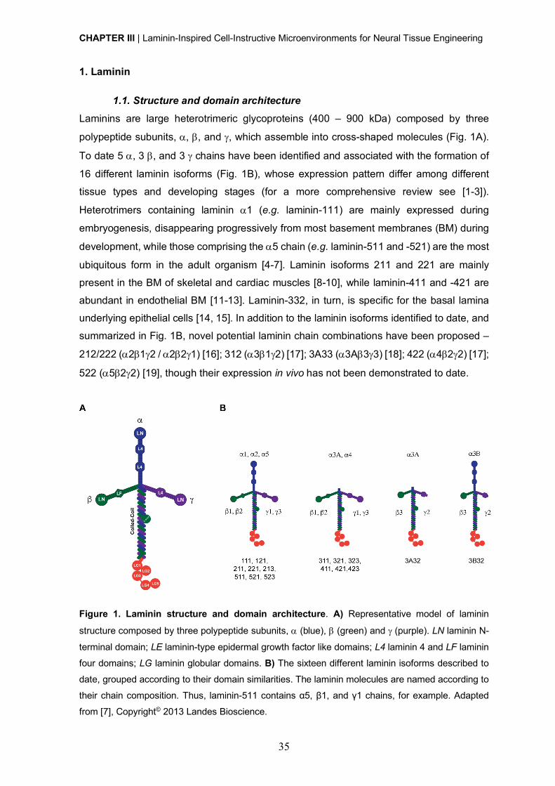
CHAPTER III | Laminin-Inspired Cell-Instructive Microenvironments for Neural Tissue Engineering
35
1. Laminin
1.1. Structure and domain architecture Laminins are large heterotrimeric glycoproteins (400 – 900 kDa) composed by three
polypeptide subunits, a, b, and g, which assemble into cross-shaped molecules (Fig. 1A).
To date 5 a, 3 b, and 3 g chains have been identified and associated with the formation of
16 different laminin isoforms (Fig. 1B), whose expression pattern differ among different
tissue types and developing stages (for a more comprehensive review see [1-3]).
Heterotrimers containing laminin a1 (e.g. laminin-111) are mainly expressed during
embryogenesis, disappearing progressively from most basement membranes (BM) during
development, while those comprising the a5 chain (e.g. laminin-511 and -521) are the most
ubiquitous form in the adult organism [4-7]. Laminin isoforms 211 and 221 are mainly
present in the BM of skeletal and cardiac muscles [8-10], while laminin-411 and -421 are
abundant in endothelial BM [11-13]. Laminin-332, in turn, is specific for the basal lamina
underlying epithelial cells [14, 15]. In addition to the laminin isoforms identified to date, and
summarized in Fig. 1B, novel potential laminin chain combinations have been proposed –
212/222 (a2b1g2 / a2b2g1) [16]; 312 (a3b1g2) [17]; 3A33 (a3Ab3g3) [18]; 422 (a4b2g2) [17];
522 (a5b2g2) [19], though their expression in vivo has not been demonstrated to date.
Figure 1. Laminin structure and domain architecture. A) Representative model of laminin
structure composed by three polypeptide subunits, a (blue), b (green) and g (purple). LN laminin N-
terminal domain; LE laminin-type epidermal growth factor like domains; L4 laminin 4 and LF laminin
four domains; LG laminin globular domains. B) The sixteen different laminin isoforms described to
date, grouped according to their domain similarities. The laminin molecules are named according to
their chain composition. Thus, laminin-511 contains α5, β1, and γ1 chains, for example. Adapted
from [7], Copyright© 2013 Landes Bioscience.
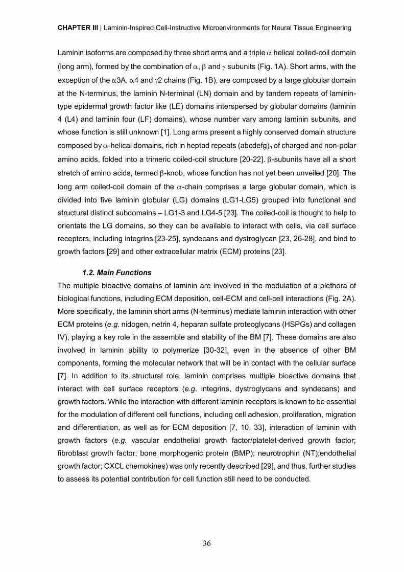
CHAPTER III | Laminin-Inspired Cell-Instructive Microenvironments for Neural Tissue Engineering
36
Laminin isoforms are composed by three short arms and a triple a helical coiled-coil domain
(long arm), formed by the combination of a, b and g subunits (Fig. 1A). Short arms, with the
exception of the a3A, a4 and g2 chains (Fig. 1B), are composed by a large globular domain
at the N-terminus, the laminin N-terminal (LN) domain and by tandem repeats of laminin-
type epidermal growth factor like (LE) domains interspersed by globular domains (laminin
4 (L4) and laminin four (LF) domains), whose number vary among laminin subunits, and
whose function is still unknown [1]. Long arms present a highly conserved domain structure
composed by a-helical domains, rich in heptad repeats (abcdefg)n of charged and non-polar
amino acids, folded into a trimeric coiled-coil structure [20-22]. b-subunits have all a short
stretch of amino acids, termed b-knob, whose function has not yet been unveiled [20]. The
long arm coiled-coil domain of the a-chain comprises a large globular domain, which is
divided into five laminin globular (LG) domains (LG1-LG5) grouped into functional and
structural distinct subdomains – LG1-3 and LG4-5 [23]. The coiled-coil is thought to help to
orientate the LG domains, so they can be available to interact with cells, via cell surface
receptors, including integrins [23-25], syndecans and dystroglycan [23, 26-28], and bind to
growth factors [29] and other extracellular matrix (ECM) proteins [23].
1.2. Main Functions The multiple bioactive domains of laminin are involved in the modulation of a plethora of
biological functions, including ECM deposition, cell-ECM and cell-cell interactions (Fig. 2A).
More specifically, the laminin short arms (N-terminus) mediate laminin interaction with other
ECM proteins (e.g. nidogen, netrin 4, heparan sulfate proteoglycans (HSPGs) and collagen
IV), playing a key role in the assemble and stability of the BM [7]. These domains are also
involved in laminin ability to polymerize [30-32], even in the absence of other BM
components, forming the molecular network that will be in contact with the cellular surface
[7]. In addition to its structural role, laminin comprises multiple bioactive domains that
interact with cell surface receptors (e.g. integrins, dystroglycans and syndecans) and
growth factors. While the interaction with different laminin receptors is known to be essential
for the modulation of different cell functions, including cell adhesion, proliferation, migration
and differentiation, as well as for ECM deposition [7, 10, 33], interaction of laminin with
growth factors (e.g. vascular endothelial growth factor/platelet-derived growth factor;
fibroblast growth factor; bone morphogenic protein (BMP); neurotrophin (NT);endothelial
growth factor; CXCL chemokines) was only recently described [29], and thus, further studies
to assess its potential contribution for cell function still need to be conducted.
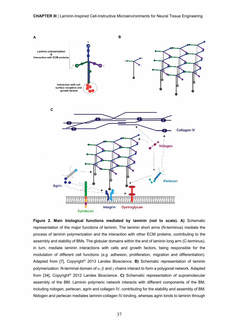
CHAPTER III | Laminin-Inspired Cell-Instructive Microenvironments for Neural Tissue Engineering
37
Figure 2. Main biological functions mediated by laminin (not to scale). A) Schematic
representation of the major functions of laminin. The laminin short arms (N-terminus) mediate the
process of laminin polymerization and the interaction with other ECM proteins, contributing to the
assembly and stability of BMs. The globular domains within the end of laminin long arm (C-terminus),
in turn, mediate laminin interactions with cells and growth factors, being responsible for the
modulation of different cell functions (e.g. adhesion, proliferation, migration and differentiation). Adapted from [7], Copyright© 2013 Landes Bioscience. B) Schematic representation of laminin
polymerization. N-terminal domain of a, b and g chains interact to form a polygonal network. Adapted
from [34], Copyright© 2012 Landes Bioscience. C) Schematic representation of supramolecular
assembly of the BM. Laminin polymeric network interacts with different components of the BM,
including nidogen, perlecan, agrin and collagen IV, contributing for the stability and assembly of BM. Nidogen and perlecan mediates laminin-collagen IV binding, whereas agrin binds to laminin through
CHAPTER III | Laminin-Inspired Cell-Instructive Microenvironments for Neural Tissue Engineering
38
its N-terminal domain. Both agrin and the laminin network are anchored to the cell surface by
interactions with integrins, syndecans and a-dystroglycan. Adapted from [34], Copyright© 2012
Landes Bioscience.
1.2.1. Laminin polymerization Laminin polymerization is a key process to direct BM assembly and organization, and
occurs by a thermally reversible mechanism dependent on the presence of calcium ions
[30-32]. This process is better explained by the three-arm interaction model (Fig. 2B) [31,
32], which proposes that the three laminin N-terminal short arms (with exception for laminins
comprising a3A-, a4- and g2-subunits (Fig. 1B)) interact with the globular N-terminal
domains of other laminins to form a polygonal network (Fig. 2B) [30-32, 35, 36]. The long
arm of the laminin heterotrimer, in turn, is predicted not to be involved in the network
formation, being free to interact with cells out of the plan of the polymer. Further studies
were conducted, to characterize in more detail the mechanism of polymerization in different
laminin isoforms [37-39]. It was demonstrated that laminin assembly is not restricted to the
self-polymerization of one specific laminin isoform with the formation of a homopolymer, but
can also occur by co-polymerization of laminin heterotrimers from different laminin isoforms
[32]. Additionally, different studies suggest that in vivo, the process of laminin assembly is
favored by laminin interaction with cell surface receptors (e.g. integrins, dystroglycan) [36,
40-42]. Nevertheless, there is still some controversy on the exact mechanism underlying
the cross-talk between laminin assembly and cell surface receptors. Some works evidenced
that laminin polymerization induces clustering of integrins and dystroglycan in the cell
membrane, and aggregation of intracellular cytoskeletal components [43, 44]. Other
studies, in turn, showed that dystroglycan null embryoid bodies are able to develop a BM
[45]. Therefore, studies to better understand the impact of different cell surface receptors
on laminin assembly need to be conducted.
Laminin polymerization can be reproduced in vitro under specific conditions [30, 46].[30].
More specifically, laminin was showed to be able to spontaneously polymerize in solution
either at neutral pH, requiring a minimal protein concentration of approximately 60 nM [30],
or can be triggered in acidic conditions independently of its concentration [46].
1.2.2. Laminin role in the assembly and stability of basement membranes BM assembly and stability depends on the proper interconnection between different ECM
components, including laminin and collagen IV, as well as other proteins, such as nidogen
(entactin) and HSPGs (e.g. agrin and perlecan) (Fig. 2C). Nidogen is a sulfated glycoprotein
ubiquitously found in the BM, known to interact strongly [47] with an LE module within the
laminin g1 subunit [48, 49]. In contrast, a weaker nidogen binding has been observed in
CHAPTER III | Laminin-Inspired Cell-Instructive Microenvironments for Neural Tissue Engineering
39
laminin g2 and g3 subunits [50]. This protein also interacts with other ECM proteins,
including perlecan, one of the major HSPGs, thus working as an intermediate for the
interaction between this HSPG and laminin. Agrin, a multi-domain HSPG, which has a key
role in synapse formation and maintenance at the neuromuscular junction [51], was showed
to mediate a high affinity interaction with a sequence of 20 conserved residues within
laminin g1 chain (dissociation constant (KD) @ 5 nM) through its N-terminal (NtA) domain
[52]. This interaction is required for the integration of agrin into the synaptic basal lamina
and other BMs [53]. Perlecan and agrin, through the direct or indirect interaction with
laminin, will mediate the interaction of the laminin polymeric network with the cell surface
assembled collagen IV, thus contributing to BM assembly and stabilization [34, 54, 55]. The
interactions mediating the incorporation of laminin polymeric structures into the
supramolecular network of the BM depend on the laminin isoform, namely on the structural
domains present on laminin short arms (Fig. 1B). While laminin-111, -211 and -511 form a
classical polymeric network, in which LN domain interactions with other ECM components
are well described, little is known about the mechanisms behind the integration of laminin
isoforms lacking LN domains in BM, such as those comprising a3A and a4 chains (Fig. 1B).
In this regard, previous studies have demonstrated that although truncated, the N-terminal
region of laminin 332 associates with other ECM components, including laminin 311 [56],
collagen VII [57, 58], collagen XVII [59] and fibulin [60]. Despite this, further studies are
necessary to better understand if these interactions are sufficient to maintain these laminin
isoforms anchored to the BM, or if additional interactions are required.
1.2.3. Cell adhesion-promoting activity Different cell surface receptors, including integrins, syndecans and dystroglycan [23] (Fig.
2B-C), interact with the LG domains in the C-terminal end of the laminin a chain. While LG1-
3 domains mediate interaction with integrin receptors [23-25], the LG4-5 pair contains
binding sites for a-dystroglycan and syndecans [23, 26-28].
Laminin-binding integrins a3b1, a6b1,a7b1 and a6b4 present different specificities
depending on the laminin isoform [61], and most specifically on the laminin a chain [62].
For example, while integrin a6b1 has a broad affinity for all laminin isoforms, a3b1 and
a6b4 bind almost exclusively to laminins comprising the a3 and a5 chains, while a7b1
prefers isoforms with the laminin a2 and a5 chains. Laminin isoforms composed by a5 are
the preferred ligands for laminin-binding integrins, whereas the ones composed by a4
chains constitute the poorest ligands [62]. Integrins binding to LG domains require that both
the LG1-LG3 domains and the coiled-coil to be intact. Indeed, some studies showed that
modifications of the LG1-LG3 domain or on the coiled-coil structure, abolishes cell

CHAPTER III | Laminin-Inspired Cell-Instructive Microenvironments for Neural Tissue Engineering
40
adhesion-promoting activity. Although integrins bind to the LG1-3 domain comprised in the
a-chain [3, 6, 63], it was demonstrated that b- and g-chains also play a role in the mediation
of laminin-integrin binding [64, 65]. In addition to the LG1-3 domains, integrin-binding to
laminins can also occur at other recognition motifs, namely at the cryptic RGD domain in
laminin a1 chain, which only becomes accessible after proteolytic cleavage [66]. a-
dystroglycan displays a narrower binding spectrum, compared to integrins, presenting a
high affinity only for laminin a1 and a2 chains [3]. Although the LG4-5 domains are the
preferred a-dystroglycan binding domains, it was demonstrated that the a2 chain contains
additional binding sites in the LG1-3 domains [3, 23, 63]. Syndecans are type I
transmembrane HSPG cell receptors that interact with laminin LG4-5 domains that are
responsible for the mediation of important cellular events [67]. In addition to a-dystroglycan
and syndecans, other non-integrin laminin receptors, which do not interact with LG domains,
were also described. These include the 110 kDa [68] and the 67 kDa cell surface receptors
[69], which bind with high affinity to the IKVAV sequence within the laminin a1 chain and
the YIGSR sequence within the laminin b1 chain, respectively. Overall, all transmembrane
receptors described will have a key role in the formation and deposition of laminin matrices
and will be involved in the modulation of cell-ECM and cell-cell interactions, as well as on
the regulation of different cell functions and fate.
Ultimately, the laminin-associated matrix proteins in concert with cell-surface receptors will
be key for the proper assembly and/or deposition of laminin matrices, and will work to fine-
tune both matrix formation and its function.
2. Laminin in the central nervous system (CNS)
Laminin is differentially expressed within central nervous system (CNS), during
development and homeostasis, with an impact on the modulation of cell function and fate.
Despite its involvement in many aspects of CNS physiology and neuronal functions, there
is still a lack of understanding on the exact role of laminin in the formation, development
and function of neuronal networks. In this regard, several studies have been conducted to
better characterize the expression and distribution of specific laminin chains or isoforms in
different CNS regions, both during embryogenesis and adulthood (Table 1).

CHAPTER III | Laminin-Inspired Cell-Instructive Microenvironments for Neural Tissue Engineering
41
Table 1. Expression and distribution of laminin subunits in the central nervous system (CNS) during
embryogenesis and adulthood. Adapted from [70], Copyright© 2018, with permission from John Wiley
and Sons
Laminin subunit
Expression/Distribution sites References
Em
bryo
gene
sis
a1 Cerebellum, neuroretina, olfactory bulbs, meninges,
cortical plate, floor plate of spinal cord and spinal cord
neurons and glial cells
[71-76]
a2 Meninges, cortical plate, choroid plexus, dorsal root
ganglion, ventricular region and spinal cord neurons and glial cells
[71-74, 76-78]
a3 Neuroectoderm, trigeminal nerve, cerebellum and spinal
cord/brain neurons and glial cells [76, 79]
a4 Dorsal root ganglion, cortical plate, endothelial cells,
spinal cord/brain neurons and glial cells [11, 72, 74, 76]
a5 Neural folds, cortical plate, spinal cord/brain neurons and
glial cells [74, 76, 80]
b1 Floor plate of spinal cord, spinal cord/brain neurons and
glial cells [75, 76]
b2 Cortical plate and spinal cord/brain neurons [74, 76]
b3 Floor plate of spinal cord (?) [75, 76]
b4 -
g1 Neural folds, neuroectoderm, basement membrane (BM) and pia, floor plate of spinal cord, spinal cord/brain
neurons and glial cells
[75, 76, 80-86]
g2 Epithelial cells and spinal cord/brain glial cells [76, 87]
g3 Cerebellar pia and retinal vascular BM [81, 88]
Ad
ulth
ood
a1 Blood vessels [89]
a2 Astrocytes, pericytes, neurons, oligodendrocytes,
microglia, ependymal cell layer, blood vessels and
fractones in neurogenic niches
[70, 84, 90-96]
a3 Microglia [70]
a4 Brain microvascular endothelial cells (BMECs), pericytes,
oligodendrocytes, microglia ependymal cell layer, blood vessels and fractones in neurogenic niches
[70, 82, 85, 86,
94, 95, 97]
a5 BMECs, pericytes, hippocampal neurons,
oligodendrocytes, microglia, ependymal cell layer, blood
vessels and fractones in neurogenic niches
[70, 82, 85, 86,
94, 95, 97-
101]
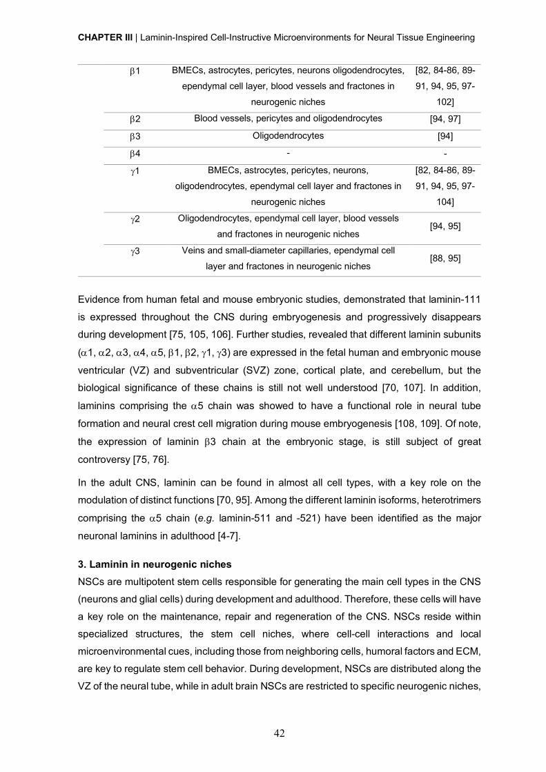
CHAPTER III | Laminin-Inspired Cell-Instructive Microenvironments for Neural Tissue Engineering
42
b1 BMECs, astrocytes, pericytes, neurons oligodendrocytes,
ependymal cell layer, blood vessels and fractones in
neurogenic niches
[82, 84-86, 89-
91, 94, 95, 97-
102]
b2 Blood vessels, pericytes and oligodendrocytes [94, 97]
b3 Oligodendrocytes [94]
b4 - -
g1 BMECs, astrocytes, pericytes, neurons,
oligodendrocytes, ependymal cell layer and fractones in
neurogenic niches
[82, 84-86, 89-
91, 94, 95, 97-
104]
g2 Oligodendrocytes, ependymal cell layer, blood vessels
and fractones in neurogenic niches [94, 95]
g3 Veins and small-diameter capillaries, ependymal cell
layer and fractones in neurogenic niches [88, 95]
Evidence from human fetal and mouse embryonic studies, demonstrated that laminin-111
is expressed throughout the CNS during embryogenesis and progressively disappears
during development [75, 105, 106]. Further studies, revealed that different laminin subunits
(a1, a2, a3, a4, a5, b1, b2, g1, g3) are expressed in the fetal human and embryonic mouse
ventricular (VZ) and subventricular (SVZ) zone, cortical plate, and cerebellum, but the
biological significance of these chains is still not well understood [70, 107]. In addition,
laminins comprising the a5 chain was showed to have a functional role in neural tube
formation and neural crest cell migration during mouse embryogenesis [108, 109]. Of note,
the expression of laminin b3 chain at the embryonic stage, is still subject of great
controversy [75, 76].
In the adult CNS, laminin can be found in almost all cell types, with a key role on the
modulation of distinct functions [70, 95]. Among the different laminin isoforms, heterotrimers
comprising the a5 chain (e.g. laminin-511 and -521) have been identified as the major
neuronal laminins in adulthood [4-7].
3. Laminin in neurogenic niches
NSCs are multipotent stem cells responsible for generating the main cell types in the CNS
(neurons and glial cells) during development and adulthood. Therefore, these cells will have
a key role on the maintenance, repair and regeneration of the CNS. NSCs reside within
specialized structures, the stem cell niches, where cell-cell interactions and local
microenvironmental cues, including those from neighboring cells, humoral factors and ECM,
are key to regulate stem cell behavior. During development, NSCs are distributed along the
VZ of the neural tube, while in adult brain NSCs are restricted to specific neurogenic niches,
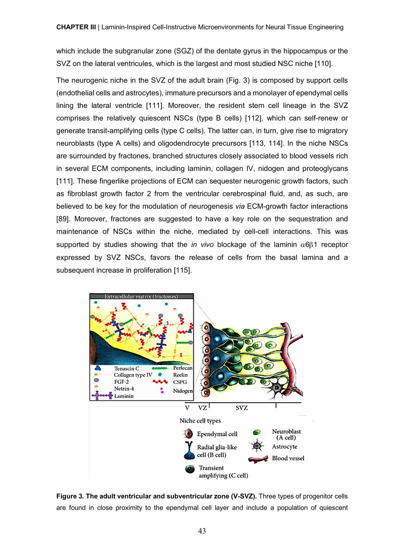
CHAPTER III | Laminin-Inspired Cell-Instructive Microenvironments for Neural Tissue Engineering
43
which include the subgranular zone (SGZ) of the dentate gyrus in the hippocampus or the
SVZ on the lateral ventricules, which is the largest and most studied NSC niche [110].
The neurogenic niche in the SVZ of the adult brain (Fig. 3) is composed by support cells
(endothelial cells and astrocytes), immature precursors and a monolayer of ependymal cells
lining the lateral ventricle [111]. Moreover, the resident stem cell lineage in the SVZ
comprises the relatively quiescent NSCs (type B cells) [112], which can self-renew or
generate transit-amplifying cells (type C cells). The latter can, in turn, give rise to migratory
neuroblasts (type A cells) and oligodendrocyte precursors [113, 114]. In the niche NSCs
are surrounded by fractones, branched structures closely associated to blood vessels rich
in several ECM components, including laminin, collagen IV, nidogen and proteoglycans
[111]. These fingerlike projections of ECM can sequester neurogenic growth factors, such
as fibroblast growth factor 2 from the ventricular cerebrospinal fluid, and, as such, are
believed to be key for the modulation of neurogenesis via ECM-growth factor interactions
[89]. Moreover, fractones are suggested to have a key role on the sequestration and
maintenance of NSCs within the niche, mediated by cell-cell interactions. This was
supported by studies showing that the in vivo blockage of the laminin a6b1 receptor
expressed by SVZ NSCs, favors the release of cells from the basal lamina and a
subsequent increase in proliferation [115].
Figure 3. The adult ventricular and subventricular zone (V-SVZ). Three types of progenitor cells
are found in close proximity to the ependymal cell layer and include a population of quiescent
CHAPTER III | Laminin-Inspired Cell-Instructive Microenvironments for Neural Tissue Engineering
44
NSCs/radial glia-like cells (type B cells) that can generate transit-amplifying cells (type C cells),
which, in turn, will give rise to neuroblasts (type A cells). Endothelial cells and astrocytes, which
function as support cells, as well as immature precursors can also be found within NSC niche. In
addition to the cellular components, the SVZ includes several ECM components (yellow) called
fractones, which include laminin, collagen IV, nidogen and proteoglycans. These contact with the
different cell types composing the niche and are involved on the modulation of different cell functions.
Adapted from [110], Copyright© 2016, Creative Commons License.
Laminin is one of the most important and well described ECM proteins within the neurogenic
niches [89, 95]. This is evidenced by its key role in the modulation of neuronal progenitors
proliferation and survival [116, 117], neurodevelopment [70] (Table 1), hippocampal
regeneration [103] and on the regulation of growth factors concentration [89]. The
expression of multiple a, b and g laminin chains have been described in association with
the ependymal cells [95], the blood vessels [95], supporting cells (e.g. astrocytes) [118],
and proliferating transit-amplifying cells (type C cells), under homeostatic conditions, as well
as with cells of the NSC lineage in the regenerative niche [95]. More specifically, laminin a1
chain can be found on blood vessels but not on fractones of adult rat, mouse or human
NSC niche [89]. Laminin-511, in turn, is the major neuronal laminin in adult mice
hippocampus BM and was showed to be important for hippocampal regeneration [100]. The
differential expression of laminin isoforms, influence their ability to mediate different
functions within the NSC niches. For example, laminin isoforms comprising the a2 chain
where shown to be required for oligodendrocyte maturation and CNS myelination in adult
mice [96] under homeostasis conditions, while laminin-411 or -511 and laminin-111 or -211
produced by endothelial cells and astrocytes, respectively, participate in the formation of
the blood-brain barrier [85]. Mutations in laminin b2 (mice) [119], g1 (mice) [120] and g3
(mice and human) [119, 121] chains, in turn, were showed to disturb processes like cortical
histogenesis, as well as the formation of the pial BM. Nevertheless, the mechanism by
which differentially expressed laminin isoforms modulate adult neurogenesis in not yet fully
understood. In this regard, some studies suggest that rather than differences in ECM
expression, changes in the expression of the major laminin receptors - integrins,
dystroglycan and syndecans, seem to be responsible for the changes in ECM signaling that
contribute to stem cell activation. Under normal conditions, although in adult mice NSCs
and precursor cells reside in similar niches being exposed to similar ECM signals, NSCs,
as result of the low expression of laminin receptors, have limited interaction with the laminin-
rich microenvironment, remaining relatively quiescent [95]. After stimulation and activation
of NSCs (e.g. in the regenerative niche that is triggered in the aftermath of a lesion), an
increase on laminin synthesis, as well as an upregulation on the expression of different
CHAPTER III | Laminin-Inspired Cell-Instructive Microenvironments for Neural Tissue Engineering
45
laminin receptors, including a6b1 integrin and syndecan-1 is observed [95], potentiating its
interaction with the cellular and extracellular microenvironment.
The interplay between the niche components and NSCs will determine the balance between
stemness and differentiation, quiescence and proliferation. In this regard, a number of
experimental approaches perturbing the niches either through the genetic or cytotoxic
ablation of the precursor cells or using specific culture models to evaluate the effect of
specific components of the niches, have been proposed to better understand how changes
in the ECM receptor expression regulates NSC behavior (for a more comprehensive review
see [122]). However, the effect of the matrix itself remains to be elucidated. Envisaging the
development of three-dimensional (3D) matrices able to recapitulate the complex
arrangement of cells and ECM in the nervous system, and dissect the different roles exerted
by the ECM and its molecular components on NSC behavior, in the last few years, much
attention has been devoted towards the development of engineered matrices able to mimic
the in vivo characteristics of NSC niches, and ultimately develop more efficient
neuroregenerative approaches.
4. Laminin-inspired hydrogels to recreate the microenvironment of neurogenic
niches
As discussed in the previous section, laminin was found to have a key role in the modulation
of NSC function and fate within neurogenic niches. More specifically, this ECM protein was
showed to be crucial for the modulation of cell adhesion and viability [123] and neuronal
outgrowth and migration [124-126]. Works published to date exploring the immobilization of
full-length laminin (Table 2) or their peptide analogues (Table 3), into hydrogel matrices, to
recreate the NSC niche microenvironment, are summarized in this section. The developed
3D platforms were applied for the in vitro study of NSC biology and to assess their
interactions with the surrounding microenvironment, as well as to evaluate the potential of
the developed matrices to serve as vehicles for NSC transplantation in the context of
neurological disorders. In addition to mimicking the ECM composition, replicating the
mechanical and structural properties of the native niche is key for the modulation of different
stem cell functions, including survival, proliferation and differentiation [127-129]. Indeed,
studies using substrates with mechanical properties similar to that of native CNS (low
compressive moduli – 0.1 – 1.0 kPa) have shown ability to support NSC function [130-135].
4.1. Hydrogels functionalized with full-length laminin The incorporation of a full-length protein onto hydrogels, and more specifically of laminin,
has been widely explored for the development of biomimetic 3D matrices, to provide the
gels with the bioactivity levels of the native protein. Indeed, several studies showed that the
CHAPTER III | Laminin-Inspired Cell-Instructive Microenvironments for Neural Tissue Engineering
46
modification of specific matrices with full-length laminin, in addition to render the
microenvironment more permissive for NSC growth and differentiation, also contributed for
an enhanced in vivo nervous regeneration [136-140]. Since the biological features of a
protein result from its multiple bioactive domains and its conformation [141-143], the
immobilization strategy explored will have a critical impact on the biofunctionality of the
engineered hydrogels. Indeed, this is evident in some of the studies reported in Table 2,
which point out that the immobilization approach significantly affects laminin function [144-
147]. Strategies explored to date, for full-length laminin immobilization have relied either on
its transient non-covalent incorporation or physical entrapment, or alternatively, on its non-
selective covalent immobilization by taking advantage of functional groups present in
multiple sites of the laminin structure, such as amines and thiols (Fig. 4). Despite widely
used, these strategies lack the ability to control the conformation and orientation of bioactive
molecules upon immobilization. As such, the exposure of key laminin bioactive domains,
including those involved in laminin polymerization and on the modulation of NSC function
and fate, can be compromised.
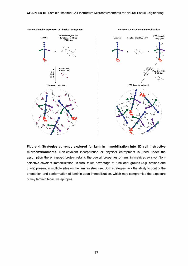
CHAPTER III | Laminin-Inspired Cell-Instructive Microenvironments for Neural Tissue Engineering
47
Figure 4. Strategies currently explored for laminin immobilization into 3D cell instructive
microenvironments. Non-covalent incorporation or physical entrapment is used under the
assumption the entrapped protein retains the overall properties of laminin matrices in vivo. Non-
selective covalent immobilization, in turn, takes advantage of functional groups (e.g. amines and thiols) present in multiple sites on the laminin structure. Both strategies lack the ability to control the
orientation and conformation of laminin upon immobilization, which may compromise the exposure
of key laminin bioactive epitopes.
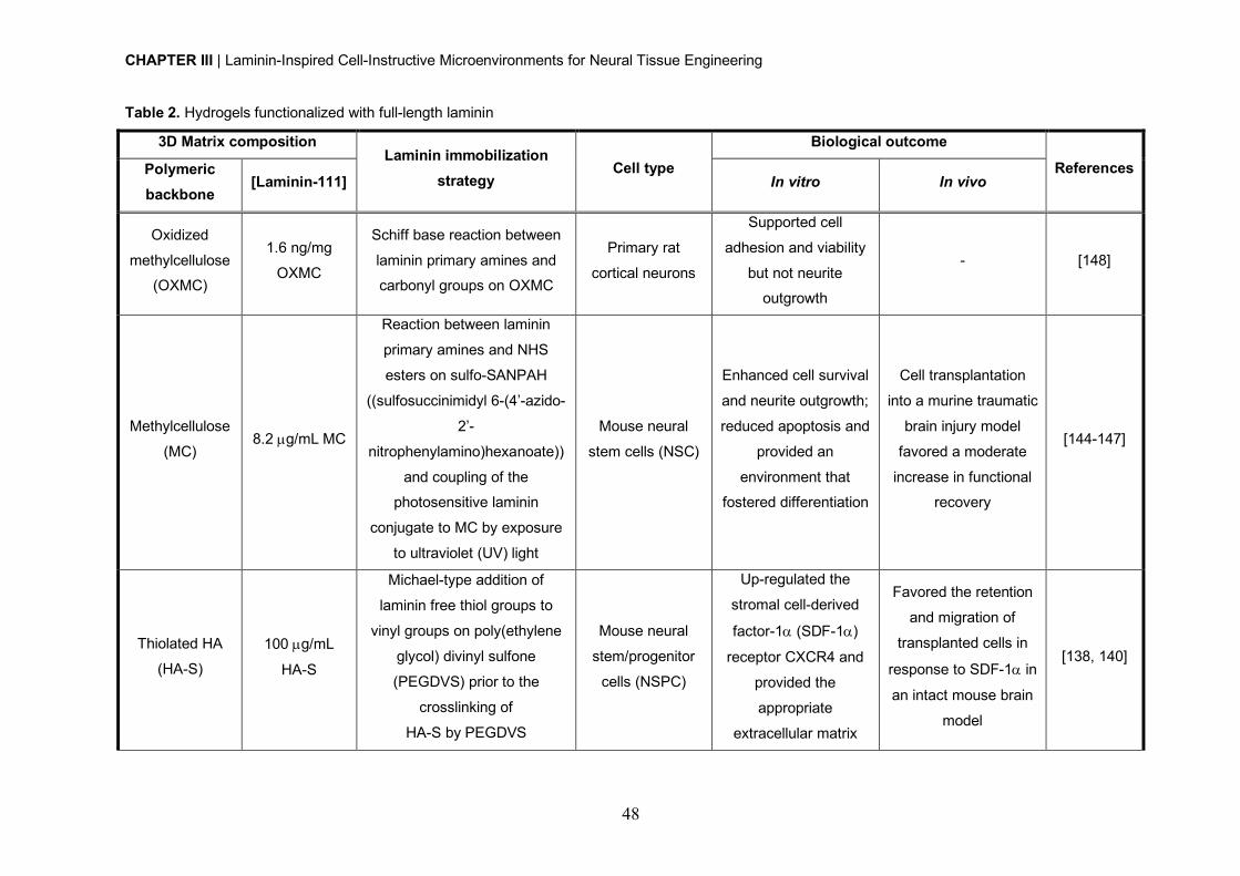
CHAPTER III | Laminin-Inspired Cell-Instructive Microenvironments for Neural Tissue Engineering
48
Table 2. Hydrogels functionalized with full-length laminin
3D Matrix composition Laminin immobilization
strategy Cell type
Biological outcome
References Polymeric backbone
[Laminin-111] In vitro In vivo
Oxidized
methylcellulose (OXMC)
1.6 ng/mg
OXMC
Schiff base reaction between
laminin primary amines and carbonyl groups on OXMC
Primary rat
cortical neurons
Supported cell
adhesion and viability
but not neurite outgrowth
- [148]
Methylcellulose
(MC) 8.2 µg/mL MC
Reaction between laminin
primary amines and NHS
esters on sulfo-SANPAH
((sulfosuccinimidyl 6-(4’-azido-
2’-
nitrophenylamino)hexanoate))
and coupling of the photosensitive laminin
conjugate to MC by exposure
to ultraviolet (UV) light
Mouse neural
stem cells (NSC)
Enhanced cell survival
and neurite outgrowth;
reduced apoptosis and
provided an
environment that fostered differentiation
Cell transplantation
into a murine traumatic
brain injury model
favored a moderate
increase in functional recovery
[144-147]
Thiolated HA
(HA-S) 100 µg/mL
HA-S
Michael-type addition of
laminin free thiol groups to
vinyl groups on poly(ethylene
glycol) divinyl sulfone
(PEGDVS) prior to the
crosslinking of
HA-S by PEGDVS
Mouse neural
stem/progenitor
cells (NSPC)
Up-regulated the
stromal cell-derived
factor-1a (SDF-1a)
receptor CXCR4 and
provided the
appropriate
extracellular matrix
Favored the retention
and migration of
transplanted cells in
response to SDF-1a in
an intact mouse brain
model
[138, 140]

CHAPTER III | Laminin-Inspired Cell-Instructive Microenvironments for Neural Tissue Engineering
49
(ECM) cues to
promote cell
chemotactic migration
in response to SDF-1a
HA-S + salmon
fibrinogen+
salmon
thrombin
100 µg/mL
HA-S
Incorporation in the hydrogel
precursor solution prior to the
cleavage of fibrinogen by
thrombin to induce the
formation of an
interpenetrating polymer
network (IPN) hydrogel
Human NSPC
Supported cell
proliferation and differentiation.
Significantly reduced
the cell-mediated
degradation seen with
fibrin alone. Supported
vascularization as
evidenced on hNSPCs
- human endothelial colony-forming cell-
derived endothelial
cells (hECFC-ECs) co-
culture studies
- [139]
Agarose
0.120 – 0.162
µg/mL agarose
gradient
Reaction between laminin-111
primary amines groups and
NHS esters on sulfo-SANPAH
and coupling of the laminin-111 photosensitive conjugate
to agarose by exposure to UV
light
Dorsal root
ganglia (DRG)
Neurite extension was
significantly higher in
the anisotropic scaffolds compared to
isotropic scaffolds
- [136]
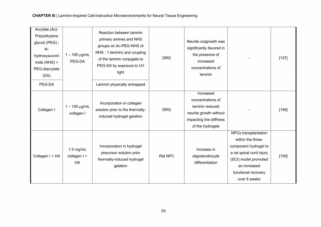
CHAPTER III | Laminin-Inspired Cell-Instructive Microenvironments for Neural Tissue Engineering
50
Acrylate (Ac)-
Poly(ethylene
glycol) (PEG) -
N-
hydroxysuccini
mide (NHS) +
PEG-diacrylate
(DA)
1 – 100 µg/mL
PEG-DA
Reaction between laminin
primary amines and NHS
groups on Ac-PEG-NHS (4
NHS : 1 laminin) and coupling
of the laminin conjugate to
PEG-DA by exposure to UV
light
DRG
Neurite outgrowth was
significantly favored in
the presence of
increased
concentrations of
laminin
- [137]
PEG-DA Laminin physically entrapped
Collagen I 1 – 100 µg/mL
collagen I
Incorporation in collagen solution prior to the thermally-
induced hydrogel gelation
DRG
Increased
concentrations of laminin reduced
neurite growth without
impacting the stiffness
of the hydrogels
- [149]
Collagen I + HA
1.5 mg/mL
collagen I + HA
Incorporation in hydrogel
precursor solution prior
thermally-induced hydrogel gelation
Rat NPC
Increase in
oligodendrocyte differentiation
NPCs transplantation
within the three-
component hydrogel to
a rat spinal cord injury
(SCI) model promoted an increased
functional recovery
over 6 weeks
[150]
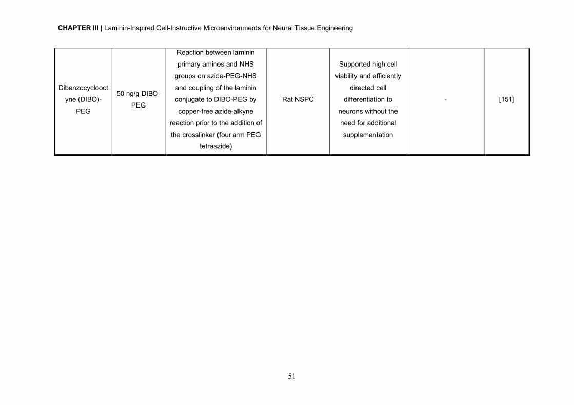
CHAPTER III | Laminin-Inspired Cell-Instructive Microenvironments for Neural Tissue Engineering
51
Dibenzocyclooct
yne (DIBO)-
PEG
50 ng/g DIBO-
PEG
Reaction between laminin
primary amines and NHS
groups on azide-PEG-NHS
and coupling of the laminin
conjugate to DIBO-PEG by
copper-free azide-alkyne
reaction prior to the addition of
the crosslinker (four arm PEG tetraazide)
Rat NSPC
Supported high cell
viability and efficiently
directed cell
differentiation to
neurons without the
need for additional
supplementation
- [151]
CHAPTER III | Laminin-Inspired Cell-Instructive Microenvironments for Neural Tissue Engineering
52
Studies published to date, exploring full-length laminin immobilization, for the development
of 3D matrices mimicking the NSC niche microenvironment, used laminin-111, as this
isoform was already showed to successfully promote neuronal outgrowth and
differentiation. Nevertheless, and considering the key role of other laminin isoforms (e.g.
laminin-521) on the in vitro modulation of neural cell behavior including neuronal adhesion,
viability and network formation [123, 152], future studies evaluating their impact on NSC
behavior and function, after incorporation within 3D matrices, should be conducted.
The endogenous process of cell replacement in the adult human CNS is far from being
sufficient to repair the damaged caused after an injury. Therefore, and although in this
section we highlight the studies exploring hydrogels functionalized with full-length laminin
for NSC culture and transplantation, the potential of these hydrogels to allow infiltration of
endogenous neural progenitors recruited to the injury site, and ultimately contribute to tissue
repair was also evaluated [153]. More specifically, a laminin-functionalized hyaluronic acid
(HA)-based hydrogel was showed to efficiently support endogenous cell infiltration and
angiogenesis, while inhibited the formation of the glial scar and promoted neurite extension
in a rat model of brain lesion.
In addition to their application in the framework of nervous regeneration and tissue
engineering, the incorporation of full-length laminin on 3D matrices has also been explored
for application in different disease contexts, including skeletal muscle [154, 155],
intervertebral disc [156, 157] and vascular regeneration [158, 159].
4.2. Hydrogels functionalized with laminin-derived peptides Laminin-derived small adhesive sequences have been increasingly explored, in alternative
to full-length laminin, to confer bioactivity to 3D matrices. These small sequences have
several advantageous features, including their ability to be chemically synthesized in large
scale, the higher resistance to denaturation and enzymatic degradation, ability to be
incorporated at higher densities for similar amounts of native protein and lower risk of induce
immune rejection [160]. Different small peptide sequences derived from laminin, including
YIGSR, IKVAV, RGD (cryptic domain in laminin a1 chain), RNIAEIIKDI, RKRLQVQSIRT,
are known to mediate neuronal adhesion, neurite outgrowth and/or NSC migration, and, as
such, have been widely explored for the design of biomimetic 3D matrices for cell culture
and transplantation (Table 3).
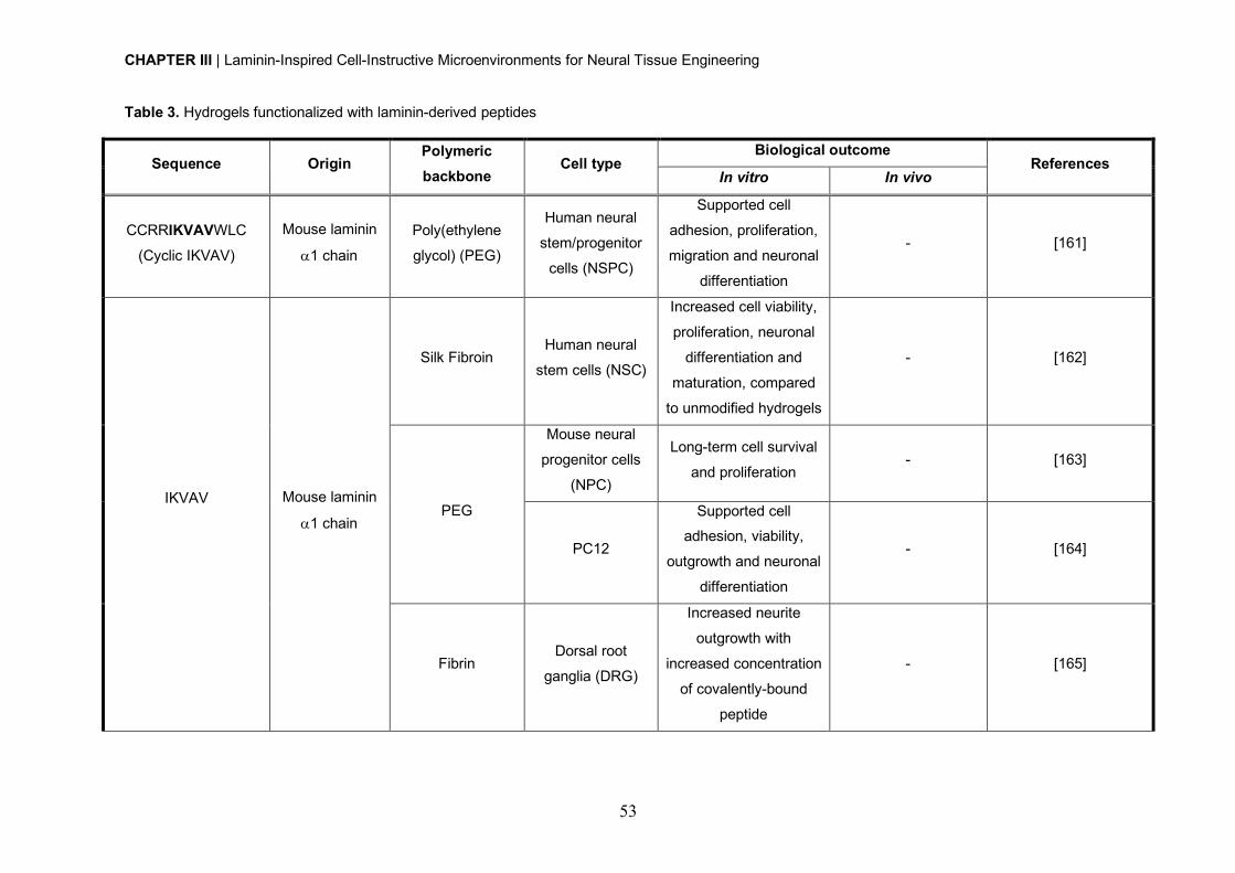
CHAPTER III | Laminin-Inspired Cell-Instructive Microenvironments for Neural Tissue Engineering
53
Table 3. Hydrogels functionalized with laminin-derived peptides
Sequence Origin Polymeric backbone
Cell type Biological outcome
References In vitro In vivo
CCRRIKVAVWLC
(Cyclic IKVAV)
Mouse laminin
a1 chain
Poly(ethylene
glycol) (PEG)
Human neural
stem/progenitor
cells (NSPC)
Supported cell
adhesion, proliferation,
migration and neuronal
differentiation
- [161]
IKVAV
Mouse laminin
a1 chain
Silk Fibroin Human neural
stem cells (NSC)
Increased cell viability,
proliferation, neuronal
differentiation and
maturation, compared
to unmodified hydrogels
- [162]
PEG
Mouse neural
progenitor cells
(NPC)
Long-term cell survival
and proliferation - [163]
PC12
Supported cell adhesion, viability,
outgrowth and neuronal
differentiation
- [164]
Fibrin Dorsal root
ganglia (DRG)
Increased neurite
outgrowth with
increased concentration
of covalently-bound
peptide
- [165]
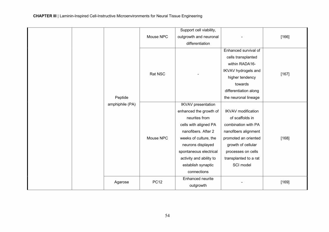
CHAPTER III | Laminin-Inspired Cell-Instructive Microenvironments for Neural Tissue Engineering
54
Peptide
amphiphile (PA)
Mouse NPC
Support cell viability,
outgrowth and neuronal
differentiation
- [166]
Rat NSC -
Enhanced survival of
cells transplanted
within RADA16-
IKVAV hydrogels and
higher tendency
towards differentiation along
the neuronal lineage
[167]
Mouse NPC
IKVAV presentation
enhanced the growth of
neurites from
cells with aligned PA
nanofibers. After 2
weeks of culture, the neurons displayed
spontaneous electrical
activity and ability to
establish synaptic
connections
IKVAV modification
of scaffolds in
combination with PA
nanofibers alignment
promoted an oriented growth of cellular
processes on cells
transplanted to a rat
SCI model
[168]
Agarose PC12 Enhanced neurite
outgrowth - [169]
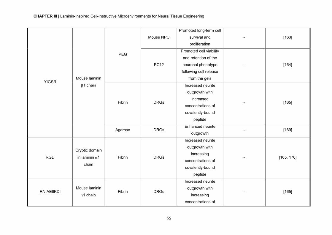
CHAPTER III | Laminin-Inspired Cell-Instructive Microenvironments for Neural Tissue Engineering
55
YIGSR Mouse laminin
b1 chain
PEG
Mouse NPC
Promoted long-term cell
survival and
proliferation
- [163]
PC12
Promoted cell viability
and retention of the
neuronal phenotype
following cell release
from the gels
- [164]
Fibrin DRGs
Increased neurite outgrowth with
increased
concentrations of
covalently-bound
peptide
- [165]
Agarose DRGs Enhanced neurite
outgrowth - [169]
RGD
Cryptic domain
in laminin a1
chain
Fibrin DRGs
Increased neurite
outgrowth with increasing
concentrations of
covalently-bound
peptide
- [165, 170]
RNIAEIIKDI Mouse laminin
g1 chain Fibrin DRGs
Increased neurite
outgrowth with
increasing
concentrations of
- [165]
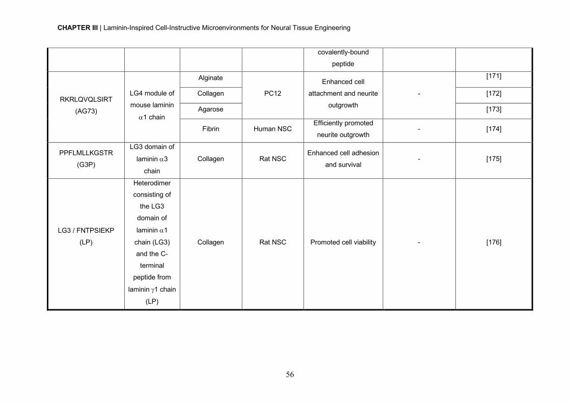
CHAPTER III | Laminin-Inspired Cell-Instructive Microenvironments for Neural Tissue Engineering
56
covalently-bound
peptide
RKRLQVQLSIRT
(AG73)
LG4 module of
mouse laminin
a1 chain
Alginate
PC12
Enhanced cell
attachment and neurite
outgrowth
-
[171]
Collagen [172]
Agarose [173]
Fibrin Human NSC Efficiently promoted
neurite outgrowth - [174]
PPFLMLLKGSTR
(G3P)
LG3 domain of
laminin a3
chain
Collagen Rat NSC Enhanced cell adhesion
and survival - [175]
LG3 / FNTPSIEKP
(LP)
Heterodimer consisting of
the LG3
domain of
laminin a1
chain (LG3) and the C-
terminal
peptide from
laminin g1 chain
(LP)
Collagen Rat NSC Promoted cell viability - [176]
CHAPTER III | Laminin-Inspired Cell-Instructive Microenvironments for Neural Tissue Engineering
57
Peptides IKVAV, found in laminin a1 chain [177] and YIGSR, found in laminin b1 chain [69,
178, 179] bind to the 110 kDa [68] and 67 kDa [69] cell surface receptor, respectively, and
are both recognized by the b1 integrin receptor subunit, which is highly expressed by neural
stem/progenitor cells (NSPCs) [180, 181]. RNIAEIIKDI is a sequence present in mouse
laminin g1 chain [182, 183] found to promote neurite outgrowth of DRGs [165]. The
RKRLQVQSIRT sequence, also known as AG73 peptide, is derived from the LG4 module
of mouse laminin a1 chain [184] and was showed to interact with syndecan-1 [185, 186]
and -4 [187]. This peptide was already successfully used to modify different natural hydrogel
matrices, including alginate, agarose, collagen and fibrin, with promising results in terms of
the promotion of neurite outgrowth in PC12 cells [171-174, 188] and hNSCs [174]. The RGD
domain sequence found in many ECM proteins, including fibronectin and laminin (as a
cryptic domain in laminin a1 chain), was shown to interact with a5b1 and anb3 integrins
[189]. Its incorporation into both natural and synthetic-derived matrices is effective in
promoting NSC adhesion and neurite outgrowth [164, 168, 190-192].
Laminin-derived peptide fragments produced by recombinant DNA technology have also
been explored for the development of 3D matrices. More specifically, a peptide derived from
the LG3 domain of laminin a3 chain - G3P peptide (PPFLMLLKGSTR), which binds with
strong affinity to a3b1 integrin [193], was recombinantly modified to include a collagen
binding domain. The resultant recombinant peptide was further explored for the
functionalization of a collagen-based hydrogel, which evidenced ability to efficiently support
NSC adhesion and survival [175]. A different study, explored the incorporation of a
recombinant heterodimer combining peptides derived from the LG3 domain of mouse
laminin a1 chain and the C-terminal peptide from laminin g1 chain (LP) [176] on collagen
hydrogels. Alternatively, a recombinant laminin fragment - E8 fragment, which is a truncated
protein composed of the C-terminal region of the a, b and g chains, was explored [194].
Unlike the recombinant peptide fragments previously described, this truncated protein
retains laminin integrin-binding activity, as evidenced by the ability of laminin-511-derived
E8 fragment to strongly promote the adhesion and proliferation of human pluripotent stem
cells, when compared to Matrigel and the full-length laminin isoform [195]. Accordingly, and
envisaging the development of collagen matrices with laminin-like adhesive activity, the N-
terminal of individual chains (a, b and/or g) in the truncated protein was modified with a
collagen-binding domain (CBD), to confer the E8 fragment collagen-binding activity. The
developed matrices were shown to support the proliferation of human induced pluripotent
stem cells (hiPSCs), both in 2D and 3D, though the morphology of cells grown in each
condition was quite different [194].
CHAPTER III | Laminin-Inspired Cell-Instructive Microenvironments for Neural Tissue Engineering
58
To better recapitulate the biological activity of laminin chains, in the last few years several
studies have been exploring the additive or synergistic effect of the combined incorporation
of different laminin cell adhesive peptides (e.g. IKVAV, YIGSR, RGD, AG73, RNIAEIIKDI).
For example, a study exploring the modification of fibrin hydrogels with a combination, in
equimolar ratios, of RGD, IKVAV, YIGSR and RNIAEIIKDI, showed that these peptides
have a synergistic effect on neurite outgrowth when used to bridge a 4 mm gap in a rat
dorsal root [165]. In a different study, the modification of dextran hydrogels with a mixture
of equimolar concentration (1:1) of IKVAV and YIGSR, was showed to better support in vitro
DRG adhesion and neurite outgrowth, when compared to RGD-modified hydrogels [196].
Despite the promising results obtained to date, the use of the “typical” equimolar
concentration of each peptide has not always proven to be efficient, in terms of supporting
cell survival and differentiation [197]. Therefore, an important factor one should consider
when developing hydrogels incorporating different ECM-derived peptides, is the need to
understand the effect of each individual factor, as well as the interacting factor effects for
each system. With this in mind, and to make the process less time-consuming, Segura and
co-workers proposed the use of multifactorial experiments. Here, candidate adhesion motifs
and growth factors were systematically varied in vitro to determine the individual and
combinatorial effects of each factor on cell activity, enabling the optimization of a system
capable of supporting hNPCs (iPS-NPCs) survival, proliferation and differentiation [197,
198]. The optimized hydrogel was further tested in vivo, in an animal model of stroke.
Results evidenced the ability of the proposed matrix to support the survival of encapsulated
hNPCs (iPS-NPCs) after transplantation into the stroke core, as well as to differentially tune
transplanted cell fate through the promotion of glial, neuronal or immature/progenitor states
[198].
5. Engineering laminin-inspired hydrogels: progress and future challenges As a consequence of the key role of laminin within neurogenic niches, the potential of
several laminin-inspired hydrogels, exploring either the full-length laminin or its peptide
analogues, in a neural regeneration context, have been explored in different works (Table
2 and 3). Proof-of-principle of the suitability of these hydrogels in terms of ability to create
a permissive microenvironment for cell growth and differentiation in vitro, as well as the in
vivo biological performance of several of these matrices when explored for cell delivery,
was highlighted in this chapter. The results obtained to date, strongly suggest that these
hydrogels may serve as powerful artificial niches to modulate NSC fate and function for
regenerative medicine applications.
To pave the way for the translation of laminin-inspired hydrogels to a clinical setting, a
number of requisites need to be fulfilled and a few key issues must be taken into
CHAPTER III | Laminin-Inspired Cell-Instructive Microenvironments for Neural Tissue Engineering
59
consideration. Different hydrogel factors known to impact stem cell fate, including
biocompatibility, controlled biodegradability and non-immunogenicity, should be considered
when designing cell-instructive microenvironments. Physical properties (e.g. mechanical
and structural properties) should also be taken into consideration, as these are known to
have an impact on cell behavior and function [127-129]. Therefore, the characterization of
the developed hydrogels in terms of physical properties, should be carried out in a number
of in vitro and in vivo set-ups, before considering clinical translation. The selection of the
most appropriate system for a specific application, is one of the most important issues to
take into consideration, as the selected building blocks should allow the ease modification
with bioactive molecules (e.g. ECM proteins and/or peptides, growth factors or drugs), while
assuring spatial and temporal control over their exposure. As described in the previous
sections, a lot of progress has already been achieved in this regard, and specifically on the
immobilization of full-length laminin and its peptide analogues. However, a major challenge
one faces when developing hydrogels with tethered bioactive factors, such as proteins and
peptides, is the selection of the most appropriate immobilization chemistry, as this should
assure control over peptide/protein conformation and temporal availability. This was proven
to have a significant impact on protein/peptide bioactivity and ultimately on their ability to
modulate cellular behavior [141-143, 199]. Therefore, the development of strategies
allowing a selective and controlled presentation of bioactive epitopes is highly desirable as
these are expected to allow the mirroring of what is observed in the native ECM.
Consequently, in recent years immobilization strategies have shift towards site-specific
conjugation approaches, such as biorthogonal chemical reactions (click chemistry),
enzymatic ligation or affinity binding, using either unnatural amino acids or engineered site-
selective amino acid sequences (for a more comprehensive review see [200]). These are
expected to provide a higher retention of bioactivity by assuring a better control over
biomolecules orientation and conformation.
Despite the effort made in the last few years to improve the potential of laminin-inspired
hydrogels, the search for a more accurate platform with application in in vitro studies and
regenerative medicine is still a long way off.
CHAPTER III | Laminin-Inspired Cell-Instructive Microenvironments for Neural Tissue Engineering
60
References
[1] M. Aumailley, L. Bruckner-Tuderman, W.G. Carter, R. Deutzmann, D. Edgar, P. Ekblom,
J. Engel, E. Engvall, E. Hohenester, J.C. Jones, H.K. Kleinman, M.P. Marinkovich, G.R.
Martin, U. Mayer, G. Meneguzzi, J.H. Miner, K. Miyazaki, M. Patarroyo, M. Paulsson, V.
Quaranta, J.R. Sanes, T. Sasaki, K. Sekiguchi, L.M. Sorokin, J.F. Talts, K. Tryggvason, J.
Uitto, I. Virtanen, K. von der Mark, U.M. Wewer, Y. Yamada, P.D. Yurchenco, A simplified
laminin nomenclature, Matrix Biol 24(5) (2005) 326-32.
[2] J. Tzu, M.P. Marinkovich, Bridging structure with function: structural, regulatory, and
developmental role of laminins, Int J Biochem Cell Biol 40(2) (2008) 199-214.
[3] M. Durbeej, Laminins, Cell Tissue Res 339(1) (2010) 259-68.
[4] J.H. Miner, R.M. Lewis, J.R. Sanes, Molecular cloning of a novel laminin chain, alpha 5,
and widespread expression in adult mouse tissues, J Biol Chem 270(48) (1995) 28523-6.
[5] J.H. Miner, P.D. Yurchenco, Laminin functions in tissue morphogenesis, Annu Rev Cell
Dev Biol 20 (2004) 255-84.
[6] A. Domogatskaya, S. Rodin, K. Tryggvason, Functional diversity of laminins, Annu Rev
Cell Dev Biol 28 (2012) 523-53.
[7] M. Aumailley, The laminin family, Cell Adh Migr 7(1) (2013) 48-55.
[8] K. Ehrig, I. Leivo, W.S. Argraves, E. Ruoslahti, E. Engvall, Merosin, a tissue-specific
basement membrane protein, is a laminin-like protein, Proc Natl Acad Sci U S A 87(9)
(1990) 3264-8.
[9] B.L. Patton, J.H. Miner, A.Y. Chiu, J.R. Sanes, Distribution and function of laminins in
the neuromuscular system of developing, adult, and mutant mice, J Cell Biol 139(6) (1997)
1507-21.
[10] P.D. Yurchenco, Integrating Activities of Laminins that Drive Basement Membrane
Assembly and Function, Curr Top Membr 76 (2015) 1-30.
[11] M. Frieser, H. Nockel, F. Pausch, C. Roder, A. Hahn, R. Deutzmann, L.M. Sorokin,
Cloning of the mouse laminin alpha 4 cDNA. Expression in a subset of endothelium, Eur J
Biochem 246(3) (1997) 727-35.
[12] J. Thyboll, J. Kortesmaa, R. Cao, R. Soininen, L. Wang, A. Iivanainen, L. Sorokin, M.
Risling, Y. Cao, K. Tryggvason, Deletion of the laminin alpha4 chain leads to impaired
microvessel maturation, Mol Cell Biol 22(4) (2002) 1194-202.
[13] N. Petajaniemi, M. Korhonen, J. Kortesmaa, K. Tryggvason, K. Sekiguchi, H. Fujiwara,
L. Sorokin, L.E. Thornell, Z. Wondimu, D. Assefa, M. Patarroyo, I. Virtanen, Localization of
laminin alpha4-chain in developing and adult human tissues, J Histochem Cytochem 50(8)
(2002) 1113-30.

CHAPTER III | Laminin-Inspired Cell-Instructive Microenvironments for Neural Tissue Engineering
61
[14] P. Rousselle, G.P. Lunstrum, D.R. Keene, R.E. Burgeson, Kalinin: an epithelium-
specific basement membrane adhesion molecule that is a component of anchoring
filaments, J Cell Biol 114(3) (1991) 567-76.
[15] R. Doliana, I. Bellina, F. Bucciotti, M. Mongiat, R. Perris, A. Colombatti, The human
alpha3b is a 'full-sized' laminin chain variant with a more widespread tissue expression than
the truncated alpha3a, FEBS Lett 417(1) (1997) 65-70.
[16] K.I. Gawlik, J.Y. Li, A. Petersen, M. Durbeej, Laminin alpha1 chain improves laminin
alpha2 chain deficient peripheral neuropathy, Hum Mol Genet 15(18) (2006) 2690-700.
[17] P.R. Macdonald, A. Lustig, M.O. Steinmetz, R.A. Kammerer, Laminin chain assembly
is regulated by specific coiled-coil interactions, J Struct Biol 170(2) (2010) 398-405.
[18] H.H. Yan, C.Y. Cheng, Laminin alpha 3 forms a complex with beta3 and gamma3
chains that serves as the ligand for alpha 6beta1-integrin at the apical ectoplasmic
specialization in adult rat testes, J Biol Chem 281(25) (2006) 17286-303.
[19] K.H. Susek, E. Korpos, J. Huppert, C. Wu, I. Savelyeva, F. Rosenbauer, C. Muller-
Tidow, S. Koschmieder, L. Sorokin, Bone marrow laminins influence hematopoietic stem
and progenitor cell cycling and homing to the bone marrow, Matrix Biol 67 (2018) 47-62.
[20] K. Beck, I. Hunter, J. Engel, Structure and function of laminin: anatomy of a multidomain
glycoprotein, FASEB J 4(2) (1990) 148-60.
[21] D.A. Parry, R.D. Fraser, J.M. Squire, Fifty years of coiled-coils and alpha-helical
bundles: a close relationship between sequence and structure, J Struct Biol 163(3) (2008)
258-69.
[22] G. Armony, E. Jacob, T. Moran, Y. Levin, T. Mehlman, Y. Levy, D. Fass, Cross-linking
reveals laminin coiled-coil architecture, Proc Natl Acad Sci U S A 113(47) (2016) 13384-
13389.
[23] R. Timpl, D. Tisi, J.F. Talts, Z. Andac, T. Sasaki, E. Hohenester, Structure and function
of laminin LG modules, Matrix Biol 19(4) (2000) 309-17.
[24] A. Sonnenberg, C.J. Linders, P.W. Modderman, C.H. Damsky, M. Aumailley, R. Timpl,
Integrin recognition of different cell-binding fragments of laminin (P1, E3, E8) and evidence
that alpha 6 beta 1 but not alpha 6 beta 4 functions as a major receptor for fragment E8, J
Cell Biol 110(6) (1990) 2145-55.
[25] M. Aumailley, R. Timpl, A. Sonnenberg, Antibody to integrin alpha 6 subunit specifically
inhibits cell-binding to laminin fragment 8, Exp Cell Res 188(1) (1990) 55-60.
[26] J.F. Talts, Z. Andac, W. Gohring, A. Brancaccio, R. Timpl, Binding of the G domains of
laminin alpha1 and alpha2 chains and perlecan to heparin, sulfatides, alpha-dystroglycan
and several extracellular matrix proteins, EMBO J 18(4) (1999) 863-70.
CHAPTER III | Laminin-Inspired Cell-Instructive Microenvironments for Neural Tissue Engineering
62
[27] D. Tisi, J.F. Talts, R. Timpl, E. Hohenester, Structure of the C-terminal laminin G-like
domain pair of the laminin alpha2 chain harbouring binding sites for alpha-dystroglycan and
heparin, EMBO J 19(7) (2000) 1432-40.
[28] D. Harrison, S.A. Hussain, A.C. Combs, J.M. Ervasti, P.D. Yurchenco, E. Hohenester,
Crystal structure and cell surface anchorage sites of laminin alpha1LG4-5, J Biol Chem
282(15) (2007) 11573-81.
[29] J. Ishihara, A. Ishihara, K. Fukunaga, K. Sasaki, M.J.V. White, P.S. Briquez, J.A.
Hubbell, Laminin heparin-binding peptides bind to several growth factors and enhance
diabetic wound healing, Nat Commun 9(1) (2018) 2163.
[30] P.D. Yurchenco, E.C. Tsilibary, A.S. Charonis, H. Furthmayr, Laminin polymerization
in vitro. Evidence for a two-step assembly with domain specificity, J Biol Chem 260(12)
(1985) 7636-44.
[31] P.D. Yurchenco, Y.S. Cheng, Self-assembly and calcium-binding sites in laminin. A
three-arm interaction model, J Biol Chem 268(23) (1993) 17286-99.
[32] Y.S. Cheng, M.F. Champliaud, R.E. Burgeson, M.P. Marinkovich, P.D. Yurchenco,
Self-assembly of laminin isoforms, J Biol Chem 272(50) (1997) 31525-32.
[33] K.J. Hamill, K. Kligys, S.B. Hopkinson, J.C. Jones, Laminin deposition in the
extracellular matrix: a complex picture emerges, J Cell Sci 122(Pt 24) (2009) 4409-17.
[34] E. Hohenester, P.D. Yurchenco, Laminins in basement membrane assembly, Cell Adh
Migr 7(1) (2013) 56-63.
[35] J.C. Schittny, P.D. Yurchenco, Terminal short arm domains of basement membrane
laminin are critical for its self-assembly, J Cell Biol 110(3) (1990) 825-32.
[36] K.K. McKee, D. Harrison, S. Capizzi, P.D. Yurchenco, Role of laminin terminal globular
domains in basement membrane assembly, J Biol Chem 282(29) (2007) 21437-47.
[37] S.A. Hussain, F. Carafoli, E. Hohenester, Determinants of laminin polymerization
revealed by the structure of the alpha5 chain amino-terminal region, EMBO Rep 12(3)
(2011) 276-82.
[38] A. Purvis, E. Hohenester, Laminin network formation studied by reconstitution of ternary
nodes in solution, J Biol Chem 287(53) (2012) 44270-7.
[39] F. Carafoli, S.A. Hussain, E. Hohenester, Crystal structures of the network-forming
short-arm tips of the laminin beta1 and gamma1 chains, PLoS One 7(7) (2012) e42473.
[40] M.W. Cohen, C. Jacobson, P.D. Yurchenco, G.E. Morris, S. Carbonetto, Laminin-
induced clustering of dystroglycan on embryonic muscle cells: comparison with agrin-
induced clustering, J Cell Biol 136(5) (1997) 1047-58.
[41] H. Colognato, P.D. Yurchenco, The laminin alpha2 expressed by dystrophic dy(2J)
mice is defective in its ability to form polymers, Curr Biol 9(22) (1999) 1327-30.
CHAPTER III | Laminin-Inspired Cell-Instructive Microenvironments for Neural Tissue Engineering
63
[42] K.K. McKee, S. Capizzi, P.D. Yurchenco, Scaffold-forming and Adhesive Contributions
of Synthetic Laminin-binding Proteins to Basement Membrane Assembly, J Biol Chem
284(13) (2009) 8984-94.
[43] H. Colognato, D.A. Winkelmann, P.D. Yurchenco, Laminin polymerization induces a
receptor-cytoskeleton network, J Cell Biol 145(3) (1999) 619-31.
[44] S. Li, P. Liquari, K.K. McKee, D. Harrison, R. Patel, S. Lee, P.D. Yurchenco, Laminin-
sulfatide binding initiates basement membrane assembly and enables receptor signaling in
Schwann cells and fibroblasts, J Cell Biol 169(1) (2005) 179-89.
[45] S. Li, D. Harrison, S. Carbonetto, R. Fassler, N. Smyth, D. Edgar, P.D. Yurchenco,
Matrix assembly, regulation, and survival functions of laminin and its receptors in embryonic
stem cell differentiation, J Cell Biol 157(7) (2002) 1279-90.
[46] E. Freire, T. Coelho-Sampaio, Self-assembly of laminin induced by acidic pH, J Biol
Chem 275(2) (2000) 817-22.
[47] M. Dziadek, M. Paulsson, R. Timpl, Identification and interaction repertoire of large
forms of the basement membrane protein nidogen, EMBO J 4(10) (1985) 2513-8.
[48] M. Paulsson, M. Aumailley, R. Deutzmann, R. Timpl, K. Beck, J. Engel, Laminin-
nidogen complex. Extraction with chelating agents and structural characterization, Eur J
Biochem 166(1) (1987) 11-9.
[49] T.R. Patel, C. Bernards, M. Meier, K. McEleney, D.J. Winzor, M. Koch, J. Stetefeld,
Structural elucidation of full-length nidogen and the laminin-nidogen complex in solution,
Matrix Biol 33 (2014) 60-7.
[50] M.S. Ho, K. Bose, S. Mokkapati, R. Nischt, N. Smyth, Nidogens-Extracellular matrix
linker molecules, Microsc Res Tech 71(5) (2008) 387-95.
[51] M.A. Ruegg, J.L. Bixby, Agrin orchestrates synaptic differentiation at the vertebrate
neuromuscular junction, Trends Neurosci 21(1) (1998) 22-7.
[52] J.B. Mascarenhas, M.A. Ruegg, U. Winzen, W. Halfter, J. Engel, J. Stetefeld, Mapping
of the laminin-binding site of the N-terminal agrin domain (NtA), EMBO J 22(3) (2003) 529-
36.
[53] G. Bezakova, M.A. Ruegg, New insights into the roles of agrin, Nat Rev Mol Cell Biol
4(4) (2003) 295-308.
[54] V.S. LeBleu, B. Macdonald, R. Kalluri, Structure and function of basement membranes,
Exp Biol Med (Maywood) 232(9) (2007) 1121-9.
[55] D.T. Behrens, D. Villone, M. Koch, G. Brunner, L. Sorokin, H. Robenek, L. Bruckner-
Tuderman, P. Bruckner, U. Hansen, The epidermal basement membrane is a composite of
separate laminin- or collagen IV-containing networks connected by aggregated perlecan,
but not by nidogens, J Biol Chem 287(22) (2012) 18700-9.
CHAPTER III | Laminin-Inspired Cell-Instructive Microenvironments for Neural Tissue Engineering
64
[56] M.F. Champliaud, G.P. Lunstrum, P. Rousselle, T. Nishiyama, D.R. Keene, R.E.
Burgeson, Human amnion contains a novel laminin variant, laminin 7, which like laminin 6,
covalently associates with laminin 5 to promote stable epithelial-stromal attachment, J Cell
Biol 132(6) (1996) 1189-98.
[57] P. Rousselle, D.R. Keene, F. Ruggiero, M.F. Champliaud, M. Rest, R.E. Burgeson,
Laminin 5 binds the NC-1 domain of type VII collagen, J Cell Biol 138(3) (1997) 719-28.
[58] M. Chen, M.P. Marinkovich, J.C. Jones, E.A. O'Toole, Y.Y. Li, D.T. Woodley, NC1
domain of type VII collagen binds to the beta3 chain of laminin 5 via a unique subdomain
within the fibronectin-like repeats, J Invest Dermatol 112(2) (1999) 177-83.
[59] K. Tasanen, L. Tunggal, G. Chometon, L. Bruckner-Tuderman, M. Aumailley,
Keratinocytes from patients lacking collagen XVII display a migratory phenotype, Am J
Pathol 164(6) (2004) 2027-38.
[60] A. Utani, M. Nomizu, Y. Yamada, Fibulin-2 binds to the short arms of laminin-5 and
laminin-1 via conserved amino acid sequences, J Biol Chem 272(5) (1997) 2814-20.
[61] M. Yamada, K. Sekiguchi, Molecular Basis of Laminin-Integrin Interactions, Curr Top
Membr 76 (2015) 197-229.
[62] R. Nishiuchi, J. Takagi, M. Hayashi, H. Ido, Y. Yagi, N. Sanzen, T. Tsuji, M. Yamada,
K. Sekiguchi, Ligand-binding specificities of laminin-binding integrins: a comprehensive
survey of laminin-integrin interactions using recombinant alpha3beta1, alpha6beta1,
alpha7beta1 and alpha6beta4 integrins, Matrix Biol 25(3) (2006) 189-97.
[63] N. Suzuki, F. Yokoyama, M. Nomizu, Functional sites in the laminin alpha chains,
Connect Tissue Res 46(3) (2005) 142-52.
[64] H. Ido, S. Ito, Y. Taniguchi, M. Hayashi, R. Sato-Nishiuchi, N. Sanzen, Y. Hayashi, S.
Futaki, K. Sekiguchi, Laminin isoforms containing the gamma3 chain are unable to bind to
integrins due to the absence of the glutamic acid residue conserved in the C-terminal
regions of the gamma1 and gamma2 chains, J Biol Chem 283(42) (2008) 28149-57.
[65] Y. Taniguchi, H. Ido, N. Sanzen, M. Hayashi, R. Sato-Nishiuchi, S. Futaki, K. Sekiguchi,
The C-terminal region of laminin beta chains modulates the integrin binding affinities of
laminins, J Biol Chem 284(12) (2009) 7820-31.
[66] M. Aumailley, R. Timpl, W. Risau, Differences in laminin fragment interactions of normal
and transformed endothelial cells, Exp Cell Res 196(2) (1991) 177-83.
[67] N. Suzuki, H. Nakatsuka, M. Mochizuki, N. Nishi, Y. Kadoya, A. Utani, S. Oishi, N. Fujii,
H.K. Kleinman, M. Nomizu, Biological activities of homologous loop regions in the laminin
alpha chain G domains, J Biol Chem 278(46) (2003) 45697-705.
[68] H.K. Kleinman, B.S. Weeks, F.B. Cannon, T.M. Sweeney, G.C. Sephel, B. Clement,
M. Zain, M.O. Olson, M. Jucker, B.A. Burrous, Identification of a 110-kDa nonintegrin cell
CHAPTER III | Laminin-Inspired Cell-Instructive Microenvironments for Neural Tissue Engineering
65
surface laminin-binding protein which recognizes an A chain neurite-promoting peptide,
Arch Biochem Biophys 290(2) (1991) 320-5.
[69] S.P. Massia, S.S. Rao, J.A. Hubbell, Covalently immobilized laminin peptide Tyr-Ile-
Gly-Ser-Arg (YIGSR) supports cell spreading and co-localization of the 67-kilodalton
laminin receptor with alpha-actinin and vinculin, J Biol Chem 268(11) (1993) 8053-9.
[70] A. Nirwane, Y. Yao, Laminins and their receptors in the CNS, Biol Rev Camb Philos
Soc (2018).
[71] R. Vuolteenaho, M. Nissinen, K. Sainio, M. Byers, R. Eddy, H. Hirvonen, T.B. Shows,
H. Sariola, E. Engvall, K. Tryggvason, Human laminin M chain (merosin): complete primary
structure, chromosomal assignment, and expression of the M and A chain in human fetal
tissues, J Cell Biol 124(3) (1994) 381-94.
[72] J.H. Miner, B.L. Patton, S.I. Lentz, D.J. Gilbert, W.D. Snider, N.A. Jenkins, N.G.
Copeland, J.R. Sanes, The laminin alpha chains: expression, developmental transitions,
and chromosomal locations of alpha1-5, identification of heterotrimeric laminins 8-11, and
cloning of a novel alpha3 isoform, J Cell Biol 137(3) (1997) 685-701.
[73] T. Sasaki, R. Giltay, U. Talts, R. Timpl, J.F. Talts, Expression and distribution of laminin
alpha1 and alpha2 chains in embryonic and adult mouse tissues: an immunochemical
approach, Exp Cell Res 275(2) (2002) 185-99.
[74] J.D. Lathia, B. Patton, D.M. Eckley, T. Magnus, M.R. Mughal, T. Sasaki, M.A. Caldwell,
M.S. Rao, M.P. Mattson, C. ffrench-Constant, Patterns of laminins and integrins in the
embryonic ventricular zone of the CNS, J Comp Neurol 505(6) (2007) 630-43.
[75] M. Wiksten, R. Liebkind, T. Laatikainen, P. Liesi, Gamma 1 laminin and its biologically
active KDI-domain may guide axons in the floor plate of human embryonic spinal cord, J
Neurosci Res 71(3) (2003) 338-52.
[76] P. Liesi, G. Fried, R.R. Stewart, Neurons and glial cells of the embryonic human brain
and spinal cord express multiple and distinct isoforms of laminin, J Neurosci Res 64(2)
(2001) 144-67.
[77] P. Kallunki, K. Sainio, R. Eddy, M. Byers, T. Kallunki, H. Sariola, K. Beck, H. Hirvonen,
T.B. Shows, K. Tryggvason, A truncated laminin chain homologous to the B2 chain:
structure, spatial expression, and chromosomal assignment, J Cell Biol 119(3) (1992) 679-
93.
[78] L.S. Campos, D.P. Leone, J.B. Relvas, C. Brakebusch, R. Fassler, U. Suter, C. ffrench-
Constant, Beta1 integrins activate a MAPK signalling pathway in neural stem cells that
contributes to their maintenance, Development 131(14) (2004) 3433-44.
[79] M.F. Galliano, D. Aberdam, A. Aguzzi, J.P. Ortonne, G. Meneguzzi, Cloning and
complete primary structure of the mouse laminin alpha 3 chain. Distinct expression pattern
of the laminin alpha 3A and alpha 3B chain isoforms, J Biol Chem 270(37) (1995) 21820-6.
CHAPTER III | Laminin-Inspired Cell-Instructive Microenvironments for Neural Tissue Engineering
66
[80] A.J. Copp, R. Carvalho, A. Wallace, L. Sorokin, T. Sasaki, N.D. Greene, P. Ybot-
Gonzalez, Regional differences in the expression of laminin isoforms during mouse neural
tube development, Matrix Biol 30(4) (2011) 301-9.
[81] N. Gersdorff, E. Kohfeldt, T. Sasaki, R. Timpl, N. Miosge, Laminin gamma3 chain binds
to nidogen and is located in murine basement membranes, J Biol Chem 280(23) (2005)
22146-53.
[82] J. Di Russo, A.L. Luik, L. Yousif, S. Budny, H. Oberleithner, V. Hofschroer, J. Klingauf,
E. van Bavel, E.N. Bakker, P. Hellstrand, A. Bhattachariya, S. Albinsson, F. Pincet, R.
Hallmann, L.M. Sorokin, Endothelial basement membrane laminin 511 is essential for shear
stress response, EMBO J 36(2) (2017) 183-201.
[83] R. Hallmann, N. Horn, M. Selg, O. Wendler, F. Pausch, L.M. Sorokin, Expression and
function of laminins in the embryonic and mature vasculature, Physiol Rev 85(3) (2005)
979-1000.
[84] M.J. Hannocks, M.E. Pizzo, J. Huppert, T. Deshpande, N.J. Abbott, R.G. Thorne, L.
Sorokin, Molecular characterization of perivascular drainage pathways in the murine brain,
J Cereb Blood Flow Metab 38(4) (2018) 669-686.
[85] M. Sixt, B. Engelhardt, F. Pausch, R. Hallmann, O. Wendler, L.M. Sorokin, Endothelial
cell laminin isoforms, laminins 8 and 10, play decisive roles in T cell recruitment across the
blood-brain barrier in experimental autoimmune encephalomyelitis, J Cell Biol 153(5) (2001)
933-46.
[86] L.F. Yousif, J. Di Russo, L. Sorokin, Laminin isoforms in endothelial and perivascular
basement membranes, Cell Adh Migr 7(1) (2013) 101-10.
[87] S. Sugiyama, A. Utani, S. Yamada, C.A. Kozak, Y. Yamada, Cloning and expression
of the mouse laminin gamma 2 (B2t) chain, a subunit of epithelial cell laminin, Eur J Biochem
228(1) (1995) 120-8.
[88] Y.N. Li, S. Radner, M.M. French, G. Pinzon-Duarte, G.H. Daly, R.E. Burgeson, M.
Koch, W.J. Brunken, The gamma3 chain of laminin is widely but differentially expressed in
murine basement membranes: expression and functional studies, Matrix Biol 31(2) (2012)
120-34.
[89] A. Kerever, J. Schnack, D. Vellinga, N. Ichikawa, C. Moon, E. Arikawa-Hirasawa, J.T.
Efird, F. Mercier, Novel extracellular matrix structures in the neural stem cell niche capture
the neurogenic factor fibroblast growth factor 2 from the extracellular milieu, Stem Cells
25(9) (2007) 2146-57.
[90] M.J. Menezes, F.K. McClenahan, C.V. Leiton, A. Aranmolate, X. Shan, H. Colognato,
The extracellular matrix protein laminin alpha2 regulates the maturation and function of the
blood-brain barrier, J Neurosci 34(46) (2014) 15260-80.

CHAPTER III | Laminin-Inspired Cell-Instructive Microenvironments for Neural Tissue Engineering
67
[91] Y. Yao, Z.L. Chen, E.H. Norris, S. Strickland, Astrocytic laminin regulates pericyte
differentiation and maintains blood brain barrier integrity, Nat Commun 5 (2014) 3413.
[92] M. Vanlandewijck, L. He, M.A. Mae, J. Andrae, K. Ando, F. Del Gaudio, K. Nahar, T.
Lebouvier, B. Lavina, L. Gouveia, Y. Sun, E. Raschperger, M. Rasanen, Y. Zarb, N.
Mochizuki, A. Keller, U. Lendahl, C. Betsholtz, A molecular atlas of cell types and zonation
in the brain vasculature, Nature 554(7693) (2018) 475-480.
[93] T. Hagg, C. Portera-Cailliau, M. Jucker, E. Engvall, Laminins of the adult mammalian
CNS; laminin-alpha2 (merosin M-) chain immunoreactivity is associated with neuronal
processes, Brain Res 764(1-2) (1997) 17-27.
[94] W.K. Kim, D. Kim, J. Cui, H.H. Jang, K.S. Kim, H.J. Lee, S.U. Kim, S.M. Ahn, Secretome
analysis of human oligodendrocytes derived from neural stem cells, PLoS One 9(1) (2014)
e84292.
[95] I. Kazanis, J.D. Lathia, T.J. Vadakkan, E. Raborn, R. Wan, M.R. Mughal, D.M. Eckley,
T. Sasaki, B. Patton, M.P. Mattson, K.K. Hirschi, M.E. Dickinson, C. ffrench-Constant,
Quiescence and activation of stem and precursor cell populations in the subependymal
zone of the mammalian brain are associated with distinct cellular and extracellular matrix
signals, J Neurosci 30(29) (2010) 9771-81.
[96] S.J. Chun, M.N. Rasband, R.L. Sidman, A.A. Habib, T. Vartanian, Integrin-linked kinase
is required for laminin-2-induced oligodendrocyte cell spreading and CNS myelination, J
Cell Biol 163(2) (2003) 397-408.
[97] A.N. Stratman, K.M. Malotte, R.D. Mahan, M.J. Davis, G.E. Davis, Pericyte recruitment
during vasculogenic tube assembly stimulates endothelial basement membrane matrix
formation, Blood 114(24) (2009) 5091-101.
[98] Z.L. Chen, J.A. Indyk, S. Strickland, The hippocampal laminin matrix is dynamic and
critical for neuronal survival, Mol Biol Cell 14(7) (2003) 2665-76.
[99] Z.L. Chen, S. Strickland, Neuronal death in the hippocampus is promoted by plasmin-
catalyzed degradation of laminin, Cell 91(7) (1997) 917-25.
[100] J.A. Indyk, Z.L. Chen, S.E. Tsirka, S. Strickland, Laminin chain expression suggests
that laminin-10 is a major isoform in the mouse hippocampus and is degraded by the tissue
plasminogen activator/plasmin protease cascade during excitotoxic injury, Neuroscience
116(2) (2003) 359-71.
[101] M.H. Omar, M. Kerrisk Campbell, X. Xiao, Q. Zhong, W.J. Brunken, J.H. Miner, C.A.
Greer, A.J. Koleske, CNS Neurons Deposit Laminin alpha5 to Stabilize Synapses, Cell Rep
21(5) (2017) 1281-1292.
[102] Y. Yin, Y. Kikkawa, J.L. Mudd, W.C. Skarnes, J.R. Sanes, J.H. Miner, Expression of
laminin chains by central neurons: analysis with gene and protein trapping techniques,
Genesis 36(2) (2003) 114-27.
CHAPTER III | Laminin-Inspired Cell-Instructive Microenvironments for Neural Tissue Engineering
68
[103] B. Grimpe, S. Dong, C. Doller, K. Temple, A.T. Malouf, J. Silver, The critical role of
basement membrane-independent laminin gamma 1 chain during axon regeneration in the
CNS, J Neurosci 22(8) (2002) 3144-60.
[104] A.J. Vaananen, P. Rauhala, R.K. Tuominen, P. Liesi, KDI tripeptide of gamma1
laminin protects rat dopaminergic neurons from 6-OHDA induced toxicity, J Neurosci Res
84(3) (2006) 655-65.
[105] P. Liesi, Do neurons in the vertebrate CNS migrate on laminin?, EMBO J 4(5) (1985)
1163-70.
[106] P. Liesi, J. Silver, Is astrocyte laminin involved in axon guidance in the mammalian
CNS?, Dev Biol 130(2) (1988) 774-85.
[107] S.A. Fietz, R. Lachmann, H. Brandl, M. Kircher, N. Samusik, R. Schroder, N.
Lakshmanaperumal, I. Henry, J. Vogt, A. Riehn, W. Distler, R. Nitsch, W. Enard, S. Paabo,
W.B. Huttner, Transcriptomes of germinal zones of human and mouse fetal neocortex
suggest a role of extracellular matrix in progenitor self-renewal, Proc Natl Acad Sci U S A
109(29) (2012) 11836-41.
[108] J.H. Miner, J. Cunningham, J.R. Sanes, Roles for laminin in embryogenesis:
exencephaly, syndactyly, and placentopathy in mice lacking the laminin alpha5 chain, J Cell
Biol 143(6) (1998) 1713-23.
[109] E.G. Coles, L.S. Gammill, J.H. Miner, M. Bronner-Fraser, Abnormalities in neural crest
cell migration in laminin alpha5 mutant mice, Dev Biol 289(1) (2006) 218-28.
[110] C. Regalado-Santiago, E. Juarez-Aguilar, J.D. Olivares-Hernandez, E. Tamariz,
Mimicking Neural Stem Cell Niche by Biocompatible Substrates, Stem Cells Int 2016 (2016)
1513285.
[111] C.A. Williams, E.B. Lavik, Engineering the CNS stem cell microenvironment, Regen
Med 4(6) (2009) 865-77.
[112] C.M. Morshead, B.A. Reynolds, C.G. Craig, M.W. McBurney, W.A. Staines, D.
Morassutti, S. Weiss, D. van der Kooy, Neural stem cells in the adult mammalian forebrain:
a relatively quiescent subpopulation of subependymal cells, Neuron 13(5) (1994) 1071-82.
[113] F. Doetsch, J.M. Garcia-Verdugo, A. Alvarez-Buylla, Regeneration of a germinal layer
in the adult mammalian brain, Proc Natl Acad Sci U S A 96(20) (1999) 11619-24.
[114] A. Alvarez-Buylla, D.A. Lim, For the long run: maintaining germinal niches in the adult
brain, Neuron 41(5) (2004) 683-6.
[115] Q. Shen, Y. Wang, E. Kokovay, G. Lin, S.M. Chuang, S.K. Goderie, B. Roysam, S.
Temple, Adult SVZ stem cells lie in a vascular niche: a quantitative analysis of niche cell-
cell interactions, Cell Stem Cell 3(3) (2008) 289-300.
CHAPTER III | Laminin-Inspired Cell-Instructive Microenvironments for Neural Tissue Engineering
69
[116] F. Barnabe-Heider, J.A. Wasylnka, K.J. Fernandes, C. Porsche, M. Sendtner, D.R.
Kaplan, F.D. Miller, Evidence that embryonic neurons regulate the onset of cortical
gliogenesis via cardiotrophin-1, Neuron 48(2) (2005) 253-65.
[117] P.E. Hall, J.D. Lathia, M.A. Caldwell, C. Ffrench-Constant, Laminin enhances the
growth of human neural stem cells in defined culture media, BMC Neurosci 9 (2008) 71.
[118] P. Liesi, D. Dahl, A. Vaheri, Laminin is produced by early rat astrocytes in primary
culture, J Cell Biol 96(3) (1983) 920-4.
[119] S. Radner, C. Banos, G. Bachay, Y.N. Li, D.D. Hunter, W.J. Brunken, K.T. Yee, beta2
and gamma3 laminins are critical cortical basement membrane components: ablation of
Lamb2 and Lamc3 genes disrupts cortical lamination and produces dysplasia, Dev
Neurobiol 73(3) (2013) 209-29.
[120] W. Halfter, S. Dong, Y.P. Yip, M. Willem, U. Mayer, A critical function of the pial
basement membrane in cortical histogenesis, J Neurosci 22(14) (2002) 6029-40.
[121] T. Barak, K.Y. Kwan, A. Louvi, V. Demirbilek, S. Saygi, B. Tuysuz, M. Choi, H. Boyaci,
K. Doerschner, Y. Zhu, H. Kaymakcalan, S. Yilmaz, M. Bakircioglu, A.O. Caglayan, A.K.
Ozturk, K. Yasuno, W.J. Brunken, E. Atalar, C. Yalcinkaya, A. Dincer, R.A. Bronen, S.
Mane, T. Ozcelik, R.P. Lifton, N. Sestan, K. Bilguvar, M. Gunel, Recessive LAMC3
mutations cause malformations of occipital cortical development, Nat Genet 43(6) (2011)
590-4.
[122] I. Kazanis, C. ffrench-Constant, Extracellular matrix and the neural stem cell niche,
Dev Neurobiol 71(11) (2011) 1006-17.
[123] A. Hyysalo, M. Ristola, M.E. Makinen, S. Hayrynen, M. Nykter, S. Narkilahti, Laminin
alpha5 substrates promote survival, network formation and functional development of
human pluripotent stem cell-derived neurons in vitro, Stem Cell Res 24 (2017) 118-127.
[124] L. Luckenbill-Edds, Laminin and the mechanism of neuronal outgrowth, Brain Res
Brain Res Rev 23(1-2) (1997) 1-27.
[125] S.K. Powell, H.K. Kleinman, Neuronal laminins and their cellular receptors, Int J
Biochem Cell Biol 29(3) (1997) 401-14.
[126] S. Plantman, M. Patarroyo, K. Fried, A. Domogatskaya, K. Tryggvason, H.
Hammarberg, S. Cullheim, Integrin-laminin interactions controlling neurite outgrowth from
adult DRG neurons in vitro, Mol Cell Neurosci 39(1) (2008) 50-62.
[127] E.A. Phelps, N.O. Enemchukwu, V.F. Fiore, J.C. Sy, N. Murthy, T.A. Sulchek, T.H.
Barker, A.J. Garcia, Maleimide cross-linked bioactive PEG hydrogel exhibits improved
reaction kinetics and cross-linking for cell encapsulation and in situ delivery, Adv Mater
24(1) (2012) 64-70, 2.
CHAPTER III | Laminin-Inspired Cell-Instructive Microenvironments for Neural Tissue Engineering
70
[128] N.O. Enemchukwu, R. Cruz-Acuna, T. Bongiorno, C.T. Johnson, J.R. Garcia, T.
Sulchek, A.J. Garcia, Synthetic matrices reveal contributions of ECM biophysical and
biochemical properties to epithelial morphogenesis, J Cell Biol 212(1) (2016) 113-24.
[129] W.M. Han, S.E. Anderson, M. Mohiuddin, D. Barros, S.A. Nakhai, E. Shin, I.F. Amaral,
A.P. Pego, A.J. Garcia, Y.C. Jang, Synthetic matrix enhances transplanted satellite cell
engraftment in dystrophic and aged skeletal muscle with comorbid trauma, Sci Adv 4(8)
(2018) eaar4008.
[130] L.A. Flanagan, Y.E. Ju, B. Marg, M. Osterfield, P.A. Janmey, Neurite branching on
deformable substrates, Neuroreport 13(18) (2002) 2411-5.
[131] A.J. Engler, S. Sen, H.L. Sweeney, D.E. Discher, Matrix elasticity directs stem cell
lineage specification, Cell 126(4) (2006) 677-89.
[132] P.C. Georges, W.J. Miller, D.F. Meaney, E.S. Sawyer, P.A. Janmey, Matrices with
compliance comparable to that of brain tissue select neuronal over glial growth in mixed
cortical cultures, Biophys J 90(8) (2006) 3012-8.
[133] K. Saha, A.J. Keung, E.F. Irwin, Y. Li, L. Little, D.V. Schaffer, K.E. Healy, Substrate
modulus directs neural stem cell behavior, Biophys. J. 95(9) (2008) 4426-38.
[134] A. Banerjee, M. Arha, S. Choudhary, R.S. Ashton, S.R. Bhatia, D.V. Schaffer, R.S.
Kane, The influence of hydrogel modulus on the proliferation and differentiation of
encapsulated neural stem cells, Biomaterials 30(27) (2009) 4695-9.
[135] S.R. Hynes, M.F. Rauch, J.P. Bertram, E.B. Lavik, A library of tunable poly(ethylene
glycol)/poly(L-lysine) hydrogels to investigate the material cues that influence neural stem
cell differentiation, J Biomed Mater Res A 89(2) (2009) 499-509.
[136] M.C. Dodla, R.V. Bellamkonda, Anisotropic scaffolds facilitate enhanced neurite
extension in vitro, J Biomed Mater Res A 78(2) (2006) 213-21.
[137] L. Marquardt, R.K. Willits, Student award winner in the undergraduate's degree
category for the Society for Biomaterials 35th Annual Meeting, Orlando, Florida, April 13-
16, 2011. Neurite growth in PEG gels: effect of mechanical stiffness and laminin
concentration, J Biomed Mater Res A 98(1) (2011) 1-6.
[138] C.P. Addington, J.M. Heffernan, C.S. Millar-Haskell, E.W. Tucker, R.W. Sirianni, S.E.
Stabenfeldt, Enhancing neural stem cell response to SDF-1alpha gradients through
hyaluronic acid-laminin hydrogels, Biomaterials 72 (2015) 11-9.
[139] J. Arulmoli, H.J. Wright, D.T.T. Phan, U. Sheth, R.A. Que, G.A. Botten, M. Keating,
E.L. Botvinick, M.M. Pathak, T.I. Zarembinski, D.S. Yanni, O.V. Razorenova, C.C.W.
Hughes, L.A. Flanagan, Combination scaffolds of salmon fibrin, hyaluronic acid, and laminin
for human neural stem cell and vascular tissue engineering, Acta Biomater 43 (2016) 122-
138.
CHAPTER III | Laminin-Inspired Cell-Instructive Microenvironments for Neural Tissue Engineering
71
[140] C.P. Addington, S. Dharmawaj, J.M. Heffernan, R.W. Sirianni, S.E. Stabenfeldt,
Hyaluronic acid-laminin hydrogels increase neural stem cell transplant retention and
migratory response to SDF-1alpha, Matrix Biol 60-61 (2017) 206-216.
[141] B.G. Keselowsky, D.M. Collard, A.J. Garcia, Surface chemistry modulates fibronectin
conformation and directs integrin binding and specificity to control cell adhesion, J Biomed
Mater Res A 66(2) (2003) 247-59.
[142] J.C. Rodriguez Hernandez, M. Salmeron Sanchez, J.M. Soria, J.L. Gomez Ribelles,
M. Monleon Pradas, Substrate chemistry-dependent conformations of single laminin
molecules on polymer surfaces are revealed by the phase signal of atomic force
microscopy, Biophys J 93(1) (2007) 202-7.
[143] O.M. Ba, M. Hindie, P. Marmey, O. Gallet, K. Anselme, A. Ponche, A.C. Duncan,
Protein covalent immobilization via its scarce thiol versus abundant amine groups: Effect
on orientation, cell binding domain exposure and conformational lability, Colloids Surf B
Biointerfaces 134 (2015) 73-80.
[144] S.E. Stabenfeldt, G. Munglani, A.J. Garcia, M.C. LaPlaca, Biomimetic
microenvironment modulates neural stem cell survival, migration, and differentiation, Tissue
Eng Part A 16(12) (2010) 3747-58.
[145] S.E. Stabenfeldt, M.C. LaPlaca, Variations in rigidity and ligand density influence
neuronal response in methylcellulose-laminin hydrogels, Acta Biomater 7(12) (2011) 4102-
8.
[146] D.K. Cullen, S.E. Stabenfeldt, C.M. Simon, C.C. Tate, M.C. LaPlaca, In vitro neural
injury model for optimization of tissue-engineered constructs, J Neurosci Res 85(16) (2007)
3642-51.
[147] S.E. Stabenfeldt, H.I. Irons, D.K. Cullen, C.C. Tate, M.C. LaPlaca, A multilevel
analysis of a methylcellulose-laminin scaffold to improve neural stem cell survival in a
traumatically injured neural environment, Cell Transplantation, Cognizant Communication
Corp, 2007, p. 346.
[148] S.E. Stabenfeldt, A.J. Garcia, M.C. LaPlaca, Thermoreversible laminin-functionalized
hydrogel for neural tissue engineering, J Biomed Mater Res A 77(4) (2006) 718-25.
[149] K.E. Swindle-Reilly, J.B. Papke, H.P. Kutosky, A. Throm, J.A. Hammer, A.B. Harkins,
R.K. Willits, The impact of laminin on 3D neurite extension in collagen gels, J Neural Eng
9(4) (2012) 046007.
[150] S.A. Geissler, A.L. Sabin, R.R. Besser, O.M. Gooden, B.D. Shirk, Q.M. Nguyen, Z.Z.
Khaing, C.E. Schmidt, Biomimetic hydrogels direct spinal progenitor cell differentiation and
promote functional recovery after spinal cord injury, J Neural Eng 15(2) (2018) 025004.
CHAPTER III | Laminin-Inspired Cell-Instructive Microenvironments for Neural Tissue Engineering
72
[151] H. Li, J. Zheng, H. Wang, M.L. Becker, N.D. Leipzig, Neural stem cell encapsulation
and differentiation in strain promoted crosslinked polyethylene glycol-based hydrogels, J
Biomater Appl 32(9) (2018) 1222-1230.
[152] A. Bergeron, H. Sherman, P. Pardo, H. Gitschier, H. Nandivada, D. Saxena, Corning®
rLaminin-521 (Human) for Expansion and Differentiation of Human Neural Stem Cells,
Corning Incorporated | Application Note (2015).
[153] S. Hou, Q. Xu, W. Tian, F. Cui, Q. Cai, J. Ma, I.S. Lee, The repair of brain lesion by
implantation of hyaluronic acid hydrogels modified with laminin, J Neurosci Methods 148(1)
(2005) 60-70.
[154] M. Marcinczyk, H. Elmashhady, M. Talovic, A. Dunn, F. Bugis, K. Garg, Laminin-111
enriched fibrin hydrogels for skeletal muscle regeneration, Biomaterials 141 (2017) 233-
242.
[155] N. Ziemkiewicz, M. Talovic, J. Madsen, L. Hill, R. Scheidt, A. Patel, G. Haas, M.
Marcinczyk, S.P. Zustiak, K. Garg, Laminin-111 functionalized polyethylene glycol
hydrogels support myogenic activity in vitro, Biomed Mater 13(6) (2018) 065007.
[156] A.T. Francisco, R.J. Mancino, R.D. Bowles, J.M. Brunger, D.M. Tainter, Y.T. Chen,
W.J. Richardson, F. Guilak, L.A. Setton, Injectable laminin-functionalized hydrogel for
nucleus pulposus regeneration, Biomaterials 34(30) (2013) 7381-8.
[157] A.T. Francisco, P.Y. Hwang, C.G. Jeong, L. Jing, J. Chen, L.A. Setton,
Photocrosslinkable laminin-functionalized polyethylene glycol hydrogel for intervertebral
disc regeneration, Acta Biomater 10(3) (2014) 1102-11.
[158] S.K. Williams, L.B. Kleinert, V. Patula-Steinbrenner, Accelerated neovascularization
and endothelialization of vascular grafts promoted by covalently bound laminin type 1, J
Biomed Mater Res A 99(1) (2011) 67-73.
[159] K. Stamati, J.V. Priestley, V. Mudera, U. Cheema, Laminin promotes vascular network
formation in 3D in vitro collagen scaffolds by regulating VEGF uptake, Exp Cell Res 327(1)
(2014) 68-77.
[160] J.H. Collier, T. Segura, Evolving the use of peptides as components of biomaterials,
Biomaterials 32(18) (2011) 4198-204.
[161] X. Li, X. Liu, B. Josey, C.J. Chou, Y. Tan, N. Zhang, X. Wen, Short laminin peptide for
improved neural stem cell growth, Stem Cells Transl Med 3(5) (2014) 662-70.
[162] W. Sun, T. Incitti, C. Migliaresi, A. Quattrone, S. Casarosa, A. Motta, Viability and
neuronal differentiation of neural stem cells encapsulated in silk fibroin hydrogel
functionalized with an IKVAV peptide, J Tissue Eng Regen Med 11(5) (2017) 1532-1541.
[163] T. Zhao, D.L. Sellers, Y. Cheng, P.J. Horner, S.H. Pun, Tunable, Injectable Hydrogels
Based on Peptide-Cross-Linked, Cyclized Polymer Nanoparticles for Neural Progenitor Cell
Delivery, Biomacromolecules 18(9) (2017) 2723-2731.
CHAPTER III | Laminin-Inspired Cell-Instructive Microenvironments for Neural Tissue Engineering
73
[164] S.P. Zustiak, S. Pubill, A. Ribeiro, J.B. Leach, Hydrolytically degradable poly(ethylene
glycol) hydrogel scaffolds as a cell delivery vehicle: characterization of PC12 cell response,
Biotechnol Prog 29(5) (2013) 1255-64.
[165] J.C. Schense, J. Bloch, P. Aebischer, J.A. Hubbell, Enzymatic incorporation of
bioactive peptides into fibrin matrices enhances neurite extension, Nat Biotechnol 18(4)
(2000) 415-9.
[166] G.A. Silva, C. Czeisler, K.L. Niece, E. Beniash, D.A. Harrington, J.A. Kessler, S.I.
Stupp, Selective differentiation of neural progenitor cells by high-epitope density nanofibers,
Science 303(5662) (2004) 1352-5.
[167] T.Y. Cheng, M.H. Chen, W.H. Chang, M.Y. Huang, T.W. Wang, Neural stem cells
encapsulated in a functionalized self-assembling peptide hydrogel for brain tissue
engineering, Biomaterials 34(8) (2013) 2005-16.
[168] E.J. Berns, S. Sur, L. Pan, J.E. Goldberger, S. Suresh, S. Zhang, J.A. Kessler, S.I.
Stupp, Aligned neurite outgrowth and directed cell migration in self-assembled monodomain
gels, Biomaterials 35(1) (2014) 185-95.
[169] R. Bellamkonda, J.P. Ranieri, P. Aebischer, Laminin oligopeptide derivatized agarose
gels allow three-dimensional neurite extension in vitro, J Neurosci Res 41(4) (1995) 501-9.
[170] J.C. Schense, J.A. Hubbell, Cross-linking exogenous bifunctional peptides into fibrin
gels with factor XIIIa, Bioconjug Chem 10(1) (1999) 75-81.
[171] Y. Yamada, K. Hozumi, F. Katagiri, Y. Kikkawa, M. Nomizu, Biological activity of
laminin peptide-conjugated alginate and chitosan matrices, Biopolymers 94(6) (2010) 711-
20.
[172] Y. Yamada, F. Katagiri, K. Hozumi, Y. Kikkawa, M. Nomizu, Cell behavior on protein
matrices containing laminin alpha1 peptide AG73, Biomaterials 32(19) (2011) 4327-35.
[173] Y. Yamada, K. Hozumi, A. Aso, A. Hotta, K. Toma, F. Katagiri, Y. Kikkawa, M. Nomizu,
Laminin active peptide/agarose matrices as multifunctional biomaterials for tissue
engineering, Biomaterials 33(16) (2012) 4118-25.
[174] A.R. Bento, Improving neurite outgrowth in 3D hydrogel matrices by mimicking cell
receptor-ECM interactions occurring in neurogenic niches: an engineering approach to
develop more efficient neural stem cell hydrogel carriers, Faculdade de Engenharia,
Universidade do Porto, 2018.
[175] M. Hiraoka, K. Kato, T. Nakaji-Hirabayashi, H. Iwata, Enhanced survival of neural cells
embedded in hydrogels composed of collagen and laminin-derived cell adhesive peptide,
Bioconjug Chem 20(5) (2009) 976-83.
[176] T. Nakaji-Hirabayashi, K. Kato, H. Iwata, Improvement of neural stem cell survival in
collagen hydrogels by incorporating laminin-derived cell adhesive polypeptides, Bioconjug
Chem 23(2) (2012) 212-21.
CHAPTER III | Laminin-Inspired Cell-Instructive Microenvironments for Neural Tissue Engineering
74
[177] K. Tashiro, G.C. Sephel, B. Weeks, M. Sasaki, G.R. Martin, H.K. Kleinman, Y.
Yamada, A synthetic peptide containing the IKVAV sequence from the A chain of laminin
mediates cell attachment, migration, and neurite outgrowth, J Biol Chem 264(27) (1989)
16174-82.
[178] J. Graf, Y. Iwamoto, M. Sasaki, G.R. Martin, H.K. Kleinman, F.A. Robey, Y. Yamada,
Identification of an amino acid sequence in laminin mediating cell attachment, chemotaxis,
and receptor binding, Cell 48(6) (1987) 989-96.
[179] H.K. Kleinman, J. Graf, Y. Iwamoto, M. Sasaki, C.S. Schasteen, Y. Yamada, G.R.
Martin, F.A. Robey, Identification of a second active site in laminin for promotion of cell
adhesion and migration and inhibition of in vivo melanoma lung colonization, Arch Biochem
Biophys 272(1) (1989) 39-45.
[180] N. Desban, J.C. Lissitzky, P. Rousselle, J.L. Duband, alpha1beta1-integrin
engagement to distinct laminin-1 domains orchestrates spreading, migration and survival of
neural crest cells through independent signaling pathways, J Cell Sci 119(Pt 15) (2006)
3206-18.
[181] L. Pan, H.A. North, V. Sahni, S.J. Jeong, T.L. McGuire, E.J. Berns, S.I. Stupp, J.A.
Kessler, beta1-Integrin and integrin linked kinase regulate astrocytic differentiation of neural
stem cells, PLoS One 9(8) (2014) e104335.
[182] P. Liesi, A. Narvanen, J. Soos, H. Sariola, G. Snounou, Identification of a neurite
outgrowth-promoting domain of laminin using synthetic peptides, FEBS Lett 244(1) (1989)
141-8.
[183] S. Murtomaki, J. Risteli, L. Risteli, U.M. Koivisto, S. Johansson, P. Liesi, Laminin and
its neurite outgrowth-promoting domain in the brain in Alzheimer's disease and Down's
syndrome patients, J Neurosci Res 32(2) (1992) 261-73.
[184] M. Nomizu, W.H. Kim, K. Yamamura, A. Utani, S.Y. Song, A. Otaka, P.P. Roller, H.K.
Kleinman, Y. Yamada, Identification of cell binding sites in the laminin alpha 1 chain
carboxyl-terminal globular domain by systematic screening of synthetic peptides, J Biol
Chem 270(35) (1995) 20583-90.
[185] M.P. Hoffman, M. Nomizu, E. Roque, S. Lee, D.W. Jung, Y. Yamada, H.K. Kleinman,
Laminin-1 and laminin-2 G-domain synthetic peptides bind syndecan-1 and are involved in
acinar formation of a human submandibular gland cell line, J Biol Chem 273(44) (1998)
28633-41.
[186] L.N. Gama-de-Souza, E. Cyreno-Oliveira, V.M. Freitas, E.S. Melo, V.F. Vilas-Boas,
A.S. Moriscot, R.G. Jaeger, Adhesion and protease activity in cell lines from human salivary
gland tumors are regulated by the laminin-derived peptide AG73, syndecan-1 and beta1
integrin, Matrix Biol 27(5) (2008) 402-19.
CHAPTER III | Laminin-Inspired Cell-Instructive Microenvironments for Neural Tissue Engineering
75
[187] N. Suzuki, N. Ichikawa, S. Kasai, M. Yamada, N. Nishi, H. Morioka, H. Yamashita, Y.
Kitagawa, A. Utani, M.P. Hoffman, M. Nomizu, Syndecan binding sites in the laminin alpha1
chain G domain, Biochemistry 42(43) (2003) 12625-33.
[188] M. Mochizuki, Y. Kadoya, Y. Wakabayashi, K. Kato, I. Okazaki, M. Yamada, T. Sato,
N. Sakairi, N. Nishi, M. Nomizu, Laminin-1 peptide-conjugated chitosan membranes as a
novel approach for cell engineering, FASEB J 17(8) (2003) 875-7.
[189] M.D. Pierschbacher, E. Ruoslahti, Cell attachment activity of fibronectin can be
duplicated by small synthetic fragments of the molecule, Nature 309(5963) (1984) 30-3.
[190] U. Freudenberg, A. Hermann, P.B. Welzel, K. Stirl, S.C. Schwarz, M. Grimmer, A.
Zieris, W. Panyanuwat, S. Zschoche, D. Meinhold, A. Storch, C. Werner, A star-PEG-
heparin hydrogel platform to aid cell replacement therapies for neurodegenerative diseases,
Biomaterials 30(28) (2009) 5049-60.
[191] K.S. Straley, S.C. Heilshorn, Independent tuning of multiple biomaterial properties
using protein engineering, Soft Matter 5(1) (2009) 114-124.
[192] Y. Luo, M.S. Shoichet, A photolabile hydrogel for guided three-dimensional cell growth
and migration, Nat Mater 3(4) (2004) 249-53.
[193] J.M. Kim, W.H. Park, B.M. Min, The PPFLMLLKGSTR motif in globular domain 3 of
the human laminin-5 alpha3 chain is crucial for integrin alpha3beta1 binding and cell
adhesion, Exp Cell Res 304(1) (2005) 317-27.
[194] R. Sato-Nishiuchi, S. Li, F. Ebisu, K. Sekiguchi, Recombinant laminin fragments
endowed with collagen-binding activity: A tool for conferring laminin-like cell-adhesive
activity to collagen matrices, Matrix Biol 65 (2018) 75-90.
[195] T. Miyazaki, S. Futaki, H. Suemori, Y. Taniguchi, M. Yamada, M. Kawasaki, M.
Hayashi, H. Kumagai, N. Nakatsuji, K. Sekiguchi, E. Kawase, Laminin E8 fragments support
efficient adhesion and expansion of dissociated human pluripotent stem cells, Nat Commun
3 (2012) 1236.
[196] S.G. Levesque, M.S. Shoichet, Synthesis of cell-adhesive dextran hydrogels and
macroporous scaffolds, Biomaterials 27(30) (2006) 5277-85.
[197] J. Lam, S.T. Carmichael, W.E. Lowry, T. Segura, Hydrogel design of experiments
methodology to optimize hydrogel for iPSC-NPC culture, Adv Healthc Mater 4(4) (2015)
534-9.
[198] P. Moshayedi, L.R. Nih, I.L. Llorente, A.R. Berg, J. Cinkornpumin, W.E. Lowry, T.
Segura, S.T. Carmichael, Systematic optimization of an engineered hydrogel allows for
selective control of human neural stem cell survival and differentiation after transplantation
in the stroke brain, Biomaterials 105 (2016) 145-155.
CHAPTER III | Laminin-Inspired Cell-Instructive Microenvironments for Neural Tissue Engineering
76
[199] D. Barros, P. Parreira, J. Furtado, F. Ferreira-da-Silva, E. Conde-Sousa, A.J. Garcia,
M.C.L. Martins, I.F. Amaral, A.P. Pego, An affinity-based approach to engineer laminin-
presenting cell instructive microenvironments, Biomaterials 192 (2019) 601-611.
[200] S.A. Fisher, A.E.G. Baker, M.S. Shoichet, Designing Peptide and Protein Modified
Hydrogels: Selecting the Optimal Conjugation Strategy, J Am Chem Soc 139(22) (2017)
7416-7427.

77
CHAPTER IV _________________________________________________________________________
Biomimetic Synthetic Self-Assembled Hydrogels
for Cell Transplantation
CHAPTER IV | Biomimetic Synthetic Self-Assembled Hydrogels for Cell Transplantation
78
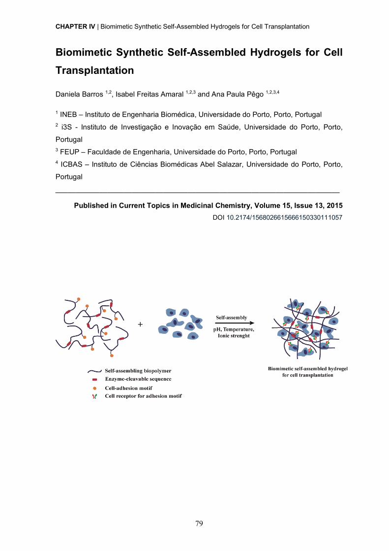
CHAPTER IV | Biomimetic Synthetic Self-Assembled Hydrogels for Cell Transplantation
79
Biomimetic Synthetic Self-Assembled Hydrogels for Cell Transplantation
Daniela Barros 1,2, Isabel Freitas Amaral 1,2,3 and Ana Paula Pêgo 1,2,3,4
1 INEB – Instituto de Engenharia Biomédica, Universidade do Porto, Porto, Portugal 2 i3S - Instituto de Investigação e Inovação em Saúde, Universidade do Porto, Porto,
Portugal 3 FEUP – Faculdade de Engenharia, Universidade do Porto, Porto, Portugal 4 ICBAS – Instituto de Ciências Biomédicas Abel Salazar, Universidade do Porto, Porto,
Portugal
_______________________________________________________________________
Published in Current Topics in Medicinal Chemistry, Volume 15, Issue 13, 2015 DOI 10.2174/1568026615666150330111057
CHAPTER IV | Biomimetic Synthetic Self-Assembled Hydrogels for Cell Transplantation
80
Abstract
The development of three-dimensional matrices capable of recapitulating the main features
of native extracellular matrix and contribute for the establishment of a favorable
microenvironment for cell behavior and fate is expected to circumvent some of the main
limitations of cell-based therapies. In this context, self-assembly has emerged as a
promising strategy to engineer cell-compatible hydrogels. A wide number of synthetically-
derived biopolymers, such as proteins, peptides and DNA/RNA, with intrinsic ability to self-
assemble into well-defined nanofibrous structures, are being explored. The resulting
hydrogels, in addition to closely resembling the architecture of native cellular
microenvironments, present a versatile and dynamic behavior that allows them to be
designed to undergo sol-to-gel transition in response to exogenous stimulus. This review
presents an overview on the state-of-the-art of the different strategies being explored for
the development of injectable synthetic self-assembled hydrogels for cell transplantation
and/or recruitment of endogenous cells, with an emphasis on their biological performance,
both in vitro and in vivo. Systems based on peptides are the most widely explored and have
already generated promising results in pre-clinical in vivo studies involving different
repair/regenerative scenarios, including cartilage, bone, nerve and heart. On the other
hand, systems based on DNA and hybrid hydrogels are now emerging for application in the
biomedical field with high potential. Finally, the main challenges hampering the translation
of these systems to the clinic and the issues that need to be addressed for these to progress
from bench-to-bedside are discussed.
CHAPTER IV | Biomimetic Synthetic Self-Assembled Hydrogels for Cell Transplantation
81
Cell transplantation in the context of regenerative medicine As our understanding of cell biology and ability to control cell behavior, both in vitro and in
vivo, improves, cell-based therapies are growing in interest within the field of regenerative
medicine [1]. Their effectiveness relies on the ability of transplanted cells to mediate a
therapeutic effect that potentiates the repair/regeneration processes [2]. Nevertheless,
before the effective translation of these therapies into the clinic, a number of issues still
have to be solved, such as low cell survival, lack of control of cell fate and poor cell
engraftment following transplantation [3, 4].
Within biological systems, cells reside in the extracellular matrix (ECM), a complex and
highly dynamic microenvironment that provides structural, mechanical, and biochemical
support to cells, ultimately determining their behavior, function and fate. In this sense, and
to improve the efficacy of cell transplantation therapies, biomaterial-based matrices
mimicking the ECM are being designed to be used as vehicles for cell delivery. The goal is
to create a permissive niche for the transplanted cells, promoting cell survival, proliferation,
differentiation, and integration within the host tissue. One of the major challenges one faces
when designing a cell-compatible matrix is the recapitulation of the instructive cellular
niches of natural tissues and the control of the bidirectional interaction between cells and
the surrounding microenvironment.
The design of such matrices must take into account a number of biological and physical
criteria, as follows:
(i) Biocompatibility. The developed matrix should be able of perform its
function without causing any toxic or undesirable effects.
(ii) Mechanical properties. The matrix mechanical properties should
closely match those of the target tissue, since these will impact cell
behavior and, consequently, determine the therapeutic efficacy of the
transplanted cells.
(iii) Permeability. The matrix should allow the continuous exchange of
gases, ions, nutrients and metabolites with the surrounding
microenvironment, which is vital for the survival and proliferation of
transplanted/encapsulated cells.
(iv) Degradation. Envisaging an application in the context of tissue
repair/regeneration a degradable matrix is preferred in the majority of
cases. It is paramount that the rate and mechanism of matrix
degradation are adjusted to the process of tissue remodeling.
CHAPTER IV | Biomimetic Synthetic Self-Assembled Hydrogels for Cell Transplantation
82
In the last few years, the incorporation of biochemical and biophysical ECM cues – cell
adhesion, cell-triggered proteolytic degradation, mechanical stimulation and ECM/growth
factor binding - into matrices of both synthetic and natural origin was explored to better
mimic the native cellular microenvironment, with different degrees of development and
success (consult references [5-7] for a comprehensive review on engineered matrices as
ECM mimics).
Hydrogels as attractive vehicles for cell transplantation Hydrogels are reticulated structures comprising cross-linked hydrophilic polymers with high
water content (over 90% of water) [8]. A number of unique features make hydrogels highly
promising matrices for cell transplantation and even for endogenous cell recruitment in the
context of tissue repair/regeneration. These include a highly hydrated nature, good
permeability and elasticity, resembling the nature of soft tissue microenvironments [5].
These properties will favor cell-cell and cell-matrix interactions, ultimately influencing cell
behavior and fate. Moreover, the structural and mechanical properties of hydrogels can be
easily tuned to mimic the tissue-specific ECM microenvironment, by varying the polymer
molecular weight and/or its concentration, or by adjusting the cross-linking degree of the
network [1]. This is crucial for the development of cell-compatible hydrogels, as cells also
respond to biophysical properties of the microenvironment not specifically related to its
molecular composition, but to the overall structural and mechanical characteristics of the
matrix.
Cells are particularly sensitive to mechanical stimuli, and small changes in matrix stiffness
or rigidity can alter cell behavior in many aspects, such as morphology, migration,
proliferation, differentiation and, in the case of stem cells, lineage specification and
commitment [9, 10]. For instance, mesenchymal stem cells (MSCs) cultured on variably
compliant poly(acrylamide) gels were found to undergo neurogenic differentiation on gels
with elastic moduli (E) mimicking brain tissue (< 1 kPa), while on gels mimicking skeletal
muscle (E = 11 kPa) and collagenous bone (E = 34 kPa) these cells differentiated along the
myogenic and osteogenic lineages, respectively [10]. The mechanism by which cells
convert mechanical signals into biochemical responses is termed mechanotransduction, a
dynamic process orchestrated by the ECM, mechanosensitive molecules and cellular
components (e.g. integrins, focal adhesions, stretch-activated ion channels, cadherins,
cytoskeleton filaments, nuclei) [11]. Particularly important for matrix elasticity sensing are
focal adhesions, multiprotein complexes formed upon integrin binding to ECM adhesion
ligands, which link the ECM to the intracellular cytoskeleton. Focal adhesions act as
mechanosensitive receptors, through which alterations in the conformation of ECM
molecules caused by mechanical stimuli (e.g. tension/loading) are transmitted to

CHAPTER IV | Biomimetic Synthetic Self-Assembled Hydrogels for Cell Transplantation
83
cytoskeleton proteins, leading to cytoskeletal (re)organization, contraction, and activation
of mechanotransduction signaling pathways affecting cell morphology, migration, and gene
expression profile.
Hydrogels, rather than implanted, can be designed to be injected and create a three-
dimensional (3D) network in situ. This is highly advantageous when envisaging clinical
translation as it (i) allows the homogenous encapsulation of cells and bioactive molecules;
(ii) enables the use of minimally invasive procedures for implantation; and (iii) ensures the
conformational filling of the injured site, which usually has an irregular shape, leaving no
gaps between the matrix and the degenerated/damaged tissue. In situ forming hydrogels
will thus favor a more close contact and, consequently, a more efficient integration of the
matrix within the host tissue. Injectable hydrogels could be further designed to have
thixotropic properties (becoming less viscous when subjected to an applied stress). This is
achieved through the control of different variables (e.g. temperature, pH and ionic strength)
that influence directly the in situ gelling process (for a more extensive review on strategies
explored to design in situ forming hydrogels see [12, 13]). When the cross-linking occurs in
the presence of cells, either mediated by (bio)chemical (e.g. enzymatically, Michael-type
addition or photopolymerization) or physical (e.g. ionic interactions or self-assembly)
processes [13], care must be taken so that the impact on cell viability is limited and minute.
These properties have made hydrogels very attractive in the design of either acellular
systems, specifically designed to allow the infiltration of endogenous cells, or cellular grafts
to promote tissue remodeling and regeneration [14].
Naturally-derived polymers (e.g. collagen, fibrin or alginate) have been widely explored in
the design of hydrogels due to their inherent biocompatibility and ability to mimic several
features of native ECM. Nevertheless, their use is very often hindered by batch-to-batch
variability, risk of immunogenicity and limited range of mechanical properties [15, 16].
Therefore, recent strategies have focused on the use of hydrogels derived from either purely
synthetic polymers (e.g. poly(ethylene glycol) (PEG) and poly(vinyl alcohol)) or
synthetically-derived biopolymers (e.g. proteins, peptides and DNA/RNA). Their synthetic
nature, as well as their well-defined and versatile structure allows them to be engineered,
through the incorporation of bioactive sequences (e.g. cell adhesive and protease
sensitive), originating bioactive and bioresponsive hydrogels [16, 17]. In this context,
synthetically-derived biopolymers are being more extensively explored, due to the several
advantageous features of these materials, when compared with purely synthetic polymers.
In contrast with the simpler and stochastic structure of purely synthetic polymers,
synthetically-derived biopolymers can be engineered to attain a complex and reproducible
structure. Moreover, by being synthesized using solid-phase synthesis or recombinant
CHAPTER IV | Biomimetic Synthetic Self-Assembled Hydrogels for Cell Transplantation
84
technology (eventually combined with genetic engineering) [18], they present a
monodisperse distribution of the molecular weights. Finally, their inherent ability to be
cleaved by enzymes allows a more tight control over the kinetics of hydrogel degradation
and, as a result, over the remodeling process.
Self-assembled hydrogels An important feature to take into account when engineering matrices to serve as ECM
mimics is the architecture of native ECM. Attaining 3D networks structurally mimicking the
nanofibrous structure of the ECM is highly desirable. This is expected to favor the creation
of an instructive microenvironment for cell anchorage and cell migration, ultimately leading
to the implant, colonization and integration within the host tissue. In this sense, “top-down”
approaches, such as electrospinning and phase separation, have been explored in the
design of scaffolds for such purposes. However, with these techniques one has a limited
control over the scaffold structure at the nanoscale level, as well as over the reproducibility
of the process. In the last few years, molecular self-assembly has emerged as the most
promising and attractive strategy in this context [19-21]. This is a natural and spontaneous
“bottom-up” approach driving the association and organization of single molecules into
supramolecular structures. Self-assembly results from the establishment of multiple non-
covalent intermolecular interactions (e.g. hydrogen bonding, electrostatic interactions, Van
der Waals forces and aromatic π-stacking) [22, 23]. A wide number of biopolymers existing
in nature (e.g. proteins, peptides, polysaccharides and DNA/RNA) present intrinsic ability
to self-assemble into highly-ordered structural elements. As a result, their building blocks
are being explored to develop synthetic self-assembled networks [18], leading to hydrogels
with a nanofibrillar structure, closely resembling the architecture of native ECM. Moreover,
the weak and transient nature of established interactions allows the design of hydrogels
that undergo a sol-to-gel transition in response to exogenous stimulus (e.g. pH, temperature
and ionic strength), which provides matrices with a “smart” and dynamic behavior [19, 24].
In the following sections, we present the state-of-the-art of synthetic self-assembled
hydrogels, focusing on their application as matrices for cell transplantation in the context of
regenerative medicine.
Engineered protein-based hydrogels A number of naturally-occurring proteins (e.g. elastin, collagen and silk) have remarkable
structural and biological properties that prompted their use in the design of self-assembled
hydrogels [25]. In particular, the ones derived from constituents of the mammalian ECM
proteins, like collagen, fibronectin or laminin, additionally present biological active peptide
domains recognized by cell adhesion receptors and cell-secreted proteolytic enzymes.
CHAPTER IV | Biomimetic Synthetic Self-Assembled Hydrogels for Cell Transplantation
85
These features have made protein-based hydrogels highly advantageous in the context of
cell therapies [26]. However, the use of proteins derived from natural sources is very often
hampered by low scalability, reproducibility, stability and price. In this sense, in the last few
years, engineered proteins have arisen as a promising alternative for the design of self-
assembled hydrogels. The synthetic nature of these proteins allows the combination in one
structure of key attributes of native ECM, including cell adhesion, growth factor and ECM
binding domains, as well as sequences that confer to the final protein elasticity and
proteolytic degradation. But while maintaining the main attractive biological features of
naturally derived proteins, precise control over the primary amino acid sequence and
molecular weight can be achieved, allowing the fine-tuning of the gelation process and of
the hydrogel structural and mechanical properties.
As the number of engineered proteins that can be used for the development of self-
assembled hydrogels [26, 27] is too large to be effectively covered by this review, only the
engineered protein-based hydrogels that have shown the most promising results, both in
vitro and in vivo, were addressed. These include hydrogels based on elastin- and silk-like
polypeptides, as well as hydrogels based on de novo engineered proteins, such as leucine
zipper coiled-coil-based polypeptides and amphiphilic block co-polypeptides.
Elastin-like polypeptides Elastin is an ECM structural protein, essentially composed of β-sheet structures, that
mediates important biological functions, such as cell-cell and ECM-cell interactions,
providing tissues with unique physical characteristics in terms of tensile strength and
elasticity [25]. A broad number of synthetic polypeptides have been designed inspired by
this natural protein, among which elastin-like polypeptides (ELPs). These are genetically
engineered sequences containing the pentapeptide repeat - (VPGXG)n - in which X can be
any amino acid except proline [28]. ELPs are thermally-responsive and thus can undergo a
reversible inverse phase transition in response to temperature changes (Fig. 1A) [25, 29].
At temperatures below a specific transition temperature (Tt), ELP molecules exist in a
disordered and bulky conformation, while above the Tt they self-associate through the
establishment of hydrophobic interactions, forming an insoluble and highly viscous
coacervate (Fig. 1A). The Tt of an ELP is highly dependent on its composition, molecular
weight and concentration, and thus can be easily tuned by changing any of these
parameters [30, 31]. The thermal responsiveness of ELPs provides a natural trigger for self-
assemble that can be exploited for the development of injectable defect-filling hydrogels
[32, 33] (for a more extensive review on ELPs stimulus responsive behavior see [34, 35]).
These molecules have been shown to be biocompatible, biodegradable and non-
immunogenic, which makes them highly attractive for tissue engineering purposes [26].
CHAPTER IV | Biomimetic Synthetic Self-Assembled Hydrogels for Cell Transplantation
86
The self-assembly of ELP-based hydrogels can also be achieved by chemical cross-linking
(Fig. 1A) [36]. For this purpose, ELPs are engineered to include lysine residues, enabling
primary amine reactive cross-linking [37]. The mechanical properties of the formed
hydrogels, as well as the rate of the gelation process, can be easily tuned by varying the
number of lysine residues and the pH of the cross-linking reaction [38]. Chemical cross-
linking was found not to affect cell viability, as shown by studies using mouse fibroblasts
[37, 39].
ELP-based hydrogels can be engineered to incorporate specific bioactive sequences, such
as cell adhesive and/or proteolytic sensitive domains, to impart them with bioactivity. As an
example, a chemically cross-linked biomimetic matrix comprising ELP structural domains
modified with the RGD cell adhesion motif and a sequence sensitive to urokinase
plasminogen activator (uPA) was designed for purposes of neural regeneration [40]. Apart
from being able to support PC-12 neuronal-like cell line adhesion, proliferation and
differentiation, the resulting hydrogel showed highly tunable degradation rates, depending
on the degree of exposure to uPA [40]. Also, a chemically cross-linked ELP hydrogel
designed with an RGD cell-adhesion motif have shown to be able of promote neurite
outgrowth of encapsulated dorsal root ganglia (DRG) cells [41].
To improve the mechanical and biological properties of ELPs, a different class of genetically
engineered materials - silk-elastin-like polypeptides (SELPs), was developed. These are
composed by repeating units of silk (GAGAGS) and elastin (GVGXP) peptide sequences
[25, 42-44]. Silk-like blocks provide chemical and thermal stability, due to their ability to form
hydrogen-bonded β-sheets, while elastin-like blocks introduce flexibility and aqueous
solubility to SELPs, by reducing their degree of crystallization [25]. The sequence and length
of silk- and elastin-like blocks, as well as their ratio strongly influence the extent of the
gelation process, as well as the biological and physicochemical properties of the developed
hydrogels [42, 45].
Leucine zipper coiled-coil-based polypeptides Engineered protein-based hydrogels assembled through the leucine zipper coiled-coil
domains have been widely explored in the context of tissue engineering. Leucine-zipper
coiled-coil domain is an α-helical structural motif with a heptad repeat sequence (abcdefg)n,
comprising hydrophobic (a and d), charged (e and g) and polar (b, c and f) residues that
form an amphiphilic α-helical structure, through the establishment of hydrophobic and ionic
interactions (Fig. 1B). The design rules guiding the formation of a coiled-coil structure were
extensively reviewed by Mason and Arndt [46]. The formed structure reversibly self-
assembles into coiled-coils, whose physical association will favor the formation of an
CHAPTER IV | Biomimetic Synthetic Self-Assembled Hydrogels for Cell Transplantation
87
engineered hydrogel network [47]. Based on the advantageous features of this self-
assembly module, a number of genetically engineered triblock copolymers were
synthesized. These are composed by a random coil water-soluble segment (Ala-Gly-rich
sequence), flanked by two short leucine zipper end motifs (each containing six heptad
repeats) that drive the formation of the network through a reversible gelation process
triggered by changes in pH and temperature (Fig. 1B) [47-49]. The formed physically cross-
linked hydrogels (Fig. 1B) display shear-thinning properties and rapid recovery after
injection, allowing encapsulated cells to survive [49]. Moreover, the degradation rate of the
developed hydrogels can be easily controlled by engineering the amino acid composition of
the leucine zipper coiled-coil [50]. Due to their modular structure these hydrogels can be
endowed with bioactivity through the incorporation, within the central domain of the triblock
polypeptide, of cell-adhesion ligands and proteolytic-sensitive sequences. Indeed, when
modified with the RGD-cell adhesion ligand, the developed matrices have shown to be able
to support human foreskin fibroblasts and human umbilical vein endothelial cells (HUVECs)
attachment, spreading and proliferation, as well as focal adhesion formation [51-53].
Amphiphilic block co-polypeptides A different approach that is being increasingly used in the development of protein-based
self-assembled hydrogels explores amphiphilic block co-polypeptides composed by a
repeating sequence of charged amino acids (Lysine (K) or Glutamate (E)), usually ranging
from 80 to 380 residues in length, and another of hydrophobic residues (Leucine (L) or
Valine (V)), comprising 10 to 40 residues in length (Fig. 1C) [54]. Such block co-
polypeptides usually present sequences like KxLy, KxVy or ExLy. Self-assembling occurs by
a process not found in nature and that is dependent on physical interactions between
hydrophobic α-helical domains (Fig. 1C) [54]. These domains associate into “twisted fibrillar
tapes with helices packed perpendicular to the fibril axes” [54]. This arrangement
contributes to the formation of a hydrogel with increased stability at high temperatures and
with ability to rapidly recover after stress, as a result of the weak and reversible nature of
the interactions mediating the self-assembly process [54, 55].
A number of self-assembled hydrogels based on di- [55, 56], tri- [57] and pentablock [58]
copolymers, differing in the length and/or composition of the hydrophobic domain, have
been proposed. The length of the hydrophobic domain significantly impacts the stability of
the formed hydrogels, with shorter segments resulting in weaker hydrogels.
Due to the similarity between the stiffness of some of these amphiphilic block co-
polypeptides and the brain tissue (~200 Pa), their influence on cellular behavior has been
further assessed in this system [56].
CHAPTER IV | Biomimetic Synthetic Self-Assembled Hydrogels for Cell Transplantation
88
Mixing-induced two-component hydrogel The concept of protein-protein interaction between specific association domains was
explored by Heilshorn´s group for the design of mixing-induced two-component hydrogels
[59, 60]. The first component of such system comprises proline-rich peptides (PPxY) linked
by hydrophilic spacers (Fig. 1D). The second is composed by engineered repeats of WW
domain linked together by hydrophilic spacers containing the cell adhesion motif RGDS
(Fig. 1D). The inclusion of such domain imparts flexibility and biofunctionality to component
2. Upon mixing, under physiological conditions, the two components are physically cross-
linked leading to the formation of a self-assembled hydrogel. The strength of the resultant
material can be tuned by changing the number of repeats per component and by adjusting
the strength of protein-protein interactions mediating the self-assemble process [60].
As in this system, self-assembly occurs without the need of an external trigger like
temperature, pH and ionic strength, physiological conditions are kept unaltered during the
cross-linking process, which is highly advantageous when envisaging the use of such
systems for cell transplantation [59]. Indeed, mixing-induced two-component hydrogels
have shown to be able to support the growth and differentiation of PC12 neuronal-like cells,
HUVECs, and primary murine neural stem cells (NSCs) [59], constituting highly promising
injectable materials for cell transplantation. Moreover, these hydrogels present shear-
thinning properties, resulting from the weak nature of the established interactions [59].
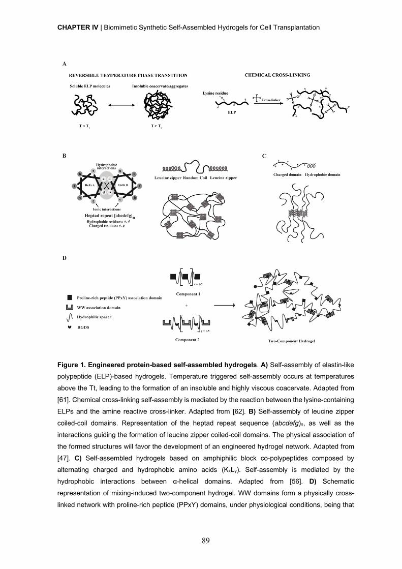
CHAPTER IV | Biomimetic Synthetic Self-Assembled Hydrogels for Cell Transplantation
89
Figure 1. Engineered protein-based self-assembled hydrogels. A) Self-assembly of elastin-like polypeptide (ELP)-based hydrogels. Temperature triggered self-assembly occurs at temperatures
above the Tt, leading to the formation of an insoluble and highly viscous coacervate. Adapted from
[61]. Chemical cross-linking self-assembly is mediated by the reaction between the lysine-containing
ELPs and the amine reactive cross-linker. Adapted from [62]. B) Self-assembly of leucine zipper
coiled-coil domains. Representation of the heptad repeat sequence (abcdefg)n, as well as the
interactions guiding the formation of leucine zipper coiled-coil domains. The physical association of
the formed structures will favor the development of an engineered hydrogel network. Adapted from
[47]. C) Self-assembled hydrogels based on amphiphilic block co-polypeptides composed by alternating charged and hydrophobic amino acids (KxLy). Self-assembly is mediated by the
hydrophobic interactions between α-helical domains. Adapted from [56]. D) Schematic
representation of mixing-induced two-component hydrogel. WW domains form a physically cross-
linked network with proline-rich peptide (PPxY) domains, under physiological conditions, being that

CHAPTER IV | Biomimetic Synthetic Self-Assembled Hydrogels for Cell Transplantation
90
the strength of this interaction varies according to the specific WW domain used. WW domains are
linked by hydrophilic spacers containing the RGDS cell-adhesion motif. Adapted from [59].
Peptide-based synthetic hydrogels Recently, hydrogels formed based on peptide self-assembly constitute one of the most
actively investigated class of biomaterials in the context of tissue engineering. Peptide-
based molecules, unlike other biopolymers (e.g. proteins and DNA/RNA), can be obtained
with high purity, reproducibility and good yields, crucial features when envisaging a clinical
application. Depending on the nature of the molecular building blocks (natural and non-
natural amino acids), peptide sequences with acidic, basic, polar, non-polar or aromatic
features can be developed [18, 63]. These features can influence, per se, different biological
functions, including cell adhesion, proliferation and differentiation [18], thus circumventing
the need of using more complex proteins. Nevertheless, the bioactivity and biofunctionality
of the resulting systems can be further improved through the incorporation of specific cell
adhesion domains (e.g. fibronectin- or collagen-derived RGDS [64-66], laminin-derived
IKVAV [67], bone-marrow homing peptides (BMHP) -1 and -2 [66] and the osteogenic
growth peptide ALK [68]), or proteolytic-sensitive sequences (e.g. matrix metalloproteinase
(MMP)-2 [69] and -13 [70] specific cleavage sites)).
The mechanisms behind the self-assembly processes mediating the formation of peptide-
based hydrogels was thoroughly reviewed by Zhang and co-workers [71, 72]. These
hydrogels are usually engineered to self-assemble according to the design rules derived
from natural proteins, by adopting basic secondary structural motifs, such as β-sheets, β-
hairpins or α-helices. However, a number of new approaches based on electrostatic,
amphiphilic or aromatic interactions are being widely explored for the development of new
self-assembled hydrogels.
In the following sections, we present the state-of-the-art of the two main categories of
peptide-based self-assembled hydrogels.
Self-assembling peptides Self-assembling peptides based on naturally-derived amino acids have been proposed as
promising structural elements for the development of novel hydrogels for tissue engineering
applications [71]. Inspired by the yeast protein zuotin [73], Zhang’s group synthesized a
number of ionic self-complementary peptides, including EAK16 [74, 75], RADA16 [76, 77],
FEK16 [78] and KLD-12 [79]. These are composed by alternating charged hydrophilic and
hydrophobic natural amino acids and have an intrinsic ability to self-assemble into highly
ordered β-sheet nanofibrous scaffolds (Fig. 2A). The self-assembly process can be further
accelerated by changes in pH or in salt solution concentration [74-76]. Ultimately, the non-
CHAPTER IV | Biomimetic Synthetic Self-Assembled Hydrogels for Cell Transplantation
91
covalent nature of the established interactions, in combination with the dependence on
external stimulus (e.g. pH, temperature and ionic strength), allows the self-assembly
process to be reversible and, as a result, the development of “smart” hydrogels [77]. The
stimulus-responsiveness of the developed hydrogels allows a more tight control over the
self-assembling process, which can be of interest for obtaining an in situ forming gel for
cells and/or biomolecules delivery. These ionic self-complementary peptides have been
successfully applied in a number of tissue engineering applications, namely as 3D matrices
for in vitro cell culture [76, 79-82] and as vehicles for cell transplantation [80] and/or
endogenous cell recruitment [83]. Peptides based on RADA16 self-assembling motif are
the most frequently employed due to the ease with which it is synthesized and modified for
specific applications. RADA16-based hydrogels are able of effectively support the
differentiation of adult rat liver progenitor cells into functional hepatocyte-like spheroid
clusters [81], PC-12 neuronal-like cell viability, neurite outgrowth and synapse formation
[80], and hippocampal neuronal cell culture [82].
To afford peptide-based self-assembled hydrogels with additional biological activity, a
number of specific bioactive sequences were incorporated. For instance, RADA16-based
hydrogels incorporating cell adhesion motifs from collagen VI (RGDS), as well as BMHP- 1
and -2, have been developed and their in vitro biological performance evaluated using
mouse adult NSCs [66]. As compared with non-functionalized matrices, the bioactive
hydrogels showed improved ability to support NSC survival, proliferation and differentiation.
[66]. Similar hydrogels comprising bioactive sequences known to enhance osteoblast
behavior, such as the osteogenic growth peptide ALK, osteopontin cell-adhesion motif
(DGR) and 2-unit RGD motifs (PGR), were also designed, and shown to support mouse
pre-osteoblastic MC3T3-E1 cells adhesion, proliferation and migration, as well as
osteogenic differentiation [68].
β-sheet self-assembly was also explored in the development of triblock peptides,
comprising an amphiphilic triblock peptide motif (Fig. 2B). The central B block comprises
alternating hydrophilic (glutamine) and hydrophobic (leucine) residues ((QL)n=1-6 repeat),
whose interactions lead to the formation of peptide dimmers [84]. These in turn, form β-
sheet hydrogen bonds with each other, which keep them aligned perpendicularly to the
peptide backbone axis, allowing the formation of highly-ordered nanofibrous matrices (Fig.
2B). On the other hand, the A block is composed by two to four positively charged lysine
residues which, due to electrostatic repulsions, will affect the conditions required for peptide
self-assembly at physiological conditions. Therefore, a balance of repulsive and attractive
forces must be attained to control the self-assembly of triblock peptides into well-defined
nanostructures and the properties of the resultant hydrogels [84]. The modular nature of
CHAPTER IV | Biomimetic Synthetic Self-Assembled Hydrogels for Cell Transplantation
92
amphiphilic triblock peptides can be harnessed to favor a more tunable control over
hydrogel mechanical properties. For instance, the careful selection of the amino acid
residues composing A and B blocks significantly influences the gelation conditions, the
length and diameter of the formed nanofibers, and the viscoelastic properties of the
resultant hydrogel [85]. To improve the bioactivity of developed hydrogels, an MMP-2
specific cleavage site and an RGD cell adhesion motif were incorporated in the central B
block and in the A block, respectively [69]. The combination of these two bioactive motifs
resulted in increased viability, spreading and migration of MSCs from human exfoliated
deciduous teeth within the hydrogel when compared with the non-functionalized ones.
An alternative design based on β-sheet motifs was developed, in which an amphiphilic β-
hairpin secondary structure was used as self-assembling motif [8, 86]. Here, peptide
sequences fold in response to different external stimulus, including pH [87], temperature
[88] and ionic strength [89]. The formed hydrogels present a controllable shear-thin recovery
kinetic, due to the weak and transient nature of the interactions mediating the self-assemble
process [90]. This allows the encapsulation and subsequent delivery of cells to the injured
sites, by minimally invasive procedures. β-hairpin-based hydrogels were shown to be
cytocompatible, being able to mediate the adhesion, migration and proliferation of NIH3T3
murine fibroblasts (seeded on their surface), even in the absence of serum proteins or cell-
adhesive motifs [91]. Through the incorporation of sequences sensitive to MMP-13, the
degradation of such hydrogels can be efficiently controlled [70].
Self-assembling peptide amphiphiles Peptide amphiphiles (PAs) are composed by a peptide sequence with a covalently linked
chemical tail (e.g. short hydrophobic sequences or aromatic moieties) with intrinsic ability
to self-assemble. These molecules can combine, thus, the structural features of amphiphilic
molecules with the chemical functionality/bioactivity of small peptide sequences [92-94]. In
the last decade, pioneering work of Stupp and co-workers resulted in the proposal of a
broad range of PAs comprising a hydrophilic sequence covalently linked to an alkyl chain
[94]. These molecules are typically composed by four distinct domains (Fig. 2C): (1) short
hydrophobic tail responsible to drive the self-assembly process; (2) β-sheet peptide domain,
which, through the formation of hydrogen bonds, promotes the cohesion of the formed
nanofibers; (3) charged residues to confer water solubility; and (4) bioactive domain to
mediate a specific biological response. Their self-assembly into long and stable nanofibers
(Fig. 2C) is driven by the hydrophobic tail and occurs under specific pH, temperature or
ionic strength conditions [95]. The resulting structures further undergo a sol-to-gel transition
under physiological conditions in the presence of multivalent ions, which allows injection
and in situ gel formation [64]. As result of the self-assembly process, the peptide sequence
CHAPTER IV | Biomimetic Synthetic Self-Assembled Hydrogels for Cell Transplantation
93
will be orientated towards the aqueous environment. This, in combination with the versatility
and modular nature of PAs, allows the easy incorporation of bioactive sequences at the
extremity of the peptide domain (Fig. 2C) and the development of bioactive hydrogels. As
an example, PAs functionalized with the IKVAV epitope from laminin, revealed higher ability
to promote NSC adhesion, migration and differentiation into a neuronal phenotype, as well
as neurite outgrowth, when compared to laminin-coated surfaces [67]. These promising
outcomes were associated to the high density of IKVAV epitopes present on the nanofibers
surface [67]. Therefore, both the epitope density and their presentation on the nanofiber
surface were found to contribute to such a pronounced biological response. Also in the
context of nerve regeneration, a hydrogel based on a PA engineered with a heparin sulfate
mimetic and an IKVAV epitope was developed [96]. The two bioactive moieties have
demonstrated a cooperative effect in the promotion of PC12 neuronal-like cells neurite
outgrowth, even when in presence of inhibitory components, such as chondrotin sulfate
proteoglycans [96]. For application in bone regenerative therapies, a hydrogel was
engineered with BMP-receptor binding peptides, termed osteopromotive domains [97]. The
developed hydrogels showed ability to promote cell survival and osteoblastic differentiation
of human bone marrow stromal cells (BMSCs) [97].
Aromatic PAs comprising short (e.g. di- or tri-) peptide sequences capped, at the N-terminal,
with a synthetic aromatic moiety have been also explored for the development of self-
assembled hydrogels. These PAs have the intrinsic ability to self-assemble and drive the
formation of highly-ordered amphiphilic hydrogels, by a combination of hydrophobic and π-
π interactions [98]. A number of PAs comprising the fluoren-9-ylmethyloxycarbonyl (Fmoc)
aromatic stacking ligand were developed [99-101]. The simplicity of the aromatic moieties
(only 2-3 amino acids in length) allows the easy modification of the peptide sequence and
a more tunable control over the structure and mechanical properties of the resultant
hydrogels [99-101]. The structure of the self-assembled PAs, resembles in several aspects
the native ECM, namely in their hydrated nature, nanofibrous architecture and rigidity
(G’~10 kPa) [100-102]. To endow these hydrogels with bioactivity, a modular design
strategy combining an Fmoc-RGD peptide with the Fmoc-diphenylalanine (Fmoc-FF)
sequence was explored [65]. The developed hydrogels showed ability to support the
adhesion, migration and proliferation of human adult dermal fibroblasts, in an integrin-
dependent manner. By changing the ratio in which the two components are mixed, a more
tunable control over hydrogel mechanical and biological properties may be achieved.
Alternatively, some authors explored the use of naphthalene (Nap), rather than the Fmoc
moiety, as aromatic capping group to synthesize the Nap-FFGEY peptide. The tyrosine
residue (Y) present in the resultant aromatic PA could be easily phosphorylated by the
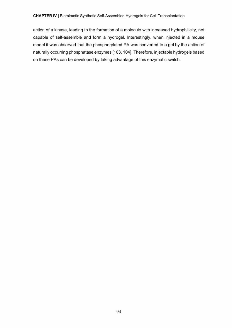
CHAPTER IV | Biomimetic Synthetic Self-Assembled Hydrogels for Cell Transplantation
94
action of a kinase, leading to the formation of a molecule with increased hydrophilicity, not
capable of self-assemble and form a hydrogel. Interestingly, when injected in a mouse
model it was observed that the phosphorylated PA was converted to a gel by the action of
naturally occurring phosphatase enzymes [103, 104]. Therefore, injectable hydrogels based
on these PAs can be developed by taking advantage of this enzymatic switch.
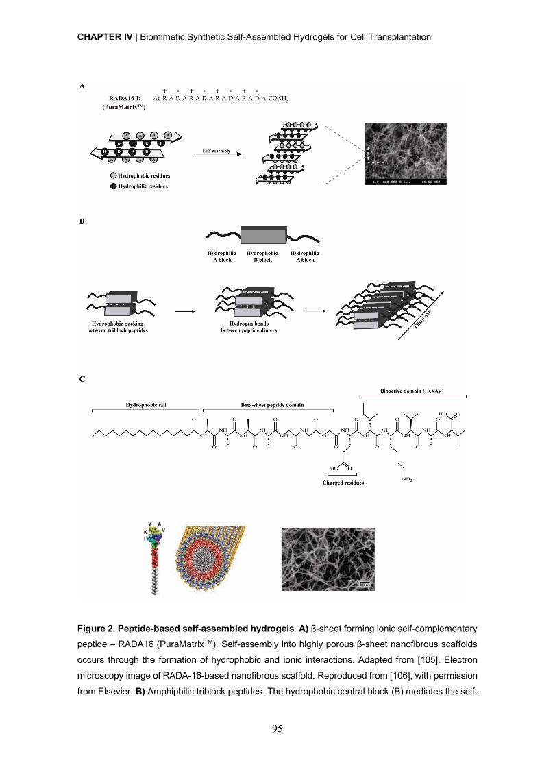
CHAPTER IV | Biomimetic Synthetic Self-Assembled Hydrogels for Cell Transplantation
95
Figure 2. Peptide-based self-assembled hydrogels. A) β-sheet forming ionic self-complementary peptide – RADA16 (PuraMatrixTM). Self-assembly into highly porous β-sheet nanofibrous scaffolds
occurs through the formation of hydrophobic and ionic interactions. Adapted from [105]. Electron
microscopy image of RADA-16-based nanofibrous scaffold. Reproduced from [106], with permission
from Elsevier. B) Amphiphilic triblock peptides. The hydrophobic central block (B) mediates the self-
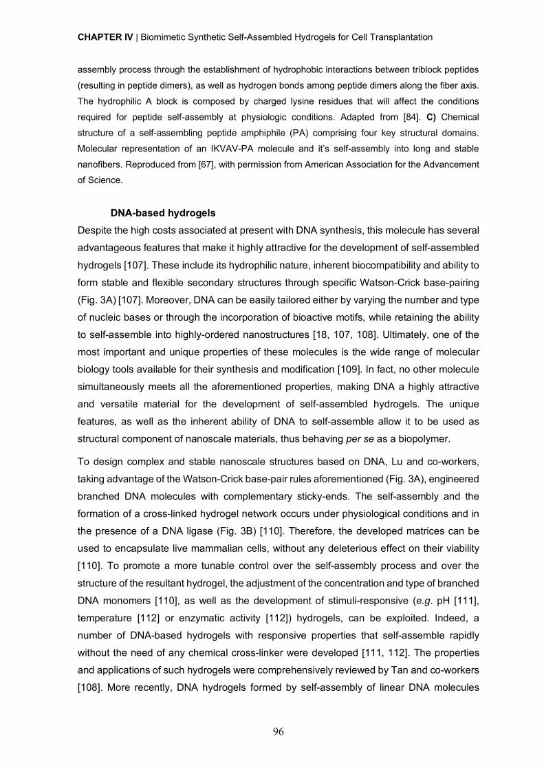
CHAPTER IV | Biomimetic Synthetic Self-Assembled Hydrogels for Cell Transplantation
96
assembly process through the establishment of hydrophobic interactions between triblock peptides
(resulting in peptide dimers), as well as hydrogen bonds among peptide dimers along the fiber axis.
The hydrophilic A block is composed by charged lysine residues that will affect the conditions
required for peptide self-assembly at physiologic conditions. Adapted from [84]. C) Chemical
structure of a self-assembling peptide amphiphile (PA) comprising four key structural domains.
Molecular representation of an IKVAV-PA molecule and it’s self-assembly into long and stable
nanofibers. Reproduced from [67], with permission from American Association for the Advancement of Science.
DNA-based hydrogels Despite the high costs associated at present with DNA synthesis, this molecule has several
advantageous features that make it highly attractive for the development of self-assembled
hydrogels [107]. These include its hydrophilic nature, inherent biocompatibility and ability to
form stable and flexible secondary structures through specific Watson-Crick base-pairing
(Fig. 3A) [107]. Moreover, DNA can be easily tailored either by varying the number and type
of nucleic bases or through the incorporation of bioactive motifs, while retaining the ability
to self-assemble into highly-ordered nanostructures [18, 107, 108]. Ultimately, one of the
most important and unique properties of these molecules is the wide range of molecular
biology tools available for their synthesis and modification [109]. In fact, no other molecule
simultaneously meets all the aforementioned properties, making DNA a highly attractive
and versatile material for the development of self-assembled hydrogels. The unique
features, as well as the inherent ability of DNA to self-assemble allow it to be used as
structural component of nanoscale materials, thus behaving per se as a biopolymer.
To design complex and stable nanoscale structures based on DNA, Lu and co-workers,
taking advantage of the Watson-Crick base-pair rules aforementioned (Fig. 3A), engineered
branched DNA molecules with complementary sticky-ends. The self-assembly and the
formation of a cross-linked hydrogel network occurs under physiological conditions and in
the presence of a DNA ligase (Fig. 3B) [110]. Therefore, the developed matrices can be
used to encapsulate live mammalian cells, without any deleterious effect on their viability
[110]. To promote a more tunable control over the self-assembly process and over the
structure of the resultant hydrogel, the adjustment of the concentration and type of branched
DNA monomers [110], as well as the development of stimuli-responsive (e.g. pH [111],
temperature [112] or enzymatic activity [112]) hydrogels, can be exploited. Indeed, a
number of DNA-based hydrogels with responsive properties that self-assemble rapidly
without the need of any chemical cross-linker were developed [111, 112]. The properties
and applications of such hydrogels were comprehensively reviewed by Tan and co-workers
[108]. More recently, DNA hydrogels formed by self-assembly of linear DNA molecules
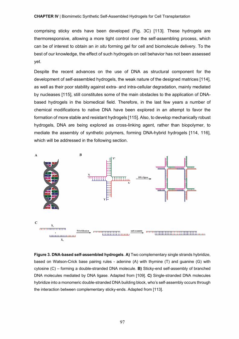
CHAPTER IV | Biomimetic Synthetic Self-Assembled Hydrogels for Cell Transplantation
97
comprising sticky ends have been developed (Fig. 3C) [113]. These hydrogels are
thermoresponsive, allowing a more tight control over the self-assembling process, which
can be of interest to obtain an in situ forming gel for cell and biomolecule delivery. To the
best of our knowledge, the effect of such hydrogels on cell behavior has not been assessed
yet.
Despite the recent advances on the use of DNA as structural component for the
development of self-assembled hydrogels, the weak nature of the designed matrices [114],
as well as their poor stability against extra- and intra-cellular degradation, mainly mediated
by nucleases [115], still constitutes some of the main obstacles to the application of DNA-
based hydrogels in the biomedical field. Therefore, in the last few years a number of
chemical modifications to native DNA have been explored in an attempt to favor the
formation of more stable and resistant hydrogels [115]. Also, to develop mechanically robust
hydrogels, DNA are being explored as cross-linking agent, rather than biopolymer, to
mediate the assembly of synthetic polymers, forming DNA-hybrid hydrogels [114, 116],
which will be addressed in the following section.
Figure 3. DNA-based self-assembled hydrogels. A) Two complementary single strands hybridize,
based on Watson-Crick base pairing rules - adenine (A) with thymine (T) and guanine (G) with
cytosine (C) – forming a double-stranded DNA molecule. B) Sticky-end self-assembly of branched
DNA molecules mediated by DNA ligase. Adapted from [109]. C) Single-stranded DNA molecules
hybridize into a monomeric double-stranded DNA building block, who’s self-assembly occurs through
the interaction between complementary sticky-ends. Adapted from [113].
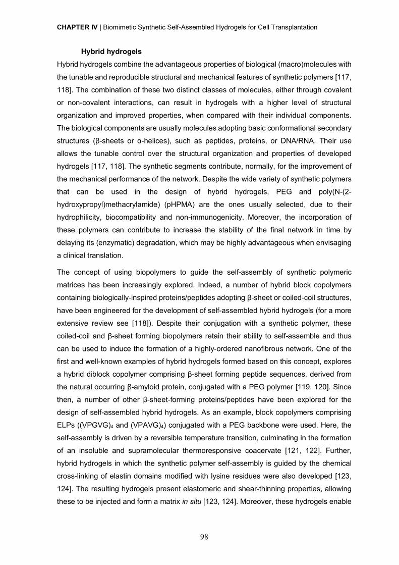
CHAPTER IV | Biomimetic Synthetic Self-Assembled Hydrogels for Cell Transplantation
98
Hybrid hydrogels Hybrid hydrogels combine the advantageous properties of biological (macro)molecules with
the tunable and reproducible structural and mechanical features of synthetic polymers [117,
118]. The combination of these two distinct classes of molecules, either through covalent
or non-covalent interactions, can result in hydrogels with a higher level of structural
organization and improved properties, when compared with their individual components.
The biological components are usually molecules adopting basic conformational secondary
structures (β-sheets or α-helices), such as peptides, proteins, or DNA/RNA. Their use
allows the tunable control over the structural organization and properties of developed
hydrogels [117, 118]. The synthetic segments contribute, normally, for the improvement of
the mechanical performance of the network. Despite the wide variety of synthetic polymers
that can be used in the design of hybrid hydrogels, PEG and poly(N-(2-
hydroxypropyl)methacrylamide) (pHPMA) are the ones usually selected, due to their
hydrophilicity, biocompatibility and non-immunogenicity. Moreover, the incorporation of
these polymers can contribute to increase the stability of the final network in time by
delaying its (enzymatic) degradation, which may be highly advantageous when envisaging
a clinical translation.
The concept of using biopolymers to guide the self-assembly of synthetic polymeric
matrices has been increasingly explored. Indeed, a number of hybrid block copolymers
containing biologically-inspired proteins/peptides adopting β-sheet or coiled-coil structures,
have been engineered for the development of self-assembled hybrid hydrogels (for a more
extensive review see [118]). Despite their conjugation with a synthetic polymer, these
coiled-coil and β-sheet forming biopolymers retain their ability to self-assemble and thus
can be used to induce the formation of a highly-ordered nanofibrous network. One of the
first and well-known examples of hybrid hydrogels formed based on this concept, explores
a hybrid diblock copolymer comprising β-sheet forming peptide sequences, derived from
the natural occurring β-amyloid protein, conjugated with a PEG polymer [119, 120]. Since
then, a number of other β-sheet-forming proteins/peptides have been explored for the
design of self-assembled hybrid hydrogels. As an example, block copolymers comprising
ELPs ((VPGVG)4 and (VPAVG)4) conjugated with a PEG backbone were used. Here, the
self-assembly is driven by a reversible temperature transition, culminating in the formation
of an insoluble and supramolecular thermoresponsive coacervate [121, 122]. Further,
hybrid hydrogels in which the synthetic polymer self-assembly is guided by the chemical
cross-linking of elastin domains modified with lysine residues were also developed [123,
124]. The resulting hydrogels present elastomeric and shear-thinning properties, allowing
these to be injected and form a matrix in situ [123, 124]. Moreover, these hydrogels enable

CHAPTER IV | Biomimetic Synthetic Self-Assembled Hydrogels for Cell Transplantation
99
a tunable control over the density of elastin cell adhesion domains, as well as of matrix
stiffness, which is highly desirable to modulate cell behavior and when studying cell-matrix
interactions. Indeed, ELP-PEG hybrid hydrogels have shown to be able of support
fibroblasts growth, migration and proliferation [123, 124]. β-sheet forming peptides were
also explored as component of hybrid graft copolymers. A pHPMA polymer grafted with
complementary β-sheet peptides were developed and endowed with bioactivity through the
coupling of RGD cell-adhesion domains to each complementary hybrid graft copolymer
(Fig. 4A). These hydrogels have shown ability to support MC3T3-E1 cell viability and
proliferation.
The self-assembly properties of coiled-coil forming peptides were also explored to create
hybrid hydrogels. Diblock copolymers of PEG and coiled-coil forming peptides that self-
assemble into a two-stranded α-helical coiled-coil conformation in aqueous solution,
illustrate such strategy [125, 126]. It has been shown that the conjugation of coiled-coil
domains with a synthetic polymer favors their thermal stability, resulting from the formation
of a PEG hydrophilic shell around the coiled-coil structure [125, 126]. Further, genetically
engineered coiled-coil forming peptides (CC1 and CC2), non-covalently grafted to a pHPMA
synthetic backbone have been explored for the development of hybrid graft copolymers
[127]. Here, the physical cross-linking and the consequent hybrid hydrogel formation were
favored by a temperature-induced conformational change of coiled-coil peptides [127]. The
temperature-responsiveness of these hydrogels allows the development of “smart”
materials and a more tunable control over the material properties. Another design involving
the pHPMA hydrophilic polymer and peptide grafts forming homodimers and higher
aggregates were also described [128, 129]. In this case, a minimum of four heptad repeats
was found to be needed to induce self-assembly and the formation of homodimers [128,
129]. The number and concentration of the copolymer, as well as temperature proved to
have a crucial role on the control of the gelation process [129]. Still, this design is very often
limited by the possibility of dimer formation between grafted peptides attached to the same
macromolecule. To avoid this and to guarantee the formation of a well-ordered 3D structure,
a modified approach was proposed. In this new design, a pHPMA synthetic backbone was
grafted with a pair of oppositely charged coiled-coil forming peptides (CCE and CCK) (Fig.
4B) [130, 131]. The oppositely charged peptide grafts self-assemble into antiparallel
heterodimeric motifs and induce the formation of a physically cross-linked hydrogel [130,
131]. Here, a reversible self-assembly occurs when the graft copolymers, CCE-P and CCK-
P, are mixed equimolarly at neutral pH.
A different approach for the design of hybrid hydrogels consists in using star-shaped
macromolecules, such as 4-arm PEG, in combination with biological molecules, responsible
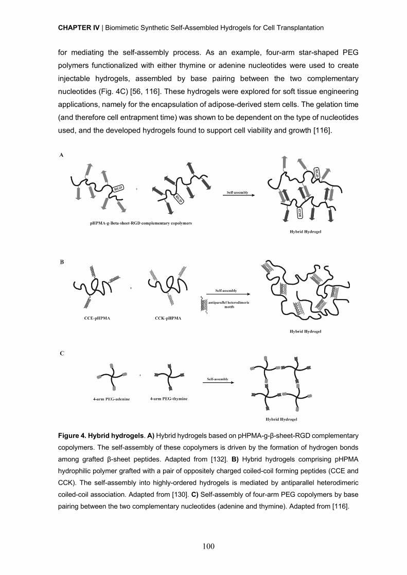
CHAPTER IV | Biomimetic Synthetic Self-Assembled Hydrogels for Cell Transplantation
100
for mediating the self-assembly process. As an example, four-arm star-shaped PEG
polymers functionalized with either thymine or adenine nucleotides were used to create
injectable hydrogels, assembled by base pairing between the two complementary
nucleotides (Fig. 4C) [56, 116]. These hydrogels were explored for soft tissue engineering
applications, namely for the encapsulation of adipose-derived stem cells. The gelation time
(and therefore cell entrapment time) was shown to be dependent on the type of nucleotides
used, and the developed hydrogels found to support cell viability and growth [116].
Figure 4. Hybrid hydrogels. A) Hybrid hydrogels based on pHPMA-g-β-sheet-RGD complementary
copolymers. The self-assembly of these copolymers is driven by the formation of hydrogen bonds
among grafted β-sheet peptides. Adapted from [132]. B) Hybrid hydrogels comprising pHPMA hydrophilic polymer grafted with a pair of oppositely charged coiled-coil forming peptides (CCE and
CCK). The self-assembly into highly-ordered hydrogels is mediated by antiparallel heterodimeric
coiled-coil association. Adapted from [130]. C) Self-assembly of four-arm PEG copolymers by base
pairing between the two complementary nucleotides (adenine and thymine). Adapted from [116].
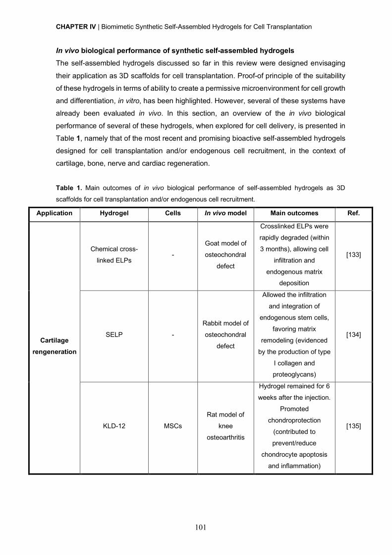
CHAPTER IV | Biomimetic Synthetic Self-Assembled Hydrogels for Cell Transplantation
101
In vivo biological performance of synthetic self-assembled hydrogels The self-assembled hydrogels discussed so far in this review were designed envisaging
their application as 3D scaffolds for cell transplantation. Proof-of principle of the suitability
of these hydrogels in terms of ability to create a permissive microenvironment for cell growth
and differentiation, in vitro, has been highlighted. However, several of these systems have
already been evaluated in vivo. In this section, an overview of the in vivo biological
performance of several of these hydrogels, when explored for cell delivery, is presented in
Table 1, namely that of the most recent and promising bioactive self-assembled hydrogels
designed for cell transplantation and/or endogenous cell recruitment, in the context of
cartilage, bone, nerve and cardiac regeneration.
Table 1. Main outcomes of in vivo biological performance of self-assembled hydrogels as 3D
scaffolds for cell transplantation and/or endogenous cell recruitment.
Application Hydrogel Cells In vivo model Main outcomes Ref.
Cartilage rengeneration
Chemical cross-
linked ELPs -
Goat model of
osteochondral
defect
Crosslinked ELPs were
rapidly degraded (within
3 months), allowing cell
infiltration and
endogenous matrix deposition
[133]
SELP -
Rabbit model of
osteochondral
defect
Allowed the infiltration
and integration of
endogenous stem cells,
favoring matrix
remodeling (evidenced
by the production of type
I collagen and proteoglycans)
[134]
KLD-12 MSCs
Rat model of
knee
osteoarthritis
Hydrogel remained for 6
weeks after the injection.
Promoted
chondroprotection
(contributed to
prevent/reduce
chondrocyte apoptosis
and inflammation)
[135]
CHAPTER IV | Biomimetic Synthetic Self-Assembled Hydrogels for Cell Transplantation
102
Poly(L-lactide-co-ε-
caprolactone)
scaffolds + mixture of KLD-12 and KLD-
12-collagen mimetic
peptide (CMP7)
BMSCs Subcutaneous implantation at
mice dorsa
Stimulated chondrogenic
differentiation of BMSCs and deposition of
chondral ECM (e.g.
glycosaminoglycans)
[136]
Bone regeneration
Demineralized bone
matrix modified with
RADA16
MSCs
Mouse model of
femoral defect
Favored the adhesion,
proliferation and
differentiation of
transplanted MSCs,
creating a permissive microenvironment for
bone regeneration (high
yield of osteogenic cells
(MSCs) and growth
factors, namely bone
morphogenetic protein-6,
platelet-derived growth
factor-BB, vascular endothelial growth factor
(VEGF) and fibroblast
growth factor)
[137]
Goat model of
femoral defect
Bone regeneration/
remodeling was
efficiently completed at
16-weeks post injury
[138]
RGDS + Phosphoserine -PA
- Rat model of
femoral defect
Increased bone
formation was observed 4 weeks after injury
[139]
Ti-6Al-4V
macroporous
structure filled with
RGDS +
phosphoserine-PA
MC3T3-E1 Rat model of
femoral defect
No significant cytotoxic
effect to cells was
observed.
Promoted de novo bone
formation in and around
the implant and of
angiogenesis, within 4 weeks
[140]
Nerve regeneration
K180L20
Amphiphilic diblock
co-polypeptide
- Mouse forebrain No detectable toxicity or
adverse tissue response. [56]
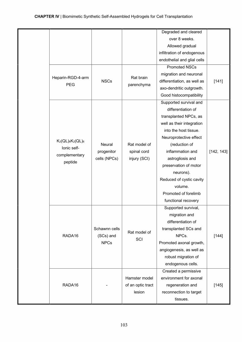
CHAPTER IV | Biomimetic Synthetic Self-Assembled Hydrogels for Cell Transplantation
103
Degraded and cleared
over 8 weeks.
Allowed gradual infiltration of endogenous
endothelial and glial cells
Heparin-RGD-4-arm
PEG NSCs
Rat brain
parenchyma
Promoted NSCs
migration and neuronal
differentiation, as well as
axo-dendritic outgrowth.
Good histocompatibility
[141]
K2(QL)6K2(QL)6
Ionic self-
complementary
peptide
Neural
progenitor
cells (NPCs)
Rat model of
spinal cord
injury (SCI)
Supported survival and
differentiation of transplanted NPCs, as
well as their integration
into the host tissue.
Neuroprotective effect
(reduction of
inflammation and
astrogliosis and preservation of motor
neurons).
Reduced of cystic cavity
volume.
Promoted of forelimb
functional recovery
[142, 143]
RADA16
Schawnn cells
(SCs) and
NPCs
Rat model of
SCI
Supported survival,
migration and
differentiation of transplanted SCs and
NPCs.
Promoted axonal growth,
angiogenesis, as well as
robust migration of
endogenous cells.
[144]
RADA16 - Hamster model of an optic tract
lesion
Created a permissive
environment for axonal regeneration and
reconnection to target
tissues.
[145]
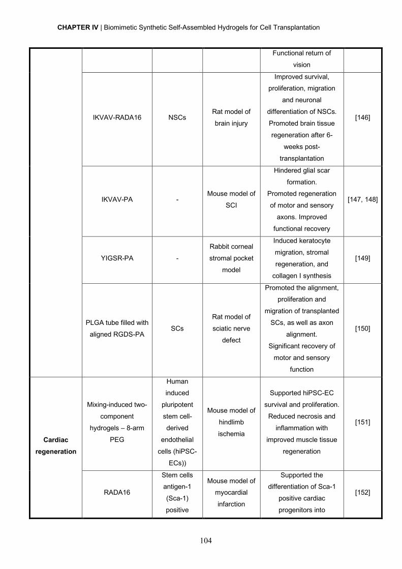
CHAPTER IV | Biomimetic Synthetic Self-Assembled Hydrogels for Cell Transplantation
104
Functional return of
vision
IKVAV-RADA16 NSCs Rat model of
brain injury
Improved survival, proliferation, migration
and neuronal
differentiation of NSCs.
Promoted brain tissue
regeneration after 6-
weeks post-
transplantation
[146]
IKVAV-PA - Mouse model of
SCI
Hindered glial scar
formation. Promoted regeneration
of motor and sensory
axons. Improved
functional recovery
[147, 148]
YIGSR-PA -
Rabbit corneal
stromal pocket
model
Induced keratocyte
migration, stromal
regeneration, and
collagen I synthesis
[149]
PLGA tube filled with
aligned RGDS-PA SCs
Rat model of
sciatic nerve
defect
Promoted the alignment, proliferation and
migration of transplanted
SCs, as well as axon
alignment.
Significant recovery of
motor and sensory
function
[150]
Cardiac regeneration
Mixing-induced two-
component
hydrogels – 8-arm
PEG
Human induced
pluripotent
stem cell-
derived
endothelial
cells (hiPSC-
ECs))
Mouse model of
hindlimb
ischemia
Supported hiPSC-EC
survival and proliferation.
Reduced necrosis and
inflammation with
improved muscle tissue
regeneration
[151]
RADA16
Stem cells antigen-1
(Sca-1)
positive
Mouse model of myocardial
infarction
Supported the differentiation of Sca-1
positive cardiac
progenitors into
[152]
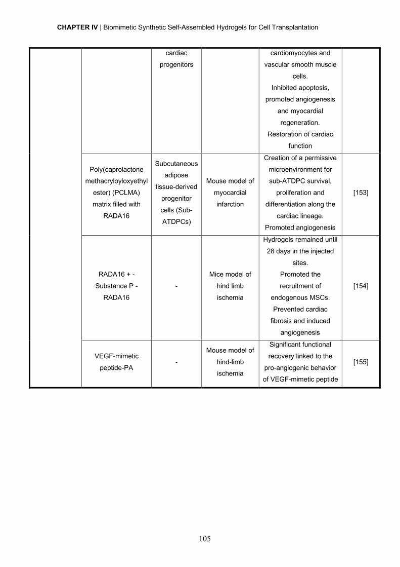
CHAPTER IV | Biomimetic Synthetic Self-Assembled Hydrogels for Cell Transplantation
105
cardiac
progenitors
cardiomyocytes and
vascular smooth muscle
cells. Inhibited apoptosis,
promoted angiogenesis
and myocardial
regeneration.
Restoration of cardiac
function
Poly(caprolactone methacryloyloxyethyl
ester) (PCLMA)
matrix filled with
RADA16
Subcutaneous adipose
tissue-derived
progenitor
cells (Sub-
ATDPCs)
Mouse model of
myocardial
infarction
Creation of a permissive
microenvironment for sub-ATDPC survival,
proliferation and
differentiation along the
cardiac lineage.
Promoted angiogenesis
[153]
RADA16 + -
Substance P -
RADA16
-
Mice model of
hind limb
ischemia
Hydrogels remained until
28 days in the injected
sites. Promoted the
recruitment of
endogenous MSCs.
Prevented cardiac
fibrosis and induced
angiogenesis
[154]
VEGF-mimetic
peptide-PA -
Mouse model of
hind-limb ischemia
Significant functional
recovery linked to the
pro-angiogenic behavior of VEGF-mimetic peptide
[155]
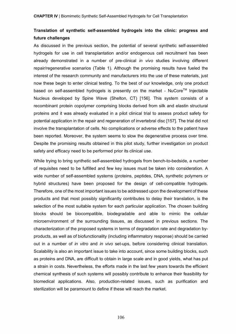
CHAPTER IV | Biomimetic Synthetic Self-Assembled Hydrogels for Cell Transplantation
106
Translation of synthetic self-assembled hydrogels into the clinic: progress and future challenges As discussed in the previous section, the potential of several synthetic self-assembled
hydrogels for use in cell transplantation and/or endogenous cell recruitment has been
already demonstrated in a number of pre-clinical in vivo studies involving different
repair/regenerative scenarios (Table 1). Although the promising results have fueled the
interest of the research community and manufacturers into the use of these materials, just
now these begin to enter clinical testing. To the best of our knowledge, only one product
based on self-assembled hydrogels is presently on the market - NuCoreTM Injectable
Nucleus developed by Spine Wave (Shelton, CT) [156]. This system consists of a
recombinant protein copolymer comprising blocks derived from silk and elastin structural
proteins and it was already evaluated in a pilot clinical trial to assess product safety for
potential application in the repair and regeneration of invertebral disc [157]. The trial did not
involve the transplantation of cells. No complications or adverse effects to the patient have
been reported. Moreover, the system seems to slow the degenerative process over time.
Despite the promising results obtained in this pilot study, further investigation on product
safety and efficacy need to be performed prior its clinical use.
While trying to bring synthetic self-assembled hydrogels from bench-to-bedside, a number
of requisites need to be fulfilled and few key issues must be taken into consideration. A
wide number of self-assembled systems (proteins, peptides, DNA, synthetic polymers or
hybrid structures) have been proposed for the design of cell-compatible hydrogels.
Therefore, one of the most important issues to be addressed upon the development of these
products and that most possibly significantly contributes to delay their translation, is the
selection of the most suitable system for each particular application. The chosen building
blocks should be biocompatible, biodegradable and able to mimic the cellular
microenvironment of the surrounding tissues, as discussed in previous sections. The
characterization of the proposed systems in terms of degradation rate and degradation by-
products, as well as of biofunctionality (including inflammatory response) should be carried
out in a number of in vitro and in vivo set-ups, before considering clinical translation.
Scalability is also an important issue to take into account, since some building blocks, such
as proteins and DNA, are difficult to obtain in large scale and in good yields, what has put
a strain in costs. Nevertheless, the efforts made in the last few years towards the efficient
chemical synthesis of such systems will possibly contribute to enhance their feasibility for
biomedical applications. Also, production-related issues, such as purification and
sterilization will be paramount to define if these will reach the market.
CHAPTER IV | Biomimetic Synthetic Self-Assembled Hydrogels for Cell Transplantation
107
A challenge while developing injectable in situ forming hydrogels is to gain control over the
3D architecture of the hydrogel fibrillar network. The building blocks of synthetic self-
assembled hydrogels typically assemble into randomly oriented networks, and the
nanofibrillar structures formed in several peptide hydrogels are also randomly oriented. To
better mimic the natural anisotropic structure and functional features of the ECM, control
over fibrillar orientation both at the nano- and the micro-scale should be achieved. This is
particularly important in hydrogels envisaged for application in highly organized tissues (e.g.
nerve and bone), in which the structural and mechanical properties of the ECM fulfill
important biomechanical properties and regulate cell behavior. For instance, longitudinally-
oriented aligned structures have been shown to induce directional axonal re-growth and
alignment of transplanted cells, by providing contact guidance, which can be of most
importance in therapies for the treatment of spinal cord and peripheral nerve injuries [158].
Attempts to develop in situ forming hydrogels of aligned self-assembled PA nanofibers were
made. This was achieved by exposing isotropic PA solutions to elevated temperatures,
under which plaque-like structures are formed, which break apart into aligned bundles of
multiple PA nanofibers upon cooling [159]. Upon pipetting the resultant liquid crystalline
solution into CaCl2-containing physiological buffer, string-like gels containing aligned
nanofibers are formed. When modified with IKVAV or RGDS epitopes, aligned PA hydrogels
containing neurons or neural progenitor cells (added to the CaCl2 solution prior hydrogel
formation), were able to guide neurite extension from neurons along the direction of the
nanofibers in vitro, and to promote the growth of oriented processes from transplanted
neural progenitor cells within the spinal cord, in vivo [160]. Control over the architecture of
in situ forming hydrogels is therefore worthy to explore, and likely to be most rewarding.
The complexity and highly dynamic nature of biological systems constitutes another major
challenge in the design and application of cell-compatible hydrogels. Hydrogels should be
engineered not only to serve as vehicles for cell and/or biomolecule delivery, but also to
guide cell behavior and fate. Therefore, hydrogels able to change their biological and/or
physical properties along the different stages of cell engraftment and tissue regeneration
are much awaited. In this context, the exploration of molecular self-assembly constitutes a
highly promising strategy, once by being mediated by multiple weak and transitory
interactions the supramolecular hydrogel structure can be easily reconfigured.
Another advantage of these synthetic hydrogels is that they can be combined with a number
of bioactive molecules, including ECM proteins, growth factors or drugs that can additionally
or synergistically modulate cell behavior and fate. Unsolved is the spatial and temporal
control over the availability of these cues. These would allow the mirroring of those
CHAPTER IV | Biomimetic Synthetic Self-Assembled Hydrogels for Cell Transplantation
108
observed in the native ECM in the context of repair/regenerative processes in which
gradients of biochemical and physical cues are key players of the process.
In conclusion, the development of biomimetic synthetic self-assembled hydrogels is rapidly
progressing, supported in part by technology advances in the field of recombinant proteins,
solid-phase synthesis and polymer chemistry. In particular, with hybrid hydrogels one can
combine the advantageous properties of biological (macro)molecules with the tunable and
reproducible structural and mechanical features of synthetic polymers, conferring superior
properties to the resulting systems. However, when envisaging the use of such hydrogels
for cellular therapies, mimicking the complexity and functionality of the natural ECM without
compromising important features for application in a clinical setting, like injectability, still
remains a major challenge.
Acknowledgments
The authors would like to acknowledge the FEDER funds through the Programa
Operacional Factores de Competitividade – COMPETE and the Portuguese funds through
FCT – Fundação para a Ciência e a Tecnologia (HMSP-ICT/0020/2010, PTDC/SAU-
BMA/118869/2010 and PEst/SAU/LA0002/2013) that supported this work. D Barros is
supported by FCT (SFRH/BI/52379/2013) and I.F. Amaral by QREN through program ON.2,
in the framework of "Project on Biomedical Engineering for Regenerative Therapies and
Cancer” (NORTE-07-0124-FEDER-000005).
CHAPTER IV | Biomimetic Synthetic Self-Assembled Hydrogels for Cell Transplantation
109
References
[1] M. Guvendiren, J.A. Burdick, Engineering synthetic hydrogel microenvironments to
instruct stem cells, Curr. Opin. Biotechnol. 24(5) (2013) 841-6.
[2] C. Wang, R.R. Varshney, D.A. Wang, Therapeutic cell delivery and fate control in
hydrogels and hydrogel hybrids, Adv. Drug Deliv. Rev. 62(7-8) (2010) 699-710.
[3] O. Lindvall, Z. Kokaia, Stem cells for the treatment of neurological disorders, Nature
441(7097) (2006) 1094-6.
[4] M.P. Lutolf, P.M. Gilbert, H.M. Blau, Designing materials to direct stem-cell fate, Nature
462(7272) (2009) 433-41.
[5] M.W. Tibbitt, K.S. Anseth, Hydrogels as Extracellular Matrix Mimics for 3D Cell Culture,
Biotechnol. Bioeng. 103(4) (2009) 655-663.
[6] H. Geckil, F. Xu, X. Zhang, S. Moon, U. Demirci, Engineering hydrogels as extracellular
matrix mimics, Nanomedicine 5(3) (2010) 469-84.
[7] K.B. Fonseca, P.L. Granja, C.C. Barrias, Engineering proteolytically-degradable artificial
extracellular matrices, Prog. Polym. Sci. 39(12) (2014) 2010-2029.
[8] P.M. Kharkar, K.L. Kiick, A.M. Kloxin, Designing degradable hydrogels for orthogonal
control of cell microenvironments, Chem. Soc. Rev. 42(17) (2013) 7335-72.
[9] C.M. Lo, H.B. Wang, M. Dembo, Y.L. Wang, Cell movement is guided by the rigidity of
the substrate, Biophys. J. 79(1) (2000) 144-52.
[10] A.J. Engler, S. Sen, H.L. Sweeney, D.E. Discher, Matrix elasticity directs stem cell
lineage specification, Cell 126(4) (2006) 677-89.
[11] D.E. Ingber, Cellular mechanotransduction: putting all the pieces together again,
FASEB J. 20(7) (2006) 811-27.
[12] R. Jin, In-situ Forming Biomimetic Hydrogels for Tissue Regeneration, in: C. Lin (Ed.),
Biomedicine, InTech2012.
[13] J.-A. Yang, J. Yeom, B.W. Hwang, A.S. Hoffman, S.K. Hahn, In situ-forming injectable
hydrogels for regenerative medicine, Prog. Polym. Sci. 39(12) (2014) 1973-1986.
[14] F. Wang, Z.Q. Li, M. Khan, K. Tamama, P. Kuppusamy, W.R. Wagner, C.K. Sen, J.J.
Guan, Injectable, rapid gelling and highly flexible hydrogel composites as growth factor and
cell carriers, Acta Biomater. 6(6) (2010) 1978-1991.
[15] G.A. Saracino, D. Cigognini, D. Silva, A. Caprini, F. Gelain, Nanomaterials design and
tests for neural tissue engineering, Chem. Soc. Rev. 42(1) (2013) 225-62.
[16] J. Zhu, R.E. Marchant, Design properties of hydrogel tissue-engineering scaffolds,
Expert Rev. Med. Devices 8(5) (2011) 607-26.
[17] E.S. Place, J.H. George, C.K. Williams, M.M. Stevens, Synthetic polymer scaffolds for
tissue engineering, Chem. Soc. Rev. 38(4) (2009) 1139-51.
CHAPTER IV | Biomimetic Synthetic Self-Assembled Hydrogels for Cell Transplantation
110
[18] N. Stephanopoulos, J.H. Ortony, S.I. Stupp, Self-Assembly for the Synthesis of
Functional Biomaterials, Acta Mater. 61(3) (2013) 912-930.
[19] T. Lu, Y. Li, T. Chen, Techniques for fabrication and construction of three-dimensional
scaffolds for tissue engineering, Int. J. Nanomedicine 8 (2013) 337-50.
[20] H. Cao, T. Liu, S.Y. Chew, The application of nanofibrous scaffolds in neural tissue
engineering, Adv. Drug Deliv. Rev. 61(12) (2009) 1055-64.
[21] M. Nune, P. Kumaraswamy, U.M. Krishnan, S. Sethuraman, Self-assembling peptide
nanofibrous scaffolds for tissue engineering: novel approaches and strategies for effective
functional regeneration, Curr. Protein Pept. Sci. 14(1) (2013) 70-84.
[22] G.M. Whitesides, B. Grzybowski, Self-assembly at all scales, Science 295(5564)
(2002) 2418-2421.
[23] S.G. Zhang, Fabrication of novel biomaterials through molecular self-assembly, Nature
Biotechnol. 21(10) (2003) 1171-1178.
[24] R.J. Mart, R.D. Osborne, M.M. Stevens, R.V. Ulijn, Peptide-based stimuli-responsive
biomaterials, Soft Matter 2(10) (2006) 822-835.
[25] R. Silva, B. Fabry, A.R. Boccaccini, Fibrous protein-based hydrogels for cell
encapsulation, Biomaterials 35(25) (2014) 6727-38.
[26] A.M. Jonker, D.W.P.M. Lowik, J.C.M. van Hest, Peptide- and Protein-Based Hydrogels,
Chem. Mater. 24(5) (2012) 759-773.
[27] S. Banta, I.R. Wheeldon, M. Blenner, Protein engineering in the development of
functional hydrogels, Annu. Rev. Biomed. Eng. 12 (2010) 167-86.
[28] T. Kowalczyk, K. Hnatuszko-Konka, A. Gerszberg, A.K. Kononowicz, Elastin-like
polypeptides as a promising family of genetically-engineered protein based polymers, World
J. Microb. Biot. 30(8) (2014) 2141-2152.
[29] W.F. Daamen, J.H. Veerkamp, J.C.M. van Hest, T.H. van Kuppevelt, Elastin as a
biomaterial for tissue engineering, Biomaterials 28(30) (2007) 4378-4398.
[30] D.W. Urry, Physical chemistry of biological free energy transduction as demonstrated
by elastic protein-based polymers, J. Phys. Chem. B 101(51) (1997) 11007-11028.
[31] D.E. Meyer, A. Chilkoti, Quantification of the effects of chain length and concentration
on the thermal behavior of elastin-like polypeptides, Biomacromolecules 5(3) (2004) 846-
51.
[32] H. Betre, S.R. Ong, F. Guilak, A. Chilkoti, B. Fermor, L.A. Setton, Chondrocytic
differentiation of human adipose-derived adult stem cells in elastin-like polypeptide,
Biomaterials 27(1) (2006) 91-9.
[33] H. Betre, L.A. Setton, D.E. Meyer, A. Chilkoti, Characterization of a genetically
engineered elastin-like polypeptide for cartilaginous tissue repair, Biomacromolecules 3(5)
(2002) 910-6.
CHAPTER IV | Biomimetic Synthetic Self-Assembled Hydrogels for Cell Transplantation
111
[34] S.R. MacEwan, A. Chilkoti, Elastin-like polypeptides: biomedical applications of tunable
biopolymers, Biopolymers 94(1) (2010) 60-77.
[35] A. Chilkoti, T. Christensen, J.A. MacKay, Stimulus responsive elastin biopolymers:
Applications in medicine and biotechnology, Curr. Opin. Chem. Biol. 10(6) (2006) 652-7.
[36] J. Lee, C.W. Macosko, D.W. Urry, Elastomeric polypentapeptides cross-linked into
matrixes and fibers, Biomacromolecules 2(1) (2001) 170-179.
[37] D.W. Lim, D.L. Nettles, L.A. Setton, A. Chilkoti, Rapid cross-linking of elastin-like
polypeptides with (hydroxymethyl)phosphines in aqueous solution, Biomacromolecules
8(5) (2007) 1463-70.
[38] K. Trabbic-Carlson, L.A. Setton, A. Chilkoti, Swelling and mechanical behaviors of
chemically cross-linked hydrogels of elastin-like polypeptides, Biomacromolecules 4(3)
(2003) 572-80.
[39] D.W. Lim, D.L. Nettles, L.A. Setton, A. Chilkoti, In situ cross-linking of elastin-like
polypeptide block copolymers for tissue repair, Biomacromolecules 9(1) (2008) 222-30.
[40] K.S. Straley, S.C. Heilshorn, Independent tuning of multiple biomaterial properties
using protein engineering, Soft Matter 5(1) (2009) 114-124.
[41] K.J. Lampe, A.L. Antaris, S.C. Heilshorn, Design of three-dimensional engineered
protein hydrogels for tailored control of neurite growth, Acta Biomater. 9(3) (2013) 5590-9.
[42] Z. Megeed, J. Cappello, H. Ghandehari, Genetically engineered silk-elastinlike protein
polymers for controlled drug delivery, Adv. Drug Deliv. Rev. 54(8) (2002) 1075-91.
[43] A. Nagarsekar, J. Crissman, M. Crissman, F. Ferrari, J. Cappello, H. Ghandehari,
Genetic synthesis and characterization of pH- and temperature-sensitive silk-elastinlike
protein block copolymers, J. Biomed. Mater. Res. 62(2) (2002) 195-203.
[44] A. Nagarsekar, J. Crissman, M. Crissman, F. Ferrari, J. Cappello, H. Ghandehari,
Genetic engineering of stimuli-sensitive silkelastin-like protein block copolymers,
Biomacromolecules 4(3) (2003) 602-607.
[45] X.X. Xia, Q. Xu, X. Hu, G. Qin, D.L. Kaplan, Tunable self-assembly of genetically
engineered silk--elastin-like protein polymers, Biomacromolecules 12(11) (2011) 3844-50.
[46] J.M. Mason, K.M. Arndt, Coiled coil domains: stability, specificity, and biological
implications, Chembiochem : a European journal of chemical biology 5(2) (2004) 170-6.
[47] W.A. Petka, J.L. Harden, K.P. McGrath, D. Wirtz, D.A. Tirrell, Reversible hydrogels
from self-assembling artificial proteins, Science 281(5375) (1998) 389-92.
[48] C. Xu, V. Breedveld, J. Kopecek, Reversible hydrogels from self-assembling genetically
engineered protein block copolymers, Biomacromolecules 6(3) (2005) 1739-49.
[49] B.D. Olsen, J.A. Kornfield, D.A. Tirrell, Yielding Behavior in Injectable Hydrogels from
Telechelic Proteins, Macromolecules 43(21) (2010) 9094-9099.
CHAPTER IV | Biomimetic Synthetic Self-Assembled Hydrogels for Cell Transplantation
112
[50] W. Shen, K.C. Zhang, J.A. Kornfield, D.A. Tirrell, Tuning the erosion rate of artificial
protein hydrogels through control of network topology, Nature Mater. 5(2) (2006) 153-158.
[51] L. Mi, S. Fischer, B. Chung, S. Sundelacruz, J.L. Harden, Self-assembling protein
hydrogels with modular integrin binding domains, Biomacromolecules 7(1) (2006) 38-47.
[52] S.E. Fischer, X. Liu, H.Q. Mao, J.L. Harden, Controlling cell adhesion to surfaces via
associating bioactive triblock proteins, Biomaterials 28(22) (2007) 3325-37.
[53] S.E. Fischer, L. Mi, H.Q. Mao, J.L. Harden, Biofunctional coatings via targeted covalent
cross-linking of associating triblock proteins, Biomacromolecules 10(9) (2009) 2408-17.
[54] T.J. Deming, Polypeptide hydrogels via a unique assembly mechanism, Soft Matter
1(1) (2005) 28-35.
[55] A.P. Nowak, V. Breedveld, L. Pakstis, B. Ozbas, D.J. Pine, D. Pochan, T.J. Deming,
Rapidly recovering hydrogel scaffolds from self-assembling diblock copolypeptide
amphiphiles, Nature 417(6887) (2002) 424-8.
[56] C.Y. Yang, B. Song, Y. Ao, A.P. Nowak, R.B. Abelowitz, R.A. Korsak, L.A. Havton, T.J.
Deming, M.V. Sofroniew, Biocompatibility of amphiphilic diblock copolypeptide hydrogels in
the central nervous system, Biomaterials 30(15) (2009) 2881-98.
[57] A.P. Nowak, J. Sato, V. Breedveld, T.J. Deming, Hydrogel formation in amphiphilic
triblock copolypeptides, Supramol. Chem. 18(5) (2006) 423-427.
[58] Z.B. Li, T.J. Deming, Tunable hydrogel morphology via self-assembly of amphiphilic
pentablock copolypeptides, Soft Matter 6(11) (2010) 2546-2551.
[59] C.T. Wong Po Foo, J.S. Lee, W. Mulyasasmita, A. Parisi-Amon, S.C. Heilshorn, Two-
component protein-engineered physical hydrogels for cell encapsulation, P Natl Acad Sci
USA 106(52) (2009) 22067-72.
[60] W. Mulyasasmita, J.S. Lee, S.C. Heilshorn, Molecular-level engineering of protein
physical hydrogels for predictive sol-gel phase behavior, Biomacromolecules 12(10) (2011)
3406-11.
[61] S. Wong, M.S. Shim, Y.J. Kwon, Synthetically designed peptide-based biomaterials
with stimuli-responsive and membrane-active properties for biomedical applications, J.
Mater. Chem. B 2(6) (2014) 595-615.
[62] L. Cai, C.B. Dinh, S.C. Heilshorn, One-pot Synthesis of Elastin-like Polypeptide
Hydrogels with Grafted VEGF-Mimetic Peptides, Biomater. Sci. 2(5) (2014) 757-765.
[63] R. Fairman, K.S. Akerfeldt, Peptides as novel smart materials, Curr. Opin. Struc. Biol.
15(4) (2005) 453-463.
[64] E. Beniash, J.D. Hartgerink, H. Storrie, J.C. Stendahl, S.I. Stupp, Self-assembling
peptide amphiphile nanofiber matrices for cell entrapment, Acta Biomater. 1(4) (2005) 387-
97.
CHAPTER IV | Biomimetic Synthetic Self-Assembled Hydrogels for Cell Transplantation
113
[65] M. Zhou, A.M. Smith, A.K. Das, N.W. Hodson, R.F. Collins, R.V. Ulijn, J.E. Gough, Self-
assembled peptide-based hydrogels as scaffolds for anchorage-dependent cells,
Biomaterials 30(13) (2009) 2523-2530.
[66] C. Cunha, S. Panseri, O. Villa, D. Silva, F. Gelain, 3D culture of adult mouse neural
stem cells within functionalized self-assembling peptide scaffolds, Int. J. Nanomedicine 6
(2011) 943-55.
[67] G.A. Silva, C. Czeisler, K.L. Niece, E. Beniash, D.A. Harrington, J.A. Kessler, S.I.
Stupp, Selective differentiation of neural progenitor cells by high-epitope density nanofibers,
Science 303(5662) (2004) 1352-5.
[68] A. Horii, X. Wang, F. Gelain, S. Zhang, Biological designer self-assembling peptide
nanofiber scaffolds significantly enhance osteoblast proliferation, differentiation and 3-D
migration, PloS one 2(2) (2007) e190.
[69] K.M. Galler, L. Aulisa, K.R. Regan, R.N. D'Souza, J.D. Hartgerink, Self-assembling
multidomain peptide hydrogels: designed susceptibility to enzymatic cleavage allows
enhanced cell migration and spreading, J. Am. Chem. Soc. 132(9) (2010) 3217-23.
[70] M.C. Giano, D.J. Pochan, J.P. Schneider, Controlled biodegradation of self-assembling
beta-hairpin peptide hydrogels by proteolysis with matrix metalloproteinase-13,
Biomaterials 32(27) (2011) 6471-7.
[71] S.G. Zhang, Emerging biological materials through molecular self-assembly,
Biotechnol. Adv. 20(5-6) (2002) 321-339.
[72] S.G. Zhang, D.M. Marini, W. Hwang, S. Santoso, Design of nanostructured biological
materials through self-assembly of peptides and proteins, Curr. Opin. Chem. Biol. 6(6)
(2002) 865-871.
[73] S. Zhang, C. Lockshin, A. Herbert, E. Winter, A. Rich, Zuotin, a putative Z-DNA binding
protein in Saccharomyces cerevisiae, EMBO J. 11(10) (1992) 3787-96.
[74] S. Zhang, T. Holmes, C. Lockshin, A. Rich, Spontaneous assembly of a self-
complementary oligopeptide to form a stable macroscopic membrane, P Natl Acad Sci USA
90(8) (1993) 3334-8.
[75] S. Zhang, C. Lockshin, R. Cook, A. Rich, Unusually stable beta-sheet formation in an
ionic self-complementary oligopeptide, Biopolymers 34(5) (1994) 663-72.
[76] S. Zhang, T.C. Holmes, C.M. DiPersio, R.O. Hynes, X. Su, A. Rich, Self-complementary
oligopeptide matrices support mammalian cell attachment, Biomaterials 16(18) (1995)
1385-93.
[77] H. Yokoi, T. Kinoshita, S. Zhang, Dynamic reassembly of peptide RADA16 nanofiber
scaffold, P Natl Acad Sci USA 102(24) (2005) 8414-9.
CHAPTER IV | Biomimetic Synthetic Self-Assembled Hydrogels for Cell Transplantation
114
[78] J.H. Collier, B.H. Hu, J.W. Ruberti, J. Zhang, P. Shum, D.H. Thompson, P.B.
Messersmith, Thermally and photochemically triggered self-assembly of peptide hydrogels,
J. Am. Chem. Soc. 123(38) (2001) 9463-4.
[79] J. Kisiday, M. Jin, B. Kurz, H. Hung, C. Semino, S. Zhang, A.J. Grodzinsky, Self-
assembling peptide hydrogel fosters chondrocyte extracellular matrix production and cell
division: implications for cartilage tissue repair, P Natl Acad Sci USA 99(15) (2002) 9996-
10001.
[80] T.C. Holmes, S. de Lacalle, X. Su, G. Liu, A. Rich, S. Zhang, Extensive neurite
outgrowth and active synapse formation on self-assembling peptide scaffolds, P Natl Acad
Sci USA 97(12) (2000) 6728-33.
[81] C.E. Semino, J.R. Merok, G.G. Crane, G. Panagiotakos, S. Zhang, Functional
differentiation of hepatocyte-like spheroid structures from putative liver progenitor cells in
three-dimensional peptide scaffolds, Differentiation; research in biological diversity 71(4-5)
(2003) 262-70.
[82] C.E. Semino, J. Kasahara, Y. Hayashi, S. Zhang, Entrapment of migrating hippocampal
neural cells in three-dimensional peptide nanofiber scaffold, Tissue Eng. 10(3-4) (2004)
643-55.
[83] D.A. Narmoneva, R. Vukmirovic, M.E. Davis, R.D. Kamm, R.T. Lee, Endothelial cells
promote cardiac myocyte survival and spatial reorganization: implications for cardiac
regeneration, Circulation 110(8) (2004) 962-8.
[84] H. Dong, S.E. Paramonov, L. Aulisa, E.L. Bakota, J.D. Hartgerink, Self-assembly of
multidomain peptides: balancing molecular frustration controls conformation and
nanostructure, J. Am. Chem. Soc. 129(41) (2007) 12468-72.
[85] L. Aulisa, H. Dong, J.D. Hartgerink, Self-assembly of multidomain peptides: sequence
variation allows control over cross-linking and viscoelasticity, Biomacromolecules 10(9)
(2009) 2694-8.
[86] D. Pantoja-Uceda, C.M. Santiveri, M.A. Jimenez, De novo design of monomeric beta-
hairpin and beta-sheet peptides, Methods Mol. Biol. 340 (2006) 27-51.
[87] J.P. Schneider, D.J. Pochan, B. Ozbas, K. Rajagopal, L. Pakstis, J. Kretsinger,
Responsive hydrogels from the intramolecular folding and self-assembly of a designed
peptide, J. Am. Chem. Soc. 124(50) (2002) 15030-7.
[88] D.J. Pochan, J.P. Schneider, J. Kretsinger, B. Ozbas, K. Rajagopal, L. Haines,
Thermally reversible hydrogels via intramolecular folding and consequent self-assembly of
a de novo designed peptide, J. Am. Chem. Soc. 125(39) (2003) 11802-3.
[89] B. Ozbas, J. Kretsinger, K. Rajagopal, J.P. Schneider, D.J. Pochan, Salt-triggered
peptide folding and consequent self-assembly into hydrogels with tunable modulus,
Macromolecules 37(19) (2004) 7331-7337.
CHAPTER IV | Biomimetic Synthetic Self-Assembled Hydrogels for Cell Transplantation
115
[90] L. Haines-Butterick, K. Rajagopal, M. Branco, D. Salick, R. Rughani, M. Pilarz, M.S.
Lamm, D.J. Pochan, J.P. Schneider, Controlling hydrogelation kinetics by peptide design
for three-dimensional encapsulation and injectable delivery of cells, P Natl Acad Sci USA
104(19) (2007) 7791-7796.
[91] J.K. Kretsinger, L.A. Haines, B. Ozbas, D.J. Pochan, J.P. Schneider, Cytocompatibility
of self-assembled beta-hairpin peptide hydrogel surfaces, Biomaterials 26(25) (2005) 5177-
86.
[92] D.W. Lowik, J.C. van Hest, Peptide based amphiphiles, Chem. Soc. Rev. 33(4) (2004)
234-45.
[93] S. Cavalli, F. Albericio, A. Kros, Amphiphilic peptides and their cross-disciplinary role
as building blocks for nanoscience, Chem. Soc. Rev. 39(1) (2010) 241-263.
[94] H. Cui, M.J. Webber, S.I. Stupp, Self-assembly of peptide amphiphiles: from molecules
to nanostructures to biomaterials, Biopolymers 94(1) (2010) 1-18.
[95] J.D. Hartgerink, E. Beniash, S.I. Stupp, Peptide-amphiphile nanofibers: a versatile
scaffold for the preparation of self-assembling materials, P Natl Acad Sci USA 99(8) (2002)
5133-8.
[96] B. Mammadov, R. Mammadov, M.O. Guler, A.B. Tekinay, Cooperative effect of
heparan sulfate and laminin mimetic peptide nanofibers on the promotion of neurite
outgrowth, Acta Biomater. 8(6) (2012) 2077-86.
[97] J.Y. Lee, J.E. Choo, Y.S. Choi, J.S. Suh, S.J. Lee, C.P. Chung, Y.J. Park, Osteoblastic
differentiation of human bone marrow stromal cells in self-assembled BMP-2 receptor-
binding peptide-amphiphiles, Biomaterials 30(21) (2009) 3532-41.
[98] S. Fleming, S. Debnath, P.W. Frederix, T. Tuttle, R.V. Ulijn, Aromatic peptide
amphiphiles: significance of the Fmoc moiety, Chem. Commun. (Camb.) 49(90) (2013)
10587-9.
[99] Y. Zhang, H. Gu, Z. Yang, B. Xu, Supramolecular hydrogels respond to ligand-receptor
interaction, J. Am. Chem. Soc. 125(45) (2003) 13680-1.
[100] V. Jayawarna, M. Ali, T.A. Jowitt, A.E. Miller, A. Saiani, J.E. Gough, R.V. Ulijn,
Nanostructured hydrogels for three-dimensional cell culture through self-assembly of
fluorenylmethoxycarbonyl-dipeptides, Adv. Mater. 18(5) (2006) 611-+.
[101] A. Mahler, M. Reches, M. Rechter, S. Cohen, E. Gazit, Rigid, self-assembled hydrogel
composed of a modified aromatic dipeptide, Adv. Mater. 18(11) (2006) 1365-+.
[102] V. Jayawarna, A. Smith, J.E. Gough, R.V. Ulijn, Three-dimensional cell culture of
chondrocytes on modified di-phenylalanine scaffolds, Biochem. Soc. Trans. 35(Pt 3) (2007)
535-7.
[103] Z.M. Yang, H.W. Gu, D.G. Fu, P. Gao, J.K. Lam, B. Xu, Enzymatic formation of
supramolecular hydrogels, Adv. Mater. 16(16) (2004) 1440-+.
CHAPTER IV | Biomimetic Synthetic Self-Assembled Hydrogels for Cell Transplantation
116
[104] Z.M. Yang, G.L. Liang, L. Wang, B. Xu, Using a kinase/phosphatase switch to regulate
a supramolecular hydrogel and forming the supramoleclar hydrogel in vivo, J. Am. Chem.
Soc. 128(9) (2006) 3038-3043.
[105] H. Tsutsumi, H. Mihara, Soft materials based on designed self-assembling peptides:
from design to application, Mol. Biosyst. 9(4) (2013) 609-17.
[106] Y. Loo, S. Zhang, C.A. Hauser, From short peptides to nanofibers to macromolecular
assemblies in biomedicine, Biotechnol. Adv. 30(3) (2012) 593-603.
[107] Z.G. Wang, B. Ding, DNA-based self-assembly for functional nanomaterials, Adv.
Mater. 25(28) (2013) 3905-14.
[108] X.L. Xiong, C.C. Wu, C.S. Zhou, G.Z. Zhu, Z. Chen, W.H. Tan, Responsive DNA-
Based Hydrogels and Their Applications, Macromol. Rapid Commun. 34(16) (2013) 1271-
1283.
[109] D. Yang, M.J. Campolongo, T.N. Nhi Tran, R.C. Ruiz, J.S. Kahn, D. Luo, Novel DNA
materials and their applications, Wiley Interdiscip. Rev. Nanomed. Nanobiotechnol. 2(6)
(2010) 648-69.
[110] S.H. Um, J.B. Lee, N. Park, S.Y. Kwon, C.C. Umbach, D. Luo, Enzyme-catalysed
assembly of DNA hydrogel, Nature Mater. 5(10) (2006) 797-801.
[111] E. Cheng, Y. Xing, P. Chen, Y. Yang, Y. Sun, D. Zhou, L. Xu, Q. Fan, D. Liu, A pH-
triggered, fast-responding DNA hydrogel, Angew. Chem. Int. Ed. Engl. 48(41) (2009) 7660-
3.
[112] Y. Xing, E. Cheng, Y. Yang, P. Chen, T. Zhang, Y. Sun, Z. Yang, D. Liu, Self-
assembled DNA hydrogels with designable thermal and enzymatic responsiveness, Adv.
Mater. 23(9) (2011) 1117-21.
[113] T. Noll, H. Schonherr, D. Wesner, M. Schopferer, T. Paululat, G. Noll, Construction of
three-dimensional DNA hydrogels from linear building blocks, Angew. Chem. Int. Ed. Engl.
53(32) (2014) 8328-32.
[114] G. Sicilia, C. Grainger-Boultby, N. Francini, J.P. Nagunsson, A.O. Saeed, F.
Fernández-Trillo, S.G. Spain, C. Alexander, Programmable polymer-DNA hydrogels with
dual input and multiscale responses, Biomater. Sci. 2 (2014) 203-211.
[115] P.M. Moreno, A.P. Pego, Therapeutic antisense oligonucleotides against cancer:
hurdling to the clinic, Front. Chem. 2 (2014) 87.
[116] H. Tan, C. Xiao, J. Sun, D. Xiong, X. Hu, Biological self-assembly of injectable
hydrogel as cell scaffold via specific nucleobase pairing, Chem. Commun. (Camb.) 48(83)
(2012) 10289-91.
[117] A. Carlsen, S. Lecommandoux, Self-assembly of polypeptide-based block copolymer
amphiphiles, Curr. Opin. Colloid Interface Sci. 14(5) (2009) 329-339.
CHAPTER IV | Biomimetic Synthetic Self-Assembled Hydrogels for Cell Transplantation
117
[118] G.W.M. Vandermeulen, H.A. Klok, Peptide/protein hybrid materials: Enhanced control
of structure and improved performance through conjugation of biological and synthetic
polymers, Macromol. Biosci. 4(4) (2004) 383-398.
[119] T.S. Burkoth, T.L.S. Benzinger, D.N.M. Jones, K. Hallenga, S.C. Meredith, D.G. Lynn,
C-terminal PEG blocks the irreversible step in beta-amyloid(10-35) fibrillogenesis, J. Am.
Chem. Soc. 120(30) (1998) 7655-7656.
[120] T.S. Burkoth, T.L.S. Benzinger, V. Urban, D.G. Lynn, S.C. Meredith, P. Thiyagarajan,
Self-assembly of A beta((10-35))-PEG block copolymer fibrils, J. Am. Chem. Soc. 121(32)
(1999) 7429-7430.
[121] M. Pechar, J. Brus, L. Kostka, C. Konak, M. Urbanova, M. Slouf, Thermoresponsive
self-assembly of short elastin-like polypentapeptides and their poly(ethylene glycol)
derivatives, Macromol. Biosci. 7(1) (2007) 56-69.
[122] L. Ayres, M.R.J. Vos, P.J.H.M. Adams, I.O. Shklyarevskiy, J.C.M. van Hest, Elastin-
based side-chain polymers synthesized by ATRP, Macromolecules 36(16) (2003) 5967-
5973.
[123] S.E. Grieshaber, A.J. Farran, S. Lin-Gibson, K.L. Kiick, X. Jia, Synthesis and
Characterization of Elastin-Mimetic Hybrid Polymers with Multiblock, Alternating Molecular
Architecture and Elastomeric Properties, Macromolecules 42(7) (2009) 2532-2541.
[124] H. Wang, L. Cai, A. Paul, A. Enejder, S.C. Heilshorn, Hybrid elastin-like polypeptide-
polyethylene glycol (ELP-PEG) hydrogels with improved transparency and independent
control of matrix mechanics and cell ligand density, Biomacromolecules 15(9) (2014) 3421-
8.
[125] M. Pechar, P. Kopeckova, L. Joss, J. Kopecek, Associative diblock copolymers of
poly(ethylene glycol) and coiled-coil peptides, Macromol. Biosci. 2(5) (2002) 199-206.
[126] G.W.M. Vandermeulen, C. Tziatzios, H.A. Klok, Reversible self-organization of
poly(ethylene glycol)-based hybrid block copolymers mediated by a De Novo four-stranded
alpha-helical coiled coil motif, Macromolecules 36(11) (2003) 4107-4114.
[127] C. Wang, R.J. Stewart, J. Kopecek, Hybrid hydrogels assembled from synthetic
polymers and coiled-coil protein domains, Nature 397(6718) (1999) 417-420.
[128] J. Yang, C. Xu, P. Kopeckova, J. Kopecek, Hybrid hydrogels self-assembled from
HPMA copolymers containing peptide grafts, Macromol. Biosci. 6(3) (2006) 201-9.
[129] K. Wu, J.Y. Yang, C. Konak, P. Kopeckova, J. Kopecek, Novel synthesis of HPMA
copolymers containing peptide grafts and their self-assembly into hybrid hydrogels,
Macromol. Chem. Phys. 209(5) (2008) 467-475.
[130] J. Yang, C. Xu, C. Wang, J. Kopecek, Refolding hydrogels self-assembled from N-(2-
hydroxypropyl)methacrylamide graft copolymers by antiparallel coiled-coil formation,
Biomacromolecules 7(4) (2006) 1187-95.
CHAPTER IV | Biomimetic Synthetic Self-Assembled Hydrogels for Cell Transplantation
118
[131] J. Yang, K. Wu, C. Konak, J. Kopecek, Dynamic light scattering study of self-assembly
of HPMA hybrid graft copolymers, Biomacromolecules 9(2) (2008) 510-7.
[132] L.C. Wu, J.Y. Yang, J. Kopecek, Hybrid hydrogels self-assembled from graft
copolymers containing complementary beta-sheets as hydroxyapatite nucleation scaffolds,
Biomaterials 32(23) (2011) 5341-5353.
[133] D.L. Nettles, K. Kitaoka, N.A. Hanson, C.M. Flahiff, B.A. Mata, E.W. Hsu, A. Chilkoti,
L.A. Setton, In situ crosslinking elastin-like polypeptide gels for application to articular
cartilage repair in a goat osteochondral defect model, Tissue Eng. Part A 14(7) (2008) 1133-
40.
[134] D.L. Nettles, T.P. Vail, C.M. Flahiff, J. Walkenhorst, A.J. Carter, L.A. Setton, Injectable
silk-elastin for articular cartilage defect repair, Trans. of the ORS Washington, DC 30 (2005)
1366 paper.
[135] J.E. Kim, S.M. Lee, S.H. Kim, P. Tatman, A.O. Gee, D.H. Kim, K.E. Lee, Y. Jung, S.J.
Kim, Effect of self-assembled peptide-mesenchymal stem cell complex on the progression
of osteoarthritis in a rat model, Int. J. Nanomedicine 9 Suppl 1 (2014) 141-57.
[136] J.E. Kim, S.H. Kim, Y. Jung, In situ chondrogenic differentiation of bone marrow
stromal cells in bioactive self-assembled peptide gels, J. Biosci. Bioeng. (2014).
[137] T. Hou, Z. Li, F. Luo, Z. Xie, X. Wu, J. Xing, S. Dong, J. Xu, A composite demineralized
bone matrix--self assembling peptide scaffold for enhancing cell and growth factor activity
in bone marrow, Biomaterials 35(22) (2014) 5689-99.
[138] Z. Li, T. Hou, F. Luo, Z. Chang, X. Wu, J. Xing, M. Deng, J. Xu, Bone marrow enriched
graft, modified by self-assembly peptide, repairs critically-sized femur defects in goats, Int.
Orthop. 38(11) (2014) 2391-8.
[139] A. Mata, Y. Geng, K.J. Henrikson, C. Aparicio, S.R. Stock, R.L. Satcher, S.I. Stupp,
Bone regeneration mediated by biomimetic mineralization of a nanofiber matrix,
Biomaterials 31(23) (2010) 6004-12.
[140] T.D. Sargeant, M.O. Guler, S.M. Oppenheimer, A. Mata, R.L. Satcher, D.C. Dunand,
S.I. Stupp, Hybrid bone implants: Self-assembly of peptide amphiphile nanofibers within
porous titanium, Biomaterials 29(2) (2008) 161-171.
[141] U. Freudenberg, A. Hermann, P.B. Welzel, K. Stirl, S.C. Schwarz, M. Grimmer, A.
Zieris, W. Panyanuwat, S. Zschoche, D. Meinhold, A. Storch, C. Werner, A star-PEG-
heparin hydrogel platform to aid cell replacement therapies for neurodegenerative diseases,
Biomaterials 30(28) (2009) 5049-60.
[142] Y. Liu, H. Ye, K. Satkunendrarajah, G.S. Yao, Y. Bayon, M.G. Fehlings, A self-
assembling peptide reduces glial scarring, attenuates post-traumatic inflammation and
promotes neurological recovery following spinal cord injury, Acta Biomater. 9(9) (2013)
8075-88.
CHAPTER IV | Biomimetic Synthetic Self-Assembled Hydrogels for Cell Transplantation
119
[143] M. Iwasaki, J.T. Wilcox, Y. Nishimura, K. Zweckberger, H. Suzuki, J. Wang, Y. Liu,
S.K. Karadimas, M.G. Fehlings, Synergistic effects of self-assembling peptide and neural
stem/progenitor cells to promote tissue repair and forelimb functional recovery in cervical
spinal cord injury, Biomaterials 35(9) (2014) 2617-29.
[144] J. Guo, H. Su, Y. Zeng, Y.X. Liang, W.M. Wong, R.G. Ellis-Behnke, K.F. So, W. Wu,
Reknitting the injured spinal cord by self-assembling peptide nanofiber scaffold,
Nanomedicine 3(4) (2007) 311-21.
[145] R.G. Ellis-Behnke, Y.X. Liang, S.W. You, D.K. Tay, S. Zhang, K.F. So, G.E.
Schneider, Nano neuro knitting: peptide nanofiber scaffold for brain repair and axon
regeneration with functional return of vision, P Natl Acad Sci USA 103(13) (2006) 5054-9.
[146] T.Y. Cheng, M.H. Chen, W.H. Chang, M.Y. Huang, T.W. Wang, Neural stem cells
encapsulated in a functionalized self-assembling peptide hydrogel for brain tissue
engineering, Biomaterials 34(8) (2013) 2005-16.
[147] V.M. Tysseling-Mattiace, V. Sahni, K.L. Niece, D. Birch, C. Czeisler, M.G. Fehlings,
S.I. Stupp, J.A. Kessler, Self-assembling nanofibers inhibit glial scar formation and promote
axon elongation after spinal cord injury, J. Neurosci. 28(14) (2008) 3814-23.
[148] V.M. Tysseling, V. Sahni, E.T. Pashuck, D. Birch, A. Hebert, C. Czeisler, S.I. Stupp,
J.A. Kessler, Self-assembling peptide amphiphile promotes plasticity of serotonergic fibers
following spinal cord injury, J. Neurosci. Res. 88(14) (2010) 3161-70.
[149] G. Uzunalli, Z. Soran, T.S. Erkal, Y.S. Dagdas, E. Dinc, A.M. Hondur, K. Bilgihan, B.
Aydin, M.O. Guler, A.B. Tekinay, Bioactive self-assembled peptide nanofibers for corneal
stroma regeneration, Acta Biomater. 10(3) (2014) 1156-66.
[150] A. Li, A. Hokugo, A. Yalom, E.J. Berns, N. Stephanopoulos, M.T. McClendon, L.A.
Segovia, I. Spigelman, S.I. Stupp, R. Jarrahy, A bioengineered peripheral nerve construct
using aligned peptide amphiphile nanofibers, Biomaterials 35(31) (2014) 8780-90.
[151] W. Mulyasasmita, L. Cai, R.E. Dewi, A. Jha, S.D. Ullmann, R.H. Luong, N.F. Huang,
S.C. Heilshorn, Avidity-controlled hydrogels for injectable co-delivery of induced pluripotent
stem cell-derived endothelial cells and growth factors, J. Control. Release 191 (2014) 71-
81.
[152] M. Tokunaga, M.L. Liu, T. Nagai, K. Iwanaga, K. Matsuura, T. Takahashi, M. Kanda,
N. Kondo, P. Wang, A.T. Naito, I. Komuro, Implantation of cardiac progenitor cells using
self-assembling peptide improves cardiac function after myocardial infarction, J. Mol. Cell
Cardiol. 49(6) (2010) 972-983.
[153] C. Soler-Botija, J.R. Bago, A. Llucia-Valldeperas, A. Valles-Lluch, C. Castells-Sala,
C. Martinez-Ramos, T. Fernandez-Muinos, J.C. Chachques, M.M. Pradas, C.E. Semino, A.
Bayes-Genis, Engineered 3D bioimplants using elastomeric scaffold, self-assembling
CHAPTER IV | Biomimetic Synthetic Self-Assembled Hydrogels for Cell Transplantation
120
peptide hydrogel, and adipose tissue-derived progenitor cells for cardiac regeneration, Am.
J. Transl. Res. 6(3) (2014) 291-301.
[154] J.H. Kim, Y. Jung, B.S. Kim, S.H. Kim, Stem cell recruitment and angiogenesis of
neuropeptide substance P coupled with self-assembling peptide nanofiber in a mouse hind
limb ischemia model, Biomaterials 34(6) (2013) 1657-68.
[155] M.J. Webber, J. Tongers, C.J. Newcomb, K.T. Marquardt, J. Bauersachs, D.W.
Losordo, S.I. Stupp, Supramolecular nanostructures that mimic VEGF as a strategy for
ischemic tissue repair, P Natl Acad Sci USA 108(33) (2011) 13438-43.
[156] L.M. Boyd, A.J. Carter, Injectable biomaterials and vertebral endplate treatment for
repair and regeneration of the intervertebral disc, Eur. Spine J. 15 Suppl 3 (2006) S414-21.
[157] U. Berlemann, O. Schwarzenbach, An injectable nucleus replacement as an adjunct
to microdiscectomy: 2 year follow-up in a pilot clinical study, Eur. Spine J. 18(11) (2009)
1706-12.
[158] D. Ceballos, X. Navarro, N. Dubey, G. Wendelschafer-Crabb, W.R. Kennedy, R.T.
Tranquillo, Magnetically aligned collagen gel filling a collagen nerve guide improves
peripheral nerve regeneration, Exp. Neurol. 158(2) (1999) 290-300.
[159] S. Zhang, M.A. Greenfield, A. Mata, L.C. Palmer, R. Bitton, J.R. Mantei, C. Aparicio,
M.O. de la Cruz, S.I. Stupp, A self-assembly pathway to aligned monodomain gels, Nature
Mater. 9(7) (2010) 594-601.
[160] E.J. Berns, S. Sur, L. Pan, J.E. Goldberger, S. Suresh, S. Zhang, J.A. Kessler, S.I.
Stupp, Aligned neurite outgrowth and directed cell migration in self-assembled monodomain
gels, Biomaterials 35(1) (2014) 185-95.

121
PART 2
CHAPTER V | An Affinity-Based Approach to Engineer Laminin Presenting Cell Instructive Microenvironments
122
123
CHAPTER V _________________________________________________________________________
An Affinity-Based Approach to Engineer Laminin-
Presenting Cell Instructive Microenvironments
CHAPTER V | An Affinity-Based Approach to Engineer Laminin Presenting Cell Instructive Microenvironments
124
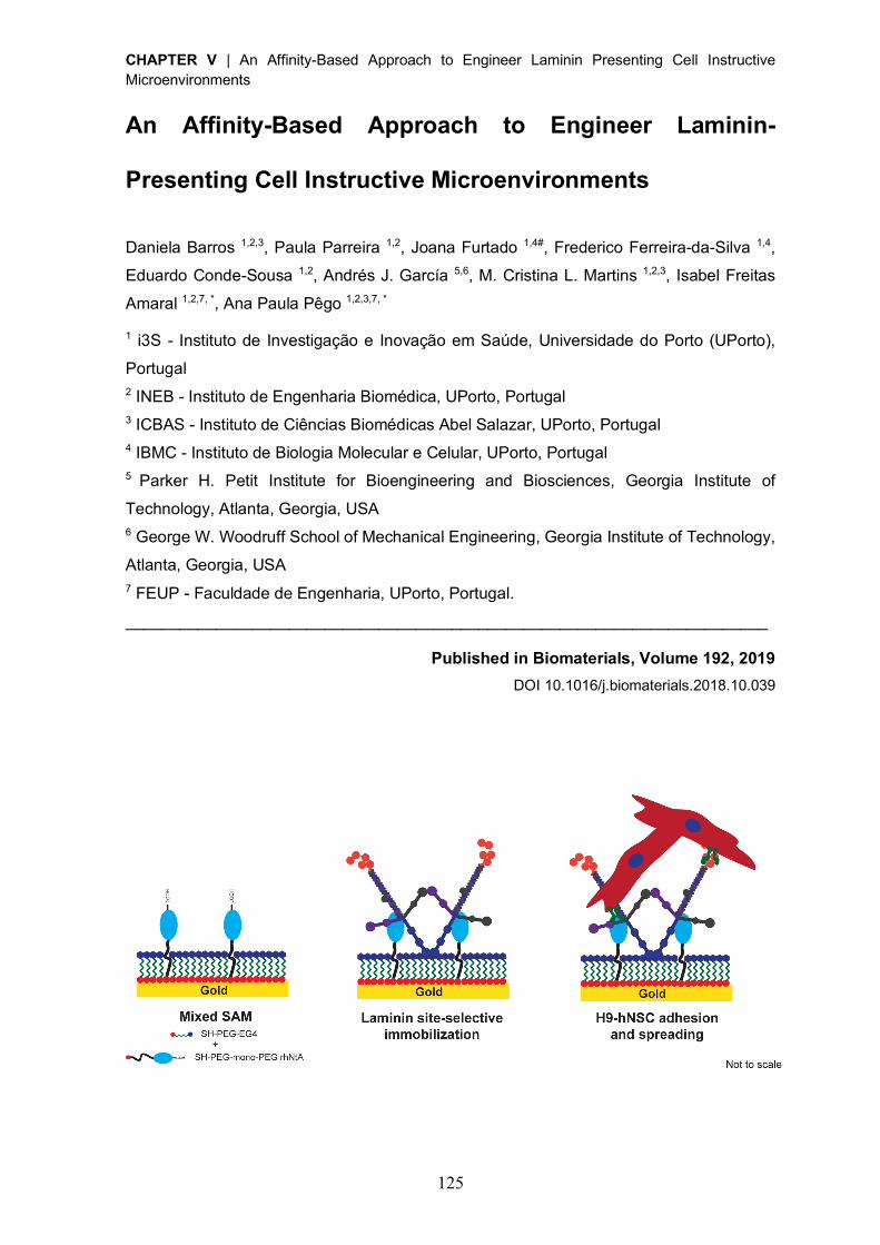
CHAPTER V | An Affinity-Based Approach to Engineer Laminin Presenting Cell Instructive Microenvironments
125
An Affinity-Based Approach to Engineer Laminin-
Presenting Cell Instructive Microenvironments
Daniela Barros 1,2,3, Paula Parreira 1,2, Joana Furtado 1,4#, Frederico Ferreira-da-Silva 1,4,
Eduardo Conde-Sousa 1,2, Andrés J. García 5,6, M. Cristina L. Martins 1,2,3, Isabel Freitas
Amaral 1,2,7, *, Ana Paula Pêgo 1,2,3,7, *
1 i3S - Instituto de Investigação e Inovação em Saúde, Universidade do Porto (UPorto),
Portugal 2 INEB - Instituto de Engenharia Biomédica, UPorto, Portugal 3 ICBAS - Instituto de Ciências Biomédicas Abel Salazar, UPorto, Portugal 4 IBMC - Instituto de Biologia Molecular e Celular, UPorto, Portugal 5 Parker H. Petit Institute for Bioengineering and Biosciences, Georgia Institute of
Technology, Atlanta, Georgia, USA 6 George W. Woodruff School of Mechanical Engineering, Georgia Institute of Technology,
Atlanta, Georgia, USA 7 FEUP - Faculdade de Engenharia, UPorto, Portugal.
_______________________________________________________________________
Published in Biomaterials, Volume 192, 2019
DOI 10.1016/j.biomaterials.2018.10.039
CHAPTER V | An Affinity-Based Approach to Engineer Laminin Presenting Cell Instructive Microenvironments
126
Abstract
Laminin immobilization into diverse biological and synthetic matrices has been explored to
replicate the microenvironment of stem cell niches and gain insight into the role of
extracellular matrix (ECM) on stem cell behavior. However, the site-specific immobilization
of this heterotrimeric glycoprotein and, consequently, control over its orientation and
bioactivity has been a challenge that has limited many of the explored strategies to date. In
this work, we establish an affinity-based approach that takes advantage of the native high
affinity interaction between laminin and the human N-terminal agrin (hNtA) domain. This
interaction is expected to promote the site-selective immobilization of laminin to a specific
substrate, while preserving the exposure of its key bioactive epitopes. Recombinant hNtA
(rhNtA) domain was produced with high purity (>90%) and successfully conjugated at its N-
terminal with a thiol-terminated poly(ethylene glycol) (PEG) without affecting its affinity to
laminin. Self-assembled monolayers (SAMs) of mono-PEGylated rhNtA on gold (mPEG
rhNtA-SAMs) were then prepared to evaluate the effectiveness of this strategy. The site-
specific immobilization of laminin onto mPEG rhNtA-SAMs was shown to better preserve
protein bioactivity in comparison to laminin immobilized on SAMs of thiol-PEG-succinimidyl
glutaramide (SH-PEG-SGA), used for the non-selective covalent immobilization of laminin,
as evidenced by its enhanced ability to efficiently self-polymerize and mediate cell adhesion
and spreading of human neural stem cells. These results highlight the potential of this novel
strategy to be used as an alternative to the conventional immobilization approaches in a
wide range of applications, including engineered coatings for neuroelectrodes and two-
dimensional (2D) substrates for cell culture, as well as biofunctionalization of three-
dimensional (3D) matrices.
CHAPTER V | An Affinity-Based Approach to Engineer Laminin Presenting Cell Instructive Microenvironments
127
1. Introduction
In the framework of regenerative medicine and tissue engineering, much attention has been
devoted towards the development of engineered matrices incorporating bioadhesive cues
present in stem cell niches, to recapitulate the dynamic nature and biological complexity of
these microenvironments, as well as gain more insight into the function of specific
extracellular matrix (ECM) components on stem cell behavior. The ECM is an essential
component of the stem cell niche, as it modulates important biological functions including
proliferation, self-renewal and differentiation of stem cells. Among major ECM constituents,
laminins play crucial and essential roles in many aspects of tissue physiology and function
[1-5]. These heterotrimeric glycoproteins comprise several bioactive domains involved in
the modulation of different biological functions. The latter include the interaction with other
ECM proteins (e.g. nidogen, netrin 4 and collagen VII) mediated by the laminin short arms
(N-terminus), which contributes to the assembly and stability of basement membranes.
These domains are also responsible for the laminin ability to polymerize [6-8], even in the
absence of other basement membrane components, forming the molecular network that will
be in contact with the cellular surface. In addition to its structural role, laminin comprises
multiple bioactive domains that interact with cell surface receptors (e.g. integrins,
dystroglycans, and syndecans), modulating different cell functions including cell adhesion,
proliferation, migration and differentiation, as well as ECM deposition [9-11].
Laminin has been incorporated into both two-dimensional (2D) [12] and three-dimensional
(3D) [13-20] cell-instructive microenvironments for applications in regenerative therapies or
to get insights into the role of laminin on the modulation of cell behavior. Strategies explored
for laminin immobilization have relied either on its non-specific adsorption or entrapment or,
alternatively, on its non-specific covalent immobilization to different substrates through the
use of functional groups present in multiple sites of the laminin structure such as amines
[13-17] and thiols [18, 20]. One of the main caveats presented by these strategies is the
inability to control orientation and conformation of laminin upon immobilization, which were
proven to be crucial for the modulation of cellular behavior [21-23]. As such, the exposure
of key laminin bioactive epitopes can be compromised. In an attempt to assure the control
over the tethering of laminin, in recent years protein immobilization strategies have shifted
toward site-specific conjugation, with special focus on biorthogonal chemical reactions (click
chemistry), enzymatic ligation and affinity binding, using either unnatural amino acids or
engineered site-selective amino acid sequences [24-26]. These strategies are expected to
provide a higher retention of bioactivity, by favoring the access to the active sites of
immobilized proteins. To the best of our knowledge, to date, only one study reported the
site-selective immobilization of laminin [27]. To control the presentation of full-length ECM
CHAPTER V | An Affinity-Based Approach to Engineer Laminin Presenting Cell Instructive Microenvironments
128
proteins without altering their bioactivity, Lee and co-workers explored click chemistry to
anchor collagen, fibronectin and laminin onto polyacrylamide gels by their N-terminus [27].
Nevertheless, despite guaranteeing the site-selective immobilization of the proteins, as
result of the use of laminin N-terminus, this approach compromises one of laminin’s
hallmark features, which is its ability to polymerize. Affinity-binding has been increasingly
explored for the site-specific and reversible conjugation of proteins and peptides, because
of its versatility and ability to generate dynamic biomimetic systems. However, the
successful implementation of this strategy is strongly dependent on the appropriate
selection of the binding pairs. Binding systems using high affinity interactions, such as
streptavidin and biotin, although often used, require the protein of interest to be either
recombinantly or chemically modified [28, 29]. In contrast, the use of natural binding
partners constitutes an attractive alternative, as strong non-covalent interactions can be
achieved without the need for protein modification [30, 31].
In this study, we examined an alternative strategy that explores the well described native
high affinity interaction (dissociation constant (KD) = 5 nM) between laminin and the N-
terminal agrin (NtA) domain [32, 33]. The agrin-binding site in laminin is localized in the
central region of its coiled-coil domain and maps to a sequence of 20 conserved residues
within the g1 chain [33, 34]. Interestingly, this interaction requires a coiled-coil conformation
of the agrin-binding site [34]. To assess the ability of this affinity-based approach to
immobilize laminin with retention of bioactivity, recombinant human NtA (rhNtA) domain was
successfully produced and further conjugated at its N-terminus with a thiol-terminated
poly(ethylene glycol) (PEG) to enable the preparation of self-assembled monolayers
(SAMs) of NtA on gold. To the best of our knowledge, this is the first report on the production
and characterization of the human variant of NtA domain. In order to study the specific
interactions between the immobilized laminin and cells, SAMs were herein used as proof-
of-concept platforms, since they can be easily produced and specifically tailored to provide
a chemically well-defined molecular monolayer [35]. The binding ligand was characterized
and its ability to mediate laminin immobilization through a high affinity interaction was shown
by solid-binding assay, surface plasmon resonance (SPR) and quartz crystal microbalance
with dissipation monitoring (QCM-D). The bioactivity of mono-PEGylated rhNtA-immobilized
laminin was subsequently investigated by evaluating its ability to self-polymerize and
modulate the behavior of human neural stem cells (hNSCs). hNSCs were herein used due
to the well described role of laminin on the modulation of neural cell behavior [36]. This
strategy represents a promising and versatile approach for the site-selective immobilization
of laminin, while preserving its bioactivity, and can be potentially useful for the development
of cell instructive microenvironments for tissue engineering and regenerative medicine.
CHAPTER V | An Affinity-Based Approach to Engineer Laminin Presenting Cell Instructive Microenvironments
129
2. Materials and Methods
2.1. Recombinant human N-terminal agrin (rhNtA) domain expression and
purification
The NtA domain, comprising residues Thr 30 – Pro 157 of the human agrin protein (Uniprot
reference O00468), was expressed in Escherichia coli (E. coli) strain BL21(DE3) using the
pCoofy2 expression vector [37] (gift from Sabine Suppmann – Addgene plasmid # 43981).
pCoofy2 is a derivative of pETM22 vector that contains a thioredoxin-poly-His6 (6x Histidine
residues) (Trx-His6) N-terminal tag followed by an HRV 3C recognition site located
downstream of the N-terminal tag [37]. Details on cloning of human NtA gene and
expression vector are presented in Supplementary Materials and Methods. The fusion
protein (Trx-His6-hNtA) was expressed by induction of log-phase cultures with 0.2 mM of
isopropyl b-D-1-thiogalactopyranoside (IPTG; Sigma-Aldrich) at 20 ºC for 5 h and purified
by immobilized metal affinity chromatography using an HisTrap column (GE Healthcare),
according to manufacturer’s instructions. A concentration gradient of 0 to 500 mM of
imidazole was applied to elute the fusion protein. The N-terminal Trx-His6 tag was then
cleaved during dialysis using HRV 3C protease (kindly provided by Biochemical and
Biophysical Technologies (B2Tech) Platform of i3S) in a protease:target protein ratio
(unit/µg) of 1:10. Untagged recombinant human NtA (rhNtA) contains additional Gly and
Pro residues at its N-terminus (Fig. S1). rhNtA was dialyzed against 10 mM phosphate
buffered saline (PBS) pH 7.4, concentrated in a 10 kDa molecular weight cut-off (MWCO)
ultrafiltration membrane (GE Healthcare) and stored at -80 ºC until further use. Final domain
concentration was estimated by measuring the absorbance at 280 nm. The purity of the
rhNtA domain was assessed by sodium dodecyl sulfate–polyacrylamide gel electrophoresis
(SDS-PAGE) analysis and Western blotting using the anti-agrin antibody - rabbit polyclonal
agrin antibody (1:5000; Abcam, ab85174). The rhNtA molecular weight was determined by
mass spectrometry, as described in section 2.7.
2.2. Solid-phase binding assay
Binding of rhNtA to immobilized laminin was assessed performing an enzyme-linked binding
assay (ELISA). 96-well microtiter ELISA plates (BD Falcon) were coated with poly(D-lysine)
(PDL; 20 μg/mL; Sigma-Aldrich) and then incubated with laminin-111 from mouse
Engelbreth-Holm-Swarm sarcoma (msLn-111;10 µg/mL; Sigma-Aldrich) in 50 mM sodium
bicarbonate buffer pH 9.6 (overnight, 4 ºC). After blocking with 2.5% (w/v) bovine serum
albumin (BSA; Biowest; 1.5 h, room temperature (RT)), wells were incubated with 2-fold
serial dilutions of rhNtA (0.1 – 100 nM; 2 h, 37 ºC). Plates were then washed with
PBS/0.05% (v/v) Tween® 20 and incubated with rabbit polyclonal agrin antibody (1:5000; 2
CHAPTER V | An Affinity-Based Approach to Engineer Laminin Presenting Cell Instructive Microenvironments
130
h, RT). A goat anti-rabbit IgG (H+L) coupled to horseradish peroxidase (HRP) (1:2000; Life
Technologies, A16023) was used as secondary antibody, and color was developed using
3,3’,5,5’ tetramethyl benzidine (TMB) as substrate (Biolegend; 30 min, RT). The reaction
was stopped with 2 M H2SO4 and the absorbance measured at 450 nm (BioTek® SynergyTM
Mx). The dissociation constant (KD) value of rhNtA to msLn-111 was estimated by non-
linear regression analysis using GraphPad Prism 6 software (San Diego).
2.3. Surface plasmon resonance (SPR)
Real-time interactions between rhNtA and immobilized laminin were detected by SPR at 25
ºC using a BIACORE X100 (GE Healthcare). Laminin was immobilized on a CM5 sensor
chip (GE Healthcare) using amine-coupling chemistry. Briefly, the flow cells surface was
activated with a 1:1 mixture of 0.1 M N-hydroxysuccinimide (NHS; Sigma-Aldrich) and 0.4
M 3-(N,N-dimethylamino) propyl-N-ethylcarbodiimide (EDC; Sigma-Aldrich) at a flow rate of
10 µL/min. msLn-111 or recombinant human laminin-521 (rhLn-521; Biolamina) (50 µg/mL
in 10 mM sodium acetate pH 3.5) were immobilized at a density of 4000 resonance units.
The reactive groups in excess were then deactivated with 1 M ethanolamine-HCl pH 8.5
(Sigma-Aldrich). For binding measurements, 5-fold serial dilutions of rhNtA (500 – 0.8 nM)
were prepared in 10 mM 4-(2-hydroxyethyl)-1-piperazineethanesulfonic acid (HEPES; pH
7.4, containing 150 mM sodium chloride (NaCl; VWR), 3 mM Ethylenediamine tetra-acetic
acid (Sigma-Aldrich) and 0.005% (v/v) surfactant P20 (GE Healthcare)) and flowed at 30
µL/min onto the flow cells. Proteins were allowed to associate and dissociate for 120 and
1600 s, respectively. Experimental results were fitted with Langmuir 1:1 binding kinetics
within Biacore X100 Evaluation software version 2.0.1 (GE Healthcare).
2.4. Culture of H9 – Derived Human Neural Stem Cells
Human neural stem cells (hNSCs) derived from the National Institutes of Health (NIH)
approved H9 (WA09) human embryonic stem cells were purchased from Life Technologies
(N7800-200). Cells were expanded according to the manufacturer’s protocol in
poly(ornithine)/msLn-111-coated tissue culture-treated plates in serum-free StemProÒ NSC
SFM growth medium (Life Technologies).
2.5. Cell adhesion centrifugation assay
To evaluate the ability of rhNtA-immobilized msLn-111 to promote the adhesion of hNSCs,
a centrifugation cell adhesion assay was performed [38]. Here, controllable and
reproducible detachment forces were applied to adherent cells, providing relative
measurements of adhesion strength [38]. 96-well tissue culture plates (Corning) were
coated with increased concentrations of rhNtA (0 – 40 µg/mL) overnight at 4 ºC and blocked
CHAPTER V | An Affinity-Based Approach to Engineer Laminin Presenting Cell Instructive Microenvironments
131
with 1% (w/v) BSA for 1 h at RT to prevent non-specific adhesion to the substrate. msLn-
111 was then added at a fixed concentration (10 µg/mL), the plate incubated for 2 h at 37
ºC and hNSC adhesion assessed as described in Supplementary Materials and Methods.
Wells incubated with 1% (w/v) BSA or 20 µg/mL poly(ornithine) / 10 µg/mL msLn-111 were
used as negative and positive control, respectively. To assess the effect of the domain per
se on cell adhesion, wells incubated with 10 µg/mL rhNtA were used.
2.6. N-terminal PEGylation of rhNtA
A 5 mg/mL rhNtA solution in 0.1 M sodium phosphate (NaH2PO4) pH 6.5 was added to thiol-
PEG-succinimidyl glutaramide (SH-PEG-SGA; 3.5 kDa; purity > 90%, Jenkem USA) at a
protein:PEG molar ratio of 1:2. The reaction was allowed to proceed for 4 h at 4 ºC and then
quenched with 2 M hydroxylamine pH 7.4. The solution was diluted to a final protein
concentration of 0.5 mg/mL with 1 mM HCl and the pH adjusted to 3.5 with 1 M HCl. The
reaction mixture was then diluted (100×) with 20 mM sodium acetate pH 4.0 and loaded
onto a HiTrap SP HP cation exchange column (GE Healthcare). A gradient of 0 to 1 M NaCl
in 20 mM sodium acetate pH 4.0 was used to elute the different PEGylated fractions. The
peak corresponding to the mono-N-terminal conjugate was dialyzed against 10 mM PBS
pH 7.4, concentrated in a 10 kDa MWCO ultrafiltration membrane (GE Healthcare) and
stored at -80 ºC until further use. Protein concentration was estimated by measuring the
absorbance at 280 nm and the conjugate was characterized by SDS-PAGE and MALDI-
TOF mass spectrometry (MALDI-TOF MS).
2.7. Protein and PEGylated conjugate characterization by mass spectrometry
The native or mono-PEGylated rhNtA, as well as unmodified SH-PEG-SGA, were analyzed
by MALDI-TOF MS. Briefly, samples were diluted 1:4 (v/v) in MALDI matrix (sinapinic acid
10 mg/mL, 50% acetonitrile, 0.1% trifluoroacetic acid, all purchased from Sigma-Aldrich),
spotted onto a target plate and analyzed in linear positive mode. Mass spectra were
internally calibrated with thioredoxin (m/z 11674) and apomyoglobin (m/z 16952). To
characterize the site of PEG conjugation to rhNtA, peptide mapping of native and mono-
PEGylated rhNtA was performed using a previously published procedure [39], which is
briefly described in Supplementary Materials and Methods.
2.8. Self-assembled monolayers (SAMs) preparation
Mixed SAMs of mono-PEGylated rhNtA (mPEG rhNtA-SAM) or SH-PEG-SGA (SGA-SAM)
were prepared on gold-coated substrates (0.5 × 0.5 cm2), obtained from Instituto de
Engenharia de Sistemas e Computadores – Microsistemas e Nanotecnologias, Portugal
(INESC-MN). Prior to use, gold surfaces were cleaned as described elsewhere [40]. Gold
CHAPTER V | An Affinity-Based Approach to Engineer Laminin Presenting Cell Instructive Microenvironments
132
substrates were then incubated in ethanolic solutions (99.9%; Merck-Millipore) of either
mono-PEGylated rhNtA (2.5, 5.0 or 10.0 µM), for laminin affinity binding; or SH-PEG-SGA
(5.0 µM), for non-selective covalent immobilization of laminin. After 4 h at RT and under
inert conditions (glove chamber saturated with dry nitrogen), SAMs were rinsed with
absolute ethanol to remove any physically adsorbed molecules. For proper monolayer
packing mPEG rhNtA- and SGA- SAMs were subsequently incubated with a 100.0 µM
absolute ethanolic solution of (11-mercaptoundecyl) tetraethylene glycol (EG4, SensoPath
Technologies) for 16 h at RT and under the inert conditions mentioned above. Afterwards,
SAMs were thoroughly rinsed with absolute ethanol (99.9%; Merck-Millipore). EG4-SAMs,
used as control surfaces, were prepared by immersing cleaned gold substrates in a 100.0
µM ethanolic solution of EG4 for 16 h at RT and under inert conditions.
2.9. Quartz crystal microbalance with dissipation monitoring (QCM-D)
Gold-coated QCM-D sensors (Biolin Scientific) were cleaned as previously described [41].
Clean sensors were further modified following the procedure reported in 2.8. A QCM-D
system (Q-Sense E4 instrument, Biolin Scientific) was used to monitor in real time the
frequency (Δf) and dissipation (ΔD) shifts related to laminin adsorption as described in
Supplementary Materials and Methods. Data was modeled using the Voigt model [41] in
the QTools® V3 software. Results are presented as mass per surface area (ng/cm2).
2.10. Laminin self-polymerization
mPEG rhNtA-SAMs were equilibrated in 10 mM HEPES pH 7.4, to favor the affinity
interaction with laminin, while SGA-SAMs were equilibrated in 100 mM sodium bicarbonate
pH 8.5, to favor the non-selective covalent interaction with laminin amine groups, and then
incubated with msLn-111 (20 µg/mL; 2 h, RT). The substrates were then thoroughly
washed, to remove any unbound laminin. To assess the ability of laminin to self-polymerize,
a msLn-111 solution (50 µg/mL) in 20 mM Tris-HCl pH 7.0, containing 1 mM CaCl2, was
used. The laminin concentration used was below the critical protein concentration
necessary to trigger laminin polymerization in neutral pH solution [6]. Mixed SAMs with
affinity- (mPEG rhNtA-SAMs) or covalent-bound (SGA-SAMs) msLn-111 were incubated
with the laminin solution for 24 h at 37 ºC [42]. Finally, samples were processed for laminin
immunostaining, as described in Supplementary Materials and Methods, and observed
under confocal laser scanning microscopy (CLSM) (TCS SP5 II; Leica Mycrosystems),
using a Plan-Apochromat 63´/1.4NA oil objective. A z-section of 15 µm was acquired and
subsequently projected into a 2D image. Processing and quantitative analysis of the
acquired images were performed in ImageJ/Fiji and MATLAB® software, respectively, as
described in detail in the Supplementary Materials and Methods.
CHAPTER V | An Affinity-Based Approach to Engineer Laminin Presenting Cell Instructive Microenvironments
133
2.11. Cell adhesion and morphology
To determine the impact of the immobilization approach on the cell adhesion-promoting
activity of laminin, mPEG rhNtA- or SGA-SAMs were equilibrated in 10 mM HEPES pH 7.4
and then incubated for 2 h at RT with rhLn-521 (20 µg/mL). After being washed with 10 mM
HEPES pH 7.4 to remove unbound laminin, hNSCs (50,000 viable cells/cm2) were added
to the substrates and incubated at 37 ºC with 5% CO2 for 24 h. Samples were processed
for F-actin/DNA staining, as described in Supplementary Materials and Methods, and
imaged with the IN Cell Analyzer 2000 imaging system (GE Healthcare) using a Nikon
20×/0.45 NA Plan Fluor objective. A z-section of 45 µm was acquired per field of view (FOV)
and then projected into a 2D image. Images spanning a total area of 4.6 mm2 were analyzed
using CellProfiler image analysis software [43]. Briefly, nuclei were segmented through an
Otsu thresholding method from the 4’,6-diamidino-2-phenylindole (DAPI) channel while
cells cytoplasm was segmented from the actin channel through a propagation from the
previously identified nucleus, using a minimum cross entropy threshold. Measurements
were obtained for total number of adhered cells, average cell spreading area (µm2) and
average occupied area (µm2) per field of view (FOV). Representative images for each
condition were also obtained by CLSM using a Plan-Apochromat 63×/1.4NA oil objective.
A z-section of 12 µm was acquired and subsequently projected into a 2D image.
2.12. Statistical Analysis
Individual experiments with biological replicates were performed at least in duplicate and
statistical analysis was performed using GraphPad Prism 6 software (San Diego).
Statistically significant differences between two conditions were detected using a non-
parametric Mann Whitney U-test. Comparisons between three or more groups were
performed with one-way ANOVA analysis, followed by the Bonferroni correction for pairwise
comparisons or the Dunnett’s two-tailed test for comparisons with the control condition. For
all analyses, differences were considered significant at p < 0.05.
3. Results and Discussion
3.1. Recombinant human N-terminal agrin (rhNtA) domain was successfully
produced
In contrast with most of reports published to date in which the NtA used was derived from
chicken agrin [32, 33], we produced the human variant of this domain because of its higher
clinical relevance. This is of particular interest when envisaging the use of this domain in
platforms for human-disease modelling, cell culture/delivery or engineered coatings for
neuroelectrodes.
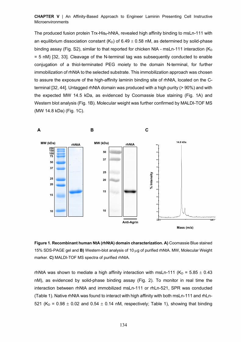
CHAPTER V | An Affinity-Based Approach to Engineer Laminin Presenting Cell Instructive Microenvironments
134
The produced fusion protein Trx-His6-hNtA, revealed high affinity binding to msLn-111 with
an equilibrium dissociation constant (KD) of 6.49 ± 0.58 nM, as determined by solid-phase
binding assay (Fig. S2), similar to that reported for chicken NtA - msLn-111 interaction (KD
= 5 nM) [32, 33]. Cleavage of the N-terminal tag was subsequently conducted to enable
conjugation of a thiol-terminated PEG moiety to the domain N-terminal, for further
immobilization of rhNtA to the selected substrate. This immobilization approach was chosen
to assure the exposure of the high-affinity laminin binding site of rhNtA, located on the C-
terminal [32, 44]. Untagged rhNtA domain was produced with a high purity (> 90%) and with
the expected MW 14.5 kDa, as evidenced by Coomassie blue staining (Fig. 1A) and
Western blot analysis (Fig. 1B). Molecular weight was further confirmed by MALDI-TOF MS
(MW 14.8 kDa) (Fig. 1C).
Figure 1. Recombinant human NtA (rhNtA) domain characterization. A) Coomassie Blue stained
15% SDS-PAGE gel and B) Western-blot analysis of 10 µg of purified rhNtA. MW, Molecular Weight
marker. C) MALDI-TOF MS spectra of purified rhNtA.
rhNtA was shown to mediate a high affinity interaction with msLn-111 (KD = 5.85 ± 0.43
nM), as evidenced by solid-phase binding assay (Fig. 2). To monitor in real time the
interaction between rhNtA and immobilized msLn-111 or rhLn-521, SPR was conducted
(Table 1). Native rhNtA was found to interact with high affinity with both msLn-111 and rhLn-
521 (KD = 0.98 ± 0.02 and 0.54 ± 0.14 nM, respectively; Table 1), showing that binding
A B C
13324. 0 16696. 2 20068. 4 23440. 6 26812. 8 30185. 0Mass (m/ z)
467. 3
0
10
20
30
40
50
60
70
80
90
100
% I
nten
sity
T OF /T OF ™ L in e a r Sp e c # 1 M C[BP = 1 4 7 5 4 .9 , 4 6 7 ]
% I
nte
nsit
y
Mass (m/z)
14.8 kDa rhNtA MW (kDa)
10
15
20 25
37 50 75 150 100 250
rhNtA MW (kDa)
10
15
20 25
37 50
Anti-Agrin
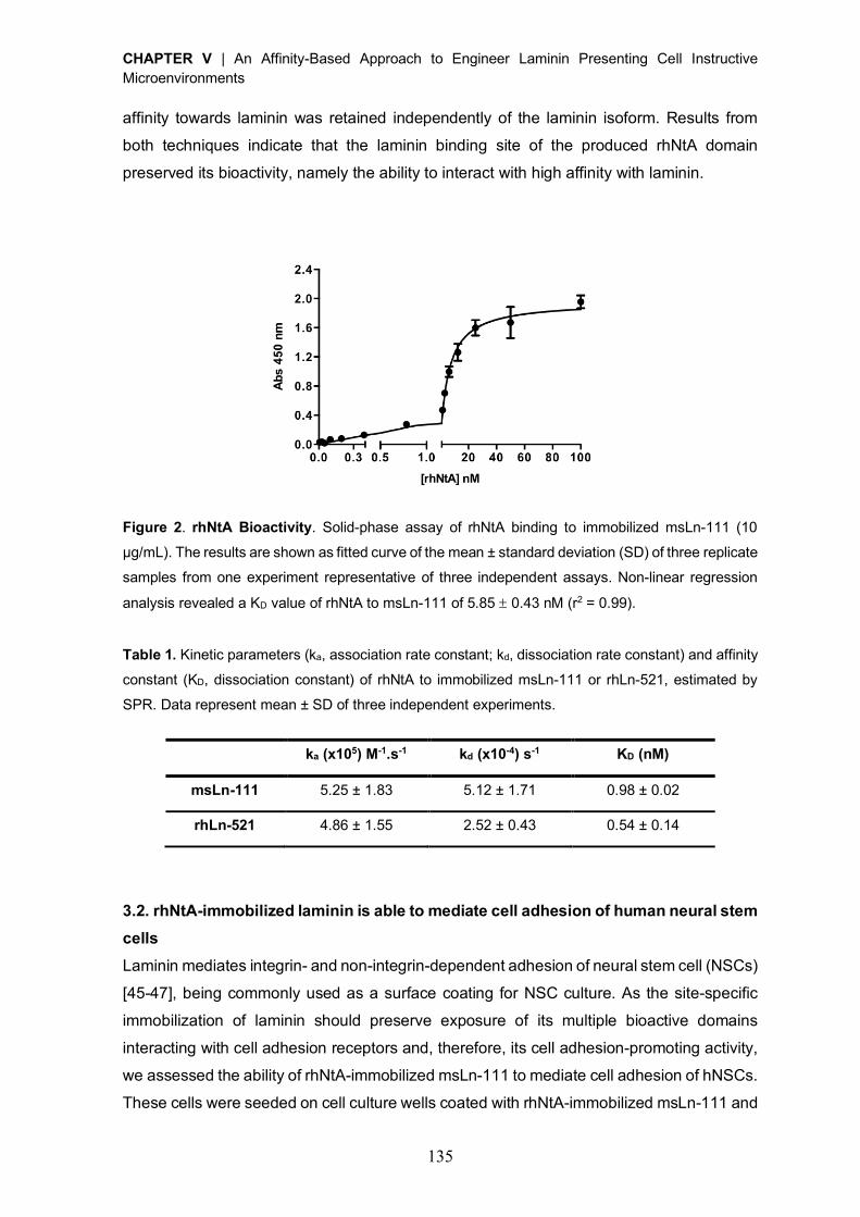
CHAPTER V | An Affinity-Based Approach to Engineer Laminin Presenting Cell Instructive Microenvironments
135
affinity towards laminin was retained independently of the laminin isoform. Results from
both techniques indicate that the laminin binding site of the produced rhNtA domain
preserved its bioactivity, namely the ability to interact with high affinity with laminin.
Figure 2. rhNtA Bioactivity. Solid-phase assay of rhNtA binding to immobilized msLn-111 (10
µg/mL). The results are shown as fitted curve of the mean ± standard deviation (SD) of three replicate
samples from one experiment representative of three independent assays. Non-linear regression
analysis revealed a KD value of rhNtA to msLn-111 of 5.85 ± 0.43 nM (r2 = 0.99).
Table 1. Kinetic parameters (ka, association rate constant; kd, dissociation rate constant) and affinity
constant (KD, dissociation constant) of rhNtA to immobilized msLn-111 or rhLn-521, estimated by
SPR. Data represent mean ± SD of three independent experiments.
ka (x105) M-1.s-1 kd (x10-4) s-1 KD (nM)
msLn-111 5.25 ± 1.83 5.12 ± 1.71 0.98 ± 0.02
rhLn-521 4.86 ± 1.55 2.52 ± 0.43 0.54 ± 0.14
3.2. rhNtA-immobilized laminin is able to mediate cell adhesion of human neural stem
cells
Laminin mediates integrin- and non-integrin-dependent adhesion of neural stem cell (NSCs)
[45-47], being commonly used as a surface coating for NSC culture. As the site-specific
immobilization of laminin should preserve exposure of its multiple bioactive domains
interacting with cell adhesion receptors and, therefore, its cell adhesion-promoting activity,
we assessed the ability of rhNtA-immobilized msLn-111 to mediate cell adhesion of hNSCs.
These cells were seeded on cell culture wells coated with rhNtA-immobilized msLn-111 and
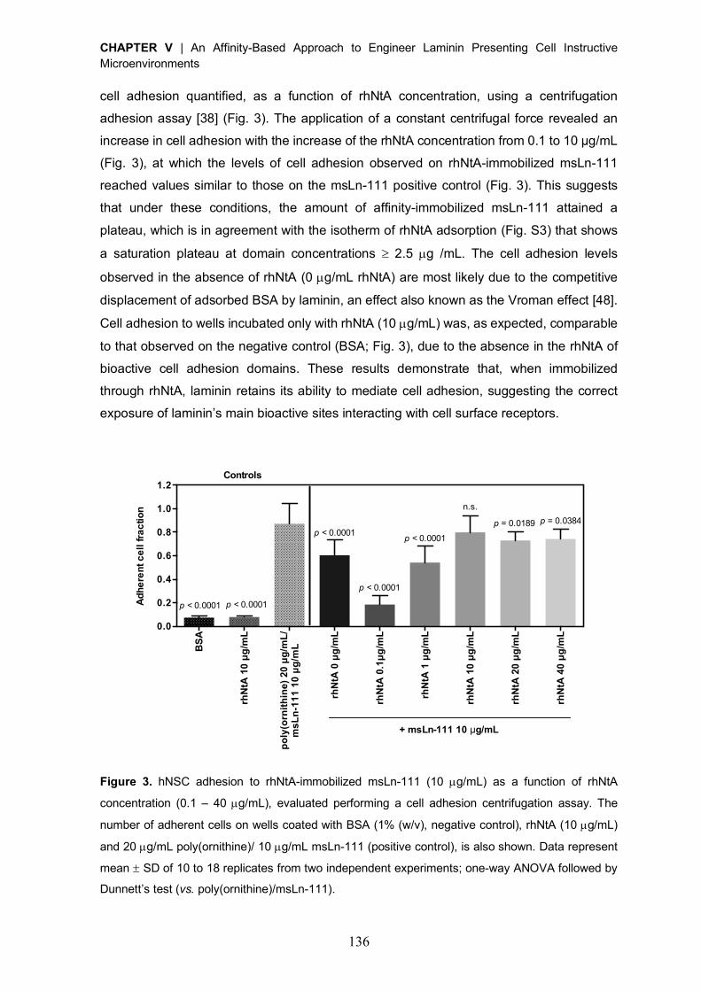
CHAPTER V | An Affinity-Based Approach to Engineer Laminin Presenting Cell Instructive Microenvironments
136
cell adhesion quantified, as a function of rhNtA concentration, using a centrifugation
adhesion assay [38] (Fig. 3). The application of a constant centrifugal force revealed an
increase in cell adhesion with the increase of the rhNtA concentration from 0.1 to 10 μg/mL
(Fig. 3), at which the levels of cell adhesion observed on rhNtA-immobilized msLn-111
reached values similar to those on the msLn-111 positive control (Fig. 3). This suggests
that under these conditions, the amount of affinity-immobilized msLn-111 attained a
plateau, which is in agreement with the isotherm of rhNtA adsorption (Fig. S3) that shows
a saturation plateau at domain concentrations ³ 2.5 µg /mL. The cell adhesion levels
observed in the absence of rhNtA (0 µg/mL rhNtA) are most likely due to the competitive
displacement of adsorbed BSA by laminin, an effect also known as the Vroman effect [48].
Cell adhesion to wells incubated only with rhNtA (10 µg/mL) was, as expected, comparable
to that observed on the negative control (BSA; Fig. 3), due to the absence in the rhNtA of
bioactive cell adhesion domains. These results demonstrate that, when immobilized
through rhNtA, laminin retains its ability to mediate cell adhesion, suggesting the correct
exposure of laminin’s main bioactive sites interacting with cell surface receptors.
Figure 3. hNSC adhesion to rhNtA-immobilized msLn-111 (10 µg/mL) as a function of rhNtA
concentration (0.1 – 40 µg/mL), evaluated performing a cell adhesion centrifugation assay. The
number of adherent cells on wells coated with BSA (1% (w/v), negative control), rhNtA (10 µg/mL)
and 20 µg/mL poly(ornithine)/ 10 µg/mL msLn-111 (positive control), is also shown. Data represent
mean ± SD of 10 to 18 replicates from two independent experiments; one-way ANOVA followed by
Dunnett’s test (vs. poly(ornithine)/msLn-111).
BS
A
rhN
tA 1
0 µg
/mL
poly
(orn
ithin
e) 2
0 µg
/mL/
msL
n-1
11 1
0 µ
g/m
L
rhN
tA 0
µg/
mL
rhN
tA 0
.1µg
/mL
rhN
tA 1
µg/
mL
rhN
tA 1
0 µg
/mL
rhN
tA 2
0 µg
/mL
rhN
tA 4
0 µg
/mL
0.0
0.2
0.4
0.6
0.8
1.0
1.2
Adh
eren
t cel
l fra
ctio
n
Immobilization kinetics
p = 0.0189
n.s.
Controls
+ msLn-111 10 µg/mL
p < 0.0001
p < 0.0001 p < 0.0001
p < 0.0001 p < 0.0001
p = 0.0384
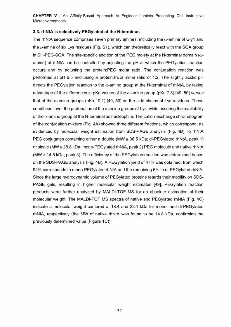
CHAPTER V | An Affinity-Based Approach to Engineer Laminin Presenting Cell Instructive Microenvironments
137
3.3. rhNtA is selectively PEGylated at the N-terminus
The rhNtA sequence comprises seven primary amines, including the a-amine of Gly1 and
the e-amine of six Lys residues (Fig. S1), which can theoretically react with the SGA group
in SH-PEG-SGA. The site-specific addition of the PEG moiety at the N-terminal domain (a-
amine) of rhNtA can be controlled by adjusting the pH at which the PEGylation reaction
occurs and by adjusting the protein:PEG molar ratio. The conjugation reaction was
performed at pH 6.5 and using a protein:PEG molar ratio of 1:2. The slightly acidic pH
directs the PEGylation reaction to the a-amino group at the N-terminal of rhNtA, by taking
advantage of the differences in pKa values of the a-amino group (pKa 7.8) [49, 50] versus
that of the e-amino groups (pKa 10.1) [49, 50] on the side chains of Lys residues. These
conditions favor the protonation of the e-amino groups of Lys, while assuring the availability
of the a-amino group at the N-terminal as nucleophile. The cation-exchange chromatogram
of the conjugation mixture (Fig. 4A) showed three different fractions, which correspond, as
evidenced by molecular weight estimation from SDS-PAGE analysis (Fig. 4B), to rhNtA
PEG conjugates containing either a double (MW @ 36.5 kDa; di-PEGylated rhNtA, peak 1)
or single (MW @ 28.8 kDa; mono-PEGylated rhNtA, peak 2) PEG molecule and native rhNtA
(MW @ 14.5 kDa, peak 3). The efficiency of the PEGylation reaction was determined based
on the SDS-PAGE analysis (Fig. 4B). A PEGylation yield of 47% was obtained, from which
94% corresponds to mono-PEGylated rhNtA and the remaining 6% to di-PEGylated rhNtA.
Since the large hydrodynamic volume of PEGylated proteins retards their mobility on SDS-
PAGE gels, resulting in higher molecular weight estimates [49], PEGylation reaction
products were further analyzed by MALDI-TOF MS for an absolute estimation of their
molecular weight. The MALDI-TOF MS spectra of native and PEGylated rhNtA (Fig. 4C)
indicate a molecular weight centered at 18.4 and 22.1 kDa for mono- and di-PEGylated
rhNtA, respectively (the MW of native rhNtA was found to be 14.8 kDa, confirming the
previously determined value (Figure 1C)).
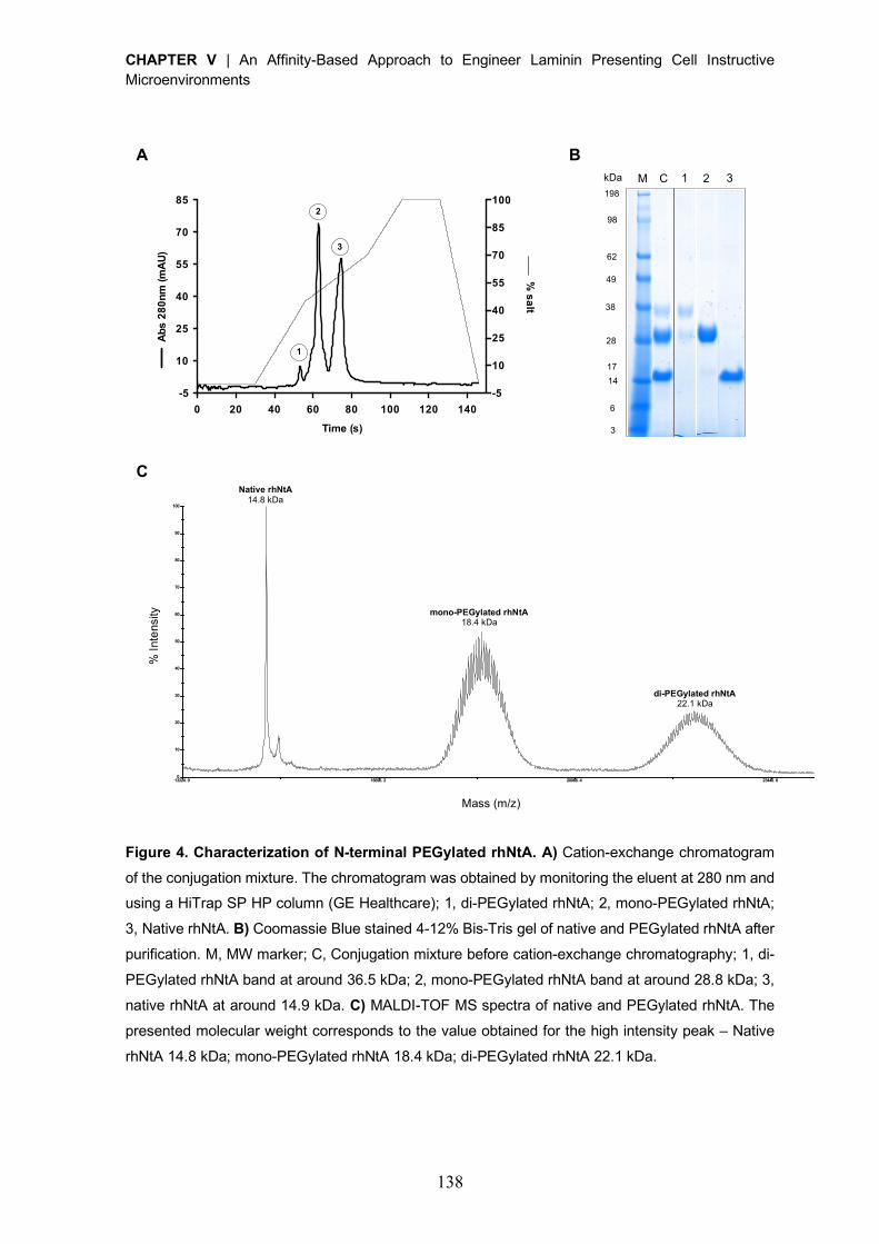
CHAPTER V | An Affinity-Based Approach to Engineer Laminin Presenting Cell Instructive Microenvironments
138
Figure 4. Characterization of N-terminal PEGylated rhNtA. A) Cation-exchange chromatogram
of the conjugation mixture. The chromatogram was obtained by monitoring the eluent at 280 nm and using a HiTrap SP HP column (GE Healthcare); 1, di-PEGylated rhNtA; 2, mono-PEGylated rhNtA;
3, Native rhNtA. B) Coomassie Blue stained 4-12% Bis-Tris gel of native and PEGylated rhNtA after
purification. M, MW marker; C, Conjugation mixture before cation-exchange chromatography; 1, di-
PEGylated rhNtA band at around 36.5 kDa; 2, mono-PEGylated rhNtA band at around 28.8 kDa; 3,
native rhNtA at around 14.9 kDa. C) MALDI-TOF MS spectra of native and PEGylated rhNtA. The
presented molecular weight corresponds to the value obtained for the high intensity peak – Native
rhNtA 14.8 kDa; mono-PEGylated rhNtA 18.4 kDa; di-PEGylated rhNtA 22.1 kDa.
0 20 40 60 80 100 120 140-5
10
25
40
55
70
85
-5
10
25
40
55
70
85
100
Time (s)
Abs
280n
m (m
AU)
% salt1
2
3
13324. 0 16696. 2 20068. 4 23440. 6 26812. 8 30185. 0Mass (m/ z)
467. 3
0
10
20
30
40
50
60
70
80
90
100
% I
nten
sity
T OF /T OF ™ L in e a r Sp e c # 1 M C[BP = 1 4 7 5 4 .9 , 4 6 7 ]
% In
tens
ity
Mass (m/z)
Native rhNtA 14.8 kDa
mono-PEGylated rhNtA 18.4 kDa
di-PEGylated rhNtA 22.1 kDa
A B
C
M C 1 2 3 kDa 198 98
62 49 38
28 17 14 6 3
CHAPTER V | An Affinity-Based Approach to Engineer Laminin Presenting Cell Instructive Microenvironments
139
The position of the conjugated PEG moiety on the purified mono-PEGylated rhNtA
conjugate was further determined by peptide mapping, comparing the fingerprint of native-
and PEGylated rhNtA. The majority of the theoretical fragments, as well as the rhNtA N-
terminus were experimentally detected in both samples (Table S2). Also, their molecular
masses, which were experimentally determined by MALDI-TOF MS, are very similar to the
predicted values obtained in web.expasy.org/peptide_mass (Table S2). Two peaks
corresponding to the N-terminus of rhNtA domain were identified in both native and mono-
PEGylated rhNtA – GPTCPER (F1) [1-7] m/z 816.3677 (Table S2, Fig. S4A) and
GPTCPERALER (F2) [1-11] m/z 1285.6278 (Table S2, Fig. S4B). In the conjugated sample,
a significant decrease in the signal-to-noise ratio was observed for F1, while the peak
correspondent to F2 was not detected. These evidences demonstrate that the a-amine of
Gly1 was selectively modified with the PEG moiety.
3.4. N-terminal PEGylated rhNtA binds with high affinity to laminin
SPR analysis was conducted to evaluate the impact of N-terminal PEGylation on rhNtA
laminin binding ability. N-terminal PEGylation led to a decrease of the binding affinity for
both msLn-111 (KD = 3.36 ± 0.11 nM) and rhLn-521 (KD = 1.58 ± 0.41 nM) (Table 2),
estimated as a 3.4- and 2.9- fold decrease, respectively, compared to native rhNtA (Table
1). This suggests that PEG conjugation to rhNtA slowed down the rate of domain
association (ka) towards immobilized laminin, while the dissociation rate (kd) was not
affected (Table 2). Interestingly, the affinity of both native (Table 1) and mono-PEGylated
rhNtA (Table 2) for rhLn-521 was found to be slightly higher than that determined for msLn-
111. Moreover, the complex formed with this laminin isoform appears to be more stable, as
evidenced by higher kd values (Table 1 and 2). Previous studies suggest that the affinity
between NtA and laminin is regulated by laminin chain composition, and that although the
agrin binding site is located within the g1 chain, a and b laminin chains may also contribute
to the binding, even though to a lesser extent, [32, 33, 51]. Therefore, the differences
observed in the binding affinity of native and mono-PEGylated rhNtA to msLn-111 and rhLn-
521 can be related to the chain composition of the laminin isoforms tested.
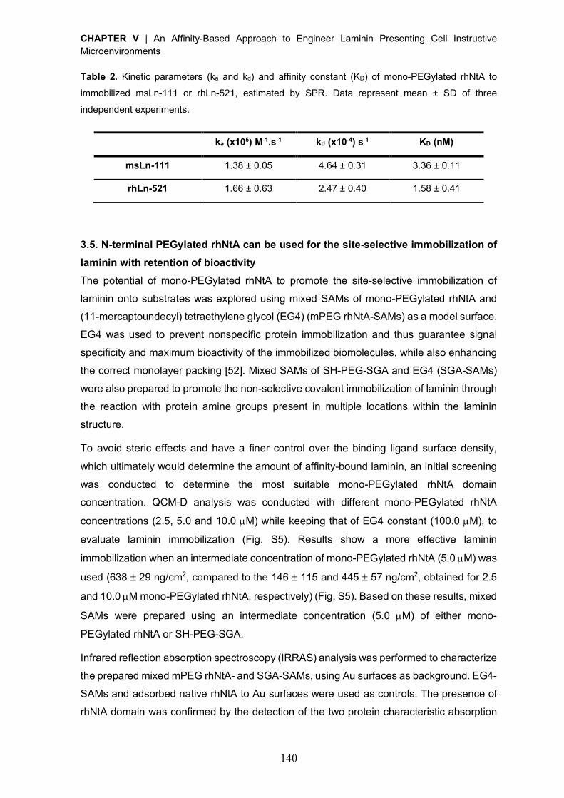
CHAPTER V | An Affinity-Based Approach to Engineer Laminin Presenting Cell Instructive Microenvironments
140
Table 2. Kinetic parameters (ka and kd) and affinity constant (KD) of mono-PEGylated rhNtA to
immobilized msLn-111 or rhLn-521, estimated by SPR. Data represent mean ± SD of three
independent experiments.
ka (x105) M-1.s-1 kd (x10-4) s-1 KD (nM)
msLn-111 1.38 ± 0.05 4.64 ± 0.31 3.36 ± 0.11
rhLn-521 1.66 ± 0.63 2.47 ± 0.40 1.58 ± 0.41
3.5. N-terminal PEGylated rhNtA can be used for the site-selective immobilization of
laminin with retention of bioactivity
The potential of mono-PEGylated rhNtA to promote the site-selective immobilization of
laminin onto substrates was explored using mixed SAMs of mono-PEGylated rhNtA and
(11-mercaptoundecyl) tetraethylene glycol (EG4) (mPEG rhNtA-SAMs) as a model surface.
EG4 was used to prevent nonspecific protein immobilization and thus guarantee signal
specificity and maximum bioactivity of the immobilized biomolecules, while also enhancing
the correct monolayer packing [52]. Mixed SAMs of SH-PEG-SGA and EG4 (SGA-SAMs)
were also prepared to promote the non-selective covalent immobilization of laminin through
the reaction with protein amine groups present in multiple locations within the laminin
structure.
To avoid steric effects and have a finer control over the binding ligand surface density,
which ultimately would determine the amount of affinity-bound laminin, an initial screening
was conducted to determine the most suitable mono-PEGylated rhNtA domain
concentration. QCM-D analysis was conducted with different mono-PEGylated rhNtA
concentrations (2.5, 5.0 and 10.0 µM) while keeping that of EG4 constant (100.0 µM), to
evaluate laminin immobilization (Fig. S5). Results show a more effective laminin
immobilization when an intermediate concentration of mono-PEGylated rhNtA (5.0 µM) was
used (638 ± 29 ng/cm2, compared to the 146 ± 115 and 445 ± 57 ng/cm2, obtained for 2.5
and 10.0 µM mono-PEGylated rhNtA, respectively) (Fig. S5). Based on these results, mixed
SAMs were prepared using an intermediate concentration (5.0 µM) of either mono-
PEGylated rhNtA or SH-PEG-SGA.
Infrared reflection absorption spectroscopy (IRRAS) analysis was performed to characterize
the prepared mixed mPEG rhNtA- and SGA-SAMs, using Au surfaces as background. EG4-
SAMs and adsorbed native rhNtA to Au surfaces were used as controls. The presence of
rhNtA domain was confirmed by the detection of the two protein characteristic absorption
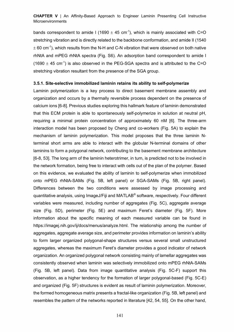
CHAPTER V | An Affinity-Based Approach to Engineer Laminin Presenting Cell Instructive Microenvironments
141
bands correspondent to amide I (1690 ± 45 cm-1), which is mainly associated with C=O
stretching vibration and is directly related to the backbone conformation, and amide II (1540
± 60 cm-1), which results from the N-H and C-N vibration that were observed on both native
rhNtA and mPEG rhNtA spectra (Fig. S6). An adsorption band correspondent to amide I
(1690 ± 45 cm-1) is also observed in the PEG-SGA spectra and is attributed to the C=O
stretching vibration resultant from the presence of the SGA group.
3.5.1. Site-selective immobilized laminin retains its ability to self-polymerize
Laminin polymerization is a key process to direct basement membrane assembly and
organization and occurs by a thermally reversible process dependent on the presence of
calcium ions [6-8]. Previous studies exploring this hallmark feature of laminin demonstrated
that this ECM protein is able to spontaneously self-polymerize in solution at neutral pH,
requiring a minimal protein concentration of approximately 60 nM [6]. The three-arm
interaction model has been proposed by Cheng and co-workers (Fig. 5A) to explain the
mechanism of laminin polymerization. This model proposes that the three laminin N-
terminal short arms are able to interact with the globular N-terminal domains of other
laminins to form a polygonal network, contributing to the basement membrane architecture
[6-8, 53]. The long arm of the laminin heterotrimer, in turn, is predicted not to be involved in
the network formation, being free to interact with cells out of the plan of the polymer. Based
on this evidence, we evaluated the ability of laminin to self-polymerize when immobilized
onto mPEG rhNtA-SAMs (Fig. 5B, left panel) or SGA-SAMs (Fig. 5B, right panel).
Differences between the two conditions were assessed by image processing and
quantitative analysis, using ImageJ/Fiji and MATLAB® software, respectively. Four different
variables were measured, including number of aggregates (Fig. 5C), aggregate average
size (Fig. 5D), perimeter (Fig. 5E) and maximum Feret’s diameter (Fig. 5F). More
information about the specific meaning of each measured variable can be found in
https://imagej.nih.gov/ij/docs/menus/analyze.html. The relationship among the number of
aggregates, aggregate average size, and perimeter provides information on laminin’s ability
to form larger organized polygonal-shape structures versus several small unstructured
aggregates, whereas the maximum Feret’s diameter provides a good indicator of network
organization. An organized polygonal network consisting mainly of lamellar aggregates was
consistently observed when laminin was selectively immobilized onto mPEG rhNtA-SAMs
(Fig. 5B, left panel). Data from image quantitative analysis (Fig. 5C-F) support this
observation, as a higher tendency for the formation of larger polygonal-based (Fig. 5C-E)
and organized (Fig. 5F) structures is evident as result of laminin polymerization. Moreover,
the formed homogeneous matrix presents a fractal-like organization (Fig. 5B, left panel) and
resembles the pattern of the networks reported in literature [42, 54, 55]. On the other hand,
CHAPTER V | An Affinity-Based Approach to Engineer Laminin Presenting Cell Instructive Microenvironments
142
the non-selective covalent immobilization of laminin onto SGA-SAMs, led to the formation
of a matrix consisting mainly of several smaller aggregates (Fig. 5C-E), which are extended
over several focal plans with no observable formation of polygon-based structures (Fig. 5B,
right panel). The inability to form a more organized structure is evidenced by the lower
values of maximum Feret’s diameter (Fig. 5F). The 3D structures of the different formed
laminin matrices can be better appreciated in the animation movies presented as Movie S1
– S2 in Supporting Information. The combination of the three independent variables
(aggregate average size, perimeter and maximum Feret’s diameter) and its analysis using
the Fisher’s combined probability test [56], also revealed statistically significant differences
(p < 0.0001) on laminin self-polymerization between mPEG rhNtA- and SGA-SAMs. Overall,
the results obtained showed that depending on the binding ligand (mono-PEGylated rhNtA
vs. SH-PEG-SGA), structural distinct matrices can be formed. Moreover, we demonstrate
that only when we promote the site-selective immobilization of laminin (mPEG rhNtA-
SAMs), one retains its natural/intrinsic ability to self-polymerize.
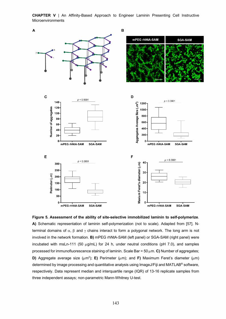
CHAPTER V | An Affinity-Based Approach to Engineer Laminin Presenting Cell Instructive Microenvironments
143
Figure 5. Assessment of the ability of site-selective immobilized laminin to self-polymerize.
A) Schematic representation of laminin self-polymerization (not to scale). Adapted from [57]. N-
terminal domains of a, b and g chains interact to form a polygonal network. The long arm is not
involved in the network formation. B) mPEG rhNtA-SAM (left panel) or SGA-SAM (right panel) were
incubated with msLn-111 (50 µg/mL) for 24 h, under neutral conditions (pH 7.0), and samples
processed for immunofluorescence staining of laminin. Scale Bar = 50 µm. C) Number of aggregates;
D) Aggregate average size (µm2); E) Perimeter (µm); and F) Maximum Feret’s diameter (µm)
determined by image processing and quantitative analysis using ImageJ/Fiji and MATLAB® software,
respectively. Data represent median and interquartile range (IQR) of 13-16 replicate samples from
three independent assays; non-parametric Mann-Whitney U-test.
CHAPTER V | An Affinity-Based Approach to Engineer Laminin Presenting Cell Instructive Microenvironments
144
3.5.2. Site-selective immobilized rhLn-521 support hNSC adhesion and spreading
rhLn-521 has a key role in the modulation of neural cell behavior, including neuronal
adhesion, viability and network formation [36, 58]. Indeed, several studies have shown the
potential of this laminin isoform to be used as a robust substratum for the culture and
renewal of human embryonic [59] and pluripotent [36] stem cells. With this in mind, to
assess laminin ability to mediate hNSC adhesion and spreading upon its binding to mPEG
rhNtA-SAMs, rhLn-521was used. QCM-D analysis was first conducted to follow the kinetics
of rhLn-521 immobilization onto SAMs (Fig. 6 A-B). Then, to characterize whether the
proposed strategy favored the exposure of rhLn-521 cell-adhesive domains, hNSC
adhesion and spreading was assessed (Fig. 6 C-E).
QCM-D monitoring of rhLn-521 immobilization onto mPEG rhNtA-SAMs indicated a protein
immobilization (638 ± 29 ng/cm2, Fig. 6B), with mass per surface area values close to the
theoretical value calculated for laminin immobilization under these conditions (952 ng/cm2).
This suggests that the immobilized mono-PEGylated rhNtA retained a 3D conformation that
allowed the selective interaction with laminin. Moreover, mPEG rhNtA SAMs led to an
immobilization degree statistically similar to that observed for uncoated Au surfaces (1130
± 348 ng/cm2), in which laminin may randomly adsorb over the entire surface. SGA-SAMs,
in contrast, presented lower amounts of immobilized rhLn-521 (342 ± 44 ng/cm2) (Figure
6B), when compared to mPEG rhNtA-SAMs. As SH-PEG-SGA moieties can randomly
interact with different amine groups within a single laminin molecule, these results suggest
that the conformation of covalently-bound laminin and its spatial arrangement at the surface
were distinct from that of affinity-bound laminin, leaving less area for the immobilization of
other laminin molecules. As expected, the amount of adsorbed rhLn-521 on EG4-SAMs,
was very low (20 ± 1 ng/cm2) (Fig. 6B).
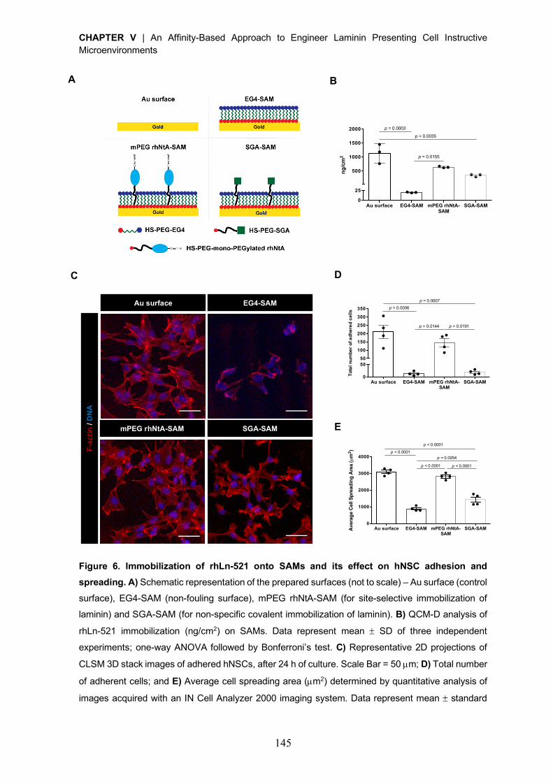
CHAPTER V | An Affinity-Based Approach to Engineer Laminin Presenting Cell Instructive Microenvironments
145
Figure 6. Immobilization of rhLn-521 onto SAMs and its effect on hNSC adhesion and
spreading. A) Schematic representation of the prepared surfaces (not to scale) – Au surface (control
surface), EG4-SAM (non-fouling surface), mPEG rhNtA-SAM (for site-selective immobilization of
laminin) and SGA-SAM (for non-specific covalent immobilization of laminin). B) QCM-D analysis of
rhLn-521 immobilization (ng/cm2) on SAMs. Data represent mean ± SD of three independent
experiments; one-way ANOVA followed by Bonferroni’s test. C) Representative 2D projections of
CLSM 3D stack images of adhered hNSCs, after 24 h of culture. Scale Bar = 50 µm; D) Total number
of adherent cells; and E) Average cell spreading area (µm2) determined by quantitative analysis of
images acquired with an IN Cell Analyzer 2000 imaging system. Data represent mean ± standard
A F
-acti
n / D
NA
mPEG rhNtA-SAM
Au surface EG4-SAM
SGA-SAM E
D
B
C
Au surface EG4-SAM mPEG rhNtA-SAM
SGA-SAM0
25
500
1000
1500
2000
ng/c
m2
p = 0.0003
p = 0.0035
p = 0.0155
Au surface EG4-SAM mPEG rhNtA-SAM
SGA-SAM0
5050
100
150
200
250
300
350
Tota
l num
ber
of a
dher
ed c
ells
p = 0.0007p = 0.0006
p = 0.0144 p = 0.0191
Au surface EG4-SAM mPEG rhNtA-SAM
SGA-SAM0
1000
2000
3000
4000
Ave
rage
Cel
l Spr
eadi
ng A
rea
(µm
2 )
p < 0.0001
p = 0.0254
p < 0.0001 p < 0.0001
p < 0.0001
CHAPTER V | An Affinity-Based Approach to Engineer Laminin Presenting Cell Instructive Microenvironments
146
error of the mean (SEM) of four replicate samples from an experiment representative of three
independent assays; one-way ANOVA followed by Bonferroni’s test.
Site-selective immobilization of laminin onto mPEG rhNtA-SAMs led to hNSC adhesion
comparable to that observed on rhLn-521-coated Au surfaces, used here as a control
surface for hNSC adhesion (Fig. 6D and S8). After 24 h of culture, cells were well-spread
(Fig. 6E and S9), exhibiting a morphology (Fig. 6C) comparable to that observed for cells
seeded on rhLn-521-coated Au surfaces (Fig. 6C). Moreover, cells adhered to mPEG
rhNtA-SAMs exhibit an average spreading area of 2812 ± 89 µm2 (Fig. 6E), comparable to
that observed for cells cultured on rhLn-521-coated Au surfaces (3089 ± 101 µm2; Figure
6E). TCPS surfaces, which are conventional used in adhesion studies, were used herein
as additional control surfaces. The morphology of cells seeded on rhLn-521- site-selective
immobilized on SAMs or -coated on Au surfaces is comparable to that observed on rhLn-
521-modified TCPS surfaces (Fig. S7). In addition, the average cell spreading area in these
conditions is similar to that observed in TCPS surfaces (3496 ± 483 µm2). The non-selective
immobilization of laminin onto SGA-SAMs, in turn, resulted in a significantly lower cell
adhesion (p = 0.0191; Fig. 6D and S8) and average spreading area (1430 ± 161 µm2; p <
0.0001; Fig. 6E and S9) when compared to mPEG rhNtA-SAMs. Moreover, cells with both
spread (Fig. 6C) and round (Fig. S7) morphology were observed. These results suggest
that the non-selective nature of the immobilization process partially compromises the
exposure of laminin cell binding domains, despite the considerable amount of rhLn-521
bound to SGA-SAMs (342.3 ± 43.6 ng/cm2) (Fig. 6B). As expected, on EG4-SAMs, the few
adherent cells (Fig. 6D and S8) presented mainly a round morphology (Fig. 6C and S7), as
evidenced by the low average cell spreading area (881 ± 70 µm2, Fig. 6E). Overall, these
results clearly demonstrate that site-specific immobilization of laminin through the use of
mono-PEGylated rhNtA better preserved laminin bioactivity in terms of ability to self-
polymerize and to mediate hNSC adhesion and spreading, when compared to non-selective
covalent immobilization approaches.
4. Conclusion
The present work demonstrates the potential of mono-PEGylated rhNtA as an effective
natural affinity binding ligand for site-selective immobilization of laminin, allowing the
preservation of laminin ability to self-polymerize and mediate cell adhesion and spreading.
This affinity binding strategy overcomes several drawbacks associated with the currently
available strategies for laminin immobilization. Moreover, this approach is highly versatile,
as result of the ability of NtA to bind the different laminin isoforms that comprise the g1 chain,
CHAPTER V | An Affinity-Based Approach to Engineer Laminin Presenting Cell Instructive Microenvironments
147
which represent more than 50% of the isoforms identified to date [60], with variations in
affinity imposed by a and b chains. Therefore, this strategy enables the immobilization of
different laminin isoforms, which can be of interest for particular cell types and for
application in specific disease contexts. Overall, the proposed strategy is highly attractive
for a broad range of applications, including 2D coatings for cell culture, functionalization of
3D matrices for cell and/or drug delivery, engineered coatings for neuroelectrodes, among
others.
Acknowledgments
The mass spectrometry technique was performed at the Proteomics i3S Scientific Platform
(Roteiro/0028/2013; LISBOA-01-0145-FEDER-022125) with the assistance of Hugo
Osório. IRRAS analysis was performed at the Biointerfaces and Nanotechnology Scientific
Platform, with the assistance of Ricardo Vidal. In Cell Analyzer experiments were performed
at the Biosciences Screening i3S Scientific Platform, member of the PPBI (PPBI-POCI-01-
0145-FEDER-022122) with the assistance of André Maia. Confocal microscopy was
conducted at the Bioimaging i3S Scientific Platform, member of the PPBI (PPBI-POCI-01-
0145-FEDER-022122), with the assistance of Maria Lázaro. This work was funded by
projects NORTE-01-0145-FEDER-000008 and NORTE-01-0145-FEDER-000012,
supported by Norte Portugal Regional Operational Programme (NORTE 2020), under the
PORTUGAL 2020 Partnership Agreement, through the European Regional Development
Fund (ERDF) and FEDER - Fundo Europeu de Desenvolvimento Regional funds through
the COMPETE 2020 - Operacional Programme for Competitiveness and
Internationalisation (POCI), Portugal 2020; by Portuguese funds through FCT/MCTES in
the framework of the project "Institute for Research and Innovation in Health Sciences"
(POCI-01-0145-FEDER-007274), and by Santa Casa da Misericordia de Lisboa through
project COMBINE (Prémio Neurociências Melo e Castro 1068-2015).
D.B. was supported by FCT PhD Programs (PD/BD/105953/2014) and Programa
Operacional Potencial Humano (POCH), in the scope of the BiotechHealth Program
(Doctoral Program on Cellular and Molecular Biotechnology Applied to Health Sciences),
Programa FLAD Healthcare 2020 and the project PARES (Prémio Albino Aroso). Eduardo
Conde-Sousa was supported by a post-doctoral grant of the project PPBI-POCI-01-0145-
FEDER-022122, in the scope of FCT National Roadmap of Research Infrastructures.
CHAPTER V | An Affinity-Based Approach to Engineer Laminin Presenting Cell Instructive Microenvironments
148
SUPPLEMENTARY DATA
Supplementary Materials and Methods
Selection and cloning of human N-terminal agrin domain (hNtA) gene and pCoofy
expression vector
The human N-terminal agrin (hNtA) domain gene was synthesized by GenScript
(Piscataway, NJ, USA) – Clone ID: B25738, from the protein sequence with the Uniprot
reference O00468 (Thr 30 – Pro 157). A gene of 395bp length was produced in pUC57
cloning vector and the conditions for expression in Escherichia coli (E. coli) optimized. A
high-throughput expression screening was conducted to select, among the several pCoofy
expression vectors available [37], the most suitable for the expression of the soluble protein.
pCoofy2 expression vector – 6080 bp (gift from Sabine Suppmann – Addgene plasmid #
43981) was the one selected. Both hNtA gene and pCoofy2 expression vector were
polymerase chain reaction (PCR) linearized using specific-designed primers and LP1
forward and LP2 reverse primers, respectively (Table S1). Following PCR amplification, the
hNtA gene was sub-cloned into the pCoofy2 expression vector using a sequence and
ligation independent cloning (SLIC) reaction, as described in [37]. The efficiency of DNA
insert incorporation into the expression vector was confirmed by colony PCR and DNA
sequencing (Macrogen).
Western blot analysis
Ten micrograms of rhNtA was resolved in a 15% SDS-PAGE gel and transferred onto a
nitrocellulose membrane (0.45 µm, GE Healthcare). Unspecific binding was prevented by
incubating the membrane with blocking solution (5% (w/v) BSA in Tris-buffered saline –
0.1% Tween® 20 (TBS-T) for 2h at RT. The membrane was then incubated with the primary
antibody - rabbit polyclonal agrin antibody (1:1000; Abcam, ab85174), overnight at 4 ºC.
After washing with PBS/0.05% (v/v) Tween® 20, the membrane was incubated with the goat
anti-rabbit IgG (H+L) coupled to horseradish peroxidase (HRP) secondary antibody
(1:2000; Life Technologies, A16023) for 1h at RT. The membrane was finally washed three
times with PBS/0.05% (v/v) Tween® 20 and the blots developed with HRP-reactive
chemiluminescence reagents (GE Healthcare).
rhNtA adsorption isotherm
96-well microtiter ELISA plates (BD Falcon) were coated with 2-fold serial dilution of rhNtA
(0 – 40 µg/mL; overnight, 4 ºC) and blocked with 1% (w/v) BSA to prevent non-specific
adhesion to substrate. The wells were then incubated with the primary antibody - rabbit
polyclonal agrin antibody (1:5000; 2h, RT). Goat anti-rabbit IgG (H+L) coupled to HRP
CHAPTER V | An Affinity-Based Approach to Engineer Laminin Presenting Cell Instructive Microenvironments
149
secondary antibody (1:2000) was incubated for 1,5 h at RT. The color was developed using
3, 3’, 5, 5’ tetramethyl benzidine (TMB) substrate (Biolegend), incubated for 30 min at RT.
The reaction was stopped using 2 M H2SO4 and the absorbance measured at 450 nm
(BioTekÒ Synergy™ Mx).
Cell adhesion centrifugation assay
hNSCs were fluorescently-labelled with 2 µM of Calcein AM (Molecular Probes, 10 min,
37ºC), suspended in StemPro® NSC SFM medium (Life Technologies) and transferred to
rhNtA-immobilized msLn-111-adsorbed surfaces at 3×10⁵ viable cells/cm2. Cells were
allowed to adhere for 5 h and before subjecting the cells to centrifugation, initial
fluorescence intensity was measured using a microwell plate reader (BioTek® SynergyTM
Mx). After overfilling the wells with 10 mM PBS pH 7.4, until a positive meniscus was
observed, the plates were sealed with transparent tape, inverted, and spun at 50 RCF for 5
min. Following cell detachment, the medium was changed, and the wells filled with fresh
medium for measurement of final fluorescence. For each well, the adherent cell fraction was
determined as the ratio of post-spin (final) to pre-spin (initial) fluorescence readings (lex =
485 nm; lem = 535 nm).
Peptide mapping
All the reagents used to perform the peptide mapping and protein identification were
purchased from Sigma-Aldrich unless otherwise stated.
Protein bands correspondent to native or mono-PEGylated rhNtA were cut from the gel,
washed with ultrapure water and de-stained with 50% (v/v) acetonitrile (ACN) in 50 mM
ammonium bicarbonate. The samples were then reduced with 50 mM dithiothreitol (20 min,
56 ºC) and alkylated with 55 mM iodoacetamide, (20 min, RT in the dark). Next, protein in
gel enzymatic digestion was performed by the addition of 20 ng of trypsin for 3 h at 37 ºC
in the presence of 0.01% surfactant (ProteaseMAXTM, Promega). Resulting peptides were
extracted from gel plugs with 2.5% trifluoroacetic acid (TFA; 15 min at 1400 rpm), dried
under vacuum and resuspended in 0.1% TFA.
Protein identification was performed by a MALDI-TOF/TOF mass spectrometer (4800 Plus,
SCIEX). Protein digests were purified by reversed-phase C18 chromatography (ZipTips®,
Millipore) following the manufacturer’s instructions and eluted in the MALDI sample plate
using the MALDI matrix alpha-Cyano-4-hydroxycinnamic acid (CHCA) elution solution at 8
mg/mL in 50% ACN, 0.1% TFA, 6 mM ammonium phosphate. Peptide mass spectra were
acquired in positive MS reflector mode in the mass range of m/z 700-5000 and internally
calibrated with trypsin autolysis peaks. Individual peptides from both native and mono-
PEGylated rhNtA were identified by peptide mass fingerprint (PMF) approach using MS-
CHAPTER V | An Affinity-Based Approach to Engineer Laminin Presenting Cell Instructive Microenvironments
150
Bridge software version 5.16.1 (ProteinProspector, University of California, San Francisco,
CA) and the protein sequence data (Figure S1). The protein search settings were: cysteine
carbamidomethylation (constant modification), methionine oxidation (variable modification),
up to two missed trypsin cleavages, minimum and maximum digest fragment mass of 500
and 4000 Da, respectively, and a minimum digest fragment length of five amino acids. To
estimate the location of the PEG moiety in the conjugate, experimental ion peaks obtained
in the peptide map of purified mono-PEGylated rhNtA conjugate were compared to the
peptide content of native rhNtA. In addition, an indirect estimation of PEG moiety position
in the rhNtA sequence was performed by comparing the mass difference between the
conjugated and unmodified PEG molecule.
Quartz crystal microbalance with dissipation monitoring (QCM-D)
For QCM-D real-time monitoring of laminin immobilization, sensors were pre-incubated with
10 mM HEPES pH 7.4 for 15 min at a constant flow rate of 0.1 µL/min followed by an
additional 15 min incubation, under static conditions, to establish the baseline. Then, 300
µL/sensor of rhLn-521 (20 µg/mL) in 10 mM HEPES pH 7.4 was injected at a constant flow
rate of 0.1 µL/min, followed by 1 h incubation in static conditions. After, the system was
flushed with 10 mM HEPES pH 7.4 during 30 min at a constant flow rate of 0.1 µL/min. All
the experiments were conducted at 25 ºC. The total amount of immobilized laminin was
calculated with application of the Voigt model [41], which takes into account the viscoelastic
contributions of the hydrated layer. The fluid density and viscosity of rhLn-521 solution were
established at 1000 kg/m3 and 0.001 kg/m∙s, respectively and data from the 5th harmonic
was used in the analysis.
Infrared reflection absorption spectroscopy (IRRAS)
IRRAS spectra were obtained using a Perkin Elmer FTIR spectrophotometer, model 2000,
coupled with a VeeMax II Accessory (PIKE) and a liquid-nitrogen-cooled MTC detector. To
avoid water vapor adsorption, instrument was purged with dry nitrogen for 5 min before and
during each sample analysis. Au substrates were here used as a background. Incident light
was p-polarized and spectra were collected using the 80º grazing angle reflection mode.
The analysis of each sample was performed through the acquisition of 100 scans with 4
cm-1 resolution.
Laminin self-polymerization – Immunocytochemistry and Image processing and analysis
mPEG rhNtA- or SGA-SAMs incubated with msLn-111 were fixed with 3.7% (w/v)
paraformaldehyde (PFA) for 20 min at RT and washed 3´ with 10 mM PBS pH 7.4. Samples
were then incubated with blocking buffer (5% (w/v) BSA in PBS for 30 min at RT), followed
CHAPTER V | An Affinity-Based Approach to Engineer Laminin Presenting Cell Instructive Microenvironments
151
by incubation with the primary antibody – rabbit polyclonal laminin antibody (1:30; Sigma-
Aldrich, L9393), overnight at 4 ºC. Samples were washed 3´ with 10 mM PBS pH 7.4 and
incubated with Alexa FluorÒ 488 conjugated anti-rabbit secondary antibody (1:300; Life
Technologies, A11034), for 1 h at RT. Samples were observed under confocal laser
scanning microscopy (CLSM, Leica TCS SP5II) using a Plan-Apochromat 63´/1.4NA oil
objective and acquired z-sections of 15 µm were processed using the ImageJ/Fiji software
[61, 62]. Briefly, the rolling ball algorithm was first applied to maximum intensity projection
2D images, to fix uneven background and followed by the application of a Gaussian blur
with s = 1 to reduce noise. Aggregates were segmented through an Otsu thresholding
method and a closing morphological filter was then used to unify structures distanced by
less than 1 µm. Lastly, all the structures with an area below 10 µm2 were removed/rejected.
For each resulting image, measurements were obtained for number of aggregates,
aggregate average size (µm2), perimeter (µm) and maximum Feret’s diameter (µm). To
evaluate differences on the data resultant from both conditions, statistical analysis of the
different variables was conducted on MATLAB® software (R2018a) using the non-
parametric Mann-Whitney U-test. Moreover, differences resultant from the combination of
the different variables measured were determined by application of the Fisher’s combined
probability test [56] and an overall p-value determined. This test is applied under the
assumption that variables must be independent from each other. As the number of
aggregates is not independent from the other variables, for the Fisher’s combined
probability test, only average size, perimeter and maximum Feret’s diameter of aggregates
were considered.
Cell adhesion and morphology – F-actin/DNA staining
hNSC adhesion and morphology was assessed, after 24 h of culture, in samples processed
for F-actin/DNA staining. Cells were fixed with 2% (w/v) PFA for 20 min at RT, permeabilized
with 0.5% (v/v) Triton X-100 (Sigma-Aldrich) for 10 min at RT and stained with Alexa FluorÒ
647 phalloidin (1:50; Biolegend, 424205) for 20 min at RT. The nuclei were counterstained
with 0.1 µg/mL of Hoechst 33342 (Life Technologies, H1399) for 20 min at RT. Samples
were imaged with the IN Cell Analyzer 2000 imaging system (GE Healthcare) using a Nikon
20×/0.45 NA Plan Fluor objective and CLSM (TCS SP5 II; Leica Mycrosystems) using a
Plan-Apochromat 63´/1.4NA oil objective.
Cell adhesion – DNA fluorescence
Cell adhesion was also estimated from Hoechst 33342 average fluorescence intensity of
samples processed for F-actin/DNA staining. Briefly, samples were transferred to the wells
of black 24-well plates (ibidi) and the fluorescence of each surface measured (lex = 350 nm;
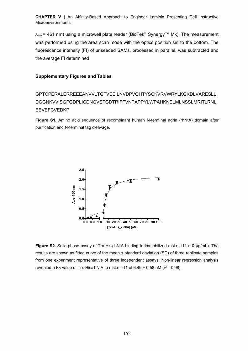
CHAPTER V | An Affinity-Based Approach to Engineer Laminin Presenting Cell Instructive Microenvironments
152
lem = 461 nm) using a microwell plate reader (BioTekÒ Synergy™ Mx). The measurement
was performed using the area scan mode with the optics position set to the bottom. The
fluorescence intensity (FI) of unseeded SAMs, processed in parallel, was subtracted and
the average FI determined.
Supplementary Figures and Tables
GPTCPERALERREEEANVVLTGTVEEILNVDPVQHTYSCKVRVWRYLKGKDLVARESLL
DGGNKVVISGFGDPLICDNQVSTGDTRIFFVNPAPPYLWPAHKNELMLNSSLMRITLRNL
EEVEFCVEDKP
Figure S1. Amino acid sequence of recombinant human N-terminal agrin (rhNtA) domain after
purification and N-terminal tag cleavage.
Figure S2. Solid-phase assay of Trx-His6-hNtA binding to immobilized msLn-111 (10 µg/mL). The results are shown as fitted curve of the mean ± standard deviation (SD) of three replicate samples
from one experiment representative of three independent assays. Non-linear regression analysis
revealed a KD value of Trx-His6-hNtA to msLn-111 of 6.49 ± 0.58 nM (r2 = 0.98).
0.0 0.5 1.0 10 20 30 40 50 60 70 80 90 1000.0
0.5
1.0
1.5
2.0
2.5
[Trx-His6-hNtA] (nM)
Abs
450
nm
Kd = 6.5nM
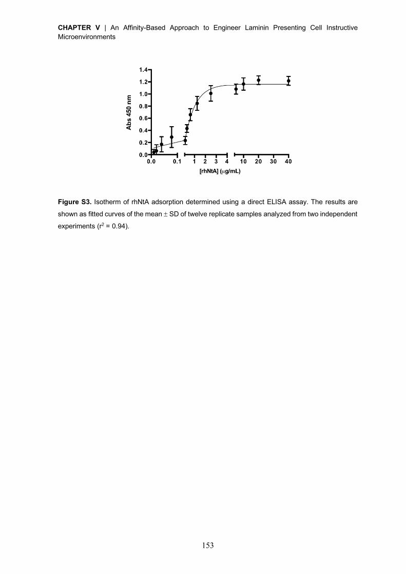
CHAPTER V | An Affinity-Based Approach to Engineer Laminin Presenting Cell Instructive Microenvironments
153
Figure S3. Isotherm of rhNtA adsorption determined using a direct ELISA assay. The results are
shown as fitted curves of the mean ± SD of twelve replicate samples analyzed from two independent
experiments (r2 = 0.94).
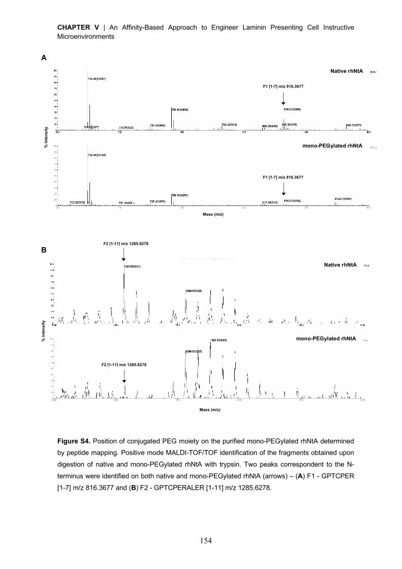
CHAPTER V | An Affinity-Based Approach to Engineer Laminin Presenting Cell Instructive Microenvironments
154
Figure S4. Position of conjugated PEG moiety on the purified mono-PEGylated rhNtA determined by peptide mapping. Positive mode MALDI-TOF/TOF identification of the fragments obtained upon
digestion of native and mono-PEGylated rhNtA with trypsin. Two peaks correspondent to the N-
terminus were identified on both native and mono-PEGylated rhNtA (arrows) – (A) F1 - GPTCPER
[1-7] m/z 816.3677 and (B) F2 - GPTCPERALER [1-11] m/z 1285.6278.
% I
nte
ns
ity
Mass (m/z)
Mass (m/z)
% I
nte
ns
ity
Native rhNtA
mono-PEGylated rhNtA
Native rhNtA
mono-PEGylated rhNtA
A
B
F1 [1-7] m/z 816.3677
F1 [1-7] m/z 816.3677
F2 [1-11] m/z 1285.6278
F2 [1-11] m/z 1285.6278
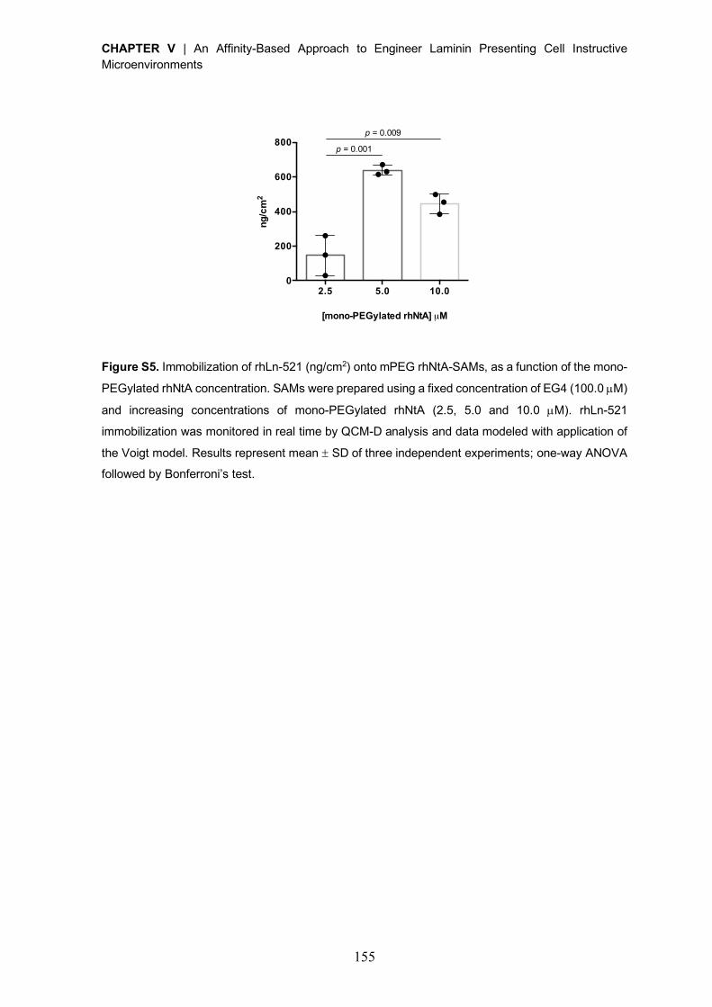
CHAPTER V | An Affinity-Based Approach to Engineer Laminin Presenting Cell Instructive Microenvironments
155
Figure S5. Immobilization of rhLn-521 (ng/cm2) onto mPEG rhNtA-SAMs, as a function of the mono-
PEGylated rhNtA concentration. SAMs were prepared using a fixed concentration of EG4 (100.0 µM)
and increasing concentrations of mono-PEGylated rhNtA (2.5, 5.0 and 10.0 µM). rhLn-521
immobilization was monitored in real time by QCM-D analysis and data modeled with application of
the Voigt model. Results represent mean ± SD of three independent experiments; one-way ANOVA
followed by Bonferroni’s test.
2.5 5.0 10.00
200
400
600
800
ng/c
m2
p = 0.001
p = 0.009
[mono-PEGylated rhNtA] µM
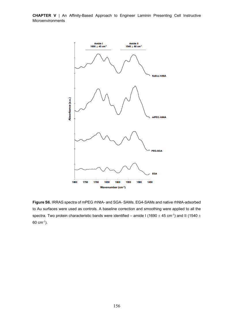
CHAPTER V | An Affinity-Based Approach to Engineer Laminin Presenting Cell Instructive Microenvironments
156
Figure S6. IRRAS spectra of mPEG rhNtA- and SGA- SAMs. EG4-SAMs and native rhNtA-adsorbed
to Au surfaces were used as controls. A baseline correction and smoothing were applied to all the
spectra. Two protein characteristic bands were identified – amide I (1690 ± 45 cm-1) and II (1540 ±
60 cm-1).
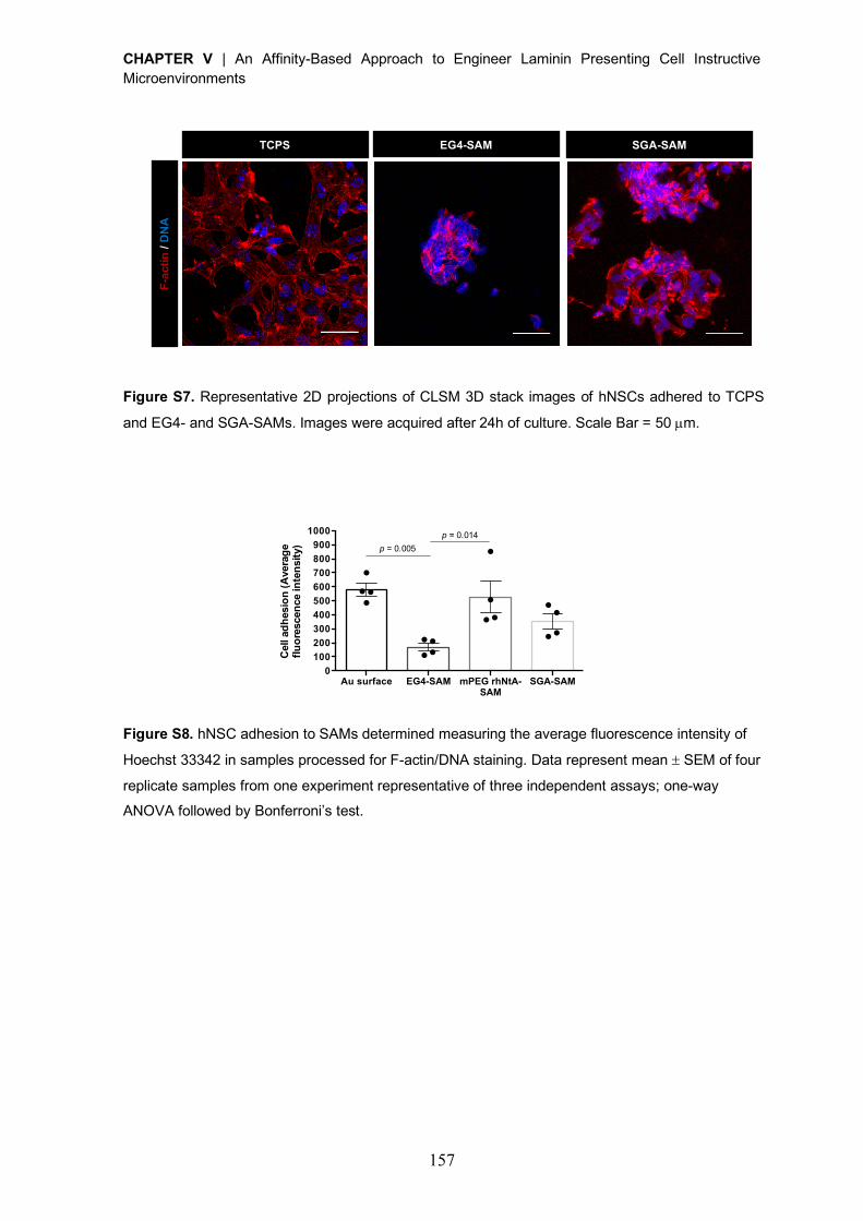
CHAPTER V | An Affinity-Based Approach to Engineer Laminin Presenting Cell Instructive Microenvironments
157
Figure S7. Representative 2D projections of CLSM 3D stack images of hNSCs adhered to TCPS
and EG4- and SGA-SAMs. Images were acquired after 24h of culture. Scale Bar = 50 µm.
Figure S8. hNSC adhesion to SAMs determined measuring the average fluorescence intensity of
Hoechst 33342 in samples processed for F-actin/DNA staining. Data represent mean ± SEM of four
replicate samples from one experiment representative of three independent assays; one-way
ANOVA followed by Bonferroni’s test.
SGA-SAM EG4-SAM F
-acti
n / D
NA
TCPS
Au surface EG4-SAM mPEG rhNtA-SAM
SGA-SAM0
100200300400500600700800900
1000
Cel
l adh
esio
n (A
vera
geflu
ores
cenc
e in
tens
ity) p = 0.005
p = 0.014
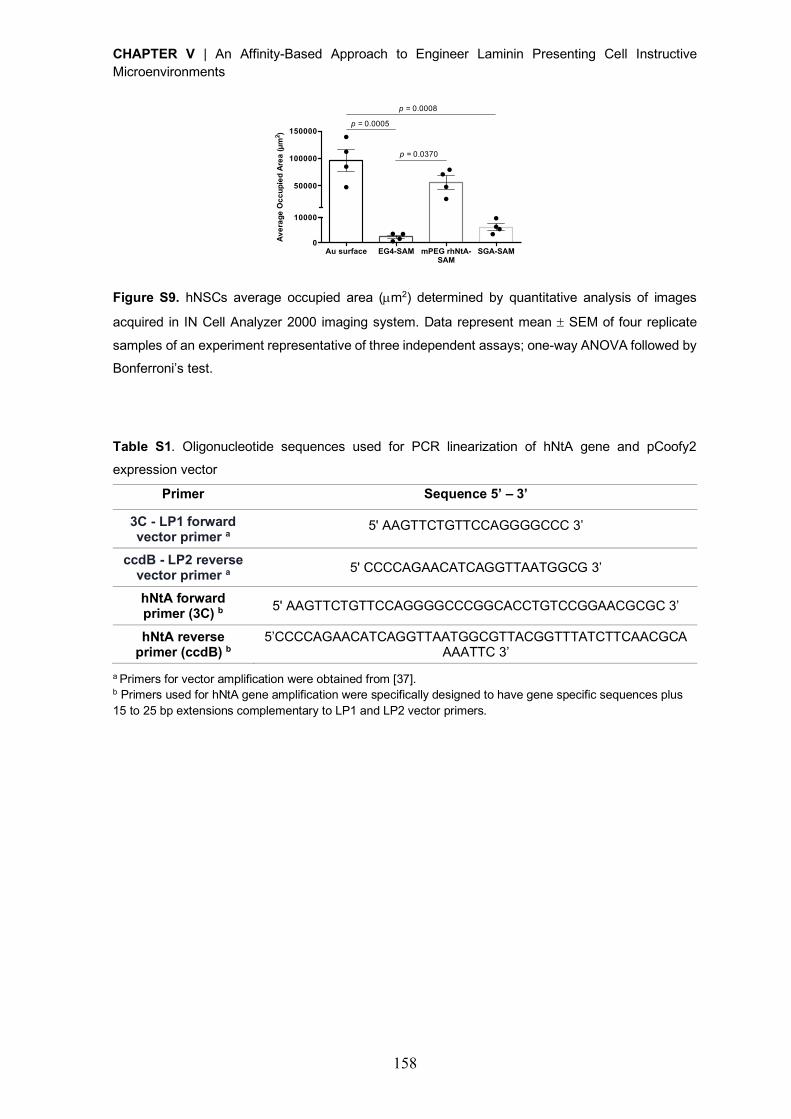
CHAPTER V | An Affinity-Based Approach to Engineer Laminin Presenting Cell Instructive Microenvironments
158
Figure S9. hNSCs average occupied area (µm2) determined by quantitative analysis of images
acquired in IN Cell Analyzer 2000 imaging system. Data represent mean ± SEM of four replicate
samples of an experiment representative of three independent assays; one-way ANOVA followed by
Bonferroni’s test.
Table S1. Oligonucleotide sequences used for PCR linearization of hNtA gene and pCoofy2
expression vector
Primer Sequence 5’ – 3’
3C - LP1 forward
vector primer a 5' AAGTTCTGTTCCAGGGGCCC 3’
ccdB - LP2 reverse
vector primer a 5' CCCCAGAACATCAGGTTAATGGCG 3’
hNtA forward
primer (3C) b 5' AAGTTCTGTTCCAGGGGCCCGGCACCTGTCCGGAACGCGC 3’
hNtA reverse
primer (ccdB) b
5’CCCCAGAACATCAGGTTAATGGCGTTACGGTTTATCTTCAACGCAAAATTC 3’
a Primers for vector amplification were obtained from [37]. b Primers used for hNtA gene amplification were specifically designed to have gene specific sequences plus 15 to 25 bp extensions complementary to LP1 and LP2 vector primers.
Au surface EG4-SAM mPEG rhNtA-SAM
SGA-SAM0
10000
50000
100000
150000
Aver
age
Occ
upie
d Ar
ea (µ
m2 )
p = 0.0008
p = 0.0005
p = 0.0370
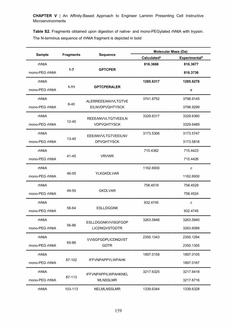
CHAPTER V | An Affinity-Based Approach to Engineer Laminin Presenting Cell Instructive Microenvironments
159
Table S2. Fragments obtained upon digestion of native- and mono-PEGylated rhNtA with trypsin.
The N-terminus sequence of rhNtA fragment is depicted in bold
Sample Fragments Sequence Molecular Mass (Da)
Calculateda Experimentalb
rhNtA 1-7 GPTCPER
816.3668 816.3677
mono-PEG rhNtA
816.3736
rhNtA 1-11 GPTCPERALER
1285.6317 1285.6278
mono-PEG rhNtA
c
rhNtA 8-40
ALERREEEANVVLTGTVEEILNVDPVQHTYSCK
3741.8752 3798.9145
mono-PEG rhNtA
3798.9299
rhNtA 12-40
REEEANVVLTGTVEEILNVDPVQHTYSCK
3329.6317 3329.6360
mono-PEG rhNtA
3329.6489
rhNtA 13-40
EEEANVVLTGTVEEILNVDPVQHTYSCK
3173.5306 3173.5747
mono-PEG rhNtA
3173.5818
rhNtA 41-45 VRVWR
715.4362 715.4423
mono-PEG rhNtA
715.4426
rhNtA 46-55 YLKGKDLVAR
1162.6930 c
mono-PEG rhNtA
1162.6930
rhNtA 49-55 GKDLVAR
758.4519 758.4529
mono-PEG rhNtA
758.4524
rhNtA 56-64 ESLLDGGNK
932.4745 c
mono-PEG rhNtA
932.4745
rhNtA 56-86
ESLLDGGNKVVISGFGDPLICDNQVSTGDTR
3263.5848 3263.5940
mono-PEG rhNtA
3263.6089
rhNtA 65-86
VVISGFGDPLICDNQVSTGDTR
2350.1343 2350.1294
mono-PEG rhNtA
2350.1355
rhNtA 87-102 IFFVNPAPPYLWPAHK
1897.0159 1897.0105
mono-PEG rhNtA
1897.0167
rhNtA 87-113
IFFVNPAPPILWPAHKNELMLNSSLMR
3217.6325 3217.6418
mono-PEG rhNtA
3217.6716
rhNtA 103-113 NELMLNSSLMR 1339.6344 1339.6328
CHAPTER V | An Affinity-Based Approach to Engineer Laminin Presenting Cell Instructive Microenvironments
160
mono-PEG rhNtA
1339.6374
rhNtA 114-130 ITLRNLEEVEFCVEDKP
2091.0426 2091.0317
mono-PEG rhNtA
2091.0422
rhNtA 118-130 NLEEVEFCVEDKP
1607.7257 1607.7244
mono-PEG rhNtA
1607.7286
a Theoretical molecular mass of the fragments calculated in web.expasy.org/peptide_mass. b Experimental molecular masses
determined by MALDI-TOF MS. c Fragment not found.
Movie S1. Projection of the 3D structures formed after laminin self-polymerization, under neutral
conditions, on mPEG rhNtA-SAMs. The projection was generated from a series of confocal optical
slices (the same shown in Fig. 5B left panel) using ImageJ software (AVI).
Movie S2. Projection of the 3D structures formed after laminin self-polymerization, under neutral conditions, on SGA-SAMs. The projection was generated from a series of confocal optical slices (the
same shown in Fig. 5B right panel) using ImageJ software (AVI).
CHAPTER V | An Affinity-Based Approach to Engineer Laminin Presenting Cell Instructive Microenvironments
161
References
[1] J.H. Miner, B.L. Patton, S.I. Lentz, D.J. Gilbert, W.D. Snider, N.A. Jenkins, N.G.
Copeland, J.R. Sanes, The laminin alpha chains: expression, developmental transitions,
and chromosomal locations of alpha1-5, identification of heterotrimeric laminins 8-11, and
cloning of a novel alpha3 isoform, J Cell Biol 137(3) (1997) 685-701.
[2] M. Falk, M. Ferletta, E. Forsberg, P. Ekblom, Restricted distribution of laminin alpha1
chain in normal adult mouse tissues, Matrix Biol 18(6) (1999) 557-68.
[3] H. Colognato, P.D. Yurchenco, Form and function: the laminin family of heterotrimers,
Dev Dyn 218(2) (2000) 213-34.
[4] T. Sasaki, R. Giltay, U. Talts, R. Timpl, J.F. Talts, Expression and distribution of laminin
alpha1 and alpha2 chains in embryonic and adult mouse tissues: an immunochemical
approach, Exp Cell Res 275(2) (2002) 185-99.
[5] J. Tzu, M.P. Marinkovich, Bridging structure with function: structural, regulatory, and
developmental role of laminins, Int J Biochem Cell Biol 40(2) (2008) 199-214.
[6] P.D. Yurchenco, E.C. Tsilibary, A.S. Charonis, H. Furthmayr, Laminin polymerization in
vitro. Evidence for a two-step assembly with domain specificity, J Biol Chem 260(12) (1985)
7636-44.
[7] P.D. Yurchenco, Y.S. Cheng, Self-assembly and calcium-binding sites in laminin. A
three-arm interaction model, J Biol Chem 268(23) (1993) 17286-99.
[8] Y.S. Cheng, M.F. Champliaud, R.E. Burgeson, M.P. Marinkovich, P.D. Yurchenco, Self-
assembly of laminin isoforms, J Biol Chem 272(50) (1997) 31525-32.
[9] J.D. Lathia, B. Patton, D.M. Eckley, T. Magnus, M.R. Mughal, T. Sasaki, M.A. Caldwell,
M.S. Rao, M.P. Mattson, C. ffrench-Constant, Patterns of laminins and integrins in the
embryonic ventricular zone of the CNS, J Comp Neurol 505(6) (2007) 630-43.
[10] K.J. Hamill, K. Kligys, S.B. Hopkinson, J.C. Jones, Laminin deposition in the
extracellular matrix: a complex picture emerges, J Cell Sci 122(Pt 24) (2009) 4409-17.
[11] M. Aumailley, The laminin family, Cell Adh Migr 7(1) (2013) 48-55.
[12] A.C. Taylor, C.H. Gonzalez, B.S. Miller, R.J. Edgington, P. Ferretti, R.B. Jackman,
Surface functionalisation of nanodiamonds for human neural stem cell adhesion and
proliferation, Sci Rep 7(1) (2017) 7307.
[13] S.E. Stabenfeldt, A.J. Garcia, M.C. LaPlaca, Thermoreversible laminin-functionalized
hydrogel for neural tissue engineering, J Biomed Mater Res A 77(4) (2006) 718-25.
[14] S.E. Stabenfeldt, G. Munglani, A.J. Garcia, M.C. LaPlaca, Biomimetic
microenvironment modulates neural stem cell survival, migration, and differentiation, Tissue
Eng Part A 16(12) (2010) 3747-58.
CHAPTER V | An Affinity-Based Approach to Engineer Laminin Presenting Cell Instructive Microenvironments
162
[15] S.E. Stabenfeldt, M.C. LaPlaca, Variations in rigidity and ligand density influence
neuronal response in methylcellulose-laminin hydrogels, Acta Biomater 7(12) (2011) 4102-
8.
[16] A.T. Francisco, R.J. Mancino, R.D. Bowles, J.M. Brunger, D.M. Tainter, Y.T. Chen,
W.J. Richardson, F. Guilak, L.A. Setton, Injectable laminin-functionalized hydrogel for
nucleus pulposus regeneration, Biomaterials 34(30) (2013) 7381-8.
[17] A.T. Francisco, P.Y. Hwang, C.G. Jeong, L. Jing, J. Chen, L.A. Setton,
Photocrosslinkable laminin-functionalized polyethylene glycol hydrogel for intervertebral
disc regeneration, Acta Biomater 10(3) (2014) 1102-11.
[18] C.P. Addington, J.M. Heffernan, C.S. Millar-Haskell, E.W. Tucker, R.W. Sirianni, S.E.
Stabenfeldt, Enhancing neural stem cell response to SDF-1alpha gradients through
hyaluronic acid-laminin hydrogels, Biomaterials 72 (2015) 11-9.
[19] J. Arulmoli, H.J. Wright, D.T.T. Phan, U. Sheth, R.A. Que, G.A. Botten, M. Keating, E.L.
Botvinick, M.M. Pathak, T.I. Zarembinski, D.S. Yanni, O.V. Razorenova, C.C.W. Hughes,
L.A. Flanagan, Combination scaffolds of salmon fibrin, hyaluronic acid, and laminin for
human neural stem cell and vascular tissue engineering, Acta Biomater 43 (2016) 122-138.
[20] C.P. Addington, S. Dharmawaj, J.M. Heffernan, R.W. Sirianni, S.E. Stabenfeldt,
Hyaluronic acid-laminin hydrogels increase neural stem cell transplant retention and
migratory response to SDF-1alpha, Matrix Biol 60-61 (2017) 206-216.
[21] B.G. Keselowsky, D.M. Collard, A.J. Garcia, Surface chemistry modulates fibronectin
conformation and directs integrin binding and specificity to control cell adhesion, J Biomed
Mater Res A 66(2) (2003) 247-59.
[22] J.C. Rodriguez Hernandez, M. Salmeron Sanchez, J.M. Soria, J.L. Gomez Ribelles, M.
Monleon Pradas, Substrate chemistry-dependent conformations of single laminin
molecules on polymer surfaces are revealed by the phase signal of atomic force
microscopy, Biophys J 93(1) (2007) 202-7.
[23] O.M. Ba, M. Hindie, P. Marmey, O. Gallet, K. Anselme, A. Ponche, A.C. Duncan,
Protein covalent immobilization via its scarce thiol versus abundant amine groups: Effect
on orientation, cell binding domain exposure and conformational lability, Colloids Surf B
Biointerfaces 134 (2015) 73-80.
[24] C.D. Spicer, B.G. Davis, Selective chemical protein modification, Nat Commun 5 (2014)
4740.
[25] O. Boutureira, G.J. Bernardes, Advances in chemical protein modification, Chem Rev
115(5) (2015) 2174-95.
CHAPTER V | An Affinity-Based Approach to Engineer Laminin Presenting Cell Instructive Microenvironments
163
[26] S.A. Fisher, A.E.G. Baker, M.S. Shoichet, Designing Peptide and Protein Modified
Hydrogels: Selecting the Optimal Conjugation Strategy, J Am Chem Soc 139(22) (2017)
7416-7427.
[27] J.P. Lee, E. Kassianidou, J.I. MacDonald, M.B. Francis, S. Kumar, N-terminal specific
conjugation of extracellular matrix proteins to 2-pyridinecarboxaldehyde functionalized
polyacrylamide hydrogels, Biomaterials 102 (2016) 268-76.
[28] C.C. Lin, K.S. Anseth, Controlling Affinity Binding with Peptide-Functionalized
Poly(ethylene glycol) Hydrogels, Adv Funct Mater 19(14) (2009) 2325.
[29] J. Parker, N. Mitrousis, M.S. Shoichet, Hydrogel for Simultaneous Tunable Growth
Factor Delivery and Enhanced Viability of Encapsulated Cells in Vitro, Biomacromolecules
17(2) (2016) 476-84.
[30] S.E. Sakiyama-Elbert, J.A. Hubbell, Controlled release of nerve growth factor from a
heparin-containing fibrin-based cell ingrowth matrix, J Control Release 69(1) (2000) 149-
58.
[31] S.E. Sakiyama-Elbert, J.A. Hubbell, Development of fibrin derivatives for controlled
release of heparin-binding growth factors, J Control Release 65(3) (2000) 389-402.
[32] J.B. Mascarenhas, M.A. Ruegg, U. Winzen, W. Halfter, J. Engel, J. Stetefeld, Mapping
of the laminin-binding site of the N-terminal agrin domain (NtA), EMBO J 22(3) (2003) 529-
36.
[33] A.J. Denzer, T. Schulthess, C. Fauser, B. Schumacher, R.A. Kammerer, J. Engel, M.A.
Ruegg, Electron microscopic structure of agrin and mapping of its binding site in laminin-1,
EMBO J 17(2) (1998) 335-43.
[34] R.A. Kammerer, T. Schulthess, R. Landwehr, B. Schumacher, A. Lustig, P.D.
Yurchenco, M.A. Ruegg, J. Engel, A.J. Denzer, Interaction of agrin with laminin requires a
coiled-coil conformation of the agrin-binding site within the laminin gamma1 chain, EMBO
J 18(23) (1999) 6762-70.
[35] G.A. Hudalla, W.L. Murphy, Chemically well-defined self-assembled monolayers for
cell culture: toward mimicking the natural ECM, Soft Matter 7(20) (2011) 9561-9571.
[36] A. Hyysalo, M. Ristola, M.E. Makinen, S. Hayrynen, M. Nykter, S. Narkilahti, Laminin
alpha5 substrates promote survival, network formation and functional development of
human pluripotent stem cell-derived neurons in vitro, Stem Cell Res 24 (2017) 118-127.
[37] J. Scholz, H. Besir, C. Strasser, S. Suppmann, A new method to customize protein
expression vectors for fast, efficient and background free parallel cloning, BMC Biotechnol
13 (2013) 12.
[38] C.D. Reyes, A.J. Garcia, A centrifugation cell adhesion assay for high-throughput
screening of biomaterial surfaces, J Biomed Mater Res A 67(1) (2003) 328-33.
CHAPTER V | An Affinity-Based Approach to Engineer Laminin Presenting Cell Instructive Microenvironments
164
[39] C. Gomes, A. Almeida, J.A. Ferreira, L. Silva, H. Santos-Sousa, J. Pinto-de-Sousa, L.L.
Santos, F. Amado, T. Schwientek, S.B. Levery, U. Mandel, H. Clausen, L. David, C.A. Reis,
H. Osorio, Glycoproteomic analysis of serum from patients with gastric precancerous
lesions, J Proteome Res 12(3) (2013) 1454-66.
[40] P. Parreira, A. Magalhaes, I.C. Goncalves, J. Gomes, R. Vidal, C.A. Reis, D.E.
Leckband, M.C. Martins, Effect of surface chemistry on bacterial adhesion, viability, and
morphology, J Biomed Mater Res A 99(3) (2011) 344-53.
[41] F. Costa, D.M. Sousa, P. Parreira, M. Lamghari, P. Gomes, M.C.L. Martins, N-
acetylcysteine-functionalized coating avoids bacterial adhesion and biofilm formation, Sci
Rep 7(1) (2017) 17374.
[42] E. Freire, F.C. Gomes, R. Linden, V.M. Neto, T. Coelho-Sampaio, Structure of laminin
substrate modulates cellular signaling for neuritogenesis, J Cell Sci 115(Pt 24) (2002) 4867-
76.
[43] A.E. Carpenter, T.R. Jones, M.R. Lamprecht, C. Clarke, I.H. Kang, O. Friman, D.A.
Guertin, J.H. Chang, R.A. Lindquist, J. Moffat, P. Golland, D.M. Sabatini, CellProfiler: image
analysis software for identifying and quantifying cell phenotypes, Genome Biol 7(10) (2006)
R100.
[44] J. Stetefeld, M. Jenny, T. Schulthess, R. Landwehr, B. Schumacher, S. Frank, M.A.
Ruegg, J. Engel, R.A. Kammerer, The laminin-binding domain of agrin is structurally related
to N-TIMP-1, Nat Struct Biol 8(8) (2001) 705-9.
[45] M.C. Tate, A.J. Garcia, B.G. Keselowsky, M.A. Schumm, D.R. Archer, M.C. LaPlaca,
Specific beta1 integrins mediate adhesion, migration, and differentiation of neural
progenitors derived from the embryonic striatum, Mol Cell Neurosci 27(1) (2004) 22-31.
[46] L.A. Flanagan, L.M. Rebaza, S. Derzic, P.H. Schwartz, E.S. Monuki, Regulation of
human neural precursor cells by laminin and integrins, J Neurosci Res 83(5) (2006) 845-
56.
[47] J. Silva, A.R. Bento, D. Barros, T.L. Laundos, S.R. Sousa, P. Quelhas, M.M. Sousa,
A.P. Pego, I.F. Amaral, Fibrin functionalization with synthetic adhesive ligands interacting
with alpha6beta1 integrin receptor enhance neurite outgrowth of embryonic stem cell-
derived neural stem/progenitors, Acta Biomater 59 (2017) 243-256.
[48] S.L. Hirsh, D.R. McKenzie, N.J. Nosworthy, J.A. Denman, O.U. Sezerman, M.M. Bilek,
The Vroman effect: competitive protein exchange with dynamic multilayer protein
aggregates, Colloids Surf B Biointerfaces 103 (2013) 395-404.
[49] O. Kinstler, G. Molineux, M. Treuheit, D. Ladd, C. Gegg, Mono-N-terminal poly(ethylene
glycol)-protein conjugates, Adv Drug Deliv Rev 54(4) (2002) 477-85.
CHAPTER V | An Affinity-Based Approach to Engineer Laminin Presenting Cell Instructive Microenvironments
165
[50] J. Wang, T. Hu, Y. Liu, G. Zhang, G. Ma, Z. Su, Kinetic and stoichiometric analysis of
the modification process for N-terminal PEGylation of staphylokinase, Anal Biochem 412(1)
(2011) 114-6.
[51] A.J. Denzer, R. Brandenberger, M. Gesemann, M. Chiquet, M.A. Ruegg, Agrin binds
to the nerve-muscle basal lamina via laminin, J Cell Biol 137(3) (1997) 671-83.
[52] P. Parreira, A. Magalhaes, C.A. Reis, T. Boren, D. Leckband, M.C. Martins,
Bioengineered surfaces promote specific protein-glycan mediated binding of the gastric
pathogen Helicobacter pylori, Acta Biomater 9(11) (2013) 8885-93.
[53] K.K. McKee, D. Harrison, S. Capizzi, P.D. Yurchenco, Role of laminin terminal globular
domains in basement membrane assembly, J Biol Chem 282(29) (2007) 21437-47.
[54] H. Colognato, D.A. Winkelmann, P.D. Yurchenco, Laminin polymerization induces a
receptor-cytoskeleton network, J Cell Biol 145(3) (1999) 619-31.
[55] C. Hochman-Mendez, M. Cantini, D. Moratal, M. Salmeron-Sanchez, T. Coelho-
Sampaio, A fractal nature for polymerized laminin, PLoS One 9(10) (2014) e109388.
[56] R.R. Sokal, F.J. Rohlf, Biometry: The Principles and Practices of Statistics in Biological
Research, W.H. Freeman and Company, 1994.
[57] E. Hohenester, P.D. Yurchenco, Laminins in basement membrane assembly, Cell Adh
Migr 7(1) (2013) 56-63.
[58] A. Bergeron, H. Sherman, P. Pardo, H. Gitschier, H. Nandivada, D. Saxena, Corning®
rLaminin-521 (Human) for Expansion and Differentiation of Human Neural Stem Cells,
Corning Incorporated | Application Note (2015).
[59] S. Rodin, L. Antonsson, C. Niaudet, O.E. Simonson, E. Salmela, E.M. Hansson, A.
Domogatskaya, Z. Xiao, P. Damdimopoulou, M. Sheikhi, J. Inzunza, A.S. Nilsson, D. Baker,
R. Kuiper, Y. Sun, E. Blennow, M. Nordenskjold, K.H. Grinnemo, J. Kere, C. Betsholtz, O.
Hovatta, K. Tryggvason, Clonal culturing of human embryonic stem cells on laminin-521/E-
cadherin matrix in defined and xeno-free environment, Nat Commun 5 (2014) 3195.
[60] M. Aumailley, L. Bruckner-Tuderman, W.G. Carter, R. Deutzmann, D. Edgar, P.
Ekblom, J. Engel, E. Engvall, E. Hohenester, J.C. Jones, H.K. Kleinman, M.P. Marinkovich,
G.R. Martin, U. Mayer, G. Meneguzzi, J.H. Miner, K. Miyazaki, M. Patarroyo, M. Paulsson,
V. Quaranta, J.R. Sanes, T. Sasaki, K. Sekiguchi, L.M. Sorokin, J.F. Talts, K. Tryggvason,
J. Uitto, I. Virtanen, K. von der Mark, U.M. Wewer, Y. Yamada, P.D. Yurchenco, A simplified
laminin nomenclature, Matrix Biol 24(5) (2005) 326-32.
[61] J. Schindelin, I. Arganda-Carreras, E. Frise, V. Kaynig, M. Longair, T. Pietzsch, S.
Preibisch, C. Rueden, S. Saalfeld, B. Schmid, J.Y. Tinevez, D.J. White, V. Hartenstein, K.
Eliceiri, P. Tomancak, A. Cardona, Fiji: an open-source platform for biological-image
analysis, Nat Methods 9(7) (2012) 676-82.
CHAPTER V | An Affinity-Based Approach to Engineer Laminin Presenting Cell Instructive Microenvironments
166
[62] C.T. Rueden, J. Schindelin, M.C. Hiner, B.E. DeZonia, A.E. Walter, E.T. Arena, K.W.
Eliceiri, ImageJ2: ImageJ for the next generation of scientific image data, BMC
Bioinformatics 18(1) (2017) 529.
167
CHAPTER VI _________________________________________________________________________
Engineering Hydrogels with Affinity-Bound
Laminin as 3D Neural Stem Cell Culture Systems

CHAPTER VI | Engineering Hydrogels with Affinity-Bound Laminin as 3D Neural Stem Cell Culture Systems
168
CHAPTER VI | Engineering Hydrogels with Affinity-Bound Laminin as 3D Neural Stem Cell Culture Systems
169
Engineering Hydrogels with Affinity-Bound Laminin as 3D
Neural Stem Cell Culture Systems
Daniela Barros 1,2,3, Eduardo Conde-Sousa 1,2, Woojin M. Han 4,5, Andrés J. García 4,5,
Isabel F. Amaral 1,2,6, Ana P. Pêgo 1,2,3,6
1 i3S - Instituto de Investigação e Inovação em Saúde, Universidade do Porto (UPorto),
Portugal 2 INEB - Instituto de Engenharia Biomédica, UPorto, Portugal 3 ICBAS - Instituto de Ciências Biomédicas Abel Salazar, UPorto, Portugal 4 Parker H. Petit Institute for Bioengineering and Biosciences, Georgia Institute of
Technology, Atlanta, Georgia, USA 5 George W. Woodruff School of Mechanical Engineering, Georgia Institute of Technology,
Atlanta, Georgia, USA 6 FEUP - Faculdade de Engenharia, UPorto, Portugal.
_______________________________________________________________________
Under Review
CHAPTER VI | Engineering Hydrogels with Affinity-Bound Laminin as 3D Neural Stem Cell Culture Systems
170
Abstract
Laminin incorporation into biological or synthetic hydrogels has been explored to
recapitulate the dynamic nature and biological complexity of neural stem cell (NSC) niches.
However, the strategies currently explored for laminin immobilization within three-
dimensional (3D) matrices do not address a critical aspect influencing cell-matrix
interactions, which is the control over laminin conformation and orientation upon
immobilization. This is a key feature for the preservation of the protein bioactivity. In this
work, we explored an affinity-based approach to mediate the site-selective immobilization
of laminin into a degradable synthetic hydrogel. Specifically, a four-arm maleimide
terminated poly(ethylene glycol) (PEG-4MAL) macromer was functionalized with a mono-
PEGylated recombinant human N-terminal agrin (NtA) domain, to promote high affinity
binding of laminin. Different NtA concentrations (10, 50 and 100 µM) were used to
investigate the impact of NtA density on laminin incorporation, hydrogel biophysical
properties, and biological outcome. Laminin was efficiently incorporated for all the
conditions tested (laminin incorporation > 95%), and the developed hydrogels revealed
mechanical properties (average storage modulus (G’) ranging from 187 to 256 Pa) within
the values preferred for NSC proliferation and neurite branching and extension. Affinity-
bound laminin PEG-4MAL hydrogels better preserve laminin bioactivity, compared to
unmodified hydrogels and hydrogels containing physically entrapped laminin, being this
effect dependent on NtA concentration. This was evidenced by the 10 µM NtA-
functionalized PEG-4MAL gels incorporating laminin that support enhanced human NSC
proliferation and neuronal network formation, compared to the latter. Overall, this work
highlights the potential of the proposed engineered matrices to be used as defined 3D
platforms for the establishment of artificial NSC niches and as extracellular matrix-mimetic
microenvironments to support human NSC transplantation.
CHAPTER VI | Engineering Hydrogels with Affinity-Bound Laminin as 3D Neural Stem Cell Culture Systems
171
1. Introduction
Neural stem cells (NSCs) hold great potential for application in regenerative medicine and
tissue engineering, specifically for the treatment of central nervous system (CNS) disorders
(e.g. traumatic brain and spinal cord injury and neurodegenerative diseases, such as
Parkinson’s and Alzheimer’s disease) [1-3]. NSCs reside within a dynamic and complex
microenvironment, the NSC niche, where cell-cell interactions and local microenvironmental
cues, including those from neighboring cells, humoral factors and extracellular matrix
(ECM), regulate stem cell behavior [4-6]. However, the intrinsic regulatory mechanisms
allowing NSCs to integrate this complex array of signals remain poorly understood, as the
traditional culture systems are unable to recapitulate several important features of these
microenvironments [7]. Therefore, the development of new platforms providing a better
understanding of the role of different niche components on the modulation of NSC fate is
highly desirable. This will be highly advantageous to better understand and overcome some
of the main drawbacks limiting the efficacy of current stem cell-based therapies. These
limitations include low cell survival, lack of control of cell fate, and poor cell engraftment
within the host tissue after transplantation [8]. Different three-dimensional (3D) cell culture
systems, including spheroids, porous scaffolds and hydrogels [9, 10], have been explored.
These platforms replicate some of the physical and chemical elements of the native stem
cell niche and provide valuable insights into cell physiology and interactions with the
surrounding microenvironment. Among the different platforms explored, hydrogels
constitute the most widespread option, as they are well-defined and tunable matrices that
share many key physical properties with native ECM [7], including high-water content, and
permeability and elasticity resembling those of soft tissue microenvironments [11, 12].
Indeed, different studies already explored both natural and synthetic polymer-based
hydrogels mimicking critical aspects of NSC niches [13-16]. However, their use as
permissive microenvironments for cell culture and transplantation is still a challenge. One
of the main caveats is related with the inability to control cell spatial organization within the
developed matrices, as this is highly dependent not only on the dynamic modulation of cell-
cell interactions but also cell-matrix interactions [4-7]. In this sense, in the last few years the
use of hybrid hydrogels, which combine the bioactivity and the dynamic responsiveness of
natural proteins, with the tunable and reproducible structural and mechanical features
provided by synthetic polymers, have been the focus of significant research [12].
Laminin has a key role in the modulation of NSC behavior, including cell adhesion and
viability [17], and neuronal outgrowth and migration [18-20]. Furthermore, laminin has other
paramount roles in CNS homeostasis, such as synapse function and stability [21, 22]. This
makes this heterotrimeric glycoprotein highly attractive for the design of NSC niche

CHAPTER VI | Engineering Hydrogels with Affinity-Bound Laminin as 3D Neural Stem Cell Culture Systems
172
microenvironments. Indeed, some works already explored laminin immobilization onto
different biological and synthetic materials [23]. However, the non-selective nature of the
immobilization strategies explored to date (i.e. physical entrapment or non-selective
covalent immobilization) prevents the control over laminin orientation and conformation
upon immobilization. This may compromise the exposure of crucial laminin bioactive
epitopes and consequently, limit its ability to modulate cellular behavior, making these
matrices ill-suited to get insight for controlled studies of cell-laminin interactions. Therefore,
laminin immobilization strategies that assure both conformation and bioactivity are highly
desirable. To fulfill these requirements, we previously proposed an affinity-based approach
for laminin immobilization [24, 25], which takes advantage of the native high-affinity
interaction (dissociation constant (KD) = 5 nM) between laminin and the human N-terminal
agrin (hNtA) domain [26, 27]. Using self-assembled monolayers, we showed that the site-
selective immobilization of laminin better preserves its bioactivity, when compared to non-
selective covalent immobilization approaches. This immobilization method translated in an
enhanced ability of laminin to polymerize and mediate human NSC (hNSC) adhesion and
spreading [24]. Consequently, hNtA was herein explored for attaining the site-selective
immobilization of laminin within a degradable synthetic hydrogel. A four-arm maleimide
terminated poly(ethylene glycol) (PEG-4MAL) macromer was used as the hydrogel base
material (Fig. 1), due to excellent cytocompatibility and in vivo tolerance [28-30], and well
characterized biochemical and biophysical properties [30-32]. PEG-4MAL macromer was
functionalized with a thiol-containing mono-PEGylated rhNtA (NtA) domain, for the site-
selective immobilization of laminin on hydrogels (Fig. 1). To allow the hNSC 3D culture that
would span the time frame of hNSC proliferation and neuronal differentiation, a mix of
protease degradable (cysteine-flanked matrix metalloproteinase 2 (MMP2)-sensitive
peptide) [33] and non-degradable (PEG-dithiol) cross-linkers was used to mediate, under
physiological conditions, the formation of a degradable hydrogel network (Fig. 1).
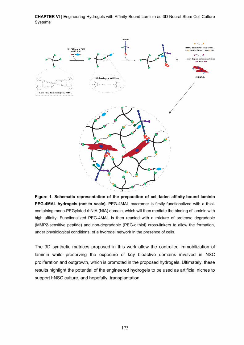
CHAPTER VI | Engineering Hydrogels with Affinity-Bound Laminin as 3D Neural Stem Cell Culture Systems
173
Figure 1. Schematic representation of the preparation of cell-laden affinity-bound laminin
PEG-4MAL hydrogels (not to scale). PEG-4MAL macromer is firstly functionalized with a thiol-containing mono-PEGylated rhNtA (NtA) domain, which will then mediate the binding of laminin with
high affinity. Functionalized PEG-4MAL is then reacted with a mixture of protease degradable
(MMP2-sensitive peptide) and non-degradable (PEG-dithiol) cross-linkers to allow the formation,
under physiological conditions, of a hydrogel network in the presence of cells.
The 3D synthetic matrices proposed in this work allow the controlled immobilization of
laminin while preserving the exposure of key bioactive domains involved in NSC
proliferation and outgrowth, which is promoted in the proposed hydrogels. Ultimately, these
results highlight the potential of the engineered hydrogels to be used as artificial niches to
support hNSC culture, and hopefully, transplantation.
CHAPTER VI | Engineering Hydrogels with Affinity-Bound Laminin as 3D Neural Stem Cell Culture Systems
174
2. Materials and Methods
2.1. Hydrogel components
A four-arm maleimide terminated poly(ethylene glycol) (PEG-4MAL; 40 kDa, > 95% purity,
> 90% substitution, Jenkem USA) was chosen as the hydrogel base material. A macromer
solution was prepared in 10 mM 4-(2-hydroxyethyl)piperazine-1-ethanesulfonic acid
(HEPES) in phosphate buffered saline (PBS, pH 7.4) at a final polymer concentration of
10.0% (w/v). Mono-PEGylated recombinant human N-terminal agrin (NtA) domain was
produced as previously described [24]. Laminin-111 from mouse Engelbreth-Holm-Swarm
sarcoma (msLn-111) was obtained from Sigma-Aldrich. A cysteine-flanked matrix
metalloproteinase-2 (MMP2)-sensitive peptide (Acetyl (Ac)-
GDCDDSGESPAY¯YTADDCDG-Amide (NH2); 2.1 kDa, > 85% purity, GenScript) and a
PEG-dithiol (3.5 kDa, ³ 95% purity, Jenkem USA) were used as the degradable and non-
degradable cross-linkers, respectively.
2.2. Mono-PEGylated rhNtA tethering efficiency
To determine the tethering efficiency of NtA to PEG-4MAL, free/unreacted thiols in the
reaction mixture were quantified using the Measure-iT thiol assay kit (Invitrogen) according
to the manufacturer’s instructions. Briefly, PEG-4MAL was functionalized with the NtA
domain (input NtA concentration: 10, 50 or 100 μM) for 15 min in 10 mM HEPES in PBS
(pH 7.4). The samples were then mixed with thiol-quantitation reagent in a black 96-well
plate and the fluorescence measured (lex = 494 nm; lem = 517 nm) using a microwell plate
reader (BioTekÒ Synergy™ Mx). A calibration curve of glutathione (0 – 55 µM) was used to
calculate the concentration of free thiols.
2.3. Preparation of affinity-bound laminin PEG-4MAL hydrogels
A schematic representation of affinity-bound laminin PEG-4MAL hydrogel preparation is
presented in Fig. 1. Briefly, PEG-4MAL was first functionalized with the NtA domain (input
NtA concentration in the gel:10, 50, or 100 μM) for 15 min in 10 mM HEPES in PBS (pH
7.4). msLn-111 (input laminin concentration in the gel: 100 µg/mL) was then added to the
mixture and allowed to bind to NtA for 30 min. Functionalized PEG-4MAL precursors were
then cross-linked into a hydrogel by addition of a mix of a MMP2-sensitive peptide and a
PEG-dithiol prepared in 10 mM HEPES in PBS (pH 6.5), at different % molar ratios (100:0;
80:20; 50:50; 20:80; 0:100). The cross-linkers were added at a 1:1 molar ratio of cysteine
residues on the cross-linkers to remaining maleimide groups on NtA-functionalized PEG-
4MAL. Hydrogels were polymerized at 37 ºC, 5% CO2 for 15 min. Unmodified PEG-4MAL
gels and PEG-4MAL gels containing entrapped msLn-111 (100 µg/mL) were also prepared
and used as controls.
CHAPTER VI | Engineering Hydrogels with Affinity-Bound Laminin as 3D Neural Stem Cell Culture Systems
175
2.4. Laminin incorporation within PEG-4MAL hydrogels
Laminin incorporation within PEG-4MAL hydrogels was quantified by an enzyme-linked
immunosorbent assay (ELISA). The hydrogels were prepared as previously described and
incubated at 37 ºC in 125 µL of PBS (pH 7.4). At day 1 and 7, the supernatant was collected,
stored at -80 ºC and then analyzed using a mouse laminin ELISA kit (Abcam, ab119572)
according to the manufacturer’s instructions. Absorbance values were measured at 450 nm
using a microwell plate reader (BioTekÒ Synergy™ Mx).
2.5. Rheological properties and mesh size estimation of affinity-bound laminin PEG-
4MAL hydrogels
The rheological properties of PEG-4MAL hydrogels were characterized using a strain-
controlled Kinexus Pro Rheometer (Malvern Instruments Ltd, Malvern, UK) and an 8 mm
diameter parallel-plate geometry. Hydrogels for each condition (Æ 8 mm) were prepared as
previously described, and allowed to hydrate in PBS (pH 7.4) at 37 ºC for 24 h. The samples
were then tested in a humidified environment at physiological temperature (37 ºC) and with
application of 30% compression (oscillatory measurement gap). Amplitude strain sweeps
(0.1 – 100% at 0.1 Hz) were initially performed for each condition to define the linear
viscoelastic region (LVR). Frequency sweeps (0.01 – 10 Hz at 1% strain) were then
performed and the storage (G’), loss (G’’) and complex (G*) modulus, as well as the phase
angle (d, º) were determined within the LVR, by averaging all data points acquired from a
0.01 – 1 Hz interval. The relative mesh size (nm) value was estimated using the following
equation [34]:
! = ($%&'( )
*+,
where G’ = storage modulus in Pa, A = Avogadro’s constant (6.022140857 ´ 1023 mol-1), R
= gas constant (8.314 m3.Pa.mol-1.K-1), T = temperature (37 ºC = 310.15 K).
2.6. Culture of human neural stem cells
Human neural stem cells (hNSCs) derived from the NIH approved H9 (WA09) human
embryonic stem cell line were purchased from Life Technologies (N7800-200). Cells were
expanded according to the manufacturer’s protocol on poly(ornithine)/msLn-111-coated
tissue coated polystyrene (TCPS) plates (Corning) in expansion medium - StemProÒ NSC
serum-free medium (SFM; Life Technologies) supplemented with basic fibroblast growth
factor (bFGF, 20 ng/mL; Life Technologies) and epidermal growth factor (EGF, 20 ng/mL;
Life Technologies).
CHAPTER VI | Engineering Hydrogels with Affinity-Bound Laminin as 3D Neural Stem Cell Culture Systems
176
2.7. Culture of hNSCs within affinity-bound laminin PEG-4MAL hydrogels
hNSCs were dissociated into single cells using StemPro accutase cell dissociation reagent
(Life Technologies) and further suspended in the solution containing laminin-functionalized
PEG-4MAL precursors (4 x 10⁶ viable cells/mL). Cell-laden hydrogels were formed by
mixing the PEG-4MAL precursor solution containing cells with the MMP2-sensitive peptide
and PEG-dithiol cross-linkers, dissolved in 10 mM HEPES in PBS (pH 6.5), at different
molar ratios (%), and subsequently incubating the polymerizing gels at 37 ºC, 5% CO2 for
15 minutes. hNSCs were initially cultured in expansion medium and, at day 2 of culture, the
medium was switched to the StemPro NSC SFM media-Neurobasal/B27 (Life
Technologies) (1:1) mix, without growth factors. At day 8, half of the medium was replaced
by the StemPro NSC SFM-Neurobasal/B27 (1:3) mix supplemented with 10 ng/mL of brain-
derived neurotrophic factor (BDNF; Peprotech) and 500 µM of N6, 2’-O-Dibutyryladenosine
3’, 5’-cyclic monophosphate sodium salt (dibutyril cAMP; Sigma). Half of the medium was
changed every other day, up to 14 days. hNSCs cultured within unmodified and PEG-4MAL
gels containing entrapped msLn-111 (100 µg/mL) were herein used as controls.
2.8. Cell viability
The distribution of viable and dead cells within PEG-4MAL hydrogels was assessed at day
7 of cell culture using a live/dead assay. Cell-hydrogel matrices were rinsed with warm PBS
pH 7.4 and incubated with 4 µM calcein AM (Life Technologies) and 6 µM propidium iodide
(PI; Life Technologies) at 37 ºC for 30 min, for detection of viable and dead cells,
respectively. After incubation, the samples were rinsed twice with PBS pH 7.4, transferred
to the culture medium, and immediately observed under confocal laser scanning
microscopy (CLSM, Leica TCS SP5). Quantitative analysis of live and dead cells was also
conducted using immunocytometry (See Supplementary Materials and Methods).
2.9. DNA quantification
The growth of hNSCs cultured within PEG-4MAL hydrogels was estimated from total cell
number after 7 days of cell culture, determined using the CyQUANT® cell proliferation assay
kit (Life Technologies) according to the manufacturer’s instructions. Briefly, cells were
extracted from the hydrogels, through sequential incubation with 1.25 mg/mL of collagenase
type II (Gibco; 1 h at 37 ºC) and StemPro accutase cell dissociation reagent (Life
Technologies; 20 min at 37 ºC) under stirring (70 rpm). Cells were mechanically dissociated
by pipetting, diluted in Glasgow Minimum Essential Medium (GMEM; Life Technologies)
supplemented with 10% (v/v) inactivated fetal bovine serum (FBS), centrifuged and the cell
pellet stored at -80 ºC. The cell pellet was then thawed at room temperature (RT) and
incubated with CyQUANT® GR dye/cell lysis buffer. Fluorescence (lex = 480 nm; lem = 520

CHAPTER VI | Engineering Hydrogels with Affinity-Bound Laminin as 3D Neural Stem Cell Culture Systems
177
nm) was measured, after mixing with CyQUANT GR dye, in a microwell plate reader
(BioTekÒ Synergy™ Mx). The total number of cells for each condition was estimated from
a standard curve generated with a known amount of hNSCs over a range of 50 to 250,000
cells. Cell samples used to generate the standard curve were measured in triplicate. For
each condition, hydrogels without cells, cultured in parallel and processed similarly to those
with cells, were used as blanks and their background fluorescence values subtracted.
2.10. hNSC neurite outgrowth
The effect of laminin site-selective immobilization on neurite outgrowth was assessed after
14 days of culture, under neuronal differentiation conditions. Samples were processed as
whole-mounts for bIII-tubulin/DNA staining as described in Supplementary Materials and
Methods, and z-sections covering a thickness of 200 µm or 80 µm were acquired with the
CLSM using an HC Plan APO CS 10´ / 0.40 NA or HC Plan APO CS 20´ / 0.70 NA
objective, respectively. Neurite outgrowth was assessed in the projected two-dimensional
(2D) images using a developed ImageJ script. Briefly, nuclei were segmented through an
Otsu thresholding method, while neurites were segmented through subtraction of the
previously identified nucleus, using a Huang thresholding method. Measurements were
obtained for total neurite length (µm) and total number of neurites per image.
2.11. Statistical Analysis
Statistical analysis was performed using GraphPad Prism 6 software (San Diego). Sample
distribution was initially tested for normality using the Kolmogorov-Smirnov test.
Comparisons between three or more groups were performed with one-way ANOVA
analysis, followed by the Bonferroni correction for pairwise comparisons or the Dunnett’s
two-tailed test for comparisons with control conditions. For all analysis, differences were
considered significant at p < 0.05.
3. Results and Discussion
3.1. PEG-4MAL hydrogels provide independent control of different biochemical and
biophysical properties
A hydrogel platform based on a PEG-4MAL macromer (Fig. 1) was selected as the base
material for this work, as we have seen it exhibits excellent in vitro and in vivo
biocompatibility with different cell types, including muscle stem cells [30] and pancreatic
islets [28]. Moreover, the modular nature of PEG-4MAL hydrogels allows independent
control over different biochemical and biophysical properties such as type and density of
cell-adhesive ligands, mechanical and structural properties, and protease-dependent
degradation [30-32]. Therefore, the use of these matrices is especially advantageous to
CHAPTER VI | Engineering Hydrogels with Affinity-Bound Laminin as 3D Neural Stem Cell Culture Systems
178
assess the effect of a particular biochemical or biophysical cue on the modulation of cell
behavior. In addition, the biophysical properties of these hydrogels were extensively
characterized in several studies from the García group [30-32]. The described properties
allowed us to initially define the molecular weight (40 kDa) and the final polymer density
(10% (w/v) of the PEG-4MAL macromer, that would result in hydrogels with mechanical
properties within the range of values preferred for NSC proliferation (100 – 1000 Pa) and
neurite branching and extension (200 – 400 Pa) [35-40].
3.1.1. Control over affinity-bound laminin PEG-4MAL hydrogel degradability is required for hNSC long-term culture Hydrogel degradability, a critical factor for matrix remodeling, was shown to be essential for
in vitro cell survival and proliferation within synthetic niches [16, 30, 32] and, therefore, is a
key feature to take into consideration when designing cell instructive microenvironments. In
this context, the incorporation of protease-sensitive peptide cross-linkers has been widely
explored to better control material degradation [33]. Among the different protease-
degradable peptides characterized to date, a fast-degrading sequence shown to be
relatively specific for MMP2 [33] was used, as this metalloproteinase is expressed at high
levels by NSCs [41, 42].
The fast gelation kinetics of the thiol-maleimide reaction (Michael-type addition; Fig. 1) may
lead to the formation of heterogeneous hydrogels, which in turn can translate into variable
cell biological outcomes [43]. Consequently, to increase the pKa of the thiol group and
thereby reduce the availability of the reactive thiolate [43], the di-thiol-containing MMP2-
sensitive cross-linker peptide used in this work, was designed to include negatively charged
amino acids flanking the thiol-bearing cysteine residue (DCDD). This strategy allowed the
thorough mixing of the hydrogel precursor solutions prior to use.
The fast-degrading nature of the selected MMP2 sequence, combined with the high levels
of MMP2 production by NSCs, led us to hypothesize that fully degradable hydrogels might
not be able to support the 3D culture of hNSCs, up to 14 days. To test this hypothesis,
affinity-bound laminin hydrogels cross-linked by addition of a MMP2-sensitive peptide
(degradable) and a PEG-dithiol (non-degradable) at different % molar ratios (100:0; 80:20;
50:50; 20:80; 0:100) were evaluated and the combination allowing the hNSC 3D culture that
would span the time frame of hNSC proliferation and neuronal differentiation established.
hNSCs showed a significantly higher number of viable cells, with cellular extensions at day
14 of cell culture, in gels formulated with a combination of degradable and non-degradable
cross-linkers, specifically for the 80% MMP2-sensitive peptide to 20% PEG-dithiol cross-
linker molar ratio, when compared to fully degradable hydrogels (p = 0.018 vs. 100:0 cross-
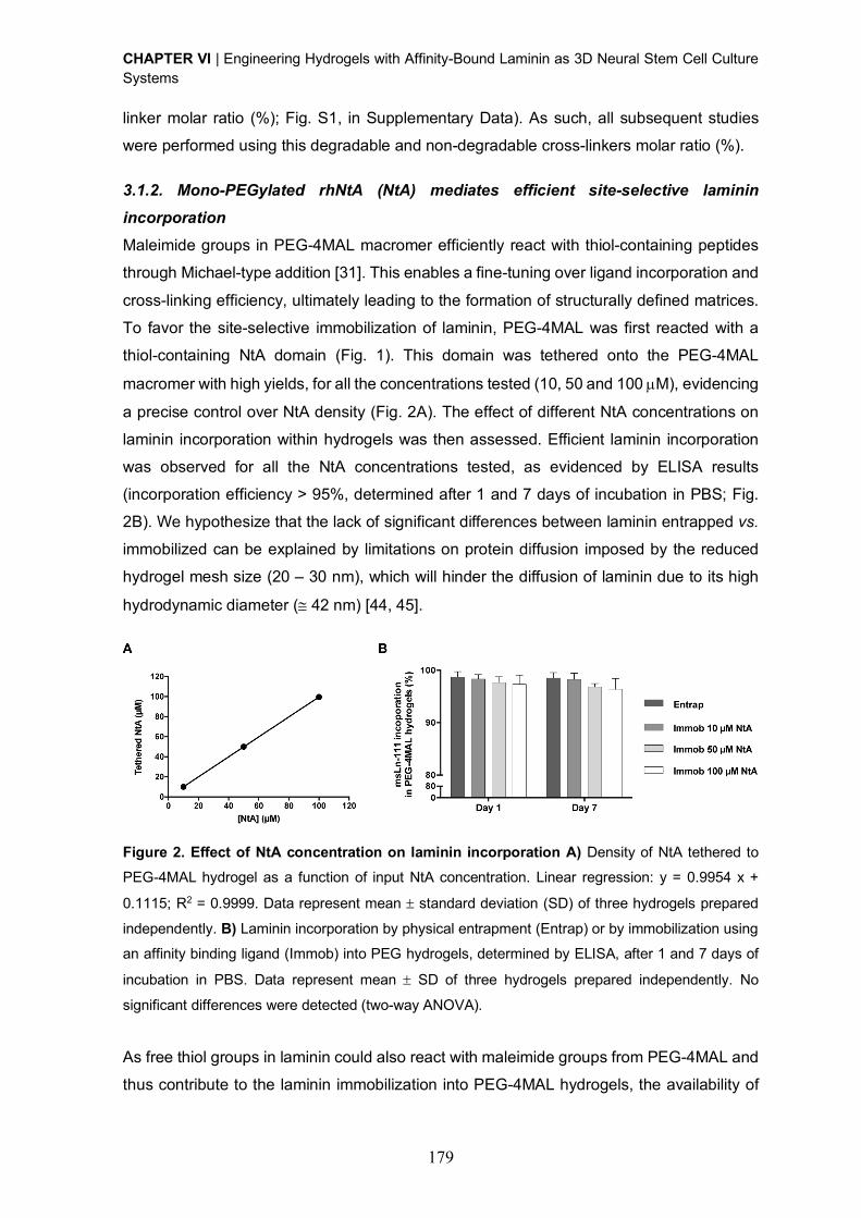
CHAPTER VI | Engineering Hydrogels with Affinity-Bound Laminin as 3D Neural Stem Cell Culture Systems
179
linker molar ratio (%); Fig. S1, in Supplementary Data). As such, all subsequent studies
were performed using this degradable and non-degradable cross-linkers molar ratio (%).
3.1.2. Mono-PEGylated rhNtA (NtA) mediates efficient site-selective laminin incorporation Maleimide groups in PEG-4MAL macromer efficiently react with thiol-containing peptides
through Michael-type addition [31]. This enables a fine-tuning over ligand incorporation and
cross-linking efficiency, ultimately leading to the formation of structurally defined matrices.
To favor the site-selective immobilization of laminin, PEG-4MAL was first reacted with a
thiol-containing NtA domain (Fig. 1). This domain was tethered onto the PEG-4MAL
macromer with high yields, for all the concentrations tested (10, 50 and 100 µM), evidencing
a precise control over NtA density (Fig. 2A). The effect of different NtA concentrations on
laminin incorporation within hydrogels was then assessed. Efficient laminin incorporation
was observed for all the NtA concentrations tested, as evidenced by ELISA results
(incorporation efficiency > 95%, determined after 1 and 7 days of incubation in PBS; Fig.
2B). We hypothesize that the lack of significant differences between laminin entrapped vs.
immobilized can be explained by limitations on protein diffusion imposed by the reduced
hydrogel mesh size (20 – 30 nm), which will hinder the diffusion of laminin due to its high
hydrodynamic diameter (@ 42 nm) [44, 45].
Figure 2. Effect of NtA concentration on laminin incorporation A) Density of NtA tethered to
PEG-4MAL hydrogel as a function of input NtA concentration. Linear regression: y = 0.9954 x +
0.1115; R2 = 0.9999. Data represent mean ± standard deviation (SD) of three hydrogels prepared
independently. B) Laminin incorporation by physical entrapment (Entrap) or by immobilization using
an affinity binding ligand (Immob) into PEG hydrogels, determined by ELISA, after 1 and 7 days of
incubation in PBS. Data represent mean ± SD of three hydrogels prepared independently. No
significant differences were detected (two-way ANOVA).
As free thiol groups in laminin could also react with maleimide groups from PEG-4MAL and
thus contribute to the laminin immobilization into PEG-4MAL hydrogels, the availability of
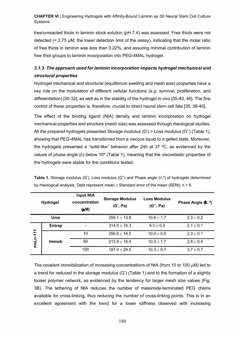
CHAPTER VI | Engineering Hydrogels with Affinity-Bound Laminin as 3D Neural Stem Cell Culture Systems
180
free/unreacted thiols in laminin stock solution (pH 7.4) was assessed. Free thiols were not
detected (< 2.75 μM, the lower detection limit of the assay), indicating that the molar ratio
of free thiols in laminin was less than 0.22%, and assuring minimal contribution of laminin
free thiol groups to laminin incorporation into PEG-4MAL hydrogel.
3.1.3. The approach used for laminin incorporation impacts hydrogel mechanical and structural properties Hydrogel mechanical and structural (equilibrium swelling and mesh size) properties have a
key role on the modulation of different cellular functions (e.g. survival, proliferation, and
differentiation) [30-32], as well as in the stability of the hydrogel in vivo [35-40, 46]. The fine
control of these properties is, therefore, crucial to direct neural stem cell fate [35, 38-40].
The effect of the binding ligand (NtA) density and laminin incorporation on hydrogel
mechanical properties and structure (mesh size) was assessed through rheological studies.
All the prepared hydrogels presented Storage modulus (G’) > Loss modulus (G’’) (Table 1),
showing that PEG-4MAL has transitioned from a viscous liquid to a gelled state. Moreover,
the hydrogels presented a “solid-like” behavior after 24h at 37 ºC, as evidenced by the
values of phase angle (d) below 10º (Table 1), meaning that the viscoelastic properties of
the hydrogels were stable for the conditions tested.
Table 1. Storage modulus (G’), Loss modulus (G’’) and Phase angle (d,º) of hydrogels determined
by rheological analysis. Data represent mean ± Standard error of the mean (SEM); n = 6.
Hydrogel
Input NtA
concentration
(µM)
Storage Modulus
(G’, Pa)
Loss Modulus
(G’’, Pa) Phase Angle (d, º)
Unm - 259.1 ± 13.8 10.6 ± 1.7 2.3 ± 0.2
msL
n-1
11
Entrap - 314.0 ± 16.3 9.3 ± 0.3 2.1 ± 0.1
Immob
10 256.0 ± 14.2 10.0 ± 0.0 2.3 ± 0.1
50 213.9 ± 16.4 10.3 ± 1.7 2.8 ± 0.4
100 187.0 ± 29.5 10.3 ± 0.7 3.7 ± 0.7
The covalent immobilization of increasing concentrations of NtA (from 10 to 100 µM) led to
a trend for reduced in the storage modulus (G’) (Table 1) and to the formation of a slightly
looser polymer network, as evidenced by the tendency for larger mesh size values (Fig.
3B). The tethering of NtA reduces the number of maleimide-terminated PEG chains
available for cross-linking, thus reducing the number of cross-linking points. This is in an
excellent agreement with the trend for a lower stiffness observed with increasing
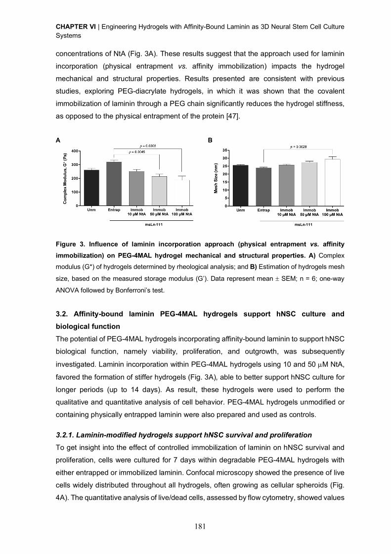
CHAPTER VI | Engineering Hydrogels with Affinity-Bound Laminin as 3D Neural Stem Cell Culture Systems
181
concentrations of NtA (Fig. 3A). These results suggest that the approach used for laminin
incorporation (physical entrapment vs. affinity immobilization) impacts the hydrogel
mechanical and structural properties. Results presented are consistent with previous
studies, exploring PEG-diacrylate hydrogels, in which it was shown that the covalent
immobilization of laminin through a PEG chain significantly reduces the hydrogel stiffness,
as opposed to the physical entrapment of the protein [47].
Figure 3. Influence of laminin incorporation approach (physical entrapment vs. affinity
immobilization) on PEG-4MAL hydrogel mechanical and structural properties. A) Complex modulus (G*) of hydrogels determined by rheological analysis; and B) Estimation of hydrogels mesh
size, based on the measured storage modulus (G’). Data represent mean ± SEM; n = 6; one-way
ANOVA followed by Bonferroni’s test.
3.2. Affinity-bound laminin PEG-4MAL hydrogels support hNSC culture and
biological function
The potential of PEG-4MAL hydrogels incorporating affinity-bound laminin to support hNSC
biological function, namely viability, proliferation, and outgrowth, was subsequently
investigated. Laminin incorporation within PEG-4MAL hydrogels using 10 and 50 µM NtA,
favored the formation of stiffer hydrogels (Fig. 3A), able to better support hNSC culture for
longer periods (up to 14 days). As result, these hydrogels were used to perform the
qualitative and quantitative analysis of cell behavior. PEG-4MAL hydrogels unmodified or
containing physically entrapped laminin were also prepared and used as controls.
3.2.1. Laminin-modified hydrogels support hNSC survival and proliferation To get insight into the effect of controlled immobilization of laminin on hNSC survival and
proliferation, cells were cultured for 7 days within degradable PEG-4MAL hydrogels with
either entrapped or immobilized laminin. Confocal microscopy showed the presence of live
cells widely distributed throughout all hydrogels, often growing as cellular spheroids (Fig.
4A). The quantitative analysis of live/dead cells, assessed by flow cytometry, showed values

CHAPTER VI | Engineering Hydrogels with Affinity-Bound Laminin as 3D Neural Stem Cell Culture Systems
182
of average cell viability greater than 75% for all the conditions tested, evidencing the good
cytocompatibility of the proposed matrices (Fig. 4B) [48].
In contrast to the physical entrapment of laminin, site-selective immobilization of laminin
using 10 µM and 50 µM NtA supported higher hNSC growth compared to unmodified
hydrogels, with significantly higher cell numbers being observed in 10 µM NtA - laminin
immobilized hydrogels (p = 0.0180) (Fig. 4C). An increase in the cell number of 1.5-, 1.9-,
4.2- and 3.1-fold (vs. initial cell density - 4 ´ 104 cells/hydrogel) was observed for
unmodified, laminin entrapped, 10 µM and 50 µM NtA - laminin immobilized hydrogels,
respectively (Fig. 4C). Since laminin incorporation in PEG-4MAL hydrogels was similar
regardless of being physically entrapped or affinity-bounded (Fig. 2B), these results suggest
that the site-selective immobilization of laminin through the use of 10 µM of NtA, better
preserved the exposure of laminin domains interacting with cell adhesion receptors, as
compared to laminin physically entrapped.
Figure 4. Ability of affinity-bound laminin to support hNSC viability and proliferation. A)
Representative 2D projections of CLSM 3D stack images of cell-laden hydrogels covering a
thickness of 300 µm, showing the distribution of live (in green) and dead (in red) cells at day 7 of cell
culture. Scale bar = 300 µm. B) Quantitative analysis of live cells at day 7, as determined by flow
cytometry. Data represent mean ± SD of three independent experiments. No significant differences

CHAPTER VI | Engineering Hydrogels with Affinity-Bound Laminin as 3D Neural Stem Cell Culture Systems
183
were detected (one-way ANOVA followed by Bonferroni’s test). C) Quantitative analysis of total cell
number at day 7, as determined by CyQuant® Cell Proliferation kit. The dotted line represents the
initial cell density/hydrogel (4 ́ 104 cells/hydrogel). Data represent mean ± SEM of three independent
experiments performed in triplicate; one-way ANOVA followed by Bonferroni’s test.
3.2.2. Controlled laminin immobilization promotes neurite outgrowth Laminin presents several bioactive domains with neurite outgrowth promoting ability, and,
as a result, plays a key role in mediating NSC migration, differentiation and neurite
extension both in vitro and in vivo [18, 20]. The effect of laminin site-selective immobilization
on hNSC neurite outgrowth was assessed after 14 days of culture under differentiation
conditions (Fig. 5). A population of differentiated neurons, expressing bIII-tubulin, with the
evident formation of a denser neuronal network, was observed within 10 µM NtA affinity-
immobilized laminin PEG-4MAL hydrogels (Fig. 5A). Differences between the conditions
tested were assessed by image processing and quantitative analysis, using ImageJ/Fiji.
Total neurite length (µm) and total number of neurites were quantified (Fig. 5B-D). The
controlled immobilization of laminin using 10 µM NtA supported a more pronounced
neuronal network formation, compared with any of the other conditions tested, as evidenced
by the higher values of total neurite length (p = 0.0035 vs. Unm; p = 0.0152 vs. Entrap; Fig.
5B) and total number of neurites (p = 0.0007 vs. Unm; p = 0.0079 vs. Entrap; Fig. 5C). The
use of 50 µM NtA, despite leading to similar amounts of laminin incorporation (Fig. 2B),
resulted in significantly lower values of total neurite length (p = 0.0056; Fig. 5C) and total
number of neurites (p = 0.0073; Fig. 5D) compared to 10 µM NtA affinity-bound laminin.
The reduction in neurite outgrowth (Fig. 5C-D) and the reduction in cell number (Fig.4C)
observed for cells cultured within hydrogels with 50 µM NtA - immobilized laminin may be
related with the inability of the protein to bind the NtA domain, as result of steric effects. The
results obtained contrast to previous studies in which no significant differences on neurite
outgrowth were observed when laminin was covalently immobilized using a non-selective
approach, vs. physically entrapped laminin [47]. This gives a good indication that the
strategy proposed in this study, indeed, potentiate laminin bioactivity.
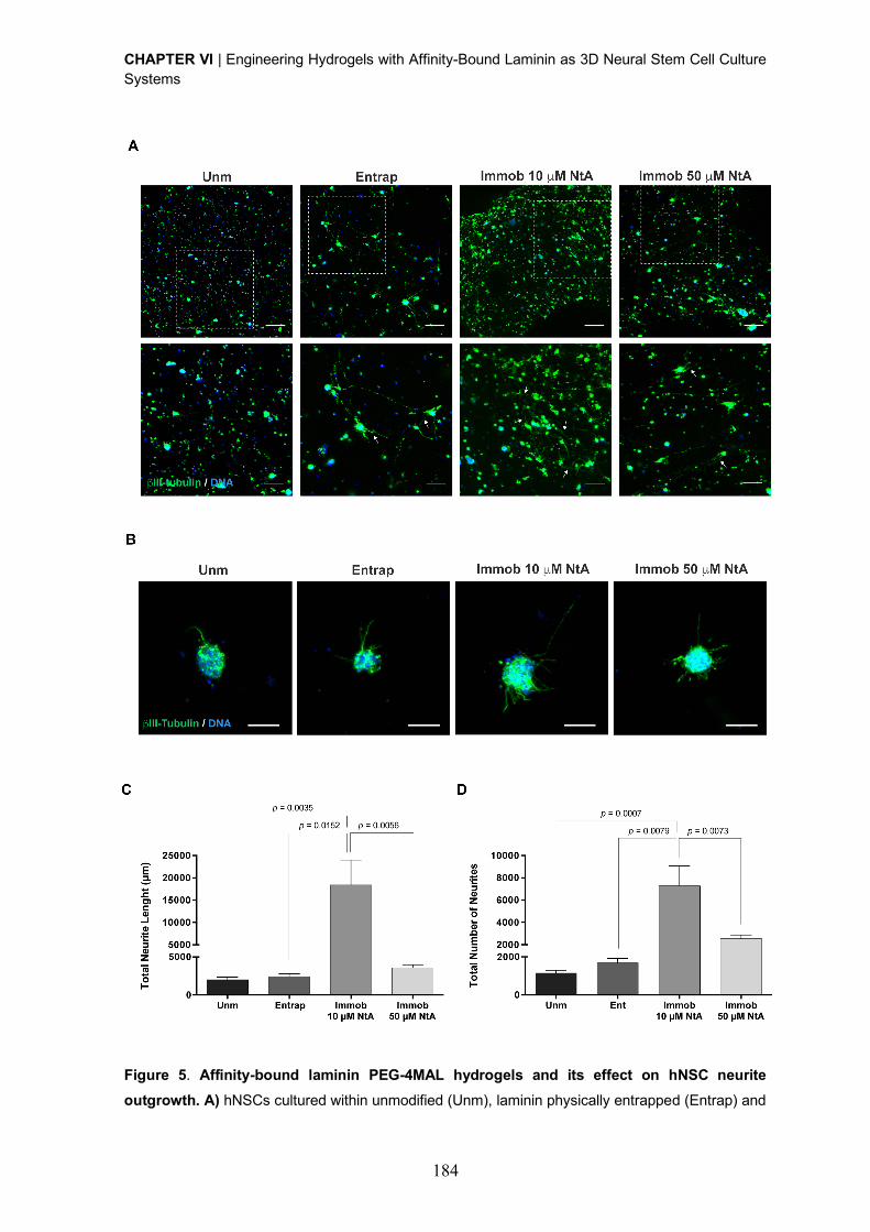
CHAPTER VI | Engineering Hydrogels with Affinity-Bound Laminin as 3D Neural Stem Cell Culture Systems
184
Figure 5. Affinity-bound laminin PEG-4MAL hydrogels and its effect on hNSC neurite
outgrowth. A) hNSCs cultured within unmodified (Unm), laminin physically entrapped (Entrap) and
CHAPTER VI | Engineering Hydrogels with Affinity-Bound Laminin as 3D Neural Stem Cell Culture Systems
185
NtA affinity-bound laminin (Immob) PEG-4MAL hydrogels after 14 days of culture, under
differentiation conditions. Images show 2D projections of CLSM 3D stack images of cells processed
for immunofluorescence labeling of bIII-tubulin/DNA covering a thickness of 200 µm (top images)
and 100 µm (bottom images). Lower panels show magnified views of selected regions highlighted
by dashed squares in the upper panel. Arrows depict neurites expressing bIII-tubulin protruding from
neurospheres. Scale Bar = 200 µm (top images); 100 µm (bottom images). B) Representative 2D
projections of CLSM 3D stack images of hNSC outgrowth, covering a thickness of 80 µm, after 14
days of culture under differentiation conditions. Scale Bar = 80 µm. B) Total Neurite Length; and C)
Total Number of Neurites determined by quantitative analysis of images acquired in CLSM. Data
represent mean ± SD of 12-15 images analyzed per condition; one-way ANOVA followed by
Bonferroni’s test.
Overall, these results clearly demonstrate that the site-selective immobilization of laminin
through the use of 10 µM NtA better preserved protein bioactivity in terms of ability to
promote hNSC proliferation and outgrowth, when compared to laminin physically
entrapped.
4. Conclusion
The present work demonstrates the potential of a PEG-4MAL hydrogel modified with
affinity-bound laminin as a dynamic 3D platform enabling NSC proliferation and
differentiation. The proposed strategy allows the oriented and controlled immobilization of
laminin, with preservation of protein bioactivity, thus overcoming some of the main
drawbacks associated with the currently available strategies (i.e. physically entrapment and
non-selective covalent immobilization). Moreover, the immobilization approach used in this
work is highly versatile, as result of the ability of NtA to bind with high affinity to different
laminin isoforms comprising the g1 chain, which represents more than 50% of the isoforms
identified to date. Therefore, the reported 3D matrices can be used for the site-selective
immobilization of different laminin isoforms and used for the study of cell-laminin
interactions occurring in different stem cell niches and disease contexts. These hydrogels
can be also of interest to provide a controlled niche with instructive cues to improve survival,
engraftment and long-term function of transplanted stem cells; for the creation of tissue-
engineered constructs; and for the development of cell expansion and biomanufacturing
systems.
CHAPTER VI | Engineering Hydrogels with Affinity-Bound Laminin as 3D Neural Stem Cell Culture Systems
186
Acknowledgments
Confocal microscopy was conducted at the Bioimaging i3S Scientific Platform, member of
the PPBI (PPBI-POCI-01-0145-FEDER-022122), with the assistance of Maria Lázaro. This
work was funded by projects NORTE-01-0145-FEDER-000008 and NORTE-01-0145-
FEDER-000012, supported by Norte Portugal Regional Operational Programme (NORTE
2020), under the PORTUGAL 2020 Partnership Agreement, through the European
Regional Development Fund (ERDF) and FEDER - Fundo Europeu de Desenvolvimento
Regional funds through the COMPETE 2020 - Operacional Programme for
Competitiveness and Internationalisation (POCI), Portugal 2020; by Portuguese funds
through FCT/MCTES in the framework of the project "Institute for Research and Innovation
in Health Sciences" (POCI-01-0145-FEDER-007274), and by Santa Casa da Misericordia
de Lisboa through project COMBINE (Prémio Neurociências Melo e Castro 1068-2015).
DB was supported by FCT PhD Programs (PD/BD/105953/2014) and Programa
Operacional Potencial Humano (POCH), in the scope of the BiotechHealth Program
(Doctoral Program on Cellular and Molecular Biotechnology Applied to Health Sciences),
Programa FLAD Healthcare 2020 and the project PARES (Prémio Albino Aroso). Eduardo
Conde-Sousa was supported by a post-doctoral grant of the project PPBI-POCI-01-0145-
FEDER-022122, in the scope of FCT National Roadmap of Research Infrastructures. AJG
acknowledges support from the National Institutes of Health (R01 AR062368).
CHAPTER VI | Engineering Hydrogels with Affinity-Bound Laminin as 3D Neural Stem Cell Culture Systems
187
SUPPLEMENTARY DATA
Supplementary Materials and Methods
Screening Degradable (SGESPAY¯YTA) : Non-degradable (PEG-dithiol) cross-linker molar
ratio (%)
After 14 days in culture, the viability of human neural stem cells (hNSCs) within hydrogels
cross-linked with different molar ratios (%) of cysteine-flanked matrix metalloproteinase-2
(MMP2)-sensitive peptide (SGESPAY¯YTA) and PEG-dithiol was assessed using a
Live/Dead assay, performed according the protocol described in section 2.8. Average
occupied area (µm2) by live (Calcein+) cells was assessed in the projected two-dimensional
(2D) images through the application of an Otsu thresholding method and determination of
the total area occupied by cells in each image.
Cell viability - Immunocytometry
Quantitative analysis of cell viability was conducted by immunocytometry performed in six-
pooled hydrogels, after hydrogel dissociation. Briefly, the hydrogels were sequentially
incubated with 1.25 mg/mL of collagenase type II (Gibco; 1h at 37 ºC) and StemPro
accutase cell dissociation reagent (Life Technologies; 20 min at 37 ºC) under stirring (70
rpm). Cells were mechanically dissociated by pipetting, diluted in Glasgow Minimal
Essential Medium (GMEM; Life Technologies) supplemented with 10% (v/v) inactivated
fetal bovine serum (FBS) and centrifuged. For live/dead staining, cells were then transferred
to a round-bottomed 96-well plate and stained with calcein AM (67 nM, 20 min at 37 ºC)
and propidium iodide (PI; 6 µM, 10 min at 37 ºC). Cells were finally washed trice and
suspended in FACS buffer (PBS pH 7.4 supplemented with 2% (v/v) FBS) for flow cytometry
analysis on BD FACSCantoTM II (BD Biosciences). Unlabeled cells were used to set the
fluorescence gates and cells stained with calcein AM and PI only were used to establish
the compensation settings. For each flow cytometry analysis, 10,000 events were acquired
inside the respective gate.
Neurite outgrowth - Immunohistochemistry
The ability of hNSCs to extend neurites was assessed after 14 days of cell culture, under
differentiation conditions, in samples processed for bIII-tubulin/DNA staining. Cell-laden
hydrogels were fixed in 3.7% (w/v) paraformaldehyde (PFA) solution diluted 1:1 in culture
media (30 min; 37 ºC) and permeabilized with 0.2% (v/v) Triton X-100 in PBS (45 min; room
temperature (RT)). Samples were then incubated with blocking buffer (5% (v/v) bovine
serum albumin (BSA) in PBS) for 1 h at RT, followed by incubation with the primary antibody
– mouse anti-bIII-tubulin (1:500; Biolegend, 801201), overnight at 4 ºC. To detect primary
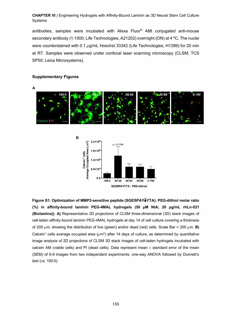
CHAPTER VI | Engineering Hydrogels with Affinity-Bound Laminin as 3D Neural Stem Cell Culture Systems
188
antibodies, samples were incubated with Alexa Fluor® 488 conjugated anti-mouse
secondary antibody (1:1000; Life Technologies, A21202) overnight (ON) at 4 ºC. The nuclei
were counterstained with 0.1 µg/mL Hoechst 33342 (Life Technologies, H1399) for 20 min
at RT. Samples were observed under confocal laser scanning microscopy (CLSM; TCS
SP5II; Leica Microsystems).
Supplementary Figures
Figure S1. Optimization of MMP2-sensitive peptide (SGESPAY¯YTA): PEG-dithiol molar ratio
(%) in affinity-bound laminin PEG-4MAL hydrogels (50 μM NtA; 20 μg/mL rhLn-521
(Biolamina)). A) Representative 2D projections of CLSM three-dimensional (3D) stack images of
cell-laden affinity-bound laminin PEG-4MAL hydrogels at day 14 of cell culture covering a thickness
of 200 µm, showing the distribution of live (green) and/or dead (red) cells. Scale Bar = 200 µm. B)
Calcein+ cells average occupied area (µm2) after 14 days of culture, as determined by quantitative
image analysis of 2D projections of CLSM 3D stack images of cell-laden hydrogels incubated with
calcein AM (viable cells) and PI (dead cells). Data represent mean ± standard error of the mean
(SEM) of 6-9 images from two independent experiments; one-way ANOVA followed by Dunnett’s test (vs. 100:0)
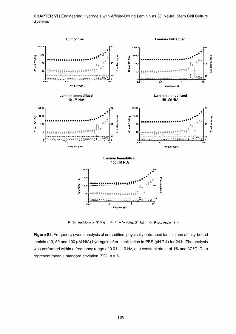
CHAPTER VI | Engineering Hydrogels with Affinity-Bound Laminin as 3D Neural Stem Cell Culture Systems
189
Figure S2. Frequency sweep analysis of unmodified, physically entrapped laminin and affinity-bound
laminin (10, 50 and 100 µM NtA) hydrogels after stabilization in PBS (pH 7.4) for 24 h. The analysis
was performed within a frequency range of 0.01 – 10 Hz, at a constant strain of 1% and 37 ºC. Data
represent mean ± standard deviation (SD); n = 6.
CHAPTER VI | Engineering Hydrogels with Affinity-Bound Laminin as 3D Neural Stem Cell Culture Systems
190
References
[1] S. Pluchino, L. Zanotti, M. Deleidi, G. Martino, Neural stem cells and their use as
therapeutic tool in neurological disorders, Brain Res Brain Res Rev 48(2) (2005) 211-9.
[2] C. Cossetti, C. Alfaro-Cervello, M. Donega, G. Tyzack, S. Pluchino, New perspectives
of tissue remodelling with neural stem and progenitor cell-based therapies, Cell Tissue Res
349(1) (2012) 321-9.
[3] F.H. Gage, S. Temple, Neural stem cells: generating and regenerating the brain, Neuron
80(3) (2013) 588-601.
[4] F. Gattazzo, A. Urciuolo, P. Bonaldo, Extracellular matrix: a dynamic microenvironment
for stem cell niche, Biochim Biophys Acta 1840(8) (2014) 2506-19.
[5] S.W. Lane, D.A. Williams, F.M. Watt, Modulating the stem cell niche for tissue
regeneration, Nat Biotechnol 32(8) (2014) 795-803.
[6] A. Vishwakarma, J. Rouwkema, P.A. Jones, J.M. Karp, The Need to Study, Mimic and
Target Stem Cell Niches, in: A. Vishwakarma, J.M. Karp (Eds.), Biology and Engineering of
Stem Cell Niches, Elsevier2017.
[7] J.L. Wilson, T.C. McDevitt, Biofunctional Hydrogels for Three-Dimensional Stem Cell
Culture, in: A. Vishwakarma, J.M. Karp (Eds.), Biology and Engineering of Stem Cell
Niches, Elsevier2017.
[8] M.H. Amer, F. Rose, K.M. Shakesheff, M. Modo, L.J. White, Translational considerations
in injectable cell-based therapeutics for neurological applications: concepts, progress and
challenges, NPJ Regen Med 2 (2017) 23.
[9] F. Ruedinger, A. Lavrentieva, C. Blume, I. Pepelanova, T. Scheper, Hydrogels for 3D
mammalian cell culture: a starting guide for laboratory practice, Appl Microbiol Biotechnol
99(2) (2015) 623-36.
[10] Technology Platforms for 3D Cell Culture: A User's Guide, John Wiley & Sons Ltd.2017.
[11] M.W. Tibbitt, K.S. Anseth, Hydrogels as Extracellular Matrix Mimics for 3D Cell Culture,
Biotechnol. Bioeng. 103(4) (2009) 655-663.
[12] D. Barros, I.F. Amaral, A.P. Pego, Biomimetic synthetic self-assembled hydrogels for
cell transplantation, Curr Top Med Chem 15(13) (2015) 1209-26.
[13] K.J. Lampe, S.C. Heilshorn, Building stem cell niches from the molecule up through
engineered peptide materials, Neurosci Lett 519(2) (2012) 138-46.
[14] J. Lam, S.T. Carmichael, W.E. Lowry, T. Segura, Hydrogel design of experiments
methodology to optimize hydrogel for iPSC-NPC culture, Adv Healthc Mater 4(4) (2015)
534-9.
CHAPTER VI | Engineering Hydrogels with Affinity-Bound Laminin as 3D Neural Stem Cell Culture Systems
191
[15] C. Regalado-Santiago, E. Juarez-Aguilar, J.D. Olivares-Hernandez, E. Tamariz,
Mimicking Neural Stem Cell Niche by Biocompatible Substrates, Stem Cells Int 2016 (2016)
1513285.
[16] C.M. Madl, B.L. LeSavage, R.E. Dewi, C.B. Dinh, R.S. Stowers, M. Khariton, K.J.
Lampe, D. Nguyen, O. Chaudhuri, A. Enejder, S.C. Heilshorn, Maintenance of neural
progenitor cell stemness in 3D hydrogels requires matrix remodelling, Nat Mater 16(12)
(2017) 1233-1242.
[17] A. Hyysalo, M. Ristola, M.E. Makinen, S. Hayrynen, M. Nykter, S. Narkilahti, Laminin
alpha5 substrates promote survival, network formation and functional development of
human pluripotent stem cell-derived neurons in vitro, Stem Cell Res 24 (2017) 118-127.
[18] L. Luckenbill-Edds, Laminin and the mechanism of neuronal outgrowth, Brain Res Brain
Res Rev 23(1-2) (1997) 1-27.
[19] S.K. Powell, H.K. Kleinman, Neuronal laminins and their cellular receptors, Int J
Biochem Cell Biol 29(3) (1997) 401-14.
[20] S. Plantman, M. Patarroyo, K. Fried, A. Domogatskaya, K. Tryggvason, H.
Hammarberg, S. Cullheim, Integrin-laminin interactions controlling neurite outgrowth from
adult DRG neurons in vitro, Mol Cell Neurosci 39(1) (2008) 50-62.
[21] R.S. Rogers, H. Nishimune, The role of laminins in the organization and function of
neuromuscular junctions, Matrix Biol 57-58 (2017) 86-105.
[22] H. Nishimune, G. Valdez, G. Jarad, C.L. Moulson, U. Muller, J.H. Miner, J.R. Sanes,
Laminins promote postsynaptic maturation by an autocrine mechanism at the
neuromuscular junction, J Cell Biol 182(6) (2008) 1201-15.
[23] M. Talovic, M. Marcinczyk, N. Ziemkiewicz, K. Garg, Laminin Enriched Scaffolds for
Tissue Engineering Applications, Advances in Tissue Engineering and Regenerative
Medicine 2(3) (2017).
[24] D. Barros, P. Parreira, J. Furtado, F. Ferreira-da-Silva, E. Conde-Sousa, A.J. Garcia,
M.C.L. Martins, I.F. Amaral, A.P. Pego, An affinity-based approach to engineer laminin-
presenting cell instructive microenvironments, Biomaterials 192 (2019) 601-611.
[25] D. Barros, I.F. Amaral, A.P. Pêgo, Laminin immobilization, methods and uses thereof,
Provisional Patent Application, 2018.
[26] J.B. Mascarenhas, M.A. Ruegg, U. Winzen, W. Halfter, J. Engel, J. Stetefeld, Mapping
of the laminin-binding site of the N-terminal agrin domain (NtA), EMBO J 22(3) (2003) 529-
36.
[27] A.J. Denzer, T. Schulthess, C. Fauser, B. Schumacher, R.A. Kammerer, J. Engel, M.A.
Ruegg, Electron microscopic structure of agrin and mapping of its binding site in laminin-1,
EMBO J 17(2) (1998) 335-43.
CHAPTER VI | Engineering Hydrogels with Affinity-Bound Laminin as 3D Neural Stem Cell Culture Systems
192
[28] J.D. Weaver, D.M. Headen, J. Aquart, C.T. Johnson, L.D. Shea, H. Shirwan, A.J.
Garcia, Vasculogenic hydrogel enhances islet survival, engraftment, and function in leading
extrahepatic sites, Sci Adv 3(6) (2017) e1700184.
[29] R. Cruz-Acuna, M. Quiros, A.E. Farkas, P.H. Dedhia, S. Huang, D. Siuda, V. Garcia-
Hernandez, A.J. Miller, J.R. Spence, A. Nusrat, A.J. Garcia, Synthetic hydrogels for human
intestinal organoid generation and colonic wound repair, Nat Cell Biol 19(11) (2017) 1326-
1335.
[30] W.M. Han, S.E. Anderson, M. Mohiuddin, D. Barros, S.A. Nakhai, E. Shin, I.F. Amaral,
A.P. Pego, A.J. Garcia, Y.C. Jang, Synthetic matrix enhances transplanted satellite cell
engraftment in dystrophic and aged skeletal muscle with comorbid trauma, Sci Adv 4(8)
(2018) eaar4008.
[31] E.A. Phelps, N.O. Enemchukwu, V.F. Fiore, J.C. Sy, N. Murthy, T.A. Sulchek, T.H.
Barker, A.J. Garcia, Maleimide cross-linked bioactive PEG hydrogel exhibits improved
reaction kinetics and cross-linking for cell encapsulation and in situ delivery, Adv Mater
24(1) (2012) 64-70, 2.
[32] N.O. Enemchukwu, R. Cruz-Acuna, T. Bongiorno, C.T. Johnson, J.R. Garcia, T.
Sulchek, A.J. Garcia, Synthetic matrices reveal contributions of ECM biophysical and
biochemical properties to epithelial morphogenesis, J Cell Biol 212(1) (2016) 113-24.
[33] J. Patterson, J.A. Hubbell, Enhanced proteolytic degradation of molecularly engineered
PEG hydrogels in response to MMP-1 and MMP-2, Biomaterials 31(30) (2010) 7836-45.
[34] M. Rubinstein, R.H. Colby, Polymer physics, Oxford University Press, 2003.
[35] L.A. Flanagan, Y.E. Ju, B. Marg, M. Osterfield, P.A. Janmey, Neurite branching on
deformable substrates, Neuroreport 13(18) (2002) 2411-5.
[36] A.J. Engler, S. Sen, H.L. Sweeney, D.E. Discher, Matrix elasticity directs stem cell
lineage specification, Cell 126(4) (2006) 677-89.
[37] P.C. Georges, W.J. Miller, D.F. Meaney, E.S. Sawyer, P.A. Janmey, Matrices with
compliance comparable to that of brain tissue select neuronal over glial growth in mixed
cortical cultures, Biophys J 90(8) (2006) 3012-8.
[38] K. Saha, A.J. Keung, E.F. Irwin, Y. Li, L. Little, D.V. Schaffer, K.E. Healy, Substrate
modulus directs neural stem cell behavior, Biophys. J. 95(9) (2008) 4426-38.
[39] A. Banerjee, M. Arha, S. Choudhary, R.S. Ashton, S.R. Bhatia, D.V. Schaffer, R.S.
Kane, The influence of hydrogel modulus on the proliferation and differentiation of
encapsulated neural stem cells, Biomaterials 30(27) (2009) 4695-9.
[40] S.R. Hynes, M.F. Rauch, J.P. Bertram, E.B. Lavik, A library of tunable poly(ethylene
glycol)/poly(L-lysine) hydrogels to investigate the material cues that influence neural stem
cell differentiation, J Biomed Mater Res A 89(2) (2009) 499-509.
CHAPTER VI | Engineering Hydrogels with Affinity-Bound Laminin as 3D Neural Stem Cell Culture Systems
193
[41] T.L. Laundos, J. Silva, M. Assuncao, P. Quelhas, C. Monteiro, C. Oliveira, M.J. Oliveira,
A.P. Pego, I.F. Amaral, Rotary orbital suspension culture of embryonic stem cell-derived
neural stem/progenitor cells: impact of hydrodynamic culture on aggregate yield,
morphology and cell phenotype, J Tissue Eng Regen Med 11(8) (2017) 2227-2240.
[42] A.R. Bento, P. Quelhas, M.J. Oliveira, A.P. Pego, I.F. Amaral, Three-dimensional
culture of single embryonic stem-derived neural/stem progenitor cells in fibrin hydrogels:
neuronal network formation and matrix remodelling, J Tissue Eng Regen Med 11(12) (2017)
3494-3507.
[43] N.J. Darling, Y.S. Hung, S. Sharma, T. Segura, Controlling the kinetics of thiol-
maleimide Michael-type addition gelation kinetics for the generation of homogenous
poly(ethylene glycol) hydrogels, Biomaterials 101 (2016) 199-206.
[44] K. Onuma, N. Kanzaki, Size Distribution and Intermolecular Interaction of Laminin-1 in
Physiological Solutions, J. Phys. Chem. 107 (2003) 11799-11804.
[45] M. Hjort, M. Bauer, S. Gunnarsson, E. Marsell, A.A. Zakharov, G. Karlsson, E. Sanfins,
C.N. Prinz, R. Wallenberg, T. Cedervall, A. Mikkelsen, Electron microscopy imaging of
proteins on gallium phosphide semiconductor nanowires, Nanoscale 8(7) (2016) 3936-43.
[46] K.J. Lampe, R.G. Mooney, K.B. Bjugstad, M.J. Mahoney, Effect of macromer weight
percent on neural cell growth in 2D and 3D nondegradable PEG hydrogel culture, J Biomed
Mater Res A 94(4) (2010) 1162-71.
[47] L. Marquardt, R.K. Willits, Student award winner in the undergraduate's degree
category for the Society for Biomaterials 35th Annual Meeting, Orlando, Florida, April 13-
16, 2011. Neurite growth in PEG gels: effect of mechanical stiffness and laminin
concentration, J Biomed Mater Res A 98(1) (2011) 1-6.
[48] I.O.f. Standardization, ISO 10993-5:2009 : Biological evaluation of medical devices
Tests for in vitro cytotoxicity, 2009.
CHAPTER VI | Engineering Hydrogels with Affinity-Bound Laminin as 3D Neural Stem Cell Culture Systems
194
186
CHAPTER VII _________________________________________________________________________
An Alternative Approach to Engineer Synthetic
Hydrogels with Affinity-Bound Laminin

CHAPTER VII | An Alternative Approach to Engineer Synthetic Hydrogels with Affinity-Bound Laminin
187
CHAPTER VII | An Alternative Approach to Engineer Synthetic Hydrogels with Affinity-Bound Laminin
197
An Alternative Approach to Engineer Synthetic Hydrogels
with Affinity-Bound Laminin
Daniela Barros 1,2,3, Andrés J. García 4,5, Isabel F. Amaral 1,2,6, Ana P. Pêgo 1,2,3,6
1 i3S - Instituto de Investigação e Inovação em Saúde, Universidade do Porto (UPorto),
Portugal 2 INEB - Instituto de Engenharia Biomédica, UPorto, Portugal 3 ICBAS - Instituto de Ciências Biomédicas Abel Salazar, UPorto, Portugal 4 Parker H. Petit Institute for Bioengineering and Biosciences, Georgia Institute of Technology,
Atlanta, Georgia, USA 5 George W. Woodruff School of Mechanical Engineering, Georgia Institute of Technology,
Atlanta, Georgia, USA 6 FEUP - Faculdade de Engenharia, UPorto, Portugal

CHAPTER VII | An Alternative Approach to Engineer Synthetic Hydrogels with Affinity-Bound Laminin
198
Abstract
Laminin-521 (Ln-521) has a key role on the modulation of neural cell behavior and have shown
potential to be used as a robust substratum for the culture and renewal of human embryonic
and pluripotent stem cells. Despite the potential of this laminin isoform, to the best of our
knowledge, no study to date has explored the immobilization of Ln-521 for the development
of three-dimensional (3D) matrices for the culture of human neural stem cells (hNSCs). In this
regard, and envisaging the development of a defined and whole-human 3D cell-instructive
platform, capable of supporting hNSC differentiation and function, in this study we describe
the preparation of a degradable synthetic hydrogel functionalized with affinity-bound
recombinant human Ln-521. The proposed methodology introduces some modifications to the
previously described approach for laminin immobilization, aiming the improvement of laminin
binding to NtA and its impact on NSC behavior. Specifically, a mono-PEGylated recombinant
human N-terminal agrin (NtA) domain was incubated with laminin and the resultant conjugate
(NtA-Ln-521) was subsequently tethered to a four-arm maleimide terminated poly(ethylene
glycol) (PEG-4MAL) macromer. The hydrogels were formed, under physiological conditions,
through the use of a mixture of protease degradable and non-degradable cross-linkers.
Affinity-bound laminin PEG-4MAL hydrogels were shown to better preserve laminin bioactivity,
as evidenced by the enhanced ability to support neurite outgrowth, when compared to
unmodified hydrogels and hydrogels with equal amount of physically entrapped laminin.
However, in the latter case, significant differences were not attained.
Overall, the results obtained show that the alternative approach herein proposed for laminin
immobilization, can be used as an effective immobilization strategy for hydrogel
biofunctionalization envisaging its use as defined platforms for hNSC culture.
CHAPTER VII | An Alternative Approach to Engineer Synthetic Hydrogels with Affinity-Bound Laminin
199
Neural stem cells (NSCs) reside within a dynamic and complex microenvironment, the NSC
niche, which provides a unique and specialized microenvironment, where cell-cell interactions
and local microenvironmental cues, including those from extracellular matrix (ECM), soluble
factors and cellular components, are key for the modulation of NSC function [1]. In this regard,
in the last few years, there was an attempt to develop new platforms to allow the dissection of
the role of individual or multiple components of the niche on the modulation of NSC function
and fate.
Laminin constitutes one of the most important ECM effectors of NSC niches [2, 3], as it
provides important biological cues to different biological processes, including cell adhesion
and viability [4] and neuronal outgrowth and migration [5-7]. Furthermore, laminin has other
key roles in CNS homeostasis, as contributing to synapse function and stability [8, 9]. Several
studies have already explored the incorporation of this ECM protein into three-dimensional
(3D) cell-instructive microenvironments [10-17], by taking advantage of different
immobilization strategies. The latter include non-covalent incorporation or physical
entrapment or, alternatively, non-specific covalent immobilization by taking advantage of
functional groups present in multiple sites of the laminin structure such as amines [10-14] and
thiols [15-17]. Although such strategies are rather efficient, their application to immobilize
proteins might be limited due to the lack of control over protein conformation and orientation
upon immobilization, which often results in a reduction or loss of bioactivity. In this regard, in
recent years, affinity-binding strategies have been increasingly explored to mediate highly site-
and conformation-specific reactions for protein and peptide immobilization [18]. Besides
allowing the retention of protein bioactivity, the versatility of this approach allows the
generation of dynamic biomimetic microenvironments. An important factor to take into
consideration when applying such strategies is the appropriate selection of the binding pairs.
In this regard, different “artificial” moieties, including streptavidin, biotin, protein A, among
others, have been widely explored for the development of 3D matrices with a broad
applicability and specificity [19, 20]. Alternatively, natural binding partners have become an
attractive alternative, as strong non-covalent interactions can be achieved without the need of
the protein of interest to be either recombinantly or chemically modified [21, 22].
Laminin-521 (Ln-521) has a key role on the modulation of neural cell behavior, including
neuronal adhesion, viability and network formation [4, 23]. Moreover, this laminin isoform
demonstrated great potential to be used as a xeno-free substratum for the culture and renewal
of human embryonic [24] and pluripotent [4] stem cells. Despite the potential of this laminin
isoform, to the best of our knowledge, no study to date, explored its immobilization for the
development of defined 3D matrices for the culture of human NSCs (hNSCs). In this regard,
and envisaging the development of a whole-human 3D cell-instructive microenvironment, to
CHAPTER VII | An Alternative Approach to Engineer Synthetic Hydrogels with Affinity-Bound Laminin
200
support hNSC fate and function, in this study we propose the development of an affinity-bound
recombinant human Ln-521 (rhLn-521) degradable synthetic hydrogel, using an
immobilization strategy previously proposed [25-27]. The proposed approach takes advantage
of the well described native high affinity interaction between laminin and the N-terminal agrin
(NtA) domain [28, 29]. A four-arm maleimide terminated poly(ethylene glycol) (PEG-4MAL)
macromer was used as the hydrogel base material (Fig. 1), due to its good biocompatibility,
both in vitro and in vivo [30-32], and well characterized biochemical and biophysical properties
[32-34]. To allow the site-selective immobilization of laminin on the PEG-4MAL hydrogel, a
thiol-containing mono-PEGylated rhNtA (NtA) domain was produced and characterized,
showing high affinity for rhLn-521 (dissociation constant (KD) = 1.58 ± 0.41 nM) [25]. To enable
the hNSC 3D culture that would span the time frame of hNSC proliferation and neuronal
differentiation, a mix of protease degradable (cysteine-flanked matrix metalloproteinase 2
(MMP2)-sensitive peptide) [35] and non-degradable (PEG-dithiol) cross-linkers was used to
mediate, under physiological conditions, the formation of a degradable hydrogel network (Fig.
1). The developed hydrogels were expected to allow the highly specific and orthogonal
immobilization of laminin to form a cell-instructive microenvironment. Moreover, the versatility
and dynamic nature of the proposed strategy can be further explored for the immobilization of
different exogenous laminin isoforms, with application in distinct disease contexts, as well as
to assess their exchangeability by endogenously secreted laminin.

CHAPTER VII | An Alternative Approach to Engineer Synthetic Hydrogels with Affinity-Bound Laminin
201
Figure 1. Schematic representation of the preparation of cell-laden affinity-bound laminin PEG-
4MAL hydrogels (not to scale). A thiol-containing mono-PEGylated rhNtA (NtA) is firstly reacted with rhLn-521 and the formed conjugate subsequently tethered to a PEG-4MAL macromer. Functionalized
PEG-4MAL is finally reacted with a mixture of protease degradable (MMP2-sensitive peptide) and non-
degradable (PEG-dithiol) cross-linkers to allow the formation, under physiological conditions, of a
hydrogel network in the presence of cells.
Preliminary studies, exploring the site-selective immobilization of rhLn-521 into a PEG-4MAL
hydrogel, using a previously proposed approach [25-27], did not evidence a significant
increase on hNSC neurite outgrowth, when compared to unmodified hydrogels and hydrogels
with equal amount of physically entrapped laminin (Fig. S1, in Supplementary Data). In line
with this outcome, we hypothesize that the lower bioactivity presented by the site-selective
immobilized laminin could be related with the methodology explored for laminin incorporation
into the hydrogel. Namely, in the previously reported immobilization scheme, a PEG-4MAL
macromer is firstly functionalized with a thiol-containing mono-PEGylated rhNtA domain,
which will then mediate the binding of laminin with high affinity. The hydrogel is subsequently
CHAPTER VII | An Alternative Approach to Engineer Synthetic Hydrogels with Affinity-Bound Laminin
202
formed by cross-linking of the laminin functionalized PEG-4MAL. Therefore, in this work, we
aimed to explore an alternative functionalization scheme. In the newly proposed approach the
NtA domain is initially incubated with the rhLn-521 and the resultant conjugate tethered to a
PEG-4MAL macromer (Fig. 1). This was proposed under the assumption that protein-protein
interaction in solution would allow a more efficient and controlled immobilization of rhLn-521,
even at lower concentrations. As maleimide groups efficiently react with thiol-containing
moieties through Michael-type addition, one expects to have a finer control over the final
conjugate incorporation. Different NtA concentrations (10, 50 and 100 µM) were initially used
to determine the most suitable concentration allowing to have a finer control over the binding
ligand (NtA) density/presentation and ultimately, over the amount of affinity-bound laminin.
To evaluate the tethering efficiency of the NtA-rhLn-521 conjugates to the PEG-4MAL
macromer and their further incorporation into the final hydrogels, radiolabeling studies were
conducted. For that, both NtA and laminin were labeled with 125I, as described elsewhere [36],
and then used for hydrogel preparation. The amount of NtA or laminin retained in each
hydrogel was determined after 1 and 7 days, by measuring the gamma activity of the hydrogel
at each time point. NtA tethering efficiency (Fig. 2A) or laminin incorporation (Fig 2B) was
expressed as a percentage of the initial gamma activity of the hydrogel, prior to incubation in
buffer. NtA domain was tethered onto the PEG-4MAL macromers with yields below 50%, for
all the concentrations tested (10, 50 and 100 µM; Fig. 2A and Table S1 in Supplementary
Data). Affinity-bound laminin revealed a laminin incorporation of 60% at day 1, which decrease
to 40-50% at day 7 (Fig. 2B and Table S1 in Supplementary Data). Site-selective
immobilization of laminin using 10 µM NtA favors a significant higher protein incorporation
compared to that observed when 100 µM NtA domain was used (p = 0.0241, day 7). The same
trend was also observed for hydrogels with physically entrapped laminin (p = 0.0146 vs.
Immob 100 µM NtA, day 7).
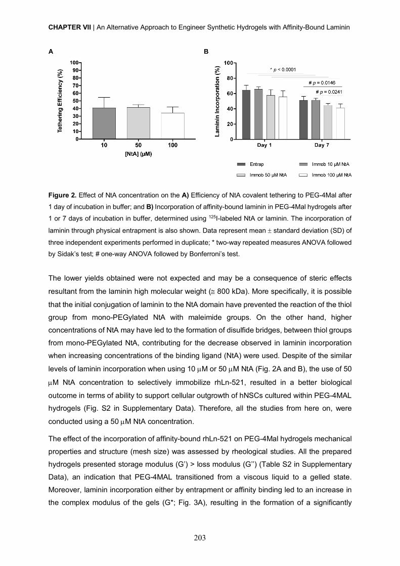
CHAPTER VII | An Alternative Approach to Engineer Synthetic Hydrogels with Affinity-Bound Laminin
203
Figure 2. Effect of NtA concentration on the A) Efficiency of NtA covalent tethering to PEG-4Mal after
1 day of incubation in buffer; and B) Incorporation of affinity-bound laminin in PEG-4Mal hydrogels after
1 or 7 days of incubation in buffer, determined using 125I-labeled NtA or laminin. The incorporation of
laminin through physical entrapment is also shown. Data represent mean ± standard deviation (SD) of
three independent experiments performed in duplicate; * two-way repeated measures ANOVA followed by Sidak’s test; # one-way ANOVA followed by Bonferroni’s test.
The lower yields obtained were not expected and may be a consequence of steric effects
resultant from the laminin high molecular weight (@ 800 kDa). More specifically, it is possible
that the initial conjugation of laminin to the NtA domain have prevented the reaction of the thiol
group from mono-PEGylated NtA with maleimide groups. On the other hand, higher
concentrations of NtA may have led to the formation of disulfide bridges, between thiol groups
from mono-PEGylated NtA, contributing for the decrease observed in laminin incorporation
when increasing concentrations of the binding ligand (NtA) were used. Despite of the similar
levels of laminin incorporation when using 10 µM or 50 µM NtA (Fig. 2A and B), the use of 50
µM NtA concentration to selectively immobilize rhLn-521, resulted in a better biological
outcome in terms of ability to support cellular outgrowth of hNSCs cultured within PEG-4MAL
hydrogels (Fig. S2 in Supplementary Data). Therefore, all the studies from here on, were
conducted using a 50 µM NtA concentration.
The effect of the incorporation of affinity-bound rhLn-521 on PEG-4Mal hydrogels mechanical
properties and structure (mesh size) was assessed by rheological studies. All the prepared
hydrogels presented storage modulus (G’) > loss modulus (G’’) (Table S2 in Supplementary
Data), an indication that PEG-4MAL transitioned from a viscous liquid to a gelled state.
Moreover, laminin incorporation either by entrapment or affinity binding led to an increase in
the complex modulus of the gels (G*; Fig. 3A), resulting in the formation of a significantly

CHAPTER VII | An Alternative Approach to Engineer Synthetic Hydrogels with Affinity-Bound Laminin
204
tighter polymer network, as evidenced by the significantly decrease in mesh size (Fig. 3B),
when compared to unmodified hydrogels. Nevertheless, the G* values found for hydrogels
containing affinity-bound laminin lie well within the range of those reported for human brain
tissue (100 – 1000 Pa) and preferred for neurite extension and branching (200 – 400 Pa) [37-
42].
Figure 3. Influence of affinity-bound laminin incorporation on PEG-4MAL hydrogel mechanical
and structural properties. A) Complex modulus (G*) of hydrogels determined by rheological analysis;
and B) Estimation of hydrogels mesh size, based on the measured storage modulus (G’). Data
represent mean ± standard error of the mean (SEM); n = 6; one-way ANOVA followed by Bonferroni’s
test.
The bioactivity of hydrogels with affinity-bound rhLn-521 was subsequently evaluated, and
compared with the performance of hydrogels containing entrapped laminin. For this purpose,
hNSCs were cultured within degradable PEG-4MAL hydrogels with either soluble or affinity-
bound rhLn-521 (20 µg/mL) and cell viability and neurite outgrowth were assessed.
Unmodified hydrogels were used as controls. The effect of the proposed strategy on hNSC
survival was evaluated after 7 days of culture. Confocal analysis showed the presence of
viable cells evenly distributed throughout the hydrogels, often growing as cellular spheroids,
for all the conditions tested (Fig. 4A). The quantitative analysis of viable/unviable cells,
assessed by flow cytometry upon cell isolation from hydrogels, showed fractions of viable cells
greater than 75% for all the conditions tested, evidencing the good cytocompatibility of the
proposed matrices (Fig. 4B) [43].

CHAPTER VII | An Alternative Approach to Engineer Synthetic Hydrogels with Affinity-Bound Laminin
205
Figure 4. Affinity-bound laminin PEG-4MAL hydrogels and its effect on hNSC viability and
neurite outgrowth. A) Distribution of viable (green) and dead (red) cells cultured within unmodified
(Unm), laminin physically entrapped (Entrap) and NtA affinity-bound laminin (Immob) PEG-4MAL
hydrogels was assessed at day 7 of cell culture, using a live/dead assay. Representative 2D projections
of confocal laser scanning microscopy (CLSM) 3D stack images of cell-laden hydrogels covering a
thickness of 200 µm, showing the distribution of live (green) and dead (red) cells. Scale bar = 200 µm.
B) Quantitative analysis of live cells at day 7, as determined by flow cytometry. Data represent mean ±
SD of three independent experiments. C) hNSCs cultured within unmodified (Unm), rhLn-521 physically
entrapped (Entrap) and 50 µM NtA-affinity-bound rhLn-521 (Immob) PEG-4MAL hydrogels after 14
days of culture, under differentiation conditions. Images show 2D projections of CLSM 3D stack images
of cells processed for immunofluorescence labeling of bIII-tubulin/DNA and covering a thickness of 200
µm (top images) and 100 µm (bottom images). Scale Bar = 200 µm (top images); 100 µm (bottom
images). D) Total Neurite Length; and E) Total Number of Neurites determined by quantitative analysis
of images acquired in CLSM. Data represent mean ± SD of 11-17 images analyzed per condition; one-
way ANOVA followed by Bonferroni’s test.
To investigate the effect of laminin site-selective immobilization on neurite outgrowth, hNSCs
were cultured for 14 days under differentiation conditions, and then processed as whole-
CHAPTER VII | An Alternative Approach to Engineer Synthetic Hydrogels with Affinity-Bound Laminin
206
mounts for bIII-tubulin and DNA staining (Fig. 4C). A population of differentiated neurons,
expressing bIII-tubulin and starting to form a neuronal network, was observed for affinity-
bound laminin PEG-4MAL hydrogels (Fig. 4C). Differences between the conditions tested
were assessed by image process and quantitative analysis, using ImageJ/Fiji. More
specifically, total neurite length (µm) and total number of neurites were quantified (Fig. 4D-F).
A significant increase in neurite outgrowth was observed for the PEG-4MAL hydrogels with
affinity-bound laminin, compared to unmodified hydrogels, as evidenced by the higher values
of total neurite length (15663 ± 7951 µm; p = 0.0253; Fig. 4D) and total number of neurites
(6327 ± 2817; p = 0.0491; Fig. 4E). Laminin entrapment also promoted neurite outgrowth in
PEG-4MAL hydrogels but its impact was not significant. Despite the tendency for increased
neurite outgrowth observed for hydrogels with affinity-bound laminin, no significant differences
on total neurite length and total number of neurites were detected when compared to
hydrogels containing laminin entrapped (Fig. 4D-E)
Overall, these results suggest that the alternative strategy explored in this work for laminin
incorporation better preserves laminin bioactivity, thus assuring the hydrogel biofunctionality.
Although this work represents a good starting point for the development of a whole-human 3D
cell instructive matrix, future studies exploring approaches that better promote the tethering of
NtA-bound laminin to PEG-4MAL with preservation of its bioactive domains, and/or allow an
increase in rhLn-521 initial concentration (> 20 µg/mL) need to be conducted.
Materials and Methods
Hydrogel components A four-arm maleimide terminated poly(ethylene glycol) (PEG-4MAL; 40 kDa, > 95% purity,
>90% substitution, Jenkem USA) was chosen as the hydrogel base material. A macromer
solution was prepared in 10 mM 4-(2-hydroxyethyl)piperazine-1-ethanesulfonic acid (HEPES)
buffer (pH 7.4) at a final polymer concentration of 10% (w/v). Mono-PEGylated recombinant
human N-terminal agrin (NtA) domain was produced as previously described [25].
Recombinant human laminin-521 (rhLn-521) was obtained from Biolamina. A cysteine-flanked
matrix metalloproteinase-2 (MMP2)-sensitive peptide (Acetyl (Ac)-
GDCDDSGESPAY¯YTADDCDG-Amide (NH2); 2.1 kDa, > 85% purity, GenScript) and a PEG-
dithiol (3.5 kDa, ³ 95% purity, Jenkem USA) were used as the degradable and non-degradable
cross-linkers, respectively.
CHAPTER VII | An Alternative Approach to Engineer Synthetic Hydrogels with Affinity-Bound Laminin
207
Preparation of affinity-bound rhLn-521 PEG-4MAL hydrogels A schematic representation of affinity-bound laminin PEG-4MAL hydrogel preparation is
presented in Fig. 1. Briefly, NtA (input concentration in the gel: 50 µM) was initially reacted
with rhLn-521 (input concentration in the gel: 20 µg/mL) for 30 min at room temperature (RT)
in 10 mM HEPES buffer (pH 7.4). PEG-4MAL was then functionalized with the previously
formed conjugate (NtA – rhLn-521) for 15 min in 10 mM HEPES buffer (pH 7.4). Functionalized
PEG-4MAL macromers were then cross-linked linked into a hydrogel by addition of a mix of a
MMP2-sensitive peptide and a PEG-dithiol dissolved in 10 mM HEPES buffer (pH 6.5), at an
80:20 molar ratio (%). The cross-linkers were added at a 1:1 molar ratio of cysteine residues
on the cross-linkers to remaining maleimide groups on NtA-rhLn521-functionalized PEG-
4MAL. Hydrogels were polymerized at 37ºC, 5% CO2 for 15 min. Unmodified PEG-4MAL gels
and PEG-4MAL gels containing entrapped rhLn-521 (20 µg/mL) were also prepared and used
as controls.
125I-radiolabeling – NtA and laminin incorporation efficiency To assess the incorporation efficiency of NtA and laminin into PEG-4MAL hydrogels, both
proteins were first labeled with 125I as described elsewhere [36] and then used for hydrogel
preparation. Briefly, NtA or laminin (5 µg) were labeled with 0.5 mCi of Na125I (Perkin Elmer)
using the Iodogen method [44], and further purified using a PD-10 Desalting Column packed
with Sephadex TM G-25 Medium (GE Healthcare), to remove free 125I ions. Fractions with yield
of 125I-labelling ≥ 98.8% were used. Functionalized hydrogels (10 µL) were prepared as
described above in radioimmunoassay (RIA) tubes, using NtA or laminin spiked with 125I-
labeled domain or protein, respectively, in order to achieve a final activity of » 2.0 ́ 105 cpm/µg
of NtA/laminin. Hydrogels containing 125I-labeled soluble laminin (20 µg/mL) were also
prepared and used as controls. The gels were processed straight away for gamma activity
counting g-counter (CAPRAC® - t Wipe Test / Well Counter; Capintec, Inc.) and further
incubated with 125 µL of phosphate buffered saline (PBS) buffer containing 2% (w/v) bovine
serum albumin (BSA) at 37 ºC. The amount of NtA or laminin retained in each hydrogel was
determined after 1 and 7 days. At each time point, the buffer solution was replaced by fresh
buffer, the gamma activity of the hydrogel measured, and NtA or laminin incorporation was
expressed as a percentage of the initial gamma activity of the hydrogel prior to incubation in
buffer.
Rheological properties and mesh size estimation of affinity-bound laminin PEG-4MAL hydrogels The viscoelastic properties of PEG-4MAL hydrogels were characterized using an MCR 302
stress-controlled rheometer (Anton Paar, Austria) with a 9 mm diameter, 2º cone angle (CP10-
CHAPTER VII | An Alternative Approach to Engineer Synthetic Hydrogels with Affinity-Bound Laminin
208
2) and plate geometry. Hydrogels for each condition (Æ 8 mm) were prepared as previously
described, and allowed to hydrate in PBS buffer at 37 ºC for 24h. The samples were then
loaded between the cone and the plate, in a humidified environment at physiological
temperature (37 ºC), and the measuring system was lowered to a 40 µm gap using TruGapTM
system. Amplitude strain sweeps (0.001 – 1% at 10 rad/s) were initially performed for each
condition to define the linear viscoelastic region (LVR). Frequency sweeps (0.1-100 rad/s at
0.01% strain) were then performed and the storage (G’), loss (G’’) and complex (G*) modulus
Determined within the LVR, by averaging all data points acquired from a 0.1 - 10 rad/s interval.
The relative mesh size (nm) value was estimated using the following equation [45]:
! = ($%&'( )
*+,
where G’ = storage modulus in Pa, A = Avogadro’s constant (6.022140857 ´ 1023 mol-1), R =
gas constant (8.314 m3.Pa.mol-1.K-1), T = temperature (37 ºC = 310.15 K) .
Culture of human neural stem cells Human neural stem cells (hNSCs) derived from the NIH approved H9 (WA09) human
embryonic stem cell line were purchased from Life Technologies (N7800-200). Cells were
expanded according to the manufacturer’s protocol on poly(ornithine)/laminin-111 from mouse
Engelbreth-Holm-Swarm sarcoma (msLn-111; Sigma-Aldrich)-coated tissue coated
polystyrene (TCPS) plates (Corning) in expansion medium - StemProÒ NSC serum free
medium (SFM; Life Technologies) supplemented with basic fibroblast growth factor (bFGF, 20
ng/mL; Life Technologies) and epidermal growth factor (EGF, 20 ng/mL; Life Technologies).
Culture of hNSCs within affinity-bound rhLn-521 PEG-4MAL hydrogels hNSCs were dissociated into single cells using StemPro Accutase cell dissociation reagent
(Life Technologies) and further suspended in the solution containing laminin-functionalized
PEG-4MAL precursors (4 x 10⁶ viable cells/mL). Cell-laden hydrogels were formed by mixing
the PEG-4MAL precursor solution containing cells with the MMP2-sensitive peptide and PEG-
dithiol cross-linkers, dissolved in 10 mM HEPES (pH 6.5), at an 80:20 molar ratio (%), and
subsequently incubating the polymerizing gels at 37 ºC, 5% CO2 for 15 minutes. hNSCs were
initially cultured in expansion medium and, at day 2 of culture, the medium was switched to
the StemPro NSC SFM media-Neurobasal/B27 (Life Technologies) (1:1) mix, without growth
factors. At day 8, half of the medium was replaced by the StemPro NSC SFM-Neurobasal/B27
(1:3) mix supplemented with 10 ng/mL of brain-derived neurotrophic factor (BDNF; Peprotech)
and 500 µM of N6, 2’-O-Dibutyryladenosine 3’, 5’-cyclic monophosphate sodium salt (dibutyril
cAMP; Sigma). Half of the medium was changed every other day, up to 14 days. hNSCs
CHAPTER VII | An Alternative Approach to Engineer Synthetic Hydrogels with Affinity-Bound Laminin
209
cultured within unmodified and PEG-4MAL gels containing entrapped rhLn-521 (20 µg/mL)
were herein used as controls.
Cell viability The distribution of viable and dead cells within PEG hydrogels was assessed at day 7 of cell
culture using a live/dead assay. Cell-hydrogel matrices were rinsed with warm PBS pH 7.4
and incubated with 4 µM calcein AM (Life Technologies) and 6 µM propidium iodide (PI; Life
Technologies) at 37 ºC for 30 min, for detection of viable and dead cells, respectively. After
incubation, the samples were rinsed twice with PBS pH 7.4, transferred to culture medium,
and immediately observed under confocal laser scanning microscopy (CLSM, Leica TCS
SP5). Quantitative analysis of live and dead cells was also conducted using immunocytometry
(See Supplementary Materials and Methods).
hNSC neurite outgrowth The effect of laminin site-selective immobilization on neurite outgrowth was assessed after 14
days of culture under neuronal differentiation conditions. Samples were processed as whole-
mounts for bIII-tubulin/DNA staining as described in Supplementary Materials and Methods,
and z-sections covering a thickness of 200 µm or 100 µm were acquired with the CLSM using
an HC Plan APO CS 10´ / 0.40 NA or HC Plan APO CS 20´ / 0.70 NA objective, respectively.
Neurite outgrowth was assessed in the projected two-dimensional (2D) images using a
developed ImageJ script. Briefly, nuclei were segmented through an Otsu thresholding
method, while neurites were segmented through subtraction of the previously identified
nucleus, using a Huang thresholding method. Measurements were obtained for total neurite
length (µm) and total number of neurites per image.
Statistical Analysis Statistical analysis was performed using GraphPad Prism 6 software (San Diego). Sample
distribution was initially tested for normality using the Kolmogorov-Smirnov test. Comparisons
between three or more groups were performed with one-way ANOVA analysis, followed by
the Bonferroni correction for pairwise comparisons. For all analysis, differences were
considered significant at p < 0.05.
Acknowledgments
Radiolabeling studies were conducted at Serviço de Medicina Nuclear – IPO Porto, with the
assistance of Dra. Carla Vaz, Dr. João Miranda dos Santos and Dr. Hugo Duarte. Confocal
microscopy was conducted at the Bioimaging i3S Scientific Platform, member of the PPBI
(PPBI-POCI-01-0145-FEDER-022122), with the assistance of Maria Lázaro. This work was
CHAPTER VII | An Alternative Approach to Engineer Synthetic Hydrogels with Affinity-Bound Laminin
210
funded by projects NORTE-01-0145-FEDER-000008 and NORTE-01-0145-FEDER-000012,
supported by Norte Portugal Regional Operational Programme (NORTE 2020), under the
PORTUGAL 2020 Partnership Agreement, through the European Regional Development
Fund (ERDF) and FEDER - Fundo Europeu de Desenvolvimento Regional funds through the
COMPETE 2020 - Operacional Programme for Competitiveness and Internationalisation
(POCI), Portugal 2020; by Portuguese funds through FCT/MCTES in the framework of the
project "Institute for Research and Innovation in Health Sciences" (POCI-01-0145-FEDER-
007274), and by Santa Casa da Misericordia de Lisboa through project COMBINE (Prémio
Neurociências Melo e Castro 1068-2015).
D.B. was supported by FCT PhD Programs (PD/BD/105953/2014) and Programa Operacional
Potencial Humano (POCH), in the scope of the BiotechHealth Program (Doctoral Program on
Cellular and Molecular Biotechnology Applied to Health Sciences), Programa FLAD
Healthcare 2020 and the project PARES (Prémio Albino Aroso).
CHAPTER VII | An Alternative Approach to Engineer Synthetic Hydrogels with Affinity-Bound Laminin
211
SUPPLEMENTARY DATA
Supplementary Materials and Methods
Cell viability - Immunocytometry
Quantitative analysis of cell viability was conducted by immunocytometry performed in six-
pooled hydrogels, after hydrogel dissociation. Briefly, the hydrogels were sequentially
incubated with 1.25 mg/mL of collagenase type II (Gibco; 1h at 37 ºC) and StemPro accutase
cell dissociation reagent (Life Technologies; 20 min at 37 ºC) under stirring (70 rpm). Cells
were mechanically dissociated by pipetting, diluted in Glasgow Minimal Essential Medium
(GMEM; Life Technologies) supplemented with 10% (v/v) inactivated fetal bovine serum (FBS)
and centrifuged. For live/dead staining cells were then transferred to a round-bottomed 96-
well plate and stained with calcein AM (67 nM, 20 min at 37 ºC) and propidium iodide (PI; 6
µM, 10 min at 37 ºC). Cells were finally washed trice and suspended in FACS buffer (PBS pH
7.4 supplemented with 2% (v/v) FBS) for flow cytometry analysis on BD FACSCantoTM II (BD
Biosciences). Unlabeled cells were used to set the fluorescence gates and cells stained with
calcein AM and PI only were used to establish the compensation settings. For each flow
cytometry analysis, 10,000 events were acquired inside the respective gate.
Neurite outgrowth - Immunohistochemistry
The ability of hNSCs to extend neurites was assessed after 14 days of culture, under
differentiation conditions, in samples processed as whole-mounts for bIII-tubulin/DNA staining.
Cells/hydrogels were fixed in 3.7% (w/v) paraformaldehyde (PFA) solution diluted 1:1 in
culture media (30 min; 37 ºC) and permeabilized with 0.2% (v/v) Triton X-100 in PBS (45 min;
RT). Samples were then incubated with blocking buffer (5% (v/v) bovine serum albumin (BSA)
in PBS) for 1 h at RT, followed by incubation with the primary antibody – mouse anti-bIII-
tubulin (1:500; Biolegend, 801201), overnight at 4 ºC. To detect primary antibodies, samples
were incubated with Alexa Fluor® 488 conjugated anti-mouse secondary antibody (1:1000;
Life Technologies, A21202) ON at 4 ºC. The nuclei were counterstained with 0.1 µg/mL
Hoechst 33342 (Life Technologies, H1399) for 20 min at RT. Samples were observed under
confocal laser scanning microscopy (CLSM; TCS SP5II; Leica Mycrosystems).
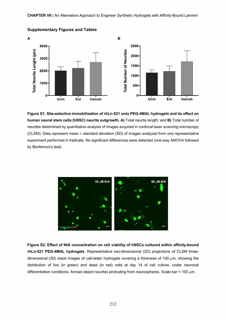
CHAPTER VII | An Alternative Approach to Engineer Synthetic Hydrogels with Affinity-Bound Laminin
212
Supplementary Figures and Tables
Figure S1. Site-selective immobilization of rhLn-521 onto PEG-4MAL hydrogels and its effect on
human neural stem cells (hNSC) neurite outgrowth. A) Total neurite length; and B) Total number of
neurites determined by quantitative analysis of images acquired in confocal laser scanning microscopy
(CLSM). Data represent mean ± standard deviation (SD) of images analyzed from one representative
experiment performed in triplicate. No significant differences were detected (one-way ANOVA followed
by Bonferroni’s test).
Figure S2. Effect of NtA concentration on cell viability of hNSCs cultured within affinity-bound
rhLn-521 PEG-4MAL hydrogels. Representative two-dimensional (2D) projections of CLSM three-
dimensional (3D) stack images of cell-laden hydrogels covering a thickness of 100 µm, showing the
distribution of live (in green) and dead (in red) cells at day 14 of cell culture, under neuronal
differentiation conditions. Arrows depict neurites protruding from neurospheres. Scale bar = 100 µm.
CHAPTER VII | An Alternative Approach to Engineer Synthetic Hydrogels with Affinity-Bound Laminin
213
Table S1. Final amount of tethered NtA and laminin (msLn-111) incorporation into PEG-4MAL
hydrogels, determined by radiolabeling analysis. Data represent mean ± SD of 3 independent
experiments performed in duplicate.
Input [NtA] (µM) Tethered [NtA] (µM)
Laminin incorporation
[msLn-111] (µg/mL)
Day 1 Day 7
0 - 12.9 ± 1.3 10.2 ± 1.0
10 4.1 ± 1.3 13.1 ± 0.6 10.3 ± 0.5
50 20.8 ± 1.7 11.5 ± 1.5 9.0 ± 0.5
100 34.3 ± 7.9 11.1 ± 1.5 8.3 ± 1.0
Table S2. Impact of rhLn-521 immobilization on the storage (G’) and loss moduli (G’’) of PEG-4MAL
hydrogels, as determined by rheological analysis. Data represent mean ± standard error of the mean
(SEM); n = 6; one-way ANOVA followed by Bonferroni’s test; ** p < 0.01 (vs. unmodified).
Hydrogel Storage Modulus (G’, Pa) Loss Modulus (G’’, Pa)
Unmodified 87.5 ± 7.7 7.1 ± 0.7
Entrapped 118.2 ± 6.2 8.3 ± 0.5
Immobilized 132.5 ± 9.9 ** 7.4 ± 0.3
CHAPTER VII | An Alternative Approach to Engineer Synthetic Hydrogels with Affinity-Bound Laminin
214
References
[1] S.W. Lane, D.A. Williams, F.M. Watt, Modulating the stem cell niche for tissue regeneration,
Nat Biotechnol 32(8) (2014) 795-803.
[2] A. Kerever, J. Schnack, D. Vellinga, N. Ichikawa, C. Moon, E. Arikawa-Hirasawa, J.T. Efird,
F. Mercier, Novel extracellular matrix structures in the neural stem cell niche capture the
neurogenic factor fibroblast growth factor 2 from the extracellular milieu, Stem Cells 25(9)
(2007) 2146-57.
[3] I. Kazanis, J.D. Lathia, T.J. Vadakkan, E. Raborn, R. Wan, M.R. Mughal, D.M. Eckley, T.
Sasaki, B. Patton, M.P. Mattson, K.K. Hirschi, M.E. Dickinson, C. ffrench-Constant,
Quiescence and activation of stem and precursor cell populations in the subependymal zone
of the mammalian brain are associated with distinct cellular and extracellular matrix signals, J
Neurosci 30(29) (2010) 9771-81.
[4] A. Hyysalo, M. Ristola, M.E. Makinen, S. Hayrynen, M. Nykter, S. Narkilahti, Laminin
alpha5 substrates promote survival, network formation and functional development of human
pluripotent stem cell-derived neurons in vitro, Stem Cell Res 24 (2017) 118-127.
[5] L. Luckenbill-Edds, Laminin and the mechanism of neuronal outgrowth, Brain Res Brain
Res Rev 23(1-2) (1997) 1-27.
[6] S.K. Powell, H.K. Kleinman, Neuronal laminins and their cellular receptors, Int J Biochem
Cell Biol 29(3) (1997) 401-14.
[7] S. Plantman, M. Patarroyo, K. Fried, A. Domogatskaya, K. Tryggvason, H. Hammarberg,
S. Cullheim, Integrin-laminin interactions controlling neurite outgrowth from adult DRG
neurons in vitro, Mol Cell Neurosci 39(1) (2008) 50-62.
[8] R.S. Rogers, H. Nishimune, The role of laminins in the organization and function of
neuromuscular junctions, Matrix Biol 57-58 (2017) 86-105.
[9] H. Nishimune, G. Valdez, G. Jarad, C.L. Moulson, U. Muller, J.H. Miner, J.R. Sanes,
Laminins promote postsynaptic maturation by an autocrine mechanism at the neuromuscular
junction, J Cell Biol 182(6) (2008) 1201-15.
[10] S.E. Stabenfeldt, A.J. Garcia, M.C. LaPlaca, Thermoreversible laminin-functionalized
hydrogel for neural tissue engineering, J Biomed Mater Res A 77(4) (2006) 718-25.
[11] S.E. Stabenfeldt, G. Munglani, A.J. Garcia, M.C. LaPlaca, Biomimetic microenvironment
modulates neural stem cell survival, migration, and differentiation, Tissue Eng Part A 16(12)
(2010) 3747-58.
[12] S.E. Stabenfeldt, M.C. LaPlaca, Variations in rigidity and ligand density influence neuronal
response in methylcellulose-laminin hydrogels, Acta Biomater 7(12) (2011) 4102-8.
CHAPTER VII | An Alternative Approach to Engineer Synthetic Hydrogels with Affinity-Bound Laminin
215
[13] A.T. Francisco, R.J. Mancino, R.D. Bowles, J.M. Brunger, D.M. Tainter, Y.T. Chen, W.J.
Richardson, F. Guilak, L.A. Setton, Injectable laminin-functionalized hydrogel for nucleus
pulposus regeneration, Biomaterials 34(30) (2013) 7381-8.
[14] A.T. Francisco, P.Y. Hwang, C.G. Jeong, L. Jing, J. Chen, L.A. Setton, Photocrosslinkable
laminin-functionalized polyethylene glycol hydrogel for intervertebral disc regeneration, Acta
Biomater 10(3) (2014) 1102-11.
[15] C.P. Addington, J.M. Heffernan, C.S. Millar-Haskell, E.W. Tucker, R.W. Sirianni, S.E.
Stabenfeldt, Enhancing neural stem cell response to SDF-1alpha gradients through hyaluronic
acid-laminin hydrogels, Biomaterials 72 (2015) 11-9.
[16] J. Arulmoli, H.J. Wright, D.T.T. Phan, U. Sheth, R.A. Que, G.A. Botten, M. Keating, E.L.
Botvinick, M.M. Pathak, T.I. Zarembinski, D.S. Yanni, O.V. Razorenova, C.C.W. Hughes, L.A.
Flanagan, Combination scaffolds of salmon fibrin, hyaluronic acid, and laminin for human
neural stem cell and vascular tissue engineering, Acta Biomater 43 (2016) 122-138.
[17] C.P. Addington, S. Dharmawaj, J.M. Heffernan, R.W. Sirianni, S.E. Stabenfeldt,
Hyaluronic acid-laminin hydrogels increase neural stem cell transplant retention and migratory
response to SDF-1alpha, Matrix Biol 60-61 (2017) 206-216.
[18] S.A. Fisher, A.E.G. Baker, M.S. Shoichet, Designing Peptide and Protein Modified
Hydrogels: Selecting the Optimal Conjugation Strategy, J Am Chem Soc 139(22) (2017) 7416-
7427.
[19] C.C. Lin, K.S. Anseth, Controlling Affinity Binding with Peptide-Functionalized
Poly(ethylene glycol) Hydrogels, Adv Funct Mater 19(14) (2009) 2325.
[20] J. Parker, N. Mitrousis, M.S. Shoichet, Hydrogel for Simultaneous Tunable Growth Factor
Delivery and Enhanced Viability of Encapsulated Cells in Vitro, Biomacromolecules 17(2)
(2016) 476-84.
[21] S.E. Sakiyama-Elbert, J.A. Hubbell, Controlled release of nerve growth factor from a
heparin-containing fibrin-based cell ingrowth matrix, J Control Release 69(1) (2000) 149-58.
[22] S.E. Sakiyama-Elbert, J.A. Hubbell, Development of fibrin derivatives for controlled
release of heparin-binding growth factors, J Control Release 65(3) (2000) 389-402.
[23] A. Bergeron, H. Sherman, P. Pardo, H. Gitschier, H. Nandivada, D. Saxena, Corning®
rLaminin-521 (Human) for Expansion and Differentiation of Human Neural Stem Cells,
Corning Incorporated | Application Note (2015).
[24] S. Rodin, L. Antonsson, C. Niaudet, O.E. Simonson, E. Salmela, E.M. Hansson, A.
Domogatskaya, Z. Xiao, P. Damdimopoulou, M. Sheikhi, J. Inzunza, A.S. Nilsson, D. Baker,
R. Kuiper, Y. Sun, E. Blennow, M. Nordenskjold, K.H. Grinnemo, J. Kere, C. Betsholtz, O.
Hovatta, K. Tryggvason, Clonal culturing of human embryonic stem cells on laminin-521/E-
cadherin matrix in defined and xeno-free environment, Nat Commun 5 (2014) 3195.
CHAPTER VII | An Alternative Approach to Engineer Synthetic Hydrogels with Affinity-Bound Laminin
216
[25] D. Barros, P. Parreira, J. Furtado, F. Ferreira-da-Silva, E. Conde-Sousa, A.J. Garcia,
M.C.L. Martins, I.F. Amaral, A.P. Pego, An affinity-based approach to engineer laminin-
presenting cell instructive microenvironments, Biomaterials 192 (2019) 601-611.
[26] D. Barros, I.F. Amaral, A.P. Pego, Laminin immobilization, methods and uses thereof,
Portuguese Provisional Patent Application no. 20181000074673, filled 2018/11/30.
[27] D. Barros, E. Conde-Sousa, A.J. Garcia, I.F. Amaral, A.P. Pego, Engineering hydrogels
with affinity-bound laminin as 3D neural stem cell culture systems Manuscript in preparation
(2019).
[28] J.B. Mascarenhas, M.A. Ruegg, U. Winzen, W. Halfter, J. Engel, J. Stetefeld, Mapping of
the laminin-binding site of the N-terminal agrin domain (NtA), EMBO J 22(3) (2003) 529-36.
[29] A.J. Denzer, T. Schulthess, C. Fauser, B. Schumacher, R.A. Kammerer, J. Engel, M.A.
Ruegg, Electron microscopic structure of agrin and mapping of its binding site in laminin-1,
EMBO J 17(2) (1998) 335-43.
[30] J.D. Weaver, D.M. Headen, J. Aquart, C.T. Johnson, L.D. Shea, H. Shirwan, A.J. Garcia,
Vasculogenic hydrogel enhances islet survival, engraftment, and function in leading
extrahepatic sites, Sci Adv 3(6) (2017) e1700184.
[31] R. Cruz-Acuna, M. Quiros, A.E. Farkas, P.H. Dedhia, S. Huang, D. Siuda, V. Garcia-
Hernandez, A.J. Miller, J.R. Spence, A. Nusrat, A.J. Garcia, Synthetic hydrogels for human
intestinal organoid generation and colonic wound repair, Nat Cell Biol 19(11) (2017) 1326-
1335.
[32] W.M. Han, S.E. Anderson, M. Mohiuddin, D. Barros, S.A. Nakhai, E. Shin, I.F. Amaral,
A.P. Pego, A.J. Garcia, Y.C. Jang, Synthetic matrix enhances transplanted satellite cell
engraftment in dystrophic and aged skeletal muscle with comorbid trauma, Sci Adv 4(8) (2018)
eaar4008.
[33] E.A. Phelps, N.O. Enemchukwu, V.F. Fiore, J.C. Sy, N. Murthy, T.A. Sulchek, T.H. Barker,
A.J. Garcia, Maleimide cross-linked bioactive PEG hydrogel exhibits improved reaction
kinetics and cross-linking for cell encapsulation and in situ delivery, Adv Mater 24(1) (2012)
64-70, 2.
[34] N.O. Enemchukwu, R. Cruz-Acuna, T. Bongiorno, C.T. Johnson, J.R. Garcia, T. Sulchek,
A.J. Garcia, Synthetic matrices reveal contributions of ECM biophysical and biochemical
properties to epithelial morphogenesis, J Cell Biol 212(1) (2016) 113-24.
[35] J. Patterson, J.A. Hubbell, Enhanced proteolytic degradation of molecularly engineered
PEG hydrogels in response to MMP-1 and MMP-2, Biomaterials 31(30) (2010) 7836-45.
[36] J. Silva, A.R. Bento, D. Barros, T.L. Laundos, S.R. Sousa, P. Quelhas, M.M. Sousa, A.P.
Pego, I.F. Amaral, Fibrin functionalization with synthetic adhesive ligands interacting with
alpha6beta1 integrin receptor enhance neurite outgrowth of embryonic stem cell-derived
neural stem/progenitors, Acta Biomater 59 (2017) 243-256.
CHAPTER VII | An Alternative Approach to Engineer Synthetic Hydrogels with Affinity-Bound Laminin
217
[37] L.A. Flanagan, Y.E. Ju, B. Marg, M. Osterfield, P.A. Janmey, Neurite branching on
deformable substrates, Neuroreport 13(18) (2002) 2411-5.
[38] A.J. Engler, S. Sen, H.L. Sweeney, D.E. Discher, Matrix elasticity directs stem cell lineage
specification, Cell 126(4) (2006) 677-89.
[39] P.C. Georges, W.J. Miller, D.F. Meaney, E.S. Sawyer, P.A. Janmey, Matrices with
compliance comparable to that of brain tissue select neuronal over glial growth in mixed
cortical cultures, Biophys J 90(8) (2006) 3012-8.
[40] K. Saha, A.J. Keung, E.F. Irwin, Y. Li, L. Little, D.V. Schaffer, K.E. Healy, Substrate
modulus directs neural stem cell behavior, Biophys. J. 95(9) (2008) 4426-38.
[41] A. Banerjee, M. Arha, S. Choudhary, R.S. Ashton, S.R. Bhatia, D.V. Schaffer, R.S. Kane,
The influence of hydrogel modulus on the proliferation and differentiation of encapsulated
neural stem cells, Biomaterials 30(27) (2009) 4695-9.
[42] S.R. Hynes, M.F. Rauch, J.P. Bertram, E.B. Lavik, A library of tunable poly(ethylene
glycol)/poly(L-lysine) hydrogels to investigate the material cues that influence neural stem cell
differentiation, J Biomed Mater Res A 89(2) (2009) 499-509.
[43] I.O.f. Standardization, ISO 10993-5:2009 : Biological evaluation of medical devices Tests
for in vitro cytotoxicity, 2009.
[44] Iodine-125, a guide to radioiodination techniques, A.L. Sciences (Ed.) Amersham
International, Little Chalfont, Buckinghamshire, UK, 1993, p. p. 64.
[45] M. Rubinstein, R.H. Colby, Polymer physics, Oxford University Press, 2003.

CHAPTER VII | An Alternative Approach to Engineer Synthetic Hydrogels with Affinity-Bound Laminin
218
219
CHAPTER VIII _________________________________________________________________________
AG73-Functionalized Synthetic Hydrogels Support
Neural Stem Cell Survival, Proliferation and
Neurite Outgrowth
CHAPTER VIII | AG73-Functionalized Synthetic Hydrogels Support Neural Stem Cell Survival, Proliferation and Neurite Outgrowth
220

CHAPTER VIII | AG73-Functionalized Synthetic Hydrogels Support Neural Stem Cell Survival, Proliferation and Neurite Outgrowth
221
AG73-Functionalized Synthetic Hydrogels Support Neural
Stem Cell Survival, Proliferation and Neurite Outgrowth
Daniela Barros 1,2,3, Andrés J. García 4,5, Isabel Freitas Amaral 1,2,6, Ana Paula Pêgo 1,2,3,6
1 i3S - Instituto de Investigação e Inovação em Saúde, Universidade do Porto (UPorto),
Portugal 2 INEB - Instituto de Engenharia Biomédica, UPorto, Portugal 3 ICBAS - Instituto de Ciências Biomédicas Abel Salazar, UPorto, Portugal 4 Parker H. Petit Institute for Bioengineering and Biosciences, Georgia Institute of
Technology, Atlanta, Georgia, USA 5 George W. Woodruff School of Mechanical Engineering, Georgia Institute of Technology,
Atlanta, Georgia, USA 6 FEUP - Faculdade de Engenharia, UPorto, Portugal
CHAPTER VIII | AG73-Functionalized Synthetic Hydrogels Support Neural Stem Cell Survival, Proliferation and Neurite Outgrowth
222
Abstract
Synthetic-based hydrogels engineered to mimic some of the elements of the native
extracellular matrix (ECM), constitute highly attractive platforms to study neural stem cell
(NSC) biology and better understand the interactions with the surrounding
microenvironment. By allowing a precise and independent control over the biochemical and
biophysical properties of the matrix, these hydrogels enable the systematic study of their
effects on cell behavior and function, thus addressing some of the main challenges
associated with the design of well-defined matrices for three-dimensional (3D) cell culture
systems. However, as these materials are usually bioinert, their modification with cell
adhesive motifs / bioactive domains, found in proteins comprised in the native NSC niches,
is highly desirable. Cell adhesive motifs interacting with syndecan receptors were showed
to have a key role on the regulation of NSC fate lineage, and thus constitute a highly
attractive option for the design of biomimetic matrices. Accordingly, in this work we
proposed the tethering of the AG73 (RKRLQVQLSIRT) peptide sequence, from the LG4
globular domain of laminin a1 chain, into a degradable synthetic-based hydrogel. A four-
arm maleimide terminated poly(ethylene glycol) (PEG-4MAL) was chosen as the base
material. The modular nature of the synthetic matrix used, allowed us to explore the effect
of AG73 peptide density on cell behavior, independently of other hydrogel biophysical and
biochemical properties. The tethering of AG73 peptide to PEG-4MAL hydrogels led to a
significant improvement of cell viability, after 7 days in culture, independently of the input
peptide concentration tested. The potential of this 3D culture system to support human NSC
(hNSC) proliferation and neurite outgrowth was further evidenced at a higher input AG73
concentration (1 mM). This was demonstrated by the significant increase in cell proliferation
(p = 0.0012 vs. 0 mM) and neurite outgrowth (p < 0001 vs. 0 mM) found after 14 days of
culture, as well as by the neuronal network established at this same time point. These
results highlight the potential of syndecan-binding adhesive motifs for the
biofunctionalization of 3D synthetic hydrogel platforms designed for the culture of hNSC,
and potentially for use as vehicles of neural stem/progenitor cells in the context of central
nervous system (CNS) injury/regeneration, foreseeing a better efficacy of the available
therapeutic strategies.

CHAPTER VIII | AG73-Functionalized Synthetic Hydrogels Support Neural Stem Cell Survival, Proliferation and Neurite Outgrowth
223
1. Introduction
Neural stem cells (NSCs) are multipotent cells that hold great potential for application in
regenerative medicine and tissue engineering, namely for the treatment of central nervous
system (CNS) disorders (e.g. traumatic brain and spinal cord injury and neurodegenerative
diseases, such as Parkinson’s and Alzheimer’s disease) [1-3]. Nevertheless, the successful
application of these cells in a regenerative context is still hampered by the lack of
understanding of the regulatory role of the different niche components on the modulation of
NSC behavior and fate. Therefore, the development of new three-dimensional (3D)
platforms incorporating different physical and chemical elements of the native NSC niches
is highly desirable. Ultimately, one expects these provide a better understanding into stem
cell biology and interactions with the surrounding microenvironment and allow to overcome
some of the main drawbacks limiting the efficacy of current stem cell-based therapies. The
latter include, low cell survival, inability to control specific cell differentiation and functionality
and poor cell engraftment within the host tissue upon transplantation [4]. In this regard,
hydrogels have been widely explored, for the development of cell-instructive
microenvironments, as they are well-defined and tunable matrices that share many key
physical properties with native ECM/tissues (e.g. high-water content, good permeability and
elasticity) [5, 6].
Matrix adhesiveness is a critical factor to take into consideration when designing cell-
compatible hydrogels, as this will significantly impact the modulation of NSC function (e.g.
viability, proliferation, outgrowth and differentiation). Therefore, several works have
explored the incorporation of bioactive cell adhesive domains, found in the native NSC
niche, to confer to hydrogels the ability to modulate cell function and fate. More specifically,
synthetic adhesive peptides derived from fibronectin (RGD [7-10]) or laminin (YIGSR [7, 10-
12], IKVAV [7, 10, 12-14], RNIAEIIKDI [7]) were used. These peptides can mediate cell
interaction via integrin receptors, which play a key role in cell adhesion and
outgrowth/migration [15]. Alternatively, the use of cell binding ligands interacting with
transmembrane heparan sulfate proteoglycans (HSPGs) has become highly attractive to
engineer biomimetic matrices, for application in advanced NSC-based therapies, as result
of their key role in the regulation of NSC stemness [16]. Syndecans in particular, have a
pivotal role on the modulation of all stages of stem cell maintenance and neurogenesis (e.g.
proliferation, self-renewal, differentiation, migration and maturation), either through
independent signaling or by working alongside with other receptors, such as integrins [17-
19]. To date, four members of the syndecan family were identified in mammals (Syndecan-
1, -2, -3 and -4) and their expression was found to differ among different cells and tissues
[20]. More specifically, within the NSC niche, syndecan-1 is highly expressed during
CHAPTER VIII | AG73-Functionalized Synthetic Hydrogels Support Neural Stem Cell Survival, Proliferation and Neurite Outgrowth
224
neurogenesis being key to support NSC proliferation and progenitor cell maintenance [21,
22], while syndecan-4 constitutes a key marker of NSC differentiation [16]. Syndecan-3 [23],
in turn, plays an essential role on the modulation of axon guidance [24, 25] and neurite
outgrowth [26, 27], and, together with syndecan-4 [28], controls neuronal migration. Lastly,
syndecan-2 expression is known to exert a remarkable effect in dendritic spine
morphogenesis [29]. AG73 (RKRLQVQLSIRT), a synthetic peptide derived from the laminin
globular 4 (LG4) module of laminin a1 chain, interacts with syndecan-1 [30, 31] and -4 [32]
promoting cell adhesion with membrane ruffling [33-35]. In addition, this peptide showed
already great potential to enhance neurite outgrowth of PC12 neuronal cells when tethered
onto alginate- [36], collagen- [37] or agarose- [38] based hydrogels, as well as onto chitosan
membranes [35]. In a previous study from our lab, we also showed that functionalization of
fibrin hydrogels with AG73 improves cell outgrowth of human NSCs (hNSCs) [39]. Despite
the promising results obtained to date, the intrinsic bioactivity presented by natural-based
polymers makes difficult to deconvolute the contribution of the syndecan-binding peptide to
the final biological outcome. Synthetic-based polymers, in turn, are chemically-defined
materials that allow a precise and independent control over the biochemical and biophysical
properties of the matrix, thus allowing a systematic study of their effects on cell
behavior/function [5]. Moreover, although these materials are usually biological inert, they
can be easily engineered to include biological signals (e.g. adhesive cues, growth factors
or protease-sensitive sequences) [5]. Therefore, they constitute a valuable resource when
deciphering the intricate relationship between the matrix components and their possible
individual influence on cell behavior. In this work, we aim to explore a synthetic-based
hydrogel to assess the effect of the AG73 syndecan-binding peptide on the modulation of
hNSC behavior. A hydrogel based on a four-arm maleimide terminated poly(ethylene glycol)
(PEG-4MAL) macromer, previously optimized by our group [40], was herein explored (Fig.
1). The modular nature of these hydrogels allows an independent control over their
mechanical and structural properties, cell-adhesive ligand type/density and protease-
dependent degradation [41, 42], which enabled the testing of different AG73 densities,
independently of other hydrogel parameters. PEG-4MAL macromer was first functionalized
with different input AG73 peptide concentrations (Fig. 1). To allow a time frame of 3D culture
sufficient for hNSCs proliferation and neuronal differentiation, a mix of protease degradable
(cysteine-flanked matrix metalloproteinase 2 (MMP2)-sensitive peptide) [43] and non-
degradable (PEG-dithiol) cross-linkers was used to mediate, under physiological conditions,
the formation of a degradable hydrogel network (Fig. 1).
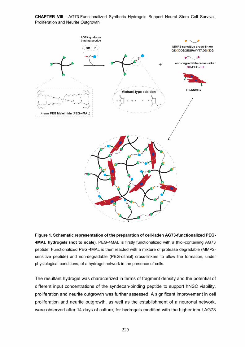
CHAPTER VIII | AG73-Functionalized Synthetic Hydrogels Support Neural Stem Cell Survival, Proliferation and Neurite Outgrowth
225
Figure 1. Schematic representation of the preparation of cell-laden AG73-functionalized PEG-
4MAL hydrogels (not to scale). PEG-4MAL is firstly functionalized with a thiol-containing AG73
peptide. Functionalized PEG-4MAL is then reacted with a mixture of protease degradable (MMP2-
sensitive peptide) and non-degradable (PEG-dithiol) cross-linkers to allow the formation, under
physiological conditions, of a hydrogel network in the presence of cells.
The resultant hydrogel was characterized in terms of fragment density and the potential of
different input concentrations of the syndecan-binding peptide to support hNSC viability,
proliferation and neurite outgrowth was further assessed. A significant improvement in cell
proliferation and neurite outgrowth, as well as the establishment of a neuronal network,
were observed after 14 days of culture, for hydrogels modified with the higher input AG73
CHAPTER VIII | AG73-Functionalized Synthetic Hydrogels Support Neural Stem Cell Survival, Proliferation and Neurite Outgrowth
226
concentration (1 mM). In summary, this study evidences the potential of syndecan-binding
adhesive motifs, for the design of 3D hydrogel platforms for the culture and transplantation
of hNSCs.
2. Materials and Methods
2.1. Hydrogel components
A four-arm maleimide terminated poly(ethylene glycol) (PEG-4MAL; 40 kDa, > 95% purity,
> 90% substitution, Jenkem USA) was chosen as the hydrogel base material. A macromer
solution was prepared in 10 mM 4-(2-hydroxyethyl)piperazine-1-ethanesulfonic acid
(HEPES) in phosphate buffered saline (PBS, pH 7.4) at a final polymer concentration of
10.0% (w/v). The syndecan-binding peptide AG73 (Acetyl (Ac)-CGGRKRLQVQLSIRT-
Amide (NH2), 1.7 kDa, > 90% purity) was obtained from GenScript. A cysteine-flanked
matrix metalloproteinase-2 (MMP2)-sensitive peptide (Acetyl (Ac)-
GDCDDSGESPAY¯YTADDCDG-Amide (NH2); 2.1 kDa, > 85% purity, GenScript) and a
PEG-dithiol (3.5 kDa, ³ 95% purity, Jenkem USA) were used as the degradable and non-
degradable cross-linkers, respectively.
2.2. AG73 tethering efficiency
To determine the tethering efficiency of AG73 to PEG-4MAL, free/unreacted thiols in the
reaction mixture were quantified using the Measure-iT thiol assay kit (Invitrogen) according
to the manufacturer’s instructions. Briefly, PEG-4MAL was functionalized with the
syndecan-binding peptide AG73 (input AG73 concentration: 0.01, 0.1, 0.5 or 1 mM) for 15
min in 10 mM HEPES in PBS (pH 7.4). The samples were then mixed with thiol-quantitation
reagent in a black 96-well plate and the fluorescence measured (lex = 494 nm; lem = 517
nm) using a microwell plate reader (BioTekÒ Synergy™ Mx). A calibration curve of
glutathione (0 – 55 µM) was used to calculate the concentration of free thiols.
2.3. Preparation of AG73-functionalized PEG-4MAL hydrogels
A schematic representation of AG73-functionalized PEG-4MAL hydrogel preparation is
presented in Fig. 1. Briefly, PEG-4MAL was first functionalized with the syndecan-binding
peptide AG73 for 15 min in 10 mM HEPES buffer (pH 7.4). Different AG73 input
concentrations were used – 0.01, 0.1, 0.5 and 1 mM. Functionalized PEG-4MAL macromers
were then cross-linked into a hydrogel by addition of a mix of a MMP2-sensitive peptide and
a PEG-dithiol prepared in 10 mM HEPES in PBS (pH 6.5), at an 80:20 molar ratio (%). The
cross-linkers were added at a 1:1 molar ratio of cysteine residues on the cross-linkers to
remaining maleimide groups on PEG-4MAL macromer, following functionalization with
CHAPTER VIII | AG73-Functionalized Synthetic Hydrogels Support Neural Stem Cell Survival, Proliferation and Neurite Outgrowth
227
AG73. Hydrogels were polymerized at 37 ºC, 5% CO2 for 15 min. Unmodified (0 mM) PEG-
4MAL gels were also prepared and used as controls.
2.4. Culture of human neural stem cells
Human neural stem cells (hNSCs) derived from the NIH approved H9 (WA09) human
embryonic stem cell line were purchased from Life Technologies (N7800-200). Cells were
expanded according to the manufacturer’s protocol on poly(ornithine)/laminin-111 from
mouse Engelbreth-Holm-Swarm sarcoma (msLn-111)-coated tissue coated polystyrene
(TCPS) plates (Corning) in expansion medium - StemProÒ NSC serum-free medium (SFM)
(Life Technologies) supplemented with basic fibroblast growth factor (bFGF, 20 ng/mL; Life
Technologies) and epidermal growth factor (EGF, 20 ng/mL; Life Technologies).
2.5. Culture of hNSCs within AG73-functionalized hydrogels
hNSCs were dissociated into single cells using StemPro accutase cell dissociation reagent
(Life Technologies) and further suspended in the solution containing AG73-functionalized
PEG-4MAL precursors (4 x 10⁶ viable cells/mL). Cell-laden hydrogels were formed by
mixing the PEG-4MAL precursor solution containing cells with the MMP2-sensitive peptide
and PEG-dithiol cross-linkers, dissolved in 10 mM HEPES (pH 6.5), at an 80:20 molar ratio
(%), and subsequently incubating the polymerizing gels at 37ºC, 5% CO2 for 15 minutes.
hNSCs were initially cultured in expansion medium and, at day 2 of culture, the medium
was switched to the StemPro NSC SFM media-Neurobasal/B27 (Life Technologies) (1:1)
mix, without growth factors. At day 8, half of the medium was replaced by the StemPro NSC
SFM-Neurobasal/B27 (1:3) mix supplemented with 10 ng/mL of brain-derived neurotrophic
factor (BDNF; Peprotech) and 500 µM of N6, 2’-O-Dibutyryladenosine 3’, 5’-cyclic
monophosphate sodium salt (dibutyril cAMP; Sigma). Half of the medium was changed
every other day, up to 14 days. hNSCs cultured within unmodified (0 mM) PEG-4MAL gels
were herein used as controls.
2.6. Cell viability
The distribution of viable and dead cells within PEG-4MAL hydrogels was assessed at day
7 of cell culture using a live/dead assay. Cell-hydrogel matrices were rinsed with warm PBS
pH 7.4 and incubated with 4 µM calcein AM (Life Technologies) and 6 µM propidium iodide
(PI; Life Technologies) at 37 ºC for 30 min, for detection of viable and dead cells,
respectively. After incubation, the samples were rinsed twice with PBS pH 7.4, transferred
to culture medium, and immediately observed under confocal laser scanning microscopy
(CLSM, Leica TCS SP5; Leica Mycrosystems). z-sections, covering a thickness of 300 µm,
were acquired using an HC Plan APO CS 10´ / 0.40 NA objective and subsequently
CHAPTER VIII | AG73-Functionalized Synthetic Hydrogels Support Neural Stem Cell Survival, Proliferation and Neurite Outgrowth
228
projected into 2D images. Quantitative analysis of cell viability was conducted by
immunocytometry performed in six-pooled hydrogels, after hydrogel dissociation. Briefly,
the hydrogels were sequentially incubated with 1.25 mg/mL of collagenase type II (Gibco;
1h at 37 ºC) and StemPro accutase cell dissociation reagent (Life Technologies; 20 min at
37 ºC, 70 rpm). Cells were mechanically dissociated by pipetting, diluted in Glasgow
Minimal Essential Medium (GMEM; Life Technologies) supplemented with 10% (v/v)
inactivated fetal bovine serum (FBS) and centrifuged. For live/dead staining, cells were then
transferred to a round-bottomed 96-well plate and stained with calcein AM (67 nM, 20 min
at 37 ºC) and PI (6 µM, 10 min at 37 ºC). Cells were finally washed trice and suspended in
FACS buffer (PBS pH 7.4 supplemented with 2% (v/v) FBS) for flow cytometry analysis (BD
FACSCantoTM II, BD Biosciences). Unlabeled cells were used to set the fluorescence gates
and cells stained with calcein AM or PI were used to establish the compensation settings.
For each flow cytometry analysis, 10,000 events were acquired.
2.7. DNA quantification
The growth of hNSCs cultured within PEG-4MAL hydrogels was estimated from total cell
number after 7 days of cell culture, using the CyQUANT® cell proliferation assay kit (Life
Technologies), according to manufacturer’s instructions. Briefly, cells were extracted from
the hydrogels, through sequential incubation with 1.25 mg/mL of collagenase type II (Gibco;
1 h at 37 ºC) and StemPro accutase cell dissociation reagent (Life Technologies; 20 min at
37 ºC) under stirring (70 rpm). Cells were mechanically dissociated by pipetting, diluted in
GMEM (Life Technologies) supplemented with 10% (v/v) FBS, centrifuged and the cell
pellet stored at -80 ºC. Cell pellet was then thawed at room temperature (RT) and incubated
with CyQUANT® GR dye/cell lysis buffer. Fluorescence (lex = 480 nm; lem = 520 nm) was
measured, after mixing with CyQUANT GR dye, in a microwell plate reader (BioTekÒ
Synergy™ Mx). The total number of cells for each condition was estimated from a standard
curve generated with a known amount of hNSCs over a range of 50 to 250,000 cells. Cell
samples used to generate the standard curve were measured in triplicate. For each
condition, hydrogels without cells, cultured in parallel and processed similarly to those with
cells, were used as blanks and their background fluorescence values subtracted.
2.8. hNSC neurite outgrowth
The effect of different input AG73 concentrations on neurite outgrowth was assessed after
14 days of culture, under neuronal differentiation conditions. Samples were processed as
whole-mounts for bIII-tubulin/DNA staining. Cell-laden hydrogels were fixed in 3.7% (w/v)
paraformaldehyde (PFA) solution diluted 1:1 in culture medium (30 min; 37 ºC) and
permeabilized with 0.2% (v/v) Triton X-100 in PBS (45 min; RT). Samples were then
CHAPTER VIII | AG73-Functionalized Synthetic Hydrogels Support Neural Stem Cell Survival, Proliferation and Neurite Outgrowth
229
incubated with blocking buffer (5% (v/v) bovine serum albumin (BSA) in PBS) for 1 h at RT,
followed by incubation with the primary antibody – mouse anti-bIII-tubulin (1:500; Biolegend,
801201), overnight (ON) at 4 ºC. To detect primary antibodies, samples were incubated
with Alexa Fluor® 488 conjugated anti-mouse secondary antibody (1:1,000; Life
Technologies, A21202) ON at 4 ºC. The nuclei were counterstained with 0.1 µg/mL Hoechst
33342 (Life Technologies, H1399) for 20 min at RT. z-sections, covering a thickness of 200
µm or 100 µm, were acquired with the CLSM using an HC Plan APO CS 10´ / 0.40 NA
objective with a zoom factor of 1 or 2, respectively, and subsequently projected into 2D
images. Neurite outgrowth was assessed in the projected 2D images using a developed
ImageJ script. Briefly, nuclei were segmented through an Otsu thresholding method, while
neurites were segmented through subtraction of the previously identified nucleus, using a
Huang thresholding method. Measurements were obtained for total neurite length (µm) and
total number of neurites per image.
2.9. Statistical Analysis
Statistical analysis was performed using GraphPad Prism 6 software (San Diego). Sample
distribution was initially tested for normality using the Kolmogorov-Smirnov test.
Comparisons with control conditions were performed with one-way ANOVA analysis,
followed by the Dunnett’s two-tailed test. For all analysis, differences were considered
significant at p < 0.05.
3. Results and Discussion
3.1. AG73 peptide is efficiently tethered to PEG-4MAL hydrogels
The key role of syndecans on the regulation of neural stem cell (NSC) fate lineage, make
these heparan sulfate proteoglycans (HSPGs) highly attractive therapeutic targets when
considering the biofunctionalization of synthetic matrices. As such, in this study we explored
the functionalization of a bioinert degradable synthetic hydrogel with AG73
(RKRLQVQLSIRT), a synthetic peptide derived from the laminin globular 4 (LG4) module
of laminin a1 chain that interacts with syndecan-1 and -4 [30-32]. For this purpose, an
hydrogel platform based on a four-arm maleimide terminated poly(ethylene glycol) (PEG-
4MAL) macromer (Fig.1) was selected as the base material for this work, due to its modular
nature, excellent in vitro and in vivo biocompatibility with different cell types [40, 42, 44, 45]
and well characterized biochemical and biophysical properties [41, 42, 46]. Maleimide
groups in PEG-4MAL macromers efficiently react with thiol-containing peptides through
Michael-type addition, enabling a fine tuning of ligand incorporation, and thus the formation
of structurally defined matrices. To assess the effect of AG73 peptide density on the human
CHAPTER VIII | AG73-Functionalized Synthetic Hydrogels Support Neural Stem Cell Survival, Proliferation and Neurite Outgrowth
230
NSC (hNSC) biological response, PEG-4MAL macromers were covalently modified with
different concentrations of this syndecan-binding peptide (0, 0.01, 0.1, 0.5 or 1mM) and the
conjugation efficiency determined by measuring the amount of free/unreacted thiols in the
reaction mixture. Free thiols were not detected (< 0.05 µM), the lower detection limit of the
assay), indicating that AG73 was tethered onto the PEG-4MAL macromer with an
immobilization efficiency higher than 96%, for all the concentrations tested.
3.2. AG73-presenting PEG-4MAL hydrogels support hNSC viability and proliferation
To get insight into the effect of AG73 density on hNSC survival and proliferation, cells were
cultured for 7 days within degradable PEG-4MAL hydrogels functionalized with different
AG73 concentrations (0, 0.01, 0.1, 0.5 and 1 mM). Confocal microscopy analysis showed,
for all the conditions tested, the presence of live cells evenly distributed throughout the
hydrogels, often growing as cellular spheroids (Fig. 2A). The quantitative analysis of
live/dead cells, assessed by flow cytometry, showed values of average viability greater than
75% for all the conditions tested, evidencing the good cytocompatibility of the proposed
matrices [47] (Fig. 2B). Interestingly, hydrogels functionalized with AG73 evidenced a
significantly higher percentage of viable cells compared to plain hydrogels (p < 0.0214 vs.
0 mM; Fig. 2B).
The tethering of AG73 peptide enhanced the ability of PEG-4MAL hydrogels to support
hNSC growth, as shown by the higher cell numbers found at day 7 of cell culture compared
to those observed for unmodified hydrogels (Fig. 2C). This effect was significant for the
highest input AG73 concentration tested (1 mM; p = 0.0012; vs. 0 mM), for which a 3.0-fold
increase in cell number (vs. 0 mM) was observed.

CHAPTER VIII | AG73-Functionalized Synthetic Hydrogels Support Neural Stem Cell Survival, Proliferation and Neurite Outgrowth
231
Figure 2. hNSC viability and proliferation in AG73-functionalized PEG-4MAL hydrogels as a
function of input peptide concentration. A) Representative 2D projections of CLSM 3D stack
images of cell-laden hydrogels covering a thickness of 300 µm, showing the distribution of live (in
green) and dead (in red) cells at day 7 of cell culture. Scale bar = 300 µm. B) Quantitative analysis
of live and dead cells at day 7, as determined by flow cytometry. Data represent mean ± SD of three
independent experiments; one-way ANOVA followed by Dunnett’s test (vs. 0 mM). C) Quantitative
analysis of total cell number at day 7, as determined by CyQuant® Cell Proliferation kit. Data
represent mean ± standard error of the mean (SEM) of three independent experiments performed in
triplicate; one-way ANOVA followed by Dunnett’s test (vs. 0 mM).
3.3. The tethering of 1 mM AG73 render PEG-4MAL hydrogels permissive to neurite
outgrowth
To unveil the effect of AG73 concentration on neurite outgrowth, hNSCs were cultured for
14 days under differentiation conditions, and then processed as whole-mounts for bIII-
tubulin and DNA staining (Fig. 3). Qualitative analysis of CLSM images (Fig. 3A) showed a
population of differentiated neurons, expressing bIII-tubulin and starting to form a neuronal
network in PEG-4MAL hydrogels functionalized with 1 mM of AG73. For lower input peptide
concentrations, the establishment of a neuronal network was less evident (Fig. 3A).
Differences between conditions were assessed by image processing and quantitative
analysis, using ImageJ/Fiji. More specifically, total neurite length (µm) and total number of
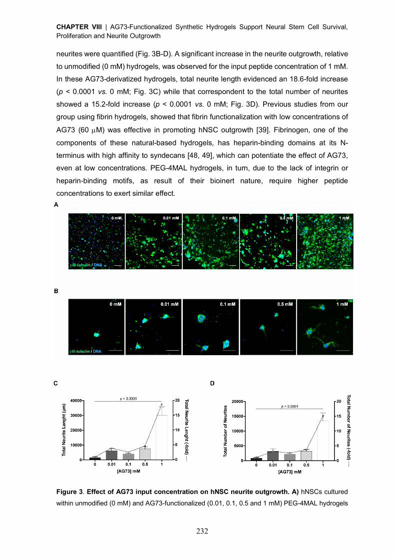
CHAPTER VIII | AG73-Functionalized Synthetic Hydrogels Support Neural Stem Cell Survival, Proliferation and Neurite Outgrowth
232
neurites were quantified (Fig. 3B-D). A significant increase in the neurite outgrowth, relative
to unmodified (0 mM) hydrogels, was observed for the input peptide concentration of 1 mM.
In these AG73-derivatized hydrogels, total neurite length evidenced an 18.6-fold increase
(p < 0.0001 vs. 0 mM; Fig. 3C) while that correspondent to the total number of neurites
showed a 15.2-fold increase (p < 0.0001 vs. 0 mM; Fig. 3D). Previous studies from our
group using fibrin hydrogels, showed that fibrin functionalization with low concentrations of
AG73 (60 µM) was effective in promoting hNSC outgrowth [39]. Fibrinogen, one of the
components of these natural-based hydrogels, has heparin-binding domains at its N-
terminus with high affinity to syndecans [48, 49], which can potentiate the effect of AG73,
even at low concentrations. PEG-4MAL hydrogels, in turn, due to the lack of integrin or
heparin-binding motifs, as result of their bioinert nature, require higher peptide
concentrations to exert similar effect.
Figure 3. Effect of AG73 input concentration on hNSC neurite outgrowth. A) hNSCs cultured
within unmodified (0 mM) and AG73-functionalized (0.01, 0.1, 0.5 and 1 mM) PEG-4MAL hydrogels
CHAPTER VIII | AG73-Functionalized Synthetic Hydrogels Support Neural Stem Cell Survival, Proliferation and Neurite Outgrowth
233
after 14 days of culture, under differentiation conditions. Images are representative of 2D projections
of CLSM 3D stack images of cells processed for immunofluorescence labeling of bIII-tubulin/DNA,
covering a thickness of 200 µm. Arrows depict neurites expressing bIII-tubulin protruding from
neurospheres. Scale Bar = 200 µm. B) Representative 2D projections of CLSM 3D stack of hNSC
outgrowth, covering a thickness of 200 µm, after 14 days of culture under differentiation conditions.
Scale Bar = 100 µm. C) Total Neurite Length; and D) Total Number of Neurites determined by
quantitative analysis of images acquired in CLSM. Absolute values (bars) and values normalized to
outgrowth in 0 mM AG73 hydrogels (dotted line) are presented. Data represent mean ± SD of 10
images analyzed per condition; one-way ANOVA followed by Dunnett’s test (vs. 0 mM).
Hydrogel mechanical and structural properties have a key role on the modulation of different
cellular functions, including survival, proliferation and neurite outgrowth [41, 42, 46]. With
this in mind, we hypothesize that the significant increase in cell number and neurite
outgrowth observed for PEG-4MAL hydrogels functionalized with the highest input AG73
concentration (1 mM), may be also related with alterations in mechanical and structural
properties of the hydrogel, resultant from the incorporation of a synthetic peptide. In a 3D
microenvironment the processes involved on the modulation of different cell functions,
including migration and neurite outgrowth, depend not only on the presence of cell adhesion
domains but also on the ability of cells to degrade the surrounding matrix. Therefore, and
although most of the studies published to date focus in understanding the independent
contribution of each of these parameters for cell function, understanding how matrix
bioactivity and mechanical properties influence each other and the impact of that concerted
action on the modulation of cell behavior is highly desirable. Indeed, a study assessing the
influence of cell-adhesive peptides (RGDS, IKVAV and YIGSR) on a PEG hydrogel physical
and mechanical properties, showed that depending on the ligand type and concentration,
the extent of their impact and whether a ligand increases or decreases a specific property,
will be different [50]. Therefore, studies to understand how the AG73 tethering impacts
PEG-4MAL hydrogel mechanical and structural properties, and how that correlates with the
observed biological outcome, should be conducted.
To better recapitulate the biological activity of laminin chains, different studies proposed the
combination of laminin-derived synthetic adhesive peptides targeting different cell receptors
(e.g. integrins and syndecans). Indeed, the modification of chitosan membranes with a
combination of syndecan- (AG73) and integrin- (A2G10, A99a or EF1zz) binding peptides,
evidenced the synergistic ability of these peptides to enhance different cell functions,
including PC12 cell adhesion and neurite outgrowth [51, 52]. Moreover, the characterization
of the expression profile of integrins and syndecans in H9-derived NSCs, previously
performed by our group, showed that 93% of the cells expressing syndecan-1 also
CHAPTER VIII | AG73-Functionalized Synthetic Hydrogels Support Neural Stem Cell Survival, Proliferation and Neurite Outgrowth
234
expressed the integrin a6 subunit [53]. Considering all these evidences, future studies
should address the functionalization of PEG-4MAL hydrogels with a combination of AG73
peptide and integrin-binding ligands, and its additive or synergistic effect on neurite
outgrowth.
4. Conclusions
The present work demonstrates that a fine control over AG73-binding peptide density allows
to maximize hNSC proliferation and neurite outgrowth, within PEG-4MAL hydrogels.
However, and despite the promising results obtained, future studies need to be conducted
to validate the observations presented in this work and better understand the role of AG73-
syndecan-binding peptide on the modulation of hNSC behavior/function. Overall, the
proposed hydrogels showed great potential to be used as 3D platforms for the in vitro
culture of hNSCs and, potentially, as vehicles for cell transplantation in the context of
nervous system regeneration/disorders.
Acknowledgments
Confocal microscopy was conducted at the Bioimaging i3S Scientific Platform, member of
the PPBI (PPBI-POCI-01-0145-FEDER-022122), with the assistance of Maria Lázaro. This
work was funded by projects NORTE-01-0145-FEDER-000008 and NORTE-01-0145-
FEDER-000012, supported by Norte Portugal Regional Operational Programme (NORTE
2020), under the PORTUGAL 2020 Partnership Agreement, through the European
Regional Development Fund (ERDF) and FEDER - Fundo Europeu de Desenvolvimento
Regional funds through the COMPETE 2020 - Operacional Programme for
Competitiveness and Internationalisation (POCI), Portugal 2020; by Portuguese funds
through FCT/MCTES in the framework of the project "Institute for Research and Innovation
in Health Sciences" (POCI-01-0145-FEDER-007274), and by Santa Casa da Misericordia
de Lisboa through project COMBINE (Prémio Neurociências Melo e Castro 1068-2015).
D.B. was supported by FCT PhD Programs (PD/BD/105953/2014) and Programa
Operacional Potencial Humano (POCH), in the scope of the BiotechHealth Program
(Doctoral Program on Cellular and Molecular Biotechnology Applied to Health Sciences),
Programa FLAD Healthcare 2020 and the project PARES (Prémio Albino Aroso).
CHAPTER VIII | AG73-Functionalized Synthetic Hydrogels Support Neural Stem Cell Survival, Proliferation and Neurite Outgrowth
235
References
[1] S. Pluchino, L. Zanotti, M. Deleidi, G. Martino, Neural stem cells and their use as
therapeutic tool in neurological disorders, Brain Res Brain Res Rev 48(2) (2005) 211-9.
[2] C. Cossetti, C. Alfaro-Cervello, M. Donega, G. Tyzack, S. Pluchino, New perspectives
of tissue remodelling with neural stem and progenitor cell-based therapies, Cell Tissue Res
349(1) (2012) 321-9.
[3] F.H. Gage, S. Temple, Neural stem cells: generating and regenerating the brain, Neuron
80(3) (2013) 588-601.
[4] M.H. Amer, F. Rose, K.M. Shakesheff, M. Modo, L.J. White, Translational considerations
in injectable cell-based therapeutics for neurological applications: concepts, progress and
challenges, NPJ Regen Med 2 (2017) 23.
[5] D. Barros, I.F. Amaral, A.P. Pego, Biomimetic synthetic self-assembled hydrogels for
cell transplantation, Curr Top Med Chem 15(13) (2015) 1209-26.
[6] J.L. Wilson, T.C. McDevitt, Biofunctional Hydrogels for Three-Dimensional Stem Cell
Culture, in: A. Vishwakarma, J.M. Karp (Eds.), Biology and Engineering of Stem Cell
Niches, Elsevier2017.
[7] J.C. Schense, J. Bloch, P. Aebischer, J.A. Hubbell, Enzymatic incorporation of bioactive
peptides into fibrin matrices enhances neurite extension, Nat Biotechnol 18(4) (2000) 415-
9.
[8] J.C. Schense, J.A. Hubbell, Three-dimensional migration of neurites is mediated by
adhesion site density and affinity, J Biol Chem 275(10) (2000) 6813-8.
[9] U. Freudenberg, A. Hermann, P.B. Welzel, K. Stirl, S.C. Schwarz, M. Grimmer, A. Zieris,
W. Panyanuwat, S. Zschoche, D. Meinhold, A. Storch, C. Werner, A star-PEG-heparin
hydrogel platform to aid cell replacement therapies for neurodegenerative diseases,
Biomaterials 30(28) (2009) 5049-60.
[10] S.P. Zustiak, S. Pubill, A. Ribeiro, J.B. Leach, Hydrolytically degradable poly(ethylene
glycol) hydrogel scaffolds as a cell delivery vehicle: characterization of PC12 cell response,
Biotechnol Prog 29(5) (2013) 1255-64.
[11] R. Bellamkonda, J.P. Ranieri, P. Aebischer, Laminin oligopeptide derivatized agarose
gels allow three-dimensional neurite extension in vitro, J Neurosci Res 41(4) (1995) 501-9.
[12] T. Zhao, D.L. Sellers, Y. Cheng, P.J. Horner, S.H. Pun, Tunable, Injectable Hydrogels
Based on Peptide-Cross-Linked, Cyclized Polymer Nanoparticles for Neural Progenitor Cell
Delivery, Biomacromolecules 18(9) (2017) 2723-2731.
[13] Y.T. Wei, W.M. Tian, X. Yu, F.Z. Cui, S.P. Hou, Q.Y. Xu, I.S. Lee, Hyaluronic acid
hydrogels with IKVAV peptides for tissue repair and axonal regeneration in an injured rat
brain, Biomed Mater 2(3) (2007) S142-6.
CHAPTER VIII | AG73-Functionalized Synthetic Hydrogels Support Neural Stem Cell Survival, Proliferation and Neurite Outgrowth
236
[14] W. Sun, T. Incitti, C. Migliaresi, A. Quattrone, S. Casarosa, A. Motta, Viability and
neuronal differentiation of neural stem cells encapsulated in silk fibroin hydrogel
functionalized with an IKVAV peptide, J Tissue Eng Regen Med 11(5) (2017) 1532-1541.
[15] L.A. Flanagan, L.M. Rebaza, S. Derzic, P.H. Schwartz, E.S. Monuki, Regulation of
human neural precursor cells by laminin and integrins, J Neurosci Res 83(5) (2006) 845-
56.
[16] L.E. Oikari, R.K. Okolicsanyi, A. Qin, C. Yu, L.R. Griffiths, L.M. Haupt, Cell surface
heparan sulfate proteoglycans as novel markers of human neural stem cell fate
determination, Stem Cell Res 16(1) (2016) 92-104.
[17] M. Ford-Perriss, K. Turner, S. Guimond, A. Apedaile, H.D. Haubeck, J. Turnbull, M.
Murphy, Localisation of specific heparan sulfate proteoglycans during the proliferative
phase of brain development, Dev Dyn 227(2) (2003) 170-84.
[18] Y. Choi, H. Chung, H. Jung, J.R. Couchman, E.S. Oh, Syndecans as cell surface
receptors: Unique structure equates with functional diversity, Matrix Biol 30(2) (2011) 93-9.
[19] F.E. Poulain, H.J. Yost, Heparan sulfate proteoglycans: a sugar code for vertebrate
development?, Development 142(20) (2015) 3456-67.
[20] X. Xian, S. Gopal, J.R. Couchman, Syndecans as receptors and organizers of the
extracellular matrix, Cell Tissue Res 339(1) (2010) 31-46.
[21] Q. Wang, L. Yang, C. Alexander, S. Temple, The niche factor syndecan-1 regulates
the maintenance and proliferation of neural progenitor cells during mammalian cortical
development, PLoS One 7(8) (2012) e42883.
[22] L. Morizur, A. Chicheportiche, L.R. Gauthier, M. Daynac, F.D. Boussin, M.A. Mouthon,
Distinct Molecular Signatures of Quiescent and Activated Adult Neural Stem Cells Reveal
Specific Interactions with Their Microenvironment, Stem Cell Reports (2018).
[23] M.M. Bespalov, Y.A. Sidorova, S. Tumova, A. Ahonen-Bishopp, A.C. Magalhaes, E.
Kulesskiy, M. Paveliev, C. Rivera, H. Rauvala, M. Saarma, Heparan sulfate proteoglycan
syndecan-3 is a novel receptor for GDNF, neurturin, and artemin, J Cell Biol 192(1) (2011)
153-69.
[24] A. Kinnunen, T. Kinnunen, M. Kaksonen, R. Nolo, P. Panula, H. Rauvala, N-syndecan
and HB-GAM (heparin-binding growth-associated molecule) associate with early axonal
tracts in the rat brain, Eur J Neurosci 10(2) (1998) 635-48.
[25] A. Kinnunen, M. Niemi, T. Kinnunen, M. Kaksonen, R. Nolo, H. Rauvala, Heparan
sulphate and HB-GAM (heparin-binding growth-associated molecule) in the development
of the thalamocortical pathway of rat brain, Eur J Neurosci 11(2) (1999) 491-502.
[26] E. Raulo, M.A. Chernousov, D.J. Carey, R. Nolo, H. Rauvala, Isolation of a neuronal
cell surface receptor of heparin binding growth-associated molecule (HB-GAM).
Identification as N-syndecan (syndecan-3), J Biol Chem 269(17) (1994) 12999-3004.
CHAPTER VIII | AG73-Functionalized Synthetic Hydrogels Support Neural Stem Cell Survival, Proliferation and Neurite Outgrowth
237
[27] T. Kinnunen, E. Raulo, R. Nolo, M. Maccarana, U. Lindahl, H. Rauvala, Neurite
outgrowth in brain neurons induced by heparin-binding growth-associated molecule (HB-
GAM) depends on the specific interaction of HB-GAM with heparan sulfate at the cell
surface, J Biol Chem 271(4) (1996) 2243-8.
[28] H.K. Matthews, L. Marchant, C. Carmona-Fontaine, S. Kuriyama, J. Larrain, M.R. Holt,
M. Parsons, R. Mayor, Directional migration of neural crest cells in vivo is regulated by
Syndecan-4/Rac1 and non-canonical Wnt signaling/RhoA, Development 135(10) (2008)
1771-80.
[29] I.M. Ethell, Y. Yamaguchi, Cell surface heparan sulfate proteoglycan syndecan-2
induces the maturation of dendritic spines in rat hippocampal neurons, J Cell Biol 144(3)
(1999) 575-86.
[30] M.P. Hoffman, M. Nomizu, E. Roque, S. Lee, D.W. Jung, Y. Yamada, H.K. Kleinman,
Laminin-1 and laminin-2 G-domain synthetic peptides bind syndecan-1 and are involved in
acinar formation of a human submandibular gland cell line, J Biol Chem 273(44) (1998)
28633-41.
[31] L.N. Gama-de-Souza, E. Cyreno-Oliveira, V.M. Freitas, E.S. Melo, V.F. Vilas-Boas,
A.S. Moriscot, R.G. Jaeger, Adhesion and protease activity in cell lines from human salivary
gland tumors are regulated by the laminin-derived peptide AG73, syndecan-1 and beta1
integrin, Matrix Biol 27(5) (2008) 402-19.
[32] N. Suzuki, N. Ichikawa, S. Kasai, M. Yamada, N. Nishi, H. Morioka, H. Yamashita, Y.
Kitagawa, A. Utani, M.P. Hoffman, M. Nomizu, Syndecan binding sites in the laminin alpha1
chain G domain, Biochemistry 42(43) (2003) 12625-33.
[33] M. Nomizu, W.H. Kim, K. Yamamura, A. Utani, S.Y. Song, A. Otaka, P.P. Roller, H.K.
Kleinman, Y. Yamada, Identification of cell binding sites in the laminin alpha 1 chain
carboxyl-terminal globular domain by systematic screening of synthetic peptides, J Biol
Chem 270(35) (1995) 20583-90.
[34] K. Hozumi, N. Suzuki, P.K. Nielsen, M. Nomizu, Y. Yamada, Laminin alpha1 chain LG4
module promotes cell attachment through syndecans and cell spreading through integrin
alpha2beta1, J Biol Chem 281(43) (2006) 32929-40.
[35] M. Mochizuki, Y. Kadoya, Y. Wakabayashi, K. Kato, I. Okazaki, M. Yamada, T. Sato,
N. Sakairi, N. Nishi, M. Nomizu, Laminin-1 peptide-conjugated chitosan membranes as a
novel approach for cell engineering, FASEB J 17(8) (2003) 875-7.
[36] Y. Yamada, K. Hozumi, F. Katagiri, Y. Kikkawa, M. Nomizu, Biological activity of laminin
peptide-conjugated alginate and chitosan matrices, Biopolymers 94(6) (2010) 711-20.
[37] Y. Yamada, F. Katagiri, K. Hozumi, Y. Kikkawa, M. Nomizu, Cell behavior on protein
matrices containing laminin alpha1 peptide AG73, Biomaterials 32(19) (2011) 4327-35.
CHAPTER VIII | AG73-Functionalized Synthetic Hydrogels Support Neural Stem Cell Survival, Proliferation and Neurite Outgrowth
238
[38] Y. Yamada, K. Hozumi, A. Aso, A. Hotta, K. Toma, F. Katagiri, Y. Kikkawa, M. Nomizu,
Laminin active peptide/agarose matrices as multifunctional biomaterials for tissue
engineering, Biomaterials 33(16) (2012) 4118-25.
[39] A.R. Bento, Improving neurite outgrowth in 3D hydrogel matrices by mimicking cell
receptor-ECM interactions occurring in neurogenic niches: an engineering approach to
develop more efficient neural stem cell hydrogel carriers, Faculdade de Engenharia,
Universidade do Porto, 2018.
[40] D. Barros, E. Conde-Sousa, A.J. Garcia, I.F. Amaral, A.P. Pego, Engineering hydrogels
with affinity-bound laminin as 3D neural stem cell culture systems Manuscript in preparation
(2019).
[41] N.O. Enemchukwu, R. Cruz-Acuna, T. Bongiorno, C.T. Johnson, J.R. Garcia, T.
Sulchek, A.J. Garcia, Synthetic matrices reveal contributions of ECM biophysical and
biochemical properties to epithelial morphogenesis, J Cell Biol 212(1) (2016) 113-24.
[42] W.M. Han, S.E. Anderson, M. Mohiuddin, D. Barros, S.A. Nakhai, E. Shin, I.F. Amaral,
A.P. Pego, A.J. Garcia, Y.C. Jang, Synthetic matrix enhances transplanted satellite cell
engraftment in dystrophic and aged skeletal muscle with comorbid trauma, Sci Adv 4(8)
(2018) eaar4008.
[43] J. Patterson, J.A. Hubbell, Enhanced proteolytic degradation of molecularly engineered
PEG hydrogels in response to MMP-1 and MMP-2, Biomaterials 31(30) (2010) 7836-45.
[44] J.D. Weaver, D.M. Headen, J. Aquart, C.T. Johnson, L.D. Shea, H. Shirwan, A.J.
Garcia, Vasculogenic hydrogel enhances islet survival, engraftment, and function in leading
extrahepatic sites, Sci Adv 3(6) (2017) e1700184.
[45] R. Cruz-Acuna, M. Quiros, A.E. Farkas, P.H. Dedhia, S. Huang, D. Siuda, V. Garcia-
Hernandez, A.J. Miller, J.R. Spence, A. Nusrat, A.J. Garcia, Synthetic hydrogels for human
intestinal organoid generation and colonic wound repair, Nat Cell Biol 19(11) (2017) 1326-
1335.
[46] E.A. Phelps, N.O. Enemchukwu, V.F. Fiore, J.C. Sy, N. Murthy, T.A. Sulchek, T.H.
Barker, A.J. Garcia, Maleimide cross-linked bioactive PEG hydrogel exhibits improved
reaction kinetics and cross-linking for cell encapsulation and in situ delivery, Adv Mater
24(1) (2012) 64-70, 2.
[47] I.O.f. Standardization, ISO 10993-5:2009 : Biological evaluation of medical devices
Tests for in vitro cytotoxicity, 2009.
[48] M.M. Martino, P.S. Briquez, A. Ranga, M.P. Lutolf, J.A. Hubbell, Heparin-binding
domain of fibrin(ogen) binds growth factors and promotes tissue repair when incorporated
within a synthetic matrix, Proc Natl Acad Sci U S A 110(12) (2013) 4563-8.
CHAPTER VIII | AG73-Functionalized Synthetic Hydrogels Support Neural Stem Cell Survival, Proliferation and Neurite Outgrowth
239
[49] A.C. Brown, T.H. Barker, Fibrin-based biomaterials: modulation of macroscopic
properties through rational design at the molecular level, Acta Biomater 10(4) (2014) 1502-
14.
[50] S.P. Zustiak, R. Durbal, J.B. Leach, Influence of cell-adhesive peptide ligands on
poly(ethylene glycol) hydrogel physical, mechanical and transport properties, Acta Biomater
6(9) (2010) 3404-14.
[51] K. Hozumi, A. Sasaki, Y. Yamada, D. Otagiri, K. Kobayashi, C. Fujimori, F. Katagiri, Y.
Kikkawa, M. Nomizu, Reconstitution of laminin-111 biological activity using multiple peptide
coupled to chitosan scaffolds, Biomaterials 33(17) (2012) 4241-50.
[52] D. Otagiri, Y. Yamada, K. Hozumi, F. Katagiri, Y. Kikkawa, M. Nomizu, Cell attachment
and spreading activity of mixed laminin peptide-chitosan membranes, Biopolymers 100(6)
(2013) 751-9.
[53] A.R. Bento, A.P. Pego, I.F. Amaral, Controlling the density of α6β1 integrin- or
syndecan-binding motifs tethered to fibrin improve the outgrowth of hES-derived neural
stem cells, 2018.

CHAPTER VIII | AG73-Functionalized Synthetic Hydrogels Support Neural Stem Cell Survival, Proliferation and Neurite Outgrowth
240

241
CHAPTER IX _________________________________________________________________________
Concluding Remarks and Future Perspectives

CHAPTER IX | Concluding Remarks and Future Perspectives
242
CHAPTER IX | Concluding Remarks and Future Perspectives
243
The main aim of the work presented in this thesis was the design of biologically relevant
matrices able to recapitulate the laminin-rich microenvironment of neural stem cell (NSC)
niches. For that, we proposed the modification of a degradable synthetic hydrogel with full-
length laminin. Additionally, a laminin-derived peptide interacting with syndecan receptors
(AG73) was also explored. The developed matrices can be used to address the role of laminin
on the modulation of NSC function and fate and, ultimately, contribute to the development of
chemically-defined platforms for NSC culture and/or NSC delivery into the central nervous
system (CNS) in the context of regenerative therapies.
Laminin is one of the most important and well described extracellular matrix (ECM) proteins
within the neurogenic niches [1, 2], with a key role in the modulation of NSC maintenance [3],
migration and differentiation [4-6]. Furthermore, laminin has other paramount roles in CNS
homeostasis, as contributing to synapse function and stability [7, 8]. Therefore, this ECM
protein constitutes a highly attractive option while considering the design of NSC niche
microenvironments.
Full-length laminin immobilization into biological and synthetic matrices was already explored
in different studies for the design of three-dimensional (3D) NSC niche microenvironments
(see Chapter III for a review). Despite the promising results, a major caveat of the
conventionally used immobilization strategies (based on non-covalent incorporation/physical
entrapment or non-selective covalent immobilization), is the lack of control over the orientation
and conformation of laminin, upon immobilization, which was proven to be crucial for the
modulation of cellular behavior, function and fate [9]. As such, one of the specific aims of this
thesis was to establish an affinity-based strategy to generate a highly versatile and dynamic
system, expected to allow the site-selective immobilization of laminin, while controlling its
orientation and bioactivity. For this purpose, in Chapter V we proposed a strategy that takes
advantage of the native high affinity interaction (dissociation constant (KD) = 5 nM) between
laminin and the N-terminal agrin (NtA) domain [10, 11]. Studies conducted to date,
characterizing this domain and its interaction with laminin, have used the NtA derived from
chicken agrin [10-13]. In the work presented in this thesis, the recombinant human (rh) variant
of the NtA domain was produced and, for the first time, its interaction with laminin
characterized [9, 14]. This is particularly important since we envisage the application of the
proposed strategy in platforms for human cell culture/delivery and human-disease modelling.
The conjugation of a thiol-terminated poly(ethylene glycol) (PEG) at the NtA N-terminus -
mono-PEGylated rhNtA, allowed us to establish an affinity-binding ligand suitable for laminin
site-selective immobilization to two-dimensional (2D) and 3D matrices. In addition, the
proposed ligand presents a high versatility, as evidenced by its high affinity for different laminin
isoforms (in the context of this thesis assessed for mouse laminin-111 (msLn-111) and

CHAPTER IX | Concluding Remarks and Future Perspectives
244
recombinant human laminin-521 (rhLn-521)). This allows the use of the proposed strategy for
the immobilization of different laminin isoforms, which can be of interest for particular cell types
and for application in specific disease contexts. The biological effectiveness of the
immobilization strategy was assessed using self-assembled monolayers (SAMs) of mono-
PEGylated rhNtA on gold. Overall, the obtained data suggested that mono-PEGylated rhNtA
works as an effective binding ligand for the site-selective immobilization of laminin, allowing
the preservation of laminin ability to polymerize and mediate human NSC (hNSC) adhesion
and spreading. In summary, in this work we provided a new affinity-based strategy that
surpasses some of the main limitations associated with the current available laminin
immobilization strategies (non-covalent incorporation/physical entrapment or non-selective
covalent immobilization), assuring the exposure of laminin cell binding domains, as well as
other distinct complementary domains that modulate important biological functions.
In the future, it would be interesting to perform further studies to establish this as an universal
strategy of coating, allowing the controlled and oriented immobilization of laminin regardless
of the 2D model surface (e.g. TCPS, Au, etc.). In addition, the potential of this strategy to
immobilize different laminin isoforms and be used as 2D coating for the culture of different cell
types should also be confirmed. Neural electrode-tissue interface research is facing an
exponential growth in the last few years [15]. Despite the promising results obtained to date,
the search for effective strategies to improve this interface is highly needed. In this regard, the
affinity-based immobilization approach presented in this thesis can be advantageous for the
development of engineered coatings for neural electrodes. This is expected to improve the
interface electrode-tissue, achieve seamless integration between the implanted electrodes
and neural tissue, and ultimately, boost neural electrode performance.
The application of the proposed affinity-based immobilization strategy was subsequently
explored for the development of 3D cell-instructive microenvironments, with potential to
support NSC proliferation and neuronal differentiation (Chapter VI). For that a degradable
synthetic hydrogel based on a four-arm maleimide-end functionalized poly(ethylene glycol)
(PEG-4MAL) macromer was selected as the base material. These hydrogels have been
extensively used to support the culture and/or delivery of different cell types, within different
regenerative contexts [16-19], and thus a high amount of information regarding the best way
to modulate their biochemical and biophysical properties is currently available. Therefore,
these hydrogels can be engineered to have the most suitable properties to support NSC
culture and transplantation. In this work, we explored msLn-111, and we showed that the
approach used for laminin incorporation (physical entrapment vs. affinity immobilization)
significantly impact the hydrogel mechanical and structural properties. The tethering of NtA,
which mediates laminin site-selective immobilization, reduces the number of maleimide-
CHAPTER IX | Concluding Remarks and Future Perspectives
245
terminated PEG chains available for cross-linking, thus reducing the number of cross-linking
points and favoring a reduction in the complex modulus (G*). Affinity-bound laminin PEG-
4MAL hydrogels showed great potential to be used as a dynamic biomimetic 3D platform
enabling NSC proliferation and neurite outgrowth, when compared to currently available
strategies for laminin immobilization, which include physical entrapment and non-selective
covalent immobilization. Overall, this work provided evidence of the potential of the proposed
engineered matrices to be used as defined 3D platforms, for the establishment of artificial
NSC niches and as temporary mimetic microenvironments to support cell transplantation. To
further consolidate the full potential of the developed 3D platform for the culture of hNSCs,
studies assessing its ability to support cell differentiation and ECM remodeling, and the
establishment of a functional neuronal network should be conducted.
The key role of rhLn-521 on the in vitro modulation of neural cell behavior [3, 20], led us to
subsequently explore the preparation of a degradable PEG-4MAL hydrogel containing affinity-
bound rhLn-521 (Chapter VII). This is of particular relevance when envisaging the
development of a whole-human 3D cell-instructive microenvironment. Preliminary studies
exploring the site-selective immobilization of rhLn-521 into a degradable PEG-4MAL hydrogel,
using the strategy proposed in Chapter VI, did not evidence a significant impact on neurite
outgrowth, when compared to unmodified hydrogels and hydrogels with equal amount of
physically entrapped laminin. Therefore, some steps of the original immobilization protocol
were altered, in an attempt to improve rLn-521 binding to NtA and its impact on NSC behavior.
An improvement on neurite outgrowth was observed, compared to unmodified hydrogels and
hydrogels with physically entrapped rhLn-521, although differences only reached statistical
significance in the former case, suggesting an impact of such modifications on protein
conformation and domain presentation to cells. Therefore, and although the data obtained
represent a good start for the development of a whole-human 3D cell instructive matrix, further
studies need to be conducted. More specifically, in the future the use of higher rhLn-521
concentrations (> 20 µg/mL) should be considered and its biological performance when
immobilized through the strategy proposed in Chapter VI assessed. In addition, the look for
alternatives to improve NtA-bound rhLn-521 tethering to PEG-4MAL is also highly desirable.
As previously pointed, the versatility and dynamic nature of the proposed strategy allows the
immobilization of different laminin isoforms within 3D matrices, thus opening doors for future
studies exploring cell-laminin interactions, occurring in different stem cell niches and disease
contexts. In addition, studies assessing the exchangeability with endogenously secreted
laminin could also be of interest. Direct evidence on the potential of the proposed
immobilization strategy to preserve the exposure of key laminin bioactive epitopes is also
critical to assess. For that, laminin peptide analogues known to mediate important laminin
CHAPTER IX | Concluding Remarks and Future Perspectives
246
functions, such as neuronal adhesion, neurite outgrowth and/or NSC migration can be used
as competitive inhibitors. Alternatively, function blocking studies using antibodies against
integrin or syndecan receptors interacting with specific laminin regions, can also be explored
in this regard. The hydrogel proposed can be also of interest to provide a controlled niche with
instructive cues to improve survival, engraftment and long-term function of transplanted stem
cells. Accordingly, and to validate the developed affinity-bound laminin hydrogel as a cell-
instructive microenvironment, in the future, in vivo studies using, for example, an experimental
model of brain or spinal cord injury, should be conducted. One of the main caveats currently
limiting the successful translation of stem cell therapies to clinic is the lack of effective cell
expansion and biomanufacturing systems. In this regard, in the last few years the systems
used for cell expansion have shifted towards the use of 3D platforms. These provide a defined
environment for adherent stem cells and organoid culture (e.g. NSCs, induced pluripotent
stem cells, etc.), while enabling the culture in large-scale bioreactor systems. Although, further
studies assessing the ability of affinity-bound laminin PEG-4MAL hydrogels to support hNSC
differentiation need to be conducted, their ability to support cell viability, proliferation and
neurite outgrowth, suggest that these hydrogels constitute a highly attractive option to be used
on the development of cell expansion and biomanufacturing systems.
Laminin-derived small adhesive sequences have also been explored, in alternative to the
incorporation of full-length laminin, to confer bioactivity to 3D matrices (see Chapter III for a
review). These small sequences can be chemically synthesized in large scale and are
commonly incorporated at higher densities, for similar amounts of native protein, usually, with
no significant impact on hydrogel biophysical properties [21]. In Chapter VIII we explored the
functionalization of a degradable synthetic hydrogel with a syndecan-binding peptide (AG73
– RKRLQVQSIRT) with neurite-promoting activity, and already successfully explored to
modify natural hydrogel matrices [22-25]. The tethering of AG73 peptide to PEG-4MAL
hydrogels led to a significant improvement of cell viability, after 7 days in culture,
independently of the input peptide concentration tested. In addition, we demonstrated that a
higher input AG73 concentration (1 mM) better supports hNSC proliferation and neurite
outgrowth. Despite the promising results obtained, future studies need to be conducted to
validate the observations presented in this work. More specifically, to verify if the biological
outcome observed for the different AG73 input concentrations could be related with
differences in hydrogel mechanical and structural properties, rheological studies need to be
conducted. In addition, for the defined optimal AG73 concentration (1 mM), future studies
should assess the ability of the developed 3D platforms to support hNSC neuronal
differentiation and maturation in long-term cultures. To get insight into the contribution of
syndecan-1 and -4 receptors for the NSC response to immobilized AG73, silencing of the
CHAPTER IX | Concluding Remarks and Future Perspectives
247
syndecan receptor expression in hNSCs, by means of siRNA, can be performed. Envisaging
the application of AG73-functionalized PEG-4MAL hydrogels in the CNS as vehicle for NSC
transplantation, its impact on axonal extension growth should be assessed, using for instance
organotypic cultures of sensory and motor neurons. To better recapitulate the biological
activity of laminin chains, we propose to address, in future studies, the functionalization of
PEG-4MAL hydrogels with a combination of AG73 peptide and integrin-binding ligands, and
evaluate its additive or synergistic effect on neurite outgrowth.
In summary, the affinity-based strategy proposed in this thesis can be successfully used as
an alternative to conventional immobilization approaches in a wide range of applications,
including engineered coatings for neuroelectrodes and for 2D substrates for cell culture, as
well as for the biofunctionalization of 3D matrices. Furthermore, the hydrogels developed
exploring the proposed strategy, showed great potential to be used as chemically-defined 3D
platforms for the in vitro culture of hNSCs and, potentially, as vehicles for hNSCs cell
transplantation in the context of nervous system regeneration/disorders. In addition, the
developed 3D matrices have potential to be successfully applied to study the molecular
mechanisms implicated in neurogenesis, in a 3D microenvironment that better mimics the in
vivo conditions. Alternatively, and envisaging the clinical translation of stem cell therapies,
affinity-bound laminin hydrogels can be also explored for the development of tissue
engineering constructs and biomanufacturing systems to expand stem cells. Ultimately, we
strongly believe this work opens new avenues in the design of more efficient hydrogel matrices
for application in NSC-based regenerative approaches for the treatment of traumatic CNS
disorders or neurodegenerative diseases.
CHAPTER IX | Concluding Remarks and Future Perspectives
248
References
[1] A. Kerever, J. Schnack, D. Vellinga, N. Ichikawa, C. Moon, E. Arikawa-Hirasawa, J.T. Efird,
F. Mercier, Novel extracellular matrix structures in the neural stem cell niche capture the
neurogenic factor fibroblast growth factor 2 from the extracellular milieu, Stem Cells 25(9)
(2007) 2146-57.
[2] I. Kazanis, J.D. Lathia, T.J. Vadakkan, E. Raborn, R. Wan, M.R. Mughal, D.M. Eckley, T.
Sasaki, B. Patton, M.P. Mattson, K.K. Hirschi, M.E. Dickinson, C. ffrench-Constant,
Quiescence and activation of stem and precursor cell populations in the subependymal zone
of the mammalian brain are associated with distinct cellular and extracellular matrix signals, J
Neurosci 30(29) (2010) 9771-81.
[3] A. Hyysalo, M. Ristola, M.E. Makinen, S. Hayrynen, M. Nykter, S. Narkilahti, Laminin
alpha5 substrates promote survival, network formation and functional development of human
pluripotent stem cell-derived neurons in vitro, Stem Cell Res 24 (2017) 118-127.
[4] L. Luckenbill-Edds, Laminin and the mechanism of neuronal outgrowth, Brain Res Brain
Res Rev 23(1-2) (1997) 1-27.
[5] S.K. Powell, H.K. Kleinman, Neuronal laminins and their cellular receptors, Int J Biochem
Cell Biol 29(3) (1997) 401-14.
[6] S. Plantman, M. Patarroyo, K. Fried, A. Domogatskaya, K. Tryggvason, H. Hammarberg,
S. Cullheim, Integrin-laminin interactions controlling neurite outgrowth from adult DRG
neurons in vitro, Mol Cell Neurosci 39(1) (2008) 50-62.
[7] R.S. Rogers, H. Nishimune, The role of laminins in the organization and function of
neuromuscular junctions, Matrix Biol 57-58 (2017) 86-105.
[8] H. Nishimune, G. Valdez, G. Jarad, C.L. Moulson, U. Muller, J.H. Miner, J.R. Sanes,
Laminins promote postsynaptic maturation by an autocrine mechanism at the neuromuscular
junction, J Cell Biol 182(6) (2008) 1201-15.
[9] D D. Barros, P. Parreira, J. Furtado, F. Ferreira-da-Silva, E. Conde-Sousa, A.J. Garcia,
M.C.L. Martins, I.F. Amaral, A.P. Pego, An affinity-based approach to engineer laminin-
presenting cell instructive microenvironments, Biomaterials 192 (2019) 601-611.
[10] J.B. Mascarenhas, M.A. Ruegg, U. Winzen, W. Halfter, J. Engel, J. Stetefeld, Mapping
of the laminin-binding site of the N-terminal agrin domain (NtA), EMBO J 22(3) (2003) 529-36.
[11] A.J. Denzer, T. Schulthess, C. Fauser, B. Schumacher, R.A. Kammerer, J. Engel, M.A.
Ruegg, Electron microscopic structure of agrin and mapping of its binding site in laminin-1,
EMBO J 17(2) (1998) 335-43.
[12] R.A. Kammerer, T. Schulthess, R. Landwehr, B. Schumacher, A. Lustig, P.D. Yurchenco,
M.A. Ruegg, J. Engel, A.J. Denzer, Interaction of agrin with laminin requires a coiled-coil
CHAPTER IX | Concluding Remarks and Future Perspectives
249
conformation of the agrin-binding site within the laminin gamma1 chain, EMBO J 18(23) (1999)
6762-70.
[13] J. Stetefeld, M. Jenny, T. Schulthess, R. Landwehr, B. Schumacher, S. Frank, M.A.
Ruegg, J. Engel, R.A. Kammerer, The laminin-binding domain of agrin is structurally related
to N-TIMP-1, Nat Struct Biol 8(8) (2001) 705-9.
[14] D. Barros, I.F. Amaral, A.P. Pego, Laminin immobilization, methods and uses thereof,
Portuguese Provisional Patent Application no. 20181000074673, filled 2018/11/30.
[15] K. Woeppel, Q. Yang, X.T. Cui, Recent Advances in Neural Electrode-Tissue Interfaces,
Curr Opin Biomed Eng 4 (2017) 21-31.
[16] D. Barros, I.F. Amaral, A.P. Pego, Biomimetic synthetic self-assembled hydrogels for cell
transplantation, Curr Top Med Chem 15(13) (2015) 1209-26.
[17] N.O. Enemchukwu, R. Cruz-Acuna, T. Bongiorno, C.T. Johnson, J.R. Garcia, T. Sulchek,
A.J. Garcia, Synthetic matrices reveal contributions of ECM biophysical and biochemical
properties to epithelial morphogenesis, J Cell Biol 212(1) (2016) 113-24.
[18] J.D. Weaver, D.M. Headen, J. Aquart, C.T. Johnson, L.D. Shea, H. Shirwan, A.J. Garcia,
Vasculogenic hydrogel enhances islet survival, engraftment, and function in leading
extrahepatic sites, Sci Adv 3(6) (2017) e1700184.
[19] W.M. Han, S.E. Anderson, M. Mohiuddin, D. Barros, S.A. Nakhai, E. Shin, I.F. Amaral,
A.P. Pego, A.J. Garcia, Y.C. Jang, Synthetic matrix enhances transplanted satellite cell
engraftment in dystrophic and aged skeletal muscle with comorbid trauma, Sci Adv 4(8) (2018)
eaar4008.
[20] A. Bergeron, H. Sherman, P. Pardo, H. Gitschier, H. Nandivada, D. Saxena, Corning®
rLaminin-521 (Human) for Expansion and Differentiation of Human Neural Stem Cells,
Corning Incorporated | Application Note (2015).
[21] J.H. Collier, T. Segura, Evolving the use of peptides as components of biomaterials,
Biomaterials 32(18) (2011) 4198-204.
[22] M. Mochizuki, Y. Kadoya, Y. Wakabayashi, K. Kato, I. Okazaki, M. Yamada, T. Sato, N.
Sakairi, N. Nishi, M. Nomizu, Laminin-1 peptide-conjugated chitosan membranes as a novel
approach for cell engineering, FASEB J 17(8) (2003) 875-7.
[23] Y. Yamada, K. Hozumi, F. Katagiri, Y. Kikkawa, M. Nomizu, Biological activity of laminin
peptide-conjugated alginate and chitosan matrices, Biopolymers 94(6) (2010) 711-20.
[24] Y. Yamada, F. Katagiri, K. Hozumi, Y. Kikkawa, M. Nomizu, Cell behavior on protein
matrices containing laminin alpha1 peptide AG73, Biomaterials 32(19) (2011) 4327-35.
[25] Y. Yamada, K. Hozumi, A. Aso, A. Hotta, K. Toma, F. Katagiri, Y. Kikkawa, M. Nomizu,
Laminin active peptide/agarose matrices as multifunctional biomaterials for tissue
engineering, Biomaterials 33(16) (2012) 4118-25.

CHAPTER IX | Concluding Remarks and Future Perspectives
250
251
APPENDIX I _________________________________________________________________________
Laminin immobilization, methods and uses thereof

APPENDIX I – Laminin immobilization, methods and uses thereof
252
APPENDIX I – Laminin immobilization, methods and uses thereof
253
Laminin immobilization, methods and uses thereof
Daniela Barros 1,2,3, Isabel Freitas Amaral 1,2,4, Ana Paula Pêgo 1,2,3,4
1 i3S - Instituto de Investigação e Inovação em Saúde, Universidade do Porto (UPorto),
Portugal
2 INEB - Instituto de Engenharia Biomédica, UPorto, Portugal
3 ICBAS - Instituto de Ciências Biomédicas Abel Salazar, UPorto, Portugal
4 FEUP - Faculdade de Engenharia, UPorto, Portugal
_________________________________________________________________________
Portuguese Provisional Patent Application no. 20181000074673, filled 2018/11/30
APPENDIX I – Laminin immobilization, methods and uses thereof
254
APPENDIX I – Laminin immobilization, methods and uses thereof
255
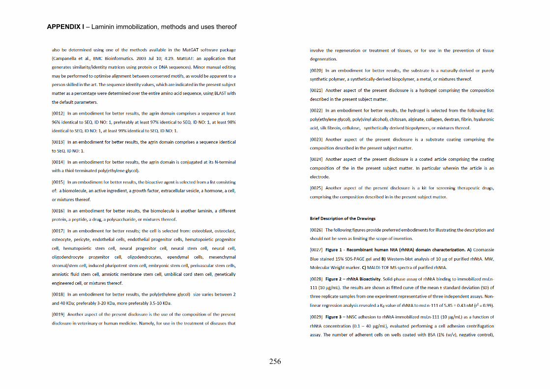
APPENDIX I – Laminin immobilization, methods and uses thereof
256
APPENDIX I – Laminin immobilization, methods and uses thereof
257
APPENDIX I – Laminin immobilization, methods and uses thereof
258
APPENDIX I – Laminin immobilization, methods and uses thereof
259
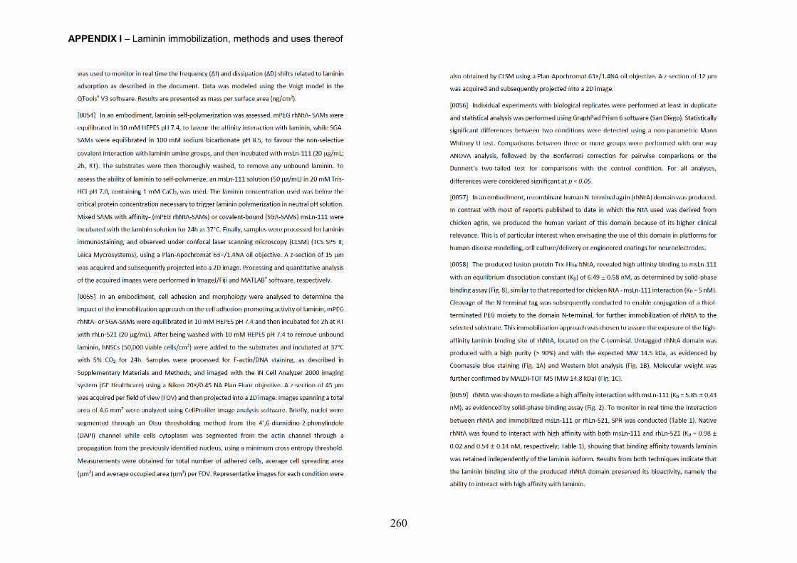
APPENDIX I – Laminin immobilization, methods and uses thereof
260
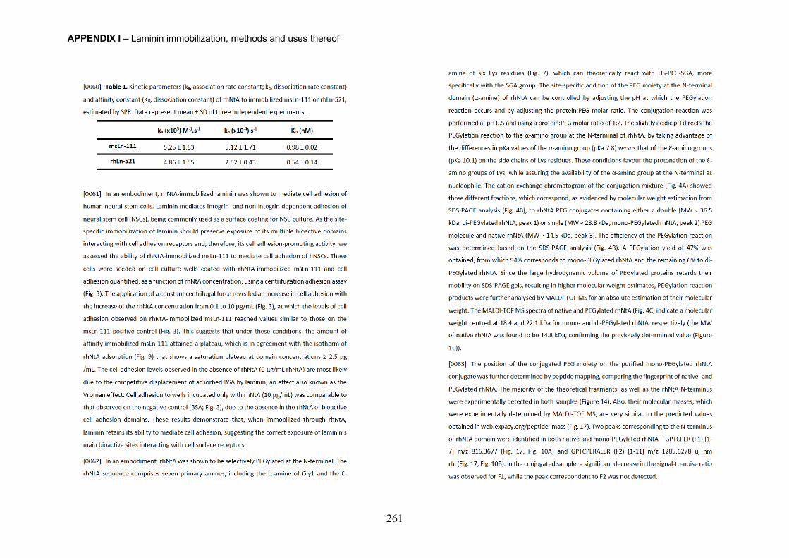
APPENDIX I – Laminin immobilization, methods and uses thereof
261
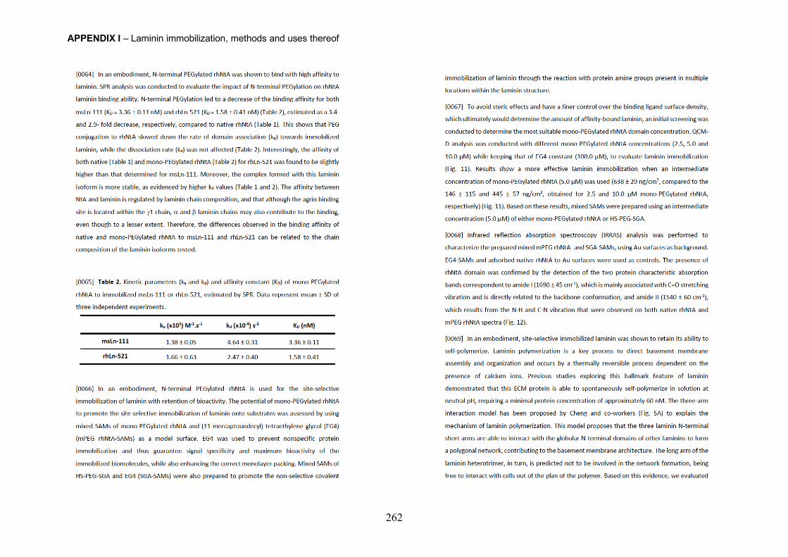
APPENDIX I – Laminin immobilization, methods and uses thereof
262
APPENDIX I – Laminin immobilization, methods and uses thereof
263
APPENDIX I – Laminin immobilization, methods and uses thereof
264
APPENDIX I – Laminin immobilization, methods and uses thereof
265
APPENDIX I – Laminin immobilization, methods and uses thereof
266
APPENDIX I – Laminin immobilization, methods and uses thereof
267
APPENDIX I – Laminin immobilization, methods and uses thereof
268
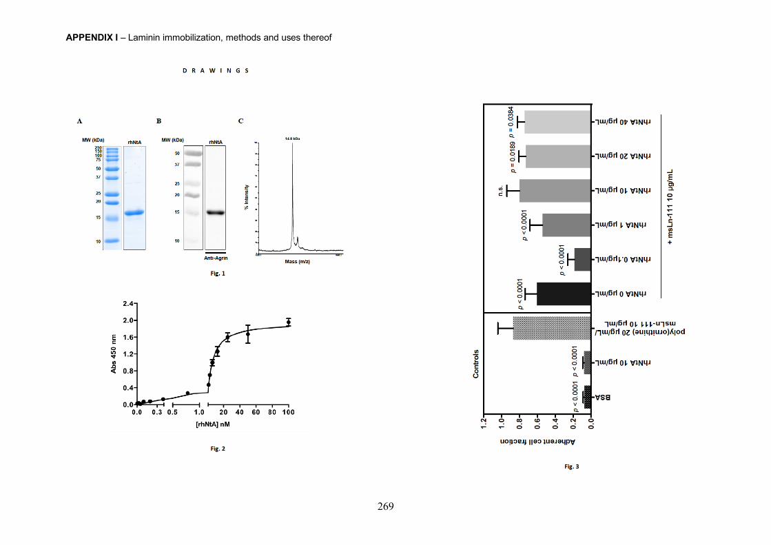
APPENDIX I – Laminin immobilization, methods and uses thereof
269
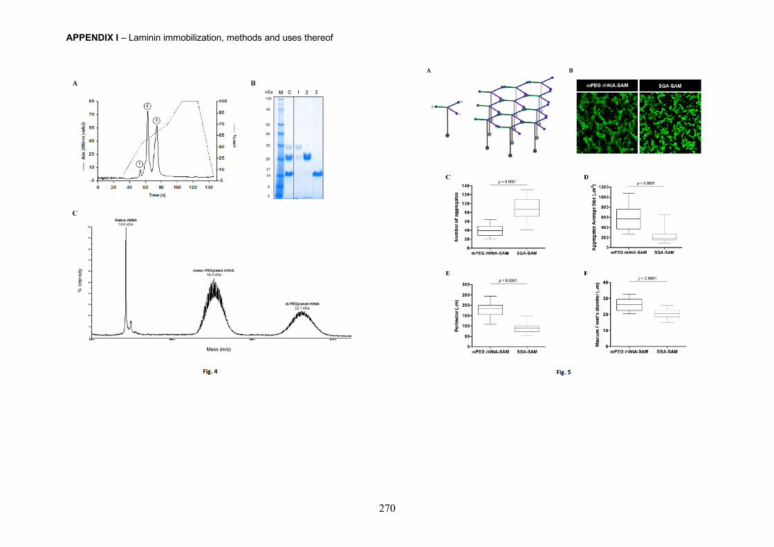
APPENDIX I – Laminin immobilization, methods and uses thereof
270
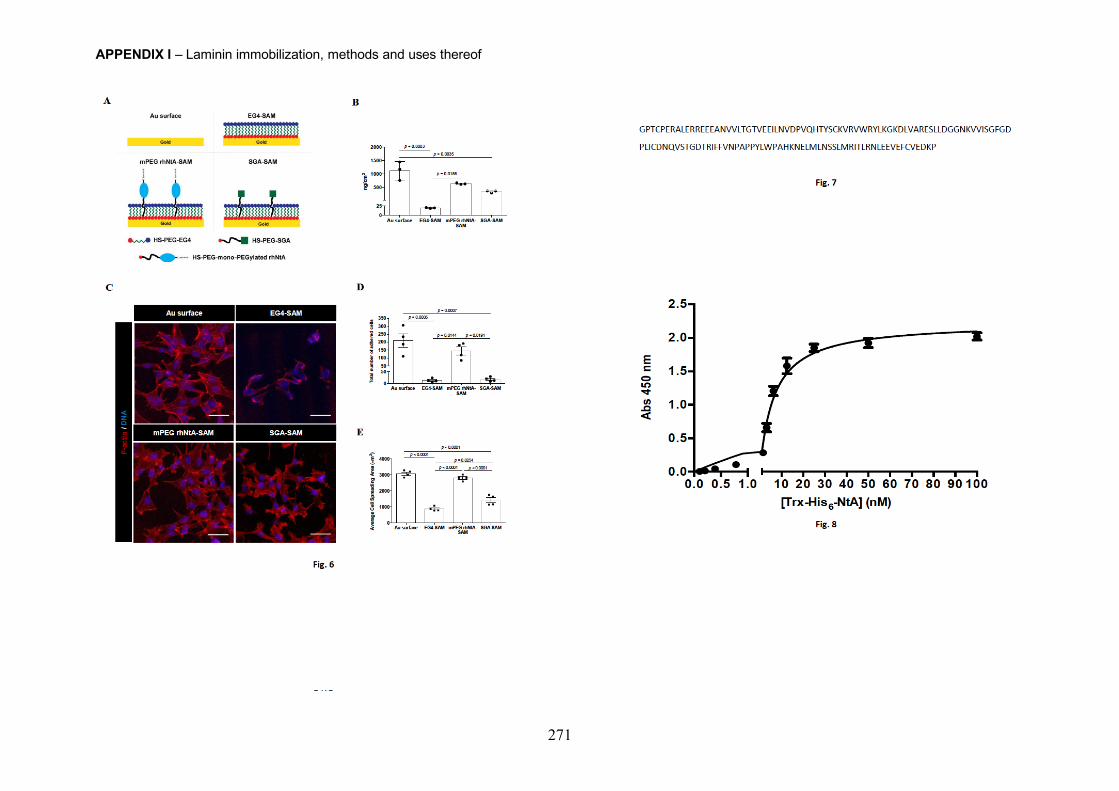
APPENDIX I – Laminin immobilization, methods and uses thereof
271
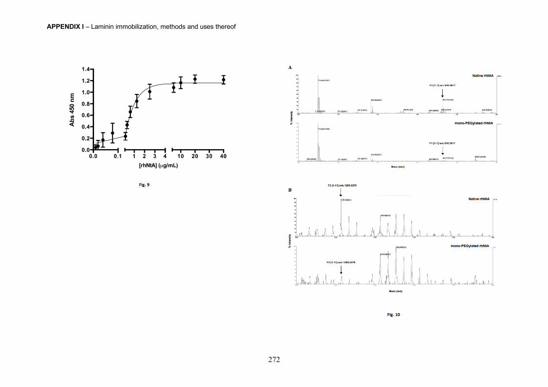
APPENDIX I – Laminin immobilization, methods and uses thereof
272
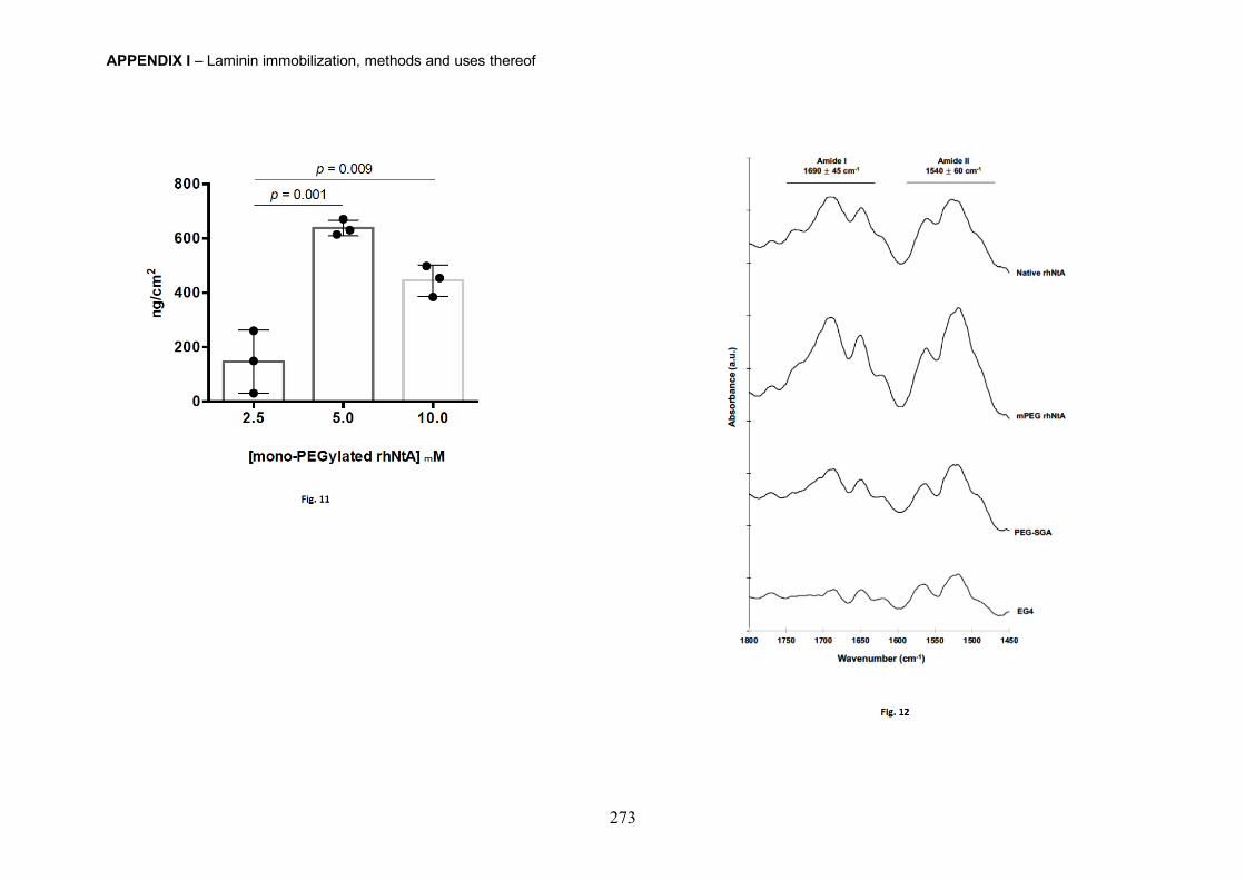
APPENDIX I – Laminin immobilization, methods and uses thereof
273
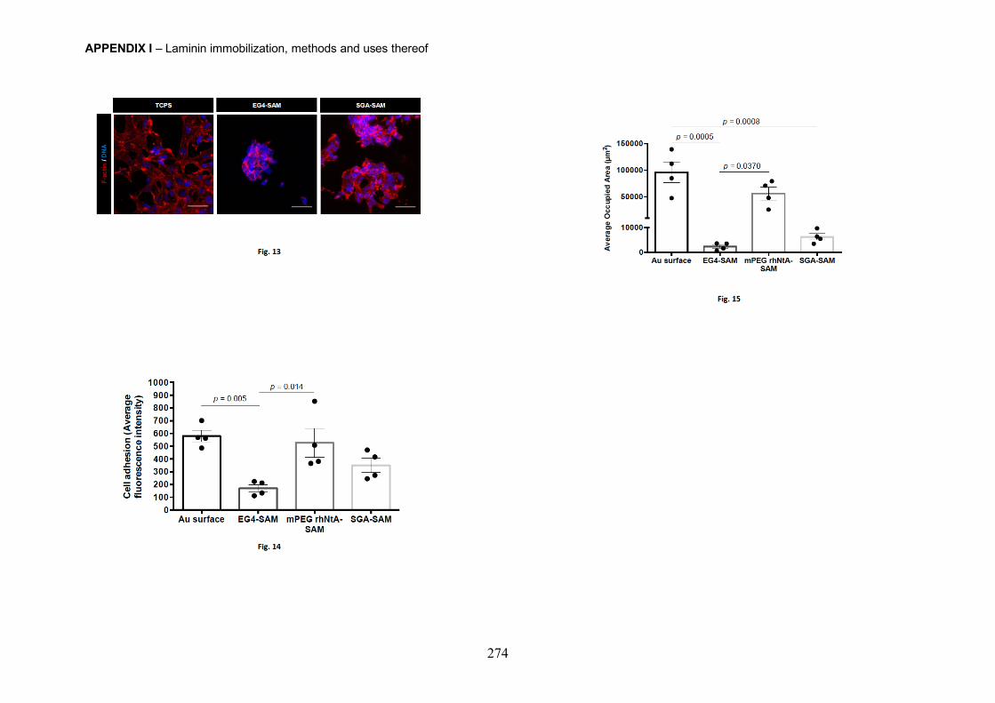
APPENDIX I – Laminin immobilization, methods and uses thereof
274
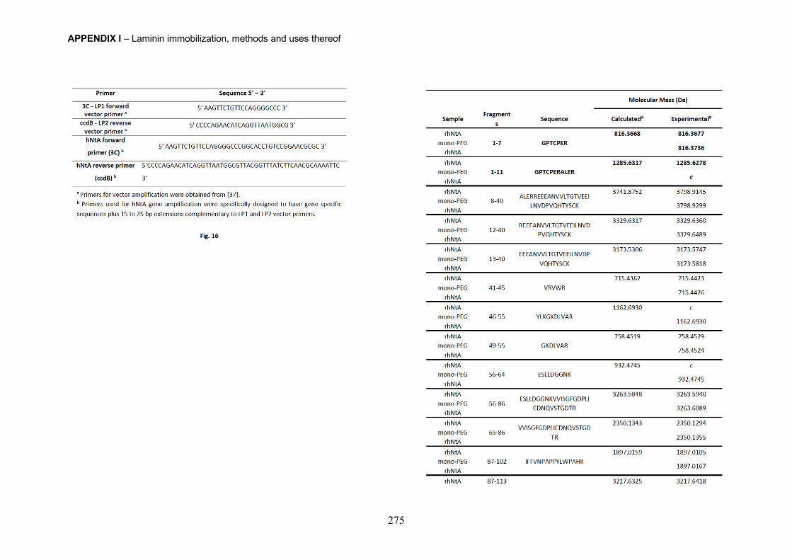
APPENDIX I – Laminin immobilization, methods and uses thereof
275
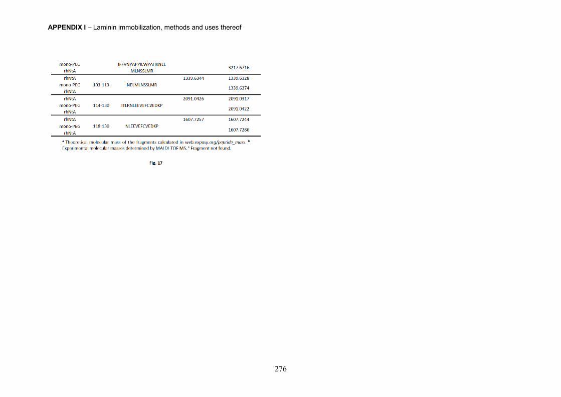
APPENDIX I – Laminin immobilization, methods and uses thereof
276
Copyright © 2022 FDOKUMEN