Laminin α5 chain is required for intestinal smooth muscle development
15
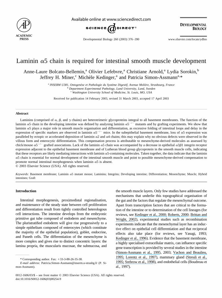
Laminin 5 chain is required for intestinal smooth muscle development Anne-Laure Bolcato-Bellemin, a Olivier Lefebvre, a Christiane Arnold, a Lydia Sorokin, b Jeffrey H. Miner, c Miche `le Kedinger, a and Patricia Simon-Assmann a, * a INSERM U381, Ontogene `se et Pathologie du Syste `me Digestif, Avenue Molie `re, Strasbourg, France b Department Experimental Pathology, Lund University, Lund, Sweden c Washington University School of Medicine, St. Louis, MO, USA Received for publication 14 February 2003, revised 31 March 2003, accepted 17 April 2003 Abstract Laminins (comprised of , , and chains) are heterotrimeric glycoproteins integral to all basement membranes. The function of the laminin 5 chain in the developing intestine was defined by analysing laminin 5 / mutants and by grafting experiments. We show that laminin 5 plays a major role in smooth muscle organisation and differentiation, as excessive folding of intestinal loops and delay in the expression of specific markers are observed in laminin 5 / mice. In the subepithelial basement membrane, loss of 5 expression was paralleled by ectopic or accelerated deposition of laminin 2 and 4 chains; this may explain why no obvious defects were observed in the villous form and enterocytic differentiation. This compensation process is attributable to mesenchyme-derived molecules as assessed by chick/mouse 5 / grafted associations. Lack of the laminin 5 chain was accompanied by a decrease in epithelial 31 integrin receptor expression adjacent to the epithelial basement membrane and of Lutheran blood group glycoprotein in the smooth muscle cells, indicating that these receptors are likely mediating interactions with laminin 5-containing molecules. Taken together, the data indicate that the laminin 5 chain is essential for normal development of the intestinal smooth muscle and point to possible mesenchyme-derived compensation to promote normal intestinal morphogenesis when laminin 5 is absent. © 2003 Elsevier Science (USA). All rights reserved. Keywords: Basement membrane; Laminin 5 mutant mouse; Laminins; Integrins; Developing intestine; Differentiation; Mesenchyme; Muscle; Hybrid intestines; Graft Introduction Intestinal morphogenesis, proximodistal regionalisation, and maintenance of the steady state between cell proliferation and differentiation result from tightly controlled heterologous cell interactions. The intestine develops from the embryonic primitive gut tube composed of endoderm and mesenchyme. The pluristratified endoderm will give rise progressively to a simple epithelium composed of enterocytes (which constitute the majority of the epithelial population), goblet, endocrine, and Paneth cells. The differentiation of the mesenchyme is more complex and gives rise to distinct concentric layers: the lamina propria, the muscularis mucosae, the submucosa, and the smooth muscle layers. Only few studies have addressed the mechanisms that underlie this topographical organisation of the gut and the factors that regulate the mesenchymal outcome. Apart from transcription factors that are critical to the forma- tion of the intestine or to determination of the cell lineages (for reviews, see Kedinger et al., 2000; Roberts, 2000; Brittan and Wright, 2002), experimental studies such as recombination experiments indicate that the mesenchymal layer has an induc- tive effect on epithelial cell differentiation and that reciprocal effects also take place (for reviews, see Yasugi, 1993; Kedinger et al., 1996). Evidence that the basement membrane, a highly specialised extracellular matrix, can influence specific gene transcription is provided by several studies in the intestine (Simon-Assmann et al., 1995, 2003; Vachon and Beaulieu, 1995; Lorentz et al., 1997), mammary gland (Streuli et al., 1995; Srebrow et al., 1998), and endothelial cells (Boudreau et al., 1997). * Corresponding author. Fax: 33-3-88-26-35-38. E-mail address: [email protected] (P. Si- mon-Assmann). R Available online at www.sciencedirect.com Developmental Biology 260 (2003) 376 –390 www.elsevier.com/locate/ydbio 0012-1606/03/$ – see front matter © 2003 Elsevier Science (USA). All rights reserved. doi:10.1016/S0012-1606(03)00254-9
-
Upload
independent -
Category
Documents
-
view
0 -
download
0
Transcript of Laminin α5 chain is required for intestinal smooth muscle development
Laminin �5 chain is required for intestinal smooth muscle development
Anne-Laure Bolcato-Bellemin,a Olivier Lefebvre,a Christiane Arnold,a Lydia Sorokin,b
Jeffrey H. Miner,c Michele Kedinger,a and Patricia Simon-Assmanna,*a INSERM U381, Ontogenese et Pathologie du Systeme Digestif, Avenue Moliere, Strasbourg, France
b Department Experimental Pathology, Lund University, Lund, Swedenc Washington University School of Medicine, St. Louis, MO, USA
Received for publication 14 February 2003, revised 31 March 2003, accepted 17 April 2003
Abstract
Laminins (comprised of �, �, and � chains) are heterotrimeric glycoproteins integral to all basement membranes. The function of thelaminin �5 chain in the developing intestine was defined by analysing laminin �5�/� mutants and by grafting experiments. We show thatlaminin �5 plays a major role in smooth muscle organisation and differentiation, as excessive folding of intestinal loops and delay in theexpression of specific markers are observed in laminin �5�/� mice. In the subepithelial basement membrane, loss of �5 expression wasparalleled by ectopic or accelerated deposition of laminin �2 and �4 chains; this may explain why no obvious defects were observed in thevillous form and enterocytic differentiation. This compensation process is attributable to mesenchyme-derived molecules as assessed bychick/mouse �5�/� grafted associations. Lack of the laminin �5 chain was accompanied by a decrease in epithelial �3�1 integrin receptorexpression adjacent to the epithelial basement membrane and of Lutheran blood group glycoprotein in the smooth muscle cells, indicatingthat these receptors are likely mediating interactions with laminin �5-containing molecules. Taken together, the data indicate that the laminin�5 chain is essential for normal development of the intestinal smooth muscle and point to possible mesenchyme-derived compensation topromote normal intestinal morphogenesis when laminin �5 is absent.© 2003 Elsevier Science (USA). All rights reserved.
Keywords: Basement membrane; Laminin �5 mutant mouse; Laminins; Integrins; Developing intestine; Differentiation; Mesenchyme; Muscle; Hybridintestines; Graft
Introduction
Intestinal morphogenesis, proximodistal regionalisation,and maintenance of the steady state between cell proliferationand differentiation result from tightly controlled heterologouscell interactions. The intestine develops from the embryonicprimitive gut tube composed of endoderm and mesenchyme.The pluristratified endoderm will give rise progressively to asimple epithelium composed of enterocytes (which constitutethe majority of the epithelial population), goblet, endocrine,and Paneth cells. The differentiation of the mesenchyme ismore complex and gives rise to distinct concentric layers: thelamina propria, the muscularis mucosae, the submucosa, and
the smooth muscle layers. Only few studies have addressed themechanisms that underlie this topographical organisation ofthe gut and the factors that regulate the mesenchymal outcome.Apart from transcription factors that are critical to the forma-tion of the intestine or to determination of the cell lineages (forreviews, see Kedinger et al., 2000; Roberts, 2000; Brittan andWright, 2002), experimental studies such as recombinationexperiments indicate that the mesenchymal layer has an induc-tive effect on epithelial cell differentiation and that reciprocaleffects also take place (for reviews, see Yasugi, 1993;Kedinger et al., 1996). Evidence that the basement membrane,a highly specialised extracellular matrix, can influence specificgene transcription is provided by several studies in the intestine(Simon-Assmann et al., 1995, 2003; Vachon and Beaulieu,1995; Lorentz et al., 1997), mammary gland (Streuli et al.,1995; Srebrow et al., 1998), and endothelial cells (Boudreau etal., 1997).
* Corresponding author. Fax: �33-3-88-26-35-38.E-mail address: [email protected] (P. Si-
mon-Assmann).
R
Available online at www.sciencedirect.com
Developmental Biology 260 (2003) 376–390 www.elsevier.com/locate/ydbio
0012-1606/03/$ – see front matter © 2003 Elsevier Science (USA). All rights reserved.doi:10.1016/S0012-1606(03)00254-9

In the intestine, basement membranes, composed oflaminins, type IV collagen, nidogen, and perlecan, are foundin two main locations: first, the subepithelial basementmembrane, which is located from the earliest phases ofdevelopment at the endoderm/mesenchyme interface andsubsequently underlies the differentiated epithelium of theadult organ. This structure has been shown to be synthesisedand secreted by both epithelial and mesenchymal cells, in astage- and cell type-specific pattern through reciprocal cellinteractions (for review, see Simon-Assmann et al., 1998).The second location is the smooth muscle basement mem-brane, which surrounds individual cells organised into aninner circular and an outer longitudinal muscle layer. Thelaminins, major components of the basement membrane, arethought to be important for organ development. All lamininmolecules are heterotrimers, with each isoform containing aunique combination of �, �, and � chains. So far, 11genetically distinct laminin chains, �1–�5, �1–�3, �1–�3,have been described. Yet, only 13 laminin isoforms withtissue-specific distribution have been convincingly demon-strated (Tunggal et al., 2000). Laminins are functionallyinvolved in cell adhesive interactions and show signal-transducing activities leading to induction of cell differen-tiation and cell migration (Colognato and Yurchenco,2000). In the mouse intestinal subepithelial basement mem-brane, at least 3 laminin isoforms have been detected: lami-nin-1 (�1�1�1), laminin-2 (�2�1�1), and laminin-10(�5�1�1) (Simo et al., 1991; Lefebvre et al., 1999; Sasakiet al., 2002a). Expression of laminin chains in the intestinalmesenchyme-derived musculature has been less intensivelystudied (Glukhova et al., 1993; Simon-Assmann et al.,1995).
The widespread distribution of the laminin �5 chainduring development and in adult tissues suggests that it is amajor laminin � chain in epithelial basement membrane(Miner et al., 1995; Sorokin et al., 1997a; Ekblom et al.,1998). The laminin �5 chain associates with the �1 chainand either �1 or �2 chains to form laminin-10 and laminin-11, respectively (Miner et al., 1997). More recently, twonovel laminin heterotrimers, one composed of �5, �2, and�3 (laminin-15) and the other by �5, �2, and �2 chains,were found to be expressed in the retinal matrix (Libby etal., 2000) and in bone marrow stromal cells (Siler et al.,2002), respectively. Among the laminin � chains, �5 showsthe broadest distribution in the intestine being present in thesubepithelial basement membrane throughout development,around smooth muscle cells with an increasing intensityparalleling their differentiation, and in basement mem-branes of large blood vessels (Lefebvre et al., 1999). Mul-tiple defects were observed in the Lam �5�/� homozygotes,including failure of anterior neural tube closure and digitseptation and dysmorphogenesis of the placental labyrinth(Miner et al., 1998). However, no detailed analysis of theintestine was carried out.
It is currently unclear whether developmental changes inbasement membrane organisation are causative or simply
reflective of morphogenetic remodelling. Although nullmice in which individual basement membrane moleculeswere deleted have been generated, revealing the importanceof some laminin chains in defined organs, there are very fewindications of any influence of these molecules on gastro-intestinal morphogenesis. In the present paper, we describemorphological and biochemical alterations in the smoothmuscle coat in intestines of embryonic laminin �5�/� mice.No defects in villous morphology were found, apparentlydue to compensatory mechanisms which can be attributed tomesenchymal cells.
Materials and methods
Animals
Laminin �5-deficient mice were generated by Miner etal. (1998) and were bred on the C57BL/6J genetic back-ground. Mice were genotyped by PCR as described (Mineret al., 1998). No homozygotes lived past embryonic day 17.Foetuses were removed by caesarean section at variousstages between the 12th and 16th day of gestation, with theday on which a vaginal plug was found considered day 0.Segments of small intestines were immediately processed.
For grafting experiments, male Swiss athymic nude (nu/nu) mice (Iffa Credo, Les Oncins, France) were used andhoused in laminar airflow cabinets under pathogen-free con-ditions.
White Leghorn chick embryos were used for graftingexperiments. The chicken eggs were incubated at 38 � 1°C,and the developmental stages were referred to as days ofincubation.
All experiments were performed in accordance with in-stitutional guidelines for animal care.
Primary antibodies
Primary antibodies to basement membrane moleculesused in immunofluorescence studies on cryosections arelisted in Table 1.
For detection of integrin subunits, the following antibod-ies were used: rat mAb 1997 against mouse integrin �1(Chemicon International, Temecula, CA), rat mAb 346-11Aagainst mouse integrin �4 (kindly provided by Dr Kennel,Oak Ridge, TN), rat mAb GoH3 against integrin �6 (Im-munotech, Marseille, France), and rabbit polyclonal anti-body 8-4 against integrin �3 subunit (generous gift from Dr.DiPersio, Albany Medical College, NY; DiPersio et al.,1995). Localisation of Lutheran was performed by using arabbit polyclonal antibody (Kikkawa et al., 2002).
To follow differentiation of epithelial cells, affinity-pu-rified polyclonal anti-villin antibodies (generous gift of Dr.Robine, Institut Curie, Paris; Robine et al., 1985) were used.To study differentiation of muscle cells, polyclonal smoothmuscle desmin antibodies (kindly provided by Prof. Gabbi-
377A.-L. Bolcato-Bellemin et al. / Developmental Biology 260 (2003) 376–390

ani; Universite de Geneve, Switzerland; Benzonana et al.,1988), monoclonal anti-desmin (clone DE-R-11, Dako,DK), and monoclonal anti-� smooth muscle actin (Sigma,St. Louis, MO) were used.
Other antibodies included mouse monoclonal anti-� tu-bulin isotype III (clone SDL.3D10; Sigma), mouse mono-clonal NCL-KI67-MM1 antibody (Novocastra LaboratoriesLtd., UK), rabbit anti-chromogranin A (DiaSorin, MN,USA), and rabbit polyclonal anti-GAPDH (Launay et al.,1989).
Recombinants and grafting procedures
Associations between mouse and chick intestinal tissuecomponents were performed by using a previously de-scribed experimental procedure (Kedinger et al., 1981) (seeFig. 5). Briefly, 5-day chick embryonic and 13-day foetalmouse intestinal anlagen from laminin �5 mutants wereisolated. Mesenchyme was separated from the endodermafter incubation of the intestinal segments in a 0.03% solu-tion of collagenase (1 h at 37°C). Two types of interspeciesrecombinations of the isolated endodermal and mesenchy-mal components were prepared: mouse laminin �5�/� mes-enchyme with chick endoderm (M�5-m/Ce), and chickmesenchyme with mouse laminin �5�/� endoderm (Cm/M�5-e). After overnight culture on agar-solidified mediumto ensure their cohesion, the hybrid intestines were graftedinto the coelomic cavity of 3-day chick embryos. The hybridintestines were harvested at day 7 after grafting.
Intestines from wild-type, heterozygote, and mutant12–14 embryonic day animals from the same litter were cutinto 2- to 3-mm pieces. They were then grafted (6 frag-ments/animal) under the skin of nude mice. Briefly, a smallincision was made on the back of the recipient and oneintestinal segment was placed into the subcutaneous tissue
and covered with skin. The grafts were taken 1 month afterimplantation and processed for subsequent analysis.
Morphological analysis andimmunofluorescence/immunohistochemistry studies
For conventional histology, samples were fixed inBouin’s solution overnight and processed for 10-�m paraf-fin wax sections. Sections were stained with Hematoxylin/Eosin, or with Periodic-Acid Schiff to visualise goblet cellsusing standard histology techniques.
For transmission electron microscopy, specimens werefixed for 2 h at 4°C in 0.2 M cacodylate-buffered 2%glutaraldehyde, pH 7.4, postfixed in cacodylate-buffered1% osmium tetroxide, pH 7.4, for 30 min at 4°C, dehy-drated, and embedded in araldite. Semithin 0.5-�m sectionswere stained with toluidine blue for histological observa-tions. Ultrathin sections were stained with uranyl acetateand lead citrate before ultrastructural observation by usingan electron microscope (CM-10; Philips Electronic Instru-ments, Mahwah, NJ).
For indirect immunofluorescence staining, intestineswere embedded in Tissue-Tek (Sakura, Labonord), frozenin isopentane cooled in liquid nitrogen, and stored at �80°Cuntil used. Transverse sections (6 �m thick), cut at –19°C,were placed onto superfrost/plus slides (Kindler GmBH,Freiburg, Germany). Incubations with antibodies were car-ried out for 2–3 h at room temperature in a moist chamber.Bound antibodies were visualised by using anti-rat (1:50;Jackson Laboratory), anti-mouse (1:250; Institut Pasteur),or anti-rabbit (1:50; Nordic) secondary antibody conjugatedwith fluorescein isothiocyanate. After mounting in a glyc-erol/PBS/phenylenediamine solution, the slides were exam-ined by using an epifluorescence microscope (AX 60;Olympus Optical Co., Hamburg, Germany). Control sec-
Table 1Specificities and sources of antibodies used for detection of basement membrane molecules
Specificities Species Antibody Source/reference
Laminin �1 rat 198 Sorokin et al., 1992Laminin �2 rabbit 401 Ringelmann et al., 1999Laminin �2 rat 4H8-3 Schuler and Sorokin, 1995Laminin �4 rabbit 377 Sorokin et al., 2000Laminin �4 rat 341 Ringelmann et al., 1999Laminin �5 rabbit 405 Sorokin et al., 2000Laminin �5 rabbit 8948 Miner et al., 1997Laminin �5 rat 4G6 Sorokin et al., 1997bLaminin �1 rat 3A4 Sixt et al., 2001Laminin �1/�1 rat 1975 Chemicon International, Inc.Laminin �2 rabbit 1116 R. Timpl (Martinsried)Laminin �1 rat 3E10 Sixt et al., 2001Laminin �2 rabbit SE144 Aberdam et al., 1994pan-laminin �1, �1, �1 rabbit LN6-7S Simo et al., 1991collagen IV rabbit coll2a De Arcangelis et al., 1996nidogen rat BE1/27 D. Edgar (Liverpool, UK)perlecan rat BMS4057 Boehringer/Ingelheim/Bioproducts
378 A.-L. Bolcato-Bellemin et al. / Developmental Biology 260 (2003) 376–390
tions were processed as above with omission of the primaryantibodies.
When mouse monoclonal antibodies were used, immu-noperoxidase staining was performed on cryosections byusing the HistoMouse-SP kit following the manufacturer’sinstructions (Zymed Laboratories, Inc., CA, USA). Sectionswere lightly counterstained with hematoxylin. Detection ofchromogranin was performed on prefixed (4% paraformal-dehyde, 2 h) and deparaffined sections. Antigen–antibodycomplexes were detected by using the Vectastain ABC Kit(Vector).
Western blot analysis
Intestines from wild-type and �5�/� E14, E15, and E16animals were homogenised with a Dounce homogeniser in a10 mM Tris–HCl, pH 7.4/0.9% NaCl solution and subse-quently sonicated. After centrifugation at 13,000g for 15min, the supernatant was collected. Proteins (10–15 �g)were incubated in Laemmli buffer containing 2% SDS (w/v)and 100 mM DTT at 100°C for 5 min. The proteins werethen separated by SDS–PAGE analysis (10% gels) andsubjected to Western blotting by using monoclonal antibod-ies to � smooth muscle actin as well as polyclonal antibod-ies to GAPDH as an internal control. After incubation withgoat anti-mouse or anti-rabbit alkaline phosphatase-linkedsecondary antibodies, the blots were developed by using theECF (enhanced chemifluorescence) substrate (ECF WesternBlotting Kit; Amersham Life Science Ltd., England). Quan-tification was determined by using a fluorescent scanninginstrument (Molecular Imager Fx; BioRad, CA, USA). TheMagicMark Western Standard (Invitrogen, Cergy Pointoise,France) was used as molecular weight marker.
Results
Excessive folding of intestinal loops is observed inlaminin �5�/� mutant mice
As the laminin �5�/� mice do not survive past E17(Miner et al., 1998), intestines were analysed between E14and E16. At these stages, laminin �5 chain was alreadypresent in control intestines at the endodermal/mesenchy-mal junction and in the serosal layer (Fig. 1A). While thesize of the E16 mutant embryos was about 80% of wildtypelittermates, the small intestine was proportionally more re-duced in length (twofold) (Fig. 1B). Yet, the diameter of theintestine was only slightly affected (Fig. 1G vs F: E14; Fig.1I vs H: E16). The laminin �5 null intestines exhibitedexcessive folding of intestinal loops (Fig. 1B and C). Inabout half of the cases, fusion of the muscular layers withabsence of mesentery and/or the serosal layer was observed(Fig. 1C–E, arrows). These gross intestinal deformities weremainly detected at the latest stage studied (E16), primarilyin the proximal but occasionally also in the most distal
regions of the intestine. In the nonfused areas, peripheralmesenchymal cells did not elongate and remained rounded(Fig. 1K vs J). No differences were observed between het-erozygous (�5�/�) and wild-type (�5�/�) intestines in theiroverall development, histology, or growth characteristics.
Between E14 and E16 in wild-type intestines, the villidevelop and concomitantly the pseudostratified epitheliumconverts to a simple monolayer (Fig. 1F and H). In mutantmice, formation of villi occurred, although they appearedfewer in number, and the epithelium looked normal mor-phologically (Figs. 1G and I and 2). The gross morphologyof the enterocytes was not modified in the laminin �5�/�
intestines. This was confirmed by ultrastructural observa-tions showing identical brush border organisation at theirapical surface (Fig. 2A and D). To analyse whether thedifferentiation of enterocytes could nevertheless have beenaffected by the lack of the laminin �5 chain, we checked byimmunofluorescence for the presence of villin, a cytoskel-etal protein found in the apical brush border membrane. Itsexpression pattern in laminin �5�/� E14 and E16 intestines(Fig. 2E) was similar to that observed in controls (Fig. 2B).We determined the number of mucous-producing gobletcells (only apparent in the late E16 foetal stage) by stainingwith Schiff’s reagent. The E16 laminin �5�/� mouse intes-tines showed a significant decrease in the number of gobletcells compared with wild-type intestines: mean of 17 gobletcells per 10 cross-sectioned villi in control intestines ascompared with two cells in laminin �5�/� intestines (Fig.2C and F). Endocrine cells (chromogranin A staining) andPaneth cells were not detected either in control or in thelaminin �5�/� intestines, as expected at E16 (not illustrat-ed). A possible effect on epithelial cell proliferation due tothe lack of laminin �5 was assayed by determining thenumber of KI67-positive cells per villous axis. No signifi-cant differences were observed between control and mutantintestines (not illustrated). This is consistent with the ab-sence of obvious alterations in the villous organisation inthe laminin �5�/� mice.
Laminin �5�/� mutants display smooth muscle defects
During intestinal morphogenesis, a subset of mesenchy-mal cells differentiate first into the inner (circular) smoothmuscle cell layer and later into the outer (longitudinal) celllayer (Kedinger et al., 1990; McHugh, 1996). A first sign ofthis differentiation is the progressive elongation and align-ment of a subset of mesenchymal cells in the peripheralregion of the mesenchyme (Fig. 1J).
As shown above (Fig. 1J and K), aberrant structuralfeatures were obvious in the mesenchymal layers whenlaminin �5 was absent. Transmission electron microscopicanalysis confirmed these observations and further showedthat contact between the serosal layer and the mesenchymalcells was loosened in laminin �5�/� intestines (not illus-trated). Cytodifferentiation of the smooth muscle cells wasdetermined by immunocytochemistry and immunoblot us-
379A.-L. Bolcato-Bellemin et al. / Developmental Biology 260 (2003) 376–390

ing smooth muscle-specific antibodies. The absence oflaminin �5 affected the differentiation process at the devel-opmental stages analysed. At the earliest stage studied, E14,a faint expression of desmin and actin was found in wild-type intestines in a circular zone of two to three cell layersin the mesenchyme that correspond to the presumptive innermuscular layer (Fig. 3A and B). Decrease in the stainingwas observed in the E14 mutants (Fig. 3C and D); obser-vation was confirmed by immunoblot experiments (Fig. 3).In the E16 intestine, when smooth muscle cells becomemore differentiated, there was significantly less smoothmuscle marker expression in the mutant compared with thecontrol, as visualised by desmin staining (Fig. 3G vs E). Theenteric nervous system which comprises the plexuses is
located in the muscle coat and in the submucosa. Thedefect in muscle differentiation does not seem to affectthe plexuses as immunostaining of neural-specific �-tu-bulin was similar between control and mutant intestines(not illustrated). Finally, a decreased rate of cell prolif-eration, as assessed by KI67 staining, was obvious in themesenchymal cell compartment in mutant intestines (Fig.3H vs F).
Laminin �5 deficiency leads to misexpression of otherlaminin chains
To determine whether the absence of laminin �5 (Fig.4E and N) affected the expression and deposition of other
Fig. 3. Altered muscle cell differentiation in laminin �5�/� embryonic small intestine. Differentiation of smooth muscle cells was assayed by immunode-tection of desmin (A, C, E, G) and alpha-smooth muscle actin (B, D) at E14 (A–D) and at E16 (E, G) in control (A, B, E) and laminin �5�/� (C, D, G) smallintestines. Immunoblotting experiment was performed on protein extracted from E14 control and laminin �5�/� intestines by using smooth muscle actinantibodies; GAPDH antibodies were applied on the same gel as internal control. Staining with the KI67 antibody on E16 laminin �5�/� (H) and control (F)intestines shows that the number of proliferating mesenchymal cells (arrowheads in F) is decreased in mutant, but no differences were observed in epithelium;e, epithelium; pml, presumptive muscular layer. Scale bars: 20 �m.
Fig. 1. View of the embryonic gastrointestinal tract (B, C), PAS-stained jejunal sections (D–K) at two embryonic developmental stages: E14 (F, G) and E16 (C–E,H–K), taken from wild-type (B, F, H, J) and laminin �5�/� (B, C–E, G, I, K) animals, and immunostaining of laminin �5 chain in the E12 intestine (A). Note thetwisted appearance of the intestine in the mutant (B, C) with a fusion of the intestinal loops in the most severe cases (arrows in C–E). The mesentery has beendissected away to visualise the looping. The general structure of the intestine is conserved in the mutant intestines at the two stages studied (G, I vs F, H). In theE16 mutant mice, note the modifications in the arrangement of the mesenchymal cells at the periphery in E16 mutant intestine (I, K) as compared with control (H,J). e, epithelium; st, stomach; pml, presumptive muscle layers. Scale bars: 0.5 cm (B), 10 �m (J, K), 20 �m (E), 30 �m (A), 50 �m (D, F–I).Fig. 2. Epithelial differentiation in E16 wild-type (A–C) and laminin �5�/� (D–F) intestines. Enterocytic differentiation was assessed by the presence of abrush border at the apical surface of cells at the ultrastructural level (A, D) and by expression of villin, a cytoskeletal protein, detected by immunofluorescence(B, E). PAS staining revealed fewer goblet cells in the laminin �5�/� small intestine (F) as compared with control (C) (arrowheads). e, epithelium; m,mesenchyme; lp, lamina propria. Scale bars: 1 �m (A, D), 50 �m (B, C, E, F).
380 A.-L. Bolcato-Bellemin et al. / Developmental Biology 260 (2003) 376–390

381A.-L. Bolcato-Bellemin et al. / Developmental Biology 260 (2003) 376–390
matrix molecules in the subepithelial and muscle base-ment membranes, we performed immunostaining on in-testinal sections at two embryonic stages, E14 and E16.
Distribution of nidogen, perlecan, and type IV collagenas well as of various laminin �, �, and � chains wasexamined. Nidogen, perlecan, and type IV collagen were
Fig. 4. Analysis of subepithelial (arrows) and muscle basement membranes in E14 (A–H) and E16 (I–T) small intestines from wild-type (A–D, I–M, S) or laminin�5�/� (E–H, N–R, T) animals, assessed by immunodetection of laminin constituent chains (A–R) and transmission electron microscopy (S, T). Cryosections werestained with antibodies as indicated. Transmission electron micrographs show the basement membrane underlying the epithelial cells in control (S) and mutant (T)intestines. e, epithelium; m, mesenchyme; pml, presumptive muscular layer. Scale bars: 100 �m (A, D, E, H–R), 50 �m (B, C, F, G), and 0.25 �m (S, T).
382 A.-L. Bolcato-Bellemin et al. / Developmental Biology 260 (2003) 376–390
present in all basement membranes of wild-type, het-erozygous, and laminin �5�/� intestines with comparablestaining, locations, and intensities (not illustrated). Datafor the individual laminin chains are summarised in Table2.
As far as the subepithelial basement membrane isconcerned, two major findings emerged from this study.First, there was an accelerated or ectopic deposition oflaminin �2 (Fig. 4F vs B) and laminin �4 (Fig. 4G vs C,and 4Q vs L) chains, respectively, in the laminin �5�/�
intestines (each chain being tested by two different anti-bodies); this phenomenon (already noted at E12) wasmore obvious at E14 as compared with E16. Second, atE16, the most striking feature was the reduction of thelaminin �2 (Fig. 4O vs J) and laminin �2 (Fig. 4P vs K)chains. The other laminin chains studied (�1, �1, �1)were unaltered at either stage (illustrated for �1 and �1;Fig. 4D and H, M and R). At the ultrastructural level, adiscernible basement membrane was observed underlyingthe epithelial cells in both control and mutant intestines;yet its appearance was irregular in the laminin �5�/�
mutants (Fig. 4S and T).As far as muscle cell layers are concerned, it should be
noted that, at E14, expression of laminin chains at theperiphery of the mesenchyme corresponding to the pre-sumptive muscle cell coat was either absent or faint inwild-type intestines as well as in the laminin �5�/� mutants(Fig. 4A–H). In the E16 wildtype intestine (Table 2), mostof the individual laminin chains were found expressed at theperiphery of cells, in the newly formed muscular layers(Fig. 4I–M). Interestingly, a loss or decrease in the stainingintensity for laminin �2 (Fig. 4O vs J), �1 (not illustrated),and �2 (Fig. 4P vs K) chains was obvious in the laminin �5mutant intestines, while expression patterns of the otherlaminin chains studied were not altered as illustrated for �4and �1 (Fig. 4L, M, Q, R).
Laminin �2 and �4 compensation at the subepithelialbasement membrane is attributable to mesenchyme-derived molecules
To determine the cellular (epithelial or mesenchymal)origin of the compensation observed for laminin �2 and �4chains at the level of the subepithelial basement membrane,we performed associations using anlagen taken from em-bryonic chick intestines and foetal mouse mutant �5�/�
intestines (Fig. 5). The recombinants, grafted in the coelo-mic cavity of chick embryos (for 7 days), developed intointestinal structures with small villi (exemplified in Fig. 5Afor a M�5-m/Ce hybrid intestine). Application of a poly-clonal antibody against laminin (recognising �1, �1, and �1chains of laminins from both species) on hybrid intestinesconfirmed the reformation of a basement membrane (Fig.5B). The cellular origins of the laminin �2 and �4 chainswere then determined by the use of antibodies specificallyrecognising mouse antigens on both types of hybrid intes-tines. In M�5-m/Ce recombinants, the anti-mouse laminin�2 (Fig. 5C) and �4 (Fig. 5D) antibodies stained the sub-epithelial basement membrane, as well as the mouse de-rived-mesenchyme. In the inverse Cm/M�5-e recombi-nants, no staining could be observed (Fig. 5E and F)showing the lack of deposition of laminin �2 and �4 chainsby the mouse endoderm. These data show that the laminin�2 and �4 compensation occurring in the mutant �5�/�
intestines can be attributed to mesenchyme-derived mole-cules.
The �3�1 integrin and the Lutheran receptor are affectedin laminin �5�/� mice
Several integrin receptors have been shown to interactwith laminin �5-containing molecules, such as laminin-10.Although preferential interactions seem to be cell type-specific, at least four integrin receptors are involved in
Table 2Summary of the expression pattern of individual laminin chains at the subepithelial (E14–E16) and smooth muscle (E16) intestinal basement membranesin control animals and laminin �5�/� mutants
Ab Epithelium (E14–E16) Muscle (E16)
LN �5�/� LN �5�/� Changes LN �5�/� LN �5�/� Changes
LN �1 mAb198 �� �� 3 � � 3LN �2 pAb401 � � m � � 3LN �2 mAb4H8-3 � � m � � 3LN �4 pAb377 � �� m �� �� 3LN �4 mAb341 � � m �� �� 3LN �1 mAb3A4 �� �� 3 �� �� 3LN �1/�1 mAb1975 �� �� 3 �� �� 3LN �2 pAb1116 � E16* � E16* n � �/� nLN �1 mAb3E10 �� �� 3 �� � nLN �2 pAbSE144 �� � E16* n � �/� n
Note. LN, laminin; mAb, monoclonal antibodies; pAb, polyclonal antibodies. E16* means that this pattern of expression is only obvious at embryonic day16.
383A.-L. Bolcato-Bellemin et al. / Developmental Biology 260 (2003) 376–390
adhesion to laminin-10: �2�1, �3�1, �6�1, and �6�4(Kikkawa et al., 2000; Pouliot et al., 2000; Nielsen andYamada, 2001; Doi et al., 2002). More recently, the Luth-
eran blood group glycoprotein was found to be a transmem-brane receptor for laminin �5 (Moulson et al., 2001;Kikkawa et al., 2002). Expression of integrin receptors and
Fig. 5. Model of hybrid intestines allowing determination of the cellular origin of the laminin �2 and �4 compensation. Two types of intestinal associationswere grafted into the coelomic cavity of 3-day chick embryos and recovered after 7 days: mouse laminin �5�/� mesenchyme/chick endoderm (M�5-m/Ce)and chick mesenchyme/mouse laminin �5�/� endoderm (Cm/Me�5-e) recombinants. Histology (hematoxylin/eosin) confirms the development of smallintestinal segments (A). Use of a rabbit polyclonal antibody that recognises �1, �1, �1 chains of laminins from both species confirmed the presence of asubepithelial basement membrane in developed intestinal hybrids (B). The application of antibodies recognising specifically mouse laminin �2 (polyclonal341 antibody; C) and �4 (rat monoclonal 341 antibody; D) antigens on M�5-m/Ce (C, D) allows the visualisation of both molecules at the epithelial/mesenchymal interface. On the inverse associations (Cm/M�5-e), no immunoreactivity could be visualised with these antibodies (laminin �2, E; laminin �4,F). e, epithelium; m, mesenchyme; arrows point to the subepithelial basement membrane region. Scale bars: 50 �m (A) and 100 �m (B–F).Fig. 6. Immunodetection of integrin subunits in intestines from E14 wild-type (A, B) or mutant laminin �5�/� (D, E) animals, and of the Lutheran receptor in E16 wild-type(C) or mutant laminin �5�/� (F) animals, as indicated. Basal staining of the �3 subunit was reduced concomitant to the lack of laminin �5 chain (arrows in D vs A). Thiswas also the case for the �1 integrin subunit, although the decrease was less obvious (E vs B). Note the strong decrease of Lutheran in the mutant muscle cells (F vs C).e, epithelium; lp, lamina propria; pml, presumptive muscular layer; arrows point to the subepithelial basement membrane region. Scale bars: 100 �m.
384 A.-L. Bolcato-Bellemin et al. / Developmental Biology 260 (2003) 376–390

of Lutheran was determined in wild-type and �5�/� mutantintestines (Fig. 6).
Among the � integrin subunits analysed, only the �3presented a difference between laminin �5�/� and wildtypeintestines. A decreased staining for integrin �3 was noted atthe basal pole of epithelial cells subjacent to the subepithe-lial basement membrane in laminin �5�/� mutant intes-tines. This difference in staining intensity, visible at E14(Fig. 6D vs A), was less obvious at E16 (not shown). Nochanges in �3 integrin expression were found in the musclecell compartment or at sites of epithelial cell–cell contacts.In the E14 intestine, the �1 subunit (Fig. 6B) appearedpredominantly localised in the mesenchymal compartment,where it is segregated in two distinct regions: over four tofive mesenchymal cells located just beneath the epithelialcells (future lamina propria), and in the inner presumptivemuscle layer; it was also located to a lesser extent at thebasolateral membrane of epithelial cells. In the �5�/� mu-tant intestines, there was a slight decrease in staining inten-sity of this integrin subunit, mostly at the subepithelialbasement membrane region, and in the mesenchyme in thevicinity of the epithelium at E14 (Fig. 6E vs B). Expressionof the �6 and �4 integrin subunits in E14 and E16 mutantintestines was similar to that in the corresponding controls(not shown).
Immunodetection of the Lutheran receptor was per-formed on the E16 intestines and revealed distinct stainingaround smooth muscle cells as well as some faint reaction atthe basal pole of epithelial cells facing the subepithelialbasement membrane (Fig. 6C). In the laminin �5�/� intes-tines, there was a striking decrease in staining of Lutheran,especially in the muscle region (Fig. 6F).
Grafting experiments allow observation of �5�/�
intestinal morphogenesis
To examine whether epithelial and muscle differentiationas well as villus morphogenesis can be achieved at laterstages of development in the absence of laminin �5, E12–E14 intestinal fragments were implanted into the subcuta-neous space of adult nude mice and allowed to develop for1 month. This strategy bypassed the embryonic lethality,permitting further development of the �5�/� intestines. Theembryonic intestines developed into spherical vascularisedstructures that often contained mucus secretion or extrudedepithelial cells. There was no striking difference in the grossmorphology between the wild-type (Fig. 7A) and �5�/�
Fig. 7. Development of the intestines of laminin �5�/� mutants aftersubcutaneous implantation in nude mice. Sections of E14-grafted control(A) and laminin �5-deficient (H) intestines harvested after 4 weeks showeda similar overall gross morphology. Cryosections of the recovered implants
(controls: B–G; �5�/� mutants: I–N) labelled for laminin �5 (B, I), �1 (C,J), �2 (D, K), �4 (E, F, L, M) chains and for desmin (G, N). Note thatconcomitant to the lack of laminin �5 chain in the developed implants (I vsB), there are some modifications in the staining pattern of basementmembrane laminins �1, �2, and �4 at the subepithelial region (J–L vsC–E) and at the muscle coat for laminin �4 (M vs F). e, epithelium; lp,lamina propria; ml, muscular layer; arrows point to the subepithelial base-ment membrane region. Scale bars: 50 �m.
385A.-L. Bolcato-Bellemin et al. / Developmental Biology 260 (2003) 376–390
(Fig. 7H) intestinal implants. Indeed, both were composedof villous structures lined by a single epithelium with nonoticeable variation in the number of mucous cells, a sub-mucosa, and a well-defined muscular coat. These structureswere very similar to those of intestinal tissues developed insitu. To assess possible laminin isoform switching patterns,staining for the major laminin � chains was performed inparallel on cryosections (Fig. 7). The absence of laminin �5at the subepithelial basement membrane in the developedimplants (Fig. 7I vs B) was accompanied by (1) an increasein intensity of laminin �1 chain staining (Fig. 7J vs C), (2)an expansion of laminin �2 chain staining (normally re-stricted to the crypt basement membrane) that extended tothe middle of the villi (Fig. 7K vs D), and (3) an ectopicdeposition of the laminin �4 chain mostly visible at the baseof the villous structures (Fig. 7L vs E). No major differencesbetween wildtype and laminin �5 null intestines were notedin the expression of the laminin �1 and �2 chains in base-ment membrane surrounding muscle cells (not illustrated).For laminin �4, a more distinct staining marked the indi-vidual smooth muscle cells in the laminin �5-deficient im-plants (Fig. 7M vs F). Smooth muscle cell differentiation,assessed by immunostaining of desmin, was similar be-tween grafted mutant and grafted control intestines (Fig. 7Nvs G), paralleling the morphological observations.
Discussion
Previously described expression patterns of laminin �5predicted that it might have an important role in intestinaldevelopment and differentiation. However, due to earlylethality of laminin �5 null mice, intestinal developmentwas not studied in detail. Here, we examined intestinaldevelopment in laminin �5�/� mice between E14 and E16and at later developmental stages through foetal intestinalgrafts. The embryonic period examined is of keen interest asit corresponds to (1) progressive formation of a single epi-thelium accompanied by the outgrowth of villi and (2)differentiation of smooth muscle cells from undifferentiatedmesenchymal cells. These events have been shown to bedependent on extracellular matrix-mediated heterotypic cellinteractions. Our results indicate that laminin �5 has acrucial role in the process of embryonic intestinal foldingand in the development of the normal musculature. How-ever, no major effects on the epithelial compartment, excepta decrease in number of goblet cells, were observed inlaminin �5-deficient intestines, probably because of com-pensatory mesenchymal contribution of the laminin �2 and�4 chains to the subepithelial basement membrane.
The expression of laminins in the small intestine hasreceived major attention, as they are both temporally (dur-ing morphogenesis) and spatially (along the crypt–villusaxis in the adult) regulated. Among the laminin � chainsstudied to date, laminin �5 shows the broadest distributionin the intestine. At early phases of development, a deposi-
tion of laminin �5 is already apparent in the undifferentiatedintestine at the endodermal/mesenchymal interface. In theadult, an increasing gradient of intensity is evident at thesubepithelial basement membrane from the base to the tip ofvilli (Lefebvre et al., 1999). As the laminin �2 chain isrestricted to the adult crypts (present data, and Sasaki et al.,2002b), one can conclude that laminin-10 (�5�1�1) is theonly �5-containing molecule found in the subepithelial in-testinal basement membrane. Of interest is the reciprocalexpression of laminin-10 found along the villus, and lami-nin-1 (�1�1�1) or -2 (�2�1�1), both confined to the cryptregion in rodent (Simo et al., 1991; Simon-Assmann et al.,1994; Lefebvre et al., 1999; Sasaki et al., 2002a).
Compensatory mechanism in the subepithelial basementmembrane
We observed that normal intestinal villus outgrowth andepithelial cell differentiation occurred in mutant mice lack-ing laminin �5 at early stages of intestinal development, aswell as at later stages in laminin �5�/�-grafted intestines.The explanation for a lack of visible phenotype as far asepithelial cells are concerned may be linked to compensa-tory mechanisms. Indeed, we found that laminin �2 and �4chains, which are not present at early stages of developmentin the intestinal subepithelial basement membrane in wild-type mice, compensate for �5 deficiency, as they weredeposited precociously/ectopically in the mutant intestines.Immunocytochemical labelling of laminin chains in associ-ations of embryonic intestinal anlagen from laminin �5mutant mice and from chick demonstrated that the mesen-chyme is solely responsible for this compensation. A com-pensatory process also occurs in more advanced develop-mental stages, assessed here by the grafting experiments,which allowed us to circumvent the embryonic lethality andto analyse further development of the mutant intestines. Atthese stages, staining of laminin �1—the expression ofwhich gradually decreases as development proceeds in nor-mal intestine (Simo et al., 1991)—was found to be intensi-fied in the mutants additionally to �2 and �4 chains. Suchcompensatory phenomena, whereby removal of one mole-cule from a site results in the ectopic accumulation of othermolecules, have also been described in laminin �5�/� ec-toderm, where laminin �1, �2, and �4 chains are upregu-lated (Miner et al., 1998), and in �5�/� lung, where branch-ing morphogenesis was associated with ectopic depositionof laminin �4 in airway basement membrane (Nguyen et al.,2002). Here, we discern for the first time the cellular originof such a compensatory mechanism.
It should be emphasised that, despite the switch in lami-nin � chain deposition occurring in �5�/� mutant intestine,a rather well-formed basement membrane structure wasvisible, as shown by ultrastructural analysis of the subepi-thelial region. This could be linked to the fact that, in themutant intestines, synthesis and deposition of laminin �1chain by epithelial cells (Simo et al., 1992) is not modified.
386 A.-L. Bolcato-Bellemin et al. / Developmental Biology 260 (2003) 376–390
Therefore, laminin-1 molecules can polymerise, thus con-tributing to the formation of the basement membrane. Lami-nin �5 chain is produced by both epithelial and mesenchy-mal cells during normal development (Lefebvre et al.,1999). In the present study, we could show that the lack oflaminin �5 in mesenchymal cells has a pronounced effect,as it will force them to secrete prematurely the laminin �2and �4 chains. An inverse situation occurs in the inflamedtissues of patients with Crohn’s disease, in which a loss ofmesenchymally derived laminin �2 chain expression incrypts was paralleled by a significant upregulation of the �5chain (Bouatrouss et al., 2000). Altogether, these data pointto the crucial role of the mesenchyme with the coordinatedactivity of epithelial cells for basement membrane assem-bly, further controlling growth and differentiation in theintestine.
Integrins and Lutheran, receptors of the laminin �5 chain
Several integrins have been implicated as receptors forlaminin-10/laminin-11, including �2�1, �3�1, �6�1, and�6�4 (Kikkawa et al., 1998, 2000; Gu et al., 1999; Tani etal., 1999; Pouliot et al., 2000; Nielsen and Yamada, 2001;Spessotto et al., 2001; Doi et al., 2002). In our experiments,lack of the laminin �5 chain is accompanied by a decreasein the �3 integrin subunit at the base of the endodermal cellsfacing the epithelial basement membrane and to a lesserextent of the �1 integrin subunit. The fact that neither �6nor �4 integrin subunits are modified in absence of laminin�5 chain led us to conclude that the �3�1 integrin is themajor receptor mediating interaction of laminin �5 contain-ing molecules of the subepithelial basement membrane.This observation correlates well with the colocalisation oflaminin �5 chain (Lefebvre et al., 1999) and of �3�1integrin in the adult intestine (Beaulieu, 1992). Interest-ingly, mice lacking both integrin �3 and �6 subunits werefound to exhibit developmental defects similar to thoseobserved in mice lacking the laminin �5 chain, includingexencephaly and syndactyly (De Arcangelis et al., 1999). Itshould be noted that the overall structure and villus mor-phogenesis of the intestine was conserved in these double�3�/�/�6�/� mutant embryos (P.S.-A. and E. Georges-Labouesse, unpublished data), as it is in the laminin �5�/�
mutants. Additionally, we show that the level of the Luth-eran transmembrane receptor, known to interact with thelaminin �5 chain (Kikkawa et al., 2002), was strikinglydecreased in the smooth muscle cells in the E16 mutant�5�/� intestines, as well as in the grafted implants (notillustrated). This is in agreement with data from Moulson etal. (2001) who showed a dramatic reduction (lung, pharynx,kidney, skin) in the basal concentration of Lutheran in micelacking laminin �5 and a significant increase in the heart oftransgenic mice overexpressing laminin �5. The varyingcapacity of laminin �5 chain to bind to precise receptors—integrin or Lutheran—between the epithelium and the mus-cle tissue is of potent interest.
Intestinal muscle defects
Gut development is known to be controlled by signallingfactors such as Sonic hedgehog that mediate cellular inter-actions, and transcription factors that directly control geneactivity. Of interest here is the intestinal phenotype of theShh homozygous mutant mice, which display multiple ab-normalities, including a reduction in the thickness of thecircular smooth muscle layer that may result from an overtmalrotation of the gut (Ramalho-Santos et al., 2000). Incontrast to our data, the hedgehog mutants present excessiveand abnormally located neurons, thus resembling Hirsch-sprung’s disease. These data indicate that Shh and the lami-nin �5 chain, although both are involved in the smoothmuscle patterning, are acting through different targets ortranscription factors. In view of our present finding of ex-cessive folding in the laminin �5 null intestines, the phe-notype of the MTG8 knock-out mice is noteworthy (Calabiet al., 2001). MTG8, a member of a small gene familyencoding transcriptional regulators (Miyoshi et al., 1993;Wolford and Prochazka, 1998), has been shown to be ex-pressed at E14.5 in the outermost gut layers (Calabi et al.,2001). In the E15.5 mutants, it was noted that the complex-ity of the midgut loops was reduced, a situation opposite tothat observed here. The phenotype linked to the null muta-tion in the Drosophila �3,5/lam A chain, the precursor ofthe vertebrate �3 and �5 chains, is also worth noting. Thismutation is embryonic lethal and results in alterations inmesoderm-derived tissue (Yarnitzky and Volk, 1995). Al-teration in the organisation of enteric smooth musculaturewas also observed in the dy/dy mouse, which expressesabnormally low levels of the laminin �2 chain (Relan et al.,1999).
Absence of compensatory mechanisms in the embryonicintestinal muscle
In contrast to the situation concerning the subepithelialbasement membrane, the absence of laminin �5 in theembryonic mesenchyme does not lead to compensation, asno significant changes in the laminin � chains were ob-served. This would explain why developmental defects inthe smooth muscle layers were observed in the E14 and E16mutant intestines. This was surprising, as the basementmembrane composition of the muscle tissue is more com-plex and variable compared with that of the epithelium.Interestingly, at later stages, in the grafted intestines, lami-nin �4 was found to be significantly increased in the musclewhen �5 was absent. This compensation is apparently effi-cient since well structured and differentiated muscle layerswere formed in the grafts. It is known that implanted intes-tines are vascularised by blood vessels originating from thehost, allowing the growth of the implants (Simon-Assmannet al., 1989). Therefore, one can postulate that regulatoryfactors (growth factors and/or chemokines) originating fromthe adult host vasculature are crucial for synthesis of lami-
387A.-L. Bolcato-Bellemin et al. / Developmental Biology 260 (2003) 376–390

nin �4. A similar situation was observed in the case of theglomerular basement membrane where endothelial cellsand/or factors present in the circulation mediate lamininisoform transition (St. John et al., 2001).
Chains associated to laminin �5
Another interesting finding that concerns both the sub-epithelial and the smooth muscle basement membranes isthe fact that expression of the laminin �2 and �2 chains wasdecreased in laminin �5 null intestines. Interestingly, the �2chain has not been detected in combination with the laminin�3 or �3 chain in the mouse intestine (Orian-Rousseau etal., 1996), which together form the well-described lami-nin-5 isoform. Therefore, our data strongly suggest that theintestinal basement membranes also contain a novel iso-form, consisting of �5�2�2 chains, that has also been de-scribed to occur in bone marrow stromal cells (Siler et al.,2002). Two further laminin isoforms have been describedwhich contain both �5 and �2 chains, laminin-11 (�5�2�1)and laminin-15 (�5�2�3). Dot blot analysis of laminin �3revealed that its mRNA is widely expressed in human tis-sues, including intestine, although at low levels (Koch et al.,1999), suggesting that both of these isoforms may be ex-pressed in the murine intestine. Our study points to the factthat expression of laminin isoforms in the intestinal smoothmuscle is still more complex than previously reported(Glukhova et al., 1993), due to the presence of numerouslaminin chains, and awaits final characterisation.
Acknowledgments
A. Klein is greatly acknowledged for her technical as-sistance in tissue sample preparation and tissue recombinantexperiments. We thank Dr. D. Aberdam (U. 385, INSERM,Nice, France), Dr. M. DiPersio (Albany Medical College,NY, USA), Dr. D. Edgar (University of Liverpool, En-gland), Prof. G. Gabbiani (Universite de Geneve, Suisse),Dr. S. Robine (Institut Curie, Paris, France), and Dr. R.Timpl (Max-Planck-Institut fur Biochemie, Martiensried,Germany) for supplying antibodies. This work was sup-ported by INSERM, an exchange program between Franceand Germany (Procope), and by grants from the ACI Bi-ologie du developpement et Physiologie Integrative (#172),the Comite Departemental de la Ligue Contre le Cancer.A-L.B.-B. was funded by fellowship from the Associationpour la Recherche sur le Cancer. L. Eleuterio is greatlythanked for her help in the finalisation of the manuscript andfigures, and M. Lavogez for animal care.
References
Aberdam, D., Aguzzi, A., Baudoin, C., Galliano, M.F., Ortonne, J.P.,Meneguzzi, G., 1994. Developmental expression of nicein adhesion
protein (Laminin-5) subunits suggests multiple morphogenic roles. CellAdhes. Commun. 2, 115–129.
Beaulieu, J.F., 1992. Differential expression of the VLA family of integrinsalong the crypt-villus axis in the human small intestine. J. Cell Sci. 102,427–436.
Benzonana, G., Skalli, O., Gabbiani, G., 1988. Correlation between thedistribution of smooth muscle or non muscle myosins and �-smoothmuscle actin in normal and pathological soft tissues. Cell Motil. Cy-toskeleton 11, 260–274.
Bouatrouss, Y., Herring-Gillam, F.E., Gosselin, J., Poisson, J., Beaulieu,J.F., 2000. Altered expression of laminins in Crohn’s disease smallintestinal mucosa. Am. J. Pathol. 156, 45–50.
Boudreau, N., Andrews, C., Srebrow, A., Ravanpay, A., Cheresh, D.A.,1997. Hox D3 induces an angiogenic phenotype. J. Cell Biol. 139,257–264.
Brittan, M., Wright, N.A., 2002. Gastrointestinal stem cells. J. Pathol. 197,492–509.
Calabi, F., Pannell, R., Pavloska, G., 2001. Gene targeting reveals a crucialrole for MTG8 in the gut. Mol. Cell. Biol. 21, 5658–5666.
Colognato, H., Yurchenco, P.D., 2000. Form and function: the lamininfamily of heterotrimers. Dev. Dyn. 218, 213–234.
De Arcangelis, A., Neuville, P., Boukamel, R., Lefebvre, O., Kedinger, M.,Simon-Assmann, P., 1996. Inhibition of laminin �1-chain expressionleads to alteration of basement membrane assembly and cell differen-tiation. J. Cell Biol. 133, 417–430.
De Arcangelis, A., Mark, M., Kreidberg, J., Sorokin, L., Georges-Labouesse, E., 1999. Synergistic activities of �3 and �6 integrins arerequired during apical ectodermal ridge formation and organogenesis inthe mouse. Development 126, 3957–3968.
DiPersio, C.M., Shah, S., Hynes, R.O., 1995. �3A�1 integrin localizes tofocal contacts in response to diverse extracellular matrix proteins.J. Cell Sci. 108, 2321–2336.
Doi, M., Thyboll, J., Kortesmaa, J., Jansson, K., Iivanainen, A., Parvardeh,M., Timpl, R., Hedin, U., Swedenborg, J., Tryggvason, K., 2002.Recombinant human laminin-10 (�5�1�1). Production, purification,and migration-promoting activity on vascular endothelial cells. J. Biol.Chem. 277, 12741–12748.
Ekblom, M., Falk, M., Salmivirta, K., Durbeej, M., Ekblom, P., 1998.Laminin isoforms and epithelial development. Ann. N. Y. Acad. Sci.857, 194–211.
Glukhova, M., Koteliansky, V., Fondacci, C., Marotte, F., Rappaport, L.,1993. Laminin variants and integrin laminin receptors in developingand adult human smooth muscle. Dev. Biol. 157, 437–447.
Gu, Y., Sorokin, L., Durbeej, M., Hjalt, T., Jonsson, J.I., Ekblom, M.,1999. Characterization of bone marrow laminins and identification of�5-containing laminins as adhesive proteins for multipotent hemato-poietic FDCP-mix cells. Blood 93, 2533–2542.
Kedinger, M., Simon, P.M., Grenier, J.F., Haffen, K., 1981. Role ofepithelial-mesenchymal interactions in the ontogenesis of intestinalbrush-border enzymes. Dev. Biol. 86, 339–347.
Kedinger, M., Simon-Assmann, P., Bouziges, F., Arnold, C., Alexandre,E., Haffen, K., 1990. Smooth muscle actin expression during rat gutdevelopment and induction in fetal skin fibroblastic cells associatedwith intestinal embryonic epithelium. Differentiation 43, 87–97.
Kedinger, M., Fritsch, C., Evans, G.S., De Arcangelis, A., Orian-Rousseau,V., Simon-Assmann, P., 1996. Role of stromal–epithelial cell interac-tions and of basement membrane molecules in the onset and mainte-nance of epithelial integrity, in: Kagnoff, M.F., Kiyono, H. (Eds.),Essentials of Mucosal Immunology, Academic Press, San Diego, pp.111–123.
Kedinger, M., Freund, J.-N., Launay, J.F., Simon-Assmann, P., 2000. Cellinteractions through the basement membrane in intestinal developmentand differentiation, in: Sanderson, I.R., Walker, W.A. (Eds.), Devel-opment of the Gastrointestinal Tract, B.C. Decker Inc., London, pp.83–102.
388 A.-L. Bolcato-Bellemin et al. / Developmental Biology 260 (2003) 376–390
Kikkawa, Y., Sanzen, N., Sekiguchi, K., 1998. Isolation and characteriza-tion of laminin-10/11 secreted by human lung carcinoma cells: laminin-10/11 mediates cell adhesion through integrin �3�1. J. Biol. Chem.273, 15854–15859.
Kikkawa, Y., Sanzen, N., Fujiwara, H., Sonnenberg, A., Sekiguchi, K.,2000. Integrin binding specificity of laminin-10/11: laminin-10/11 arerecognized by �3�1, �6�1 and �6�4 integrins. J. Cell Sci. 113,869–876.
Kikkawa, Y., Moulson, C.L., Virtanen, I., Miner, J.H., 2002. Identificationof the binding site for the Lutheran blood group glycoprotein onlaminin �5 through expression of chimeric laminin chains in vivo.J. Biol. Chem. 277, 44864–44869.
Koch, M., Olson, P.F., Albus, A., Jin, W., Hunter, D.D., Brunken, W.J.,Burgeson, R.E., Champliaud, M.F., 1999. Characterization and expres-sion of the laminin �3 chain: a novel, non-basement membrane-asso-ciated, laminin chain. J. Cell Biol. 145, 605–618.
Launay, J.F., Jellali, A., Vanier, M.T., 1989. Glyceraldehyde-3-phosphatedehydrogenase is a microtubule binding protein in a human colontumor cell line. Biochim. Biophys. Acta 996, 103–109.
Lefebvre, O., Sorokin, L., Kedinger, M., Simon-Assmann, P., 1999. De-velopmental expression and cellular origin of the laminin �2, �4, and�5 chains in the intestine. Dev. Biol. 210, 135–150.
Libby, R.T., Champliaud, M.F., Claudepierre, T., Xu, Y., Gibbons, E.P.,Koch, M., Burgeson, R.E., Hunter, D.D., Brunken, W.J., 2000. Lami-nin expression in adult and developing retinae: evidence of two novelCNS laminins. J. Neurosci. 20, 6517–6528.
Lorentz, O., Duluc, I., De Arcangelis, A., Simon-Assmann, P., Kedinger,M., Freund, J.-N., 1997. Key role of the Cdx2 homeobox gene inextracellular matrix-mediated intestinal cell differentiation. J. CellBiol. 139, 1553–1565.
McHugh, K.M., 1996. Molecular analysis of gastrointestinal smooth mus-cle development. J. Pediat. Gastroenterol. Nutr. 23, 379–394.
Miner, J.H., Lewis, R.M., Sanes, J.R., 1995. Molecular cloning of a novellaminin chain, �5, and widespread expression in adult mouse tissues.J. Biol. Chem. 270, 28523–28526.
Miner, J.H., Patton, B.L., Lentz, S.I., Gilbert, D.J., Jenkins, N.A., Cope-land, N.G., Sanes, J.R., 1997. The laminin � chains: expression, de-velopmental transitions and chromosomal locations of � 1-5, identifi-cation of heterotrimeric laminins 8-11, and cloning of a novel �3isoform. J. Cell Biol. 137, 685–701.
Miner, J.H., Cunningham, J., Sanes, J.R., 1998. Roles for laminin inembryogenesis: Exencephaly, syndactyly, and placentopathy in micelacking the laminin �5 chain. J. Cell Biol. 143, 1713–1723.
Miyoshi, H., Kozu, T., Shimizu, K., Enomoto, K., Maseki, N., Kaneko, Y.,Kamada, N., Ohki, M., 1993. The t(8;21) translocation in acute myeloidleukemia results in production of an AML1-MTG8 fusion transcript.EMBO J. 12, 2715–2721.
Moulson, C.L., Li, C., Miner, J.H., 2001. Localization of Lutheran, a novellaminin receptor, in normal, knockout, and transgenic mice suggests aninteraction with laminin �5 in vivo. Dev. Dyn. 222, 101–114.
Nguyen, N.M., Miner, J.H., Pierce, R.A., Senior, R.M., 2002. Laminin�5 is required for lobar septation and visceral pleural basement mem-brane formation in the developing mouse lung. Dev. Biol. 246, 231–244.
Nielsen, P.K., Yamada, Y., 2001. Identification of cell-binding sites on thelaminin �5 N-terminal domain by site-directed mutagenesis. J. Biol.Chem. 276, 10906–10912.
Orian-Rousseau, V., Aberdam, D., Fontao, L., Chevalier, L., Meneguzzi,G., Kedinger, M., Simon-Assmann, P., 1996. Developmental expres-sion of laminin-5 and HD1 in the intestine: epithelial to mesenchymalshift for the laminin �2 chain subunit deposition. Dev. Dyn. 206,12–23.
Pouliot, N., Connolly, L.M., Moritz, R.L., Simpson, R.J., Burgess, A.W.,2000. Colon cancer cells adhesion and spreading on autocrine lami-
nin-10 is mediated by multiple integrin receptors and modulated byEGF receptor stimulation. Exp. Cell Res. 261, 360–371.
Ramalho-Santos, M., Melton, D.A., McMahon, A.P., 2000. Hedgehogsignals regulate multiple aspects of gastrointestinal development. De-velopment 127, 2763–2772.
Relan, N.K., Yang, Y., Beqaj, S., Miner, J.H., Schuger, L., 1999. Cellelongation induces laminin �2 chain expression in mouse embryonicmesenchymal cells: role in visceral myogenesis. J. Cell Biol. 147,1341–1350.
Ringelmann, B., Roder, C., Hallmann, R., Maley, M., Davies, M.,Grounds, M., Sorokin, L., 1999. Expression of laminin �1, �2, �4 and�5 chains, fibronectin and tenascin-C in skeletal muscle of dystrophic129ReJ dy/dy mice. Exp. Cell Res. 246, 165–182.
Roberts, D.J., 2000. Molecular mechanisms of development of the gastro-intestinal tract. Dev. Dyn. 219, 109–120.
Robine, S., Huet, C., Moll, R., Sahuquillo-Merino, C., Coudrier, E.,Zweibaum, A., Louvard, D., 1985. Can villin be used to identifymalignant and undifferentiated normal digestive epithelial cells? Proc.Natl. Acad. Sci. USA 82, 8488–8492.
Sasaki, T., Giltay, R., Talts, U., Timpl, R., Talts, J.F., 2002a. Expressionand distribution of laminin �1 and �2 chains in embryonic and adultmouse tissues: an immunochemical approach. Exp. Cell Res. 275,185–199.
Sasaki, T., Mann, K., Miner, J.H., Miosge, N., Timpl, R., 2002b. DomainIV of mouse laminin �1 and �2 chains. Eur. J. Biochem. 269, 431–442.
Schuler, F., Sorokin, L., 1995. Expression of laminin isoforms in mousemyogenic cells in vitro and in vivo. J. Cell Sci. 108, 3795–3805.
Siler, U., Rousselle, P., Muller, C.A., Klein, G., 2002. Laminin �2 chain asa stromal cell marker of the human bone marrow microenvironment.Br. J. Haematol. 119, 212–220.
Simo, P., Simon-Assmann, P., Bouziges, F., Leberquier, C., Kedinger, M.,Ekblom, P., Sorokin, L., 1991. Changes in the expression of lamininduring intestinal development. Development 112, 477–487.
Simo, P., Bouziges, F., Lissitzky, J.C., Sorokin, L., Kedinger, M., Simon-Assmann, P., 1992. Dual and asynchronous deposition of lamininchains at the epithelial-mesenchymal interface in the gut. Gastroenter-ology 102, 1835–1845.
Simon-Assmann, P., Bouziges, F., Vigny, M., Kedinger, M., 1989. Originand deposition of basement membrane heparan sulfate proteoglycan inthe developing intestine. J. Cell Biol. 109, 1837–1848.
Simon-Assmann, P., Kedinger, M., De Arcangelis, A., Rousseau, V., Simo,P., 1995. Extracellular matrix components in intestinal development.Experientia 51, 883–900.
Simon-Assmann, P., Duclos, B., Orian-Rousseau, V., Arnold, C., Mathelin,C., Engvall, E., Kedinger, M., 1994. Differential expression of lamininisoforms and �6-�4 integrin subunits in the developing human andmouse intestine. Dev. Dyn. 201, 71–85.
Simon-Assmann, P., Lefebvre, O., Bellissent-Waydelich, A., Olsen, J.,Orian-Rousseau, V., De Arcangelis, A., 1998. The laminins: role inintestinal morphogenesis and differentiation. Ann. N. Y. Acad. Sci.859, 46–64 .
Simon-Assmann, P., Bolcato-Bellemin, A.L., Turck, N., Piccinni, S., Ol-sen, J., Launay, J.F., Lefebvre, O., Kedinger, M., 2003. Basementmembrane laminins in normal and pathological intestine, in: Johnstone,P. (Ed.), Disease Progression and Carcinogenesis in the Gastrointesti-nal Tract, Kluwer Academic Publishers, London, pp. 223–239 .
Sixt, M., Hallmann, R., Wendler, O., Scharffetter-Kochanek, K., Sorokin,L.M., 2001. Cell adhesion and migration properties of �2-integrinnegative polymorphonuclear granulocytes on defined extracellular ma-trix molecules. Relevance for leukocyte extravasation. J. Biol. Chem.276, 18878–18887.
Sorokin, L., Conzelmann, S., Ekblom, P., Battaglia, C., Aumailley, M.,Timpl, R., 1992. Monoclonal antibodies against laminin-A chain frag-ment-E3 and their effects on binding to cells and proteoglycan and onkidney development. Exp. Cell Res. 201, 137–144.
389A.-L. Bolcato-Bellemin et al. / Developmental Biology 260 (2003) 376–390
Sorokin, L., Pausch, F., Durbeej, M., Ekblom, P., 1997a. Differentialexpression of five laminin �(1-5) chains in developing and adult mousekidney. Dev. Dyn. 210, 446–462.
Sorokin, L., Pausch, F., Frieser, M., Kroger, S., Ohage, E., Deutzmann, R.,1997b. Developmental regulation of the laminin �5 chain suggests arole in epithelial and endothelial cell maturation. Dev. Biol. 189,285–300.
Sorokin, L.M., Maley, M.A.L., Moch, H., von der Mark, H., von der Mark,K., Cadalbert, L., Karosi, S., Davies, M.J., McGeachie, J.K., Grounds,M.D., 2000. Laminin �4 and integrin �6 are upregulated in regener-ating dy/dy skeletal muscle: comparative expression of laminin andintegrin isoforms in muscles regenerating after crush injury. Exp. CellRes. 256, 500–514.
Spessotto, P., Yin, Z., Magro, G., Deutzmann, R., Chiu, A., Colombatti, A.,Perris, R., 2001. Laminin isoforms 8 and 10 are primary components ofthe subendothelial basement membrane promoting interaction withneoplastic lymphocytes. Cancer Res. 61, 339–347.
Srebrow, A., Friedmann, Y., Ravanpay, A., Daniel, C.W., Bissell, M.J.,1998. Expression of Hoxa-1 and Hoxb-7 regulated by extracellularmatrix-dependent signals in mammary epithelial cells. J. Cell Biochem.69, 377–391.
St. John, P.L., Wang, R., Yin, Y., Miner, J.H., Robert, B., Abrahamson,D.R., 2001. Glomerular laminin isoform transitions: errors in meta-
nephric culture are corrected by grafting. Am. J. Physiol. RenalPhysiol. 280, F695–F705.
Streuli, C.H., Edwards, G.M., Delcommenne, M., Whitelaw, C.B.A., Bur-don, T.G., Schindler, C., Watson, C.J., 1995. Stat5 as a target forregulation by extracellular matrix. J. Biol. Chem. 270, 21639–21644.
Tani, T., Lehto, V.-P., Virtanen, I., 1999. Expression of laminins 1 and 10in carcinoma cells and comparison of their roles in cell adhesion. Exp.Cell Res. 248, 115–121.
Tunggal, P., Smyth, N., Paulsson, M., Ott, M.C., 2000. Laminins: structureand genetic regulation. Microsc. Res. Tech. 51, 214–227.
Vachon, P.H., Beaulieu, J.F., 1995. Extracellular heterotrimeric lamininpromotes differentiation in human enterocytes. Am. J. Physiol. 268,G857–G867.
Wolford, J.K., Prochazka, M., 1998. Structure and expression of the humanMTG8/ETO gene. Gene 212, 103–109.
Yarnitzky, T., Volk, T., 1995. Laminin is required for heart, somaticmuscles, and gut development in the Drosophila embryo. Dev. Biol.169, 609–618.
Yasugi, S., 1993. Review: role of epithelial–mesenchymal interactions indifferentiation of epithelium of vertebrate digestive organs. Dev.Growth Differ. 35, 1–9.
390 A.-L. Bolcato-Bellemin et al. / Developmental Biology 260 (2003) 376–390