Antibody blockade of the Cripto CFC domain suppresses tumor cell growth in vivo
Upload
independentCategory
view
1download
0
Transgenic Expression of Human LAMA5 SuppressesMurine Lama5 mRNA and Laminin a5 Protein DepositionBrooke M. Steenhard1,3, Adrian Zelenchuk1,3, Larysa Stroganova1,3, Kathryn Isom1,3, Patricia L. St.
John1,3, Glen K. Andrews1,2, Kenneth R. Peterson1,2,3, Dale R. Abrahamson1,3*
1 Departments of Anatomy and Cell Biology, University of Kansas Medical Center, Kansas City, Kansas, United States of America, 2 Biochemistry and Molecular Biology,
University of Kansas Medical Center, Kansas City, Kansas, United States of America, 3 The Kidney Institute, University of Kansas Medical Center, Kansas City, Kansas, United
States of America
Abstract
Laminin a5 is required for kidney glomerular basement membrane (GBM) assembly, and mice with targeted deletions of theLama5 gene fail to form glomeruli. As a tool to begin to understand factors regulating the expression of the LAMA5 gene,we generated transgenic mice carrying the human LAMA5 locus in a bacterial artificial chromosome. These mice depositedhuman laminin a5 protein into basement membranes in heart, liver, spleen and kidney. Here, we characterized two lines oftransgenics; Line 13 expressed ,6 times more LAMA5 than Line 25. Mice from both lines were healthy, and kidney functionand morphology were normal. Examination of developing glomeruli from fetal LAMA5 transgenics showed that the humantransgene was expressed at the correct stage of glomerular development, and deposited into the nascent GBMsimultaneously with mouse laminin a5. Expression of human LAMA5 did not affect the timing of the mouse laminin a1–a5isoform switch, or that for mouse laminin b1–b2. Immunoelectron microscopy showed that human laminin a5 originated inboth glomerular endothelial cells and podocytes, known to be origins for mouse laminin a5 normally. Notably, in neonataltransgenics expressing the highest levels of human LAMA5, there was a striking reduction of mouse laminin a5 protein inkidney basement membranes compared to wildtype, and significantly lower levels of mouse Lama5 mRNA. This suggeststhe presence in kidney of a laminin expression monitor, which may be important for regulating the overall production ofbasement membrane protein.
Citation: Steenhard BM, Zelenchuk A, Stroganova L, Isom K, St. John PL, et al. (2011) Transgenic Expression of Human LAMA5 Suppresses Murine Lama5 mRNAand Laminin a5 Protein Deposition. PLoS ONE 6(9): e23926. doi:10.1371/journal.pone.0023926
Editor: Christos Chatziantoniou, Institut National de la Sante et de la Recherche Medicale, France
Received May 3, 2011; Accepted July 31, 2011; Published September 7, 2011
Copyright: � 2011 Steenhard et al. This is an open-access article distributed under the terms of the Creative Commons Attribution License, which permitsunrestricted use, distribution, and reproduction in any medium, provided the original author and source are credited.
Funding: Funds came from National Institutes of Health (NIH) grants 5P01DK065123 and 5P20RR024214, and a Lied Pilot grant from the University of KansasMedical Center Research Institute. The JEOL JEM-1400 transmission electron microscope was purchased with funds from NIH grant 1S10RR027564. The fundershad no role in study design, data collection and analysis, decision to publish, or preparation of the manuscript.
Competing Interests: The authors have declared that no competing interests exist.
* E-mail: [email protected]
Introduction
The human kidney contains approximately one million
individual nephrons, each beginning with a glomerulus, which is
a unique capillary tuft that largely restricts the passage of serum
albumin and larger proteins into the primary nephron filtrate. All
three layers of the glomerular capillary wall, namely the
glomerular endothelial cells, glomerular epithelial cells (podocytes),
and an intervening glomerular basement membrane (GBM), are
required for maintenance of normal filtration barrier properties
[1–3]. For example, enzymatic degradation of glycosaminoglycans
within the glomerular endothelial surface glycocalyx results in an
increased fractional clearance for albumin [4]. Additionally,
blockage of podocyte-derived VEGF signaling causes glomerular
endothelial cell abnormalities in developing or mature kidneys and
proteinuria [5,6].
A host of defects that affect the podocyte and its specialized
intercellular junction, the epithelial slit diaphragm, also cause
abnormal glomerular permeabilities [1–3]. These include muta-
tions in the NPHS1 gene encoding the slit diaphragm component,
nephrin, which causes congenital nephrotic syndrome of the
Finnish type and results in massive proteinuria at birth [7].
Mutations to NPHS2, which encodes another slit diaphragm
protein, podocin, also causes proteinuria in autosomal recessive
steroid-resistant nephrotic syndrome, a disease often diagnosed in
childhood [8]. Intracellularly, podocin and nephrin are both
linked indirectly to the actin cytoskeleton through interaction with
CD2-associated protein (CD2ap) [9,10]. Slit diaphragms are
absent in mice that lack nephrin [11] or podocin [12] and these
animals die perinatally with renal failure. Mice deficient in CD2ap
also die from renal failure, but at 6–7 weeks of age [10].
Like all basement membranes, the GBM is composed of type
IV collagen, laminin, nidogen, and proteoglycans [13]. Unlike
most other basement membranes, however, the GBM undergoes
type IV collagen and laminin isoform substitution during
glomerular development [14,15]. Specifically, basement mem-
branes within the earliest glomerular regions of comma- and S-
shaped nephric figures contain collagen a1a2a1(IV) and laminin
a1b1c1 (LN-111). These isoforms are later replaced by collagen
a3a4a5(IV), laminin a5b1c1 (LN-511), and laminin a5b2c1 (LN-
521) as glomerular capillary loops expand. Subsequently, LN-521
is the only laminin isoform found in GBMs of fully mature
glomeruli. Collagen a1a2a1(IV) and all of the GBM laminin
chains originate from both glomerular endothelial cells and
podocytes [16,17], but collagen a3a4a5(IV) derives solely from
podocytes [17].
PLoS ONE | www.plosone.org 1 September 2011 | Volume 6 | Issue 9 | e23926

The reasons why the GBM collagen IV and laminin
composition changes during development are not fully understood,
but evidence indicates that this is necessary for final glomerular
maturation and full acquisition and maintenance of filtration
barrier properties. Alport disease, which is a familial nephropathy
marked by focal splitting, thinning, and regional thickening of the
GBM and leads to renal failure, is caused by mutations in either
the COL4A3, COL4A4, or COL4A5 genes encoding the collagen
a3(IV), a4(IV), and a5(IV) protein chains, respectively [18,19].
Most Alport patients fail to assemble a stable network of collagen
a3a4a5(IV) in the GBM, and there is retention of the infantile,
collagen a1a2a1(IV) network. This isoform appears to be more
Figure 1. Human LAMA5 BAC transgenic mice deposit humanlaminin a5 protein in kidney basement membranes. A:Immunofluorescence micrograph of frozen kidney section from atransgenic mouse labeled with mouse anti-human laminin a5, followedby goat anti-mouse IgG2a conjugated to Alexa Fluor-594. Basementmembranes within glomeruli (G) and surrounding tubules (T) immuno-label in bright linear patterns. B: Section from a wildtype littermateincubated with anti-human laminin a5 is negative. In both A and B, cellnuclei are stained blue by DAPI. Magnification: 2006; scale bar = 50 mm.doi:10.1371/journal.pone.0023926.g001
Figure 2. Human LAMA5 BAC transgenic mice deposit humanlaminin a5 protein in basement membranes. Frozen sections oftissues from human LAMA5 BAC transgenics immunolabeled withmouse anti-human laminin a5 antibody and goat anti- mouse IgG2a
conjugated to Alexa Fluor-488. Human laminin a5 is deposited widely inmost or all basement membranes of heart (A), liver (B), and spleen (C).Magnification: 2006; scale bar = 50 mm.doi:10.1371/journal.pone.0023926.g002
Human LAMA5 Transgenics Suppress Mouse Lama5
PLoS ONE | www.plosone.org 2 September 2011 | Volume 6 | Issue 9 | e23926

Human LAMA5 Transgenics Suppress Mouse Lama5
PLoS ONE | www.plosone.org 3 September 2011 | Volume 6 | Issue 9 | e23926

susceptible to proteolysis, which may explain why the GBMs of
Alport patients ultimately deteriorate [19]. A model of Alport
disease has been created in mice through the deletion of the Col4a3
gene [20–22], and these animals die of renal failure 2–4 months
after birth with the same glomerular defects as those seen in Alport
patients. The mouse Alport phenotype can be rescued when
transgenic mice expressing human COL4A3-COL4A4 genes are
crossed onto the mouse Col4a3 knockout background [23].
Failure to undergo laminin isoform transitioning from LN-111
to LN-521 also results in kidney malfunction in mice and in
humans. Although normal glomerular development is seen in mice
with laminin b2 deficiencies, they eventually exhibit podocyte foot
process broadening, proteinuria, and die of renal failure [24].
Humans with mutations in the LAMB2 gene suffer from Pierson
syndrome, which usually presents at birth as congenital nephrotic
syndrome with severe neuromuscular junction abnormalities
(owing to the presence of laminin b2 in the neuromuscular
junction basement membrane as well) [25].
There are no human mutations described for LAMA5, but
experiments in mice have shown its expression to be absolutely
crucial for normal glomerular development and function. Mice
with deletions of Lama5 die before birth with neural tube closure
defects and placental dysmorphogenesis [26]. In kidney, a stable
GBM fails to assemble, and endothelial cells do not form
vascularized glomerular tufts [27]. This Lama5 knockout pheno-
type can be partially rescued when fetal kidneys from Lama5
mutants are grafted into newborn kidneys of normal, wildtype
hosts [28]. In this case, host endothelial cells, which express
laminin a5, migrate into the engrafted Lama5 null kidneys and
vascularized glomeruli form within grafts. The host endothelial
cell-derived laminin a5 does not project across the full width of
these GBMs, however. This results in an unusual situation where
there is retention of the infantile laminin a1 on the outer, sub-
podocyte layer of matrix and laminin a5 is present only on the
inner, subendothelial layer. Additionally, these hybrid GBMs are
abnormally wide and not as well condensed as normal GBM, and
podocyte foot processes are absent [28]. In other experiments,
deletion of Lama5 only in podocytes results in mild to severe
proteinuria, and variable defects in GBM and podocyte ultra-
structure [29]. In this same study, expression of a human LAMA5
transgene under control of a doxycyclin inducible, podocyte-
specific expression system rescues glomerular and tubular defects
caused by a hypomorphic Lama5 mutation [29].
Taken together, these findings demonstrate that the timely
expression of LN-521 is needed for glomerular endothelial cell and
podocyte differentiation, and the appearance of collagen
a3a4a5(IV) is required for long term GBM stability. However,
very little is known at the gene level regarding activation of any of
the mature GBM protein isoforms, what silences transcription of
infantile chain genes at the appropriate developmental stages, and
how the infantile collagen a1a2a1(IV) and LN-111 networks are
removed from developing GBM. In addition, we do not
understand what causes upregulation of Lama5 in Col4a3 knockout
(Alport) mice, which may be an important contributor to fibrosis in
that model [30].
To begin addressing some of these questions, we have developed
bacterial artificial chromosome (BAC) transgenic mice expressing
human LAMA5. These transgenics deposited apparently large
amounts of human laminin a5 protein in basement membranes
widely, and, specifically in glomeruli, at the appropriate
developmental stage. Expression of human LAMA5 did not appear
harmful and kidney functional tests and morphology were normal.
The results suggest that the BAC used for transgenic injections
contained all of the necessary regulatory information for proper
LAMA5 expression. Of great interest, in kidneys from lines with the
highest levels of human LAMA5 expression, there were significant
decreases in native mouse Lama5 mRNA and mouse laminin a5
protein deposition.
Materials and Methods
Generation of human chromsome 20 BAC transgenicsAll experiments with mice strictly followed policies and
procedures established by the Animal Welfare Act and the Public
Health Service Policy on the Humane Care and Use of
Laboratory Animals. Human BAC clone RP11-1023E23, con-
taining ,189 kB of human chromosome 20, was obtained from
Empire Genomics (Buffalo, NY). Bacteria were grown in 15 mg/ml
chloramphenicol overnight at 33uC, then BAC DNA was purified
using a Nucleobond kit (Clontech, Mountain View, CA). The
clone was verified by end-sequencing, polymerase chain reaction
and restriction enzyme digestion followed by pulsed field gel
electrophoresis. DNA was injected into FvB6C57Bl/6 F1 hybrid
oocytes by personnel from the Transgenic and Gene Targeting
Institutional Facility at the University of Kansas Medical Center.
To screen founders, human-specific primers were designed to
intronic regions of all five genes residing along the BAC clone:
OSBPL2, ADRM1, LAMA5, RPS21 and C20orf151 using the Roche
Universal Probe Library (Roche Applied Science, Indianapolis,
IN). Primers were tested for specificity using mouse and human
genomic DNA (Table S1). Genomic DNA from each founder was
purified using Qiagen tissue kits (Valencia, CA). Founders that
amplified all 5 human gene products were mated to wildtype
C57Bl/6 mice, and several stable lines were established. Colonies
from two of these lines, which expressed high and moderate levels
of human LAMA5 (Lines 13 and 25, respectively), were maintained
by mating BAC transgenic heterozygotes with wildtype C57Bl/6
mice.
Verification of human LAMA5 expressionTransgenic N1 BAC progeny, at various ages, were killed and
heart, liver, spleen and kidney tissues were promptly harvested,
surrounded by Tissue Tek O.C.T. Compound (Electron Micros-
copy Sciences, Fort Washington, PA), and frozen immediately in
isopentane chilled in a dry ice-acetone bath. Cryostat sections, five
mm thick, were labeled with mouse anti-human laminin a5
Figure 3. Characterization of two different human LAMA5 BAC transgenic lines. A–D: Fresh frozen kidney sections labeled with mouse anti-human laminin a5 antibody, followed by anti-mouse IgG conjugated to Alexa Fluor-594. Sections from Line 13 and Line 25 were imaged by routineimmunofluorescence microscopy (A and B), and by scanning confocal microscopy (C and D), using the same exposure settings. Linear basementmembrane labeling of anti-human laminin a5 is seen in both line 13 (A and C) and 25 (B and D), but fluorescent signal appears brighter in Line 13.Magnification (A and B): 2006; scale bar = 50 mm. Magnification (C and D): 6306; scale bar = 20 mm. E: Quantification of glomerularimmunofluorescence intensities shows significantly higher expression of human laminin a5 in GBMs of Line 13 mice, *** p,0.0001. F: Relativetransgene copy number estimates were made using cycle threshold from quantitative real time PCR of human LAMA5 genomic primers normalized tothresholds obtained with mouse hemoglobin A (Hba) primers. Compared to Line 25, Line 13 has more than 6 times as many copies of LAMA5,**p = 0.0013. G: Whole kidney total RNA from Line 13 or Line 25 (n = 3 samples per line) was amplified with human LAMA5 intron-spanning primers,relative to PPIA (cyclophilin). Considerably more LAMA5 mRNA is detected in Line 13, *** p,0.0001.doi:10.1371/journal.pone.0023926.g003
Human LAMA5 Transgenics Suppress Mouse Lama5
PLoS ONE | www.plosone.org 4 September 2011 | Volume 6 | Issue 9 | e23926

antibody (1:500 dilution, clone 4C7, Millipore, Billerica, MA) or
rabbit anti-mouse laminin a5 antibody (1:200 dilution; antibody
was a kind gift from Dr Jeffery Miner, Washington University, St
Louis, MO). Appropriate, species specific secondary antibodies
conjugated to Alexa Fluor dyes were used at a 1:200 dilution
(Invitrogen/Molecular Probes, Eugene, OR). In some cases,
sections were dually labeled with monoclonal rat anti-mouse
laminin a1 or b1 IgGs (50 mg/ml) [31] and rabbit anti-mouse
laminin a5 (1:200) or rabbit anti-mouse laminin b2 (1:2,000) (from
Dr. Jeffery Miner). Slides were mounted using Prolong Gold plus
DAPI (Molecular Probes). Sections were viewed and imaged by
standard epifluorescence on a Leica SM5000B microscope
(Bannockburn, IL). Slides were also examined with a Zeiss LSM
510 scanning laser confocal microscope (Thornwood, NY) and
images were captured at an optical section thickness of 0.2 mm.
qPCRAt the time of sacrifice, a portion of kidney tissue was collected in
RNAlater (Qiagen) and stored at 280uC. Total RNA was purified
using a RNeasy Mini kit (Qiagen), incubated with primers designed
to hybridize specifically with human LAMA5, mouse Lama5,
Lamb1, and Lamc1 RNA (Table S1) and Quantitect SYBR Green
RT-PCR reagents. Products were amplified and quantified in an
iCycler (BioRad, Hercules, CA). Relative RNA abundance was
determined using the comparative Ct method [32]. Normal human
kidney total RNA served as a reference and was purchased from
Clontech Laboratories, Inc. (Mountain View, CA).
Estimate of BAC LAMA5 copy numberA standard curve was prepared containing between 1 and 128
copies of human BAC clone RP11-1023E23 in 5 nanograms of
wildtype mouse genomic DNA. Genomic DNA was isolated from
transgenic liver tissue from both Line 13 and Line 25 using
DNeasy Blood and Tissue kit (Qiagen). The standard curve and
genomic DNA samples were amplified with human LAMA5
genomic primers and mouse hemoglobin alpha primers (Table S1)
with the RT2 SYBR Green/Fluorescein qPCR Master Mix
(SABiosciences, Frederick, MD). Relative abundance was estimat-
ed using the comparative Ct method [32].
Blood and urine chemistriesBlood urea nitrogen was measured from serum samples using
the QuantiChrom Urea Assay kit (BioAsssay Systems, Hayward,
CA). Urinary albumin was measured using an enzyme-linked
immunosorbent assay mouse Albuwell kit (Exocell, Philadelphia,
PA). In some cases, urine was first resolved on polyacrylamide gels,
which were then stained with Coomassie Blue.
Electron microscopyFor routine electron microscopy, kidneys were fixed and processed
as described [33] and imaged on a JEOL JEM-1400 transmission
electron microscope. For postfixation immunoelectron microscopy,
2-mm wedges of kidney cortices were fixed with 1% paraformalde-
hyde and 0.05% glutaraldehyde in 0.1 M sodium phosphate buffer,
pH 7.3, for 1.5 hours on ice. Tissues were washed, equilibrated with
30% sucrose in buffer, and snap frozen in tissue freezing medium
(Triangle Biomedical Sciences, Durham, NC) by using isopentane
chilled in a dry ice-acetone bath. Frozen sections (30 mm thick) were
collected on Thermanox coverslips (Miles Laboratories, Inc.,
Figure 4. Normal kidney histology and glomerular capillaryultrastructure in Line 13 human LAMA5 BAC transgenics. A:Semithin kidney section from an 8 week old transgenic stained withToluidine Blue. Profiles of glomeruli (G) and proximal tubules (PT) arenormal and there is no evidence of fibrosis or other defects.Magnification: 4006; scale bar = 50 mm. B: Electron micrograph ofglomerular capillary loops from a human LAMA5 BAC transgenic mouseshowing normal mesangial (M) architecture and open capillary lumens(CL). Magnification: 7,0006; scale bar = 500 nm. C: Higher power view ofglomerular capillary loops showing fenestrated endothelium (En), andnormal, interdigitating foot processes (fp). The glomerular basementmembrane (GBM) also appears to be of normal density and width. CL:
capillary lumen, Po: Podocyte cell body. Magnification: 13,5006; scalebar = 500 nm.doi:10.1371/journal.pone.0023926.g004
Human LAMA5 Transgenics Suppress Mouse Lama5
PLoS ONE | www.plosone.org 5 September 2011 | Volume 6 | Issue 9 | e23926

Naperville, IL), and then air dried at room temperature. Sections
were blocked for 30 minutes each in 0.5 M ammonium chloride in
PBS and then with 5% goat serum and 0.1% bovine serum albumin
in PBS. Sections were then immunolabeled with anti-human laminin
a5 clone 4C7 ascites fluid (diluted 1:50 in PBS) for 1 hour, and
washed with PBS. Sections were then incubated with rabbit anti-
mouse IgG2a-HRP (MP Biomedicals, Solon, OH; 50 mg/ml) for
1 hour, washed, refixed in Karnovsky’s fixative, developed for
peroxidase histochemistry, and processed for electron microscopy as
described previously [17].
Immunoprecipitation and Western blottingKidneys were harvested from postnatal day 0–4 (P0-4) mice
(human BAC LAMA5 Line 13 transgenic and wildtype littermate
controls), frozen in liquid nitrogen, and stored at 280uC. Kidneys
were dounce homogenized in lysis buffer (10 mM Tris, pH 7.5,
150 mM NaCl, 20 mM beta-glycerol phosphate, 5 mM ethlyene-
diaminetetraacetic acid, 10% glycerol, 2 mM sodium fluoride,
0.5% Igepal, 16 protease inhibitor cocktail [Sigma-Aldrich, St
Louis, MO]), sheared five times by passage through a syringe fitted
with a 21 gauge needle, extracted in lysis buffer for 2 hours at 4uC,
and centrifuged 10 minutes, 14,000 g at 4uC. Protein content of
the supernatant was determined using a DC Protein Assay Kit I
(BioRad, Hercules, CA), and the concentration was adjusted to
2.2 mg/ml with lysis buffer. Ten microliters of anti-human laminin
a5 mouse monoclonal 4C7 IgG was incubated overnight with the
supernatant. To recover 4C7, Fastflow protein G sepharose beads
(Protein G Sepharose Fastflow, GE Healthcare, Piscataway, NJ)
were added and the solutions were gently rotated for 4 hours at
4uC. Beads were then washed three times with lysis buffer, and
three times with tris-buffered saline. Beads were boiled in 26SDS
sample buffer (containing dithiothreitol) and stored at 280uC.
The eluted material was electrophoresed in 5% polyacrylamide
gels and transferred to polyvinylidene fluoride membranes.
Membranes were probed with rat anti-mouse laminin c1
(ab17792, Abcam, Cambridge, UK) and rat anti-mouse laminin
b1 (clone 5A2) [31] IgGs using Super Signal Western Blot
Enhancer (Thermo Fisher Scientific, Rockford, IL). Following
incubation with anti-rat IgG-HRP secondary antibodies (GE
Healthcare), blots were developed with Pierce chemiluminescence
detection (Thermo Fisher Scientific).
Results and Discussion
Identification of human LAMA5 BAC transgenicsTransgenic mice were established from 25 progeny born from
zygotes injected with BAC DNA RP11-1023E23 (position
60273754–60463260 of human chromosome 20). Genomic
DNA was screened with human-specific primers designed to
intronic regions of the five genes found in this region of human
chromosome 20 (Table S1). Here we characterize two stable
transgenic lines that expressed all five human genes, indicating
integration and germ line transmission of the complete BAC DNA.
To confirm transcription and translation of the human LAMA5
gene in the BAC transgenic mice, frozen sections of kidney from
transgenic and wildtype siblings were incubated with monoclonal
mouse anti-human laminin a5 IgG and fluorochrome-conjugated
secondary antibody. As shown in Figure 1, glomerular and tubular
basement membranes from the BAC transgenics were immuno-
labeled in a bright, linear pattern (Fig. 1A), whereas there was no
labeling of sections from wildtype controls (Fig. 1B). In addition,
sections of heart, liver, and spleen from BAC transgenics all
demonstrated linear basement membrane labeling with anti-
human laminin a5 (Figs. 2A–C).
Figure 5. Human LAMA5 expression occurs temporally correctly and coordinately with expression of mouse Lama5. A–C: Early GBM invascular cleft of a comma-shaped nephric figure in neonatal human LAMA5 transgenic immunolabeled with anti-mouse laminin a1 (A, green) andanti-human laminin a5 (B, red). At this early stage of glomerular development, the nascent GBM within the vascular cleft (arrowheads) containspredominantly the laminin a1 isoform with only trace amounts of laminin a5 present. D–F: At a slightly later stage of glomerular development, lineardeposition of both mouse and human laminin a5 is evident in the GBM (arrow). Digitally merged images in F show colocalization of mouse andhuman laminin a5 in the same GBM (arrow). Magnification: 2506; scale bar = 20 mm.doi:10.1371/journal.pone.0023926.g005
Human LAMA5 Transgenics Suppress Mouse Lama5
PLoS ONE | www.plosone.org 6 September 2011 | Volume 6 | Issue 9 | e23926

Quantification of human LAMA5 expressionAlthough basement membranes in multiple organs from BAC
transgenics were positive for human laminin a5, the intensity of
immunofluorescence signals varied between the two different
transgenic lines. For example, kidney glomerular and tubular
basement membrane immunofluorescence in Line 13 appeared to
be more intense than in Line 25 (Figs. 3A–D). When glomerular
immunolabeling profiles were quantified by pixel intensity
measurements of confocal microscope images, Line 13 glomeruli
were more than 6 fold brighter than those in Line 25 (Fig. 3E). We
interpreted the increased immunofluorescence signals in Line 13
to signify more human laminin a5 protein deposition.
To estimate relative gene copy number in the human BAC
transgenics, a standard curve was amplified from human clone
RP11-1023E23 DNA in 2-fold serial dilution (representing from 1
to 128 copies) spiked into 5 nanograms of mouse genomic DNA.
Genomic liver DNA was purified from N2 progeny of Lines 13
and 25 and amplified using human-specific LAMA5 primers, as
well as mouse hemoglobin alpha (Hba) primers as a control (Table
S1). Line 13 was estimated to have 6 times as many copies as line
25 (Fig. 3F). Similarly, mRNA expression levels were dramatically
different between line 13 and 25, with line 13 expressing
approximately 18 fold greater LAMA5 message than Line 25
(Fig. 3G). These quantitative results suggest that the higher
immunofluorescence signal seen in line 13 glomeruli was likely due
to the higher LAMA5 copy number and gene expression.
Normal kidney function in human LAMA5 BACtransgenics
In both transgenic lines, integration of the human LAMA5 BAC
into the mouse genome did not result in any overt phenotype for
more than 8 months after birth. Heterozygous transgenic animals
were active and fertile. Urine obtained from transgenic human
LAMA5 BAC mice at 4, 8, 20 and 24 weeks of age was separated by
polyacrylamide gel electrophoresis (PAGE) alongside albumin
standards, and stained with Coomassie Blue. There was no
evidence of urinary albumin by PAGE in any of these samples
(not shown). For further verification, albumin was quantified in
urine from a total of 8 wildtype and 12 Line 13 transgenic mice at 4
weeks of age using a mouse albumin enzyme-linked immunosorbent
assay. There were no significant differences in urinary albumin
(wildtype = 6.7 mg/mL, BAC transgenic = 11.7 mg/mL, p = 0.111).
Albumin levels quantified by enzyme-linked immunosorbent assay
were also within normal limits in urine from transgenic mice at 8
weeks and 20 weeks of age. There were also no differences in blood
Figure 6. Expression of human LAMA5 does not alter timing of mouse laminin isoform substitution. A–C: S-shaped nephric figureimmunolabled for mouse laminin a1 (A, red) and mouse laminin a5 (B, green). Deposition of laminin a1 declines markedly (arrowhead) as laminin a5appears. D–F: By the time glomeruli reach the capillary loop stage, laminin a1 is seen only in mesangial regions whereas laminin a5 occurs in loopGBMs (arrow). G–I: The normal laminin b1–b2 switch occurs somewhat slower than that for laminin a1–a5. G: Laminin b1 can be seen in vascular clefts(arrowhead) as well as in capillary loop stage GBMs (arrow). H: Trace amounts of laminin b2 are seen in vascular clefts and it becomes much moreabundant in GBMs of capillary loop stage glomeruli (arrow). Magnification: 4006; scale bar = 20 mm.doi:10.1371/journal.pone.0023926.g006
Human LAMA5 Transgenics Suppress Mouse Lama5
PLoS ONE | www.plosone.org 7 September 2011 | Volume 6 | Issue 9 | e23926
urea nitrogen levels at 8 weeks of age (wildtype = 37.4 mg/dL, BAC
transgenic = 36.5 mg/dL, p = 0.465).
Consistent with the renal function data, normal kidney histology
was seen in transgenic human LAMA5 BAC samples ranging in
age from 3 days to 8 weeks old (Fig. 4A). The ultrastructure of
glomeruli from 8 week old Line 13 transgenic mice also appeared
entirely normal and displayed open capillary loops, fenestrated
endothelium, GBMs of uniform thickness and density, and regular
podocyte foot process registration (Figs. 4B and 4C). Similarly,
there were no structural defects observed in kidney tubules or
vasculature.
We next asked whether expression of human laminin a5 protein
in developing glomeruli followed the same temporal and spatial
patterns observed for the intrinsic mouse protein. During normal
glomerulogenesis, laminins containing the a5 chain (LN- 511 and
LN-521) are first detected in the vascular clefts of comma- and S-
shaped nephrons, where they replace the LN-111 isoform [14].
Subsequently, LN-521 is the only laminin isoform deposited into
the GBM during glomerular maturation [13,14]. To determine if
the normal sequence of laminin a5 synthesis was occurring in the
human LAMA5 BAC transgenics, kidney sections from Line 13
newborn mice underwent double immunolabeling with anti-
mouse laminin a1 and anti-human laminin a5 IgGs. As seen in
Figure 5A, developing GBM within the vascular cleft of comma-
shaped nephric figures contained mainly the endogenous mouse
laminin a1, with only trace amounts of human laminin a5 of BAC
origin (Figs. 5A–C arrowheads). At slightly later stages of
glomerular development (S-shaped), more laminin a5 of both
mouse and BAC origin was evident in the forming GBM (Figs. 5D–
F, arrows). This signifies that the developmental expression of
human LAMA5 paralleled that for its murine homolog, and that
both mouse and human laminin a5 chains were deposited
concurrently within the same basement membrane segments.
We also wondered whether the expression of human LAMA5
might have affected the temporal isoform switching schedule of
endogenous mouse laminin [13–14]. As shown in Figures 6A–C,
early GBMs of S-shaped nephric figures contained both laminin
a1 and a5, but at the capillary loop stage of glomerular
development and thereafter, GBMs contained laminin a5
exclusively (Figs. 6D–F). The laminin b1–b2 isoform switch also
occurred properly and on time (Figs. 6G–I).
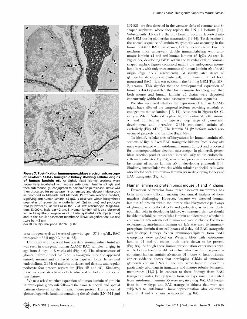
To identify cellular sites of biosynthesis for human laminin a5,
sections of lightly fixed BAC transgenic kidneys from 3 day old
mice were treated with anti-human laminin a5 IgG and processed
for immunoperoxidase electron microscopy. In glomeruli, perox-
idase reaction product was seen intracellularly within endothelial
cells and podocytes (Fig. 7A), which have previously been shown to
be origins of mouse laminin a5 in developing glomeruli [16].
Similarly, intracellular vesicles within tubular epithelial cells were
also labeled with anti-human laminin a5 in developing kidneys of
BAC transgenics (Fig. 7B).
Human laminin a5 protein binds mouse b1 and c1 chainsExtraction of proteins from intact basement membranes has
been notoriously difficult, making biochemical analyses of these
matrices challenging. However, because we detected human
laminin a5 protein within the intracellular biosynthetic pathways
of glomerular endothelial cells and podocytes, as well as tubule
epithelial cells in developing kidney, we reasoned that we should
be able to solubilize intracellular laminin and determine whether it
contained a heterotrimer of human and mouse chains. For these
experiments, anti-human laminin a5 IgG was used to immuno-
precipitate laminin from cell lysates of 2 day old BAC transgenic
and wildtype kidneys. When immunoprecipitates from BAC
transgenics were probed on Western blots with anti-mouse
laminin b1 and c1 chains, both were shown to be present
(Fig. 8A). Although these immunoprecipitation experiments with
whole kidney lysates could not define which nephron segment(s)
contained human laminin a5-mouse b1-mouse c1 heterotrimers,
earlier evidence shows that developing GBMs of immature
glomeruli contain LN-511, and this same laminin isoform is
particularly abundant in immature and mature tubular basement
membranes [14,34]. In contrast to these findings from BAC
transgenic lysates, kidney lysates from wildtype mice that eluted
from anti-human laminin a5 were negative (Fig. 8A). Cell lysates
from both wildtype and BAC transgenic kidneys that were not
subjected to anti-human immunoprecipitation also contained
laminin b1 and c1 chains, as expected (Fig. 8A).
Figure 7. Post-fixation immunoperoxidase electron microscopyof newborn LAMA5 transgenic kidney showing cellular originsof human laminin a5. A: Lightly fixed kidney sections weresequentially incubated with mouse anti-human laminin a5 IgG andthen anti-mouse IgG conjugated to horseradish peroxidase. Tissue wasthen processed for peroxidase histochemistry and electron microscopyas described in Materials and Methods. Peroxidase reaction product,signifying anti-human laminin a5 IgG, is observed within biosyntheticorganelles of glomerular endothelial cell (En) (arrows) and podocyte(Po) (arrowheads), as well as in the GBM. Ret: reticulocyte. Magnifica-tion: 13,0006. Scale bar = 2 mm. B: Human laminin a5 is also observedwithin biosynthetic organelles of tubular epithelial cells (Ep) (arrows)and in the tubular basement membrane (TBM). Magnification: 7,0006;scale bar = 2 mm.doi:10.1371/journal.pone.0023926.g007
Human LAMA5 Transgenics Suppress Mouse Lama5
PLoS ONE | www.plosone.org 8 September 2011 | Volume 6 | Issue 9 | e23926

As mentioned earlier, the only laminin isoform in fully mature
GBM is LN-521. To determine whether human laminin a5-mouse
b2-mouse c1 heterotrimers were also present in developing
transgenic kidney lysates, anti-human laminin a5 immunoprecip-
itates were probed on Western blots with anti-mouse laminin b2.
The results from these experiments thus far were negative.
However, glomeruli represent only a small fraction of total kidney
mass, and our negative result could have been due to insufficient
sample within the total kidney lysate contributed specifically by
maturing glomeruli. Because we saw what appeared to be large
amounts of human laminin a5 in developing and mature
glomeruli (Figs. 1, 3, and 5) we think that it is probable that
human laminin a5-mouse b2-mouse c1 heterotrimers are present
within GBMs of the BAC transgenics. Further evidence in support
of this came from double label immunofluorescence microscopy of
adult transgenic glomeruli where, in most capillary loops, there
was linear co-localization of anti-mouse laminin b2 and anti-
human laminin a5 antibodies within GBMs (Figs. 8B–D).
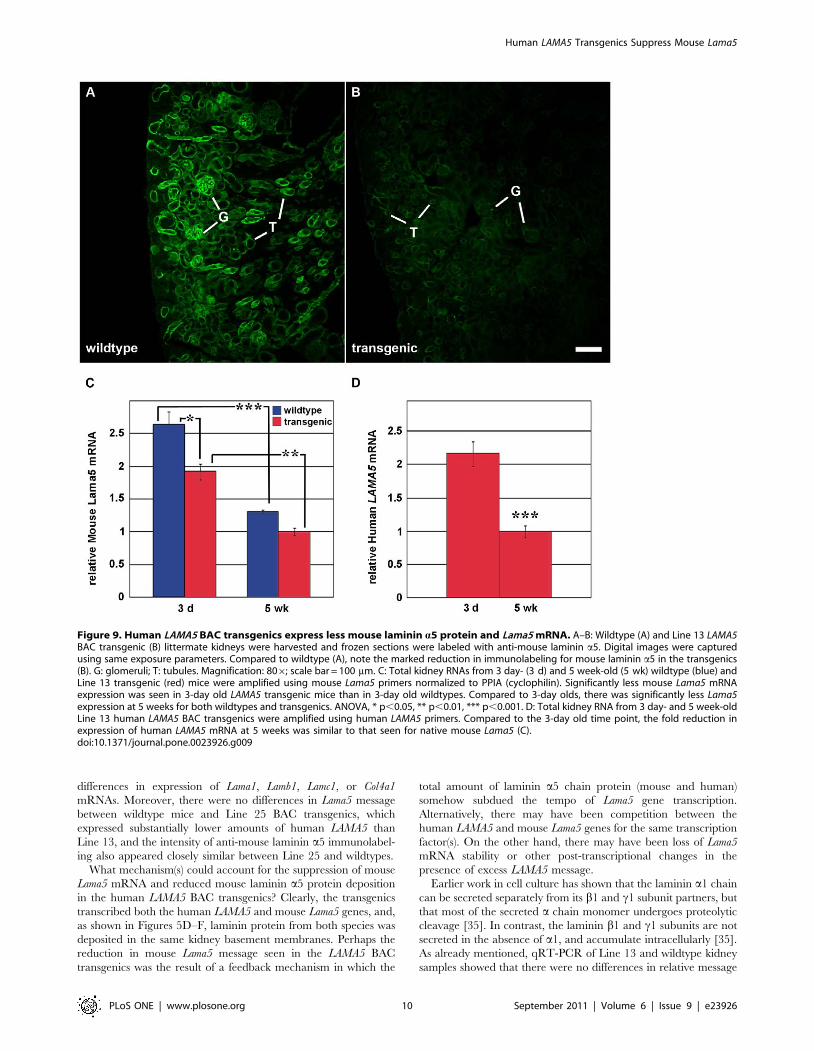
Expression of human LAMA5 suppresses mouse Lama5We next wondered whether the expression of apparently
abundant human laminin a5 in Line 13 kidney affected expression
of native mouse laminin. For these experiments, frozen sections
from newborn wildtype and Line 13 BAC transgenic kidneys were
labeled with anti-mouse laminin a5 and examined by immuno-
fluorescence microscopy. In sections from wildtype kidney, bright
linear immunolabeling for mouse laminin a5 was seen throughout
glomerular and tubular basement membranes (Fig. 9A). By
comparison, however, sections of human LAMA5 BAC transgenic
kidney showed an obvious and marked reduction in intensity of
immunolabel for mouse laminin a5 (Fig. 9B), and glomeruli and
tubules were equally affected. An earlier study in which
doxycycline-inducible human LAMA5 is expressed specifically in
podocytes also noted what appeared to be a reduction of mouse
laminin a5 within the GBM [29], but this observation was not
pursued further.
To begin to investigate mechanisms accounting for the
reduction in mouse laminin a5 protein, RNA was isolated from
kidneys of 3 day old and 5 week old wildtype and Line 13
transgenic mice. Quantitative RT-PCR using mouse-specific
primers showed that relative Lama5 mRNA levels were signifi-
cantly higher in 3 day old kidneys than at 5 weeks of age, and this
was true for both wildtype and human LAMA5 BAC transgenics
(Fig. 9C). Similarly, the relative amount of LAMA5 mRNA in 3
day old BAC transgenics was significantly higher than at 5 weeks,
and the fold decrease at 5 weeks was approximately the same as
that for mouse Lama5 (Fig. 9D). We interpret these findings to
reflect a burst of basement membrane assembly that accompanies
the rapid induction and elongation of nephrons that occurs during
kidney development. This would require more message for
basement membrane proteins in kidneys from 3 day olds than at
5 weeks of age, when kidneys have reached their nearly mature
size and basement membrane assembly has essentially concluded.
Our results from 3 day old, Line 13 LAMA5 BAC transgenic
kidneys also showed less mouse Lama5 mRNA when compared to
wildtype (Fig. 9C). Although the reduction in mouse Lama5
mRNA in transgenics was not as striking as what was seen at the
protein level by immunofluorescence, it nevertheless was statisti-
cally significant (Fig. 9C). In addition, the loss was specific for
Lama5 as qRT-PCR of Line 13 and wildtype kidneys showed no
Figure 8. Human laminin a5 forms heterotrimers with mouse b1 and c1 chains and co-localizes with mouse laminin b2 in GBMs. A:Postnatal day 2 kidneys were harvested from wildtype (Wt) or human LAMA5 BAC transgenic littermates (Tg). Lysates were incubated with anti-human LAMA5 antibody 4C7, and recovered with protein G beads (+). Western blotting with chain-specific anti-laminin b1 (top blot) or anti-lamininc1 (bottom blot) shows that both wildtype and transgenic lysates contain laminin b1 and laminin c1. Lysates from wildtype kidneysimmunoprecipitated with anti-human laminin a5 4C7 antibody do not contain mouse laminin b1 or c1, but both chains are present inimmunoprecipitates from transgenic kidney. B–D: Double label immunofluorescence microscopy of fully mature LAMA5 transgenic glomeruli showswidespread co-localization of mouse laminin b2 and human laminin a5 in GBMs (arrows). Magnification: 6006; scale bar = 20 mm.doi:10.1371/journal.pone.0023926.g008
Human LAMA5 Transgenics Suppress Mouse Lama5
PLoS ONE | www.plosone.org 9 September 2011 | Volume 6 | Issue 9 | e23926
differences in expression of Lama1, Lamb1, Lamc1, or Col4a1
mRNAs. Moreover, there were no differences in Lama5 message
between wildtype mice and Line 25 BAC transgenics, which
expressed substantially lower amounts of human LAMA5 than
Line 13, and the intensity of anti-mouse laminin a5 immunolabel-
ing also appeared closely similar between Line 25 and wildtypes.
What mechanism(s) could account for the suppression of mouse
Lama5 mRNA and reduced mouse laminin a5 protein deposition
in the human LAMA5 BAC transgenics? Clearly, the transgenics
transcribed both the human LAMA5 and mouse Lama5 genes, and,
as shown in Figures 5D–F, laminin protein from both species was
deposited in the same kidney basement membranes. Perhaps the
reduction in mouse Lama5 message seen in the LAMA5 BAC
transgenics was the result of a feedback mechanism in which the
total amount of laminin a5 chain protein (mouse and human)
somehow subdued the tempo of Lama5 gene transcription.
Alternatively, there may have been competition between the
human LAMA5 and mouse Lama5 genes for the same transcription
factor(s). On the other hand, there may have been loss of Lama5
mRNA stability or other post-transcriptional changes in the
presence of excess LAMA5 message.
Earlier work in cell culture has shown that the laminin a1 chain
can be secreted separately from its b1 and c1 subunit partners, but
that most of the secreted a chain monomer undergoes proteolytic
cleavage [35]. In contrast, the laminin b1 and c1 subunits are not
secreted in the absence of a1, and accumulate intracellularly [35].
As already mentioned, qRT-PCR of Line 13 and wildtype kidney
samples showed that there were no differences in relative message
Figure 9. Human LAMA5 BAC transgenics express less mouse laminin a5 protein and Lama5 mRNA. A–B: Wildtype (A) and Line 13 LAMA5BAC transgenic (B) littermate kidneys were harvested and frozen sections were labeled with anti-mouse laminin a5. Digital images were capturedusing same exposure parameters. Compared to wildtype (A), note the marked reduction in immunolabeling for mouse laminin a5 in the transgenics(B). G: glomeruli; T: tubules. Magnification: 806; scale bar = 100 mm. C: Total kidney RNAs from 3 day- (3 d) and 5 week-old (5 wk) wildtype (blue) andLine 13 transgenic (red) mice were amplified using mouse Lama5 primers normalized to PPIA (cyclophilin). Significantly less mouse Lama5 mRNAexpression was seen in 3-day old LAMA5 transgenic mice than in 3-day old wildtypes. Compared to 3-day olds, there was significantly less Lama5expression at 5 weeks for both wildtypes and transgenics. ANOVA, * p,0.05, ** p,0.01, *** p,0.001. D: Total kidney RNA from 3 day- and 5 week-oldLine 13 human LAMA5 BAC transgenics were amplified using human LAMA5 primers. Compared to the 3-day old time point, the fold reduction inexpression of human LAMA5 mRNA at 5 weeks was similar to that seen for native mouse Lama5 (C).doi:10.1371/journal.pone.0023926.g009
Human LAMA5 Transgenics Suppress Mouse Lama5
PLoS ONE | www.plosone.org 10 September 2011 | Volume 6 | Issue 9 | e23926
abundance for Lamb1 or Lamc1, which encode laminin b1 and c1
chains, respectively, and which we showed were binding partners
for human laminin a5 (Fig. 8). Perhaps the secretion of a stable
human laminin a5-mouse b1-mouse c1 chimeric heterotrimer,
and entirely mouse heterotrimers, was rate-limited by the amounts
of laminin b and c chains available for heterotrimerization.
Nevertheless, how the abundance of laminin a5 protein could
autoregulate Lama5 gene transcription is not at all clear, but this
could be an important control mechanism that becomes defective
in fibrotic conditions where there is overproduction of basement
membrane protein.
In summary, we have generated transgenic lines of mice that
express human LAMA5 in temporally and spatially correct
contexts within kidney, indicating that the appropriate genetic
control elements are present. Unexpectedly, a transgenic line
expressing the highest amounts of human laminin a5 suppressed
mouse Lama5 mRNA and mouse protein deposition. These
transgenics may prove useful for understanding regulation of
laminin gene expression and provide new clues regarding
mechanisms of basement membrane assembly.
Supporting Information
Table S1 Primers complementary to human (capitalized gene
symbols) and mouse were designed using the indicated accession
numbers as templates, and each pair was given a unique primer
designation. Primer sequence and length in basepairs is also
shown.
(DOC)
Acknowledgments
We thank Dr. Jeffrey H. Miner (Washington University, St. Louis, MO,
USA) for providing anti-mouse laminin a5 and anti-mouse laminin b2
antibodies, Dr. Melissa Larson, Director of the Transgenic and Gene
Targeting Institutional Facility at the University of Kansas Medical Center
for technical assistance, and Dr. Jay Vivian, Department of Pathology and
Laboratory Medicine at the University of Kansas Medical Center for
technical advice.
Author Contributions
Conceived and designed the experiments: BMS AZ LS KI PLS GKA KRP
DRA. Performed the experiments: BMS AZ LS KI PLS. Analyzed the
data: BMS AZ LS KI PLS GKA KRP DRA. Wrote the paper: BMS GKA
KRP DRA.
References
1. Haraldsson B, Nystrom J, Deen WM (2008) Properties of the glomerular barrierand mechanisms of proteinuria. Physiol Rev 88: 451–487.
2. Haraldsson B, Jeansson M (2009) Glomerular filtration barrier. Curr Opin
Nephrol Hypertens 18: 331–335.3. Patrakka J, Tryggvason K (2010) Molecular make-up of the glomerular filtration
barrier. Biochem Biophys Res Commun 396: 164–169.4. Jeansson M, Haraldsson B (2006) Morphological and functional evidence for an
important role of the endothelial cell glycocalyx in the glomerular barrier.Am J Physiol Renal Physiol 290: F111–F116.
5. Eremina V, Sood J, Haigh J, Nagy A, Lajoie G, et al. (2003) Glomerular-specific
alterations of VEGF-A expression lead to distinct congenital and acquired renaldiseases. J Clin Invest 111: 707–716.
6. Eremina V, Jefferson JA, Kowalewska J, Hochster H, Haas M, et al. (2008) VEGFinhibition and renal thrombotic microangiopathy. New Engl J Med 358: 1129–1136.
7. Kestila M, Lenkkeri U, Mannikko M, Lamerdin J, McCready P, et al. (1998)
Positionally cloned gene for a novel glomerular protein – nephrin - is mutated incongenital nephrotic syndrome. Mol Cell 1: 575–582.
8. Boute N, Gribouval O, Roselli S, Benessy F, Lee H, et al. (2000) NPHS2,encoding the glomerular protein podocin, is mutated in autosomal recessive
steroid-resistant nephrotic syndrome. Nat Genet 24: 349–354.9. Schwarz K, Simons M, Reiser J, Saleem MA, Faul C, et al. (2001) Podocin,
a raft-associated component of the glomerular slit diaphragm, interacts with
CD2AP and nephrin. J Clin Invest 108: 1621–1629.10. Shih NY, Li J, Karpitskii V, Nguyen A, Dustin ML, et al. (1999) Congenital nephrotic
syndrome in mice lacking CD2-associated protein. Science 286: 312–315.11. Putaala H, Soininen R, Kilpelainen P, Wartiovaara J, Tryggvason K (2001) The
murine nephrin gene is specifically expressed in kidney, brain, and pancreas:
inactivation of the gene leads to massive proteinuria and neonatal death. HumMol Genet 10: 1–8.
12. Roselli S, Heidet L, Sich M, Henger A, Kretzler M, et al. (2004) Earlyglomerular filtration defect and severe renal disease in podocin-deficient mice.
Mol Cell Biol 24: 550–560.13. Miner JH (2011) Glomerular basement membrane composition and the
filtration barrier. Pediatr Nephrol Feb 26(9): 1413–1417. [Epub ahead of print.].
14. Miner JH (1998) Developmental biology of glomerular basement membranecomponents. Curr Opin Nephrol Hypertens 7: 13–19.
15. Abrahamson DR (2009) Development of kidney glomerular endothelial cells andtheir role in basement membrane assembly. Organogenesis 5: 275–287.
16. St John PL, Abrahamson DR (2001) Glomerular endothelial cells and podocytes
jointly synthesize laminin-1 and -11 chains. Kidney Int 60: 1037–1046.17. Abrahamson DR, Hudson BG, Stroganova L, Borza DB, St John PL (2009)
Cellular origins of glomerular basement membrane type IV collagen networks.J Am Soc Nephrol 20: 1471–1479.
18. Hudson BG, Tryggvason K, Sundaramoorthy M, Neilson EG (2003) Alport’s
syndrome, Goodpasture’s syndrome, and type IV collagen. N Engl J Med348: 2543–2556.
19. Hudson BG (2004) The molecular basis of Goodpasture and Alport syndrome:Beacons for the discovery of the collagen IV family. J Am Soc Nephrol
15: 2514–2527.
20. Cosgrove D, Meehan DT, Grunkemeyer JA, Kornak JM, Sayers R, et al. (1996)
Collagen COL4A3 knockout: A mouse model for autosomal Alport syndrome.
Genes Dev 10: 2981–2992.
21. Miner JH, Sanes JR (1996) Molecular and functional defects in kidneys of mice
lacking collagen alpha 3(IV): Implications for Alport syndrome. J Cell Biol 135:
1403–1413.
22. Lu W, Phillips CL, Killen PD, Hlaing T, Harrison WR, et al. (1999) Insertional
mutation of the collagen genes Col4a3 and Col4a4 in a mouse model of Alport
syndrome. Genomics 61: 113–124.
23. Heidet L, Borza DB, Jouin M, Sich M, Mattei MG, et al. (2003) A human-
mouse chimera of the a3a4a5(IV) collagen protomer rescues the renal
phenotype in Col4a32/2 Alport mice. Am J Pathol 163: 1633–1644.
24. Noakes PG, Miner JH, Gautam M, Cunningham JM, Sanes JR, et al. (1995)
The renal glomerulus of mice lacking s-laminin/laminin b2: Nephrosis despite
molecular compensation by laminin b1. Nat Genet 10: 400–406.
25. Miner JH, Go G, Cunningham J, Patton BL, Jarad G (2006) Transgenic
isolation of skeletal muscle and kidney defects in laminin beta 2 mutant mice:
Implications for Pierson syndrome. Development 133: 967–975.
26. Miner JH, Cunningham J, Sanes JR (1998) Roles for laminin in embryogenesis:
Exencephaly, syndactyly, and placentopathy in mice lacking the laminin a5
chain. J Cell Biol 143: 1713–1723.
27. Miner JH, Li C (2000) Defective glomerulogenesis in the absence of laminin a5
demonstrates a developmental role for the kidney glomerular basement
membrane. Dev Biol 217: 278–289.
28. Abrahamson DR, St. John PL, Isom K, Robert B, Miner JH (2007) Partial
rescue of glomerular laminin a5 mutation by wildtype endothelial cells produce
hybrid glomeruli. J Am Soc Nephrol 18: 2285–2297.
29. Goldberg S, Adair-Kirk TL, Senior RM, Miner JH (2010) Maintenance of
glomerular filtration barrier integrity requires laminin a5. J Am Soc Nephrol 21:
579–586.
30. Abrahamson DR, Isom K, Roach E, Stroganova L, Zelenchuk A, et al. (2007)
Laminin compensation in collagen a3(IV) knockout (Alport) glomeruli
contributes to permeability defects. J Am Soc Nephrol 18: 1465–1472.
31. Abrahamson DR, Irwin MH, St John PL, Perry EW, Accavitti MA, et al. (1989)
Selective immunoreactivities of kidney basement membranes to monoclonal
antibodies against laminin: Localization of the end of the long arm and the short
arms to discrete microdomains. J Cell Biol 109: 3477–3491.
32. Livak KJ, Schmittgen TD (2001) Analysis of relative gene expression data using
real-time quantitative PCR and the 2(2Delta Delta C(T)) method. Methods
2001 25(4): 402–408.
33. Abrahamson DR, St John PL (1992) Loss of laminin epitopes during glomerular
basement membrane assembly in developing mouse kidneys. J Histochem
Cytochem 40: 1943–1953.
34. Miner JH (2005) Building the glomerulus: A matricentric view. J Am Soc
Nephrol 16: 857–861.
35. Yurchenco PD, Quan Y, Colognato H, Mathus T, Harrison D, et al. (1997) The
a chain of laminin-1 is independently secreted and drives secretion of its b- and
c-chain partners. Proc Natl Acad Sci USA 94: 10189–10194.
Human LAMA5 Transgenics Suppress Mouse Lama5
PLoS ONE | www.plosone.org 11 September 2011 | Volume 6 | Issue 9 | e23926
Copyright © 2022 FDOKUMEN