CXCL10 blockade protects mice from cyclophosphamide-induced cystitis
Upload
independentCategory
view
0download
0
The Journal of Clinical Investigation | August 2003 | Volume 112 | Number 4 575
IntroductionPerturbations of signaling pathways controlling cellproliferation, such as pathways regulated by the EGFand TGF-β ligand superfamilies, are often associatedwith cell transformation and malignant tumors. Manycomponents of these two pathways, which include src,ras, EGFR, HER-2, Smad2, Alk4, TGF-βRII, andPDGFR, have been identified as oncogenes and/or
tumor suppressors and have been actively pursued asdrug targets (1). Success in the clinic with therapeuticsthat modulate cell signaling pathways has been docu-mented with mAb’s against HER-2 (trastuzumab, Her-ceptin; Genentech Inc., South San Francsico, Califor-nia, USA) and EGFR (IMC-C225) (for review see ref. 2),as well as with small-molecule inhibitors of PDGFRtyrosine kinase activity (3). These successes highlightthe importance of studying signaling moleculesknown to be perturbed in cancer and evaluating themas therapeutic intervention points.
Cripto, a glycosylphosphatidylinositol-linked (GPI-linked) membrane protein, is overexpressed in 75–80%of human breast, colon, and lung cancers, as well as50–60% of testicular, stomach, pancreatic, and ovariancancers (4). In addition, the level of Cripto expressionincreases with the degree of dysplasia in several ofthese cancers. In vitro, Cripto exhibits many propertiesof an oncogene, including transformation of immor-talized cells, induction of cell migration, and stimula-tion of branching morphogenesis (4). Furthermore,antisense inhibition of Cripto expression in colon can-cer cells inhibits their growth in soft agar and theirtumor-forming potential (5). Evidence in vivo that
Antibody blockade of the Cripto CFC domain suppresses tumor cell growth in vivo
Heather B. Adkins,1 Caterina Bianco,2 Susan G. Schiffer,1 Paul Rayhorn,1
Mohammad Zafari,1 Anne E. Cheung,1 Olivia Orozco,1 Dian Olson,1
Antonella De Luca,3 Ling Ling Chen,1 Konrad Miatkowski1 Chris Benjamin,1
Nicola Normanno,3 Kevin P. Williams,1 Matthew Jarpe,1 Doreen LePage,1
David Salomon,2 and Michele Sanicola1
1Biogen Inc., Cambridge, Massachusetts, USA2National Cancer Institute, Bethesda, Maryland, USA3Oncologia Sperimentale D, Istituto Nazionale per lo Studio e la Cura dei Tumori–Fondazione Pascale, Naples, Italy
Cripto, a cell surface–associated protein belonging to the EGF-CFC family of growth factor–likemolecules, is overexpressed in many human solid tumors, including 70–80% of breast and colontumors, yet how it promotes cell transformation is unclear. During embryogenesis, Cripto com-plexes with Alk4 via its unique cysteine-rich CFC domain to facilitate signaling by the TGF-β ligandNodal. We report, for the first time to our knowledge, that Cripto can directly bind to another TGF-βligand, Activin B, and that Cripto overexpression blocks Activin B growth inhibition of breast can-cer cells. This result suggests a novel mechanism for antagonizing Activin signaling that could pro-mote tumorigenesis by deregulating growth homeostasis. We show that an anti–CFC domain anti-body, A8.G3.5, both disrupts Cripto-Nodal signaling and reverses Cripto blockade of Activin B–induced growth suppression by blocking Cripto’s association with either Alk4 or Activin B. In twoxenograft models, testicular and colon cancer, A8.G3.5 inhibited tumor cell growth by up to 70%.Both Nodal and Activin B expression was found in the xenograft tumor, suggesting that either lig-and could be promoting tumorigenesis. These data validate that functional blockade of Criptoinhibits tumor growth and highlight antibodies that block Cripto signaling mediated through itsCFC domain as an important class of antibodies for further therapeutic development.
J. Clin. Invest. 112:575–587 (2003). doi:10.1172/JCI200317788.
Received for publication January 7, 2003, and accepted in revised formMay 20, 2003.
Address correspondence to: Michele Sanicola, Biogen Inc., 14 Cambridge Center, Cambridge, Massachusetts 02142, USA.Phone: (617) 679-3307; Fax: (617) 914-7140; E-mail: [email protected] P. Williams’ present address is: Amphora Discovery Corp.,Research Triangle Park, North Carolina, USA.Conflict of interest: H.B. Adkins, S.G. Schiffer, P. Rayhorn, M. Zafari, A.E. Cheung, O. Orozco, D. Olson, L.L. Chen, K. Miatkowski, C. Benjamin, K.P. Williams, M. Jarpe, and D. LePage are employees of Biogen Inc. D. Salomon is acollaborator with Biogen Inc.Nonstandard abbreviations used: Activin (Act); Act type IIreceptor (ActRII); Cripto, FRL-1, and Cryptic domain (CFCdomain); recombinant human Cripto (hCr); hCr expressed as ahuman IgG1 Fc fusion protein (Cr-hFc); HA, hemagglutinin;forkhead activin signal transducer (FAST).
See the related Commentary beginning on page 500.
576 The Journal of Clinical Investigation | August 2003 | Volume 112 | Number 4
Cripto overexpression induces tumor formationcomes from studies of MMTV-Cripto transgenic micethat overexpress Cripto in the mammary gland. Hyper-plasias are seen in these mice, and papillary carcino-mas develop in aged, multiparous animals (C. Wech-selberger and D. Salomon, unpublished observations).
Genetic experiments in mice and zebrafish definedCripto as a coreceptor for Nodal, a TGF-β family lig-and (6–10). Cripto-dependent Nodal signaling isrequired for early embryogenesis, and signalingdepends on the Activin type II serine/threonine kinasereceptor (ActRII) and the Activin type I serine/threo-nine kinase receptor (Alk4), which, once activated,phosphorylate the downstream transcriptional coac-tivator Smad2 (11–13). Human Cripto is the originalmember of the EGF-CFC family of proteins definedby two conserved adjacent functional motifs: a variantEGF (“EGF-like”) domain and a unique cysteine-richdomain, the CFC domain (named for Cripto, FRL-1,and Cryptic). Site-directed mutagenesis experimentsdemonstrated that Cripto binds to Alk4 through itsCFC domain to facilitate signaling through the Smadpathway (12, 14). Our group has previously shownthat fucosylation of Cripto at a unique glycosylationsite within the EGF-like domain is essential for Nodalsignaling (15), and mutations in the EGF-like domainhave been shown to disrupt Cripto-Nodal interactions(12). Although these experiments have characterizedCripto-Nodal signaling in the embryo, they do notnecessarily predict Cripto’s function in cancer. Nodalexpression is predominantly embryonically restricted,raising the question of whether Cripto could be mod-ulating other TGF-β family members in adult tissues.
The role of TGF-β family members as tumor sup-pressors and promoters in cancer is well documented(16–19). In normal tissue, TGF-β plays a tumor-sup-pressive role, but during tumorigenesis, TGF-β pro-motes tumor progression as changes in its expressionand decreased cellular response to TGF-β favor itsoncogenic properties (19). Furthermore, recentreports have shown positive effects of blocking theTGF-β pathway as a potential therapeutic approachfor the treatment of breast cancer (20, 21). Like TGF-β,Activins, which consist of dimers of β subunits (βA orβB), are reported to be tumor suppressive for breast,liver, and kidney cells (18). In prostate cancer, reducedActRII expression is correlated with malignant pro-gression (22), and in pancreatic cancer, mutations inAlk4 have been reported (23). Resistance to Activin-induced growth suppression in some breast epithelialcell lines has been attributed to downregulation ofeither ActRII or Alk4, or loss of Smad4 expression(24). Thus, alterations in Activin receptor functionthat result in loss of response to Activin may be animportant early step in tumorigenesis.
In the present study, we investigated Cripto’s role inmodulating signaling by TGF-β family ligands in cancercells using mAb’s specific to different domains of Crip-to, and we investigated whether functional blockade of
Cripto could inhibit tumor cell growth in vivo. BecauseActivin, like Nodal, utilizes ActRII and Alk4 to signalthrough the Smad pathway, we examined whether Crip-to could modulate Activin signaling via its interactionwith Alk4. We present evidence that Cripto can antago-nize Activin B–induced growth suppression, but notActivin A–induced growth suppression, in breast cancercells by directly interacting with Activin B. Furthermore,Cripto’s antagonism of Activin B (Act B) can be reversedby an anti–CFC domain Cripto antibody that blocksCripto-Alk4 binding. These data suggest a novel Cripto-dependent mechanism for deregulating cell growthhomeostasis that could promote tumorigenesis. More-over, we present evidence for Nodal expression in fullytransformed testicular and colon cancer cell lines, andevidence that antibodies to both Cripto EGF and CFCdomains will block Nodal signaling in these cells. Final-ly, we demonstrate that a Cripto anti–CFC domain anti-body, which can disrupt both Cripto-Nodal signalingand Cripto–Act B interactions, inhibits the growth of tes-ticular and colon cancer xenograft models in vivo, andwe highlight blockade of Cripto function through itsCFC domain as a therapeutic intervention point.
MethodsProduction and screening of anti–human Cripto mAb’s.Experimental procedures involving the use of animalswere approved by the Biogen Institutional Animal Careand Use Committee. Mice were immunized intraperi-toneally with 25 µg of recombinant human Cripto (hCr)expressed as a human IgG1 Fc fusion protein (Cr-hFc)with incomplete Freund’s adjuvant (Invitrogen LifeTechnologies, Carlsbad, California, USA) administeredintraperitoneally at a different site and boosted threetimes. Mice were boosted intraperitoneally with 100 µgCr-hFc 3 days before fusion and boosted intravenouslywith 100 µg Cr-hFc 1 day before fusion. Mouse spleencells were fused with FL653 myeloma cells at a ratio ofone spleen cell to six myeloma cells and plated into 96-well plates in selection media. Hybridoma supernatantswere initially screened by flow cytometry for binding tocell surface–expressed Cripto on human tumor celllines, including NTERA (embryonic carcinoma),NCCIT (testicular), and SiHa (cervical). Classificationof mAb’s into distinct groups was based on their reac-tivity in an ELISA assay to different domains of hCrexpressed as hFc fusion proteins: Cr-hFc (amino acids1–169), CrEGF-hFc (amino acids 75–112), and CrCFC-hFc (amino acids 112–169). Cripto proteins were coat-ed on 96-well plates for 1 hour at 37°C in 0.1 MNaHPO4 (pH 9.0) at 0.5 µg/ml, 100 µl/well, and blockedwith PBS/10% donor calf serum. Antibodies diluted inPBS/0.05% Tween-20 were incubated for 1 hour at37°C, washed with PBS/0.05% Tween-20, and probedwith anti-mouse HRP-conjugated antibody (PierceChemical Co., Rockford, Illinois, USA). Bound anti-bodies were detected by 3,3′-5,5′–tetramethylbenzidine,stopped with 1N H2SO4, and read at 450 nm. None ofthese mAb’s bound LTβR-hFc, which was used as a

negative control protein. The mAb’s were further char-acterized for competition for binding Cr-hFc using Bia-core technology (Biacore Inc., Piscataway, New Jersey,USA), and binding to Cripto-specific peptides by ELISA.
Immunohistochemistry. Immunohistochemical local-ization was performed with anti-Cripto mAb’sA10.B2.18 (breast and NCCIT) or B3.F6.17 (colon andGEO) using a VECTASTAIN Elite peroxidase kit (Vec-tor Laboratories Inc., Burlingame, California, USA).Mouse IgG was used as a negative control for all sam-ples. Paraffin sections of colon tumor tissue (Imgenex,San Diego, California, USA), breast tumor tissue(Novagen, Madison, Wisconsin, USA), and NCCIT andGEO tumor cells, passaged once in an athymic nudemouse, were deparaffinized and rehydrated beforeincubation in 1% H2O2/methanol to block endoge-nous peroxidase. After blocking in 2% goat serum inPBS, sections were incubated with 2 µg/ml primaryantibody in blocking solution overnight at 4°C. Sec-tions were treated with biotinylated anti-mouse IgGthen avidin-biotinylated HRP complex. Color wasdeveloped using Vector NovaRED or DAB substratekits (Vector Laboratories Inc.). Sections were counter-stained with hematoxylin. Immunofluorescence wasperformed essentially the same way, but using 1% BSAin PBS as blocking reagent, 2 µg/ml A10.B2.18 or 1E6,and Cy3-conjugated anti-mouse secondary antibodyat 1:300 (Jackson ImmunoResearch Laboratories Inc.,West Grove, Pennsylvania, USA).
Protein expression and purification, FACS and signalingassays. Cr-hFc, CrEGF-hFc, and CFC-hFc were expressedand purified as described (15). Cripto CFC-hFc (aminoacids 112–169) was generated by PCR amplification andsubcloned as described before (15). FACS analysis wasperformed essentially as described before (11). For Alk4blocking studies, 293 cells were cotransfected with aplasmid expressing Alk4 with a C-terminal hemagglu-tinin (HA) epitope tag (Alk4-HA) (gift of M. Whitman,Harvard Medical School, Boston, Massachusetts, USA)and a puromycin expression plasmid at a 10:1 ratio togenerate a clonal cell line, as previously described (25).For blocking, 10 µg/ml of Cr-hFc was preincubated onice with 20 µg/ml of mAb before addition to cells. Sig-naling assays were performed essentially as describedpreviously (15). Briefly, NCCIT or T47D cells were trans-fected with 15 ng/well (n2)7-luciferase, 100 ng/well fork-head activin signal transducer (FAST) transcription fac-tor, and/or 100 ng/well Nodal expression plasmid (giftof E. Robertson, Harvard University, Cambridge, Mass-achusetts, USA) on a 24-well plate. Alternatively, cellswere treated with 25 ng/ml Act B. Antibodies wereadded on the first day of transfection and again 24hours before luciferase reading.
Growth inhibition assays. T47D cells (American TypeCulture Collection, Manassas, Virginia, USA) weretransfected with an ecotropic receptor expression plas-mid (EcoR; gift of B. Elenbaas, Biogen Inc., Cambridge,Massachusetts, USA) and selected in RPMI/10% FCS/10µg/ml insulin containing 100 µg/ml hygromycin. A
polyclonal line of T47D-EcoR that permitted infectionof pBABE-GFP murine leukemia virus (MLV) wasgrown out, infected with pBABE-hCr-PURO MLV, andselected in puromycin media. This oligoclonal line(T47D-hCr) was analyzed by FACS for hCr expressionwith specific anti-Cripto antibodies. Approximately4,000 cells per well of T47D-EcoR or T47D-hCr wereplated in a 96-well plate in media containing 2% serumwith or without 10 ng/ml Activin A (Act A) or Act B(R&D Systems Inc., Minneapolis, Minnesota, USA) or10 µg/ml A8.G3.5. Medium with factors was replaceddaily for 7–8 days. The plate was harvested by additionof 20 µl/well CellTiter AQueous One solution(Promega Corp., Madison, Wisconsin, USA), incuba-tion for 2 hours at 37°C, and reading at 490 nm.
Immunoprecipitations and immunoblots. Cr-hFc (1 µg)was prebound to protein A-Sepharose in NP40 bufferand then incubated with 1 µg of Act A or Act B (R&DSystems Inc.) at 4°C overnight. Immunoprecipitatedprotein was washed three times in ice-cold NP40 buffer,and a portion of the sample was run on a polyacry-lamide gel and then blotted with either anti–Act A oranti–Act B mAb’s (R&D Systems Inc.). For antibodyblocking, 1 µg of Cr-hFc was incubated with the indi-cated amount of antibody prior to binding to protein A-Sepharose. For the CrEGFmt-hFc experiment, 100 µl ofunpurified cell supernatant from 293 cells expressingthe hFc proteins was prebound to protein A-Sepharosefor 1 hour at 4°C. Then 1 µg of Act B or 1.5 ml of 293cell supernatant expressing Nodal was added and incu-bated at 4°C overnight. Nodal was detected using a rab-bit polyclonal antibody against a Nodal-specific peptide.
For phospho-Smad and MAPK analysis, T47D orT47D-hCr cells were plated at 4 × 10⁶ cells/ml in 100-mm dishes. Cells were serum-starved overnight andincubated with indicated amounts of Act A, Act B, orEGF for the indicated time. Cells were lysed in NP40buffer containing protease inhibitors, 1 mM sodiumorthovanadate, and 20 mM sodium fluoride, separatedon a polyacrylamide gel, and blotted onto nitrocellulose.The membrane was immunoblotted with phospho-Smad2 antibody or p44/42 MAPK antibody (Cell Sig-naling Inc., Beverly, Massachusetts, USA) and boundantibody was probed with peroxidase-conjugated don-key anti-rabbit IgG antibody (Jackson ImmunoResearchLaboratories Inc., West Grove, Pennsylvania, USA). ForCripto analysis in tumor lines, cells were lysed in RIPAbuffer plus protease inhibitors, and 6, 15, and 30 µg oftotal protein lysate (CHO, NCCIT, and GEO, respec-tively) was separated on a10–20% SDS polyacrylamidegel and transferred to nitrocellulose. The blot wasprobed with 0.1 µg/ml A10.B2.18 and detected withanti-mouse–HRP. Immunoreactivity was revealed usingSuperSignal West Dura Substrate (Pierce Chemical Co.).
Biacore studies. Cr-hFc and LTβR-hFc were immobi-lized by standard amine coupling to CM5 sensorchips (Pharmacia Biosensor AB) (26), and Act A orB at 5 µg/ml was then captured in HBS buffer (10mM HEPES, 150 mM NaCl, 3.4 mM EDTA, 0.005%
The Journal of Clinical Investigation | August 2003 | Volume 112 | Number 4 577

578 The Journal of Clinical Investigation | August 2003 | Volume 112 | Number 4
Biacore surfactant P20, pH 7.4; Biacore Inc.). Experi-ments were performed at 25°C with a 10-µl/min flowrate. For data analysis, nonspecific binding to theblank-flow cell was subtracted from each sensorgramto obtain specific-binding responses using BIAevalu-ation 3.0 software (Biacore Inc.).
Human xenograft tumor models. NCCIT, a mediastinalmixed germ cell human testicular carcinoma cell line(American Type Culture Collection), was maintained inRPMI 1640/10% FBS without antibiotics. GEO, a carci-noma cell line (American Type Culture Collection), wasmaintained in DMEM/10% FBS without antibiotics.Male athymic nude mice, 6–8 weeks old (HarlanSprague Dawley Inc., Indianapolis, Indiana, USA), wereacclimated for 1 week before the study. The day beforetumor implantation, mice were numbered and ran-domized into treatment (n = 10) and vehicle control (n = 30) groups, initial body weights were recorded, andthe first treatments were administered. Studies were runas randomized, double-blinded trials. Antibodies wereadministered intraperitoneally on a q14d treatmentschedule (dosing every 14 days). Clinical-grade Plati-nol-AQ, cisplatin injection (Bristol-Myers Squibb Co.,Seattle, Washington, USA), was the positive chemother-apeutic control agent, administered subcutaneously ona q2dx6 schedule (i.e., dosing every other day for a totalof 6 treatments). Animals were implanted (day 0) sub-cutaneously with 5 × 10⁶ cells (with 0.2 ml matrigel forNCCIT cells). Tumors were measured every 3 or 4 days.Tumor size (mm) was converted to volume (mg) usingthe formula (length × width2)/2. Data are reported aschange in tumor weight with tumors normalized toeach animal’s first tumor measurement.
RT-PCR. Methods and primers for RT-PCR analysisof hCripto and Alk4 were previously described (27,28). Because the human homolog of murine Nodalhas not been confirmed, we used primers based on ahuman Nodal–like cDNA sequence in the GenBankdatabase (accession no. AI050866): 5′-CATGAAAGC-TATAGGTGACTTCATCC-3′ (Nodal-like foreword), 5′-TGTAAATGAAGGGCTCAGTGGA-3′ (Nodal-like reverse)(29) Human Act B primers are 5′-TGAAGCGGCA-CATCTTGAGC-3′ (hAct B forward) and 5′-ACACT-TTGACCCGCACCTTC-3′ (hAct B reverse). Murine ActB primers were previously published (30). RT-PCRfrom NCCIT and GEO cell lines was performed essen-tially as described previously (28, 29). RNA and sub-sequent cDNA were prepared from NCCIT xenografttumor using RNeasy Protect Kit (QIAGEN Inc., Valen-cia, California, USA) and SuperScript First-StrandSynthesis System (Invitrogen Corp., San Diego, Cal-ifornia, USA) with random-hexamer priming. PCRreactions were then performed with Advantage 2 PCRkit (CLONTECH Laboratories Inc., Palo Alto, Cali-fornia, USA) for 30 cycles at an annealing tempera-ture of 62°C using the Nodal or murine Act B primersets. Resulting PCR products were subcloned intopCRII-TOPO vector (Invitrogen Corp.), and thesequences were confirmed.
ResultsGeneration of Cripto mAb’s that recognize functionally impor-tant epitopes. A panel of murine mAb’s against recombi-nant human Cripto (hCr) expressed as human IgG1 Fcfusion protein (Cr-hFc) was generated (Table 1). Anti-Cripto mAb’s were identified by their ability to bind tohuman tumor cell lines known to express Cripto andby their ability to bind to Cripto EGF and CFCdomains by ELISA (see Methods). Based on this analy-sis, four classes of mAb’s were found: N-terminal “tip,”N-terminal, EGF domain, and CFC domain (Table 1and Figure 1a). These antibodies were tested for cross-reactivity to human Cryptic, a closely related familymember, by binding in a flow cytometry assay to 293cells expressing an HA-tagged human Cryptic expres-sion plasmid. Although Cryptic expression was con-firmed by Western blotting with an anti-HA polyclon-al antibody, no cross-reactivity of these Criptoantibodies to Cryptic was observed (see SupplementalFigure 2; www.jci.org/cgi/content/full/112/4/575/DC1). The ability of these mAb’s to recognize key sig-naling domains of Cripto was examined by analysis ofbinding to Cripto mutants. Mutations in the EGFdomain (N85G/T88A) prevent fucosylation of Cripto,disrupting its ability to bind Nodal and signal throughSmad2 (12, 15, 31). Cripto mAb’s were tested for bind-ing to hCr(N85G/T88A) expressed on human 293 cellsby flow cytometry, and no difference was observed inbinding to the fucosylation mutant compared withwild-type hCr, with the exception of the anti-EGFmAb’s (Table 1). Cripto mAb’s were also tested for bind-ing to a Cripto CFC domain mutant, hCr(H120G/W123G), which abrogates Cripto binding to Alk4 (12).Only the anti-CFC mAb’s, A8.G3.5, A10.A10.30, andA6.C12.11, failed to bind to this mutant (Table 1).Thus, we have identified anti–EGF domain andanti–CFC domain Cripto mAb’s that specifically rec-ognize the epitopes defined by mutational analysis tobe necessary for Cripto-Nodal signaling.
Cripto mAb’s were further characterized by immuno-histochemical and immunofluorescent staining ofCripto expression on various tumor samples andtumor cell lines with N-terminal mAb’s A10.B2.18 andB3.F6.17. These two antibodies recognize the same N-terminal Cripto peptide sequence and cross-blockeach other for binding to Cripto by Biacore analysis(data not shown). Thus, A10.B2.18 and B3.F6.17 rec-ognize indistinguishable epitopes. By Western blot,A10.B2.18 recognizes two Cripto protein bandsbetween 15 and 18 kDa in both CHO cells expressingrecombinant Cripto protein and NCCIT and GEOtumor cell lines expressing tumor-derived Cripto (Fig-ure 1b). Similar results were obtained with B3.F6.17(data not shown). We tested whether these antibodiesrecognize cell surface–associated Cripto on humanbreast tumor samples. Indeed, predominant cell sur-face staining was observed with A10.B2.18 (Figure 1c)or B3.F6.17 (data not shown), whereas the isotype con-trol antibody, anti–human LFA3 1E6, did not stain the

tumor tissue. In agreement with previously publisheddata using a Cripto polyclonal antibody (32), someCripto expression is also seen in the cytoplasm. Anexample of immunohistochemical staining of Cripto
overexpression in human breast and colon tumor tis-sue and human NCCIT testicular cell and GEO coloncell xenograft tumor samples is shown in Figure 1d.Our results with these mAb’s in human breast andcolon tumor samples (n > 50) are in agreement withpublished Cripto polyclonal antibody staining data inwhich Cripto overexpression was detected in approxi-mately 80% of breast and colon cancers (28, 32, 33).
Cripto antibodies inhibit Cripto-Nodal signaling in trans-formed cells. Anti-Cripto mAb’s were analyzed for block-ing known signaling functions of human Cripto. Crip-to binds Nodal via its EGF-like domain (12), and wepreviously showed that the EGF domain fucosylationmutant (N85G/T88A) disrupts Cripto-Nodal signalingin a FAST transcription factor–dependent (n2)7-luciferase reporter assay (15). Therefore, we testedwhether anti–EGF domain mAb’s could inhibit Crip-to-dependent Nodal signaling in this assay. Anti–EGFdomain mAb A27.F6.1, which does not recognize theCripto fucosylation mutant (Table 1), was tested forinhibition of Cripto-dependent Nodal activation ofFAST/(n2)7-luciferase in NCCIT cells. At concentra-tions of 1–30 µg/ml, A27.F6.1 decreased the luciferaseactivity by 48–66% in the presence of Nodal (Figure 2a),indicating that anti–EGF domain mAb A27.F6.1 couldinhibit Cripto-dependent Nodal signaling through theSmad2/3 pathway in this transformed human cell line.
The Journal of Clinical Investigation | August 2003 | Volume 112 | Number 4 579
Table 1Anti-Cripto mAb’s map to different functional domains
ELISAA FACS (%)B
Antibody Cr CrEGF CrCFC N85G/T88A H120G/W123GA8.H3.2 + + – 100 118A19.E2.7 + + – ND NDB3.F6.17 + – – 111 142A10.B2.18 + – – 90 NDA27.F6.1 + + – 15 121A8.H3.1 + + – 3 149A40.G12.8 + + – 16 176B6.G7.10 + + – 41 166A10.A10.30 + – + 83 29A8.G3.5 + – + 45 1A6.F8.6 + – + 109 87A6.C12.11 + – + 84 21
ACr, CrEGF, and CrCFC correspond to the hFc fusion proteins coating theELISA plate, and plus symbols represent positive binding of the mAb’s to theseproteins. BFACS binding is represented as the mean fluorescence intensity ofthe mAb binding to the hCr mutant expressed on 293 cells as a percentage ofthe mAb binding to wild-type hCr in the same experiment. Color code of anti-bodies corresponds to binding regions shown in Figure 1a. ND, not done.
Figure 1(a) Diagram of mature hCr protein: N-terminal tip (black), N-terminal region (blue), EGF-like domain (red), and CFC domain (green).Amino acid 169 is N-terminal to the GPI-linkage site. Asterisks indicate positions of mutants described in Table 1. (b) Western blot ofCripto expressed in CHO cells (recombinant) or endogenously expressed in NCCIT and GEO tumor cell lines using A10.B2.18. (c)Immunofluorescent staining of cell surface Cripto on human breast tumor tissue with A10.B2.18 (left) or control antibody 1E6 (right).(d) Immunohistochemical staining of tumor sections with anti-Cripto mAb’s A10.B2.18 (breast and NCCIT) and B3.F6.17 (colon andGEO) or mouse IgG as a negative control. Color variations result from the use of different substrates, either Vector NovaRED or DAB sub-strate kits (Vector Laboratories Inc.).

580 The Journal of Clinical Investigation | August 2003 | Volume 112 | Number 4
We obtained similar results with the anti–EGF domainmAb’s A8.H3.1 and A40.G12.8 (data not shown).
Cripto-dependent Nodal signaling is also known tobe dependent on Cripto’s interaction with Alk4 via theCripto CFC domain (11, 12). Previously, we reportedthat Cr-hFc binds to Alk4 expressed on 293 cells in aflow cytometry assay (11). This assay was used to testwhether any of the Cripto mAb’s could block Cripto-Alk4 interactions. Cr-hFc (10 µg/ml) preincubated withthe mAb’s (20 µg/ml) was added to 293-Alk4 cells andassayed for binding by flow cytometry. Anti–CFCdomain antibodies A8.G3.5 and A6.F8.6 blocked thisinteraction by over 90% (Figure 2b). Other CFC domainmAb’s, A6.C12.11 and A10.A10.30, were able to blockCripto-Alk4 interaction by about 30%, while N-termi-nal mAb’s (e.g., A10.B2.18) or EGF domain mAb’s (e.g.,A27.F6.1) were unable to block Cripto-Alk4 binding.Notably, the mAb’s that blocked Cripto-Alk4 bindingmost effectively are also the mAb’s that fail to bind theCFC domain mutant (H120G/W123G) (Table 1).
Anti-CFC mAb’s were further tested for their ability toblock Cripto-dependent Nodal signaling by presumablyblocking Cripto-Alk4 interactions. Anti-CFC mAbA8.G3.5, a potent blocker of Cripto-Alk4 binding, wastested for inhibition of Nodal signaling in NCCIT cells.At 10 and 30 µg/ml, A8.G3.5 reproducibly blockedNodal signaling by about 35% in this assay (Figure 2c).
No blocking effect was observed at lower concentra-tions, but similar effects were observed with otheranti–CFC domain mAb’s (data not shown). Theseresults demonstrate that blocking both the EGF and theCFC domains of Cripto will inhibit Nodal signaling.
Cripto disrupts Act B–induced growth suppression of breastcancer cells. Activin, like Nodal, signals through Alk4,which has been associated with functional alterationsand mutations in human cancer (18, 34). Therefore, weexamined whether Cripto could modulate Activin sig-naling in cancer cells through its interaction with Alk4.One biological consequence of Activin signaling is inhi-bition of proliferation of several cell types (18).Although NCCIT cells are responsive to Activin signal-ing in a FAST/(n2)7-luciferase assay, they are notgrowth-inhibited by Activin in vitro. Therefore, we choseto use T47D cells, an estrogen receptor–positive humanbreast cancer cell line that is known to be growth-inhib-ited by Activin (35, 36). Because T47D cells express verylow levels of Cripto (data not shown), we generated anoligoclonal T47D line expressing high levels of full-length Cripto (T47D-hCr) using the pBABE retroviralexpression system (37), which contains full-lengthhuman Cripto cDNA. Since T47D cells lose responsive-ness to Activin through extensive passaging, the nearly100% transduction efficiency with this system allowedus to make a cell line with very few passages in culture.
Figure 2Anti-CFC mAb’s block Nodal signaling and Cripto-Alk4 interactions. (a) NCCIT cells were transfected with plasmids expressing (n2)7-luciferase and Nodal (column 1) or (n2)7-luciferase, FAST, and Nodal (columns 2–6). A27.F6.1 was added at 0, 1, 3, 10, or 30 µg/mlfor 16 hours before luciferase reading (P < 0.001 for each dose). (b) Human 293-Alk4 cells were incubated with Cr-hFc or Cr-hFc pre-bound with mAb and assayed for binding by FACS using an anti-hFc PE-conjugated secondary mAb. The mean fluorescence intensityof Cr-hFc/mAb bound to 293-Alk4 cells is represented as a percentage of Cr-hFc binding with no mAb. (c) NCCIT cells were assayed forblocking of Cripto-Nodal signaling by A8.G3.5 at 10 µg/ml (P = 0.002) or 30 µg/ml (P = 0.05) as described in a.

Using flow cytometry and anti-Cripto antibodies, thenumber of Cripto molecules on T47D-hCr cells was esti-mated to be 16,000–22,000 per cell, which is compara-ble to the number on human tumor cell lines that over-express Cripto, such as NCCIT (10,000 per cell) andGEO (40,000–60,000 per cell; data not shown).
For this study, T47D and T47D-hCr cells were grownin low-serum conditions, with or without 25 ng/ml ActA or Act B, and assayed for proliferating cells using anonradioactive modified MTT assay. Untreated T47Dand T47D-hCr cells did not differ in their proliferationrates either in normal media or in low-serum condi-tions. However, proliferation of parental T47D cellswas inhibited by Act A and Act B by approximately 40%compared with that of untreated cells (Figure 3a). Incontrast, proliferation of T47D-hCr cells was not inhib-ited by Act B, but, surprisingly, these Cripto-expressingcells were sensitive to growth inhibition by Act A.Therefore, Cripto’s inhibitory effect on Activin-inducedgrowth suppression of these cells is specific to Act B.
Next, we examined whether Cripto’s effect on Act Bsignaling inhibits the Smad2 pathway by preventingphosphorylation of the endogenous Smad2 expressedin these cells. T47D and T47D-hCr cells were stimu-lated with 25 ng/ml Act A or Act B for 0, 10, 30, and
60 minutes. Lysates were subject to SDS-PAGE andimmunoblotted with an anti–phospho-Smad2 poly-clonal antibody. Act A stimulated Smad2 phosphory-lation in both cell lines in a time-dependent manner,with the highest increase at 60 minutes (Figure 3b).T47D cells treated with Act B showed a similarincrease in Smad2 phosphorylation over this timeperiod. However, T47D-hCr cells did not have a sig-nificant increase in Smad2 activation until 30 min-utes, and the signal was dampened compared withthat in parental cells. A smaller, unregulated band wasdetected by the anti–phospho-Smad2 antibody andserves as a loading control. The decrease and delay inSmad2 activation by Act B in the T47D-hCr cells indi-cates that Cripto’s antagonism of Act B signalinginhibits the downstream Smad2 pathway.
Cripto has been shown to stimulate p42 MAPK inmammary epithelial cells (11, 38), so we examinedwhether the MAPK pathway could be activated inT47D-hCr cells and whether this pathway is modulat-ed by Activin treatment. We observed no increase inp42 MAPK phosphorylation in T47D-hCr cells com-pared with the parental cells, and neither Act A nor ActB stimulated p42 MAPK in either cell line (Figure 3c).However, as a control, EGF was able to generate a
The Journal of Clinical Investigation | August 2003 | Volume 112 | Number 4 581
Figure 3Overexpression of Cripto blocks Act B growth suppression of breast epithelial cells. (a) T47D or T47D-hCr cells were grown in mediaalone (white bars) or media plus 25 ng/ml Act A (black bars) or Act B (gray bars). Proliferation was measured by an MTT assay after 8days. n = 4; the experiment was repeated at least twice. (b) Lysates of T47D and T47D-hCr cells stimulated with 25 ng/ml of Act A (leftpanel) or Act B (right panel) for 0, 10, 30, and 60 minutes were immunoblotted with anti–phospho-Smad2 polyclonal antibody. B, blanksample of T47D untreated cells. The arrow points to the phospho-Smad2 band. (c) T47D and T47D-hCr cells were stimulated with 0, 5,25, or 50 ng/ml Act A, Act B, or EGF for 1 hour and then lysed and probed for p44/42 MAPK.

582 The Journal of Clinical Investigation | August 2003 | Volume 112 | Number 4
on and off rates. Future studies will determine whe-ther association with other components, such as Alk4,can stabilize the Cripto–Act B interaction.
Cripto is known to interact with Nodal through itsEGF domain, and mutations (N85G/T88A) in thefucosylation site of the EGF domain abrogate Nodalbinding (12, 31). Therefore, we investigated whetherthese same residues that are important for Nodal bind-ing would also be critical for Act B binding. A Cr-hFcprotein (CrEGFmt-hFc) containing the N85G/T88Adouble mutations was generated and used to immuno-precipitate Act B. Act B was able to bind equally well toCrEGFmt-hFc as to the wild-type Cr-hFc in this assay(Figure 4c). Due to the present difficulty in purifyingactive Nodal protein, unpurified Nodal expressed insupernatant of human 293 cells was used in animmunoprecipitation experiment with CrEGFmt-hFc.As expected, Nodal was unable to bind as well toCrEGFmt-hFc protein as it bound to wild-type Cr-hFc(Figure 4c). This result indicates that the criticalresidues of Cripto for Act B binding are different fromthose that interact with Nodal, and while the fucosy-lation site of Cripto is necessary for Nodal binding(Figure 4c and ref. 12), it does not affect Cripto–Act Binteractions significantly.
To identify which domain of Cripto is responsible forinteraction with Act B, we tested whether anti–N-termi-nal B3.F6.17, anti–EGF domain A27.F6.1, or anti–CFCdomain A8.G3.5 could block Cripto’s association withAct B in an immunoprecipitation assay. Purified Cr-hFcwas preincubated with different amounts of the mAbprior to binding to protein A-Sepharose. Act Bimmunoprecipitation and immunoblotting were then
robust p42 MAPK response in both cell lines. The lackof observable MAPK stimulation in T47D-hCr cellscould be due to differences in cell lines, or to the use ofmembrane-bound Cripto in this study versus a solubleCripto and/or Cripto peptides in previous reports.However, this result supports the conclusion thatCripto antagonizes Act B–induced growth suppressionby preventing Act B from signaling through Smad2.
Cripto binds directly to Act B, and binding is blocked by anti-CFC mAb A8.G3.5. Because Cripto’s effect on the Activinsignaling pathway is specific to Act B, we examinedwhether Cripto binds directly to Act B. Act A or Act Bwas immunoprecipitated with purified Cr-hFc pre-bound to protein A-Sepharose and then immunoblot-ted with mAb’s specific for Act A or B. Indeed, Cr-hFcbound specifically to Act B in this experiment (Figure4a), indicating that Cripto binds to Act B independent-ly of additional factors. We also analyzed Cripto bind-ing to TGF-β family members Act A, Act B, TGF-β1,TGF-β2, BMP2, BMP7, and GDNF by ELISA and foundthat Cripto specifically interacted with Act B and notother family members (see Supplemental Figure 1;www.jci.org/cgi/content/full/112/4/575/DC1).
We also analyzed the Cripto–Act B interaction usingBiacore technology. Act B directly bound with highintensity to Cr-hFc immobilized on a Biacore chip,but not to a control LTβR-hFc protein (Figure 4b). Incontrast, the level of Act A binding to Cr-hFc was neg-ligible. We estimated that the apparent affinity ofCr-hFc binding to Act B in solution by a competitionformat assay is about 1 nM (M. Jarpe, unpublishedobservations). Our results show that Cripto can binddirectly to Act B with a high apparent affinity and fast
Figure 4Cripto binds directly to Act B, and bindingis blocked by anti-CFC mAb A8.G3.5. (a)Act A or Act B was immunoprecipitatedwith Cr-hFc prebound to protein A-Seph-arose. Immunoprecipitated protein was im-munoblotted using anti–Act A (left panel)or anti–Act B (right panel) mAb’s. Act A andAct B (10 ng) were loaded on each gel forcomparison. (b) Act B binds to Cr-hFc in aBiacore assay. Act A (left panels) or B (rightpanels) was flowed over Biacore chipsimmobilized with Cr-hFc (top panels) orLTβR-hFc (bottom panels) and assayed forbinding by surface plasmon resonanceusing a Biacore 2000 biosensor system (Bia-core Inc.). Nonspecific binding to the blank-flow cell was subtracted from each sensor-gram to obtain the specific-bindingresponses. RU, resonance units. (c) PurifiedAct B or cell supernatant containing Nodalwas immunoprecipitated with Cr-hFc orCrEGFmt-hFc as described in a. (d) For anti-body blocking, Cr-hFc was preincubatedwith 0.1–3 µg of B3.F6.17, A27.F6.1, orA8.G3.5 prior to Act B binding.IP, intraperi-toneal. WB, Western blot.
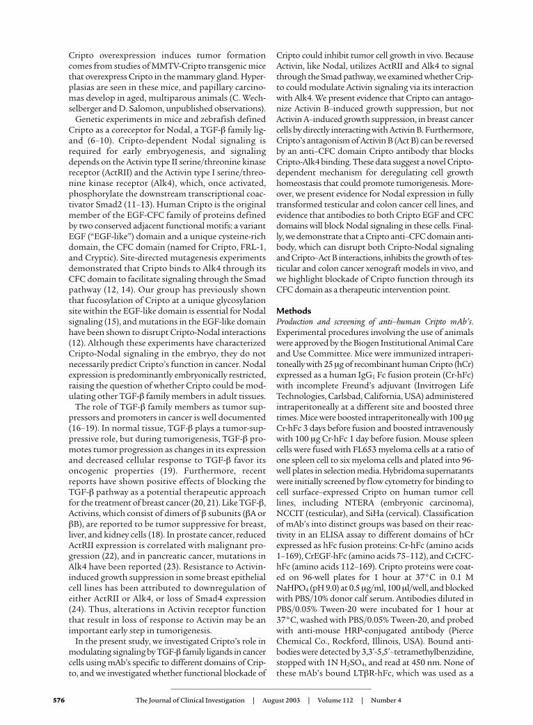
performed as described earlier. Only the anti-CFC mAbA8.G3.5 inhibited Cripto’s ability to interact with Act B(Figure 4d). These data suggest that the CFC domain ofCripto, which interacts with Alk4, is also an importantdomain for association with Act B and further supportthe conclusion that Cripto’s interactions with Act B andNodal utilize different residues on Cripto.
Anti-CFC antibody A8.G3.5 restores Act B–induced growthsuppression in Cripto-overexpressing cells. Due to the obser-vation that anti-CFC mAb A8.G3.5 can block Cripto–Act B interactions, we tested this antibody for dis-rupting Cripto’s inhibitory effect on Act B signaling inT47D cells. Indeed, when T47D-hCr cells were treatedwith Act B in the presence of the anti-CFC mAbA8.G3.5, growth suppression was restored (Figure 5a).Anti-EGF mAb A27.F6.1, by contrast, was unable torestore Act B growth suppression of these cells (Figure5b). Neither mAb had any effect on the response of theparental T47D cells to Act B. To address whetherA8.G3.5 restores Act B signaling through the Smadpathway, we tested whether A8.G3.5 could increase anAct B–induced FAST/(n2)7-luciferase response inT47D-hCr cells. T47D-hCr cells are weakly responsiveto Act B signaling, but in the presence of A8.G3.5,there was an increase in signal in a dose-dependentmanner (Figure 5c). However, A27.F6.1, which doesnot block Cripto–Act B interactions, was unable toincrease the Act B response. These results suggest that,to deregulate Act B growth suppression, Criptosequesters Act B from its receptors, possibly by alsobinding Alk4 and excluding ActRII, and prevents ActB from sending growth control signals.
Cripto mAb’s that block Cripto signaling functions in vitroinhibit tumor growth in vivo. To test whether Cripto actsas a signaling molecule to promote tumor growth, we
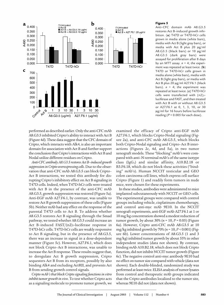
examined the efficacy of Cripto anti-EGF mAbA27.F6.1, which blocks Cripto-Nodal signaling (Fig-ure 2a), and anti-CFC mAb A8.G3.5, which blocksboth Cripto-Nodal signaling and Cripto–Act B inter-actions (Figures 2c, 4d, and 5a), in two tumorxenograft models. These “blocking” mAb’s were com-pared with anti–N-terminal mAb’s of the same isotypeclass (IgG1) and similar affinity, A10.B2.18 orB3.F6.18, which do not block these activities (“bind-ing” mAb’s). Human NCCIT testicular and GEOcolon carcinoma cell lines, which express cell surfaceCripto (Figure 1) and readily form tumors in nudemice, were chosen for these experiments.
In these studies, antibodies were administered to miceimplanted subcutaneously with NCCIT or GEO cells.The experimental groups were compared with controlgroups including vehicle, cisplatinum chemotherapy,and control anti-myc mAb 9E10. In the NCCITxenograft experiments, anti-EGF mAb A27.F6.1 at 1 or10 mg/kg concentration showed a modest reduction oftumor growth, by about 30% (n = 10, P = 0.05) (Figure6a). However, Cripto anti-CFC mAb A8.G3.5 at 10mg/kg inhibited growth by 70% (n = 10, P < 0.001) (Fig-ure 6b). Lower concentrations of A8.G3.5 (1 and 3mg/kg) inhibited tumor growth by about 55% in otherindependent studies (data not shown). By contrast,binding mAb A10.B2.18, which does not block Criptofunction, did not inhibit NCCIT tumor growth (Figure6c). The negative control anti-myc antibody 9E10 hadno effect on tumor size compared with vehicle (data notshown). Each double-blinded, randomized study wasperformed at least twice. ELISA analysis of tumor lysatesfrom control and therapeutic mAb groups indicatedthat the Cripto antibodies localized to the tumor site,whereas 9E10 did not (data not shown).
The Journal of Clinical Investigation | August 2003 | Volume 112 | Number 4 583
Figure 5Anti–CFC domain mAb A8.G3.5restores Act B–induced growth inhi-bition. (a) T47D or T47D-hCr cellsgrown in media alone (white bars),media with Act B (light gray bars), ormedia with Act B plus 20 µg/mlA8.G3.5 (black bars) or 10 µg/mlA8.G3.5 (dark gray bars) wereassayed for proliferation after 8 daysby an MTT assay. n = 4; the experi-ment was repeated at least twice. (b)T47D or T47D-hCr cells grown inmedia alone (white bars), media withAct B (light gray bars), or media withAct B plus 20 µg/ml A27.F6.1 (blackbars). n = 4; the experiment wasrepeated at least twice. (c) T47D-hCrcells were transfected with (n2)7-luciferase and FAST, and then treatedwith Act B with or without A8.G3.5or A27.F6.1 at 0, 1, 3, 10, or 30µg/ml for 16 hours before luciferasereading (P < 0.005 for each dose).
584 The Journal of Clinical Investigation | August 2003 | Volume 112 | Number 4
Anti-CFC mAb A8.G3.5 was tested in a secondxenograft model, GEO colon carcinoma cells expressinghigh levels of Cripto, because Cripto overexpression hasbeen observed in 80% of colon carcinomas examined (4).A8.G3.5 also exhibited anti-tumor-growth activity inthis model, with approximately 50% reduction in tumorgrowth at 3 mg/kg (P = 0.05) (Figure 6d). Conversely, N-terminal binding mAb B3.F6.17 at 10 mg/kg had noeffect on GEO cell tumor growth (Figure 6e). Theseresults indicate that anti-CFC mAb A8.G3.5, which
blocks Cripto’s ability to modulate both the Act B andthe Nodal pathways, is more efficacious in inhibitingtumor growth in vivo than anti-EGF mAb A27.F6.1,which inhibits only the Cripto-Nodal pathway. BothmAb’s have similar binding affinities and internaliza-tion profiles (M. Jarpe, unpublished data); thus, the dif-ference in activities is likely a result of differences in theblocking of Cripto signaling functions.
To address the possible contributions of Nodaland/or Act B to tumor growth in these two tumor
Figure 6Anti–CFC domain mAb blocks NCCIT and GEO tumor growth in vivo. (a) Response of NCCIT xenograft to A27.F6.1: vehicle control (opentriangles), cis-platinum (cross-hatches), 1 mg/kg A27.F6.1 (filled squares), 10 mg/kg A27.F6.1 (open circles). (b) Response of NCCITxenograft to A8.G3.5: vehicle control (open triangles), cis-platinum (cross-hatches), 10 mg/kg A8.G3.5 (closed triangles). (c) Responseof NCCIT xenograft to A10.B2.18: vehicle control (open triangles), cis-platinum (cross-hatches), 1 mg/kg A10.B2.18 (filled squares), 3mg/kg A10.B2.18 (filled circles). (d) Response of GEO xenograft to A8.G3.5: vehicle control (open triangles), cis-platinum (cross-hatch-es), 1 mg/kg A8.G3.5 (filled squares), 3 mg/kg A8.G3.5 (open circles), 10 mg/kg A8.G3.5 (filled triangles). (e) Response of GEO cellxenograft to B3.F6.17: vehicle control (filled squares), cis-platinum (cross-hatches), 10 mg/kg B3.F6.17 (open circles). (f) RT-PCR expres-sion analysis of mRNA transcripts for Cripto, Alk4, Nodal, and Act B in GEO, NCCIT, or MCF-7 cells. Controls include samples withoutreverse transcriptase (–RT) and samples in which template was replaced with H2O. (g) RT-PCR expression analysis of mRNA transcriptsfor murine Act B or human Nodal in NCCIT xenograft tumor sample.

models, we examined the expression of human Nodal,Act B, Cripto, and Alk4 expression by RT-PCR inNCCIT and GEO cells. As expected, both cell linesexpress Alk4 and Cripto (Figure 6f). We also observedexpression of a Nodal transcript in both cell lines, sug-gesting that this embryonic gene can be re-expressed inhuman tumors. This result is in agreement with ourstudies documenting Nodal mRNA expression in anumber of human tumor cell lines (see SupplementalTable 1; www.jci.org/cgi/content/full/112/4/575/DC1).Act B expression was not detected in the NCCIT orGEO cell lines by this method, whereas a faint bandwas seen in the control MCF-7 breast cancer line (Fig-ure 6f). However, Act B is known to be able to act in aparacrine fashion either from a stromal source or incirculation (18), so we analyzed NCCIT xenografttumors for the presence of murine Act B that could besupplied by the host mouse. RNA was extracted fromthe tumor sample and subjected to RT-PCR analysisfor murine Act B and human Nodal expression. Usingprimers specific for murine Act B, an approximately350-bp band was detected (Figure 6g). This DNA frag-ment was isolated and sequenced to verify that it wasindeed a fragment of murine Act B. As expected, ahuman Nodal PCR product was observed (Figure 6g),and its sequence was confirmed since NCCIT cellsexpress Nodal (Figure 6f). Thus, both Nodal and Act Bare available to the tumor in vivo, so we cannot at thistime distinguish between the relative contributions ofthese ligands to tumor growth.
DiscussionIn the present study, we investigated the role of Crip-to in mediating signaling by Nodal and Activin, twoTGF-β ligands that use Alk4 and ActRII to signal, andblockade of these functions with anti-Cripto mAb’s tar-geted to the EGF and CFC domains. We report, for thefirst time to our knowledge, that Cripto can bind direct-ly to Act B and antagonize its ability to elicit a growth-inhibitory response. We show that Act B interacts with
a Cripto fucosylation mutant, whereas Nodal does not,indicating that these ligands interact with different epi-topes on Cripto. Both ligands were found to be presentin xenograft tumor samples, indicating that both couldplay a role in promoting tumor cell growth. Further-more, we demonstrate that an anti–EGF domain anti-body, A27.F6.1, blocks Cripto-Nodal signaling and thatan anti–CFC domain antibody, A8.G3.5, blocks bothCripto-Nodal signaling and Cripto–Act B interactions.While both antibodies inhibited tumor growth in vivo,A8.G3.5 was a more potent inhibitor, suggesting thatantibodies that block the signaling activities mediatedby Cripto’s CFC domain are the most important classfor therapeutic development.
Our data show that overexpression of Cripto antago-nizes Act B growth suppression of breast cancer cells.Activin, like TGF-β, has been proposed to play a tumor-suppressive role in normal tissues (18). Because manytumor cell lines lose responsiveness to Activin, loss ofgrowth suppression by Activin is probably an impor-tant early step in tumorigenesis in some cell types.Alterations in the Activin pathway have been noted inmany cancers, including reduced receptor expression inprostate or breast cancer cells (23, 24) and Alk4 muta-tions in pancreatic cancer (23). Therefore, overexpres-sion of Cripto may represent yet another way to dereg-ulate the Activin pathway, in addition to mutation orloss of receptor expression. Addition of anti-CFC mAbA8.G3.5 reversed the Cripto inhibition of Act B growthsuppression and prevented Act B from binding Cripto,whereas an anti-EGF mAb had no effect. This resultindicates that Cripto inhibition of Activin signaling ismediated by its CFC domain. In our model, we suggestthat Cripto blocks Act B signaling by binding andsequestering Act B from the type II receptor and/orforming nonproductive complexes with Alk4 (Figure7). One could also envision that overexpression of Crip-to could block Act A signaling by sequestering Alk4,but we have not seen this activity in our experiments todate. Both the Cripto–Act B and the Cripto-Alk4 bind-ing activities can be blocked by A8.G3.5; however, thespecificity of Cripto for Act B would suggest that bind-ing Act B is important. At this point, formation of atrimeric “dead signaling complex” among Cripto, Alk4,and Act B (Figure 7) cannot be ruled out and might beconfirmed by further experimentation.
While this paper was in review, Gray et al. (39) pub-lished a study demonstrating Cripto antagonism of ActA signaling. They show by cross-linking experimentsthat Cripto can associate with Act A and ActRII whenthese receptors are overexpressed in 293 cells. Unlikeour data, which demonstrate a direct interactionbetween Cripto and Act B, but not Act A, Gray et al.observe Act A cross-linking to Cripto only in the pres-ence of ActRII. In addition, while they observe thatCripto antagonizes Act A signaling in a luciferase assay,we have never observed an effect of Cripto overexpres-sion on Act A–induced growth suppression, an impor-tant biological consequence of Activin signaling. These
The Journal of Clinical Investigation | August 2003 | Volume 112 | Number 4 585
Figure 7Model of Cripto’s inhibition of Act B signaling through the Smad2pathway. Left: Activin (black) signaling via Alk4 (red), ActRII (blue),and Smad2 leads to regulated cell proliferation in many tissues andorgans. Right: Cripto-dependent Nodal (yellow) signaling throughSmad2 leads to differentiation and patterning in the early embryo.Cripto is represented in green. Center: Cripto inhibits Act B signal-ing via binding to Alk4 and Act B, resulting in deregulated growthand hyperproliferation. Three possible inhibitory complexes areproposed: Cripto/Act B, Cripto/Alk4, and Cripto/Act B/Alk4.Smad2-p, phospho-Smad2.

586 The Journal of Clinical Investigation | August 2003 | Volume 112 | Number 4
discrepancies may reflect different sources of Act A ordifferent cell lines. Furthermore, Gray et al. show thatan EGF domain deletion mutant does not associatewith Act A and conclude that Act A and Nodal havesimilar or overlapping sites of interaction on Cripto. Incontrast, we observe binding of Act B to a Cripto EGFdomain fucosylation mutant, hCr(N85G/T88A).Nodal does bind hCr(N85G/T88A), indicating thatthese residues are important for Nodal binding, butnot for Act B binding. Moreover, we show that only theanti–CFC domain antibody is able to block Act B frombinding Cripto. Thus, while we cannot rule out possi-ble contributions of the EGF domain to binding of ActB, we conclude that the CFC domain residues are criti-cal for Cripto–Act B interaction.
That Cripto can bind directly to Act B demon-strates that Cripto can modulate other TGF-β lig-ands besides Nodal. Thus, Cripto may be a moregeneral modulator of ligand access to cell surfacereceptors than was previously known. Other exam-ples of cell surface proteins shown to modulateTGF-β family ligand access to receptors includeInhibin-binding protein (40), β-glycan (41), and theGDNF ligand coreceptors GFRα1–4 (42–44). Whilethis paper was in review, Cheng et al. (45) demon-strated that Cripto interacts with Vg1/GDF1 as arequired coreceptor for signaling during embryoge-nesis. Thus, it will be important to determinewhether GDF1 is re-expressed in cancer and howCripto’s interaction with GDF1, Nodal, and Act B isregulated. Because we show that Act B can bind to aCripto fucosylation mutant, but Nodal binding tothis mutant is impaired, it is intriguing to speculatethat Cripto’s fucosylation state may play a role inallowing differential articulation of Nodal andActivin signals, similar to the way fucosylation ofNotch’s EGF-like repeat modifies its ligand sensi-tivity to Delta and Serrate (46–49).
In vivo, A8.G3.5 is the most potent Cripto mAb,inhibiting tumor volume by up to 70% in double-blinded, randomized NCCIT xenograft studies.While it is formally possible that the inhibition oftumor growth results from a mechanism other thanblockade of Cripto’s function, nonblocking anti–N-terminal Cripto mAb’s of the same isotype class(IgG1) and similar affinity (A10.B2.18 and B3.F6.17)had no effect on tumor growth in both NCCIT andGEO xenograft models. In addition, preliminaryexperiments with a nonblocking anti-CFC CriptomAb that did not interfere with Cripto-Nodal sig-naling or Cripto-Alk4 interactions in vitro had noeffect on NCCIT tumor growth in vivo (see Supple-mental Figure 3; www.jci.org/cgi/content/full/112/4/575/DC1). In contrast, anti-EGF mAb A27.F6.1,which blocks only the Cripto-Nodal signaling path-way, but not Cripto’s antagonism of Act B, had mar-ginal yet reproducible inhibition of tumor growth, byabout 30%. This result suggests that blockade of bothpathways by targeting of the CFC domain may be
important for efficacy in vivo. These results validatethe therapeutic hypothesis that functional blockadeof Cripto inhibits tumor growth and point to theanti–CFC domain mAb’s as a potentially importantclass of mAb’s for further development.
We propose that, like loss of TGF-β responsiveness,loss of Activin responsiveness may be an importantearly step in cell transformation, and Cripto overex-pression may mediate the loss of Act B signaling insome tissues by antagonizing Act B growth inhibition.In fact, Cripto protein expression as an early indicatorof tumor progression has been cited as a possiblemarker to track dysplasia to breast tumor formation(4). Later in tumorigenesis, whether Cripto functionsby facilitating signaling from Nodal or antagonizesAct B activity is not known. In the fully transformedcell lines NCCIT and GEO, we detected Nodal RNAexpression, indicating that Cripto could be facilitat-ing positive signals from Nodal to promote tumorgrowth. We have also observed Nodal transcripts in avariety of lung, colon, and breast carcinoma cell lines(see Supplemental Table 1; www.jci.org/cgi/content/full/112/4/575/DC1), suggesting that even thoughNodal is not known to be widely expressed in adulttissue, it may be re-expressed in tumors. The lack ofactive, purified Nodal protein in the field and theabsence of Nodal mAb’s have left the question ofwhether Nodal protein is expressed in human tumorsamples unanswered for the present. We also detectedmurine Act B RNA expression in the NCCIT xeno-graft tumor, indicating that Act B is expressed fromstromal sources and is available to the tumor cells.Thus, at this time we cannot distinguish between thecontributions of Nodal and Act B in stimulatinggrowth of these tumors, nor between the contribu-tions of other TGF-β family members with whichCripto may interact, such as GDF1 (45).In summary, we generated Cripto antibodies that rec-ognize epitopes critical for Cripto function and usedthem to dissect Cripto’s ability to modulate Nodaland Act B signaling in cancer cells. We demonstratedthat Cripto can bind and disrupt Act B growth inhi-bition, highlighting a possible new mechanism bywhich Cripto could promote cell transformation. Wealso detected Nodal expression in human tumor celllines, suggesting that Cripto may facilitate positivegrowth-promoting signals from Nodal in cancer. Fur-thermore, we show that by targeting the CFC domainof Cripto with mAb A8.G3.5, we can block both Crip-to-Nodal signaling and Cripto–Act B antagonism. Invivo, A8.G3.5 exhibited the greatest activity, inhibit-ing tumor growth by up to 70%, suggesting that tar-geting the multiple functions of Cripto’s CFC domaincould be important for achieving the greatest efficacyin vivo. Thus, the ability of Cripto antibodies to inhib-it tumor growth defines Cripto as an important onco-gene and blocking of anti-CFC Cripto mAb’s as animportant new therapeutic approach for treatment ofhuman solid tumors.

The Journal of Clinical Investigation | August 2003 | Volume 112 | Number 4 587
AcknowledgmentsThe authors wish to thank Joan Brugge, MalcolmWhitman, Dan Haber, Martin Scott, Brian Elenbaas,Stephen Fawell, Victor Koteliansky, and Richard Catefor critical reading of the manuscript. We thank CindyBottiglio, Kendall Szeliga, and Rebecca Kelly forexpertise in performing animal xenograft studies. Weare especially grateful to Carl-Henrik Heldin, MalcolmWhitman, and Michael Shen for sharing reagents. Wealso thank the FACS and DNA sequencing facilities atBiogen Inc. for expert technical support. Nicola Nor-manno was supported by a grant from the Associ-azione Italiana per la Ricerca sul Cancro.
1. Gibbs, J.B. 2000. Anticancer drug targets: growth factors and growthfactor signaling. J. Clin. Invest. 105:9–13.
2. de Bono, J.S., and Rowinsky, E.K. 2002. The ErbB receptor family: atherapeutic target for cancer. Trends Mol. Med. 8(Suppl. 4):S19–S26.
3. George, D. 2001. Platelet-derived growth factor receptors: a therapeu-tic target in solid tumors. Semin. Oncol. 28:27–33.
4. Adamson, E.D., Minchiotti, G., and Salomon, D.S. 2002. Cripto: a tumor growth factor and more. J. Cell. Physiol. 190:267–278.
5. Ciardiello, F., et al. 1994. Inhibition of CRIPTO expression and tumori-genicity in human colon cancer cells by antisense RNA andoligodeoxynucleotides. Oncogene. 9:291–298.
6. Brennan, J., et al. 2001. Nodal signalling in the epiblast patterns theearly mouse embryo. Nature. 411:965–969.
7. Ding, J., et al. 1998. Cripto is required for correct orientation of theanterior-posterior axis in the mouse embryo. Nature. 395:702–707.
8. Gritsman, K., et al. 1999. The EGF-CFC protein one-eyed pinhead isessential for nodal signaling. Cell. 97:121–132.
9. Shen, M.M., and Schier, A.F. 2000. The EGF-CFC gene family in verte-brate development. Trends Genet. 16:303–309.
10. Yan, Y.T., et al. 1999. Conserved requirement for EGF-CFC genes invertebrate left-right axis formation. Genes Dev. 13:2527–2537.
11. Bianco, C., et al. 2002. Cripto-1 activates nodal- and ALK4-dependentand -independent signaling pathways in mammary epithelial cells. Mol.Cell. Biol. 22:2586–2597.
12. Yeo, C., and Whitman, M. 2001. Nodal signals to Smads through Crip-to-dependent and Cripto-independent mechanisms. Mol. Cell.7:949–957.
13. Kumar, A., et al. 2001. Nodal signaling uses activin and transforminggrowth factor-beta receptor-regulated Smads. J. Biol. Chem.276:656–661.
14. Bianco, C., et al. 2002. Detection and localization of Cripto-1 bindingin mouse mammary epithelial cells and in the mouse mammary glandusing an immunoglobulin-cripto-1 fusion protein. J. Cell. Physiol.190:74–82.
15. Schiffer, S.G., et al. 2001. Fucosylation of Cripto is required for its abil-ity to facilitate nodal signaling. J. Biol. Chem. 276:37769–37778.
16. Barcellos-Hoff, M.H., and Ewan, K.B. 2000. Transforming growth fac-tor-beta and breast cancer: mammary gland development. Breast Can-cer Res. 2:92–99.
17. Derynck, R., Akhurst, R.J., and Balmain, A. 2001. TGF-beta signalingin tumor suppression and cancer progression. Nat. Genet. 29:117–129.
18. Risbridger, G.P., Schmitt, J.F., and Robertson, D.M. 2001. Activins andinhibins in endocrine and other tumors. Endocr. Rev. 22:836–858.
19. Wakefield, L.M., and Roberts, A.B. 2002. TGF-beta signaling: positiveand negative effects on tumorigenesis. Curr. Opin. Genet. Dev. 12:22–29.
20. Muraoka, R.S., et al. 2002. Blockade of TGF-β inhibits mammarytumor cell viability, migration, and metastases. J. Clin. Invest.109:1551–1559. doi:10.1172/JCI200215234.
21. Yang, Y.A., et al. 2002. Lifetime exposure to a soluble TGF-β antago-nist protects mice against metastasis without adverse side effects. J. Clin. Invest. 109:1607–1615. doi:10.1172/JCI200215333.
22. van Schaik, R.H., et al. 2000. Variations in activin receptor, inhibin/activin subunit and follistatin mRNAs in human prostate tumour tis-sues. Br. J. Cancer. 82:112–117.
23. Su, G.H., et al. 2001. ACVR1B (ALK4, activin receptor type 1B) genemutations in pancreatic carcinoma. Proc. Natl. Acad. Sci. U. S. A.98:3254–3257.
24. Kalkhoven, E., et al. 1995. Resistance to transforming growth factorbeta and activin due to reduced receptor expression in human breasttumor cell lines. Cell Growth Differ. 6:1151–1161.
25. Adkins, H.B., Blacklow, S.C., and Young, J.A. 2001. Two functionallydistinct forms of a retroviral receptor explain the nonreciprocal recep-tor interference among subgroups B, D, and E avian leukosis viruses.J. Virol. 75:3520–3526.
26. Johnsson, B., Lofas, S., and Lindquist, G. 1991. Immobilization of pro-teins to a carboxymethyldextran-modified gold surface for biospecif-ic interaction analysis in surface plasmon resonance sensors. Anal.Biochem. 198:268–277.
27. Alexander, J.M., Bikkal, H.A., Zervas, N.T., Laws, E.R., Jr., and Klibanski,A. 1996. Tumor-specific expression and alternate splicing of messengerribonucleic acid encoding activin/transforming growth factor-beta recep-tors in human pituitary adenomas. J. Clin. Endocrinol. Metab. 81:783–790.
28. Normanno, N., et al. 1995. Expression of messenger RNA foramphiregulin, heregulin, and cripto-1, three new members of the epi-dermal growth factor family, in human breast carcinomas. Breast Can-cer Res. Treat. 35:293–297.
29. Normanno, N., et al. 2003. CRIPTO-1 overexpression leads toenhanced invasiveness and resistance to anoikis in human MCF-7breast cancer cells. J. Cell. Physiol. In press.
30. Albano, R.M., Groome, N., and Smith, J.C. 1993. Activins are expressedin preimplantation mouse embryos and in ES and EC cells and are reg-ulated on their differentiation. Development. 117:711–723.
31. Yan, Y.T., et al. 2002. Dual roles of Cripto as a ligand and coreceptor inthe nodal signaling pathway. Mol. Cell. Biol. 22:4439–4449.
32. Qi, C.F., et al. 1994. Expression of transforming growth factor alpha,amphiregulin and cripto-1 in human breast carcinomas. Br. J. Cancer.69:903–910.
33. Saeki, T., et al. 1995. Association of epidermal growth factor-relatedpeptides and type I receptor tyrosine kinase receptors with prognosisof human colorectal carcinomas. Jpn. J. Clin. Oncol. 25:240–249.
34. Chen, Y.G., Lui, H.M., Lin, S.L., Lee, J.M., and Ying, S.Y. 2002. Regula-tion of cell proliferation, apoptosis, and carcinogenesis by activin. Exp.Biol. Med. (Maywood). 227:75–87.
35. Cocolakis, E., Lemay, S., Ali, S., and Lebrun, J.J. 2001. The p38 MAPKpathway is required for cell growth inhibition of human breast cancercells in response to activin. J. Biol. Chem. 276:18430–18436.
36. Liu, Q.Y., et al. 1996. Inhibitory effects of activin on the growth andmorpholgenesis of primary and transformed mammary epithelial cells.Cancer Res. 56:1155–1163.
37. Hahn, W.C., et al. 1999. Creation of human tumour cells with definedgenetic elements. Nature. 400:464–468.
38. Kannan, S., et al. 1997. Cripto enhances the tyrosine phosphorylationof Shc and activates mitogen-activated protein kinase (MAPK) inmammary epithelial cells. J. Biol. Chem. 272:3330–3335.
39. Gray, P.C., et al. 2003. Cripto forms a complex with activin and type IIactivin receptors and can block activin signaling. Proc. Natl. Acad. Sci.U. S. A. 100:5193–5198.
40. Chong, H., et al. 2000. Structure and expression of a membrane com-ponent of the inhibin receptor system. Endocrinology. 141:2600–2607.
41. Massague, J., and Chen, Y.G. 2000. Controlling TGF-beta signaling.Genes Dev. 14:627–644.
42. Jing, S., et al. 1996. GDNF-induced activation of the ret protein tyro-sine kinase is mediated by GDNFR-alpha, a novel receptor for GDNF.Cell. 85:1113–1124.
43. Sanicola, M., et al. 1997. Glial cell line-derived neurotrophic factor-dependent RET activation can be mediated by two different cell-sur-face accessory proteins. Proc. Natl. Acad. Sci. USA. 94:6238–6243.
44. Treanor, J.J., et al. 1996. Characterization of a multicomponent recep-tor for GDNF. Nature. 382:80–83.
45. Cheng, S.K., Olale, F., Bennett, J.T., Brivanlou, A.H., and Schier, A.F.2003. EGF-CFC proteins are essential coreceptors for the TGF-beta sig-nals Vg1 and GDF1. Genes Dev. 17:31–36.
46. Bruckner, K., Perez, L., Clausen, H., and Cohen, S. 2000. Glycosyl-transferase activity of Fringe modulates Notch-Delta interactions.Nature. 406:411–415.
47. Hicks, C., et al. 2000. Fringe differentially modulates Jagged1 andDelta1 signalling through Notch1 and Notch2. Nat. Cell Biol.2:515–520.
48. Moloney, D.J., et al. 2000. Fringe is a glycosyltransferase that modifiesNotch. Nature. 406:369–375.
49. Moloney, D.J., et al. 2000. Mammalian Notch1 is modified with twounusual forms of O-linked glycosylation found on epidermal growthfactor-like modules. J. Biol. Chem. 275:9604–9611.
Copyright © 2022 FDOKUMEN