Antibody blockade of the Cripto CFC domain suppresses tumor cell growth in vivo
Cell Stem Cell
Article
Cripto Regulates Hematopoietic Stem Cellsas a Hypoxic-Niche-Related Factorthrough Cell Surface Receptor GRP78Kenichi Miharada,1,* Goran Karlsson,1 Matilda Rehn,1 Emma Rorby,1 Kavitha Siva,1 Jorg Cammenga,1
and Stefan Karlsson1,*1Department for Molecular Medicine and Gene Therapy, Lund Strategic Center for Stem Cell Biology, Lund University, Lund, 221 84, Sweden
*Correspondence: [email protected] (K.M.), [email protected] (S.K.)DOI 10.1016/j.stem.2011.07.016
SUMMARY
Hematopoietic stem cells (HSCs) are maintained inhypoxic niches in endosteal regions of bones. Herewe demonstrate that Cripto and its receptor GRP78are important regulators of HSCs in the niche. Flowcytometry analyses revealed two distinct subpopula-tions of CD34�KSL cells based on the expression ofGRP78, and these populations showed differentreconstitution potential in transplantation assays.GRP78+HSCs mainly reside in the endosteal area,are more hypoxic, and exhibit a lower mitochondrialpotential, and their HSC capacity was maintainedin vitro by Cripto through induction of higher glyco-lytic activity. Additionally, HIF-1a KO mice havedecreased numbers of GRP78+HSCs and reducedexpression of Cripto in the endosteal niche. Further-more, blocking GRP78 induced amovement of HSCsfrom the endosteal to the central marrow area. Thesedata suggest that Cripto/GRP78 signaling is animportant pathway that regulates HSC quiescenceand maintains HSCs in hypoxia as an intermediaryof HIF-1a.
INTRODUCTION
Hematopoietic stem cells (HSCs) represent a heterogeneous
population of cells that can exist in both dormant and active
states. Similarly, there is also heterogeneity with regard to their
differentiation potential, in that both myeloid-biased and
lymphoid-biased HSCs have been characterized (Dykstra
et al., 2007; Challen et al., 2010; Kent et al., 2009; Morita et al.,
2010). Historically, manymarkers andmethods have been devel-
oped to purify HSCs and distinguish different types of HSC
subpopulations (Li and Johnson, 1995; Goodell et al., 1996;
Osawa et al., 1996; Arai et al., 2004; Kiel et al., 2005; Morita
et al., 2010; Schroeder, 2010). However, there may be further
heterogeneity of hitherto unknown HSC subpopulations, and
some of the existing markers are nonfunctional and/or intrinsic
regulators. Recent studies report the reversibility of these
subgroups in the HSC compartment, indicating that these sub-
groups are not hierarchial (Dykstra et al., 2007; Haug et al.,
330 Cell Stem Cell 9, 330–344, October 7, 2011 ª2011 Elsevier Inc.
2008; Wilson et al., 2008; Challen et al., 2010). These variant
subsets of HSCs seem to have different (or specific) signal
responsiveness and reactions to regulatory factors, depending
on their intrinsic/extrinsic gene expression pattern; e.g., sub-
types of HSC populations identified by side population (SP)
pattern and CD150 intensity indicated differential responses to
TGF-b1, which has been reported as an effective factor to induce
HSC hibernation (Yamazaki et al., 2009; Challen et al., 2010).
HSCs are maintained in a specific environment referred to as
the stem cell niche. Previous studies have suggested several
types of niche components, and dormant and active HSCs
may be maintained in different types of niches (e.g., osteoblastic
niche and perivascular niche). Other cell types, for example
endothelial cells and mesenchymal stem cells, may supply niche
function as well (Scadden, 2006; Arai and Suda, 2007; Morrison
and Spradling, 2008; Li and Clevers, 2010; Nakamura et al.,
2010). Dormant HSCs are understood to reside in the osteo-
blastic niche, a relatively hypoxic region mainly existing in the
endosteal area of bones and sinusoids (Parmar et al., 2007; Ku-
bota et al., 2008). Under hypoxic conditions, HSCs have glycol-
ysis-biased metabolic activity instead of mitochondrial oxidative
phosphorylation (Simsek et al., 2010). One of the master regula-
tors of HSC metabolism under hypoxia is hypoxia-inducible
factor-1a (HIF-1a) (Danet et al., 2003; Takubo et al., 2010). The
HIF-1 complex regulates several downstream genes and is
stabilized under hypoxia, whereas it is immediately dissociated
in the normoxic situation (Keith and Simon, 2007). In the stem
cell niche, HSCs must be influenced by various types of niche-
related factors, e.g., soluble factors, cell adhesion molecules,
and extracellular matrix (Morrison and Spradling, 2008; Li and
Clevers, 2010). However, key regulators that sustain dormant
HSCs within the hypoxic niche and control the metabolic activity
of HSCs, particularly extrinsic niche factors, remain largely
unknown.
Cripto, also known as teratocarcinoma-derived growth factor
1 (TDGF-1), is the first member of the EGF-CFC protein family,
which includes Cripto, FRL-1, and Cryptic (Ciccodicola et al.,
1989; Saloman et al., 2000). A soluble form and a GPI-anchored
cell membrane form have been identified, but their functional
difference is unclear at present (Bianco et al., 2010). The Cripto
gene has been identified as a gene that regulates tumorigenesis,
embryogenesis, and embryonic stem cells (ESCs) (Ding et al.,
1998; Sato et al., 2003; Bianco et al., 2010), and is also a critical
regulator of myocardial development (Xu et al., 1998; Bianco
et al., 2009). Molecular studies have revealed multiple functions
Cell Stem Cell
Cripto/GRP78 Regulates Hematopoietic Stem Cells
of Cripto as both a ligand and a coreceptor and subsequent
activating downstream signals. Cripto blocks TGF-b signaling
by directly binding to TGF-b and the type II TGF-b receptor,
and thereby competes with the type I receptor for binding to
the TGF-b-type II receptor complex (Gray et al., 2006). In addi-
tion, Cripto/Nodal/Growth Differentiation Factor-1/-3 (GDF1
andGDF3) signaling is important during embryogenesis, through
Cripto’s binding to the Activin type II and type I (ALK4) receptors,
which subsequently triggers phosphorylation of Smad2/3 and
the formation of a complex with Smad4 (Karlsson et al., 2007;
Bianco et al., 2010). Furthermore, Cripto has been shown to
directly associate with the 78 kDa glucose-regulated protein
(GRP78) on the cell surface, and as a consequence regu-
lates cell growth, promotes tumor progression, downregulates
E-Cadherin, and decreases cell adhesion (Shani et al., 2008;
Kelber et al., 2009). GRP78 is amember of the heat shock protein
70 (HSP70) family and is usually expressed on the endoplasmic
reticulum (ER), but some types of tumor cells express GRP78
protein on the cell membrane as a target receptor for some
ligands, including Cripto (Gonzalez-Gronow et al., 2009). The
Cripto/GRP78 association on the cell surface facilitates c-Src/
MAPK/PI3K signaling and also controls Smad2/3 signaling by
Activin/Nodal/TGF-b at an intermediate level (Kelber et al.,
2009). Thus, Cripto and its receptors have important roles for
tissue development and cell growth. However, the role of Cripto
in the regulation of hematopoiesis is unknown at present.
In this work, we have observed that Cripto/GRP78 signaling
may be one of the critical regulatory pathways to control
dormancy of HSCs in the hypoxic niche environment by inducing
high glycolysis activity in HSCs downstream of HIF-1a. Further-
more, the cell surfacemolecule GRP78 is an important functional
marker for a subpopulation of HSCs that tend to be quiescent,
and this marker can therefore distinguish between HSCs that
are dormant or activated.
RESULTS
Cripto Is Highly Expressed in HSCs and Can MaintainTheir Stem Cell Function Ex VivoBecause CriptomRNA expression is highly upregulated in undif-
ferentiated ESCs and is associated with their immaturity (Sato
et al., 2003; Hough et al., 2009), we asked whether Cripto
expression was similarly restricted to an immature population
of hematopoietic cells. To determine this, quantitative real-time
PCR was performed on several kinds of purified hematopoietic
stem/progenitor populations. Surprisingly, the findings showed
that the expression level of Cripto mRNA was strongly related
to their differentiation stage in the hematopoietic hierarchy.
Notably, long-term hematopoietic stem cells (LT-HSCs) indi-
cated higher expression than any other hematopoietic popula-
tion tested (Figure 1A). In ESC studies, Cripto has been well
known as a sensitive marker for their immaturity because the
expression of Cripto is immediately downregulated when ESCs
start to differentiate (Sato et al., 2003; Hough et al., 2009). There-
fore the levels of Cripto gene expression in hematopoietic
stem/progenitor cells seem to be analogous to those seen in
ESCs. Next, we tested the effects of Cripto protein on HSCs.
CD34�KSL cells were sorted and cultured in vitro with or without
500 ng/ml of recombinant mouse Cripto (rmCripto) protein and
C
a CFU assay was performed to analyze whether rmCripto had
any effect on the colony-forming ability of the cultured cells.
The findings showed that rmCripto enhanced cell proliferation
(5-fold at day 14 compared with culture without rmCripto) (Fig-
ure 1B) and colony formation (3- to 3.5-fold). Cripto-treated cells
generated around 100 GEMM mixed colonies after 14 days of
culture, while the control cells cultured without Cripto did not
form any mixed colonies (Figure 1C). To evaluate the HSC
expansion/maintenance ability of Cripto, we performed compet-
itive repopulation assay by transplanting either freshly isolated
CD34�KSL cells or the cultured cells into lethally irradiated
mice after cultivation with or without rmCripto. The findings
showed that rmCripto succeeded in maintaining their stem cell
function after 14 days of culture while the cells cultured in the
control condition failed to reconstitute recipients (Figure 1D),
although the percentage of donor-derived cells (chimerism) in
peripheral blood (PB) was lower than when fresh cells were
used. Furthermore, we performed secondary transplantation
and the donor-derived cells were engrafted into secondary
recipients (Figure 1E). These data demonstrate that Cripto has
the ability to maintain functional HSCs in culture ex vivo.
GRP78 Is a Functional Cell Surface Markerto Distinguish Two Subsets of HSCsSeveral proteins have been reported as candidate cell surface
receptors for Cripto (Strizzi et al., 2005; Shani et al., 2008; Bianco
et al., 2010). We focused on GRP78 and Glypican-1 as possible
receptors for Cripto and tested whether HSCs expressed them
on their cell surface. Surprisingly, flow cytometry analysis re-
vealed that CD34�KSL cells were clearly separated into two
subpopulations based on the expression level of cell-surface
GRP78 (Figure 2A and Figure S1A available online). Among
CD34�KSL cells, approximately one-third of cells were GRP78+
cells (32.12% ± 7.93%, n = 6). We also tested SLAM marker
CD150 on CD34�KSL cells with GRP78 staining because
CD34�KSL cells have been observed to be fractionated to func-
tionally different subpopulations due to CD150 expression (Kent
et al., 2009; Challen et al., 2010; Morita et al., 2010). The findings
showed that a larger number of GRP78+CD34�KSL cells were
CD150+, while GRP78�CD34�KSL cells included both CD150+
and CD150� cells in an almost similar frequency (Figure S1B).
In contrast, Glypican-1 was expressed on the majority of
CD34�KSL cells (Figure S1B). GRP78 was also expressed on
the many, more mature hematopoietic lineage cells, although
the percentage of positive cells was relatively lower than found
on CD34�KSL cells (Figure S1C).
In order to clarify whether these HSC subpopulations have any
functional difference, GRP78+CD34�KSL cells (GRP78+HSCs)
or GRP78�CD34�KSL cells (GRP78�HSCs) were clonally sorted
into 96-well plates and incubated. GRP78+HSCs showed a
tendency to generate a higher proportion of GEMM mixed colo-
nies (approximately 58%) compared with GRP78�HSCs (39%),
although the difference was not significant (Figure S1D). To ask
whether GRP78+HSCs are a unique target population for Cripto,
both GRP78+ and GRP78�HSCs were sorted and cultured with
or without rmCripto and then subjected to a CFU assay. Total
cell count of the culture showed that rmCripto enhanced cell
growth only for GRP78+HSCs (Figure S1E). After 7 days in
culture, GRP78+HSCs reacted to rmCripto and generated
ell Stem Cell 9, 330–344, October 7, 2011 ª2011 Elsevier Inc. 331
Figure 1. Cripto Is Highly Expressed in LT-HSCs and Maintains the Reconstitution Ability of HSCs after Ex Vivo Culture
(A) Quantitative Real-Time PCR analysis for Cripto expression in LT-HSC (CD34�Flt3�KSL), ST-HSC (CD34+Flt3�KSL), LMPP (CD34+Flt3+KSL), Lineage�, andLineage+ cells. Each value is normalized to HPRT expression and mean ± SD are shown. Significance is calculated against LT-HSC (n = 3).
(B) Cell count for cultured cells with or without rmCripto. Thirty CD34�KSL cells were directly sorted into SFEM media supplemented with 100 ng/ml of mSCF
and hTPO with or without 500 ng/ml of rmCripto in 96-well plates. Absolute cell number was counted at day 7 (left) and day 14 (right) using Trypan blue
(mean ± SD, n = 4).
(C) Colony forming unit assay (CFU assay) for cultured cells. All colony assays were initiated by plating 30 CD34�KSL cells and scoring colonies at day 7 (left)
and day 14 (right) (mean ± SD, n = 8). The number of GEMMmixed colonies was significantly increased after treatment with rmCripto (day 7: p = 0.0473, day 14:
p = 0.0422). GEMM: granulocytes, erythrocytes, macrophages, with or without megakaryocytes; E: erythrocytes; GM: granulocytes and macrophages;
G: granulocytes; M: macrophages.
(D) Competitive repopulation assay using fresh or cultured cells. Results from 4 weeks (left) and 16 weeks (right) after transplantation are shown (n = 10).
Each circle indicates one mouse and red bars indicate average chimerism.
(E) Secondary transplantation. Each circle indicates one mouse and red bars indicate average chimerism. Results from 12 weeks after transplantation are shown
(n = 8).
Cell Stem Cell
Cripto/GRP78 Regulates Hematopoietic Stem Cells
332 Cell Stem Cell 9, 330–344, October 7, 2011 ª2011 Elsevier Inc.
Cell Stem Cell
Cripto/GRP78 Regulates Hematopoietic Stem Cells
increased numbers of colonies, whereas GRP78�HSCs did not
(Figures 2B and 2C). To confirm this, we also tried to neutralize
the Cripto/GRP78 signaling on GRP78+HSCs by using blocking
antibody against GRP78 (N-20) (Davidson et al., 2005; Kelber
et al., 2009). N-20 inhibited the effect of rmCripto on growth of
the GRP78+HSCs and their colony formation, but not of
GRP78�HSCs (Figures 2B and 2C). Interestingly, the single addi-
tion of the N-20 antibody generated less growth/colonies than
the control condition (Figures 2B and 2C). This data suggests
that HSCs might secrete some amount of Cripto by themselves
(as expected by real-time PCR assay in Figure 1A), which aids
viability of HSCs through its binding to the cell surface GRP78
protein in an autocrine fashion.
In order to ask whether both GRP78+ and GRP78�HSC have
equivalent HSC function, we used the competitive repopula-
tion assay to test these subpopulations. Either GRP78+ or
GRP78�HSCs were sorted out and freshly transplanted or
cultured with or without rmCripto for 14 days after transplanta-
tion. Monthly PB analysis in recipient mice demonstrated that
both GRP78+ and GRP78�HSCs were able to reconstitute recip-
ients over the long term, but the two populations exhibit different
behavior (Figure 2D). The chimerism of cells derived from
GRP78+HSCs was at a lower level than that from GRP78�HSCsand slowly increased later, whereas GRP78�HSCs generated
higher chimerism than GRP78+ cells at the early stage but the
reconstitution decreased slightly over time (Figure 2D). However,
the difference in reconstitution between these subpopulations
was not significant at 4 months after transplantation (Figure 2D).
Importantly, rmCripto reacted only to GRP78+HSCs and the
cultured cells successfully reconstituted recipients, whereas
GRP78�HSCs failed to reconstitute (Figure 2D). The chimerism
in recipient mice derived fromGRP78+HSCs cultured with Cripto
was somewhat higher than the level generated with freshly
isolated GRP78+HSCs during the first month; however, the
reconstitution did not increase over time as with freshly isolated
cells (Figure 2D). These findings strongly suggest that GRP78
can separate HSCs into two different types of subsets and that
GRP78+HSCs are the unique target population for Cripto.
Furthermore, the N-20 antibody inhibited the effect of rmCripto
on GRP78+HSCs in culture and the cells failed to engraft
(Figure 2D). These findings confirm the functionality of GRP78
as the receptor for Cripto. An additional difference observed
in the PB reconstitution was that GRP78+HSCs generated a
relatively myeloid-biased lineage pattern in contrast to GRP78�
HSCs thatgeneratedamore lymphoid-biasedpattern (Figure2E).
Such myeloid- and lymphoid-biased HSC subtypes have been
characterized to have different responses to TGF-b signals
(Challen et al., 2010). The report identified two subsets of SP
cells: lower-SP, which contains more myeloid-biased HSCs
and reacts positively to TGF-b; and upper-SP, which contains
lymphoid-biased HSCs and is growth inhibited when exposed
to TGF-b. Because Cripto has been reported to bind to the
TGF-b receptor complex, we decided to clarify the relationship
betweenGRP78+/GRP78�HSCs and identified HSCSP subsets.
The findings show that Cripto blocks TGF-b signaling in HSCs
and that lower-SP subset and the GRP78+HSC subset are quite
similar, but there is not a complete overlap (Figures S1F–S1H).
Over 4 months after transplantation, all mice were sacrificed
and bonemarrow (BM) cells were analyzed. The assays revealed
C
that both GRP78+ and GRP78�HSCs can generate each other
in vivo (Figures 2F and 2G). This data indicate that GRP78
expression on HSCs is ‘‘reversible,’’ and not hierarchy related.
Generally chimerism in PB and in BM is at a similar level (Fig-
ure 2F). However, GRP78+HSCs cultured with Cripto generated
much higher levels of CD34�KSL cells in vivo (Figure 2F). More
surprisingly, the vast majority of engrafted HSCs (represented
by the CD34�KSL population) derived from cultured GRP78+
HSCs were GRP78+ cells, even though the ratio of GRP78+/
GRP78� cells in both freshly isolated GRP78+ and GRP78�
HSC-derived cells generated similar proportions to those seen
in the original donor mice (Figures 2F and 2G). Consequently,
chimerism in recipients’ PB and the percentage of GRP78+ cells
in donor-derived HSC fractions were inversely related (Fig-
ure S1I). Especially, the higher content of GRP78+ cells in the
HSC compartment seemed to be critical for the lineage
distribution.
When we performed secondary transplantation from primary
recipients, the results showed a similar transient pattern of
chimerism derived from fresh GRP78+HSC transplant recipients
and GRP78�HSCs transplanted mice (Figure 2H). This data was
expected because BM cells from primary recipients of both fresh
GRP78+ and GRP78�HSC transplanted mice had similar per-
centages of GRP78+/GRP78� cells in the HSC compartment
when they had been transplanted. Also, BM cells from primary
recipients’ cultured GRP78+HSCs were engrafted, but the
chimerism level was low (Figure 2H). Our findings show that
cell-surface GRP78 is a useful marker to prospectively isolate
these HSC subpopulations.
Cripto Signaling for HSCs Is Involved in the Akt Pathwayand Upregulates Glycolytic-Enzyme-Related Proteinsthrough GRP78Cripto has been known to activate the c-Src/PI3K/MAPK path-
way and to regulate TGF-b/Smad pathway. In order to under-
stand the mechanism of Cripto/GRP78 signaling in HSCs, we
analyzed phosphorylation of key components in these signaling
pathways. Intracellular staining analyses of phosphorylated pro-
teins using flow cytometry for GRP78+HSCs with Cripto treat-
ment demonstrated a significant increase in phosphorylated
Akt, 4E-BP1, and S6 compared with that in untreated cells after
a short period of stimulation, while GRP78�HSCs showed no
response at any time point (Figures 3A–3D and 3F). In contrast,
there was no difference observed in the levels of phosphorylated
Smad2/3 between Cripto-treated and nontreated cells, both in
GRP78+ and GRP78� fractions (Figures 3E and 3F). These
data suggest that Cripto signaling in HSCs (at least in vitro)
may be related to Akt signaling, but not Smad signaling. The
Akt signal transduction was detected up to 90 min after the
Cripto stimulation but it is not clear whether this pathway contin-
uously stimulates HSCs for longer periods. We therefore per-
formed proteomics analysis to further study the possible mech-
anisms and to identify a downstream target or targets. As
a model for HSCs, we used BM cells immortalized by induction
of LIM-homeobox gene Lhx2 (Lhx2 cells), which represent an
HSC-like cell line (Pinto do O et al., 2002). Using flow cytometry
analysis, Lhx2 cells exhibit a CD34�KSL HSC immunopheno-
type, with the exception of CD11b, in which Lhx2 cells are posi-
tive but primary HSCs are negative (Pinto do O et al., 2002 and
ell Stem Cell 9, 330–344, October 7, 2011 ª2011 Elsevier Inc. 333
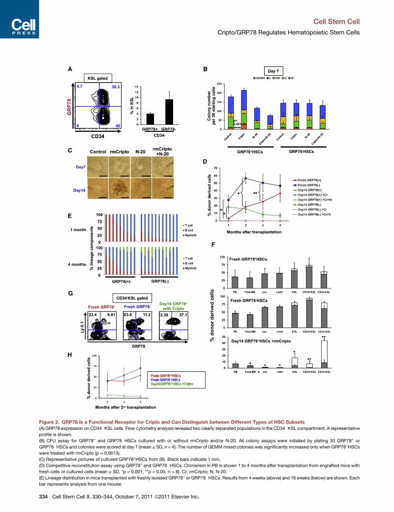
Figure 2. GRP78 Is a Functional Receptor for Cripto and Can Distinguish between Different Types of HSC Subsets(A) GRP78 expression on CD34�KSL cells. Flow cytometry analysis revealed two clearly separated populations in the CD34�KSL compartment. A representative
profile is shown.
(B) CFU assay for GRP78+ and GRP78�HSCs cultured with or without rmCripto and/or N-20. All colony assays were initiated by plating 30 GRP78+ or
GRP78�HSCs and colonies were scored at day 7 (mean ± SD, n = 4). The number of GEMMmixed colonies was significantly increased only when GRP78+HSCs
were treated with rmCripto (p = 0.0013).
(C) Representative pictures of cultured GRP78+HSCs from (B). Black bars indicate 1 mm.
(D) Competitive reconstitution assay using GRP78+ and GRP78�HSCs. Chimerism in PB is shown 1 to 4 months after transplantation from engrafted mice with
fresh cells or cultured cells (mean ± SD, *p < 0.001, **p < 0.05, n = 8). Cr: rmCripto; N: N-20.
(E) Lineage distribution in mice transplanted with freshly isolated GRP78+ or GRP78�HSCs. Results from 4 weeks (above) and 16 weeks (below) are shown. Each
bar represents analysis from one mouse.
Cell Stem Cell
Cripto/GRP78 Regulates Hematopoietic Stem Cells
334 Cell Stem Cell 9, 330–344, October 7, 2011 ª2011 Elsevier Inc.
Cell Stem Cell
Cripto/GRP78 Regulates Hematopoietic Stem Cells
Figure S2A). Surprisingly, Lhx2 cells had both GRP78+ and
GRP78� subpopulations and GRP78+Lhx2 cells responded to
Cripto in vitro (Figure 3G and Figures S2A and S2B). The findings
suggest that Cripto inhibits differentiation of Lhx2 cells.
As an approach to identify upregulated proteins during
Cripto/GRP78 signaling, we used 2D difference gel electropho-
resis (2D-DIGE) proteomics technology. The GRP78+ population
was isolated from Lhx2 cells in order to concentrate the signal,
and the cells were cultured in a standard medium supplemented
with or without rmCripto for 24 hr. The cultured cells and freshly
isolated cells were prepared in a lysis buffer to facilitate the
collection of protein samples (Figure 3G). Each sample was
separated by 2D gel electrophoresis (Figures 3H–3J). Protein
spots, specifically increased in the sample from cells cultured
with rmCripto, were picked up. Using this approach, 21 spots
were selected and their amino acid sequences were analyzed
(Figure 3K). The findings demonstrated that many of the pro-
teins whose production was increased were glycolytic enzymes,
e.g., pyruvate kinase, phosphoglycerate kinase 1, and glyceral-
dehydes-3-phosphate dehydrogenase (GAPDH) (Figure 3K
and Table 1). Importantly, the same proteins were aligned in
several sets of two spots, strongly indicating that these pro-
teins were phosphorylated (Table 1). HSCs are thought to have
relatively greater glycolysis ability because of their hypoxic
condition (Simsek et al., 2010). Therefore, the increased and/or
phosphorylation activity of glycolytic enzymes suggests a
role for Cripto in the metabolic regulation of HSCs. As another
interesting clue, several actin-polymerization-related proteins
were detected in the 2D-DIGE analysis, including Moesin,
Gamma-actin, and Cofilin-1 (Table 1). Although the importance
of actin polymerization for cell motility of lymphocytes and
for homing of HSCs has been well known (Kahn et al., 2004;
Huang and Burkhardt, 2007), the relationship between actin
polymerization and HSC maintenance is unknown. However,
some actin-polymerization-related genes, e.g., WASp family
verprolin-homologous protein 2 (WAVE2), have been reported
as required regulators for hematopoietic repopulation capacity
(Ogaeri et al., 2009). Some of the genes for these listed proteins
have previously been reported as upregulated during embryonic
cardiomyocyte differentiation induced by Cripto (Bianco et al.,
2009).
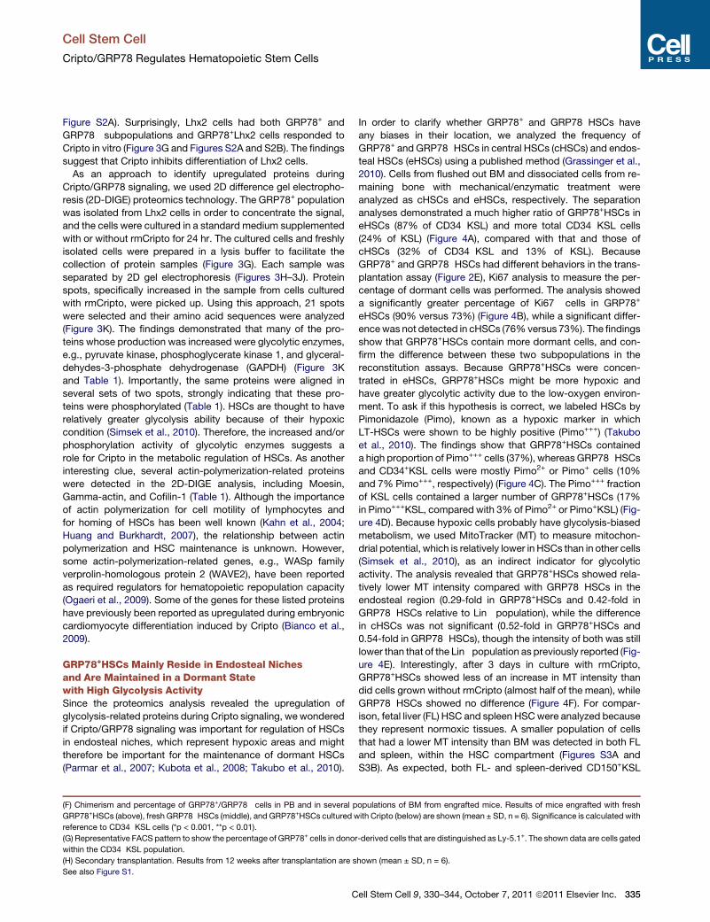
GRP78+HSCs Mainly Reside in Endosteal Nichesand Are Maintained in a Dormant Statewith High Glycolysis ActivitySince the proteomics analysis revealed the upregulation of
glycolysis-related proteins during Cripto signaling, we wondered
if Cripto/GRP78 signaling was important for regulation of HSCs
in endosteal niches, which represent hypoxic areas and might
therefore be important for the maintenance of dormant HSCs
(Parmar et al., 2007; Kubota et al., 2008; Takubo et al., 2010).
(F) Chimerism and percentage of GRP78+/GRP78� cells in PB and in several p
GRP78+HSCs (above), fresh GRP78�HSCs (middle), and GRP78+HSCs cultured w
reference to CD34�KSL cells (*p < 0.001, **p < 0.01).
(G) Representative FACS pattern to show the percentage of GRP78+ cells in donor
within the CD34�KSL population.
(H) Secondary transplantation. Results from 12 weeks after transplantation are s
See also Figure S1.
C
In order to clarify whether GRP78+ and GRP78�HSCs have
any biases in their location, we analyzed the frequency of
GRP78+ and GRP78�HSCs in central HSCs (cHSCs) and endos-
teal HSCs (eHSCs) using a published method (Grassinger et al.,
2010). Cells from flushed out BM and dissociated cells from re-
maining bone with mechanical/enzymatic treatment were
analyzed as cHSCs and eHSCs, respectively. The separation
analyses demonstrated a much higher ratio of GRP78+HSCs in
eHSCs (87% of CD34�KSL) and more total CD34�KSL cells
(24% of KSL) (Figure 4A), compared with that and those of
cHSCs (32% of CD34�KSL and 13% of KSL). Because
GRP78+ and GRP78�HSCs had different behaviors in the trans-
plantation assay (Figure 2E), Ki67 analysis to measure the per-
centage of dormant cells was performed. The analysis showed
a significantly greater percentage of Ki67� cells in GRP78+
eHSCs (90% versus 73%) (Figure 4B), while a significant differ-
encewas not detected in cHSCs (76%versus 73%). The findings
show that GRP78+HSCs contain more dormant cells, and con-
firm the difference between these two subpopulations in the
reconstitution assays. Because GRP78+HSCs were concen-
trated in eHSCs, GRP78+HSCs might be more hypoxic and
have greater glycolytic activity due to the low-oxygen environ-
ment. To ask if this hypothesis is correct, we labeled HSCs by
Pimonidazole (Pimo), known as a hypoxic marker in which
LT-HSCs were shown to be highly positive (Pimo+++) (Takubo
et al., 2010). The findings show that GRP78+HSCs contained
a high proportion of Pimo+++ cells (37%), whereas GRP78�HSCsand CD34+KSL cells were mostly Pimo2+ or Pimo+ cells (10%
and 7% Pimo+++, respectively) (Figure 4C). The Pimo+++ fraction
of KSL cells contained a larger number of GRP78+HSCs (17%
in Pimo+++KSL, compared with 3% of Pimo2+ or Pimo+KSL) (Fig-
ure 4D). Because hypoxic cells probably have glycolysis-biased
metabolism, we used MitoTracker (MT) to measure mitochon-
drial potential, which is relatively lower in HSCs than in other cells
(Simsek et al., 2010), as an indirect indicator for glycolytic
activity. The analysis revealed that GRP78+HSCs showed rela-
tively lower MT intensity compared with GRP78�HSCs in the
endosteal region (0.29-fold in GRP78+HSCs and 0.42-fold in
GRP78�HSCs relative to Lin� population), while the difference
in cHSCs was not significant (0.52-fold in GRP78+HSCs and
0.54-fold in GRP78�HSCs), though the intensity of both was still
lower than that of the Lin� population as previously reported (Fig-
ure 4E). Interestingly, after 3 days in culture with rmCripto,
GRP78+HSCs showed less of an increase in MT intensity than
did cells grown without rmCripto (almost half of the mean), while
GRP78�HSCs showed no difference (Figure 4F). For compar-
ison, fetal liver (FL) HSC and spleen HSCwere analyzed because
they represent normoxic tissues. A smaller population of cells
that had a lower MT intensity than BM was detected in both FL
and spleen, within the HSC compartment (Figures S3A and
S3B). As expected, both FL- and spleen-derived CD150+KSL
opulations of BM from engrafted mice. Results of mice engrafted with fresh
ith Cripto (below) are shown (mean ± SD, n = 6). Significance is calculated with
-derived cells that are distinguished as Ly-5.1+. The shown data are cells gated
hown (mean ± SD, n = 6).
ell Stem Cell 9, 330–344, October 7, 2011 ª2011 Elsevier Inc. 335

Figure 3. Cripto Signaling Is Involved in Akt Pathway and Upregulates Glycolysis-Related Proteins
(A) Intracellular staining of phosphorylated Akt (Ser473) in GRP78+HSCs and GRP78�HSCs treated with or without rmCripto. Representative flow cytometry
patterns are shown. Black lines in Fresh panel mean starting materials. Blue lines show control condition (mSCF and hTPO) and red lines show rmCripto-treated
samples. Each faint-colored line represents isotype controls. Analyses were performed after 15 min, 90 min, 12 hr, and 36 hr in culture.
(B) Intracellular staining of phosphorylated 4E-BP1 (Thr37/46).
(C) Intracellular staining of phosphorylated S6 (Ser235/236).
(D) Intracellular staining of phosphorylated Src (Tyr416).
(E) Intracellular staining of Smad2/3 (Ser423/425).
(F) Phosphorylated protein levels in Cripto-treated or nontreated GRP78+/GRP78�HSCs. All data represent mean fluorescence intensity (MFI) ± SD (n = 4,
*p < 0.05, **p < 0.01).
Cell Stem Cell
Cripto/GRP78 Regulates Hematopoietic Stem Cells
336 Cell Stem Cell 9, 330–344, October 7, 2011 ª2011 Elsevier Inc.
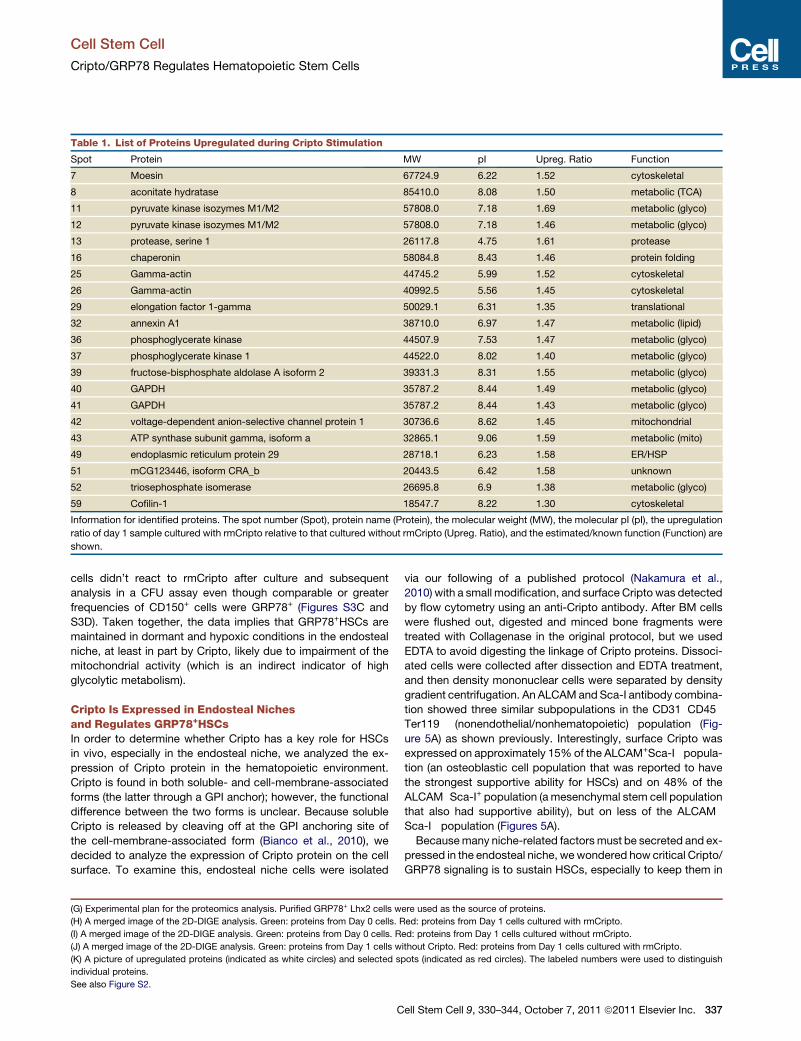
Table 1. List of Proteins Upregulated during Cripto Stimulation
Spot Protein MW pI Upreg. Ratio Function
7 Moesin 67724.9 6.22 1.52 cytoskeletal
8 aconitate hydratase 85410.0 8.08 1.50 metabolic (TCA)
11 pyruvate kinase isozymes M1/M2 57808.0 7.18 1.69 metabolic (glyco)
12 pyruvate kinase isozymes M1/M2 57808.0 7.18 1.46 metabolic (glyco)
13 protease, serine 1 26117.8 4.75 1.61 protease
16 chaperonin 58084.8 8.43 1.46 protein folding
25 Gamma-actin 44745.2 5.99 1.52 cytoskeletal
26 Gamma-actin 40992.5 5.56 1.45 cytoskeletal
29 elongation factor 1-gamma 50029.1 6.31 1.35 translational
32 annexin A1 38710.0 6.97 1.47 metabolic (lipid)
36 phosphoglycerate kinase 44507.9 7.53 1.47 metabolic (glyco)
37 phosphoglycerate kinase 1 44522.0 8.02 1.40 metabolic (glyco)
39 fructose-bisphosphate aldolase A isoform 2 39331.3 8.31 1.55 metabolic (glyco)
40 GAPDH 35787.2 8.44 1.49 metabolic (glyco)
41 GAPDH 35787.2 8.44 1.43 metabolic (glyco)
42 voltage-dependent anion-selective channel protein 1 30736.6 8.62 1.45 mitochondrial
43 ATP synthase subunit gamma, isoform a 32865.1 9.06 1.59 metabolic (mito)
49 endoplasmic reticulum protein 29 28718.1 6.23 1.58 ER/HSP
51 mCG123446, isoform CRA_b 20443.5 6.42 1.58 unknown
52 triosephosphate isomerase 26695.8 6.9 1.38 metabolic (glyco)
59 Cofilin-1 18547.7 8.22 1.30 cytoskeletal
Information for identified proteins. The spot number (Spot), protein name (Protein), the molecular weight (MW), the molecular pI (pI), the upregulation
ratio of day 1 sample cultured with rmCripto relative to that cultured without rmCripto (Upreg. Ratio), and the estimated/known function (Function) are
shown.
Cell Stem Cell
Cripto/GRP78 Regulates Hematopoietic Stem Cells
cells didn’t react to rmCripto after culture and subsequent
analysis in a CFU assay even though comparable or greater
frequencies of CD150+ cells were GRP78+ (Figures S3C and
S3D). Taken together, the data implies that GRP78+HSCs are
maintained in dormant and hypoxic conditions in the endosteal
niche, at least in part by Cripto, likely due to impairment of the
mitochondrial activity (which is an indirect indicator of high
glycolytic metabolism).
Cripto Is Expressed in Endosteal Nichesand Regulates GRP78+HSCsIn order to determine whether Cripto has a key role for HSCs
in vivo, especially in the endosteal niche, we analyzed the ex-
pression of Cripto protein in the hematopoietic environment.
Cripto is found in both soluble- and cell-membrane-associated
forms (the latter through a GPI anchor); however, the functional
difference between the two forms is unclear. Because soluble
Cripto is released by cleaving off at the GPI anchoring site of
the cell-membrane-associated form (Bianco et al., 2010), we
decided to analyze the expression of Cripto protein on the cell
surface. To examine this, endosteal niche cells were isolated
(G) Experimental plan for the proteomics analysis. Purified GRP78+ Lhx2 cells w
(H) A merged image of the 2D-DIGE analysis. Green: proteins from Day 0 cells. R
(I) A merged image of the 2D-DIGE analysis. Green: proteins from Day 0 cells. R
(J) A merged image of the 2D-DIGE analysis. Green: proteins from Day 1 cells w
(K) A picture of upregulated proteins (indicated as white circles) and selected s
individual proteins.
See also Figure S2.
C
via our following of a published protocol (Nakamura et al.,
2010) with a small modification, and surface Cripto was detected
by flow cytometry using an anti-Cripto antibody. After BM cells
were flushed out, digested and minced bone fragments were
treated with Collagenase in the original protocol, but we used
EDTA to avoid digesting the linkage of Cripto proteins. Dissoci-
ated cells were collected after dissection and EDTA treatment,
and then density mononuclear cells were separated by density
gradient centrifugation. An ALCAMand Sca-I antibody combina-
tion showed three similar subpopulations in the CD31�CD45�
Ter119� (nonendothelial/nonhematopoietic) population (Fig-
ure 5A) as shown previously. Interestingly, surface Cripto was
expressed on approximately 15%of the ALCAM+Sca-I� popula-
tion (an osteoblastic cell population that was reported to have
the strongest supportive ability for HSCs) and on 48% of the
ALCAM�Sca-I+ population (amesenchymal stem cell population
that also had supportive ability), but on less of the ALCAM�
Sca-I� population (Figures 5A).
Becausemany niche-related factors must be secreted and ex-
pressed in the endosteal niche, wewondered how critical Cripto/
GRP78 signaling is to sustain HSCs, especially to keep them in
ere used as the source of proteins.
ed: proteins from Day 1 cells cultured with rmCripto.
ed: proteins from Day 1 cells cultured without rmCripto.
ithout Cripto. Red: proteins from Day 1 cells cultured with rmCripto.
pots (indicated as red circles). The labeled numbers were used to distinguish
ell Stem Cell 9, 330–344, October 7, 2011 ª2011 Elsevier Inc. 337
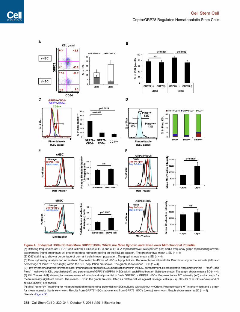
Figure 4. Endosteal HSCs Contain More GRP78+HSCs, Which Are More Hypoxic and Have Lower Mitochondrial Potential
(A) Differing frequencies of GRP78+ and GRP78�HSCs in eHSCs and cHSCs. A representative FACS pattern (left) and a frequency graph representing several
experiments (right) are shown. All presented data represent gating on the KSL population. The graph shows mean ± SD (n = 4).
(B) Ki67 staining to show a percentage of dormant cells in each population. The graph shows mean ± SD (n = 4).
(C) Flow cytometry analysis for intracellular Pimonidazole (Pimo) of HSC subpopulations. Representative intracellular Pimo intensity in the subsets (left) and
percentage of Pimo+++ cells (right) within the KSL population are shown. The graph shows mean ± SD (n = 4).
(D) Flowcytometry analysis for intracellularPimonidazole (Pimo) ofHSCsubpopulationswithin theKSLcompartment. Representative frequency of Pimo+, Pimo2+, and
Pimo+++ cells within KSL population (left) and percentage of GRP78+/GRP78�HSCswithin each Pimo fraction (right) are shown. The graph shows mean ± SD (n = 4).
(E) MitoTracker (MT) staining for measurement of mitochondrial potential in fresh GRP78+ or GRP78�HSCs. Representative MT intensity (left) and a graph for
mean intensity (right) are shown. The means ± SD in the graph are calculated as relative values against Lineage�cells (n = 4). Results of eHSCs (above) and of
cHSCs (below) are shown.
(F) MitoTracker (MT) staining for measurement of mitochondrial potential in HSCs cultured with/without rmCripto. Representative MT intensity (left) and a graph
for mean intensity (right) are shown. Results from GRP78+HSCs (above) and from GRP78�HSCs (below) are shown. Graph shows mean ± SD (n = 4).
See also Figure S3.
Cell Stem Cell
Cripto/GRP78 Regulates Hematopoietic Stem Cells
338 Cell Stem Cell 9, 330–344, October 7, 2011 ª2011 Elsevier Inc.

Figure 5. Endosteal Niche Component Cells Express Cripto and Play a Critical Role for Maintenance of Dormant HSCs
(A) Flow cytometry analysis of cell surface Cripto on bone-associated niche cells. Cells isolated from bone fragments were subdivided into three different
populations based on the expression of ALCAM and Sca-I within the CD31�CD45�Ter119� fraction, and the cell-membrane-associated form of Cripto protein on
each subpopulation was detected using anti-Cripto antibody.
(B) Experimental plan for experiments using N-20 antibody injection in vivo. N-20 or control goat IgG (500 mg/kg) was injected into 8-week-old C57BL/6 female
mice five times in 2 weeks.
(C) Flow cytometry analysis of GRP78+ and GRP78�HSCs in cHSCs from injectedmice. Themeans ± SD percentage of GRP78+CD34� or GRP78�CD34� cells in
the KSL population are shown (n = 4).
(D) Flow cytometry analysis for GRP78+ and GRP78�HSCs in eHSCs of injected mice. The means ± SD percentage of GRP78+CD34� or GRP78�CD34� cells in
the KSL population are shown (n = 4).
See also Figure S4.
Cell Stem Cell
Cripto/GRP78 Regulates Hematopoietic Stem Cells
a dormant state in vivo. To this end, we injected N-20 blocking
antibody into healthy normal mice (Figure 5B) in order to analyze
if HSC frequency was changed. Surprisingly, in central BM,
GRP78�HSCs were significantly increased in N-20-injected
mice compared with the control group (18.8% in N-20 against
9.2% in control, within KSL), while GRP78+HSCs were not
affected (Figure 5C). In contrast, the endosteal HSC fraction
showed reduced frequency of GRP78+HSCs (9.6% in N-20
against 17.1% in control, within KSL), but not of GRP78�HSCs(Figure 5D). Elevated numbers of GRP78�HSCs might be
derived from mobilized GRP78+HSCs from the endosteal niche
because the surface expression of GRP78 was also reported
to be regulated by hypoxia (Hardy and Raiter, 2010). We didn’t
see any critical effect of PB, lineage composition in PB and
BM, or HSC frequency in PB and spleen (Figures S4A–S4D) on
blood cell count. These data suggest that Cripto/GRP78 sig-
naling is critical to hold GRP78+HSCs in the endosteal niche,
and inhibition of this pathway allows GRP78+HSCs to mobilize
into the central BM area, probably together with phenotypic
change to GRP78�HSCs.
C
HIF-1a Controls HSC State under Hypoxia throughCripto/GRP78 SignalingRecent papers have reported that the cell surface expression of
GRP78 is upregulated under hypoxia (Østergaard et al., 2009;
Hardy and Raiter, 2010). Furthermore, Cripto is also known to
be upregulated under hypoxia, due to the binding of HIF-1
complex to the promoter region of Cripto (Bianco et al., 2009,
2010). Therefore, to determine whether Cripto signaling is regu-
lated by HIF-1a under hypoxia in hematopoietic cells as well,
HIF-1a conditional knockout (cKO) mice were used to analyze
the effect of HIF-1a deletion on HSC maintenance by Cripto.
Flow cytometry analyses demonstrated normal frequency of
CD34�KSL cells and GRP78+HSCs in central BM of cKO mice
compared with pIpC-induced Mx1-Cre control mice (Figures
6A and 6C). In contrast, the endosteal HSC analyses demon-
strated decreased numbers of both GRP78+HSCs and total
CD34�KSL cells (Figures 6B and 6C). A previous paper reported
the exhaustion of functional HSCs in HIF-1a deleted mice even
though the frequency of immunophenotypic HSCs was normal
(Takubo et al., 2010). Our data could explain a possible
ell Stem Cell 9, 330–344, October 7, 2011 ª2011 Elsevier Inc. 339

Figure 6. HIF-1a Knockout Mice Contain Reduced Frequency of GRP78+HSCs and Impaired Expression of Cripto
(A) Comparable frequency of the central GRP78+HSCs in HIF-1a cKO mice and controls. Representative FACS pattern of HIF-1a+/+ (left) and HIF-1aD/D (right)
mice are shown. All data represent gating on the KSL population.
(B) Reduced frequency of the endosteal GRP78+HSCs in HIF-1a cKOmice. Representative FACS pattern of HIF-1a+/+ (left) and HIF-1aD/D (right) mice are shown.
All data represent gating on the KSL population.
(C) Reduced frequency of GRP78+HSCs in HIF-1a cKO mice. The graph shows mean ± SD (n = 3).
(D) Decreased expression of Cripto mRNA in KSL population of HIF-1a cKO mice. The graph shows mean ± SD (n = 3).
(E)Decreasedfrequencyof theendosteal nichepopulationsofHIF-1acKOmice.RepresentativeFACSpatternsofHIF-1a+/+ (left) andHIF-1aD/D (right)miceareshown.
Cell Stem Cell
Cripto/GRP78 Regulates Hematopoietic Stem Cells
340 Cell Stem Cell 9, 330–344, October 7, 2011 ª2011 Elsevier Inc.

Cell Stem Cell
Cripto/GRP78 Regulates Hematopoietic Stem Cells
mechanism for this. Because Bianco et al. (2010) report the
binding of HIF-1a to a promoter region of Cripto, thereby regu-
lating Cripto mRNA levels, the expression level of Cripto mRNA
was analyzed in the deleted mice. Analysis of the KSL cells
showed that HIF-1a deleted mice had a trend toward lower
expression levels of Cripto mRNA compared with those in
Mx1-Cre control mice, although the difference was not signifi-
cant (Figure 6D). Expression of cell surface Cripto protein on
the endosteal niche component cells was compared between
HIF-1a deleted and control mice. Surprisingly, ALCAM+/Sca-I�
and ALCAM�/Sca-I+ cells showed clearly decreased frequency
in HIF-1a deleted mice (Figures 6E and 6G). Moreover, the level
of cell surface Cripto was also decreased in ALCAM�/Sca-I+
populations (Figures 6F and 6G). These findings strongly suggest
that Cripto/GRP78 signaling in HSCs is controlled by HIF-1a
(Figure 6H).
DISCUSSION
Currently, the concept of the hematopoietic stem cell niche has
been broadly accepted. The key components, including osteo-
blasts, fibroblasts, adipocytes, endothelial cells, vascular cells,
and mesenchymal stem cells, are thought to secrete several
types of cytokines and chemokines, e.g., SCF, TPO, SDF-1,
Shh, and Angiopoietins, along with Wnt family, BMP family,
and TGF-b family members (Kiel and Morrison, 2008; Morrison
and Spradling, 2008; Li and Clevers 2010). These factors may
orchestrate each other and regulate HSC dormancy and self-
renewal as niche-related factors, but hypoxia is also critical for
normal regulation of HSCs in the niche (Parmar et al., 2007; Sim-
sek et al., 2010; Takubo et al., 2010). The hypoxia causes HSCs
to change the metabolism to a more glycolysis-biased energy
flow, and this is mainly regulated by HIF-1a. The HIF-1 complex
has a critical role as a regulator for HSCs under hypoxia by itself,
but it also controls the expression of several genes, both in HSCs
and in niche component cells.
In this study, we identified an important niche-related factor,
Cripto, and we have shown that it has an essential role for main-
tenance of dormant HSCs. The ex vivo culture experiments
demonstrated that Cripto maintained functional HSCs for
2 weeks in culture in contrast to the cultured control cells that
had no ability to reconstitute recipients (Figure 1D). The signal
of Cripto protein was entered through the cell surface receptor
GRP78, which was expressed on a subpopulation of HSCs.
This subpopulation, GRP78+HSCs, had a higher proportion of
Ki67� cells and exhibited slow reconstitution and slow differen-
tiation into mature cells in PB (Figure 2D and Figure 4B).
Separation of eHSCs and cHSCs (Grassinger et al., 2010) re-
vealed biased location of GRP78+HSCs within the endosteal
(F) Decreased expression of cell surface Cripto on the niche cells from HIF-1a cK
mice are shown.
(G) Decreased frequency of the endosteal niche cells from HIF-1a cKO mice. A
(below left), and ALCAM�/Sca-Ihigh (below right) are shown. The expression o
mean ± SD (n = 3).
(H) Hypothetical model of Cripto/GRP78 signaling as an intermediary of HIF-1a
protein bind to cell surface GRP78, which leads to glycolysis-biased metabolism
regulated by several stem-cell-related pathways, especially HIF-1 complex, which
niche component cells have the same regulation for Cripto by HIF-1a.
C
area, which is more hypoxic (Figure 4A). Certainly GRP78+HSCs
contained a significantly greater ratio of intracellular Pimo+++
cells, which are hypoxic cells, although we couldn’t detect larger
differences in eHSCs compared with cHSCs probably due to the
lower rate of accession of Pimo into the endosteum (data not
shown). The hypoxia induces glycolysis-biased metabolism in
HSCs and this was indirectly confirmed by the measurement of
mitochondrial potential, which was lower in the endosteal
GRP78+HSCs. Most importantly, even under the normoxic
ex vivo culture condition, in which the HIF-1 complex must be
dissociated, Cripto allowed theGRP78+HSCs tomaintain a lower
mitochondrial potential (Figure 4F). Although the HIF-1 complex
is known to enhance glycolysis in cells, this result proposed a
crucial role for Cripto as an intermediary of, and an executor
for, enhancement of glycolysis activity through HIF-1 signaling
(Figure 6H). Another niche environment assay using the endos-
teal bone-associated cell isolation method (Nakamura et al.,
2010) revealed the differential expression of Cripto protein on
the cell membrane of niche component cells (Figure 5A).
Because the Cripto promoter region is known to have hypoxia
binding elements (HRE) onto which the stabilized HIF-1 complex
binds, it would be expected that the endosteal niche cells (which
alsomust be hypoxic) express Cripto protein, but the reasonwhy
the ALCAM�Sca-I� subset didn’t express Cripto is unclear
(Figure 5A).
The Cripto promoter region is known to have TCF/LEF binding
elements (TBE) and Smad binding elements (SBE) (Bianco et al.,
2010). Both Wnt and Smad pathways have been considered as
critical regulators for HSCs (Karlsson et al., 2007; Malhotra and
Kincade, 2009). Furthermore, Oct-4 and Nanog have been re-
ported to bind to the promoter region and all of them are thought
to positively regulate Cripto expression (Bianco et al., 2010).
Thus, many types of stemness-related signaling pathways
control Cripto expression. Therefore, greater expression levels
of Cripto in HSCs (Figure 1A) can possibly be the result of
various stem cell maintenance signals. Interestingly, because
Wnt and Smad pathways are stimulated by Cripto protein
(Bianco et al., 2010), cells might have a positive feedback
machinery for Cripto regulation. In the transplantation assay,
our data suggested an irreversible phenotype of GRP78+HSCs
after culture with Cripto; these cells and their progeny showed
reduced ability to differentiate (Figures 2F and 2G). These fixed
cells indicated greater chimerism only in the HSC fraction. The
positive feedback regulation of Cripto signaling might be a
possible mechanism of this irreversibility since long-term culture
in Cripto disturbs the feedback regulation and may lead to less
activated HSCs.
To prove the implication of Cripto/GRP78 signaling and HSCs
in vivo, we demonstrated the exhaustion of HSCs from endosteal
O mice. Representative FACS patterns of HIF-1a+/+ (black) and HIF-1aD/D (red)
LCAM+/Sca-I� (above left), ALCAM�/Sca-I� (above right), ALCAM�/Sca-Ilow
f Cripto is decreased in niche cells from HIF-1aD/D mice. The graph shows
during hypoxia. Secreted form and cell-membrane-anchoring form of Cripto
and less actin polymerization for HSCs. The expression of Cripto is positively
binds to the HRE site on the Cripto promoter region under hypoxia. Endosteal
ell Stem Cell 9, 330–344, October 7, 2011 ª2011 Elsevier Inc. 341
Cell Stem Cell
Cripto/GRP78 Regulates Hematopoietic Stem Cells
region to central BM by injection of GRP78-blocking antibody
(Figures 5C and 5D). It is noteworthy that the inhibition of Cripto
signaling didn’t affect the HSC frequency in PB and spleen,
unlike HSC mobilization using G-CSF (Figures S4C and S4E).
Furthermore, HIF-1a cKO analysis revealed a reduced number
of GRP78+HSCs (Figures 6A and 6B). A previous report showed
the exhaustion of LT-HSCs (Takubo et al., 2010), and this may
possibly be explained by the failure of GRP78+HSCmaintenance
caused by impaired Cripto expression. As previously described,
the Cripto promoter region has binding sites for other stem-cell-
related molecules. These may sustain the remaining Cripto ex-
pression. Because PI3K/Akt signaling, which was demonstrated
to be upregulated by Cripto/GRP78 signaling, is known to be
required for heat shock proteins to protect HIF-1a from VHL-
independent degradation (Figure 3F and Zhou et al., 2004), the
PI3K/Akt pathway may also directly effect the stabilization of
the HIF-1 complex, which leads to glycolytic metabolism. Pre-
sumably these results mean that Cripto/GRP78 signaling has
a special role as a gatekeeper for HSCs under hypoxia (through
regulation by the HIF-1 complex) (Figure 6H). Although the roles
of Cripto and GRP78 have been understood during development
and tumorigenesis, our new findings demonstrate an additional
role for Cripto/GRP78 signaling in the regulation of HSC within
the hypoxic niche.
EXPERIMENTAL PROCEDURES
Mice and Cell Lines
All C57BL/6NTac (Ly-5.2) and B6/SJL (Ly-5.1) mice used for this study were
purchased from Taconic. HIF-1aflox/flox mice (B6.129-Hif1atm3Rsjo/J) were
purchased from The Jackson Laboratory and mated to interferon-inducible
Mx1-Cre mice. Generation of HIF-1aD/D mice was induced by intraperitoneal
injection of 400 mg of pIpC (SIGMA) on 3 alternative days. As a control, Mx1-
Cre mice were injected with the same amount/schedule of pIpC. Four weeks
after the last injection of pIpC, mice were checked regarding gene deletion
and used for the experiments.
The Lhx2 cell line was maintained in IMDM (HyClone, Thermo Scientific)
supplemented with 5% of FCS, 1%Penicillin/Streptomycin, 1% b-mercaptoe-
thanol, 100 ng/ml recombinant mouse SCF (Peprotech), and 10 ng/ml re-
combinant human IL-6 (Peprotech).
Purification of HSCs
HSCs were purified from BM of 6- to 8-week-old mice. Low-density mononu-
clear cells were separated by density gradient centrifugation on Lymphoprep
solution (Axis-Shield) and then c-kit+ cells were enriched by using magnetic
separation system (MACS�) with anti-c-kit magnetic beads (Miltenyi Biotec).
The enriched cells were stained with anti-GRP78 (ET-21, SIGMA), -CD34,
-c-kit, -Sca-1, -Lineage cocktail, 7AAD, and secondary antibodies (goat
anti-rabbit IgG FITC and Streptavidin-APC). HSCpopulationswere then sorted
on FACS Aria. Information for antibodies used can be found in the Supple-
mental Experimental Procedures.
Quantitative Real-Time RT-PCR
Five to twenty thousand CD34�Flt3�KSL (LT-HSC), CD34+Flt3�KSL(ST-HSC), CD34+Flt3+KSL (LMPP), Lineage marker� (Lin�), and Lineage
marker+ (Lin+) cells were directly sorted into the Buffer RLT contained in
RNeasy�Micro Kit (QIAGEN), and total RNAwas extracted by the kit following
the manufacturer’s protocols. Extracted RNA was reverse transcribed by
SuperScript� III (Invitrogen).
Primers for mouse Cripto (Mm03024051_g1) and mouse hypoxanthine
phosphoribosyl transferase (HPRT, Mm00446968_m1) were obtained from
Applied Biosystems. Real-time PCR reactions were performed on 7900 HT
Fast Real-Time PCR System (Applied Biosystems), and the findings were
normalized using HPRT amplification.
342 Cell Stem Cell 9, 330–344, October 7, 2011 ª2011 Elsevier Inc.
Recombinant Protein and Blocking Antibody
Recombinant mouse Cripto was obtained from R&D Systems�. Anti-GRP78
blocking antibody (N-20) was obtained from Santa Cruz Biotechnology�.
In Vitro Culture System and CFU Assay
Sorted cells were cultured in the serum-free medium (StemSpan�SFEM,
StemCell Technologies) supplemented with 100 ng/ml of SCF and 100 ng/ml
of TPO (Peprotech), and either with or without recombinant Cripto for
14 days. Thirty freshly isolated cells or a part (one-third for day 7, one-tenth
for day14) of cells from the total culture were plated in MethoCult� M3434
methylcellulose medium (StemCell Technologies).
Transplantation Assay
Competitive repopulation assay was performed by using the Ly-5 congenic
mouse system. Twenty freshly isolated cells or all the cultured cells from
Ly-5.1 mice were mixed with 200,000 total BM competitor cells from Ly-5.2
mice, and then transplanted into Ly-5.2 mice irradiated with 900 centi-Gray
(cGy). Every 4 weeks after transplantation, PB from tail vein of recipient mice
was collected and stained with anti-CD45.1 (Ly-5.1), -CD45.2 (Ly-5.2), -CD4,
-CD8, -Gr-1, -CD11b, and -B220 antibodies after red blood cell lysis using
ammonium chloride. Over 16 weeks after transplantation, all recipient mice
were sacrificed and BM cells were harvested.
eHSC/cHSC Isolation and Endosteal Niche Cell Preparation
Endosteal HSCs and Central HSCs were isolated after we followed the
protocol used in the original paper (Grassinger et al., 2010). Endosteal niche
cells were isolated as described earlier (Nakamura et al., 2010) with a small
modification.
Hypoxic HSC Assay
Hypoxic cells were labeled by using a dose of 60 mg/kg of Hypoxicprobe-1
(solid Pimo HCl) intravenously injected and detected by staining with anti-
Pimo antibody (FITC-MAb1) (Hypoxyprobe, Inc.), as described earlier (Takubo
et al., 2010).
Intracellular Staining for Phosphorylated Proteins
c-kit+ cells isolated from BM cells were cultured in SFEM supplemented
with 100 ng/ml of mSCF and hTPO, with or without 500 ng/ml rmCripto, or
freshly stainedwith anti-GRP78, -CD34, c-kit, -Sca-I, and -Lineage antibodies.
Cultured cells were stained similarly at several time points (15 min, 90 min,
12 hr, or 90 min). Then cells were fixed in BD Cytofix/Cytoperm� Fixation
and Permeabilization Solution for 20 min. Fixed cells were stained with Alexa
Fluor� 488 conjugated antibodies (Supplemental Experimental Procedures).
2D-DIGE Proteomics Assay
GRP78+ Lhx2 cells were isolated using the MACS� system with anti-GRP78
antibody and anti-rabbit IgGmagnetic beads (Miltenyi Biotec). Separated cells
were cultured for 24 hr with or without 500 ng/ml of rmCripto, or suspended in
cell lysis buffer. Cultured cells were also suspended in the same buffer. Lysed
proteins were precipitated and floated onto ethanol. 2D-DIGE was performed,
obtained 2D-DIGE images were merged, and the intensity of each protein
spot was evaluated using DeCyder software. Protein ID of selected spots
specifically upregulated in the sample after 24 hrs’ culture with Cripto were
analyzed by mass spectrometry (MALDI-TOF) and aligned to an amino acid
database for identification. All proteomics analyses were done by Applied
Biomics Inc.
Statistical Analysis
Statistical significance was determined using the Turkey-Kramer method for
comparison of multiple groups or the two-tailed Student’s t test for compar-
ison of two groups. ANOVA andmultiple comparison analyseswere performed
on the SPSS (IBM).
SUPPLEMENTAL INFORMATION
Supplemental Information for this article includes four figures and Supple-
mental Experimental Procedures and can be found with this article online at
doi:10.1016/j.stem.2011.07.016.
Cell Stem Cell
Cripto/GRP78 Regulates Hematopoietic Stem Cells
ACKNOWLEDGMENTS
We thank Leif Carlsson for providing Lhx2 cells; Susie Nilsson for sending
a detailed protocol for analyzing eHSCs/cHSCs; Tariq Enver and Jonas Lars-
son for critical discussions; and Zhi Ma for excellent advice on flow cytometry
analyses. This work was supported by the Hemato-Linne grant (Swedish
Research Council Linnaeus), the Swedish Cancer Foundation (Cancerfonden),
The Swedish Cancer Society (S.K.), the Swedish Children’s Cancer Society
(S.K.), the Swedish Medical Research Council (S.K.), The Tobias Prize
awarded by The Royal Swedish Academy of Sciences financed by The Tobias
Foundation (S.K.), and the EU project grants CONSERT, STEMEXPAND, and
PERSIST. K.M. was funded by the EU-funded NOVEXPAND project in Marie
Curie actions. The Lund StemCell Center was supported by a Center of Excel-
lence grant in life sciences from the Swedish Foundation for Strategic
Research.
Received: April 28, 2011
Revised: July 14, 2011
Accepted: July 29, 2011
Published: October 6, 2011
REFERENCES
Arai, F., Hirao, A., Ohmura, M., Sato, H., Matsuoka, S., Takubo, K., Ito, K., Koh,
G.Y., and Suda, T. (2004). Tie2/angiopoietin-1 signaling regulates hematopoi-
etic stem cell quiescence in the bone marrow niche. Cell 118, 149–161.
Arai, F., and Suda, T. (2007). Maintenance of quiescent hematopoietic stem
cells in the osteoblastic niche. Ann. N Y Acad. Sci. 1106, 41–53.
Bianco, C., Cotten, C., Lonardo, E., Strizzi, L., Baraty, C., Mancino, M.,
Gonzales, M., Watanabe, K., Nagaoka, T., Berry, C., et al. (2009). Cripto-1 is
required for hypoxia to induce cardiac differentiation of mouse embryonic
stem cells. Am. J. Pathol. 175, 2146–2158.
Bianco, C., Rangel, M.C., Castro, N.P., Nagaoka, T., Rollman, K., Gonzales,
M., and Salomon, D.S. (2010). Role of Cripto-1 in stem cell maintenance and
malignant progression. Am. J. Pathol. 177, 532–540.
Challen, G.A., Boles, N.C., Chambers, S.M., and Goodell, M.A. (2010). Distinct
hematopoietic stem cell subtypes are differentially regulated by TGF-b1. Cell
Stem Cell 6, 265–278.
Ciccodicola, A., Dono, R., Obici, S., Simeone, A., Zollo, M., and Persico, M.G.
(1989). Molecular characterization of a gene of the ‘EGF family’ expressed in
undifferentiated human NTERA2 teratocarcinoma cells. EMBO J. 8, 1987–
1991.
Danet, G.H., Pan, Y., Luongo, J.L., Bonnet, D.A., and Simon, M.C. (2003).
Expansion of human SCID-repopulating cells under hypoxic conditions.
J. Clin. Invest. 112, 126–135.
Davidson, D.J., Haskell, C., Majest, S., Kherzai, A., Egan, D.A., Walter, K.A.,
Schneider, A., Gubbins, E.F., Solomon, L., Chen, Z., et al. (2005). Kringle 5
of human plasminogen induces apoptosis of endothelial and tumor cells
through surface-expressed glucose-regulated protein 78. Cancer Res. 65,
4663–4672.
Ding, J., Yang, L., Yan, Y.T., Chen, A., Desai, N., Wynshaw-Boris, A., and
Shen, M.M. (1998). Cripto is required for correct orientation of the anterior-
posterior axis in the mouse embryo. Nature 395, 702–707.
Dykstra, B., Kent, D., Bowie, M., McCaffrey, L., Hamilton, M., Lyons, K., Lee,
S.J., Brinkman, R., and Eaves, C. (2007). Long-term propagation of distinct
hematopoietic differentiation programs in vivo. Cell Stem Cell 1, 218–229.
Gonzalez-Gronow, M., Selim, M.A., Papalas, J., and Pizzo, S.V. (2009).
GRP78: a multifunctional receptor on the cell surface. Antioxid. Redox
Signal. 11, 2299–2306.
Goodell, M.A., Brose, K., Paradis, G., Conner, A.S., and Mulligan, R.C. (1996).
Isolation and functional properties of murine hematopoietic stem cells that are
replicating in vivo. J. Exp. Med. 183, 1797–1806.
Grassinger, J., Haylock, D.N., Williams, B., Olsen, G.H., and Nilsson, S.K.
(2010). Phenotypically identical hemopoietic stem cells isolated from different
C
regions of bone marrow have different biologic potential. Blood 116, 3185–
3196.
Gray, P.C., Shani, G., Aung, K., Kelber, J., and Vale, W. (2006). Cripto binds
transforming growth factor b (TGF-b) and inhibits TGF-b signaling. Mol. Cell.
Biol. 26, 9268–9278.
Hardy, B., and Raiter, A. (2010). Peptide-binding heat shock protein GRP78
protects cardiomyocytes from hypoxia-induced apoptosis. J. Mol. Med. 88,
1157–1167.
Haug, J.S., He, X.C., Grindley, J.C., Wunderlich, J.P., Gaudenz, K., Ross, J.T.,
Paulson, A., Wagner, K.P., Xie, Y., Zhu, R., et al. (2008). N-cadherin expression
level distinguishes reserved versus primed states of hematopoietic stem cells.
Cell Stem Cell 2, 367–379.
Hough, S.R., Laslett, A.L., Grimmond, S.B., Kolle, G., and Pera, M.F. (2009). A
continuum of cell states spans pluripotency and lineage commitment in human
embryonic stem cells. PLoS ONE 4, e7708.
Huang, Y., and Burkhardt, J.K. (2007). T-cell-receptor-dependent actin regu-
latory mechanisms. J. Cell Sci. 120, 723–730.
Kahn, J., Byk, T., Jansson-Sjostrand, L., Petit, I., Shivtiel, S., Nagler, A.,
Hardan, I., Deutsch, V., Gazit, Z., Gazit, D., et al. (2004). Overexpression of
CXCR4 on human CD34+ progenitors increases their proliferation, migration,
and NOD/SCID repopulation. Blood 103, 2942–2949.
Karlsson, G., Blank, U., Moody, J.L., Ehinger, M., Singbrant, S., Deng, C.X.,
and Karlsson, S. (2007). Smad4 is critical for self-renewal of hematopoietic
stem cells. J. Exp. Med. 204, 467–474.
Keith, B., and Simon, M.C. (2007). Hypoxia-inducible factors, stem cells, and
cancer. Cell 129, 465–472.
Kelber, J.A., Panopoulos, A.D., Shani, G., Booker, E.C., Belmonte, J.C., Vale,
W.W., and Gray, P.C. (2009). Blockade of Cripto binding to cell surface GRP78
inhibits oncogenic Cripto signaling via MAPK/PI3K and Smad2/3 pathways.
Oncogene 28, 2324–2336.
Kent, D.G., Copley, M.R., Benz, C., Wohrer, S., Dykstra, B.J., Ma, E., Cheyne,
J., Zhao, Y., Bowie, M.B., Zhao, Y., et al. (2009). Prospective isolation and
molecular characterization of hematopoietic stem cells with durable self-
renewal potential. Blood 113, 6342–6350.
Kiel, M.J., and Morrison, S.J. (2008). Uncertainty in the niches that maintain
haematopoietic stem cells. Nat. Rev. Immunol. 8, 290–301.
Kiel, M.J., Yilmaz, O.H., Iwashita, T., Yilmaz, O.H., Terhorst, C., and Morrison,
S.J. (2005). SLAM family receptors distinguish hematopoietic stem and
progenitor cells and reveal endothelial niches for stem cells. Cell 121, 1109–
1121.
Kubota, Y., Takubo, K., and Suda, T. (2008). Bone marrow long label-retaining
cells reside in the sinusoidal hypoxic niche. Biochem. Biophys. Res. Commun.
366, 335–339.
Li, L., and Clevers, H. (2010). Coexistence of quiescent and active adult stem
cells in mammals. Science 327, 542–545.
Li, C.L., and Johnson, G.R. (1995). Murine hematopoietic stem and progenitor
cells: I. Enrichment and biologic characterization. Blood 85, 1472–1479.
Malhotra, S., and Kincade, P.W. (2009). Wnt-related molecules and signaling
pathway equilibrium in hematopoiesis. Cell Stem Cell 4, 27–36.
Morita, Y., Ema, H., and Nakauchi, H. (2010). Heterogeneity and hierarchy
within the most primitive hematopoietic stem cell compartment. J. Exp.
Med. 207, 1173–1182.
Morrison, S.J., and Spradling, A.C. (2008). Stem cells and niches: mechanisms
that promote stem cell maintenance throughout life. Cell 132, 598–611.
Nakamura, Y., Arai, F., Iwasaki, H., Hosokawa, K., Kobayashi, I., Gomei, Y.,
Matsumoto, Y., Yoshihara, H., and Suda, T. (2010). Isolation and characteriza-
tion of endosteal niche cell populations that regulate hematopoietic stem cells.
Blood 116, 1422–1432.
Ogaeri, T., Eto, K., Otsu, M., Ema, H., and Nakauchi, H. (2009). The actin poly-
merization regulator WAVE2 is required for early bone marrow repopulation by
hematopoietic stem cells. Stem Cells 27, 1120–1129.
ell Stem Cell 9, 330–344, October 7, 2011 ª2011 Elsevier Inc. 343

Cell Stem Cell
Cripto/GRP78 Regulates Hematopoietic Stem Cells
Osawa, M., Hanada, K., Hamada, H., and Nakauchi, H. (1996). Long-term
lymphohematopoietic reconstitution by a single CD34-low/negative hemato-
poietic stem cell. Science 273, 242–245.
Østergaard, L., Simonsen, U., Eskildsen-Helmond, Y., Vorum, H., Uldbjerg, N.,
Honore, B., and Mulvany, M.J. (2009). Proteomics reveals lowering oxygen
alters cytoskeletal and endoplasmatic stress proteins in human endothelial
cells. Proteomics 9, 4457–4467.
Parmar, K., Mauch, P., Vergilio, J.A., Sackstein, R., and Down, J.D. (2007).
Distribution of hematopoietic stem cells in the bone marrow according to
regional hypoxia. Proc. Natl. Acad. Sci. USA 104, 5431–5436.
Pinto do O, P., Richter, K., and Carlsson, L. (2002). Hematopoietic progenitor/
stem cells immortalized by Lhx2 generate functional hematopoietic cells
in vivo. Blood 99, 3939–3946.
Saloman, D.S., Bianco, C., Ebert, A.D., Khan, N.I., De Santis, M., Normanno,
N., Wechselberger, C., Seno, M., Williams, K., Sanicola, M., et al. (2000).
The EGF-CFC family: novel epidermal growth factor-related proteins in devel-
opment and cancer. Endocr. Relat. Cancer 7, 199–226.
Sato, N., Sanjuan, I.M., Heke, M., Uchida, M., Naef, F., and Brivanlou, A.H.
(2003). Molecular signature of human embryonic stem cells and its comparison
with the mouse. Dev. Biol. 260, 404–413.
Scadden, D.T. (2006). The stem-cell niche as an entity of action. Nature 441,
1075–1079.
Schroeder, T. (2010). Hematopoietic stem cell heterogeneity: subtypes, not
unpredictable behavior. Cell Stem Cell 6, 203–207.
Shani, G., Fischer, W.H., Justice, N.J., Kelber, J.A., Vale, W., and Gray, P.C.
(2008). GRP78 and Cripto form a complex at the cell surface and collaborate
344 Cell Stem Cell 9, 330–344, October 7, 2011 ª2011 Elsevier Inc.
to inhibit transforming growth factor b signaling and enhance cell growth.
Mol. Cell. Biol. 28, 666–677.
Simsek, T., Kocabas, F., Zheng, J., Deberardinis, R.J., Mahmoud, A.I., Olson,
E.N., Schneider, J.W., Zhang, C.C., and Sadek, H.A. (2010). The distinct
metabolic profile of hematopoietic stem cells reflects their location in a hypoxic
niche. Cell Stem Cell 7, 380–390.
Strizzi, L., Bianco, C., Normanno, N., and Salomon, D. (2005). Cripto-1: amulti-
functional modulator during embryogenesis and oncogenesis. Oncogene 24,
5731–5741.
Takubo, K., Goda, N., Yamada, W., Iriuchishima, H., Ikeda, E., Kubota, Y.,
Shima, H., Johnson, R.S., Hirao, A., Suematsu, M., and Suda, T. (2010).
Regulation of the HIF-1a level is essential for hematopoietic stem cells. Cell
Stem Cell 7, 391–402.
Wilson, A., Laurenti, E., Oser, G., van der Wath, R.C., Blanco-Bose, W.,
Jaworski, M., Offner, S., Dunant, C.F., Eshkind, L., Bockamp, E., et al.
(2008). Hematopoietic stem cells reversibly switch from dormancy to self-
renewal during homeostasis and repair. Cell 135, 1118–1129.
Xu, C., Liguori, G., Adamson, E.D., and Persico, M.G. (1998). Specific arrest of
cardiogenesis in cultured embryonic stem cells lacking Cripto-1. Dev. Biol.
196, 237–247.
Yamazaki, S., Iwama, A., Takayanagi, S., Eto, K., Ema, H., and Nakauchi, H.
(2009). TGF-b as a candidate bone marrow niche signal to induce hematopoi-
etic stem cell hibernation. Blood 113, 1250–1256.
Zhou, J., Schmid, T., Frank, R., and Brune, B. (2004). PI3K/Akt is required
for heat shock proteins to protect hypoxia-inducible factor 1a frompVHL-inde-
pendent degradation. J. Biol. Chem. 279, 13506–13513.
Copyright © 2022 FDOKUMEN