Effect of Cobalt(II) Oxide and Manganese(IV) Oxide on Sintering of Tin(IV) Oxide

271
Hugo Cerecettoa, 1,2,4-Triazine N-oxide Derivatives: Studies asMercedes Gonzaleza,
Potential Hypoxic Cytotoxins. Part IIIMariela Rissoa,Patricia Saenza,Claudio Olea-Azarb, New 5-(2-arylethenyl)-1,2,4-triazine N-oxide and N,N�-dioxide derivatives wereAna M. Brunoc, synthesized in order to obtain compounds as selective hypoxic cell cytotoxins.Amaia Azquetad, The desired products were obtained when the 5-methyl heterocycle reacted withAdela Lopez de Ceraind, the corresponding iminium electrophiles. The new compounds were tested forAntonio Monged their cytotoxicity in oxia and hypoxia. Some of them proved to be less active in
hypoxic conditions than Tirapazamine, 3-aminobenzo[1,2-e]1,2,4-triazinea Departamento de Quımica N1,N4-dioxide. Derivative 11, 6-methyl-5-[2-(5-nitrofuryl)ethenyl)-1,2,4-triazine N4-
Organica, Facultad de oxide, was the most cytotoxic compound, but it was non-selective. Some deriva-Quımica � Facultad de tives were studied as DNA-binding agents in oxic conditions showing poorCiencias, Universidad de la affinity for this biomolecule. This result showed that the cytotoxic activity in oxiaRepublica, is DNA damage not dependent. Electrochemical and ESR spectroscopy studiesMontevideo, Uruguay were performed in order to determine the ability of compounds to produce rad-
b Departamento de Quımica icals and the relation of these in the mechanism of cytotoxicity.Inorganica y Analıtica,
Keywords: 1,2,4-Triazine N-oxide; Bioreductive compounds; Redox properties.Facultad de CienciasQuımicas y Farmaceuticas,Universidad de Chile,Santiago, Chile
c Centro de Sıntesis y Estudiode Nuevos CompuestosAntineoplasicos, Facultad deFarmacia y Bioquımica,Universidad de Buenos Aires,Buenos Aires, Argentina
d Unidad en I+D deMedicamentos, CIFA,Universidad de Navarra,Pamplona, Espana
Introduction
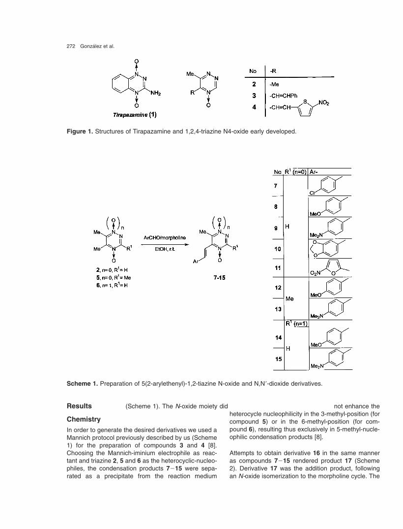
Several N-oxides of aromatic heterocyclic amineshave been described as hypoxia-selective cytotoxicagents (bioreductive drugs) [1-3]. One of the mostinteresting compound is Tirapazamine (3-amino-benzo[1,2-e]1,2,4-triazine N 1,N 4-dioxide, 1, Figure 1).This drug possesses differential hypoxic cytotoxicityfor a great number of cell lines of hamster, mouse andhuman beings, with a hypoxic cytotoxicity ratio (HCR,relationship between concentration of drug in air andconcentration of drug in hypoxia that produce thesame level of cell killing) of approximately 25-200 forthe different cellular lines [4-6]. In order to describe thebehavior of 1,2,4-triazine N-oxide as bioreductive
Correspondence: Mercedes Gonzalez, Departamento deQuımica Organica, Facultad de Quımica � Facultad deCiencias, Universidad de la Republica, Igua 4225, 11400Montevideo, Uruguay. Phone: +598 2 525-86-18 (ext. 216),Fax +598 2 525-07-49; e-mail: [email protected]
agent, we described recently the biological charac-terization of 1,2,4-triazine N 4-oxide and N 1,N 4-dioxidederivatives as hypoxia selective cytotoxins (i.e. deriva-tives 2, 3, and 4, Figure 1) [7, 8]. These derivativesshowed poor cytotoxic selectivity against V79 cells inhypoxic conditions, however, compound 4 (Figure 1)displayed excellent V79 cell-growth inhibition even at5 µM in air and hypoxia, i.e. as potent as Tirapaz-amine. In order to obtain more active compounds andto gain insight into the molecular requirements for bio-reductive activity we included different arylethenyl sub-stituents in the 5-position of the 1,2,4-triazine N 4-oxideand N 1,N 4-dioxide system (Scheme 1).
In this paper, we report the synthesis of some selected5-(2-arylethenyl)-1,2,4-triazine N-oxide derivatives.We analyzed the cytotoxic activity of the new com-pounds in oxia and hypoxia against V79 cells andstudied some of them for their aerobic DNA-affinityproperties. Furthermore, we present the electrochemi-cal and ESR spectroscopic studies that permit to ex-plain the observed activity.
272 Gonzalez et al.
Figure 1. Structures of Tirapazamine and 1,2,4-triazine N4-oxide early developed.
Scheme 1. Preparation of 5(2-arylethenyl)-1,2-tiazine N-oxide and N,N�-dioxide derivatives.
Results
Chemistry
In order to generate the desired derivatives we used aMannich protocol previously described by us (Scheme1) for the preparation of compounds 3 and 4 [8].Choosing the Mannich-iminium electrophile as reac-tant and triazine 2, 5 and 6 as the heterocyclic-nucleo-philes, the condensation products 7�15 were sepa-rated as a precipitate from the reaction medium
(Scheme 1). The N-oxide moiety did not enhance theheterocycle nucleophilicity in the 3-methyl-position (forcompound 5) or in the 6-methyl-position (for com-pound 6), resulting thus exclusively in 5-methyl-nucle-ophilic condensation products [8].
Attempts to obtain derivative 16 in the same manneras compounds 7�15 rendered product 17 (Scheme2). Derivative 17 was the addition product, followingan N-oxide isomerization to the morpholine cycle. The

273
Scheme 2. Generation of compound 17.
product 17, unlike the other addition products, was in-soluble in EtOH, so it precipitated in the reaction me-dium. Attempts to obtain the desired derivative 16, im-proving the solubility of 17, conducted us to changethe reaction solvent to DMF or DMSO. In the first case,17 precipitated not reacting for several days. Althoughproduct 17 was soluble in DMSO, it was impossibleto transform it into compound 16. Compound 17 wassubmitted to the action of different bases under differ-
Table 1. Experimental conditions assayed in the attempts to obtain compound 17.
Base Solvent Temperature Results
r. t. NREt3N Et3N
Reflux NR
r. t. NRPiperidine DMSO
Reflux NR
r. t. NREtONa EtOH
Reflux Decomposition of 17
NR � no reaction, r.t. � room temperature.
ent conditions but with unsuccessful results, it was notpossible to obtain derivative 16 in any case (see as-sayed experimental conditions and results in Table 1).
All new compounds were identified by IR, MS, andone- and two-dimensional 1H NMR, 13C NMR experi-ments, and their purity was established by TLC andmicroanalysis. In all cases, the E-isomer of the olefinwas the unique stereo species identified in solution by1H NMR and 13C NMR experiments from the isolatedcompounds. The olefinic coupling constant, near to16 Hz for all derivatives, was used to assign thestereochemistry.
The unequivocal condensation or addition site (com-pound 17) was determined by means of HMQC forone-bond correlation and from sequences of HMBCfor long distance/carbon correlation. Thus, 2�-CH cou-pled exclusively with the heterocyclic quaternary 5-carbon, which was determined by its correlation withthe 3-H-heterocycle (determined by HMBC for com-pounds 7�11, 14�15, and 17, Figure 2A). In deriva-tives 12�13 the HMBC-correlations and the character-istic chemical shifts of 3-, 5-, and 6-methyl protons andchemical shifts of the heterocyclic 5-carbon in parentcompound 5 allowed us to assign the reaction site(Figure 2B).
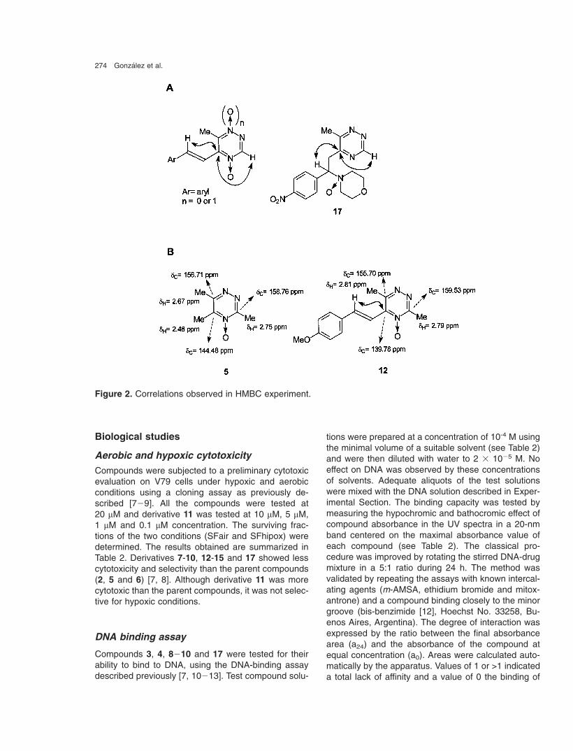
274 Gonzalez et al.
Figure 2. Correlations observed in HMBC experiment.
Biological studies
Aerobic and hypoxic cytotoxicity
Compounds were subjected to a preliminary cytotoxicevaluation on V79 cells under hypoxic and aerobicconditions using a cloning assay as previously de-scribed [7�9]. All the compounds were tested at20 µM and derivative 11 was tested at 10 µM, 5 µM,1 µM and 0.1 µM concentration. The surviving frac-tions of the two conditions (SFair and SFhipox) weredetermined. The results obtained are summarized inTable 2. Derivatives 7-10, 12-15 and 17 showed lesscytotoxicity and selectivity than the parent compounds(2, 5 and 6) [7, 8]. Although derivative 11 was morecytotoxic than the parent compounds, it was not selec-tive for hypoxic conditions.
DNA binding assay
Compounds 3, 4, 8�10 and 17 were tested for theirability to bind to DNA, using the DNA-binding assaydescribed previously [7, 10�13]. Test compound solu-
tions were prepared at a concentration of 10-4 M usingthe minimal volume of a suitable solvent (see Table 2)and were then diluted with water to 2 � 10�5 M. Noeffect on DNA was observed by these concentrationsof solvents. Adequate aliquots of the test solutionswere mixed with the DNA solution described in Exper-imental Section. The binding capacity was tested bymeasuring the hypochromic and bathocromic effect ofcompound absorbance in the UV spectra in a 20-nmband centered on the maximal absorbance value ofeach compound (see Table 2). The classical pro-cedure was improved by rotating the stirred DNA-drugmixture in a 5:1 ratio during 24 h. The method wasvalidated by repeating the assays with known intercal-ating agents (m-AMSA, ethidium bromide and mitox-antrone) and a compound binding closely to the minorgroove (bis-benzimide [12], Hoechst No. 33258, Bu-enos Aires, Argentina). The degree of interaction wasexpressed by the ratio between the final absorbancearea (a24) and the absorbance of the compound atequal concentration (a0). Areas were calculated auto-matically by the apparatus. Values of 1 or >1 indicateda total lack of affinity and a value of 0 the binding of

275
Table 2. Biological characterization and reduction potential of 1,2,4-triazine derivatives.
Compound Biological characterization Epc vs SCE§
(EpcNO2/EpcNOx)(V)Doses (µM) SFair (%)#,† SFhypox a24/a0 (solvent)
(%)†,‡
1 20 100 ± 0 0 ± 0 ND � / �0.90�
2� 20 75 ± 16 74 ± 8 1.25 (EtOH) � / �1.613g 20 100 ± 0 86 ± 11 0.96 (DMF) � / �1.124¿ 5 0 ± 0 0 ± 0
1 69 ± 10 100 ± 0 0.96 (DMF) �0.56 / �1.030.1 92 ± 6 100 ± 0
7 20 100 ± 5 90 ± 5 ND � / �1.158 20 69 ± 10 100 ± 0 0.96 (DMF) � / �1.289 20 100 ± 5 100 ± 5 0.96 (DMF) � / �1.30
10 20 55 ± 11 78 ± 10 0.96 (DMF) � / �1.1811 20 0 ± 0 0 ± 0
10 0 ± 0 0 ± 05 0 ± 0 0 ± 0 ND �0.63 / �0.941 0 ± 0 0 ± 0
0.1 70 ± 10 100 ± 012 � ND ND �13 20 100 ± 5 71 ± 12 ND � / �1.3614 20 100 ± 0 90 ± 5 ND � / �1.1215 20 100 ± 5 80 ± 10 ND � / �1.1617 20 100 ± 0 100 ± 0 0.93 (DMF) �0.98 / �1.27
Reference 5 100 ± 0 0 ± 0 ND �m-AMSA � � � 0.30 (EtOH) �Ethidium bromide � � � 0.50 (EtOH) �Mitoxantrone � � � 0.00 (EtOH) �Bis-benzimide � � � 0.57 (EtOH) �
# Survival fraction in air; † Tests were carried out in duplicate; ‡ Survival fraction in hypoxia; § Peaks potentials(~± 0.01 V) measured at a scan rate of 0.50 V/s; ND�Not determined; � From reference [14]; g From reference[7]; ¿ From reference [8]; Reference: 7-Chloro-3-[3-(N,N-dimethylamino)propylamino]-2-quinoxalinecarbonitrilehydrochloride.
the entire compound to DNA. The values of coefficienta24/a0 are summarized in Table 2.
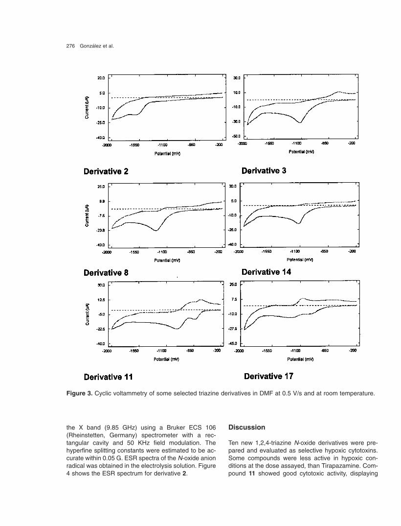
Cyclic voltammetry
In order to determine the electrochemical character-istic of the studied derivatives, and its relationship withtheir cytotoxicity, experiments of cyclic voltammetry[14, 15] were performed in organic medium. These N-oxide derivatives displayed comparable voltammetricbehavior in DMF, showing one reduction peak and theanodic counterpart for the N 4-oxide derivatives andtwo reduction waves for the N 1,N 4-dioxide derivatives.However, in the case of another electroactive moiety
in the molecule (i.e. nitro moiety in derivatives 4, 11 or17), a new wave appeared at a less negative potential.Table 2 lists the values of the nitro cathodic peaks(EpcNO2) and the N-oxide cathodic peaks (EpcNOx)for the compounds biologically evaluated. Figure 3shows some selected voltamograms. The first wavecorresponds to a quasireversible process; this stepwas studied by ESR spectroscopy.
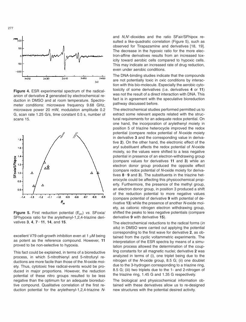
ESR Spectroscopy
The anion radicals produced in the electrochemicalprocess was characterized for compound 2 by ESRspectroscopy [16, 17]. ESR spectra were recorded in
276 Gonzalez et al.
Figure 3. Cyclic voltammetry of some selected triazine derivatives in DMF at 0.5 V/s and at room temperature.
the X band (9.85 GHz) using a Bruker ECS 106(Rheinstetten, Germany) spectrometer with a rec-tangular cavity and 50 KHz field modulation. Thehyperfine splitting constants were estimated to be ac-curate within 0.05 G. ESR spectra of the N-oxide anionradical was obtained in the electrolysis solution. Figure4 shows the ESR spectrum for derivative 2.
Discussion
Ten new 1,2,4-triazine N-oxide derivatives were pre-pared and evaluated as selective hypoxic cytotoxins.Some compounds were less active in hypoxic con-ditions at the dose assayed, than Tirapazamine. Com-pound 11 showed good cytotoxic activity, displaying
277
Figure 4. ESR experimental spectrum of the radical-anion of derivative 2 generated by electrochemical re-duction in DMSO and at room temperature. Spectro-meter conditions: microwave frequency 9.68 GHz,microwave power 20 mW, modulation amplitude 0.2G, scan rate 1.25 G/s, time constant 0.5 s, number ofscans 15.
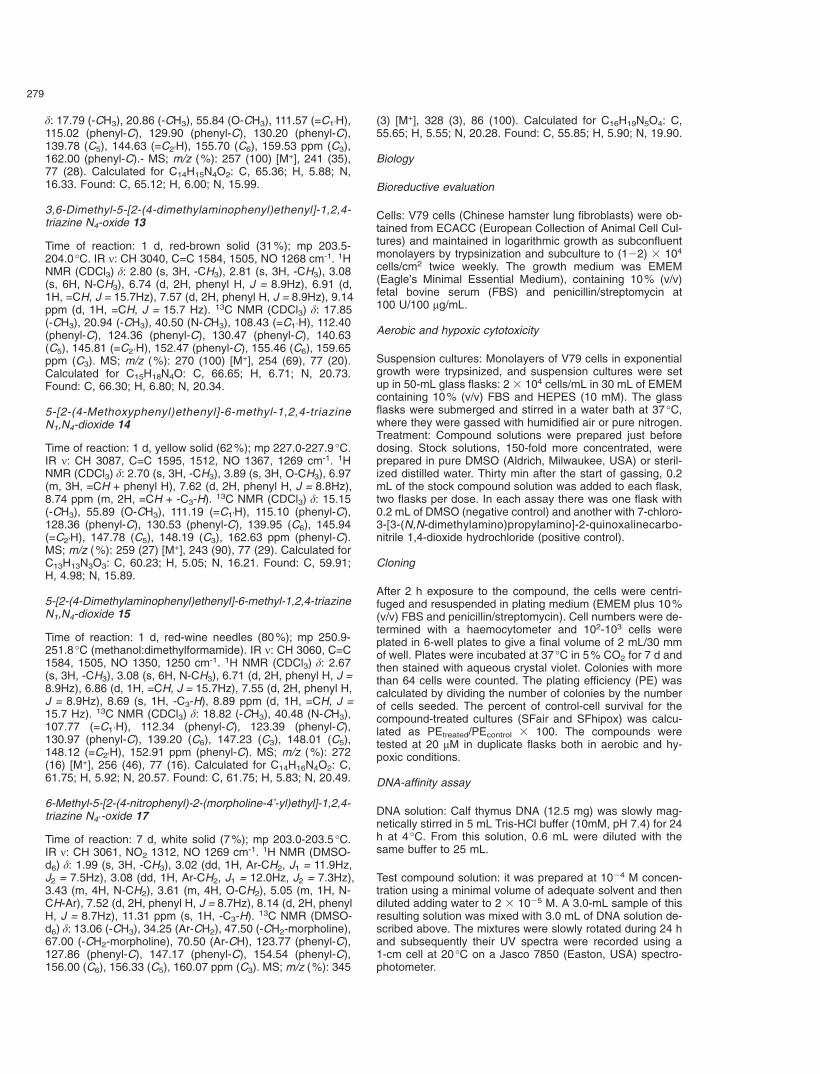
Figure 5. First reduction potential (Epc) vs. SFoxia/SFhypoxia ratio for the arylethenyl-1,2,4-triazine deri-vatives 3, 4, 7�11, 14, and 15.
excellent V79 cell-growth inhibition even at 1 µM beingas potent as the reference compound. However, 11proved to be non-selective to hypoxia.
This fact could be explained, in terms of a bioreductiveprocess, in which 5-nitrothienyl and 5-nitrofuryl re-ductions are more facile than those of the N-oxide moi-ety. Thus, cytotoxic free radical-events would be pro-duced in major proportions. However, the reductionpotential of these nitro groups resulted to be lessnegative than the optimum for an adequate bioreduc-tive compound. Qualitative correlation of the first re-duction potential for the arylethenyl-1,2,4-triazine N-
and N,N�-dioxides and the ratio SFair/SFhipox re-sulted a like-quadratic correlation (Figure 5), such asobserved for Tirapazamine and derivatives [18, 19].The decrease in the hypoxic ratio for the more elec-tron-affine derivatives results from an increased tox-icity toward aerobic cells compared to hypoxic cells.This may indicate an increased rate of drug reduction,even under aerobic conditions.
The DNA-binding studies indicate that the compoundsare not potentially toxic in oxic conditions by interac-tion with this bio-molecule. Especially the aerobic cyto-toxicity of some derivatives (i.e. derivatives 4 or 11)was not the result of a direct interaction with DNA. Thisfact is in agreement with the speculative bioreductionpathway discussed before.
The electrochemical studies performed permitted us toextract some relevant aspects related with the struc-tural requirements for an adequate redox potential. Onone hand, the incorporation of arylethenyl moiety inposition 5 of triazine heterocycle improved the redoxpotential (compare redox potential of N-oxide moietyin derivative 3 and the corresponding value in deriva-tive 2). On the other hand, the electronic effect of thearyl substituent affects the redox potential of N-oxidemoiety, so the values were shifted to a less negativepotential in presence of an electron-withdrawing group(compare values for derivatives 11 and 3) while anelectron donor group produced the opposite effect(compare redox potential of N-oxide moiety for deriva-tives 8�9 and 3). The substituents in the triazine het-erocycle could be affecting this physicochemical prop-erty. Furthermore, the presence of the methyl group,an electron donor group, in position 3 produced a shiftof the reduction potential to more negative values(compare potential of derivative 9 with potential of de-rivative 13) while the presence of another N-oxide moi-ety, as cationic nitrogen electron withdrawing group,shifted the peaks to less negative potentials (comparederivative 9 with derivative 15).
The electrochemical reductions to the radical forms (insitu) in DMSO were carried out applying the potentialcorresponding to the first wave for derivative 2, as ob-tained from the cyclic voltammetric experiments. Theinterpretation of the ESR spectra by means of a simu-lation process allowed the determination of the coup-ling constants for all magnetic nuclei; derivative 2 wasanalyzed in terms of (i), one triplet being due to thenitrogen of the N-oxide group, 8.5 G; (ii) one doubletdue to the 3-hydrogen corresponding to a triazine ring,8.5 G; (iii) two triplets due to the 1- and 2-nitrogen ofthe triazine ring, 1.45 G and 1.35 G respectively.
The biological and physicochemical information ob-tained with these derivatives allow us to re-designednew structures with the potential desired activity.

278 Gonzalez et al.
Acknowledgments
This research has been supported by Comision Hono-raria de Lucha Contra el Cancer (Uruguay), CSIC(Universidad de la Republica, Uruguay), FONDECYT� No 1030949 (Chile) and CYTED. We thank for amaster grant for P.S. from Programa de Desarrollo deCiencias Basicas (PEDECIBA-Uruguay) and a grantfor M.R. from RELACQ.
Experimental
Chemistry
All starting materials were commercially available research-grade chemicals and used without further purification. All sol-vents were dried and distilled prior to use. All the reactionswere carried out in a nitrogen atmosphere. The typical work-up included washing with brine and drying the organic layerwith sodium sulfate before concentration. Melting points weredetermined using a Leitz Microscope Heating Stage Model350 (Wetzlar, Germany) apparatus and are uncorrected. El-emental analyses were obtained from vacuum-dried samples(over phosphorous pentoxide at 3�4 mm Hg, 24 h at roomtemperature), performed on a Fisons EA 1108 CHNS-O ana-lyzer (Valencia, USA), and were within ± 0.4% of theoreticalvalues. Infrared spectra were recorded on a Perkin Elmer1310 (Jügesheim, Germany) apparatus, using potassium bro-mide tablets; the frequencies are expressed in cm�1. 1H-NMR, 13C-NMR spectra and HETCOR experiments were re-corded on a Bruker DPX-400 (at 400 MHz and 100 MHz)instrument, with tetramethylsilane as the internal referenceand in the indicated solvent. Mass spectra were recorded ona Shimadzu GC-MS QP 1100 EX (Kyoto, Japan) instrumentusing electron impact ionization at 70 eV. The compounds2�6 were prepared as reported previously [7, 8].
General procedure for the synthesis of derivatives 7�15and 17
A solution of triazine (2, 5 or 6, 1.0 equiv.) in EtOH as solventwas added dropwise to a stirred mixture of morpholine (1.0equiv.) and the corresponding aldehyde (1.0 equiv.) in EtOHas solvent. The mixture was maintained at room temperature.For all derivatives, except for derivative 12, the precipitatewas filtered off, washed with ethanol and purified as indi-cated. For derivative 12, the mixture of reaction was concen-trated in vacuo and the residue was purified by column chro-matography (SiO2, CH2Cl2: MeOH (0�5%)).
5-[2-(4-Chlorophenyl)ethenyl]-6-methyl-1,2,4-triazine N4-oxide 7
Time of reaction 5 d, yellow-brown needles (18%); mp159.5�160.0 °C (petroleum ether:ethyl acetate). IR ν: CH3040, C=C 1603, 1501, NO 1287 cm�1. � 1H NMR (CDCl3)δ: 2.85 (s, 3H, -CH3), 7.04 (d, 1H, =CH, J = 16.0 Hz), 7.28 (d,2H, phenyl H, J = 6.3 Hz), 7.57 (d, 2H, phenyl H, J = 6.3Hz),9.04 (d, 1H, =CH, J = 16.0 Hz), 9.14 ppm (s, 1H, -C3-H).- 13CNMR (CDCl3) δ: 20.82 (-CH3), 113.66 (=C1’H), 129.64(phenyl-C), 129.81 (phenyl-C), 134.84 (phenyl-C), 137.08(phenyl-C), 140.14 (C5), 143.79 (=C2’H), 150.19 (C3), 157.29ppm (C6).- MS; m/z (%): 247 (100) [M+], 231 (16), 77 (11).Calculated for C12H10ClN3O: C, 58.19; H, 4.07; N, 16.97.Found: C, 58.10; H, 4.02; N, 16.78.
5-[2-(4-Methoxyphenyl)ethenyl]-6-methyl-1,2,4-triazine N4-oxide 8
Time of reaction: 1 d, yellow needles (74%); mp164.0�164.5 °C (petroleum ether:ethyl acetate). IR ν: CH3042, C=C 1592, 1499, NO 1270 cm�1. � 1H NMR (CDCl3)δ: 2.83 (s, 3H, -CH3), 3.87 (s, 3H, O-CH3), 6.95 (m, 3H, =CH+ phenyl H), 7.61 (d, 2H, phenyl H, J = 8.7Hz), 9.09 (d, 1H, =CH, J = 15.9 Hz), 9.12 ppm (s, 1H, -C3-H). � 13C NMR(CDCl3) δ: 20.87 (-CH3), 55.86 (O-CH3), 110.76 (=C1’H),115.06 (phenyl-C), 129.07 (phenyl-C), 130.38 (phenyl-C),141.15 (C5), 145.32 (=C2�H), 150.14 (C3), 157.08 ppm (C6),162.34 (phenyl-C). � MS; m/z (%): 243 (100) [M+], 227 (39),77 (31). Calculated for C13H13N3O2: C, 64.19; H, 5.39; N,17.27. Found: C, 64.08; H, 5.21; N, 17.32.
5-[2-(4-Dimethylaminophenyl)ethenyl]-6-methyl-1,2,4-triazineN4-oxide 9
Time of reaction: 1 d, red needles (40%); mp 242.5�243.1 °C(ethyl acetate:methanol). IR ν: CH 3040, C=C 1619, 1501,NO 1281 cm�1. � 1H NMR (CDCl3) δ: 2.81 (s, 3H, -CH3),3.07 (s, 6H, N-CH3), 6.73 (d, 2H, phenyl H, J = 9.0Hz), 6.88(d, 1H, =CH, J = 15.7 Hz), 7.57 (d, 2H, phenyl H, J = 9.0Hz),9.09 (s, 1H, -C3-H), 9.15 ppm (d, 1H, =CH, J = 15.7 Hz). �13C NMR (CDCl3) δ: 20.94 (-CH3), 40.40 (N-CH3), 107.58 (=C1�H), 112.18 (phenyl-C), 124.08 (phenyl-C), 130.68 (phenyl-C), 141.07 (C5), 146.46 (=C2�H), 150.05 (C3), 152.64 (phenyl-C), 156.77 ppm (C6).- MS; m/z (%): 256 (55) [M+], 240 (99),77 (13). Calculated for C14H16N4O: C, 65.61; H, 6.29; N,21.86. Found: C, 65.55; H, 6.30; N, 21.82.
6-Methyl-5-[2-(3,4-methylendioxyphenyl)ethenyl]-1,2,4-tri-azine N4-oxide 10
Time of reaction: 1 d, yellow needles (60%); mp 213.3-214.8(d) °C (methanol:dimethylformamide). IR ν: CH 3048, C=C1598, 1501, NO 1287 cm�1. � 1H NMR (CDCl3) δ: 2.84 (s,3H, -CH3), 6.02 (s, 2H, O-CH2), 6.85 (m, 2H, =CH + phenylH), 7.12 (m, 2H, phenyl H), 9.03 (d, 1H, =CH, J = 15.8 Hz),9.10 ppm (s, 1H, -C3-H).- 13C NMR (CDCl3) δ: 20.73 (-CH3),102.15 (O-CH2), 106.52 (phenyl-C), 109.20 (=C1’H), 111.06(phenyl-C), 125.65 (phenyl-C), 130.77 (phenyl-C), 140.58(C5), 145.37 (=C2’H), 149.08 (phenyl-C), 150.08 (C3), 150.59(phenyl-C), 157.18 ppm (C6).- MS; m/z (%): 257 (45) [M+],241 (57), 240 (100). Calculated for C13H11N3O3: C, 60.70; H,4.31; N, 16.33. Found: C, 60.62; H, 4.28; N, 16.33.
6-Methyl-5-[2-(5-nitrofuryl)ethenyl)-1,2,4-triazine N4-oxide 11
Time of reaction: 5 d, brown solid (15%); mp 233.6-234.2 °C.IR ν: CH 3096, C=C 1607, 1568, NO2 1316, NO 1279 cm�1.� 1H NMR (acetone-d6) δ: 2.86 (s, 3H, -CH3), 7.30 (d, 1H,furyl H, J = 3.9 Hz), 7.39 (d, 1H, =CH, J = 15.9 Hz), 7.64 (d,1H, furyl H, J = 3.9 Hz), 8.99 (d, 1H, =CH, J = 15.9 Hz), 9.27ppm (s, 1H, -C3-H). � 13C NMR (acetone-d6) δ: 20.52 (-CH3),114.30 (furyl-C), 116.75 (furyl-C), 117.61 (=C1’H), 127.25(=C2’H), 135.97 (C5), 150.29 (C3), 151.00 (furyl-C), 152.00(furyl-C), 157.25 ppm (C6).- MS; m/z (%): 248 (6) [M+], 232(83), 51 (100). Calculated for C10H8N4O4: C, 48.39; H, 3.25;N, 22.57. Found: C, 48.01; H, 2.97; N, 22.18.
3,6-Dimethyl-5-[2-(4-methoxyphenyl)ethenyl]-1,2,4-triazineN4-oxide 12
Time of reaction: 1 d, yellow oil (30%). 1H NMR (CDCl3) δ:2.79 (s, 3H, -CH3), 2.81 (s, 3H, -CH3), 3.87 (s, 3H, O-CH3),6.97 (m, 3H, =CH + phenyl H), 7.59 (d, 2H, phenyl H, J = 8.7Hz), 9.05 ppm (d, 1H, =CH, J = 15.9 Hz).- 13C NMR (CDCl3)
279
δ: 17.79 (-CH3), 20.86 (-CH3), 55.84 (O-CH3), 111.57 (=C1’H),115.02 (phenyl-C), 129.90 (phenyl-C), 130.20 (phenyl-C),139.78 (C5), 144.63 (=C2’H), 155.70 (C6), 159.53 ppm (C3),162.00 (phenyl-C).- MS; m/z (%): 257 (100) [M+], 241 (35),77 (28). Calculated for C14H15N4O2: C, 65.36; H, 5.88; N,16.33. Found: C, 65.12; H, 6.00; N, 15.99.
3,6-Dimethyl-5-[2-(4-dimethylaminophenyl)ethenyl]-1,2,4-triazine N4-oxide 13
Time of reaction: 1 d, red-brown solid (31%); mp 203.5-204.0 °C. IR ν: CH 3040, C=C 1584, 1505, NO 1268 cm-1. 1HNMR (CDCl3) δ: 2.80 (s, 3H, -CH3), 2.81 (s, 3H, -CH3), 3.08(s, 6H, N-CH3), 6.74 (d, 2H, phenyl H, J = 8.9Hz), 6.91 (d,1H, =CH, J = 15.7Hz), 7.57 (d, 2H, phenyl H, J = 8.9Hz), 9.14ppm (d, 1H, =CH, J = 15.7 Hz). 13C NMR (CDCl3) δ: 17.85(-CH3), 20.94 (-CH3), 40.50 (N-CH3), 108.43 (=C1�H), 112.40(phenyl-C), 124.36 (phenyl-C), 130.47 (phenyl-C), 140.63(C5), 145.81 (=C2�H), 152.47 (phenyl-C), 155.46 (C6), 159.65ppm (C3). MS; m/z (%): 270 (100) [M+], 254 (69), 77 (20).Calculated for C15H18N4O: C, 66.65; H, 6.71; N, 20.73.Found: C, 66.30; H, 6.80; N, 20.34.
5-[2-(4-Methoxyphenyl)ethenyl]-6-methyl-1,2,4-triazineN1,N4-dioxide 14
Time of reaction: 1 d, yellow solid (62%); mp 227.0-227.9 °C.IR ν: CH 3087, C=C 1595, 1512, NO 1367, 1269 cm-1. 1HNMR (CDCl3) δ: 2.70 (s, 3H, -CH3), 3.89 (s, 3H, O-CH3), 6.97(m, 3H, =CH + phenyl H), 7.62 (d, 2H, phenyl H, J = 8.8Hz),8.74 ppm (m, 2H, =CH + -C3-H). 13C NMR (CDCl3) δ: 15.15(-CH3), 55.89 (O-CH3), 111.19 (=C1’H), 115.10 (phenyl-C),128.36 (phenyl-C), 130.53 (phenyl-C), 139.95 (C6), 145.94(=C2’H), 147.78 (C5), 148.19 (C3), 162.63 ppm (phenyl-C).MS; m/z (%): 259 (27) [M+], 243 (90), 77 (29). Calculated forC13H13N3O3: C, 60.23; H, 5.05; N, 16.21. Found: C, 59.91;H, 4.98; N, 15.89.
5-[2-(4-Dimethylaminophenyl)ethenyl]-6-methyl-1,2,4-triazineN1,N4-dioxide 15
Time of reaction: 1 d, red-wine needles (80%); mp 250.9-251.8 °C (methanol:dimethylformamide). IR ν: CH 3060, C=C1584, 1505, NO 1350, 1250 cm-1. 1H NMR (CDCl3) δ: 2.67(s, 3H, -CH3), 3.08 (s, 6H, N-CH3), 6.71 (d, 2H, phenyl H, J =8.9Hz), 6.86 (d, 1H, =CH, J = 15.7Hz), 7.55 (d, 2H, phenyl H,J = 8.9Hz), 8.69 (s, 1H, -C3-H), 8.89 ppm (d, 1H, =CH, J =15.7 Hz). 13C NMR (CDCl3) δ: 18.82 (-CH3), 40.48 (N-CH3),107.77 (=C1�H), 112.34 (phenyl-C), 123.39 (phenyl-C),130.97 (phenyl-C), 139.20 (C6), 147.23 (C3), 148.01 (C5),148.12 (=C2’H), 152.91 ppm (phenyl-C). MS; m/z (%): 272(16) [M+], 256 (46), 77 (16). Calculated for C14H16N4O2: C,61.75; H, 5.92; N, 20.57. Found: C, 61.75; H, 5.83; N, 20.49.
6-Methyl-5-[2-(4-nitrophenyl)-2-(morpholine-4’-yl)ethyl]-1,2,4-triazine N4�-oxide 17
Time of reaction: 7 d, white solid (7%); mp 203.0-203.5 °C.IR ν: CH 3061, NO2 1312, NO 1269 cm-1. 1H NMR (DMSO-d6) δ: 1.99 (s, 3H, -CH3), 3.02 (dd, 1H, Ar-CH2, J1 = 11.9Hz,J2 = 7.5Hz), 3.08 (dd, 1H, Ar-CH2, J1 = 12.0Hz, J2 = 7.3Hz),3.43 (m, 4H, N-CH2), 3.61 (m, 4H, O-CH2), 5.05 (m, 1H, N-CH-Ar), 7.52 (d, 2H, phenyl H, J = 8.7Hz), 8.14 (d, 2H, phenylH, J = 8.7Hz), 11.31 ppm (s, 1H, -C3-H). 13C NMR (DMSO-d6) δ: 13.06 (-CH3), 34.25 (Ar-CH2), 47.50 (-CH2-morpholine),67.00 (-CH2-morpholine), 70.50 (Ar-CH), 123.77 (phenyl-C),127.86 (phenyl-C), 147.17 (phenyl-C), 154.54 (phenyl-C),156.00 (C6), 156.33 (C5), 160.07 ppm (C3). MS; m/z (%): 345
(3) [M+], 328 (3), 86 (100). Calculated for C16H19N5O4: C,55.65; H, 5.55; N, 20.28. Found: C, 55.85; H, 5.90; N, 19.90.
Biology
Bioreductive evaluation
Cells: V79 cells (Chinese hamster lung fibroblasts) were ob-tained from ECACC (European Collection of Animal Cell Cul-tures) and maintained in logarithmic growth as subconfluentmonolayers by trypsinization and subculture to (1�2) � 104
cells/cm2 twice weekly. The growth medium was EMEM(Eagle’s Minimal Essential Medium), containing 10% (v/v)fetal bovine serum (FBS) and penicillin/streptomycin at100 U/100 µg/mL.
Aerobic and hypoxic cytotoxicity
Suspension cultures: Monolayers of V79 cells in exponentialgrowth were trypsinized, and suspension cultures were setup in 50-mL glass flasks: 2 � 104 cells/mL in 30 mL of EMEMcontaining 10% (v/v) FBS and HEPES (10 mM). The glassflasks were submerged and stirred in a water bath at 37 °C,where they were gassed with humidified air or pure nitrogen.Treatment: Compound solutions were prepared just beforedosing. Stock solutions, 150-fold more concentrated, wereprepared in pure DMSO (Aldrich, Milwaukee, USA) or steril-ized distilled water. Thirty min after the start of gassing, 0.2mL of the stock compound solution was added to each flask,two flasks per dose. In each assay there was one flask with0.2 mL of DMSO (negative control) and another with 7-chloro-3-[3-(N,N-dimethylamino)propylamino]-2-quinoxalinecarbo-nitrile 1,4-dioxide hydrochloride (positive control).
Cloning
After 2 h exposure to the compound, the cells were centri-fuged and resuspended in plating medium (EMEM plus 10%(v/v) FBS and penicillin/streptomycin). Cell numbers were de-termined with a haemocytometer and 102-103 cells wereplated in 6-well plates to give a final volume of 2 mL/30 mmof well. Plates were incubated at 37 °C in 5% CO2 for 7 d andthen stained with aqueous crystal violet. Colonies with morethan 64 cells were counted. The plating efficiency (PE) wascalculated by dividing the number of colonies by the numberof cells seeded. The percent of control-cell survival for thecompound-treated cultures (SFair and SFhipox) was calcu-lated as PEtreated/PEcontrol � 100. The compounds weretested at 20 µM in duplicate flasks both in aerobic and hy-poxic conditions.
DNA-affinity assay
DNA solution: Calf thymus DNA (12.5 mg) was slowly mag-netically stirred in 5 mL Tris-HCl buffer (10mM, pH 7.4) for 24h at 4 °C. From this solution, 0.6 mL were diluted with thesame buffer to 25 mL.
Test compound solution: it was prepared at 10�4 M concen-tration using a minimal volume of adequate solvent and thendiluted adding water to 2 � 10�5 M. A 3.0-mL sample of thisresulting solution was mixed with 3.0 mL of DNA solution de-scribed above. The mixtures were slowly rotated during 24 hand subsequently their UV spectra were recorded using a1-cm cell at 20 °C on a Jasco 7850 (Easton, USA) spectro-photometer.

280 Gonzalez et al.
Cyclic voltammetry and ESR studies
DMF (spectroscopy grade) was obtained from Aldrich. Tetra-butylammonium perchlorate (TBAP) used as supporting elec-trolyte was obtained from Fluka (Buchs, Switzerland). Cyclicvoltammetry was carried out using a BAS-Epsilon EC (Bio-analytical Systems, Inc., West Lafayette, USA) instrument ina BAS C3 cell, in N,N-dimethylformamide with tetrabutylam-monium perchlorate (ca. 0.1 mol/mL) as the supporting elec-trolyte and purged with nitrogen at room temperature. Athree-electrode cell configuration was used, with a platinumworking electrode, with a platinum wire auxiliary electrode,with a saturated calomel reference electrode. Voltage scanrates ranged from 0.05-1.0 V/s. The N-oxide radicals weregenerated by electrolytic reduction in situ at room tempera-ture.
References
[1] A. Monge, F. J. Martınez-Crespo, A. Lopez de Cerain,J. A. Palop, S. Narro, V. Senador, A. Marın, Y. Sainz, M.Gonzalez, E. Hamilton, A. J. Barker, J. Med. Chem.1995, 38, 4488�4494.
[2] M. A. Ortega, M. J. Morancho, F. J. Martınez-Crespo, Y.Sainz, M. E. Montoya, A. Lopez de Cerain, A. Monge,Eur. J. Med. Chem. 2000, 35, 21�30.
[3] H. Cerecetto, M. Gonzalez, Minirev. Med. Chem. 2001,1, 219�231.
[4] A. K. Costa, M. A. Baker, J. M. Brown, J. R. Trudell,Cancer Res. 1989, 49, 925�929.
[5] I. J. Stratford, M. A. Stephens, Int. J. Radiat. Oncol. Biol.Phys. 1989, 16, 973�976.
[6] G. E. Adams, I. J. Stratford, H. S. Edwards, J. C. M.Bremner, S. Coles, Int. J. Radiat. Oncol. Biol. Phys.1992, 22, 717�720.
[7] H. Cerecetto, M. Gonzalez, S. Onetto, M. Risso, P.Saenz, G. Seoane, A. M. Bruno, J. Alarcon, C. Olea-Azar, A. Lopez de Cerain, O. Ezpeleta, A. Monge, Med.Chem. Res. 2001, 10, 328�337.
[8] H. Cerecetto, M. Gonzalez, S. Onetto, P. Saenz, O.Ezpeleta, A. Lopez de Cerain, A. Monge, Arch. Pharm.Pharm. Med. Chem., in press.
[9] M. Boiani, H. Cerecetto, M. Gonzalez, M. Risso, C.Olea-Azar, O. E. Piro, E. E. Castellano, A. Lopez deCerain, O. Ezpeleta, A. Monge-Vega, Eur. J. Med.Chem. 2001, 36, 771�782.
[10] R. Seshadri, M. Israel, W. Pegg, J. Med. Chem. 1983,26, 11�15.
[11] J. Trent, G. Clark, A. Kumar, W. Wilson, D. Boykin, J.Hall, R. Tidwell, B. Blagburg, S. Neidle, J. Med. Chem.1996, 39, 4554�4562.
[12] S. E. Asıs, A. M. Bruno, A. R. Martınez, M. V. Sevilla, C.H. Gaozza, A. M. Romano, J. D. Coussio, G. Ciccia,Farmaco 1999, 54, 517�523.
[13] H. Cerecetto, M. Gonzalez, M. Risso, G. Seoane,A. Lopez de Cerain, O. Ezpeleta, A. Monge, L. Suescun,A. Mombru, A. M. Bruno, Arch. Pharm. Pharm. Med.Chem. 2000, 333, 387�393.
[14] A. Monge, J. A. Palop, A. Lopez de Cerain, V. Senador,F. J. Martınez-Crespo, Y. Sainz, S. Narro, E. Garcıa, C.de Miguel, M. Gonzalez, E. Hamilton, A. J. Barker, E. D.Clarke, D. T. Greenhow, J. Med. Chem. 1995, 38,1786�1792.
[15] C. Olea-Azar, C. Rigol, F. Mendizabal, R. Briones,H. Cerecetto, R. Di Maio, M. Gonzalez, W. Porcal, M.Risso, Spectrochimica Acta Part A 2003, 59, 69�74.
[16] C. Olea-Azar, A. Atria, R. Di Maio, G. Seoane, H.Cerecetto, Spectroscopy Letters 1998, 31, 849�857.
[17] C. Olea-Azar, A.M. Atria, F. Mendizabal, R. Di Maio,G. Seoane, H. Cerecetto, Spectroscopy Letters 1998,31, 99�109.
[18] E. M. Zeman, M. A. Baker, M. J. Lemmon, C. I. Pearson,J. A. Adams, J. M. Brown, W. W. Lee, M. Tracy, Int. J.Radiation Oncology Biol. Phys. 1989, 16, 977�981.
[19] J. H. Tocher, N. S. Virk, D. I. Edwards, Biochem. Phar-macol. 1990, 39, 781�786.
Copyright © 2022 FDOKUMEN