Endothelial Progenitor Cells Bind and Inhibit Platelet Function and Thrombus Formation
Upload
independentCategory
view
0download
0
ORIGINAL PAPER
Acridine derivatives inhibit lysozyme aggregation
Zuzana Gazova Æ Andrea Bellova Æ Zuzana Daxnerova ÆJan Imrich Æ Pavol Kristian Æ Jana Tomascikova ÆJaroslava Bagelova Æ Diana Fedunova Æ Marian Antalik
Received: 7 February 2008 / Accepted: 11 March 2008 / Published online: 3 April 2008
� EBSA 2008
Abstract We have screened a library of structurally dis-
tinct acridine derivatives (19 compounds) for their ability to
inhibit lysozyme amyloid aggregation in vitro. Studied ac-
ridines were divided into three structurally different groups
depending on the molecule planarity and type of the side
chain—planar acridines, spiroacridines and tetrahydroacri-
dines. Thioflavine T fluorescence assay and transmission
electron microscopy were used for monitoring the inhibiting
activity of acridines. We have found that both the structure of
the acridine side chains and molecule planarity influence
their antiamyloidogenic activity. The planar acridines
inhibited lysozyme aggregation effectively. Spiroacridines
and tetrahydroacridines had no significant effect on the
prevention of lysozyme fibrillization, probably resulting
from the presence of the heterocyclic 5-membered ring and
non-planarity of molecule. Moreover, in the presence of
some tetrahydroacridines the enhanced extent of aggregation
was detected. We identified the most active acridine deri-
vates from studied compound library characterized by low
micromolar IC50 values, which indicate their possible
application for therapeutic purpose.
Keywords Protein aggregation � Amyloid � Lysozyme �Acridine
Introduction
Protein amyloid aggregation has been recognized as a
hallmark of more than 20 human diseases, including Alz-
heimer’s, Parkinson’s and Huntington’s diseases, type II
diabetes, prion-related transmissible spongiform encepha-
lopathies, and hereditary amyloidosis (Dobson 1999; Koo
et al. 1999; Sipe 2005). The amyloid diseases are, in terms
of incidence, one of the most important groups of pathol-
ogies in the developed world. The conversion of a specific
protein or protein fragment from soluble native state into
insoluble amyloid fibrils results in the formation of protein
deposits in a variety of organs and tissues with a single
predominant protein component that is characteristic of
each disease (Lansbury 1999; Merlini and Bellotti 2003;
Stefani and Dobson 2003; Sipe 2005).
Numerous proteins have been identified as forming
amyloid in vivo (Bennett 2005; Lee et al. 2001; Goedert
and Spillantini 2006; Nguyen et al. 1995). Although the
proteins differ in their primary and tertiary structures, as
well as their size and function, the highly ordered amyloid
fibrils formed from these proteins share common mor-
phological and histochemical staining properties (Cooper
1974; Chamberlain et al. 2000; Serpell et al. 2000). The
amyloid fibrils possess a common cross-b structural motif,
having b-strands oriented perpendicular to the fibril axis,
and they bind selectively the aromatic dyes Congo red
(Klunk et al. 1989) and Thioflavin T (LeVine 1993). Due to
the morphological similarities between many fibrils from
different protein building blocks, it has been hypothesized
that different proteins follow similar fibril formation
pathways (Dobson 1999). However, the precise mechanism
of the amyloid aggregation is still not clear.
It is generally accepted that protein aggregation has
toxic consequence to different cell types suggesting its
Regional Biophysics Conference of the National Biophysical
Societies of Austria, Croatia, Hungary, Italy, Serbia, and Slovenia.
Z. Gazova (&) � J. Bagelova � D. Fedunova � M. Antalik
Department of Biophysics, Institute of Experimental Physics,
Watsonova 47, 040 01 Kosice, Slovakia
e-mail: [email protected]
A. Bellova � Z. Daxnerova � J. Imrich � P. Kristian �J. Tomascikova � M. Antalik
Faculty of Sciences, P. J. Safarik University,
Moyzesova 11, 041 54 Kosice, Slovakia
123
Eur Biophys J (2008) 37:1261–1270
DOI 10.1007/s00249-008-0313-0
key role in cell impairment and death (Dobson 2001;
Khlistunova et al. 2006; Baglioni et al. 2006). Recently,
there is growing evidence that soluble oligomers rather
than mature amyloid fibrils may be the main toxic species
in amyloid-related disorders (Ferreira et al. 2007; Haass
and Selkoe 2007; Walsh and Selkoe 2004). Although this
concept was originally introduced in the investigation of
the neurotoxicity of Ab oligomers and their role in the
pathogenesis of Alzheimer’s disease (Lambert et al. 1998;
Walsh et al. 2002; Lue et al. 1999; Wang et al. 1999), this
notion has now been considerably expanded to include
several other proteins involved in amyloid diseases
(Conway et al. 2000; Reixach at al. 2004). The reason
why early aggregates are more toxic than mature amyloid
fibrils is not yet clear. In isolated cells, such toxicity has
been shown to result from increased membrane perme-
ability with disruption of membrane integrity and
formation of ion channels (Hou et al. 2000; Demuro et al.
2005; Canale et al. 2006), oxidative stress (Moreira et al.
2005; De Felice et al. 2007) and deregulation of cell
homeostasis by accumulation of intracellular amyloid
(Gouras et al. 2005).
More recently, it has been found that amyloid poly-
merization is not only possible with disease-associated
proteins, but also with proteins that are not associated with
any known amyloid disease under certain conditions in
vitro (Chiti et al. 1999; Fandrich et al. 2001). Amyloid
fibrils and prefibrillar assemblies formed from non-disease
related proteins have similar morphological features and
cytotoxicity as those detected for disease-associated pro-
teins (Vieira et al. 2007; Guijarro et al. 1998). This has led
to the suggestion that ability to form amyloid aggregates is
a generic property of polypeptide chains, and that most or
indeed all peptides and proteins have the potential to form
such structures in vitro under appropriate conditions (Chiti
et al. 1999; Bucciantini et al. 2002). Therefore, the study of
the amyloid aggregation of non-disease associated proteins
can add to our understanding of possible inhibition of
amyloid aggregation.
Hereditary systemic amyloidosis is associated with one
of the best known of all proteins-lysozyme. This disease
results from single point mutations in the gene giving rise
to variant proteins which form massive amyloid deposits in
the liver and kidney of individuals affected by this disease
(Valleix et al. 2002; Yazaki et al. 2003). Studies of these
proteins have shown that amyloid formation of the variants
is due to a tendency to favour partially denaturated struc-
tures (Funahashi et al. 1996; Canet et al. 1999; Booth et al.
1997). The ability to form amyloid aggregates in vitro has
been found for the single point mutants and wild-type
human lysozymes (Pepys et al. 1993; Morozova-Roche
et al. 2000) and also for hen egg white lysozyme (Cao et al.
2004; Vernaglia et al. 2004).
Currently, there are no effective cures for amyloid dis-
eases, but experiments from various cell and animal models
suggest that the reduction of amyloid aggregation is ben-
eficial (Khlistunova et al. 2006; Roberson et al. 2007). The
antiaggregating activity has been identified for a range of
substances including the antibodies, synthetic peptides,
heat shock proteins, and chemical compounds. A great
number of diverse small molecule compounds have been
found to inhibit or reduce the aggregation of various pro-
teins, particularly in relation to Ab deposition, aggregation
of lysozyme and transthyretin (DeFelice et al. 2001; Vieira
et al. 2006; Raghu et al. 2002) and the formation of pro-
tease-resistant forms of the prion protein (Caughey et al.
1998). Recently, it has been detected that anthraquinones
are able to inhibit tau aggregate formation in vitro (Pick-
hardt et al. 2005) and in cells (Khlistunova et al. 2006).
Similar effect was observed also for other low molecular
weight compounds as phenothiazines (Wischik et al. 1996),
N-Phenylamine derivatives (Pickhardt et al. 2007), poly-
phenols and porphyrins (Taniguchi et al. 2005). Acridine-
based compounds were identified as potent inhibitors of
protease-resistant forms of the prion protein (Korth et al.
2001; May et al. 2003). Thus, small molecules could pro-
vide a basis for the development of tools for the treatment
of amyloid pathology.
In this paper, we studied anti-amyloidogenic ability of
low molecular weight compounds, acridines, as it has been
shown that some acridine and bis-acridine derivatives are
able to reduce scrapie prion concentration in infected cells
(May et al. 2003). We screened a library of structurally
distinct acridine derivatives (19 compounds) for their
ability to inhibit lysozyme amyloid aggregation in vitro.
Materials and methods
Chemicals and proteins
Lysozyme from chicken egg white (CEW lysozyme)
(lyophilized powder, lot number L 6876, -50,000 units
mg-1 protein), thioflavinT (ThT) and Congo red (CR) were
obtained from the Sigma Chemical Company (St Louis,
MO). Guanidine hydrochloride (GdnHCl) was purchased
from Fluka. The protein concentrations were determined
spectrophotometrically (Specord S100, Analytik Jena),
using extinction coefficient (at k = 280 nm) of 2.63 Lg-1
cm-1 (Vernaglia et al. 2004). Acridine derivatives investi-
gated here were synthesized at the Department of Organic
Chemistry, Faculty of Science at P. J. Safarik University
(P1, P2, P5–7, P9, P10–Tomascikova et al. 2007;
P4–Tomascikova et al. 2008; T1–Kristian et al. 1998; P3, P8,
S1–S5, T2–T4, unpublished results). The stock solutions of
10 mM acridines in DMSO were freshly prepared. The
1262 Eur Biophys J (2008) 37:1261–1270
123
volume of DMSO in measuring samples was lower than 2%.
All other chemicals were of analytical reagent grade and
were purchased from Fisher or Sigma. All solutions were
prepared with deionized water.
Lysozyme aggregation
Solution of lysozyme (10 lM) was prepared in 20 mM
potassium phosphate in the presence of 3M GdnHCl, pH
6.3 ± 0.1 as described elsewhere (Vernaglia et al. 2004).
The solution was adjusted to 50�C and stirred constantly
for 2 h with a Teflon-coated magnetic stirring rod.
Lysozyme aggregation was followed by ThT fluorescence
assay, Congo red assay and by transmission electron
microscopy.
Thioflavin T (ThT) fluorescence assay
Lysozyme fibril formation was monitored by characteristic
changes in ThT fluorescence intensity. Thioflavin was
added to the lysozyme samples (10 lM) to a final concen-
tration of 20 lM and the fluorescence intensity was
measured using a fluorimeter (type RF-5000 Schimadzu).
The excitation was set at 440 nm and the emission recorded
at 485 nm. Fluorescence measurements were performed in
semimicro-quartz cuvettes with a 1-cm excitation light
path; slits were adjusted to 1.5 and 3.0 nm for the excitation
and emission accordingly.
Congo red (CR) assay
The lysozyme amyloid aggregates were examined by
measuring the CR absorbances of 10 lM lysozyme sample
solutions and the free dye controls (CR was added to final
concentration of 5 lM) in 10 mM phosphate buffer, pH
7.4. Specific binding of CR to amyloid aggregates resulted
in the absorbance maximum red shift of CR as it was
described by Klunk et al. (1989). Specifically, a large
shoulder peak should appear around 540 nm. The spectrum
was recorded by UV-visible spectrometer (Specord S100,
Analytik Jena) from 400 to 700 nm. CR was freshly pre-
pared and incubated with lysozyme solutions and control
solutions at room temperature for at least 30 min before
recording the absorption spectrum.
Transmission electron microscopy
Protein solutions diluted to 10–50 lM were placed on 300-
mesh formvar-coated copper grid. After adsorption for
45 s, the samples were washed with distilled water. The
grids were then stained with 2% uranyl acetate for 45 s.
The excess of stain was removed, and the samples were
allowed to air-dry. The samples were analyzed utilizing a
Tesla BS 500 operating at 60 kV.
Screening of lysozyme aggregation inhibitors
Inhibiting activity of acridine derivatives were detected by
ThT assay. Acridine derivative (200 lM, final concentra-
tion) was added to lysozyme solution (10 lM) prepared in
20 mM potassium phosphate, 3 M GdnHCl, pH 6.3 and
stirred constantly for 2 h at 50�C. After incubation, ThT
was added to a final concentration of 20 lM, and signal
was measured by spectrofluorimeter (Schimadzu, type
RF-5000) at excitation of 440 nm and emission of 485 nm.
As a control the protein was replaced with water to mea-
sure the fluorescence of the acridine. For acridine
derivatives showing substantial inhibiting ability (50%
decreasing of the fluorescence intensity observed for
lysozyme aggregates alone) we measured the inhibition
of lysozyme fibrillization for compound concentrations
of 1 mM down to 10 pM at a 10 lM concentration of
lysozyme. The single experiment was performed in tripli-
cates and final value is average of measured values.
Results and discussion
The primary cause of protein aggregation processes is not
well understood; however, there is a strong interest to
identify compounds that inhibit aggregation and might be
developed into drugs. Therefore, we screened a library of
structurally distinct acridine derivatives (19 compounds),
which were synthesized in our laboratory for their ability to
inhibit formation of CEW lysozyme amyloid aggregates in
vitro. According to the structural similarity, we divided
acridine derivatives into three groups—planar acridines,
spiroacridines and tetrahydroacridines. The planar acridines
are characterized by planar tricyclic core and aliphatic side
chain with various lengths and terminal groups in C-9
position of acridine skeleton. In the case of the spiroacri-
dines the aliphatic side chain is substituted by heterocyclic
5-membered ring. Tetrahydroacridines, unlike the acridines
belonging to the first class, are characterized by non-planar
heterocyclic core. The chemical structures are shown in
Table 1. The lysozyme fibrillization was confirmed by ThT
fluorescence assay as increase of ThT fluorescence intensity
(Fig. 1a), CR assay as the red shift of the CR absorbance
maximum (Fig. 1b) and by transmission electron micros-
copy (Fig. 4a).
To make primary screening we tested the ability of
acridine derivatives to inhibit formation of lysozyme
amyloid aggregates in presence of 200 lM of acridine
compounds by ThT fluorescence assay, which is sensitive
to the interaction between the dye and the assembled
Eur Biophys J (2008) 37:1261–1270 1263
123
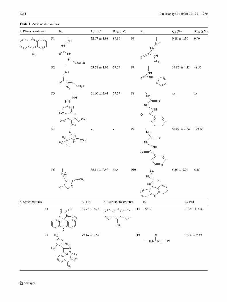
Table 1 Acridine derivatives
1. Planar acridines Rx Irel (%)a IC50 (lM) Rx Irel (%) IC50 (lM)
N
Rx
P1NH
HN
S NH
PhOMe (4)
52.97 ± 1.98 89.10 P6
S NH
CH3
NH
HN
9.18 ± 1.50 9.99
P2
O
S
N
N Ph
OCH3(4)
NH
23.58 ± 1.05 57.79 P7
N
NH
NH
S
14.87 ± 1.42 48.57
P3
NHS
OOAc
OAc
OAc
OAc
NHHN
31.80 ± 2.61 75.57 P8
SNH
NHO
NHxx xx
P4N
O
NS
CO2HH3C
H3CCH3
xx xx P9
SNH
NH
O
N
NH55.88 ± 4.06 182.10
P5
H2C
O
NN
S
CH3
88.11 ± 0.93 N/A P10
NH
N
SNH
NH 5.55 ± 0.91 6.45
2. Spiroacridines Irel (%) 3. Tetrahydroacridines Rx Irel (%)
S1
N
NH
NH
S
CH3
83.97 ± 7.72 N
Rx
T1 –NCS 113.93 ± 8.81
S2
CH3
O
N
N
CH3
CH3
CH3
88.16 ± 6.65 T2
NHNH2Pr
S 133.6 ± 2.48
1264 Eur Biophys J (2008) 37:1261–1270
123
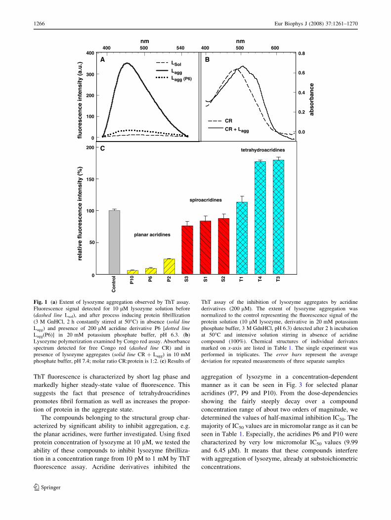
b-structured fibrils or oligomers. The inhibiting activity
was quantified as percentage of maximal ThT fluorescence
observed for lysozyme fibrillization without acridine
(control sample, taken as 100%). The extent of the reduc-
tion of fluorescence intensity characterizes the inhibiting
potential of acridines (lower fluorescence value indicates
more effective inhibitor). Representative results of the
primary screening characterizing each structural class of
acridines are shown in Fig. 1c. Normalized fluorescence
intensities obtained for all studied acridine derivatives are
given in Table 1. The planar acridines (P1-P10) caused
extensive decline of ThT fluorescence (to values lower than
50–95% of the control sample) indicating their significant
ability to inhibit lysozyme amyloid aggregation. The
exception was observed only for planar acridine P5, whose
inhibiting capability was minimal (about 10% decrease of
fluorescence intensity). From the screening the derivatives
P4 and P8 were excluded on account of their very intensive
fluorescence signal detected in studied wavelength range.
The effect of spiroacridines on the inhibition of lysozyme
polymerization was very weak. It follows from measured
fluorescence intensities, which were about 80% of that
observed for the control sample. By this method, we also
found that tetrahydroacridines had no influence on the
prevention of lysozyme fibrillization. Moreover, T3 and T4
derivatives promoted lysozyme aggregation significantly.
The presented results from ThT experiments could be
confirmed by CR assay. However, the monitoring of the
acridine inhibiting activities by CR binding was excluded
as the compounds possess intensive absorption peaks in the
same region as CR.
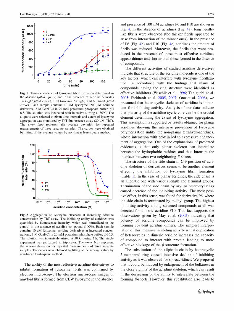
We were interested to investigate effect of acridine
structure in more details; therefore, we studied the kinetics
of the fibril formation in presence of acridine derivatives
belonging to each structural group. By ThT fluorescence
assay we characterized the time dependencies of the extent
of lysozyme aggregation in the presence of acridine
derivatives P10, T4 and S1 (Fig. 2). The data suggest
that acridines altered the shape of the curve detected for
lysozyme fibrillization, which can be characterized by
sigmoidal profile and lag phase taking about 1 h and the
steady-state value achieved at about 2 h. The presence of
planar acridine P10 caused intensive inhibition of fibril
formation. During the studied time interval the fluores-
cence intensities were very low and the steady-state value
indicating the final proportion of the lysozyme aggregation
is markedly reduced. Similar results were observed for
all planar acridine derivatives (except for P5) suggesting
the high-inhibiting effectivity of these compounds. Spiro-
acridine S1 had no significant effect on the lysozyme
aggregation as the time course and steady-state value of
ThT fluorescence are similar to those detected for lyso-
zyme fibrillization in absence of compounds. This curve,
representing a typical dependence detected for all spiro-
acridines, supports the fact that spirocaridines are very
weak inhibitors. Unlike the planar acridines, the tetrahy-
droacridines favour lysozyme aggregation as it is shown for
tetrahydroacridine T4 in Fig. 2. The time dependence of
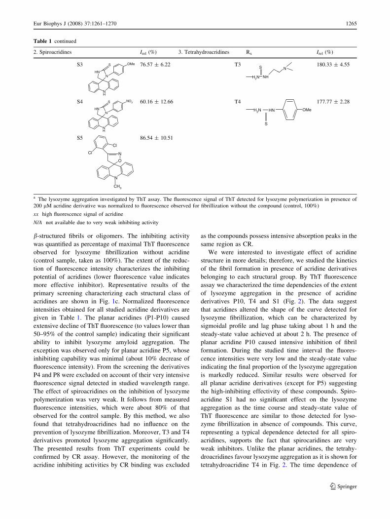
Table 1 continued
2. Spiroacridines Irel (%) 3. Tetrahydroacridines Rx Irel (%)
S3
NNH
NH
S OMe 76.57 ± 6.22 T3
NHH2N
S N180.33 ± 4.55
S4
NNH
NH
S NO2 60.16 ± 12.66 T4
H2N HN
S
OMe
177.77 ± 2.28
S5
CH3
O
N
N
Cl
Cl
86.54 ± 10.51
a The lysozyme aggregation investigated by ThT assay. The fluorescence signal of ThT detected for lysozyme polymerization in presence of
200 lM acridine derivative was normalized to fluorescence observed for fibrillization without the compound (control, 100%)
xx high fluorescence signal of acridine
N/A not available due to very weak inhibiting activity
Eur Biophys J (2008) 37:1261–1270 1265
123
ThT fluorescence is characterized by short lag phase and
markedly higher steady-state value of fluorescence. This
suggests the fact that presence of tetrahydroacridines
promotes fibril formation as well as increases the propor-
tion of protein in the aggregate state.
The compounds belonging to the structural group char-
acterized by significant ability to inhibit aggregation, e.g.
the planar acridines, were further investigated. Using fixed
protein concentration of lysozyme at 10 lM, we tested the
ability of these compounds to inhibit lysozyme fibrilliza-
tion in a concentration range from 10 pM to 1 mM by ThT
fluorescence assay. Acridine derivatives inhibited the
aggregation of lysozyme in a concentration-dependent
manner as it can be seen in Fig. 3 for selected planar
acridines (P7, P9 and P10). From the dose-dependencies
showing the fairly steeply decay over a compound
concentration range of about two orders of magnitude, we
determined the values of half-maximal inhibition IC50. The
majority of IC50 values are in micromolar range as it can be
seen in Table 1. Especially, the acridines P6 and P10 were
characterized by very low micromolar IC50 values (9.99
and 6.45 lM). It means that these compounds interfere
with aggregation of lysozyme, already at substoichiometric
concentrations.
400400
nm nm
CR
CR + Lagg
500 540 400
LSol
LaggLagg
500 6000.8
0.6
abso
rban
ce
tetrahydroacridines
spiroacridines
planar acridines
0.4
0.2
0.0
300
200
100
200
150
100
P10 P6
P2
S3
S1
S2
T1
T4
T3
Co
ntr
ol
50
0
0
flu
ore
scen
ce in
ten
sity
(a.
u.)
re
lati
ve f
luo
resc
ence
inte
nsi
ty (
%)
(P6)
A
C
B
Fig. 1 (a) Extent of lysozyme aggregation observed by ThT assay.
Fluorescence signal detected for 10 lM lysozyme solution before
(dashed line Lsol), and after process inducing protein fibrillization
(3 M GnHCl, 2 h constantly stirred at 50�C) in absence (solid lineLagg) and presence of 200 lM acridine derivative P6 [dotted lineLagg(P6)] in 20 mM potassium phosphate buffer, pH 6.3. (b)
Lysozyme polymerization examined by Congo red assay. Absorbance
spectrum detected for free Congo red (dashed line CR) and in
presence of lysozyme aggregates (solid line CR + Lagg) in 10 mM
phosphate buffer, pH 7.4; molar ratio CR:protein is 1:2. (c) Results of
ThT assay of the inhibition of lysozyme aggregates by acridine
derivatives (200 lM). The extent of lysozyme aggregation was
normalized to the control representing the fluorescence signal of the
protein solution (10 lM lysozyme, derivative in 20 mM potassium
phosphate buffer, 3 M GdnHCl, pH 6.3) detected after 2 h incubation
at 50�C and intensive solution stirring in absence of acridine
compound (100%). Chemical structures of individual derivates
marked on x-axis are listed in Table 1. The single experiment was
performed in triplicates. The error bars represent the average
deviation for repeated measurements of three separate samples
1266 Eur Biophys J (2008) 37:1261–1270
123
The ability of the most effective acridine derivatives to
inhibit formation of lysozyme fibrils was confirmed by
electron microscopy. The electron microscope images of
amyloid fibrils formed from CEW lysozyme in the absence
and presence of 100 lM acridines P6 and P10 are shown in
Fig. 4. In the absence of acridines (Fig. 4a), long needle-
like fibrils were observed (the thicker fibrils appeared to
arise from interaction of the thinner ones). In the presence
of P6 (Fig. 4b) and P10 (Fig. 4c) acridines the amount of
fibrils was reduced. Moreover, the fibrils that were pro-
duced in the presence of these most effective acridines
appear thinner and shorter than those formed in the absence
of compounds.
The different activities of studied acridine derivatives
indicate that structure of the acridine molecule is one of the
key factors, which can interfere with lysozyme fibrilliza-
tion. In accordance with the findings that many of
compounds having the ring structure were identified as
effective inhibitors (Wischik et al. 1996; Taniguchi et al.
2005; Pickhardt et al. 2005, 2007; Ono et al. 2006), we
presumed that heterocyclic skeleton of acridine is impor-
tant for inhibiting activity. Analysis of our data indicate
that planarity of the acridine cyclic core can be the crucial
element determining the extent of lysozyme aggregation.
This assumption is supported by results obtained for planar
acridines showing the intensive prevention of lysozyme
polymerization unlike the non-planar tetrahydroacridines,
whose interaction with protein led to expressive enhance-
ment of aggregation. One of the explanations of presented
evidences is that only planar skeleton can intercalate
between the hydrophobic residues and thus interrupt the
interface between two neighboring b-sheets.
The structure of the side chain in C-9 position of acri-
dine skeleton of derivatives seems to be another element
effecting the inhibition of lysozyme fibril formation
(Table 1). In the case of planar acridines, the side chain is
an aliphatic one with various length and terminal groups.
Termination of the side chain by aryl or heteroaryl rings
caused decrease of the inhibiting activity. The most posi-
tive effect, in this sense, was found for derivative P6, where
the side chain is terminated by methyl group. The highest
inhibiting activity among screened compounds at all was
detected for dimeric acridine P10. This fact supports the
observations given by May et al. (2003) indicating that
potency of acridine compounds can be improved by
forming covalent acridine dimers. The simplest interpre-
tation of this intensive inhibiting activity is that duplication
of heterocycles in dimeric acridine increases the capacity
of compound to interact with protein leading to more
effective blockage of the b-structure formation.
The substitution of the aliphatic chain by heterocyclic
5-membered ring caused intensive decline of inhibiting
activity as it was observed for spiroacridines. We proposed
that it could be induced by enlargement of the bulkiness in
the close vicinity of the acridine skeleton, which can result
in the decreasing of the ability to intercalate between the
forming b-sheets. However, this substitution also leads to
120
100
80
60
40P7P9P1020
0
10-10 10-9 10-8 10-7 10-6 10-5 10-4 10-3
rela
tive
flu
ore
scen
ce
acridine concentration (M)
inte
nsi
ty (
%)
Fig. 3 Aggregation of lysozyme observed at increasing acridine
concentration by ThT assay. The inhibiting ability of acridines was
quantified by fluorescence intensity, which was normalized to the
control in the absence of acridine compound (100%). Each sample
contains 10 lM lysozyme, acridine derivatives at increased concen-
trations, 3 M GdnHCl in 20 mM potassium phosphate buffer, pH 6.3.
The solution was intensively stirred at 50�C during 2 h. The single
experiment was performed in triplicates. The error bars represent
the average deviation for repeated measurements of three separate
samples. The curves were obtained by fitting of the average values by
non-linear least-square method
1200fl
uo
resc
ence
inte
nsi
ty (
a.u
.)
1000
800
600
400
200
0
0 20
LL + P10
L +T4L +S1
40 60 80 100 120time (min)
Fig. 2 Time-dependence of lysozyme fibril formation determined in
the absence (filled square) and in the presence of acridine derivates
T4 (light filled circle), P10 (inverted triangle) and S1 (dark filledcircle). Each sample contains 10 lM lysozyme, 200 lM acridine
derivative, 3 M GdnHCl in 20 mM potassium phosphate buffer, pH
6.3. The solution was incubated with intensive stirring at 50�C. The
aliquots were selected at given time intervals and extent of lysozyme
aggregation was monitored by ThT fluorescence assay (20 lM–ThT).
The error bars represent the average deviation for repeated
measurements of three separate samples. The curves were obtained
by fitting of the average values by non-linear least-square method
Eur Biophys J (2008) 37:1261–1270 1267
123

another important modification of the molecule. The amino
group moiety with sp2 nitrogen atom in the middle ring of
the planar acridines is a hydrogen bond acceptor contrary
to spiroacridines, where sp3 nitrogen atom amine moiety,
due to the loss of the unsaturation in the ring, makes it a
hydrogen bond donor. This modification can be also
important in interfering with the interaction between the
compounds and the protein.
In summary, work reported here is concerned with the
identification of compounds with anti-amyloid effect. We
found that synthesized acridine derivatives are able
to prevent formation of CEW lysozyme fibrillization
depending on the structure of acridine molecules. We
supposed that planarity of the core ring structure as well as
the behaviours of the side chain binding to cyclic skeleton
intensively influence the extent of the lysozyme aggrega-
tion. We determined very effective inhibitors of CEW
lysozyme fibrillization, namely planar acridine derivatives
P6 and P10, characterized by inhibiting activity at low
micromolar concentrations. This fact is important for a
potential therapeutic use of these compounds in the
prevention of the human lysozyme amyloidoses. It is
interesting that other type of acridine derivatives had
capability to inhibit amyloid aggregation of different,
unrelated proteins, namely Quinacrine and Quinacrine
mustard inhibited the formation of amyloid fibrils of tau
and Ab peptide (Taniguchi et al. 2005). The anti-scrapie
activity probably through inhibition of the formation of
protease-resistant prion protein has been found also
for some other acridine derivatives (Caughey et al. 1998;
Priola et al. 2000; May et al. 2003). These evidences could
mean that anti-amyloid acridine may have relevance not
only to lysozyme-related hereditary amyloidosis but also
to amyloid diseases in general.
Acknowledgments This work was supported by the research grants
from the Slovak Grant Agency VEGA No. 7055, 2471, 6167, 0056
and VVGS grant PF8/2007/CH. Dr. M. Vilkova, Dr. S. Hamulakova
and Dr. E. Balentova are thanked for kind providing of selected tested
compounds.
References
Baglioni S, Casamenti F, Bucciantini M, Luheshi L, Taddei N, Chiti
F, Dobson CM, Stefani M (2006) Prefibrillar amyloid aggregates
could be generic toxins in higher organisms. J Neurosci
26(31):8160–8167
Bennett MC (2005) The role of a-synuclein in neurodegenerative
diseases. Pharmacol Ther 105:311–331
Booth D, Sunde M, Bellotti V, Robinson CV, Hutchinson WL, Fraser
PE, Hawkins PN, Dobson CM, Radford SE, Blake CCF, Pepys
MB (1997) Instability, unfolding and aggregation of human
lysozyme variants underlying amyloid fibrillogenesis. Nature
385:787–793
Bucciantini M, Giannoni E, Chiti F, Baroni F, Formigli L, Zurdo J,
Taddei N, Ramponi G, Dobson CM, Stefani M (2002) Inherent
toxicity of aggregates implies a common mechanism for protein
misfoling disease. Nature 416:507–511
Canale C, Torrassa S, Rispoli P, Pelini A, Rolandi R, Bucciantini A,
Stefani M, Gliozzi A (2006) Natively folded HypF-N and its
early amyloid aggregates interact with phospholipid monolayers
and destabilize supported lipid bilayers. Biophys J 91:4675–4588
Chamberlain AK, MacPhee CE, Zurdo J, Morozova-Roche LA, Hill
HA, Dobson CM, Davis JJ (2000) Ultrastructural organization of
amyloid fibrils by atomic force microscopy. Biophys J 79:3282–
3293
Canet D, Sunde M, Last AM, Miranker A, Spencer A, Robinson CV,
Dobson CM (1999) Mechanistic studies of the folding of human
lysozyme and the origin of amyloidogenic behaviour in its
disease related variants. Biochemistry 38:6419–6427
Cao A, Hu D, Lai L (2004) Formation of amyloid fibrils from fully
reduced hen egg white lysozyme. Protein Sci 13:319–324
Caughey WS, Raymond LD, Horiuchi M, Caughey B (1998)
Inhibition of protease-resistant prion protein formation by
porphyrins and phthalocyanines. Proc Natl Acad Sci USA
95(21):12117–12122
Fig. 4 TEM images of
lysozyme solution after process
inducing protein fibrillization
(3 M GnHCl, 2 h constantly
stirred at 50�C in 20 mM
potassium phosphate buffer,
pH 6.3) in absence (a) and in
presence of 100 lM acridine
derivatives P6 (b) and P10 (c).
The bars represent 500 nm
1268 Eur Biophys J (2008) 37:1261–1270
123

Chiti F, Webster P, Taddei N, Clark A, Stefani M, Ramponi G,
Dobson CM (1999) Designing conditions for in vitro formation
of amyloid protofilaments and fibrils. Proc Natl Acad Sci USA
96:3590–3594
Conway KA, Lee SJ, Rochet JC, Ding TT, Williamson RE, Lansbury
PT Jr (2000) Acceleration of oligomerization, not fibrilization, is
a shared property of both a-synuclein mutations linked to early-
onset Parkinson’s disease: implications for pathogenesis and
therapy. Proc Natl Acad Sci USA 97:571–576
Cooper JH (1974) Selective staining as a function of amyloid
composition and structure: histochemical analysis of the alkaline
Congo Red, standardized toluidine blue and iodine methods. Lab
Invest 31:232–238
DeFelice FG, Houzel JC, Garcia-Abreu J, Louzada PRF, Afonso RC,
Meirelles NL, Lent R, Neto MV, Ferreira ST (2001) Inhibition of
Alzheimer’s disease b-amyloid aggregation, neurotoxicity, and
in vivo deposition by nitrophenols: implications for Alzheimer’s
theraphy. FASEB J 15:1297–1299
DeFelice FG, Velasco PT, Lambert MP, Viola K, Fernandez SJ,
Ferreira ST, Klein WL (2007) Abeta oligomers induce neuronal
oxidative stress through an N-methyl-D-aspartate receptor-
dependent mechanism that is blocked by the Alzheimer drug
memantine. J Biol Chem 282:11590–11601
Demuro A, Mina E, Kayed R, Milton SC, Parker I, Glabe CG (2005)
Calcium dysregulation and membrane disruption as a ubiquitous
neurotoxic mechanism of soluble amyloid oligomers. J Biol
Chem 280:17294–17300
Dobson CM (1999) Protein misfolding, evolution and disease. Trends
Biochem Sci 24:329–332
Dobson CM (2001) The structural basis of protein folding and its
links with human disease. Philos Trans Rsoc Lond B Biol Sci
356:133–145
Fandrich M, Fletcher MA, Dobson CM (2001) Amyloid fibrils from
muscle myoglobin. Nature 410:165–166
Ferreira ST, Vieira MN, De Felice FG (2007) Soluble protein
oligomers as emerging toxins in Alzheimer’s and other amyloid
diseases, IUBMB Life 59:332–345
Funahashi J, Takano K, Ogasahara K, Yamagata Y, Yutani K (1996)
The structure, stability, and folding process of amyloidogenic
mutant human lysozyme. J Biochem 120:1216–1223
Goedert M, Spillantini MG (2006) A century of Alzheimer’s disease.
Science 314:777–781
Gouras GK, Almeida CG, Takahashi RH (2005) Intraneuronal Abeta
accumulation and origin of plaques in Alzheimer’s disease.
Neurobiol Aging 26:1235–1244
Guijarro JII, Sunde M, Jones JA, Campbell ID, Dobson CM (1998)
Amyloid fibril formation by an SH3 domain. Proc Natl Acad Sci
USA 95:4224–4228
Haass C, Selkoe DJ (2007) Soluble oligomers in neurodegeneration:
lessons from the Alzheimer’s amyloid beta-peptide. Nat Rev
Mol Cell Biol 8:101–112
Hou X, Parkington HC, Coleman HA, Mechler A, Martin LL, Aguilar
MI, Small DH (2000) Transthyretin oligomers induce calcium
influx via voltage-gated calcium channels. J Mol Biol
300(5):1033–1039
Khlistunova I, Biernat J, Wang YP, Pickhardt M, von Bergen M,
Gazova Z, Mandelkow EM, Mandelkow E (2006) Inducible
expression of Tau repeat domain in cell models of tauopathy:
aggregation is toxic to cells but can be reversed by inhibitor
drugs. J Biol Chem 281:1205–1214
Klunk WE, Pettigrew JW, Abraham DJ (1989) Quantitative
evaluation of congo red binding to amyloid-like proteins with
a beta-pleated sheet conformation. J Histochem. Cytochem
37(8):1273–1281
Koo EH, Lansbury PT, Kelly JW (1999) Amyloid diseases: abnormal
protein aggregation in neurodegeneration. Proc Natl Acad Sci
USA 96:9989–9990
Korth C, May BCH, Cohen FE, Prusiner SB (2001) Acridine and
phenothiazine derivatives as pharmacotherapeutics for prion
disease. PNAS 98:9836–9841
Kristian P, Hamulakova S, Bernat J, Imrich J, Voss G, Busova T
(1998) Synthesis of acetylcholinesterase inhibitors on the basis
of 9-isothiocyanato-1,2,3,4-tetrahydroacridine: 2-[(1,2,3,4-tetra-
hydroacridin-9-yl)imino]-3-substituted 1,3-thiazolidin-4-ones.
Heterocycles 49:197–204
Lambert MP, Barlow AK, Chromy BA, Edwards C, Freed R, Liosatos
M, Morgan TE, Rozovsky I, Tromer B, Violc KL, Wals P, Zhang
C, Finch CE Krafft GA, Klein WLn (1998) Diffusible, nonfibr-
illar ligands derived from Abeta1–42 are potent central nervous
system neurotoxins. Proc Natl Acad Sci USA 95:6448–6453
Lansbury PT Jr (1999) Evolution of amyloid: what normal protein
folding may tell us about fibrillogenesis and disease. Proc Natl
Acad Sci USA 96:3342–3344
Lee VM, Goedert M, Trojanowski JQ (2001) Neurodegenerative
tauopathies. Annu Rev Neurosci 24:1121–1159
LeVine H (1993) Thioflavine T interaction with synthetic Alzheimers
disease beta-amyloid peptides: detection of amyloid aggregation
in solution. Protein Sci 2:404–410
Lue LF, Kuo YM, Roher AE, Brachova L, Shen Y, Sue L, Beach T,
Kurth JH, Rydel RE, Rogers J (1999) Soluble amyloid beta
peptide concentration as a predictor of synaptic change in
Alzheimers disease. Am J Pathol 155(3):853–862
May BCH, Fafarman AT, Hong SB, Rogers M, Deady LW, Prusiner
SB, Cohen FE (2003) Potent inhibition of scrapie prion
replication in cultured cells by bis-acridines. PNAS 100:3416–
3421
Merlini G, Bellotti V (2003) Molecular mechanisms of amyloidosis.
N Engl J Med 349:583–596
Moreira PI, Honda K, Liu Q, Santos MS, Oliviera CR, Aliev G,
Nunomura A, Zhu X, Smith MA, Perry G (2005) Oxidative
stress: the old enemy in Alzheimers disease pathophysiology.
Curr Alzheimer Res 2:403–408
Morozova-Roche LA, Zurdo J, Spencer A, Noppe W, Receveur V,
Archer DB, Joniau M, Dobson CM (2000) Amyloid fibril
formation an seeding by wild-type human lysozyme and its
disease-related mutational variants. J Struct Biol 130:339–351
Nguyen JT, Inouye H, Baldwin MA, Fletterick RJ, Cohen FE,
Prusiner SB, Kirschner DA (1995) X-ray diffraction of scrapie
prion rods and PrP peptides. J Nol Biol 252:412–422
Ono K, Hamaguchi T, Naiki H, Yamada M (2006) Anti-amyloido-
genic effect of antioxidants: implication for the prevention and
therapeutics of Alzheimer’s disease. Biochim Biophys Acta
1762:575–586
Pepys MB, Hawkins PN, Booth DR, Vigushin DM, Tennent GA,
Soutar AK, Totty N, Nguyen O, Blake CCF, Terry CJ, Feest TG,
Zalin AM, Hsuan JJ (1993) Human lysozyme gene mutations
cause hereditary systemic amyloidosis. Nature 362:553–557
Pickhardt M, Gazova Z, von Bergen M, Khlistunova I, Wang Y,
Hascher A, Mandelkow EM, Biernat J, Mandelkow E (2005)
Anthraquinones inhibit tau aggregation and dissolve Alzheimer’s
paired helical filaments in vitro and in cells. J Biol Chem
280:3628–3635
Pickhardt M, Biernat J, Khlistunova I, Wang Y-P, Gazova Z,
Mandelkow E-M, Mandelkow E (2007) N-Phenylamine deriv-
atives as aggregation inhibitors in cell models of tauopathy. Curr
Alzheimer Res 4:397–402
Priola SA, Raines A, Caughey B (2000) Porphyrin and phthalocy-
anine antiscrapie compounds. Science 287(5457):1503–1506
Eur Biophys J (2008) 37:1261–1270 1269
123
Raghu P, Reddy GB, Sivakumar B (2002) Inhibition of transthyretin
amyloid fibril formation by 2,4-dinitrophenol through tetramer
stabilization. Arch Biochem Biophys 400:43–47
Reixach N, Deechongkit S, Jiang X, Kelly JW, Buxbaum JN (2004)
Tissue damage in the amyloidosis: transthyretin monomers and
nonnative oligomers are the major cytotoxic species in tissue
culture. Proc Natl Acad Sci USA 101:2817–2822
Roberson ED, Scearce-Levie K, Palop JJ, Yan F, Cheng IH, Wu T,
Gerstein H, Yu GQ, Mucke L (2007) Reducing endogenous tau
ameliorates amyloid beta-induced deficits in an Alzheimer’s
disease mouse model. Science 316(5825):750–754
Serpell LC, Sunde M, Benson MD, Tennent GA, Pepys MB, Fraser
PE (2000) The protofilament substructure of amyloid fibrils.
J Mol Biol 300(5):1033–1039
Sipse JD (2005) Amyloid Proteins. Wiley-VCH Verlag GmbH and
Co. KgaA, Weinheim Germany
Stefani M, Dobson CM (2003) Protein aggregation and aggregate
toxicity: new insights into protein folding, misfolding diseases
and biological evolution. J Mol Med 81:678–699
Taniguchi S, Suzuki N, Masuda M, Hisanaga S, Iwatsubo T, Goedert
M, Hasegawa M (2005) Inhibition of heparin-induced tau
filament formation by phenotiazines, polyphenols, and porphy-
rins. J Biol Chem 280(9):7614–7623
Tomascikova J, Imrich J, Danihel I, Bohm S, Kristian P (2007)
Heterocyclization of Acridin-9-yl Thiosemicarbazides with
Dimethyl Acetylenedicarboxylate. Coll Czech Chem Commun
72:347–362
Tomascikova J, Danihel I, Bohm S, Imrich J, Kristian P, Potocnak I,
Cejka J, Klika KD (2008) Molecular and solid-state structure of
methyl [2-(acridin-9-ylimino)-3-(tert-butylamino)-4-oxothiazoli-
din-5-ylidene]acetate. J Mol Struct 875:419–426
Valleix S, Drunat S, Philit JB, Adoue D, Piette JC, Droz D (2002)
Hereditary renal amyloidosis caused by a new variant lysozyme
W64R in a French family. Kidney Int 61:907–912
Vieira MNN, Figueroa-Villar JD, Meirelles MNL, Ferreira ST, De
Felice FG (2006) Small Molecule Inhibitors of Lysozyme
Amyloid Aggregation. Cell Biochem Biophys 44:549–553
Vieira MNN, Forny-Germano L, Saraiva LM, Sebollela A, Martinez
AMB, Houzel J-C, De Felice FG, Ferreira ST (2007) Soluble
oligomers from a non-disease related protein mimic Ab-induced
tau hyperphosphorylation and neurodegeneration. J Neurochem
103:736–748
Vernaglia BA, Huang J, Clark ED (2004) Guanidie hydrochloride can
induce amyloid fiblril firmation from hen egg-white lysozyme.
Biomacromolecules 5:1362–1370
Walsh DM, Klyubin I, Fadeeva JV, Cullen WK, Anwyl R, Wolfe MS,
Rowan MJ, Selkoe DJ (2002) Naturally secreted oligomers of
amyloid beta protein potently inhibit hippocampal long-term
potentiation in vivo. Nature 416:535–539
Walsh DM, Selkoe DJ (2004) Oligomers on the brain: the emerging
role of soluble protein aggregates in neurodegeneration. Protein
Pept Lett 11(3):213–228
Wang J, Dickson DW, Trojanowski JQ, Lee VM (1999) The levels of
soluble versus insoluble brain Abeta distinguish Alzheimers
disease from normal and pathologic aging. Expert Neurol
158:328–337
Wischik CM, Edwards PC, Lai RYK, Roth M, Harrington CR (1996)
Selective inhibition of Alzheimer disease-like tau aggregation by
phenothiazines. PNAS 93(20):11213–11218
Yazaki M, Farrell SA, Benson MD (2003) A novel lysozyme
mutation Phe57Ile associated with hereditary renal amyloidosis.
Kidney Int 63:1652–1657
1270 Eur Biophys J (2008) 37:1261–1270
123
Copyright © 2022 FDOKUMEN