Effects of Salivary Acetylcholinesterase on the Cytotoxicity of ...
Upload
independentCategory
view
0download
0
Bioorganic & Medicinal Chemistry 16 (2008) 7759–7769
Contents lists available at ScienceDirect
Bioorganic & Medicinal Chemistry
journal homepage: www.elsevier .com/locate /bmc
New tacrine-dihydropyridine hybrids that inhibit acetylcholinesterase,calcium entry, and exhibit neuroprotection properties
Rafael León a,b,*, Cristóbal de los Ríos a,b, José Marco-Contelles a,*, Oscar Huertas d, Xavier Barril e,F. Javier Luque d,*, Manuela G. López b, Antonio G. García b,c, Mercedes Villarroya b,*
a Laboratorio de Radicales Libres (IQOG, CSIC), C/Juan de la Cierva 3, 28006 Madrid, Spainb Instituto Teófilo Hernando, Departamento de Farmacología y Terapéutica, Facultad de Medicina, Universidad Autónoma de Madrid, C/Arzobispo Morcillo 4, 28029 Madrid, Spainc Servicio de Farmacología Clínica, Hospital Universitario de la Princesa, C/Diego de León 62, 28006 Madrid, Spaind Departament de Fisicoquímica and Institut de Biomedicina (IBUB), Facultat de Farmàcia, Universitat de Barcelona, Avda. Diagonal 643, Barcelona, Spaine Institució Catalana de Recerca i Estudis Avançats (ICREA), Departament de Fisicoquímica and Institut de Biomedicina, Facultat de Farmàcia, Universitat de Barcelona,Avda. Diagonal 643, Barcelona, Spain
a r t i c l e i n f o a b s t r a c t
Article history:Received 6 February 2008Revised 24 June 2008Accepted 2 July 2008Available online 8 July 2008
Keywords:Tacrine-dihydropyridine hybridsAChEBuChEKinetic analysisInhibition mechanismVoltage-dependent calcium channelsNeuroprotectionMolecular modeling
0968-0896/$ - see front matter � 2008 Elsevier Ltd. Adoi:10.1016/j.bmc.2008.07.005
* Corresponding authors. Tel.: +34 91 5622900; faxE-mail addresses: [email protected] (J. Marco-C
Javier Luque).
In this communication, we describe the synthesis and biological evaluation of tacripyrimedones 1–5, aseries of new tacrine-1,4-dihydropyridine hybrids bearing the general structure of 11-amino-12-aryl-3,3-dimethyl-3,4,5,7,8,9,10,12-octahydrodibenzo[b,g][1,8]naphthyridine-1(2H)-one. These multifunc-tional compounds are moderately potent and selective AChEIs, with no activity toward BuChE. Kineticanalysis and molecular modeling studies point out that the new compounds preferentially bind theperipheral anionic site of AChE. In addition, compounds 1–5 show an excellent neuroprotective profile,and a moderate blocking effect of L-type voltage-dependent calcium channels due to the mitigation of[Ca2+] elevation elicited by K+ depolarization. Therefore, they represent a new family of molecules withpotential therapeutic application for the treatment of Alzheimer’s disease.
� 2008 Elsevier Ltd. All rights reserved.
1. Introduction
Alzheimer’s disease (AD) is an age-related neurodegenerativedisease characterized by progressive memory loss and other cogni-tive impairments. Although the etiology of AD is not well known,there are diverse factors such as amyloid-b (Ab) deposits, s-proteinaggregation, oxidative stress, or low levels of acetylcholine (ACh)that are thought to play significant roles in the disease.1 The cho-linergic theory of AD suggests that the selective loss of cholinergicneurons in AD results in a deficit of ACh in specific regions of thebrain that mediate learning and memory functions.2 The primaryapproach for treating AD has, therefore, aimed at increasing theACh levels in the brain by using acetylcholinesterase inhibitors(AChEIs) such as donepezil, galantamine, or rivastigmine.3 On theother hand, Ca2+ overload seems to be the main factor initiatingthe processes leading to cell death. Several lines of evidence showthat Ca2+ dysfunction, involved in the pathogenesis of AD,4 aug-
ll rights reserved.
: +34 91 5644853.ontelles), [email protected] (F.
ments Ab formation,5a and s hyperphosphorylation.5b Ca2+ entrythrough the L-subtype of high-voltage activated Ca2+ channels(Cav1.1–1.4) causes both Ca2+ overload and mitochondrial disrup-tion, which activate the apoptotic cascade and cell death.6 Hence,blocking the entrance of Ca2+ through calcium channels could bea good strategy to prevent cell death.
The current multi-target approach in drug design7 for the treat-ment of AD includes novel tacrine–melatonin hybrids,8a dualinhibitors of AChE and monoamine oxidase (MAO),8b dual AChEIand serotonin transporters inhibitors,8c potent AChEIs with antiox-idant and neuroprotective properties,8d or gallamine–tacrine hy-brids designed to bind the cholinergic and M2 muscarinicreceptors.8e
Since 1,4-dihydropyridines (DHPs) are compounds that selec-tively block L-type Ca2+ channels, we considered the synthesisand the pharmacological study of new multipotent hybrid com-pounds, based on an AChEI and a DHP, such as tacrine and nimodi-pine (Chart 1).9 Besides inhibition of AChE and blockade of voltage-dependent calcium channels (VDCC), we were also interested incompounds targeted to prevent oxidative stress. Recent researchhas demonstrated that oxidative damage is an event that precedes

NNH
O
X
NH2
Tacripyrimedones (II) (this work)
NNH
EtO
O NH2
Tacripyrines (I) (ref. 13)
N
NH2
Tacrine (AChEI)
Nimodipine (1,4-DHP)
NH
O
O
O
OOMe
NO2
X
Chart 1.
7760 R. León et al. / Bioorg. Med. Chem. 16 (2008) 7759–7769
the appearance of other pathological hallmarks of AD.10 Thus,drugs that scavenge reactive oxygen species (ROS) may have a par-ticular therapeutic efficacy.11,12
Based on this strategy, we have recently reported preliminarystudies on the synthesis and biological activity of ethyl esters of 5-amino-4-aryl-1,4,6,7,8,9-hexahydro-2-methyl-benzo[b][1,8]naph-thyridine-3-carboxylic acid (I) (Chart 1) that we have named astacripyrines, as the first examples of tacrine-DHP hybrids describedin the literature.13 Prompted by the interesting biological profile ofthese compounds (very selective and potent AChEIs, excellentneuroprotective profile and moderate Ca2+ channel blockade ef-fects), we have now prepared new analogs of the general structureII (Chart 1), for which we propose the short name of tacripyrime-dones, where we have incorporated a functionalized cyclohexanering annulated onto the DHP-ring present in type I compounds, in or-der to start a general structure–activity relationship (SAR) analysistargeted to improve the biological activities found in the lead-typecompounds I.13
Accordingly, in this work we report the synthesis and biologicalevaluation, including AChE/BuChE (butyrylcholinesterase) inhibi-tion, kinetic analysis, and molecular modeling of the AChE inhibi-tion, blockade of cytosolic Ca2+ elevation, and the neuroprotective
AlCl3,
1,2-dichlor
reflux
O
NH
CN
NH2
X
O
6 X= C-H
7 X= C-F
8 X= C-Me
9 X= C-OMe
10 X= N
Scheme
activity for the novel tacrine-DHP hybrids tacripyrimedones 1–5(Scheme 1).
2. Results and discussion
2.1. Synthesis of 11-amino-12-aryl-3,3-dimethyl-3,4,5,7,8,9,10,12-octahydrodibenzo[b,g][1,8]naphthyridine-1(2H)-ones (1–5)
The synthesis of tacripyrimedones 1–5 was easily achieved, inexcellent yields, using the Friedländer reaction14 between 2-ami-no-4-aryl-7,7-dimethyl-5-oxo-1,4,5,6,7,8-hexahydroquinoline-3-carbonitriles (6–10)15 and cyclohexanone, under standard condi-tions (Scheme 1).16 Precursors 6–10, readily available compoundsin multigram quantities, are prepared by refluxing equimolaramounts of the corresponding arylidenemalononitrile, dimedone,and an excess of ammonium acetate in acetic acid as solvent.15
Compounds 1–5 are racemic octahydrobenzo[b,g][1,8]naph-thyridines substituted at C12 with an aromatic ring, incorporatingdifferent types of substituents at C40 (1–4) or a 40-pyridyl ring (5).These molecules have been conveniently characterized by theirspectroscopic and analytical data. The 1H and 13C NMR data (seeSection 4) are in good agreement with the values found for related
oethane
NH
X
N
NH2O
1 (97%)
2 (90%)
3 (96%)
4 (83%)
5 (88%)
1.
R. León et al. / Bioorg. Med. Chem. 16 (2008) 7759–7769 7761
compounds,13,15 and the assignments are based on standard NMRexperiments (1H–1H COSY, HMQC, and HMBC).
2.2. Pharmacology
2.2.1. Studies of AChE/BuChE inhibitionAccording to standard methodology17,18 (see Section 4), tacripy-
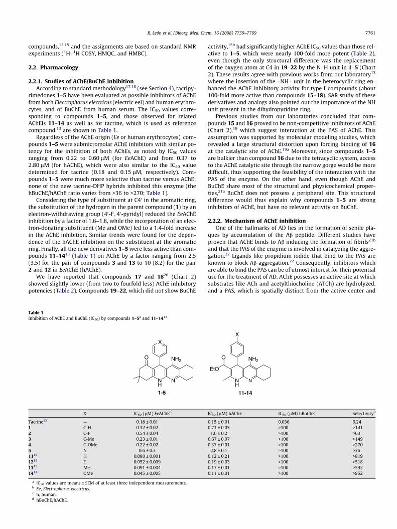
rimedones 1–5 have been evaluated as possible inhibitors of AChEfrom both Electrophorus electricus (electric eel) and human erythro-cytes, and of BuChE from human serum. The IC50 values corre-sponding to compounds 1–5, and those observed for relatedAChEIs 11–14 as well as for tacrine, which is used as referencecompound,13 are shown in Table 1.
Regardless of the AChE origin (Ee or human erythrocytes), com-pounds 1–5 were submicromolar AChE inhibitors with similar po-tency for the inhibition of both AChEs, as noted by IC50 valuesranging from 0.22 to 0.60 lM (for EeAChE) and from 0.37 to2.80 lM (for hAChE), which were also similar to the IC50 valuedetermined for tacrine (0.18 and 0.15 lM, respectively). Com-pounds 1–5 were much more selective than tacrine versus AChE;none of the new tacrine-DHP hybrids inhibited this enzyme (thehBuChE/hAChE ratio varies from >36 to >270; Table 1).
Considering the type of substituent at C40 in the aromatic ring,the substitution of the hydrogen in the parent compound (1) by anelectron-withdrawing group (40-F, 40-pyridyl) reduced the EeAChEinhibition by a factor of 1.6–1.8, while the incorporation of an elec-tron-donating substituent (Me and OMe) led to a 1.4-fold increasein the AChE inhibition. Similar trends were found for the depen-dence of the hAChE inhibition on the substituent at the aromaticring. Finally, all the new derivatives 1–5 were less active than com-pounds 11–1413 (Table 1) on AChE by a factor ranging from 2.5(3.5) for the pair of compounds 3 and 13 to 10 (8.2) for the pair2 and 12 in EeAChE (hAChE).
We have reported that compounds 17 and 1820 (Chart 2)showed slightly lower (from two to fourfold less) AChE inhibitorypotencies (Table 2). Compounds 19–22, which did not show BuChE
Table 1Inhibition of AChE and BuChE (IC50) by compounds 1–5a and 11–1413
X
NH
N
NH2
1-5
O
X IC50 (lM) EeAChEb
Tacrine13 — 0.18 ± 0.011 C-H 0.32 ± 0.022 C-F 0.54 ± 0.043 C-Me 0.23 ± 0.014 C-OMe 0.22 ± 0.025 N 0.6 ± 0.31113 H 0.080 ± 0.0011213 F 0.052 ± 0.0091313 Me 0.091 ± 0.0041413 OMe 0.045 ± 0.005
a IC50 values are means ± SEM of at least three independent measurements.b Ee, Electrophorus electricus.c h, human.d hBuChE/hAChE.
activity,15b had significantly higher AChE IC50 values than those rel-ative to 1–5, which were nearly 100-fold more potent (Table 2),even though the only structural difference was the replacementof the oxygen atom at C4 in 19–22 by the N–H unit in 1–5 (Chart2). These results agree with previous works from our laboratory13
where the insertion of the –NH– unit in the heterocyclic ring en-hanced the AChE inhibitory activity for type I compounds (about100-fold more active than compounds 15–18). SAR study of thesederivatives and analogs also pointed out the importance of the NHunit present in the dihydropyridine ring.
Previous studies from our laboratories concluded that com-pounds 15 and 16 proved to be non-competitive inhibitors of AChE(Chart 2),19 which suggest interaction at the PAS of AChE. Thisassumption was supported by molecular modeling studies, whichrevealed a large structural distortion upon forcing binding of 16at the catalytic site of AChE.19a Moreover, since compounds 1–5are bulkier than compound 16 due to the tetracyclic system, accessto the AChE catalytic site through the narrow gorge would be moredifficult, thus supporting the feasibility of the interaction with thePAS of the enzyme. On the other hand, even though AChE andBuChE share most of the structural and physicochemical proper-ties,21a BuChE does not possess a peripheral site. This structuraldifference would thus explain why compounds 1–5 are stronginhibitors of AChE, but have no relevant activity on BuChE.
2.2.2. Mechanism of AChE inhibitionOne of the hallmarks of AD lies in the formation of senile pla-
ques by accumulation of the Ab peptide. Different studies haveproven that AChE binds to Ab inducing the formation of fibrils21b
and that the PAS of the enzyme is involved in catalyzing the aggre-gation.22 Ligands like propidium iodide that bind to the PAS areknown to block Ab aggregation.22 Consequently, inhibitors whichare able to bind the PAS can be of utmost interest for their potentialuse for the treatment of AD. AChE possesses an active site at whichsubstrates like ACh and acetylthiocholine (ATCh) are hydrolyzed,and a PAS, which is spatially distinct from the active center and
NH
EtO
N
NH2
11-14
O
X
IC50 (lM) hAChE IC50 (lM) hBuChEc Selectivityd
0.15 ± 0.01 0.036 0.240.71 ± 0.03 >100 >141
1.6 ± 0.2 >100 >630.67 ± 0.07 >100 >1490.37 ± 0.01 >100 >270
2.8 ± 0.1 >100 >360.12 ± 0.21 >100 >8190.19 ± 0.03 >100 >5180.17 ± 0.01 >100 >5920.11 ± 0.01 >100 >952

O N
EtO2CNH2
OMe
15
N N
EtO2CNH2
16
O N
NH2
19-22
O
O N
EtO2CNH2
N
17
N N
EtO2CNH2
N
18
X
NH
N
NH2
1-5
O
X
Chart 2.
Table 2Inhibition of AChE by compounds 1–5 and 15–22a
X IC50 EeAChE (lM)
Tacrine13 — 0.18 ± 0.011 C-H 0.32 ± 0.022 C-F 0.54 ± 0.043 C-Me 0.23 ± 0.014 C-OMe 0.22 ± 0.025 X = N 0.60 ± 0.301519 — 0.87 ± 0.201619 — 0.82 ± 0.401720 — 3.00 ± 0.301820 — 1.40 ± 0.401915c H 49.0 ± 1.002015c F 70.0 ± 5.002115c Me 52.0 ± 2.002215c OMe 4.10 ± 3.00
a IC50 values are means ± SEM of at least three independent measurements.
7762 R. León et al. / Bioorg. Med. Chem. 16 (2008) 7759–7769
where selective inhibitors can bind with high affinity and specific-ity.23 Hydrolysis of ACh proceeds through the formation of an acyl-enzyme intermediate, and inhibitors that bind the PAS can also af-fect steady-state kinetics.24
Based on the preceding considerations and on the hypothesisthat the rather wide shape of molecules such as 1–5 might impairthe entrance into the AChE gorge and favor the binding at the PAS,we studied the steady-state kinetics of compounds 1–5 as well asthat of tacrine and propidium. The graphical analysis of the steady-state inhibition data can give information about the binding modeof the selected compounds, and allow the measurement of their Ki,as well.
The kinetic characterization of these AChE inhibitors was car-ried out as follows. Reactions were measured in the presence ofdifferent concentrations of substrate and data were analyzedaccording to the Linewever–Burk method (Fig. 1). Five differentconcentrations of each inhibitor were used. Values of Ki were cal-culated from the slope of a secondary plot (see Section 4 and Table3).
The Ki value found for tacrine agrees with the data reported inthe literature,25 and the reciprocal plots describing the AChE inhi-bition (Fig. 1A) show both increasing slopes and increasing inter-cepts with higher inhibition concentration, a pattern thatindicates mixed inhibition. In the reciprocal plots for compound
4 and propidium (Fig. 1B and C, respectively), lines crossing the xaxis in the same point reveal unchanged Km and decreased Vmax
at increasing inhibitor concentrations. This is a typical trend fornon-competitive inhibition, and indicates that these compoundsbind to a site different to the catalytic one.26 Since propidiumhas been described as a selective ligand for the AChE PAS,23 thesame behavior is therefore expected for compound 4.
The hypothesis of the binding at the AChE PAS for tacripyrime-dones 1–5 is also consistent with their BuChE/AChE selectivity (Ta-ble 1), since binding at the PAS is mediated by the residue Trp279(EeAChE) present at the entrance of the gorge in AChE, but absentin BuChE.
2.2.3. Molecular modelingThe AChE binding mode of tacripyrimedones 1–5 (Scheme 1)
was explored by means of docking studies performed for 4 inhAChE using the rDock program (see Section 4). To verify the reli-ability of the docking protocol, docking was also extended to ta-crine and propidium taking advantage of the known X-raycrystallographic binding mode of these compounds at the catalyticand peripheral sites, respectively, of AChE. The docking results con-firmed the capability of the method to reproduce the experimentalbinding mode (see Figs. 2 and 3), thus giving confidence to thebinding mode predicted for 4.
Attempts to dock 4 in the catalytic site of hAChE were unsuc-cessful, as expected from our previous molecular dynamics studiesperformed for 15 and 1619a (Chart 2), which pointed out that thecatalytic site of AChE is not flexible enough to easily accommodatethose compounds. In contrast, it was successfully docked in thePAS, which is in agreement with the non-competitive inhibitionmechanism exhibited by 4. According to the putative binding modeof the R-enantiomer of 4 (see Fig. 4), the inhibitor is well stackedagainst the indole ring of Trp286 (hAChE, with a distance fromthe central aminopyridine unit of 4 to the midpoint of the indolering of Trp286 of �3.6 Å), while forming hydrogen-bonds with sev-eral residues. Thus, the NH group of the 1,8-naphthyridine unit ishydrogen-bonded to the hydroxyl group of Tyr72, which in turnis hydrogen-bonded to the carboxylate group of Asp74 (the lattergroup might also form hydrogen-bond interactions with Tyr341and Thr83). The oxygen atom of the carbonyl group is correctlypositioned to hydrogen-bond to the imidazole ring of His287, thusmimicking a similar interaction found between one of the NH2

Figure. 1. Double reciprocal plots of the reaction velocity versus acetyltiocholine(ATCh) concentration in the presence and absence of inhibitor. The study wasperformed in 0.1 M PBS solution, pH 8 and five different concentrations of substrate.Concentrations of tacrine, compound 4, and propidium were as follows. Tacrine:Control (j) [I] = 0, (d) [I] = 5 nM, (N) [I] = 15 nM, (.) [I] = 30 nM, (�) [I] = 60 nM, (?)[I] = 500 nM. Compound 4: Control (j) [I] = 0, (d) [I] = 5 nM, (N) [I] = 15 nM, (.)[I] = 30 nM, (�) [I] = 60 nM, (?) [I] = 120 nM. Propidium: Control (j) [I] = 0, (d)[I] = 0,5 nM, (N) [I] = 1 lM, (.) [I] = 10 lM, (�) [I] = 30 lM, (?) [I] = 100 lM. (A)Lineweaver–Burk study of tacrine. Insert: slope (j) of the double reciprocal plotsversus [tacrine]. (B) Lineweaver–Burk study of compound 4. Insert: slope (j) of thedouble reciprocal plots versus [4]. (C) Lineweaver–Burk study of propidium. Insert:slope (j) of the double reciprocal plots versus [propidium]. Data are expressed asmean ± SEM of at least four experiments.
Figure. 2. Two views of the predicted binding mode of tacrine at the catalytic site ofthe AChE, taking advantage of the X-ray crystallographic structures of the Torpedocalifornica AChE complex (PDB entry 1ACJ). Docked and X-ray structures of theligand are colored by atom and in gray, respectively. For the sake of clarity,hydrogen atoms are omitted.
R. León et al. / Bioorg. Med. Chem. 16 (2008) 7759–7769 7763
groups in propidium with His287 (PDB entry 1N5R). Finally, theamino group is properly oriented toward the hydroxyl and car-bonyl groups of Ser293, even though those interactions should be
Table 3IC50 for inhibition of AChE, Ki’s and type of inhibition for tacrine, propidium, andcompounds 1–5 with EeAChE
IC50 AChE (lM) Ki ± SE (nM) Type of inhibition
Tacrine 0.18 20.2 ± 0.12 Mixed1 0.32 61.3 ± 3.21 Non-competitive2 0.54 106.0 ± 4.67 Non-competitive3 0.23 38.9 ± 1.85 Non-competitive4 0.22 41.9 ± 2.52 Non-competitive5 0.60 157.0 ± 4.76 Non-competitivePropidium 11.0 6800 ± 60 Non-competitive
assisted by a water molecule as the distance between the interact-ing groups of inhibitor and Ser293 amounts to �6.4 Å. Finally, the40-methoxybenzene unit is pointing toward the solvent, adopting asimilar arrangement as the aliphatic chain containing the quater-nary ammonium group in propidium, thus avoiding steric clasheswith residues at the PAS.
It is worth-noting that these features allow us to explain the100-fold increase in the inhibitory potency observed when com-paring compounds 1–4 to 19–22 (see above and Table 2); thisdifference can be ascribed to the loss of the hydrogen-bondinteraction formed between the inhibitor and Tyr72 uponreplacement of the NH moiety by O. Furthermore, since a similarchange in the inhibitory potency is observed for compounds 11–14 relative to 15–18, it can be speculated that tacripyrines I bindto the PAS of AChE mimicking the binding mode of 4. This bind-ing mode would also explain the relative insensitivity of theinhibitory potency of compounds 1–4 to the presence of the sub-stituent in position 40 or to the replacement of the benzene ringby a pyridine in 5 (see Table 1). Finally, the slightly lower po-tency of compounds 1–4 relative to 11–14 likely reflects a de-crease in the flexibility of the carbonyl group to interact with

Figure. 3. Two views of the predicted binding mode of propidium at the peripheralsite of the AChE, taking advantage of the X-ray crystallographic structures of themouse AChE complex (PDB entry 1N5R). Docked and X-ray structures are coloredby atom and in gray, respectively. For the sake of clarity, hydrogen atoms areomitted.
Figure. 4. Representation of the putative binding modes of R (top) and S (bottom)enantiomers of compound 4 (colored by atom) in the PAS of hAChE (shown byribbon). Selected residues of the protein are also displayed (Trp286 (hAChE), Tyr72,Asp74, His287, and Ser293; Thr75 is also shown in the bottom plot). Favorablehydrogen-bond interactions are shown by blue lines, and unfavorable contacts areshown by red lines (see text for details). For the sake of clarity, hydrogen atoms areomitted.
7764 R. León et al. / Bioorg. Med. Chem. 16 (2008) 7759–7769
His287. On the other hand, the lack of a tryptophan residuemimicking Trp286 (hAChE) in the peripheral site of hBuChE (re-placed by Ala277), as well as the replacement of Tyr72 in hAChEby Asn68 in hBuChE, could also explain the large AChE/BuChEselectivity of these compounds.
Finally, binding of the S-enantiomer of 4 (see Fig. 4) appears tobe less favorable. Thus, even though the inhibitor is stacked againstTrp286 (hAChE) and the 40-methoxybenzene unit has a similar ori-entation in the binding modes of R- and S-enantiomers, the inhib-itor is not as buried in the PAS as the R-enantiomer. Moreover, thealternative hydrogen-bond interactions formed between the 1,8-naphthyridine NH and NH2 groups with Thr75 and His287 arecounterbalanced by unfavorable contacts, such as those due tothe carbonyl and pyridine nitrogen groups of S-4, which are point-ing toward the Trp286 (hAChE) carbonyl (O� � �O distance of �3.9 Å)and Tyr72 oxygen (N� � �O distance of �2.9 Å) groups, respectively.
Overall, these results suggest that the AChE inhibitory potencyof tacripyrimedones 1–5 should be ascribed to the R-enantiomer,whose binding mode in the PAS provides a basis to explain thenon-competitive inhibition of these compounds and the main con-clusions of the SAR study.
2.3. Effects of new compounds on the changes of [Ca2+]c evokedby K+
Since our therapeutic strategy pursues the design of new multi-potent drugs for the treatment of Alzheimer’s disease, we haveinvestigated the effect of compounds 1–5 on the elevations of[Ca2+]c induced by high K+, as well as their neuroprotective actionsagainst free radicals or Ca2+ overload induced by high extracellularK+ concentration.
To study a possible blockade of voltage-dependant Ca2+ chan-nels (VDCC) by these compounds, which could prevent excessCa2+ entry to the cells, SH-SY5Y human neuroblastoma cells, previ-ously loaded with the fluorescent dye Fluo-4AM, were incubated inthe presence of the compounds for 10 min and then stimulatedwith a concentrated solution of KCl, so that the final concentrationwas 70 mM K+. Changes in fluorescence as a consequence of the[Ca2+]c increase elicited by high K+ concentrations were measuredin a fluorescence plate-reader FluoStar Optima (BMG).
As shown in Table 4, the percentage of inhibition of [Ca2+]c in-crease by compounds 1–5 was statistically significant. In fact, com-

Table 5Cell viability in the presence of compounds 1–5 (0.3 lM), nimodipine (0.3 lM), andnimodipine (0.3 lM) expressed as % LDH released in the presence of 70 mM K+ and %protection
X
NH
N
NH2
1-5
O
X LDH release (% vehicle) % Protection
Tacrine13 90.4 ± 4.1 ns 13.4 ± 7.2Nimodipine13 75.00 ± 2.24*** 35.9 ± 2.81 C-H 68.9 ± 2.6*** 40.7 ± 3.52 C-F 66.5 ± 2.5*** 41.7 ± 2.93 C-Me 64.2 ± 0.0*** 46.4 ± 1.54 C-OMe 65.5 ± 2.0*** 44.1 ± 3.75 N 70.3 ± 2.3*** 39.7 ± 2.7
Data are expressed as means ± SEM of at least three different cultures in quadru-plicate. LDH released was calculated for each individual experiment considering100% the extracellular LDH released in the presence of vehicle with respect to thetotal. To calculate % protection, LDH release was normalized as follows: In eachindividual quadruplicate experiment, LDH release obtained in non-treated cells(basal) was subtracted from the LDH released upon 70 K+ treatment and normalizedto 100% and that value was subtracted from 100 ***p < 0.001; ns, not significant.
Table 6Cell viability in the presence of compounds 1–5 (0.3 lM), catalase (50 U/mL), tacrine(0.3 lM), and nimodipine (0.3 lM) expressed as % LDH released in the presence of60 lM H2O2 and % protection
X
NH
N
NH2
1-5
O
X LDH release (% vehicle) % Protection
Catalase13 — 18.93 ± 1.77*** 88.34 ± 2.80Tacrine13 — 100.34 ± 3.37 ns 0Nimodipine13 — 75.42 ± 2.12*** 36.03 ± 2.821 C-H 86.44 ± 3.69** 17.40 ± 4.852 C-F 64.39 ± 1.24*** 46.84 ± 1.533 C-Me 84.35 ± 3.39*** 20.17 ± 4.484 C-OMe 69.02 ± 2.72*** 40.73 ± 3.495 N 69.94 ± 2.08*** 39.66 ± 2.80
Data are expressed as means ± SEM of at least three different cultures in quadru-plicate. LDH released was calculated for each individual experiment considering100% the extracellular LDH released in the presence of vehicle with respect to thetotal. To calculate % protection, LDH release was normalized as follows: In eachindividual quadruplicate experiment, LDH release obtained in non-treated cells(basal) was subtracted from the LDH released upon 60 lM H2O2 treatment andnormalized to 100% and that value was subtracted from 100. **p < 0.01; ***p < 0.001;ns, not significant.
Table 4Effect of compounds 1–5 on the [Ca2+]c increase elicited by 70 mM K+ in SH-SY5Y cells(% inhibition with respect to a control without any drug)
X
NH
N
NH2
1-5
O
X % Inhibition in SH-SY5Y cells
Nimodipine15b — 45.5 ± 6.1***
1 C-H 24.63 ± 3.55 ns2 C-F 32.88 ± 3.65**
3 C-Me 30.95 ± 3.25**
4 C-OMe 31.04 ± 4.05**
5 N 45.11 ± 2.99***
All new compounds 1–5 and nimodipine were tested at the concentration of0.3 lM.Data are expressed as means ± SEM of at least three different cultures in quadru-plicate; *p < 0.05; **p < 0.01; ***p < 0.001; ns, not significant.
R. León et al. / Bioorg. Med. Chem. 16 (2008) 7759–7769 7765
pound 5 bearing the 40-pyridyl ring gave a 45% blockade, which ishigher than that found for the parent compound 1 and similar tothat obtained for nimodipine.
2.4. Neuroprotection
The neuroprotective effect of compounds 1–5 was determined onSH-SY5Y cells exposed during 24 h to a medium with a depolarizingconcentration of KCl (70 mM), which induces Ca2+ overload and theconsequent cell death. Drugs, at the concentration of 0.3 lM, wereadministered 24 h before the incubation of cells with high K+
(70 mM; hypertonic) and maintained for an additional 24 h. There-after, release of lactic dehydrogenase (LDH) into the medium wasmeasured as a parameter of cell death.27 Basal release of LDH bythe cells was subtracted from the values obtained after incubationwith 70 mM K+ in the presence or absence of the compounds.
Compounds 1–5 proved to be strong neuroprotectants, withprotection (%) values higher than those measured for tacrine ornimodipine (see Table 5). Interestingly, the neuroprotection wasindependent of the type of the substituent at the aromatic ring atC12, the best compound being molecule 3 (46%) with a 40-Megroup at C12.
Regarding neuroprotection against free radicals, compounds 1–5 were much less efficient than catalase, a neuroprotectant againstfree radical-induced toxicity, which was taken as a positive control.However, the compounds were more efficient than tacrine, whichhad any neuroprotecting effect (Table 6) and were slightly betterthan nimodipine. Compound 2, which bears a 40-F group at the aro-matic ring, afforded the highest protection (around 47%).
To further determine if the protection against H2O2-inducedtoxicity could be due to a possible reactive oxygen species (ROS)scavenging effect of the compounds, we measured the reductionof fluorescence caused by the fluorescent dye H2DCFDA in the pres-ence of 60 lM H2O2 alone or in the presence of compounds 1–5.None of the compounds tested decreased the fluorescence inducedby H2O2 in a statistically significant manner (see Table 7).
3. Conclusions
The present study reports the synthesis and biological evalua-tion of tacripyrimedones, a series of new tacrine/1,4-dihydropyri-
dine hybrids. Regarding AChE inhibition, compounds 1–5 have aninhibitory potency similar to that of tacrine, but are highly selec-tive with respect to BuChE. These new compounds present a

Table 7Decrease of H2O2-generated reactive oxygen species (ROS) in the presence ofcompounds 1–5 (0.3 lM) and catalase (50 U/mL) expressed as % fluorescence inducedby 60 lM H2O2
X
NH
N
NH2
1-5
O
X Fluorescence (% H2O2)
Vehicle — 100Catalase — 31 ± 6.2***
1 C-H 87.9 ± 7.7ns
2 C-F 95.7 ± 5.6ns
3 C-Me 79.9 ± 8.1ns
4 C-OMe 87.8 ± 3.2ns
5 N 82.9 ± 3.1ns
Data are expressed as means ± SEM of at least three different experiments in qua-druplicate. ***p < 0.001; ns, not significant.
7766 R. León et al. / Bioorg. Med. Chem. 16 (2008) 7759–7769
non-competitive type of AChE inhibition, suggesting that they bindto the PAS of the enzyme, as confirmed by the results of dockingstudies, which indicates that the inhibitory potency should bemainly ascribed to the R-enantiomer. Moreover, the predictedbinding mode also allows us to explain the nearly 100-fold in-crease in potency arising upon replacement of the oxygen atomat C4 in 19–22 by the naphthyridine NH unit present in 1-5. Ki val-ues show that tacripyrimedones present a high nanomolar affinityfor the PAS center of the enzyme.
Compounds 1–5 decreased the rise in [Ca2+]c induced by K+ inneuroblastoma cells. This effect correlates with their neuroprotec-tive effect against Ca2+ overload, which was significant for all com-pounds and independent of the type of substituent. Finally,products 1–5 were also neuroprotective against free radical toxic-ity with values of protection ranging from 17% to 40%, at the con-centration of 0.5 lM, the most active being compound 2, whichbears a 40-F group. This effect was not probably due to scavengingof ROS, since none of the compounds reduced in a significant man-ner the fluorescence due to H2O2, measured with the fluorescentdye H2DCFDA.
Based on these findings, tacripyrimedones 1–5 appear to bepromising compounds, and the present molecular modeling resultsprovide a relevant basis to pursue our efforts by exploring thestructure–activity relationships of enantiomerically pure R- andS-tacripyrimedones.
4. Experimental
4.1. General methods
Reactions were monitored by TLC using precoated silica gel alu-minum plates containing a fluorescent indicator (Merck, 5539).Detection was done by UV (254 nm) followed by charring with sul-furic–acetic acid spray, 1% aqueous potassium permanganate solu-tion, or 0.5% phosphomolybdic acid in 95% EtOH. AnhydrousNa2SO4 was used to dry organic solutions during work-ups andthe removal of solvents was carried out under vacuum with a ro-tary evaporator. Flash column chromatography was performedusing silica gel 60 (230–400 mesh, Merck). Melting points weredetermined on a Kofler block and are uncorrected. IR spectra were
obtained on a Perkin-Elmer Spectrum One spectrophotometer. 1HNMR spectra were recorded with a Varian VXR-200S spectrometer,using tetramethylsilane as internal standard and 13C NMR spectrawere recorded with a Bruker WP-200-SY. All the assignments forprotons and carbons were in agreement with 2D COSY, gHSQC,gHMBC, and 1D NOESY spectra. Elemental analyses were carriedout on a Carlo Erba EA 1108 apparatus.
4.2. General method for the Friedländer reaction
Aluminum chloride (1.2–1.7 equiv) was suspended in dry 1,2-dichloroethane (10 mL) at rt under argon. The corresponding dihy-dropyridine (1 equiv) and cyclohexanone (1.2–1.7 equiv) wereadded. The reaction mixture was heated under reflux (10–24 h).When the reaction was over (TLC analysis), a mixture of THF/H2O(1:1) was added at rt. An aqueous solution of sodium hydroxide(10%) was added dropwise to the mixture until the aqueous solu-tion was basic. After stirring for 30 min, the mixture was extractedthree times with dichloromethane. The organic layer was washedwith brine, dried over anhydrous sodium sulfate, filtered, and thesolvent was evaporated. The resultant solid was purified by silicagel flash chromatography using methanol/dichloromethane mix-tures as eluent to give pure compounds.
4.2.1. 11-Amino-3,3-dimethyl-3,4,5,7,8,9,10,12-octahydro-12-phenyl-dibenzo[b,g][1,8] naphthyridine-1(2H)-one (1)
Following the procedures Section 4.1, the reaction of compound615a (200 mg, 0.68 mmol) with AlCl3 (136 mg, 1.02 mmol), inClCH2CH2Cl (5 mL), and cyclohexanone (100 mg, 1.02 mmol), after3 h, gave product 1 (247 mg, 97%): mp 325–327 �C; IR (KBr) m 3393,3225, 2931, 2862, 1616, 1600, 1439, 1396, 1246 cm�1; 1H NMR(see Table 8); 13C NMR (see Table 9); MS (API-ES+) m/z: [M+1]+
374.2; [M+Na]+ 396.1; [M+K]+ 412.2; [2M+Na]+ 769.5. Anal. Calcdfor C24H27N3O: C, 77.18; H, 7.29; N, 11.25. Found: C, 76.99; H,7.30; N, 11.14.
4.2.2. 11-Amino-12-(40-fluorophenyl)-3,3-dimethyl-3,4,5,7,8,9,10,12-octahydrodibenzo [b,g][1,8]naphthyridine-1(2H)-one (2)
Following the procedures in Section 4 and 1, the reaction ofcompound 715a (200 mg, 0.64 mmol) with AlCl3 (128 mg,0.96 mmol) and cyclohexanone (94 mg, 0.96 mmol), in ClCH2CH2Cl(5 mL), after 4.5 h, gave molecule 2 (300 mg, 90%): mp 325–327 �C;IR (KBr) m 3396, 3217, 2931, 2862, 1616, 1504, 1441, 1396,1245 cm�1; 1H NMR (see Table 8); 13C NMR (see Table 9); MS(API-ES+) m/z: [M+1]+ 392.3; [M+Na]+ 415.2; [2M+Na]+ 807.3. Anal.Calcd for C24H26FN3O: C, 73.63; H, 6.69; N, 10.73. Found: C, 73.48;H, 6.57; N, 10.48.
4.2.3. 11-Amino-3,3-dimethyl-12-(40-methylphenyl)-3,4,5,7,8,9,10,12-octahydrodibenzo[b,g][1,8]naphthyridine-1(2H)-one (3)
Following the procedures in Section 4.1, the reaction of com-pound 815a (200 mg, 0.65 mmol) with AlCl3 (146 mg, 0.97 mmol)and cyclohexanone (96 mg, 0.97 mmol), in ClCH2CH2Cl (5 mL),after 4 h, afforded compound 3 (243 mg, 96%): mp 336–338 �C;IR (KBr) m 3394, 3217, 29.31, 2862, 1614, 1600, 1439, 1246 cm�1;1H NMR (see Table 8); 13C NMR (see Table 9); MS (API-ES+) m/z:[M+1]+ 388.3; [M+Na]+ 410.2; [2M+Na]+ 797.5. Anal. Calcd forC25H29N3O: C, 77.48; H, 7.54; N, 10.84. Found: C, 77.35; H, 7.66;N, 10.57.
4.2.4. 11-Amino-12-(40-methoxyphenyl)-3,3-dimethyl-3,4,5,7,8,9,10,12-octahidrodibenzo[b,g][1,8]naphthyridine-1(2H)-one (4)
Following procedures in Section 4.1, the reaction of compound915a (200 mg, 0.61 mmol) with AlCl3 (123 mg, 0.92 mmol) andcyclohexanone (91 mg, 0.92 mmol), in ClCH2CH2Cl (5 mL), after5 h, provided product 4 (203 mg, 83%): mp 318–320 �C; IR (KBr)

Table 8Selected 1H NMR (DMSO-d6, 300 MHz, d) for compounds 1–5
X
NH
N
NH2
12
3
12
12a
5a
11a11
6a7
9
1010a
4a 84
1´ 6´2´
3´
4´
5´
(1-5)
1 X= H2 X= C-F3 X= C-Me4 X= C-OMe5 X= N
O
NH NH2 H12 H2 H4 H7 H10 H9,H8 (CH3)2C3
1 9.54 (s) 5,40 (s) 5.12 (s) 2.52–2.36 2.23–1.96 2.61 (s) 2.34–2.20 (m) 1.73 (s) 1.04, 0.87 (s)JAB = 16.1 JAB = 16.0
2 9.50 (s) 5.36 (s) 5.09 (s) 2.44–2.27 2.15–1.87 2.53 (s) 2.30–1.92 (m) 1.66 (s) 0.97, 0.80 (s)JAB = 17.0 JAB = 16.0
3 9.44 (s) 5.25 (s) 4.99 (s) 2.49–2.34 2.14–1.88 2.52 (s) 2.33–2.13 (m) 1.65 (s) 0.97, 0.87 (s)JAB = 16.0 JAB = 16.0
4 9.42 (s) 5.25 (s) 4.98 (s) 2.48–2.27 2.14–1.89 2.53 (s) 2.29–2.18 (m) 1.66 (s) 0.97, 0.82 (s)JAB = 17.0 JAB = 16.0
5 9.59 (s) 5.47 (s) 5.15 (s) 2.6–2.8 2.16–1.91 2.53 (s) 2.20–2.18 (m) 1.66 (s) 0.97, 0.82 (s)JAB = 17. JAB = 16.0
Table 9Selected 13C NMR (DMSO-d6, 300 MHz, d) for compounds 1–5
C2 C3 C4 C4a C5a C6a C7 C8 C9 C10 C10a C11 C11a C12 C12a
1 50.6 32.4 40.1 152.2 152.4 147.1 32.2 22.6 22.7 23.2 111.5 150.4 100.8 34.9 107.92 50.5 32.4 40.1 152.8 150.4 147.1 32.2 22.6 22.7 23.2 111.5 150.3 100.5 34.1 107.83 50.6 32.4 40.1 152.3 152.2 147.1 32.2 22.6 22.8 23.2 111.5 150.3 100.9 34.6 108.04 50.2 32.6 40.3 151.8 151.7 146.7 31.8 22.3 22.4 22.9 111.1 150.9 100.7 33.7 107.85 50.4 32.4 40.1 153.2 152.8 147.2 32.2 22.6 22.7 23.2 111.6 150.6 99.1 34.4 106.5
R. León et al. / Bioorg. Med. Chem. 16 (2008) 7759–7769 7767
m 3390, 3210, 2934, 1609, 1601, 1507, 1439, 1249 cm�1; 1H NMR(see Table 8); 13C NMR (see Table 9); MS (API-ES+) m/z: [M+1]+
404.2; [M+Na]+ 426.2; [2M+Na]+ 829.0. Anal. Calcd forC25H29N3O2: C, 74.41; H, 7.24; N, 10.41. Found: C, 74.33; H, 7.15;N, 10.37.
4.2.5. 11-Amino-3,3-dimethyl-3,4,5,7,8,9,10,12-octahydro-12-(40-pyridyl)-dibenzo[b,g][1,8]naphthyridine-1(2H)-one (5)
Following the procedures in Section 4.1, the reaction of com-pound 1015b (200 mg, 0.68 mmol) with AlCl3 (136 mg, 1.02 mmol)and cyclohexanone (100 mg, 1.02 mmol), in ClCH2CH2Cl (5 mL),after 4 h, furnished compound 10 (221 mg, 88%): mp 315–318 �C;IR (KBr) m 3392, 3227, 2932, 2869, 1618, 1601, 1499, 1441,1245 cm�1; 1H NMR (see Table 8); 13C NMR (see Table 9); MS(API-ES+) m/z: [M+1]+ 375.2; [M+Na]+ 397.2; [2M+Na]+ 771.5. Anal.Calcd for C23H26N4O: C, 73.77; H, 7.00; N, 14.96. Found: C, 74.02; H,6.98; N, 14.87.
4.3. Pharmacological studies
4.3.1. Measurement of AChE activity in Electrophorus electricusTo assess the inhibitory activity of the compounds toward AChE,
we followed the spectrophotometric method of Rappaport17 usingpurified AChE from Electric eel ( E. electricus) and acetylcholinechloride (29.5 mM) as a substrate. The reaction took place in a finalvolume of 2.5 mL of an aqueous solution containing 0.78 U AChEand 1.9 mM m-nitrophenol to produce a yellow color which is lost
as a function of enzyme activity. Inhibition curves were made byincubating with the different compounds for 30 min; a samplewithout any compound was always present to determine the100% of enzyme activity. After the 30 min incubation, the loss ofyellow color by m-nitrophenol was evaluated by measuring absor-bance at 405 nm in a spectrophometric plate reader (iEMS ReaderMF, Labsystems). The concentration of compound that produces50% AChE activity inhibition (IC50) was calculated by transformingthe values of absorbance to Rappaport enzymatic activity unitsextrapolating from a calibration curve previously obtained. Dataare means ± SEM of at least three different experiments intriplicate.
4.3.2. Measurement of AChE activity in hAChEThe inhibitory activity of the compounds toward hAChE was
determined following the method of Ellman18 using AChE from hu-man erythrocytes and acetylthiocholine chloride (0.53 mM) as asubstrate. The reaction took place in a final volume of 3 mL of aphosphate-buffered solution at pH 8, containing 0.09 U of hAChEand 333 lM of 5,50-dithiobis-2-nitrobenzoic acid (DTNB), whichproduces the yellow anion 5-thio-2-nitrobenzoic acid. Inhibitioncurves were made by incubating with the different compoundsfor 15 min; a sample without any compound was always presentto determine the 100% of enzymatic activity. After the 15 min incu-bation period, the production of color, as an indication of enzymaticactivity, was evaluated by measuring absorbance at 412 nm in aspectrophotometer plate reader (iEMS Reader MF, Labsystems).

7768 R. León et al. / Bioorg. Med. Chem. 16 (2008) 7759–7769
4.3.3. Measurement of BuChE activityThe inhibitory activity of the compounds toward BuChE was
determined following the method of Ellman18 using BuChE fromhuman serum and butyrylthiocholine chloride (5 mM) as a sub-strate. The reaction took place in a final volume of 1 mL of a phos-phate-buffered solution at pH 7.2, containing 0.035 U of BuChE and0.25 mM 5,50-dithiobis-2-nitrobenzoic acid (DTNB), which pro-duces the yellow anion 5-thio-2-nitrobenzoic acid. Inhibitioncurves were made by incubating with the different compoundsfor 15 min; a sample without any compound was always presentto determine the 100% of enzymatic activity. After the 15 min incu-bation period, the production of color, as an indication of enzy-matic activity, was evaluated by measuring absorbance at412 nm in a spectrophotometric plate reader (iEMS Reader MF,Labsystems).
4.3.4. Kinetic characterization of AChE inhibitionAcetylcholinesterase (Electric eel), DTNB, and acetylthiocholine
were purchased from Sigma–Aldrich chemical company. Inhibi-tors were solubilized in DMSO at a stock concentration of 10�2
M and maintained at +4 �C. AChE inhibition was assayed spectro-photometrically at 30 �C according to the method of Ellman.18 As-say buffer was phosphate-buffered solution (PBS) 0.1 M, pH 8.0. Astock solution of AChE (270 U/mL) in assay buffer was kept at�80 �C, and a 1:70 dilution was prepared immediately beforestarting the measurement. ATCh (10 mM) and DTNB (1 mM) weredissolved in assay buffer and kept at �80 �C. Tacrine–HCl was dis-solved in water and stock solutions of compounds 1–5 were pre-pared in DMSO. For inhibitors, at least five concentrations wereused.
Experiments were performed in a transparent 48-well platecontaining each well 350 lL of the DTNB solution, variable vol-umes of buffer solution between 604 and 504 lL, 1 lL DMSO orinhibitor solution to give desired final concentration, and five dif-ferent concentrations of ATCh (0.1, 0.25, 0.5, 0.75, and 1 mM).The mixture was thoroughly mixed. The reaction was initiated byadding 45 lL of an AChE solution (0.18 U/mL) at 30 �C to give a fi-nal volume of 1 mL. Uninhibited enzyme activity was determinedby adding DMSO instead of the inhibitor solution. Determinationof Michaelis constant for the substrate ATCh was done at 6 differ-ent concentrations (0.1–1.5 mM) to give a value ofKm = 650 ± 40 lM, and Vmax = 1.48 ± 0.05 min�1. The rates ofAChE-catalyzed ATCh hydrolysis were corrected by those of thenon-enzymatic hydrolysis of ATCh as determined by using 45 lLof assay buffer, instead of the enzyme solution. Progress curveswere monitored at 412 nm over 2 min in a fluorescence plate-read-er Fluostar Optima (BMG-technologies, Germany) absorbanceready. Progress curves were characterized by a linear steady-stateturnover of the substrate and values of a linear regression were fit-ted according to Lineweaver–Burk replots using Origin software(version 7.0). Double reciprocal plots were generated using thesame statistical package. The relative Ki’s and types of inhibitionwere determined by plotting the slopes of the double reciprocalplots versus concentration of inhibitor as described elsewhere.28
Briefly, from the initial Michaelis–Menten equation for Linewe-aver–Burk plot that one for a mixed non-competitive inhibitionis developed
ð1=V0 ¼ 1=Vmax þ ðKM=VmaxÞ=½S�Þ ð1Þ1
Vo¼ 1þ ½I�
K I
� �1
Vmaxþ KM
Vmax1þ ½I�
K i
� �1½S� ; ð2Þ
where the slope for each inhibitor concentration is
q ¼ ðKM=VmaxÞð1þ ½I�=K iÞ ð3Þ
or written as a ‘‘y = a + bx” equation
q ¼ KM
Vmaxþ KM
Vmax � K i½I� ð4Þ
Thus, representing slopes versus concentration inhibitor, we can cal-culate KM/Vmax from y axis intercept (parameter a) and the Ki as a/b.All rate measurements were performed in quadruplicate.
4.3.5. Culture of SH-SY5Y cellsSH-SY5Y cells, at passages between 3 and 16 after de-freezing,
were maintained in a Dulbecco’s modified Eagle’s medium(DMEM) containing 15 non-essential amino-acids (NEAAs) andsupplemented with 10% fetal calf serum (FCS), 1 mM glutamine,50 U/mL penicillin, and 50 lg/mL streptomycin (reagents fromGIBCO, Madrid, Spain). Cultures were seeded into flasks containingsupplemented medium and maintained at 37 �C in 5% CO2/humid-ified air. Stock cultures were passaged 1:4 twice weekly. For as-says, SH-SY5Y cells were sub-cultured in 48-well plates at aseeding density of 2 � 105 cells per well, or in 96-well plates at aseeding density of 8 � 104 cells per well. For the cytotoxicityexperiments, cells were treated with drugs before confluence, inDMEM free of serum.
4.3.6. Measurement of cytosolic Ca2+ concentrationsFor these experiments, SH-SY5Y neuroblastoma cells were
grown at confluence in 96-well black dishes. Cells were loadedwith 4 lM fluo 4/AM for 1 h at 37 �C in DMEM. Then, cells werewashed twice with Krebs–Hepes solution and kept at room tem-perature for 30 min before the beginning of the experiment. Fluo-rescence was measured in a fluorescence microplate reader(FLUOstar Optima, BMG, Germany). Wavelengths of excitationand emission were 485 and 520 nm, respectively.
4.3.7. Measurement of lactic dehydrogenase (LDH) activityExtracellular and intracellular LDH activity was spectrophoto-
metrically measured using a Cytotoxicity Cell Death kit (Roche-Boehringer. Mannheim, Germany) according to the manufac-turer’s indications. Total LDH activity was defined as the sumof intracellular and extracellular LDH activity; released LDHwas defined as the percentage of extracellular compared to totalLDH activity.
4.3.8. Measurement of reactive oxygen species (ROS)To measure cellular ROS, we have used the molecular probe
H2DCFDA.29 SH-SY5Y cells were loaded with 10 lM/L H2DCFDA,which diffuses through the cell membrane and is hydrolyzed byintracellular esterases to the non-fluorescent form dichlorofluores-cein (DCFH). DCFH reacts with intracellular H2O2 to form dichloro-fluorescein (DFC), a green fluorescent dye. Fluorescence wasmeasured in a fluorescence microplate reader (FLUOstar Optima,Biogen). Wavelengths of excitation and emission were 485 and520 nm, respectively.
4.3.9. Molecular modelingDocking was performed with the program rDock, which is an
extension of the program RiboDock30 using an empirical scoringfunction calibrated based on protein–ligand complexes.31 This pro-gram uses an empirical scoring function for attractive and repul-sive polar interactions in combination with a Lennard–Jonespotential to account for the van der Waals term. Ligand internalenergies are accounted for using the same terms as the intermolec-ular potential, plus a dihedral potential derived from the Triposforce field.32 A full search can be performed using a genetic algo-rithm where ligand docking poses are represented using a conven-tional chromosome representation of translation, rotation, androtatable bond dihedral angles, while the receptor site was keptfixed.
R. León et al. / Bioorg. Med. Chem. 16 (2008) 7759–7769 7769
The reliability of rDock was assessed by docking tacrine at thecatalytic site of the Torpedo californica AChE and propidium atthe peripheral site of the mouse AChE, taking advantage of the X-ray crystallographic structures of the two complexes (PDB entries1ACJ and 1N5R).33,34 Docking of compound 4 was performed usinga structural model of the human enzyme used in our previousstudies.35,36 Superposition of the X-ray crystallographic structuresconfirmed unambiguously the structural similarity of the ligand-binding sites. Water molecules were removed from the coordi-nates, and the docking volume was defined as the space within10 Å of the ligands for both catalytic and peripheral binding sites.Before docking, the structure of the ligands was built up and en-ergy minimized at the MP2/6-31G* level using Gaussian03.37 Eachcompound was subjected to 100 docking runs, and the outputdocking modes were analyzed by visual inspection in conjunctionwith the docking scores.
Acknowledgments
R.L. thanks the Ministerio de Educación y Ciencia (MEC) for afellowship (AP20020576). This work was supported by FundaciónTeófilo Hernando, FMUAM Red CIEN (Instituto de Salud CarlosIII), MEC (Grants Nos. BFI2003-02722; SAF2006-08764-C02-01;SAF2006-08540; CTQ2005-08797), Instituto de Salud Carlos III [Re-tic ‘‘RENEVAS” (RD06-2006-1002)], Comunidad de Madrid S/SAL-0275-2006), CSIC-GRICES project (2007PT-13), AECI (A/7492/07).
References
1. Scarpini, E.; Scheltens, P.; Feldman, H. Lancet Neurol. 2003, 2, 539.2. Talesa, V. N. Mech. Ageing Dev. 2001, 122, 1961.3. Lahiri, D. K.; Rogers, J. T.; Greig, N. H.; Sambamurti, K. Curr. Pharm. Des. 2004,
10, 3111.4. Selkoe, D. J. Annu. Rev. Neurosci. 1989, 12, 463.5. (a) Kruman, I.; Guo, Q.; Mattson, M. P. J. Neurosci. Res. 1998, 51, 293; (b)
Mattson, M. P.; Cheng, B.; Davis, D.; Bryant, K.; Lieberburg, I.; Rydel, R. F. J.Neurosci. 1992, 12, 376.
6. Cano-Abad, M. F.; Villarroya, M.; García, A. G.; Gabilán, N. H.; López, M. G. J. Biol.Chem. 2001, 276, 39695.
7. (a) Espinoza-Fonseca, L. M. Bioorg. Med. Chem. 2006, 14, 896; (b) Youdim, M. B.H.; Buccafusco, J. J. Trends Pharmacol. Sci. 2005, 26, 27; (c) Morphy, R.; Rankovic,Z. J. Med. Chem. 2005, 48, 6523; (d) Meunier, B. Acc. Chem. Res. 2008, 41, 69; (e)Decker, M. J. Med. Chem. 2006, 49, 5411; (f) Muñoz-Ruiz, P.; Rubio, L.; García-Palomero, E.; Dorronsoro, I.; del Monte-Millán, M.; Valenzuela, R.; Usán, P.; deAustria, C.; Bartolini, M.; Andrisano, V.; Bidon-Chanal, A.; Orozco, M.; Luque, F.J.; Medina, M.; Martínez, A. J. Med. Chem. 2005, 48, 7223.
8. (a) Rodríguez-Franco, M. I.; Fernández-Bachiller, M. I.; Pérez, C.; Hernández-Ledesma, B.; Bartolomé, B. J. Med. Chem. 2006, 49, 459; (b) Sterling, J.; Herzig,Y.; Goren, T.; Finkelstein, N.; Lerner, D.; Goldenberg, W.; Miskolczi, I.; Molnar,S.; Rantal, F.; Tamas, T.; Toth, G.; Zagyva, A.; Zekany, A.; Lavian, G.; Gross, A.;Friedman, R.; Razin, M.; Huang, W.; Krais, B.; Chorev, M.; Youdim, M. B.;Weinstock, M. J. Med. Chem. 2002, 45, 5260; (c) Toda, N.; Tago, K.; Marumoto,S.; Takami, K.; Ori, M.; Yamada, N.; Koyama, K.; Naruto, S.; Abe, K.; Yamazaki,R.; Hara, T.; Aoyagi, A.; Abe, Y.; Kaneko, T.; Kogen, H. Bioorg. Med. Chem. 2003,11, 4389; (d) Bolognesi, M. L.; Banzi, R.; Bartolini, M.; Cavalli, A.; Tarozzi, A.;Andrisano, V.; Minarini, A.; Rosini, M.; Tumiatti, V.; Bergamini, C.; Fato, R.;Lenaz, G.; Hrelia, P.; Cattaneo, A.; Recanatini, M.; Melchiorre, C. J. Med. Chem.2007, 59, 4882; (e) Elsinghorst, P. W.; Cieslik, J. S.; Mohr, K.; Tränkle, C.;Gütschow, M. J. Med. Chem. 2007, 50, 5685.
9. Choudhary, M. I.; Nawaz, S. A.; Haq, Z.; Azim, M. K.; Ghayur, M. N.; Lodhi, M. A.;Jalil, S.; Khalid, A.; Ahmed, A.; Rode, B. M.; Rahman, A.; Gilani, A. H.; Ahmad, V.U. Biochem. Biophys. Res. Commun. 2005, 332, 1171.
10. Perry, G.; Cash, A. D.; Smith, M. A. J. Biomed. Biotechnol. 2002, 2, 120.
11. Tan, D. X.; Manchester, L. C.; Sainz, R.; Mayo, J. C.; Alvares, F. L., et al ExpertOpin. Ther. Pat. 2003, 13, 1513.
12. Klatte, E. T.; Scharre, D. W.; Nagaraja, H. N.; Davis, R. A.; Beversdorf, D. Q.Alzheimer Dis. Assoc. Disord. 2003, 17, 113.
13. Marco-Contelles, J.; León, R.; de los Ríos, C.; Guglietta, A.; Terencio, J.; López, M.G.; García, A. G.; Villarroya, M. J. Med. Chem. 2006, 49, 7607.
14. Cheng, C. C.; Yan, S. J. Org. React. 1982, 28, 37.15. For compounds 6–9, see (a) Tu, S.; Zhang, J.; Zhu, X.; Zhang, Y.; Wang, Q.;
Xu, J.; Jiang, B.; Jia, R.; Zhang, J.; Shi, F. J. Heterocycl. Chem. 2006, 43, 985.and references cited therein; Compound 10 has been reported in (b) León,R.; de los Ríos, C.; Marco-Contelles, J.; López, M. G.; García, A. G.; Villarroya,M. Eur. J. Med. Chem. 2008, 43, 668; (c) Marco-Contelles, J.; León, R.; de losRíos, C.; García, A. G.; López, M. G.; Villarroya, M. Bioorg. Med. Chem. 2006,14, 8176.
16. León, R.; Marco-Contelles, J.; García, A. G.; Villarroya, M. Bioorg. Med. Chem.2005, 13, 1167. and references cited therein.
17. Rappaport, F.; Fischl, J.; Pinto, N. Clin. Chim. Acta 1959, 4, 227.18. Ellman, G. L.; Courtney, K. D.; Andres, B., Jr.; Featherstone, R. M. Biochem.
Pharmacol. 1961, 7, 88.19. (a) Marco, J. L.; de los Ríos, C.; García, A. G.; Villarroya, M.; Carreiras, M. C.;
Martins, C.; Eléuterio, A.; Morreale, A.; Orozco, M.; Luque, F. J. Bioorg. Med.Chem. 2004, 12, 2199; (b) Marco, J. L.; de los Ríos, C.; Carreiras, M. C.; Baños, J.E.; Badía, A.; Vivas, N. M. Bioorg. Med. Chem. 2001, 9, 727.
20. Marco-Contelles, J.; León, R.; López, M. G.; García, A. G.; Villarroya, M. Eur. J.Med. Chem. 2006, 41, 1464.
21. (a) Chatonnet, A.; Lockridge, O. Biochem. J. 1989, 260, 625–634; (b)Bartolini, M.; Bertucci, C.; Cavrini, V.; Andrisano, V. Biochem. Pharmacol.2003, 65, 407.
22. (a) Álvarez, A.; Alarcón, R.; Opazo, C.; Campos, E. O.; Muñoz, F. J.; Calderón, F.H.; Dajas, F.; Gentry, M. K.; Doctor, B. P.; De Mello, F. G.; Inestrosa, N. C. J.Neurosci. 1998, 18, 3213; (b) Campos, E. O.; Álvarez, A.; Inestrosa, N. C.Neurochem. Res. 1998, 23, 135.
23. (a) Inestrosa, N . C.; Álvarez, A.; Pérez, C. A.; Moreno, R. D.; Vicente, M.; Linker,C.; Casanueva, O. I.; Soto, C.; Garrido, J. Neuron 1996, 16, 881; (b) Inestrosa, N.C.; Álvarez, A.; Calderón, F. Mol. Psychiatry 1996, 1, 359.
24. Radic, Z.; Reiner, E.; Taylor, P. Mol. Pharmacol. 1991, 39, 98.25. Krupka, R. M.; Laidler, K. J. J. Am. Chem. Soc. 1961, 83, 1454.26. Rampa, A.; Bisi, A.; Belluti, F.; Gobbi, S.; Valenti, P.; Andrisano, V.; Cavrini, V.;
Cavalli, A.; Recanatini, M. Bioorg. Med. Chem. 2000, 8, 497.27. Maroto, R.; De la Fuente, M. T.; Artalejo, A. R.; Abad, F.; López, M. G.; García-
Sancho, J.; García, A. G. Eur. J. Pharmacol. 1994, 270, 331.28. Enzyme Kinetics; Segel, I. H., Ed.; John Wiley: Toronto, 1975.29. Ha, H. C.; Woster, P. M.; Yager, J. D.; Casero, R. A. Proc. Natl. Acad. Sci. U.S.A.
1997, 94, 11557.30. Morley, S. D.; Afshar, M. J. Comput.-Aided Mol. Des. 2004, 18, 189.31. Barril, X.; Hubbard, R. E.; Morley, S. D. Mini-Rev. Med. Chem. 2004, 4, 779.32. Clark, M.; Cramer, R. D., III; Van Opdenbosch, N. J. Comput. Chem. 1989, 10, 982.33. Harel, M.; Schalk, I.; Ehret-Sabatier, L.; Bouet, F.; Goeldner, M.; Hirth, C.;
Axelsen, P. H.; Silman, I.; Sussman, J. L. Proc. Natl. Acad. Sci. U.S.A. 1993, 90,9031.
34. Bourne, Y.; Taylor, P.; Radic, Z.; Marchot, P. EMBO J. 2003, 22, 1.35. Camps, P.; Formosa, X.; Galdeano, C.; Gómez, T.; Muñoz-Torrero, D.;
Scarpellini, M.; Viayna, E.; Badia, A.; Clos, M. V.; Camins, A.; Pallàs, M.;Bartolini, M.; Mancini, F.; Andriasno, V.; Estelrich, J.; Lizondo, M.; Bidon-Chanal, A.; Luque, F. J. J. Med. Chem. 2008, 51, 3588.
36. Kryger, G.; Harel, M.; Giles, K.; Toker, L.; Velan, B.; Lazar, A.; Kronman, C.;Barak, D.; Ariel, N.; Shafferman, A.; Silman, I.; Sussman, J. L. Acta Crystallogr.Sect. D 2000, 56, 1385.
37. Frisch, M. J.; Trucks, G. W.; Schlegel, H. B.; Scuseria, G. E.; Robb, M. A.;Cheeseman, J. R.; Montgomery, J. A., Jr.; Vreven, T.; Kudin, K. N.; Burant, J. C.;Millam, J. M.; Iyengar, S. S.; Tomasi, J.; Barone, V.; Mennucci, B.; Cossi, M.;Scalmani, G.; Rega, N.; Petersson, G. A.; Nakatsuji, H.; Hada, M.; Ehara, M.;Toyota, K.; Fukuda, R.; Hasegawa, J.; Ishida, M.; Nakajima, T.; Honda, Y.; Kitao,O.; Nakai, H.; Klene, M.; Li, X.; Knox, J. E.; Hratchian, H. P.; Cross, J. B.; Adamo,C.; Jaramillo, J.; Gomperts, R.; Stratmann, R. E.; Yazyev, O.; Austin, A. J.; Cammi,R.; Pomelli, C.; Ochterski, J. W.; Ayala, P. Y.; Morokuma, K.; Voth, G. A.;Salvador, P.; Dannenberg, J. J.; Zakrzewski, V. G.; Dapprich, S.; Daniels, A. D.;Strain, M. C.; Farkas, O.; Malick, D. K.; Rabuck, A. D.; Raghavachari, K.; D. K.Malick, Foresman, J. B.; Ortiz, J. V.; Cui, Q.; Baboul, A. G.; Clifford, S.; Cioslowski,J.; Stefanov, B. B.; Liu, G.; Liashenko, A.; Piskorz, P.; Komaromi, I.; Martin, R. L.;Fox, D. J.; Keith, T.; Al-Laham, M. A.; Peng, C. Y.; Nanayakkara, A.; Challacombe,M.; Gill, P. M. W.; Johnson, B.; Chen, W.; Wong, M. W.; González, C.; Pople, J. A.Gaussian 03, Revision B.04, Gaussian, Inc., Pittsburgh PA, 2003.
Copyright © 2022 FDOKUMEN