elegans Secreted Lipid-Binding Protein NRF-5 Mediates PS ...
Upload
independentCategory
view
1download
0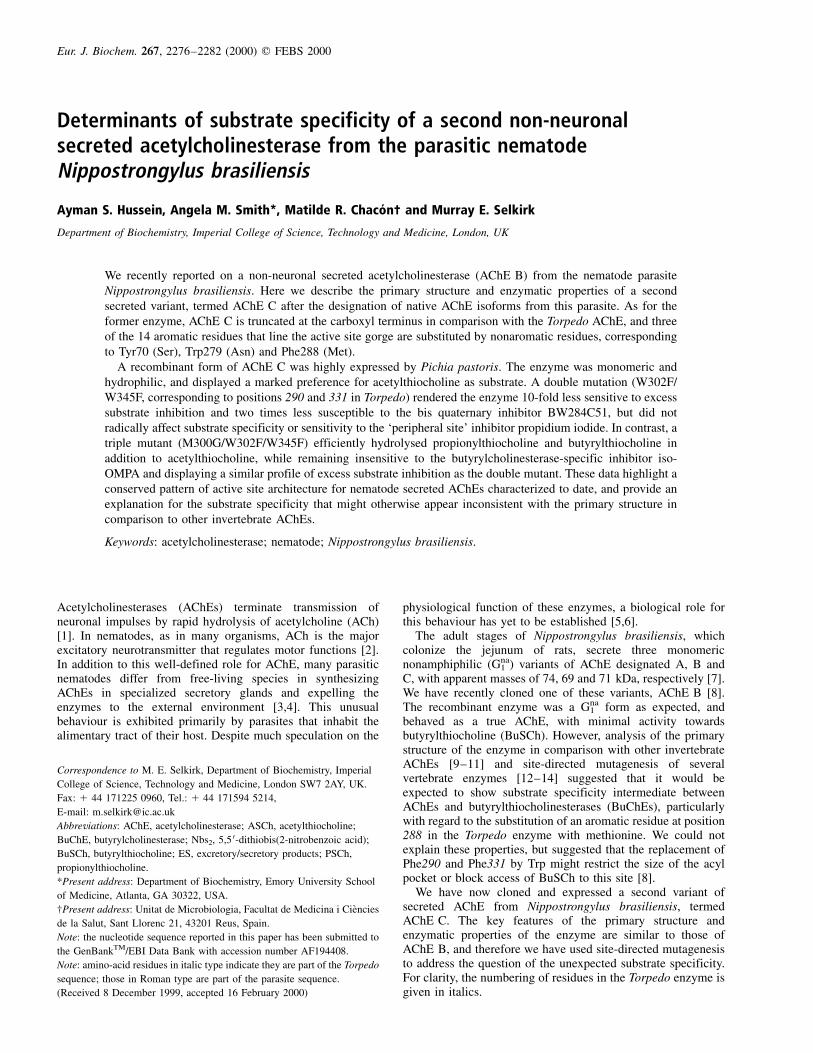
Determinants of substrate specificity of a second non-neuronalsecreted acetylcholinesterase from the parasitic nematodeNippostrongylus brasiliensis
Ayman S. Hussein, Angela M. Smith*, Matilde R. Chaco n² and Murray E. Selkirk
Department of Biochemistry, Imperial College of Science, Technology and Medicine, London, UK
We recently reported on a non-neuronal secreted acetylcholinesterase (AChE B) from the nematode parasite
Nippostrongylus brasiliensis. Here we describe the primary structure and enzymatic properties of a second
secreted variant, termed AChE C after the designation of native AChE isoforms from this parasite. As for the
former enzyme, AChE C is truncated at the carboxyl terminus in comparison with the Torpedo AChE, and three
of the 14 aromatic residues that line the active site gorge are substituted by nonaromatic residues, corresponding
to Tyr70 (Ser), Trp279 (Asn) and Phe288 (Met).
A recombinant form of AChE C was highly expressed by Pichia pastoris. The enzyme was monomeric and
hydrophilic, and displayed a marked preference for acetylthiocholine as substrate. A double mutation (W302F/
W345F, corresponding to positions 290 and 331 in Torpedo) rendered the enzyme 10-fold less sensitive to excess
substrate inhibition and two times less susceptible to the bis quaternary inhibitor BW284C51, but did not
radically affect substrate specificity or sensitivity to the `peripheral site' inhibitor propidium iodide. In contrast, a
triple mutant (M300G/W302F/W345F) efficiently hydrolysed propionylthiocholine and butyrylthiocholine in
addition to acetylthiocholine, while remaining insensitive to the butyrylcholinesterase-specific inhibitor iso-
OMPA and displaying a similar profile of excess substrate inhibition as the double mutant. These data highlight a
conserved pattern of active site architecture for nematode secreted AChEs characterized to date, and provide an
explanation for the substrate specificity that might otherwise appear inconsistent with the primary structure in
comparison to other invertebrate AChEs.
Keywords: acetylcholinesterase; nematode; Nippostrongylus brasiliensis.
Acetylcholinesterases (AChEs) terminate transmission ofneuronal impulses by rapid hydrolysis of acetylcholine (ACh)[1]. In nematodes, as in many organisms, ACh is the majorexcitatory neurotransmitter that regulates motor functions [2].In addition to this well-defined role for AChE, many parasiticnematodes differ from free-living species in synthesizingAChEs in specialized secretory glands and expelling theenzymes to the external environment [3,4]. This unusualbehaviour is exhibited primarily by parasites that inhabit thealimentary tract of their host. Despite much speculation on the
physiological function of these enzymes, a biological role forthis behaviour has yet to be established [5,6].
The adult stages of Nippostrongylus brasiliensis, whichcolonize the jejunum of rats, secrete three monomericnonamphiphilic (Gna
1 ) variants of AChE designated A, B andC, with apparent masses of 74, 69 and 71 kDa, respectively [7].We have recently cloned one of these variants, AChE B [8].The recombinant enzyme was a Gna
1 form as expected, andbehaved as a true AChE, with minimal activity towardsbutyrylthiocholine (BuSCh). However, analysis of the primarystructure of the enzyme in comparison with other invertebrateAChEs [9±11] and site-directed mutagenesis of severalvertebrate enzymes [12±14] suggested that it would beexpected to show substrate specificity intermediate betweenAChEs and butyrylthiocholinesterases (BuChEs), particularlywith regard to the substitution of an aromatic residue at position288 in the Torpedo enzyme with methionine. We could notexplain these properties, but suggested that the replacement ofPhe290 and Phe331 by Trp might restrict the size of the acylpocket or block access of BuSCh to this site [8].
We have now cloned and expressed a second variant ofsecreted AChE from Nippostrongylus brasiliensis, termedAChE C. The key features of the primary structure andenzymatic properties of the enzyme are similar to those ofAChE B, and therefore we have used site-directed mutagenesisto address the question of the unexpected substrate specificity.For clarity, the numbering of residues in the Torpedo enzyme isgiven in italics.
Eur. J. Biochem. 267, 2276±2282 (2000) q FEBS 2000
Correspondence to M. E. Selkirk, Department of Biochemistry, Imperial
College of Science, Technology and Medicine, London SW7 2AY, UK.
Fax: 1 44 171225 0960, Tel.: 1 44 171594 5214,
E-mail: [email protected]
Abbreviations: AChE, acetylcholinesterase; ASCh, acetylthiocholine;
BuChE, butyrylcholinesterase; Nbs2, 5,5 0-dithiobis(2-nitrobenzoic acid);
BuSCh, butyrylthiocholine; ES, excretory/secretory products; PSCh,
propionylthiocholine.
*Present address: Department of Biochemistry, Emory University School
of Medicine, Atlanta, GA 30322, USA.
²Present address: Unitat de Microbiologia, Facultat de Medicina i CieÁncies
de la Salut, Sant Llorenc 21, 43201 Reus, Spain.
Note: the nucleotide sequence reported in this paper has been submitted to
the GenBankTM/EBI Data Bank with accession number AF194408.
Note: amino-acid residues in italic type indicate they are part of the Torpedo
sequence; those in Roman type are part of the parasite sequence.
(Received 8 December 1999, accepted 16 February 2000)
q FEBS 2000 Secreted acetylcholinesterase from N. brasiliensis (Eur. J. Biochem. 267) 2277
M A T E R I A L S A N D M E T H O D S
Parasites
N. brasiliensis were isolated from the small intestine of maleSprague-Dawley rats 4 days post-infection. Excretory/secretory(ES) products were collected from culture supernatants asdescribed [7] and used for nondenaturating gel electrophoresis.
Cloning, sequencing and site directed mutagenesis
A lambda ZAP cDNA library was screened with a cDNA probeencoding AChE B, and clones sequenced by dideoxy chaintermination following in vivo excision with Exassist helperphage. As a result, a cDNA clone was isolated that encoded aprotein with an amino-acid sequence distinct from that ofAChE B. This protein was designated AChE C. Alignment ofAChE C with AChEs from other sources was performed usingthe CLUSTALW programme [15]. Single, double and triplemutants of AChE C were generated for M300G (Phe288 inTorpedo), W302F (Phe290 in Torpedo), and W345F (Phe331 inTorpedo) using the Quickchange site-directed mutagenesis kit(Stratagene), by conversion of ATG to GGG, and TGG to TTT,respectively. Mutations were confirmed by sequencing prior toexpression as secreted proteins in Pichia pastoris.
Expression in Pichia pastoris
The cDNA clones encoding wild-type and mutant AChE Cwere ligated into Pst1/Xbal-digested pPICZaB and plasmidslinearized with BstXI. Competent P. pastoris were transfectedwith the constructs, which encoded the mature proteins withan N-terminal signal peptide provided by the preprosequence of the a-mating factor of Saccharomyces cerevisiae,and a C-terminal polyhistidine tag. Recombinant yeast weregrown and expression induced as previously described [8].Culture supernatants were harvested at different time pointspostmethanol induction and analysed for AChE activity andprotein content. Wild-type and mutant AChE C were purifiedfrom culture supernatants of transformed P. pastoris by nickel-agarose chromatography (Qiagen) as previously described [8]and the purity of the enzyme determined by SDS/PAGE.
Sucrose density centrifugation
Sedimentation analysis was performed in 2±20% sucrosegradients (11 mL, with a 0.5 mL cushion of 50% sucrose) inNaCl/Pi in the presence or absence of 1.0% Triton X-100. Thepurified recombinant enzyme was applied to the gradient andcentrifuged for 16 h at 36 000 r.p.m. at 4 8C in an SW41 rotor(170 000 g). Fractions (0.25 mL) were collected and assayedfor AChE activity. Bovine liver catalase (11.3 S) and E. colialkaline phosphatase (6.1 S) were included as internalstandards.
Denaturing and nondenaturing electrophoresis
SDS/PAGE was performed on 12% polyacrylamide gelsfollowed by staining with Coomassie brilliant blue. Purifiedrecombinant AChE C, AChE B and ES products were resolvedunder nondenaturing conditions by electrophoresis in 7.5%polyacrylamide gels in Tris/Borate/EDTA buffer, pH 8.0, andenzyme activity visualized by the method of Karnovsky andRoots [16].
AChE activity: substrate and inhibitor specificities
AChE activity was determined according to Ellman et al. [17].The standard incubation mixture contained 1 mm acetylthio-choline (ASCh) iodide as substrate in the presence of 1 mm5,5 0-dithiobis(2-nitrobenzoic acid) (Nbs2) in 100 mm sodiumphosphate pH 7.0 at 20 8C. The reaction was monitored bymeasuring the absorbance at 412 nm, and hydrolysis of ASChcalculated from the extinction coefficient of Nbs2 [17]. Oneunit of activity is defined as 1 mmol of substrate hydrolysed permin at 20 8C. The Km values for ASCh, butyrylthiocholine(BuSCh), and propionylthiocholine (PSCh) of wild-type andmutant enzymes were determined by linear regression fromplots of 1/V against 1/S, utilizing substrate concentrations in therange 0.01±1 mm (i.e. below excess substrate inhibition). Thesubstrate inhibition constant (Kss) values were determined byplotting 1/V against S, utilizing a range of substrate concen-trations between 5 and 30 mm for wild type and 30 and 80 mmfor mutant AChE. Inhibition constants (Ki) for BW284C51 andpropidium iodide were determined using two fixed inhibitorand six ASCh concentrations between 0.01 and 1.0 mm. Assayswere carried out in triplicate in the presence or absence ofinhibitor. Kinetic constants were determined using graphpadprism 2.0 software (Graphpad, San Diego, CA).
R E S U L T S
Cloning and structural features of AChE C
In order to isolate cDNAs encoding alternative forms ofsecreted AChEs, we screened a lambda ZAP cDNA library witha full-length clone for AChE B [8]. Several clones wereisolated by this procedure that differed significantly innucleotide sequence from AChE B. The longest clone was1848 bp, containing a single ORF of 560 amino acids, aconsensus polyadenylation site (AATAAA) and a poly(A) tail(GenBankTM accession number AF194408). The amino-acidsequence of the predicted mature protein was 90% identicalto AChE B, 28% identical to Caenorhabditis elegans ACE-1[11] and 35% identical to Torpedo californica AChE [18].Figure 1 depicts the alignment of AChE C with AChE B ofN. brasiliensis, ACE-1 of C. elegans and the T subunit ofT. californica AChE. By analogy with the N-terminalsequence of mature AChE B, the protein is numbered fromthe first aspartate residue. Although lacking an initiatormethionine, the protein has a largely hydrophobic N-terminalstretch of 22 amino acids similar to that of AChE B andindicative of a signal peptide. Serine, glutamate and histidineresidues are present in positions corresponding to the catalytictriad of AChEs [19]. Six cysteine residues are found inpositions that correspond to those responsible for the threeconserved intramolecular disulphide bonds, but as for AChE B,an additional two cysteine residues are present at positions 231and 262. The protein is truncated at the C-terminus incomparison to AChEs from other sources, and there are twoconsensus sequences for N-linked glycosylation at positions124 and 376.
Eleven of the 14 aromatic residues that line the wall of theactive site gorge in the Torpedo enzyme [19] are eitherconserved or show conservative substitutions. Two of thenonconservative substitutions, Asn289 (Trp279) and Met300(Phe288) are identical to AChE B, whereas Tyr70 is substitutedby Ser65 rather than threonine.
2278 M. E. Selkirk et al. (Eur. J. Biochem. 267) q FEBS 2000
Fig. 1. Alignment of N. brasiliensis AChE C
with other AChEs. The derived amino-acid
sequence of N. brasiliensis AChE C was aligned
with that of AChE B, ACE-1 from C. elegans
(Ce), and the T subunit of T. californica (Tc).
T. californica AChE is numbered from
N-terminus of the mature protein (marked `1'),
and AChE C by analogy with the N-terminal
sequence of AChE B [8]. The residues in the
catalytic triad (Ser-His-Glu) are indicated with an
asterisk, and the position of two potential sites for
N-linked glycosylation marked with squares.
Cysteine residues at positions known to be
involved in intramolecular disulphide bonds are
indicated by solid triangles, whereas open
triangles depict the two additional cysteines in
the N. brasiliensis enzymes. The positions of
aromatic residues that line the active site gorge
in Torpedo AChE are marked with circles.
Solid circles indicate conserved residues or
conservative substitutions at these positions in the
N. brasiliensis AChEs, whereas open circles
indicate nonaromatic residues.
Table 1. Kinetic parameters of wild-type and mutant enzymes. Km (mm), Kss (mm), kcat (min21) and kcat /Km (m21 ´min21) for acetylthiocholine (ASCh),
propionylthicholine (PSCh) and butyrylthiocholine (BuSCh) were determined as described in Materials and methods. Figures represent the mean ^ SD of
three independent experiments. ND, not determined.
Substrate Wild-type W302F, W345F M300G, W302F, W345F
ASCh
Km 0.14 �^ 0.01 0.20 �^ 0.02 0.30 �^ 0.03
Kss 1.31 �^ 0.21 13.15 �^ 4.50 18.30 �^ 2.67
kcat 1.10 �^ 0.23 � 105 1.46 �^ 0.30 � 105 4.01 �^ 0.41 � 105
kcat /Km 7.86 � 108 7.30 � 108 13.37 � 108
PSCh
Km 0.23 �^ 0.02 0.15 �^ 0.02 0.12 �^ 0.04
Kss 2.10 �^ 0.56 7.29 �^ 3.24 10.31 �^ 2.54
kcat 1.01 �^ 0.17 � 105 1.11 �^ 0.28 � 105 1.21 �^ 0.19 � 105
kcat /Km 4.39 � 108 7.40 � 108 10.08 � 108
BuSCh
Km ND ND 0.12 �^ 0.03
Kss ND ND ND
kcat ND ND 0.38 �^ 0.04 � 105
kcat /Km ND ND 3.17 � 108
q FEBS 2000 Secreted acetylcholinesterase from N. brasiliensis (Eur. J. Biochem. 267) 2279
Expression and properties of recombinant enzyme
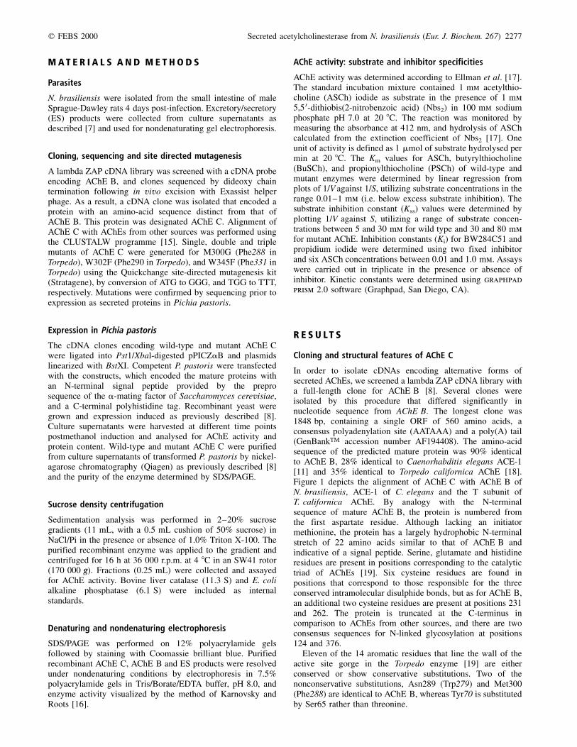
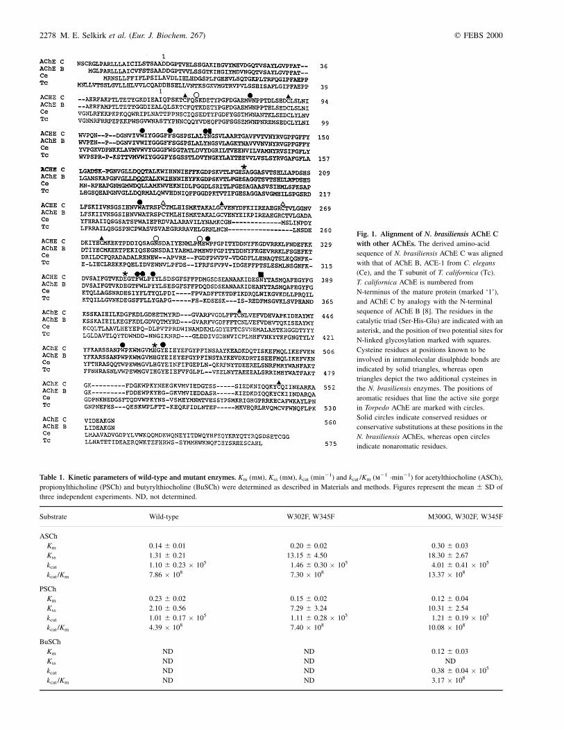
AChE C was next expressed as a secreted protein in P. pastoris.Approximately 50 mg total protein was secreted per litre ofculture medium by 80 h postmethanol induction, and more thanhalf of this was accounted for by AChE C (Fig. 2). The purifiedenzyme had an apparent mass of 73 kDa when resolved bySDS/PAGE under reducing conditions. Sucrose density gradientcentrifugation identified AChE C as a monomeric non-amphiphilic (Gna
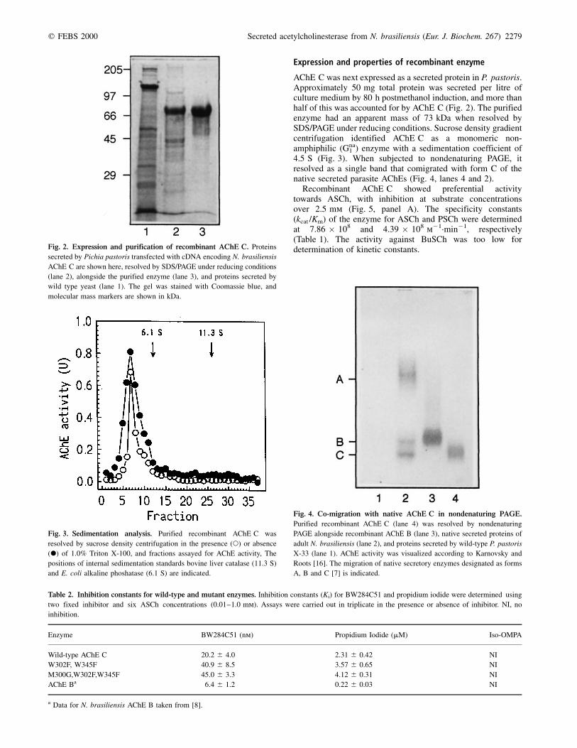
1 ) enzyme with a sedimentation coefficient of4.5 S (Fig. 3). When subjected to nondenaturing PAGE, itresolved as a single band that comigrated with form C of thenative secreted parasite AChEs (Fig. 4, lanes 4 and 2).
Recombinant AChE C showed preferential activitytowards ASCh, with inhibition at substrate concentrationsover 2.5 mm (Fig. 5, panel A). The specificity constants(kcat /Km) of the enzyme for ASCh and PSCh were determinedat 7.86 � 108 and 4.39 � 108 m21´min21, respectively(Table 1). The activity against BuSCh was too low fordetermination of kinetic constants.Fig. 2. Expression and purification of recombinant AChE C. Proteins
secreted by Pichia pastoris transfected with cDNA encoding N. brasiliensis
AChE C are shown here, resolved by SDS/PAGE under reducing conditions
(lane 2), alongside the purified enzyme (lane 3), and proteins secreted by
wild type yeast (lane 1). The gel was stained with Coomassie blue, and
molecular mass markers are shown in kDa.
Table 2. Inhibition constants for wild-type and mutant enzymes. Inhibition constants (Ki) for BW284C51 and propidium iodide were determined using
two fixed inhibitor and six ASCh concentrations (0.01±1.0 mm). Assays were carried out in triplicate in the presence or absence of inhibitor. NI, no
inhibition.
Enzyme BW284C51 (nm) Propidium Iodide (mM) Iso-OMPA
Wild-type AChE C 20.2 �^ 4.0 2.31 �^ 0.42 NI
W302F, W345F 40.9 �^ 8.5 3.57 �^ 0.65 NI
M300G,W302F,W345F 45.0 �^ 3.3 4.12 �^ 0.31 NI
AChE Ba 6.4 �^ 1.2 0.22 �^ 0.03 NI
a Data for N. brasiliensis AChE B taken from [8].
Fig. 3. Sedimentation analysis. Purified recombinant AChE C was
resolved by sucrose density centrifugation in the presence (W) or absence
(X) of 1.0% Triton X-100, and fractions assayed for AChE activity, The
positions of internal sedimentation standards bovine liver catalase (11.3 S)
and E. coli alkaline phoshatase (6.1 S) are indicated.
Fig. 4. Co-migration with native AChE C in nondenaturing PAGE.
Purified recombinant AChE C (lane 4) was resolved by nondenaturing
PAGE alongside recombinant AChE B (lane 3), native secreted proteins of
adult N. brasiliensis (lane 2), and proteins secreted by wild-type P. pastoris
X-33 (lane 1). AChE activity was visualized according to Karnovsky and
Roots [16]. The migration of native secretory enzymes designated as forms
A, B and C [7] is indicated.

2280 M. E. Selkirk et al. (Eur. J. Biochem. 267) q FEBS 2000
Generation and properties of mutant enzymes
AChE C thus behaved as a true AChE with minimal activitytowards BuSCh, despite the substitution of an aromatic residueat position 288 in the Torpedo enzyme with methionine. Theseproperties are identical to those recently described for AChE Bof N. brasiliensis [8], and we therefore sought to resolve thisapparent anomaly by site-directed mutagenesis. Both parasiteenzymes contain tryptophan in place of phenylalanine atpositions 290 and 331, and based on the assumption that thelarger sidechain might restrict the size of or access of substrateto the acyl pocket, we performed single and double mutationsin which these residues were replaced by phenylalanine. Thesingle mutations, namely W302F (Phe290) or W345F(Phe331), did not significantly alter the properties of AChE Cwith respect to substrate specificity or excess substrateinhibition (data not shown). The double mutant (W302F/345F), however, was approximately 10 times less sensitive toexcess substrate inhibition with ASCh. It showed slightlyenhanced activity against PSCh, although BuSCh was stillinefficiently hydrolysed. In fact, a reproducible observation wasthat BuSCh at concentrations greater than 1 mm was hydro-lysed less efficiently by the double mutant than by the wild-typeenzyme, although we have no explanation for this (Fig. 5, panelB and Table 1).
We next constructed a triple mutant (M300G/W302F/W345F) to generate an enzyme with residues in these positionsidentical to those of C. elegans ACE-1, which efficientlyhydrolyses both ASCh and BuSCh [11]. The enzyme had asimilar profile of excess substrate inhibition by ASCh asW302F/345F, but now readily accepted BuSCh, and had alower Km and enhanced activity towards PSCh (Fig. 5, panel Cand Table 1). Although the Km of the triple mutant for ASChwas approximately two-fold that of the wild type enzyme, itshowed a four-fold enhancement of turnover and an overallimproved catalytic efficiency with this substrate (Table 1). Theproperties of the single mutant M300G were not significantlyaltered from those of the wild-type enzyme (data not shown).
The susceptibility of wild-type and mutant AChE C tocholinesterase inhibitors was also examined. None of theenzymes were affected by the butyrylcholinesterase inhibitoriso-OMPA at concentrations up to 10 mm (data not shown), butthe wild-type enzyme was highly sensitive to BW284C51, aspecific inhibitor of AChEs [20] with a Ki of 20.2 nm, and the`peripheral' site ligand propidium iodide, with a Ki of 2.31 mm,in a competitive and noncompetitive manner, respectively. Bothmutant enzymes were less susceptible to inhibition byBW284C51, but showed no significant difference in inhibitionby propidium iodide (Table 2).
D I S C U S S I O N
We describe here the primary structure and properties of anacetylcholinesterase (AChE C) secreted by the parasiticnematode N. brasiliensis. This is the second example of thistype of enzyme to be reported. Both AChE C and AChE B aremaximally expressed by the parasite in the later stages ofinfection of the mammalian host. AChE C can be distinguishedfrom AChE B by SDS/PAGE on the basis of its mass, and bynondenaturing PAGE by its more acidic nature, which results inaccelerated migration. The recent determination of the primarystructure of AChE B revealed some very unusual features [8]that are largely conserved in AChE C. AChE C is similarlytruncated at the C-terminus, which aligns closely with the endof the catalytic domain of the vertebrate enzymes. The parasite
Fig. 5. Substrate specificity of wild type and mutant AChE C. The rate
of hydrolysis of ASCh (B), PSCh (P) and BuSCh (O) were determined as
described in Materials and methods, utilizing a range of substrate
concentrations between 0.05 and 30 mm for the wild-type enzyme (panel
A), and between 0.05 and 80 mm for the double mutant W302F/W345F
(panel B), and the triple mutant M300G/W302F/W345F (panel C).
q FEBS 2000 Secreted acetylcholinesterase from N. brasiliensis (Eur. J. Biochem. 267) 2281
enzymes therefore do not contain sequences corresponding tothe peptides of vertebrate AChEs that define hydrophobic (H)or tailed (T) subunits [21], and lack the C-terminal cysteineresidue involved in intermolecular disulphide-bridge formation,thus explaining their monomeric nature. In this respect,however, they show some similarity with nonamphiphilicmonomeric AChEs from snake venom [22,23]. In additionto the six cysteine residues implicated in intramoleculardisulphide bridges, AChE C has two cysteines at positions 231and 261, the former of which is predicted to lie on themolecular surface and the latter lies in an insertion of 17 aminoacids (relative to Torpedo AChE, see Fig. 1) which is difficultto model, but most probably forms part of a loop at the surfacestabilized by the second disulphide bond. It is notable that boththis insertion and the additional two cysteine residues areconserved in AChE B in identical positions [8], and we aretherefore in the process of determining whether these could beinvolved in an additional disulphide bridge.
Both the native and the recombinant enzymes are known tobe glycosylated, although the sites have not been mapped.AChE C differs from AChE B in lacking a consensus sequencefor N-linked glycosylation at position 480. There are thereforetwo consensus sequences common to both enzymes at positions124 and 376. Of these, Asn376 would appear to be the mostlikely candidate for glycosylation, as Asn124 is positioneddirectly next to a tyrosine residue predicted to line the activesite gorge (Tyr130 in Torpedo). No other acetylcholinesterasesappear to have consensus sequences for glycosylation athomologous sites.
The active site gorge of the Torpedo enzyme is lined by 14aromatic residues that are highly conserved in AChEs fromdifferent species, and thought to contribute to a substrateguidance mechanism [19]. Eleven of these residues are eitherconserved or show conservative substitutions in both of theparasite secreted enzymes. Of the three nonconservativesubstitutions, AChE C differs subtly from AChE B in posses-sing a serine residue at position 65 rather than a threonine,corresponding to Tyr70 in Torpedo AChE.
As these are the only two secreted AChEs from parasiticnematodes to be cloned and sequenced thus far, it is prematureto predict whether the primary structure of analogous enzymeswill follow a similar pattern. All such enzymes analysed to dateshow a marked substrate preference for acetylcholine. TheAChE secreted by the hookworm Necator americanus exists asa hydrophilic dimer [24], whereas both Gna
1 and Gna2 forms have
been identified in secreted products of Trichostrongyluscolubriformis [25]. Nevertheless, despite the fact that thethird variant of the N. brasiliensis secreted AChEs (AChE A)has not been fully characterized, the similarity between theother two forms in primary structure and enzymatic propertiesbegs the question of why this parasite secretes multiple AChEs.Forms B and C are produced relatively soon after form A, andtherefore sequential production of variants that would escapebinding by antibodies and possible neutralization of enzymeactivity is an unlikely explanation, made even more improbableby the close similarity in sequence between the two enzymescharacterized to date. Given that the enzymes are secreted intothe extremely hydrolytic environment provided by the intestinaltract, another possibility is that the AChEs differ in theirsusceptibility to proteolytic degradation, although this has notyet been assessed. It is also possible that the secreted AChEsare targetted to different sites in the jejunal mucosa, and this iscurrently under investigation.
Both N. brasiliensis AChE B and C have a methionineresidue in the position corresponding to Phe288 in the Torpedo
enzyme. This was somewhat surprising given their minimalactivity with BuSCh as substrate, as mutagenesis studies onTorpedo and human AChE have shown that the presence of anaromatic amino acid in this position and at 290 block access ofBuSCh to the active site [12±14]. Moreover, the presence ofleucine and glycine at position 288 in Drosophila melanogasterand C. elegans ACE-1, respectively, has been proposed to beresponsible for the activity of these enzymes against BuSCh[10,11]. We therefore investigated the possibility that thesubstitution of Phe290 and Phe331 by Trp in the nematodeenzymes might restrict the size of the acyl pocket or entrance ofsubstrate to this site by virtue of the bulkier sidechains. Asshown in Fig. 5, the double mutation of tryptophan tophenylalanine at these positions had little effect on substratespecificity. In contrast, the triple mutant (M300G/W302F/W345F) showed good activity with PSCh and BuSCh (Fig. 5and Table 1), indicating that the collective effect of largerresidues in all these three positions acted to restrict access oflarger substrates. The triple mutant showed a higher Km , but anenhanced turnover of ASCh (Table 1). Although we have noexplanation for this, the larger binding pocket might increasethe rate of acylation, deacylation and/or product release.
Perhaps more surprising was the effect on excess substrateinhibition, which was shifted approximately 10-fold in themutant enzymes (Fig. 5 and Table 1). Mutation of Phe297 inmouse AChE (Phe290 in Torpedo) to isoleucine has beenreported to abolish substrate inhibition, in addition to reducingcatalytic activity of the enzyme [13]. Single mutations ofTrp302 (Phe290 in Torpedo) and Trp345 (Phe331 in Torpedo)in the N. brasiliensis AChE to Phe had no effect on substrateinhibition or specific activity (data not shown). In the doublemutant (W302F/W345F), decreased sensitivity to substrateinhibition was observed, with little effect on kcat or the bindingof the `peripheral' site inhibitor propidium iodide. Althoughthese residues are not implicated in binding of propidium,Trp302 is presumably involved in substrate orientation, andboth this residue and Trp345 may be involved in allostericalterations following binding of a second substrate molecule toa lower affinity site. Mutation of both these residues tophenylalanine appears sufficient to influence these putativeallosteric effects without altering catalytic efficiency of thenematode enzyme.
AChE C is sensitive to propidium iodide, although the Ki
value of 2.31 mm (Table 2) is approximately 10-fold higherthan that recorded for AChE B [8]. AChE C has an acidicresidue (Glu) at position 295 (285 in Torpedo) that has beendemonstrated to be important for sensitivity of the Bungarusfasciatus AChE to peripheral site inhibitors [23], but as forAChE B, two other aromatic residues that contribute to thisphenomenon, Tyr70 and Trp279 [12,14,23] are substituted bynonaromatic amino acids. Tyr70 is replaced by serine inAChE C and threonine in AChE B, whereas both parasiteenzymes have an asparagine in place of Trp279.
Neither the wild-type nor any of the mutant nematodeenzymes reported here were susceptible to inhibition by iso-OMPA, despite the triple mutant showing reasonable activitywith BuSCh, in contrast to mutagenesis studies of vertebrateAChEs [12±14]. The observed differences in activity andinhibitor profiles between the N. brasiliensis and vertebrateAChEs can only be satisfactorily resolved by structuralanalysis, which will be facilitated by the high level ofexpression of the nematode enzymes in P. pastoris.
We are currently working to determine the function of thenematode secreted AChEs, the expression of which have beendemonstrated to be positively regulated by elements of the host
2282 M. E. Selkirk et al. (Eur. J. Biochem. 267) q FEBS 2000
immune system [26,27]. We have recently identified a popu-lation of cells in the lamina propria and draining mesentericlymph nodes of rats that express muscarinic ACh receptors inresponse to infection with the parasite (W. S. Russell, S. M.Henson & M. E. Selkirk, unpublished data). Identification ofthese cells and determination of their response to ACh will helpto define the function of the nematode secreted AChEs, inaddition to uncovering a possible link between the entericnervous and immune systems.
A C K N O W L E D G E M E N T S
This work was supported by the Wellcome Trust. We thank Israel Silman
and Xavier Cousin for constructive comments.
R E F E R E N C E S
1. Barnard, E.A. (1974) Enzymatic destruction of acetylcholine. In The
Peripheral Nervous System (Hubbard, J.I., ed.), pp. 201±224. Plenum
Press, New York, USA.
2. Rand, J.B. & Nonet, M.L. (1997) Synaptic transmission. In C. elegans
II (Riddle, D.L.T., Blumenthal, T.B.J., Meyer, B.J. & Priess, J.R.,
eds), pp. 611±643. Cold Spring Harbor Laboratory Press, Cold
Spring Harbor, New York, USA.
3. Ogilvie, B.M., Rothwell, T.L.W., Bremner, K.C., Schitzerling, H.J.,
Nolan, J. & Keith, R.K. (1973) Acetylcholinesterase secretion by
parasitic nematodes, 1. Evidence for secretion of the enzyme by a
number of species. Int. J. Parasitol. 3, 589±597.
4. McLaren, D.J., Burt, J.S. & Ogilvie, B.M. (1974) The anterior glands
of adult Necator americanus (nematoda: strongyloidea) II. Cyto-
chemical and functional studies. Int. J. Parasitol. 4, 39±46.
5. Rhoads, M.L. (1981) Cholinesterase in the parasitic nematode
Stephanurus dentatus. Characterisation and sex dependence of a
secretory cholinesterase. J. Biol. Chem. 256, 9316±9323.
6. Lee, D.L. (1996) Why do some nematode parasites of the alimentary
tract secrete acetylcholinesterase? Int. J. Parasitol. 26, 499±508.
7. Grigg, M.E., Tang, L., Hussein, A.S. & Selkirk, M.E. (1997)
Purification and properties of monomeric (G1) forms of acetyl-
cholinesterase secreted by Nippostrongylus brasiliensis. Mol.
Biochem. Parasitol. 90, 513±524.
8. Hussein, A.S., ChacoÂn, M.R., Smith, A.M., Tosado-Acevedo, R. &
Selkirk, M.E. (1999) Cloning, expression and properties of a non-
neuronal secreted acetylcholinesterase from the parasitic nematode
Nippostrongylus brasiliensis. J. Biol. Chem. 274, 9312±9319.
9. Hall, L.M.C. & Spierer, P. (1986) The Ace locus of Drosophila
melanogaster: structural gene for acetylcholinesterase with an
unusual 5 0 leader. EMBO J. 5, 2949±2954.
10. Gnagey, A.L., Forte, M. & Rosenberry, T.L. (1987) Isolation and
characterization of acetylcholinesterase from Drosophila. J. Biol.
Chem. 262, 13290±13298.
11. Arpagaus, M., Fedon, Y., Cousin, X., Chatonnet, A., BergeÂ, J.-B.,
Fournier, D. & Toutant, J.-P. (1994) cDNA sequence, gene
structure, and in vitro expression of ace-1, the gene encoding
acetylcholinesterase of class A in the nematode Caenorhabditis
elegans. J. Biol. Chem. 269, 9957±9965.
12. Harel, M., Sussman, J.L., Krejci, E., Bon, S., Chanal, P., MassoulieÂ,
J. & Silman, I. (1992) Conversion of acetylcholinesterase to
butyrylcholinesterase: modelling and mutagenesis. Proc. Natl Acad.
Sci. USA 89, 10827±10831.
13. Vellom, D.C., Radic, Z., Li, Y., Pickering, N.A., Camp, S. & Taylor, P.
(1993) Amino acid residues controlling acetylcholinesterase and
butyrylcholinesterase specificity. Biochemistry 32, 12±17.
14. Ordentlich, A., Barak, D., Kronman, C., Flashner, Y., Leitner, M.,
Segall, Y., Ariel, N., Cohen, S., Velan, B. & Shafferman, A. (1993)
Dissection of the human acetylcholinesterase active center deter-
minants of substrate specificity. J. Biol. Chem. 268, 17083±17095.
15. Thompson, J.D., Higgins, D.G. & Gibson, T.J. (1994) CLUSTAL W:
improving the sensitivity of progressive multiple sequence alignment
through sequence weighting, position-specific gap penalties and
weight matrix choice. Nucleic Acids Res. 22, 4673±4680.
16. Karnovsky, M.J. & Roots, L. (1964) A `direct-coloring' method for
cholinesterases. J. Histochem. Cytochem. 12, 219±221.
17. Ellman, G.L., Courtney, K.D., Andres, V. & Featherstone, R.M. (1961)
A new and rapid colorimetric determination of acetylcholinesterase
activity. Biochem. Pharmacol. 7, 88±95.
18. Schumacher, M., Camp, S., Maulet, Y., Newton, M., MacPhee-Quigley,
K., Taylor, S.S., Friedmann, T. & Taylor, P. (1986) Primary structure
of Torpedo californica acetylcholinesterase deduced from its cDNA
sequence. Nature 319, 407±409.
19. Sussman, J.L., Harel, M., Frolow, F., Oefner, C., Goldman, C., Toker,
L. & Silman, I. (1991) Atomic structure of acetylcholinesterase from
Torpedo californica: a prototypic acetylcholine-binding protein.
Science 253, 872±879.
20. Austin, L. & Berry, W.K. (1953) Two selective inhibitors of
cholinesterase. Biochem. J. 54, 695±700.
21. MassoulieÂ, J., Sussman, J.L., Bon, S. & Silman, I. (1993) Structure
and functions of acetylcholinesterase and butyrylcholinesterase.
Prog. Brain Res. 98, 139±146.
22. Cousin, X., CreÂminon, C., Grassi, J., MeÂflah, K., Cornu, G., Saliou, B.,
Bon, S., MassoulieÂ, J. & Bon, C. (1996) Acetylcholinesterase from
Bungarus venom: a monomeric species. FEBS Lett. 387, 196±200.
23. Cousin, X., Bon, S., Duval, N., MassoulieÂ, J. & Bon, C. (1996) Cloning
and expression of acetylcholinesterase from Bungarus fasciatus
venom. J. Biol. Chem. 271, 15099±15108.
24. Pritchard, D.I., Brown, A. & Toutant, J.-P. (1994) The molecular forms
of acetylcholinesterase from Necator americanus (Nematoda), a
hookworm parasite of the human intestine. Eur. J. Biochem. 219,
317±323.
25. Griffiths, G. & Pritchard, D.I. (1994) Purification and biochemical
characterisation of acetylcholinesterase (AChE) from the excretory/
secretory products of Trichostrongylus colubriformis. Parasitology
108, 579±586.
26. Jones, V.E. & Ogilvie, B.M. (1972) Protective immunity to
Nippostrongylus brasiliensis in the rat III. Modulation of worm
acetylcholinesterase by antibodies. Immunology 22, 119±129.
27. Sanderson, B.E., Jenkins, D.C. & Ogilvie, B.M. (1972) Nippo-
strongylus brasiliensis: relation between immune damage and
acetylcholinesterase levels. Int. J. Parasitol. 2, 227±232.
Copyright © 2022 FDOKUMEN