Metazoan parasite communities of three endemic cichlid fish ...
Taxonomic and Functional Microbial Signatures of theEndemic Marine Sponge Arenosclera brasiliensisAmaro E. Trindade-Silva1,2, Cintia Rua2, Genivaldo G. Z. Silva3, Bas E. Dutilh3,4, Ana Paula B. Moreira2,
Robert A. Edwards3,5, Eduardo Hajdu6, Gisele Lobo-Hajdu7, Ana Tereza Vasconcelos8,
Roberto G. S. Berlinck1, Fabiano L. Thompson2*
1 Instituto de Quımica de Sao Carlos, Universidade de Sao Paulo, Sao Carlos, Brazil, 2 Instituto de Biologia, Universidade Federal do Rio de Janeiro, Rio de Janeiro, Brazil,
3 Department of Computer Science, San Diego State University, San Diego, United States of America, 4 Centre for Molecular and Biomolecular Informatics, Nijmegen
Centre for Molecular Life Sciences, Radboud University Nijmegen Medical Centre, Nijmegen, The Netherlands, 5 Division of Mathematics and Computer Science, Argonne
National Laboratory, Argonne, United States of America, 6 Departamento de Invertebrados, Museu Nacional, Universidade Federal do Rio de Janeiro, Rio de Janeiro, Brazil,
7 Instituto de Biologia Roberto Alcantara Gomes, Universidade do Estado do Rio de Janeiro, Rio de Janeiro, Brazil, 8 Laboratorio Nacional de Computacao Cientıfica,
Petropolis, Brazil
Abstract
The endemic marine sponge Arenosclera brasiliensis (Porifera, Demospongiae, Haplosclerida) is a known source of secondarymetabolites such as arenosclerins A-C. In the present study, we established the composition of the A. brasiliensismicrobiome and the metabolic pathways associated with this community. We used 454 shotgun pyrosequencing togenerate approximately 640,000 high-quality sponge-derived sequences (,150 Mb). Clustering analysis including sponge,seawater and twenty-three other metagenomes derived from marine animal microbiomes shows that A. brasiliensis containsa specific microbiome. Fourteen bacterial phyla (including Proteobacteria, Cyanobacteria, Actinobacteria, Bacteroidetes,Firmicutes and Cloroflexi) were consistently found in the A. brasiliensis metagenomes. The A. brasiliensis microbiome isenriched for Betaproteobacteria (e.g., Burkholderia) and Gammaproteobacteria (e.g., Pseudomonas and Alteromonas)compared with the surrounding planktonic microbial communities. Functional analysis based on Rapid Annotation usingSubsystem Technology (RAST) indicated that the A. brasiliensis microbiome is enriched for sequences associated withmembrane transport and one-carbon metabolism. In addition, there was an overrepresentation of sequences associatedwith aerobic and anaerobic metabolism as well as the synthesis and degradation of secondary metabolites. This studyrepresents the first analysis of sponge-associated microbial communities via shotgun pyrosequencing, a strategy commonlyapplied in similar analyses in other marine invertebrate hosts, such as corals and algae. We demonstrate that A. brasiliensishas a unique microbiome that is distinct from that of the surrounding planktonic microbes and from other marineorganisms, indicating a species-specific microbiome.
Citation: Trindade-Silva AE, Rua C, Silva GGZ, Dutilh BE, Moreira APB, et al. (2012) Taxonomic and Functional Microbial Signatures of the Endemic Marine SpongeArenosclera brasiliensis. PLoS ONE 7(7): e39905. doi:10.1371/journal.pone.0039905
Editor: Jonathan H. Badger, J. Craig Venter Institute, United States of America
Received March 19, 2012; Accepted May 29, 2012; Published July 2, 2012
Copyright: � 2012 Trindade-Silva et al. This is an open-access article distributed under the terms of the Creative Commons Attribution License, which permitsunrestricted use, distribution, and reproduction in any medium, provided the original author and source are credited.
Funding: The authors thank the financial support provided by Fundacao de Amparo a Pesquisa do Estado de Sao Paulo (FAPESP) to RGSB, by FAPESP scholarship2009/11612-1 to AETS, by Conselho Nacional de Desenvolvimento Cientıfico e Tecnologico (CNPq), Coordenacao de Aperfeicoamento de Pessoal de NıvelSuperior (CAPES), and Fundacao de Amparo a Pesquisa do Estado do Rio de Janeiro (FAPERJ) to FLT, and by Dutch Science foundation (NWO) Veni grant:016.111.075 to BED. The funders had no role in study design, data collection and analysis, decision to publish, or preparation of the manuscript.
Competing Interests: The authors have declared that no competing interests exist.
* E-mail: [email protected]
Introduction
Sponges are probably the most primitive metazoans, with fossil
records for this group dating from 635 to 750 million years ago
[1]. As much as 40% of sponge wet weight may be comprised of
microbes, including sponge-specific prokaryotic communities
[2,3,4,5,6]. There are at least 15,000 sponge species on the
planet, inhabiting different types of environments from the deep
sea to riverine systems. Pioneering electron microscopy and
cultivation-dependent approaches suggested the existence of three
groups of sponge-associated microbes: mesohyl sponge-specific
microbes, intracellular symbionts, and non-specific transient
microbial communities that are shared between sponges and the
surrounding water column [2]. Cultivation-independent taxonom-
ic characterization using 16S rRNA library sequencing approach-
es provided a broader, cultivation-independent taxonomic char-
acterization of sponge microbiomes and revealed significant
microbial diversity that included sponge-exclusive microbes, such
as the organisms classified as belonging to the candidate bacterial
phylum Poribacteria [7].
The most recent studies applying massively parallel 16S rRNA
gene tag sequencing, based on approximately 32,000 tag
sequences (read length .125 nt), suggest that the sponge
microbiome may be classified into three main groups (species-
specific, variable, and core) [8,9]. Species-specific microbes
comprise 72% of the taxa detected in sponges, whereas only 2%
of the detected taxa correspond to the core found in several species
of sponges. The five Mediterranean sponges Aplysina aerophoba,
Aplysina cavernicola, Ircinia variabilis, Petrosia ficiformis, and Pseudocorti-
cium jarrei share a core microbiome containing operational
taxonomic units (OTUs) from the phyla Acidobacteria, Chloroflexi,
Nitrospira, Poribacteria, and Proteobacteria [8]. The phylum Chlamydiae
PLoS ONE | www.plosone.org 1 July 2012 | Volume 7 | Issue 7 | e39905
appears to be found only in association with A. cavernicola, whereas
the phylum Lenthisphaerae occurs only in association with I. variabilis
[8]. The core community could represent globally distributed
microbes that are horizontally acquired from the environment by
a sponge, whereas the species-specific community could consist of
microbes with a distribution restricted (endemic) to a single sponge
species that are vertically acquired from the progenitor [9]. A
recent study investigating Cymbastela concentrica microbial diversity
helped to further elucidate possible genetic mechanisms involved
in the establishment of species-specific microbiomes [10]. The
authors generated 190,623 shotgun sequences (92.6 Mbp) and
3,545 16S rRNA sequences (.1,200 bp in length per sequence).
Gammaproteobacteria, Phyllobacteriaceae, Sphingomondales, Neisseriales,
and Nitrospiraceae constituted the vast majority of the microbiome
of C. concentrica. Based on 16S rRNA identification (97% identity
cutoff), only thirty-four different OTUs were common between C.
concentrica and the surrounding seawater, supporting the idea of
selection for a specific microbiome, possibly consisting of several
(though non-culturable) bacteria. Functional analysis performed
via shotgun sequencing using the COG database suggested that
the majority of the detected genes (.85%) belong to bacteria. The
authors also found a large number of sequences identified as
transposable insertion elements. The sponge metagenomes con-
tained a greater number of sequences identified as COG0610
(restriction enzymes, type I helicase) and COG1715 (restriction
endonuclease) than the surrounding seawater. Both COG groups
include specific DNA modification and restriction systems in
bacteria, and therefore, the authors hypothesized that this would
facilitate horizontal DNA exchange between sponge microorgan-
isms and protect against DNA exchange with planktonic
organisms in the surrounding seawater [10].
Despite the significant advances in the study of sponge microbial
diversity in the Mediterranean and Pacific, very little is known
concerning the functional and taxonomic diversity of sponge
microbiomes in the South Atlantic. A recent 16S rRNA sequence-
based study in Rio de Janeiro (southeastern Atlantic region)
generated 133 bacterial sequences from the sponges Hymeniacidon
heliophila and Polymastia janeirensis [14]. These two sponge species
appear to share several bacterial taxa affiliated with Cyanobacteria
and Proteobacteria. Alphaproteobacteria was the most abundant group.
An analysis of 254 archaeal 16S rRNA partial sequences from the
sponges Hymeniacidon heliophila, Polymastia janeirensis, Paraleucilla
magna, and Petromica citrina suggested that Crenarchaeota, a phylum
that is well represented in sequence databases and is related to the
Thaumarchaeotal sponge symbiont Cenarchaeum symbiosum, dominated
the archaeal microbiome of the sponge P. citrina [15].
Sponges belonging to the order Haplosclerida (class Demos-
pongiae) are a rich source of polycyclic alkylpiperidine alkaloids
[16], whose microbial secondary metabolites remain unknown.
These secondary metabolites may be important in regulating the
composition of the sponge microbiome. Arenosclera brasiliensis is a
shallow-water haplosclerid sponge endemic to Rio de Janeiro State
(Brazil) that colonizes rocky shores in the Buzios and Arraial do
Cabo areas [17]. Arenosclera brasiliensis is white, cream or beige in
color and exhibits a soft texture. It is approximately 15 cm wide,
7 cm long and 12 cm in height. It has circular or oval-shaped
osculuns (9 mm in diameter) and spicules of up to 110 mm in
length. A. brasiliensis contains arenosclerins A-C, which are novel
cytotoxic, antimicrobial alkylpiperidine alkaloids, for which the
actual biosynthetic origin (whether they are produced by microbes)
is still unknown [18,19,20]. In the present, study we determined
the composition of the A. brasiliensis microbiome and the major
metabolic pathways in this organism. This is the first metagenomic
characterization of the Brazilian endemic sponge A. brasiliensis. We
used 454 shotgun pyrosequencing to generate more than 640,000
high-quality sponge-derived sequences (,150 Mb), representing
an unprecedented amount of metagenomic information for
sponges of the southeastern Atlantic.
Materials and Methods
Sponge and Water SamplingOn two occasions (May 2010 and January 2011), three
specimens of A. brasiliensis were collected at a depth of ,5 m via
SCUBA diving off the rocky coast of Joao Fernandinho’s Beach on
the Armacao dos Buzios peninsula in the state of Rio de Janeiro
(22u449490S/41u529540W), Brazil. The specimens were transport-
ed to the laboratory in ,20 L of temperature-conditioned
(,24uC) aerated seawater. The samples were processed, and
DNA was extracted on the same day (see below). We collected
specimens at a distance of at least 5 meters from each other.
During each expedition, a volume of 8 L of water from the water
column surrounding the sponges was also collected. From these
samples, 4 replicates (2 L each) of seawater were pre-filtered
through a 20 mm nylon filter followed by a 5 mm filter and, finally,
using a 0.22 mm polyethersulfone SterivexTM-GP filter (Millipore,
Billerica, MA, USA,) for sampling planktonic microorganisms
(approximately 2 h of filtering at no more than 45 psi). The
Sterivex filters containing microbial cells were immediately filled
with 2 ml of SET buffer and stored in liquid nitrogen [21].
DNA Extraction, Pyrosequencing and AnnotationIn the laboratory, individual sponges were transferred to a
container filled with 250 ml of sterile seawater and left for 5–10
minutes to wash away unassociated microorganisms by recircu-
lating the water. The sponge tissue was dried via compression
between stacks of sterile paper towels and then dissected with a
scalpel into 0.5–1 cm3 pieces, carefully removing any macroscopic
organisms associated with the sponge tissue (i.e., nematodes or
polychaetes). Approximately 1 g of processed tissue was then
frozen using liquid nitrogen and ground, and DNA was extracted
and purified using 4 M guanidine hydrochloride, 50 mM Tris-
HCl pH 8.0, 0.05 M EDTA, 0.5% sodium-N’-lauroylsarcosine,
and 1% b-mercaptoethanol, followed by a phenol/chloroform
step, as described previously [22]. The DNA from microorganisms
retained in the Sterivex filters was extracted as previously
described [21]. Approximately 0.5 mg of total DNA from A.
brasiliensis tissue or Sterivex-filtered microbes was then sequenced
at the Laboratorio Nacional de Computacao Cientıfica (LNCC)
(from February to April of 2011) using 454-pyrosequencing
methodology [23] with GS-FLX TITANIUM chemistry (Roche
Applied Science). Unassembled 454-generated sequencing reads
were annotated using the Meta-Genome Rapid Annotation using
Subsystems Technology (MG-RAST) server [24], version 3.0,
utilizing (SEED) Subsystems Technology and the GenBank
database for functional and organismal classifications, respectively.
All BLAST queries were conducted with a cutoff E-value of 1025
and a minimum alignment length of 50 bp.
To determine the functional contributions of specific bacterial
groups, the functional annotations (hierarchical level 1) were
separated individually using the MG-RAST Workbench tool
and submitted for organismal classifications (E-value = 1025; and
minimum alignment length = 50 bp). A table compiling the
functional contributions of the organisms in each sponge sample
(Ab1 to Ab6) was then generated and uploaded to the Statistical
Analysis of Metagenomic Profiles (STAMP) software package,
version 2.0.0 [25]. Statistical tests were conducted treating each
Metagenomics of Arenosclera brasiliensis
PLoS ONE | www.plosone.org 2 July 2012 | Volume 7 | Issue 7 | e39905
organismal group as a sample, as explained below (Statistical
analysis).
Metagenomic samples were also queried via BLASTn [26]
searches against the genome of the haplosclerid sponge Amphimedon
queenslandica [27]. BLAST queries were conducted with the same
stringency parameters as described above.
Statistical AnalysisThe functional contributions of each organism that differed
significantly (p-value ,0.05) were identified via analysis of
variance (ANOVA) with the Tukey-Kramer post-hoc test (confi-
dence interval of 0.95) and the Benjamini-Hochberg multiple test
false discovery rates (FDR) correction [28,29] using STAMP. All
other statistical calculations were conducted with R-2.14.0 (www.
r-project.org) using the ShotgunFunctionalizeR package [30].
Rarefaction curves were derived based on each sample’s
organismal classifications at the species level. Sample diversity
was also estimated for the species hierarchy using Chao’s,
Simpson’s, and Shannon’s diversity indices. Direct comparisons
of A. brasiliensis (Ab) and water (JF) samples were performed using a
non-parametric Poisson model. The default Benjamini-Hochberg
False Discovery Rate was used to generate corrected P-values (q-
values). Functional (or taxonomic) classifications with q-values
,1025 were considered to represent significant differences. Effects
of unequal sample sizes were removed using the total number of
reads assigned to each hierarchical level as an offset of each
metagenome, as implemented by the program.
Cross-assembly (crAss)The sequencing reads from all eight metagenomes were
combined with 23 other metagenomes obtained from the following
marine animal microbiomes: the Australian sponge Cymbastela
concentrica and two water samples collected at the same site [PMID:
20520651], healthy and morbid fish [PMID: 18337718], the
mussel species Mytilus galloprovincialis and M. edulis [PMID:
20111607] and a whale fall [PMID: 15845853]. All of these
metagenomes were cross-assembled using gsAssembler [23], and
the results were visualized using the metagenome cross-assembly
tool crAss (http://sf.net/p/crAss). Briefly, crAss calculates a
distance matrix between all pairs of metagenomes and corrects
for sample size using the SHOT formula, which has previously
been used to correct for genome size when calculating phyloge-
netic distances between species [31,32,33]. This distance matrix
was converted into a cladogram using BioNJ [34] and was
visualized using Drawtree [35].
Taxonomic ProfilesThe sequencing reads from all thirty-one marine animal
metagenomes (listed above) were queried using BLASTn searches
(version 2.2.25, E-value cutoff of #1025) [PMID: 2231712]
against the GenBank NT database (January 16th 2012 version)
[PMID: 22144687], and the NCBI taxonomy IDs [PMID:
18940862] of the top hits were recorded. For each taxon, we
counted the number of reads that mapped to it, and in instances
where multiple top hits with an equal BLASTn bitscore occurred,
the read was divided equally. We calculated the number of reads
mapped to parent clades by cumulatively summing the reads in
daughter clades. From the taxonomic profiles created in this
manner (Table S1), we calculated a distance matrix based on the
Wootters distance metric [36].
di, j~ cos{1Xn
k~1
ffiffiffiffiffiffiffiffiffiffiffipki:pkj
p !
:
This formula is based on the fractions of reads from
metagenomes i and j, represented by pki and pkj , respectively,
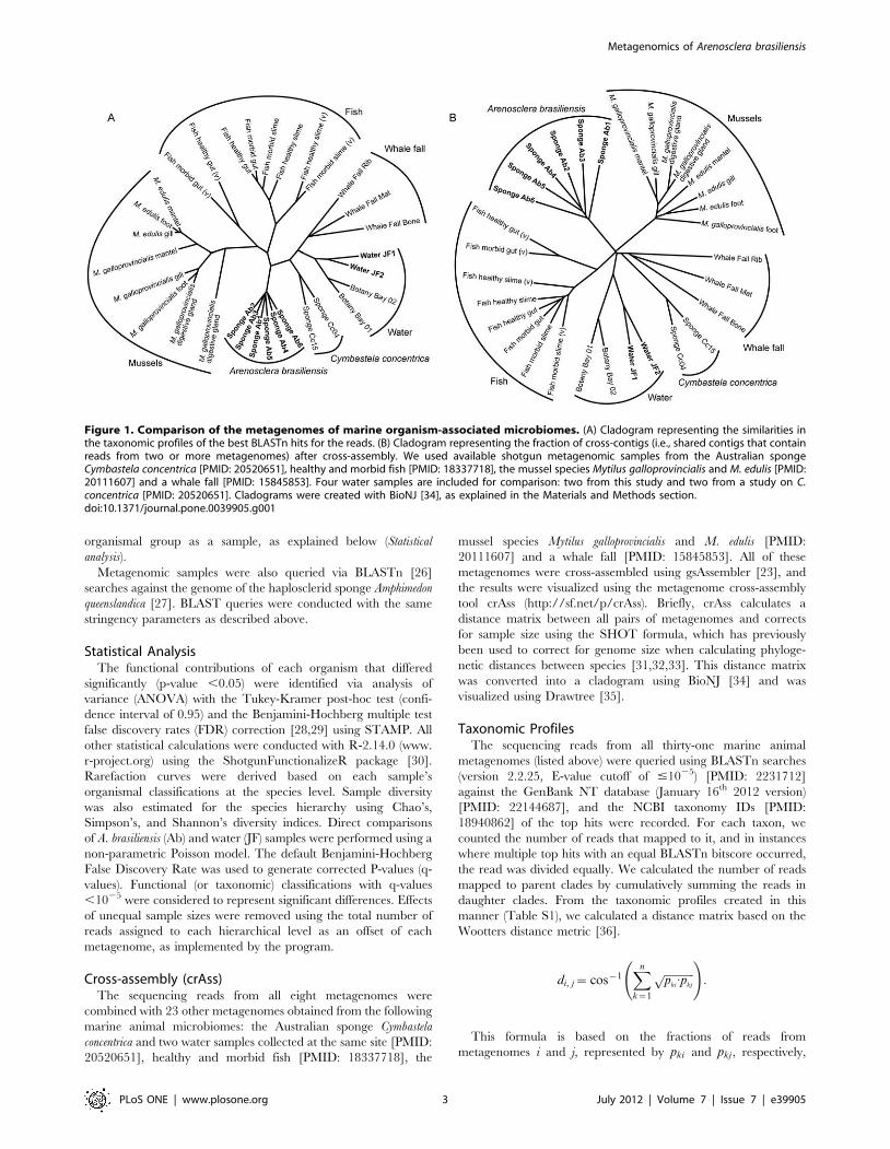
Figure 1. Comparison of the metagenomes of marine organism-associated microbiomes. (A) Cladogram representing the similarities inthe taxonomic profiles of the best BLASTn hits for the reads. (B) Cladogram representing the fraction of cross-contigs (i.e., shared contigs that containreads from two or more metagenomes) after cross-assembly. We used available shotgun metagenomic samples from the Australian spongeCymbastela concentrica [PMID: 20520651], healthy and morbid fish [PMID: 18337718], the mussel species Mytilus galloprovincialis and M. edulis [PMID:20111607] and a whale fall [PMID: 15845853]. Four water samples are included for comparison: two from this study and two from a study on C.concentrica [PMID: 20520651]. Cladograms were created with BioNJ [34], as explained in the Materials and Methods section.doi:10.1371/journal.pone.0039905.g001
Metagenomics of Arenosclera brasiliensis
PLoS ONE | www.plosone.org 3 July 2012 | Volume 7 | Issue 7 | e39905
that are incorporated into contig k. It estimates the minimum
number of jumps required to move from one distribution to
another, where a jump reflects a statistical fluctuation typical of a
finite sampling event. The distance is normalized so that it is
independent of the sample size. The distance matrix was
transformed into a cladogram using BioNJ [34].
Results
We generated approximately 674,000 (200 Mbases) high-quality
shotgun metagenomic sequences (averaging 293 nt in length) from
six sponge specimens (Ab1 to Ab6) and two seawater samples (JF1
and JF2) (Table S2). According to MG-RAST analyses, only 5.5%
(30,427 sequences) and 8.9% (49,206 sequences) of the sponge-
derived sequences were identified within the taxonomic hierarchy
and subsystems, respectively (Table S2). These percentages were
seven times lower than those obtained for the water-derived
sequences, indicating a large reservoir of novel biodiversity in the
sponge metagenomes. To detect metagenomic sequences origi-
nating from the A. brasiliensis genome, the six samples from this
species (Ab1 to Ab6) were compared using BLASTn to the
genome of Amphimedon queenslandica [27], which is the only genome
available for Porifera. A. queenslandica is a phylogenetic neighbor of
A. brasiliensis, also belonging to the haplosclerid suborder
Haplosclerina. Only ,0.3% of A. brasiliensis-derived sequences
matched A. queenslandica genomic regions (Table S2). Although this
percentage is relatively low, it was ten times higher than that
obtained when the water-derived data were used for comparison
(,0.03%) (Table S2).
The Core Microbiome of A. brasiliensisThe majority of the sequences detected in the A. brasiliensis
(93%) and seawater (83%) samples were identified as bacterial in
origin (Figure S1A). A significant portion of this microbial diversity
was covered by our sequencing efforts (Figure S1B). A comple-
mentary clustering analysis (see Materials and Methods) showed that
the A. brasiliensis-derived metagenomic sequences formed a
cohesive cluster that clearly separates them from the clusters
formed by the microorganisms associated with other marine
animals (Figure 1A and 1B). The A. brasiliensis clade also branched
apart from the clade formed by the microbiome of the sponge C.
concentrica, while the seawater samples collected near both sponge
species (JF1, JF2 and Botany Bay 1 and 2) could be grouped into a
main clade of planktonic microorganisms (Figure 1A and 1B,
water clade). These results strongly indicate that A. brasiliensis
contains a specifically associated microbial community. The
abundances of sequences identified as eukaryotes (,3–5%) and
Archaea (,1%) were similar in the sponge and seawater groups,
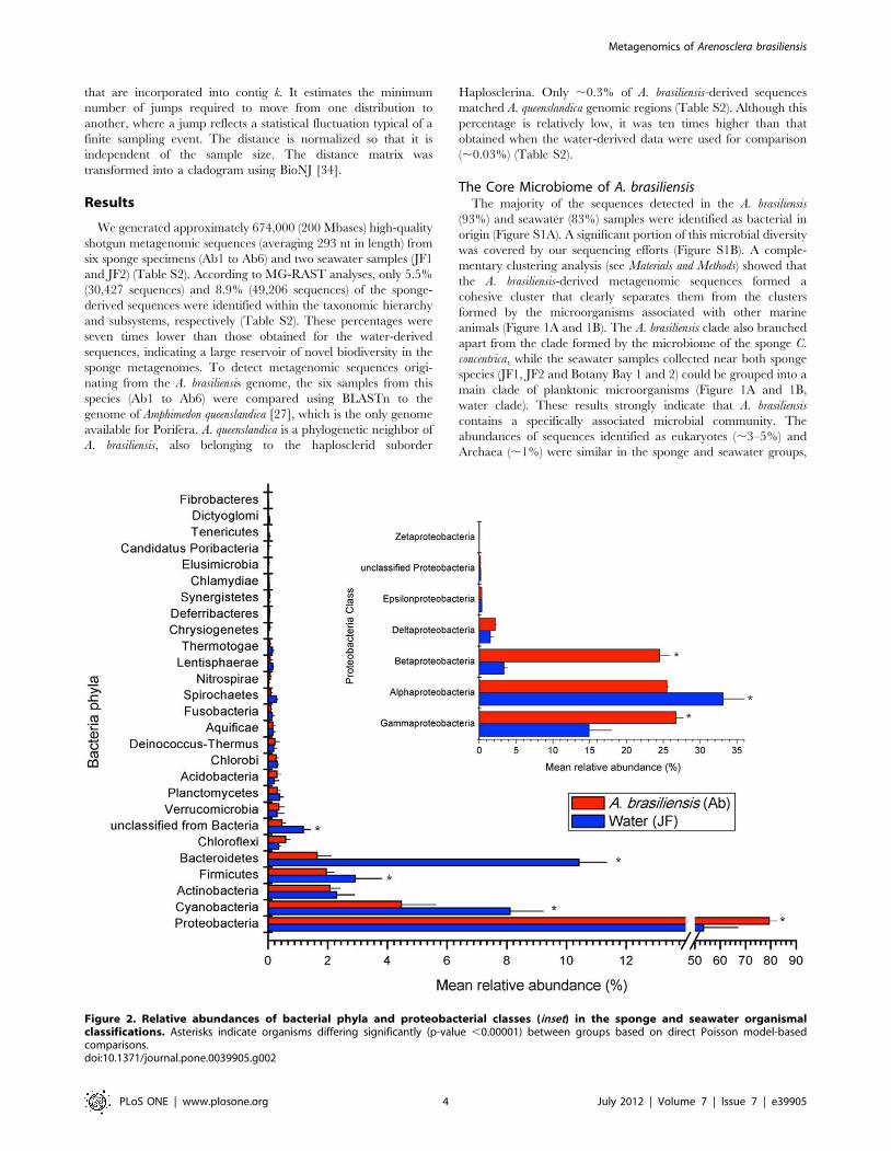
Figure 2. Relative abundances of bacterial phyla and proteobacterial classes (inset) in the sponge and seawater organismalclassifications. Asterisks indicate organisms differing significantly (p-value ,0.00001) between groups based on direct Poisson model-basedcomparisons.doi:10.1371/journal.pone.0039905.g002
Metagenomics of Arenosclera brasiliensis
PLoS ONE | www.plosone.org 4 July 2012 | Volume 7 | Issue 7 | e39905
whereas viruses were more abundant in the seawater metagen-
omes (Figure S1A).
The microbiome of A. brasiliensis was rather homogeneous across
the different specimens, and a total of 26 bacterial phyla were
detected (Figure 2). Fourteen phyla were found consistently in all
of the metagenomes (Figure 2). Proteobacteria, Cyanobacteria,
Actinobacteria, Bacteroidetes, Firmicutes, and Cloroflexi were present at
abundances of $1%. Planctomycetes, Verrucomicrobia, Chlorobi,
Deinococcus-Thermus, Nitrospirae, Aquificae, Spirochaetes, Acidobacteria,
and Fusobacteria were present at abundances of #1% (Figure 2).
Proteobacteria was the most abundant phylum in the sponge (,79%)
and water (,53%) metagenomes (Figure 2). The ShotgunFunc-
tionalizeR non-parametric Poisson model showed that sponge
samples were significantly enriched for Beta- (,24.5%) and
Gammaproteobacteria (,26.7%) sequences (adjusted p-value
,0.00001), whereas Alphaproteobacteria sequences were overrepre-
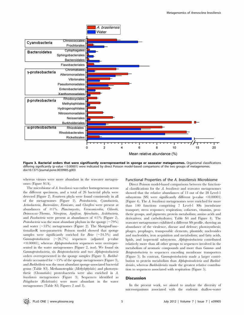
sented in the water metagenomes (Figure 2, inset). We found six
Gammaproteobacteria, six Betaproteobacteria and two Alphaproteobacteria
orders overrepresented in the sponge samples (Figure 3). Burkhol-
deriales accounted for ,15% of the sponge metagenomes (Figure 3),
and Burkholderia was the most abundant sponge-associated bacterial
genus (Table S3). Methanotrophic (Methylophylales) and photosyn-
thetic (Chromatiales) proteobacteria were also enriched in A.
brasiliensis metagenomes (Figure 3). Sequences identified as
Pelagibacter (Rickettsiales) were more abundant in the water
metagenomes (Table S3; Figures 2 and 3).
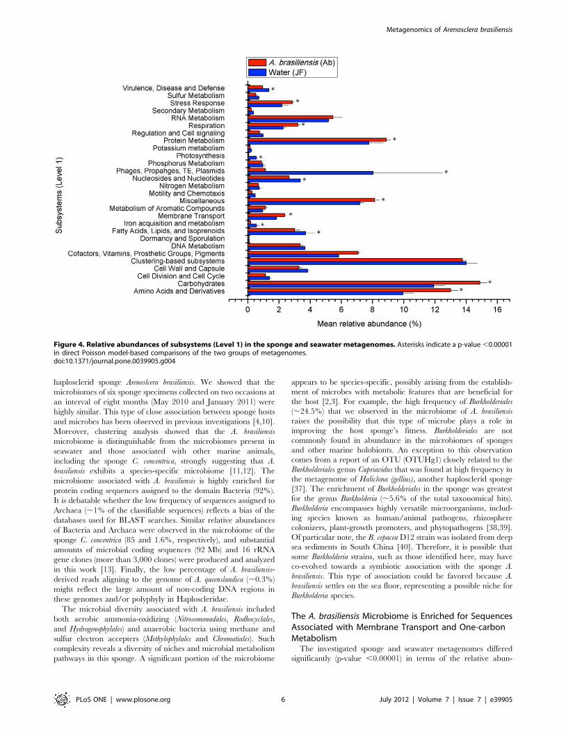
Functional Properties of the A. brasiliensis MicrobiomeDirect Poisson model-based comparisons between the function-
al classifications for the A. brasiliensis and seawater metagenomes
showed that the relative abundances of 13 out of the 28 Level-1
subsystems (SS) were significantly different (p-value ,0.00001)
(Figure 4). The A. brasiliensis metagenomes were enriched for more
than 140 functions comprising 7 Level-1 SSs (membrane
transport; stress responses; respiration; cofactors, vitamins, pros-
thetic groups, and pigments; protein metabolism; amino acids and
derivatives; and carbohydrates; Table S4 and Figure 4). The
seawater metagenomes exhibited a different SS profile, showing an
abundance of the virulence, disease and defense; photosynthesis;
phages, prophages, transposable elements, plasmids; nucleosides
and nucleotides, iron acquisition and metabolism; and fatty acids,
lipids, and isoprenoid subsystems. Alphaproteobacteria contributed
relatively more than all other groups to sequences involved in the
metabolism of aromatic compounds and more than Gamma- and
Betaproteobacteria to sequences encoding membrane transporters
(Figure 5). In contrast, Gammaproteobacteria made a larger contri-
bution to protein metabolism than Alphaproteoabcteria and Burkhol-
deriales, whereas Burkholderiales made the greatest relative contribu-
tion to sequences associated with respiration (Figure 5).
Discussion
In the present work, we aimed to analyze the diversity of
microorganisms associated with the endemic shallow-water
Figure 3. Bacterial orders that were significantly overrepresented in sponge or seawater metagenomes. Organismal classificationsdiffering significantly (p-value ,0.00001) were indicated by direct Poisson model-based comparisons of the two groups of metagenomes.doi:10.1371/journal.pone.0039905.g003
Metagenomics of Arenosclera brasiliensis
PLoS ONE | www.plosone.org 5 July 2012 | Volume 7 | Issue 7 | e39905
haplosclerid sponge Arenoslcera brasiliensis. We showed that the
microbiomes of six sponge specimens collected on two occasions at
an interval of eight months (May 2010 and January 2011) were
highly similar. This type of close association between sponge hosts
and microbes has been observed in previous investigations [4,10].
Moreover, clustering analysis showed that the A. brasiliensis
microbiome is distinguishable from the microbiomes present in
seawater and those associated with other marine animals,
including the sponge C. concentrica, strongly suggesting that A.
brasiliensis exhibits a species-specific microbiome [11,12]. The
microbiome associated with A. brasiliensis is highly enriched for
protein coding sequences assigned to the domain Bacteria (92%).
It is debatable whether the low frequency of sequences assigned to
Archaea (,1% of the classifiable sequences) reflects a bias of the
databases used for BLAST searches. Similar relative abundances
of Bacteria and Archaea were observed in the microbiome of the
sponge C. concentrica (85 and 1.6%, respectively), and substantial
amounts of microbial coding sequences (92 Mb) and 16 rRNA
gene clones (more than 3,000 clones) were produced and analyzed
in this work [13]. Finally, the low percentage of A. brasiliensis-
derived reads aligning to the genome of A. queenslandica (,0.3%)
might reflect the large amount of non-coding DNA regions in
these genomes and/or polyphyly in Haploscleridae.
The microbial diversity associated with A. brasiliensis included
both aerobic ammonia-oxidizing (Nitrosomonadales, Rodhocyclales,
and Hydrogenophylales) and anaerobic bacteria using methane and
sulfur electron accepters (Methylophylales and Chromatiales). Such
complexity reveals a diversity of niches and microbial metabolism
pathways in this sponge. A significant portion of the microbiome
appears to be species-specific, possibly arising from the establish-
ment of microbes with metabolic features that are beneficial for
the host [2,3]. For example, the high frequency of Burkholderiales
(,24.5%) that we observed in the microbiome of A. brasiliensis
raises the possibility that this type of microbe plays a role in
improving the host sponge’s fitness. Burkholderiales are not
commonly found in abundance in the microbiomes of sponges
and other marine holobionts. An exception to this observation
comes from a report of an OTU (OTUHg1) closely related to the
Burkholderiales genus Cupriavidus that was found at high frequency in
the metagenome of Haliclona (gellius), another haplosclerid sponge
[37]. The enrichment of Burkholderiales in the sponge was greatest
for the genus Burkholderia (,5.6% of the total taxonomical hits).
Burkholderia encompasses highly versatile microorganisms, includ-
ing species known as human/animal pathogens, rhizosphere
colonizers, plant-growth promoters, and phytopathogens [38,39].
Of particular note, the B. cepacea D12 strain was isolated from deep
sea sediments in South China [40]. Therefore, it is possible that
some Burkholderia strains, such as those identified here, may have
co-evolved towards a symbiotic association with the sponge A.
brasiliensis. This type of association could be favored because A.
brasiliensis settles on the sea floor, representing a possible niche for
Burkholderia species.
The A. brasiliensis Microbiome is Enriched for SequencesAssociated with Membrane Transport and One-carbonMetabolism
The investigated sponge and seawater metagenomes differed
significantly (p-value ,0.00001) in terms of the relative abun-
Figure 4. Relative abundances of subsystems (Level 1) in the sponge and seawater metagenomes. Asterisks indicate a p-value ,0.00001in direct Poisson model-based comparisons of the two groups of metagenomes.doi:10.1371/journal.pone.0039905.g004
Metagenomics of Arenosclera brasiliensis
PLoS ONE | www.plosone.org 6 July 2012 | Volume 7 | Issue 7 | e39905
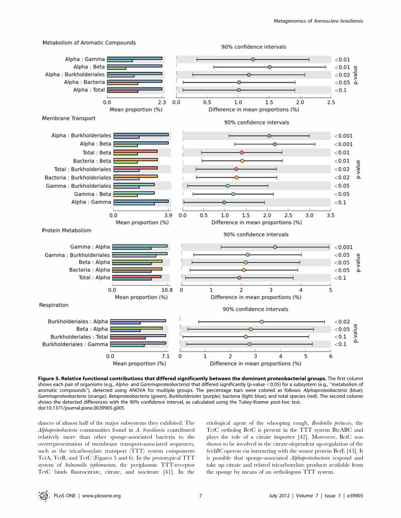
dances of almost half of the major subsystems they exhibited. The
Alphaproteobacteria communities found in A. brasiliensis contributed
relatively more than other sponge-associated bacteria to the
overrepresentation of membrane transport-associated sequences,
such as the tricarboxylate transport (TTT) system components
TctA, TctB, and TctC (Figures 5 and 6). In the prototypical TTT
system of Salmonella typhimurium, the periplasmic TTT-receptor
TctC binds fluorocitrate, citrate, and isocitrate [41]. In the
etiological agent of the whooping cough, Bordetella pertussis, the
TctC ortholog BctC is present in the TTT system BtcABC and
plays the role of a citrate importer [42]. Moreover, BctC was
shown to be involved in the citrate-dependent up-regulation of the
bctABC operon via interacting with the sensor protein BctE [43]. It
is possible that sponge-associated Alphaproteobacteria respond and
take up citrate and related tricarboxylate products available from
the sponge by means of an orthologous TTT system.
Figure 5. Relative functional contributions that differed significantly between the dominant proteobacterial groups. The first columnshows each pair of organisms (e.g., Alpha- and Gammaproteobacteria) that differed significantly (p-value ,0.05) for a subsystem (e.g., ‘‘metabolism ofaromatic compounds’’), detected using ANOVA for multiple groups. The percentage bars were colored as follows: Alphaproteobacteria (blue);Gammaproteobacteria (orange); Betaproteobacteria (green), Burkholderiales (purple); bacteria (light blue); and total species (red). The second columnshows the detected differences with the 90% confidence interval, as calculated using the Tukey-Kramer post-hoc test.doi:10.1371/journal.pone.0039905.g005
Metagenomics of Arenosclera brasiliensis
PLoS ONE | www.plosone.org 7 July 2012 | Volume 7 | Issue 7 | e39905
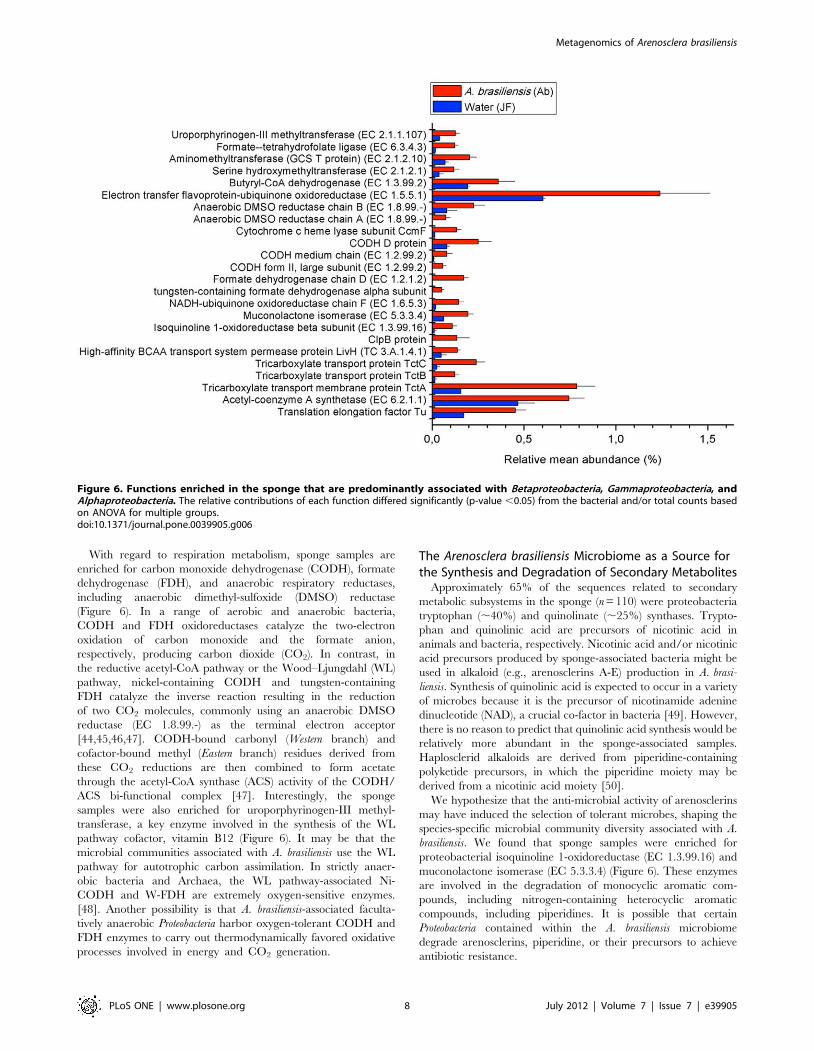
With regard to respiration metabolism, sponge samples are
enriched for carbon monoxide dehydrogenase (CODH), formate
dehydrogenase (FDH), and anaerobic respiratory reductases,
including anaerobic dimethyl-sulfoxide (DMSO) reductase
(Figure 6). In a range of aerobic and anaerobic bacteria,
CODH and FDH oxidoreductases catalyze the two-electron
oxidation of carbon monoxide and the formate anion,
respectively, producing carbon dioxide (CO2). In contrast, in
the reductive acetyl-CoA pathway or the Wood–Ljungdahl (WL)
pathway, nickel-containing CODH and tungsten-containing
FDH catalyze the inverse reaction resulting in the reduction
of two CO2 molecules, commonly using an anaerobic DMSO
reductase (EC 1.8.99.-) as the terminal electron acceptor
[44,45,46,47]. CODH-bound carbonyl (Western branch) and
cofactor-bound methyl (Eastern branch) residues derived from
these CO2 reductions are then combined to form acetate
through the acetyl-CoA synthase (ACS) activity of the CODH/
ACS bi-functional complex [47]. Interestingly, the sponge
samples were also enriched for uroporphyrinogen-III methyl-
transferase, a key enzyme involved in the synthesis of the WL
pathway cofactor, vitamin B12 (Figure 6). It may be that the
microbial communities associated with A. brasiliensis use the WL
pathway for autotrophic carbon assimilation. In strictly anaer-
obic bacteria and Archaea, the WL pathway-associated Ni-
CODH and W-FDH are extremely oxygen-sensitive enzymes.
[48]. Another possibility is that A. brasiliensis-associated faculta-
tively anaerobic Proteobacteria harbor oxygen-tolerant CODH and
FDH enzymes to carry out thermodynamically favored oxidative
processes involved in energy and CO2 generation.
The Arenosclera brasiliensis Microbiome as a Source forthe Synthesis and Degradation of Secondary Metabolites
Approximately 65% of the sequences related to secondary
metabolic subsystems in the sponge (n = 110) were proteobacteria
tryptophan (,40%) and quinolinate (,25%) synthases. Trypto-
phan and quinolinic acid are precursors of nicotinic acid in
animals and bacteria, respectively. Nicotinic acid and/or nicotinic
acid precursors produced by sponge-associated bacteria might be
used in alkaloid (e.g., arenosclerins A-E) production in A. brasi-
liensis. Synthesis of quinolinic acid is expected to occur in a variety
of microbes because it is the precursor of nicotinamide adenine
dinucleotide (NAD), a crucial co-factor in bacteria [49]. However,
there is no reason to predict that quinolinic acid synthesis would be
relatively more abundant in the sponge-associated samples.
Haplosclerid alkaloids are derived from piperidine-containing
polyketide precursors, in which the piperidine moiety may be
derived from a nicotinic acid moiety [50].
We hypothesize that the anti-microbial activity of arenosclerins
may have induced the selection of tolerant microbes, shaping the
species-specific microbial community diversity associated with A.
brasiliensis. We found that sponge samples were enriched for
proteobacterial isoquinoline 1-oxidoreductase (EC 1.3.99.16) and
muconolactone isomerase (EC 5.3.3.4) (Figure 6). These enzymes
are involved in the degradation of monocyclic aromatic com-
pounds, including nitrogen-containing heterocyclic aromatic
compounds, including piperidines. It is possible that certain
Proteobacteria contained within the A. brasiliensis microbiome
degrade arenosclerins, piperidine, or their precursors to achieve
antibiotic resistance.
Figure 6. Functions enriched in the sponge that are predominantly associated with Betaproteobacteria, Gammaproteobacteria, andAlphaproteobacteria. The relative contributions of each function differed significantly (p-value ,0.05) from the bacterial and/or total counts basedon ANOVA for multiple groups.doi:10.1371/journal.pone.0039905.g006
Metagenomics of Arenosclera brasiliensis
PLoS ONE | www.plosone.org 8 July 2012 | Volume 7 | Issue 7 | e39905
In this scenario, species belonging to the most abundant sponge-
associated bacterial genus, Burkholderia, are strong candidates for
regulating the production and/or degradation of polyketide-
derived alkaloids extracted from A. brasiliensis. In the last five
years, Burkholderia species have been shown to produce a diversity
of bioactive polyketide-derived compounds, including i) rhizoxins,
which are antimitotic macrolides produced by B. rhizoxina, the
endosymbiote of the rice-pathogenic fungus Rhizopus sp. [51,52]; ii)
thailandamides and quorum-sensing regulated bactobolin antibi-
otics, produced by B. thailandensis [53]; iii) the food-poisoning toxin
bongkrek acid, synthesized by B. gladioli [54]; and iv) the potent
antibiotics the enacyloxins, produced by the Burkholderia cepacea
complex (Bcc) species B. ambifaria [55]. In addition, Burkholderia are
known to exhibit a special versatile mechanism for degrading
natural or synthetic aromatic hydrocarbons, with some species
being enriched for central and peripheral aromatic catabolic
pathways [40,56,57].
Concluding RemarksThis report provides the first large-scale analysis of the
taxonomic and metabolic diversity of the microbiome of the
sponge A. brasiliensis. Our results demonstrate that a complex
microbiome exists within this sponge that presents a particular
metabolic profile. We show that the A. brasiliensis microbiome is
unique, differing from the microbiomes present in the water
column surrounding these sponges and those associated with other
marine organisms. Within the taxonomic signature of the A.
brasiliensis microbiome, we detected an enrichment of Betaproteo-
bacteria (e.g., Burkholderia) and Gammaproteobacteria (Pseudomonas and
Alteromonas), indicating species specificity. Our results allowed us to
speculate about diversity of niches in the sponge that might harbor
anaerobes using methane and sulfur electron acceptors and those
that might conduct thermodynamically favored oxidative process-
es involved in energy and CO2 generation. Our results may also
suggest specific roles of Burkholderia sp. in this symbiosis. Finally, we
hypothesized that secondary metabolites might have shaped the
microbial community structure observed in A. brasiliensis. Studies
are underway to uncover the diversity of polyketide synthase (PKS)
genes and isolate possible sponge-specific (e.g., Burkholderia)
symbionts from A. brasiliensis.
Supporting Information
Figure S1 Composition of the investigated metagen-omes. (A) Distribution of sponge and seawater taxonomic hits at
the Domain hierarchical level, with sponge and seawater data
represented by red and blue pie charts, respectively. (B) Sample
rarefaction curves at the species hierarchical level.
(TIF)
Table S1 Taxonomic profiles for the thirty-one ana-lyzed marine-animal metagenomes. Profiles were derived
by BLASTn mapping of sequencing reads to the Genbank NT
database and cumulatively summing the reads for higher-order
clades (see Materials and methods).
(XLSX)
Table S2 Metagenomes overall numbers. 1 - QC – MG-
RAST 3.0 applied quality control of the reads. 2 - Values obtained
considering the post QC metadata. Abbreviations: Seqs. –
Sequences; Class. – Classifications.
(DOC)
Table S3 Most abundant bacterial genera in sponge andwater metagenomes. 1– Relative percentage from the total
number of organism classifications.
(DOC)
Table S4 Functions overrepresented in the A. brasilien-sis metagenomes.(DOC)
Acknowledgments
We thank the SISBIO-ICMBio (Sistema de Autorizacao e Informacao em
Biodiversidade do Instituto Chico Mendes de Conservacao da Biodiversi-
dade) and the Secretaria Municipal de Meio Ambiente e Pesca de Armacao
de Buzios for providing legal authorization (licenses 11175-1 and 10357-1)
for sample collection at the Parque Natural dos Corais. We would also like
to thank Dr. Gustavo B. Gregoracci for helpful ideas regarding the
statistical analysis of the metagenomic data.
Author Contributions
Conceived and designed the experiments: FLT RGSB AETS. Performed
the experiments: AETS AP ATV GLH EH. Analyzed the data: AETS
GGZS BED RAE. Contributed reagents/materials/analysis tools: FLT
RGSB GLH BED RAE ATV. Wrote the paper: AETS FLT BED.
References
1. Love GD, Grosjean E, Stalvies C, Fike DA, Grotzinger JP, et al. (2009) Fossil
steroids record the appearance of Demospongiae during the Cryogenian period.
Nature 457: 718–721.
2. Taylor MW, Radax R, Steger D, Wagner M (2007) Sponge-associated
microorganisms: evolution, ecology, and biotechnological potential. Microbiol
Mol Biol Rev 71: 295–347.
3. Hentschel U, Hopke J, Horn M, Friedrich AB, Wagner M, et al. (2002)
Molecular Evidence for a Uniform Microbial Community in Sponges from
Different Oceans. Applied and Environmental Microbiology 68: 4431–4440.
4. Webster NS, Taylor MW (2012) Marine sponges and their microbial symbionts:
love and other relationships. Environmental Microbiology 14: 335–346.
5. Vacelet J (1975) Etude en microscopie electronique de l’association entre
bacteries et spongiaires du genre Verongia (Dictyoceratida). J Microsc Biol Cell
23: 271–288.
6. Vacelet J, Donadey C (1977) Electron-microscope study of association between
some sponges and bacteria. J Exp Mar Biol Ecol 30: 301–314.
7. Fieseler L, Horn M, Wagner M, Hentschel U (2004) Discovery of the novel
candidate phylum ‘‘Poribacteria’’ in marine sponges. Applied and Environmen-
tal Microbiology 70: 3724–3732.
8. Sogin ML, Morrison HG, Huber JA, Mark Welch D, Huse SM, et al. (2006)
Microbial diversity in the deep sea and the underexplored ‘‘rare biosphere’’.
Proceedings of the National Academy of Sciences of the United States of
America 103: 12115–12120.
9. Lee OO, Wang Y, Yang J, Lafi FF, Al-Suwailem A, et al. (2010) Pyrosequencing
reveals highly diverse and species-specific microbial communities in sponges
from the Red Sea. The ISME Journal 5: 650–664.
10. Webster NS, Taylor MW, Behnam F, Lucker S, Rattei T, et al. (2010) Deepsequencing reveals exceptional diversity and modes of transmission for bacterial
sponge symbionts. Environ Microbiol 12: 2070–2082.
11. Schmitt S, Tsai P, Bell J, Fromont J, Ilan M, et al. (2011) Assessing the complexsponge microbiota: core, variable and species-specific bacterial communities in
marine sponges. The ISME Journal 6: 564–576.
12. Schmitt S, Hentschel U, Taylor MW (2011) Deep sequencing reveals diversity
and community structure of complex microbiota in five Mediterranean sponges.Hydrobiologia 687: 341–351.
13. Thomas T, Rusch D, DeMaere MZ, Yung PY, Lewis M, et al. (2010) Functional
genomic signatures of sponge bacteria reveal unique and shared features ofsymbiosis. The ISME Journal 4: 1557–1567.
14. Turque AS, Cardoso AM, Silveira CB, Vieira RP, Freitas FAD, et al. (2008)
Bacterial communities of the marine sponges Hymeniacidon heliophila and
Polymastia janeirensis and their environment in Rio de Janeiro, Brazil. MarineBiology 155: 135–146.
15. Turque AS, Batista D, Silveira CB, Cardoso AM, Vieira RP, et al. (2010)
Environmental Shaping of Sponge Associated Archaeal Communities. PLoSOne 5: e15774.
16. de Oliveira JHHL, Nascimento AM, Kossuga MH, Cavalcanti BC, Pessoa CO,
et al. (2007) Cytotoxic Alkylpiperidine Alkaloids from the Brazilian Marine
Sponge Pachychalina alcaloidifera. J Nat Prod 70: 538–543.
Metagenomics of Arenosclera brasiliensis
PLoS ONE | www.plosone.org 9 July 2012 | Volume 7 | Issue 7 | e39905
17. Muricy G, Ribeiro SM (1999) Shallow-water Haplosclerida (Porifera, Demos-
pongiae) from Rio de Janeiro State, Brazil (Southwestern Atlantic). Beaufortia
49: 83–108.
18. Torres YR, Berlinck RG, Magalhaes A, Schefer AB, Ferreira AG, et al. (2000)
Arenosclerins A-C and haliclonacyclamine E, new tetracyclic alkaloids from a
Brazilian endemic Haplosclerid sponge Arenosclera brasiliensis. J Nat Prod 63:
1098–1105.
19. Torres YR, Berlinck RG, Nascimento GG, Fortier SC, Pessoa C, et al. (2002)
Antibacterial activity against resistant bacteria and cytotoxicity of four alkaloid
toxins isolated from the marine sponge Arenosclera brasiliensis. Toxicon 40:
885–891.
20. Stankevicins L, Aiub C, Maria LC, Lobo-Hajdu G, Felzenszwalb I (2008)
Genotoxic and antigenotoxic evaluation of extracts from Arenosclera brasilien-
sis, a Brazilian marine sponge. Toxicol In Vitro 22: 1869–1877.
21. Thompson FL, Bruce T, Gonzalez A, Cardoso A, Clementino M, et al. (2011)
Coastal bacterioplankton community diversity along a latitudinal gradient in
Latin America by means of V6 tag pyrosequencing. Archives of Microbiology
193: 105–114.
22. Lobo-Hajdu G, Guimaraes ACR, Salgado A, Lamarao FRM, Vieiralves T, et al.
(2004) Intragenomic, Intra- and Interspecific Variation inthe rDNA ITS of
Porifera revealed by PCR-Single-Strand Conformation Polymorphism (PCR-
SSCP). Boll Mus Ist Biol Univ Genova 68: 413–423.
23. Margulies M, Egholm M, Altman WE, Attiya S, Bader JS, et al. (2005) Genome
sequencing in microfabricated high-density picolitre reactors. Nature 437: 376–
380.
24. Meyer F, Paarmann D, D’Souza M, Olson R, Glass EM, et al. (2008) The
metagenomics RAST server - a public resource for the automatic phylogenetic
and functional analysis of metagenomes. BMC bioinformatics 9: 386.
25. Parks DH, Beiko RG (2010) Identifying biologically relevant differences between
metagenomic communities. Bioinformatics 26: 715–721.
26. Altschul SF, Gish W, Miller W, Myers EW, Lipman DJ (1990) Basic local
alignment search tool. J Mol Biol 215: 403–410.
27. Srivastava M, Simakov O, Chapman J, Fahey B, Gauthier MEA, et al. (2010)
The Amphimedon queenslandica genome and the evolution of animal
complexity. Nature 466: 720-U723.
28. Bluman AG (2007) Elementary statistics: A step by step approach. McGraw Hill
Higher Education, New York, New York 6th edition.
29. Benjamini Y, Hochberg Y (1995) Controlling the false discovery rate: a practical
and powerful approach to multiple testing. J Roy Stat Soc B 57: 289–300.
30. Kristiansson E, Hugenholtz P, Dalevi D (2009) ShotgunFunctionalizeR: an R-
package for functional comparison of metagenomes. Bioinformatics 25: 2737–
2738.
31. Korbel JO, Snel B, Huynen MA, Bork P (2002) SHOT: a web server for the
construction of genome phylogenies. Trends Genet 18: 158–162.
32. Dutilh BE, Huynen MA, Bruno WJ, Snel B (2004) The Consistent Phylogenetic
Signal in Genome Trees Revealed by Reducing the Impact of Noise. Journal of
Molecular Evolution 58: 527–539.
33. Dutilh BE, van Noort V, van der Heijden RTJM, Boekhout T, Snel B, et al.
(2007) Assessment of phylogenomic and orthology approaches for phylogenetic
inference. Bioinformatics 23: 815–824.
34. Gascuel O (1997) BIONJ: An Improved Version of the NJ Algorithm Based on a
Simple Model of Sequence Data. Molecular Biology and Evolution 14: 685–695.
35. Felsenstein J (1989) PHYLIP - Phylogeny Inference Package (Version 3.2).
Cladistics 5: 164–166.
36. Wootters KW (1980) Statistical distance and Hilbert space. Physical Review D
23: 357–362.
37. Sipkema D, Holmes B, Nichols SA, Blanch HW (2009) Biological Characterisa-
tion of Haliclona (?gellius) sp.: Sponge and Associated Microorganisms.
Microbial Ecology 58: 903–920.
38. Coenye T, Vandamme P (2003) Diversity and significance of Burkholderia
species occupying diverse ecological niches. Environmental Microbiology 5:719–729.
39. Vial L, Chapalain A, Groleau M-C, Deziel E (2011) The various lifestyles of the
Burkholderia cepacia complex species: a tribute to adaptation. EnvironmentalMicrobiology 13: 1–12.
40. Wang Y, Yin B, Hong Y, Yan Y, Gu J-D (2008) Degradation of dimethylcarboxylic phthalate ester by Burkholderia cepacia DA2 isolated from marine
sediment of South China Sea. Ecotoxicology 17: 845–852.
41. Winnen B, Hvorup RN, Saier MH Jr (2003) The tripartite tricarboxylatetransporter (TTT) family. Research in Microbiology 154: 457–465.
42. Antoine R, Jacob-Dubuisson F, Drobecq H, Willery E, Lesjean S, et al. (2003)Overrepresentation of a Gene Family Encoding Extracytoplasmic Solute
Receptors in Bordetella. Journal of Bacteriology 185: 1470–1474.43. Antoine R, Huvent I, Chemlal K, Deray I, Raze D, et al. (2005) The Periplasmic
Binding Protein of a Tripartite Tricarboxylate Transporter is Involved in Signal
Transduction. Journal of Molecular Biology 351: 799–809.44. Drennan CL, Heo J, Sintchak MD, Schreiter E, Ludden PW (2001) Life on
carbon monoxide: X-ray structure of Rhodospirillum rubrum Ni-Fe-S carbonmonoxide dehydrogenase. Proceedings of the National Academy of Sciences 98:
11973–11978.
45. Pierce E, Xie G, Barabote RD, Saunders E, Han CS, et al. (2008) The completegenome sequence of Moorella thermoacetica (f. Clostridium thermoaceticum).
Environ Microbiol 10: 2550–2573.46. Raaijmakers H, Macieira S, Dias JM, Teixeira S, Bursakov S, et al. (2002) Gene
sequence and the 1.8 A crystal structure of the tungsten-containing formatedehydrogenase from Desulfovibrio gigas. Structure 10: 1261–1272.
47. Ragsdale SW, Pierce E (2008) Acetogenesis and the Wood-Ljungdahl pathway
of CO(2) fixation. Biochim Biophys Acta 1784: 1873–1898.48. Berg IA, Kockelkorn D, Ramos-Vera WH, Say RF, Zarzycki J, et al. (2010)
Autotrophic carbon fixation in archaea. Nat Rev Microbiol 8: 447–460.49. Ollagnierdechoudens S, Loiseau L, Sanakis Y, Barras F, Fontecave M (2005)
Quinolinate synthetase, an iron–sulfur enzyme in NAD biosynthesis. Febs
Letters 579: 3737–3743.50. Fontana A (2006) Biogenetic Proposals and Biosynthetic Studies on Secondary
Metabolites of Opisthobranch Molluscs. Guido Cimino & Margherita Gavagnin(Eds): Molluscs From Chemo-ecological Study to Biotechnological Application
Springer-Verlag Berlin Heidelberg: 304–332.51. Partida-Martinez LP, Hertweck C (2005) Pathogenic fungus harbours
endosymbiotic bacteria for toxin production. Nature 437: 884–888.
52. Partida-Martinez LP, Hertweck C (2007) A gene cluster encoding rhizoxinbiosynthesis in ‘‘Burkholderia rhizoxina’’, the bacterial endosymbiont of the
fungus Rhizopus microsporus. Chembiochem 8: 41–45.53. Nguyen T, Ishida K, Jenke-Kodama H, Dittmann E, Gurgui C, et al. (2008)
Exploiting the mosaic structure of trans-acyltransferase polyketide synthases for
natural product discovery and pathway dissection. Nat Biotechnol 26: 225–233.54. Rohm B, Scherlach K, Hertweck C (2010) Biosynthesis of the mitochondrial
adenine nucleotide translocase (ATPase) inhibitor bongkrekic acid in Burk-holderia gladioli. Organic & Biomolecular Chemistry 8: 1520–1522.
55. Mahenthiralingam E, Song L, Sass A, White J, Wilmot C, et al. (2011)Enacyloxins Are Products of an Unusual Hybrid Modular Polyketide Synthase
Encoded by a Cryptic Burkholderia ambifaria Genomic Island. Chemistry &
Biology 18: 665–677.56. Chain PSG, Denef VJ, Konstantinidis KT, Vergez LM, Agullo L, et al. (2006)
Inaugural Article: Burkholderia xenovorans LB400 harbors a multi-replicon,9.73-Mbp genome shaped for versatility. Proceedings of the National Academy
of Sciences 103: 15280–15287.
57. O’Sullivan LA, Weightman AJ, Jones TH, Marchbank AM, Tiedje JM, et al.(2007) Identifying the genetic basis of ecologically and biotechnologically useful
functions of the bacterium Burkholderia vietnamiensis. Environmental Micro-biology 9: 1017–1034.
Metagenomics of Arenosclera brasiliensis
PLoS ONE | www.plosone.org 10 July 2012 | Volume 7 | Issue 7 | e39905
Copyright © 2022 FDOKUMEN


![Descriptors for Sponge Gourd [Luffa cylindrica (L.) Roem.]](https://static.fdokumen.com/doc/165x107/63187e763394f2252e02b92e/descriptors-for-sponge-gourd-luffa-cylindrica-l-roem.jpg)