Sid4: A secreted vertebrate immunoglobulin protein with roles in zebrafish embryogenesis
15
Sid4: A secreted vertebrate immunoglobulin protein with roles in zebrafish embryogenesis P.J. diIorio a, T , A. Runko b , C.A. Farrell a , N. Roy b a Division of Diabetes, University of Massachusetts Medical School, 373 Plantation Street, Suite 218, Worcester, MA 01605, USA b Department of Biochemistry and Pharmacology, University of Massachusetts Medical School, Worcester, MA 01605, USA Received for publication 11 December 2004, revised 22 February 2005, accepted 23 February 2005 Abstract The small members of the immunoglobulin superfamily (IGSF) are a molecularly diverse group of proteins composed solely of immunoglobulin domains. They may be secreted or tethered to the cell mebrane via GPI linkages and are proposed to have important functions in vivo. However, very few small IGSFs have been functionally characterized. During an ongoing in situ hybridization analysis of expressed sequence tags in zebrafish we identified secreted immunoglobulin domain 4 (sid4 ), a gene encoding a soluble vertebrate protein composed solely of four immunoglobulin domains. Throughout development, sid4 is expressed in regions of the embryo undergoing active cell division and migration. Functional analysis using morpholino antisense oligonucleotides demonstrates that timing of gene expression is normal in morphants, but these embryos are smaller and exhibit defects in epiboly and patterning of axial and prechordal mesoderm. Analyses of chordin , pax2, krox20, and dlx2 expression in morphants demonstrate that early brain patterning is normal but later organization of hindbrain neurons and development of cranial neural crest are perturbed. Levels of apoptosis in morphants were normal prior to 90% epiboly, but were elevated after 10 h post-fertilization (hpf). Apoptosis does not account for early patterning defects of axial mesoderm, but likely contributes to overall reduction in embryo size. Phylogenetic analysis demonstrates that Sid4 is strikingly similar to the fibronectin binding Ig domains of Perlecan/HSPG2. Overall, our data demonstrate a fundamental role for sid4 , possibly as a co-factor in extracellular matrix (ECM) interactions, in processes underlying tissue patterning and organogenesis in a vertebrate. D 2005 Elsevier Inc. All rights reserved. Keywords: Apoptosis; Development; Extracellular matrix; Gastrulation; Immunoglobulin; Morpholino; Morphogenesis; Zebrafish Introduction Proteins belonging to the IGSF are structurally diverse and have multiple evolutionary origins. All members utilize the immunoglobulin (Ig) domain to participate in processes that depend on protein interactions, such as co-receptors for growth factors (Mongiat et al., 2000), adhesion to cells and extracellular matrix (Rougon and Hobert, 2003; Vaughn and Bjorkman, 1996), pathfinding by axons (Panicker et al., 2003), and blood vessel branching (Rossant and Howard, 2002). Unlike the large, multimeric members of the family, small IGSFs are composed solely of Ig domains. They may be secreted or tethered to the cell membrane via GPI linkages (Rougon and Hobert, 2003). The molecular diversity of the small IGSF subfamily has been documented in Drosophila (Nakamura et al., 2002) and Caenorhabditis elegans (Teichmann and Chothia, 2000) but very little is known about their in vivo functions. Two small Igs, Drosophila Beaten path (Beat) 1a and Hemolin, are secreted and act as anti-adhesives to disrupt cell–cell interactions 0012-1606/$ - see front matter D 2005 Elsevier Inc. All rights reserved. doi:10.1016/j.ydbio.2005.02.036 T Corresponding author. Fax: +1 508 856 4093. E-mail address: [email protected] (P.J. diIorio). Developmental Biology 282 (2005) 55 – 69 www.elsevier.com/locate/ydbio
-
Upload
independent -
Category
Documents
-
view
1 -
download
0
Transcript of Sid4: A secreted vertebrate immunoglobulin protein with roles in zebrafish embryogenesis

www.elsevier.com/locate/ydbio
Developmental Biology
Sid4: A secreted vertebrate immunoglobulin protein with roles in
zebrafish embryogenesis
P.J. diIorioa,T, A. Runkob, C.A. Farrella, N. Royb
aDivision of Diabetes, University of Massachusetts Medical School, 373 Plantation Street, Suite 218, Worcester, MA 01605, USAbDepartment of Biochemistry and Pharmacology, University of Massachusetts Medical School, Worcester, MA 01605, USA
Received for publication 11 December 2004, revised 22 February 2005, accepted 23 February 2005
Abstract
The small members of the immunoglobulin superfamily (IGSF) are a molecularly diverse group of proteins composed solely of
immunoglobulin domains. They may be secreted or tethered to the cell mebrane via GPI linkages and are proposed to have important
functions in vivo. However, very few small IGSFs have been functionally characterized. During an ongoing in situ hybridization analysis of
expressed sequence tags in zebrafish we identified secreted immunoglobulin domain 4 (sid4), a gene encoding a soluble vertebrate protein
composed solely of four immunoglobulin domains.
Throughout development, sid4 is expressed in regions of the embryo undergoing active cell division and migration. Functional analysis
using morpholino antisense oligonucleotides demonstrates that timing of gene expression is normal in morphants, but these embryos are
smaller and exhibit defects in epiboly and patterning of axial and prechordal mesoderm. Analyses of chordin, pax2, krox20, and dlx2
expression in morphants demonstrate that early brain patterning is normal but later organization of hindbrain neurons and development of
cranial neural crest are perturbed.
Levels of apoptosis in morphants were normal prior to 90% epiboly, but were elevated after 10 h post-fertilization (hpf). Apoptosis does
not account for early patterning defects of axial mesoderm, but likely contributes to overall reduction in embryo size. Phylogenetic analysis
demonstrates that Sid4 is strikingly similar to the fibronectin binding Ig domains of Perlecan/HSPG2. Overall, our data demonstrate a
fundamental role for sid4, possibly as a co-factor in extracellular matrix (ECM) interactions, in processes underlying tissue patterning and
organogenesis in a vertebrate.
D 2005 Elsevier Inc. All rights reserved.
Keywords: Apoptosis; Development; Extracellular matrix; Gastrulation; Immunoglobulin; Morpholino; Morphogenesis; Zebrafish
Introduction
Proteins belonging to the IGSF are structurally diverse
and have multiple evolutionary origins. All members utilize
the immunoglobulin (Ig) domain to participate in processes
that depend on protein interactions, such as co-receptors for
growth factors (Mongiat et al., 2000), adhesion to cells and
extracellular matrix (Rougon and Hobert, 2003; Vaughn and
0012-1606/$ - see front matter D 2005 Elsevier Inc. All rights reserved.
doi:10.1016/j.ydbio.2005.02.036
T Corresponding author. Fax: +1 508 856 4093.
E-mail address: [email protected] (P.J. diIorio).
Bjorkman, 1996), pathfinding by axons (Panicker et al.,
2003), and blood vessel branching (Rossant and Howard,
2002).
Unlike the large, multimeric members of the family,
small IGSFs are composed solely of Ig domains. They may
be secreted or tethered to the cell membrane via GPI
linkages (Rougon and Hobert, 2003). The molecular
diversity of the small IGSF subfamily has been documented
in Drosophila (Nakamura et al., 2002) and Caenorhabditis
elegans (Teichmann and Chothia, 2000) but very little is
known about their in vivo functions. Two small Igs,
Drosophila Beaten path (Beat) 1a and Hemolin, are secreted
and act as anti-adhesives to disrupt cell–cell interactions
282 (2005) 55–69

P.J. diIorio et al. / Developmental Biology 282 (2005) 55–6956
during motorneuron axon migration (Fambrough and Good-
man, 1996) and hemocytic aggregation in response to
bacterial infection (Bettencourt et al., 1997, 1999; Kanost et
al., 1994; Sun et al., 1990), respectively. BeatIa and
Hemolin are also necessary for normal embryo development
(Bettencourt et al., 2002; Pipes et al., 2001).
During an ongoing in situ hybridization screen of
zebrafish expressed sequence tags (ESTs), we identified
secreted immunoglobulin domain 4 (after Sid1, (Yoder et
al., 2002)). sid4 is a novel vertebrate gene that, like moth
Hemolin (Sun et al., 1990), encodes a secreted protein
composed of four Ig domains and contains no other
conserved motifs. sid4 mRNA is maternally provisioned
and zygotically expressed throughout development in
regions of the embryo containing actively dividing and
migrating cells.
Suppression of Sid4 by morpholino injection causes
defects in morphogenesis but not cellular differentiation.
Morphant embryos are reduced in size and exhibit impaired
epiboly and morphogenesis of axial and somitic mesoderm.
Despite morphological similarities to ventralized Wnt and
dorsalized BMP mutants, expression of bmp4 and chordin is
normal in 6 hpf morphants. At 10 hpf, the conclusion of
gastrulation in zebrafish, morphants first exhibit elevated
levels of apoptosis, suggesting that decreased cell viability
is secondary to the morphogenetic defect.
Although sid4 mRNA is ubiquitously expressed through
16 hpf, morphants exhibit spatially and temporally restricted
defects in morphogenesis of axial and prechordal meso-
derm. For example, at similar stages of development,
patterning of axial mesoderm is disrupted while anterior
migration of prechordal mesoderm is normal. Additionally,
while early hatching gland development appears normal,
later anterolateral spreading of these cells is inhibited.
Phylogenetic sequence analysis demonstrates that Sid4 is
closely related to mouse Perlecan/HSPG2 and shares
significant similarity with the fibronectin binding Ig
domains of this large, multimeric protein. These data,
together with our analyses of somitic mesoderm, cranial
neural crest, and branchiomotor neurons suggest that this
novel, vertebrate member of the IGSF may function as a
secreted receptor or cofactor with important roles in some
ECM interactions during embryogenesis.
Methods
Fish maintenance
Wild type and transgenic embryos expressing green
fluorescent protein under the control of an Islet-1 enhancer
(Higashijima et al., 2000) were collected from natural
matings and reared in 1/3 Ringer’s (Westerfield, 2000).
Embryos were staged using morphological criteria up to 24
h post fertilization (hpf) and by time of development at
28.58C thereafter (Kimmel et al., 1995).
Cloning and molecular characterization of sid4
IMAGE clone 3719398 was initially identified as the
zebrafish homolog of HSPG2/Perlecan. However, our
preliminary BLAST analyses of this clone and human
HSPG2 (gi184426) against the zebrafish genome demon-
strated that these are distinct genetic loci. sid4 was selected
for functional characterization because it appeared to be a
novel, developmentally expressed gene located in a region
of the zebrafish genome containing an abundance of
uncharacterized, predicted genes (Sanger, Zv4).
Full-length sid4 (GENBANK AY494978) was isolated
from 60 hpf total RNA (RNeasy, Qiagen). AMV-GeneRacer
(Invitrogen) was used to amplify 5V and 3V cDNA ends with
the oligonucleotides 5VCGCCTCGCTGGAGCCCACATQGAT3V and 5VTGACGGTGCCGTTCTGACCATCGCTAQA3V, respectively. Amplification products were cloned into
pCRII (Invitrogen) and multiple clones sequenced. A
consensus cDNA sequence and conceptual translation were
obtained using SeqMan (DNASTAR v. 4.0). ClustalW was
used to produce nucleotide and amino acid sequence
alignments (Macvector v. 7.2). Full-length sid4 is identical
to the available sequences in 64 recently deposited
AGENCOURT ESTs (e.g gi46159009).
In situ hybridization
pSport containing sid4 was linearized with Sal1 and Not1
and digoxigenin-labeled probes transcribed with Sp6 (anti-
sense) and T7 (sense) RNA polymerases, respectively.
Additional RNA probes used were zebrafish flh (Talbot et
al., 1995), bmp4 (Hwang et al., 1997), chordin (Schulte-
Merker et al., 1997), unc-45 (Etheridge et al., 2002), pax-2
(Krauss et al., 1991), hgg-1 (Vogel and Gerster, 1997),
goosecoid (gsc) (Schulte-Merker et al., 1994; Thisse et al.,
1994), krox-20 (Oxtoby and Jowett, 1993), and dlx-2
(Akimenko et al., 1994). In situ hybridization was carried
out as described previously (diIorio et al., 2002; Willett et al.,
1997).
Morpholino design and microinjection
Eight exons for sid4 were localized to linkage group 25
(Sanger Zv4). We identified nucleotide sequence corre-
sponding to the first 52 amino acids of sid4, but to date
these have not been mapped. Exon 5 and flanking introns
were amplified by standard PCR from genomic DNA using
the oligonucleotides 5VCTAGATCACTTACGAGATGATCQTTTGCGTGAA3V and 5VCCACAATCAGA-GATCTGCAQGCTCTGGA3V. Productswere cloned into pCRII (Invitrogen)and sequenced. To verify the specificity of morpholino-
induced phenotypes, two morpholinos were designed against
distinct regions of sid4 mRNA. The morpholino
5VGAGCTGCTGTCTGGAGCTTCATCAT3V (t-mo) was
designed against the sid4 translation start site and
5VTGGTGATGGTGTGTTTACC-GGAGGC3V (s-mo) targets

P.J. diIorio et al. / Developmental Biology 282 (2005) 55–69 57
a predicted splice donor site at the boundary of exon 5 and
intron 5. The respective control morpholinos 5VGACCTCCTGTCTGCAG-CTTGATGAT 3V (t-co) and
5VTGCTGTTGGTGTCTTTACCCGACGC3V (s-co) both
contain 5 mismatched bases. Morpholinos were reconstituted
to 1 mM in 1� Danieau’s buffer (Gene Tools) and diluted in
1� Danieau’s buffer containing 0.1% phenol red for injection
into 1–2 cell embryos. Unless specified, s-mo was used
because (1) both morpholinos exhibited comparable effects on
epiboly and patterning of somitic mesoderm, prechordal
mesoderm and blood, (2) unlike inhibiting translation, exon
deletion is irreversible, and (3) RT-PCR can be used to assay
for exon deletion from mRNA processed in vivo.
To assess the qualitative effects of s-mo on sid4 mRNA,
total RNAwas extracted from 6 hpf s-co and s-mo embryos
(RNeasy, Qiagen) and 500 ng used in standard RT-PCR
(One-Step, Qiagen). The oligonucleotides 5VATGATGAA-GCTCCAGACAGCAGCTCT3V and 5VCTGTAATTAAGCQTTTGACCGTGACTCT3V span the entire sid4 mRNA.
Amplification products were cloned into pCRII (Invitrogen)
and multiple clones sequenced.
sid4 Overexpression
Full-length sid4 was amplified from adult liver total
RNA using 5VCAACCATCGATAGATCTGCCACCAQ
TGATGAAGCTCCAGACA3V containing an introduced,
consensus Kozak sequence (Kozak, 1987), and 5VCAAQCCGAATTCAGCTTTGACCGTGACTCTGCTGA3V.Amplification products were digested with ClaI and EcoRI
and ligated into pCS2. Four independent clones of pCS2
containing full-length sid4 were linearized with NotI and
capped mRNA transcribed from each with Sp6 (mMessage
Machine, Ambion; RiboMax mRNA synthesis, Promega).
Individual mRNA samples were purified by a number of
protocols including: Phenol extraction, RNA mini prep kit
(Qiagen), Trizol extraction, and MegaClear column purifi-
cation (Ambion). mRNA was re-suspended in sterile,
RNase-free water and quality assessed on 1% agarose gels
both before and after injection.
To assess in vivo expression of sid4 mRNA, pools of one
hundred 12-hpf embryos were injected with various con-
structs and lysed in 150 AL 1� Kamen Sample Buffer
(BioRad). In addition to sid4 mRNA, we injected super-
coiled plasmid DNA used to synthesize sid4 mRNA as a
positive control for sid4 translation. Embryo lysates and
molecular weight markers were separated by SDS-PAGE
and visualized with silver stain.
TUNEL staining
Embryos were injected with 150 AM s-mo and fixed for
TUNEL staining at 6 hpf, 90% epiboly, and 10 hpf. These
stages represent the onset of gastrulation, a period of active
migration, and the end of gastrulation, respectively. TUNEL
was performed as described (Barrallo-Gimeno et al., 2004)
and was optimized in our lab to detect endogenous
apoptosis in the lens of 24 hpf embryos.
Imaging
In situ hybridized and TUNEL stained embryos were
post-fixed in 4% paraformaldehyde and cleared in 80%
glycerol. Live embryos were mounted in 3% methylcellu-
lose in 1/3 Ringer’s saline (Westerfield, 2000). Digital
images were acquired with a Nikon Eclipse E600 micro-
scope equipped with Nomarski optics. Images were
acquired with a Spot Insight QE digital camera using Spot
software (V. 3.5) and processed in Adobe Photoshop version
7.0.
Phylogenetic reconstruction
BLAST analyses were used to identify human HSPG2/
Perlecan (gi184426) as the protein containing Ig domains
most similar to Sid4 and Drosophila melanogaster neural
adhesion molecule Neuroglian/L1 (gi14286138) as most
similar to Hemolins from Hyalophora cecropia (gi2146896)
and Manduca sexta (gi1346269). Drosophila HSPG2
(gi21727889), zebrafish L1 (gi36143027), Heterodontus
francisci (horn shark) secreted immunoglobulin mu
(gi70055) and Homo sapiens secreted Ig epsilon
(gi70024) were included in the analysis. Protein sequences
were aligned by ClustalW and evolutionary relationships
reconstructed using the neighbor joining method (Saitou and
Nei, 1987) (best tree option, uncorrected bpQ; Macvector v.
7.2). Pairwise alignment was used to identify regions of
highest similarity between Sid4 and mouse Perlecan/
HSPG2. Sid4 signal peptide was excluded from this
analysis.
Results
sid4 is a secreted IGSF
We identified a 1.146-kb mRNA expressed in 48 hpf
embryonic zebrafish liver that encodes a predicted 382
amino acid protein (GenBank Accession AY494978).
Comparison of Sid4 protein sequence to the NCBI
Conserved Domain Database (v. 1.63) identified four Ig
domains similar in sequence to those found in Heparan
Sulfate Proteoglycan-2 (HSPG-2/Perlecan) and neural cell
adhesion molecule Neuroglian/L1 (Fig. 1A). The best
available data indicate that zebrafish HSPG2 (ENS-
DART00000031997, Zv. 4) resides on chromosome 23
while sid4 is found on linkage group 25. Sid4 contains a
signal peptide for secretion, but no other conserved
motifs.
Detailed analyses of Ig domain organization in vertebrate
and arthropod proteins revealed strikingly conserved struc-
tural features in sid4 and the insect innate immune protein

P.J. diIorio et al. / Developmental Biology 282 (2005) 55–6958

Fig. 2. sid4 expression during normal zebrafish development. (A) Embryos hybridized to sid4 antisense probe exhibited ubiquitous expression throughout early
embryogenesis. (B) No signal was seen in embryos hybridized to the sense probe. (C) sid4 is expressed throughout the blastoderm at 4 hpf and (D) in the
hypoblast (h), epiblast (ep), and ventral blastomeres (v) of gastrulating embryos. (E) sid4 is detected in the eye primordia (e), somites (s) and central nervous
system of 16 hpf embryos. (F) sid4 is expressed in the forebrain (fb), hindbrain (hb), eyes, and pharyngeal arch region (p) of 24 hpf embryos. (G) dlx-2
expression is more restricted than sid4 within the pharyngeal arch. (H) The inner cell mass (icm) and (I) hatching gland (hg) of 24 hpf embryos also express
sid4. (J) sid4 is absent from the 28-hpf hindbrain (red arrow) but prominently expressed in ventricle IV (IV), telencephalon (t), and diencephalon (d). (K)
Scattered blood cells (bc) and (L) fin buds (fb) express sid4 at 40 hpf. (M) By 48 hpf, sid4 is restricted to the liver, where it remains through 7 days of
development (N). Lateral views in A–D; H, K. Dorsal views in E–G; J–N. Anterodorsal view in I. Embryonic stages are indicated at the lower left of each
panel.
P.J. diIorio et al. / Developmental Biology 282 (2005) 55–69 59
Hemolin (Fig. 1). Despite only 24% sequence identity, the
two proteins are 66% similar when conservative substitu-
tions are considered. The four Ig domains are similarly
spaced in the zebrafish and insect proteins and each domain
is flanked by conserved cysteine residues (Fig. 1A). Both
Hyalophora Hemolin and Sid4 contain single, putative
N-glycosylation sites (Fig. 1A). This modification occurs
between domains 1 and 2 of sid4 and within domain 3 of
Hemolin (Su et al., 1998). Mature Hemolin forms a
U-shaped horseshoe structure (Su et al., 1998) that places
these glycosylation sites in very similar relative positions in
the fully folded zebrafish and moth proteins.
From an analysis of contig sequences (Zv2 and 3), we
found that the entire sid4 coding region is contained in 9.75
kb of genomic sequence spanning 9 exons (Fig. 1B). Ig
domains I–III of sid4 are contained in exons 2–7. Like
insect Hemolin, only the C-terminal Ig domain IV is
Fig. 1. Zebrafish Sid4 and moth Hemolin exhibit similar Ig domain organization
alignment. Signal peptides are italicized; conserved cysteines are in red; putative N
splice-inhibiting morpholino (B) sid4 is encoded by nine exons (Roman superscrip
boundary (uppercase and lowercase purple, respectively). t-mo targets the transla
in-frame stop codon is in green.
encoded by a single exon (Fig. 1B). Sid4 is basic, with a
pI of 8.62 and a predicted molecular weight of 38.3 kDa for
the mature, processed protein.
sid4 is expressed in morphogenetically active cells during
development
To identify tissues expressing sid4 mRNA, we performed
whole embryo in situ hybridization at selected stages of
development. Expression at all stages analyzed by in situ
was confirmed by standard RT-PCR (not shown). sid4
mRNAwas detected in embryos prior to the onset of zygotic
gene expression (Fig. 2C). sid4 was expressed throughout
the blastoderm of gastrulating embryos (Fig. 2D) and this
ubiquitous expression continued through 16 hpf, with
elevated levels in the eye primordia, somites, and hindbrain
(Fig. 2E). Expression in the 16 hpf hindbrain coincides with
(A). Predicted Ig domains (underscored) were superimposed after sequence
-glycosylation sites are in blue. Overlying x denotes amino acids deleted by
ts) spanning 9.75 kb of genomic sequence. s-mo targets the exon5–intron5
tionsal start site (purple). Signal peptide sequence is italicized and the first

P.J. diIorio et al. / Developmental Biology 282 (2005) 55–6960
the onset of cranial neural crest migration away from this
region (Schilling and Kimmel, 1994).
By 24 hpf, sid4 is widely expressed throughout the brain
but is absent from the ventricles (Fig. 2F). Locally increased
levels of sid4 expression were also apparent in the 24 hpf
trunk mesoderm, pharyngeal arch region (Fig. 2F), and
inner cell mass (Fig. 2H). Within the prospective pharyngeal
arch region, sid4 is expressed in a broader, more continuous
domain than the neural crest marker dlx-2 (Fig. 2G)
(Akimenko et al., 1994; Schilling and Kimmel, 1994).
sid4 is expressed in the hatching gland of the 24-hpf
embryo (Fig. 2I), a highly migratory derivative of the
prechordal mesoderm (Inohaya et al., 1997).
By 28 hpf, sid4 expression is lost from the hindbrain but
is strongly expressed in the telencephalon, diencephalon and
ventricle IVof the hindbrain (Fig. 2J). At 40 hpf, expression
of sid4 is weaker in neural tissues, and transiently expressed
in putative blood cells (Fig. 2K) and fin buds (Fig. 2L). sid4
Fig. 3. Morpholino injection inhibits correct sid4 mRNA splicing and disrupts dev
response s-mo-specific deletion of sid4 exon 5. Treatments are given at the top
sequenced. (B–G) Lateral views of live 24 hpf zebrafish embryos injected with 150
s-co. (C) Mildly affected morphant embryos exhibit expanded inner cell mass and
Brain regionalization is normal in s-co and (E) mildly affected morphants. (F) In th
and brain structures indistinct. Magnification is the same in all panels. Anterior i
boundary.
is detected in the liver by 48 hpf (Fig. 2M) and remains
liver-specific through 7 days of development (Fig. 2N), a
period of rapid expansion in organ size.
sid4 morpholino causes morphogenetic defects in
developing embryos
To study the in vivo function of Sid4, we microinjected
splice-inhibiting (s-mo) antisense morpholino oligonucleo-
tides and their respective controls into single cell embryos.
At the molecular level, we observed a reduction in size of
the sid4 RT-PCR product after injection of s-mo (Fig. 3A).
Sequencing multiple clones of each indicated band con-
firmed deletion of exon 5 (not shown). Native sid4 mRNA
was detected in embryos injected with 100 AM s-mo but
was absent from those injected with 150 AM (Fig. 3). The
latter dose was used in all subsequent analyses. RNA
isolated from s-co embryos also contained low levels of
elopment of mesoderm and neuroectoderm. (A) RT-PCR demonstrates dose-
of each lane. Arrows indicate amplification products that were cloned and
AM s-mo and s-co (B–F) and t-mo (G). (B) Tail mesoderm is unaffected by
defects in caudal somite morphology. Anterior somites appear normal. (D)
e majority of s-mo morphants, somites are rounded, tails severely truncated
s to the left. hb, hindbrain; icm, inner cell mass; mhb, midbrain–hindbrain

P.J. diIorio et al. / Developmental Biology 282 (2005) 55–69 61
exon-deleted sid4, suggesting that 5 bp mismatch still
retains some affinity for sid4 mRNA. Control embryos,
however, never developed the morphological defects seen
in morphants.
To assess the overall effect of sid4 morpholinos on
embryogenesis we examined the gross morphology of
control and morphant embryos at 24 hpf. Morphants
exhibited defects that ranged in severity. Mild morphants
(19%) were normally sized and formed yolk extensions but
displayed defects in the inner cell mass (ICM) and caudal
trunk mesoderm (Fig. 3C). This defect appeared specific to
caudal mesoderm because somites anterior to the ICM
defect were normally shaped while more posterior somites
were rounded at their antero-posterior boundaries (Fig.
3C). In comparison to controls (Fig. 3D), brain region-
alization was morphologically normal in mild, 24 hpf s-mo
morphants (Fig. 3E). The majority of s-mo morphants
(60%) did not form yolk extensions, exhibited more severe
posterior truncations and misshapen somites, and were
much smaller than control embryos (bstrong morphantsQ,Fig. 3F). Brain regionalization of strong s-mo morphants
was indistinct (Fig. 3F). The remaining s-mo morphants
(21%) exhibited a range of intermediate defects (not
shown).
Fig. 4. sid4 morpholinos cause morphogenetic defects and increased apoptosis. (A
blastoderm margin appears to constrict before the blastoderm completely covers t
epiboly) do not exhibit apoptosis. (E) Flh is appropriately expressed in the neur
anterior f lh+ cells appears reduced in the morphants. (G) Flh-expressing cells exten
arrow) inhibit normal tail bud formation and patterning of caudal f lh+cells. (I) Ap
f lh-expressing cells (black arrows) converge normally and extend to form a narro
and the notochord field is much broader than controls. (L) In many cases, the distr
patterns. Ratios of embryos exhibiting a given phenotype are in upper right. Lat
treatments are indicated below each panel. In situ probes and TUNEL are indicat
sid4 MO affects morphogenesis but not differentiation
To understand the molecular and cellular bases for the
defects in 24 hpf morphants, we examined gastrulation in
live embryos and tissue formation by whole embryo in situ
hybridization with tissue-and developmental stage-specific
riboprobes.
Epiboly
During zebrafish epiboly the blastoderm migrates over
the surface of the yolk, with the leading edge enclosing the
yolk by 10 hpf. A subset of morphants (11%) exhibited
prominent yolk extrusions, possibly due to premature
constriction of the blastoderm (Figs. 4A, B). Comparable
disruptions of epiboly and formation of yolk extrusions
were produced by sid4 s-mo (Fig. 4A) and t-mo (Fig. 4B),
demonstrating that these defects are specific to attenuation
of Sid4 protein activity. While these embryos likely
represent a minority of morphants, their phenotypes suggest
that abrogating Sid4 activity may alter the temporal
relationship between yolk plug formation and active
constriction at the blastoderm edge. The majority of
morphants at this stage do not appear developmentally
) s-mo and (B) t-mo produce similar epiboly defects. In these embryos, the
he yolk. (C) Actively gastrulating control and (D) morphant embryos (90%
al plate and polster of 10 hpf s-co and (F) s-mo embryos. The number of
d into the tail bud (blue arrow) of s-co embryos. (H) Yolk extrusions (green
optosis increases in the caudal mesoderm of 10 hpf morphants. (J) Caudal
w, compact notochord. (K) Convergence is impaired in morphant embryos
ibution of apoptotic cells in 10 hpf morphants was similar to f lh expression
eral views in A–D; G–I. Dorsal views in E, F, J–L. Embryonic stages and
ed in lower right.

P.J. diIorio et al. / Developmental Biology 282 (2005) 55–6962
retarded because flh expression patterns (Fig. 4) are similar
to normal 10 hpf embryos.
Axial mesoderm
We examined notochord formation in 10 hpf embryos
using riboprobes to zebrafish f lh/floating head, a marker of
lateral neural plate, anterior prechordal mesoderm, and axial
mesoderm/notochord (Melby et al., 1997; Talbot et al.,
1995).
The f lh expression pattern was normal in the lateral
neural plate and polster of both control and morphant
embryos (Figs. 4E–H). However, the numbers of anterior
cells expressing flh appears reduced in morphants (Fig. 4F)
and is consistent with the overall reduction in size of these
embryos (Fig. 3F). Despite apparent reductions in cell
number, the pattern of f lh expression in morphants suggests
that the spatial and temporal onset of anterior f lh expression
is normal.
Normally, caudal f lh-expressing cells extend into the
tailbud (Fig. 4G). Axial cells migrate normally to the shield
and extend to form a compact, narrow notochord (Fig. 4J).
In morphants, normal tail bud formation is disrupted (Fig.
4H) and caudal f lh-expressing cells do not converge on the
midline (Fig. 4K). The anterior notochord of morphants
(Fig. 4K) was shortened and broader than that seen in
controls (Fig. 4J). The formation of yolk extrusions in 11%
of morphants only partially accounts for mispatterning of
f lh-expressing cells in 61% of mo-injected embryos and
suggests that more subtle changes resulting from s-mo can
cause defects in f lh expression pattern.
One possible explanation for the reduced f lh expression
domains and overall body size was that sid4 is required for
cell survival. TUNEL analysis revealed no significant
differences in levels of apoptosis in control (Fig. 4C) and
morphant (Fig. 4D) embryos up to 90% epiboly. Because
sid4 is expressed from the earliest stages of development
examined, it is unlikely that the changes in morphant f lh
gene expression at 10 hpf are due to apoptosis. It is likely,
however, that apoptosis may contribute to reductions in cell
number and overall body size later in development. In 10
hpf morphants, increased apoptosis was concentrated in the
posterior of the embryo (Fig. 4I) and apoptotic cells were
often distributed in patterns similar to morphant f lh
expression (Fig. 4L).
Somites and blood
Expression of zebrafish unc45, a gene expressed in
developing muscle (Etheridge et al., 2002), was normal in
16 hpf control embryos (Fig. 5A). In contrast, somite
morphogenesis was severely disrupted in 16 hpf morphants
(Fig. 5B). In morphants, posterior somites that failed to
converge on the midline were broadened and more diffusely
organized than controls (Fig. 5B). However, this malforma-
tion of somites was also seen in somites anterior to the
convergence defect (Fig. 5B). This suggests that defects in
somite patterning may not be a consequence of the
convergence defect, but these two aspects of morphant
development may be due to a common mechanism. Injection
of sid4 mRNA (not shown; see below) and expression DNA
(Fig. 5C; see below) failed to induce any specific morpho-
logical or gene expression defects.
Normal differentiation of red blood cells (RBCs) (Figs.
5D–F), another derivative of the ventral mesoderm, was
unaffected in morphants (Figs. 5E, F). While RBCs are
clearly located near the dorsal aorta and axial vein in control
embryos (Fig. 5D), few cells were seen outside of the ICM
in s-mo (Fig. 5E) and t-mo (Fig. 5F) morphants. Using
Nomarski optics to study live embryos, we could see
occasional circulating cells (not shown), suggesting that the
failure of cells to enter circulation was not due to defective
blood vessel formation or to the expanded ICM. Instead, it is
more likely that expansion of the ICM results from the
inability of RBCs to emigrate. This interpretation is
supported by our observations of sid4 expression in the
ICM (Fig. 2H) and normal blood cells (Fig. 2K), but not
vasculature, and is consistent with the observed aggregation
of Hemolin-deficient hemocytes (Bettencourt et al., 1997)
and increased neuronal adhesion in Beat1a-deficient fly
embryos (Fambrough and Goodman, 1996; Pipes et al.,
2001).
Prechordal mesoderm
Because sid4 is ubiquitously expressed during early
development, we expected s-mo injection to produce defects
throughout the embryo. Our first approach was to test
whether all mesodermal derivatives were equally affected by
examining development of the pre-chordal mesoderm.
At 7 and 10 hpf anterior migration of gsc-(Figs. 5G, H)
and hgg1-expressing cells (Figs. 5I, J) was similar in control
and morphant embryos. However, while the anterior border
of migration was unchanged in morphants (Figs. 5H, J), the
lateral spreading of prechordal mesoderm characteristic of
10 hpf control embryos (Figs. 5K, M) was similarly
inhibited in both s-mo and t-mo morphants (Figs. 5L, N),
with Hgg1-expressing cells remaining as a single cluster in
the midline. These observations suggest that, despite
ubiquitous mRNA expression, sid4 has distinct functions
in axial and prechordal mesoderm at similar stages of
development. Futher, Sid4 has temporally distinct functions
during early migration and later anterolateral spreading of
hatching gland cells. It is possible that these spatial and
temporal shifts in early sid4 function reflect dynamic
expression of its ligand.
Sid4 morphants exhibit reduced eyes and brain develop-
ment like Wnt signaling mutants (Miller-Bertoglio et al.,
1999) but, like BMP signaling mutants, also have severely
reduced somitic mesoderm (Dick et al., 2000). To determine
whether Sid4 morphants are ventralized or dorsalized we
examined expression of bmp4 and chordin. The normal

Fig. 5. Sid4 function during development of ventral and prechordal mesoderm. (A) In s-co embryos, somitic mesoderm develops normally. (B) Somite shape
and convergence of caudal somitic mesoderm are disrupted in s-mo embryos. Somites anterior (black arrows) to and involved in (red arrows) the convergence
defect are similarly malformed. (C) Somite development was normal in embryos injected with 25 pg/nl of sid4 expression DNA. (D–F) Red blood cells stained
with o-dianisidine. (D) Red blood cells can be seen within the vasculature of 24 hpf control embryos (green arrows). (E) In s-mo and (F) t-mo embryos, isolated
red blood cells are seen in the proximity of blood vessels (green arrows), but the vast majority remain clumped within the abnormally expanded inner cell mass.
(G) Anterior migration of gsc-expressing cells was similar in 7 hpf s-co and (H) s-mo embryos. (I) The anterior position of hgg1 hybridization beneath the eye
primordium of 10 hpf s-co and (J) s-mo embryos was also normal. (K) Lateral migration of hatching gland cells is normal in s-co and (M) t-co morphants. (L)
In both 10 hpf s-mo and (N) t-mo morphants, hatching gland cells do not spread but remain in the midline. Embryonic stages and treatment are indicated below
each panel. In situ probes are indicated at the lower right of each panel. Anterior is to the left. A–J are lateral views. K–N are anterodorsal views.
P.J. diIorio et al. / Developmental Biology 282 (2005) 55–69 63
dorsal limits of bmp4 expression (Fig. 6A) were unchanged
in 6 hpf morphants (Fig. 6B), suggesting that, at this stage,
Sid4 morphants are neither dorsalized nor ventralized.
Likewise, chordin expression was similar in control (Fig.
6C) and morphant (Fig. 6D) embryos, suggesting that
morphants retain normal domains of BMP activity. These
data support our previous observations that, in morphants,
early goosecoid and hgg1 expression (Fig. 5) is normal and
that early stage morphants are not apoptotic (Fig. 4).
sid4 MO affects neuroectoderm patterning but not cell
differentiation
Early brain patterning
In 16 hpf wild type embryos, Pax-2 is expressed in the
eye primordia and demarcates the midbrain–hindbrain
boundary (MHB) (Krauss et al., 1991). The anterior–
posterior (AP) patterning of pax-2 expressing neural tissue
was not altered in control (Fig. 7A) or morphant (Fig. 7B)
embryos. As for flh, the number of Pax2-expressing cells in
morphants is reduced (Fig. 7B). The AP pattern of Krox-20
was normal in rhombomeres 3 and 5 of 14 hpf control (Fig.
7C) and morphant (Figs. 7D–F) embryos. However,
rhombomeres were broader and the margins less well
defined in 50% of morphants (Figs. 7D–F). In rare cases,
cells forming rhombomere 5 did not converge on the
midline (Fig. 7F). Since chordin expression is normal in
early morphants (Figs. 6C, D), aberrant krox20 expression
may be due to failure of convergence.
Branchiomotor neurons
We further investigated the role of Sid4 in neural
development by analyzing Isl-1 expressing neurons in the
hindbrains of Isl-1-GFP transgenic fish (Higashijima et al.,

Fig. 6. Dorsal and ventral tissues of the zebrafish gastrula are normally
patterned in sid4 morphants. (A) The normal, dorsal margin of bmp4
expression is unchanged in (B) morphants. (C) No alterations of normal
chordin expression were seen in (D) morphants. The light blue staining seen
in C and D is a property of the BMP Purple substrate (Roche). Animal pole
views in A, B; dorsal views in C, D. Embryonic stage is indicated below
each panel. In situ probe is in lower right. Ratios of embryos exhibiting the
phenotype are in the upper right.
Fig. 7. sid4 s-mo causes defects in local patterning of branchiomotor
neurons and cranial neural crest development without altering early
anteroposterior patterning of the brain. (A) The pattern of pax-2 expression
is normal in eye primordia, midbrain hindbrain boundary, and otic vesicles
of both control and (B) morphant embryos, although the number of pax-2
cells within each expression domain of morphants appears reduced. (C–F)
The AP pattern of krox20 expression in rhombomeres 3 and 5 and in
migrating neural crest (white arrow) is unaffected in controls (C) and
morphants (D–F). (E, F) Rhombomeres are broadened and boundaries more
diffuse in morphants. (G) Branchiomotor neuron patterning is normal in
control embryos but increasingly disorganized in (H) mildly affected and (I)
caudally truncated embryos. (J) Normal migration of pigmented, non-retinal
epithelium over the eyes of 48 hpf control embryos. (K) Pigment cells are
concentrated in the dorsal portion of the eye in morphants (red arrow) and
fail to migrate ventrally. (L) Normal forebrain expression of dlx-2 in 24 hpf
control embryos. (M) Diencephalic, dlx-2 expressing neural crest cells are
reduced or absent in morphants. This phenotype does not recover in 31 hpf
embryos (insets in L, M). Dorsal views in A–I. Lateral views in J–M.
Anterior is to the left in all panels. Embryos in J and K were fixed and
cleared in glycerol. The right eye was removed and the left eye shown in
both panels. d, diencephalon; e, eye; mhb, midbrain hindbrain boundary;
ov, otic vesicle; t, telencephalon. In situ probes are indicated in the lower
right of each panel.
P.J. diIorio et al. / Developmental Biology 282 (2005) 55–6964
2000). In 48 hpf control (Fig. 7G) and morphant (Figs. 7H, I)
embryos, patterning of rhombomeres is normal and con-
sistent with krox20 expression. Specifically, the AP
sequence of rhombomeres is normal in morphants but
neurons are disorganized within rhombomeres (Figs. 7H,
I). The close correspondence of 48 hpf Isl-1GFP and 14 hpf
krox20 expression patterns suggests that apoptosis does not
substantially contribute to this neural phenotype. Instead, as
for zebrafish trilobite mutants, aberrant GFP expression in
the hindbrain may be due to defective, caudal migration of
neurons out of rhombomere 4 (Bingham et al., 2002;
Chandrasekhar et al., 1997; Jessen et al., 2002). This would
be consistent with our observations of defective migration in
axial (Fig. 4) and prechordal (Fig. 5) mesoderm and with our
sequence analysisthat suggests Sid4 mediates cellular
interactions with the extracellular matrix (see below). As
expected, the degree of neuronal disorganization was
associated with the severity of tail truncation. More severe
disorganization was seen in 19 severely truncated embryos,
with the rest (17 embryos) either normal or exhibiting minor
defects in patterning (not shown).
Cranial neural crest
Cranial neural crest is a highly migratory cell population
that delaminates from the hindbrain and contributes to,
among other tissues, the zebrafish eye, pigment cells,
forebrain, and pharyngeal arch (Nissen et al., 2003;
Schilling and Kimmel, 1994; Schilling et al., 1996). A role
for sid4 in cranial neural crest development was suggested
by its prominent expression in these anatomical sites during
development (Figs. 2C, D).
Patterns of non-retinal pigmented epithelium over the eye
are normal in control embryos (Fig. 7J) but disrupted in
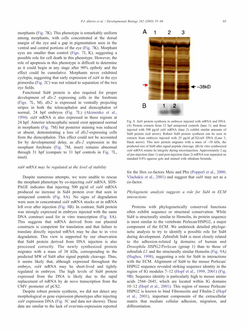
Fig. 8. Sid4 protein synthesis in embryos injected with mRNA and DNA.
(A) Protein extracts from 12 hpf uninjected controls (lane 1), and those
injected with 500 pg/nl sid4 mRNA (lane 2) exhibit similar amounts of
Sid4 protein (red arrow). Robust Sid4 protein synthesis can be seen in
extracts from embryos injected with 25 pg/nl pCS2sid4 DNA (Lane 3,
black arrow). This new protein migrates with a mass of ~38 kDa, the
predicted size of Sid4 after signal peptide cleavage. (B) In vitro synthesized
sid4 mRNA retains its integrity during microinjection. Approximately 2 Agof pre-injection (lane 1) and post-injection (lane 2) mRNAwas separated on
standard 0.8% agarose gels and stained with ethidium bromide.
P.J. diIorio et al. / Developmental Biology 282 (2005) 55–69 65
morphants (Fig. 7K). This phenotype is remarkably uniform
among morphants, with cells concentrated at the dorsal
margin of the eye and a gap in pigmentation seen in the
ventral and central portions of the eye (Fig. 7K). Morphant
eyes are smaller than control (Figs. 7J, K), suggesting a
possible role for cell death in this phenotype. However, the
role of apoptosis in this phenotype is difficult to determine
as it could begin at any stage after 90% epiboly and the
effect could be cumulative. Morphants never exhibited
cyclopia, suggesting that early expression of sid4 in the eye
primordia (Fig. 2C) was not related to separation of the two
eye fields.
Functional Sid4 protein is also required for proper
development of dlx-2 expressing cells in the forebrain
(Figs. 7L, M). dlx2 is expressed in ventrally projecting
stripes in both the telencaphalon and diencephalon of
normal, 24 hpf embryos (Fig. 7L) (Akimenko et al.,
1994). sid4 mRNA is also expressed in these regions at
24 hpf. Anterior telencephalic neural crest appeared normal
in morphants (Fig. 7M) but posterior staining was reduced
or absent, demonstrating a loss of dlx2-expressing cells
from the diencephalon. This effect could not be accounted
for by developmental delay, as dlx-2 expression in the
morphant forebrain (Fig. 7M, inset) remains abnormal
through 31 hpf (compare to 31 hpf controls in Fig. 7L,
inset).
sid4 mRNA may be regulated at the level of stability
Despite numerous attempts, we were unable to rescue
the morphant phenotype by co-injecting sid4 mRNA. SDS-
PAGE indicates that injecting 500 pg/nl of sid4 mRNA
produced no increase in Sid4 protein over that seen in
uninjected controls (Fig. 8A). No signs of degradation
were seen in concentrated sid4 mRNA stocks or in mRNA
left over after injection (Fig. 8B). In contrast, Sid4 protein
was strongly expressed in embryos injected with the same
DNA construct used for in vitro transcription (Fig. 8A).
This suggests that mRNA derived from our plasmid
constructs is competent for translation and that failure to
translate directly injected mRNA may be due to in vivo
degradation. This view is supported by our observation
that Sid4 protein derived from DNA injection is also
processed correctly. The newly synthesized protein
migrates with a mass of 38 kDa, corresponding to the
predicted MW of Sid4 after signal peptide cleavage. Thus,
it seems likely that, although expressed throughout the
embryo, sid4 mRNA may be short-lived and tightly
regulated in embryos. The high levels of Sid4 protein
expressed from the DNA is likely due to the rapid
replacement of mRNA by de novo transcription from the
CMV promoter of pCS2.
Despite robust protein synthesis, we did not detect any
morphological or gene expression phenotypes after injecting
sid4 expression DNA (Fig. 5C and data not shown). These
data are similar to the lack of over/mis-expression reported
for the Hox co-factors Meis and Pbx (Popperl et al., 2000;
Vlachakis et al., 2001) and suggest that sid4 may act as a
co-factor.
Phylogenetic analysis suggests a role for Sid4 in ECM
interactions
Proteins with phylogenetically conserved functions
often exhibit sequence or structural conservation. While
Sid4 is structurally similar to Hemolin, its protein sequence
is most similar to the vertebrate Perlecan/HSPG2, a major
component of the ECM. We undertook detailed phyloge-
netic analysis to try to identify a possible role for Sid4
during development. Zebrafish Sid4 is most closely related
to the adhesion-related Ig domains of human and
Drosophila HSPG2/Perlecan (group 1) than to those of
zebrafish L1 and the structurally similar Hemolin (Fig. 9A)
(Hughes, 1998), suggesting a role for Sid4 in interactions
with the ECM. Alignment of Sid4 to the mouse Perlecan/
HSPG2 sequence revealed striking sequence identity in the
region of IG modules 7–12 (Hopf et al., 1999, 2001) (Fig.
9B). Sequence identity is particularly high to mouse amino
acids 2566–2645, which are located within IG domains
10–12 (Hopf et al., 2001). This region of mouse Perlecan/
HSPG2 is known to bind Fibronectin and Fibulin-2 (Hopf
et al., 2001), important components of the extracellular
matrix that mediate cellular adhesion, migration, and
differentiation.

P.J. diIorio et al. / Developmental Biology 282 (2005) 55–6966
Discussion
Despite their molecular diversity and importance in
physiology and embryonic development, few small,
secreted immunoglobulin-containing proteins have been
functionally characterized in vertebrates (Chiquet-
Ehrismann and Chiquet, 2003; Sage and Bornstein, 1991;
Sodek et al., 2002). In this paper we analyzed the
embryological function of zebrafish sid4, which encodes
a novel, secreted, vertebrate Ig protein. Our data suggest
that Sid4 has important roles during vertebrate morpho-
Fig. 9. Phylogenetic analysis suggests a role for Sid4 in mediating cellular inte
vertebrate and invertebrate HSPG2 (group1) but more distantly related to Hemoli
epsilon served as out-groups. Numbers along each branch denote pairwise genet
binding Ig domains 10–13 (aa 2341–2705) of mouse HSPG2. Sid4 signal peptide
conservative substitutions.
genesis, possibly by mediating cellular interactions with
the ECM.
sid4 morphants are phenotypically similar to several
zebrafish mutants
During embryogenesis, sid4 is expressed in multiple cell
types undergoing rapid growth or migration. These include
gastrulating axial and somitic mesoderm, prechordal meso-
derm, eye primordia, and cranial neural crest. Intriguingly,
sid4 morphants are phenotypically similar to a number of
ractions with the extracellular matrix. (A) Sid4 is most closely related to
n and L1 (group 2). The immunity-related horn shark Ig mu and human Ig
ic distances (uncorrected bpQ). (B) Sid4 is most similar to the fibronectin-
was excluded from analysis. Shaded boxes highlight identical residues and

P.J. diIorio et al. / Developmental Biology 282 (2005) 55–69 67
zebrafish mutants with diverse genetic bases. For example,
sid4 morphants exhibit severe loss of caudal, ventrally-
derived mesoderm as found in BMP (Myers et al., 2002a) and
FGF (Draper et al., 2003) signaling mutants. However, bmp4
expression is normal in sid4 morphants, suggesting that the
ventral mesoderm of early embryos is correctly specified
and patterned. Normal chordin expression in sid4 morphants
indicates that bmp signaling is also correctly regulated and
further suggests that defects in this pathway do not
underlie the mis-patterning of axial and somitic mesoderm.
The overall development of sid4 morphant brain and
eyes also resembles prickle (Wnt) morphants (Carreira-
Barbosa et al., 2003; Veeman et al., 2003). Unlike prickle
morphants, which exhibit delayed migration of prechordal
mesoderm (Carreira-Barbosa et al., 2003; Veeman et al.,
2003), the initial anterior migration of zebrafish hgg1
expressing cells is normal. Morphants also exhibit broad-
ening of somitic mesoderm, with loss of normal chevron-
shaped somites, as seen in covergent extension mutants
(Myers et al., 2002b). Strong sid4 morphants exhibit failure
of axial and somitic mesoderm to converge on the midline,
but, unlike many convergent-extension mutants (Heisenberg
and Nusslein-Volhard, 1997; Marlow et al., 1998), never
exhibit cyclopia.
Sid4 morphants exhibit many characteristics of the
epiboly mutants (Kane et al., 1996) and microtubule-
defective embryos (Strahle and Jesuthasan, 1993). For
example, strong sid4 morphants exhibit defects in blasto-
derm migration and byolk plugQ closure. Failure of the
blastoderm to fully enclose the yolk impairs convergence of
the somitic mesoderm on the midline (Kane et al., 1996;
Strahle and Jesuthasan, 1993). Only the most severe sid4
morphants undergo true epiboly arrest and yolk lysis.
Retraction of the blastoderm after arrest was not observed
(Kane et al., 1996). The underlying genetics of delayed
epiboly remain unclear. That Sid4 is a secreted protein with
sequence similarity to extracellular matrix-binding domains
suggests that defective cell–substrate interactions may
contribute to delayed epiboly.
By a number of criteria, including gene expression (gsc,
hgg1, bmp4 and chordin) and TUNEL analysis, we found
that early (up to 90% epiboly) sid4 morphants were normal.
Only during later gastrulation and tail bud formation did
morphants exhibit changes in flh, unc-45, and hgg1
expression. Further, increased apoptosis was seen in
morphants from 90% epiboly onward, after the aberrant
patterning of future notochord and somitic mesoderm was
established. We interpret these data to mean that apoptosis
does not contribute to the initial patterning of axial and
somitic mesoderm but may account for the reduced numbers
of cells expressing a particular marker and for the overall
reduction in morphant embryo size. We believe that the
most parsimonious explanation for the similarities and
differences of sid4 morphants to the zebrafish mutants
may be that sid4 participates in a process common to all of
these mutants, such as adhesion or migration.
sid4 may function as a co-factor in a heterophilic process
Despite ubiquitous mRNA expression, sid4 knockdown
has distinct spatial and temporal effects in the early
zebrafish embryo. For example, at similar developmental
stages, morphogenesis of axial and somitic mesoderm is
impaired while migration of prechordal mesoderm is
normal. Further, sid4 knockdown differentially affects later
development of prechordal mesoderm, demonstrating a
stage-specific function in a single tissue during periods of
ubiquitous expression of sid4. These data suggest that 1)
sid4 translation may be temporally and spatially regulated or
2) sid4 interacts with a protein(s) that are in low abundance
or exhibit tissue-and stage-specific restricted expression.
Thus, the availability of potential ligands may be saturated
by endogenous levels of Sid4 expression during normal
development. Scarcity of binding sites may explain why
overexpression of sid4 expression DNA does not produce
any overt phenotype, even though injection of this construct
yields abundant Sid4 protein. Intriguingly, similar explan-
ations likely account for the lack of overexpression
phenotypes seen for the essential Hox co-factors Pbx and
Meis (Popperl et al., 2000; Vlachakis et al., 2001).
Sid4 is similar to the fibronectin-binding Igs of
perlecan/HSPG2
A role for Sid4 in interactions with the ECM is suggested
by phylogenetic analysis. Despite structural similarity to
Hemolin, Sid4 is most closely related to insect and vertebrate
Perlecan/HSPG2. Within mouse Perlecan/HSPG2, Sid4 is
highly similar to Ig domains 10–13, a region of HSPG2
known to bind Fibronectin and Fibulin-2 (Hopf et al., 1999),
important components of the ECM. Given these observa-
tions, and the striking similarities of sid4 morphants to
Fibronectin- and Integrin h1-deficient Xenopus embryos
(Marsden and DeSimone, 2003), we hypothesize that Sid4
may function as an adaptor molecule or as an anti-adhesive in
cell–matrix interactions. Morpholino-induced disruption of
Sid4 function may disrupt these interactions during gastru-
lation and lead to anoikis, or substrate-dependent cell death
(Grossmann, 2002).
More detailed analyses of Sid4, including identification
of potential ligands, extracellular localization and regulation
by transcription factors are ongoing. These studies will add
to a growing understanding of the molecular mechanisms by
which a small, secreted immunoglobulin protein functions
in vertebrate morphogenesis.
Acknowledgments
We thank A. Rossini, D. Greiner and J. Mordes for
generous support. C. Sagerstrom, R. Bortell, R. Bettencourt,
M. Ekker and D. Melton critically reviewed the data.
Shannon Knuth (GeneTools) provided invaluable assistance

P.J. diIorio et al. / Developmental Biology 282 (2005) 55–6968
in morpholino design. This work was supported by the
Diabetes and Endocrinology Research Center Grant 3P03-
DK32520 from the National Institutes of Health and the
Iacocca Foundation (P.J.D.).
References
Akimenko, M.A., Ekker, M., Wegner, J., Lin, W., Westerfield, M., 1994.
Combinatorial expression of three zebrafish genes related to distal-
less: part of a homeobox gene code for the head. J. Neurosci. 14,
3475–3486.
Barrallo-Gimeno, A., Holzschuh, J., Driever, W., Knapik, E.W., 2004.
Neural crest survival and differentiation in zebrafish depends on mont
blanc/tfap2a gene function. Development 131, 1463–1477.
Bettencourt, R., Lanz-Mendoza, H., Lindquist, K.R., Faye, I., 1997. Cell
adhesion properties of hemolin, an insect immune protein in the Ig
superfamily. Eur. J. Biochem. 250, 630–637.
Bettencourt, R., Gunne, H., Gastinel, L., Steiner, H., Faye, I., 1999.
Implications of hemolin glycosylation and Ca2+-binding on homophilic
and cellular interactions. Eur. J. Biochem. 266, 964–976.
Bettencourt, R., Terenius, O., Faye, I., 2002. Hemolin gene silencing by ds-
RNA injected into Cecropia pupae is lethal to next generation embryos.
Insect Mol. Biol. 11, 267–271.
Bingham, S., Higashijima, S., Okamoto, H., Chandrasekhar, A., 2002. The
zebrafish trilobite gene is essential for tangential migration of
branchiomotor neurons. Dev. Biol. 242, 149–160.
Carreira-Barbosa, F., Concha, M.L., Takeuchi, M., Ueno, N., Wilson, S.W.,
Tada, M., 2003. Prickle 1 regulates cell movements during gastrulation
and neuronal migration in zebrafish. Development 130, 4037–4046.
Chandrasekhar, A., Moens, C.B., Warren Jr., J.T., Kimmel, C.B., Kuwada,
J.Y., 1997. Development of branchiomotor neurons in zebrafish.
Development 124, 2633–2644.
Chiquet-Ehrismann, R., Chiquet, M., 2003. Tenascins: regulation and
putative functions during pathological stress. J. Pathol. 200, 488–499.
Dick, A., Hild, M., Bauer, H., Imai, Y., Maifeld, H., Schier, A.F., Talbot,
W.S., Bouwmeester, T., Hammerschmidt, M., 2000. Essential role of
Bmp7 (snailhouse) and its prodomain in dorsoventral patterning of the
zebrafish embryo. Development 127, 343–354.
diIorio, P.J., Moss, J.B., Sbrogna, J.L., Karlstrom, R.O., Moss, L.G., 2002.
Sonic hedgehog is required early in pancreatic islet development. Dev.
Biol. 244, 75–84.
Draper, B.W., Stock, D.W., Kimmel, C.B., 2003. Zebrafish fgf24 functions
with fgf8 to promote posterior mesodermal development. Development
130, 4639–4654.
Etheridge, L., diIorio, P.J., Sagerstrom, C.G., 2002. A zebrafish unc-45-
related gene expressed during muscle development. Dev. Dyn. 224,
457–460.
Fambrough, D., Goodman, C.S., 1996. The Drosophila beaten path gene
encodes a novel secreted protein that regulates defasciculation at motor
axon choice points. Cell 87, 1049–1058.
Grossmann, J., 2002. Molecular mechanisms of detachment-induced
apoptosis—Anoikis. Apoptosis 7, 247–260.
Heisenberg, C.P., Nusslein-Volhard, C., 1997. The function of silberblick in
the positioning of the eye anlage in the zebrafish embryo. Dev. Biol.
184, 85–94.
Higashijima, S., Hotta, Y., Okamoto, H., 2000. Visualization of cranial
motor neurons in live transgenic zebrafish expressing green fluorescent
protein under the control of the islet-1 promoter/enhancer. J. Neurosci.
20, 206–218.
Hopf, M., Gohring, W., Kohfeldt, E., Yamada, Y., Timpl, R., 1999.
Recombinant domain IV of perlecan binds to nidogens, laminin-
nidogen complex, fibronectin, fibulin-2 and heparin. Eur. J. Biochem.
259, 917–925.
Hopf, M., Gohring, W., Mann, K., Timpl, R., 2001. Mapping of binding
sites for nidogens, fibulin-2, fibronectin and heparin to different IG
modules of perlecan. J. Mol. Biol. 311, 529–541.
Hughes, A.L., 1998. Protein phylogenies provide evidence of a radical
discontinuity between arthropod and vertebrate immune systems.
Immunogenetics 47, 283–296.
Hwang, S.P., Tsou, M.F., Lin, Y.C., Liu, C.H., 1997. The zebrafish BMP4
gene: sequence analysis and expression pattern during embryonic
development. DNA Cell Biol. 16, 1003–1011.
Inohaya, K., Yasumasu, S., Araki, K., Naruse, K., Yamazaki, K., Yasumasu,
I., Iuchi, I., Yamagami, K., 1997. Species-dependent migration of fish
hatching gland cells that express astacin-like proteases in common
[corrected]. Dev. Growth Differ. 39, 191–197.
Jessen, J.R., Topczewski, J., Bingham, S., Sepich, D.S., Marlow, F.,
Chandrasekhar, A., Solnica-Krezel, L., 2002. Zebrafish trilobite
identifies new roles for Strabismus in gastrulation and neuronal
movements. Nat. Cell Biol. 4, 610–615.
Kane, D.A., Hammerschmidt, M., Mullins, M.C., Maischein, H.M., Brand,
M., van Eeden, F.J., Furutani-Seiki, M., Granato, M., Haffter, P.,
Heisenberg, C.P., Jiang, Y.J., Kelsh, R.N., Odenthal, J., Warga, R.M.,
Nusslein-Volhard, C., 1996. The zebrafish epiboly mutants. Develop-
ment 123, 47–55.
Kanost, M.R., Zepp, M.K., Ladendorff, N.E., Andersson, L.A., 1994.
Isolation and characterization of a hemocyte aggregation inhibitor from
hemolymph of Manduca sexta larvae. Arch. Insect Biochem. Physiol.
27, 123–136.
Kimmel, C.B., Ballard, W.W., Kimmel, S.R., Ullmann, B., Schilling, T.F.,
1995. Stages of embryonic development of the zebrafish. Dev. Dyn.
203, 253–310.
Kozak, M., 1987. An analysis of 5V-noncoding sequences from 699
vertebrate messenger RNAs. Nucleic Acids Res 15, 8125–8148.
Krauss, S., Johansen, T., Korzh, V., Fjose, A., 1991. Expression of the
zebrafish paired box gene pax[zf-b] during early neurogenesis.
Development 113, 1193–1206.
Marlow, F., Zwartkruis, F., Malicki, J., Neuhauss, S.C., Abbas, L.,
Weaver, M., Driever, W., Solnica-Krezel, L., 1998. Functional
interactions of genes mediating convergent extension, knypek and
trilobite, during the partitioning of the eye primordium in zebrafish.
Dev. Biol. 203, 382–399.
Marsden, M., DeSimone, D.W., 2003. Integrin–ECM interactions regulate
cadherin-dependent cell adhesion and are required for convergent
extension in Xenopus. Curr. Biol. 13, 1182–1191.
Melby, A.E., Kimelman, D., Kimmel, C.B., 1997. Spatial regulation of
floating head expression in the developing notochord. Dev. Dyn. 209,
156–165.
Miller-Bertoglio, V., Carmany-Rampey, A., Furthauer, M., Gonzalez, E.M.,
Thisse, C., Thisse, B., Halpern, M.E., Solnica-Krezel, L., 1999.
Maternal and zygotic activity of the zebrafish ogon locus antagonizes
BMP signaling. Dev. Biol. 214, 72–86.
Mongiat, M., Taylor, K., Otto, J., Aho, S., Uitto, J., Whitelock, J.M.,
Iozzo, R.V., 2000. The protein core of the proteoglycan perlecan
binds specifically to fibroblast growth factor-7. J. Biol. Chem. 275,
7095–7100.
Myers, D.C., Sepich, D.S., Solnica-Krezel, L., 2002a. Bmp activity gradient
regulates convergent extension during zebrafish gastrulation. Dev. Biol.
243, 81–98.
Myers, D.C., Sepich, D.S., Solnica-Krezel, L., 2002b. Convergence and
extension in vertebrate gastrulae: cell movements according to or in
search of identity? Trends Genet. 18, 447–455.
Nakamura, M., Baldwin, D., Hannaford, S., Palka, J., Montell, C., 2002.
Defective proboscis extension response (DPR), a member of the Ig
superfamily required for the gustatory response to salt. J. Neurosci. 22,
3463–3472.
Nissen, R.M., Yan, J., Amsterdam, A., Hopkins, N., Burgess, S.M., 2003.
Zebrafish foxi one modulates cellular responses to Fgf signaling
required for the integrity of ear and jaw patterning. Development 130,
2543–2554.
Oxtoby, E., Jowett, T., 1993. Cloning of the zebrafish krox-20 gene

P.J. diIorio et al. / Developmental Biology 282 (2005) 55–69 69
(krx-20) and its expression during hindbrain development. Nucleic
Acids Res. 21, 1087–1095.
Panicker, A.K., Buhusi, M., Thelen, K., Maness, P.F., 2003. Cellular
signalling mechanisms of neural cell adhesion molecules. Front Biosci.
8, d900–d911.
Pipes, G.C., Lin, Q., Riley, S.E., Goodman, C.S., 2001. The Beat generation:
a multigene family encoding IgSF proteins related to the Beat axon
guidance molecule in Drosophila. Development 128, 4545–4552.
Popperl, H., Rikhof, H., Chang, H., Haffter, P., Kimmel, C.B., Moens, C.B.,
2000. Lazarus is a novel pbx gene that globally mediates hox gene
function in zebrafish. Mol. Cell 6, 255–267.
Rossant, J., Howard, L., 2002. Signaling pathways in vascular develop-
ment. Annu. Rev. Cell Dev. Biol. 18, 541–573.
Rougon, G., Hobert, O., 2003. New insights into the diversity and function
of neuronal immunoglobulin superfamily molecules. Annu. Rev.
Neurosci. 26, 207–238.
Sage, E.H., Bornstein, P., 1991. Extracellular proteins that modulate cell–
matrix interactions, SPARC, tenascin, and thrombospondin. J. Biol.
Chem. 266, 14831–14834.
Saitou, N., Nei, M., 1987. The neighbor-joining method: a new method for
reconstructing phylogenetic trees. Mol. Biol. Evol. 4, 406–425.
Schilling, T.F., Kimmel, C.B., 1994. Segment and cell type lineage
restrictions during pharyngeal arch development in the zebrafish
embryo. Development 120, 483–494.
Schilling, T.F., Walker, C., Kimmel, C.B., 1996. The chinless mutation
and neural crest cell interactions in zebrafish jaw development.
Development 122, 1417–1426.
Schulte-Merker, S., Hammerschmidt, M., Beuchle, D., Cho, K.W., De
Robertis, E.M., Nusslein-Volhard, C., 1994. Expression of zebrafish
goosecoid and no tail gene products in wild-type and mutant no tail
embryos. Development 120, 843–852.
Schulte-Merker, S., Lee, K.J., McMahon, A.P., Hammerschmidt, M., 1997.
The zebrafish organizer requires chordino. Nature 387, 862–863.
Sodek, J., Zhu, B., Huynh, M.H., Brown, T.J., Ringuette, M., 2002. Novel
functions of the matricellular proteins osteopontin and osteonectin/
SPARC. Connect. Tissue Res. 43, 308–319.
Strahle, U., Jesuthasan, S., 1993. Ultraviolet irradiation impairs epiboly in
zebrafish embryos: evidence for a microtubule-dependent mechanism of
epiboly. Development 119, 909–919.
Su, X.D., Gastinel, L.N., Vaughn, D.E., Faye, I., Poon, P., Bjorkman, P.J.,
1998. Crystal structure of hemolin: a horseshoe shape with implications
for homophilic adhesion. Science 281, 991–995.
Sun, S.C., Lindstrom, I., Boman, H.G., Faye, I., Schmidt, O., 1990.
Hemolin: an insect-immune protein belonging to the immunoglobulin
superfamily. Science 250, 1729–1732.
Talbot, W.S., Trevarrow, B., Halpern, M.E., Melby, A.E., Farr, G.,
Postlethwait, J.H., Jowett, T., Kimmel, C.B., Kimelman, D., 1995. A
homeobox gene essential for zebrafish notochord development. Nature
378, 150–157.
Teichmann, S.A., Chothia, C., 2000. Immunoglobulin superfamily proteins
in Caenorhabditis elegans. J. Mol. Biol. 296, 1367–1383.
Thisse, C., Thisse, B., Halpern, M.E., Postlethwait, J.H., 1994. Goosecoid
expression in neurectoderm and mesendoderm is disrupted in zebrafish
cyclops gastrulas. Dev. Biol. 164, 420–429.
Vaughn, D.E., Bjorkman, P.J., 1996. The (Greek) key to structures of neural
adhesion molecules. Neuron 16, 261–273.
Veeman, M.T., Slusarski, D.C., Kaykas, A., Louie, S.H., Moon, R.T., 2003.
Zebrafish prickle, a modulator of noncanonical wnt/fz signaling,
regulates gastrulation movements. Curr. Biol. 13, 680–685.
Vlachakis, N., Choe, S.K., Sagerstrom, C.G., 2001. Meis3 synergizes with
Pbx4 and Hoxb1b in promoting hindbrain fates in the zebrafish.
Development 128, 1299–1312.
Vogel, A., Gerster, T., 1997. Expression of a cathepsin L gene in anterior
mesoderm and hatching gland. Dev. Genes Evol. 206, 477–479.
Westerfield, M., 2000. The Zebrafish Book. A Guide for the Laboratory
Use of Zebrafish (Danio Rerion). University of Oregon Press,
Eugene.
Willett, C.E., Zapata, A.G., Hopkins, N., Steiner, L.A., 1997. Expression of
zebrafish rag genes during early development identifies the thymus.
Dev. Biol. 182, 331–341.
Yoder, J.A., Nielsen, M.E., Amemiya, C.T., Litman, G.W., 2002. Zebrafish
as an immunological model system. Microbes Infect. 4, 1469–1478.