Characterization of the immunoglobulin heavy chain ...
9
Characterization of the immunoglobulin heavy chain complementarity determining region (CDR)-III sequences from human B cell precursor acute lymphoblastic leukemia cells. H Kiyoi, … , K Horibe, R Ohno J Clin Invest. 1992;89(3):739-746. https://doi.org/10.1172/JCI115650. Sequence analysis of the immunoglobulin heavy chain complementarity determining region (CDR)-III of B-lineage cells at various stages has provided important insights concerning B cell maturation and selection. Knowledge of human CDR-III sequences has been relatively limited compared with that of the murine system. We analyzed the CDR-III sequences of B cell precursor acute lymphoblastic leukemia (pre-B ALL) cells in 23 newly diagnosed and 10 relapsed patients, in order to elucidate the organization of CDR-III in B cell precursors. We found a very low frequency of somatic mutations in D and JH regions, preferential use of DLR, DXP, DHQ52, and DN elements, and of 3' side JH segments, and no predominant usage of D coding frames. Unusual joinings such as VH-D-D-JH and VH-JH were observed in three, and one sequences, respectively. We compared the CDR-III sequences derived from 10 patients between diagnosis and relapse. Two of them had three spots of mutated nucleotides at relapse, all of which were found in the N region near the D segments. Our data showed the possibility of somatic mutation at relapse, in addition to developmentally regulated rearrangement of the immunoglobulin gene at the stage of B cell precursors. Research Article Find the latest version: https://jci.me/115650/pdf
-
Upload
khangminh22 -
Category
Documents
-
view
4 -
download
0
Transcript of Characterization of the immunoglobulin heavy chain ...
Characterization of the immunoglobulin heavy chaincomplementarity determining region (CDR)-III sequences fromhuman B cell precursor acute lymphoblastic leukemia cells.
H Kiyoi, … , K Horibe, R Ohno
J Clin Invest. 1992;89(3):739-746. https://doi.org/10.1172/JCI115650.
Sequence analysis of the immunoglobulin heavy chain complementarity determining region (CDR)-III of B-lineage cells atvarious stages has provided important insights concerning B cell maturation and selection. Knowledge of human CDR-IIIsequences has been relatively limited compared with that of the murine system. We analyzed the CDR-III sequences of Bcell precursor acute lymphoblastic leukemia (pre-B ALL) cells in 23 newly diagnosed and 10 relapsed patients, in order toelucidate the organization of CDR-III in B cell precursors. We found a very low frequency of somatic mutations in D andJH regions, preferential use of DLR, DXP, DHQ52, and DN elements, and of 3' side JH segments, and no predominantusage of D coding frames. Unusual joinings such as VH-D-D-JH and VH-JH were observed in three, and one sequences,respectively. We compared the CDR-III sequences derived from 10 patients between diagnosis and relapse. Two of themhad three spots of mutated nucleotides at relapse, all of which were found in the N region near the D segments. Our datashowed the possibility of somatic mutation at relapse, in addition to developmentally regulated rearrangement of theimmunoglobulin gene at the stage of B cell precursors.
Research Article
Find the latest version:
https://jci.me/115650/pdf
Characterization of the Immunoglobulin Heavy ChainComplementarity Determining Region (CDR)-III Sequences from Human B cellPrecursor Acute Lymphoblastic Leukemia CellsHitoshi Kiyoi,* Tomoki Naoe,* Keizo Horibe,t and Ryuzo Ohno*Department of Medicine, the Branch Hospital Nagoya University School of Medicine, Higashi-ku, Nagoya 461, Japan; %
and $Department of Pediatrics, Nagoya University School of Medicine, Showa-ku, Nagoya 466, Japan
Abstract
Sequence analysis of the immunoglobulin heavy chain comple-mentarity determining region (CDR)-III of B-lineage cells atvarious stages has provided important insights concerning Bcell maturation and selection. Knowledge of human CDR-IIIsequences has been relatively limited compared with that of themurine system. Weanalyzed the CDR-III sequences of B cellprecursor acute lymphoblastic leukemia (pre-B ALL) cells in23 newly diagnosed and 10 relapsed patients, in order to eluci-date the organization of CDR-III in B cell precursors. Wefound a very low frequency of somatic mutations in D and JHregions, preferential use of DLR, Dxp, DHQS2, and DNelements,and of 3' side JH segments, and no predominant usage of Dcoding frames. Unusual joinings such as VH-D-D-JH and VH_JH were observed in three, and one sequences, respectively. Wecompared the CDR-III sequences derived from 10 patients be-tween diagnosis and relapse. Two of them had three spots ofmutated nucleotides at relapse, all of which were found in the Nregion near the D segments. Our data showed the possibility ofsomatic mutation at relapse, in addition to developmentally reg-ulated rearrangement of the immunoglobulin gene at the stageof B cell precursors. (J. Clin. Invest. 1992. 89:739-746.) Keywords: immunoglobulin - CDR-III * pre-B ALL * somatic muta-tion * PCR
Introduction
The immunoglobulin heavy chain (IgH)' variable region is en-coded by three separate genes: variable (VH), diversity (D), andjoining (JH) segments. During B lymphocyte differentiation,the IgH gene is assembled in two steps. Dand JH segments arejoined first, followed by VHto D-JH joining. During these join-ing steps, antibody diversity is generated by the following mech-
Address correspondence and reprint requests to Tomoki Naoe, Depart-ment of Medicine, the Branch Hospital Nagoya University School ofMedicine, 1-1-20, Daiko-Minami, Higashi-ku, Nagoya 461, Japan
Received for publication 19 March 1991 and in revised form 13September 1991.
1. Abbreviations used in this paper: CDR, complementarity determin-ing region; D, diversity segment; dd-NTP-Fs, four dideoxynucleotideterminators; IgH, immunoglobulin heavy chain; JH, joining segment;P, palindromic; PCR, polymerase chain reaction; pre-B ALL, B cellprecursor acute lymphoblastic leukemia; SSC, standard saline citrate;Tdt, terminal deoxynucleotidyl transferase; VH, variable segment.
anisms: (a) somatic recombination of multiple VH, D, and JHsegments; (b) variation of joining sites and addition of the Nregion nucleotides; and (c) somatic mutation (1-3).
The assembled variable region of the Ig gene contains threehypervariable regions surrounded by framework regions thatare relatively well conserved. These regions are called comple-mentarity determining regions (CDR)-I, II, and III, and arethought to contact the antigenic epitope directly ( 1-4). CDR-Iand II are encoded by a VH segment, and CDR-III is encodedby D elements, VH-D and D-JH junctional segments. As aresult of these mechanisms, enormous diversity of CDR-III iscreated from a limited number of germline D segments (1-3).
In murine system, recent studies have demonstrated severalcharacteristics of the IgH gene at each stage of B cell differentia-tion. First, the preferential use of the most 5' D segments, andthe most JH-proximal Dsegments, is developed during initial Dto JH joining in immature B cells (5). Second, the CDR-IIIsequences from fetal and neonatal B-lineage cells preferentiallyuse the JH-proximal VH genes (6-9). In contrast, adult B-lin-eage cells show random VHutilization (9-13). Third, N regionnucleotides are thought to be added by the enzyme terminaldeoxynucleotidyl transferase (TdT) (14-17), and very few Nregion nucleotides are observed in most fetal and neonatal VH_D-JH junctional sequences. The addition of N regions thusappears to be a developmentally regulated process in B cells(18-20). Fourth, one particular reading frame of the D ele-ments is preferentially used in productively rearranged VH-D-JH genes, and other reading frames are used at the stage of D-JHjoining, although the joining border varies in each clone (21,22). It is suggested that the selection of reading frames is devel-opmentally regulated at the stage of B cell ontogeny.
In the human system, DHQ52-J 1 joining was frequently ob-served in Epstein-Barr virus-transformed fetal B cell lines (23).Another study showed the biased use of JH segments (J3 > J4> J5 > J1, J2) and the DHQ52element in fetal liver (24). Re-cently Yamada et al. reported that 3' side JH segments (J4, J5,and J6) were preferentially used, and all coding frames of germ-line D genes were used to generate CDR-IIIs in adult humanperipheral B cells (25). Until now, molecular studies of humanB-lineage malignancies at various stages have provided manyinsights into B cell ontogeny. For example, follicular B celllymphoma has extensive somatic mutations in the VH re-gions (26-28), although B cell precursor leukemia has few mu-tations (29).
In this study, in order to elucidate the characteristics ofCDR-III at the stage of B cell precursors, we have analyzed theCDR-III sequences from human B cell precursor acute lympho-blastic leukemia (pre-B ALL) cells using the polymerase chainreaction (PCR) amplification and a fluorescent chain-terminat-ing dideoxy-nucleotide sequencing system. Wedemonstratehere the structure of the N region, the low frequency of somatic
Immunoglobulin Heavy Chain Complementarity Determining Region (CDR)-III Sequences 739
J. Clin. Invest.© The American Society for Clinical Investigation, Inc.0021-9738/92/03/0739/08 $2.00Volume 89, March 1992, 739-746
mutations in the D and JH regions, and the segments in the Dand JH genes which are preferentially used. In addition, wecompared the CDR-III sequences between initial diagnosis andrelapse, and found the possibility of somatic mutation in pre-BALL cells at relapse.
Methods
Leukemia cells. Bone marrow samples were taken from 23 patients ( 13children and 10 adults) with pre-B ALL at the time of initial diagnosisor relapse, after informed consent was obtained. All bone marrow sam-ples contained over 90% leukemia cells. The diagnosis of ALL in eachpatient was confirmed by standard morphological and immunopheno-typing studies. All leukemia cells had an LI or L2 French-American-British (FAB) subtype, expressed HLA-DR, CDO0, and CD19 anti-gens, and lacked immunophenotypic evidence of both T (CD1, CD2,CD3, CD4, CD5, CD7, CD8) and mature B-cell lineage (CD20 andsurface Ig). The expression of cytoplasmic Mchain and TdT was exam-ined by an immunofluorescence test. Karyotypic analysis revealed thatfive patients (patients S.B., S.I., F.K., S.M., and C.S.) had the Philadel-phia chromosome (Ph'). In the 10 relapsed patients, the morphologi-cal, immunophenotypical, and karyotypical features of their leukemiacells were identical with those at initial diagnosis. These data are sum-marized in Table I.
Southern blot analysis. High molecular weight DNAwas extractedfrom leukemia cells according to a previously published method (30).Rearrangements of the IgH gene were analyzed by Southern blot analy-sis (31), using a probe made of the subcloned 4.3-kb fragment (PstI-HindIII) from the genomic clone containing JH region. High molecularweight DNAfrom pre-B ALL was digested with HindIII, EcoRI, or acombination of HindIll and BamHI restriction endonucleases(Boehringer Mannheim Yamanouchi, Tokyo, Japan), electrophoresedin an 0.8% agarose gel, and blotted onto nitrocellulose membranes.Hybridization was carried out with 32P-labeled nick-translated probesunder previously published conditions (31). Membranes were washedtwice with 2x standard saline citrate (SSC), containing 0.1% SDS, andtwice with 0.1 x SSC, containing 0.1% SDSand exposed to x-ray films.
Oligonucleotide primers. Oligonucleotide primers for PCRamplifi-cation of CDR-III were synthesized on a DNAsynthesizer (model 381-A; Applied Biosystems, Inc., Foster City, CA). The consensus se-quences of the VHand JH segments were determined according to thepublished sequence data (32-34). Recently, several PCRprimers forthe amplification of the human IgH CDR-III have been reported(35-37). We also synthesized similar primers: Vcomm: 5'-GAGT-C-GAC(A/T)C(A/G)GC(G/CXG/A)TGTA(T/C)T(T/A)CTG-3', andJcomm: 5'-CCAAG-CTTACCTGAGGAGACGGTGA-3'that was de-signed to correspond more specifically to the published IgH sequencesthan those previously reported. These primers contain SalI and HindIlIcloning sites, respectively, to allow ligation of the amplified sequencesinto recombinant vectors (38).
PCRamplification of the CDR-III sequences. PCRwas essentiallyperformed as described by Saiki et al. (39). A 50-Mul reaction mixturecontained 500 ng of genomic DNA, 50 mMKC1, 10 mMTris-HCl (pH8.4), 1.0 to 1.5 mMMgCl2, 100 ,g/ml gelatin, 0.25MgM of VcommandJcommprimers, 200,uM of each deoxynucleotide triphosphate (dATP,dCTP, dGTP, and dTTP; Boehringer Mannheim Yamanouchi). Thereaction mixture was first incubated at 95°C for 10 min to denaturedouble-stranded DNA, followed by 1 min at 55-58°C to anneal primerand template. Primer extension was started by the addition of 2.5 UTaq polymerase (AmpliTaq; Perkin-Elmer Cetus Corp., Norwalk, CT),and allowed to proceed for 1 min at 72°C. Subsequent denaturing,annealing, and extension steps were performed at 92°C for 1 min, at 55to 58°C for 1 min and at 72°C for 1 min, respectively, for 40 cycles on aprogram temperature control system (model PC-700; Astec, Fukuoka,Japan).
DNAsequencing of amplified fragments. The amplified fragmentswere separated through 4%NuSieve GTGagarose gels (FMC BioProd-
ucts, Rockland, ME) and 12%polyacrylamide gels in TBEbuffer (0.09MTris-borate, 0.002 MEDTA: pH 8.0). Amplified fragments wereisolated from the polyacrylamide gels by the crush and soak technique,then digested with Sall and HindIII restriction endonucleases(Boehringer Mannheim Yamanouchi), and ligated to the Ml3mpl8phage vector (Takara, Kyoto, Japan). The ligated materials were trans-fected into Escherichia coli strain JM109. More than five recombinantplaques per one clone were picked up and cultured in 2 X TY medium(16 g/liter Bacto-tryptone; Difco Laboratories, Inc., Detroit, 10 g/literBacto-yeast extract; Difco Laboratories, 5 g/liter NaCl). Single-stranded phage DNAwas prepared from these cultures. DNAwas se-quenced using fluorescent chain-terminating dideoxynucleotides on aGenesis 2000 (DuPont Co., Wilmington, DE) (40). This sequencingsystem uses a special set of four dideoxynucleotide terminators (dd-NTP-Fs), each having a unique member of the succinyl fluoresceinfamily attached. Single-stranded phage DNA, M13 MI-primer (Ta-kara), and reaction buffer were heated at 90'C for 2 min and thentransferred to a 370C water bath for 10 min to allow the primer toanneal. At this time, a single solution containing a mixture of eachdeoxynucleotide triphosphate (dCTP, dTTP, 7-deaza dATP, and 7-deaza dGTP), dd-NTP-Fs, DTT, and T-7 DNApolymerase (Takara)was prepared. An aliquot of this solution was added to the annealedmixture, and after a 5-min incubation, the unincorporated nucleotide,proteins, and reaction buffer were removed by ammoniumacetate/eth-anol precipitation. The DNAwas washed with 70%ethanol and driedup, followed by resuspension in the loading solution. The mixture wasthen applied to the auto sequencer (Genesis 2000; DuPont Co.). Thesequence data of each clone was determined if all the sequences fromeach recombinant plaque were identical.
Results
Southern blot analysis. All leukemia cells analyzed here demon-strated rearranged bands of the JH region in either HindIII,EcoRI, or the combination of HindIll and BamHI digests. Inthe 10 relapsed patients, the rearranged patterns were identicalwith those at initial diagnosis (summarized on Table I).
Analysis of the CDR-III sequences from pre-B ALL. Allgenomic DNAs from the pre-B ALL cells were well amplifiedusing the Vcommand Jcommprimers. However, DNAfromnormal peripheral blood lymphocytes did not result in a signifi-cant band, and gave a faint smear after an additional 40-cyclePCRamplification (data not shown). In patient S.I., two ampli-fied bands were observed that presumably corresponded to tworearranged IgH genes. We analyzed both bands and deter-mined their CDR-III sequences.
The CDR-III sequences from the pre-B ALL cells areshown in Fig. 1. In the amplified sequences from all patientsexcept T.E., we could determine D elements followed by JHsequences. In patient T.E., a VH gene joined directly to a JHgene without a D region. Similar joining clones have been re-ported in peripheral B cells (25). Wedo not know if this joiningresults from deletions of the D region during VHto D-JH join-ing, or whether it reflects extensive exonuclease modificationof the D region in the VH-D-JH gene.
Dsegment. All D-coding elements were tentatively assignedto germline D segments (32, 41, 42). Somatic mutations of Delements were rare (4/412; 0.97%), although three patients(K.N., N.K., and C.S.) showed mutated Delements: CAto TGin DNI; A to Gin DHQ52; and Gto Cin DHQ52, respectively. TheDNAs obtained from these three patients at the time of bothinitial diagnosis and relapse were independently analyzed. Thesequences of the mutated D elements were completely identi-cal in each patient. The D element of patient K.N. may be
740 H. Kiyoi, T. Naoe, K. Horibe, and R. Ohno
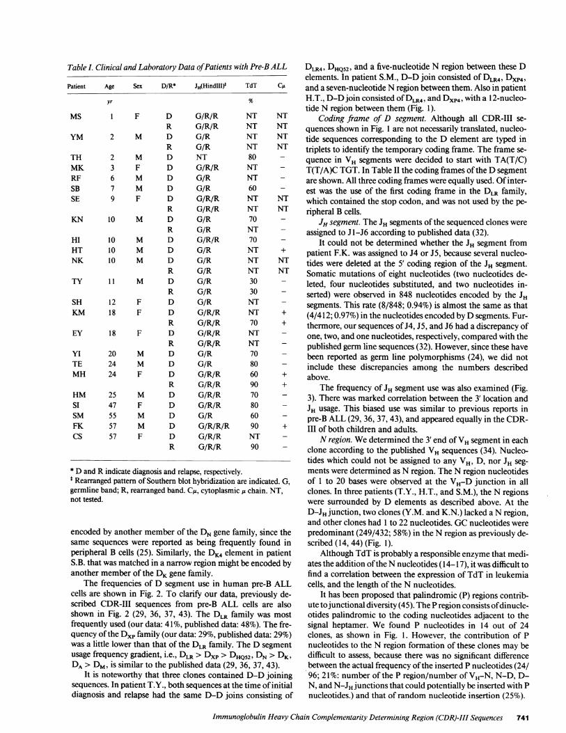
Table I. Clinical and Laboratory Data of Patients with Pre-B ALL
Patient Age Sex D/R* JH(HindIII)
yr
MS 1 F D G/R/RR G/R/R
YM 2 M D G/RR G/R
TH 2 M D NTMK 3 F D G/R/RRF 6 M D G/RSB 7 M D G/RSE 9 F D G/R/R
R G/R/RKN 10 M D G/R
R G/RHI 10 M D G/R/RHT 10 M D G/RNK 10 M D G/R
R G/RTY 11 M D G/R
R G/RSH 12 F D G/RKM 18 F D G/R/R
R G/R/REY 18 F D G/R/R
R G/R/RYI 20 M D G/RTE 24 M D G/RMH 24 F D G/R/R
R G/R/RHM 25 M D G/R/RSI 47 F D G/R/RSM 55 M D G/RFK 57 M D G/R/R/RCS 57 F D G/R/R
R G/R/R
TdT
NTNTNTNT80NTNT60NTNT70NT70NTNTNT3030NTNT70NTNT7080609070806090NT90
CA
* Dand R indicate diagnosis and relapse, respectively.$ Rearranged pattern of Southern blot hybridization are indicated. G,germline band; R, rearranged band. CA, cytoplasmic g chain. NT,not tested.
encoded by another member of the DNgene family, since thesame sequences were reported as being frequently found inperipheral B cells (25). Similarly, the DK4 element in patientS.B. that was matched in a narrow region might be encoded byanother member of the DKgene family.
The frequencies of D segment use in human pre-B ALLcells are shown in Fig. 2. To clarify our data, previously de-scribed CDR-III sequences from pre-B ALL cells are alsoshown in Fig. 2 (29, 36, 37, 43). The DLR family was mostfrequently used (our data: 41%, published data: 48%). The fre-quency of the DXPfamily (our data: 29%, published data: 29%)was a little lower than that of the DLR family. The D segmentusage frequency gradient, i.e., DLR > DXP> DHQ52, DN> DK,DA> DM, is similar to the published data (29, 36, 37, 43).
It is noteworthy that three clones contained D-D joiningsequences. In patient T.Y., both sequences at the time of initialdiagnosis and relapse had the same D-D joins consisting of
DLR4, DHQ52, and a five-nucleotide N region between these Delements. In patient S.M., D-D join consisted of DLR4, DxP4,and a seven-nucleotide N region between them. Also in patientH.T., D-D join consisted of DLR4, and Dxp4 with a 12-nucleo-tide N region between them (Fig. 1).
Coding frame of D segment. Although all CDR-III se-quences shown in Fig. 1 are not necessarily translated, nucleo-tide sequences corresponding to the D element are typed intriplets to identify the temporary coding frame. The frame se-quence in VH segments were decided to start with TA(T/C)T(T/A)C TGT. In Table II the coding frames of the Dsegmentare shown. All three coding frames were equally used. Of inter-est was the use of the first coding frame in the DLR family,which contained the stop codon, and was not used by the pe-ripheral B cells.
JH segment. The JH segments of the sequenced clones wereassigned to J -J6 according to published data (32).
It could not be determined whether the JH segment frompatient F.K. was assigned to J4 or J5, because several nucleo-tides were deleted at the 5' coding region of the JH segment.Somatic mutations of eight nucleotides (two nucleotides de-leted, four nucleotides substituted, and two nucleotides in-serted) were observed in 848 nucleotides encoded by the JHsegments. This rate (8/848; 0.94%) is almost the same as that(4/412; 0.97%) in the nucleotides encoded by Dsegments. Fur-thermore, our sequences of J4, J5, and J6 had a discrepancy ofone, two, and one nucleotides, respectively, compared with thepublished germ line sequences (32). However, since these havebeen reported as germ line polymorphisms (24), we did notinclude these discrepancies among the numbers describedabove.
The frequency of JH segment use was also examined (Fig.3). There was marked correlation between the 3 location andJH usage. This biased use was similar to previous reports inpre-B ALL (29, 36, 37, 43), and appeared equally in the CDR-III of both children and adults.
Nregion. Wedetermined the 3 end of VHsegment in eachclone according to the published VH sequences (34). Nucleo-tides which could not be assigned to any VH, D, nor JH seg-ments were determined as N region. The N region nucleotidesof 1 to 20 bases were observed at the VH-D junction in allclones. In three patients (T.Y., H.T., and S.M.), the N regionswere surrounded by D elements as described above. At theD-JH junction, two clones (Y.M. and K.N.) lacked a N region,and other clones had 1 to 22 nucleotides. GCnucleotides werepredominant (249/432; 58%) in the N region as previously de-scribed (14, 44) (Fig. 1).
Although TdT is probably a responsible enzyme that medi-ates the addition of the Nnucleotides ( 14-17), it was difficult tofind a correlation between the expression of TdT in leukemiacells, and the length of the N nucleotides.
It has been proposed that palindromic (P) regions contrib-ute to junctional diversity (45). The P region consists of dinucle-otides palindromic to the coding nucleotides adjacent to thesignal heptamer. We found P nucleotides in 14 out of 24clones, as shown in Fig. 1. However, the contribution of Pnucleotides to the N region formation of these clones may bedifficult to assess, because there was no significant differencebetween the actual frequency of the inserted P nucleotides (24/96; 21%: number of the P region/number of VH-N, N-D, D-N, and N-JH junctions that could potentially be inserted with Pnucleotides.) and that of random nucleotide insertion (25%).
Immunoglobulin Heavy Chain Complementarity Determining Region (CDR)-III Sequences 741

Case 3' end of V N, D, N region 5' end of J ]D J
TGOGAGA
TGCAA
TGCAAGA
TGOGAGA
TGOGAGA
TGOG
TGOGfAA
TG0GAAA
T
TGOGAGA
TGOGAGAV
TGOGAGAG
TGOGGAG
TGOG4GAG
TGCGAG
TGOGAGAG
TG
TGCAA
TGCAA
gcttt ggA TTA CGAmTTG GAG tgtttcccttccg..... ... ... .. .. .... ..... .............
LA tcgcgggtag ggA lTA CGA liT TTGGAGTG Ift cccttcacgaaactcgga
aaggg GAT 7TT TGG AGT GGT TAT ccccgccgatccg*-ca ... ... .... t.
; cccgggga TAC GAT ATT TTG ACT GGT TAT TA tc........ ..... ... ..... ..... ... .... ... . ...... ..
gc TATTTAT GGT TCG GGc ccaaagg
ctccatccgtctacg gTG ACT ACt gt
tccgag ggT GGT TAC cctctaggac
tggtgg GTA TAG CAG CTg gctggtac
tgg tGG GTA TAG CAGTGG CTG GTA C... .. ... ... ... ... ... ... .
tccagtccctccggacgt ggA TAG CAG CA CTG GTg gggcagctggtaatgggtcgg.................. ............... ... .. .. .... .... .. ... .....................
ggg gGT AGT ACC AGC TGCccaatgagcgcccgctgg... . .. ... ... ... ... ..................
A taatatgtcgggggga ctA GGA TAT TGT AGT AGT AOC AGC TGC TAT Gag ggaaggaa
ttgtttcccgttac tAG GAT All GTA GTA GTA OCA GCT GCT ATG gagt
cctga ATT GTA GTA GTAOCA GCT GCT ATG Cgc ctgata
A c TAT TGT ACT AA g
A gcccgggaaggag gaG GTG GTA Ggc
gatg ATT GTG GTGGTG ATT GCT A.... ... .... .... .... .... ....
ctcagggg TGG GGAgacttt
agaaggatttaaagtt gTA GCT GGGattatgtagt................ ....... ... . .... .... ... .........
gT AAC TOG GGg cgg.. ... * - --'ta
aatatcgaaatgag gAC CAG CTG Cca aaatgatag tAG TGG TTA HTA ctacgg
aagat aGA TAT TGT AGT AGT ACC AGC TGC TAT ata taA ACT GGGGAa ctaacctttct..... ... ... ... ... ... ... ... ... ... ... . ... ... ... ........ ...
gcctgcggggagtgccccc tTA TTG TAG TAG TAC CAG CTG CTA Taa atc ggA OGA ITT llG GAG TGGi TTA ggcgcctt
acta
T................GAACCI...
* 11
T.A.TA..G.GGOCAG.....CTGG
I *
..................
ACTGGT'rGCOTGGOAGAACC.................................
ACTAATACGTATGGALGTCTGGGGCCAAGGGCACG
...............................
*ACTGrrGCWOCGCAGA~ACIC
TTATACTGGGCCAGAAOGG.............................
t *
AGTWrGC CWACWLn rw
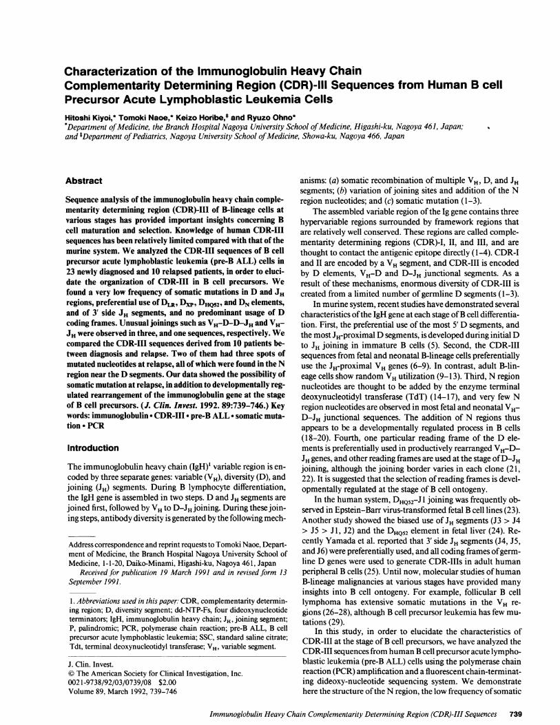
Figure 1. CDR-III sequences from pre-B ALL cells. Dand R indicate diagnosis and relapse, respectively. N region nucleotides are indicated bylower-case letters. D elements which could be tentatively assigned to previously described Dgenes are underlined. Dots indicate identical nu-cleotides. * nucleotide discrepancy with previously described germline JH segment. These nucleotide discrepancies were developed at the sameposition in each JH segment, and were thought to be germline polymorphisms. $ nucleotide mutation. § nucleotide insertion. 11 nucleotide dele-tion. P region nucleotide.
742 H. Kiyoi, T. Naoe, K. Horibe, and R. Ohno
DR
D
DR
DR
D
D
D
D
DR
DR
DR
D
D
D
D
D
DR
D
DR
DR
D
DR
D
D
EY
Si
KM
MS
Yl
Si
SB
FK
KN
MH
SE
HI
MK
SH
HM
RF
YM
TH
NK
CS
HT
TY
SM
TE
XP4
i XP4
i XP4
IXPI
XP'1
A4/I
K4
N4
Ni
Ni
LR4
LR4
LR4
LR4
LR1
LR2
LR3
L352
HQ52
HQ52
DAH
4
6
4
6
5
5
5
45
4
5
6
6
4
6
6
6
6
2
4
4
5
4
5
6
LR4 &HQ52
LR4 &XP4
_~~~~~~~~~~~----F-
C'rACTACrACrACGGTATGGACGTCTGGGGOCAAGGGAOCACGG...
t § *AAUGC......
TACTACTACGGTArjC2ACGTCr2GGGOCAAGGGACCACGG
ACrAC7rTGACrACrC4GGOCAGWAAOOCTGG
TArCArOGAT GGAOGTCTGGOCAGAAG
ILK4 h
T
L _ -i
r
I

I IIDxp1 Dxp'1 DA4/1
11 I I '7DK4 DOK DN4 D1 Dml DM2 DLR5 DLR4 DUR1 DLR2 DLR3 DHa52
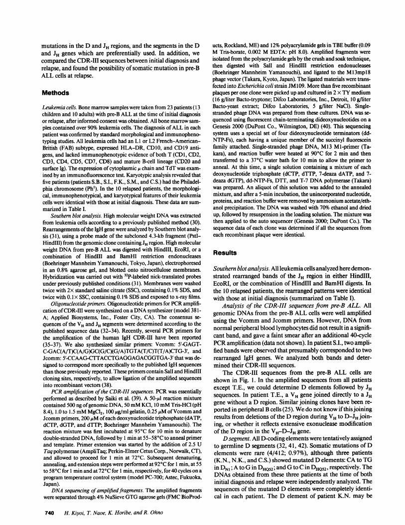
Figure 2. D element usage amonghuman pre-B ALL cells. Distribu-tion of D element among 24 clonessequenced here (black bars), andamong 21 clones previously re-ported (open bars) (29, 36, 37, 43).The clones which could be exam-ined both at initial diagnosis and re-lapse were counted as one clone.
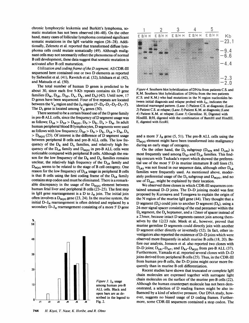
Comparison of CDR-III sequences between initial diagno-sis and relapse. Wecompared the CDR-III sequences derivedfrom 10 patients between initial diagnosis and relapse. Se-quencing data were determined by examining several recombi-nant clones from each sample to exclude the possibilities ofclonal heterogeneity and sequencing error. Although eight ofthem had completely matched sequences, the other two (K.M.and C.S.) differed by two or three nucleotides. To elucidate thatthese sequence differences were not due to Taq polymerasereplication errors during early stages of the PCRamplification,we amplified genomic DNAfrom the initial and relapse speci-mens three times independently, and sequenced five recombi-nant clones derived from each amplified sample. Since eachsequenced data (15 clones per one specimen) was completelyidentical, we could rule out the possibility of Taq polymerasereplication errors. The mutated clones at relapse were thoughtto be the same as the original clones, because Southern blotanalysis using JH probe showed similar patterns (Fig. 4). Addi-tionally, in patient K.M., a nucleotide insertion in the JH seg-ment was observed at diagnosis, and also at relapse. In patientC.S., the D element mutation was observed in both clones.These nucleotide differences at relapse were therefore not dueto clonal change but to somatic mutation. All these mutationsoccurred at the N region. In patient K.M., two nucleotides, GGin the VH-Dxp4 junction, and one nucleotide Cadjacent to the3' end of the Dxp4 element, had changed to CAand T at relapse,respectively. In patient C.S., two nucleotides GGdownstreamfrom the DHQ52element had changed to TA at relapse (Fig. 1).
Discussion
Knowledge of the DNAsequences of the human IgH variableregion has so far been limited, in contrast to that of the murinesystem. Recently a rapid cloning and sequencing method forIgH CDR-III using PCRhas been developed (25, 35-38, 43).Using this technique, we analyzed the CDR-III sequences frompre-B ALL cells.
Somatic mutation of the CDR-III sequences from pre-BALL cells. As mentioned in Results, we found several charac-teristics of the CDR-III sequences from pre-B ALL cells. So-
matic mutation of Dand JH segments was very rare (4/412 in Dsegments, 8/848 in JH segments). Furthermore, all of our se-quences of J4, J5, and J6 had a discrepancy of one, two, andone nucleotides, respectively, compared with published germline sequences (32). However, since these have been previouslypointed out as germline polymorphisms (24), and the majorityof sequenced human JH segments had the same nucleotide se-quences as ours (24, 25, 29, 36, 37, 43), we did not includethese discrepancies among the numbers described above.
Somatic mutation appears to occur at specific stages of Bcell development. Bird et al. reported that the rearranged VHgenes from pre-B ALL showed no evidence of mutation (29). In
Table II. Usage of D coding Frame
1st 2nd 3rd
DXP4 4(4) 1 (2) 0 (0)DXPI 0 (1) 1 (1) 0 (1)DXP'1 0 (0) 1 (3) 0 (1)DAI/4 0(0) 0(0) 1 (1)DK4 0 (0) 0 (1) 1(2)DK1 0 (0) 0 (0) 0 (0)DN4 0 (0) 0 (0) 1 (2)DN1 0 (1) 0 (0) 2 (3)DM1 0 (0) 0 (0) 0 (0)DM2 0 (0) 0(0) 0(0)DLR5 0(0) 0 (0) 0 (0)DLR4 2 (4) 3 (4) 2 (3)DLR1 0 (0) 1 (3) 0 (0)DLR2 0 (1) 0 (0) 1(3)DLR3 0 (0) 0 (0) 1(2)DHQ52 2 (3) 0 (1) 2 (2)
Total 8 (14) 7 (15) 11 (20)
The coding frame of the D elements was temporary decided to startwith TA(T/C) T(T/A)C TGT in the VH region. All three codingframes were equally used. The total number of our data and previ-ously described data (29, 36, 37, 43) are listed in parentheses.
Immunoglobulin Heavy Chain Complementarity Determining Region (CDR)-III Sequences 743
30i
20-
1 0-
nfL7'
uDxp4
L-I
chronic lymphocytic leukemia and Burkitt's lymphoma, so-matic mutation has not been observed (46-48). On the otherhand, many cases of follicular lymphoma contained significantsomatic mutations in the IgH variable region (26-28). Addi-tionally, Zelenetz et al. reported that transformed diffuse lym-phoma cells could mutate somatically (49). Although malig-nant cells may not necessarily reflect the phenomena of normalB cell development, these data suggest that somatic mutation isactivated after B cell maturation.
Utilization and codingframe of the Dsegment. All CDR-IIIsequenced here contained one or two D elements as reportedby Siebenlist et al. (41), Ravetch et al. (32), Ichihara et al. (42),and Matsuda et al. (50).
The total number of human D genes is predicted to beabout 30, since each five 9-Kb repeats contains six D genefamilies (DM, DLR, Dxp, DA, DK, and DN) (42). Until now, 17D genes have been sequenced. Four of five repeats are locatedbetween the VHregion and the JH region (5'-D4-D,-D2-D3-3').The D5 gene is located among VHgenes (50).
There seemed to be a certain biased use of the Dgene familyin pre-B ALL cells, since the frequency of Dsegment usage wasas follows; DLR> Dxp > DHQ52, DN>DK, DA> DM. In adulthuman peripheral blood B lymphocytes, Dsegments were usedas follows with low frequency; Dxp > DN>DK, DLR>DM, DA> DHQ52(25). Of interest is the difference of D segment usagebetween peripheral B cells and pre-B ALL cells. The low fre-quency of the DK and DA families, and relatively high fre-quency of the DLR family and DHQ52 in pre-B ALL cells werenoticeable compared with peripheral B cells. Although the rea-son for the low frequency of the DKand DA families remainsunclear, the relatively high frequency of the DLR family andDHQ52seems to be related to the stage of B cell ontogeny. Onereason for the low frequency of DLRusage in peripheral B cellsis that B cells using the first coding frame of the DLR familycontains stop codon and must be eliminated. There is a remark-able discrepancy in the usage of the DHQ52 element betweenhuman fetal liver and peripheral B cells (23-25). The first stepin IgH gene rearrangement is a D to JH join. The initial joinoften involves a DHQ52gene (23, 24). In the murine system, theinitial D-JH rearrangement is often deleted and replaced by asecondary D-JH rearrangement consisting of a more 5' Dgene
50]
40-
30-
20
10-
0
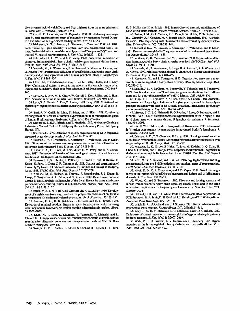
I_ ~~~~~Figure 3. JH usage
among human pre-BALL cells. Black andopen bars are as de-scribed in the legend to
JA J2 J3 4 J5 J6 Fig. 2.
1 2 3 4 5E BHH E B'H H E BH H E BH H E BH H Kb
4*i~u.-'4';'-23.1
*, ,. * ^ * --06.6
-4.4M !*
- 2.3-2.0
Figure 4. Southern blot hybridization of DNAsfrom patients C.S. andK.M. Southern blot hybridization of DNAs from the two patients(C.S. and K.M.) who had mutations in the N region nucleotides be-tween initial diagnosis and relapse probed with JH, indicates theidentical rearranged pattern. (Lane 1) Patient C.S. at diagnosis; (Lane2) Patient C.S. at relapse; (Lane 3) Patient K.M. at diagnosis; (Lane4) Patient K.M. at relapse; (Lane 5) Germline. H, Digested withHindIlI. B/H, digested with the combination of BamHI and HindlIl.E, digested with EcoRI.
and a more 3' JH gene (5, 51). The pre-B ALL cells using theDHQ52element might have been transformed into malignancyduring an early stage of ontogeny.
On the other hand, the D4 subgroup (Dxp4 and DmR4) ismost frequently used among DXPand DLR families. This find-ing concurs with Tsukada's report which showed the preferen-tial use of the most 5' D in murine immature B cell lines (5).DLR5 was not found in our sequence data, although other DRfamilies were frequently used. As mentioned above, moder-ately preferential usage of the D4 subgroup and DHQS2, and nouse of DLR5, might be explained by their location.
Weobserved three clones in which CDR-III sequences con-tained unusual D-D joins. The D-D joining model was firstproposed by Kurosawa and Tonegawa to explain the origin ofthe N region of the murine IgH gene (44). They thought that aD segment (Da) could join to another D segment (Db), using a24-mer signal spacer consisting of the end pentamer within theDa segment, the Da heptamer, and a 1 2mer of spacer instead ofa 23mer, because intact D segments cannot join among them-selves by the 12/23 rule. Meek et al., however, proved thatmurine germline D segments could directly join with anotherD segment either directly or invertedly (52). In fact, other in-vestigators also reported the existence of D-D joins which wereobserved more frequently in adult murine B cells (18, 20). Be-fore our analysis, Jonsson et al. also reported two clones withD-D joins; DLR1-Dxpl and DK4-DHQ52 from pre-B ALL (37).Furthermore, Yamada et al. reported several clones with D-Djoins derived from peripheral B cells (25). Thus, in the CDR-IIIfrom human pre-B cells, the D-D joins might occur more fre-quently than in murine B cell differentiation.
Recent studies have shown that truncated or complete IgHchain molecules are expressed together with surrogate lightchain molecules on the surface of the murine pre-B cells (22).Although the human counterpart molecule has not been dem-onstrated, a selection of D reading frames might be also in-fluenced by a kind of selective pressure. Our DNAstudy, how-ever, suggests no biased usage of D coding frames. Further-more, some CDR-III sequences contained a stop codon. The
744 H. Kiyoi, T. Naoe, K. Horibe, and R. Ohno
I

transcripts should be analyzed to elucidate the selection of cod-ing frames.
Utilization of the JH segment. Wefound a clear correlationbetween JH utilization and the 3' localization of JH. At an earlystage of B cells in fetal liver, DHQS2-J 1 joining was frequentlyobserved (23). During B cell differentiation, a second D-JH or athird D-JH might occur before VH-D-JH rearrangement is ac-complished (51). In this case the most 5' D and the most 3' JHwould join at a later stage. This explanation matches our obser-vations, and supports the one-dimensional tracking model ofrecombination (6, 53). Schroeder et al. reported a striking gra-dient of JH usage in human fetal liver as follows: J3 > J4 > J5> J2, J6 (24). Yamada et al. reported a different gradient of JHusage in peripheral B cells as follows: J4 > J6 > J5 > J3 (25). Inour findings and previously reported sequences derived frompre-B ALL cells (29, 36, 37, 43), J4, J5, and J6 segments wereequally used. It is not clear at the present time whether thesediscrepancies are due to the different utilization of B cells ineach stage of ontogeny, or the selection resulting from malig-nant transformation.
Mutated CDR-III sequence at relapse. 2 out of 10 patientshad mutated CDR-III sequences at relapse. All of these changeswere observed within the N region near the D segments; inaddition, the G nucleotide was most frequently substituted.Misincorporations due to the relative lack of fidelity of Taqpolymerase might be the important problem with our ap-proach. In order to rule out the possibility of Taq polymerasereplication errors, we examined the genomic DNAsfrom thesetwo patients three times independently, and confirmed the mu-tations. In the earlier studies, a cumulative error frequency wasreported about 0.25% after 30 cycles of PCR(39). However,recent studies using lower dNTPand Mg2" ion concentrations(200 uM each dNTP, and 1.5 mMMgCI2), which were thesame as ours, have reported a significant increase in the fidelityof Taq polymerase. Now it is recognized that the cumulativeerror frequency is less than 6.6 X 10', and the average muta-tion rate is 5 X 10⁻⁶ errors per nucleotide incorporated percycle, assuming 25 cycles of doubling (54, 55).
Generally, somatic mutation has been thought to occur inmature B cells after antigenic stimulation. In the murine sys-tem, somatic mutation in antibody variable genes appearedearly after primary immunization (56). In a pre-B cell line,hypermutation at the IgH locus whose rate was determined tobe 0.3-1 X 10' per cell generation, was reported (57). On theother hand, in human systems it is not known from what stagehuman B cells mutate, although somatic mutations were fre-quently observed in follicular B cell lymphoma (26-28, 49).Our data, however, suggests that a primary mutation mightoccur at specific sequences even at the stage of pre-B cell.
Recently Yamada et al. reported the usefulness of the tu-mor-specific CDR-III sequence to detect and monitor minimalresidual disease at the level of l0', using the PCRamplifica-tion (43). Wealso reported the detection of minimal residualdisease at the level of 106 in a patient who had received anallogeneic bone marrow transplantation using two-step PCRamplification (38). However the mutated CDR-III sequencecannot be detected, since these detection systems are based onthe enormous diversity of CDR-III. The clinical usefulness ofthese systems, therefore, must be limited when the leukemiacells have mutated CDR-III sequences at relapse.
Structural analysis of CDR-III sequence is important toelucidate the B cell repertoire. As presented here, the sequential
study of clinical lymphoid tumor samples can serve as a valu-able model in B cell differentiation. Additionally, a rapid clon-ing and sequencing method of CDR-III could be applied tomany clinical fields.
Acknowledgments
Wethank Dr. Yoshikazu Kurosawa for helpful advice and discus-sion, and Yujiro Mon and Alan F. Brunner (Aloka Co., Ltd.) for tech-nical support for DNAsequencing.
This work was supported by grants from the Japanese Ministry ofHealth and Welfare.
References
1. Tonegawa, S. 1983. Somatic generation of antibody diversity. Nature(Lond.). 302:575-581.
2. Honjo, T. 1983. Immunoglobulin genes. Annu. Rev. Immunol. 1:499-528.3. Alt, F. W., T. K. Blackwell, R. A. DePinho, M. G. Reth, and G. D. Yanco-
poulos. 1986. Regulation of genome rearrangement events during lymphocytedifferentiation. Immunol. Rev. 89:5-30.
4. Wu, T. T., and E. A. Kabat. 1970. An analysis of the sequences of thevariable regions of Bence Jones proteins and myeloma light chains and theirimplications for antibody complementarity. J. Exp. Med. 132:211-250.
5. Tsukada, S., H. Sugiyama, Y. Oka, and S. Kishimoto. 1990. Estimation ofD segment usage in initial D to JH joinings in a murine immature B cell line. J.Immunol. 144:4053-4059.
6. Yancopoulos, G. D., S. V. Desiderio, M. Paskind, J. F. Kearney, D. Balti-more, and F. W. Alt. 1984. Preferential utilization of the most JH-proximal VHgene segments in pre-B-cell lines. Nature (Lond.). 311:727-733.
7. Perlmutter, R. M., J. F. Kearney, S. P. Chang, and L. E. Hood. 1985.Developmentally controlled expression of immunoglobulin VH genes. Science(Wash. DC). 227:1597-1600.
8. Jeong, H. D., and J. M. Teale. 1988. Comparison of the fetal and adultfunctional B cell repertoires by analysis of VH gene family expression. J. Exp.Med. 168:589-603.
9. Dildrop, R., U. Krawinkel, E. Winter, and K. Rajewsky. 1985. VH-geneexpression in murine lipopolysaccharide blasts distributes over the nine knownVH-gene groups and may be random. Eur. J. Immunol. 15:1154-1156.
10. Wu, G. E., and C. J. Paige. 1986. VHgene family utilization in coloniesderived from B and pre-B cells detected by the RNAcolony blot assay. EMBO(Eur. Mol. BioL. Organ.) J. 5:3475-3481.
11. Schulze, D. H., and G. Kelsoe. 1987. Genotypic analysis of B cell coloniesby in situ hybridization. Stoichiometric expression of three VH families in adultC57BL/6 and BALB/c mice. J. Exp. Med. 166:163-172.
12. Yancopoulos, G. D., B. A. Malynn, and F. W. Alt. 1988. Developmentallyregulated and strain-specific expression of murine VHgene families. J. Exp. Med.168:417-435.
13. Sheehan, K. M., and P. H. Brodeur. 1989. Molecular cloning of the pri-mary IgH repertoire: A quantative analysis of VH gene usage in adult mice.EMBO(Eur. Mol. Biol. Organ) J. 8:2313-2320.
14. Alt, F. W., and D. Baltimore. 1982. Joining of immunoglobulin heavychain gene segments: implications from a chromosome with evidence of threeD-JH fusions. Proc. Natl. Acad. Sci. USA. 79:4118-4112.
15. Desiderio, S. V., G. D. Yancopoulos, M. Paskind, E. Thomas, M. A. Boss,N. Landau, F. W. Alt, and D. Baltimore. 1984. Insertion of N regions into heavy-chain genes is correlated with expression of terminal deoxytransferase in B cells.Nature (Lond.). 311:752-755.
16. Landau, N. R., D. G. Schatz, M. Rosa, and D. Baltimore. 1987. Increasedfrequency of N-region insertion in a murine pre-B-cell line infected with a termi-nal deoxynucleotidyl transferase retroviral expression vector. Mol. Cell. Biol.7:3237-3243.
17. Lieber, M. R., J. E. Hesse, K. Mizuuchi, and M. Gellert. 1988. LymphoidV(D)J recombination: Nucleotide insertion at signal joints as well as codingjoints. Proc. Natl. Acad. Sci. USA. 85:8588-8592.
18. Feeney, A. J. 1990. Lack of N regions in fetal and neonatal mouse immu-noglobulin V-D-J junctional sequences. J. Exp. Med. 172:1377-1390.
19. Gu, H., I. Forster, and K. Rajewsky. 1990. Sequence homologies, N se-quence insertion and JH gene utilization in VHDJHjoining: implications for thejoining mechanism and the ontogenetic timing of Ly 1 B cell and B-CLL progeni-tor generation. EMBO(Eur. Mol. Biol. Organ.) J. 9:2133-2140.
20. Bangs, L. A., I. E. Sanz, and J. M. Teale. 1991. Comparison of D, JH andjunctional diversity in the fetal, adult, and aged B cell repertoires. J. Immunol.146: 1996-2004.
21. Ichihara, Y., H. Hayashida, S. Miyazawa, and Y. Kurosawa. 1989. OnlyDFL16, Dsp2, and DQ52gene families exist in mouse immunoglobulin heavy chain
Immunoglobulin Heavy Chain Complementarity Determining Region (CDR)-III Sequences 745
diversity gene loci, of which DFL16 and Dsp2 originate from the same primordialDHgene. Eur. J. Immunol. 19:1849-1854.
22. Gu, H., D. Kitamura, and K Rajewsky. 1991. B cell development regu-lated by gene rearrangement: arrest of maturation by membrane-bound D, pro-tein and selection of DHelement reading frames. Cell. 65:47-54.
23. Nickerson, K. G., J. Berman, E. Glickman, L. Chess, and F. W. Alt. 1989.Early human IgH gene assembly in Epstein-Barr virus-transformed fetal B celllines. Preferential utilization of the most JH-proximal Dsegment (DQ52) and twounusual VH-related rearrangements. J. Exp. Med. 169:1391-1403.
24. Schroeder JR, H. W., and J. Y. Wang. 1990. Preferential utilization ofconserved immunoglobulin heavy chain variable gene segments during humanfetal life. Proc. Natl. Acad. Sci. USA. 87:6146-6150.
25. Yamada, M., R. Wasserman, B. A. Reichard, S. Shane, A. J. Caton, andG. Rovera. 1991. Preferential utilization of specific immunoglobulin heavy chaindiversity and joining segments in adult human peripheral blood B lymphocytes.J. Exp. Med. 173:395-407.
26. Cleary, M., T. C. Meeker, S. Levy, E. Lee, M. Trela, J. Sklar, and R. Levy.1986. Clustering of extensive somatic mutations in the variable region of animmunoglobulin heavy chain gene from a human B cell lymphoma. Cell. 44:97-106.
27. Levy, R., S. Levy, M. L. Cleary, W. Carroll, S. Kon, J. Bird, and J. Sklar.1987. Somatic mutation in human B-cell tumors. Immunol. Rev. 96:43-58.
28. Levy, S., E. Mendel, S. Kon, Z. Avnur, and R. Levy. 1988. Mutational hotspots in Ig V region genes of human follicularlymphomas. J. Exp. Med. 168:475-489.
29. Bird, J., N. Galili, M. Link, D. Stites, and J. Sklar. 1988. Continuingrearrangement but absence of somatic hypermutation in immunoglobulin genesof human B cell precursor leukemia. J. Exp. Med. 168:229-245.
30. Sambrook, J., E. F. Fritsch, and T. Maniatis. 1989. Molecular Cloning: aLaboratory Manual. 2nd ed. Cold Spring Harbor Laboratory Press, Cold SpringHarbor, NY.
31. Southern, E. 1975. Detection of specific sequence among DNAfragmentsseparated by gel electrophoresis. J. Mol. Biol. 98:503-517.
32. Ravetch, J. V., U. Siebenlist, S. Korsmeyer, T. Waldmann, and P. Leder.1981. Structure of the human immunoglobulin mu locus: Characterization ofembryonic and rearranged J and Dgenes. Cell. 27:583-591.
33. Kabat, E. A., T. T. Wu, M. Reid-Miller, H. M. Perry, and K. S. Gottes-man. 1987. Sequences of Proteins of Immunological Interest. 4th ed. NationalInstitutes of Health publication, Bethesda, MD.
34. Berman, J. E., S. J. Mellis, R. Pollock, C. L. Smith, H. Suh, B. Heinke, C.Kowal, U. Surti, L. Chess, C. R. Cantor, et al. 1988. Content and organization ofhuman Ig VH locus: Definition of three new VHfamilies and linkage to the Ig VHlocus. 1988. EMBO(Eur. Mol. Biol. Organ.) J. 7:727-738.
35. Yamada, M., S. Hudson, 0. Tournay, S. Bittenbender, S. S. Shane, B.Lange, Y. Tsujimoto, A. J. Caton, and G. Rovera. 1989. Detection of minimaldisease in hematopoietic malignancies of the B-cell lineage by using third-com-plementarity-determining region (CDR-III)-specific probes. Proc. Nail. Acad.Sci. USA. 86:5123-5127.
36. Brisco, M. J., L. W. Tan, A. M. Orsborn, and A. A. Morley. 1990. Develop-ment of a highly sensitive assay, based on the polymerase chain reaction, for rareB-lymphocyte clones in a polyclonal population. Br. J. Haematol. 75:163-167.
37. Jonsson, 0. G., R. K. Kitchens, F. C. Scott, and R. G. Smith. 1990.Detection of minimal residual disease in acute lymphoblastic leukemia usingimmunoglobulin hypervariable region specific oligonucleotide probes. Blood.76:2072-2079.
38. Kiyoi, H., T. Naoe, K. Kitamura, T. Yamauchi, T. Ichihashi, and R.Ohno. 1991. Disappearance of minimal residual lymphoblastic leukemia cells sixmonths after allogeneic bone marrow transplantation without GVHD. BoneMarrow Transplant. 8:59-62.
39. Saiki, R. K., D. H. Gelfand, S. Stoffel, S. J. Scharf, R. Higuchi, G. T. Horn,
K B. Mullis, and H. A. Erlich. 1988. Primer-directed enzymic amplification ofDNAwith a thermosatable DNApolymerase. Science (Wash. DC). 239:487-491.
40. Prober, J. M., G. L. Trainor, R. J. Dam, F. W. Hobbs, C. W. Robertson,R. J. Zagursky, A. J. Cocuzza, M. A. Jensen, and K Baumeister. 1987. A systemfor rapid DNAsequencing with fluorescent chain-terminating dideoxynucleo-tides. Science (Wash. DC). 238:336-341.
41. Siebenlist, U., J. V. Ravetch, S. Korsmeyer, T. Waldmann, and P. Leder.1981. Human immunoglobulin Dsegments encoded in tandem multigenic fami-lies. Nature (Lond.). 294:631-635.
42. Ichihara, Y., H. Matsuoka, and Y. Kurosawa. 1988. Organization of hu-man immunoglobulin heavy chain diversity gene loci. EMBO(Eur. Mol. Biol.Organ.) J. 7:4141-4150.
43. Yamada, M., R. Wasserman, B. Lange, B. A. Reichard, R. B. Womer, andG. Rovera. 1990. Minimal residual disease in childhood B-lineage lymphoblasticleukemia. N. Engl. J. Med. 323:448-455.
44. Kurosawa, Y., and S. Tonegawa. 1982. Organization, structure, and as-sembly of immunoglobulin heavy chain diversity DNAsegments. J. Exp. Med.155:201-217.
45. Lafaille, J. L., A. DeCloux, M. Bonneville, Y. Takagaki, and S. Tonegawa.1989. Junctional sequences of T cell receptor genes: implications for T cell lin-eages and for a novel intermediate of V-(D)-J joining. Cell. 59:859-870.
46. Kipps, T. J., E. Tomhave, P. P. Chen, and D. A. Carson. 1988. Autoanti-body-associated kappa light chain variable region gene expressed in chronic lym-phocytic leukemia with little or no somatic mutation. Implications for etiologyand immunotherapy. J. Exp. Med. 167:840-852.
47. Meeker, T. C., J. C. Grimaldi, R. O'Rourke, J. Loeb, G. Juliusson, and S.Einhorn. 1988. Lack of detectable somatic hypermutation in the V region of theIg H chain gene of a human chronic B lymphocytic leukemia. J. Immunol.141:3994-3998.
48. Caroll, W. L., M. Yu, M. P. Link, and S. J. Korsmeyer. 1989. Absence ofIg V region gene somatic hypermutation in advanced Burkitt's lymphoma. J.Immunol. 143:692-698.
49. Zelenetz, A. D., T. T. Chen, and R. Levy. 1991. Histologic transformationof follicular lymphoma to diffuse lymphoma represents tumor progression by asingle malignant B cell. J. Exp. Med. 173:197-207.
50. Matsuda, F., K. H. Lee, S. Nakai, T. Sato, M. Kodaira, S. Q. Zong, H.Ohno, S. Fukuhara, and T. Honjo. 1988. Dispersed localization of D segments inthe human immunoglobulin heavy-chain locus. EMBO(Eur. Mol. Biol. Organ.)J. 7:1047-1051.
51. Reth, M. G., S. Jackson, and F. W. Alt. 1986. VHDJHformation and DJHreplacement during pre-B differentiation: non-random usage of gene segments.EMBO(Eur. Mol. Biol. Organ) J. 5:2131-2138.
52. Meek, K. D., C. A. Hasemann, and J. D. Capra. 1989. Novel rearrange-ments at the immunoglobulin D locus: Inversions and fusions add to IgH somaticdiversity. J. Exp. Med. 170:39-57.
53. Wood, C., and S. Tonegawa. 1983. Diversity and joining segments ofmouse immunoglobulin heavy chain genes are closely linked and in the sameorientation: implications for the joining mechanism. Proc. Natl. Acad. Sci. USA.80:3030-3034.
54. Gelfand, D. H., and T. J. White. 1990. Thermostable DNApolymerase. InPCRProtocols. M. A. Innis, D. H. Gelfand, J. J. Shinsky, and T. J. White, editors.Academic Press, San Diego, CA. 129-141.
55. Erlich, H. A., D. Gelfand, and J. J. Sninsky. 1991. Recent advances in thepolymerase chain reaction. Science (Wash. DC). 252:1643-1651.
56. Levy, N. S., U. V. Malipiero, S. G. Lebecque, and P. J. Gearhart. 1989.Early onset of somatic mutation in immunoglobulin VHgenes during the primaryimmune response. J. Exp. Med. 169:2007-2019.
57. Wabl, M., P. D. Burrows, A. V. Gabain, and C. Steinberg. 1985. Hyper-mutation at the immunoglobulin heavy chain locus in a pre-B-cell line. Proc.Natl. Acad. Sci. USA. 82:479-482.
746 H. Kiyoi, T. Naoe, K. Horibe, and R. Ohno