Phage Display and Peptide Mapping of an Immunoglobulin Light Chain Fibril-Related Conformational...
24
Phage Display and Peptide Mapping of an Immunoglobulin Light Chain Fibril-Related Conformational Epitope † Brian O’Nuallain, Amy Allen, Demet Ataman, Deborah T. Weiss, Alan Solomon * , and Jonathan S. Wall Human Immunology and Cancer/Alzheimer’s Disease and Amyloid-Related Disorders Research Program, Department of Medicine, University of Tennessee Graduate School of Medicine, Knoxville, Tennessee 37920 Abstract Amyloid fibrils and partially unfolded intermediates can be distinguished serologically from native amyloidogenic precursor proteins or peptides. In this regard, we previously had reported that mAb 11-1F4, generated by immunizing mice with a thermally denatured variable domain (V L ) fragment of the human κ4 Bence Jones protein Len, bound to a non-native conformational epitope located within the N-terminal 18 residues of fibrillar, as well as partially denatured, Ig light chains (O’Nuallain B. et al. (2006) Biochemistry 46, 1240–247). To define further the antibody binding site, we used random peptide phage display and epitope mapping of V L Len using wild-type and alanine- mutated Len peptides where it was shown that the antibody epitope was reliant on up to 10 of the first 15 residues of protein Len. Comparison of V κ and V λ N-terminal germline consensus sequences with protein Len and 11-1F4-binding phages indicated that this antibody’s cross-reactivity with light chains was related to an invariant proline at position(s) 7 and/or 8, bulky hydrophobic residues at positions 11 and 13, and additionally, to the ability to accommodate amino acid diversity at positions 1–4. Sequence alignments of the phage peptides revealed a central proline, often flanked by aromatic residues. Taken together, these results have provided evidence for the structural basis of the specificity of 11-1F4 for both κ and λ light chain fibrils. We posit that the associated binding site involves a rare type VI β-turn or touch-turn that is anchored by a cis-proline residue. The identification of an 11-1F4-related mimotope should facilitate development of pan-light chain fibril-reactive antibodies that could be used in the diagnosis and treatment of patients with AL amyloidosis. The amyloidoses are a group of over 20 protein misfolding disorders associated with often fatal sporadic and hereditary disorders, including Alzheimer’s disease, type II diabetes, and primary (AL) amyloidosis (1–4). All types of amyloid, regardless of amino acid sequence, share tertiary structural and tinctorial features, i.e., they are formed from fibrils composed of extended β- sheets oriented perpendicularly to the long fibril axis (5,6) and bind the dyes thioflavin T and Congo red (7,8). Additionally, fibrils, as well as their assembly intermediates, contain generic conformational epitopes not present on the native proteins (9–16). These common characteristics reflect a similar fibrillogenic pathway(s) involving the assembly of soluble higher-order intermediates into fibrils that are structurally dissimilar to their precursors (2, 13,17). † This work was supported in part by the University of Tennessee Medical Center’s Physicians Medical Education and Research Foundation, a NIBIB/NINDS Bioengineering Research Partnership Award EB 00789, USPHS Research Grant CA 100506 from the National Cancer Institute, and the Aslan Foundation. A.S. is an American Cancer Society Clinical Research Professor. *To whom correspondence should be addressed. Tel: 865-305-9167. Fax: 865-305-6865. E-mail: E-mail: [email protected]. 1 Abbreviations: Aβ, amyloid β peptide consisting of the first 40 amino acids; AL amyloidosis, light chain-associated amyloidosis; LC, light chain; EuLISA, europium (Eu 3+ )-linked immunosorbant assay; mAb, monoclonal antibody; PBSA, phosphate buffered saline containing 0.05% sodium azide; ThT, thioflavin T; V L , light chain variable region. NIH Public Access Author Manuscript Biochemistry. Author manuscript; available in PMC 2009 May 6. Published in final edited form as: Biochemistry. 2007 November 13; 46(45): 13049–13058. doi:10.1021/bi701255m. NIH-PA Author Manuscript NIH-PA Author Manuscript NIH-PA Author Manuscript
-
Upload
independent -
Category
Documents
-
view
0 -
download
0
Transcript of Phage Display and Peptide Mapping of an Immunoglobulin Light Chain Fibril-Related Conformational...

Phage Display and Peptide Mapping of an Immunoglobulin LightChain Fibril-Related Conformational Epitope†
Brian O’Nuallain, Amy Allen, Demet Ataman, Deborah T. Weiss, Alan Solomon*, and JonathanS. WallHuman Immunology and Cancer/Alzheimer’s Disease and Amyloid-Related Disorders ResearchProgram, Department of Medicine, University of Tennessee Graduate School of Medicine, Knoxville,Tennessee 37920
AbstractAmyloid fibrils and partially unfolded intermediates can be distinguished serologically from nativeamyloidogenic precursor proteins or peptides. In this regard, we previously had reported that mAb11-1F4, generated by immunizing mice with a thermally denatured variable domain (VL) fragmentof the human κ4 Bence Jones protein Len, bound to a non-native conformational epitope locatedwithin the N-terminal 18 residues of fibrillar, as well as partially denatured, Ig light chains(O’Nuallain B. et al. (2006) Biochemistry 46, 1240–247). To define further the antibody binding site,we used random peptide phage display and epitope mapping of VL Len using wild-type and alanine-mutated Len peptides where it was shown that the antibody epitope was reliant on up to 10 of thefirst 15 residues of protein Len. Comparison of Vκ and Vλ N-terminal germline consensus sequenceswith protein Len and 11-1F4-binding phages indicated that this antibody’s cross-reactivity with lightchains was related to an invariant proline at position(s) 7 and/or 8, bulky hydrophobic residues atpositions 11 and 13, and additionally, to the ability to accommodate amino acid diversity at positions1–4. Sequence alignments of the phage peptides revealed a central proline, often flanked by aromaticresidues. Taken together, these results have provided evidence for the structural basis of thespecificity of 11-1F4 for both κ and λ light chain fibrils. We posit that the associated binding siteinvolves a rare type VI β-turn or touch-turn that is anchored by a cis-proline residue. The identificationof an 11-1F4-related mimotope should facilitate development of pan-light chain fibril-reactiveantibodies that could be used in the diagnosis and treatment of patients with AL amyloidosis.
The amyloidoses are a group of over 20 protein misfolding disorders associated with often fatalsporadic and hereditary disorders, including Alzheimer’s disease, type II diabetes, and primary(AL) amyloidosis (1–4). All types of amyloid, regardless of amino acid sequence, share tertiarystructural and tinctorial features, i.e., they are formed from fibrils composed of extended β-sheets oriented perpendicularly to the long fibril axis (5,6) and bind the dyes thioflavin T andCongo red (7,8). Additionally, fibrils, as well as their assembly intermediates, contain genericconformational epitopes not present on the native proteins (9–16). These commoncharacteristics reflect a similar fibrillogenic pathway(s) involving the assembly of solublehigher-order intermediates into fibrils that are structurally dissimilar to their precursors (2,13,17).
†This work was supported in part by the University of Tennessee Medical Center’s Physicians Medical Education and ResearchFoundation, a NIBIB/NINDS Bioengineering Research Partnership Award EB 00789, USPHS Research Grant CA 100506 from theNational Cancer Institute, and the Aslan Foundation. A.S. is an American Cancer Society Clinical Research Professor.*To whom correspondence should be addressed. Tel: 865-305-9167. Fax: 865-305-6865. E-mail: E-mail: [email protected]: Aβ, amyloid β peptide consisting of the first 40 amino acids; AL amyloidosis, light chain-associated amyloidosis; LC,light chain; EuLISA, europium (Eu3+)-linked immunosorbant assay; mAb, monoclonal antibody; PBSA, phosphate buffered salinecontaining 0.05% sodium azide; ThT, thioflavin T; VL, light chain variable region.
NIH Public AccessAuthor ManuscriptBiochemistry. Author manuscript; available in PMC 2009 May 6.
Published in final edited form as:Biochemistry. 2007 November 13; 46(45): 13049–13058. doi:10.1021/bi701255m.
NIH
-PA Author Manuscript
NIH
-PA Author Manuscript
NIH
-PA Author Manuscript

Recently, we determined that the murine mAb 11-1F4, generated by immunizing mice with athermally denatured variable region (VL) fragment of the human κ4 Bence Jones protein Len(18), bound a cryptic, non-native epitope located within the first 18 residues of Ig light chains(LC) (15,19) (Figure 1). This antibody has shown therapeutic efficacy in a mouse model, asdemonstrated by its ability to accelerate the removal of induced human AL amyloidomas, aneffect that was independent of the κ or λ LC isotype or VL subgroup of the injected fibrils(12). Additionally, mAb 11-1F4 inhibited de novo VL fibrillogenesis (19).
Using random peptide phage display (20,21) and epitope mapping with wild-type and alaninemutated (22) Len peptides, we now have defined the nature of the epitope recognized on LCfibrils by mAb 11-1F4. Through phage peptide mimetics, as well as the discovery of amimotope of 11-1F4’s fibril-related epitope, we have shown that this cross-reactivity involves10 of the first 15 LC residues. Additionally, our findings indicate that the 11-1F4conformational epitope involves a rare type VI β-turn or touch-turn that is anchored by a cis-proline residue.
MATERIALS AND METHODSProteins, Peptides, and Antibodies
Recombinant (r) Vκ4 Len and Vλ6 Wil were produced in E. coli, as previously described(23). The lyophilized proteins were dissolved in distilled water (~1 mg/ml, ~80 µM), 10× PBScontaining 0.5% sodium azide (PBSA) was added to yield a concentration of 1x, and thesamples passed through a 0.22 µm PVDF 25 mm Millex®-GV syringe-driven filter unit(Millipore, Beillerica, MA). For competition studies, the proteins were centrifuged at 100,000×g for 2 h at room temperature. Protein concentrations were determined spectrophotometricallyusing 24,535 and 13,075 M−1 cm−1 as the E280s for the Len and Wil proteins, respectively(ProtParam; http://us.expasy. org/tools/protparam.html); the final solutions were aliquoted andstored at −20 °C.
Synthetic phage and peptides encompassing all (or part) of the first 22 residues of Len wereobtained from the Keck Biotechnology Center at Yale University(http://info.med.yale.edu/wmkeck/; New Haven, CT). Additionally, a peptide consisting of thefirst 15 Len residues, designated Len(1–15), was purchased from Sigma-Aldrich (St. Louis,Missouri). A custom Len(1–18) peptide library containing single alanine point mutations wasgenerated by solid-phase synthesis at Mimotopes (Raleigh, NC) and the Aβ40 peptide wasobtained from Quality controlled Biochemicals (Hopkinton, MA). Mass spectrometricanalyses revealed the purity of the phage and Len preparations to be ~50% and that of theAβ40 peptide, >90%.
Before use, each lyophilized peptide was disaggregated by sequential exposure to TFA andHFIP, as described previously (15,24). After removal of the organic solvents by evaporationunder argon, they were dissolved (0.25–1 mg/mL) in distilled water or 2 mM NaOH (dependingon the pI) and 10× PBSA added to a final concentration of 1×. Prior to use, peptides from theLen(1–18) library were diluted from dimethylfluoride into PBSA. Len and phage samples werecentrifuged at 20,800 ×g for 20 min at room temperature and Aβ at 50,000 ×g for 18 h at 4 °C. The concentration of peptide in each supernatant was determined, based on a BSA standardcurve, in a bicinchoninic acid colorimetric assay (micro-BCA®, Pierce, Rockford, IL). Aβsamples were used immediately; Len and phage peptide preparations were stored at −4 °C forup to 48 h.
Bioreactor generated murine mAb 11-1F4 was furnished by the National Cancer Institute’sBiopharmaceutical Development Program (SAIC-Fredrick, Inc., Fredrick, MD) and itsconcentration determined (E280, 1.4 M−1 cm−1).
O’Nuallain et al. Page 2
Biochemistry. Author manuscript; available in PMC 2009 May 6.
NIH
-PA Author Manuscript
NIH
-PA Author Manuscript
NIH
-PA Author Manuscript

Preparation of Amyloid FibrilsFibrils composed of rVκ4 Len and rVλ6 Wil were generated from filtered, soluble precursorproteins by agitation in an orbital shaker, as previously described (25). Briefly, 80 µM of proteinin PBSA was placed in a closed 15-ml volume polypropylene tube which was shaken at 225rpm at 37 °C. Aβ fibrils were generated by incubating the soluble precursor peptide at aconcentration of ~30 µM in PBSA at 37 °C, as previously described (14). Maximum fibrilformation occurred within 2 wks, as evidenced by ThT fluorescence (26,27), after which thecomponents were harvested by centrifugation (20,800 ×g for 25 min), sonicated, and stored at−20 °C.
Antibody Production and CharacterizationBALB/c mice were immunized x5 with 50-µg injections of an 11-1F4-binding synthetic phagepeptide, designated a12 (NH2-KHYAAFPENLLI-CONH2) and conjugated via 1-ethyl-3-[3-dimethylaminopropyl] carbodiimide HCL to keyhole limpet hemocyanin (Pierce). Thereactivity of sera obtained 1 wk after the final injection against microtiter plate-immobilizeda12 peptide, VL fibrils, and collagen (Sigma) was determined in a europium (Eu3+) basedimmunosorbant assay (EuLISA) (16,18), using both biotinyl-goat anti-mouse IgG and anti-IgM for detection.
Selection of 11-1F4 Binding PhageTo remove phage-containing 12-mer peptides that potentially bound nonspecifically to mAb11-1F4, the Ph.D.-12™ phage-display library (New England Biolabs) was preabsorbed withthe murine monoclonal IgGκ protein MOPC-31C (Sigma). Briefly, 200 µL of 2 × 10¹¹
unamplified phage was mixed for 4 h at room temperature with 50 µL of ~20 µg MOPC-31Cattached to blocked (PBS containing 0.5% BSA) protein G magnetic beads. The resultant phagecomplexes were pelleted using a 6-tube magnetic separation rack (New England Biolabs) andthe amount of unbound phage in the supernatant (60% recovery) determined by standard plaquetitering procedures (28).
Isolation of 11-1F4-bound phage from the absorbed library was achieved by 4 rounds ofbiopanning. The selection process involved alternating the antibody binding protein (ProteinG or A) and the blocking agent (0.5% BSA vs. 1% gelatin in PBS) on the magnetic beads.Antibody-bound phage was eluted using the 11-1F4-binding Len(1–30) peptide (15,19). Thefirst round consisted of gentle mixing and incubation of 25 nM antibody with 1.2 × 10¹¹
absorbed phage in 0.3 mL of PBS containing 0.05% Tween 20 (PBST) for 1 h at roomtemperature. The sample then was added to 50 µL protein G beads blocked with 0.5% BSAand, after incubation, the beads were washed x10 with 1 mL PBST; 11-1F4-bound phage waseluted with 3 µM Len(1–30) in 0.3 mL of PBST and then amplified from 3.9 × 10⁶ to ~3 ×10¹³, using standard amplification and plaque titrating procedures (28). An aliquot (1.2 ×10¹¹) of the amplified phage was used to pan against 11-1F4 bound to gelatin-blocked proteinA beads. After 2 additional steps, the antibody-reactive phage were stored in 20% glycerol at−20 °C for up to 6 mo, with no observable loss in antibody reactivity.
Isolation and Sequencing of Clonal PhagesFor phage plaque hybridization, a Protran® BA85 0.45 µm nitrocellulose membrane(Schleicher and Schuell), soaked in transfer buffer (PBST), was placed on top of an LB/IPTG/Xgal agar plate containing ~100 plaques and incubated at 37 °C for 1 h. The membrane waslifted and blocked with assay buffer (0.5% gelatin in PBS containing 1% Tween 20), unboundblocking agent removed, and 30 nM of mAb 11-1F4 added. After incubation and washing x3with PBST, HRP-labeled goat anti-mouse IgG antibody (Jackson Immunoresearch Labs, WestGrove, PA) was added and, after washing, phage-bound 11-1F4 was detected by
O’Nuallain et al. Page 3
Biochemistry. Author manuscript; available in PMC 2009 May 6.
NIH
-PA Author Manuscript
NIH
-PA Author Manuscript
NIH
-PA Author Manuscript

chemiluminescence using ECL™ (Amersham Biosciences Corp., Piscataway, NJ) as thesubstrate. An 11-1F4 binding phage plaque was defined by the presence of a strong positivesignal detected on duplicate hybridizations. In total, 96 positive plaques were picked, amplified,and phage DNA-purified using a QIAprep Spin M13 Kit (QIAGEN Inc., Valencia, CA).Sequencing was performed with an ABI PRISM® 3100 Genetic Analyzer (AppliedBiosystems, Foster city, CA) with a Big Dye™ v3.0 Cycle Sequencing kit (AppliedBiosystems) at the University of Tennessee's Molecular Biology Resources Facility. Phagepeptide sequences were aligned using CLUSTALW (29).
11-1F4 Binding and Competition AssaysThe relative strength of mAb 11-1F4 binding with clonal phage, peptides, and rVL Len wasdetermined by EuLISA (15,30) in the presence or absence of NaSCN (31). The NaSCN assayinvolved coating 5 × 10¹⁰ phage (as determined from A269nm (28)) in PBS onto each well ofa high-binding microtiter plate (EIA/RIA Easy Wash, ™COSTAR, Corning, NY) which wascovered and incubated overnight at 37 °C. The plate was washed x3 with PBST, blocked with1% BSA in PBS, and a solution of 1 nM 11-1F4 in assay buffer (blocking buffer containing0.05 % Tween 20) added. After a 1-h incubation and a washing step, 0.7 M NaSCN in 50 mMphosphate, pH 7.5, was added and the plate incubated for 15 min at 37 °C, followed (afterwashing) by sequential addition of biotin-labeled goat antimouse IgG (Sigma), Eu3+-streptavidin conjugate (Perkin Elmer), and low-pH Eu3+ releasing enhancement solution(Perkin Elmer) (15). For Len and phage peptide competition EuLISAs, up to 1 mM polypeptidecompetitor in solution with 2 nM 11-1F4 mAb was included. In the case of Jto and Aβ40, theseanalyses involved preincubation of 500 nM 11-1F4 with ~0.2 mg/ml competitor for 40 min at37 °C.
All microtiter plate experiments were done in triplicate in assay buffer (1% BSA in PBScontaining 0.05% sodium azide and 0.05% Tween 20). Immediately prior to immobilization,amyloid fibrils and collagen stocks were sonicated (2 × 30 sec bursts) with a probe sonicdisruptor (Teledyne/Tekmar, Mason, OH). The error bars in figures represent SD. Values forEC50 and IC50 were determined from sigmoidally fit antibody binding curves (SigmaPlot 2000ver. 6; Systat Software, Inc.).
ResultsPhage Peptides have Position-Specific Sequence Identity with VL Len
Alignment of the amino acid sequence of VL Len with 11-1F4-binding phage peptides indicatedthat the epitope recognized by this mAb was located within the first 14 residues (Figure 1).With rare exception, the peptides contained at least 1 proline corresponding to Pro8 in VL Lenand one half had a second proline. Based on these results, 11-1F4-binding peptides weregrouped into two families: -X-Pro-X- or -Pro-X-Pro- (Figure 1, Groups I and II).
Analyses of consensus sequences for phage peptides assigned to Groups I or II (50% and 70%threshold values, respectively) showed that there was a high proportion of identical orphysiochemically similar amino acids (32) at 10 of the first 14 positions of protein Len (Figure1 and Figure 2a), with maximum conserved identity occurring at positions 5, 8, 9, 11, and 13(Thr, Pro, Asp, Leu, and Val, respectively). This correlation was greater than would beexpected, based on the observed frequency of amino acids in the random peptide phage displaylibrary (http://www.neb.com/nebecomm/ManualFiles/ manualE8110.pdf). The overallconsensus sequence, incorporating peptides from both Groups I and II, revealed that the regionof greatest homology spanned a 6-polar residue stretch between positions 5 and 10 (Figure 1).Similarity also was observed within the first 4 positions of the phage peptide sequences thatcontained a high percentage of aromatic resides at position 4; however, these amino acids were
O’Nuallain et al. Page 4
Biochemistry. Author manuscript; available in PMC 2009 May 6.
NIH
-PA Author Manuscript
NIH
-PA Author Manuscript
NIH
-PA Author Manuscript

dissimilar to those found in the Len molecule (Figure 1 and Figure 2a, b). Three additionalpositions -- 6, 7, and 12 (Gln, Ser, and Ala) -- also exhibited considerable sequenceheterogeneity, with a predominance of aromatic residues at position 7.
In contrast to VL Len, the presence of more polar residues within the first 4 positions of thephage peptides resulted in a hydrophilic N-terminal (Figure 2c). Further characterization oftheir physiochemical properties, such as pI, electrostatic charge, stability, aliphaticity, andhydropathicity (http://us.expasy.org/cgi-bin/protparam) (33), indicated that these moleculeswere more basic [pI values = 5.60 ± 1.5 vs 3.56 for Len(1–15)].
The 11-1F4 Epitope is Within the First 15 Residues of VL LenmAb 11-1F4 bound equally to plate-immobilized VL Len and Len peptides encompassing thefirst 15 residues, with EC50 values in the subnanomolar range (Figure 3a, Table 1). In contrast,the reactivity of 11-1F4 with Len(1–13) was ~50× weaker, thus indicating the importance ofSer14 and/or Leu15 for this interaction. Consistent with these observations, both the Len(1–22) and Len(1–15) peptides were similarly effective at inhibiting 11-1F4 binding to plate-immobilized Len(1–22), whereas Len(1–13) was ~30× less effective (Figure 3b, Table 1).
11-1F4 Binding to Non-Native VL Len, Len LC, Len Peptides, and PhagesA broad range of IC50 values (0.5 - <0.01 M) were obtained when ~50 11-1F4-binding phageswere evaluated (Figure 1). Notably, the strongest antibody complexes (IC50, ~4 M) occurredwith phages a12, f11, c3, c4, g4, f5, and h3 (Figure 1 and Figure 4a). In contrast, the b2 phageand several others that were isolated based on positive interactions with the antibody boundweakly in the presence of NaSCN. As expected, no or very weak binding was observed withpolyclonal phages from the unselected library. Presumably, the lower assay signal that wasfound for 11-1F4 binding to the immobilized phage (as compared to that for protein Len)reflected differences in epitope density that resulted from antigen immobilization.
To confirm that mAb 11-1F4 reactivity with the phage was via the 12-mer peptide mimeticsexpressed on its surface, synthetic peptides representing high-affinity (a12, g4, and f11) orweak binding (b2 and a7) phage were tested in the NaSCN assay. With the exception of the a7molecule, the antibody reacted with the isolated peptides in a fashion similar to that of theentire phage. Three -- a12, g4 and f11 -- bound the mAb 11-1F4 in the presence of ~3.5 MNaSCN, comparable to that seen when VL Len was used as substrate (Figure 4a, b, Table 1).In contrast, the a7 synthetic peptide interacted with 11-1F4 at much greater concentrations ofchaotrope, as compared to the phage-expressed a7 derivative (IC50, 2.3 M vs 0.54 M),indicating an increase in stabilizing hydrophobic and/or electrostatic interactions between theantibody and the synthetic peptide (Table 1).
Consistent with results obtained using the NaSCN-titration assay, antibody binding curvesshowed that mAb 11-F4 bound with similar affinity to both the a12 synthetic peptide and VLLen with EC50s of 0.5 and 0.2 nM, respectively (Figure 4c). However, antibody binding to thea7, g4, and f11 synthetic peptides was much weaker than with the respective phage and wascomparable to the interaction with the b2 peptide that formed a relatively weak complex with11-1F4 in the presence of NaSCN (Figure 4c, Table 1). Presumably, these assay-relateddiscrepancies reflect differences in the methods used to measure antibody binding.
To ascertain if the epitope recognized by mAb 11-1F4 was retained when the synthetic phagepeptides were in solution, their ability to inhibit the reactivity of 11-1F4 with plateimmobilizedLen(1–22) was determined. These studies showed that each had a similar capacity with IC50sin the 1.5 – 6.7 µM range (Figure 5, Table 1), values ~20-fold higher than that for Len(1–22);in contrast, the native protein Len failed to suppress the reaction (IC50, > 30.0 µM).
O’Nuallain et al. Page 5
Biochemistry. Author manuscript; available in PMC 2009 May 6.
NIH
-PA Author Manuscript
NIH
-PA Author Manuscript
NIH
-PA Author Manuscript

Alanine Scanning MutagenesisThe substitution of alanine for proline, asparagine, leucine, or valine at positions, 8, 9, 11, and13, respectively, markedly hindered the interaction of the Len(1–18) with mAb 11-1F4,yielding ~20-400-fold higher EC50 values (Figure 6, Table 2). The greatest effect was observedwith Pro8Ala. These results were consistent with the significant sequence identity that occurredat these 4 positions in the phage peptides and VL Len, as well as our previous finding that11-1F4-binding to Len peptides was drastically reduced by the substitution of serine for prolineat position 8 (15). In addition, the relatively large decrease in antibody binding with Asp9Ala,Leu11Ala, and Val13Ala substitutions correlated with the lack of an alanine residue at thesecorresponding positions in the phage peptides (Table 2).
The substitution of alanine for any of the first 4 residues of Len(1–18) had little effect on 11-1F4binding (Table 2); however, stronger interactions occurred with the Ile2Ala and Val3Alavariants that generated EC50 values ~4× greater than that for the wild-type peptide (Figure 6,Table 2). Increased antibody binding to the Ile2Ala variant was in agreement with the frequentoccurrence (19.2%) of alanine at this position in the phage peptides (Table 2). In contrast,alanine was never observed in the location equivalent to Val3. Instead, polar residues wereabundant, including a high percentage of aromatic amino acids (Table 2), thus suggesting thatthese differences reflected variations in side-chain usage for maintaining the conformationalepitope recognized by 11-1F4 in the Len and the phage-derived peptides.
Several other variations were observed between the Ala scanning and phage peptide alignmentanalyses (Table 2). The Leu15Ala substitution in Len(1–18) resulted in ~6× weaker peptideinteraction with 11-1F4, implying an important role of leucine for this reactivity; however, thisresidue was absent at this position in the 11-1F4 binding phage peptides (Figure 1). In addition,the Thr5Ala and Ser10Ala substitutions in Len(1–18) had little effect on antibody binding,although there was a preference for hydroxyl side chains at these positions in the phage peptides(Figure 2 and Figure 7, Table 2).
Immunizing Mice with the a12 Peptide Generated Anti-Light Chain Fibril AntibodiesTo validate that the phage peptides mimicked the structure of the fibril-associated epitoperecognized by 11-1F4, mice were immunized with KLH-conjugated a12 peptide. The resultantimmune sera contained antibodies that had reactivity similar to that for mAb 11-1F4; namely,they had specificity for the a12 and Len(1–22) peptides, as well as synthetic fibrils composedof Vκ6 Wil and Vλ4 Len (Figure 7). Neither 11-1F4 nor the a12-immune serum recognizednon-amyloid triple-helix collagen fibrils. In addition, sera from non-immunized mice werevirtually unreactive with the test molecules.
11-1F4 Binding to Aβ40 FibrilsThe reactivity of mAb 11-1F4 with Aβ40 fibrils was demonstrated by EuLISA. As shown inFigure 8, the EC50 of this interaction was ~100 nM, a value comparable to that obtained withLC fibrils (15). To determine if the binding could be inhibited by LC fibrils, we carried outcompetition studies where we found that sonicated Vλ6 (Jto) and Aβ40 fibrils, at 50-foldexcesses, were equally potent as inhibitors, indicating that the antibody used the same paratopefor binding to heterologous fibrils.
DISCUSSIONThe aggregation of amyloidogenic proteins into insoluble fibrils is associated with a series ofconformational transitions that result in the exposure of neo-antigenic sites that are associatedwith amyloid fibrils and assembly intermediates (10–14,16,34,35), as well as the loss of nativeepitopes (36,37). We previously had demonstrated that mAb 11-1F4 bound to a cryptic epitope
O’Nuallain et al. Page 6
Biochemistry. Author manuscript; available in PMC 2009 May 6.
NIH
-PA Author Manuscript
NIH
-PA Author Manuscript
NIH
-PA Author Manuscript
present on κ and λ LC amyloid fibrils, as well as partially denatured precursor proteins, butdid not when these proteins were in their native state (15,19). Using overlapping peptidemapping techniques, the epitope was localized within the first 18 amino acids of the Vκ4 Lenimmunogen. We hypothesized that the LC fibril binding site involved an edge-strandperturbation, similar to the neo-epitope formed by TTR fibrils (38). Additionally, therequirement of Pro8 (conserved within virtually all κ and λ LCs (39,40)) for this interaction tooccur led to our postulation that this residue anchored a β-turn and led to a peptide chainreversal, analogous to that formed from trans-proline in β2-microglobulin fibrils (41,42). Tounderstand more fully the molecular basis of mAb 11-1F4’s reactivity with both κ and λ LCs,as well as the nature of the fibril-specific epitope recognized by this reagent, we probed thestructural characteristics of this determinant using peptide phage display and peptide epitopemapping.
Our studies indicated that 11-1F4 binding to Vκ4 Len and non-κ4 light chains was reliant onan invariant proline and up to 9 other residues (Asp1, Ile2, Val3, Met4, Asp9, Leu11, Val13,Ser14, and Leu15). We attribute the stronger 11-1F4 interactions with κ4 fibrils, as comparedwith those formed from other LC isotypes (EC50s, 0.2 nM vs. ~ 100 nM, respectively (15)), tothe presence of an Asp at position 9, given that the single germline gene that encodes Vκ4proteins specifies this amino acid at this location (39); additionally, the Asp9 substitution inLen(1–18) resulted in a 60-fold weaker antibody binding with an EC50 value similar to thatfor non-κ4 LCs. Further, ~60% of the 11-1F4-binding phage peptides contained a negativelycharged amino acid (Asp or Glu) at position 9.
Inspection of germline Vκ and Vλ N-terminal consensus sequences revealed identity, comparedto Vκ4 LCs, at 9 positions (1, 4, 5, 6, 8, 10, 11, 12, and 14) that we had implicated in mAb11-1F4 binding (Figure 9a). Notably, this antibody reacts with similar affinity to all non-κ4AL fibrils, partially denatured LCs, and non-LC fibrils (12, 15). Presumably, this reflects anability to accommodate significant side-chain diversity within the 11-1F4 binding site that weattribute to an unusually large pocket resulting from an uncommonly short (i.e., 2-residue)heavy chain CDR3 (C. Dealwis and A.S., unpublished data).
Phage display analyses and alanine-scanning mutagenesis of the Len(1–18) indicated that mAb11-1F4 specificity depended primarily on the presence of an invariant prolyl residue at position(s) 7 and/or 8. The requirement of a ubiquitous proline for 11-1F4 binding is unlikely to bedue to direct interactions with this residue, since the antibody does not recognize proline-richcollagen (15) (Figure 7). Antibody binding also was highly dependent on the presence of aleucine and valine at positions 11 and 13, respectively. Notably, there were high numbers ofLeu11 and Val13 within the 11-1F4-binding phage peptides and these residues also arespecified by Vκ2, Vκ4, and Vλ7 germline genes (39).
Although all κ LCs exhibit significant sequence identity with the first 4 residues of protein Len(a region important for 11-1F4 binding (15)), λ proteins are dissimilar. Nevertheless, thesedifferences did not affect antibody reactivity since 11-1F4 bound equally to κ (non-κ4) and λLCs (15). The ability of 11-1F4 to tolerate LC side-chain diversity within this region wasdemonstrated by the formation of stable antibody-phage complexes that occurred with phagepeptides containing an N-terminal glutamine or serine (residues typically found in λ LCs, incontrast to aspartic or glutamic acid common to κ proteins). Additionally, the presence of anaromatic amino acid at the third position in ~20% of the 11-1F4-binding phage peptides (andpresent in one-third of all λ proteins) indicates the capability of this antibody to accommodateside-chain diversity within this region.
O’Nuallain et al. Page 7
Biochemistry. Author manuscript; available in PMC 2009 May 6.
NIH
-PA Author Manuscript
NIH
-PA Author Manuscript
NIH
-PA Author Manuscript

Confirmation of the Structure of the Neo-Epitope Bound by 11-1F4Based on x-ray crystallographic analysis of the VL dimer Len (43), we posited that 11-1F4bound a non-native structure present within the first 18 LC residues (15) and that this interactionrequired a peptide chain reversal that moved a portion of strand A (4.1) adjacent to strand B(4.2), following the formation of a Pro8-anchored type I or cis-Pro8 type VI-β turn (44) ortouch-turn (45). In native LCs, these residues are involved in β-sheet formation and, further,a cis-Pro8 anchors an open turn or bend within strand A (Figure 9b); thus, a chain reversalwould reposition the first 4 residues, shown to be critical for maintaining antibody-binding, inclose proximity to the equally essential amino acids between positions 11 and 15.
The phage peptide analyses provide further support for a β-turn epitope, as evidenced by thecharacteristic distribution of amino acids next to the ubiquitous central prolyl residue. Suchturns generally consist of a stretch of polar residues flanking a central proline(s) which, in turn,is bordered on either side by amino acids frequently found in the β-strands of β-hairpin peptides(46,47). Additionally, the clustering of aromatic and hydrophobic residues at the N-and C-termini of the phage peptides presumably provides the driving force for β-hairpin folding(48). Our experimental data also indicate that the epitope is likely to be anchored by a type VIβ-turn or touch turn, since the unusually high propensity for an aromatic residue to precede thecentral proline in the phage peptides is consistent with a cis-proline conformation (49–51).Additionally, the large decrease in 11-1F4 binding that occurred with the substitution of prolinefor alanine at position 8 of Len(1–18) is consistent with a cis-proline β-turn, as such a dramaticeffect would not occur in β-turns anchored by a trans-prolyl residue (44,45). The presence of2 adjacent prolines, which are found in rare type VI β-turns (52–54), also was seen in ~50%of all phage peptide sequences and occurs in the majority of proteins encoded by Vλgenes. Weposit that mAb 11-1F4’s unusually large binding pocket enables interaction with Aβ fibrils,which, although lacking a prolyl residue, putatively contain a type I β-turn (55).
Generation of Pan-LC Fibril-Reactive Antibodies Using the a12 MimotopeThe finding that the a12 peptide is a mimotope (56) (i.e., induced a murine antibody responsethat mimicked 11-1F4 cross-reactivity) suggests that this molecule may serve as a vaccine togenerate therapeutic antibodies in patients with AL amyloidosis. Recently, phage peptidemimotopes also have been identified against conformational epitopes on Aβ42 fibrils (57);presumably, this class of molecules has several advantages over immunization with soluble orfibrillar amyloidogenic proteins, including enhanced immunogenicity compared to self-antigens and reduced toxicity. Additionally, it is more likely that a pan-LC fibril-reactiveantibody response that is not biased towards a κ or λ isotype would be obtained using the a12peptide.
Our future efforts will be devoted towards the generation of pan-LC fibril-reactive mAbs usingthe a12 immunogen and confirmation of the structure of the neo-epitope bound by mAb 11-1F4through x-ray crystallographic studies of an 11-1F4-Len(1–15) complex, as well asdetermination of the solution structure of wild-type and chemically modified Len(1–15) (withstabilizers of the central cis-proline (58,59)) using CD, NMR, and/or FRET analysis. Thesestudies should provide the necessary information for generation of novel reagents to be usedas therapeutic and/or diagnostic tools in patients with AL amyloidosis.
REFERENCES1. Comenzo RL. Current and emerging views and treatments of systemic immunoglobulin light-chain
(Al) amyloidosis. Contrib. Nephrol 2007;153:195–210. [PubMed: 17075231]2. Stefani M. Protein misfolding and aggregation: new examples in medicine and biology of the dark side
of the protein world. Biochim. Biophys. Acta 2004;1739:5–25. [PubMed: 15607113]
O’Nuallain et al. Page 8
Biochemistry. Author manuscript; available in PMC 2009 May 6.
NIH
-PA Author Manuscript
NIH
-PA Author Manuscript
NIH
-PA Author Manuscript

3. Westermark P, Benson MD, Buxbaum JN, Cohen AS, Frangione B, Ikeda S, Masters CL, Merlini G,Saraiva MJ, Sipe JD. Amyloid: toward terminology clarification. Report from the NomenclatureCommittee of the International Society of Amyloidosis. Amyloid 2005;12:1–4. [PubMed: 16076605]
4. Ross CA, Poirier MA. Protein aggregation and neurodegenerative disease. Nat. Med 2004:S10–S17.[PubMed: 15272267]
5. Makin OS, Atkins E, Sikorski P, Johansson J, Serpell LC. Molecular basis for amyloid fibril formationand stability. Proc. Natl. Acad. Sci. U.S.A 2005;102:315–320. [PubMed: 15630094]
6. Makin OS, Serpell LC. Structures for amyloid fibrils. FEBS J 2005;272:5950–5961. [PubMed:16302960]
7. LeVine H 3rd. Thioflavine T interaction with synthetic Alzheimer's disease beta-amyloid peptides:detection of amyloid aggregation in solution. Protein Sci 1993;2:404–410. [PubMed: 8453378]
8. Westermark GT, Johnson KH, Westermark P. Staining methods for identification of amyloid in tissue.Methods Enzymol 1999;309:3–25. [PubMed: 10507013]
9. Dumoulin M, Dobson CM. Probing the origins, diagnosis and treatment of amyloid diseases usingantibodies. Biochimie 2004;86:589–600. [PubMed: 15556268]
10. Franklin EC, Zucker-Franklin D. Antisera specific for human amyloid reactive with conformationalantigens. Proc. Soc. Exp. Biol. Med 1972;140:565–568. [PubMed: 4114010]
11. Glabe CG. Conformation-dependent antibodies target diseases of protein misfolding. TrendsBiochem. Sci 2004;29:542–547. [PubMed: 15450609]
12. Hrncic R, Wall J, Wolfenbarger DA, Murphy CL, Schell M, Weiss DT, Solomon A. Antibody-mediated resolution of light chain-associated amyloid deposits. Am. J. Pathol 2000;157:1239–1246.[PubMed: 11021828]
13. Kayed R, Head E, Thompson JL, McIntire TM, Milton SC, Cotman CW, Glabe CG. Commonstructure of soluble amyloid oligomers implies common mechanism of pathogenesis. Science2003;300:486–489. [PubMed: 12702875]
14. O'Nuallain B, Wetzel R. Conformational Abs recognizing a generic amyloid fibril epitope. Proc. Natl.Acad. Sci. U.S.A 2002;99:1485–1490. [PubMed: 11818542]
15. O'Nuallain B, Allen A, Kennel SJ, Weiss DT, Solomon A, Wall JS. Localization of a conformationalepitope common to non-native and fibrillar immunoglobulin light chains. Biochemistry2007;46:1240–1247. [PubMed: 17260953]
16. O'Nuallain B, Hrncic R, Wall JS, Weiss DT, Solomon A. Diagnostic and therapeutic potential ofamyloid-reactive IgG antibodies contained in human sera. J. Immunol 2006;176:7071–7078.[PubMed: 16709869]
17. Kourie JI, Henry CL. Ion channel formation and membrane-linked pathologies of misfoldedhydrophobic proteins: the role of dangerous unchaperoned molecules. Clin. Exp. Pharmacol. Physiol2002;29:741–753. [PubMed: 12165037]
18. Schneider M, Hilschmann N. The primary structure of a monoclonal immunoglobulin L-chain ofkappa-type, subgroup IV (Bence-Jones protein Len). A new subgroup of the kappa-type L-chain.Hoppe Seylers Z. Physiol. Chem 1974;355:1164–1168. [PubMed: 4218841]
19. O'Nuallain, B.; Murphy, CL.; Wolfenbarger, DA.; Kennel, S.; Solomon, A.; Wall, JS. The amyloid-reactive monoclonal antibody 11-1F4 binds a cryptic epitope on fibrils and partially denaturedimmunoglobulin light chains and inhibits fibrillogenesis. In: Grateau, G.; Kyle, RA.; Skinner, M.,editors. Xth International symposium on Amyloid and Amyloidosis: from molecular dissection totherapeutics. Boca Raton, Florida, USA, Tours, Loire Valley: CRC Press; 2004. p. 482-484.
20. Benhar I. Biotechnological applications of phage and cell display. Biotechnol. Adv 2001;19:1–33.[PubMed: 14538090]
21. Wang LF, Yu M. Epitope identification and discovery using phage display libraries: applications invaccine development and diagnostics. Curr. Drug Targets 2004;5:1–15. [PubMed: 14738215]
22. Morrison KL, Weiss GA. Combinatorial alanine-scanning. Curr. Opin. Chem. Biol 2001;5:302–307.[PubMed: 11479122]
23. Wall J, Schell M, Murphy C, Hrncic R, Stevens FJ, Solomon A. Thermodynamic instability of humanlambda 6 light chains: correlation with fibrillogenicity. Biochemistry 1999;38:14101–14108.[PubMed: 10529258]
O’Nuallain et al. Page 9
Biochemistry. Author manuscript; available in PMC 2009 May 6.
NIH
-PA Author Manuscript
NIH
-PA Author Manuscript
NIH
-PA Author Manuscript

24. O'Nuallain B, Williams AD, Westermark P, Wetzel R. Seeding specificity in amyloid growth inducedby heterologous fibrils. J. Biol. Chem 2004;279:17490–17499. [PubMed: 14752113]
25. Wall J, Murphy CL, Solomon A. In vitro immunoglobulin light chain fibrillogenesis. MethodsEnzymol 1999;309:204–217. [PubMed: 10507026]
26. Naiki H, Gejyo F. Kinetic analysis of amyloid fibril formation. Methods Enzymol 1999;309:305–318. [PubMed: 10507032]
27. Levine H 3rd. Qunatification of beta-sheet amyloid fibril structures with thioflavin T methods.Methods Enzymol 1999:274–284. [PubMed: 10507030]
28. Bonnycastle, LLC.; Menendez, A.; Scott, JK. General phage methods. In: Barbas, CF.; Burton, DR.;Scott, JK.; Silverman, GJ., editors. Phage Display: A Laboratory Manual. New York: Cold SpringHarbor Laboratory Press; 2001.
29. Diamandis EP. Immunoassays with time-resolved fluorescence spectroscopy: principles andapplications. Clin. Biochem 1988;21:139–150. [PubMed: 3292080]
30. Goldblatt, D. Simple solid phase assays of avidity. In: Johnstone, AP.; Turner, MW., editors.Immunochemistry 2: A pratical approach. Oxford: Oxford University press; 1997. p. 31-51.
31. Taylor WR. The classification of amino acid conservation. J. Theor. Biol 1986;119:205–218.[PubMed: 3461222]
32. Goldsteins G, Persson H, Andersson K, Olofsson A, Dacklin I, Edvinsson A, Saraiva MJ, LundgrenE. Exposure of cryptic epitopes on transthyretin only in amyloid and in amyloidogenic mutants. Proc.Natl. Acad. Sci. U.S.A 1999;96:3108–3113. [PubMed: 10077645]
33. Linke RP, Zucker-Franklin D, Franklin ED. Morphologic, chemical, and immunologic studies ofamyloid-like fibrils formed from Bence Jones Proteins by proteolysis. J. Immunol 1973;111:10–23.[PubMed: 4123366]
34. Franklin EC, Pras M. Immunologic studies of water-soluble human amyloid fibrils. Comparativestudies of eight amyloid preparations. J. Exp. Med 1969;130:797–808. [PubMed: 5343434]
35. Yuan FF, Biffin S, Brazier MW, Suarez M, Cappai R, Hill AF, Collins SJ, Sullivan JS, Middleton D,Multhaup G, Geczy AF, Masters CL. Detection of prion epitopes on PrP and PrP of transmissiblespongiform encephalopathies using specific monoclonal antibodies to PrP. Immunol. Cell Biol2005;83:632–637. [PubMed: 16266315]
36. Costa PM, Teixeira A, Saraiva MJ, Costa PP. Immunoassay for transthyretin variants associated withamyloid neuropathy. Scand. J. Immunol 1993;38:177–182. [PubMed: 8394031]
37. Kabat, EA.; Wu, TT.; Perry, HM.; Gottesman, KS.; Foeller, C. NIH. , editor. Sequence of proteinsof immunological interest. 5 ed.NIH; 1992.
38. Huber R, Steigemann W. Two cis-prolines in the Bence-Jones protein Rei and the cis-pro-bend. FEBSLett 1974;48:235–237. [PubMed: 4435223]
39. Ivanova MI, Sawaya MR, Gingery M, Attinger A, Eisenberg D. An amyloid-forming segment ofbeta2-microglobulin suggests a molecular model for the fibril. Proc. Natl. Acad. Sci. U.S.A2004;101:10584–10589. [PubMed: 15249659]
40. Jahn TR, Parker MJ, Homans SW, Radford SE. Amyloid formation under physiological conditionsproceeds via a native-like folding intermediate. Nat. Struct. Mol. Biol 2006;13:195–201. [PubMed:16491092]
41. Huang DB, Chang CH, Ainsworth C, Johnson G, Solomon A, Stevens FJ, Schiffer M. Variable domainstructure of kappa IV human light chain Len: high homology to the murine light chain McPC603.Mol. Immunol 1997;34:1291–1301. [PubMed: 9683271]
42. Rose GD, Gierasch LM, Smith JA. Turns in peptides and proteins. Adv. Protein Chem 1985;37:1–109. [PubMed: 2865874]
43. Videau LL, Arendall WB 3rd, Richardson JS. The cis-Pro touch-turn: a rare motif preferred atfunctional sites. Proteins 2004;56:298–309. [PubMed: 15211513]
44. Gellman SH. Minimal model systems for beta sheet secondary structure in proteins. Curr. Opin. Chem.Biol 1998;2:717–725. [PubMed: 9914187]
45. Russell SJ, Blandl T, Skelton NJ, Cochran AG. Stability of cyclic beta-hairpins: asymmetriccontributions from side chains of a hydrogen-bonded cross-strand residue pair. J. Am. Chem. Soc2003;125:388–395. [PubMed: 12517150]
O’Nuallain et al. Page 10
Biochemistry. Author manuscript; available in PMC 2009 May 6.
NIH
-PA Author Manuscript
NIH
-PA Author Manuscript
NIH
-PA Author Manuscript

46. Maynard AJ, Sharman GJ, Searle MS. Origin of beta-hairpin stability in solution: structural andthermodynamic analysis of the folding of a model peptide supports hydrophobic stabilization inwater. J. Am. Chem. Soc 1998;120:1996–2007.
47. Demchuk E, Bashford D, Case DA. Dynamics of a type VI reverse turn in a linear peptide in aqueoussolution. Fold. Des 1997;2:35–46. [PubMed: 9080197]
48. Wu WJ, Raleigh DP. Local control of peptide conformation: stabilization of cis proline peptide bondsby aromatic proline interactions. Biopolymers 1998;45:381–394. [PubMed: 9530015]
49. Yao J, Dyson HJ, Wright PE. Three-dimensional structure of a type VI turn in a linear peptide inwater solution. Evidence for stacking of aromatic rings as a major stabilizing factor. J. Mol. Biol1994;243:754–766. [PubMed: 7966294]
50. Meng HY, Thomas KM, Lee AE, Zondlo NJ. Effects of i and i+3 residue identity on cis-transisomerism of the aromatic(i+1)-prolyl(i+2) amide bond: implications for type VI beta-turn formation.Biopolymers 2006;84:192–204. [PubMed: 16208767]
51. Guruprasad K, Rajkumar S. Beta-and gamma-turns in proteins revisited: a new set of amino acid turn-type dependent positional preferences and potentials. J. Biosci 2000;25:143–156. [PubMed:10878855]
52. Balamurugan B, Samaya Mohan K, Ramesh J, Roshan MN, Sumathi K, Sekar K. SSEP-2.0:Secondary Structural Elements of Proteins. Acta Crystallogr. D. Biol. Crystallogr 2005;61:634–636.[PubMed: 15858275]
53. Che Y, Marshall GR. Impact of cis-proline analogs on peptide conformation. Biopolymers2006;81:392–406. [PubMed: 16358327]
54. Che Y, Marshall GR. Impact of azaproline on Peptide conformation. J. Org. Chem 2004;69:9030–9042. [PubMed: 15609935]
55. Li L, Darden TA, Bartolotti L, Kominos D, Pedersen LG. An atomic model for the pleated β-sheetstructure of Aβ amyloid protofilaments. Biophys. J 1999;76:2871–2878. [PubMed: 10354415]
56. Thompson JD, Higgins DG, Gibson TJ. CLUSTAL W: improving the sensitivity of progressivemultiple sequence alignment through sequence weighting, position-specific gap penalties and weightmatrix choice. Nucleic Acids Res 1994;22:4673–4680. [PubMed: 7984417]
57. Kyte J, Doolittle RF. A simple method for displaying the hydropathic character of a protein. J. Mol.Biol 1982;157:105–132. [PubMed: 7108955]
58. Meloen RH, Puijk WC, Slootstra JW. Mimotopes: realization of an unlikely concept. J. Mol. Recognit2000;13:352–359. [PubMed: 11114068]
59. Gevorkian G, Petrushina I, Manoutcharian K, Ghochikyan A, Acero G, Vasilevko V, Cribbs DH,Agadjanyan MG. Mimotopes of conformational epitopes in fibrillar β-amyloid. J. Neuroimmunol2004;156:10–20. [PubMed: 15465592]
O’Nuallain et al. Page 11
Biochemistry. Author manuscript; available in PMC 2009 May 6.
NIH
-PA Author Manuscript
NIH
-PA Author Manuscript
NIH
-PA Author Manuscript

FIGURE 1.Multiple sequence alignments and consensus sequences for 11-1F4-binding phage peptides.Phage peptides were aligned (28) and divided into Groups I and II, based on the presence ofone of the two proline (-X-Pro-X- and Pro-X-Pro) or cysteine (-le/Leu-Cys and Ile/Leu-X-Cys-) motifs, and each group adjusted to give the best alignment with the N-terminal sequenceof VL Len. The hydrophobic and/or aromatic residues are bolded and prolines underlined.Phage peptide consensus sequences were determined using the program CONSENSUS(http://www.bork.embl-heidelberg.de/Alignment/). Abbreviations include: a, aromatic (F, H,Y, W); h, hydrophobic, (A, C, F, I, L, M, V); l, aliphatic (I, L, V); o, S or T; p, polar (D, E, G,
O’Nuallain et al. Page 12
Biochemistry. Author manuscript; available in PMC 2009 May 6.
NIH
-PA Author Manuscript
NIH
-PA Author Manuscript
NIH
-PA Author Manuscript

H, K, N, P, Q, R, S, T, W, Y); s, small side chain (A, C, D, G, N, P, S, T, V) (31), and eachuppercase letter represents the particular amino acid.
O’Nuallain et al. Page 13
Biochemistry. Author manuscript; available in PMC 2009 May 6.
NIH
-PA Author Manuscript
NIH
-PA Author Manuscript
NIH
-PA Author Manuscript
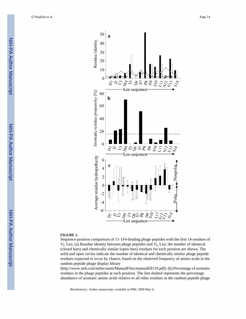
FIGURE 2.Sequence-position comparison of 11-1F4-binding phage peptides with the first 14 residues ofVL Len. (a) Residue identity between phage peptides and VL Len; the number of identical(closed bars) and chemically similar (open bars) residues for each position are shown. Thesolid and open circles indicate the number of identical and chemically similar phage peptideresidues expected to occur by chance, based on the observed frequency of amino acids in therandom peptide phage display library(http://www.neb.com/nebecomm/ManualFiles/manualE8110.pdf). (b) Percentage of aromaticresidues in the phage peptides at each position. The line dashed represents the percentageabundance of aromatic amino acids relative to all other residues in the random peptide phage
O’Nuallain et al. Page 14
Biochemistry. Author manuscript; available in PMC 2009 May 6.
NIH
-PA Author Manuscript
NIH
-PA Author Manuscript
NIH
-PA Author Manuscript
display library. (c) Average residue hydropathicity values (33) for phage peptides (closed bars)relative to those of the Len(1–14) peptide (open bars). Each sequence position averagehydropathicity value was determined from the sum of residue hydropathicity values dividedby the number of total residues. The dashed line shows the average hydropathicity value forall 20 amino acids, corrected for the abundance of each in the random peptide phage displaylibrary. All sequence-position comparisons were determined using the multiple sequencealignments shown in Figure 1.
O’Nuallain et al. Page 15
Biochemistry. Author manuscript; available in PMC 2009 May 6.
NIH
-PA Author Manuscript
NIH
-PA Author Manuscript
NIH
-PA Author Manuscript

FIGURE 3.Binding of mAb 11-1F4 to plate-immobilized Len peptides. (a) Binding of 11-1F4 to Lenpeptides. (b) Competitive inhibition of 2 nM 11-1F4 binding to Len(1–22) in the presence orabsence of Len peptides. Len(1–15), (△); Len(1–13), (▲); LenP8S(1–22), (♦); and Len(1–22), (○). The dashed line indicates 50% inhibition.
O’Nuallain et al. Page 16
Biochemistry. Author manuscript; available in PMC 2009 May 6.
NIH
-PA Author Manuscript
NIH
-PA Author Manuscript
NIH
-PA Author Manuscript

FIGURE 4.11-1F4-binding to plate-immobilized phage, VL Len, and synthetic phage peptides. NaSCNtitration curves for antibody binding to (a) phage, protein Len, or (b) synthetic phage peptides.(c) 11-1F4 titration curves for binding to synthetic phage peptides. IC50 and EC50 values weredetermined from the midpoint of each titration curve. Len, (△); f11, (●); b2, (○); and a12, (♦)phage particles or peptide; and unamplified phage display library control (■).
O’Nuallain et al. Page 17
Biochemistry. Author manuscript; available in PMC 2009 May 6.
NIH
-PA Author Manuscript
NIH
-PA Author Manuscript
NIH
-PA Author Manuscript

FIGURE 5.Binding of mAb 11-1F4 to plate-immobilized Len(1–22) and phage peptides in the presenceand absence of peptide inhibitors. (a) Binding of 11-1F4 to phage peptides or Len(1–22). (b)Competitive inhibition of 2 nM 11-1F4 binding to Len(1–22) in the presence of phage peptidesor Len(1–22). The dashed line indicates 50% inhibition. a12, (▼); g4, (■); b2, (▽); a7, (□);and Len(1–22), (○).
O’Nuallain et al. Page 18
Biochemistry. Author manuscript; available in PMC 2009 May 6.
NIH
-PA Author Manuscript
NIH
-PA Author Manuscript
NIH
-PA Author Manuscript
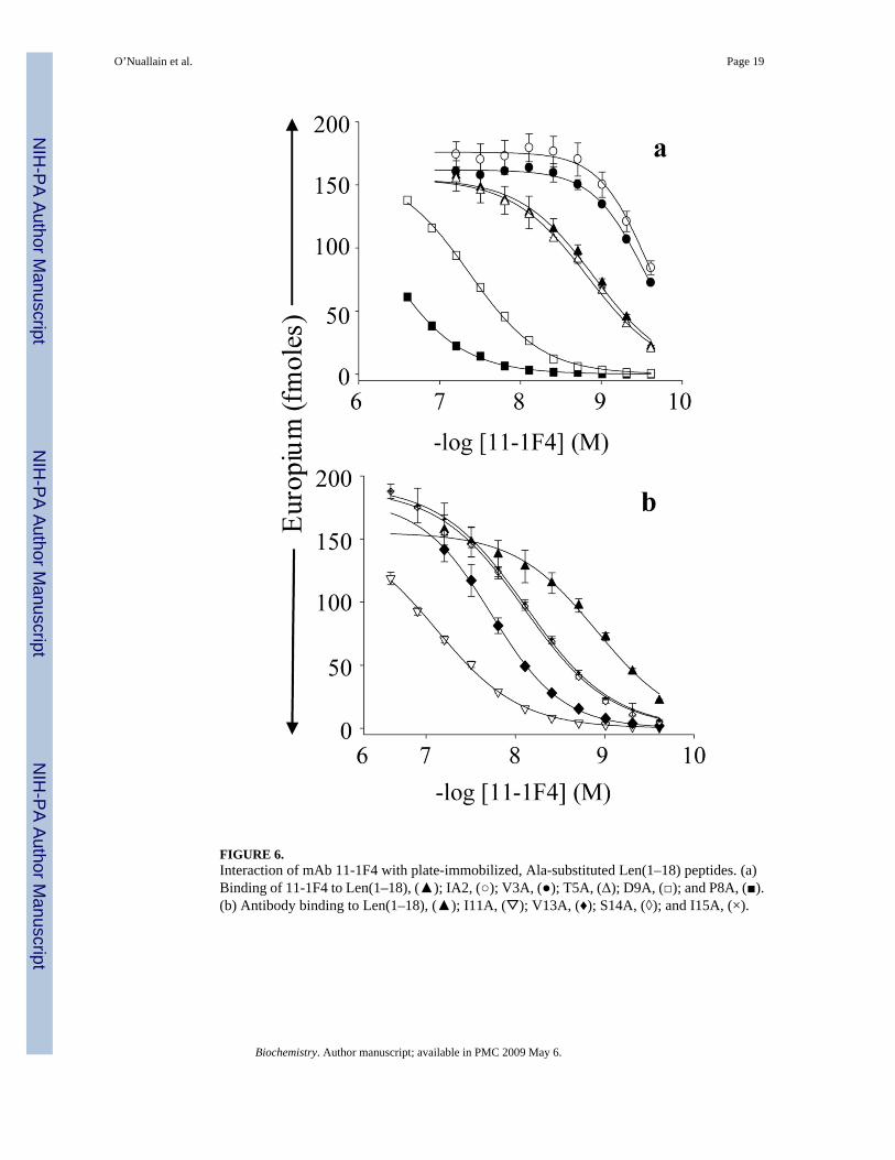
FIGURE 6.Interaction of mAb 11-1F4 with plate-immobilized, Ala-substituted Len(1–18) peptides. (a)Binding of 11-1F4 to Len(1–18), (▲); IA2, (○); V3A, (●); T5A, (Δ); D9A, (□); and P8A, (■).(b) Antibody binding to Len(1–18), (▲); I11A, (▽); V13A, (♦); S14A, (◊); and I15A, (×).
O’Nuallain et al. Page 19
Biochemistry. Author manuscript; available in PMC 2009 May 6.
NIH
-PA Author Manuscript
NIH
-PA Author Manuscript
NIH
-PA Author Manuscript

FIGURE 7.Characterization of the specificity of anti-phage peptide immune serum. Reactivity of serumfrom an A12-immunized mouse (solid bars) and non-immunized mouse (open bars) againstthe immunogen, Len(1–22), as well as VL Len, and collagen fibrils.
O’Nuallain et al. Page 20
Biochemistry. Author manuscript; available in PMC 2009 May 6.
NIH
-PA Author Manuscript
NIH
-PA Author Manuscript
NIH
-PA Author Manuscript
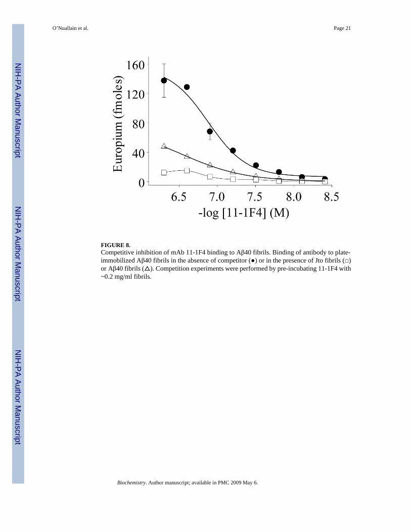
FIGURE 8.Competitive inhibition of mAb 11-1F4 binding to Aβ40 fibrils. Binding of antibody to plate-immobilized Aβ40 fibrils in the absence of competitor (●) or in the presence of Jto fibrils (□)or Aβ40 fibrils (△). Competition experiments were performed by pre-incubating 11-1F4 with~0.2 mg/ml fibrils.
O’Nuallain et al. Page 21
Biochemistry. Author manuscript; available in PMC 2009 May 6.
NIH
-PA Author Manuscript
NIH
-PA Author Manuscript
NIH
-PA Author Manuscript
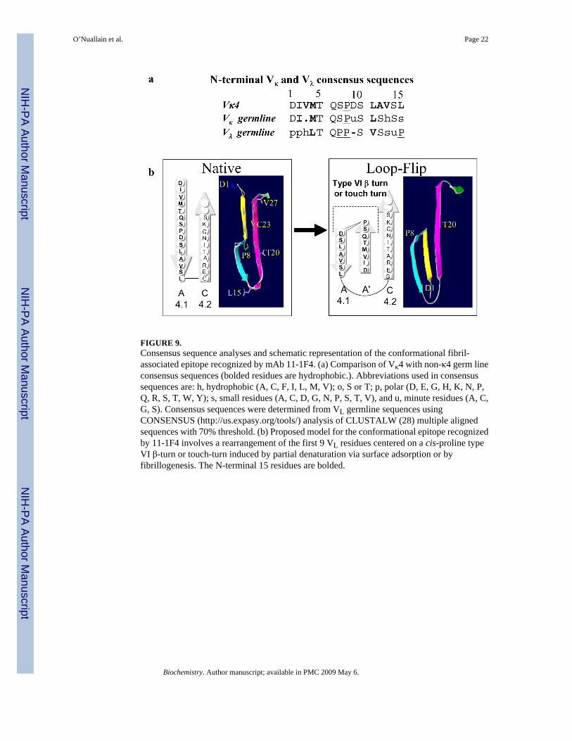
FIGURE 9.Consensus sequence analyses and schematic representation of the conformational fibril-associated epitope recognized by mAb 11-1F4. (a) Comparison of Vκ4 with non-κ4 germ lineconsensus sequences (bolded residues are hydrophobic.). Abbreviations used in consensussequences are: h, hydrophobic (A, C, F, I, L, M, V); o, S or T; p, polar (D, E, G, H, K, N, P,Q, R, S, T, W, Y); s, small residues (A, C, D, G, N, P, S, T, V), and u, minute residues (A, C,G, S). Consensus sequences were determined from VL germline sequences usingCONSENSUS (http://us.expasy.org/tools/) analysis of CLUSTALW (28) multiple alignedsequences with 70% threshold. (b) Proposed model for the conformational epitope recognizedby 11-1F4 involves a rearrangement of the first 9 VL residues centered on a cis-proline typeVI β-turn or touch-turn induced by partial denaturation via surface adsorption or byfibrillogenesis. The N-terminal 15 residues are bolded.
O’Nuallain et al. Page 22
Biochemistry. Author manuscript; available in PMC 2009 May 6.
NIH
-PA Author Manuscript
NIH
-PA Author Manuscript
NIH
-PA Author Manuscript

NIH
-PA Author Manuscript
NIH
-PA Author Manuscript
NIH
-PA Author Manuscript
O’Nuallain et al. Page 23Ta
ble
111
-1F4
bin
ding
to p
late
imm
obili
zed
phag
e an
d Le
n pe
ptid
es a
nd th
e in
hibi
tory
eff
ects
of
thes
e m
olec
ules
on
antib
ody
bind
ing
toim
mob
ilize
d Le
n(1–
22).
Imm
obili
zed
Poly
pept
ide
Solu
tion-
Phas
e C
ompe
titio
n
Poly
pept
ide
Phag
e Pe
ptid
e G
roup
aSe
quen
ceb
EC
50c (n
M)
NaS
CN
ass
ay IC
50d (µ
M)
IC50
e (µM
)
Len
VL
n.a.
fD
IVM
TQSP
DSL
AV
SLG
ERA
TIN
…0.
18 ±
0.0
33.
9≫
30
a12
IK
-HY
AA
FPEN
LL
I0.
54 ±
0.3
3.7
(3.9
)1.
5 ±
0.1
g4I
-VQ
YSS
SPEA
LRV
14 ±
6.0
3.7
(4.0
)3.
2 ±
0.0
f11
II-I
VY
TPH
PEY
LW
-P32
± 0
.13.
3 (3
.9)
6.7
± 0.
1
b2II
TYQ
YSP
YPE
PVM
44 ±
0.1
0.5
(0.5
)4.
6 ±
0.1
a7II
KPT
-YSP
YPF
SM--
P18
± 0
.12.
3 (0
.5)
1.7
± 0.
1
Len(
1–22
)n.
a.D
IVM
TQSP
DSL
AV
SLG
ERA
TIN
0.24
± 0
.11.
370.
07 ±
0.0
1
Len(
1–15
)n.
a.D
IVM
TQSP
DSL
AV
SL0.
39 ±
0.0
5n.
d.g
0.27
± 0
.01
Len(
1–13
)n.
a.D
IVM
TQSP
DSL
AV
16 ±
0.1
n.d.
33 ±
0.0
7a Ph
age
pept
ides
wer
e gr
oupe
d ba
sed
on th
e pr
esen
ce o
f hig
hly
cons
erve
d pr
olin
e re
sidu
es, a
s sho
wn
in F
ig. 1
.
b Pept
ide
sequ
ence
s wer
e al
igne
d by
CLU
STA
LW (2
9); h
ydro
phob
ic a
nd/o
r aro
mat
ic re
sidu
es a
re b
olde
d an
d pr
olin
e is
und
erlin
ed.
c Each
EC
50 v
alue
was
det
erm
ined
from
the
mid
poin
t of 1
1-1F
4 tit
ratio
n cu
rves
, as s
how
n in
Fig
ure
4a a
nd F
igur
e 7a
.
d Each
IC50
val
ue w
as d
eter
min
ed fr
om th
e m
idpo
int o
f NaS
CN
titra
tion
curv
es (s
ee F
igur
e 3a
and
b).
IC50
val
ues w
ith o
r with
out b
rack
ets w
ere
dete
rmin
ed fo
r 11-
1F4
bind
ing
to p
hage
pep
tides
alo
neor
exp
ress
ed o
n ph
age
parti
cles
, res
pect
ivel
y.
e Each
IC50
val
ue w
as d
eter
min
ed fr
om th
e m
idpo
int o
f pep
tide
com
petit
ion
curv
es sh
own
in F
igur
e 4b
and
Fig
ure
7b.
f n.a.
, not
app
licab
le.
g n.d.
, not
det
erm
ined
.
Biochemistry. Author manuscript; available in PMC 2009 May 6.

NIH
-PA Author Manuscript
NIH
-PA Author Manuscript
NIH
-PA Author Manuscript
O’Nuallain et al. Page 24Ta
ble
2Pe
ptid
e ph
age
disp
lay
and
alan
ine
scan
ning
ana
lysi
s of N
-term
inal
VL
Len
resi
dues
impl
icat
ed in
11-
1F4
bind
ing.
VL L
en r
esid
ue p
ositi
onPh
age
pept
ide
Len
(1–1
8) A
lani
ne sc
anni
ngPh
age
and
Len
(1–1
8) R
esid
ue P
refe
renc
esd
Iden
titya (%
)A
la (%
)E
C50
b (nM
)E
C50
WT
EC
50c
D1
3.8
03.
91 ±
0.0
012.
7S
or T
I215
.419
.20.
27 ±
0.0
010.
22A
la o
r any
am
ino
acid
V3
9.6
00.
29 ±
0.0
040.
24Po
lar
M4
15.4
02.
51 ±
0.0
12.
1A
rom
atic
(pre
fera
bly
Tyr)
e
T582
.710
.01.
48 ±
0.0
11.
2Sm
all (
pref
erab
ly S
or T
)
Q6
5.8
4.0
2.33
± 0
.01
1.9
Pola
r
S726
.98.
00.
62 ±
0.0
10.
51Po
lar (
pref
erab
ly a
rom
atic
)
P810
00
>500
± 1
0>4
00In
varia
nt p
rolin
e
D9
61.5
2.0
43.2
± 0
.236
Neg
ativ
ely
char
ged,
D/E
S10
40.4
6.3
0.85
± 0
.003
0.71
Smal
l pol
ar (p
refe
rabl
y S
or T
)
L11
80.8
077
.1 ±
0.7
64L
or b
ulky
hyd
roph
obic
A12
5.8
02.
86 ±
0.0
2d2.
4B
ulky
hyd
roph
obic
V13
73.0
2.6
18.6
± 0
.116
V o
r alip
hatic
S14
30.7
07.
96 ±
0.0
46.
6S
or T
L15
n.a.
fn.
a.7.
64 ±
0.0
26.
4(L
)
G16
n.a.
n.a.
2.12
± 0
.01
1.8
n.a.
E17
n.a.
n.a.
2.48
± 0
.01
2.1
n.a.
R18
n.a.
n.a.
0.55
± 0
.002
0.5
n.a.
a Sequ
ence
iden
tity
for e
ach
phag
e pep
tide c
ompa
red
to V
L Le
n w
as d
eter
min
ed as
the s
um o
f the
per
cent
ages
of i
dent
ical
and
chem
ical
ly si
mila
r (32
) res
idue
s at e
ach
posi
tion
from
the m
ultip
le se
quen
ceal
ignm
ents
of p
hage
pep
tide
grou
ps I
and
II sh
own
in F
igur
e 1.
b EC50
val
ues w
ere
dete
rmin
ed fr
om th
e m
idpo
int o
f 11-
1F4
titra
tion
curv
es sh
own
in F
igur
e 6.
c WT
EC50
is th
e EC
50 v
alue
for 1
1-1F
4 bi
ndin
g to
pla
te-im
mob
ilize
d w
ild-ty
pe L
en(1
–18)
.
d Res
idue
pre
fere
nce
at e
ach
posi
tion
in L
en(1
–18)
was
det
erm
ined
from
the
mul
tiple
sequ
ence
alig
nmen
ts o
f pha
ge p
eptid
e gr
oups
I an
d II
with
VL
Len,
and
the
bind
ing
of 1
1-1F
4 to
Ala
subs
titut
ed L
en(1
–18)
pep
tides
(Fig
ure
1, F
igur
e 2,
and
Fig
ure
6). R
esid
ue p
refe
renc
es w
ith o
r with
out b
rack
ets a
re th
ose
that
wer
e pr
efer
red
for L
en a
nd p
hage
pep
tides
, res
pect
ivel
y.
e Res
idue
pre
fere
nce
in b
rack
ets a
re th
ose
spec
ific
to th
e Le
n(1–
18) p
eptid
e.
f n.a.
, not
app
licab
le.
Biochemistry. Author manuscript; available in PMC 2009 May 6.