Phage Therapy Related Microbial Succession Associated with ...
CHAPTER 1
Phage Therapy—History fromTwort and d’Herelle ThroughSoviet Experience to CurrentApproaches
Nina Chanishvili1
Contents I. General Background 4
A. Discovery of bacteriophages 4
B. Early clinical trials 6
C. Early commercial production of bacteriophages 8
D. George Eliava and the Eliava Institute of
Bacteriophage 8
II. Mass Application of Phages 10
A. Wound treatment 10
B. Persistence of phages in animals and humans 14
C. Treatment of intestinal Salmonella and Shigella
infections 16
D. Combined use of phage- and sero- therapies 25
E. Use of bacteriophages for prophylaxis 27
III. Current Status of Georgian Research and Their
Impact on Worldwide Phage Therapy Studies 31
Ongoing research and clinical trials 31
References 35
Advances in Virus Research, Volume 83 # 2012 Elsevier Inc.ISSN 0065-3527, DOI: 10.1016/B978-0-12-394438-2.00001-3 All rights reserved.
Laboratory for Genetics of Microorganisms and Bacteriophages, Eliava Institute of Bacteriophage,Microbiology & Virology, Tbilisi, Georgia, USA1 Corresponding author, E-mail: [email protected]
Abstract Felix d’Herelle proposed the use of bacteriophages for the therapy
of human and animal bacterial infections at the beginning of the
20th century. This approach, however, was not widely accepted in
the West. After the emergence of antibiotics in 1940s, phage
research was diverted to a more fundamental level. At the same
time, phage therapy was widely practiced in the Soviet Union
due to collaboration of Felix d’Herelle with his Georgian colleagues.
The majority of the articles dedicated to this subject are from the
1930s and 1940s. The old Soviet literature indicates that phage ther-
apy was used extensively to treat a wide range of bacterial infections
in the areas of dermatology (Beridze, 1938), ophthalmology (Rodigina,
1938), urology (Tsulukidze, 1938), stomatology (Ruchko and Tretyak,
1936), pediatrics (Alexandrova et al., 1935; Lurie, 1938), otolaryngology
(Ermolieva, 1939), and surgery (Tsulukidze, 1940, 1941). These articles
were published in Russian and thus were not readily available to
Western scientists. The Western skepticism toward phage therapy
itself was again followed by renewed interest and reappraisal, mainly
due to the emergence of drug-resistant bacteria. Often the experi-
ments described in the old Soviet articles were not designed prop-
erly: the use of placebos and the coding of preparations were absent
frommost of the studies, number of patients in the experimental and
control groups was unequal or missing, sometimes no control groups
were used at all, or patients treated previously unsuccessfully with
antibiotics were employed as an experimental group and as control.
The results obtained and the efficiency of phage prophylaxis were
estimated by comparing with results obtained in previous years. In
most publications, phage titers and descriptions of methods used for
evaluation of the results are not specified. Nevertheless, past experi-
ence indicates some effectiveness of phage therapy and prophylaxis.
Therefore, these clinical results should not be neglected when
designing any future studies.
I. GENERAL BACKGROUND
A. Discovery of bacteriophages
During World War I, rather sensational news was spread—the viruses‘‘eaters of micro-organisms’’ have been discovered by Felix d’Herelle,who developed a phage preparation to treat WorldWar I soldiers affectedby dysentery (Hausler, 2008). Doctors all over the world were excited bythis news, especially because reports published in medical journals thatviruses–bacteriophages are harmless for humans and animals and can beapplied successfully as therapeutic means (Alessandrini and Doria, 1924;Bruynoghe and Maisin, 1921; Compton, 1929; Costa Cruz, 1924; Parfitt,2005; Rice, 1930; Spence and McKinley, 1924; Sulakvelidze et al., 2001).
4 Nina Chanishvili
Actually, the history of bacteriophages started long before this event.In 1896, the English bacteriologist Ernest Hanbery Hankin tried to enu-merate the number of Vibrio cholera in a cubic millimeter of water in theriver Ganges. Samples were taken in places where the river enters andexits the city Agra (Adhya andMerril, 2006; Kazhal and Iftimovich, 1968).Surprisingly, Hankin (1896) counted 100,000 infective units in the cubicmillimeter of water in the entrance to the city, while their numberdecreased to only 90 where the river exited the city. This strange Ganghesself-purification could not find its explanation by that time and wasreferred to as ‘‘Hankin’s phenomenon.’’
In 1906–1909, Felix d’Herelle traveled across Mexico, where he met anentomologist who attracted his attention to the violent epidemics amonglocust. d’Herelle isolated bacterium Coccobaccilus aeridiorum (presentlyknown as Enterobacter aerogenes) (Summers, 1999, 2001). Motivated by anambitious desire to use these bacteria deliberately against plagues oflocust, he conducted a number of experiments in Tunis and Guyana.During these experiments, d’Herelle observed the appearance of trans-parent holes (‘‘taches vierges’’) in the bacterial lawn. He presumed thatthey were caused by a virus that was mistakenly supposed to be a cause ofthe locust infection. d’Herrele isolated this virus and used it against locustbut without any success. He started to pursue an idea to use the hostbacteria against locust again and did not take into consideration thestrange ‘‘taches vierges’’ (clear plaques).
In 1915, the well-known British journal The Lancet published an articlewritten by FrederickTwort about ‘‘the transmissible bacterial lyses’’ (Twort,1915), in which Twort described his observation of ‘‘the eaten edges of thecolonies of Staphylococcus.’’ Hemanaged to filter the appropriate cultures ofStaphylococcus and spotted the filtrate on the lawn of different Staphylococcusstrains. Thus, he received a clear zone of lysis again and again. However,Twort could not explain the observed event and provided only its descrip-tion. This was the very first publication on bacteriophages. d’Herelle readthis article, which reminded him of his own observations in Mexico andTunis (Summers, 2001). He suspected that the filtered agent was a bacterialvirus—an invisible invader that destroys bacteria.
The discovery or rediscovery of bacteriophages by Felix d’Herelle afterFrederick Twort is frequently associated with an outbreak of severehemorrhagic dysentery among French troops stationed at Maisons-Laffitte (on the outskirts of Paris) in July–August 1915 (Sulakvelidzeet al., 2001; Summers, 2001). Several soldiers were hospitalized, andd’Herelle was assigned to conduct an investigation of the outbreak. Dur-ing these studies, he made bacterium-free filtrates of the patients’ fecalsamples andmixed and incubated themwith Shigella strains isolated fromthe patients. A portion of the mixtures was inoculated into experimentalanimals (as part of d’Herelle’s studies on developing a vaccine against
Phage Therapy 5

bacterial dysentery), and a portion was spread on agar medium in orderto observe the growth of the bacteria. On these agar cultures d’Herelleobserved again the appearance of small, clear areas, which he initiallycalled taches, then taches vierges, and, later, plaques (Summers, 1999).D’Herelle’s findings were presented during the September 1917 meetingof the Academy of Sciences and were subsequently published in themeeting’s proceedings (d’Herelle, 1917). In contrast to Hankin andTwort, d’Herelle had little doubt about the nature of the phenomenon,and he proposed that it was caused by a virus capable of parasitizingbacteria. The name ‘‘bacteriophage’’ (from ‘‘bacteria’’ and Greek j age~in
phagein ‘‘to eat’’) was also proposed by d’Herelle (Summers, 1999).The discovery of bacteriophages was inevitable. Similar phenomena
have been observed in remote regions of the world by different scientists.At the end of the 19th century,N.F. Gamaleya (later aHonoraryMember oftheAcademy of theUSSR) published an article in theRussianArchives of thePathological and Clinical Medicine (Gamaleya, 1898). In this article hedescribed the lysis of Bacillus antracis in distilled water, after which thewater obtained an ability to lyse other strains ofB. antracis. In 1917, a youngGeorgian scientist George Eliava had observed mysterious disappearanceof V. cholera cells (d’Herrelle, 1935; Georgadze and Makashvili, 1979).
The greatest merit of Felix d’Herelle is that he advanced the idea ofusing bacteriophages for the treatment of human and animal bacterialdiseases. For this idea he deserved the Noble Prize, to which he wasnominated eight times, every year since 1925, although he was neverawarded one [cited by Hausler (2008) according to Nobel Archives].
B. Early clinical trials
Not long after his discovery, d’Herelle used phages to treat dysentery,which was probably the first attempt to use bacteriophages therapeuti-cally. The studies were conducted at the Hopital des Enfants-Malades inParis in 1919 under the clinical supervision of Professor Victor-HenriHutinel, the hospital’s chief of pediatrics (Summers, 1999). The phagepreparation was ingested by d’Herelle, Hutinel, and several hospitalinterns in order to confirm its safety before administering it the next dayto a 12-year-old boy with severe dysentery. The patient’s symptomsceased after a single administration of d’Herelle’s antidysentery phages,and the boy fully recovered within a few days. The efficacy of the phagepreparation was ‘‘confirmed’’ shortly afterward when three additionalpatients having bacterial dysentery and treated with one dose of thepreparation started to recover within 24 hr of treatment. However, resultsof these studies were not published immediately and, therefore, the firstreported application of phages to treat infectious diseases of humanscame later from Richard Bruynoghe and Joseph Maisin (1921), who
6 Nina Chanishvili
used bacteriophages to treat staphylococcal skin disease. The bacterio-phages were injected into and around surgically opened lesions, and theauthors reported regression of the infections within 24 to 48 hr. Severalsimilarly promising studies followed (Rice, 1930; Schelss, 1932; Stout,1933). Encouraged by these early results, d’Herelle and others continuedstudies of the therapeutic use of phages (e.g., d’Herelle used variousphage preparations to treat thousands of people having cholera and/orbubonic plague in India (Kazhal and Iftimovich, 1968; Summers, 1999;2001).
In 1916–1930, d’Herelle and collaborators undertook numerous expe-ditions to China, Laos, India, Vietnam, and Africa to combat epidemicscaused by cholera and plague with bacteriophages against these patho-gens. According to Romanian medical historians Kazhal and Iftimovich(1968), the first attempts to use cholera bacteriophages for treatment andprophylaxis were performed by Felix d’Herelle and George Eliava in1931. According to these authors, the Institute of Vaccine and Sera inTbilisi produced the first commercial anticholera phage preparation,which was reported to be used successfully for the control of epidemicsthreatening the southeast territories of the USSR (Kazhal and Iftimovich,1968). According to the estimations published at that time, due to theapplication of bacteriophages it became possible to reduce the mortalityof cholera in India to 10% (Kazhal and Iftimovich, 1968). This fact isdescribed by d’Herelle’s himself in the book ‘‘Bacteriophage and thePhenomenon of Recovery’’ published in the Russian language in 1935 inTbilisi, Georgia.1 According to d’Herelle (1935), cholera epidemicsoccurred in Punjab region in 1927. Patients were treated orally with 2 mlof cholera phages diluted in 20 ml of water but the titer of phage in thesepreparations is unknown. If the patient vomited, a repeated dose ofphages (5 ml diluted in 100 ml of water) was administered slowly witha teaspoon. The control group of patients treated themselves using a folkmedicine (plant extracts). Of 14,450 people who lived in nine villages ofthe Punjab region, only 73 have been treated with the phages. d’Herelleexplained it by mentioning that people in India opposed any newmedicalmeasures and rarely permitted him and his colleagues to use phages fortreatment. Thus, only desperately ill patients were subjected to phagetherapy. Altogether 118 persons were included in the control group inwhich 74 lethal outcomes (62.7%) were registered, whereas in the experi-mental group the mortality rate was almost one-tenth, with 5 cases out of73 (6.8%) (d’Herelle, 1935).
1 After the execution of George Eliava, the book was impounded and access to it was limited to ‘‘forprofessional use only.’’
Phage Therapy 7

C. Early commercial production of bacteriophages
In his book, d’Herellementioned the establishment of two industrial centersfor the production of bacteriophages against cholera in 1931 in India(d’Herelle, 1935; Kazhal and Iftimovich, 1968). D’Herelle’s commerciallaboratory in Paris produced at least five phage preparations againstvarious bacterial infections. The preparations were called Bacte-coli-phage,Bacte-rhino-phage, Bacte-intesti-phage, Bacte-pyo-phage, and Bacte-staphy-phageand were marketed by a company that later became the large Frenchcompany L’Oreal (Sulakvelidze et al., 2001; Summers, 1999, 2001).
The Oswaldo Cruz Institute in Rio de Janeiro, Brazil, started produc-tion of the antidysentery bacteriophages in 1924 to combat dysentery inLatin American countries (d’Herelle, 1935; Dublanchet and Bourne, 2007).Within a year the institute produced 10,000 vials of phages, which weresent to hospitals around Brazil (Hausler, 2008). Therapeutic phages werealso produced in the United States. In the 1940s, the Eli Lilly Company(Indianapolis, IN) produced seven phage products for human use, includ-ing preparations targeted against staphylococci, streptococci, Escherichiacoli, and other bacterial pathogens (Sulakvelidze et al., 2001). These pre-parations consisted of phage-lysed, bacteriologically sterile broth culturesof the targeted bacteria (e.g., Colo-lysate, Ento-lysate, Neiso-lysate, andStaphylo-lysate) or the same preparations in a water-soluble jelly base(e.g., Colo-jel, Ento-jel, and Staphylo-jel). They were used to treat variousinfections, including abscesses, suppurating wounds, vaginitis, acute andchronic infections of the upper respiratory tract, and mastoid infections.However, the efficacy of phage preparations was controversial (Eaton andBayne-Jones, 1934; Krueger and Scribner, 1941). This could be caused bythe absence of viable phages, low phage titer, or narrow strain range ofphage in these preparations as, for example, discovered in the case ofsome commercial antistaphylococcal phage preparations (Rakieten, 1932).As a result, with the advent of antibiotics, commercial production oftherapeutic phages ceased in most Western countries.
Despite apparently promising results of phage therapy in certaincases, it had been abandoned in Western countries for more than80 years, but not in the former Soviet Union where d’Herelle, togetherwith his close friend and associate George Eliava, founded the Bacterio-phage Institute in Tbilisi (Georgia).
D. George Eliava and the Eliava Institute of Bacteriophage
In the history of medicine little is known about George Eliava, who was acolorful central figure in phage history. Without the support of Eliavaprovided to Felix d’Herelle, much of the knowledge on phage therapywould not be achieved. Because of his progressive thinking, tireless
8 Nina Chanishvili

activities, and close collaboration with many foreign scientists, includingd’Herelle, George Eliava became a victim of Stalin’s regime in 1937. Hewas pronounced to be a ‘‘people’s enemy’’ and was executed. All photo-graphs and documents belonging to George Eliava were destroyed bythe KGB. His memory was restored only after the reassessment of theoutcomes of the Red Terror and Stalin’s regime by Gorbachev in 1989 andrehabilitation of its victims (Smith, 1996).
George Eliava was born on January 13, 1892, in the village ofSachkhere, West Georgia. In 1909 he entered the Novorosyisk Universityin Odessa (Georgadze and Makashvili, 1979). He wished to study litera-ture. During his first years at the university he joined the student revolu-tionary movement and was expelled from the university with no rights toenter any other university in Russia. However, his rich and powerfulrelatives managed to send him to Geneva (Switzerland) where in 1912–1914 Eliava took a course in bacteriology. During the summer of 1914,Eliava came back to Georgia for vacations, but after the start of the WorldWar I he could not return to the university in Geneva for his studies. Dueto the efforts of his relatives he received permission to continue his studiesat the Faculty of Medicine of the Moscow University in Russia, which hefinished in 1916. In the same year he was appointed as head of theCaucasian front-line bacteriology laboratory in Trabzon. Beginning in1917, he took lead of the Tbilisi Bacteriology Laboratory, which belongedto the Caucasian Cities’ Union. In 1919–1921, 1925–1927, and 1931, Eliavaworked at the Pasteur Institute in Paris, together with famous bacteriol-ogists: Emile Roux, Charles Nicolle (Nobel Prize winner in Medicine in1903), Albert Calmette, and Gaston Ramon, among others. In the begin-ning of the 1920’s he met Felix d’Herelle there. In 1923, Eliava initiatedfoundation of the Institute of Bacteriology (the present Eliava Institute ofBacteriophage, Microbiology & Virology; EIBMV). He became its firstdirector. Simultaneously with his administrative activities in 1927–1937,Eliava was teaching at Tbilisi State University and leading the departmentof hygiene and later the department of microbiology. In 1934, he initiatedestablishment of the Anti Plague Station in Tbilisi (the present NationalCenter of Disease Control)(Georgadze and Makashvili, 1979). AlthoughEliava did not publish many articles, all of them touch significant topics,sounding rather contemporarily even now (d’Herelle and Eliava, 1921a,b;Eliava, 1930; Eliava and Legraoux, 1921; Eliava and Pozersky, 1921a,b;Eliava and Suarez 1927a,b; Nattan-Larrier et al., 1931). Together withd’Herelle, Eliava discovered bacteriophage lysins (d’Herelle and Eliava,1921a) and, in collaboration with E. Pozersky (1921a), found that thesubstance quinine, which was known already in ancient China as atreatment against malaria, specifically affected bacteriophages as well.Eliava was one of the first who drew out an assumption that bacterio-phages may change the nature of the host bacteria (Eliava and
Phage Therapy 9
Pozersky,1921b).2 He studied the immune response to phage therapyalready in 1921 (d’Herelle and Eliava, 1921b), adsorption of bacterio-phages on leukocytes (Eliava, 1930), and permeability through placenta(Nattan-Larrier et al., 1931).
In November 1921, Eliava returned to Georgia with scientific equip-ment worth approximately 100,000 FF, which was a gift of the PasteurInstitute to Georgian colleagues (Georgadze, 1974).
George Eliava met Felix d’Herelle in Paris during one of his early staysat the Pasteur Institute and was fascinated by d’Herelle’s ideas of usingbacteriophages for therapy. Eliava invited d’Herelle to Georgia wherethey spent altogether 18 months in 1933 and 1934 collaborating withother Georgian colleagues (Georgadze, 1974). D’Herelle intended tomove to Tbilisi permanently (a cottage built for his use still stands onthe institute’s grounds); however, his intentions could not be realized. In1937, Eliava, together with his wife Amelia Vol-Levitskaya (Polish operasinger), was arrested and executed as a ‘‘people’s enemy’’ for being inintellectual opposition with Laurenti Beria, the chief of the secret police toJoseph Stalin. According to another version, Beria considered Eliava as hiscompetitor with him for a woman, Tinatin Jikia (librarian at the Bacterio-phage Institute).
Frustrated and disillusioned, d’Herelle never returned to Georgia.Nonetheless, the institute survived and later became one of the largestfacilities in the world engaged in the development of therapeutic phagepreparations. During the best times the scientific staff of the instituteenumerated about 100–120 people, including technicians, while the indus-trial part employed approximately 500–600 people, including specialistsand support personnel. At that time the institute produced phage pre-parations (often several tons a day) against a dozen bacterial pathogens,including staphylococci, Pseudomonas, Proteus, and many enteric patho-gens (Georgadze, 1974). Most of the Soviet studies reviewed in this articleinvolved phages developed and produced at the EIBMV (Chanishvili,2009).
II. MASS APPLICATION OF PHAGES
A. Wound treatment
From a review of historic literature, it is apparent that phage therapy trialswere active in the 1930s and 1940s throughout Georgia, Russia, Ukraine,Belarus, and Azerbaijan in the Soviet Union. Observations of cases
2 This is known now as lysogenic conversion—a change in the properties of the bacterium as a result of phageinfection and lysogeny.
10 Nina Chanishvili

associated with road accidents and septic infection carried out at theOstroumovkaya hospital in Moscow by Kokin and colleagues (cited byKrestnikova, 1947) led to the development of methods and instructionsfor intramuscular and even intravenous use of phages, which was crucialin cases of generalized infections. Results of these observations werereported at conferences held in March, June, and December of 1940.These methods and instructions were approved by the Soviet SupremeRed ArmyMilitary-Sanitarian Office and were applied to the treatment ofsoldiers in the Red Army during World War II and continued after it.
The application of phage therapy to surgical and wound treatmentbegan during the Finnish Campaign in 1939–1940, with the first review ofthis work published by Kokin (1941, 1946). Kokin described the applica-tion of mixtures of bacteriophages infecting anaerobes, Staphylococcus andStreptococcus, and produced by the EIBMV, Tbilisi, Georgia, for the treat-ment of gas gangrene. The mixture was applied to 767 infected soldierswith a lethal outcome (death) observed in 18.8% of cases compared with42.2% in the control group of soldiers treated with other methods. Usingthe same mixture of phages, other authors observed a 19.2% lethal out-come in a group of soldiers compared with 54.2% in a group treated withother medications (Lvov and Pasternak, 1947—cited by Krestovnikova,1947). In addition to its therapeutic use, this phage preparation was usedby the mobile sanitary brigades as an emergency treatment for wounds(prophylaxis of gas gangrene). Krestovnikova (1947) summarizes theobservations of three mobile sanitary brigades carried out over periodsof 2–6 weeks following evacuation to front-line hospitals.
The first brigade treated 2500 soldiers with phages. According toreports from that time, only 35 soldiers (1.4%) in this group revealedsymptoms of gas gangrene, whereas in the control group of 7918wounded soldiers, 342 (4.3%) were infected. The second brigade appliedphage therapy to 941 soldiers, of whom only 14 (1.4%) suffered from gasgangrene, in contrast to 6.8% of soldiers in the control group treated byother methods. The third brigade treated 2584 soldiers, 18 (0.7%) of whomdeveloped symptoms of gas gangrene, whereas 2.3% in the control groupdeveloped these symptoms. Comparison of data described by thesethree independent brigades showed an average 30% decrease in thenumber of gas gangrene as a consequence of the prophylactic treatmentof wounds through application of the phage mixtures (Kokin, 1946;Krestnikova, 1947).
One of the pioneers in the application of phages in surgery was A.P.Tsulukidze, a professor of medicine, who began using such preparationsin 1931 for the treatment of various diseases. In 1938 a prominent Sovietsurgeon, Burdenko, recommended the use of phages against purulentinfections. However, the same Burdenko, on Stalin’s orders, falsified theresults of Soviet studies on Katy"n murders of Polish officers, committed
Phage Therapy 11

by Soviet NKWD in 1941, and thus his scientific credibility may remain, atleast in part, in question (Cienciala et al., 2007).
An experimental series of phage preparations containing componentsagainst bacteria related to Staphylococcus, Streptococcus, Escherichia coli,and Proteus species was tested at the end of the 1930s in the surgical andgynecology clinics in Moscow (cited by Krestnikova, 1947).
According to Tsulukidze (1940), the wounds were analyzed bacterio-logically by blood analysis carried out before the initiation of phagetherapy and during surgical manipulations (bandages, puncture, applica-tion of phages, etc.). Prior to obtaining the results of bacteriologicalanalysis, a Pio-bacteriophage preparation or a mixture of Strepto- andStaphylo-bacteriophages was applied topically or directly to the accessi-ble part of the wound. The condition of the wound was described thor-oughly, and temperature, pulse, patient’s rate of breathing, and so onwere all recorded. These examinations were also performed after eachphage application. The majority of patients arriving from the front linewith wounds were bedridden and in serious condition: 38.3% had softtissue injuries and 61.7% had bone injuries. In the majority of cases,phages were administered during the first 6 days after initial infection.
Initially the application of phage therapy was used only for the mostsevere cases where a lethal outcome was expected. Later a wider group ofpatients was involved in the study. In themajority of cases, bacteriologicalanalysis indicated the presence of mixed bacterial infections (Tsulukidze,1940). Only in rare cases were patients arriving directly from the battle-field characterized with a monoinfection. Subcutaneous injection ofphages was performed three to four times every second day to avoidthe development of antiphage antibodies. Additionally, phages weresprayed onto the top of the wound each time bandages were changed.
All patients with injuries of the soft tissues (38.3%) underwent ‘‘ordi-nary therapy,’’ which in this instance implied treatment with chlora-mines, rivanol, and Vishnevsky ointment.3 These patients often hadmajor tissue damage with penetrating or perforating wounds. Thewounds were characterized by the accumulation of pus, infections, andsurrounding inflammation, sometimes with necrotic foci. A number ofcases with an abscess/phlegmon around a bullet or mine fragment under-went cuts performed during first aid in a field hospital prior to the start ofphage therapy. Many of these patients suffered from serious intoxication,
3 Vishnevky ointment was developed by a prominent Russian surgeon, Alexander Vasilyevich Vishnevsky. Itconsists of birch tar (3 g), xeroform (3 g), and castor oil (94 g). The ointment has both predrying and antisepticeffects and facilitates tissue regeneration. It is used for the treatment of wounds, burns, bed sores, chilblain,fistulas, skin ulcers, psoriasis, trophic sores, and capillary diseases of lower extremities such as trombophle-bitis, obliterating endarteritis.
12 Nina Chanishvili

fever, and gangrenous inflammation and required the removal of wood orbullet particles. Treatment of such patients was performed by removal oftampons (used to fill the space of missing tissue), purification of woundsby treatment with iodine and alcohol, and then washing with 2% of asodium chloride solution followed by spraying with phages. Simulta-neously, 5–10 ml of phages (titer unknown) was injected remote fromthe wound into the stomach wall, shoulder, or hip. The wound wasbandaged with gauze soaked in phages. Tampons and drainages werenot applied. According to reports from that time, no cases treated withthis method required additional surgical intervention. After the first oneto two phage applications the body temperature used to be normalized.Only three to four such treatments were normally required to achieve acomplete cure and blood test results were also improved. Because therecovery from traumatic injuries and numerous lesions required anextended period, wounds were stitched after phage therapy on the 6th–8th day of treatment so that further infection was unlikely. In general,treatment with phage therapy took a number of days, whereas ‘‘ordi-nary’’ or standard therapy took several weeks.
Patients with bone injuries or open fractures arrived following first aidusing splints. The wounds were purified, tampons with chloramines, riv-anol and Vishnevsky ointment were removed, and wounds were treatedwith 2% sodium chloride solution and then sprayed with phages. Simulta-neously, phages were injected intramuscularly or subcutaneously at a siteremote from the wound. Following this a blind plaster cast was applied. Asecond approach involved phage therapy, stitching of the wound, and thenthe application of plaster. A third approach involved application of aplaster cast directly after a single treatment with phages. According todirect reports from that time, treatment with phages resulted in a fasterrelease of pain, improvement in patients’ general conditions, and clearsymptoms of wound healing after 2 or 3 days. Tsulukidze and colleaguesreported that they were able to avoid moisturizing of the plaster, necessityof its frequent change, and the development of foul odor (usually asso-ciated with secondary infections) by using phage therapy prior toplastering. The plaster could remain unchanged for up to 60 days, allowingfor the earlier start of physical therapy (Tsulukidze, 1940, 1941). They alsoclaimed that in the cases of severe hips, shins, forearms, and shouldersinjuries, which normally require amputation, no amputationwas necessaryif wounds were left for 10–30 days in blind plasters. Moreover, phagetherapy allowed the application of stitches and/or plaster casts as earlyas the 10th–11th day after the start of treatment.
The war and the need for therapeutic preparations inspired Sovietdoctors to perform new trials with phages and to develop novel methodsfor phage administration. This period was one of the most fruitful in the
Phage Therapy 13
development of phage therapy in the former Soviet Union (Kokin, 1941,1946; Krestnikova, 1947). A book written by Alexander Tsulukidze‘‘Experience of the Use of Bacteriophages in Conditions of War Trauma’’(1941), which summarizes the results obtained after Finnish Campaign, isespecially interesting for military surgeons.
Tsulukidze (1941) described 20 hospital cases where bacteriophagesagainst anaerobic bacteria were used in combination with Strepto- andStaphylo-bacteriophages. Seventeen out of 20 wounded soldiers receiveda mixture of phages directly on the battlefield, while 3 were treated withphages on arrival at the hospital. All 20 patients were in a severecondition when they arrived at the hospital, and bacteriological studiesshowed that all were infected with Clostridium perfringens. In each casethe wounds were serious, including 19 cases of injuries of the lowerextremities. According to Tsulukidze (1941) there were significant differ-ences in the development of infection between soldiers treated withphages soon after injury and those that received phages later. In soldierstreated earlier, the wounds were clear of infection sooner and granula-tion appeared rapidly, temperature was normalized in a shorter periodof time, and foul odors did not develop or were insignificant. Tsulukidze(1941) reported that soldiers who had received phages on the battlefielddid not develop any complications. However, 2 patients who werenot treated with phages prior to arrival at the hospital developedgeneralized sepsis and died. In these severe cases, other therapies wereapplied in addition to phage therapy, such as ultraviolet irradiation,alcohol, oxygen treatment of wounds, and blood transfusion, but theydid not help.
On the basis of aforementioned observations, Tsulukidze et al. (1941)assumed that infection with C. perfringens was usually accompaniedwith streptococcal and staphylococcal infections, as the latter producedconditions favorable for the growth of Clostridia species. Use of aphage mixture targeting all three bacteria was therefore considered bene-ficial. Tsulukidze (1941) proposed that a strategy for the treatment ofanaerobic infections should be based on the combined use of phagetherapy and antigangrenous serum. He presumed that phages wouldlyse the bacteria causing the infection, while the serum would neutralizethe toxins.
B. Persistence of phages in animals and humans
In order to work out the proper strategy for phage therapy and prophy-laxis it was necessary to answer the question: How long will the variousforms of phages persist in vivo?
Kaplan (1941) studied the persistence of dysenteric phages in 178people. Each was given 10 ml of phage suspension orally three times at
14 Nina Chanishvili

5-day intervals. Prior to prophylactic ‘‘phaging’’4 these people were stud-ied for phage carriage. The dysenteric phages in a very low titer5 werefound in the stools of only two patients. According to Kaplan (1941),phages given prophylactically were released with the stool samples fora maximum of 10 days, with titers decreasing gradually and always lowerthan the initial titer of orally received phages. No phages were found inthe urine even 48 hr after phage administration (Kaplan, 1941).
Karpov and Yavorskya (1949) performed a number of animal studies.Based on the results obtained, they concluded that parenteral administra-tion of phages was followed by its rapid spread in the organism. Depend-ing on the dose of phages administered, it was found in blood for 2 to9 days, as was estimated by a standard plaque assay with a specificbacterial host. The phages emerged in the spleen between the 2nd and10th day, in the liver between the 3 rd and 8th day, and typicallyremained in the intestines from 4 to 10 days. In a number of cases thephages were identified in the intestines within 24–48 hr in a titer 102–103
(by Gratia, 1939). Later Karpov (1950) used a murine model including 134animals (67 in the experimental group and 67 in the control group) todemonstrate that mice fed with phages against typhoid and/or paraty-phoid bacteria and then injected with appropriate bacteria intravenouslyor parenterally (5 107 CFU/ml) were prevented from the developmentof infection, opposite to mice of the control group. In addition, Karpovnoted an activation of the body’s immune response (phagocytosis6) in theexperimental group of mice. The phages were given to mice in a dose of1 ml (titer unknown) on a piece of white bread soaked in soda solution.Bacterial injections were carried out between the 1st and 11th days afterphage administration (Karpov, 1950)
To demonstrate the survival of phages in the human body, Karpovand Yavorskaya (1949) undertook human studies. They focused theirinterests on oral administration of phages and the doses administered.The study was carried out on 112 patients 7–19 years of age. Seventy-threepeople received 10 ml of phage suspension, and 39 people received 30 mlof similar suspension orally (titer 10- 7 by Appelmans, 1921). During thefollowing 5–7 days, the stool samples of phage-treated people were exam-ined for the presence of phages. For this purpose, a small portion of thestool sample was inoculated into a meat–peptone medium and the mix-ture was incubated at 37 !C for 18–20 hr. After incubation, chloroformwas added to each mixture and the samples were filtered. The filtrate was
4 Application of bacteriophage preparations for prophylaxis of bacterial infections, mainly oral.5 Presumably the phages titers of prophylactic preparation were in the range of 107–108 plaque-forming unit(PFU)/ml, whereas the titer of the phage derived from the patients’ body (feces, urine) prior to ‘‘phaging’’ wasabout 101–102 PFU/ml by Gratia (1939).6 The method of phagocytosis assay is not described in the original source.
Phage Therapy 15

spot tested for the presence of specific phages on the lawn of the phage-sensitive typhoid culture. Phages that were specific for the lawn bacteriawere detected in several stool samples. The proportion of people whosestools contained specific phages within 24–48 hr was much higher amongthose people who were given a 30-ml dose (56%) than among those whoreceived 10 ml (15.1%). The authors also observed a sharp decrease in thenumber of specific phages in stool samples after 24 and 48 hr in the firstgroup who only received 10 ml as opposed to the other group where thisnumber increased and then decreased gradually (Karpov andYavorskaya,1949). The increase of number of phages followed by their decrease inthe stool most likely resulted from the propagation of phages in theintestines occurring soon after their administration and their persistencein intestines as long as their host bacteria were present there.
C. Treatment of intestinal Salmonella and Shigella infections
In the 1920s and 1940s the intestinal infections caused by Salmonella andShigella species were a huge problem all over the world (Alessandrini andDoria, 1924; Chanishvili et al., 2001; Compton, 1929; Costa-Cruz, 1924;Eaton and Bayne-Jones, 1934; Karamov, 1938; Krestnikova, 1947).Karamov (1938) provided epidemiological data on mortality rates atdifferent times and at various geographic locations. The mortality ratein case of typhoid fever varied between 7 and 10%. In Baku (Azerbaijan)in 1932 themortality index was equal to 5.8%. Similarly, in one of themainhospitals in Leningrad (Russia) the mortality rate in 1931 also attained5.8%. During the water outbreak in Rostov (Russia) in 1926, this indexwas8.24% and in 1926 in Hannover, Germany, was 11.4%. These figuresindicated the urgent need of introducing novel therapeutic means tocombat infections. According to Karamov (1938), clinical studies on phagetherapy against Salmonella typhi and S. paratyphi infections performed on60 patients in Baku (Azerbaijan) were unsuccessful. The phages wereadministered orally once a day in 10-ml doses for 10 days. Karimov(1938) mentioned that phage therapy did not decrease the mortalityrate, which remained at the level of 12% in 1936 and was comparable tothe mortality rate in the case of other sorts of treatment used.
Despite this, studies on the development of therapeutic phages in theformer Soviet Union were continued. Zabrezhinsky and Gorstkina-Shevandrova (1946) published an article that focused on the potential ofphage therapy for the treatment of typhoid fever. The authors citedprevious investigations carried out by Braude and Koshkina (cited byZabrezhinsky and Gorstkina-Shevandrova, 1946), who tried to treat 35patients suffering from typhoid fever. Bacteriophages were administeredorally in 10-ml doses (titer unknown) three times every 12 hr. Braude andKoshkina reported a slight improvement in patients’ conditions, in
16 Nina Chanishvili
particular, shortening of the shivering period. However, a cure was notachieved. According to Zabrezhinsky and Gorstkina-Shevandrova (1946),Alexandrov et al. (1940) applied phages to treat 57 cases of typhoid fever.In the first group of 20 patients they applied intramuscular phage injections(2–3 ml) two to three times and observed an improvement in 12 cases. Inthe second group of 20 patients, phageswere administered intramuscularlyand orally; an improvement was reported in 14 cases. In the third group,where the phages were applied only orally, an improvement was reportedin only 5 cases. Thus, (Alexandrov et al., 1940) concluded that the best resultwas achieved using a combined intramuscular and oral application ofphages (cited by Zabrezhinsky and Gorstkina-Shevandrova, 1946). Unfor-tunately, the authors did not provide any data concerning the titer ofphages in their preparations.
Zabrezhinsky and Gorstkina-Shevandrova (1946) referred to contem-porary studies and concluded that the anti-infectious effect of the bacter-iophages was not limited to its ability to lyse bacteria, but that the phagesalso induced bacterial mutations. Perhaps they could have had in mindlysogenic conversion. In particular, according to these authors, the phagesdeprived the bacteria of Duran–Reynolds7 and, thus, made them unableto colonize the epithelial tissues. They supposed that the phages arecharacterized by invasive activity, that is, can enter the tissues evenwhen administered orally, and do not neutralize toxins. Due to the latter,they could not be assured of a 100% success rate of phage therapy. In theirstudies on patients suffering from typhoid fever, experimental and con-trol groups included 50 patients each. The diagnosis of all patients wasapproved bacteriologically or serologically by methods that were usedcommonly at that time. A daily dose of 30 ml of the typhoid phages (titer10- 8–10- 9 by Appelmans, 1921)8 that, according to previously performedin vitro tests, lysed the infecting bacteria, was given to patients over 3consecutive days. The authors reported a positive effect in 32 cases (64%).The persistence of fever was shorter than 20 days and was observedin 56% of patients in this group in comparison with the control groupin which such a short period of fever was observed only in 22%of patients. The authors concluded that early start of phage treatmentleads to a higher efficacy of therapy. Start of phage treatment after the
7 Diffusion factors, agents of an enzymatic nature capable of increasing the permeability of connective tissue(chiefly by depolymerizing its basic substance, hyaluronic acid) and thus of speeding the diffusion and passageinto the lymphatic capillaries of the water, salts, metabolic products of tissues, and infecting bacteria. Thesefactors were discovered by the Spanish scientist F. Duran-Reynals in 1928 (Wikipedia).8 It should be noted that the Appelmans test does not give a possibility of precise enumeration of the phageparticles per milliliter. Usually, the titer by Appelmans (1921) differs significantly from the titers by Gratia(1939). According to observations, titers equal to 10-9 by Appelmans correspond to 104 or 105 by Gratia.However, according to the Appelmans test it is possible to determine the time in which phage-resistantbacterial mutants may emerge. According to regulations in use in Russia and Georgia, lysis of bacterial cellstreated with preparations of therapeutic phages should continue for 24–48 hr (Appelmans, 1921)
Phage Therapy 17

15th day of infection was not advised (Zabrezhinsky and Gorstkina-Shevandrova, 1946).
Astashkevich (1950) reported on the treatment of 52 cases of typhoidand paratyphoid diseases and compared the conditions of phage-treatedpatients with the conditions of 40 patients in the control group. Dataconcerning diagnosis, age, severity of infection, and rapidity of hospitali-zation of phage-treated and untreated patients were comparable. A poly-valent bacteriophage against S. typhi and S. paratyphi produced by theSverdlovsk Institute of Epidemiology andMicrobiology was used in thesestudies. Based on the estimation that the phages remain in the body nolonger than 6–7 days, the authors applied the following regimen of phagetherapy. Patients were given the phages for 3 successive days each weekthroughout their stay in the clinic. A single dose of phages was 10 ml (titerunknown). The phages were administered orally before meals togetherwith an equal amount of 5% soda solution. This method of administrationwas assumed to ensure the maintenance of a sufficient concentration ofbacteriophages in the body throughout the duration of infection, that is,for approximately 14 days after the decrease of temperature. In the major-ity of cases (35), phage therapy was started on the 6th day of infection andno later than the 20th day. A patient with severe infection received a totalof 225 ml of bacteriophage lysates,9 a patient with middle severityreceived 145 ml, and a patient with a mild form of infection received105 ml. The authors reported that fever persisted for a shorter period inthe experimental group of patients (38 days) as compared to the controlgroup (52 days). At the same time the authors observed a higher rate ofpostinfection complications in the experimental group (37.7%) as com-pared to the control group (27.5%). The authors explained these results bythe fact that the experimental group included more patients with severeand middle severity infections (80.8%) than the control group (70%). Thefrequency of relapse of infection in both groups was the same (13.5%).
An interesting study was performed by Manolov et al. (1948), whoreported on intravenous applications of bacteriophages for the treatmentof typhoid fever. The authors applied 20–25 ml of appropriate bacterio-phage suspension (titer unknown) intravenously. The phages were sus-pended in a saline solution so that it contained a minimal amount oforganic ballast.10 The safety of these bacteriophages was proven in experi-ments on rabbits and mice. Intravenous administration of the typhoidbacteriophages in animal models was reported by the authors to protectthe mice from the development of infection when challenged by a double
9 Lysates contained live bacteriophages,solid cellular debris and soluble cell components, including bacterialantigens. The latter may have an impact on the final outcome of therapy independently of bacteriophages dueto stimulation of the body immune system, as is observed in the case of vaccination.10 Mainly proteins and polysaccharides, that is, substances that may cause allergic reactions.
18 Nina Chanishvili

dose of the typhoid culture (a single dose was not defined in the originalsource). Fifteen to 20 min after intravenous administration of the phagesto patients, the patients claimed to be shivering. After 2–3 hr, a rise intemperature was reported, which in some cases was followed with nau-sea and vomiting. At 12–14 hr after phage injection the temperature wasnormalized; however, after 24 hr it rose again to the same level. This ledthe authors to apply several phage injections every day or every otherday. Unfortunately, the number of patients was not determined in thisstudy. However, comparison of the results of aforementioned studies,with results of phage therapy using orally administered phages, led theauthors to the following conclusions:
1. Low doses (2–10 ml) (titer unknown) of typhoid bacteriophagesadministered orally are inefficient. Additionally, oral administration ofbacteriophages is not reasonable because of the specificity of pathogenesisof typhoid fever.
2. Intravenous administration of 20–25 ml of typhoid bacteriophageprepared in a saline solution every day over 3 days was not followed byany serious side effects. However, this treatment led to a decrease oftemperature and shortening of the fever period, improvement of thegeneral patient’s condition, and a complete cure (Manolov, 1948).
How often the cases of complete cure were observed is unknown.Mikaelyan (1949) reported on the results of phage therapy in car-
riers of S. typhi or S. paratyphi who were food-processing industryworkers. The study was performed in 1941–1945. Monovalent phagesagainst these pathogens produced by the Tbilisi Institute of Vaccineand Sera were used in this experiment (titer 10- 7 by Appelmans, 1921).The phages were given only to those patients that remained carriers ofthe pathogenic bacteria a long time after initial conventional treatmenthad ceased (the time period between cessation of conventional treat-ment and initiation of phage treatment was not precised). These so-called convalescents received a dose of phages (15 ml) (titer 10- 7 byAppelmans, 1921) mixed with an equal volume of 10% sodium bicar-bonate (NaHCO3) solution. Doses were administered three times onceper 5 days. Bacteriological study of the stool samples was performedthree to five times, 7–10 days after the start of phage treatment. In 1941,65 food industry workers were tested for being bacterial carriers.Twelve of them stopped producing typhoid bacteria within 14 days,while the rest (53) continued to be carriers for 15–20 days, as deter-mined based on analysis of stool samples. These patients were dividedinto two groups: one consisting of 29 persons who underwent specifictreatment with appropriate phages and the other consisting of 24persons left as a control. All patients from the phage-treated groupwere examined bacteriologically four to six times at intervals of 7–10 days. Results of the examination showed that 26 patients out of 29
Phage Therapy 19

(89.6%) were cured from a bacterial carrier state of S. typhi andS. paratyphi within 14 days, which was determined based on the resultsof stool analysis. Three remaining patients continued the production ofpathogenic bacteria in stool for 20–22 days, after which these caseswere cured as well. Repeated examination of stool samples after 6–8 months (21 patients) and 1 year (8 patients) following the phagetreatment did not reveal the presence of bacterial pathogens in anycase (the author did not specify which identification method was usedin this case). Moreover, a study of the health status of family membersof the phage-treated patients and relative bacteriological analysis didnot reveal any cases of the disease. The study of stool samples of 24patients in the control group demonstrated that in 22 cases the bacte-rial carrier state continued for 15–84 days; in 2 cases this period was aslong as 104–120 days after initial identification of the pathogens. In1942–1945, a group of 64 food workers was included into the experi-ment. Altogether in 1941–1945 129 patients underwent phage treatmentand a positive outcome was reported in 101 cases (89.4%) (Mikaelyan,1949).
A similar study was published by Gnutenko (1951), who also reportedthe results of bacteriophage treatment of workers in the food processingindustry—carriers of S. typhi or S. paratyphi. Twenty-one carriers, mainlyover 40 years of age (18 women and 3 men), underwent phage treatmentduring 1940–1941 and in 1944. Previous treatments using other methodswere reported to be unsuccessful and it was decided to apply a combinedphage therapy by intramuscular phage injections, as well as rectal andduodenal administrations of phages. In those cases where duodenaltreatment was not possible, oral administration was used. Duodenaladministration was considered to have two important advantages:(a) phages were transported directly into the area colonized by the patho-genic bacteria and (b) the phage preparation was not affected by gastricacids and therefore was supposed to retain its initial activity. The rectaltreatment was believed to provide a supplementary treatment, as bacteriasurviving in the gallbladder were supposed to be killed by phages in therectum. To evaluate the potential for the migration of phages from therectum to the gallbladder, samples of bile were checked for the presenceof both administered therapeutic phages and naturally occurring phages.Differences in plaque tests and host range differences between phagesserved to distinguish therapeutic phages from naturally occurring phages.This method of phage differentiation was commonly used by researchersat that time. Similarly, stool samples of the patients were studied for thepresence of specific bacteriophages prior to administration of the thera-peutic phage preparation. Thirty milliliters of phage preparation wasadministered as an enema after washing the intestines with a cleansingenema (titer of phage preparation is unknown). After 12–14 hr, a probe
20 Nina Chanishvili

was introduced directly into the gallbladder to take a bile sample, fol-lowed by administration of a 20-ml phage preparation. After 5–6 hr,patients were given an intramuscular injection of phages (5–10 ml) (titerunknown). The aforementioned procedures were repeated three to fivetimes as a single course of treatment. This complete course was repeatedtwo to three times, presumably with a week interval. After each course oftreatment, feces were examined bacteriologically for the presence of path-ogenic bacteria (the original source does not give a precise description ofthe method applied). After this series of treatments, 6 of the 10 carriers ofS. typhiwere reported to be clear of the tested bacteria, whereas 4 showedno change, presumably meaning that the titer of pathogenic bacteriaremained the same before and after treatment. Eight of the 11 S. paratyphicarriers were reported to be cleared, whereas 3 did not respond to thetreatment. The authors recommended carrying out such treatments underhospital conditions, performed with at least three courses of treatmentover 10–15 days (Gnutenko, 1951).
The broadest clinical study on the therapeutic effect of dysentericphages was reported by Sapir (1939). The author described altogether1064 cases of dysentery treated with bacteriophages in two differentclinics in Moscow. The patients’ group included 767 men and 297women ranging in age from newborns to 79 years old. Dysentery wasdiagnosed using bacteriological tests (362 patients), clinical observations(512 cases), and clinical colitis (190). Bacteriological analysis of 362patients indicated that dysentery in 289 cases was caused by ShigellaShiga-Kruse11 (22 resulting in a lethal outcome), 69 cases by S. Hiss-Flexneri,12
and 4 cases by S. Shmitz-Shtitzeri.13 A standard phage therapy, using thedysenteric bacteriophage preparation developed by the Mechnikov Insti-tute in Moscow, was applied to every age group as described below. Theexact content of this preparation has not been specified.
A daily dose of phage for an adult was 20 ml and for a child was 10 ml(titer 10-9–11 by Appelmans, 1921). The dose was divided into two portionsand given to patients at midnight on the day of arrival and at 4:00 a.m. tominimize the inactivation of phage by any meal residues. The comparabletime of phage administration to all patients facilitated the evaluation of theresults of phage therapy. Patients were given a magnesium–soda solution(magnesium 10 g/literþsoda 20 g/liter) initially 6 hr prior to phage ther-apy and then every 2 hr repeatedly six times following 12 hr. Adults weregiven 100 ml of the solution at each administration, and children weregiven 10–50 ml depending on their age. The solution was given with theaim of providing optimal conditions for phage propagation and also to
11 Shigella dysenteriae type 1.12 Shigella flexneri.13 Shigella dysenteriae type 2.
Phage Therapy 21
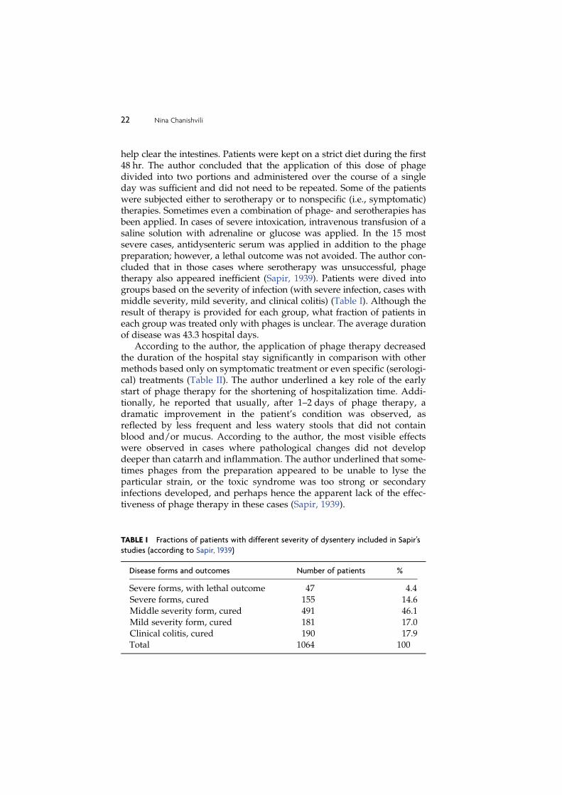
help clear the intestines. Patients were kept on a strict diet during the first48 hr. The author concluded that the application of this dose of phagedivided into two portions and administered over the course of a singleday was sufficient and did not need to be repeated. Some of the patientswere subjected either to serotherapy or to nonspecific (i.e., symptomatic)therapies. Sometimes even a combination of phage- and serotherapies hasbeen applied. In cases of severe intoxication, intravenous transfusion of asaline solution with adrenaline or glucose was applied. In the 15 mostsevere cases, antidysenteric serum was applied in addition to the phagepreparation; however, a lethal outcome was not avoided. The author con-cluded that in those cases where serotherapy was unsuccessful, phagetherapy also appeared inefficient (Sapir, 1939). Patients were dived intogroups based on the severity of infection (with severe infection, cases withmiddle severity, mild severity, and clinical colitis) (Table I). Although theresult of therapy is provided for each group, what fraction of patients ineach group was treated only with phages is unclear. The average durationof disease was 43.3 hospital days.
According to the author, the application of phage therapy decreasedthe duration of the hospital stay significantly in comparison with othermethods based only on symptomatic treatment or even specific (serologi-cal) treatments (Table II). The author underlined a key role of the earlystart of phage therapy for the shortening of hospitalization time. Addi-tionally, he reported that usually, after 1–2 days of phage therapy, adramatic improvement in the patient’s condition was observed, asreflected by less frequent and less watery stools that did not containblood and/or mucus. According to the author, the most visible effectswere observed in cases where pathological changes did not developdeeper than catarrh and inflammation. The author underlined that some-times phages from the preparation appeared to be unable to lyse theparticular strain, or the toxic syndrome was too strong or secondaryinfections developed, and perhaps hence the apparent lack of the effec-tiveness of phage therapy in these cases (Sapir, 1939).
TABLE I Fractions of patients with different severity of dysentery included in Sapir’s
studies (according to Sapir, 1939)
Disease forms and outcomes Number of patients %
Severe forms, with lethal outcome
Severe forms, cured
Middle severity form, cured
Mild severity form, curedClinical colitis, cured
47
155
491
181190
4.4
14.6
46.1
17.017.9
Total 1064 100
22 Nina Chanishvili

Sapir (1939) also reported that after 1 dayof phage treatment the numberof patients with bloody stools decreased from 100 to 74; on the 5th day oftreatment, only 4 patients remained suffering from this symptom. After1 week of phage therapy, 95% of patients did not show pathological symp-toms and could be released from the hospital. A lethal outcome wasregistered in 47 cases (4.4%). However, the author emphasized that thesepatients suffered from dysenterial pancolitis and severe degenerativechanges of the parenchyma of various organs, ulcers of thick gut, and soon, which are typical for long-term and severe intoxication, as verified bypostmortem pathoanatomical studies. The author concluded that:
1. Phages should be given to every patient that shows symptoms ofdysentery, independently of whether the patient is arriving to the hospi-tal, visiting ambulance, or asking for medical help at home. This measurewould have both therapeutic and prophylactic effects.
2. Combined phage- and serotherapy should be used in special casessuch as hypertoxicosis among adults and for the treatment of toxic andsubtoxic syndrome among young children. In all the aforementioned cases,the author also recommended the application of proper doses of serum andits injection at least 6 hr prior to the start of phage therapy (Sapir, 1939).What is the exact basis for the latter recommendation is unclear.
Lipkin andNikolskaya (1940) performed phage therapy on 100 patientssuffering from dysentery. A control group of 50 patients received ordi-nary medication, such as purgative salts, which were used in most cases.In 21 cases the patients underwent sero-therapy. In 5 severe cases, acombined phage- and serotherapy was used. All patients were main-tained under the same conditions, in terms of care, diet, and so on. Phagesproduced by the Tbilisi Institute of Vaccine and Sera and KuibishevInstitute of Epidemiology and Microbiology were used in these studies.The titers of these phage preparations were 10- 9–11 (by Appelmans, 1921).Five milliliters of phages were given to patients orally together with 2%soda solution three times per day. After receiving the phages, patientsfasted all day. In almost every case the phage treatment was continued for1 day. Only in 6 cases was the phage treatment at the same dose per-formed over 2 days. Sixty-six percent of patients received the phages
TABLE II Duration of hospitalization after application of different treatment
methodologies (according to Sapir, 1939)
Type of disease Applied therapy Duration of hospital stay (days)
Severe form Serotherapy 21.9
Severe form Phage therapy 20.8
Middle severity form Nonspecific therapy 16.5Middle severity form Serotherapy 14.9
Middle severity form Phage therapy 11.5
Phage Therapy 23

within the first 5 days of the start of infection. Development of the diseasewas evaluated through observations of stool frequency, presence ofmucus, blood, tensions, and so on.
Lipkin and Nikolskaya (1940) reported a significant effect of phagetherapy even in cases where treatment was started rather late. Twenty-five percent of patients (out of 100) stopped reporting painful symptomsby the 2nd day of treatment; 79% did not show pathological symptoms bythe 4th day and 100% by the 6th day after which the stool was normalized.These data are in contrast to results obtained with ‘‘ordinary therapy,’’where only 2% (1 case out of 50 patients) showed an improvement on the2nd day of treatment, 14% on the 4th day, and 46% on the 6th day. It isnoteworthy that patients with relatively easy cases were included in thecontrol group, whereas those with relatively severe illnesses wereincluded in the experimental group. According to the authors, the factthat these patients showed an improvement as soon as they got phagetreatment illustrated the effectiveness of this method (Lipkin andNikolskaya, 1940). The relief of symptoms in patients treated with ser-otherapy was recorded in 33% of cases (7 persons out of 21) on the 4th dayof treatment and in 67% on the 6th day, indicating that serotherapyresulted in a slower relief of symptoms than phage therapy. Five patientssubjected to serotherapy remained sick over 10 days. These patients laterunderwent phage therapy as well (without further serotherapy).
Phage preparations were considered to be particularly efficient for thetreatment of intestinal infections. Vlasov and Artemenko (1946) describedthe results of treating with phages of 30 persons with chronic dysentery.Many of these chronic patients were exhausted by infection and werebedridden. A dry tablet preparation known as ‘‘phage-vaccine’’—a combined preparation comprising 106 killed cells/ml and 10-7 ofbacteriophages—wasused (byAppelmans, 1921). Thepatients had sufferedwith infections for 1–2 years, and in 70% of cases, rectoscopic examinationindicated the presence of bleeding ulcers. Prior to combined ‘‘phage-vac-cine’’ therapy, all the patients underwent unsuccessful multiple courses(one to eight times) of therapy with antibiotics and sulfonamide prepara-tions. After ‘‘phage-vaccine’’ therapy, the authors reported a cure in the caseof 26 patients (86.7%) within 10–20 days. Assessment of the results wasbased on improvements in the general condition of patients, includingnormalization of comprogram test14 results formation of stools and recov-ery of the mucous layer of the sigmoid colon and rectum (Vlasov andArtemenko, 1946).
14 A type of stool analysis —one of the methods allowing evaluation of the function of the digestive system.Fecal analysis helps to reveal gastric, small and large intestines, and pancreas, liver, and gallbladder problems.Coprogram A coprogram gives an information about the stool quality, according to which a doctor can judgeon the status of the digestive process. In addition, analysis of feces is useful for the detection of intestinalparasitic diseases (helminthes, giardiasis, etc.)
24 Nina Chanishvili

D. Combined use of phage- and sero- therapies
Authors of early publications suggested that an effective use of combinedbacteriophage therapy with sero-therapy required consideration of theexisting ‘‘competition’’ between phages and anti-bacterial serum as illu-strated in 1934 by Lavrik (cited by Krestnikova, 1947). This author showedthat the bacterial cells treated with anti-bacterial serum were unable to beinfected with phages. This phenomenon was confirmed in an in vitro andin vivo experiments by Belkina (1939) (cited by Krestnikova, 1947), whostudied the interaction of phages with cells of Proteus sp. and Shigellashigae strains. Phages added to a mixture of bacteria and antibacterialserum that was specific to these bacteria did not prevent agglutinationand did not lyse bacteria, indicating that the specific serum binds to thebacterial cell surface and thus prevents phage infection. In animal studieswith mice infected with Proteus sp. and S. shigae, the best results wereachieved when the phages were administered 3 hours prior to applicationof the specific serum, which resulted in the survival of 70-–80% of mice.When the serum was administered prior to the phages, 100% of mice inboth experimental and control groups died (cited by Krestnikova, 1947).However, these findings were not always taken into consideration and,according to other authors, specific antibacterial sera were often given tothe patients prior to phage therapy. Articles describing the combined useof specific antibacterial sera and bacteriophage preparations were dis-cussed in Section II.C (Karamov, 1938; Sapir, 1939; Zabrezhinsky andGorstkina-Shevandrova, 1946). In most of the articles devoted to thecombined use of phage- and serotherapy, prophylactic antidysentericor anti-Salmonella sera were given to the patients immediately on arrivalat the hospital and preferably 6 hr prior to the start of phage therapy(Sapir, 1939).
Both antidysenteric serum and appropriate bacteriophage prepara-tions were reported to be used to treat dysentery. Ionov et al. (1939)published their observations on the specific action of antidysentericserum and bacteriophages. This study was carried out in 1935–1936 andinvolved 502 patients (175 children and 327 adults). Bacteriological anal-ysis of stool samples showed the presence of S. shigae and S. flexneri. Of327 adults, 175 were treated only with phages, 90 with antidysentericserum, and 62 received a combined phage and serotherapy. A polyvalentantidysenteric phage preparation and monovalent sera against S. shigae,S.flexneri, and S. Hiss-Russel15 were used in these studies. In the group ofpatients treated with phages only, the preparations were initially admi-nistered orally as a single 10- to 15-ml dose (for adults) and then the samedose of phages (10–15 ml) was given simultaneously both orally and per
15 S. Hiss-Russel¼S. flexneri
Phage Therapy 25

rectum as an enema (titer of phage in the preparations was not providedby the authors). A control group consisted of patients receiving standardmedications (‘‘ordinary therapy’’). The disappearance of blood from thestools was observed in 50% of patients treated with phages as comparedto 20–30% in the control group (Ionov et al., 1939). The combination ofphage- and serotherapy was used in the treatment of 62 adults who didnot demonstrate any symptoms of improvement after phage therapy. Theserum was administered as a subcutaneous injection. In 27 cases theserum was administered during the first 4 days after phage therapywith a positive effect being observed in 14 cases during the first weekand in 7 cases after 3 weeks. Six patients died. Of the 35 patients thatreceived serotherapy later than the 5th day following phage therapy, only8 recovered within a week, 17 recovered within 3 weeks, and 10 patientsdied. It was therefore recommended to develop a combined phage- andserotherapy. The authors emphasized that to optimize the effectiveness ofphage therapy it is important to start it at the early stages of infection.They noted that the stool samples of 43% of the patients undergoingphage therapy were free of pathogenic bacteria—the cause of infec-tion—after 2–3 days. Bacterial carriage was reported to be much lessfrequent in the phage-treated patients compared with the control grouptreated by ‘‘ordinary therapy’’ (i.e., with standard medication). Ofpatients treated with ordinary therapy, 47.9% continued to have patho-genic bacteria in their stools after 15 days, whereas only 20.2% of thepatients undergoing phage therapy retained pathogenic bacteria in theirstools. Of patients treated with phages, 1.7% died compared with 3.5% ofthose treated with serum and 5.8% of those receiving ordinary treatment,the details of which are not specified in the original source. The authorsindicated that ordinary therapy was usually applied in cases of mildseverity, whereas combined phage- and serotherapy was applied in themost severe cases (Ionov et al., 1939).
Golubtsov (1940) reported a comparative study carried out in 1939of the effect of sero- and phage therapies among children sufferingfrom dysentery. The children were divided into two groups: group Iconsisted of 22 children treated with serotherapy and group II comprised18 children treated with specific bacteriophages. The majority of childrenin each group were aged between 5 and 18 months, with 7 childrenbetween 1.5 and 2 years old.16 Bacteriological analysis was only successfulin 59% of cases but indicated that S. Shiga-Kruse and S. Hiss-Russelpredominated.
Golubtsiov (1940) subgrouped the dysenteric diseases in childrenaccording to their severity into three types: nontoxic (mild), toxic, and
16 The author did not mention how these seven were distributed between the two groups.
26 Nina Chanishvili

chronic. Evaluation of the effectiveness of therapy was based on bodytemperature, presence of toxic syndrome and cramps, and stool charac-teristics (frequency, presence of blood, mucus, pus, etc.).
Children of group I received antidysenteric serum during the firstdays after infection. The serum was administered intramuscularly indoses of 10,000–20,000 antitoxic units per injection within a 3- to 4-dayperiod. In 47% of cases, toxic syndrome and vomiting were reported to berelieved and an improvement in the general condition of patients wasobserved, as evidenced by better reactions of the infants to surroundingpeople and events, better sleep patterns, and reemergence of appetite.Serotherapy did not have a significant effect on body temperature andstool quality and frequency. A decrease in temperature was observed inonly 27% of cases with changes of stool frequency in 37% of cases.
Children of group II received the specific antidysenteric phages, whichwere administered orally in the morning before meals, in a dose of 1.0 mlfor infants below 1 year, 1.5 ml for children of 1.5 years, and 2.0 ml for2 year olds (titer unknown). The therapeutic effect was indicated by adecrease in temperature in 77% of patients with an improvement in stoolcharacteristics and a decrease in stool frequency observed in 60%, andrelief of cramps in 53.3%. A relief of toxic syndrome17 was observed onlyin 17% of cases (Golubtsov, 1940).
Summarizing these data, Golubtsov (1940) concluded that the effectsof sero- and phage therapies are different. Serotherapy decreased theintensity and frequency of toxic syndrome significantly, whereas phagetherapy had no such effect but gave better results for the improvementof pathological changes in the colon (such as damage of the mucouslayer). The author recommended the application of specific phages inthe early stages of the disease and in combination with serotherapy(Golubtsov, 1940).
E. Use of bacteriophages for prophylaxis
Phages have also been used extensively in the former Soviet Union forprophylaxis in regions with a high incidence of infections and also incommunities where the rapid spread of infections may occur, such askindergartens, schools, and military accommodation (Agafonov et al.,1984; Anpilov and Proskudin, 1985; Belikova, 1941; Blankov, 1941;Blankov and Zherebtsov, 1941; Florova and Cherkass, 1965;Kagan et al.,1964; Karpov, 1946).
17 Toxic syndrome (TS) is a toxic condition that occurs in response to the impact of toxic substances, e.g.,produced inside the body by bacteria. TS is characterized by severe metabolic disorders and functions ofvarious organs and systems, primarily the central nervous and cardiovascular systems.
Phage Therapy 27
The application of phages for prophylaxis was carried out in 1929–1930 against the bacterial diseases that were the most serious problems atthat time, such as dysentery, typhoid fever, and staphylococcal infections.The first mass application of dysenteric bacteriophages in the USSR wasperformed in Alchevsk (Donbas region) in Ukraine in 1930 (Ruchevski,‘‘Vrachebnaya gazeta,’’ 1931, 21: 1586, cited by Krestnikova, 1947). Anexperiment on the prophylactic use of phages was carried out success-fully later in 1935 on thousands of people in regions with a high incidenceof dysentery (Melnik et al., 1935). Results were reported at scientificconferences in 1934 and 1936 in Kiev and in 1939 in Moscow after whichthe dysenterial phage preparation was finally approved as a preventivemeasure for mass application (Krestnikova, 1947). According to Krestni-kova (1947), it was recommended that repeated seasonal prophylactic‘‘phaging’’ be carried out in areas where the dysentery was endemic.Later modifications included the supply of dysenterial phages in drytablet forms, which also began to be included in clinical studies.
One of the later studies by Babalova et al. (1968) in 1963–1964 describesthe results of preventive treatment carried out with phage tablets havingan acid-resistant coating. This polyvalent antidysenteric preparationcontained different phages against the following bacteria: S. flexneri(10- 7–10- 9 by Appelmans, 1921), S. sonnei (10- 7–10- 8), S. grigorieva-shiga(10- 7–10- 9), S. stutzeri18 (10- 6–10- 7), and S. newcastle19 (10- 6). A study of thepersistence of bacteriophages in children was also carried out andshowed that phages could be detected in the body for 5–7 days. Theauthors mentioned that phages active against S. flexneri serotypes 1aand 2a and S. grigorieva-shiga20 were especially long lasting and could beisolated from stool samples longer than other phages included in thispreparation, such as that active against S. newcastle. Perhaps the latter wasdependent rather on the individual properties of these phages by them-selves and could have nothing to do with their specificity.
According to Babalova et al. (1968), a prophylactic experiment wascarried out by specially trained staff of the Tbilisi Institute of Vaccineand Sera21 in collaboration with the regional sanitary-epidemic stationsin two Tbilisi districts characterized by distinct epidemic situations, one ofwhich was known to have above average infrastructure and hence a lowersusceptibility to infection outbreaks. Prophylactic ‘‘phaging’’ was carriedout during the period fromMay 8 to October 25, 1963 and fromMay 15 toOctober 31, 1964. Special attentionwas given to themethod of selecting theexperimental and control groups of children. Children living in each
18 Shigella dysenteriae type 1 and 2, respectively.19 Shigella flexneri type 6.20 S. dysenteriae type 1.21 Former name of the Eliava Institute of Bacteriophage, Microbiology & Virology in Tbilisi, Georgia.
28 Nina Chanishvili

district were registered and divided into two equal groups according tosanitary-epidemic conditions. Prophylactic ‘‘phaging’’ was carried out onchildren living on one side of the street, whereas those living on the otherside did not get the phage treatment and thus formed a control group.Doctors visited each child from both experimental and control groups atleast once a week, challenging the experimental group with a new dose ofphages and performing observations, registering any cases of disease, andproviding this information to the center.
Phages were given to children aged between 6 months and 7 years.Tablets were administered before meals or 2 hr after meals. Children aged6 months–5 years received one tablet (equal to 20 ml of phage suspension;the titers of each component of this polyvalent dysenteric preparationwere given earlier), and children over 5 years received two tablets. Theaverage number of observation days in experimental groups and controlgroups was comparable, 108.6 and 109.5, respectively. The effect of phageprophylaxis was evaluated on the basis of clinical symptoms rather thanon bacteriological analysis, as only single suspected cases of intestinaldisease were studied bacteriologically. The incidence of acute dysenteryin the control group was 3.8 times higher than in the experimental group.The observed difference was significant, as verified by the statisticalanalysis of results. The effects of phage prophylaxis differed betweendifferent age groups. For example, in the age group 6 months–1 year,the epidemic index (EI)22 was 5.7; in the age group 1–3 years the EI was3.7, and in the age group 3–5 years the EI was 2.4. In the 5- to 7-year-oldage group, no cases were registered. The authors concluded that a 3.8-folddecrease of disease incidence in children of 6 months–7 years of age dueto prophylactic ‘‘phaging’’ was a promising result. They suggested usingthe tablet form of dysenteric bacteriophages for mass application(Babalova et al., 1968).
Later, studies on mass prophylaxis of intestinal diseases by the appli-cation of phages were performed in the Red Army units by militarydoctors as well. For prevention of dysentery and typhoid epidemics,specific phages were also used with two tablets administered onceevery 5–7 days during the outbreak season. The authors reported thatthe prophylactic use of phages resulted in a 6- to 8-fold decrease in thenumber of intestinal infection cases in the tested groups in comparisonwith the control groups (Agafonov et al., 1984; Anpilov and Proskudin,1984; Kurochka et al., 1987).
An interesting experiment is described by Sayamov (1963) andPlankina et al. (1961), who present the results of therapeutic and prophylactic
22 EI is the ratio between the expected and the observed number of infected persons calculated per 1000persons. EI is used here as a tool to measure the effectiveness of prophylactic measures.
Phage Therapy 29

trials using anticholera bacteriophage preparation, performed by Sovietdoctors in East Pakistan in 1958 and in Afghanistan in 1960. The bacterio-phage preparation was developed by an in vitro cultivation of bacterio-phages on V. cholera strains isolated directly in epidemic site on mediareconstructing in vivo conditions, as suggested by Nikonov in 1959 (citedaccording to Sayamov, 1963). In particular, these media contained bileand fragments of small intestine in Tyrode’s solution.23 The ability ofselected phage clones to propagate in vivo was increased via a procedureknown as adaptation, which is based on sequential, alternate passagesof bacteriophages through the small intestine of guinea pigs24 andthrough bile.
During a cholera outbreak in Dacca (East Pakistan) in 1958, Sovietdoctors used ‘‘adapted’’ phages to treat 22 patients, all in serious condi-tions and with signs of anuria25 (Sayamov, 1963). Typically, each patientwas given 5–10 ml of phage suspension in a saline solution intravenouslyonce and 30 ml of phage suspension orally each day for 3 consecutivedays (phage titer in these suspensions was not provided). Only two, themost seriously sick and dehydrated, patients died, whereas the fatalityrate among Kandahar hospital patients treated symptomatically and/orwith antibiotics (mainly oxytetracycline) was officially estimated to beabout 50%. At the same time, to prevent the spread of cholera, Sovietdoctors were reported to treat about 30,000 people with phages prophy-lactically in seven highly populated centers of East Pakistan (Sayamov,1963). Details of this treatment are not specified. No cholera cases werereported in any of these centers following such a prophylactic action.
According to Sayamov (1963), the epidemic in Afganistan started onAugust 6, 1960. Only persons suffering from severe forms of cholera weresent to the hospital, where the usual symptomatic treatment, togetherwithantibiotics (mainly oxytetracycline), was given.On the recommendation ofthe Soviet doctors, all patients were transferred to an isolation block. OnOctober 7 and 8, phage prophylaxis started, which included the patients aswell as the healthy hospital staff and students being on training at that time(altogether over 1600 persons). A single dose of the phages in salinesolution (5–10 ml) was administered intravenously26 or intramuscularly,
23 A solution containing salts and glucose.24 A segment of small intestine of an anesthetized, living guinea pig was isolated carefully using two ligaturesso that the branches of the blood vessels supplying the isolated loop were left intact. This technique wassuggested by De and Chatterjee in 1953 (cited by Sayamov, 1963). The isolated loop was infected withV. cholera using 100–1000 Vibrio cells. After 24 hr the majority of infected animals died. If anticholerabacteriophage was injected into the same loop simultaneously or after a certain period of time, animalssurvived. However, the aim of this experiment was to obtain bacteriophage clones that would be able topropagate efficiently in in vivo conditions. Therefore, phages isolated from one animal were used to cureanother animal, and so forth (Sayamov, 1963).25 Complete suppression of urine formation and excretion.26 Purification of phages for injections by CsCl or CsSO4 gradient centrifugation had never been applied.
30 Nina Chanishvili
followed by an oral daily dose (30 ml, children received 20 ml) for 3 days.As a result of these measures, a lethal outcome was observed only in 4 outof 119 cholera patients (3.5%). What is more, no new cases of cholera weredetected in the hospital after the start of therapeutic and prophylacticphage treatment between October 7 and 10. In 80% of patients treatedwith bacteriophages in the Kabul hospitals, the initial diagnosis wasconfirmed bacteriologically as well. No complications were observedafter intramuscular injection of phages, whereas chills and headacheswere sometimes observed after intravenous administration.
As described by Sayamov (1963), in October 1960 cholera appeared inthe Kataghan province in the north of Afghanistan. A number of patientswere accommodated in hospitals set up in the towns of Pul-i-Khumri andKunduz, but because of a shortage of hospital beds and transportationproblems, many sick people remained at home. To prevent the spread ofcholera, Soviet doctors, in cooperation with Afghan colleagues, arrangeda massive therapeutic and prophylactic action among the cholera-strickenrural population. Most of the patients treated at home (about 90%)received a dose of 20–30 ml (or, in particularly severe cases, as much as50 ml) of phage suspension in one or two intramuscular injections (intra-venous administration was not used). A single dose of cholera vaccinewas administered simultaneously as well. The healthy part of each villagepopulation was treated the same way. No new cases of cholera occurredin rural populations as a result of this combined therapeutic and prophy-lactic action but, as underlined by Sayamov (1963), only when the wholepopulation in a given village was subjected to the phage treatment.Altogether approximately 270,000 persons underwent phage treatment.Whether any control groups were included in these therapeutic/prophy-lactic actions, as well as what was the contribution of phages and whatwas the contribution of vaccine in the observed prophylactic effect, isunknown. However, Sayamov (1963) concluded that the suspension ofanticholera bacteriophages is a valuable preparation, which could be usedas an important supplement to the standard measures for cholera control.
III. CURRENT STATUS OF GEORGIAN RESEARCH AND THEIR
IMPACT ON WORLDWIDE PHAGE THERAPY STUDIES
Ongoing research and clinical trials
Production and use of phages for therapy and prophylaxis never stoppedat the Eliava Institute of Bacteriophage; however, the scale is much smal-ler than before the break off of the Soviet Union. Therapeutic and pro-phylactic phages are currently produced by the small spin-off company‘‘Eliava BioPreparations, Ltd.’’ established by the Eliava IBMV. In the
Phage Therapy 31

past, the scale of phages production covered the needs of the greatest partof the Soviet countries, including the Caucasian and Middle Asian repub-lics. Today, production of the ‘‘Eliava BioPreparations, Ltd.’’ fully satis-fies the Georgian market. In 2010 the company started to export phagesoutside Georgia into Azerbaijan. Another spin-off company, ‘‘Eliava Dia-ngnostics,’’ receives, on average, 50–60 patients daily seeking the phagetreatment, often after unsuccessful antibiotic therapy. In 2010, a day clinic,‘‘The Eliava Phage Therapy Center,’’ was opened, which renders servicesto local and foreign patients.
These achievements would be impossible without intensive researchheld at the Eliava Institute of Bacteriophage, most of which are conductedin close collaboration with the international scientific community. On theone hand, this helps Georgian scientists (1) to develop and introducemodern techniques into their research and (2) to design proper experi-ments corresponding to Western standards. On the other hand, collabo-rative projects help Western researchers to avoid the ‘‘discovery ofbicycles,’’ which may occur easily without having background knowl-edge in the development of phage preparations and their application inpractice. Over 50 collaborative international projects have been accom-plished during the period of 1995–2010, which resulted in a number ofsignificant publications (Ceyssens et al., 2011; Chanishvili et al., 2001, 2009;Glonti et al., 2010; Khawaldeh et al., 2011; Kutateladze and Adamia, 2010;Kutter et al., 2010; Markoishvili et al., 2002; Merabishvili et al., 2009; Pirnayet al., 2011; Sulakvelidze et al., 2001; Verbeken et al., 2007).
Among other achievements in phage therapy, it is necessary to men-tion the treatment of cases of secondary infections associated with cysticfibrosis. Since 2008, together with the National Center of Cystic Fibrosis inTbilisi, phage preparations against secondary infections have been usedin eight patients with cystic fibrosis. Phages were applied in infants andadults via a nebulizer several times for 6–10 days. Simultaneously,patients were treated by conventional antibiotics, antimucus medications,and vitamins. Phage application caused a substantial decrease of bacterialcounts in the patients’ sputum samples and an improvement of the gen-eral health condition. Due to bacteriophage therapy, long-term remissionsof infections were achieved (Kutateladze and Adamia, 2010).
Despite economic difficulties during 1990–2000, the institute’s staffmanaged to elaborate new phage-based products, such as PhageBioDerm,which is a novel wound-healing preparation consisting of a biodegradablepolymer impregnated with an antibiotic and lytic bacteriophages(Markoishvili et al., 2002). It has been licensed for sale in the Republic ofGeorgia (one of the former Soviet Union republics). In 1999–2000, inTbilisi, Georgia, 107 patients who had ulcers that had failed to respond toconventional therapy were treated with PhagoBioDerm alone or in com-bination with other interventions. The wounds/ulcers healed completely
32 Nina Chanishvili
in 67 (70%) of 96 patients for whom follow-up data were available. In 22cases inwhichmicrobiological datawere available, healingwas associatedwith a concomitant elimination of, or a reduction in, specific pathogenicbacteria in the ulcers. Bacteriophages in PhagoBioDerm helped clear thewounds frommultidrug-resistant S. aureus. Results suggest that this slow-release biopolymer is safe and of possible benefit in the management ofrefractory wounds and support the apparent utility of bacteriophages inthis setting. Further studies, including carefully designed clinical trials,will be required to evaluate rigorously the efficacy of this novel wound-dressing preparation (Markoishvili et al., 2002; Sulakvelidze et al., 2001).
While the phage preparations are in everyday use in Georgia and in anumber of ex-Soviet countries, there are many obstacles for the clinicalapplication of bacteriophages in Western countries, such as the percep-tion of viruses as ‘‘enemies of life’’ (Villareal, 2005), the lack of a specificframe for phage therapy in current medicinal product regulation (Pirneyet al., 2011), and the absence of well-defined and safe bacteriophagepreparations. To evaluate the safety and efficacy of bacteriophages inthe treatment of burn wound infections in a controlled clinical trial, ahighly purified and fully defined bacteriophage cocktail (BFC-1) wasprepared, which is active against the P. aeruginosa and the S. aureus strainsactually circulating in the Burn Centre of the Queen Astrid MilitaryHospital (Merabishvili et al., 2009). Based on successive selection rounds,three bacteriophages were retained from an initial pool of 82 P. aeruginosaand 8 S. aureus bacteriophages, specific for prevalent P. aeruginosa andS. aureus strains in the Burn Centre of the Queen Astrid Military Hospitalin Brussels, Belgium. This cocktail, consisting of P. aeruginosa phages 14/1(Myoviridae) and PNM (Podoviridae) and S. aureus phage ISP (Myovir-idae), was produced and purified of endotoxin. Quality control includedstability (shelf life); determination of pyrogenicity, sterility, and cyto-toxicity; confirmation of the absence of temperate bacteriophages; andtransmission electron microscopy-based confirmation of the presence ofthe expected virion morphologic particles, as well as of their specificinteraction with the target bacteria. Bacteriophage genome and proteomeanalysis confirmed the lytic nature of the bacteriophages, the absence oftoxin-encoding genes, and showed that the selected phages 14/1, PNM,and ISP are close relatives of, respectively, F8, jKMV, and phage G1. Thebacteriophage cocktail is currently being evaluated in a pilot clinicalstudy cleared by a leading medical ethical committee. No adverse reac-tions were observed (Merabishvili et al., 2009). A detailed descriptionof the quality-controlled, small-scale phage preparation was consideredas a first step to promote the concept of phage treatment in Westernmedicine. In addition, it supported the creation of a discussion platformfor regulatory framework for approval of phage therapy (Pirneyet al., 2011).
Phage Therapy 33

Antibiotics are becoming ineffective as important bacterial pathogensevolve to outsmart them. However, the antibiotic pipeline is running drywith only a few new antibacterial drugs expected to make it to the marketin the foreseeable future. Bacteria resistant to all available antibacterialdrugs, so-called superbugs, are emerging worldwide. Evolutionary ecol-ogy might inform practical attempts to bring these pathogens understronger human control (Williams, 2010).
In this context, various laboratories worldwide and a handful of smallpharmaceutical companies are turning to phages (Thiel, 2004). There ishope that commercial interest shown by these companies will finally leadto recognition of phage therapy by the Western world.
Despite its long (eastern European) history, phage therapy is notcurrently authorized for routine use on humans in the West. Today, it isonly approved in some former Soviet republics such as Russia and Geor-gia, where commercial phage preparations are sold in pharmacies(Chanishvili, 2009). In Poland, a recent member of the European Union,phage therapy is considered an ‘‘experimental treatment’’ covered by thePhysician Practice Act (Polish Law Gazette N! 28 of 1997) and the Declara-tion of Helsinki, administered only when other therapeutic options do notexist (Gorski et al., 2009; Pirney et al., 2011). In France, therapeutic made-to-order phage preparations from the Institute Pasteur (Paris and Lyon)were used until the beginning of the nineties. Today, a French practi-tioner, Alain Dublanchet, still uses commercial phage preparations (pur-chased in Russia and Georgia) to treat severe infections (Pirney et al.,2011). Despite the absence of a specific framework for phage therapy(Gorski et al., 2009; Pirney et al., 2011), a pilot clinical trial in burn woundswas approved by an ethical committee in Belgium (Kutter et al., 2010). Inthe United States, a Food and Drug Administration (FDA)-approvedphase I clinical trial was conducted. No safety concerns were found(Kutter et al., 2010). A British phage therapy company conducted aphase I/II clinical trial in chronic otitis. This study was approved throughthe UK Medicines and Healthcare products Regulatory Agency and theCentral Office for Research Ethics Committees ethical review process(Kutter et al., 2010).
To avoid drug-licensing difficulties, some U.S.-based phage compa-nies decided to first develop phage products for the decontamination offood, plants, fields, and livestock (Thiel, 2005). They hope to create reve-nue to fund research into human therapeutics and to familiarize theauthorities and the general public about phages. Phages for decontami-nating food plants, ready-to-eat meat, poultry products, cheese, and liveanimals that will be slaughtered for human consumption were approvedby the FDA and are now in use. An increasing number of publicationsdedicated to this subject appears every year (Abuladze et al., 2008;Leverentz et al., 2003; Sabour and Griffiths, 2010). However, many
34 Nina Chanishvili

scientists are concerned about the impact of such massive and wide-spread applications of phages on natural microbial communities becauseof their role in the horizontal spread of drug resistance and virulencegenes (Sabour and Griffiths, 2010). It is presumed that phage-derivedproducts (e.g., cell wall-degrading enzymes such as endolysins) can andprobably will be licensed and marketed within a few years. Of course,these phage products lack the capacity of self-replication and adaptationin the infectious site, which is a disadvantage in comparison with livebacteriophages. Because of their chemical nature and resemblance toantibiotics, they may also select resistance, but presumably at a lowerrate than antibiotics (Fenton et al, 2010; Hoopes et al., 2009; Sabour andGriffiths, 2010).
Phage therapy has great potential in clinical practice, especially inchronic cases. As with antibiotic treatment, there are likely to be impor-tant evolutionary consequences related to phage therapy (Levin and Bull,2004) if it is implemented widely and without sufficient oversight. In thepast, phages have been used massively for the prophylaxis of bacterialinfections in humans, as is shown in this chapter. In numerous cases, thedescriptions of observed effects indicated positive results of phage ther-apy, although the parallel control studies either were not performed orwere documented incompletely in view of current requirements. Also, thespecificity of phages allows applying them for diagnostic purposes, some-times even leading to the identification of nonculturable bacteria(Loessner et al., 1996; Rees and Dodd, 2006). This practice should not beunderestimated in planning future phage research.
REFERENCES
Abuladze, T., Li, M., Menetrez, M. Y., Dean, T., Senecal, A., and Sulakvelidze, A. (2008).Bacteriophages Reduce reduce experimental contamination of hard surfaces, tomato,spinach, broccoli, and ground beef by Escherichia coli O157:H7. Appl. Environ. Microbiol.74:6230–6238.
Adhya, S., and Merril, C. (2006). The road to phage therapy. Nature 443:754–755.Agafonov, B. I., Khokhlov, D. T., and Zolochevsky, M. A. (1984). Epidemiology of typhoid-
paratyphoid infections and their prophylactics. Military-Med. J. 6:36–40.Alessandrini, A., and Doria, R. (1924). Il batteriofago nella terapia del tifo addominale.
Policlinico, sez prat. 31:109.Alexandrov, M. B., Dyakova, A. A., Raikhelson, I. A., andMelnik, E. G. (1940). Bacteriophage
therapy of typhoid fever. In ‘‘Abstracts of the Inter-institutional Conference on Bacter-iophages Research, December 21–25, 1940, Moscow, 30’’.
Alexandrova, M. B., Zhivago, N. L., Alekseeva, L. N., and Zeitlenok, N. A. (1935). Phagetherapy against dysentery. J. Microbiol. Epidemiol. Immunol. 11:860–868.
Anpilov, L. I., and Prokudin, A. A. (1984). Prophylactic effectiveness of the dry polyvalentdysentery bacteriophage in organized communities. Military-Med. J. 5:39–40.
Appelmans, R. (1921). Le dosage du bacteriophage. C.R. Soc. Biol. 85:1098–1099.
Phage Therapy 35

Astashkevich, H. M. (1950). Experience of application of phage therapy for treatment oftypoid and paratyphoid diseases. J. Microb. Epidemiol. Immun. 7:42.
Babalova, E. G., Katsitadze, K. T., Sakvarelidze, L. A., Imnaishvili, N. Sh., Sharashidze, T. G.,Badashvili, V. A., Kiknadze, G. P., Meipariani, A. N., Gendzekhadze, N. D.,Machavariani, E. V., Gogoberidze, K. L., Gozalov, E. I., et al. (1968). On the issue ofprophylactic importance of the dry dysenteric bacteriophage. J. Microbiol. Epidemiol.Immunol. 2:143–145.
Belikova, M. A. (1941). Experience of phage prophilaxis of summer dysentery among theyoung children performed in the city Stalingrad. J. Microbiol. Epidemiol. Immunol. 5–6:142.
Beridze, M. A. (1938). Role of Bacteriophage Therapy in Combating of Purulent Skin Infec-tions. Medgiz, Tbilisi, Georgia.
Blankov, B. I. (1941). Analysis of the results of phage prophylaxis of dysentery among thecontacting people. Report # 1. J. Microbiol. Epidemiol. Immunol. 5–6:125–131.
Blankov, B. I., and Zherebtsov, I. D. (1941). Experience on the multiple phaging of thecontacting population in the fight against dysentery. Report # 2. J. Microbiol. Epidemiol.Immunol. 5–6:131–136.
Bruynoghe, R., and Maisin, J. (1921). Essais de therapeutique au moyen du bacteriophage.C.R. Soc. Biol. 85:1120–1121.
Ceyssens, P.-J., Glonti, T., Kropinski, A. M., Lavigne, R., Chanishvili, N., Kulakov, L.,Lashkhi, N., Tediashvili, M., and Merabishvili, M. (2011). Phenotypic and genotypicvariations within a single bacteriophage species. Virol. J. 8:134.
Chanishvili, N. (2009). A Literature Review of the Practical Application of BacteriophageResearch. EIBMV, Tbilisi, Georgia.
Chanishvili, N., Chanishvili, T., Tediashvili, M., and Barrow, P. A. (2001). Phages and theirapplication against drug-resistant bacteria. J. Chem. Technol. Biotechnol. 76:689–699.
Cienciala, A. M., Lebiedieva, N. S., and Materski, W. (2007). In ‘‘Katy"n: A crime withoutpunishment’’, pp. 226–229. Yale University Press.
Compton, A. (1929). Antidysentery bacteriophage in treatment of dysentery: A record of 66cases treated, with inferences. Lancet 2:273.
Costa Cruz, J. (1924). Le traitement des dysenteries bacillaires par le bacteriophage. C.R. SocBiol. 91:845.
De, S. N., and Chatterjee, D. N. (1953). Experimental study on mechanism of action of Vibriocholerae on intestinal mucus membrane. J. Pathol. Bacteriol. 66:559–562.
d’Herelle, F. (1935). Bacteriophage and Phenomenon of Recovery. TSU Press, Tbilisi,Georgia.
d’Herelle, F., and Eliava, G. (1921a). Unicite du bacteriae sur la lysine du bacteriophage. C.R.Soc. Biol. 85:701–702.
d’Herelle, F., and Eliava, G. (1921b). Sur le serum antibacteriophage. C.R. Soc. Biol.84:719–721.
d’Herelle, F. (1917). Sur un microbe invisible antagoniste des bacilles dysenteriques. C.R.Acad. Sci. (Paris) 165:373–375.
Dublanchet, A., and Bourne, S. (2007). The epic of phage therapy. Can. J. Infect. Dis. Med.Microbiol. 18:15–18.
Eaton, M. D., and Bayne-Jones, S. (1934). Bacteriophage therapy: Review of the principlesand results of the use of bacteriophage in the treatment of infections. JAMA 23:1769–1939.
Eliava, G. (1930). Au sujet de l ‘adsorption du bacteriophage par les leucocytes. C.R. Soc. Biol.105:829–831.
Eliava, G., and Legraoux, R. (1921). Sur un liquide eu se maintiant invariable les nombre desbacteries des cultures. Ann. Institute Pasteur 35:713–717.
Eliava, G., and Pozersky, E. (1921a). D’laction destructive des sels de quinine sur le bacterio-phage de D’Herelle. C.R. Soc. Biol. 85:139–141.
36 Nina Chanishvili

Eliava, G., and Pozersky, E. (1921b). Sur les caracter nouveaux presentes par le Bacille deShiga avant resiste a l’action du bacteriophage de d’Herelle. C.R. Soc. Biol. 84:708–710.
Eliava, G., and Suarez, E. (1927a). Au sujet de l ‘ultrafiltration du corpuscule bacteriophage.C.R. Soc. Biol. 96:462–464.
Eliava, G., and Suarez, E. (1927b). Dimensions du carpuscule bacteriophage. C.R. Soc. Biol.96:460–462.
Ermolieva, V. V. (1939). About bacteriophage and its application. J. Microbiol. Epidemiol.Immunol. 9:9–17.
Fenton, M., Ross, P., McAuliffe, O., O’Mahony, J., and Coffey, A. (2010). Recombinantbacteriophage lysins as antibacterials. Bioeng. Bugs 1:9–16.
Florova, N. N., and Cherkass, F. K. (1965). Results of mass application of polyvalent dysen-teric bacteriophage. J. Microbiol. Epidemiol. Immunol. 3:118–125.
Gamaleya, N. F. (1898). Bacterial lyzins: The enzymes destroying bacteria. Russ. Arch. Pathol.Clin. Med. Bacteriol. 6:607–613.
Georgadze, I. A. (1974). Fifty years of the Tbilisi Scientific-Research Institute of Vaccine andSera of the Ministry of Health of the USSR. In ‘‘Selected Articles of the Jubilee Dedi-cated to 50th Anniversary of the Tbilisi Institute of Vaccine and Sera’’. TIVS, Tbilisi,Georgia.
Georgadze, I. A., and Makashvili, E. G. (1979). George Eliava. In ‘‘The Georgian SovietEncyclopedia,’’ Vol. 4, p. 125.
Glonti, T., Chanishvili, N., and Taylor, P. W. (2010). Bacteriophage-derived enzyme thatdepolymerizes the alginic acid capsule associated with cystic fibrosis isolates of Pseudo-monas aeruginosa. J. Appl. Environ. Microbiol. 108:695–702.
Gnutenko, M. P. (1951). Treatment of S. typhi and paratyphi bacterial carriers with bacterio-phages. J. Microbiol. Epidemiol. Immunol. 5:56–60.
Golubtsov, G. V. (1940). Sero- and phage- therapies of dysentery among babies and infants.In ‘‘Selected Articles of the 1st Scientific Conference of the Bashkir Medical University,March 23–25, 1939’’, pp. 13–16. Ufa, Russia.
Gorski, A., Miedzybrodzki, R., Borysowski, J., Weber-Dabrowska, B., Łobocka, M.,Fortuna, W., Letkiewicz, S., Zimecki, M., and Filby, G. (2009). Bacteriophage therapyfor the treatment of infections. Curr. Opin. Invest. Drugs 10:766–774.
Hankin, E. H. (1896). L’action bactericide des eaux de la Jumna et du Gange sur le vibrion ducholera. Ann. Inst. Pasteur 10:511.
Hausler, T. (2008). ‘‘Viruses vs. Superbugs’’. MacMillan, New York.Hoopes, T., Stark, C. J., Kim, H. A., Sussman, D. J., Donovan, D. M., and Nelson, D. C. (2009).
Use of a bacteriophage lysine PlyC, as an enzyme disinfectant against Streptococcus equi.Appl. Environ. Microbiol. 75:1388–1394.
Ionov, I. D., Erez, S. L., and Goldenberg, E. Y. (1939). Specific therapy of dysentery. In‘‘Variability of Microorganisms and Bacteriophage Research’’ (Proceedings of the Scien-tific Conference, 1936), pp. 337–346. Kiev, Ukraine.
Kagan, M. I., Kuznetsova, E. V., and Teleshevskaya, E. A. (1964). To the issue of epidemiceffectiveness of the planned phaging in the day nurseries. J. Microbiol. Epidemiol. Immunol.7:89–102.
Kaplan, A. S. (1941). Duration of maintenance of the polyvalent dysenteric bacteriophage inthe human organism in case of its prophylactic use. J. Microbiol. Epidemiol. Immunol.5–6:163.
Karamov, S. (1938). Experience of phage therapy for treatment of typhoid fever. In ‘‘SelectedArticles of Azerbaijani Institute of Epidemiology andMicrobiology’’, Vol. 6, pp. 101–105.Baku, Azerbaijan SSR.
Karpov, S. P. (1946). The specific bacteriophage in relation to the issue of combating typhoidand paratyphoid diseases. J. Microbiol. Epidemiol. Immunol. 1–2:40–44.
Phage Therapy 37

Karpov, S. P. (1950). Mechanism of prophylactic action of typhoid-paratyphoid bacterio-phage (dynamics of the phage release from the organism of the parenterally phagedwhite mice). J. Microbiol. Epidemiol. Immunol. 7:43–45.
Karpov, S. P., and Yavorskaya, B. M. (1949). Mechanism of prophylactic action of typhoidparatyphoid bacteriophage (dynamics of the phage release from the organism of thephaged people). J. Microbiol. Epidemiol. Immunol. 7:40–42.
Kazhal, N., and Iftimovich, R. (1968). From the History of Fight Against Bacteria and Viruses.Nauchnoe Izdatelstvo, Buchares.
Khawaldeh, A., Morales, S., Dillon, B., Alavidze, Z., Ginn, A. N., Thomas, L.,Chapman, S. J., Dublanchet, A., Smithyman, A., and Iredell, J. R. (2011). Bacteriophagetherapy for refractory Pseudomonas aeruginosa urinary tract infection. J. Med. Microbiol.60:1697–1700.
Kokin, G. A. (1941). Use of bacteriophages in surgery. Soviet Medicine (‘‘Sovietskaya Med-itsina’’) 9, 15–18.
Kokin, G. A. (1946). Phage therapy and phage prophylaxis of gas gangrene. In ‘‘Experience ofthe Soviet Military Medicine during the Great Patriotic War 1941–1945’’, Vol. 3,pp. 56–63. Medgiz, Moscow.
Krestovnikova, V. A. (1947). Phage treatment and phage prophylactics and their approval inthe works of the Soviet researchers. J. Microb. Epidemiol. Immunol. 3:56–65.
Krueger, A. P., and Scribner, E. J. (1941). Bacteriophage therapy. II. The bacteriophage: Itsnature and its therapeutic use. JAMA 19:2160–2277.
Kurochka, V. K., Karniz, A. F., and Khodyrev, A. P. (1987). Experiences of implementation ofpreventive anti-epidemic measures in the center of intestinal infections with watertransmission mechanism of morbidity. Military-Med. J. 7:36–37.
Kutateladze, M., and Adamia, R. (2010). Bacteriophages as potential new therapeutics toreplace or supplement antibiotics. Trends Biotechnol. 28:591–595.
Kutter, E., De Vos, D., Gvasalia, G., Alavidze, Z., Gogokhia, L., Kuhl, S., and Abedon, S. T.(2010). Phage therapy in clinical practice: Treatment of human infections. Curr. Pharm.Biotechnol. 11:69–86.
Leverentz, B., Convay, W. C., Camp, M. J., Janisiewicz, W. J., Abuladze, T., Yang, M.,Saftner, R., and Sulakvelidze, A. (2003). Biocontrol of Listeria monocytogenes on fresh-cutproduce by treatment with lytic bacteriophages and a bacteriocin. Appl. Environ. Micro-biol. 69:4519–4526.
Levin, B. R., and Bull, J. J. (2004). Population and evolutionary dynamics of phage therapy.Nat Rev. Microbiol. 2:166–173.
Lipkin, and Nikolskaya (1940). Experience of phage therapy of dysentery. In ‘‘SelectedArticles of the Kuibishev Red Army Military-Medical Academy’’, Vol. 4, pp. 193–198.KRAMMA, Kuibishev, Russian SSR.
Loessner, M. J., Rees, C. E., Stewart, G. S., and Scherer, S. (1996). Construction of luciferasereporter bacteriophage A511::luxAB for rapid and sensitive detection of viable Listeriacells. Appl. Environ. Microbiol. 62:1133–1140.
Lurie, M. N. (1938). Treatment of dysentery and haemolytic intestinal diseases amongchildren and adults. In ‘‘Selected articles of Azerbaijani Institute of Epidemiology andMicrobiology, ’’ Vol. 6, pp. 31–34.
Manolov, D. G., Sekunova, V. N., and Somova, E. E. (1948). Experience of therapy oftyphoid fever by intravenous administration of the phage. J. Microbiol. Epidemiol.Immunol. 4:33.
Markoishvili, K., Tsitlanadze, G., Katsarava, R., Morris, J. G., and Sulakvelidze, A. (2002).A novel sustained-release matrix based on biodegradable poly(ester amide)s andimpregnated with bacteriophages and an antibiotic shows promise in management ofinfected venous stasis ulcers and other poorly healing wounds. Intl. J. Dermatol.41:453–458.
38 Nina Chanishvili

Melnik, M. I., Nikhinson, I. M., and Khastovich, R. I. (1935). Phage prophylaxis of dysentery. In‘‘Proceedings of theMechnikov Institute inKharkov’’, Vol. 1(1):MIKH, Kharkov, UkrainianSSR89.
Merabishvili, M., Pirnay, J.-P., Verbeken, G., Chanishvili, N., Tediashvili, M., Lashkhi, N.,Glonti, T., Krylov, V., Mast, J., Van Parys, L., Lavigne, R., Volckaert, G., et al. (2009).Quality-controlled small-scale production of a well-defined bacteriophage cocktail foruse in human clinical trials. PLoS One 4:e4944.
Mikaelyan, V. G. (1949). Phage therapy of the bacterial carriers of typhoid bacteria. In‘‘Proceedings of the Yerevan Medical Institute’’, Vol. 6, pp. 54–59. YMI, Yerevan,Armenian SSR.
Nattan-Larrier, L., Eliava, G., and Riegard, L. (1931). Bacteriophage et permeabilite placen-taire. C.R. Soc. Biol. 106:794–797.
Parfitt, T. (2005). Georgia: An unlikely stronghold for bacteriophage therapy. Lancet365:2166–2167.
Pirnay, J.-P., De Vos, D., Verbeken, G., Merabishvili, M., Chanishvili, N., Vaneechoutte, M.,Zizi, M., Laire, G., Lavigne, R., Huys, I., Van den Mooter, G., Buckling, A., et al. (2011).The phage therapy paradigm: Pret-a-porter or sur-mesure? Pharm. Res. 28:934–937.
Plankina, Z. A., Nikonov, A. G., Sayamov, R. M., and Kotliarova, R. I. (1961). Control ofcholera in Afghanistan. J. Microbiol. Epidemiol. Immunol. 32:202–204.
Rakieten, M. L. (1932). Studies with Staphylococcus bacteriophage. I. The preparation ofpolyvalent Staphylococcus bacteriophage. Yale J. Biol. Med. 4:807–818.
Rees, C., and Dodd, C. (2006). Phage for rapid detection and control of bacterial pathogens infood. Adv. Appl. Microbiol. 59:159–186.
Rice, T. B. (1930). Use of bacteriophage filtrates in treatment of suppurative conditions:Report of 300 cases. Am. J. Med. Sci. 179:345–360.
Rodigina, A. M. (1938). Pneumococcal bacteriophage: Its application for treatment of theulcerous corneal serpens. Perm, Russia.
Ruchko, I., and Tretyak, K. (1936). Therapeutic effect of Staphylococcus phage for oral anddental infections. Soviet Stomatology (‘‘Sovetskaya Stomatologia’’) 1:11–20.
Sabour, P. M., and Griffiths, M. W. (2010). Bacteriophage in the Control of Food andWaterborne Pathogens. ASM Press, Washington, DC.
Sapir, I. B. (1939). Observations and recommendations related to phage therapy of dysentery.In ‘‘Proceedings of the Moscow Institute of Infectious Diseases after I.I. Mechnikov’’,pp. 135–151.
Sayamov, R. M. (1963). Treatment and prophylaxis of cholera with bacteriophage. Bull. WHO28:361–367.
Schless, R. A. (1932). Staphylococcus aureusmeningitis: Treatment with specific bacteriophage.Am. J. Dis. Child. 44:813–822.
Smith, K. E. (1996). Remembering Stalin’s Victims: Popular Memory and the End of theUSSR. Cornell University Press, NY.
Stout, B. F. (1933). Bacteriophage therapy. Texas State J. Med. 29:205–209.Sulakvelidze, A., Alavidze, Z., and Morris, J. G., Jr. (2001). Bacteriophage Therapy. Anti-
microb. Agents Chemother. 45:649–659.Summers,W. C. (1999). Felix d’Herelle and the Origins of Molecular Biology. Yale University
Press, New Haven, CT.Summers, W. C. (2001). Bacteriophage therapy. Annu. Rev. Microbiol. 55:437–451.Thiel, K. (2004). Old dogma, new tricks: 21st century phage therapy.Nat. Biotechnol. 22:31–36.Tsulukidze, A. P. (1938). Application of phages in urology. Urology (‘‘Urologia’’) 15:10–13.Tsulukidze, A. P. (1940). Phage treatment in surgery. Surgery (‘‘Khirurgia’’) 12:132–133.Tsulukidze, A. P. (1941). Experience of Use of Bacteriophages in the Conditions of War
Traumatism. Gruzmedgiz, Tbilisi, Georgia.Twort, F. (1915). An investigation on the nature of ultramicroscopic viruses. Lancet 11:1241.
Phage Therapy 39

Verbeken, G., De Vos, D., Veneechoutte, M., Merabishvili, M., Zizi, M., and Pirnay, J. P.(2007). European regulatory conundrum of phage therapy. Future Microbiol. 2:485–491.
Villareal, L. P. (2005). Overall issues of viral and host evolution. In ‘‘Virus and Evolution ofLife’’ (I. P. Villareal, ed.), pp. 1–28. ASM Press, Washington, DC.
Vlasov, K. F., and Artemenko, E. A. (1946). Treatment of chronic dysentery. Soviet Med.(‘‘Sovetskaya Medicina’’) 10:22–28.
Williams, P. D. (2010). Darwinian interventions: Taming pathogens through evolutionaryecology. Trends Parasitol. 26:83–92.
Zabrezhinsky, L. M., and Gorstkina-Shevendrova, L. A. (1946). Phage therapy of typhoidfever. J. Microbiol. Epidemiol. Immunol. 4, 25–27. (1940). Experience of phage therapy ofdysentery. In ‘‘Selected Articles of the Kuibishev Red ArmyMilitary-Medical Academy’’,4, pp. 193–198. KRAMMA, Kuibishev, Russian SSR.
40 Nina Chanishvili
Copyright © 2022 FDOKUMEN