Efficient identification of phosphatidylserine-binding proteins by ORF phage display
Upload
dental-umarylandCategory
view
0download
0
Biology and Genome Sequence of Streptococcus mutans PhageM102AD
Allan L. Delisle,a* Ming Guo,a* Natalia I. Chalmers,a* Gerard J. Barcak,b Geneviève M. Rousseau,c and Sylvain Moineauc
Department of Biomedical Sciences, School of Dentistry, University of Maryland, Baltimore, Maryland, USAa; Department of Biochemistry and Molecular Biology, School ofMedicine, University of Maryland, Baltimore, Maryland, USAb; and Département de Biochimie, de Microbiologie et de Bio-informatique, Faculté des Sciences et de Génie,Groupe de Recherche en Écologie Buccale, Faculté de Médecine Dentaire, Université Laval, Félix d’Herelle Reference Center for Bacterial Viruses, Québec City, QC,Canadac
M102AD is the new designation for a Streptococcus mutans phage described in 1993 as phage M102. This change was necessi-tated by the genome analysis of another S. mutans phage named M102, which revealed differences from the genome sequencereported here. Additional host range analyses confirmed that S. mutans phage M102AD infects only a few serotype c strains.Phage M102AD adsorbed very slowly to its host, and it cannot adsorb to serotype e and f strains of S. mutans. M102AD adsorp-tion was blocked by c-specific antiserum. Phage M102AD also adsorbed equally well to heat-treated and trypsin-treated cells,suggesting carbohydrate receptors. Saliva and polysaccharide production did not inhibit plaque formation. The genome of thissiphophage consisted of a linear, double-stranded, 30,664-bp DNA molecule, with a GC content of 39.6%. Analysis of the ge-nome extremities indicated the presence of a 3=-overhang cos site that was 11 nucleotides long. Bioinformatic analyses identified40 open reading frames, all in the same orientation. No lysogeny-related genes were found, indicating that phage M102AD isstrictly virulent. No obvious virulence factor gene candidates were found. Twelve proteins were identified in the virion structureby mass spectrometry. Comparative genomic analysis revealed a close relationship between S. mutans phages M102AD andM102 as well as with Streptococcus thermophilus phages. This study also highlights the importance of conducting research withbiological materials obtained from recognized microbial collections.
The group of oral bacteria generally associated with caries for-mation in mammals is referred to as the “mutans” group of
streptococci. These Gram-positive lactic acid-producing organ-isms consist of at least nine serotypes (a to h and k) (10, 32, 40)distributed among seven phenotypically related species (49):Streptococcus criceti (serotype a), Streptococcus ratti (serotype b),Streptococcus mutans (serotypes c, e, f, and k), Streptococcus sobri-nus (serotypes d and g), Streptococcus downei (serotype h), Strep-tococcus macacae (serotype c), and Streptococcus ferus (serotype c).It is generally accepted that S. mutans is primarily responsible forthe initiation of human enamel caries since it has been identifiedas the presumed etiologic agent(s) in 70 to 100% of many patientpopulations studied (26). S. mutans typically produces a highlyadhesive dextran that enables it to colonize tooth surfaces (18, 39).
Bacterial viruses (bacteriophages or phages) play an importantrole in the evolution of human complex communities, includingthe oral cavity (21, 34). Even though relatively little is knownabout S. mutans phages (48), they may be important ecologicalfactors in determining the establishment, maintenance, andpathogenicity of this cariogen in the oral cavity. Phages of S. mu-tans may also be of considerable interest for the control and pre-vention of oral infections (18). In addition, some of their encodedgene products such as peptidoglycan hydrolases (endolysins) mayhave clinical utility as diagnostic and therapeutic agents to treat orprevent dental caries (17). They have the potential to lyse and killthis species of cariogenic bacteria in vivo, possibly in a targetedmanner, leaving intact noncariogenic organisms believed to beimportant for maintaining good oral health.
All serotypes of S. mutans produce cell wall rhamnose-glucosepolysaccharides (RGPs) consisting of a backbone structure of�1,2- and �1,3-linked rhamnosyl units with glucose side chainslinked to alternate molecules on the cell wall (35, 41). Each sero-
type RGP has unique linkages of its glucose side chains (35). It hasbeen shown recently that the glucose side chain of RGP is indis-pensable for adsorption and infection of S. mutans phage M102(41).
It has been recently suggested that S. mutans is often attackedby M102-like bacteriophages (48), and the complete genomic se-quence of the reference phage M102 was determined. This phagewas isolated and reported by Tiraby’s group (Université Paul Sa-batier, France) in 1988 (5). It contains 31,147 bp with a GC con-tent of 39.2% and cohesive ends (3= overhangs of 11 nucleotides[nt]) and carries 41 open reading frames (ORFs) (47). This phage,which belongs to the Siphoviridae family, has a tail of 269 nm inlength and an icosahedral capsid with a diameter of 63 nm. Thisreference phage is also conserved at the Felix d’Herelle ReferenceCenter for Bacterial Viruses.
Another stock of phage M102 was received in the late 1980s atthe University of Maryland from the same French group (5), and
Received 29 November 2011 Accepted 18 January 2012
Published ahead of print 27 January 2012
Address correspondence to Sylvain Moineau, [email protected].
* Present address: Allan L. Delisle, Carolina Preserve, Cary, North Carolina, USA;Ming Guo, Department of Neurology, School of Medicine, University of Maryland,Baltimore, Maryland, USA; Natalia I. Chalmers, Department of Pediatric Dentistry,University of Maryland, Baltimore, Maryland, USA.
This publication is dedicated to the memory of Gerard J. Barcak, who died 22September 2007, after a valiant year-long battle with cancer. He was a drivingforce behind this project and will be sorely missed by his many friends andcolleagues.
Copyright © 2012, American Society for Microbiology. All Rights Reserved.
doi:10.1128/AEM.07726-11
2264 aem.asm.org 0099-2240/12/$12.00 Applied and Environmental Microbiology p. 2264–2271
this phage was characterized to a limited extent a few years later(11). This phage was believed to be identical to the reference phageM102 described above by van der Ploeg (47), who obtained it fromthe same original source. However, genome sequencing of theAmerican stock revealed some distinct differences from the refer-ence M102 phage. Because of these differences, we have renamedthis phage M102AD (vB_SmuS_M102AD).
Here, we present the complete genomic sequence of the sero-type c-specific S. mutans phage M102AD as well as its structuralproteome. We also report the adsorption features of this strictlyvirulent phage in comparison with those of two other S. mutansphages, namely, e10 (infecting serotype e strains) and f1 (infectingserotype f strains). Finally, its evolutionary relatedness to otherphages of Gram-positive bacteria is discussed.
MATERIALS AND METHODSPhages and bacterial hosts. Phages M102AD, e10, and f1 (11) were grownon S. mutans host strains P42S (serotype c), LM7S (serotype e), and OMZ175S (serotype f), respectively. These host bacteria are all spontaneousstreptomycin-resistant mutants of the original host strains (11). Strains ofS. mutans were routinely grown in TSYPS broth (30 g/liter Trypticase soybroth [TSB; BBL], 5 g/liter yeast extract, 5 g/liter K2HPO4, and 0.5 g/literstreptomycin sulfate) at 34°C under static conditions, in air, or on platescontaining TSYPS solidified with 15 g/liter agar. Top agar overlays forphage assays contained 7.5 g/liter agar or 3.5 g/liter agarose. For adsorp-tion rate determinations, S. mutans cultures were grown in TSYPS pre-treated with 50 mg/ml of dextranase (Sigma D8144; 400 to 800 units/mg)and 50 mg/ml invertase (Sigma I4504; grade VII; �300 units/mg) to re-move trace amounts of sucrose and glucans (30). After the enzymes wereadded, the medium was incubated for 1 h at 50°C before autoclaving. Bothphage M102AD and its host are stored at the Felix d’Hérelle ReferenceCenter for Bacterial Viruses (www.phage.ulaval.ca).
Phage amplification and purification. Phage were grown by infectinglog-phase TSYPS broth cultures of the appropriate hosts at a multiplicityof infection (MOI) of 0.1 and incubating them for 16 h at 34°C. PhageDNA was extracted and purified using a standard proteinase K-cetyltri-methylammonium bromide (CTAB) procedure (19), modified as previ-ously described (12), and used as sequencing templates (see below). Forgenomic and proteomic studies, phage were concentrated by polyethyleneglycol precipitation, followed by two rounds of ultracentrifugationthrough a CsCl gradient (9).
Host range analysis. The host range of phage M102AD was tested byspreading log-phase bacterial cultures on TSB agar with BHI top agaroverlay and, after solidification, spotting the overlays with undiluted and10-fold serial dilutions of phage. The TSAYE broth, top agar, and agarplates were also used (31). Plates were then incubated overnight at 37°C.Phage sensitivity was indicated by the presence of discrete plaques in areasspotted with serially diluted phage suspensions.
Phage adsorption studies. Cells grown overnight in enzyme-treatedTSYPS medium (see above) were harvested by centrifugation for 20 min at8,000 � g (Sorvall SS34 rotor) and resuspension of the pellets in STMCbuffer (100 mM NaCl, 50 mM Tris-HCl, 10 mM MgCl2, 2 mM CaCl2, pH7.8). This wash was repeated, and the cells were diluted into sterile STMCbuffer to an optical density at 600 nm (OD600) of 1.0. We added 0.1 ml ofphage suspension (106 phage/ml) to 0.9 ml of the washed cell suspension(approximately 5 � 109 cells/ml) at various time intervals during incuba-tion at 37°C, and samples were removed and centrifuged (5 min at 8,000rpm in a Sorvall SS34 rotor). The number of free unadsorbed phage in thesupernatants was then determined by plating 0.1 ml of serial 10-fold di-lutions with 0.3 ml of the appropriate host cells (grown overnight inTSYPS broth at 37°C) in 3 ml of molten top agar. Controls consisted of theequivalent amounts of phage added to sterile buffer, which were incu-bated and centrifuged and the titers of which were estimated to determinethe amount of phage added to each experimental mixture. Adsorption
rates were calculated from the slope of log plots of unadsorbed phage ateach time interval.
The ability of phage to adsorb to heat-treated cells was determinedwith log-phase cells grown in enzyme-treated medium (30) after washingand resuspending them in STMC buffer to an OD600 of 1.0, as above.Heat-treated cells were prepared by placing cell suspensions in an 85°Cwater bath for 30 min. After cooling to room temperature, 0.1 ml of phagesuspension was added to 0.9 ml of cells and adsorption was allowed toproceed for 20 and 40 min at 37°C, after which the mixtures were centri-fuged at low speed and the numbers of free unadsorbed phage remainingin the supernatants were determined by diluting and plating with theappropriate host. Controls consisted of phage incubated with unheatedcell suspensions.
The effect of saliva on phage adsorption was tested by preincubating0.9 ml of washed cells with 0.1 ml of freshly collected, stimulated saliva(pooled from three lab personnel) and incubating the mixture for 20 minat 37°C. Phage was then added and allowed to adsorb to the treated cellsfor 20 or 40 min. The mixtures were centrifuged, and the number ofunadsorbed phage remaining in the supernatant was determined as de-scribed above. Controls were prepared by adding the same amount ofphage to buffer alone and to buffer with 0.1 ml of saliva.
Phage neutralization assays. To determine whether S. mutans sero-type-specific antisera could block phage adsorption, 20 �l of antiserumwas spotted onto top agar overlays prepared with sufficient phage to pro-duce semiconfluent lysis (ca. 5,000 plaques/plate). After incubation, theabsence of plaques in the antiserum spots indicated that phage adsorptionhad not occurred. Blocking of phage adsorption was also quantitativelyassessed by pretreating 0.9 ml of STMC-washed host cells with 10 �l ofserotype c-, e-, or f-specific antiserum for 30 min at 37°C, adding phage,allowing 30 min for phage adsorption, and centrifuging the mixtures atlow speed to remove cells and adsorbed phage. The numbers of free un-adsorbed phage in each supernatant were determined by titration onplates. The six S. mutans rabbit antisera used were specific for serotype a,b, c, d/g, e, or f and were obtained from and prepared by the NationalInstitute of Dental Research (NIH, Bethesda, MD) as part of the 1980National Caries Program. Each serum was preadsorbed with cells of allfive of the nonhomologous serotypes to remove cross-reacting antibodies.Protein concentrations of the resulting sera ranged from 9 to 13 mg/ml.The sera were diluted 1:2 with nonimmune rabbit serum and preservedwith 0.1% sodium azide.
Effect of sugars on phage adsorption. Efficiency-of-plaquing (EOP)assays were used to determine if the extracellular glucan polysaccharidesthat S. mutans synthesizes from sucrose reduce or block phage adsorption.Host cells were pregrown overnight in TSY broth containing either 2.5g/liter glucose or 5.0 g/liter sucrose. The resulting cultures were used asinocula to seed both TSY-glucose and TSY-sucrose top agars containingknown dilutions of phage. The mixtures were then overlaid onto TSY-glucose or TSY-sucrose bottom agar-containing plates. The EOP was cal-culated as the ratio of phage titer under experimental conditions to thetiter using glucose-containing medium.
Phage morphology. Electron micrographs were obtained as describedelsewhere (14). To prepare the grids, 10 �l of 2% uranyl acetate wasdeposited in a sterile petri dish and a copper Formvar carbon-coated grid(200 mesh; Pelco International) was deposited upside down on the uranylacetate drop. The phage preparation (10 �l) was added 30 s after the gridand mixed with the uranyl acetate by pipetting up and down. The grid wasremoved after 60 s and put on a blotting paper facing up. The grids weredried for at least 5 min prior to observation (14). Phages were observed at80 kV with a JEOL 1230 transmission electron microscope.
Genome sequencing. Initial sequences of the M102AD genome wereobtained by sequencing random Sau3AI partial digest fragments cloned(using conventional methods) into the vector pIBI (International Bio-technologies), using standard pUC sequencing primers. The initial se-quences were extended in both directions to their 5= ends by primer walk-ing. The Biopolymer Laboratory (University of Maryland, Baltimore,
Biology and Genome Sequence of S. mutans Phage M102AD
April 2012 Volume 78 Number 7 aem.asm.org 2265
MD) carried out the sequencing and synthesized all sequencing and clon-ing primers. The cos site was determined by sequencing ligated phageM102AD DNA preparations using converging PCR primers (synthesizedby Eurogentec) at the Pavillon Charles-Eugene Marchand on the campusof the Université Laval.
Bioinformatic analyses. The Staden package (43) and BioEdit wereused for contig assembly, sequence analyses, and editing (37). Open read-ing frames were predicted using ORF Finder and GeneMark.hmm 2.4(29). Comparisons of translated sequences were made with BLASTP2.2.13 (3). The estimated molecular masses and pIs were obtained usingthe tool Compute pI/Mw (ca.expasy.org). The genome of M102AD wasalso analyzed for the presence of tRNAs using the tRNAscan-SE server(28) and the ARAGORN program (23).
Identification of genes encoding structural proteins. The proteinconcentrations of CsCl-purified phage preparations were measured in aBeckman DU530 spectrophotometer using the Bradford method (7).Samples (7.2 �g) were mixed with 2� sample loading buffer and boiledfor 5 min before being loaded onto PAGE gels, composed of a 4% stackinggel and a 12% separating gel (22). After migration, the gel was stained withCoomassie blue and protein bands were extracted from the gel, digestedwith trypsin, and identified by nano-high-pressure liquid chromatogra-phy (HPLC)–tandem mass spectrometry (MS/MS) at the proteomicsplatform of the Génome Québec Innovation Centre at McGill University.
Nucleotide sequence accession number. The complete genome se-quence of M102AD has been deposited in GenBank under accession num-ber DQ386162.
RESULTS AND DISCUSSIONPhage origin. Phage M102AD was received in 1987 from the cor-responding author of the paper describing the isolation of phageM102 (5). The same phage was also sent to other investigators(47). While experimentally comparing the restriction profiles(EcoRV and EcoRI) of the two phage genomes, we noticed somediscrepancies in the numbers of restricted fragments, suggestingthe presence of two distinct phages (data not shown). Thus, wedecided to further characterize phage M102AD.
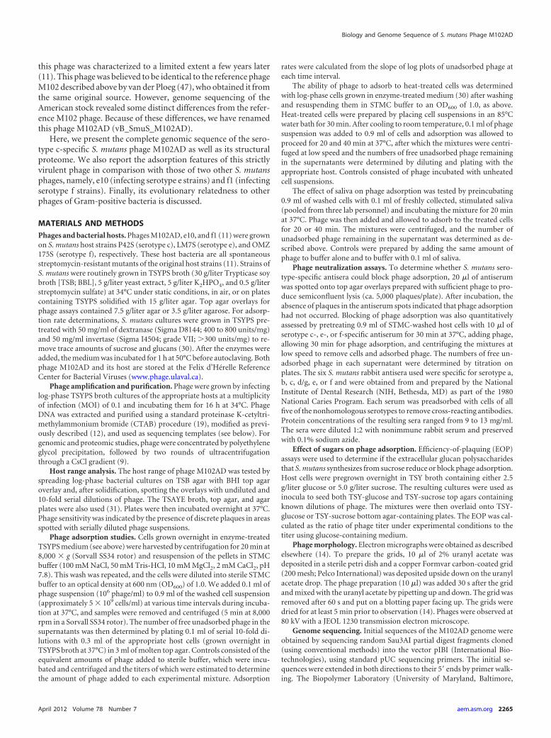
Phage morphology. Electron microscopy confirmed thatphage M102AD belongs to the Siphoviridae family with B1 mor-phology (Fig. 1). The diameter of its capsid was estimated at 67 �0.4 nm, while the length of its noncontractile tail was estimated at283 � 7 nm with a width of 8.3 � 0.1 nm. These dimensions are inagreement with those observed for phage M102 (47).
Host range. Previous tests suggested that the host range ofphage M102AD was limited to occasional serotype c strains of S.mutans (11). Here, the lytic spectrum of this phage was tested on25 additional S. mutans strains: CM7, ATCC 202022, 7A, 123.1, Q,59.1, UA96, 29B, T9, T8, N, 87.1, 5A, ATCC 25175, 11A, 136.1, JB,3B, Ny257, 22A, F, NG003, T7B, and JH 1140 (31) and UA159 (2).Only strain F was sensitive to phage M102AD, confirming its nar-row host range.
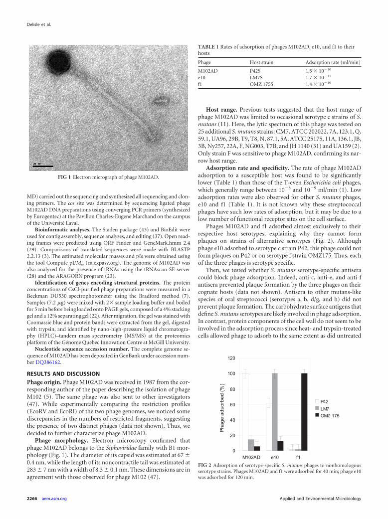
Adsorption rate and specificity. The rate of phage M102ADadsorption to a susceptible host was found to be significantlylower (Table 1) than those of the T-even Escherichia coli phages,which generally range between 10�8 and 10�9 ml/min (1). Lowadsorption rates were also observed for other S. mutans phages,e10 and f1 (Table 1). It is not known why these streptococcalphages have such low rates of adsorption, but it may be due to alow number of functional receptor sites on the cell surface.
Phages M102AD and f1 adsorbed almost exclusively to theirrespective host serotypes, explaining why they cannot formplaques on strains of alternative serotypes (Fig. 2). Althoughphage e10 adsorbed to serotype c strain P42, this phage could notform plaques on P42 or on serotype f strain OMZ175. Thus, eachof the three phages is serotype specific.
Then, we tested whether S. mutans serotype-specific antiseracould block phage adsorption. Indeed, anti-c, anti-e, and anti-fantisera prevented plaque formation by the three phages on theircognate hosts (data not shown). Antisera to other mutans-likespecies of oral streptococci (serotypes a, b, d/g, and h) did notprevent plaque formation. The carbohydrate surface antigens thatdefine S. mutans serotypes are likely involved in phage adsorption.In contrast, protein components of the cell wall do not seem to beinvolved in the adsorption process since heat- and trypsin-treatedcells allowed phage to adsorb to the same extent as did untreated
FIG 1 Electron micrograph of phage M102AD.
TABLE 1 Rates of adsorption of phages M102AD, e10, and f1 to theirhosts
Phage Host strain Adsorption rate (ml/min)
M102AD P42S 1.5 � 10�10
e10 LM7S 1.7 � 10�11
f1 OMZ 175S 1.4 � 10�10
FIG 2 Adsorption of serotype-specific S. mutans phages to nonhomologousserotype strains. Phages M102AD and f1 were adsorbed for 40 min; phage e10was adsorbed for 120 min.
Delisle et al.
2266 aem.asm.org Applied and Environmental Microbiology
cells (data not shown). Finally, the lack of inhibition of phageadsorption by saliva indicates that salivary components are notlikely to interfere with phage infection and replication in the oralcavity.
Finally, as the data shown in Table 2 indicate, the unique su-crose-derived glucan polymers produced by S. mutans do not sig-nificantly interfere with phage adsorption or replication. Thesedata support the view that the production of polysaccharides doesnot confer a significant phage resistance phenotype (8, 13, 45).
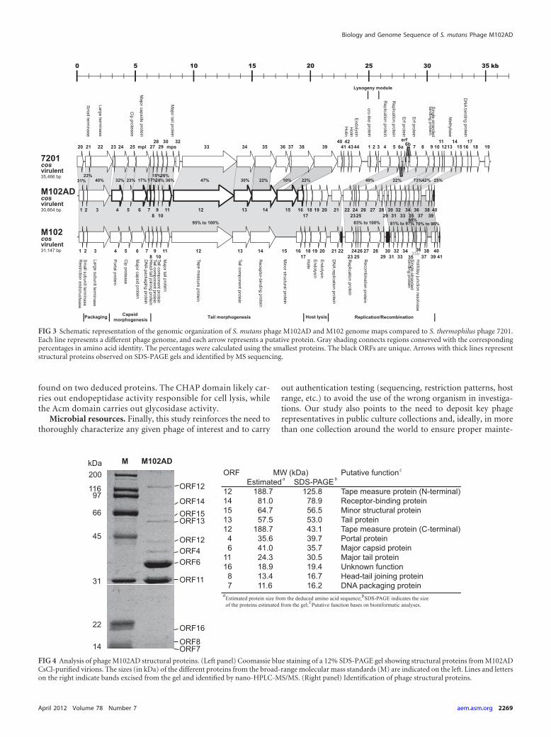
Complete nucleotide sequence. The linear genome of phageM102AD contains 30,664 bp with an average G�C content of39.6% and has 3=-overhanging cos sites (5=-CCGCGTGAATA-3=)of 11 nucleotides. The genome of phage M102AD is slightlysmaller than that of phage M102 (31,147 bp) while the cos sites areidentical (47). To our knowledge, M102 and M102AD are the onlyS. mutans phage genomes available. All of the 40 ORFs of phageM102AD were identified on the same strand, and they started withATG, TTG, or GTG initiation codons. Of the 40 ORFs, 23 could beassigned a putative function. Moreover, every ORF was precededby a region with similarities to the Shine-Dalgarno sequence, AGAAAGGAGG, which is complementary to the 10 nucleotides ofthe 3= end of the 16S rRNA of S. mutans. Two to 11 nucleotidesseparate the Shine-Dalgarno sequence from the initiation codon(Table 3). No tRNA was found in the genome of phage M102AD.Two significant intergenic regions were observed between orf37and orf38 (385 bp) as well as between orf20 and orf21 (290 nt).
Comparison with the M102 phage genome. The M102 andM102AD genomes share 90.8% identity at the nucleotide level andthus are, as expected, highly related. Nonetheless, phage M102ADhas one small extra ORF (orf40) not present in phage M102 whileM102 also has three small supplementary ORFs (orf22, orf30, andorf41). For these four cases, it appears that two genes (orf22 andorf30) were acquired in the genome of phage M102 (or deleted inM102AD) while the other two ORFs (orf40/M102AD and orf41/M102) are phage specific, as they are located at the same genomiclocation. Interestingly, only four deduced ORFs share 100% iden-tity (ORF18/holin, ORF19/endolysin, ORF20/endolysin, andORF25/unknown function) at the amino acid level (Table 3).Overall, 32 ORFs (out of 40) from phage M102AD shared over90% identity with S. mutans phage M102. Only ORF40 had nohomology with a known protein, and thus, its origin is unknown(Table 3).
On the other hand, orf34 of the M102AD genome (Table 3; Fig.3) matches with a portion of the M102 genome that was not pre-viously identified as an open reading frame (47). Upon furtheranalyses, a new ORF (named orf35b) was added to the phage M102genome, bringing its total to 42 ORFs compared to the 40 ORFs ofphage M102AD. The position of orf35b on the genome of M102 isbetween bp 28827 and 29099, and it would encode a protein of 90amino acids (aa). ORF34 of phage M102AD has 88% identity(79/90 amino acids) with ORF35b of phage M102, but its functionis unknown.
Overall, the degree of divergence between two phages (M102and M102AD) derived from the same laboratory is striking. Therelatively recent diversification of the phage M102AD genome wasdue mainly to point mutations as well as gene deletion and addi-tions (4, 20). At this time, it is unclear why and when these eventsoccurred due to the lack of precise documentation on the han-dling and serial amplification of M102AD since the late 1980s.
Comparative analysis with Streptococcus thermophilus phages.Twenty ORFs of phage M102AD also share identity with ORFs ofsome Streptococcus thermophilus phages, namely, those of the cos-type group (44). A genomic relationship between S. mutans phageM102 and S. thermophilus phage DT1 (46) was previously ob-served (47), although here we found phage M102AD to be closerto S. thermophilus phage 7201. Interestingly, ORF33 has a 73%amino acid match with ORF9 of 7201, which appears to be a sin-gle-stranded binding protein (38).
It has been demonstrated elsewhere that S. thermophilus phagescan be separated into at least two different clusters based on themode of packaging and the number of major structural proteins(24). S. thermophilus cos-type phages, such as phages 7201 andDT1, have cohesive genomic extremities and two major structuralproteins. Interestingly, the genomic sequence of S. mutans phageM102AD has a 3=-overhanging cos site, and the analysis of itsstructural proteins clearly identified two major structural pro-teins, namely, ORF6 (major capsid protein) and ORF11 (majortail protein) (Fig. 4).
Moreover, 9 out of 10 structural proteins of phage M102ADidentified by nano-HPLC-MS/MS were similar to structural pro-teins found in cos-type S. thermophilus phages such as phage DT1(46). Many genes coding for these conserved ORF proteins arepart of the core genome of S. thermophilus phages (36). Almost allthe detected phage M102AD proteins had approximately the ex-pected size, except for orf12, coding for the tape measure protein(TMP). The TMP, which is responsible for determining taillength, was found in two separate bands (Fig. 4). These bandscorresponded to the N-terminal and C-terminal parts of this pro-tein. The tape measure protein, which is very similar to that of theS. thermophilus phage MD2 (16), has two domains, namely, aCHAP (cysteine, histidine-dependent aminohydrolase/peptidase)domain and an SLT (soluble lytic transglycosylase) domain. Theymay be involved, after the phage adsorption process, in locallydegrading the peptidoglycan to allow DNA entry into the cell.Finally, the putative receptor binding protein (RBP/ORF14) ofphage M102AD has similarities at the amino acid level with twoORFs of phage 7201 (ORF35 and ORF38) and may reflect themodular organization of RBPs (25, 42).
At this time, it is unclear why S. mutans phages are closelyrelated to S. thermophilus phages, at least from a structural per-spective. The latest phylogenetic analyses of streptococci still clas-sify S. thermophilus, which is not a natural inhabitant of the hu-
TABLE 2 Effect of sucrose-derived surface polysaccharides on the EOPsof S. mutans phages
Phage HostSugar in liquidmedium
Sugar inplatingmedium EOP
M102AD P42S Glucose Glucose 1.0M102AD P42S Glucose Sucrose 0.6M102AD P42S Sucrose Sucrose 0.6M102AD P42S Sucrose Glucose 0.3e10 LM7S Glucose Glucose 1.0e10 LM7S Glucose Sucrose 0.7e10 LM7S Sucrose Sucrose 0.2e10 LM7S Sucrose Glucose 0.2f1 OMZ 175S Glucose Glucose 1.0f1 OMZ 175S Glucose Sucrose 0.3f1 OMZ 175S Sucrose Sucrose 0.8f1 OMZ 175S Sucrose Glucose 0.7
Biology and Genome Sequence of S. mutans Phage M102AD
April 2012 Volume 78 Number 7 aem.asm.org 2267
man oral mucosa, into the salivarius group with Streptococcussalivarius and Streptococcus vestibularis (33). Clearly, other strep-tococcal phage genomic sequences and the associated analysis ofgene functions are needed to get further insight into their evolu-tion.
Comparison with endolysins of other streptococcal phages.As indicated above, the lysis module is 100% conserved be-tween M102 (47) and M102AD. The M102AD phage has twoputative endolysins encoded by orf19 and orf20. ORF19 con-
tains the Acm (1,4-�-N-acetylmuramidase) domain, which be-longs to the glycosyl hydrolase 25 family (27). This 273-aa ORFcorresponds to amino acids 148 through 366 of the 443 aa ofthe Streptococcus agalactiae B30 phage endolysin (15). ORF20has a CHAP domain of 171 aa that is similar to amino acids 3 to138 of the 250 aa of the Streptococcus pyogenes MGAS10394phage endolysin (6). Usually, the CHAP domain can be foundat the N-terminal end of the endolysin and the Acm domaincan be found at the C-terminal end. Here, both domains were
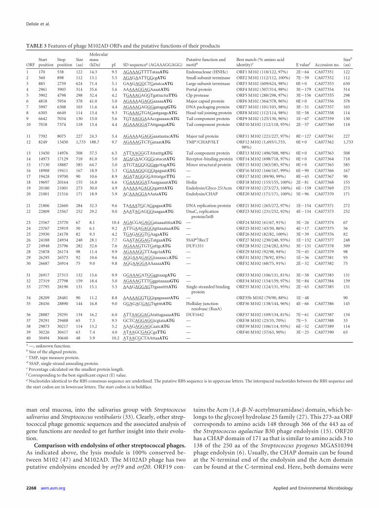
TABLE 3 Features of phage M102AD ORFs and the putative functions of their products
ORFStartposition
Stopposition
Size(aa)
Molecularmass(kDa) pI SD sequenceg (AGAAAGGAGG)
Putative function andmotifa
Best match (% amino acididentity)e E valuef Accession no.
Sizeb
(aa)
1 170 538 122 14.3 9.5 AGAAAGTTTTataaATG Endonuclease (HNHc) ORF1 M102 (118/122, 97%) 2E�64 CA077351 1222 560 898 112 13.1 5.5 AGAGAATTGGcgATG Small subunit terminase ORF2 M102 (112/112, 100%) 7E�59 CA077352 1123 885 2759 624 71.4 5.1 GAAGAGGCTGatatcaATG Large subunit terminase ORF3 M102 (609/624, 98%) 0E�0 CA077353 6304 2961 3905 314 35.6 5.6 AAAAAGGAGAaaatATG Portal protein ORF4 M102 (307/314, 98%) 3E�179 CA077354 3145 3902 4798 298 32.4 4.2 TGAAAGAGGTgattacttaTTG Clp protease ORF5 M102 (288/298, 97%) 3E�156 CA077355 2986 4818 5954 378 41.0 5.0 AGAAAAGAGGaaaaaATG Major capsid protein ORF6 M102 (364/378, 96%) 0E�0 CA077356 3787 5997 6308 103 11.6 4.4 AGAAAGAGGGgatagagGTG DNA packaging protein ORF7 M102 (101/103, 98%) 3E�51 CA077357 1038 6305 6649 114 13.4 9.3 TGAAAGTGAGaatgaagcATG Head-tail joining protein ORF8 M102 (112/114, 98%) 5E�58 CA077358 1149 6642 7034 130 15.0 5.6 TGTAAGGAAAccgaaaaacATG Tail component protein ORF9 M102 (125/130, 96%) 1E�67 CA077359 13010 7018 7374 118 13.4 4.4 AGAAAGGATAttgaagaATG Tail component protein ORF10 M102 (112/118, 95%) 2E�57 CA077360 118
11 7392 8075 227 24.3 5.4 AGAAAAGAGGaaataatacATG Major tail protein ORF11 M102 (221/227, 97%) 8E�127 CA077361 22712 8249 13450 1,733 188.7 9.7 AGAAAGTCTGataatATG TMPc/CHAP/SLT ORF12 M102 (1,693/1,733,
98%)0E�0 CA077362 1,733
13 13450 14976 508 57.5 6.5 ATTAAGGGTAttatttgATG Tail component protein ORF13 M102 (496/508, 98%) 0E�0 CA077363 50814 14973 17129 718 81.0 5.0 AGAGAACGGGcatacatATG Receptor-binding protein ORF14 M102 (698/718, 97%) 0E�0 CA077364 71815 17130 18887 585 64.7 5.0 ATGTAGGGGGggcttagATG Minor structural protein ORF15 M102 (565/585, 97%) 0E�0 CA077365 58516 18908 19411 167 18.9 5.3 CGAAAGGGGGtgagaatATG — ORF16 M102 (166/167, 99%) 6E�90 CA077366 16717 19428 19700 90 10.6 8.9 AGATAGGGGAtttatgatTTG — ORF17 M102 (89/90, 99%) 8E�43 CA077367 9018 19697 20164 155 16.8 6.6 CGAAAGGATAaagaaaatATG Holin ORF18 M102 (155/155, 100%) 2E�81 CA077368 15519 20180 21001 273 30.0 4.9 AAAAAAGAGGtgattttATG Endolysin/Glyco 25/Acm ORF19 M102 (273/273, 100%) 6E�159 CA077369 27320 21001 21516 171 18.9 5.9 ACAAAGGAAAataATG Endolysin/CHAP ORF20 M102 (171/171, 100%) 5E�96 CA077370 171
21 21806 22660 284 32.3 9.6 TAAAATGCAGgagaaATG DNA replication protein ORF21 M102 (265/272, 97%) 1E�154 CA077371 27222 22809 23567 252 29.2 9.0 AAATAGAGGGtaagaaATG DnaC, replication
protein/IstBORF23 M102 (231/252, 92%) 4E�134 CA077373 252
23 23567 23770 67 8.1 10.4 AGACGAGAGGataaaattttaATG — ORF24 M102 (61/67, 91%) 3E�26 CA077374 6724 23767 23919 50 6.1 9.2 ATTGAAGAGGtgttaaataaATG — ORF25 M102 (43/50, 86%) 4E�17 CA077375 5625 23930 24178 82 9.3 4.2 TGAGAGGTGAagaATG — ORF26 M102 (82/82, 100%) 3E�39 CA077376 8226 24188 24934 248 28.1 7.7 GAATAGGAGTatgaaATG SSAPd/RecT ORF27 M102 (230/248, 93%) 1E�132 CA077377 24827 24948 25796 282 32.6 7.6 AGAAAGTGTGattgcATG DUF1351 ORF28 M102 (234/282, 83%) 3E�131 CA077378 30928 25878 26174 98 11.4 9.9 AGAAAGGTTAagctaATG — ORF29 M102 (92/98, 94%) 7E�45 CA077379 9829 26295 26573 92 10.6 9.6 AGGAAAGAGGtaaaaaccATG — ORF31 M102 (78/92, 85%) 1E�36 CA077381 9330 26687 26914 75 9.0 9.8 AGGAAGGAAAaaaaATG — ORF32 M102 (68/75, 91%) 2E�32 CA077382 75
31 26917 27315 132 15.6 9.9 GGAAAGATGGggtaaagATG — ORF33 M102 (106/131, 81%) 3E�58 CA077383 13132 27319 27798 159 18.4 5.0 AGAAAGTTTGggataaaaaGTG — ORF34 M102 (154/159, 97%) 5E�84 CA077384 15933 27795 28190 131 15.1 5.5 AAAGAGGAGTtgaaattttATG Single-stranded binding
proteinORF35 M102 (124/131, 95%) 2E�65 CA077385 131
34 28209 28481 90 11.2 8.8 AAAAAGGTGGtgagaaaatATG — ORF35b M102 (79/90, 88%) 1E�48 9035 28456 28890 144 16.8 9.0 GGAGACGAGTtgttttATG Holliday junction
resolvase (RusA)ORF36 M102 (138/144, 96%) 4E�66 CA077386 145
36 28887 29291 134 16.2 6.0 ATTAAGGAGAtattagaaaaATG DUF1642 ORF37 M102 (109/134, 81%) 7E�61 CA077387 13437 29291 29488 65 7.3 9.5 GCTCAGGAGGtcgtataATG — ORF38 M102 (23/33, 70%) 7E�5 CA077388 3338 29873 30217 114 13.2 5.2 AAAGAGGAGCaatcATG — ORF39 M102 (106/114, 93%) 6E�52 CA077389 11439 30226 30417 63 7.4 4.0 ATAAGCGAGCgaTTG — ORF40 M102 (57/63, 90%) 3E�25 CA077390 6340 30494 30640 48 5.9 10.2 ATAACGCTAAttaaATG —
a —, unknown function.b Size of the aligned protein.c TMP, tape measure protein.d SSAP, single-strand annealing protein.e Percentage calculated on the smallest protein length.f Corresponding to the best significant expect (E) value.g Nucleotides identical to the RBS consensus sequence are underlined. The putative RBS sequence is in uppercase letters. The interspaced nucleotides between the RBS sequence andthe start codon are in lowercase letters. The start codon is in boldface.
Delisle et al.
2268 aem.asm.org Applied and Environmental Microbiology
found on two deduced proteins. The CHAP domain likely car-ries out endopeptidase activity responsible for cell lysis, whilethe Acm domain carries out glycosidase activity.
Microbial resources. Finally, this study reinforces the need tothoroughly characterize any given phage of interest and to carry
out authentication testing (sequencing, restriction patterns, hostrange, etc.) to avoid the use of the wrong organism in investiga-tions. Our study also points to the need to deposit key phagerepresentatives in public culture collections and, ideally, in morethan one collection around the world to ensure proper mainte-
FIG 3 Schematic representation of the genomic organization of S. mutans phage M102AD and M102 genome maps compared to S. thermophilus phage 7201.Each line represents a different phage genome, and each arrow represents a putative protein. Gray shading connects regions conserved with the correspondingpercentages in amino acid identity. The percentages were calculated using the smallest proteins. The black ORFs are unique. Arrows with thick lines representstructural proteins observed on SDS-PAGE gels and identified by MS sequencing.
FIG 4 Analysis of phage M102AD structural proteins. (Left panel) Coomassie blue staining of a 12% SDS-PAGE gel showing structural proteins from M102ADCsCl-purified virions. The sizes (in kDa) of the different proteins from the broad-range molecular mass standards (M) are indicated on the left. Lines and letterson the right indicate bands excised from the gel and identified by nano-HPLC-MS/MS. (Right panel) Identification of phage structural proteins.
Biology and Genome Sequence of S. mutans Phage M102AD
April 2012 Volume 78 Number 7 aem.asm.org 2269
nance and continued access to them for future studies (14). Toooften, biological materials are either lost or mishandled due to theretirement of principal investigators. A well-ordered detailed doc-umentation system should also be maintained.
ACKNOWLEDGMENTS
We thank B.-A. Conway for editorial assistance; C. Levesque for suggestions;R. Young for helpful discussions; and B. Burall, F. Williams, and B. Saville forperforming the phage adsorption and EOP experiments as well as D. Trem-blay for the electron micrograph. We also thank G. Nicholas and M. Lavoiefor providing additional strains of S. mutans for host range testing.
A.L.D. acknowledges the financial support of the NIDCR, NIH, forsupport of the cloning and sequencing experiments conducted at UM(grant DE13181). S.M. is grateful to the Canadian Institutes of HealthResearch (Team Grant-Emerging: Novel Alternatives to Antibiotics) forfinancial support of the sequencing, protein, and computer analyses con-ducted at UL.
REFERENCES1. Adams MH. 1959. Bacteriophages. Interscience Publishing, Inc, New
York, NY.2. Ajdic D, et al. 2002. Genome sequence of Streptococcus mutans UA159, a
cariogenic dental pathogen. Proc. Natl. Acad. Sci. U. S. A. 99:14434 –14439.
3. Altschul SF, et al. 1997. Gapped BLAST and PSI-BLAST: a new genera-tion of protein database search programs. Nucleic Acids Res. 25:3389 –3402.
4. Arber W. 2003. Elements for a theory of molecular evolution. Gene 317:3–11.
5. Armau E, Bousque JL, Boue D, Tiraby G. 1988. Isolation of lytic bacte-riophages for Streptococcus mutans and Streptococcus sobrinus. J. Dent. Res.67:121.
6. Banks DJ, et al. 2004. Progress toward characterization of the group AStreptococcus metagenome: complete genome sequence of a macrolide-resistant serotype M6 strain. J. Infect. Dis. 190:727–738.
7. Bradford MM. 1976. A rapid and sensitive method for the quantitation ofmicrogram quantities of protein utilizing the principle of protein-dyebinding. Anal. Biochem. 72:248 –254.
8. Broadbent JR, McMahon DJ, Welker DL, Oberg CJ, Moineau S. 2003.Biochemistry, genetics, and applications of exopolysaccharide productionin Streptococcus thermophilus: a review. J. Dairy Sci. 86:407– 423.
9. Chibani Azaïez SR, Fliss I, Simard RE, Moineau S. 1998. Monoclonalantibodies raised against native major capsid proteins of lactococcal c2-like bacteriophages. Appl. Environ. Microbiol. 64:4255– 4259.
10. Coykendall AL. 1989. Classification and identification of the viridansstreptococci. Clin. Microbiol. Rev. 2:315–328.
11. Delisle AL, Rostkowski CA. 1993. Lytic bacteriophages of Streptoccoccusmutans. Curr. Microbiol. 27:163–167.
12. Delisle AL, Barcak GJ, Guo M. 2006. Isolation and expression of the lysisgenes of Actinomyces naeslundii phage Av-1. Appl. Environ. Microbiol.72:1110 –1117.
13. Deveau H, van Calsteren M-R, Moineau S. 2002. The effect of exopoly-saccharides on phage-host interactions in Lactococcus lactis. Appl. Envi-ron. Microbiol. 68:4364 – 4369.
14. Deveau H, Labrie SJ, Chopin M-C, Moineau S. 2006. Biodiversity andclassification of lactococcal phages. Appl. Environ. Microbiol. 72:4338 –4346.
15. Donovan DM, et al. 2006. The cell lysis activity of the endolysin from theStreptococcus agalactiae bacteriophage B30 relies on the CHAP endopep-tidase domain. Appl. Environ. Microbiol. 72:5108 –5112.
16. Duplessis M, Lévesque CM, Moineau S. 2006. Characterization of Strep-tococcus thermophilus host range phage mutants. Appl. Environ. Micro-biol. 72:3036 –3041.
17. Fischetti VA. 2010. Bacteriophage endolysins: a novel anti-infective tocontrol Gram-positive pathogens. Int. J. Med. Microbiol. 300:357–362.
18. Hitch G, Pratten J, Taylor PW. 2004. Isolation of bacteriophages fromthe oral cavity. Lett. Appl. Microbiol. 39:215–219.
19. Kricker M, Carlson K. 1994. Isolation of T4 phage DNA, p 455– 456. InKaram JD, et al (ed), Molecular biology of bacteriophage T4. ASM Press,Washington, DC.
20. Labrie SJ, Moineau S. 2007. Abortive infection mechanisms and pro-phage sequences significantly influence the genetic make-up of emerginglytic lactococcal phages. J. Bacteriol. 189:1482–1487.
21. Labrie SJ, Samson JE, Moineau S. 2010. Bacteriophage resistance mech-anisms. Nat. Rev. Microbiol. 8:317–327.
22. Laemmli UK. 1970. Cleavage of structural proteins during the assembly ofthe head of bacteriophage T4. Nature 227:680 – 685.
23. Laslett D, Canback B. 2004. ARAGORN, a program to detect tRNA genesand tmRNA genes in nucleotide sequences. Nucleic Acids Res. 32:11–16.
24. Le Marrec C, et al. 1997. Two groups of bacteriophages infecting Strep-tococcus thermophilus can be distinguished on the basis of mode of pack-aging and genetic determinants for major structural proteins. Appl. Envi-ron. Microbiol. 63:3246 –3253.
25. Lévesque C, et al. 2005. Genomic organization and molecular analysis ofvirulent bacteriophage 2972 infecting an exopolysaccharide-producingStreptococcus thermophilus strain. Appl. Environ. Microbiol. 71:4057–4068.
26. Loesche WJ. 1986. Role of Streptococcus mutans in human dental decay.Microbiol. Rev. 50:353–380.
27. López R, García E. 2004. Recent trends on the molecular biology ofpneumococcal capsules, lytic enzymes, and bacteriophage. FEMS Micro-biol. Rev. 28:553–580.
28. Lowe TM, Eddy SR. 1997. tRNAscan-SE: a program for improved detec-tion of transfer RNA genes in genomic sequence. Nucleic Acids Res. 25:955–964.
29. Lukashin AV, Borodovsky M. 1998. GenMark.hmm: new solutions forgene finding. Nucleic Acids Res. 26:1107–1115.
30. Lü-Lü, et al. 1992. Chelating agents inhibit activity and prevent expression ofstreptococcal glucan-binding lectins. Infect. Immun. 60:3807–3813.
31. Morency H, Mota-Meira M, Lapointe G, Lacroix C, Lavoie MC. 2001.Comparison of the activity spectra against pathogens of bacterial strainsproducing a mutacin or a lantibiotic. Can. J. Microbiol. 47:322–331.
32. Nakano K, Nomura R, Nakagawa I, Hamada S, Ooshima T. 2004.Demonstration of Streptococcus mutans with a cell wall polysaccharidespecific to a new serotype, k, in the human oral cavity. J. Clin. Microbiol.42:198 –202.
33. Pombert J-F, Sistek V, Boissinot M, Frenette M. 2009. Evolutionaryrelationships among salivarius streptococci as inferred from multilocusphylogenies based on 16S rRNA-encoding, recA, secA, and secY gene se-quences. BMC Microbiol. 9:232.
34. Pride DT, et al. 2011. Analysis of streptococcal CRISPRs from humansaliva reveals substantial sequence diversity within and between subjectsover time. Genome Res. 21:126 –136.
35. Pritchard DG, Gregory RL, Michalek SM, McGhee JR. 1986. Biochem-ical aspects of serotype carbohydrate antigens of Streptococcus mutans, p39 – 49. In Hamada S, et al (ed), Molecular microbiology and immunologyof Streptococcus mutans. Elsevier, Amsterdam, Netherlands.
36. Quiberoni A, Moineau S, Rousseau GM, Reinheimer J, AckermannH-W. 2010. Review: Streptococcus thermophilus bacteriophages. Int. DairyJ. 20:657– 664.
37. Rousseau GM, Moineau S. 2009. Evolution of Lactococcus lactis phageswithin a cheese factory. Appl. Environ. Microbiol. 75:5336 –5344.
38. Scaltriti E, et al. 2009. Structure and function of phage p2 ORF34p2, a newtype of single stranded DNA binding protein. Mol. Microbiol. 73:1156 –1170.
39. Schilling KM, Bowen WH. 1992. Glucans synthesized in situ in experi-mental salivary pellicle function as specific binding sites for Streptococcusmutans. Infect. Immun. 60:284 –295.
40. Schleifer KH, Kilpper-Bälz R, Kraus J, Gehring F. 1984. Relatedness andclassification of Streptococcus mutans and “mutans-like” streptococci. J.Dent. Res. 63:1047–1050.
41. Shibata Y, Yamashita Y, van der Ploeg JR. 2009. The serotype-specificglucose side chain of rhamnose-glucose polysaccharides is essential foradsorption of bacteriophage M102 to Streptococcus mutans. FEMS Micro-biol. Lett. 294:68 –73.
42. Spinelli S, et al. 2006. Lactococcal bacteriophage p2 receptor bindingprotein structure suggests a common ancestor gene with bacterial andmammalian viruses. Nat. Struct. Mol. Biol. 13:85– 89.
43. Staden R. 1996. The Staden sequence analysis package. Mol. Biotechnol.5:233–241.
44. Stanley E, Walsh L, van der Zwet A, Fitzgerald GF, van Sinderen D.
Delisle et al.
2270 aem.asm.org Applied and Environmental Microbiology
2000. Identification of four loci isolated from two Streptococcus thermo-philus phage genomes responsible for mediating bacteriophage resistance.FEMS Microbiol. Lett. 182:271–277.
45. Sutherland IW, Hughes KA, Skillman LC, Tait K. 2004. The interactionof phage and biofilms. FEMS Microbiol. Lett. 232:1– 6.
46. Tremblay DM, Moineau S. 1999. Complete genomic sequence of the lyticbacteriophage DT1 of Streptococcus thermophilus. Virology 255:63–76.
47. van der Ploeg JR. 2007. Genome sequence of Streptococcus mutans bac-teriophage M102. FEMS Microbiol. Lett. 275:130 –138.
48. van der Ploeg JR. 2009. Analysis of CRISPR in Streptococcus mutanssuggests frequent occurrence of acquired immunity against infection byM102-like bacteriophages. Microbiology 155:1966 –1976.
49. Whiley RA, Beighton D. 1998. Current classification of the oral strepto-cocci. Oral Microbiol. Immunol. 13:195–216.
Biology and Genome Sequence of S. mutans Phage M102AD
April 2012 Volume 78 Number 7 aem.asm.org 2271
Copyright © 2022 FDOKUMEN