Genetic and Molecular Analysis of GogB, a Phage-encoded Type III-secreted Substrate in Salmonella...
14
Genetic and Molecular Analysis of GogB, a Phage- encoded Type III-secreted Substrate in Salmonella enterica Serovar Typhimurium with Autonomous Expression from its Associated Phage Brian K. Coombes 1 , Mark E. Wickham 1 , Nat. F. Brown 1 Sebastien Lemire 2 , Lionello Bossi 2 , William W. L. Hsiao 3 Fiona S. L. Brinkman 3 and B. Brett Finlay 1,4,5 * 1 Michael Smith Laboratories University of British Columbia Vancouver, BC, Canada V6T 1Z3 2 Centre de Ge ´ne ´tique Mole ´culaire, Centre National de la Recherche Scientifique 91198 France 3 Department of Molecular Biology and Biochemistry Simon Fraser University Burnaby, BC, Canada V5A 1S6 4 Department of Biochemistry and Molecular Biology University of British Columbia Vancouver, BC, Canada V6T 1Z3 5 Department of Microbiology and Immunology, University of British Columbia, Vancouver BC, Canada V6T 1Z3 Salmonella enterica serovar Typhimurium is lysogenized by several temperate bacteriophages that encode lysogenic conversion genes, which can act as virulence factors during infection and contribute to the genetic diversity and pathogenic potential of the lysogen. We have investigated the temperate bacteriophage called Gifsy-1 in S. enterica serovar Typhimurium and show here that the product of the gogB gene encoded within this phage shares similarity with proteins from other Gram-negative pathogens. The amino-terminal portion of GogB shares similarity with leucine-rich repeat-containing virulence-associated proteins from other Gram-negative pathogens, whereas the carboxyl-terminal portion of GogB shares similarity with uncharacterized proteins in other pathogens. We show that GogB is secreted by both type III secretion systems encoded in Salmonella Pathogenicity Island-1 (SPI-1) and SPI-2 but translocation into host cells is a SPI-2-mediated process. Once translocated, GogB localizes to the cytoplasm of infected host cells. The genetic regulation of gogB in Salmonella is influenced by the transcriptional activator, SsrB, under SPI-2- inducing conditions, but the modular nature of the gogB gene allows for autonomous expression and type III secretion following horizontal gene transfer into a heterologous pathogen. These data define the first autonomously expressed lysogenic conversion gene within Gifsy-1 that acts as a modular and promiscuous type III-secreted substrate of the infection process. q 2005 Elsevier Ltd. All rights reserved. Keywords: Salmonella; Gifsy; lambdoid phage; GogB; type III secretion *Corresponding author Introduction Salmonella enterica are facultative intracellular Gram-negative bacteria that are important enteric pathogens for humans and commercial livestock. Infection with these organisms causes a self-limit- ing enterocolitis that can progress to a serious systemic disease in ruminants and humans called enteric fever. The infecting S. enterica serovar and the host species influence systemic disease pro- gression. For example, serovar Typhi is a human host-restricted pathogen, whereas serovar Typhi- murium exhibits a wide host range including humans, mice, cattle and chickens. Two major virulence determinants that contribute to Salmonella pathogenesis are encoded in separate chromosomal pathogenicity islands called Salmonella Patho- genicity Island: SPI-1 1 and SPI-2. 2,3 Each encodes a suite of molecules that assemble into a type III secretion system that can translocate effector pro- teins directly from the bacterial cell into target host cells. Effector proteins translocated by the SPI-1 type III secretion system influence early cyto- skeletal and membrane rearrangements involved 0022-2836/$ - see front matter q 2005 Elsevier Ltd. All rights reserved. Abbreviations used: SPI-1, Salmonella Pathogenicity Island-1; SARC11, Salmonella Reference Collection C-11; LRR, leucine-rich repeat; HMM, hidden Markov model; LEE, locus of enterocyte effacement; FRT, FLP recognition target; RLU, relative light units. E-mail address of the corresponding author: bfi[email protected] doi:10.1016/j.jmb.2005.03.024 J. Mol. Biol. (2005) 348, 817–830
Transcript of Genetic and Molecular Analysis of GogB, a Phage-encoded Type III-secreted Substrate in Salmonella...

doi:10.1016/j.jmb.2005.03.024 J. Mol. Biol. (2005) 348, 817–830
Genetic and Molecular Analysis of GogB, a Phage-encoded Type III-secreted Substrate in Salmonellaenterica Serovar Typhimurium with AutonomousExpression from its Associated Phage
Brian K. Coombes1, Mark E. Wickham1, Nat. F. Brown1
Sebastien Lemire2, Lionello Bossi2, William W. L. Hsiao3
Fiona S. L. Brinkman3 and B. Brett Finlay1,4,5*
1Michael Smith LaboratoriesUniversity of British ColumbiaVancouver, BC, CanadaV6T 1Z3
2Centre de GenetiqueMoleculaire, Centre National dela Recherche Scientifique91198 France
3Department of MolecularBiology and BiochemistrySimon Fraser UniversityBurnaby, BC, Canada V5A 1S6
4Department of Biochemistryand Molecular BiologyUniversity of British ColumbiaVancouver, BC, CanadaV6T 1Z3
5Department of Microbiologyand Immunology, University ofBritish Columbia, VancouverBC, Canada V6T 1Z3
0022-2836/$ - see front matter q 2005 E
Abbreviations used: SPI-1, SalmonIsland-1; SARC11, Salmonella RefereLRR, leucine-rich repeat; HMM, hidLEE, locus of enterocyte effacement;target; RLU, relative light units.E-mail address of the correspond
Salmonella enterica serovar Typhimurium is lysogenized by severaltemperate bacteriophages that encode lysogenic conversion genes, whichcan act as virulence factors during infection and contribute to the geneticdiversity and pathogenic potential of the lysogen. We have investigated thetemperate bacteriophage called Gifsy-1 in S. enterica serovar Typhimuriumand show here that the product of the gogB gene encoded within this phageshares similarity with proteins from other Gram-negative pathogens.The amino-terminal portion of GogB shares similarity with leucine-richrepeat-containing virulence-associated proteins from other Gram-negativepathogens, whereas the carboxyl-terminal portion of GogB sharessimilarity with uncharacterized proteins in other pathogens. We showthat GogB is secreted by both type III secretion systems encoded inSalmonella Pathogenicity Island-1 (SPI-1) and SPI-2 but translocation intohost cells is a SPI-2-mediated process. Once translocated, GogB localizes tothe cytoplasm of infected host cells. The genetic regulation of gogB inSalmonella is influenced by the transcriptional activator, SsrB, under SPI-2-inducing conditions, but the modular nature of the gogB gene allows forautonomous expression and type III secretion following horizontal genetransfer into a heterologous pathogen. These data define the firstautonomously expressed lysogenic conversion gene within Gifsy-1 thatacts as a modular and promiscuous type III-secreted substrate of theinfection process.
q 2005 Elsevier Ltd. All rights reserved.
Keywords: Salmonella; Gifsy; lambdoid phage; GogB; type III secretion
*Corresponding authorIntroduction
Salmonella enterica are facultative intracellularGram-negative bacteria that are important entericpathogens for humans and commercial livestock.Infection with these organisms causes a self-limit-ing enterocolitis that can progress to a serioussystemic disease in ruminants and humans called
lsevier Ltd. All rights reserve
ella Pathogenicitynce Collection C-11;den Markov model;FRT, FLP recognition
ing author:
enteric fever. The infecting S. enterica serovar andthe host species influence systemic disease pro-gression. For example, serovar Typhi is a humanhost-restricted pathogen, whereas serovar Typhi-murium exhibits a wide host range includinghumans, mice, cattle and chickens. Two majorvirulence determinants that contribute to Salmonellapathogenesis are encoded in separate chromosomalpathogenicity islands called Salmonella Patho-genicity Island: SPI-11 and SPI-2.2,3 Each encodes asuite of molecules that assemble into a type IIIsecretion system that can translocate effector pro-teins directly from the bacterial cell into target hostcells. Effector proteins translocated by the SPI-1type III secretion system influence early cyto-skeletal and membrane rearrangements involved
d.
818 Type III Secretion of GogB
in bacterial uptake into target cells,4 whereas SPI-2is generally thought to play a role during intra-cellular infection by allowing the formation ofSalmonella replicative vacuoles and avoiding hostcell defences.5 The effector protein repertoire ofSalmonella is comprised of proteins encoded withinthe SPI-1 and SPI-2 virulence loci, as well as in otherregions of the chromosome. Effector genes areusually contained within or are in close proximityto mobile genetic elements such as insertionsequences, lysogenic phage andprophage remnants,suggesting acquisition by horizontal gene transfer.
Horizontal gene transfer has strongly influencedthe evolution of bacterial pathogenesis.6 In particu-lar, the acquisition of virulence genes via bacterio-phages, a process called lysogenic conversion,facilitates the evolution of more virulent bacterialstrains7 and has strongly influenced the geneticdiversity and pathogenic potential of Salmonella.8–11
The repertoire of lysogenic phage found in differentSalmonella strains is variable within the Typhi-murium serovar. For example, S. enterica serovarTyphimurium strain 14028s contains the activeGifsy-3 phage, whereas LT2 and SL1344 strains,although susceptible to Gifsy-3 infection, are notGifsy-3 lysogens.11 Strain SL1344 carries anadditional phage sopEF,12 which is not found in14028s, and encodes a type III-secreted nucleotideexchange factor for host cellular Rho GTPases(SopE)13 involved in bacterial invasion. Two otherlambdoid phages, Gifsy-1 and Gifsy-2, are presentin most S. enterica serovar Typhimurium strainsstudied.14 Several lysogenic conversion geneswithin these and other phage appear to be type IIIsecretion-dependent translocated effectors duringinfection, including the SPI-1 effectors SopE215,16
and SspH117 and the SPI-2 effectors SspH217 andSseI18 (also known as GtgB/SrfH).
Some virulence-associated factors have beenidentified in the Gifsy phage family, althoughmost of these proteins appear to be non-secretedproteins (with the exception of SseI in Gifsy-2 andSspH1 in Gifsy-3) involved in improving bacterialfitness. Within Gifsy-2, at least three non-secretedvirulence factors have been described, includingGtgE,19 required for full virulence of Salmonellastrains lysogenized by Gifsy-2; GrvA,20 described asan “antivirulence” gene because a gvrA mutant ismore virulent in mice than the wild-type strain; andSodCI,21–23 a periplasmic copper/zinc superoxidedismutase that protects Salmonella lysogens fromthe oxidative burst, probably during systemiccolonization of the reticuloendothelial system.One gene in Gifsy-1, gipA, has been described as avirulence gene required for Salmonella replication inPeyer’s patches of the mouse intestine,24 althoughthe exact function of this protein (also present inS. bongori strain S4410 and Salmonella ReferenceCollection C-11 (SARC11)25) during infection hasnot been identified. Virulence studies of Salmonellastrains cured of Gifsy-1, Gifsy-2, or both lysogenicphage revealed that Gifsy-2 plays a major role inSalmonella virulence in mice,26 most likely by the
contribution of SodCI11,22 and GtgE.19 Gifsy-2-curedstrains are significantly impaired in their ability toestablish a systemic infection in mice while curingGifsy-1 attenuates virulence only in the absence ofGifsy-2 suggesting that Gifsy-2 might encode aprotein(s) with similar function to that in Gifsy-1.26
GogB is a Gifsy-1-encoded protein with a similarN terminus to a group of leucine-rich repeat (LRR)-containing proteins from other pathogens.11 Here,we analysed the amino acid sequence of GogB andshow that it also shares similarity with the carboxyl-terminal domain of a second group of proteinsfound in enteric pathogens. Further, we demon-strate that GogB is a secreted substrate of both theSPI-1 and SPI-2 type III secretion systems inSalmonella and that SPI-2 translocates GogB to thehost cytoplasm during infection. Finally, wedemonstrate that gogB exists as a genetic modulewith autonomous expression and type III secretionwith respect to other Gifsy phage elements inSalmonella, a feature that can be fully recapitulatedfollowing horizontal transfer to another pathogenwith a type III secretion system.
Results
GogB homologues fall into two distinct groupsincluding virulence factors and uncharacterisedproteins
GogB is the first ORF located in the Gifsy-1prophage in S. enterica serovar Typhimurium(Figure 1(a) and (b)). We analysed the primaryamino acid sequence of GogB and found that itcontains a partial leucine-rich repeat (LRR) domainat the N terminus, as predicted11 and defined here(Figure 1(c)). A consensus LRR domain from threetype III-secreted virulence proteins of bacterialpathogens, including SspH2 and SlrP fromSalmonella and YopM from Yersinia, was definedusing a hidden Markov model (HMM) and used tocompare the LRR domain of GogB. The domainHMM predicted GogB to contain two partial LRRsat amino acid residues 91–136 and 172–217 withperiodic repetitions of leucine residues at everysixth or seventh position (Figure 1(c)). The primaryamino acid sequence of GogB was then used tosearch databases for homologous proteins. A groupof proteins was identified sharing similarity withGogB that was restricted entirely to the amino-terminal domain containing the LRR region (Figure2(a) and Table 1) as predicted for YopM.11 YersiniaYopM is secreted by the ysc type III secretion systemin Y. enterocolitica,27 and IpaH7.828 and IpaH9.829
are encoded on the invasion plasmid and aredelivered to host cells by the Shigella Mxi/Spatype III secretion system. Yersinia Y3399 is a secretedprotein of unknown function (Table 1). Interest-ingly, the amino-terminal LRR domain of GogB alsoshares 36% amino acid identity (29/80 amino acidresidues) with another SPI-2-secreted effector inSalmonella called SspH2,17 encoded in a phage

Figure 1. Chromosomal organization of Salmonella prophages and genetic organization of the Gifsy-1 lysogen. (a) Thechromosomal location of four prophages (Gifsy-1, Gifsy-2, Fels-1 Fels-2) in Salmonella and two prophage remnants withtheir associated lysogenic conversion genes indicated. The location of SPI-1 and SPI-2 is indicated in red. (b) Scaledgenetic organizational map of Gifsy-1 and location of gogB. Nucleotide positions are indicated above the genetic map.The Gifsy-1 prophage map is oriented according to increasing centisome units, therefore gogB appears on the left-end ofthe map and the lambdoid int-xis region is oriented to the right-end of the map. (c) Alignment of the GogB N-terminalleucine-rich repeat region with a HMM LRR domain. Two predicted LRRs are shown and amino acid positions areindicated to the left of each sequence string.
Type III Secretion of GogB 819
remnant in some strains of serovar Typhimurium.The similarity between these two proteins is limitedto amino acid residues 64–143 of GogB and aminoacid residues 246–321 of SspH2. A second secretedprotein called SspH117 also shares 29% identity(32/108 amino acid residues) with GogB in the LRRregion (amino acid residues 61–151 of GogB andamino acid residues 217–324 of SspH1). Strain14028s releases a lambdoid phage, named Gifsy-3,which harbours the sspH1 gene.11 Despite itssimilarity to SspH1 and SspH2, GogB lacks the
canonical N-terminal WEK(I/M)XXFF amino acidmotif common to some SPI-2 type III-secretedproteins18,30 that is thought to function as a SPI-2type III secretion signal in Salmonella.Upon further examination of the homology data,
we identified a second group of proteins bearingsignificant similarity to GogB predominantly at thecarboxyl-terminal domain downstream of the LRRregion (Figure 2(b) and Table 1), which were notpreviously identified in homology searches. Inter-estingly, none of these proteins are characterized as

Figure 2 (legend opposite)
820 Type III Secretion of GogB
virulence factors or defined as type III secretionsystem substrates. One of these proteins, Z1829, isencoded by a cryptic prophage designated CP-933Nin EHEC O157:H731 indicating that it was likelyacquired by phage-mediated horizontal genetransfer. OrfL from rabbit enteropathogenicEscherichia coli is encoded adjacent to the locus ofenterocyte effacement (LEE), the pathogenicityisland containing the genetic elements requiredfor type III secretion in attaching and effacingpathogens.
GogB is secreted by both the SPI-1 and SPI-2-encoded type III secretion systems
Due to sequence similarity to secreted virulencefactors we hypothesized that GogB could be a
substrate for the type III secretion system(s) inSalmonella. In addition, the lack of a predictedN-terminal signal peptide suggested that GogBmight have a signal peptide-independent secretionmechanism, such as type III. To test this hypothesis,we expressed epitope-tagged GogB in wild-typeSalmonella, an SPI-1 apparatus mutant (invATKan),and an SPI-2 apparatus mutant (DssaR) and testedwhole bacterial cell lysates and secreted proteinfractions for the presence of GogB-HA undervarious growth conditions. During growth in LB,expression and secretion of GogB-HA was notdifferent in the different strain backgrounds testedexcept for the invATKan mutant in which secretionof GogB-HA was slightly lower compared to wild-type bacteria (Figure 3(a)). During growth in aminimal medium (LPM) previously shown to

Figure 2. GogB shares similarity with two distinct families of pathogen-associated proteins. (a) Amino-terminalsimilarity class. Amino acid sequence alignment of GogB, Yersinia Y3399 and YopM, and two Shigella flexneri proteins,IpaH7.8 and IpaH9.8. Identical residues are highlighted in red boxes. (b) Carboxyl-terminal similarity class. Amino acidsequence alignment of GogB, two Yersinia petis proteins (YP2634 and Y1471), OrfL from rabbit enteropathogenic E. coliand Z1829 from human enterohemorrhagic E. coli O157:H7. Sequence alignments were performed using Clustalw(http://www.ebi.ac.uk/clustalw) and formatted using ESPript v.2.2 (http://prodes.toulouse.inra.fr/ESPript/cgi-bin/ESPript.cgi).
Table 1. Summary of amino acid homology betweenGogB and related proteins
Homologousgroup % Identity % Similarity
Type IIIsubstrate
Amino terminal groupYopM 25 (58/226) 39 (90/226) YesIpaH7.8 28 (44/154) 45 (70/154) YesIpaH9.8 32 (28/87) 56 (49/87) YesY3399 25 (65/257) 40 (104/257) YesSspH1 29 (32/108) 45 (45/108) YesSspH2 36 (29/80) 55 (44/80) Yes
Carboxyl terminal groupZ1829 30 (134/439) 51 (226/439) UnknownOrfL 30 (140/454) 49 (225/454) UnknownY1471 36 (120/330) 50 (166/330) UnknownYP2634 36 (120/330) 50 (166/330) Unknown
Type III Secretion of GogB 821
induce the expression of SPI-2 virulence genes,32–34
secretion of GogB-HA was reduced, but notcompletely eliminated, in the single SPI-2 apparatusmutant (DssaR) and in the single SPI-1 apparatusmutant (invATKan) (Figure 3(a)). We also testedwhether a DssaL strain, shown previously to limitsecretion of SPI-2 translocon components but notSPI-2 effectors encoded outside of SPI-232 wascapable of secreting GogB in vitro. As shown inFigure 3(a), the DssaL strain expressed and secretedequivalent amounts of GogB compared to wild-typebacteria. Since neither of the single type IIIapparatus mutants could completely restrict GogBsecretion, we hypothesized that GogB could be asubstrate for both the SPI-1 and SPI-2 virulence-associated type III secretion systems in Salmonella.

Figure 3. GogB is a type III-secreted substrate. (a) TheSPI-1 and SPI-2-encoded type III secretion system cansecrete GogB-HA. Various Salmonella strains expressingGogB-HA were grown in LB (top panel), or a minimalmedium (LPM) at pH 7.0 (middle panel) or 5.8 (bottompanel). Whole bacterial pellet fractions and secretedproteins from each culture were analyzed by Westernblot for GogB-HA. P, whole bacterial pellet; S, secretedprotein fraction. (b) A Salmonella mutant defective in bothvirulence-associated type III secretion systems does notsecrete GogB-HA. Wild-type Salmonella and a SPI-1 andSPI-2 double mutant (DssaR invATKan) expressing GogB-HA were grown in the indicated media and pelletfractions and secreted protein fractions were analyzedby Western blot for GogB-HA (top panel) and thebacterial cytoplasmic protein, DnaK (bottom panel).
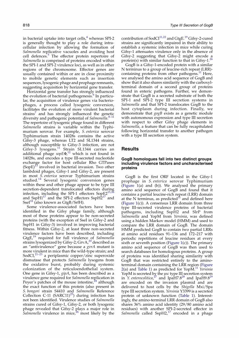
Figure 4. gogB expression is co-regulated with SPI-2under SPI-2-inducing conditions. (a) Western blot show-ing GogB-HA expression in various Salmonella strains andgrowth conditions. Wild-type Salmonella and a mutantdefective for production of the SPI-2 two-componentregulatory component, SsrB, were transformed withpgogB-HA and then grown under SPI-2-inducing con-ditions. Whole bacterial fractions (P) and secreted proteinfractions (S) were analyzed for the presence of GogB-HAand DnaK. (b) Wild-type Salmonella and a mutantdefective for production of the SPI-1 activator, HilA,were transformed with pgogB-HA and then grown underSPI-1-inducing conditions (see Materials and Methods).Whole bacterial lysates were analysed by Western blot forGogB-HA expression. (c) b-Galactosidase activity assays.The activity of the gogB promoter was assessed with atranscriptional promoter fusion to lacZ on the Salmonellachromosome. Wild-type and ssrB mutant reporter strainswere grown under SPI-2-inducing conditions and b-galactosidase activity was measured. Data are given asthe mean relative light units (RLU) normalized to theoptical density of the bacterial culture with standarderrors.
822 Type III Secretion of GogB
To examine this further, we constructed a doublemutant lacking both SPI-1 and SPI-2-mediatedtype III secretion activity (DssaR invATKan) andexamined the secretion of GogB-HA. Secretion ofGogB-HAwas completely eliminated in the doublemutant (Figure 3(b)) confirming that GogB can besecreted by both of the virulence-associated type IIIsecretion systems in Salmonella.
GogB expression and secretion is autonomouswith respect to other Gifsy prophage elements
Expression of GogB by Salmonella may require
additional elements encoded within Gifsy-1. To testthis hypothesis, we transformed Gifsy-1-curedSalmonella26 with pgogB-HA and tested expressionand secretion under various conditions. Theexpression level of GogB-HA was not different inGifsy-1 cured strains compared to wild-type bac-teria (Figure 3(a)), indicating that GogB expressionand type III secretion is independent of Gifsy-1prophage genes.
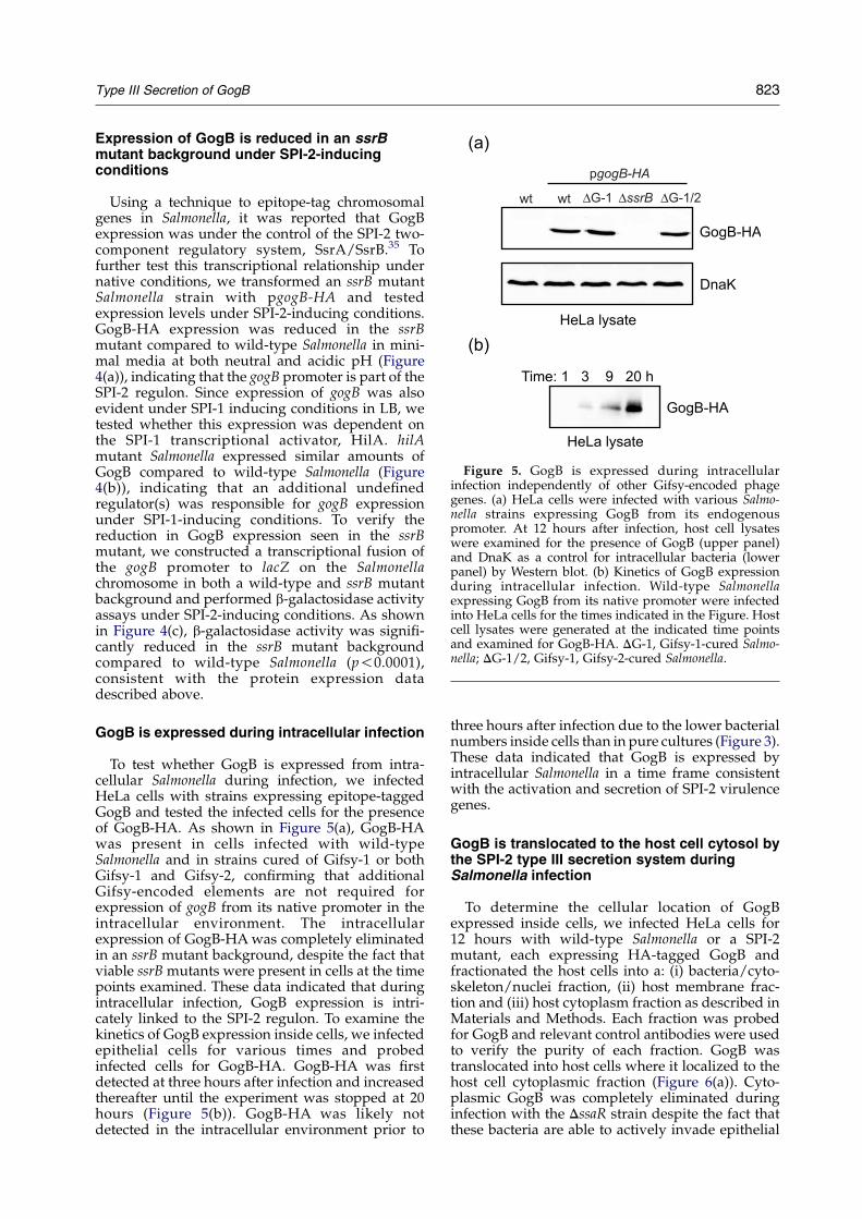
Figure 5. GogB is expressed during intracellularinfection independently of other Gifsy-encoded phagegenes. (a) HeLa cells were infected with various Salmo-nella strains expressing GogB from its endogenouspromoter. At 12 hours after infection, host cell lysateswere examined for the presence of GogB (upper panel)and DnaK as a control for intracellular bacteria (lowerpanel) by Western blot. (b) Kinetics of GogB expressionduring intracellular infection. Wild-type Salmonellaexpressing GogB from its native promoter were infectedinto HeLa cells for the times indicated in the Figure. Hostcell lysates were generated at the indicated time pointsand examined for GogB-HA. DG-1, Gifsy-1-cured Salmo-nella; DG-1/2, Gifsy-1, Gifsy-2-cured Salmonella.
Type III Secretion of GogB 823
Expression of GogB is reduced in an ssrBmutant background under SPI-2-inducingconditions
Using a technique to epitope-tag chromosomalgenes in Salmonella, it was reported that GogBexpression was under the control of the SPI-2 two-component regulatory system, SsrA/SsrB.35 Tofurther test this transcriptional relationship undernative conditions, we transformed an ssrB mutantSalmonella strain with pgogB-HA and testedexpression levels under SPI-2-inducing conditions.GogB-HA expression was reduced in the ssrBmutant compared to wild-type Salmonella in mini-mal media at both neutral and acidic pH (Figure4(a)), indicating that the gogB promoter is part of theSPI-2 regulon. Since expression of gogB was alsoevident under SPI-1 inducing conditions in LB, wetested whether this expression was dependent onthe SPI-1 transcriptional activator, HilA. hilAmutant Salmonella expressed similar amounts ofGogB compared to wild-type Salmonella (Figure4(b)), indicating that an additional undefinedregulator(s) was responsible for gogB expressionunder SPI-1-inducing conditions. To verify thereduction in GogB expression seen in the ssrBmutant, we constructed a transcriptional fusion ofthe gogB promoter to lacZ on the Salmonellachromosome in both a wild-type and ssrB mutantbackground and performed b-galactosidase activityassays under SPI-2-inducing conditions. As shownin Figure 4(c), b-galactosidase activity was signifi-cantly reduced in the ssrB mutant backgroundcompared to wild-type Salmonella (p!0.0001),consistent with the protein expression datadescribed above.
GogB is expressed during intracellular infection
To test whether GogB is expressed from intra-cellular Salmonella during infection, we infectedHeLa cells with strains expressing epitope-taggedGogB and tested the infected cells for the presenceof GogB-HA. As shown in Figure 5(a), GogB-HAwas present in cells infected with wild-typeSalmonella and in strains cured of Gifsy-1 or bothGifsy-1 and Gifsy-2, confirming that additionalGifsy-encoded elements are not required forexpression of gogB from its native promoter in theintracellular environment. The intracellularexpression of GogB-HAwas completely eliminatedin an ssrB mutant background, despite the fact thatviable ssrB mutants were present in cells at the timepoints examined. These data indicated that duringintracellular infection, GogB expression is intri-cately linked to the SPI-2 regulon. To examine thekinetics of GogB expression inside cells, we infectedepithelial cells for various times and probedinfected cells for GogB-HA. GogB-HA was firstdetected at three hours after infection and increasedthereafter until the experiment was stopped at 20hours (Figure 5(b)). GogB-HA was likely notdetected in the intracellular environment prior to
three hours after infection due to the lower bacterialnumbers inside cells than in pure cultures (Figure 3).These data indicated that GogB is expressed byintracellular Salmonella in a time frame consistentwith the activation and secretion of SPI-2 virulencegenes.
GogB is translocated to the host cell cytosol bythe SPI-2 type III secretion system duringSalmonella infection
To determine the cellular location of GogBexpressed inside cells, we infected HeLa cells for12 hours with wild-type Salmonella or a SPI-2mutant, each expressing HA-tagged GogB andfractionated the host cells into a: (i) bacteria/cyto-skeleton/nuclei fraction, (ii) host membrane frac-tion and (iii) host cytoplasm fraction as described inMaterials and Methods. Each fraction was probedfor GogB and relevant control antibodies were usedto verify the purity of each fraction. GogB wastranslocated into host cells where it localized to thehost cell cytoplasmic fraction (Figure 6(a)). Cyto-plasmic GogB was completely eliminated duringinfection with the DssaR strain despite the fact thatthese bacteria are able to actively invade epithelial

Figure 6. GogB is translocated into the host cellcytoplasm by the SPI-2 type III secretion system duringinfection. Wild-type Salmonella and the SPI-2 mutant,DssaR, each expressing epitope-tagged GogB from itsnative promoter (a) or wild-type Salmonella expressingepitope-tagged SifA from its native promoter (b) wereused to infect HeLa cells for 12 hours. Infected host cellswere fractionated by differential centrifugation into hostnuclei/cytoskeleton/bacteria fraction (P), a host mem-brane fraction (M) and host cytoplasmic fraction (C)according to the methods described in Materials andMethods. An equal volume of each fraction was analysedby Western blot using anti-HA, anti-DnaK, anti-calnexinand anti-b tubulin antibodies.
Figure 7. gogB is organized into a modular genetic unitcapable of expression and type III secretion in aheterologous host. Wild-type enteropathogenic E. coli(EPEC) and an EPEC type III secretion mutant (DescN)were transformed with a plasmid containing the gogBgene and upstream regulatory sequence. Bacteria weregrown in the presence or absence of EGTA underconditions that activate LEE-gene expression and typeIII secretion. Whole cell lysates (P) and secreted proteinfractions (S) were tested by Western blot for the presenceof GogB-HA (upper panel) and the E. coli type III-secretedeffector, Tir (middle panel). Blots were also probed for thebacterial cytoplasmic molecule, DnaK (lower panel).
824 Type III Secretion of GogB
cells and are competent for SPI-1-mediated type IIIsecretion. These data indicated that translocationinto the host cytosol was dependent on the SPI-2type III secretion system. As a control for SPI-1-mediated type III secretion, we probed all host cellfractions with anti-SopB antiserum, which demon-strated that this SPI-1 effector protein localized tothe host membrane fraction in both wild-type andDssaR-infected cells at the time points examined(data not shown). Analysis of the host cell fractionswith specific antibodies confirmed the purity ofeach fraction. Calnexin, a host cell integral mem-brane protein, was absent from the host cytoplasmicfraction, and b-tubulin, a host cell cytoplasmicprotein, was absent from the host membranefraction. DnaK, a non-secreted bacterial protein,was present only in the low-speed pellet, demon-strating a lack of bacterial contamination of the host
membrane and cytosolic fractions. As a furthercontrol for the host subcellular fractionation pro-cedure, we infected cells with Salmonella expressingHA-tagged SifA, a SPI-2-translocated virulencefactor that localizes to host cell membranes andthe cytoskeleton upon delivery.36,37 As expected,translocated SifA localized to the host cell mem-brane fraction and bacteria/cytoskeleton fractionbut was not present in the cytoplasmic fraction(Figure 6(b)).
GogB is a modular genetic unit capable ofautonomous expression and type III secretion inother pathogenic bacterial systems
Since GogB expression and secretion were inde-pendent of other Gifsy phage-encoded elements inSalmonella, we hypothesized that gogB is organizedinto an autonomous genetic module that could behorizontally transferred and utilized directly as atype III-secreted substrate by other pathogenicbacteria. To test this hypothesis, we transferredgogB with its upstream promoter into wild-typeenteropathogenic E. coli (EPEC) and a type IIIsecretion mutant deficient in the type III secretionATPase, escN, and tested whether GogB expressionand secretion could be achieved in this hetero-logous host. GogB was expressed from its nativepromoter in EPEC when the bacteria were grown inDMEM (Figure 7), a medium that inducesexpression of the LEE pathogenicity island andinduces type III secretion. Analysis of the secretedproteins from these cultures demonstrated thatGogB secretion was type III-dependent since GogBwas eliminated completely in the secreted protein

Type III Secretion of GogB 825
fraction from the DescN strain. Depletion of calciumfrom DMEM leads to increased secretion of type IIIeffector molecules in diffusely adhering EPEC,38
which we have confirmed for other attaching andeffacing enteropathogens.39 To test whether type IIIsecretion of GogB in EPEC integrated into thisCa2C-dependent secretion regulation, we expressedepitope-tagged GogB in EPEC during growth inDMEM in which the calcium concentration hadbeen reduced by chelation with EGTA. Growth ofEPEC in calcium-depleted DMEM led to increasedsecretion of GogB, which was entirely type IIIdependent as the DescN strain did not secrete GogBunder the same growth conditions with calciumchelation (Figure 7). As a control, we examined thetype III-dependent secretion of the effector proteinTir (translocated intimin receptor), specific toattaching and effacing pathogens. As expected Tirsecretion was entirely type III-dependent, as theescN mutant did not secrete Tir under any con-ditions. Upon calcium chelation, secretion of Tirinto the medium increased dramatically comparedto medium with 1.8 mM free calcium. The patternof GogB expression and type III secretion in EPECwas similar to that of Tir, demonstrating typeIII-dependence of secretion and integration into acalcium-regulated secretion response (Figure 7).Collectively, these data indicate that GogB can behorizontally transferred as a genetic module intoother pathogens with a type III secretion system andintegrate into the existing regulatory and secretionmachinery governing type III secretion in therecipient bacteria.
Discussion
We have investigated the genetic and molecularfeatures of GogB expression, secretion and trans-location into host cells and report here that gogBis a modular lysogenic conversion gene dis-playing autonomous expression and type IIIsecretion with respect to other Gifsy phage-encodedfactors.
Phage-encoded molecules contribute greatly tothe pathogenic potential of lysogens.7,10 The mobili-zation of lysogenic conversion genes into bacterialgenomes can affect bacterial virulence in severalways, including modifying bacterial adhesionproperties to host tissues, resisting immune effectorfunctions, elaborating exotoxins, and increasingtransmission of lysogenic clones. The secretion ofphage-encoded molecules into the surroundingenvironment can enhance several of these virulencetraits while other molecules exert their effects aftertranslocation into target host cells by the bacterialtype III secretion system. Based on the high degreeof carboxyl-terminal similarity between GogB andproteins in other pathogens (Z1829 in EHECO157:H7, OrfL in REPEC, and the Yersinia proteinsYP2634 and Y1471), we predict that these uncharac-terised proteins are also substrates for their respect-ive type III secretion systems and that they interact
with an evolutionarily conserved pathway in targetcells. Given the secretion and translocation datadescribed here for GogB, investigation of theseother putative type III secretion substrates seemswarranted.Phage evolution can involve the exchange of
individual genetic modules. In this scheme, replace-ment of one phage module by a sequence-unrelated(but functionally similar) module from anotherphage reconstitutes a genetically intact phage withincreased genetic diversity.40 Such modules typi-cally encode discrete phage biological functionssuch as head and tail formation, excision andintegration, recombination and DNA packaging(see annotation, Figure 1(b)). It has been suggestedthat the genetic organization of phage-encodedvirulence genes (or fitness factors) also involvesindividual modules that can be expressed inde-pendently from their associated phage.10,41 Basedon the work described here, GogB exists in Gifsy-1as an independent transcriptional unit that allowsautonomous expression in the absence of otherGifsy phage elements. This was evident by the factthat Salmonella strains cured of either Gifsy-1 orboth Gifsy-1 and Gifsy-2 were able to express GogBfrom its native promoter and to secrete GogBwithout additional Gifsy phage proteins. The abilityof the gogB module to be expressed and secreted ina type III-dependent manner in a heterologousbacterial pathogen suggests that acquisition of gogBby horizontal gene transfer may allow for a suddengain of function for the lysogen, providing it has atype III secretion system for delivery of the neweffector. The presence of a predicted 36 base stem–loop structure at nucleotides 51–86 downstream ofthe gogB stop codon also suggests the presence of afactor-independent transcription terminator partici-pating in the modular nature of this lysogenicconversion gene. The autonomous pattern of gogBexpression contrasts what is known about somephage-encoded virulence factors. For example,Wagner and colleagues42 showed that the stx2gene (encoding Shiga toxin 2 on a lambdoidphage) in a clinical isolate of E. coli O157:H7 isunder the control of a late phage promoter thatrequires prophage induction and activation of thephage lytic cascade to achieve transcriptionalactivation and release of the toxin. Similarly, stx1expression and production of Shiga toxin 1 is underthe control of phage-encoded transcription fac-tors,43 and the transcriptional activation of severalphage-encoded staphylococcal enterotoxins isdependent on phage transcription factors andpromoters.44
Phage-encoded virulence modules can be hori-zontally transferred between phages of differentclasses by homologous or illegitimate recombina-tion,45,46 indicating that genetic diversity of patho-gens can be achieved in the absence of de novolysogenization, but rather by incorporation of genemodules into existing lysogens. Such a mechanismovercomes the resistance of some bacterial strains tosuperinfection by similar phage yet is compatible

826 Type III Secretion of GogB
with the need for pathogens to achieve greatergenetic diversity. It is possible that GogB and itsrelated proteins, specifically EHEC Z1829, mighthave been acquired in this fashion. Z1829 isencoded by a cryptic lambdoid phage (CP-933N)in O-island 50 of EHEC O157:H7 31 that appears tohave undergone genetic attrition in various phagegenes but has retained Z1829 in the phage tailregion. Closer inspection of the nucleotide contentof gogB and the remainder of Gifsy-1 may alsosuggest a more recent acquisition of gogB and itsregulatory information by Gifsy-1. The gogB openreading frame has a GCC content of 32.93%, twostandard deviations lower than the mean GCCcontent of the rest of Gifsy-1 (52.56%) and theSalmonella genome (52.80%). That gogB has recentlybeen acquired by Gifsy-1 is further supported bythe fact that some S. enterica strains appear positivefor the Gifsy-1 phage but negative for gogB.25
Additionally, at least two S. enterica strains(S. enterica SARC5 and S. enterica serovar Arizonae)appear positive for gogB yet negative for Gifsy-1.25
While the genomic context of gogB in theseapparently Gifsy-1-negative strains remains to beinvestigated, this does suggest that gogB is amobilisable element capable of integrating intodifferent genetic contexts to be retained as anautonomous unit. The fact that the gogB modulecould be transferred horizontally to a heterologousbacterial pathogen, was expressed independently ofother phage components and was integrated intothe existing type III secretion system and secretionregulation features of the recipient bacteria stronglysupports this notion. Our attempts to generateGifsy-1 lysogens in enteropathogenic and entero-hemorrhagic E. coli have been unsuccessful.However, pathogenic E. coli contain numerousprophages and cryptic prophages of various ori-gins,9,10 suggesting perhaps that these strainspossess superinfection immunity. It will be ofinterest to test whether gogB can be incorporatedinto existing lysogens that are resistant to Gifsy-1lysogenization and whether the acquisition of gogBunder these conditions confers a selective advan-tage to the recipient bacteria in the appropriateinfection model.
That gogB is located between the Gifsy-1structural gene operon and the attachment site,a region of the lambdoid phage capable ofaccommodating lysogenic conversion genes, isparticularly interesting since gogB appears to be amosaic of two families of homologous genes.It is possible that gogB may have arisen byillegitimate recombination between a lysogenicconversion gene containing an N-terminaltype III secretion signal and LRR domain,with another phage gene from the C-termi-nal homology group. While it is thought thatfew mosaic boundaries fall within open readingframes (a mosaic of two structurally distinctmolecules is likely to be non-functional47) themodular domain structure of many bacterial
effectors48–51 may lend itself to the likelihood ofsuch mosaic genes being functional.
One of the interesting features of autonomousgogB expression was its co-regulation with otherSPI-2 virulence genes by SsrB, the transcriptionalactivator of the SsrA/SsrB two-component regula-tory system. The ability of lysogenic conversiongenes to integrate into the existing transcriptionalregulation circuitry and to use the type III secretionmachinery for directed delivery into host cells is animportant aspect in the co-evolution of bacteria-phage biology and allows the recipient bacteria tobenefit directly from lysogeny. However, it has notbeen fully elucidated whether this represents animmediate acquisition of a virulence trait, orwhether the newly acquired virulence genes requireadditional fine-tuning at the genetic level tointegrate properly into regulatory and secretoryvirulence pathways. Clearly, the utility of newlyacquired virulence properties encoded by lysogenicconversion genes also depends on the recipientbacterial strain, its genetic components and existingmolecular “hardware” for delivering the newmolecule to a productive site of action. Even thoughGogB was secreted by both the SPI-1 and SPI-2-encoded type III secretion systems in vitro, severallines of evidence indicate that SPI-2 is likely themajor route of GogB translocation into host cellsduring infection. First, secretion of GogB wasreduced when bacteria were grown under SPI-2-inducing conditions at neutral pH, yet secretionwas enhanced when bacteria were shifted to anacidic pH environment in which assembly of theSPI-2 type III apparatus takes place.52 Secondly,gogB expression was co-regulated with other SPI-2genes and was decreased in an ssrB mutant lackingthe SPI-2 transcriptional activator. In contrast,gogB expression under SPI-1-inducing conditionswas not co-regulated with other HilA-dependentvirulence genes encoded within SPI-1. Third, gogBexpression was eliminated completely in an ssrBmutant during the intracellular infection processand fourth, translocation of GogB into the hostcytosol was eliminated in a SPI-2 mutant that wasfully competent for cell invasion and SPI-1-mediated type III translocation.
Previous work indicated that genetic elementsencoded within Gifsy-1 influence the virulencepotential of Salmonella but this phenotype wasonly apparent in the absence of Gifsy-2.26 It ishypothesized that one or more additional factorsencoded in Gifsy-2 might perform functionallysimilar roles to GogB or other Gifsy-1 factors duringinfection, although an obvious homologue of GogBis not apparent in Gifsy-2. It was recently reportedthat two signature-tagged mutants of Gifsy-1(STM2586, STM2587) were attenuated for caecalcolonization of 14-day old chicks.53 These genes areencoded immediately downstream of gogB on theopposite strand in the tail fibre region of Gifsy-1.Both genes are annotated as phage tail assemblyproteins, so their putative role in bacterial patho-genesis or in improving bacterial fitness is currently

Type III Secretion of GogB 827
unknown. The EHEC O157:H7 gogB homologue(Z1829) was not identified in a signature-taggedscreen of bovine intestinal colonization,54 althoughit is possible that this particular mutant was notpresent in the library that was screened. It alsoremains possible that other EHEC strains will usethis protein as a virulence factor during coloniza-tion. In fact, such variation in the use of virulencefactors might be expected between different strainsof the same species, given that different strains cancontain a variable assortment of lysogenic phageswith functionally overlapping gene sets. It alsoseems likely that some phage-encoded virulencefactors perform functions during a specific stage ofinfection or in a specific host species.53 In this way,acquisition of phage-encoded virulence geneswould be a critical step in allowing previouslytissue- or host-restricted bacteria access to newecological niches,7 an event that often precedes theemergence of new strains with epidemic virulencepotential.12,55–57 It also stands to reason that thefunctional importance of host- or tissue-specificvirulence factors would remain cryptic in theabsence of the proper host context. Information onthe intracellular function of GogB and its homo-logues from other pathogenic bacteria shouldlead to important gains in our understanding ofevolutionarily conserved host–pathogen-phageinteractions.
†www.cbs.dtu.dk/services/SignalP/
Materials and Methods
Bacterial strains and plasmid construction
S. enterica serovar Typhimurium strain SL1344 wasused in this study and all mutants are derivatives ofSL1344. SL1344 strains cured of the Gifsy-1 prophage orboth Gifsy-1 and Gifsy-2 prophages have been describedpreviously.11,26 Enteropathogenic E. coli strain E2348/69and an isogenic mutant lacking type III secretion (DescN)were used as heterologous hosts for expression andsecretion experiments. A Salmonella mutant defective forboth SPI-1 and SPI-2-mediated type III secretion wasconstructed by generalized P22 transduction of a markedinvATKan mutation to a strain with an unmarked, in-frame deletion of ssaR. Bacterial cultures were grown inLuria–Bertani (LB) liquid medium or on LB agar platessupplemented with the appropriate antibiotic(s) at thefollowing concentrations: kanamycin, 50 mg mlK1; carbe-nicillin, 100 mg mlK1; streptomycin, 50 mg mlK1. To con-struct an epitope-tagged version of GogB, the gogB openreading frame plus w1 kb of the upstream regulatoryregion was amplified by PCR from purified SL1344chromosomal DNA with the primers BKC41 and BKC42(5 0ACG CGT CGA CGT CAA TAA TCA GCG CCT GCA3 0 and 50 GAA GAT CTA CGA TTT CTA TTT TTA GGCTTA TAT TTA TCC 3 0), digested with SalI and BglII andcloned into the corresponding restriction sites of apACYC184-based plasmid called psseAT2HA58 inwhich the sseA coding region had been removed as aSalI/BglII fragment. The gogB coding region, in-framehemagglutinin epitope and upstream promoter regionwas then removed from this pACYC-based vector withSalI and XbaI and cloned into the corresponding sites ofpWSK29 and pWSK12959 such that the gogB coding
region and promoter were in the opposite orientation tothe vector-encoded lacZ promoter.
Bioinformatics analysis of gogB
A sequence comparison between GogB and proteins inpublic databases was performed using the Blastp algo-rithm and Clustal W. The primary amino acid sequence ofa family of proteins containing leucine-rich repeats wasused to define a domain hidden Markov model that wascompared to the primary amino acid sequence of GogB.Signal peptides were predicted using Signalp version2.0b2†60 using default parameters for Gram-negativebacteria.
Cell culture and infection assays
HeLa cells were maintained in Dulbecco’s ModifiedEagle medium (DMEM; Hyclone, Logan, Utah) supple-mented with 10% (v/v) foetal calf serum (FCS). Cellswere seeded in 24-well culture dishes 18 hours prior toinfection. All cells were cultured at 37 8C in 5% CO2. Forgentamicin protection assays, HeLa cells were infectedwith late log-phase bacteria for ten minutes asdescribed.58 At the indicated times after infection,gentamicin-treated cells were washed with PBS andthen solubilized in 0.25 ml of 1% (w/v) Triton X-100,0.1% (w/v) SDS in PBS. Lysates weremixed with an equalvolume of SDS-PAGE sample buffer (100 mM Tris–HCl(pH 6.8), 20% (v/v) glycerol, 4% (w/v) SDS, 0.002% (v/v)bromophenol blue and 200 mM dithiothreitol) andprobed by Western blotting for the presence of epitope-tagged GogB.
In vitro secretion assays and analysis of secretedproteins
Salmonella strains were grown overnight in LB broth,washed in either LB or low phosphate, low magnesium-containing medium (LPM)32 and then inoculated at a1 : 50 dilution in either 3 ml of LB, LPM pH 7.0 or LPM pH5.8. Growth in LB promotes activation of SPI-1 virulencegenes while LPM is a SPI-2-inducing medium. Salmonellacultures were grown at 37 8C with shaking for three to sixhours after which A600 was measured. For enteropatho-genic E. coli studies, wild-type EPEC strain E2348/69 andan isogenic derivative with a deletion of the essential typeIII secretion component escN (DescN) were grown over-night in standing LB broth, washed in PBS and inoculatedat a 1 : 50 dilution into DMEM or DMEM containing2 mM EGTA to chelate free Ca2C. EPEC cultures weregrown standing for six hours at 37 8C and 5% CO2 afterwhich A600 was measured and used to standardize theprotein samples. Bacteria were collected by centrifugationfor two minutes at 12,000 rpm (13,400g) (4 8C). Thesupernatant was passed through a low-protein binding0.22 mm HT Tuffryn membrane (Pall Life Sciences) andthe sterilized flow-through was precipitated with tri-chloroacetic acid (10% (v/v) final concentration) at 4 8Cfor four to 16 hours. Precipitated proteins were washedwith ice-cold acetone and solubilized with a volume of2! SDS-sample buffer adjusted according to the A600 ofthe original culture. Proteins from equivalent numbersof bacterial cells were separated on SDS-12% polyacryl-amide gels, transferred to nitrocellulose membranes andthen blocked in Tris-buffered saline containing 0.1% (v/v)

828 Type III Secretion of GogB
Tween 20 (TBST) and 5% (w/v) powdered non-fat milkfor one hour at room temperature. Primary antibodiesused were mouse anti-HA (Covance; 1 : 1500), mouseanti-DnaK (Stressgen Biotechnologies; 1 : 4000), and ratanti-Tir (1 : 5000). Secondary antibodies conjugated tohorseradish peroxidase were used at a 1 : 5000 dilution inTBST for one hour at room temperature. Antibodycomplexes were detected using enhanced chemilumi-nescence (Amersham Biosciences).
Chemiluminescent b-galactosidase assays
gogB promoter activity was examined using transcrip-tional fusions to lacZ and a chemiluminescence assaydescribed.32 A promoterless lacZY gene cassette wasintegrated downstream of the gogB promoter on theSalmonella chromosome using FLP-mediated site-specificrecombination between pCE3661 andwild-type Salmonellaor an ssrB Salmonella mutant that contained a chromo-somal FLP recognition target (FRT) site downstream ofthe gogB promoter.35 The resulting strains contain atranscriptional fusion of the gogB promoter to lacZ. ThelacZ reporter strains were cultured overnight in LBmedium, then washed twice in LPM medium andinoculated into fresh LPM medium at pH 5.8. Cultureswere incubated with shaking at 37 8C for five hours. Forb-galactosidase assays, 0.2 ml of the culture was removedand the bacteria were pelleted by centrifugation. Thesupernatant was discarded and the bacterial pellet wasresuspended in 0.2 ml of PBS and then lysed with 50 ml ofchloroform. 2 ml of the lysate was transferred to a well of ablack microtitre plate containing 100 ml of Galacto-Starsubstrate mix (Applied Biosystems, Bedford, MA). Reac-tions were incubated for 60 minutes at room temperatureand luminescence was detected with a Spectrafluor Plus(TECAN, Austria). Light emission was recorded asrelative light units (RLU) and normalized to the opticaldensity based on A600 measurements derived frommatched bacterial cultures. Each condition was per-formed in quadruplicate and averaged.
Translocation assay and host cell fractionationprocedure
Host cells were infected with Salmonella strains expres-sing epitope-tagged GogB or epitope-tagged SifA asdescribed above. Infected host cells were mechanicallyfractionated and separated by differential centrifugation.Infected cells were washed and scraped into PBS, thenmechanically disrupted by passage through a 22-gaugeneedle in a buffer containing 3 mM imidazole (pH 7.4),250 mM sucrose, 0.5 mMEDTA, and a cocktail of proteaseinhibitors. Differential centrifugation was used to gen-erate a: (i) bacteria/nuclei/cytoskeleton fraction (3000gpellet), (ii) a membrane fraction (41,000g pellet) and (iii) acytoplasmic fraction (41,000g supernatant). Each fractionwas analysed by SDS-PAGE and Western blotting usingthe following primary antibodies: mouse anti-HA(1 : 1500), mouse anti-DnaK (1 : 4000), rabbit anti-calnexin(1 : 1000, Stressgen), mouse anti-b tubulin (1 : 1000, cloneE7, Developmental Studies Hybridoma Bank, Universityof Iowa), rabbit anti-SopB (1 : 1000). All primary anti-bodies were diluted in a blocking solution of TBST with5% (w/v) non-fat milk. Secondary antibodies conjugatedto horseradish peroxidase were used at 1 : 5000 andantigen–antibody complexes were detected usingenhanced chemiluminescence.
Acknowledgements
We thank Claudia Lupp, Philip Hardwidge,Wanyin Deng and Jose Puente for helpful discus-sions and insightful comments and we gratefullyacknowledge David Goode for bioinformatics sup-port related to this work. This work was supportedby grants to B.B.F. from the Canadian Institutes ofHealth Research (CIHR), The Howard HughesMedical Institute (HHMI) and the Canadian Bac-terial Diseases Network. B.K.C. & M.E.W. arerecipients of postdoctoral fellowships from theCIHR and Michael Smith Foundation for HealthResearch (MSFHR), N.F.B. is a MSFHR postdoctoralfellow and W.W.L.H. is a recipient of a MSFHRTrainee Award and a CIHR Doctoral Scholarship.F.S.L.B. is a MSFHR scholar, B.B.F. is a CIHRDistinguished Investigator, an HHMI InternationalResearch Scholar and the University of BritishColumbia Peter Wall Distinguished Professor.
References
1. Galan, J. E. (1996). Molecular genetic bases ofSalmonella entry into host cells. Mol. Microbiol. 20,263–271.
2. Ochman, H., Soncini, F. C., Solomon, F. & Groisman,E. A. (1996). Identification of a pathogenicity islandrequired for Salmonella survival in host cells. Proc. NatlAcad. Sci. USA, 93, 7800–7804.
3. Shea, J. E., Hensel, M., Gleeson, C. & Holden, D. W.(1996). Identification of a virulence locus encodinga second type III secretion system in Salmonellatyphimurium. Proc. Natl Acad. Sci. USA, 93, 2593–2597.
4. Galan, J. E. (1999). Interaction of Salmonella with hostcells through the centisome 63 type III secretionsystem. Curr. Opin. Microbiol. 2, 46–50.
5. Waterman, S. R. & Holden, D. W. (2003). Functionsand effectors of the Salmonella pathogenicity island 2type III secretion system. Cell. Microbiol. 5, 501–511.
6. Ochman, H., Lawrence, J. G. & Groisman, E. A. (2000).Lateral gene transfer and the nature of bacterialinnovation. Nature, 405, 299–304.
7. Wagner, P. L. & Waldor, M. K. (2002). Bacteriophagecontrol of bacterial virulence. Infect. Immun. 70,3985–3993.
8. Thomson, N., Baker, S., Pickard, D., Fookes, M.,Anjum, M., Hamlin, N. et al. (2004). The role ofprophage-like elements in the diversity of Salmonellaenterica serovars. J. Mol. Biol. 339, 279–300.
9. Boyd, E. F. & Brussow, H. (2002). Common themesamong bacteriophage-encoded virulence factors anddiversity among the bacteriophages involved. TrendsMicrobiol. 10, 521–529.
10. Brussow, H., Canchaya, C. & Hardt, W. D. (2004).Phages and the evolution of bacterial pathogens: fromgenomic rearrangements to lysogenic conversion.Microbiol. Mol. Biol. Rev. 68, 560–602.
11. Figueroa-Bossi, N., Uzzau, S., Maloriol, D. & Bossi, L.(2001). Variable assortment of prophages provides atransferable repertoire of pathogenic determinants inSalmonella. Mol. Microbiol. 39, 260–271.
12. Mirold, S., Rabsch, W., Rohde, M., Stender, S.,Tschape, H., Russmann, H. et al. (1999). Isolationof a temperate bacteriophage encoding the type III

Type III Secretion of GogB 829
effector protein SopE from an epidemic Salmonellatyphimurium strain. Proc. Natl Acad. Sci. USA, 96,9845–9850.
13. Hardt, W. D., Chen, L. M., Schuebel, K. E., Bustelo,X. R. & Galan, J. E. (1998). S. typhimurium encodes anactivator of Rho GTPases that induces membraneruffling and nuclear responses in host cells. Cell, 93,815–826.
14. Figueroa-Bossi, N., Coissac, E., Netter, P. & Bossi, L.(1997). Unsuspected prophage-like elements inSalmonella typhimurium. Mol. Microbiol. 25, 161–173.
15. Bakshi, C. S., Singh, V. P., Wood, M. W., Jones, P. W.,Wallis, T. S. & Galyov, E. E. (2000). Identification ofSopE2, a Salmonella secreted protein which is highlyhomologous to SopE and involved in bacterialinvasion of epithelial cells. J. Bacteriol. 182, 2341–2344.
16. Stender, S., Friebel, A., Linder, S., Rohde, M., Mirold,S. & Hardt, W. D. (2000). Identification of SopE2 fromSalmonella typhimurium, a conserved guanine nucleo-tide exchange factor for Cdc42 of the host cell. Mol.Microbiol. 36, 1206–1221.
17. Miao, E. A., Scherer, C. A., Tsolis, R. M., Kingsley,R. A., Adams, L. G., Baumler, A. J. & Miller, S. I.(1999). Salmonella typhimurium leucine-rich repeatproteins are targeted to the SPI1 and SPI2 type IIIsecretion systems. Mol. Microbiol. 34, 850–864.
18. Miao, E. A. & Miller, S. I. (2000). A conserved aminoacid sequence directing intracellular type III secretionby Salmonella typhimurium. Proc. Natl Acad. Sci. USA,97, 7539–7544.
19. Ho, T. D., Figueroa-Bossi, N., Wang, M., Uzzau, S.,Bossi, L. & Slauch, J. M. (2002). Identification of GtgE,a novel virulence factor encoded on the Gifsy-2bacteriophage of Salmonella enterica serovar Typhi-murium. J. Bacteriol. 184, 5234–5239.
20. Ho, T. D. & Slauch, J. M. (2001). Characterization ofgrvA, an antivirulence gene on the gifsy-2 phage inSalmonella enterica serovar typhimurium. J. Bacteriol.183, 611–620.
21. De Groote, M. A., Ochsner, U. A., Shiloh, M. U.,Nathan, C., McCord, J. M., Dinauer, M. C. et al. (1997).Periplasmic superoxide dismutase protects Salmonellafrom products of phagocyte NADPH-oxidase andnitric oxide synthase. Proc. Natl Acad. Sci. USA, 94,13997–14001.
22. Fang, F. C., DeGroote, M. A., Foster, J. W., Baumler,A. J., Ochsner, U., Testerman, T. et al. (1999). VirulentSalmonella typhimurium has two periplasmic Cu,Zn-superoxide dismutases. Proc. Natl Acad. Sci. USA,96, 7502–7507.
23. Farrant, J. L., Sansone, A., Canvin, J. R., Pallen, M. J.,Langford, P. R., Wallis, T. S. et al. (1997). Bacterialcopper- and zinc-cofactored superoxide dismutasecontributes to the pathogenesis of systemic salmo-nellosis. Mol. Microbiol. 25, 785–796.
24. Stanley, T. L., Ellermeier, C. D. & Slauch, J. M. (2000).Tissue-specific gene expression identifies a gene in thelysogenic phage Gifsy-1 that affects Salmonella entericaserovar typhimurium survival in Peyer’s patches.J. Bacteriol. 182, 4406–4413.
25. Porwollik, S., Wong, R. M. & McClelland, M. (2002).Evolutionary genomics of Salmonella: gene acqui-sitions revealed by microarray analysis. Proc. NatlAcad. Sci. USA, 99, 8956–8961.
26. Figueroa-Bossi, N. & Bossi, L. (1999). Inducibleprophages contribute to Salmonella virulence inmice. Mol. Microbiol. 33, 167–176.
27. Boland, A., Sory, M. P., Iriarte, M., Kerbourch, C.,Wattiau, P. & Cornelis, G. R. (1996). Status of YopM
and YopN in the Yersinia Yop virulon: YopM ofY. enterocolitica is internalized inside the cytosol ofPU5-1.8 macrophages by the YopB, D, N deliveryapparatus. EMBO J. 15, 5191–5201.
28. Buchrieser, C., Glaser, P., Rusniok, C., Nedjari, H.,D’Hauteville, H., Kunst, F. et al. (2000). The virulenceplasmid pWR100 and the repertoire of proteinssecreted by the type III secretion apparatus of Shigellaflexneri. Mol. Microbiol. 38, 760–771.
29. Toyotome, T., Suzuki, T., Kuwae, A., Nonaka, T.,Fukuda, H., Imajoh-Ohmi, S. et al. (2001). Shigellaprotein IpaH(9.8) is secreted from bacteria withinmammalian cells and transported to the nucleus.J. Biol. Chem. 276, 32071–32079.
30. Brumell, J. H., Marcus, S. L. & Finlay, B. B. (2000).N-terminal conservation of putative type III secretedeffectors of Salmonella typhimurium. Mol. Microbiol. 36,773–774.
31. Perna, N. T., Plunkett, G., III, Burland, V., Mau, B.,Glasner, J. D., Rose, D. J. et al. (2001). Genomesequence of enterohaemorrhagic Escherichia coliO157:H7. Nature, 409, 529–533.
32. Coombes, B. K., Brown, N. F., Valdez, Y., Brumell, J. H.& Finlay, B. B. (2004). Expression and secretion ofSalmonella Pathogenicity Island-2 virulence genes inresponse to acidification exhibit differential require-ments of a functional type III secretion apparatus andSsaL. J. Biol. Chem. 279, 49804–49815.
33. Beuzon, C. R., Banks, G., Deiwick, J., Hensel, M. &Holden, D. W. (1999). pH-dependent secretion ofSseB, a product of the SPI-2 type III secretion systemof Salmonella typhimurium. Mol. Microbiol. 33, 806–816.
34. Deiwick, J., Nikolaus, T., Erdogan, S. & Hensel, M.(1999). Environmental regulation of Salmonella patho-genicity island 2 gene expression. Mol. Microbiol. 31,1759–1773.
35. Uzzau, S., Figueroa-Bossi, N., Rubino, S. & Bossi, L.(2001). Epitope tagging of chromosomal genes inSalmonella. Proc. Natl Acad. Sci. USA, 98, 15264–15269.
36. Brumell, J. H., Goosney, D. L. & Finlay, B. B. (2002).SifA, a type III secreted effector of Salmonellatyphimurium, directs Salmonella-induced filament(Sif) formation along microtubules. Traffic, 3, 407–415.
37. Boucrot, E., Beuzon, C. R., Holden, D. W., Gorvel, J. P.& Meresse, S. (2003). Salmonella typhimurium SifAeffector protein requires its membrane-anchoringC-terminal hexapeptide for its biological function.J. Biol. Chem. 278, 14196–14202.
38. Ide, T., Michgehl, S., Knappstein, S., Heusipp, G. &Schmidt, M. A. (2003). Differential modulation byCa2C of type III secretion of diffusely adheringenteropathogenic Escherichia coli. Infect. Immun. 71,1725–1732.
39. Deng, W., Li, Y., Hardwidge, P. R., Frey, E. A.,Pfuetzner, R. A., Lee, S. et al. (2005). Regulation oftype III secretion hierarchy of translocators andeffectors in attaching and effacing bacterial patho-gens. Infect. Immun. In the press.
40. Botstein, D. (1980). A theory of modular evolution forbacteriophages. Ann. NY Acad. Sci. 354, 484–490.
41. Hendrix, R. W., Lawrence, J. G., Hatfull, G. F. &Casjens, S. (2000). The origins and ongoing evolutionof viruses. Trends Microbiol. 8, 504–508.
42. Wagner, P. L., Neely, M. N., Zhang, X., Acheson, D.W.,Waldor, M. K. & Friedman, D. I. (2001). Role for aphage promoter in Shiga toxin 2 expression from apathogenic Escherichia coli strain. J. Bacteriol. 183,2081–2085.
43. Wagner, P. L., Livny, J., Neely, M. N., Acheson, D. W.,
830 Type III Secretion of GogB
Friedman, D. I. &Waldor, M. K. (2002). Bacteriophagecontrol of Shiga toxin 1 production and release byEscherichia coli. Mol. Microbiol. 44, 957–970.
44. Sumby, P. & Waldor, M. K. (2003). Transcription of thetoxin genes present within the Staphylococcal phagephiSa3ms is intimately linked with the phage’s lifecycle. J. Bacteriol. 185, 6841–6851.
45. Mirold, S., Rabsch, W., Tschape, H. & Hardt, W. D.(2001). Transfer of the Salmonella type III effector sopEbetween unrelated phage families. J. Mol. Biol. 312,7–16.
46. Hendrix, R. W., Smith, M. C., Burns, R. N., Ford, M. E.& Hatfull, G. F. (1999). Evolutionary relationshipsamong diverse bacteriophages and prophages:all the world’s a phage. Proc. Natl Acad. Sci. USA, 96,2192–2197.
47. Hendrix, R. W. (2002). Bacteriophages: evolution ofthe majority. Theor. Popul. Biol. 61, 471–480.
48. Kaniga, K., Uralil, J., Bliska, J. B. & Galan, J. E. (1996).A secreted protein tyrosine phosphatase withmodular effector domains in the bacterial pathogenSalmonella typhimurium. Mol. Microbiol. 21, 633–641.
49. Evdokimov, A. G., Tropea, J. E., Routzahn, K. M.,Copeland, T. D. &Waugh, D. S. (2001). Structure of theN-terminal domain of Yersinia pestis YopH at 2.0 Aresolution. Acta. Crystallog. sect. D, 57, 793–799.
50. Fu, Y. & Galan, J. E. (1998). The Salmonella typhimuriumtyrosine phosphatase SptP is translocated into hostcells and disrupts the actin cytoskeleton. Mol. Micro-biol. 27, 359–368.
51. Olsson, J., Edqvist, P. J., Broms, J. E., Forsberg, A.,Wolf-Watz, H. & Francis, M. S. (2004). The YopDtranslocator of Yersinia pseudotuberculosis is a multi-functional protein comprised of discrete domains.J. Bacteriol. 186, 4110–4123.
52. Rappl, C., Deiwick, J. & Hensel, M. (2003). Acidic pHis required for the functional assembly of the type IIIsecretion system encoded by Salmonella pathogenicityisland 2. FEMS Microbiol. Letters, 226, 363–372.
53. Morgan, E., Campbell, J. D., Rowe, S. C., Bispham, J.,
Stevens, M. P., Bowen, A. J. et al. (2004). Identificationof host-specific colonization factors of Salmonellaenterica serovar Typhimurium. Mol. Microbiol. 54,994–1010.
54. Dziva, F., van Diemen, P. M., Stevens, M. P., Smith,A. J. & Wallis, T. S. (2004). Identification of Escherichiacoli O157: H7 genes influencing colonization of thebovine gastrointestinal tract using signature-taggedmutagenesis. Microbiology, 150, 3631–3645.
55. Waldor, M. K. & Mekalanos, J. J. (1996). Lysogenicconversion by a filamentous phage encoding choleratoxin. Science, 272, 1910–1914.
56. Zhang, S., Santos, R. L., Tsolis, R. M., Mirold, S.,Hardt, W. D., Adams, L. G. & Baumler, A. J. (2002).Phage mediated horizontal transfer of the sopE1 geneincreases enteropathogenicity of Salmonella entericaserotype Typhimurium for calves. FEMS Microbiol.Letters, 217, 243–247.
57. Karaolis, D. K., Johnson, J. A., Bailey, C. C., Boedeker,E. C., Kaper, J. B. & Reeves, P. R. (1998). A Vibriocholerae pathogenicity island associated with epi-demic and pandemic strains. Proc. Natl Acad. Sci.USA, 95, 3134–3139.
58. Coombes, B. K., Brown, N. F., Kujat-Choy, S., Vallance,B. A. & Finlay, B. B. (2003). SseA is required fortranslocation of Salmonella pathogenicity island-2effectors into host cells. Microbes Infect. 5, 561–570.
59. Wang, R. F. & Kushner, S. R. (1991). Constructionof versatile low-copy-number vectors for cloning,sequencing and gene expression in Escherichia coli.Gene, 100, 195–199.
60. Nielsen, H., Engelbrecht, J., Brunak, S. & von Heijne,G. (1997). Identification of prokaryotic and eukaryoticsignal peptides and prediction of their cleavage sites.Protein Eng. 10, 1–6.
61. Ellermeier, C. D., Janakiraman, A. & Slauch, J. M.(2002). Construction of targeted single copy lacfusions using lambda Red and FLP-mediated site-specific recombination in bacteria. Gene, 290, 153–161.
Edited by J. Karn
(Received 8 December 2004; received in revised form 26 February 2005; accepted 1 March 2005)