Transport of Streptococcus pneumoniae Capsular Polysaccharide in MHC Class II Tubules
Upload
khangminh22Category
view
0download
0
1
Screening of Streptococcus pneumoniae ABC transporters for
their role in virulence and investigation of their lipoprotein
components as vaccine candidates
Shilpa Basavanna
University College London
Thesis for doctoral degree (PhD), 2011

2
Declaration
I, Shilpa Basavanna confirm that the work presented in this thesis is my own. Where
information has been derived from other sources, I confirm that this has been indicated
in the thesis.

3
Abstract
Streptococcus pneumoniae causes life-threatening invasive diseases in children and
older adults. Although effective at reducing the incidence of disease, the two currently
available vaccines against S. pneumoniae have significant limitations that a vaccine
based on protein antigens may overcome. For this thesis I have investigated the role of
S. pneumoniae ABC transporters during infection, and have assessed as potential
vaccine candidates the lipoprotein components of two ABC transporters.
Eleven ABC transporters were chosen for investigation of their role during
infection, and disruption mutant strains were successfully constructed for 9 of these.
Two mutant strains disrupting the Sp0148-52 and Sp0749-53 ABC transporters, which
BLAST searches suggest have methionine and branched chain amino acids (BCAAs) as
substrates respectively, were markedly attenuated in systemic and pulmonary mice
models of virulence. Western blotting and PCR confirmed that the lipoprotein
components of these ABC transporters, Sp0149 and Sp0749, are present in all the S.
pneumoniae strains investigated and are membrane-localised. Radioactive and
fluorescence ligand binding experiments showed the Sp0749 lipoprotein specifically
bound to BCAAs, confirming Sp0749-53 encodes a BCAA ABC transporter.
Vaccination of mice with His6-Sp0149 and His6-Sp0749 induced specific IgG
which was able to increase complement activity against and phagocytosis of S.
pneumoniae. Intranasal immunisation of mice with His6-Sp0749 and His6-PiaA,
affected the immune response to subsequent intranasal challenge with S. pneumoniae
and increased lung inflammation upon subsequent S. pneumoniae intranasal challenge.
However, both intraperitoneal and intranasal immunisation of mice with His6-Sp0149
and His6-Sp0749 resulted in only minor degrees of protection against S. pneumoniae
septicaemia and pneumonia respectively. These results demonstrate that the S.

4
pneumoniae BCAA ABC transporter is required for the full virulence, and its
lipoprotein component can elicit immune responses against invasive S. pneumoniae that
although weakly protective could contribute towards a multivalent vaccine.

5
Table of contents
Chapter 1
Introduction to Streptococcus pneumoniae
1. 1 GENERAL, 32
1.2 CLINICAL CONSIDERATIONS, 33
1.2.1 Nasopharyngeal carriage, 33
1.2.2 Diseases caused by S. pneumoniae, 35
(a) Acute otitis media, 35
(b) Septicaemia, 37
(c) Pneumonia , 37
(d) Meningitis, 38
1.3 VACCINES AGAINST S. PNEUMONIAE, 39
(a) S. pneumoniae serotype distribution, 39
(b) S. pneumoniae capsular polysaccharide vaccines, 40
(c) S. pneumoniae conjugate vaccines, 41
(d) S. pneumoniae protein vaccines and lipoproteins as vaccines, 42
(e) Live, killed and DNA vaccines, 44
1.4 VIRULENCE FACTORS INVOLVED IN THE PATHOGENESIS OF S. PNEUMONIAE
INFECTIONS, 45
1.4.1 Adhesion, 48
(a) Capsule, 50
(b) IgA1 protease, 50
(c) Phosphorylcholine (PC), 51
(d) Neuraminidases (NanA, NanB, NanC), 53

6
(e) Exogylcosidases, 54
(f) Pneumococcal surface adhesion (PsaA), 55
(g) Pneumococcal surface protein C / Choline binding proteinA (PspC /
CbpA), 55
(h) Pneumococcal adhesion and virulence factor A (PavA), 57
(i) Streptococcal lipoprotein rotamase (SlrA), 57
(j) Pili, 58
(k) Phase variation, 59
1.4.2 Invasion of tissues, 60
(a) Hyaluronidase, 61
(b) Plasminogen (PLG) binding proteins, 61
(c) PC, 62
(d) Pav A, 63
(e) Pneumolysin (Ply), 63
1.4.3 Evasion of host immunity, 64
(a) Polysaccharide capsule and cell wall, 65
(b) Pneumococcal surface protein A (PspA), 66
(c) Pneumolysin, 67
(d) CbpA, 68
(e) Endonuclease A, 69
1.4.4 Inflammation, tissue damage and the induction of septic shock, 69
(a) Pneumolysin, 70
(b) Cell wall and cell wall polysaccharides and Autolysin (LytA), 71
(c) Pyruvate oxidase (Spx), 72
1.4.5 Replication and growth in vivo, 73

7
1.4.6 Regulation of S. pneumoniae virulence, 74
1.5 ATP BINDING CASSETTE TRANSPORTERS (ABC TRANSPORTERS), 76
1.5.1 Structure of ABC transporter, 79
1.5.2 Processing of lipoproteins, 79
1.5.3 Roles of ABC transporters in S. pneumoniae and other bacterial
pathogens, 81
1.5.4 Role of components bacterial ABC transporters as vaccine
candidates, 84
1.5.5 Lipoprotein components of ABC transporters investigated as vaccine
candidates in S. pneumoniae, 85
1.5.6 Conclusion, 87
1.6 HYPOTHESIS AND AIMS, 88
Chapter 2
Materials and methods
2.1 BACTERIAL STRAINS, 89
2.2 MEDIA AND GROWTH CONDITIONS, 89
2.3 DNA METHODS, 90
2.3.1 Extraction of plasmid DNA from E. coli, 90
2.3.2 Extraction of genomic DNA from S. pneumoniae, 90
2.3.3 Gel extraction and purification of DNA, 90
2.3.4 PCR, 91
2.3.5 Restriction digestion, 91
2.3.6 Ligation, 92
2.3.7 Overlap extension PCR (OEP), 92
(a) Initial PCR, 93

8
(b) Fusion of the initial PCR products without primers, 93
(c) Amplification of fused PCR products, 93
2.3.8 Transformation of E. coli, 94
2.3.9 Transformation of S. pneumoniae, 94
2.3.10 Nucleotide sequencing, 95
2.4 RNA METHODS, 95
2.4.1 RNA extraction from S. pneumoniae, 95
2.4.2 RNA extraction from S. pneumoniae by acid-phenol method, 95
2.4.3 RT-PCR, 96
2.5 PROTEIN METHODS, 97
2.5.1 Induction of positive clones for protein expression, 97
2.5.2 Purification of 6xHis-tagged lipoproteins by Ni-NTA affinity
chromatography under native conditions, 98
2.5.3 Preparation of dialysis tubing, 99
2.5.4 Dialysis of purified lipoproteins, 99
2.5.5 Protein concentration determination by Bicinchonic acid (BCA)
method (Pierce), 99
2.5.6 Whole cell lysate preparation from S. pneumoniae, 100
2.5.7 Extraction of S. pneumoniae membrane proteins by Triton X-114, 100
2.5.8 SDS-PAGE, 101
2.5.9 Western blotting, 101
2.5.10 Tryptophan fluorescence spectroscopy, 102
2.5.11 Radioactive substrate binding assay, 102
2.5.12 Streptonigrin assay, 103
2.5.13 Radioactive substrate uptake assays, 103

9
2.8 IN VIVO METHODS, 104
2.8.1 In vivo studies of S. pneumoniae in mouse models, 104
2.8.2 IP vaccination schedule and challenge experiment, 105
2.8.3 IN vaccination schedule and challenge experiment, 106
2.8.4 Immune cell surface marker staining recovered from target
organs, 107
2.8.5 Cytological analysis of BALF, 108
2.8.6 Histological analysis of lung sectioning, 108
2.9 IMMUNE ASSAYS, 109
2.9.1 Enzyme Linked Immunosorbant assay (ELISA), 109
2.9.2 C3 deposition and IgG binding assay, 110
2.9.3 Opsonophagocytosis, 110
(a) FAMSE labelling of S. pneumoniae for opsonophagocytosis, 111
(b) Tissue culture of HL60 for opsonophagocytosis, 111
(c) Opsonophagocytosis, 112
2.10 FLUORESCENCE-ACTIVATED CELL SORTING (FACS) ANALYSIS, 112
2.11 COMPUTER ANALYSIS, 113
2.12 STATISTICAL ANALYSIS, 113
Chapter 3
Screening of S. pneumoniae ABC transporters
3.1 IDENTIFICATION AND SELECTION OF ABC TRANSPORTER GENES, 121
3.2 AMINO ACID HOMOLOGY OF TIGR4 ABC TRANSPORTER GENES WITH
OTHER BACTERIAL SPECIES, 122
3.3 CONSTRUCTION OF THE DISRUPTION MUTANTS OF S. PNEUMONIAE BY
INSERTIONAL DUPLICATION MUTAGENESIS, 122

10
3.4 STABILITY OF THE MUTATION, 128
3.5 GROWTH CURVES IN THY, 131
3.6 PHENOTYPE ANALYSIS BY COMPETITIVE INDEX, 131
3.7 ANALYSIS OF THE GENETIC ORGANISATION OF THE ABC TRANSPORTER
OPERONS BY RT-PCR, 136
(a) Sp0090-0092, 137
(b) Sp0148-0152, 137
(c) Sp0607-10, 138
(d) Sp0749-53, 138
(e) Sp0846-48, 138
(f) Sp1686-90, 138
(g) Sp1796-98, 145
(h) Sp1824-26, 145
(i) Sp2108-10, 145
3.8 SUMMARY, 145
Chapter 4
Detailed phenotype analysis of the effects of mutation
of Sp0149 and Sp0750-53
4.1 GENE EXPRESSION OF SP0149 AND SP0749 BY SEMI-QUANTITATIVE RT-PCR, 147
4.2 CONSTRUCTION OF SP0149 AND SP0750-53 DELETION MUTANT STRAINS IN S.
PNEUMONIAE, 152
4.3 LOCALISATION STUDIES OF SP0149 AND SP0749 LIPOPROTEINS, 157
4.4 IN VITRO PHENOTYPE ANALYSIS OF THE S. PNEUMONIAE ΔSP0149 AND
ΔSP0750-53 DELETION MUTANTS, 160

11
4.4.1 Growth curve of ΔSp0149 and ΔSp0750-53 mutants in THY, 160
4.4.2 Growth curve of ΔSp0149 and ΔSp0750-53 mutants in cation
depleted media, 161
4.4.3 Streptonigrin sensitivity test, 164
4.4.4 Growth curve of ΔSp0149 and ΔSp0750-53 mutants in chemically
defined medium (Cden), 166
4.4.5 Azaleucine toxicity test, 166
4.4.6 Uptake assays, 168
4.4.7 Analysis of substrate specificity by tryptophan fluorescence
spectroscopy, 170
4.4.8 Radioactive substrate binding assay, 175
4.5 IN VIVO PHENOTYPE ANALYSIS OF THE S. PNEUMONIAE ΔSP0149 AND ΔSP0750-
53 DELETION MUTANTS, 176
4.5.1 Competitive index, 176
4.5.2 Survival curves, 180
4.6 SUMMARY, 180
Chapter 5
Investigation of Sp0149 and Sp0749 vaccine potential
against systemic S. pneumoniae disease
5.1 EXPRESSION AND PURIFICATION OF SP0149 AND SP0749 LIPOPROTEINS, 182
5.2 CONSERVATION OF SP0149 AND SP0749 GENES IN S. PNEUMONIAE STRAINS, 189
5.3 ELISAS, 191
5.4 C3 DEPOSITION, 193
5.5 OPSONOPHAGOCYTOSIS, 196

12
5.6 ACTIVE IMMUNIZATION STUDIES, 198
5.7 SUMMARY, 203
Chapter 6
Investigation of Sp0749 vaccine potential against S.
pneumoniae pneumonia
6.1 EXPRESSION AND PURIFICATION OF PIAA AND PIUA LIPOPROTEINS, 204
6.2 ELISA, 206
6.3 BACTERIAL CFU’S IN TARGET ORGANS, 207
6.4 CYTOSPINS OF BALF, 210
6.5 IMMUNE RESPONSE TO CT, HIS6-PIAA AND HIS6-PIAA PLUS HIS6-SP0749 IN
IMMUNISATION AND S. PNEUMONIAE CHALLENGE, 213
6.6 HISTOLOGICAL ANALYSIS OF AFTER S. PNEUMONIAE CHALLENGE OF
INTRANASALLY VACCINATED MICE, 219
6.7 SURVIVAL OF THE HIS6-SP0149 AND HIS6-SP0749 IMMUNISED MICE AFTER S.
PNEUMONIAE CHALLENGE, 221
6.8 SUMMARY, 225
Chapter 7
Discussion
7.1 SUMMARY, 250
7.2 POTENTIAL FUTURE DIRECTIONS, 253
References, 254
Appendix, 276

13
List of figures
Figure 1.1 Diseases caused by S. pneumoniae, 36
Figure 1.2 Schematic diagram of various virulence factors involved in S.
pneumoniae adherence to host cells, 49
Figure 1.3 Schematic diagram of Gram positive ABC transporter, 80
Figure 1.4 Processing of the lipoproteins in Gram positive bacteria, 83
Fig 3.1 Genetic organisation of S. pneumoniae ABC transporters chosen for
the phenotype studies, 123
Figure 3.2 Schematic diagram of pID701 plasmid used to construct S. pneumoniae
mutant strains using insertional duplication mutagenesis, 129
Fig 3.3 Diagram of insertional duplication mutagenesis, 130
Fig 3.4, 3.5 Growth curves of S. pneumoniae wild-type and ABC transporter mutant
strains in THY, 133, 134
Fig 3.6 Genetic organisation of Sp0148-0153 by RT-PCR, 139
Fig 3.7 Genetic organisation of Sp0749-0753 by RT-PCR, 140
Fig 3.8 Genetic organisation of Sp0846-0848 by RT-PCR, 141
Fig 3.9 Genetic organisation of Sp2108-2110 by RT-PCR, 142
Fig 4.1, 4.2 Semi-quantitative RT-PCR and densitometry analysis of 16S rRNA,
psaA, Sp0149, Sp0749 and Sp1386 in THY, human blood and mice
blood, 148, 150
Fig 4.3 Schematic diagram of the deletion of Sp149 by overlap extension PCR
(OEP), 153
Fig 4.4 Schematic diagram of the deletion of Sp750-53 by overlap extension
PCR (OEP), 154
Fig 4.5 Generation of ΔSp0149 and ΔSp0750-53 deletion constructs, 155

14
Fig 4.6 Schematic diagram showing the deletion of Sp0149 gene from the
Sp0148-53 operon and Sp0750-53 genes from Sp0749-53 operon, 156
Fig 4.7 Subcellular localisation of Sp0149 and Sp0749, 158
Fig 4.8 Growth curves of the wild-type, ΔSp0149 and ΔSp0750-53 deletion
mutant strains of S. pneumoniae (0100993) measured in THY medium,
162
Fig 4.9 Growth curves of wild-type and ΔSp0149 S. pneumoniae strains in
Chelex-THY medium supplemented with and without individual cations
and streptonigrin assay, 163
Fig 4.10 Growth of S. pneumoniae wild-type, ΔSp0149 and ΔSp750-53 strains in
Cden medium in the presence and absence of branched chain amino
acids, 165
Fig 4.11 Growth of S. pneumoniae (0100993) wild-type and Sp0750- IDM mutant
strains in THY medium at different concentrations of azaleucine, 167
Fig 4.12 Radioactive leucine and maltose uptake using serotype 2 (D39) S.
pneumoniae wild-type and ΔSp0750-53 strains, 169
Fig 4.13, 4.14 and 4.15 Flourescence spectroscopy analysis of the purified His6-Sp0749
lipoprotein, 172, 173, 174
Fig 4.16 Radioactive substrate binding assay, 177
Fig 4.17 In vivo CIs using ΔSp0149 and ΔSp0750-53 deletion mutants, 178
Fig 4.18 In vivo survival curves of ΔSp0149 and ΔSp0750-53, 179
Fig 5.1 Cloning of Sp0149 and Sp0749 in pQE30UA and pQE30
expression plasmid, 184
Fig 5.2 Amplification of Sp0149 and Sp0749 lipoprotein genes and colony PCR,
185

15
Fig 5.3, 5.4 Amino acid sequence of His6-Sp0149 and His6-Sp0749, 186, 187
Fig 5.5 Expression and purification of His6-Sp0149 and His6-Sp0749, 188
Fig 5.6 Conservation of Sp0149 and Sp0749 in representative
S. pneumonaie capsular serotypes, 190
Fig 5.7 IgG, IgG1a and IgG2a antibody titres measured by ELISA after IP
immunisations with the purified His6-Sp0149 and His6-Sp0749, 192
Fig 5.8, 5.9 Flow cytometry analysis of in vitro C3 deposition by anti-Sp0149 and
anti-Sp0749 on the surface of S. pneumoniae, 194, 195
Fig 5.10 Flow cytometry analysis of in vitro opsonophagocytosis by anti-Sp0149
and anti-Sp0749 on the surface of S. pneumoniae, 197
Fig 5.11, 5.12 IP active immunisation of CD mice with purified His6-Sp0149, His6-
Sp0749, PspA and alum and S. pneumoniae challenge, 199, 200
Fig 5.13 IP active immunisation of Balb/c mice with purified His6-Sp0149, His6-
Sp0749, PsaA and alum and S. pneumoniae challenge in Balb/c mice,
201
Fig 6.1 IgG and IgA antibody titres measured by ELISA after IN immunisations
with the purified His6-PiaA and His6-Sp0749, 205
Fig 6.2 Determination of S. pneumoniae cfu from the target fluids and organs of
IN immunised mice with His6-PiaA and His6-PiaA plus Sp0749, 208
Fig 6.3 Differential cell counts of macrophages, lymphocytes and neutrophils
present in the BALF pre and post S. pneumoniae challenge, 212
Fig 6.4 Flow cytometry analysis of activation of CD4 and CD8 positive cells in
the BALF of CBA/Ca mice upon immunisation and subsequent S.
pneumonaie challenge, 214

16
Fig 6.5 Flow cytometry analysis of activation of CD4 and CD8 positive cells in
the lungs of CBA/Ca mice upon immunisation and subsequent S.
pneumonaie challenge, 215
Fig 6.6 Flow cytometry analysis of activation of B220 positive cells in the BALF
and lungs of CBA/Ca mice upon immunisation and subsequent S.
pneumonaie challenge, 216
Fig 6.7 Flow cytometry analysis of macrophage activation in the BALF and
lungs of CBA/Ca mice upon immunisation and subsequent S.
pneumonaie challenge, 217
Fig 6.8, 6.9 IN immunisation with CT, PiaA, PiuA, Sp0749 and combinations of
PiaA with Sp0749 and S. pneumonaie challenge of CD1 mice, 222, 223
Fig 6.10 IN immunisation with CT, PiaA, PiuA, Sp0749 and combinations of
PiaA with Sp0749 and S. pneumonaie challenge of CBA/Ca mice, 224
Fig 7.1 Flow chart to summarise the novel results of Sp0149-52 and Sp0749-53
ABC transporters, 249

17
List of tables
Table 1.1 S. pneumoniae serotype distribution, 40
Table 1.2 Different approaches of immunisation against S. pneumoniae, 46
Table 1.3 Role of S. pneumoniae virulence factors in pathogenesis, 77
Table 1.4 ABC transporters investigated before, 84
Table 2.1 Mouse strains used for different in vivo experiments in this study, 106
Table 2.2 Primers used during this study, 115
Table 2.3 Plasmids constructed during the study, 119
Table 2.4 S. pneumoniae mutant strains constructed during the study, 120
Table 3.1 Homology of TIGR4 genes encoding components of S. pneumoniae
ABC transporter with other bacterial genomes using BLAST
alignment, 125
Table 3.2 List of mutants constructed with the Sp number, possible substrate
specificity and disruption site , 128
Table 3.3 In vitro and in vivo phenotype analysis of S. pneumoniae ABC
transporter mutant strains by competitive index (CI), 135
Table 3.4 RT-PCR table, 143
Table 6.1 Median and interquartile range (IQR) of S. pneumoniae (D39) cfu
determined from BALF, lungs and blood after the IN immunisation, 209
Table 6.2 Median and IQRs of the total cell counts pre and post S. pneumoniae
challenge in BALF and lungs of CBA/Ca mice after the IN
immunisation, 211
Table 6.3 Histological analysis of the level of inflammation in the lungs of
CBA/Ca mice after the IN immunisation and S. pneumoniae challenge,
220

18

19
Abbreviations
AOM Acute otitis media
ATP Adenosine triphosphate
ABC ATP binding casette
A549 Human alveolar epithelial cell line
ahpD Alkyl hydroxyl peroxidase D
AMP Adenosine monophosphate
ARTEMIS Primer designing software
AliA Oligopeptide A
AmiACDEF Oligopeptide ACDEF uptake ABC transporter
AdcABC Zinc ABC uptake ABC transporter
AIB amino isobutyric acid
B220 B cell surface marker
Balb/c Inbred mouse strain
BALF Bronchoalveolar lavage fluid
BgaA β-galactosidase
BMEC human brain microvascular endothelial cells
Blp Bacteriocin-like peptide
BSA Bovine serum albumin
BCA Bicinchonic acid
BALF Bronchoalveolar lavage fluid
BSA-T Bovine serum albumin-Tween
BLAST Basic Local Alignment Search Tool
BamHI Restriction enzyme
Bp Base pairs

20
BCAA Branched chain amino acids
CAP Community acquired pneumonia
CNS Central nervous system
CFU Colony forming units
CSF Cerebrospinal fluid
CRM197 7-valent vaccine conjugated to non-toxic mutant diphtheria
toxoid
CbpA Choline binding protein A
CBA/N Inbred mouse strain
CBP Choline binding proteins
COS-7 Cell line derived from the kidney cells of African green
monkey
C3 Complement component 3
C3b Component formed by cleavage of C3
iC3b Inactive product of C3b
C1q Multivalent complement complex
CRASP1 Complement regulator acquiring surface protein
CRP C-reactive protein
CtsR Negative regulator of heat shock response
Clp Heat shock porteins
CCR Carbon catabolite repressor
CcpA Catabolite control protein A
CSP Competence stimulating peptide
CO2 Carbon-di-oxide
CaCl2 Calcium chloride

21
CSP1 Competence stimulating peptide 1
cDNA complementary DNA
CL Cell lysates
CD1 Outbred mice
CI Competetive index
CT Cholera toxin
CD4 Cluster of differentiation 4
CD8 Cluster of differentiation 8
CD45RB Cluster of differentiation 45 isoform
CD80 Cluster of differentiation 80
ComABCDE Competence ABCDE
Cat gene Chloramphenicol resistance gene
C Complementation
Cden medium Chemically defined medium
CDM Chemically defined medium
14C Radiocarbon / radioactive isotope of carbon
Co2+ Cobalt
DNA Deoxyribonucleic acid
D562 Detroit cell line
DOLOP Database of bacterial lipoproteins
dNTPs Deoxynucleoside triphosphates
DTT Dithiothreitol
DOC Deoxycholic acid / deoxycholate
dNP dinitrophenol
DMSO Dimethyl sulfoxide

22
Δ Delta symbol represents deletion mutation
ECM Extracellular matrix
Eno Enolase
End A Endonuclease A
EDTA Ethylenediaminetetraacetic acid
EB buffer Elution buffer
erm gene erythromycin gene
ECL Enhanced chemiluminescence
ELISA Enzyme linked immunosorbent assay
Fab fragment Antigen binding fragment
Fc fragment Crystallizable fragment
FimA Fimbrial adhesion A
Fbp54 Fibronectin binding protein 54
FeoABC Salmonella typhimurium iron uptake ABC transporter
FT Flow through
FITC Fluorescien isothiocyanate
FAMSE 5,6-carboxyfluorescein-succinidyl ester
FACS Flourescence-activated cell sorting analysis
Fe3+ Ferric iron
F primer Forward primer
For Forward primer
GlcNAc N-acetylglucosamine
GalNAc N-acetylgalactosamine
GAPDH Glyceraldehyde-3-phosphate dehydrogenase
G+C Guanine+Cytosine

23
G+LLP Gram positive lipoprotein recognition pattern
Gln Glutamime
GF/F Glass microfibre filters
HIV Human immunodeficiency virus
hpIgR Human polymeric immunoglobin receptor
Hic Factor H binding protein
Hep cell-line Human laryngeal epidermoid carcinoma cell line
HUVEC Human umbilical vein endothelial cells
HK Histidine kinase
H2O2 Hydrogen peroxide
HRP Hrse raddish peroxidase
HBSS Hanks buffered salt solution
HBSS+ Ca+Mg Hanks buffered salt solution + calcium + magnesium
HL60 Human promyelocytic leukemia cells
IL Interleukin
IPD Invasive pneumococcal disease
IgG Immunoglobulin G
IgA Immunoglobulin A
IgA1 Immunoglobulin A1
IL1 β Interleukin 1 beta
IPTG Isopropyl ß thiogalactosidase
IP Intraperitoneal
IN Intranasal
I-A-I-E Subregions of major histocompatibility complex II (MHC II)
IQR Interquartile range

24
IDM Insertional duplication mutagenesis
Kb Kilobases
KDa Kilodalton
LytA Autolysin
LTA Lipotechoic acid
LraI Lipoprotein receptor-associated antigen I
Lgt Prolipoprotein diacylglyceryl transferase
Lsp Prolipoprotein signal peptidase
Lnt Apolipoprotein N-acyltransferase
LB Luria Bertani
LIV Leucine Isoleucine valine
MIP Macrophage inflammatory protein
MBL Mannose binding lectin
MSD Membrane spanning domain
Msm Multiple sugar binding ABC transporter
MtsABC Iron uptake ABC transporter
mM milli moles
μM micro moles
MgSO4 Magnesium sulfate
mA milli Ampere
μCi micro Curie
μL micro litre
mL milli litre
MOI Multiplicity of infection
mRNA messenger ribonucleic acid

25
mg ml-1 milligrams per millilitre
MLST Multi-Locus Sequencing Typing
MHC Major Histocompatibility Complex
Nan A,B,C Neuraminidase A, B, C
NETs Neutrophil extracellular traps
NFAT Nuclear factor of activated T cells
NO Nitric oxide
nM nano moles
nM nano meters
NAES Sodium acetate + EDTA + SDS
Ni-NTA Nickel tagged-Nitrilotriacetic acid
NaH2PO4 Sodium phosphate
NCBI National Center for Biotechnology Information
NaCl Sodium chloride
N-terminal Amino terminal of protein sequence
OMPC Meningococcal outer membrane compex
O-variant Opaque variant
OMP Outer membrane proteins
OD Optical density
OEP Overlap extension PCR
OP Opsonophagocytosis
Ori Origin of replication
ORF Open reading frame
Ply Pneumolysin
PPV Pneumococcal polysaccharide vaccine

26
PCV Pneumococcal conjugate vaccine
Pnc D PCV with diphtheria toxoid as protein carrier
Pnc T PCV with tetanus toxoid as protein carrier
PspA Pneumococcal surface protein A
PspC Pneumococcal surface protein C
PsaA Pneumococcal surface antigen A
PiaA Pneumococcal iron acquisition A
PiuA Pneumococcal iron utilisation A
PpmA Putative proteinase maturation protein
PrtA Cell-wall associated precursor protein
PavA Pneumococcal fibronectin binding protein
PC Phosphorylcholine
PAFr Platelet activating factor receptor
PAF Platelet activating factor
Pce Phosphorylcholine esterase
pIgR polymeric immunoglobulin receptor
PPI Pneumococcal pathogenicity island
PLG Plasminogen binding proteins
PA Plasminogen activators
PMNL Polymorphonuclear leukocytes
Por1A Porin 1 A
PAMPs Pathogen associated molecular patterns
PcsB Protein required for cell division and separation
PotABCD Polyamine transporter ABCD
PstS Phosphate specific transporter

27
PCR Polymerase chain reaction
PBS Phosphate buffered saline
PitADBC Pneumococcal iron transporter ADBC
PO4 buffer Phosphate buffer
p-value Statistical significance
% Percentage
Rlr islet RofA like-regulator
Rrg ABC RlrA regulated gene ABC
ROI Reactive oxygen intermediates
RNI Reactive nitrogen intermediates
Reg M,R Orthologues of Ccp
RR Response regulator
Rpm Rotations per minute
RT-PCR Reverse transcriptase-polymerase chain reaction
RPMI Roswell Park Memorial Institute
RBC Red blood cells
R primer Reverse primer
Rev Reverse primer
16S rRNA 16 subunit ribosomal ribonucleic acid
ST Serotype
SrtH β-N-acetylglucosaminidase
ScaA Streptococcal coaggregation adherence A
SsaB Streptococcus sanguis adhesion B
SpsA S. pneumoniae secretory IgA binding protein
SIgA Secretory IgA

28
SlrA Streptococcal lipoprotein rotamase
Srt Sortase
SK-MES-1 Lung squamous cell carcinoma cell line
Spx Puruvate oxidase
STM Signature tagged mutagenesis
SBP Substrate binding protein
SRP Signal recognition particle
Sec Sec translocase
Sit ABCD Salmonella iron transporter
SloABC S. mutans LraI operon
SDS-PAGE Sodium dodecyl sulfate –polyacrylamide gel electrophoresis
s seconds
SalI Restriction enzyme
SacI Restriction enzyme
TIGR The Institute of Genomic Research
TNF-α Tumor necrosis factor alpha
TI T-cell independent immune response
TA Teichoic acid
T-variant Transparant variant
TLR-4 Toll-like receptor-4
TCSTS Two-component signal transduction system
TCS Two-component system
THY Todd-Hewit yeast extract
TBS-T Tris buffered saline-tween
Tris-HCL Tris-hydrochloric acid

29
TSA Tris sodium chloride azide
TA Thymine Adenine
Th1, 2 response T-helper 1, 2 response
USA United States of America
UK United Kingdom
UCL University College London
WHO World Health Organisatin
W1, 2 Washes 1, 2
XbaI Restriction enzyme
YidC E. coli secretory protein
Ybt Yersinobactin
Zn2+ Zinc

30
Acknowledgements
Thank you is an inadequate word to express my gratitude towards Dr. Brown. I consider
myself fortunate to have a supervisor like Dr. Brown who has been extremely
supportive ever since I started working with him. His invaluable guidance,
encouragement and patience has made a lot of difference in the preparation of this
thesis. I also convey my thanks to Professor Geoff Laurent, Graduate tutor Dr. Rachel
Chambers and my secondary supervisor Dr. Helen Baxenadale for their inputs and
constructive criticism. My thesis preparation would not have been fun and enjoyable
experience without Jose, Suneeta and Catherine who have given me both technical and
emotional support which I will always remember and appreciate. I would also like to
express my sincere thanks to Dr. Arthur Hosie and Alex Webb, KCL and Dr. Gavin
Thomas, University of York who helped and guided me with radioactive and
spectroscopy assays. I also thank Mr. Steve Bottoms for performing histological
analysis, and Sylwia Wilkosz who gladly agreed to correct some of my thesis sections
inspite of her busy work. I would also like to thank all the CRR members who made the
experience memorable. Thanks to UCLH charities and British Lung Foundation for
funding this project.
Importantly, I would like to thank my husband, Madhu and lovely daughter, Apthi who
have been an inspiration and my parents, brother and in-laws who have always been
very supportive.

31
Dedicated to my mother Gnanamba

32
Chapter 1
Introduction to Streptococcus pneumoniae
1. 1 GENERAL
Streptococcus pneumoniae are Gram-positive, encapsulated cocci, facultative
anaerobic and α-haemolytic bacteria usually arranged in short chains. S. pneumoniae
is a human pathogen which inhabits the upper respiratory tract of healthy individuals
and commonly causes diseases such as pneumonia, septicaemia, acute otitis media
and meningitis. It can rarely cause other infections such as sinusitis, arthritis,
pericarditis and peritonitis. Infants, elderly people and patients with predisposing
medical conditions such as chronic lung disease, alcohol abuse, malignant disease,
immunosuppressive therapy, diabetes, splenectomy and renal dialysis are particularly
susceptible to S. pneumoniae infections (McKenzie et al., 2000). Based on the
polysaccharide capsule, more than 90 serotypes of S. pneumoniae have been
identified.
S. pneumoniae belongs to the Streptococcus mitis-Streptococcus oralis group
(Smit group) of viridans group streptococci, also known as the oral streptococcal
group which includes S. mitis, S. oralis, S. cristatus, S. infantis, S. peroris. To
differentiate S. pneumoniae from other viridans group streptococci, four phenotype
tests are routinely performed, which are colony morphology, optochin sensitivity and
bile solubility tests and agglutination with antipolysaccharide capsular antibodies.
However, some S. pneumoniae strains resistant to optochin and bile insoluble (bile
insoluble-atypical pneumococci) have been reported (Whatmore et al., 2000), and
concerns have also been raised on the sensitivity and specificity of the commercially

33
available agglutination test kits containing capsular antibodies to known S.
pneumoniae serotypes as S. pneumoniae cells lacking capsule (atypical pneumococci)
may not react and cross-reaction has been observed with other S. mitis group bacteria
(Arbique et al., 2004). The availability of complete genome sequence of the capsular
serotype 4 strain (TIGR4) of S. pneumoniae and virulence studies performed in
animal models has allowed conserved virulence genes to be used as an alternative
way to identify S. pneumoniae. Amplification of virulence genes of S. pneumoniae
such as autolysin gene (lytA), pneumolysin gene (ply), pencillin binding protein genes
and specific regions of 16s rRNA gene have all been used to identify S. pneumoniae
from other viridans group streptococci (Arbique et al., 2004), although contradictory
results suggest that the lytA, and ply genes can be occasionally present in both S. mitis
and S. oralis (Kawamura et al., 1999; Muller-Graf et al., 1999; Whatmore et al.,
2000). Despite the inaccuracies of each of the above techniques, optochin sensitivity
test and bile solubility tests remain the routine methods for identification of S.
pneumoniae.
1.2 CLINICAL CONSIDERATIONS
1.2.1 Nasopharyngeal carriage
S. pneumoniae is a commensal residing in the upper respiratory tract of healthy
children and adults. Transmission of the bacteria between people is through airborne
droplets of respiratory secretions (Obaro and Adegbola, 2002). The first step in the
nasopharyngeal colonisation of S. pneumoniae is adherence of the bacteria on to the
epithelial surface with the help of various surface proteins (McCullers and Tuomanen,
2001). Transition from asymptomatic nasopharyngeal carriage to invasive disease is

34
thought to depend on the balance between the host’s defence mechanisms and the
ability of the bacteria to cause the disease.
The normal flora of the nasopharynx includes various haemolytic streptococci,
S. pneumoniae, Haemophilus influenzae, Neisseria meningitidis, Staphylococcus
aureus and Moraxella cattarrhalis (Bogaert et al., 2004). Despite the genetic
dissimilarities between S. pneumoniae, H. influenzae and N. meningitidis, they share
some common features in their mode of pathogenesis, such as asymptomatic
nasopharyngeal carriage with the capability of invading the lungs, blood stream, ear,
brain and a tendency to target the elderly and the very young (McCullers and
Tuomanen, 2001). Competition between the pathogenic bacteria during the
colonisation in the respiratory tract has also been reported, where hydrogen peroxide
produced by S. pneumoniae is bactericidal to other bacteria residing in the respiratory
tract. Phosphorylcholine, a cell wall component produced by S. pneumoniae and H.
influenzae mediates the adherence to the receptor of platelet activating factor leading
to competition between the two bacteria. Since phosphorylcholine is antigenic,
antibodies raised against one species of bacteria can cross-react and aid in the
clearance of various bacterial species also expressing phosphorylcholine (Pericone et
al., 2000). Bogaert et al and Regev-Yochay et al have shown an age effect on the
dominant pathogen colonising the nasopharynx. S. aureus colonizes the nasopharynx
of 50% of children by the age of 10 years compared to 10% in their early years, whilst
S. pneumoniae colonisaton was highest in infants (43%). Bogaert et al performed a
cohort study on age-dependent carriage in children and adults aged 1-19 years and
found that the peak of S. pneumoniae colonisation was 55% at the age of 3 years,
declining to 8% after the age of 10 years. Other factors of relevance in S. pneumoniae
colonisaton include socioeconomic background and environmental factors such as the

35
number of siblings, larger family size, income, smoking, children attending day care
centre and previous respiratory viral infections (Vives et al., 1997) (Bogaert et al.,
2004). The incidence of nasopharyngeal carriage in children in developing countries
such as Gambia (Lloyd-Evans et al., 1996), Pakistan (Mastro et al., 1993), Papua
New Guinea (Gratten et al., 1989), Zambia (Frederiksen and Henrichsen, 1988),
Australian aborinal infants (Watson et al., 2006) and the Philippines (Lankinen et al.,
1994) is higher than in developed countries.
1.2.2 Diseases caused by S. pneumoniae
S. pneumoniae causes less severe mucosal infections such as sinusitis and acute otitis
media and also causes more severe diseases such as pneumonia, septicaemia,
meningitis. Transition from an asymptomatic carrier state to disease causing state
occurs when the bacteria are aspirated from the nasopharynx into the lungs causing
pneumonia or when the bacteria penetrate the nasopharyngeal mucosa and invade the
blood stream to cause septicaemia and invasion across the blood-brain barrier leading
to meningitis (Figure 1.1). S. pneumoniae also invades the blood from the lungs as a
consequence of pneumonia. Risk of invasive disease after pneumococcal colonisation
is higher in certain ethnic groups such as African American, Native American and
Alaskan native population.
(a) Acute otitis media
Acute otitis media (AOM) is a common infection of the middle ear and is associated
with the upper respiratory tract infections in up to 90% children. Nasopharyngeal
carriage is the main risk factor for bacterial AOM. S. pneumoniae serotypes isolated
from the nasopharynx and the middle ear during AOM are usually the same (Syrjanen

36
et al., 2005), and the rate of nasopharyngeal carriage is higher in children with
respiratory infections and AOM (Syrjanen et al., 2001). A Finnish cohort study
performed in children found S. pneumoniae in 26% of patients with AOM, with most
of the other cases due to M. cattarrhalis (23%) and H. influenzae (23%) (Kilpi et al.,
2001). Synergy between S. pneumoniae and the influenza viruses has been reported
(Syrjanen et al., 2005) (McCullers, 2006) in which a previous viral infection of the
respiratory tract helps the establishment of bacterial superinfection. Influenza virus
induces epithelial damage, and impairs the ability of respiratory epithelium to clear
pathogens. Furthermore, the neuraminidase activity of the virus cleaves the terminal
sialic acid from the cell surface aiding S. pneumoniae adherence (McCullers, 2006).
Figure 1.1: Diseases caused by S. pneumoniae
Aerosol
Nasopharyngeal carriage
Aspiration Acute otits media
Pneumonia Bacteremia
Meningitis / Sepsis

37
(b) Septicaemia
Septicaemia is a systemic infection during which the bacteria invade in to the blood
and multiply, and may seed other sites to cause focal disease. Septicaemia caused by
S. pneumoniae usually occurs secondary to pneumonia, but S. pneumoniae is also
capable of establishing bacteremia in the absence of evidence of infection,
presumably due to direct invasion from the nasophayrnx, a phenomena observed in
15% of cases of bacteremia in children (Gillespie and Balakrishnan, 2000). The
incidence of bacteremia caused by S. pneumoniae is higher than bacteremia caused by
other bacterial pathogens such as H. influenzae, N. meningitidis and S. aureus in
children ≤ 1 year, the incidence decreasing with age (Schutzman et al., 1991; Eskola
et al., 1992; Brent et al., 2006). The incidence of bacteremia is also high in people
over 65 years of age, and in adults with predisposing medical conditions (Breiman et
al., 1990). Other factors contributing to the high incidence of bacteremia are ethnicity,
living conditions such as crowding, socioeconomic conditions such as family size,
active / passive smoking and recent antibiotic use and age. The incidence of invasive
S. pneumoniae infection in certain ethnic groups such as Alaskan natives were 4 times
higher than non-natives and are associated with lack of breast feeding, attendance of
day care centre and smoking tobacco (Gessner et al., 1995; Davidson et al., 1994).
(c) Pneumonia
S. pneumoniae is the commonest cause of community acquired pneumonia (CAP)
(Lim, 2004). Translocation of the pneumococci from the nasopharynx into lower
respiratory tract (lungs) is thought to occur through microaspiration (Obaro and
Adegbola, 2002). S. pneumoniae is particularly likely to be established in the lungs in
conditions where the host’s structural barriers are altered such as damage to epithelial

38
lining and ciliary function following respiratory viral infection and smoking. The
incidence of pneumonia is increased in metabolic and nutritional abnormalities such
as diabetes mellitus and vitamin A deficiency (Obaro and Adegbola, 2002) and in
patients with impaired immunity such as HIV infection, patients on
immunosuppressive therapy and congenital immunoglobulin and complement
deficiences (Bogaert et al., 2004). Respiratory tract infections with influenza virus
and / or S. pneumoniae are a serious worldwide health problem and together are the
6th leading cause of death worldwide (McCullers and Tuomanen, 2001).
(d) Meningitis
Meningitis is usually caused by nasopharyngeal colonisation with S. pneumoniae
followed by asymptomatic bacteraemia and invasion of the central nervous system
(CNS). S. pneumoniae meningitis has a 20% mortality and is particularly associated
with moderate to severe neurological damage which is observed in 50% of the
survivors (Arditi et al., 1998) (Muhe and Klugman, 1999). Invasion of the CNS
occurs by crossing the blood-brain barrier but the mechanism used by S. pneumoniae
to cause meningitis is not properly understood. In the CSF, S. pneumoniae can
multiply and reach cell densities of 109 cfu ml-1, which induces a strong inflammatory
reaction in response to cell wall components after autolysis of the bacteria.
Proinflammatory cytokines (TNF-α, IL-1 and IL-6) and chemokines [IL-8,
macrophage inflammatory protein (MIP-1 and MIP-2)] are known to be raised, which
leads to the recruitment of macrophages and granulocytes for the clearance of
bacteria. The neurological damage partially results as a consequence of the severity of
the inflammatory reaction to S. pneumoniae (Meli et al., 2002).

39
1.3 VACCINES AGAINST S. PNEUMONIAE
After S. pneumoniae was isolated and cultured by Sternberg and Pasteur
simultaneously in 1881, pursuit for an effective vaccine against S. pneumoniae
infections has continued until today. Although a whole-cell pneumococcal vaccine
clinical trial performed in 1911 was unsuccessful, groundwork for the role of
antibodies against S. pneumoniae capsular polysaccharide was laid which initiated the
development of the polyvalent pneumococcal capsular polysaccharide vaccine
(Reinert, 2004). At present, the pneumococcal capsular polysaccharide vaccines
(PPV) and pneumococcal conjugate vaccine (PCV) have their own advantages and
disadvantages. Protein based vaccines may offer some advantages over the PPV and
are also currently under investigation.
(a) S. pneumoniae serotype distribution
As the existing vaccines protect against specific S. pneumoniae serotypes, the
distribution of disease causing serotypes will dictate the potential efficacy of the
vaccine at reducing S. pneumoniae disease. There are more than 90 serotypes of S.
pneumoniae and the serotype distribution varies with geographic area and age group.
In the USA, serotypes 4, 6, 9, 14, 18, 19, 23 causes 80-90% of invasive diseases, and
in Europe the same serotypes cause 61-81% invasive diseases in children. In India,
serotypes 14, 8, 19F, 7 and 11 are known to cause systemic and ophthalmic infections
(Kar et al., 2006) and serotypes 1, 6, 19, 5, 23 and 7 are prevalent in south India, with
serotype 1 being the main cause of meningitis and pneumonia (Kanungo and
Rajalakshmi, 2001). In Taiwan, serotypes 14, 23F, 6B, 19F, and 3 were found to be
prevalent in 1999-2004. The reasons for the variation in serotype prevalence with

40
geographic location are unclear, but could include socio-economic status and
differences in the age distribution in the study population (Hausdorff, 2002).
Table 1.1 S. pneumoniae serotype distribution causing invasive pneumococcal diseases
(IPD) in various parts of the world (Hausdorff, 2002, Sleeman et al, 2001)
(b) S. pneumoniae capsular polysaccharide vaccines
The pneumococcal capsular polysaccharide vaccine is a polyvalent vaccine based on
the formulation of various capsular polysaccharide antigens. In 1977, a 14-valent
capsular PPV was licensed first and was followed by a 23-valent vaccine in 1983
which is still in current use (French, 2003). The capsular polysaccharide antigens
included are from 23 serotypes of S. pneumoniae that commonly cause invasive
disease and provides a broad spectrum of protection (up to 90% of invasive strains)
even though the epidemiology of the distribution of serotypes differs according to the
geographical area (Jefferson and Demicheli, 2002). In the U.S.A, PPV are
recommended for elderly people aged 65 and over, and in children above 2 years or
adults with the predispositions to S. pneumoniae infections. Although the PPV is
effective in healthy adults, it is not recommended to children under the age of 2 years
as it only induces a T-cell independent immune response (TI) which is ineffective in
S. pneumoniae ST causing IPD Geographical distribution of IPD STs
Developing countries 1, 5, 7, 14, 19, 23
Latin America, W. Europe 1, 3, 4, 6, 7, 9, 14, 15, 18, 19, 23
United Kingdom. 1, 3, 4, 6, 7, 8, 14, 18, 19
Australia, Canada, U.S.A. 4, 6, 9, 12, 14, 18, 19, 23

41
infants. The antibody concentration and protection induced by PPV wanes with time
and this vaccine does not effectively induce protection in the elderly (French, 2003).
Furthermore PPV induces protection mainly against septicaemia and meningitis and
its efficacy at preventing S. pneumoniae pneumonia is poor (Huss et al., 2009;
Moberley et al., 2008).
(c) S. pneumoniae conjugate vaccines
To improve the efficacy of PPV, epidemiologically important serotypes of S.
pnemoniae are covalently coupled to protein carriers such as diphtheria toxoid (PncD
conjugate vaccine), tetanus toxoid (PncT), and meningococcal outer membrane
complex (OMPC). Covalent coupling of the polysaccharide antigens with the carrier
proteins converts the immune response to T-cell dependent and increases the
immunogenicity.
The first PCV licensed in the U.S in 2000 was 7-valent vaccine conjugated to
non-toxic mutant diphtheria toxoid (CRM197), commercially known as Prevenar®
(Wyeth-Ayerst laboratories, Philadelphia, USA) and later introduced in Europe in
2001. The serotypes present in the 7-valent PCV are 4, 6B, 9V, 14, 18C, 19F and 23F
which cause 51-82% of invasive infections in children and 58% of those causing
AOM. Two more PCVs, a 9-valent PCV including the additional serotypes 1 and 5
and an 11-valent vaccine including the additional serotypes 3 and 7V are in clinical
trials. Although the addition of 4 more serotypes covers 73-92% of invasive
infections, increasing the vaccine valency within the limits of the quantity of protein
carrier and also maintaining the immunogenicity is technically difficult.
High antibody responses are observed in children to conjugate vaccines
compared to no response to the unconjugated polysaccharide antigens (Feikin et al.,

42
2004). In addition to children, other risk group such as elderly and
immunocompromised patients (bone marrow transplant patients, HIV-infected
people) who have shown suboptimal immunogenicity to polysaccharide vaccines have
exhibited good antibody response to the conjugate vaccine (Reinert, 2004). Clinical
trials have shown that conjugated vaccines are very effective at preventing invasive
infections due to S. pneumoniae, and unlike the unconjugated vaccine also prevent
pneumonia and otitis media in children (WHO report, 2007, 2008). In addition, the
incidence of S. pneumoniae infections caused by vaccine serotypes in adults is
reduced presumably due to herd immunity effects (WHO report, 2008).
The main disadvantages with conjugate vaccines are the limitation of the
number of capsular serotypes that could be used in vaccine formulation and high costs
of its production. As more than 90 capsular serotypes are present and only a limited
number of capsular serotypes are used in the conjugate vaccine, serotype replacement
may occur, where the ‘vaccine serotype’ may be replaced by the ‘non-vaccine
serotype’ (Singleton et al., 2007) (Munoz-Almagro et al., 2008). Similarly, the
conjugate vaccine formulation may need to be varied based on the geographic area as
the prevalence of serotypes in different countries may vary. Although 9 and 11 valent
conjugate vaccines offer better protection against S. pneumoniae invasive diseases,
nevertheless, as the number of serotypes increases, the cost of preparation increases.
Due to this reason, there are major difficulties in introducing a conjugated vaccine to
developing countries (Ortqvist, 2001).
(d) S. pneumoniae protein vaccines and lipoproteins as vaccines
The limitations posed by the polysaccharide and conjugate vaccines have stimulated
research for an alternative S. pneumoniae vaccine. Targeting conserved antigens that

43
are important for survival of S. pneumoniae in the host could be a promising
vaccination strategy and could be used as an alternative to the polysaccharide and
conjugate vaccines. Protein based vaccines are known to induce a thymus-dependent
immune response in young children contributing to immunological memory. Hence
the development of a protein based vaccine can provide protection against most of the
S. pneumoniae serotypes, thereby overcoming limitations of serotype-specific
protection as observed in capsular polysaccharide vaccines and serotype replacement
phenomenon observed in conjugate vaccines. Furthermore, protein based vaccines can
be produced by relatively inexpensive recombinant DNA technology.
Several S. pneumoniae protein antigens are being investigated and their
efficacies as vaccines in animal models are currently under evaluation. Most of the S.
pneumoniae proteins evaluated in animal models are cell surface proteins, although
proteins that are secreted or cytoplasmic have also been invesitgated. Potential protein
vaccine candidates that have shown protection in animal models are the choline
binding proteins PspA and PspC (also called CbpA), the lipoproteins PsaA, PiaA and
PiuA (Brown et al., 2001; Garmory and Titball, 2004; Jomaa et al., 2006), inactivated
toxin Ply (Paton, 1998), PpmA, a putative proteinase maturation protein which is
important during the secretion of proteins (Overweg et al., 2000), PrtA, a cell-wall
associated precursor protein (Bethe et al., 2001) and PavA, a fibronectin binding
protein (Swiatlo and Ware, 2003). As it is evident that these proteins contribute at
different stages during pathogenesis, a combination of protein antigens rather than a
single antigen may provide a superior degree of protection against nasopharyngeal
carriage and invasive S. pneumoniae diseases (Ogunniyi et al., 2000), (Briles et al.,
2000; Brown et al., 2001; Jomaa et al., 2006).

44
(e) Live, killed and DNA vaccines
Roche et al have used S. pneumoniae deletion mutant strains lacking capsular
polysaccharide, pneumolysin and PspA as live attenuated vaccines. The live
attenuated vaccine was able to colonise the upper respiratory tract, demonstrating
significant increased levels of serum and mucosal antibody titres, and a single
intranasal administration of the live attenuated vaccine without adjuvant was
sufficient to induce both systemic and mucosal protection against S. pneumoniae
challenge (Roche et al., 2007). Oliveria et al have expressed PsaA antigen in certain
species of lactic acid bacteria such as Lactobacillus casei, L. plantarum, and L.
helveticus. Following the intranasal inoculation of these lactic acid bacteria
expressing PsaA in mice, increased levels of specific IgG and IgA antibodies were
obtained. Also the intranasal immunisation with these recombinant lactic acid bacteria
reduced the nasopharyngeal colonisation upon S. pneumoniae challenge (Oliveira et
al., 2006). Similar experiments performed by Campos et al in L. casei expressing
PspA as mucosal vaccine in mice induced specific anti-PspA antibodies, deposition of
complement on the surface of S. pneumonaie and led to increased survival of
immunized mice after a systemic challenge with S. pneumoniae (Campos et al.,
2008). Hvalbye et al demonstrated that the intranasal immunisation of mice with heat
inactivated S. pneumoniae (serotype 4) induced specific anti-capsular polysaccharide
antibodies in serum and mucosal secretions. Following intraperitoneal S. pneumoniae
serotype 4 challenge, the intranasally immunised mice were protected against both
systemic and pulmonary infection (Hvalbye et al., 1999). Lesinski et al have
synthesised the oligodeoxynucleotides encoding the peptide mimic of the serotype 4
capsular polysaccharide of S. pneumoniae, which was then ligated into an expression
vector and used for the immunisation in Balb/c mice. Epidermal immunisation of this

45
DNA vaccine in Balb/c mice was able to elicit antibodies specific to serotype 4
capsular polysaccharide of S.pneumoniae (Lesinski et al., 2001). Ferreira et al have
shown that the DNA vaccine vectors expressing the N-terminal region of PspA confer
systemic protection against S. pneumoniae in the intraperitoneally immunised mice,
and that the level of protection using PspA as a DNA vaccine was similar to that of
protection observed with PspA as protein vaccine (Ferreira et al., 2006). Moore et al
have demonstrated protection against serotype 4 S. pneumoniae in the CBA/N mice
immunised with PspA DNA and PspA protein using prime / boost strategy and have
demonstrated enhanced antibody response using this method (Moore et al., 2006).
Table 1.2 summarises the different approaches of experimental immunisation and the
efficacy as vaccine candidates are evaluated based on the survival rate after S.
pneumoniae challenge.
Identifying novel conserved protein antigens and investigating the best protein
combinations and / or alternate vaccines such as DNA, live and killed vacines may
provide an alternative method to PPV and PCV vaccines that are effective at
preventing S. pneumoniae nasopharyngeal carriage and invasive infection. However,
there is lack of consensus of best method to compare the efficiency of the vaccine
candidates due to variations in mouse strain, route of infection, bacterial strain, and
adjuvant used.
1.4 VIRULENCE FACTORS INVOLVED IN THE PATHOGENESIS OF S. PNEUMONIAE
INFECTIONS
As discussed above, S. pneumoniae inhabit the nasopharynx as a commensal and
spread to different hosts as an aerosol or by mucosal contact (Hava et al., 2003).
During invasive disease, various virulence factors of S. pneumoniae contribute to the

46
Vaccine candidates Route of immunisation / Whether successful In vivo models References
Pneumolysin (Ply)
IN (Yes), IP (Yes)
Mice
Briles et al. 2003, 2000, Ogunniyi et
al. 1996 Pneumococcal surface adhesin A (Psa A)
IV (Yes), IP (No)
Mice
Seo et al. 2002, Gor et al. 2002, Talkington et
al. 1996
Pneumococcal surface protein A (Psp A)
IN (Yes), IV (Yes), IP (Yes)
Mice
Briles et al. 2000, 2003, Arulanandam et al. 2001
Pneumococcal surface protein C (Psp C)
IN (No), IV (Yes), IP (Yes)
Mice
Ogunniyi et al. 2001, Balachandran et
al. 2002
Pneumococcal histidine triad A, B, D, E (Pht A, B, C, D)
SC (Yes)
Mice
Adamou et al. 2001, Hamel et al. 2004, Zang et al. 2001
Neuraminidase A (Nan A)
IP (No), IN (Yes)
Chinchilla, mice
Long et al. 2004, Lock et al. 1988
Table 1.2: Different approaches of immunisation against S. pneumoniae
Autolysin A (lyt A)
IN (No), IP (Yes)
Mice
Berry et al. 1989, Lock et al. 1992
Pneumococcal iron
utilisation A (Piu A)
IN (Yes), IP (Yes)
Mice
Brown et al. 2001, Jomaa et al. 2005
Pneumococcal iron acquisition A (Pia A)
IN (Yes), IP (Yes)
Mice
Brown et al. 2001, Jomaa et al. 2005
Novel surface proteins (Sp46, Sp91, Sp128, Sp130)
SC (Yes)
Mice
Wizemann et al. 2001
Protein vaccines

47
Vaccine candidates
Route of immunisation / Whether successful
In vivo models
Reference
DNA vaccines + protein boost
psp A DNA + Psp A protein
IM (Yes)
Mice
Moore et al. 2006
Mutant of cps, psp A and ply
IN (Yes)
Mice
Roche et al. 2007
psa A, psp A gene expressed in lactic acid bacteria
IN (Yes)
Mice
Oliveira et al. 2006, Campos et
al. 2008
Killed S. pneumoniae vaccines
Heat inactivated serotype 4 S. pneumoniae
IN (Yes)
Mice
Hvalbye et al. 1999
IP: Intraperitoneal, IM: Intramuscular, IN: Intranasal, IV: Intravenous, SC: Subcutaneous, cps: capsular polysaccharide. Partly adapted from Tai,S.S., 2006.
Live attenuated S. pneumoniae vaccines

48
establishment of the bacteria in different parts of the human body such as ear, lungs,
blood and brain. Stages important for the development of invasive S pneumoniae
disease can be divided into the following categories:
(1) adhesion to nasopharyngeal epithelium
(2) invasion of underlying tissue and the blood.
(3) evasion of the host’s immune response
(4) induction of inflammation and direct tissue damage
(5) replication and growth in vivo
(6) co-ordinated expression of genes responsible for S. pneumoniae growth and
survival at different stages of infection
Nasopharyngeal epithelial adhesion is the prerequisite for S. pneumoniae
colonisation which may progress to invasion of the underlying tissues and further
dissemination of the bacteria into the blood. While stages 3, 5 and 6 are essential for
nasopharyngeal colonisation of S. pneumoniae, invasive infection also leads to stage
4, the induction of inflammation. These stages of infection do not necessarily occur in
a sequential manner; for example, although invasion usually follows adhesion, growth
in vivo and coordinated expression of genes in reaction to environmental changes will
occur continuously.
Various S. pneumoniae virulence factors contribute to each of these stages and
their involvement in pathogenesis is discussed in the following section (Table 1.3).
1.4.1 Adhesion
Adherence of S. pneumoniae to the mucosa of the nasopharynx is the first step to
either remain as a commensal or to progress to cause an invasive disease. S.
pneumoniae engages cell surface proteins, enzymes and other surface molecules to

49
Fig 1.2: Schematic diagram of various virulence factors involved in S. pneumoniae
adherence to host cells. Adapted from Bogaert et al., 2004.
successfully adhere to the host’s tissues. These virulence factors not only initiate the
adherence but also promote the localised persistence / colonisation of S. pneumoniae,
which would otherwise be cleared from the nasopharynx by the host’s physiological
fluids such as mucus and saliva. S. pneumoniae virulence factors often exhibit
specificity for the host’s cell surface receptors and mediate adhesion (Fig 1.2). Some
of the identified S. pneumoniae virulence factors known to contribute to S.
pneumoniae adherence including the capsule, IgA1 protease, phosphorylcholine,
neuraminidases, exoglycosidases, pneumococcal surface adhesin, choline binding
protein A, pneumococcal adhesion and virulence factor A, streptococcal lipoprotein
rotamase and the newly identified streptococcal pili and are discussed below.

50
(a) Capsule
The capsule of S. pneumoniae is the outermost covering comprised of polysaccharide.
A possible role of the S. pneumoniae polysaccharide capsule in the nasopharyngeal
colonisation has been recently identified by Nelson et al using both in vitro and in
vivo methods. Histological observations of the nasal tissue of mice following
intranasal inoculation of the S. pneumoniae wild-type and mutant unencapsulated
strains demonstrated that the encapsulated S. pneumoniae were able to remain on the
mucosal surface for as long as 2 weeks, suggesting the establishment of a stable
nasopharyngeal colonisation. Unlike the wild-type strain, the unencapsulated strain of
S. pneumoniae was agglutinated in the luminal mucus, and was unable to transit to the
mucosal surface suggesting a role of capsule in avoiding mucus-mediated clearance of
S. pneumoniae from the nasopharynx (Nelson et al., 2007). A probable mechanism for
the evasion of the mucosal entrapment of encapsulated S. pneumoniae is thought to be
the electrostatic repulsion created due to similar negative charge imparted by sialic
acid of the mucus and the S. pneumoniae capsular polysaccharide. This electrostatic
repulsion created between mucus and the S. pneumoniae capsule may hinder
mucociliary clearance of S. pneumoniae in the nasal passage and thereby contribute to
the early stages of S. pneumoniae colonisation (Nelson et al., 2007).
(b) IgA1 protease
IgA1 proteases are enzymes produced by S. pneumoniae and other bacteria residing in
the upper respiratory tract. These enzymes cleave the IgA1 immunoglobulin which
comprises of over 90% of IgA antibody in the respiratory secretions. IgA1 proteases
produced by S. pneumoniae are cell wall associated (Weiser, 2006), and specifically
cleave the proline-threonine or proline-serine peptide bonds at the hinge region of

51
human IgA1, thereby separating the Fab fragments (antigen-binding region) from the
Fc fragment (responsible for secondary effector functions) of the IgA1 antibody. It
has been suggested that following binding of the host’s IgA1 antibodies to S.
pneumoniae capsular polysaccharide, secreted IgA1 protease cleaves the Fc region of
the IgA1 antibody, and this facilitates S. pneumoniae adhesion to host cells. The
authors have shown that the incubation of encapsulated, IgA1 protease producing S.
pneumoniae strain with human IgA1 antibody increased the adherence of S.
pneumoniae to the Detroit cell line (a human pharyngeal epithelial cell line), where as
S. pneumoniae mutant strains unable to produce IgA1 protease failed to adhere to the
Detroit cell line upon treatment with IgA1 antibody. The suggested mechanism
behind IgA1 protease mediated S. pneumoniae adherence is that the ionic interactions
between IgA1-Fab and the S. pneumoniae polysaccharide capsule strips the S.
pneumoniae polysaccharide capsule from the bacteria, thereby unmasking the cell
wall phosphorylcholine (PC) of S. pneumoniae. The cell wall PC of S. pneumoniae
can then interact with the platelet activating factor receptor (PAFr) present on the host
epithelial cells for the adherence (Weiser, 2006). However, the specificity of S.
pneumoniae IgA1 protease to human IgA1 antibodies prevents effective experiments
in mouse models of infection to support the above in vitro data (Weiser et al., 2003).
(c) Phosphorylcholine (PC)
The cell wall of S. pneumoniae plays a very important role in the adhesion of S.
pneumoniae to the host tissues. The cell wall of S. pneumoniae is composed of
peptidoglycan, teichoic acid (TA) and lipoteichoic acid (LTA). Although LTA is
chemically identical to TA, LTA is attached to the cell membrane by a lipid moiety.
Both LTA and TA contain phosphorylcholine (PC), which plays a major role in S.

52
pneumoniae adhesion and also anchors an important class of S. pneumoniae surface
proteins called choline binding proteins (CBPs). The role of PC in the adherence of S.
pneumoniae during asymptomatic colonisation is unknown but in vitro and in vivo
experiments have demonstrated an important role of PC in the adherence of S.
pneumoniae during the conversion from an asymptomatic colonisation to the onset of
an invasive disease. Studies have demonstrated that PC of S. pneumoniae has
increased affinity for a host cell surface receptor, PAFr which is present only on the
activated cells, therefore enhancing S. pneumoniae adherence in the presence of
inflammation (Prescott et al., 2000). PAFr is a G-protein coupled receptor for the
platelet activating factor (PAF), a glycerophospholipid which is produced
predominantly by platelets as well as epithelial cells, endothelial cells, neutrophils and
macrophages. In vitro studies have demonstrated PC mediated adherence of S.
pneumoniae to human endothelial, epithelial and PAFr-transfected COS-7 cells
(Cundell et al., 1995) and human tracheal epithelial cells in vitro ((Ishizuka et al.,
2001). Furthermore, Cundell and colleagues have demonstrated the interaction of S.
pneumoniae PC with the PAFr in an in vivo rabbit model of pneumonia. The authors
have observed that following administration of exogenous IL-1 during the intranasal
challenge of S. pneumoniae in rabbits, there was an increased recovery of S.
pneumoniae from the bronchoalveolar lavage than from control rabbits, while
administration of a PAFr antagonist reduced the colonisation and progression to
pneumonia by >90% (Cundell et al., 1995). Further evidence for the importance of PC
has been provided by the studies of the cell wall hydrolase, phosphorylcholine
esterase, Pce (CbpE) which modulates the amount of PC on the S. pneumoniae cell
wall. Inactivation of the gene encoding Pce causes an altered S. pneumoniae colony
morphology and decreased colonisation of the nasopharynx in rats (Vollmer and

53
Tomasz, 2001). Pce has also been shown to be important for the virulence of some S.
pneumoniae strains, such as a serotype 3 strain, possibly by increasing the choline
residues on the cell wall thereby increasing the affinity to PAFr and hence invasion
(Hammerschmidt, 2006).
(d) Neuraminidases (NanA, NanB, NanC)
Neuraminidase enzymes are another group of virulence factors produced by S.
pneumoniae. These enzymes cause damage to the host tissue by cleaving the terminal
sialic acid of glycans, mucins and glycoproteins present on the cell surface, which is
thought to expose receptors that facilitate S. pneumoniae adherence and invasion of
the host tissues. To date three neuraminidases have been identified, NanA and NanB
with molecular weight of 108 and 75 kDa respectively (Camara et al., 1994) and
NanC which is a homologue of NanB, with as yet unknown function. Both NanA and
NanB are produced by all S. pneumoniae strains, however NanC, a homologue of
NanB, is produced by only some strains of S. pneumoniae (Pettigrew et al., 2006).
NanA and NanB exhibit very little homology at the amino acid level and the activity
of NanA is much higher than NanB (Jedrzejas, 2001). Both enzymes are exported
proteins, however NanA has a C-terminal LPXTGX motif suggesting that it is
covalently anchored to peptidoglycan. It is unclear why S. pneumoniae produces two
neuraminidases, but it has been suggested that they may function at different stages
during adhesion or invasion and this is supported by the differences in their molecular
weights and optimum pH (maximum activity of NanA at pH 5.0, and of NanB is at
pH 7.0) (Berry et al., 1996; Jedrzejas, 2001). Both NanA and NanB have been shown
to be important for colonisation, pneumonia and sepsis (Manco et al., 2006).

54
(e) Exogylcosidases
Glycosylated human cell surfaces aid in functions such as cell-cell interactions and
binding and transport of positively charged molecules. Glycosylation of human cell
surface involves the deposition of sugar residues on the inner mannose residues with
later elongation by linkage of N-acetylglucosamine (GlcNAc), then galactose residues
which are further linked to sialic acid residues (King et al., 2006). S. pneumoniae
recognises and cleaves the glycosylated surface of the host’s nasopharyngeal
epithelium to aid its adherence. S. pneumoniae has been shown to produce cell-
surface associated enzymes, known as exoglycosidases. These include NanA (Camara
et al., 1991), β-galactosidase (BgaA) (Zahner and Hakenbeck, 2000) and β-N-
acetylglucosaminidase (StrH) (King et al., 2006). NanA contributes to the adhesion of
S. pneumoniae by cleaving the terminal sialic acid present on the host epithelial cells
as discussed in an earlier section. BgaA, another S. pneumoniae exoglycosidase,
specifically cleaves the terminal β(1-4) galactose linked to GlcNAc, and StrH cleaves
the terminal β 1-linked N-acetylglucosamine residues on the host cell surfaces.
Studies have shown that NanA, BgaA and StrH cleave the terminal sialic acid,
terminal galactose and terminal N-acetylglucosamine in a sequential manner to expose
the mannose residues necessary for the adherence of S. pneumoniae (King et al.,
2006). Although in vivo studies of colonisation in infant rats demonstrated no
attenuation of virulence following the intranasal inoculation of the S. pneumoniae
triple mutant strain of nanA, bgaA and strH, in vitro this triple mutant showed
significant reduction in adherence to the Detroit cell line when compared to the wild-
type S. pneumoniae (King et al., 2006). These results suggest that although NanA,
BgaA and StrH contribute to S. pneumoniae adherence in vitro the role in vivo of
these exoglycosidases remains unclear.

55
(f) Pneumococcal surface adhesion (PsaA)
PsaA is a 37-kDa surface lipoprotein component of a S. pneumoniae manganese
uptake ATP binding cassette transporter (ABC transporter) (Berry and Paton, 1996).
psaA- mutants have reduced degree of adhesion to type II pneumocytes (A549 cell
line) (Berry and Paton, 1996) and the Detroit cell line (D562) (Romero-Steiner et al.,
2003) suggesting that PsaA is important for the nasopharyngeal colonisation. PsaA
belongs to a family of surface associated proteins called lipoprotein receptor-
associated antigen I (Lra I) and the PsaA homologues such as FimA of Streptococcus.
parasanguis (Burnette-Curley et al., 1995), ScaA of Streptococcus gordonii
(Kolenbrander et al., 1998) SsaB of Streptococcus sanguis (Ganeshkumar et al.,
1993) are identified to function as adhesins and some also transport manganese
(Kolenbrander et al., 1998). It is unclear why a cation ABC transporter affects
adhesion, and it may be that the secondary effects of loss of manganese uptake cause
the reduced adhesion of psaA- strains to the epithelial cells. However, a recent study
by Anderton et al. has demonstrated that the PsaA protein of S. pneumoniae
specifically binds to the transmembrane glycoprotein, E-cadherin, in D562
monolayers (Anderton et al., 2007). So perhaps PsaA in common with many virulence
factors has dual functions during infection, acting as both a direct adhesin and for
manganese uptake.
(g) Pneumococcal surface protein C / Choline binding proteinA (PspC / CbpA)
CbpA is a cell surface expressed CBP. CbpA, also known as PspC, SpsA (S.
pneumoniae secretory IgA binding protein) and Hic (Factor H binding protein) has
multiple functions in adhesion during carriage and lung infection (Balachandran et al.,
2002). CbpA is known to bind to the human polymeric Ig receptors (hpIgR) produced

56
by human nasopharyngeal cell lines in vitro (Zhang et al., 2000). pIgR is synthesised
and exported to the apical surface of epithelial cells. Once at the cell surface, the
secretory component of pIgR is released which then binds to IgA, forming secretory
IgA (SIgA) (Phalipon and Corthesy, 2003). It has been suggested that the presence of
excess free secretory component of IgG and secretory IgA (SIgA) in the mucosal
cavity may lead to the binding of CbpA to SIgA or IgG, thereby aiding the adhesion
of S. pneumoniae to the nasopharynx (Elm et al., 2004; Hammerschmidt et al., 1997).
This has been supported by in vivo studies, where S. pneumoniae mutant strains
deficient in CbpA have reduced colonisation of infant rats nasopharynx (Rosenow et
al., 1997). In addition, S. pneumoniae nasopharyngeal colonisation is reduced in pIgR
knockout mice (Zhang et al., 2000; Hammerschmidt, 2006).
In addition to the pIgR binding, S. pneumoniae CbpA also exhibits binding to
the complement components C3 (Smith and Hostetter, 2000) and factor H (fH, a
glycoprotein and also functions as fluid phase regulator of host’s complement
components). Surface bound fH on S. pneumoniae has been shown to prevent the
complement mediated opsonophagocytosis of S. pneumoniae (Dave et al., 2001) and
will be discussed in evasion of immunity section. A recent study has demonstrated
that host cell surface fH bound to CbpA thereby mediating the adherence of S.
pneumoniae to the nasopharyngeal cells (D562 cell line), lung epithelial cells (A549
cell line) and human brain derived endothelial cells in vitro (Hammerschmidt et al.,
2007). Furthermore, microarray analysis of gene expression demonstrated an
upregulation of CbpA by S. pneumoniae attached to the nasopharyngeal epithelial
cells (Detroit cells) in vitro, but not by the S. pneumoniae isolated from the blood and
CSF of mice suggesting CbpA is important for the interaction of S. pneumonaie with
epithelium (Orihuela et al., 2004).

57
(h) Pneumococcal adhesion and virulence factor A (PavA)
Pneumococcal adhesion and virulence factor A (PavA) is an outer cell surface protein,
although it lacks both the typical LPXTG anchorage motif of Gram positive cell wall
associated proteins or choline binding domains typical of CBPs. PavA binds to
immobilized fibronectin, a mammalian glycoprotein present in either soluble or less
soluble forms. The soluble form of fibronectin is present in body fluids such as
plasma, CSF and amniotic fluid, whereas the less soluble form is present in the
extracellular matrix and basement membrane (van der et al., 1995). Binding to
fibronectin is observed for the oral bacterium, S. gordonii, which belongs to the mitis
group of streptococci, and for other Gram positive bacteria such as S. aureus and S.
pyogenes, and is thought to be important for adherence to epithelial cells. PavA of S.
pneumoniae exhibits 67% amino acid identity to Fbp54, a fibronectin binding protein
of S. pyogenes (Holmes et al., 2001). pavA- strains of S. pneumoniae exhibited
reduced adherence to A549 and Hep-2 epithelial cells in vitro suggesting that it also
has a role for adhesion of S. pneumoniae in the nasopharynx (Pracht et al., 2005).
(i) Streptococcal lipoprotein rotamase (SlrA)
SlrA is a S. pneumoniae lipoprotein and belongs to the family of peptidyl-prolyl
isomerases (PPIases). Although the function of SlrA is not known, SlrA has been
demonstrated to be important for the nasopharyngeal colonisation of S. pneumoniae in
mice, as the mutant strains of SlrA exhibited increased clearance from the
nasopharynx when compared to the wild-type S. pneumoniae. Moreover, in vitro
adherence studies also demonstrated reduced adherence of the mutant strains of SlrA

58
to the Detroit cell line suggesting that SlrA may play an important role during
adhesion of S. pneumoniae to the host cells (Hermans et al., 2006).
(j) Pili
The presence of the pili and their role in S. pneumoniae adhesion has been recently
identified (Barocchi et al., 2006). S. pneumoniae pili are long appendages that extend
beyond the polysaccharide capsule and S. pneumoniae capsular serotypes with better
colonising and invasive potential such as serotype 4 and 19F have been identified to
possess pili. The S. pneumoniae pilus is encoded by a pathogenicity island (PI), the rlr
islet, which consist of DNA segments that differ in genetic content compared to the
same location in strains that do not possess pili. PIs confer particular virulence traits
on bacterial pathogens, and are often present in pathogenic but absent in non
pathogenic strains of the same or related species (Hacker and Kaper, 2000). The rlrA
pathogenicity islet is a cluster of 7 genes found in approximately 27% of the invasive
S. pneumoniae isolates (Aguiar et al., 2008) . Of the 7 genes, three of them (rrgA,
rrgB, rrgC) encode proteins with LPXTG motifs suggesting they are surface proteins,
and 3 encode for the sortase homologues srtB, srtC and srtD which catalyse the
covalent attachment of rrgA, rrgB, rrgC, which contain LPXTG motifs, to the
bacterial cell wall (Hava et al., 2003). RrgA is an important pilus subunit and is
responsible for increased adherence to the human respiratory epithelial cell line (A549
cell line) in vitro (Nelson et al., 2007). S. pneumoniae mutant strains expressing no
RrgA have a significant reduction in their adherence to the A549 cell line even though
they still form pili, suggesting that the expression of RrgA on the pili is essential for
adhesion. In support of the in vitro adherence studies, in a mouse model the rrgA-
mutant strain was significantly reduced in its ability to colonise the nasopharynx

59
compared to the wild-type S. pneumoniae. In contrast, rrgB- and rrgC
- mutant strains
of S. pneumoniae did not exhibit any significant difference in vitro adherence studies
or in nasopharyngeal colonisation of mice when compared to the wild-type S.
pneumoniae (Nelson et al., 2007).
(k) Phase variation
S. pneumoniae undergoes a spontaneous but reversible phenomenon known as phase
variation. Three different phase variants have been identified, transparent (T),
semitransparent and opaque phase variants (O) (Weiser et al., 1994). T variants of S.
pneumoniae are thought to be better colonisers of the nasopharynx due to the presence
of high levels of TA and PC in the cell wall and higher expression of CbpA aiding
adherence, whereas O variants are thought to be capable of escaping phagocytosis
during blood stream infections or septicaemia because of the presence of more
capsular polysaccharide (Cundell et al., 1995). The frequency of spontaneous phase
variation is known to be strain specific among S. pneumoniae serotypes, and is
relatively low.
In vivo colonisation in the infant rat model demonstrated that the transparent
variants of S. pneumonaie showed significantly higher nasopharyngeal colonisation
than the opaque variants (Weiser et al., 1994). The in vivo observations were
supported by the in vitro adhesion studies where the T variants of S. pneumoniae
exhibited increased adherence compared to opaque variants to the buccal epithelial,
cytokine-activated A549 lung epithelia (which leads to increased expression of PAFr
on the cell surface), and vascular endothelial cell lines. In vitro adhesion assays of S.
pneumoniae to various carbohydrate sugars, which represent the glycoconjugate
receptors such as Gal, GalNAc, GlcNAc on the resting epithelial cell surface,

60
demonstrated increased adherence of T variants to these immobilized glycoconjugates
compared to the O variants of S. pneumoniae (Cundell et al., 1995). These results
suggest that phase variation of S. pneumoniae is an important phenomenon where the
relative levels of capsule and PC expression are altered to enable better colonisation.
Overall, a large range of virulence factors have been identified that influence
S. pneumoniae adherence to cell lines, some of which are important for
nasopharyngeal colonisation. Why there is such variety in adhesins and variety in
their corresponding host ligands is not at all clear, and whether each adhesin has a role
at different stages of infection or whether there is broad overlap in their particular
roles and therefore a great deal of redundancy requires further investigation.
1.4.2 Invasion of tissues
Following S. pneumoniae adhesion to the nasopharynx, invasion of the underlying
tissues to cause diseases such as pneumonia and / or meningitis occurs through the
interaction of S. pneumoniae virulence factors with the host and probably as a
consequence of the host inflammatory response as well. S. pneumoniae is known to
produce enzymes and harbour surface proteins that degrade the host’s extracellular
matrix (ECM) allowing access to deeper tissues and thus promoting invasion.
Dissemination of S. pneumoniae into deeper tissues can occur by the paracellular
route which involves breaking of tight junctions or by intracellular transmigration
(transcytosis) of epithelial and endothelial cells. Intracellular transmigration requires
the internalisation of S. pneumoniae by non-phagocytic host cells such as epithelial
and endothelial cells and is thought to provide a survival advantage for bacteria which
would otherwise be cleared by immune cells.

61
Some of the identified S. pneumoniae virulence factors known to contribute to
tissue invasion include hyaluronidase, plasminogen binding proteins, PC, CbpA,
PavA and pneumolysin are discussed below.
(a) Hyaluronidase
Hyaluronidase, an enzyme produced by S. pnemoniae degrades hyaluronic acid, a
chemical component of the connective tissue. Degradation of the connective tissue
helps in gaining access into the blood stream. It has been demonstrated that some S.
pneumoniae strains with higher hyaluronidase acivity are capable of escaping into the
blood brain barrier to cause meningitis (Kostyukova et al., 1995). S. pneumoniae
strains isolated from patients with meningitis and meningoencephalitis have exhibited
an increased hyaluronidase activity than strains isolated from patients with otitis
media (Volkova et al., 1994; Gillespie and Balakrishnan, 2000).
(b) Plasminogen (PLG) binding proteins
S. pneumoniae possesses housekeeping enzymes which are involved in various
metabolic functions, but certain housekeeping enzymes also contribute to virulence.
Two key housekeeping enzymes of the glycolytic pathway, α-enolase (Eno) and
glyceraldehyde-3-phosphate dehydrogenase (GAPDH) may have important roles for
S. pneumoniae virulence (Bergmann et al., 2003). Both Eno and GAPDH are surface
exposed and are displayed on the cell wall of S. pneumoniae and are proteolytic in
nature. These enzymes lack the signal peptide and cell wall anchoring motifs and their
mechanism of secretion is not known. Eno exhibits affinity to both plasminogen and
plasmin and GAPDH to plasmin (Bergmann et al., 2004; Kolberg et al., 2006).
Plasmin is an enzyme which is essential for the degradation of extracellular matrix by

62
fibrinolysis for vessel formation and angiogenesis. S. pneumoniae and other
pathogens are known to bind to host plasmin which might facilitate invasion and
further dissemination by leading to degradation of the extracellular fibrin matrix
deposited at sites of infection. Plasminogen however is a pro-enzyme form of plasmin
and its conversion to the active form is mediated by the host’s physiological
plasminogen activators (PA) such as tissue-type PA or urokinase PA which hydrolyse
plasminogen. However several streptococcal and staphylococal bacterial pathogens
are known to secrete PAs which bind to plasminogen, including S. pneumoniae
enolase and GAPDH (Ehinger et al., 2004). The inability of eno mutant strains of S.
pneumoniae to dissolve fibrin and their attenuation in pulmonary mouse models of
infection suggest the role of S. pneumoniae enolase in plasmin-mediated invasion of
host tissues (Bergmann et al., 2005).
(c) PC
PC contributes to the invasion by facilitating the S. pneumoniae transmigration across
the host cell barriers. In vitro transmigration studies were performed using
monolayers of rat and human brain microvascular endothelial cells (BMEC) to
understand the role of S. pneumoniae PC in tissue invasion. Transparent variants were
able to transcytose from the apical surface to the basal surface of rat and human
BMEC via binding of PC to the PAFr, even though both the transparent and opaque
variants of S. pneumoniae were able to enter into the endothelial cells. This study
demonstrates the selective preference of the S. pneumoniae transparent variants which
exhibit higher levels of PC, therefore specific binding to the host’s cell-surface
associated PAFr during tissue invasion when compared to S. pneumoniae opaque
variants (Ring et al., 1998).

63
The in vivo models of S. pneumoniae invasion demonstrated reduced mortality
and delayed progression to pneumonia in PAF-/- mice than the wild-type mice,
following the intranasal inoculation of S. pneumoniae, suggesting the PAFr is
exploited by S. pneumoniae to progress from adhesion to invasion (Rijneveld et al.,
2004).
(d) Pav A
Pav A has been shown to contribute not only to the adhesion but also invasion, as the
PavA- strains were attenuated in virulence both in murine pulmonary and also
septicaemia models of S. pneumoniae infection. In vitro invasion studies
demonstrated decreased internalisation of PavA- strains of S. pneumoniae by human
brain-derived microvascular endothelial cells and human umbilical vein derived
endothelial cells. These results suggest that PavA could be necessary for translocation
of S. pneumoniae across cellular layers, although the mechanisms involved are not
described (Pracht et al., 2005).
(e) Pneumolysin (Ply)
Pneumolysin is a toxin present in the bacterial cytoplasm with a molecular weight of
35 kDa, produced by all the clinically important serotypes of S. pneumoniae.
Pneumolysin belongs to the thiol-activated cytolysin family, which are cholesterol
binding toxins that permeabilize the cell membrane, aggregate and fuse the cells to
form a large vacuole (Tilley et al., 2005). Pneumolysin lacks the N-terminal signal
sequence required for secretion and is known to be secreted during the late
logarithmic growth phase and the release of pneumolysin is dependent on autolysin
causing S. pneumoniae autolysis (Cockeran et al., 2002). Contradictory reports have

64
shown that the pneumolysin is also produced during the early logarithmic growth
independent of autolysin and the mechanism may vary between strains of S.
pneumoniae (Balachandran et al., 2001).
Pneumolysin contributes to the host tissue invasion by disrupting the alveolar
capillary boundary and cell junctions in the lungs. This in turn provides the required
nutrients for S. pneumoniae and helps the penetration of the bacteria into the
epithelium to obtain access to the blood stream (Jedrzejas, 2001). During acute otitis
media, the hair cells of cochlea are highly sensitive to the cytotoxic effects of
pneumolysin (Tuomanen, 2000). In vitro studies have demonstrated that exogenous
pneumolysin and S. pneumoniae secreting pneumolysin causes severe damage to the
human brain microvascular endothelial cell line representing an in vitro blood brain
barrier suggesting its possible role during meningitis (Zysk et al., 2001). In vitro
studies have also demonstrated that S. pneumoniae culture filtrates containing
pneumolysin and pneumolysin alone are both capable of damaging the respiratory
epithelium and slowing its ciliary function (Feldman et al., 1990).
1.4.3 Evasion of host immunity
For successful colonisation of the nasopharynx and / or the invasion of host’s tissue
and blood S. pneumoniae will also have to evade the host’s immune response to
infection. Clearance of bacterial pathogens from the host requires the innate and
adaptive immune responses and the components of complement system are important
in both these responses. The complement system consists of around 30 serum and cell
surface proteins organised into three different enzyme cascades termed the classical,
alternative and mannose binding lectin (MBL) pathways. Each pathway is activated
by different mechanisms but all result in opsonisation of bacteria with breakdown

65
products (C3b and iC3b) of the central complement component C3 (Walport, 2001).
Complement, in particular (Alper et al., 1970; Gross et al., 1978; Winkelstein, 1981;
Sampson et al., 1982; Mold et al., 2002) the classical and alternative pathways, and
phagocytosis (Giebink et al., 1977) are both vital for immunity to S. pneumoniae, and
it is not surprising that S. pneumoniae has several mechanisms which affect these
elements of host immunity as discussed below.
(a) Polysaccharide capsule and cell wall
The polysaccharide capsule is an important virulence determinant of S. pneumoniae
and is thought to aid the bacterium by inhibiting complement activity and
phagocytosis, thereby allowing the S. pneumoniae to multiply in the blood stream and
possibly cause septicaemia. The capsule consists of high molecular weight polymers
made up of repeating units of oligosaccharides containing 2-8 monosaccharides.
Oligosaccharides such as glucuronic acid, arabinitol, ribitol and phosphorylcholine
may be present in the capsule. More than 90 capsular serotypes have been identified
based on the sugar composition and the linkages (Hardy et al., 2000). The degree of
virulence of a particular serotype is dependent on the chemical composition rather
than the thickness of the capsule, and perhaps reflects the varying ability of different
serotypes to evade complement-dependent and independent opsonophagocytosis and
hence to establish invasive infection (AlonsoDeVelasco et al., 1995).
As well as conferring protection against phagocytosis and complement
deposition, the capsule of S. pneumoniae inhibits neutrophil extracellular traps
(NETs)-mediated killing. NETs are extracellular fibrous structures consisting of DNA
backbone, histones and neutrophil granular proteins produced by activated neutrophils
in the host as the first line defense against bacterial pathogens (Wartha et al., 2007). It

66
has been demonstrated that NETs capture S. pneumoniae thereby confining the
infection, and reducing the spread of the bacteria to other parts of the host (Beiter et
al., 2006). However encapsulated S. pneumoniae serotypes have reduced trapping by
NETs and also exhibited resistance to NET-mediated killing. In addition to the
polysaccharide capsule, D-alanylation of another surface- exposed polymer, the LTA
of S. pneumoniae also confers resistance to NET-mediated killing. Incorporation of D-
alanine residues into LTA imparts positive charge on the bacterial surface thereby
repelling the antimicrobial peptides (AMPs) secreted by neutrophils which are used to
recognise and kill the bacteria. D-alanylation of LTA has been shown to contribute to
the virulence of S. pneumoniae during the early stages of invasive infection when
capsule expression is low (Wartha et al., 2007)
(b) Pneumococcal surface protein A (PspA)
Pneumococcal surface protein A (PspA) is a CBP which is anchored to the S.
pneumoniae cell wall and has a molecular weight of 84 KDa. PspA is immunogenic
and elicits protective antibodies (McDaniel et al., 1994). PspA prevents both classical
and alternative pathway mediated C3b deposition on S. pneumoniae (Tu et al., 1999)
and pspA- strains are reduced in virulence during systemic infection (Yuste et al.,
2005; Briles et al., 2000). Addition of anti-PspA antibodies enhanced the deposition
of C3 on the surface of PspA+ strains of S. pneumoniae.
PspA also interacts with human lactoferrin, an iron-binding glycoprotein
present in milk and mucosal secretions that is also released by polymorphonuclear
leukocytes during inflammation. Lactoferrin is bactericidal in nature and inhibits
cytokine and complement activation, as well as potentially inhibiting bacterial growth
by sequestering iron. Lactoferrin is formed of hololactoferrin, containing the binding

67
site for Fe2+ or Fe3+, and apolactoferrin. Apolactoferrin, is thought to be bactericidal
by membrane destabilisation upon interaction with S. pneumoniae, and PspA is
known to bind and prevent apolactoferrin-mediated killing of S. pneumoniae.
Interaction of PspA with human lactoferrin has been demonstrated in fresh human
saliva suggesting that PspA may contribute to carriage by preventing apolactoferrin-
mediated killing of S. pneumoniae (Shaper et al., 2004).
(c) Pneumolysin
In addition to the inhibition of the mucocliliary clearance (ie physical defences) by
pneumolysin which has been discussed in the invasion of host tissues, pneumolysin
also contributes to the evasion of host’s immunity. Pneumolysin is known to activate
C1q directly or indirectly by binding to the Fc receptor of IgG in vitro thereby
activating the complement by classical pathway (Rossjohn et al., 1998; Mitchell et al.,
1991). Using unvaccinated human sera and mice sera with no prior exposure to S.
pneumoniae Yuste and collegues have demonstrated an increased in vitro C3
deposition on the ply- mutant compared to wild-type S. pneumoniae. The above
mentioned in vitro result was supported by in vivo mixed infection experiments in
wild-type and complement-deficient mice demonstrating that pneumolysin affects
classical pathway-mediated complement immunity against S. pneumoniae. Yuste and
collegues have also showed that the PspA and pneumolysin interactions with
complement are synergistic and allows the progress of infection from lungs to the
blood (Yuste et al., 2005). However, it is not clear how pneumolysin prevents the
complement deposition on S. pneumoniae. Extracellularly released pneumolysin may
divert complement activity away from the bacterial surface and also consume the
available C3 upon binding to C1q (Paton, 1996).

68
As well as preventing complement activity, pneumolysin has been shown to
have direct inhibitory effects on white cells. In vitro results have shown that highly
purified pneumolysin is capable of interfering with the bactericidal function of
polymorphonuclear leukocytes (PMNL) and is shown to inhibit the PMNL
antimicrobial function at an extremely low dose thereby enhancing the virulence of S.
pneumoniae (Paton and Ferrante, 1983).
(d) CbpA
S. pneumoniae evades complement-mediated innate immunity by recruiting
complement factor H (FH), a complement alternative pathway inhibitor. S.
pneumoniae CbpA and its allelic variants (Janulczyk et al., 2000; Dave et al., 2001)
have been demonstrated to bind to human FH. FH is a 155-kDa plasma glycoprotein,
which inhibits the alternative pathway of the complement system by preventing the
binding of factor B to C3b, enhancing the decay of the C3-convertase (C3bBb) and
acting as a cofactor for the cleavage of C3b by complement factor I (Lu et al., 2006).
Deposition of FH on host tissue and cellular surfaces prevents nonspecific damage and
avoids the wasteful consumption of complement components (Lu et al., 2006).
Interaction of CbpA with FH leads to the degradation of C3b, which further prevents
the opsonisation of S. pneumoniae by the components of the alternative complement
pathway (Shaper et al., 2004; Tai, 2006). Receptors similar to CbpA are expressed in
other pathogenic bacteria that bind to FH such as M6 of group A streptococci
(Kotarsky et al., 1998), YadA of Yersinia enterocolitica ((Roggenkamp et al., 1996),
Por1A of N. gonorrhoea (Ram et al., 1998), and CRASP1 of Borrelia burgdorferi
(Rossmann et al., 2007). Using an in vivo mouse model, mice intranasally infected
with S. pneumoniae preincubated with FH had increased bacteraemia and lung

69
invasion compared to that of mice challenged with D39 alone (Quin et al., 2007). As
discussed above, using in vitro adherence assays, Quin and collegues have also
demonstrated that S. pneumoniae preincubated with FH showed increased adherence
to human cell lines such as endothelial (HUVEC) and lung epithelial cells (SK-MES-
1).
(e) Endonuclease A
One of the characteristic features of S. pneumoniae pneumonia is the neutrophil
infiltration to the site of infection. Recently it has been shown that the activated
neutrophils release neutrophil extracellular traps (NETs) which are antimicrobial
proteins bound to DNA scaffold. NETs not only function by killing the bacteria
extracellularly but may confine the trapped bacteria in lungs during pneumonia
thereby reducing the possible spread of the pathogen to cause septicaemia. Bacterial
pathogens such as S. enterica, serovar Typhimurium, Shigella flexneri, S. aureus and
the yeast Candida. albicans are trapped and killed by NETs. Although NETs are
unable to kill S. pneumoniae, they prevent the rapid dissemination of S. pneumoniae
to other compartments of the host. EndA produced by S. pneumoniae have the ability
to degrade the extracellular DNA present in NETs (Beiter et al., 2006). endA mutant
strains of S pneumoniae are unable to degrade NETs and after intranasal challenge in
mice are out-competed by wild-type S. pneumoniae both in the lungs and the
bloodstream (Beiter et al., 2006).
1.4.4 Inflammation, tissue damage and the induction of septic shock
Powerful inflammatory response may be mediated by immune cells such as
macrophages during the bacterial clearance from the host or may also be triggered

70
directly by S. pneumoniae virulence determinants upon interaction with host cells.
The inflammatory response consists of the release of cytokines in response to
cytotoxic metabolites released upon recognition of the S. pneumoniae virulence
determinants which are also called as pathogen associated molecular patterns
(PAMPs). Inflammatory response may also occur as the result of S. pneumoniae
mediated injury to host cells, activation of complement cascade, and the release of
nitric oxide and H2O2 produced by the host and / or S. pneumoniae. The inflammatory
response may lead to tissue injury and have detrimental effects on the host like septic
shock. S. pneumoniae virulence determinants such as pneumolysin, cell wall
components, autolysin and pyruvate oxidase that are known mediators of
inflammation and tissue injury are discussed in this section.
(a) Pneumolysin
Pneumolysin by itself can reproduce the acute lung injury associated with fatal S.
pneumoniae pneumonia (Feldman et al., 1991). Treatment of a mouse model of
disease with exogenous pneumolysin can cause increased lung vascular permeability,
decreased resident alveolar macrophage population and recruitment of neutrophil and
monocytes in the alveolar space (Maus et al., 2004). Pneumolysin is known to injure
immune (Paton and Ferrante, 1983; Nandoskar et al., 1986) and respiratory cells
(Rubins et al., 1993; Rubins et al., 1992) in vitro and is shown to activate the
phospholipase A in the pulmonary artery endothelium. The activated phospholipase A
breaks down different phospholipids present in the cell membrane and releases free
fatty acids and lysophosphatides. The metabolites released due to lysophosphatides
are cytotoxic and recruit activated neutrophils causing more lung injury and
inflammation. The inflammation is further enhanced by pneumolysin as it is capable

71
of activating the classical complement pathway without the need of specific antibody
(Paton et al., 1984). Pneumolysin is also known to induce the release of nitric oxide
and inflammatory cytokines such as tumour necrosis factor-α (TNF- α), interleukin-6
(IL-6) (Malley et al., 2003), and IL1β (Shoma et al., 2008; Malley et al., 2003;
Houldsworth et al., 1994) by macrophages. Pneumolysin in combination with H2O2
induces apoptosis of brain cells during experimental S. pneumoniae meningitis (Braun
et al., 2002). Protection against S. pneumoniae infections is mediated by TLR-4
mediated pneumolysin induced apoptosis (Srivastava et al., 2005; Malley et al.,
2003). Recently it has been demonstrated that S. pneumoniae pneumolysin activates
nuclear factor of activated T cells (NFAT) signalling pathway independently of TLR
which in turn upregulates the expression of inflammatory mediators (Koga et al.,
2008). Overall, pneumolysin has a variety of mechanisms which induce inflammation
and seems to make a significant contribution to the strong pro-inflammatory nature of
S. pneumoniae infection.
(b) Cell wall and cell wall polysaccharides and Autolysin (LytA)
Cell wall components of S. pneumoniae are mainly composed of peptidoglycan, TA
and LTA induce acute inflammation during S. pneumoniae otitis media, septicaemia,
pneumonia and meningitis. Cell wall components have been shown to mimic the
symptoms of infection in animal models of otitis media (Ripley-Petzoldt et al., 1988),
pneumonia (Tuomanen et al., 1987) and meningitis (Tuomanen et al., 1986). The
majority of the inflammation associated with host defence could be driven by TA and
LTA, which also can activate the alternative complement pathway (Winkelstein and
Tomasz, 1978). LTA and TA bind to the acute phase reactant C-reactive protein
(CRP) via their PC component and so can also activate the classical complement

72
pathway (Winkelstein and Tomasz, 1978). TA containing cell wall fragments have
been shown to induce leukocytosis and increase vascular permeability in pneumonia
(Tuomanen et al., 1987) and meningitis (Tuomanen et al., 1985) in animal models of
infection. The S. pneumoniae cell wall is also responsible for the induction of
cytokines such as IL-1, IL-6, TNFα and PAF upon binding to epithelia, endothelia and
macrophages (Riesenfeld-Orn et al., 1989; Heumann et al., 1994; Cabellos et al.,
1992) and induce procoagulant activity on the surface of endothelial cells (Geelen et
al., 1992).
Autolysin (Lyt A) is an enzyme that lyses S. pneumoniae by degrading the cell
wall components. Autolysin is a surface protein and belongs to CBPs anchored non-
covalently to the choline present in the cell wall instead of the usual type of anchorage
using the LPXTG motif. Autolysin is produced during the late logarithmic growth
phase and contributes to the inflammation by cell wall degradation and S. pneumoniae
lysis as this releases LTA- and TA-containing cell wall products as well as
pneumolysin (Gillespie and Balakrishnan, 2000).
(c) Pyruvate oxidase (Spx)
S. pneumoniae is a facultative anaerobe and due to its inability to produce catalase
fails to neutralise reactive oxygen intermediates (ROI) and so produces large amounts
of hydrogen peroxide (H2O2). In addition to the production of H2O2, S. pneumoniae
infection also induces the production by host cells such as phagocytes, eosinophilic
granulocytes and mononuclear phagocytes of anti-bacterial reactive nitrogen
intermediates (RNI) and nitric oxide (NO). Although ROI and RNI produced by host
cells are antimicrobial in nature, they also damage host cells and peroxynitrite, a toxic
oxidant produced by combination of ROI and RNI compounds, cause cell death.

73
Although cytotoxicity due to the production of reactive intermediates by host cells is
not completely understood, H2O2 produced by S. pneumoniae is apoptotic in nature
(Hoffmann et al., 2006).
1.4.5 Replication and growth in vivo
As well as adhering to host tissues and avoiding immune responses, all pathogenic
bacteria have to be able live and replicate in the physiological conditions found in the
host, that is at a temperature of 37°C, an osmolality of approximately 290
milliosmolality per kg, high oxygen tension, low availability of iron and other
micronutrients and restricted nutritional opportunities. This has been emphasised by
signature tagged mutagenesis (STM) screens for identifying genes involved in the
virulence of micro-organisms, which usually identify large numbers of genes involved
in growth and replication under stress conditions (Polissi et al., 1998; Lau et al., 2001;
Hava and Camilli, 2002). One such group were sugar uptake and metabolism genes
which included phosphotransferase system (PTS) and ABC transporters (Tettelin et
al., 2001)
Investigation of PPI1 (pathogenicity island 1) by Brown et al led to the
identification of a three gene operon phgABC also required for the full virulence both
in pulmonary and systemic models of infection. The disruption of this operon impairs
the growth of S. pneumonaie under high osmotic and oxidative conditions (Brown et
al., 2004). Alkyl hydroxyl peroxidase D (ahpD) is important for resistance to killing
by H2O2 in vitro, suggesting that the operon is responsive to oxidative stress, and is
required for S. pneumoniae virulence in pneumonia and bacteraemia models of
infection in mice (Paterson et al., 2006). PcsB is an essential hydrolase which aids in
the separation and cell division of S. pneumoniae and the in vitro results have

74
demonstrated that the pcsB expression is upregulated during stress such as the
presence of high temperature and osmolarity. However, in vivo data to demonstrate
the role of S. pneumoniae PcsB in virulence is not yet available (Mills et al., 2007).
Another large group of proteins that are important for the replication and
survival of S. pneumoniae in vivo are ABC transporters which will be discussed in
detail in the section on ATP binding cassette transporters.
1.4.6 Regulation of S. pneumoniae virulence
STM screening of 3 different serotypes of S. pneumoniae (serotypes 3, 4, 19F) have
identified transcriptional regulators necessary for the regulation of virulence genes
during various stages of disease establishment and also in response to the host’s
(external) stimuli. Progression from colonisation to invasive disease involves the
adaptation of S. pneumoniae to different environmental niches in the host, during
which many virulence factors are differentially expressed. It is therefore likely that
S.pneumoniae undergoes controlled changes in the expression of various virulence
factors during different stages of disease development and in different sites of
infection. This hypothesis is supported by microarray data of transcriptional factors
from mice models showing variations in gene expression (Orihuela et al., 2004).
There are 33 identified S. pneumoniae transcriptional regulators 20 transcription
factors (Tettelin et al., 2001) (Hava et al., 2003) and 13 two-component signal
transduction systems (TCSTS) (Throup et al., 2000) (Lange et al., 1999) whose role
in the regulation of virulence were either demonstrated or described as putative based
on the sequence data. However, unlike other major pathogens such as S. pyogenes, as
yet no regulator has been shown to control an invasive virulence phenotype for S.

75
pneumoniae. Several regulators are known to influence virulence and are discussed
below but the exact mechanisms involved are often not clear.
CtsR is a transcription factor that negatively regulates the expression of the S.
pneumoniae Clp ATP-dependent protease, which in turn regulates the expression of
LytA, CbpA, CbpE, CbpF and CbpJ and also contributes to the genetic transformation
of S. pneumoniae (Chastanet et al., 2001), colonisation in the nasopharynx and
survival in the lungs after intranasal challenge thereby contributing towards S.
pneumoniae virulence (Kwon et al., 2004). Another transcriptional regulator termed
carbon catabolite repression (CCR) allows the utilisation of preferred sugars by
silencing the genes specific for nonpreferred sugars. Catabolite control protein A
(CcpA), another regulator, regulates sugar metabolism and is required for S.
pneumoniae colonisation and survival in lungs (Iyer et al., 2005). RegM, which is a
homologue of CcpA, is required for the growth of S. pneumoniae in blood and hence
contributes to virulence. Mutation of RegM results in the reduced transcription of the
capsular polysaccharide biosynthesis locus (Giammarinaro and Paton, 2002). RegR,
another transcriptional regulator has been shown to regulate hyaluronidase activity of
S. pneumoniae and controls competence. It also regulates the S. pneumoniae
adherence to A459 epithelial cells in vitro and also contributes to S. pnemoniae
virulence in vivo (Chapuy-Regaud et al., 2003).
TCSTS or TCS of S. pneumoniae consists of 13 histidine kinases (HK) paired
with response regulators (RR) and an orphan unpaired RR. Of the 13 TCS, 10 have
been shown to play role in the virulence of S. pneumoniae including the orphan RR.
TCS12 consists of a HK encoded by comD and an RR encoded by comE and responds
to competence-stimulating peptide (CSP), activates competence and also contributes
to S. pneumoniae virulence. TCS05 also called CiaRH, was the first TCS to be

76
identified and is known to affect virulence, competence and antibiotic resistance.
TCS13 (also called blpTCS for bacteriocin-like peptide) controls a 16-gene quorum-
sensing regulon which regulates the synthesis and export of bacteriocin-like peptides
and immunity proteins. Bacteriocins are bacterial products which inhibit the growth of
related strains or species of bacteria and immunity proteins protect the bacteriocin-
producing bacteria. The role of the blp product of S. pneumoniae in lung is unclear but
they might be cytotoxic to host cells as well as other bacteria (Paterson et al., 2006).
In conclusion, there is some evidence for several regulators that are important for
virulence but little is understood about the role of different transcriptional regulators
within specific sites during infection such as lungs and blood. At present the research
into the regulation of S. pneumoniae gene expression is confused as various regulators
have significant differences in their regulons when two different strains have been
investigated. Further studies are required to understand the regulation of virulence-
associated genes in S. pneumoniae (McCluskey et al., 2004).
1.5 ATP BINDING CASSETTE TRANSPORTERS (ABC TRANSPORTERS)
ATP binding cassette transporters are proteins that are widespread among both
prokaryotic and eukaryotic organisms. In bacteria, ABC transporters are involved in
the import of wide variety of substrates such as sugars, amino acids, peptides,
polyamines, sulphate and metal ions such as iron, molybdate. They are also
responsible for the export of capsular polysaccharide in Gram-negative bacteria,
secretion of antibiotics by antibiotic resistant bacteria and drug -resistant bacteria and
secretion of extracellular toxins, role in translational regulation and DNA repair.

77
Class Virulence factor Role in vivo
Adhesion Invasion Immune Inflammation Replication evasion Carbohydrate Capsule
PC LTA, TA CBPs CbpA PspA
Cell wall PavA proteins Pili Enzymes / IgA1 protease Toxins Nan A, B, C Exoglycosidases Hyl PLGs-α enolase -GAPDH Ply EndA LytA Pyruvate oxidase
Lipoproteins PsaA PiuA PiaA PitA SlrA Adc
Table 1.3: Role of S. pneumoniae virulence factors in pathogenesis

78
Class Virulence factor Role in vivo
Adhesion Invasion Immune Inflammation Replication / evasion survival
Others PhgABCD AhpD PcsB
Transcriptional CcpA regulators of RegM virulence RegR gene expression Lsp
Two-component TCS02 systems TCS04 TCS05 TCS06 TCS09 TCS12 TCS13

79
Approximately 5% of the E. coli and B. subtilis genomes are encoded by the
components of ABC transporters (Garmory and Titball, 2004).
1.5.1 Structure of ABC transporters
The structure of ABC transporters differ in both Gram positive and Gram negative
bacteria. A typical ABC transporter consists of four membrane-associated domains also
known as transmembrane domains or integral membrane domains. Integral membrane
domain consists of two ATP binding domains (ATPase) and two membrane spanning
domains (MSD / permeases) which may be fused in many ways to form multidomain
polypeptides. A typical MSD consists of six putative α-helical transmembrane segments
which act as a channel through which the substrates are transported into the cell.
Permeases or MSDs are also known as bacterial importers and are bound to the solute
binding proteins (SBPs) in both Gram positive and Gram negative bacteria. In Gram
negative bacteria, SBPs are also known as periplasmic solute binding proteins as they
bind to the substrates and import them into the inner membrane. Outer membrane
proteins (OMPs), such as porins, import the substrates across the outer membrane from
the external environment. In contrast, as the Gram positive bacteria lacks the
periplasmic space and outer membrane, the solute binding proteins are anchored to the
outer surface of the cell with the help of lipid moiety (lipoproteins) aiding in the
transport of substrates (Garmory and Titball, 2004; Davidson et al., 2008) (Fig 1.3).
1.5.2 Processing of lipoproteins
Lipoproteins are a group of surface proteins performing important functions such as
antibiotic resistance, substrate binding, adhesions, protein secretion, sensing, spore

80
Lipoprotein
Permease
Cell wall
Substrate
Cell membrane
ATPase
ATP ADP + Pi
CapsuleLipoprotein
Permease
Cell wall
Substrate
Cell membrane
ATPase
ATP ADP + Pi
Capsule
Figure 1.3: Schematic diagram of Gram positive ABC transporter.
formation, conjugation etc (Sutcliffe and Russell, 1995). The annotated genome
sequence of S. pneumoniae has 47 lipoproteins in TIGR4 strain (Bergmann and
Hammerschmidt, 2006). Lipoproteins in various Gram positive and Gram negative
bacterial pathogens are known to be important during infection (Pennini et al., 2006;
Reglier-Poupet et al., 2003), while some of them can induce protective immune
responses by the host (Sadziene and Barbour, 1996). The investigated S. pneumoniae
lipoproteins that are important in virulence and known to induce protection against S.
pneumoniae are PiuA, PiaA (Jomaa et al., 2006; Brown et al., 2001) and PsaA (Seo et
al., 2002; Miyaji et al., 2001).
Lipoprotein processing in bacteria is of ‘Braun’ type and is conserved among
prokaryotes (Sutcliffe and Harrington, 2002). Three different enzymes are required for
the processing of the lipoproteins: Lgt (prolipoprotein diacylglyceryl transferase), Lsp
(prolipoprotein signal peptidase) and Lnt (apolipoprotein N-acyltransferase) in Gram
negative bacteria such as E. coli and S. typhimurium Lgt and Lsp are known to be
involved in the lipoprotein processing of low G+C content Gram positive bacteria such

81
as Bacillus subtilis (Fig 1.4) (Sutcliffe and Russell, 1995; Sankaran et al., 1995).
Various bioinformatics analyses have been used to predict bacterial lipoproteins
(G+LPP (Sutcliffe and Harrington, 2002), LipoP (Juncker et al., 2003) and DOLOP
(Madan and Sankaran, 2002). In Gram positive bacteria, the N-terminal amino acid
sequence of the unmodified prolipoprotein (signal peptide sequence) is followed by a
lipobox which is a unique amino acid sequence [LVI] [ASTVI] [GAS] C where leucine
is highly conserved and cysteine is strictly conserved. It is to this cysteine residue that
the cell membrane lipid is anchored through a disulphide bridge (prolipoprotein).
Following the lipid attachment, the enzyme Lsp cleaves the prolipoprotein removing the
signal peptide amino acid sequence. In E. coli it has been shown that the localization of
lipoproteins in the inner membrane is directed by aspartic acid at the +2 position
(Masuda et al., 2002) and the translocation of the processed lipoprotein is known to
occurs through the SRP/Sec/YidC secretory pathway (Froderberg et al., 2004).
However, the mechanisms controlling the lipoprotein localization and translocation in
Gram positive bacteria are not known and need to be investigated. Recent studies in
Listeria monocytogenes, Streptococcus agalactiae and S. uberis have identified that Lsp
can still cleave the unlipidated prolipoprotein (Baumgartner et al., 2007; Henneke et al.,
2008; Denham et al., 2009) indicating that the processing of lipoproteins may not
follow a sequential pattern. However, Denham et al have identified that the full length
unprocessed prolipoproteins were shed from the S. uberis cells independent of Lsp
(Denham et al., 2009).
1.5.3 Roles of ABC transporters in S. pneumoniae and other bacterial pathogens
ABC transporters in pathogenic bacteria are known to play important roles in the
nutrient acquisition, antibiotic resistance and other functions essential for the survival in

82
the host, thereby contributing to the virulence of the bacteria. Such ABC transporters
from various pathogenic bacteria have been identified by in vivo screening techniques
such as signature tagged mutagenesis (STM) which allows the large scale identification
of attenuated phenotype of mutant strains. STM of S. pneumoniae has revealed the
virulence associated ABC transporter genes homologous to polyamine transport in E.
coli (potA and potF), glutamine transport in Bacillus subtilis (glnH and glnQ) and sugar
transport in S. mutans (msmK) (Polissi et al., 1998). Similarly, STM performed in
Yersinia spp. identified phosphate and nitrogen uptake ABC transporters (Darwin and
Miller, 1999). In Staphylococcus aureus, STM identified oligopeptide transporter genes
which formed the largest class of genes required for full virulence (Coulter et al., 1998).
Other ABC transporters known to contribute to the virulence in the infection models are
metal ion ABC transport systems such as iron, zinc, manganese as metal ions act as
important cofactors for various metabolic pathways. Iron-binding siderophores,
haemoproteins or glycoproteins used to acquire iron have been identified in Yersinia.
pestis. These are also known as yersiniabactin (Ybt) (Fetherston et al., 1999), FeoABC
(Boyer et al., 2002), iron uptake ABC transporter, SitABCD which transports both
manganese and iron are required for the full virulence in Salmonella enterica serovar
Typhimurium (Janakiraman and Slauch, 2000). Piu and Pia are iron uptake ABC
transporters required for the full virulence of S. pneumoniae (Brown et al., 2001). The
PsaABC manganese transporter is known to transport manganese and is also acts as an
adhesin

83
Figure 1.4: Processing of the lipoproteins in Gram positive bacteria described by
(Garcia-del and Cossart, 2007; Sutcliffe and Harrington, 2002)

84
for pneumocytes (Berry and Paton, 1996). MtsABC of Streptococcus pyogenes
(Janulczyk et al., 2003) and SloABC of Streptococcus mutans transport both iron and
manganese (Paik et al., 2003).
The annotated S. pneumoniae TIGR 4 genome contains 73 ABC transporters
(Harland et al., 2005) of which 33 genes encode putative lipoproteins, 24 of which are
organised as operons with genes encoding other components of ABC transporter
proteins (ATPases, permeases and or additional lipoproteins). Eleven of these have been
previously described, (Table 1.4). However the roles of the remaining ABC transporters
in S. pneumoniae virulence need to be investigated.
1.5.4 Role of components of bacterial ABC transporters as vaccine candidates
Components of ABC transporters have shown to be protective in animal models as
protein vaccine candidates against various pathogenic bacteria. In S. pneumoniae, solute
binding components (SBP / lipoproteins) that have been demonstrated to be protective
against both systemic and pulmonary infections are pneumococcal iron acquisition (Pia)
and pneumococcal iron uptake (Piu) ABC transporters. PsaA is a
Table 1.4: ABC transporters investigated before.
Operon Given name Function Reference
Sp0042 comABCDE Competence Havarstein et al Sp0241-243 pitADBC Iron uptake J.S.Brown et al. Sp0366 aliA Oligopeptide A.R.Kerr Sp0601-604 vex123-pep27-vncRS Vancomycin tolerance E.I. Toumanen et al Sp1032-35 piuA Iron uptake J.S.Brown et al Sp1396-1400 pstSCAB Phosphate uptake E.I.Toumanen et al Sp1648-50 psaABC Manganese uptake J.P.Claverys et al Sp1869-72 piaA Iron uptake J.S.Brown et al. Sp1887-91 amiACDEF Oligopeptide A.R.Kerr et al Sp2169-71 adcABC Zinc uptake J.P.Claverys et al Sp1526-27 aliB Oligopeptide A.R.Kerr et al

85
manganese uptake ABC transporter present in S. pneumoniae. Although, the degree of
protection afforded by PsaA against systemic infection is not good, it does provide
protection against the nasopharyngeal carriage of S. pneumoniae. The vaccine against
Lyme disease caused by Borrelia burgdorferi is based on the outer-surface lipoprotein
A (Steere et al., 1998). Three conserved lipoproteins identified from the genome
sequence of serogroup B of Neisseria meningitidis were able to induce bactericidal
activity (Pizza et al., 2000), and active immunisation with 5 S. pyogenes lipoproteins are
protective (Lei et al., 2004). PstS, a surface exposed SBP of phosphate transport system
on mycobacteria is highly immunogenic when administered to mice through the
intramuscular route as DNA vaccine and exhibits protection against intravenous
challenge with M. tuberculosis (Garmory and Titball, 2004).
1.5.5 Lipoprotein components of ABC transporters investigated as vaccine
candidates in S. pneumoniae
Immunisation of lipoprotein components of S. pneumoniae ABC transporter proteins
such as PsaA, PiaA, PiuA and PotD, lipoprotein component of the polyamine ABC
transporter have been shown to protect against S. pneumoniae infections. Anti-PsaA
antibodies naturally developed in humans or elicited by recombinant PsaA in animals
reduce the adherence of S. pneumoniae to nasopharyngeal epithelial cells in vitro.
(Romero-Steiner et al., 2003) and Intranasal (IN) immunisation with recombinant PsaA
provides protection against the nasopharyngeal carriage of S. pneumoniae and has little
effect on the remaining microflora (Pimenta et al., 2006). IN immunisation with
combination of PsaA with other proteins such as PspC has shown to protect against
naopharyngeal carriage in mice (Briles et al., 2000). PiaA and PiuA are the lipoprotein
components of two separate iron uptake ABC transporters required for the full virulence

86
of S. pneumoniae. Systemic immunisation (intraperitoneal, IP) (Brown et al., 2001) and
mucosal immunisation (IN) (Jomaa et al., 2006) with PiuA and PiaA induced protective
antibody response in mice against IP and IN challenge with S. pneumoniae. Flow
cytometry analysis of in vitro opsonophagocytosis demonstrated that anti-PiaA and anti-
PiuA aids in the complement-dependent and complement-independent clearance of S.
pneumoniae (Jomaa et al., 2005). Amplification of piaA and piuA genes have shown
that piaA is strictly conserved among typical S. pneumoniae and completely absent in
oral streptococci and S. mitis group whereas piuA is conserved in typical S. pneumoniae,
and it is also present in atypical S. pneumoniae plus closely related species of S. mitis
and S. oralis group. PotD is yet another lipoprotein component investigated as potential
vaccine candidate and has demonstrated protection against S. pneumoniae septicaemia
in mice upon immunisation with the recombinant PotD and S. pneumoniae challenge
intraperitoneally (Shah and Swiatlo, 2006). Recently Shah and collegues have also
demonstrated that the IN immunisation with recombinant PotD (rPotD) protects against
nasopharyngeal carriage of S. pneumoniae serotypes (TIGR4 and 19F) in mice, and that
the rPotD immunised mice clear S. pneumoniae from target organs such as brain, lungs,
olfactory bulbs compared with the control mice (Shah et al., 2009). Although other
groups of surface exposed proteins such as putative proteinase maturation protein A
(PpmA) and streptococcal lipoprotein rotamase A (SlrA) are known to be immunogenic,
their efficacy as vaccine candidates needs to be investigated (Bergmann and
Hammerschmidt, 2006).
Of the S. pneumoniae lipoproteins investigated, PsaA (Sampson et al., 1997)
PiaA and PiuA (Whalan et al., 2006) have been demonstrated to be conserved between
S. pneumoniae strains. However, these lipoproteins are less likely to be surface exposed
probably because the lipoproteins are likely to be membrane bound underneath the

87
peptidoglycan and capsule layers (Tai et al., 2003; Lawrence et al., 1998). Nevertheless
their functional importance in the acquisition of nutrients and therefore survival of S.
pneumoniae in the host makes it less likely for these genes to be lost by mutation as a S.
pneumoniae adaptation to vaccine-induced immunity.
1.5.6 Conclusion
S. pneumoniae is a major worldwide cause of morbidity and mortality especially
amongst children and the elderly. Currently, capsular polysaccharide vaccines and
conjugate vaccines are administered to prevent S. pneumoniae diseases. The most
unconjugated capsular polysaccharide vaccines does not stimulate adequate immunity in
infants, elderly and immunocompromised patients, and this drawback has been
overcome by conjugating the capsular polysaccharide antigens to a protein carrier to
make a vaccine that is effective in infants. However the conjugated vaccine has only
limited serotype coverage and is expensive. Therefore conserved S. pnemoniae surface
proteins such as ABC transporter lipoproteins are being evaluated for their potential role
in future vaccination against S. pneumoniae diseases. PsaA, PiaA, PiuA and PotD are
among the many lipoproteins of the S. pneumoniae ABC transporters that have been
successfully investigated as potential vaccine candidates. However these are only a
handful of the S. pneumoniae ABC transporter SBP components identified by genome
sequencing, and the uninvestigated lipoprotein components may be important for the S.
pneumoniae virulence and could be additional potential vaccines candidates.

88
1.6 HYPOTHESIS
‘Previously uninvestigated ABC transporters may be required for the in vivo growth and
full virulence of S. pneumoniae, and their lipoprotein components could be potential
novel vaccine candidates’.
AIMS
To identify previously uninvestigated S. pneumoniae ABC transporters required
for S. pneumoniae in vivo growth and virulence in mouse models
To characterise the function of previously uninvestigated ABC transporters that
are important for S. pneumoniae virulence
To investigate the potential of the corresponding lipoprotein(s) components of
ABC transporters found to be important for virulence as vaccine candidates using
mouse models of S. pneumoniae septicaemia and pneumonia.

89
Chapter 2
Materials and methods
2.1 BACTERIAL STRAINS
The E. coli strains DH5α, Novablue competent cells (Novagen), JM109 (Promega) and
M15 (Qiagen) were used for cloning procedures. The capsular serotype 3 S. pneumoniae
strain 0100993, originally isolated from a patient with pneumonia and obtained from
Smithkline Beecham PLC (Lau et al., 2001), was used to construct S. pneumoniae
mutant strains for the in vitro and in vivo phenotype analysis. While most of the in vitro
and in vivo phenotype analyses were performed in 0100993 strain, some experiments
such as radioactive uptakes assays were performed using capsular serotype 2 strain D39
since mucoid colonies of 0100993 strain prevented effective pelleting of bacteria.
Vaccination studies were mostly performed using the capsular serotype 2 strain D39
since this strain has been previously used for these experiments (Brown et al., 2001b).
S. pneumoniae, E. coli mutant strains and plasmids constructed and primers used for
this thesis are listed in the tables 2.2, 2.3 and 2.4.
2.2 MEDIA AND GROWTH CONDITIONS
E. coli DH5α was cultured using Luria Bertani (LB) medium (Sambrook J. et al., 1989)
at 37ºC. Liquid cultures were grown with continuous shaking at 200 rotations per
minute (rpm) and colonies were grown on LB agar plates. Plasmids pID701 was
selected using chloramphenicol (10 μg ml-1) and pST-1, pGEM-Teasy, pQE30,
pQE30UA plasmids were selected using appropriate antibiotics according to
manufacturer’s instructions. For long term storage, strains were cultured overnight in

90
LB broth with appropriate antibiotic selection and stored at -70ºC in 10% glycerol. S.
pneumoniae strains were cultured at 37ºC in the presence of CO2 on Columbia agar
(Oxoid) supplemented with 5% horse blood (TCS Biosciences), or in Todd-Hewitt
broth supplemented with 0.5% yeast-extract (Oxoid). Mutant strains were grown on
blood agar plates in the presence of appropriate antibiotic (10 µg ml-1 chloramphenicol
and 0.2 µg ml-1 erythromycin). Broth culture growth was monitored by measuring
optical density (OD) at 580nm. Single use 0.5 ml aliquots of THY broth culture (O.D580
0.3 - 0.4) of different S. pneumoniae strains were stored at -70ºC in 10% glycerol.
2.3 DNA METHODS
2.3.1 Extraction of plasmid DNA from E. coli
Plasmid DNA was isolated from E. coli using the Qiaprep® Spin Miniprep kit (50 μl at
400 ng ul-1) or the Eppendorff Fast PlasmidTM Miniprep kit (50 μl at 400 ng ul-1).
Plasmid concentrations were quantified by comparing band intensity to the Bioline
Hyperladder 1 after electrophoresis in a 0.8-1% agarose gel.
2.3.2 Extraction of genomic DNA from S. pneumoniae
4-6 ml of S. pneumoniae culture (OD580 ~ 0.4) was resuspended in 200 μl of 50 mM
EDTA and 0.1% deoxycholate and the genomic DNA extracted using Wizard genomic
DNA kit (Promega) according to the manufacturer’s instructions.
2.3.3 Gel extraction and purification of DNA
Specific DNA fragments obtained by restriction digestion or PCR were fractionated
using 1% agarose gels and purified using the QIAquick gel extraction kit. Restriction
digests or PCR were purified by using Qiagen® QIA quick columns. 5 volumes of PB

91
buffer (Qiagen®) were added to the reaction product, mixed and transferred to Qiagen®
QIAquick columns and centrifuged at 13000 rpm for 1 minute. The membrane bound
DNA was washed 2 times with 750 µl of PE buffer (Qiagen®) and centrifuged for 2
minutes to remove traces of PE buffer, then eluted in 30 µl of distilled water or EB
buffer (10 mM Tris-Cl pH8.0) (Qiagen®).
2.3.4 PCR
Templates for PCR were either genomic DNA, plasmid DNA or E. coli colonies picked
directly from transformation plates. In a reaction volume of 200 µl, 100 pmoles of
primers (Invitrogen), 200 µM of dNTPs (Promega) and 0.5 units (U) of Taq DNA
polymerase (Sigma) were used. The standard PCR cycle was an initial denaturing step
at 94ºC for 4 minutes, 94ºC for 30 seconds, followed by an annealing step at 50ºC for 30
seconds and an extension step at 72ºC for 45 seconds, for 35 cycles, with a final
extension at 72ºC for 10 minutes. PCR products were purified by Qiagen® QIAquick
columns (see above) and visualised on 1% agarose electrophoresis gels.
2.3.5 Restriction digestion
When necessary, PCR products and plasmids were digested with appropriate restriction
enzymes. 4 µl of PCR products / plasmid preparations were digested by 2-4 µl of
restriction enzymes at 37ºC. For ligations, the digested plasmid products were
dephosphorylated by the addition of 5 U of calf intestinal alkaline phosphatase (MBI
Fermentas, 10 U µl-1) and incubating for up to 30 minutes at 37ºC. Once digested and
dephosphorylated, PCR products and plasmids were cleaned using a Qiagen® spin kit
(see above) and eluted in 20 µl or 40 µl of distilled water or EB buffer respectively (10
mM Tris-Cl pH 8.0) respectively.

92
2.3.6 Ligation
Ligations were performed in a total volume of 20 µl of 1x ligation buffer (50 mM Tris-
Cl pH 7.6, 1 mM ATP, 1 mM DTT, 5% w/v PEG 8000) with 1 U of T4 DNA ligase
(Gibco RL). Ligation reactions were performed overnight at 16ºC with variable ratios of
prepared plasmid DNA and insert DNA (3:1 to 1:3). Negative controls included
plasmids ligated without insert DNA and reactions with no added T4 DNA ligase.
2.3.7 Overlap extension PCR (OEP)
The deletion construct of Sp0149 was obtained by flanking the erythromycin gene
(antibiotic resistance) between Sp0148 and Sp0150 genes which are upstream and
downstream to Sp0149 respectively. To obtain the Sp0149 deletion construct, fragments
of Sp0148 and Sp0150 genes were amplified from the genomic DNA of S. pneumoniae
(0100993 strain). The erm gene was amplified from the erm cassette of pACH74 (a
suicide vector carrying erm for selection in S. pneumoniae, a kind gift from J. Paton).
Similarly the deletion construct of Sp0750-53 was obtained by flanking the
erythromycin gene (antibiotic resistance) between Sp0749 and Sp0754 genes which are
upstream and downstream of Sp0750-53 respectively. To obtain the Sp0750-53 deletion
construct, fragments of Sp0749 and Sp0754 genes were amplified from the genomic
DNA of S. pneumoniae (0100993 strain). All the primers (table 2.2) were designed from
5’to 3’ direction but the reverse primer of the Sp0148 and Sp0749 and the forward
primer of the Sp0150 and Sp0754 had linkers that were complementary to the 5’ and 3’
portion of the erm gene at their 5’ ends (Shevchuk et al., 2004).

93
(a) Initial PCR
The individual PCR products were amplified in a reaction volume of 200 µl, 100
pmoles of primers, 200 µM of dNTPs (Bioline) and 0.5 U of Taq DNA polymerase
(Sigma) using a PCR cycle of an initial denaturing step at 94ºC for 4 minutes, 94ºC for
30 seconds, followed by an annealing step at 55ºC for 45 seconds and an extension step
at 72ºC for 1 minute, for 30 cycles, with a final extension at 72ºC for 10 minutes. PCR
products were purified by Qiagen® QIAquick columns (see above) and visualised on
1% agarose gels.
(b) Fusion of the initial PCR products without primers
Purified individual PCR products were pooled in a reaction volume of 20 µl which
contained 8.7 µl of nuclease free water, 1 µl of buffer, 1 µl of 2 mM dNTPs, 0.4 µl of
50 mM MgSO4, 2 µl (approximately 50 ng) of fragment 1 and 3, 5 µl (approximately 50
ng) of fragment 2 and 0.2 µl of Taq polymerase (Bioline). The PCR cycle was an initial
denaturing step at 94ºC for 2 minutes, 94ºC for 20 seconds, followed by an annealing
step at 50ºC for 30 seconds and an extension step at 72ºC for 1 minute, for 10 cycles,
and a final hold at 4ºC.
(c) Amplification of fused PCR products
Final amplification was performed in a reaction volume of 100 µl which contained 68.2
µl of nuclease free water, 10 µl of buffer, 10 µl of 2 mM dNTPs, 4 µl of 50 mM
MgSO4, 100 pmoles of forward primer of the fragment 1 and reverse primer of fragment
3, 3 µl of unpurified PCR product from the step (b) and 0.8 µl of Taq polymerase. The
PCR cycle was an initial denaturing step at 94ºC for 2 minutes, 94ºC for 20 seconds,
followed by an annealing step at 50ºC for 30 seconds and an extension step at 72ºC for

94
2.30 minutes, for 35 cycles, with a final extension at 72ºC for 3 minutes and a final hold
at 4ºC. The fusion PCR products were then analysed on 1% agarose gel, desired DNA
bands excised and purified using Qiagen® QIAquick columns and the resulting deletion
constructs were used to transform S. pneumoniae serotype 3 strain (0100993).
2.3.8 Transformation of E. coli
Transformation of E. coli was performed by mixing 5 µl of ligation reaction with 100 µl
of competent E. coli DH5α cells (prepared by the rubidium chloride method, Sambrook
et al. 1989) and using heat shock at 42ºC (Sambrook et al. 1989). The transformation
mix was then plated on LB agar containing the required antibiotic. (10 µg ml-1
chloramphenicol or 100 µg ml-1 carbenicillin) and incubated overnight at 37ºC. Correct
inserts in the plasmids from the transformants were confirmed by colony PCR,
restriction digests and / or sequencing.
2.3.9 Transformation of S. pneumoniae
A single S. pneumoniae colony from an overnight incubation on a blood agar plate was
inoculated into THY pH 6.8 broth and cultured until the O.D580 reached 0.015. The
culture was pelleted at 15,000 rpm at 4ºC for 10 minutes and the pellet resuspended in 1
ml of pre-warmed THY pH 8.0 broth containing 1 mM CaCl2 and 0.2% BSA. The
resuspended solution was transferred into Falcon tubes and competence stimulating
peptide-І (CSP1) (kind gift from D. Morrison) was added to a final concentration of 200
ng ml-1 (Håvarstein et al., 1995; Lau et al., 2001). The transformation mix was
incubated at room temperature for 5 minutes before the addition of transforming DNA
(circular plasmid or linear constructs) followed by incubation at 37ºC for 2 hours in the
presence of CO2. 200 µl of the above transformation mixture was spread onto antibiotic

95
containing blood agar plates and the identity of successful transformants confirmed by
PCR and / or sequencing.
2.3.10 Nucleotide sequencing
Mutant identities were confirmed by PCR followed by sequencing of the PCR products
obtained using mutant genomic DNA, a target gene specific forward primer and a vector
specific reverse primer. DNA sequencing was performed by Lark Technologies Inc. UK
or UCL sequencing services using the Big Dye™ Terminator technique and gene
specific PCR primers.
2.4 RNA METHODS
2.4.1 RNA extraction from S. pneumoniae
RNA was extracted from S. pneumoniae using the SV total RNA extraction kit
(Promega). S. pneumoniae colonies from overnight grown blood agar were inoculated
into 20 ml of THY and incubated at 37ºC in the presence of CO2. Once OD580 reached
0.2-0.3, 2-5 ml of the culture was centrifuged at 13,000 rpm for 10 minutes at room
temperature. The pellet was resuspended in 100 µl of TE (50 mM Tris, 5 mM EDTA)
and 0.01% DOC (deoxycholate) and incubated for 10 minutes. Further steps of the
extraction were performed according to the manufacturer’s instructions. To inhibit RNA
degradation, 0.5-1 µl of RNAsin was added to the completed RNA preparation and
aliquots of 5-8 µl were made and stored at -70ºC until use.
2.4.2 RNA extraction from S. pneumoniae by acid-phenol method
Total RNA from S. pneumoniae grown in human and mouse blood was extracted as
described by Ogunniyi et al, (2002). S. pneumoniae was harvested from the human and

96
mouse blood by brief centrifugation at 825 g at 4ºC for 5 minutes to pellet the
erythrocytes and leucocytes. The resulting supernatant which is a mixture of plasma and
thrombocytes along with the bacteria was centrifuged further at 15500 g at 4ºC for 5
minutes. The supernatant was discarded and the bacterial pellet was then used to extract
the total RNA.
The bacterial pellet was resuspended in 300 µl of pre-warmed (65ºC) acid-
phenol (Ambion) and incubated for 5 minutes at 65 C followed by further addition of
300 µl of pre-warmed NAES buffer and incubation at 65 C for 5 minutes with
intermittent mixing. The mixture was cooled on ice for 1 minute and centrifuged at
15500 g for 1 minute to separate the phases. The aqueous phase was re-extracted twice
with acid-phenol and NAES buffer and later extracted twice with 300 µl of chloroform.
To this mixture, sodium acetate was added at a final concentration of 300 mM followed
by addition of 2 volumes of ethanol and RNA was precipitated at -20 C overnight. The
precipitated RNA was centrifuged at 6000 rpm for 5 minutes and the resulting pellet
was washed in 70% ethanol and resuspended in 50 µl of nuclease-free water. To the
resulting RNA, recombinant RNasin ribonuclease inhibitor (Promega N251A) was
added to a final concentration of 1 U µl-1 and then treated with 0·5 U µl-1 RQ1 RNase-
free DNase (Promega M610A) at 37 C for 40 minutes. Aliquots of this RNA were
stored at -70 C until use. An aliquot was used to check the purity by RT-PCR as
described in section 2.4.3 with and without reverse transcriptase using gene specific
primers.
2.4.3 RT-PCR
RT-PCR was performed using the Access RT-PCR system (Promega). In the final
volume of 25 µl, 5 µl AMV buffer, 0.5 µl of 5 mM dNTPs (Promega), 1 µl of 25 mM

97
MgSO4, 1.5 U each of AMV reverse transcriptase and Tfl DNA polymerase, 1 µl of
RNA with 0.1 µl stop solution and primers at 120 nanomoles final concentration were
added and the volume was made up with nuclease free water. Amplification of target
specific DNA was performed at 48ºC for 45 minutes, followed by PCR amplification of
the cDNA with the following parameters of 94ºC for 2 minutes, 94ºC for 30 seconds,
55ºC for 30 seconds, 72ºC for 45 seconds, 72ºC for 10 minutes for 35 cycles.
2.5 PROTEIN METHODS
2.5.1 Induction of positive clones for protein expression
Protein induction was performed according to The QIAexpressionistTM manual.
Colonies of E. coli M15 strain were confirmed for the insert and the orientation of the
lipoprotein gene by colony PCR using the vector specific forward primer (pQE30F) and
insert specific reverse primers (Sp0149Rev, Sp0749Rv, PiaARv and PiuARv). These
colonies were picked and inoculated in 1.5 ml LB broth containing carbenicillin (100 µg
ml-1) and kanamycin (25 µg ml-1). An extra culture was inoculated to serve as a non-
induced control. These cultures were grown overnight at 37ºC at 220 rpm (rotations per
minute). 500 µl of the above overnight culture was inoculated into 10 ml LB containing
carbenicillin (100 µg ml-1) and kanamycin (25 µg ml-1) and grown at 37ºC at 220 rpm
until the OD580 reached 0.5-0.7. Protein expression by the cultures was induced by
adding isopropyl ß thiogalactosidase (IPTG) to a final concentration of 1 mM and
incubation for an additional 4-5 hours. The cells were harvested by centrifugation at
4800 g for 5 minutes and supernatant was discarded. The harvested cells were
resuspended in 400 µl of buffer B (appendix) and lysed by gentle vortexing until the
solution became translucent. The lysate was centrifuged to remove the cell debris
(termed pellet), and the supernatant was transferred to a fresh tube. 2.5 µl of 5x SDS-

98
PAGE sample buffer (appendix) was added to 10 µl of the supernatant and boiled at
95ºC for 5 minutes to denature the proteins for analysis by SDS-PAGE.
2.5.2 Purification of 6xHis-tagged lipoproteins by Ni-NTA affinity
chromatography under native conditions
Purification of His-tagged lipoproteins was performed according to the
QIAexpressionistTM manual. 10 ml of LB containing 100 µl ml-1 of carbenicillin and 25
µl ml-1 of kanamycin was inoculated with a positive clone cultured on LB agar
containing 100 µl ml-1 of carbenicillin and cultured at 37ºC with vigorous shaking at
220 rpm overnight. 100 ml of pre-warmed LB medium with 100 µl ml-1 of carbenicillin
and 25 µl ml-1 of kanamycin was inoculated with 5 ml of the overnight culture and
cultured until the OD580 reached 0.6. The culture was then induced with IPTG at a final
concentration of 1 mM and cultured for an additional 4-5 hours. The bacteria were
pelleted by centrifugation at 4000 g for 20 minutes and the pellets stored at -20ºC until
use. The cell pellets were thawed for 15 minutes on ice and resuspended in 2-5 ml per
gram of wet weight in cell lysis buffer (appendix) and sonicated on ice using six 10
second bursts at 200-300W, with 10 seconds cooling between each burst. The lysates
were centrifuged at 10,000 g for 20-30 minutes at 4ºC to pellet the cellular debris and
the supernatant (CL) was saved for further use. 5 µl of 2x SDS-PAGE sample buffer
was added to 5 µl of cell lysate and stored at -20ºC for analysis. 1 ml of 50% Nickel
tagged Nitrilotriacetic acid (Ni-NTA) agarose slurry was added to 4 ml of the cleared
cell lysate and mixed by shaking at 200 rpm at 4ºC for 1 hour. The lysate-Ni-NTA
slurry was then loaded into the protein purification columns with the bottom outlet
capped. After loading, the outlet caps were removed to collect the flow-through (FT). 5
µl of 2x SDS-PAGE sample buffer was added to 5 µl of flow-through (FT) and were

99
stored at -20ºC for the analysis by SDS-PAGE. The columns were washed twice with 4
ml of wash buffer (appendix) and the successive fractions (W1 &W2) were saved for
SDS-PAGE analysis. Finally the protein was eluted with four 0.5 ml volumes of elution
buffer (appendix), and the eluate collected in 4 tubes and analysed by SDS-PAGE.
2.5.3 Preparation of dialysis tubing
Dialysis tubings were prepared as follows. The tubings were first boiled in 500 mls of
2% w/v sodium bicarbonate and 1 mM EDTA pH 8.0 for 10 minutes, rinsed in distilled
water, boiled again in 1 mM EDTA pH 8.0 for 10 minutes, cooled and finally stored
submerged in ethanol at 4ºC. The tubings were washed with distilled water before use.
2.5.4 Dialysis of purified lipoproteins
One end of the treated dialysis tubing was tied with a thread, the purified protein sample
was loaded carefully and then the other end was also tied. The tubing was tied to a rod,
placed in 10 mM NaH2PO4 and dialysed extensively overnight at 4 ºC. The dialysed
protein samples were removed and transferred into a fresh tube and an aliquot was used
to determine the protein concentration. The proteins were mixed in a final concentration
of 50% sterile glycerol and stored at -20ºC.
2.5.5 Protein concentration determination by Bicinchonic acid (BCA) method
(Pierce)
2 mg ml-1 bovine serum albumin (BSA) was diluted in 10 mM NaH2PO4 to 25, 125,
250, 500, 750, 1000, 1500 and 2000 µg ml-1 and used as protein standards. The negative
control consisted of 10 mM NaH2PO4. BCA working reagent was prepared by mixing
50 parts of BCA reagent A with 1 part of BCA reagent B. 10 µl of each diluted standard

100
and the unknown samples were added into a microtitre well and 200 µl of BCA working
reagent added to each well and mixed for 30 seconds. The plate was incubated for 30
minutes at 37ºC and cooled after incubation to room temperature. Absorbance was
measured at 562 nm on a plate reader. The protein concentration was determined by
plotting a standard curve with the BSA values on the X-axis and the absorbance at 562
nm on the Y-axis. The concentrations of the unknown protein samples were determined
using the albumin values as standard.
2.5.6 Whole cell lysate preparation from S. pneumoniae
Different serotypes of S. pneumoniae were grown in THY medium until the OD580
reached 0.6 and the cells were centrifuged for 10 minutes at 4800 g. The supernatant
was discarded and the pellet resuspended in sterile phosphate buffered saline (PBS) and
sonicated on ice using six 10 second bursts at 200-300W with 10 seconds of cooling
between each burst. The cell lysate was centrifuged for 30 minutes at 4800 g, the
supernatants saved for further analysis and the pellet containing cell debris discarded. 5
µl of SDS-PAGE sample buffer (appendix) was added to 30 µl of the supernatant and
boiled at 95ºC for 5 minutes to denature the proteins for analysis by SDS-PAGE and
Western blotting.
2.5.7 Extraction of S. pneumoniae membrane proteins by Triton X-114
Membrane proteins were extracted from S. pneumoniae according to Khandavilli et al.
(2008). S. pneumoniae grown in THY to mid-logarithmic phase were pelleted at 3000 g
for 20 minutes at 4ºC and then resuspended in 0.1% DOC. This mixture was cooled for
2 minutes and then sonicated for 15 seconds with 10 seconds cooling between each
sonication. 800 µl of PBS and 100 µl of 10% Triton X-114 was added to this mixture

101
and incubated at 4ºC for 2 hours, and then centrifuged at 13,000 rpm for 10 minutes at
4ºC. The resulting supernatant was incubated at 37ºC for 30 minutes, followed by
centrifugation at 13,000 rpm for 5 minutes at room temperature to separate the phases.
The upper phase was removed and 1 ml of PBS was added to the lower phase (Triton X-
114 phase) and incubated at 4ºC for an hour, followed by incubation at 37ºC for 30
minutes and further centrifugation at 13,000 rpm for 5 minutes at room temperature.
The upper phase was removed and the Triton X-114 phase was then diluted at 1:2 with
PBS and stored at -20ºC until use.
2.5.8 SDS-PAGE
SDS-PAGE gels were prepared using SDS-PAGE cassettes (Invitrogen) according to
Sambrook et al. 5 µl of SDS-PAGE sample buffer (appendix) was added to 30 µl of the
supernatant, heated at 95ºC for 5 minutes before loading and electrophoresis using 1x
Tris-glycine electrophoresis buffer (appendix) at a constant 120 volts for 11/2 hours. The
gels were stained with 0.1% Coomassie Brilliant Blue R250 stain (appendix) for 2
hours, and then destained using 40% methanol and 10% acetic acid until the
background staining had disappeared.
2.5.9 Western blotting
After electrophoresis, the SDS-PAGE gels were transferred to HyBond Nitrocellulose
membranes (Amersham Biosciences) using a constant current of 40 mA per membrane
for 2 hours and the semi-dry blotting method (Sambrook et al). Transferred membranes
were washed for 5 minutes in 1x transfer buffer (appendix), and stained in Ponceau S
solution (Sigma) to confirm the transfer of proteins. After washing to remove the stain,
the membranes were incubated overnight at 4ºC in 5% milk in 1x TBS-TWEEN (TBS-

102
T, appendix) to block any non-specific binding of antibodies. The membranes were
incubated with primary antibody (polyclonal mouse serum at 1/2000 dilution) at room
temperature for 2 hours, washed 2-3 times with 1x TBS-T for one hour, and then
incubated with secondary antibody (goat anti-mouse IgG conjugated with HRP [Dako
Cytomation] at 1/2000 dilution) at room temperature for 2 hours. After washing 3 times
with 1x TBS-T for 10 minutes, the membranes were analysed using the ECLTM Western
blotting analysis system according to the manufacturer’s instructions and exposed to
photographic film for up to 2 minutes before developing.
2.5.10 Tryptophan fluorescence spectroscopy
The protein concentration of purified lipoprotein was estimated and the protein dialysed
in 10 mM NaH2PO4 and then used for the tryptophan fluorescence spectroscopy. The
assay was performed using a Hitachi F-2500 spectrofluorimeter at an excitation
wavelength of 280 nm (slit width 3 nm) and an emission wavelength of 309 nm (slit
width 3 nm). Ligands (individual amino acids) were solubilised in 10 mM NaH2PO4.
The assay was performed by mixing 0·5 µM protein in 1·5 ml 50 mM Tris/HCl pH 8 in
the sample cuvette maintained at 25ºC in the spectrofluorimeter with continous stirring.
The slit width of the spectrofluorimeter was adjusted to avoid photobleaching of the
protein and fluorescence changes upon addition of ligands were recorded (Thomas et
al., 2006).
2.5.11 Radioactive substrate binding assay
100 µl of purified lipoprotein was first incubated on ice for 15 minutes. A mixture of
14C-labelled and non-radioactive ligand (GE Healthcare, United Kingdom) at 5 µM
concentration was added to the protein and further incubated on ice for 10 minutes. To

103
this mixture, 1 ml of saturated ammonium sulphate was added and incubated on ice for
20 minutes, then filtered using glass fibre filter papers (GF/F; Whatman, United
Kingdom), the filters washed with 4 ml of saturated ammonium sulphate and dried for 5
minutes. The filters were allowed to dissolve in the Ready safe scintillation cocktail
(Beckman Coulter, United Kingdom) for 20 minutes and the radioactivity was
determined using Wallac 1214 RackBeta liquid scintillation counter after brief
vortexing of the samples.
2.5.12 Streptonigrin assay
For streptonigrin assay (Brown et al., 2001a), frozen S. pneumoniae strains were thawed
and pelleted by centrifugation at 13,000 pm at 4ºC, then resuspended in 2.5 µg ml-1 or 5
µg ml-1 of streptonigrin (Sigma) and incubated at 37ºC. The reaction culture was serially
diluted and appropriate dilutions were plated for cfu before (0 minute) and after (20 and
40 minutes) the addition of streptonigrin. Results are represented as the percent survival
of wild-type and mutant strains of S. pneumoniae on exposure to streptonigrin.
2.5.13 Radioactive substrate uptake assays
Radioactive uptake assays were performed as described by (Webb et al., 2008) with
some modifications. Wild-type and mutant strains of S. pneumoniae were inoculated in
THY medium until the O.D620 reaches 0.2-0.4. Bacteria were harvested at 6000 rpm for
20 minutes and resuspended in potassium phosphate buffer (appendix) to an
approximate O.D620 between 0.8-1.1. The radioactive uptake assay was performed in a
total volume of 1 ml which contained 0.85 ml of bacteria, 50 µl of 0.5 mM non-
radiolabelled test substrate and 100 µl of 2.5 µCi radiolabelled test substrate. 150 µl of
sample was taken immediately after the addition of non-radiolabelled substrate followed

104
by the addition of radiolabelled substrate at time 0. Equal volumes of samples were
taken at 1, 2 and 3 minutes, and immediately filtered through glass fibre filters, then
washed with potassium phosphate buffer. 50 µl of total isotope control was directly
added into a scintillation vial immediately after the 3rd minute. Washed filters were
placed in scintillation vials, 5 ml of Ready safe scintillation cocktail (Beckman Coulter)
was added to all the vials and radioactivity was counted using a Wallac 1214 RackBeta
liquid scintillation counter.
2.8 IN VIVO METHODS
2.8.1 In vivo studies of S. pneumoniae in mouse models
Male outbred white mice (strain CD1; Charles River Breeders) which were 4-8 weeks
old were used for the in vivo experiments as they have been previously used in our
laboratory for competitive indicies experiments. The use of different mouse strains for
different in vivo experiments is listed in Table 2.1. The colony forming units per ml
(cfu) from the frozen stocks of S. pneumoniae were first determined by plating serial
dilutions, and diluted in 0.9 ml PBS to the cfu concentration required for inoculation.
For mixed infections, equivalent numbers of bacteria from the wild-type and mutant
strains were used. To determine the equivalent numbers of bacteria from the wild-type
and mutant strains, the frozen THY stocks of the wild-type and mutant strains whose
cfu has been previously determined were thawed, calculated to obtain equivalent
numbers and mixed. The mixed strains were then centrifuged to remove THY and
resuspended in sterile PBS, serially diluted and appropriate dilutions were plated on
plain media and media containing appropriate antibiotic to enumerate the wild-type and
mutant bacterial cfu. For the systemic models of infection, 100 µl containing 1x10³ cfu
of bacteria were injected intraperitonially (IP) per mouse. For pneumonia models of

105
infection, mice were deeply anaesthetized by inhalation of halothane and 40 µl
containing 5 x 10⁶ cfu of bacteria per mouse inoculated intranasally (IN). After 24 hours
(IP) or 48 hours (IN), mice were sacrificed and the target organs (spleens for IP
inoculations, and lungs and spleen for IN inoculations) were homogenised in 0.5 ml of
PBS. Appropriate dilutions of organ homogenates were plated on plain and antibiotic
blood agar plates and incubated overnight (Brown et al., 2001). Bacterial cfu were
enumerated and the competitive index (CI) calculated using the formula: ratio of mutant
to wild-type strain recovered from mice divided by the ratio of mutant to wild-type
strain in the inoculum. The CI technique is useful in analysing the role of surface
proteins such as ABC transporters, however this technique cannot be used to analyse the
role of secreted proteins (Holden et al, 2000). All the in vivo CI and survival
experiments using the wild-type and mutant strains were performed using 0100993 S.
pneumoniae strain. To recover blood, mice were deeply anaesthetized by IP injection of
pentobarbital (100 µl) and the blood obtained by cardiac puncture or from the femoral
artery. The blood was stored on ice during transport and allowed to clot for 2-3 hours at
4ºC and then centrifuged for 10 minutes at 10,000 rpm to obtain serum. Aliquots of sera
were stored at 4ºC for ELISAs and the remaining aliquots stored at -70ºC for C3
binding and opsonophagocytosis assays.
2.8.2 IP vaccination schedule and challenge experiment
Purified proteins were prepared for vaccination at 100 µg ml-1 in 50% glycerol and 10%
alum (Pierce) as adjuvant. Alum alone was used as the negative control. For the
vaccination experiments, proteins were mixed with 10% alum at room temperature for 2
hours, and 100 µl (10 µg of protein) injected intraperitonially into groups of 24 mice. 2
booster doses of proteins were prepared as above and were given at 7 to 10 day intervals

106
after the initial vaccination. After 2 weeks, serum was recovered from 5 mice from
each group, and the remaining 19 mice challenged intraperitonially with 100 µl
containing 1 x 10⁵ or 1 x 10³cfu of S. pneumoniae D39 strain. Mice were routinely
observed every 3-4 hours from 2 days to 5 days post-challenge and then twice a day
for a further 9 days. When they exhibited hunched posture and poor mobility, mice
were sacrificed and the survival rates of each vaccination group recorded (Brown et
al., 2001).
2.8.3 IN vaccination schedule and challenge experiment
Intranasal vaccination was performed by mixing the purified proteins and cholera
toxin (CT) (Sigma) as the adjuvant at 2.5 µg ml-1 in a final volume of 10 µl. Groups
of 20 mice were deeply anaesthetized by inhalation of halothane and 10 µl of the
Table 2.1: Mouse strains used for different in vivo experiments during this study
Mouse strain Reasons
CD1 BALB/C CBA/Ca
CI, survival studies IP and IN immunisation IP immunisation IN immunisation
Established model for phenotype analysis Less expensive, greater relevance if positive results were obtained as this mouse strain is outbred and genetically variable Inbred mouse strain and therefore immunologically less variable Inbred mouse strain, susceptible to intranasal infection and immunologically less variable
Experiment

107
prepared proteins were intranasally administered. 2 booster doses of proteins were
prepared as above and were given at 7 intervals after the initial vaccination. After 2
weeks, serum, bronchoalveolar lavage fluid (BALF) and lungs was recovered from 5
mice from each group for immune assays, and the remaining 15 mice challenged
intranasally with 50 µl of PBS containing 1 x 10⁵ or 1 x 10⁶ cfu of S. pneumoniae D39
or 0100993 strain. Mice were routinely observed every 3-4 hours from 2 days to 5
days post-challenge and then twice a day for further 9 days. When they exhibited
hunched posture and poor mobility, mice were sacrificed and the survival rates of
each vaccination group recorded (Jomaa et al., 2006).
Survival curves were further refined by quantitative analysis of the S.
pneumoniae cfu from the target fluids and organs such as bronchoalveolar lavage
fluid, lungs and blood after IN immunisation and S. pneumoniae challenge.
2.8.4 Immune cell surface marker staining recovered from target organs
Immune cell surface markers such as CD4, CD8, B220, CD45RB, macrophage
surface markers such as CD80 and I-A-I-E (MHC class II antigens) were stained on
the recovered cells from the lungs and BALF of mice. Aseptically the lungs were
homogenised by passage through a nylon sieve and washing with 3 ml of RPMI
(containing L-glutamine) directly into a Falcon tube and centrifuged at 1200 rpm for 5
minutes. The resulting supernatant was discarded and the pellet was resuspended in 3
ml RBC lysis buffer (appendix) for 3 minutes at room temperature, followed by
addition of 3 ml RPMI to gently resuspend the pellet, which was then centrifuged at
1200 rpm for 5 minutes. The resulting pellet was resuspended in 1 ml RPMI and an
aliquot of 50 µl of cells was stained with trypan blue (Sigma) and total cell counts

108
were determined using a haemocytometer. Approximately 500,000 cells were added
into each well of a round bottomed microdish plate (Nunc) based on the total cell
counts, centrifuged for 2 minutes at 2000 rpm and the supernatant aspirated. To the
pellet, 30 µl of antibody solution (appendix) prepared in PBS / 1% BSA / 0.1% azide
(appendix) was added and incubated in dark for 20 minutes at room temperature. This
incubation was followed by addition of 100 µl of PBS / 1% BSA / 0.1% azide,
centrifuged at 2000 rpm for 2 minutes. The supernatant was aspirated and the pellet
washed with 200 µl of PBS followed by another wash in PBS / 1% BSA / 0.1% azide.
The pellet was fixed in 100 µl of 3% paraformaldehyde (appendix) and finally
analysed by flow cytometry (Brown et al., 2002). Immune cell surface marker
staining in mice BALF was performed by determining the total cell counts in an
aliquot of 50 µl stained with trypan blue using a haemocytometer followed by staining
the immune cell surface marker as described above.
2.8.5 Cytological analysis of BALF
500,000 cells from the BALF were centrifuged onto poly-l-lysine microscope slides
(Cytospin 3; Shandon, UK). The slides were air dried overnight and then stained
using DiffQuik (DADE AG, Switzerland). 100 cells per sample were counted and the
number of macrophages, monocytes and neutrophils were recorded based on the
cellular morphology (Hodges et al., 2004).
2.8.6 Histological analysis of lung sectioning
The degree of inflammation of lungs before and after the intranasal vaccination was
analysed by histological observation. The left lung from each mouse was fixed in 4%
neutral buffered formalin, processed to paraffin wax, and stained using haematoxylin

109
and eosin. The degree of inflammation during S. pneumoniae infection was
determined by a previously described scoring system (Bergeron et al., 1998;Yuste et
al., 2007b). Microscopic observation of the lung cross section at x10 magnification
was first estimated to determine the extent of inflammation. Later, at x200
magnification, six fields were scored as: 1 (no visible inflammatory change), 2
(minimal swelling of alveolar walls with slight change in architecture), 3 (increased
swelling with presence of erythrocytes and inflammatory cells and an increase in type
II pneumocytes), and 4 (considerable haemorrhage with inflammatory cell influx,
widespread alveolar disorganisation with interstitial swelling and pneumocyte
proliferation). A total score for each mouse was obtained by multiplying the
percentage of the involved lung by the mean score for the areas analysed, and data
presented as medians with IQRs (Yuste et al., 2007).
2.9 IMMUNE ASSAYS
2.9.1 Enzyme Linked Immunosorbant assay (ELISA)
Microtitre ELISA plates were coated with 100 µl of antigen at a concentration of 5 µg
ml-1 in TSA buffer (appendix) and incubated overnight at 4ºC. The plates were
washed 5 times with ELISA wash buffer (appendix) and soaked in 150 µl of 2%
BSA-Tween (appendix) for 2-4 hours at 37ºC and washed again for 3-4 times with
ELISA wash buffer. A 1/1000 dilution of mouse serum was made in BSA-Tween
diluent buffer and added to the first well, and then 2 fold dilutions transferred to
subsequent wells and incubated at 37ºC for 4 hours. The wells were then washed 6
times with ELISA wash buffer and incubated overnight at 4ºC in 100 µl of 1/15000
dilution of goat anti-mouse IgG conjugated to alkaline phosphate (Sigma) diluted in
enzyme diluent (appendix). The ELISA plate was then washed 5 times in ELISA

110
wash buffer and 100 µl of dinitrophenol (dNP) substrate solution added to each well
and incubated for 1 hour at 37ºC (dNP substrate solution was prepared by adding 1
tablet of dNP (Sigma) in 5 ml of water). The absorbance was read at 405 nm and
antibody titre was calculated as the lowest dilution at 405 nm giving an OD equal to
or greater than 0.3.
2.9.2 C3 deposition and IgG binding assay
To analyse C3 deposition on S. pneumoniae, stocks were thawed and washed with
PBS to remove THY and glycerol and the bacteria diluted in PBS to obtain 5 x 10⁶
cfu in 50 µl. To these aliquots of bacteria, 10 µl of mouse serum was added and the
mix incubated for 20 minutes at 37ºC, washed twice with 300 µl of ice cold PBS-
Tween 20 (0.1%) by centrifugation at 10,000 rpm for 5 minutes and resuspending the
bacterial pellet in 50 µl of 1:300 dilution of fluorescein isothiocyanate-conjugated
polyclonal goat anti-mouse C3 antibody (ICN Cappel) followed by incubation on ice
for 30 minutes. The labelled bacteria were then washed twice in 500 µl of PBS-Tween
20 (0.1%), the bacterial cells were fixed using 3% paraformaldehyde and finally
analysed by flow cytometry. IgG binding to the bacteria in different sera was analysed
using the same method but incubating the bacteria with a 1:100 dilution of
phycoerythrin-conjugated goat anti-mouse IgG (Jackson Immunoresearch).
2.9.3 Opsonophagocytosis
Opsonophagocytosis assay is a method to determine the serum opsonic activity
following immunisation. However, in this assay the functional ability of anti-Sp0149
and anti-Sp0749 antisera to aid in S. pneumoniae association with the phagocytes is
analysed using flow cytometry.

111
(a) FAMSE labelling of S. pneumoniae for opsonophagocytosis
The frozen bacterial stocks were streaked on to plain blood agar and incubated
overnight at 37ºC in the presence of 5% CO2. A loopful of bacteria was inoculated in
15 ml THY and cultured until the OD600 reached 0.6-0.7. The bacteria were
centrifuged at 4000 rpm for 10 minutes, washed once with 0.1 M sodium bicarbonate
buffer and 50 µl of 10 mg ml-1 FAM-SE solution (5,6-carboxyfluorescein-succinidyl
ester; Molecular Probes, Eugene, Oreg.) prepared in DMSO was added. After
incubation at 37ºC for 1 hour without shaking, the labelled bacteria washed 5 times in
opsonophagocytosis buffer (HBSS + Ca + Mg) (Invitrogen) until no free dye was
present. Aliquots of labelled S. pneumoniae were prepared containing 10% glycerol
and serial dilutions of an aliquot were plated on plain blood agar plate to determine
the bacterial counts and the remaining aliquots stored at -70ºC until use.
(b) Tissue culture of HL60 for opsonophagocytosis
HL60 (Human promyelocytic leukemia cells; CCL240; American Type Culture
Collection, Rockville, Md.) were cultured in RPMI medium (Invitrogen) containing
20% foetal bovine serum (Invitrogen), 1% glutamine (Invitrogen) and 1% penicillin /
streptomycin (Invitrogen) to a cell density of 5 x 10⁵ cells ml-1. Addition of 100 mM
N,N-dimethylformamide (Sigma) and culturing for 5 days stimulated differentiation of
the HL60 cells into granulocytes. The undifferentiated HL60 cell line was passaged
daily for maintenance and propagation. Differentiated HL60 cells were washed twice
by centrifuging at 1000 g for 7 minutes with HBSS (Hanks balanced salt solution)
with 0.1% BSA, without Ca2+ and Mg2+. Cells were washed once in

112
opsonophagocytosis buffer (HBSS + Ca2+ + Mg2+), resuspended in 5 ml of
opsonophagocytosis buffer and counted using haemocytometer.
(c) Opsonophagocytosis
Different dilutions of mouse sera were prepared in opsonophagocytosis buffer and 10
µl of the diluted serum was added to a 96 well plate. To obtain a multiplicity of
infection (MOI) of 10 bacteria per HL60, 106 cfu of FAM-SE labelled bacteria were
added to each well and the plate was incubated for 20 minutes at 37ºC with gentle
shaking at 150 rpm. When necessary 10 µl of 1/300 dilution of baby rabbit serum
(Sigma) was added as a source of exogenous complement and the plate was incubated
at 37ºC with gentle shaking at 150 rpm for 15 minutes. 105 washed and differentiated
HL60 cells were added to each well and the plate incubated at 37ºC with gentle
shaking at 150 rpm for 30 minutes. The cells were fixed by adding 50 µl of 3%
paraformaldehyde to all the wells and analysed by flow cytometry.
2.10 FLUORESCENCE-ACTIVATED CELL SORTING (FACS) ANALYSIS
A minimum of 25,000 bacteria and 6,000 HL60 cells were analysed for the C3b
deposition, IgG binding and opsonophagocytosis asays using a FACSCalibur flow
cytometer (BD Biosciences). Cell Quest/Pro software (BD Biosciences) was used to
acquire and analyse the data. Bacteria and the cells that were positive or negative for
the above mentioned assays were sorted using FL-1 histogram, with the X-axis
representing the intensity of fluorescence and the number of cells of varying intensity
represented in the Y-axis. The left peak in the histogram determines the bacteria and
cells with negative results, while the right peak determines the bacteria and cells with
positive results for the assays. The percentage of positive bacteria and cells were

113
determined by creating a gate (M1 marker) around the positive peak which represents
the proportion of bacteria positive for C3b / iC3b or IgG and the proportion of HL60
cells associated with FAMSE-labelled bacteria. For the immune cell surface marker
staining from BALF and lungs, lymphocytes were determined using their forward and
side scatter properties and the identity and proportion of different sub-populations
confirmed using antibodies to the cell surface markers CD4, CD8 (T cell subsets) and
B220 (B cells). Activated T cells were identifed by double staining with anti-CD4
and -CD8 and anti-CD45RB, with activated cells recognised by loss of the CD45RB
expression. Lymphocyte counts are presented as the percent of CD4, CD8 and B220
positive cells present in the lymphocyte gate, with a minimum of 10,000 (lungs) or
1000 (BALF) cells counted (Thorpe et al., 2007).
2.11 COMPUTER ANALYSIS
The genomic DNA sequence of S. pneumoniae serotype 4 strain was obtained from
The Institute for Genomic Research website (http://www.tigr.org). Blast searches and
alignments of the available complete and incomplete bacterial nucleotide and protein
databases were performed using NCBI website (http:// www.ncbi.nlm.nih.gov/blast).
Primers for both PCR and RT-PCR were designed using ARTEMIS software.
2.12 STATISTICAL ANALYSIS
Competitive indices of the mutant strains were compared to a theoretical value of 1.0
and the significance were determined using Student’s t-test. All the in vitro growth
curves were performed in triplicates and represented as means and standard
deviations. Results of growth curves, radioactive uptakes, radioactive substrate
binding assays, ELISA, OP assay, C3b deposition and IgG binding assays and

114
cytospins were analysed using two-tailed t-tests. Results of survival experiments were
compared using the log rank method. Non-parametric data (eg target organ CFU,
proportions of lymphocyte and macrophages and lung histology results) were
analysed using the Kruskal Wallis (if three or more groups) or Mann Whitney U (if
only two groups) tests.

115
Table 2.2: Primers used during this study
Primer name Sequence Sp0090.1 GCT CTA GAC ATT GAG AGA GAC AAC TGG Sp0090.2 CGC TCT AGA CAA GAA GTA AGG GAA C Sp0149.1 GCT CTA GAG GCT CTT GCA GCT TGC GG Sp0149.2 CGC TCT AGA GGC TTT CGT TTG TAG CGT C Sp0610.1 GCT CTA GAT TAC GGA GAC TAC CAC G Sp0610.2 CGC TCT AGA TCC ACC AGA TAG CAT GCC Sp0710.1 GCT CTA GAT TGG GCG TTA CGA TTG Sp0710.2 CGC TCT AGA GTG CCT GTC CAC TTT C Sp0750.1 GCT CTA GAC GCG CTG TTA GCC CTA GG Sp0750.2 GCT CTA GAT GAT ACT GCA CGC ATG GC Sp846.1 GCT CTA GAT GTT AGC AGG CCT TCT TG Sp846.2 CGC TCT AGA GCC CCT GCA ATT TCA AC Sp1690.1 GCT CTA GAT TGC GCT AGC GGC TGT TG Sp1690.2 GCT CTA GAG CTT CCT CCA CCA CTA CG Sp1798.1 GCT CTA GAT GAC TGT CCC CGG TTT AG Sp1798.2 CGC TCT AGA TTG TTG ATT GGT CCT CCC Sp1826.1 GCT CTA GAC GAC TGC TTC TTC ATC TG Sp1826.2 CGC TCT AGA ATT GCC CGT CCT GTA CC Sp2084.1 GCT CTA GAT TTG GGC TTG TTG CCT G Sp2084.2 CGC TCT AGA TGT GAC CAC TTG TTG ACC Sp2108.1 GCT CTA GAA CTG CTA CAC TTG CTA G Sp2108.2 CGC TCT AGA ACC AAG GCT ACC TAC Cm.1 GCT CTA GAT TAT AAA AGC CAG TCA AT Cm.2 GCT CTA GAT TGA TTT TTA ATG GAT AA Sp1 TCG AGA TCT ATC GAT GCA Sp3 GGA TCC ATA TGA CGT CGA R-20 CAG CTA TGA CCA TGA TTA CG Sp0090.3 CCT GCA AAT AGG AGT ATA C Sp0149.3 GGT TTC TCA GTT TTT AGG Sp0610.3 GAT TTG ACT GTC CCG ATC G Sp0610.4 GCT CTA GAG AGG AGG AGT TCC GGA TG Sp0710.3 CTC CGC CAT GCA GGA TTC Sp0710.4 GCT CTA GAG GTC AGG CGA TTG CTA TCG Sp0750.3 GTG AGG GAA AAC CCT CGG Sp0846.3 GTG GAT TTG TTG CCA ACG Sp1690.3 CGT GCG AAT GCC TCA TCA C Sp1798.3 GGA GGT TGC TAT GAA TAG Sp1826.3 CCA ACT ATT ACT GTA AAC Sp2084.3 GTA GTA AGC CTA CAC AAG Sp2108.3 CTA TTC TTT AGG AGG AAT AC Sp090RT1 GAG TGA CGC CAT CAC TGG Sp090RT2 GAC CAT CCT TGC AGT CGG Sp091RT1 CCG TGA AAT AGC GTA GGC Sp091RT2 GCC AAA GCG GTC GGC TTG Sp092RT1 GAG CGA CTA CAC TAG CAG

116
Primer name Sequence
Sp092RT2 CAC TGG TAC TGT AGA CCC Sp95RT1 GTG GTC TGC AGC AAA TTG Sp095RT2 CGA GTG CCG TGT TCA CCC Sp096RT1 CAA ACC GTC TAG CAA GGC Sp0090RT1.1 CAC ACA ATG CTG GTC AGC Sp0091RT2.1 CCA TTG GAA CAA TCA AGG C 90rt1.2 CTG GGC ATC TGG CCC TAT C 91rt2.2 GGT AGG ATA CAA GAG AGG Sp148RT1 GCT CAG GGT CAA GAT GAG Sp149RT1 GGT TGT GAG TAG TCT GTG Sp149RT2 GGG AAA CAT CAC CTA AGG Sp150RT1 GTG CCA TGA CAA AGG GCG Sp150RT2 CCA CAG GGC GTT TCA GTC Sp151RT1 CCT CTG CTG TCT TTT GGC Sp151RT2 CAG CGT TGG CAG GTG CCC Sp152RT1 CTG ACG GGC AAA GAA GGC Sp152RT2 GTG ACG ACT GTG ACC TTG Sp153RT1 GCG CCC AAA GCC TAT ATG Sp0151RT1.1 CGC TGG AGC TTC AAC AGA C Sp0152RT2.1 CAG AAA GTC GCT CCG CTC Sp606RT1 GCT AGT CCT GGA ATC TAG Sp607RT2 CTC TAC TCT GTT ATC GCC Sp607RT1 CCA GAG AAG GTT GGG AAC Sp608RT2 CTC CCT CCA TTG ACC AAC C Sp608RT1 GGT CAC TGA TAA GAG GTA G Sp609RT2 CTT CCC TGC ACG CTC ATC Sp609RT1 CCA GAA TAA GTA CCG GTC Sp610RT2 CCT CCT CTT TGA TGA ACC Sp610RT1 CAG GAC AAC AAC TTG TCC Sp611RT1 CAA TAG CCT GAC GAA TCC Sp0749RT1 CGA TGC AGA CCA CAA CAC Sp750RT2 CCT AGG GCT AAC AGC GC Sp0750RT1 GGT GCG GCT CTT GGT GG Sp0751RT2 CAC CAA TCG CCA TGA AAC C Sp751RT1 GAC CAG GTG GAC TCC TTG G Sp0752RT2 GTC CAA GTC CCA AAG AGG Sp0752RT1 GGC CGT TTA ATC GCT CAA G Sp0753RT2 GGG CGC GTC CCA TGG CAA G 750RT1.1 GAT GGG GGT TAC TCC AGG 751RT2.1 CCC AGA GTT GCT ACC GC 751RT1.1 GGT GCG ATT GTT TCG G 752RT2.1 GGT TCC GTA GCA AGG G 753RT1.1 GGA GAA TCG TCC TAT CAG 754RT2.1 CAG GCA GACGGT GCA AAC C Sp845RT1 CCC TGG CGG TCA AGT GAT C Sp846RT2 CAC CTT GGT ATT ATC ACG C

117
Primer name Sequence
Sp846RT1 GTG GGT TGG ATG TCG GTG Sp847RT2 GCA GAC TGC CAA AGG CTG Sp847RT1 GGT ATT TCT GCC AAG CGG Sp848RT2 GTA AAG ATG AGA GGT GCT G Sp1686RT2 CGC ATC TAC CAT CTC GCG AC Sp1687RT1 GAA GTG GCC GCA AGG GAG Sp1687RT2 CAG TTG ATA TGA ACC TCC Sp1688RT1 GTT GTG CTA CCG ATT GTA G Sp1688RT2 CTG CAA CGA TGG TCG CAC Sp1689RT1 GGT GCT TCA AGT TGG CAG Sp1689RT2 GGC GTA CTC TGT GAA GAG Sp1690RT1 GGG CCA AGT GTA CAA GCT G Sp1690RT2 CCT TGA GTA AAG GAG TGC C Sp1691RT1 CCA TGG CTG TTA CAT CGC C Sp1794RT1 GGC AAT TGC CGG GCA ACG Sp1795RT1 CCA AGA ACA CAA GTG GGC Sp1795RT2 CAA GTG CTC CCA AGT CAC Sp1796RT1 CCA AAT GGC GTT TGG ATC Sp1796RT2 GGA GAT AAC GGT GAA CTG Sp1797RT1 GCT TTT GTA GGA CAG TGG Sp1797RT2 GCT GGT AAC CTT CTA CAG Sp1798RT1 GCT TCA ACG CTC TAC ACG Sp1798RT2 GTG CGT ATA AAA TGA GCT G Sp1799RT1 GCA AAC GTA TTC CTC ATG Sp1822RT1 CAG GAG GTA AAT GAA AGG C Sp1823RT1 GCA TTT GGG CTT CGG CAG Sp1823RT2 CCA GCG CCT AGA AAA CCC Sp1824RT1 GGA AGA TGC AGC AAG AAG Sp1824RT2 CAG TCG CTA AAA CCA CTC Sp1825RT1 GTC GGT ACA CCA GTA GAG Sp1825RT2 CCA CGC TTT TCA GGT TCC Sp1826RT1 GGG GTT CTG GTG CAT TAG Sp1826RT2 CAC GGT CTG CTA ATT GAG C Sp1827RT1 GAA GAG GAG GAA GAA GAG Sp1827RT2 CTC CGT TTC ACG ATT TTC Sp1828RT1 GAC ATG TGT AGA GAT ACA TG Sp1824RT2.1 GAT GTA CAG CAT CAA GGG Sp1825RT1.1 GAC CGA TGA AAC AGT CCA CG Sp1825RT1.1 CCA CGC TTT TCA GGT TCC Sp1826RT2.1 GCC CTC AAA GAT CTA CCT G Sp1827RT1.1 GCT ACA ACT GTA TTC CCA G Sp1828RT2.1 CTA GGC TCG GGA GAA GGC Sp1825RT1.2 ACC ACA TCC TGA AGG CCA Sp1826RT2.2 GAA TGA TGT GGG GTT CTG Sp2108RT1 GTC TGC AGT TTG GGA TCC AG Sp2109RT2 GGA ATG ATA GAC AGC AGG G

118
Primer name Sequence
Sp2109RT1 CAA TGG CGG CAG CTG TTA C Sp2110RT2 GTT GAG GTA CCA AGT ACC G 2107RT1.1 CAA CGA AGT CGT AAG CAC 2108RT2.1 CCA AGC ACC GCA GCG CTC 2108RT1.1 CCC AGC TAA TAC TGA GGC 2109RT2.1 GGT TGC TCA ATT CAG GGG 2110RT1.1 CCG TAG GTC TCC AAA CC 2111RT2.1 GGA TAG GTC TCC TGG GAG 16s.1 GGT GAG TAA CGC GTA GGT AA 16s.2 ACG ATC CGA AAA CCT TCT TC PsaA.1 CGT TCC GAT TGG GCA AGA C PsaA.2 GCA CTT GGA ACA CCA TAG Sp0149.1 G GCT CTT GCA GCT TGC GG Sp0149.2 GGC TTT CGT TTG TAG CGT C Sp0749.1 T GTG GAG AAG TGA AGT CTG GA Sp0749.2: GCA CGG TAA GCG TCA AGG AAGG Sp0148F CAC CAA TTG CCC AAA ATC C Ery-Sp0148R TATT TTAT ATT TTT GTT CAT GAT TCT TTC TCC TTA AAA
ATA
Ery-Sp0150F ATT ATT TAA CGG GAG GAA ATA A TAA GAA ACA GGG
AGG TGG GAG Sp0150R CCA AGG CAT TTT TGG TCC C Sp0749F CAC TGA CAA TGC CAG TGA CTA TGC Ery-Sp0749R TAT TT TAT ATT TTT GTT CAT AAG ATT CAC TCT TTC TAT
TTA TAA Ery- Sp0753F ATT ATT TAA CGG GAG GAA ATA A AAC ATT CCA GTG
GAT TGT TTT AG Sp0754R GC GGA ATA TTG ACT GTA TGG GAG Sp0149 For TGC GGA AAC TCA GAA AAG AAA GCA Sp0149 Rev TTA CCA AAC TGG TTG ATC CAA ACC A Sp0749Fwd CGG GAT CCT GTG GAG AAG TGA AGT CTG GA Sp0749Rv CGG GAT CCT TAT GGT TTT ACA ACT TCT GC Sp1032 (piaA) Fwd TGT TCT TCT AAT TCT GTT AAA AAT G Sp1032 (piaA) Rv TTA TTT CGC ATT TTT GCA TG Sp1872 (PiuA) Fwd TGT AGT ACA AAC TCA AGC AC Sp1872 (PiuA) Rv TTA TTT CAA AGC TTT TTG TAT G pQE30F CCC GAA AAG TGC CAC CTG

119
Table 2.3: Plasmids constructed during the study
Abcr : Antibiotic resistance Ampr : Ampicillin resistant Kanr
: Kanamycin resistant a No restriction enzyme utilized during ligation.
Plasmid name
E. coli strain
Target gene (bp)
Insert size
Restriction site Plasmid backbone
Abc r
pPC110.1 DH5α |Sp0090 337 5’ XbaI 3’ pID701 Cmr
pPC111.1 DH5α Sp0149 392 5’ XbaI 3’ pID701 Cmr
pPC112.1 DH5α Sp0610 315 5’ XbaI 3’ pID701 Cmr
pPC114.1 DH5α Sp1798 357 5’ XbaI 3’ pID701 Cmr
pPC115.1 DH5α Sp1826 425 5’ XbaI 3’ pID701 Cmr
pPC116.1 DH5α Sp0750 462 5’ XbaI 3’ pID701 Cmr
pPC117.1 DH5α Sp1690 657 5’ XbaI 3’ pID701 Cmr
pPC118.1 DH5α Sp0846 590 5’ XbaI 3’ pID701 Cmr
pPC119.1 DH5α Sp2084 500 5’ XbaI 3’ pID701 Cmr
pPC120.1 DH5α Sp2108 450 5’ XbaI 3’ pID701 Cmr
ΔSp0149 ΔSp0750-53 pPC749
JM109 JM109 Novablue
Sp0149 ΔSp0750-53 Sp0749
2040 2454 1200
5’a ; 3’a 5’a ; 3’a 5’ BamHI; 3’ SalI
pGEM-T easy pGEM-T easy pST-1
Ampr Ampr Ampr
pPc138
M15
Sp0149
800
5’a ; 3’a
pQE30UA
Ampr / Kanr
pPC139 Sp1032-pQE30UA Sp1872-pQE30UA
M15 M15 M15
Sp0749 Sp1032 Sp1872
1200 969 900
5’ BamHI ; 3’ SalI 5’a ; 3’a
5’a ; 3’a
pQE30
pQE30UA pQE30UA
Ampr / Kanr
Ampr / Kanr
Ampr / Kanr

120
Table 2.4: S. pneumoniae mutant strains constructed during the study
Strain Target gene
ABC transporter
Description
110.1
111.1
111.1D39
112.1
114.1
115.1
116.1
116.1D39
117.1
118.1
119.1
120.1
ΔSp0149
ΔSp0750-53
ΔSp0149D39
ΔSp0750-53D39
Sp0090 Sp0149 Sp0149 Sp0610 Sp1798 Sp1826 Sp0750 Sp0750 Sp1690 Sp0846 Sp2084 Sp2108 Sp0149 Sp0750-53 Sp0149 Sp0750-53
Sp0090-92 Sp0149-152 Sp0149-152 Sp0607-610 Sp1796-98 Sp1824-26 Sp0749-753 Sp0749-753 Sp1689-90 Sp0844-848 Sp2084-87 Sp2108-10 Sp0149-53 Sp0749-53 Sp0149-53 Sp0749-53
0100993 containing an insertion made with plasmid pPC 110.1: Cmr
0100993 containing an insertion made with plasmid pPC 111.1: Cmr
D39 containing an insertion made from 111.1 genomic DNA 0100993 containing an insertion made with plasmid pPC 112.1: Cmr
0100993 containing an insertion made with plasmid pPC 114.1: Cmr
0100993 containing an insertion made with plasmid pPC 115.1: Cmr
0100993 containing an insertion made with plasmid pPC 116.1: Cmr
D39 containing an insertion made from 116.1 genomic DNA
0100993 containing an insertion made with plasmid pPC 117.1: Cmr
0100993 containing an insertion made with plasmid pPC 118.1: Cmr
0100993 containing an insertion made with plasmid pPCPC 119.1: Cmr
0100993 containing an insertion made with plasmid pPC 120.1: Cmr
0100993 containing the Sp0149 deletion construct: ermr
0100993 containing the Sp0750-53 deletion construct: ermr
D39 containing the Sp0149 deletion construct from 0100993: ermr
D39 containing the Sp0750-53 deletion construct from 0100993: ermr

121
Chapter 3
Screening of S. pneumoniae ABC transporters
Eleven S. pneumoniae ABC transporters were chosen from the annotated completely
sequenced genome of serotype 4 strain (TIGR4) for the investigation. Amino acid
homologies of the chosen TIGR4 ABC transporters with other bacterial species were
identified using the Basic Local Alignment Search Tool (BLAST). Genetic
organisation studies of the chosen ABC transporters were analysed by RT-PCR in the
serotype 3 (0100993 strain) using the available TIGR4 genome information. Nine
disruption mutants were then constructed in 0100993 strain and their in vitro and in
vivo phenotypes were analysed. These results are presented in this chapter.
3.1 IDENTIFICATION AND SELECTION OF ABC TRANSPORTER GENES
The ABC transporters chosen for these studies were identified from the annotated
completely sequenced genome of the capsular serotype 4 strain of S. pneumoniae
(TIGR4) (Tettelin et al., 2001). The genome contains 73 ABC transporters (Harland
et al., 2005), 33 genes encoding putative lipoproteins, 24 of which are organised as
operons with genes encoding other components of ABC transporter proteins
(ATPases, permeases and or additional lipoproteins). Eleven of these have been
previously described, (Table 1.3). Of the remaining 13 candidates, we selected 11
ABC transporters from the TIGR4 genome of S. pneumoniae for further investigation
(Sp0090-0092, Sp0149-52, Sp0607-10, Sp0707-11, Sp0749-53, Sp0846-48, Sp1689-
90, Sp1796-98, Sp1826-28, Sp2084-87 and Sp2108-2110). The genetic organisation
of these selected ABC transporters are shown in figure 3.1.

122
3.2 AMINO ACID HOMOLOGY OF TIGR4 ABC TRANSPORTER GENES WITH OTHER
BACTERIAL SPECIES
To identify the close homologues of the S. pneumoniae ABC transporters, the derived
amino acid sequence of each gene within the putative ABC transporter operons of
TIGR4 genome were aligned against the amino acid sequence of completely
sequenced R6 genome (an avirulent serotype 2 strain of S. pneumoniae) and other
bacterial genomes using BLAST and the NCBI website (http://www.tigr.org). Six of
the TIGR4 ABC transporter proteins (Sp0090-92, Sp0148-52, Sp0707-11, Sp0749-53,
Sp0846-48, Sp2108-10) had >90% identity to the proteins encoded by genes in the R6
strain. The exceptions were: Sp0607 with 85% identity and 86% similarity; Sp1826
with 89% identity and 90% similarity and Sp0610, Sp1796, Sp1797, Sp1798, whose
amino acid sequences were completely absent in the R6 strain of S. pneumoniae.
Other bacterial species which contained genes encoding proteins exhibited homology
of at least 50% identity and 60% similarity to the genes encoding proteins present in
the TIGR4 genome included S. suis, S. agalactaie, S. pyogenes, S. mutans and S.
thermophilus. In contrast, proteins encoded by the Sp1688-90, Sp1824-26 and
Sp2084-87 operon had no close homologues amongst streptococci probably
suggesting that these genes may not be horizontally transferred. Analysis of the S.
pneumoniae ABC transporter genes, their homology to other bacterial species and
their probable function according to BLAST similarities are listed in the Table 3.1.
3.3 CONSTRUCTION OF THE DISRUPTION MUTANTS OF S. PNEUMONIAE BY
INSERTIONAL DUPLICATION MUTAGENESIS
To investigate the role of these ABC transporters in virulence in mouse models of

Sp0090-92 : Putative ABC transporter
Sp0090 Sp0091 Sp0092
Permease Permease Lipoprotein
Sp0149-0153 : Metal ion ABC transporter, unknown function
Sp0148 Sp0149 Sp0150 Sp0151 Sp0152 Sp0153
Lipoprotein Lipoprotein Peptidase ATPase Permease Hypothetical
Sp0607-0610 : Putative amino acid ABC transporter
Sp0607 Sp0608 Sp0609 Sp0610
Permease Permease Lipoprotein ATPase
Sp0708-711: Putative glutamine ABC transporter
Sp0707 Sp0708 Sp0709 Sp0710 Sp0711
PermeaseATPase ? ATPase
Sp0749 Sp0750 Sp0751 Sp0752 Sp0753
Lipoprotein Permease Permease ATPase ATPase
Sp0749- 0753 : Putative branched chain amino acid ABC transporter
Permease
123

Sp0846 Sp0847 Sp0848
ATPase Permease Permease
Sp0846-0848 : Putative sugar ABC transporter
Sp1688 Sp1689 Sp1690
Permease Permease Lipoprotein
Sp1688-90 : ABC transporter, unknown function
Sp1824 Sp1825 Sp1826
Permease ATPase Lipoprotein
Sp1824-26 : Putative iron uptake ABC transporter
Sp1796-98 : ABC transporter, unknown function
Sp1796 Sp1797 Sp1798
Permease Permease Permease
Sp2084 Sp2085 Sp2086 Sp2087
Lipoprotein Permease Permease ATPase
Sp2084-87 : Putative phosphate ABC transporter
Sp2108 Sp2109 Sp2110
Lipoprotein Permease Permease
Sp2108-10 : Putative maltodextrin ABC transporter
Fig 3.1 The genetic organisation of TIGR4 S. pneumoniae ABC transporters chosen for the
phenotype studies. Shaded arrows indicate the genes disrupted by insertional duplication
mutagenesis 124

125
Table 3.1 BLAST alignments of the derived amino acid sequences of the investigated S. pneumoniae (TIGR4) ABC transporters.
Genea Size
(amino
acids)
Protein name Organism % Identity /
Similarity
Possible
substrate
(transport
classification)b
Sp0090 Sp0091 Sp0092
319 307 491
EfaeDRAFT_2526 EfaeDRAFT_2527 EfaeDRAFT_2538
Enterococcus faecium
E. faecium
E. faecium
61/80 (311) 60/79 (300) 53/70 (486)
Sugar
(CUT1 3.A.1.1) Sp0148 Sp0149 Sp0150 Sp0151 Sp0152
276 284 457 353 230
SMU.1942c SPs1626 SMU.1940c SsiuDRAFT_0064 SPs1624
Streptococcus mutans
Streptococcus pyogenes
S. mutans
Streptococcus suis
S. pyogenes
55/76 (237) 67/81 (278) 70/85 (457) 80/90 (352) 71/89 (230)
Methionine (MUT 3.A.1.24)
Sp0607 Sp0608 Sp0609 Sp0610
219 219 254 252
SMU.1522 SMU.1521 StheL01000593 StheL01000592
S. mutans
S. mutans
Streptococcus thermophilus
S. thermophilus
74/85 (213) 62/81 (222) 60/73 (232) 82/90 (251)
Amino acid (PAAT 3.A.1.3)
Sp0707 Sp0709 Sp0710 Sp0711
215 252 225 206
SsuiDRAFT_0032 SGO_0983 SGO_0984 SGO_0985
S. suis
Streptococcus gordonii
S. gordonii
S. gordonii
60/77 (213) 88/94 (252) 89/95 (225) 87/94 (226)
Amino acid (PAAT 3.A.1.3)
Sp0749 Sp0750 Sp0751 Sp0752 Sp0753
386 289 318 254 236
SAG1582 SAK_1597 gbs1630 SsuiDRAFT_0078 SsuiDRAFT_0077
Streptococcus agalactiae
S. agalactiae
S. agalactiae
S. suis
S. suis
53/73 (390) 83/93 (289) 73/88 (252) 85/93 (254) 87/96 (236)
BCAAc
(HAAT 3.A.1.4)
Sp0846 Sp0847 Sp0848
511 352 318
Spy1227 SAK_1051 Spy0928
S. pyogenes
S. agalactiae
S. pyogenes
81/91 (508) 77/88 (353) 80/92 (318)
Ribonucleoside (CUT2 3.A.1.2)
Sp1688 Sp1689 Sp1690
277 294 445
PM1760 PM1761 PM1762
Pasteurella multocida
P. multocida
P. multocida
80/91 (277) 79/93 (291) 70/85 407
Sugar (CUT1 3.A.1.1)
Sp1796 Sp1797 Sp1798
538 305 305
SsuiDRAFT_0524 SsuiDRAFT_0525 SsuiDRAFT_0526
S. suis
S. suis
S. suis
78/89 (537) 82/94 (294) 84/95 (303)
Sugar (CUT1 3.A.1.1)
Sp1824 Sp1825 Sp1826
563 336 355
Lxx14070 STH2752 Lxx14040
Leifsonia xyli
Symbiobacterium
thermophilum
L. xyli
31/48 (539) 45/59 (343) 30/48 (323)
Cation (BIT 3.A.1.20)
Sp2084 Sp2085 Sp2086 Sp2087
291 287 271 250
RUMOBE_00498 Cthe_1604 DORLON_00312 BACCAP_00261
Ruminococcus. obeum
Clostridium thermocellum
Dorea longicatena
Bacteroides capillosus
46/65 (287) 61/98 (284) 59/80 (287) 72/86 (253)
Phosphate (PhoT 3.A.1.7)
Sp2108 Sp2109 Sp2110
423 435 280
M_28Spy1048 Spy1301 SsuiDRAFT_0440
S. pyogenes
S. pyogenes
S. suis
53/67 (420) 66/82 (430) 85/93 (280)
maltodextrin (CUT1 3.A.1.1)
aTIGR4 genome Sp number.

126
bTransport classification and subfamily and number according to the transport classification system based on sequence similarity (32,45) (http://www.tcdb.org/). cBCAA Branched-chain amino acid.

127
S. pneumoniae septicaemia and pneumonia, and to identify their growth defects in in
vitro conditions,, mutant strains were constructed containing a disrupted copy of one
of the genes encoding each of the ABC transporters using insertional duplication
mutagenesis (IDM) in 0100993 strain of the capsular serotype 3 S. pneumoniae. The
0100993 strain was used to construct IDM mutants of the chosen ABC transporters
because previous virulence studies of S. pneumoniae ABC transporters were
performed by constructing IDM mutants in 0100993 strain in our laboratory. In
general, the first gene of the probable operon was disrupted to prevent the
transcription of the downstream genes and therefore ensure the maximum loss of
function of the putative ABC transporter operon.
To construct S. pneumoniae mutant strains by IDM, forward and reverse
primers were designed (Table 2.1) to amplify an internal portion of the target gene,
which was amplified by PCR using 0100993 strain of serotype 3 genomic DNA as the
template (Fig 3.2 A). An Xba1 restriction enzyme site was linked to the primers at
5’and 3’end and the amplified PCR products were digested with XbaI and ligated into
pID701 (Fig 3.2 B). pID701 is a suicide vector derived from pEVP3 which carries
chloramphenicol for selection in S. pneumoniae and E. coli (Claverys et al., 1995;
Lau et al., 2001). After transformation into E. coli DH5α, positive clones of were
identified by colony PCR using insert specific forward and reverse primers and
plasmid DNA was extracted. The 0100993 strain of S. pneumoniae was then
transformed with recombinant pID701 containing the target gene as described in
chapter 2, plasmid constructs were confirmed by sequencing the PCR products
generated by plasmid specific primer Sp1 or Sp3, upstream and downstream primers
from the target region using the genomic DNA extracted from the mutant strains of S.
pneumoniae as the template (Fig 3.3 A and B). Of the chosen 11 ABC transporters,

128
mutants were successfully constructed for 10 of them (Table 3.2) Despite repeated
attempts and confirmation by sequencing that the plasmid was correctly constructed,
no corresponding mutants were obtained for Sp0710 possibly because some regions
of S. pneumoniae genome may be resistant to transformation or because the disruption
of this gene may prove to be lethal for the bacteria. The Sp0090-, Sp0149
-, Sp0610-,
Sp1796- and Sp1824
- mutant strains were constructed by Suneeta Khandavilli.
Table 3.2: List of mutants constructed in 0100993 strain with the Sp number, possible
substrate specificity and disruption site.
aBranched chain amino acids
3.4 STABILITY OF THE MUTATION
All the isogenic mutants listed in the table 3.2 were checked for the stability of the
Mutant
name
Plasmid name ABC transporter
gene
Putative
substrate
Site of disruption
Sp0090- pPC110.1 Sp0090-92 Sugar Sp0090
Sp0149- pPC111.1 Sp0149-152 Metal ion Sp0149
Sp0610- pPC112.1 Sp0607-610 Amino acid Sp0610
Sp0750- pPC116.1 Sp0749-753 BCCAa Sp0750
Sp0846- pPC118.1 Sp0846-848 Sugar Sp0846
Sp1690- pPC117.1 Sp1689-90 Sugar Sp1690
Sp1796- pPC114.1 Sp1796-98 Sugar Sp1798
Sp1824- pPC115.1 Sp1824-26 Iron Sp1826
Sp2084- pPC119.1 Sp2084-87 Phosphate Sp2084
Sp2108- pPC120.1 Sp2108-2110 Maltose Sp2108

A
Xba1
Xba1 Xba1PCR product
Cat gene
Ori
pID701
2906 bp
B
Figure 3.2 Schematic diagram of pID701 plasmid used to construct S. pneumoniae mutants
in 0100993 strain using insertional duplication mutagenesis.
129

1. PCR amplification of an internal portion of target gene
2. Ligation into suicide vector
3. Transformation in S. pneumoniae
Sp1
Sp3
P3 P1
P2P2 P4
P1
P1
P2
pID701
Sp1
Sp3
P2 P4
P3 P1
target gene
Sp0749.3 Sp0749.3+ Sp1 +Sp3 Kb ladder
B
A
Fig 3.3 Diagram of insertional duplication mutagenesis, showing the integration of the
entire plasmid into the target gene flanked by a duplication of the insert targeting DNA.
(1 and 2) Successful ligation of the PCR product into pID701 was confirmed by PCR
with primers Sp1 and P1 or P2 (depending on the insert orientation). (3 and B)
Successful transformation into S. pneumoniae (0100993 strain) was confirmed by PCR using P3
or P4 and Sp1 or Sp3 (depending on the orientation of the insert).130

131
mutation by culturing the mutant strains in THY broth in the absence of antibiotic
(and therefore with no selective pressure for the mutation) for two 8 hours growth
cycles followed by plating onto plain and antibiotic containing Columbia agar plates.
S. pneumoniae mutant strains Sp0090-, Sp0149
-, Sp0610-, Sp0750
-, Sp0846-, Sp1690
-,
Sp1796-, Sp1824
-, and Sp2108- had similar number of colonies on the antibiotic-free
and antibiotic-supplemented plates confirming the mutation was stable over these
time periods. However, Sp2084- had an unstable mutation, as the number of colonies
recovered from these mutant strains were very low on the blood agar containing the
antibiotic (19 cfu) compared to the number of colonies recovered on the
unsupplemented blood agar (>1000 cfu) when a 10-4 dilution of the second 8 hour
THY culture was plated.
3.5 GROWTH CURVES IN THY
The in vitro phenotypes of the mutants strains were investigated by comparing the
growth rates of the mutant strains to the wild-type strains in the undefined complete
medium (THY). Equal numbers of the wild-type and the mutant bacteria obtained
from thawed stocks were inoculated into THY, cultured at 37°C in the presence of 5%
CO2 and the optical density recorded at one hourly intervals for 8 hours. All the
mutant strains exhibited good growth in THY, similar to the wild-type strain (Fig 3.4
and 3.5), suggesting that the disruption of the target genes of the ABC transporters
had little effect on the growth of S. pneumoniae in a complete laboratory medium.
3.6 PHENOTYPE ANALYSIS BY COMPETITIVE INDEX
To investigate the role of the selected ABC transporters in mouse models of
pulmonary (IN inoculation) and systemic (IP inoculation) infection, mixed inocula of

132
wild-type and mutant strains were inoculated into mice by either intraperitoneal or
intranasal routes and the competitive indices (CI) determined by plating the bacteria
recovered from the target organs (spleen and lungs) on plain and chloramphenicol
containing medium. The in vitro phenotypes of the mutant strains were also
investigated using CIs to identify any subtle growth defects in complete medium
(THY), high osmotic stress medium (THY + 100mM NaCl) and normal physiological
fluid (human blood) and relate these to the in vivo phenotype. The cfu of wild-type
and mutant strains of bacteria recovered from various target organs and media were
enumerated by plating and CIs calculated using the formula described in Chapter 2. A
CI of 1 shows there is no difference in virulence or growth between wild-type and
mutant strains whereas CIs less than 1 suggest a degree of attenuation in virulence or
growth. The CIs of the mutants in different in vitro growth conditions and mouse
models of septicaemia (IP) and pneumonia (IN) infection are shown in the table 3.3.
Most of the mutants had CIs close to 1.0 in both complete medium (THY) and
high osmotic stress medium (THY + 100mM NaCl), suggesting that the disruption
performed in the target gene of the ABC transporters had no effect on growth in rich
laboratory medium or in the presence of high osmotic stress. The exceptions were
Sp0610-which had impaired growth in high osmotic media and Sp0750
- which had
mildly impaired growth in THY. In contrast, in normal physiological fluid, that is
human blood, several mutants (Sp0090-, Sp0149
-, Sp1824-, Sp0750
-), and to a lesser
extent Sp0610-, exhibited decreased CIs. This suggests these mutants have a particular
problem growing under the greater stress conditions (presence of immune cells,
restricted nutrient availability) found in physiological fluid compared to THY. In both
systemic and pulmonary infection, Sp0149- and Sp0750
- were markedly attenuated in

00.10.20.30.40.50.60.7
2 4 6 8
OD
580
A
hours
wild-typeSp0090-
00.10.20.30.40.50.60.7
2 4 6 8
OD
580
B
hours
wild-typeSp0149-
00.10.20.30.40.50.60.7
2 4 6 8
OD
580
C
hours
wild-typeSp0610-
00.10.20.30.40.50.60.7
2 4 6 8
OD
580
D
hours
wild-typeSp0750-
00.10.20.30.40.50.60.70.8
2 4 6 8
OD
580
E
hours
wild-type
Sp0846-
00.10.20.30.40.50.60.7
2 4 6 8
OD
580
F
hours
wild-typeSp1690-
Fig 3.4 Growth of mutant strains (squares) of ABC transporters and the wild-type (diamonds)
of S. pneumoniae,ST3 (0100993 strain) measured in THY medium. Results presented are the
mean of triplicate samples for each strain. 133

00.10.20.30.40.50.60.7
2 4 6 8
OD
580
A
hours
Sp1796-
00.10.20.30.40.50.60.7
2 4 6 8
OD
580
B
hours
wild-typeSp1824-
00.10.20.30.40.50.60.7
2 4 6 8
OD
580
C
hours
wild-typeSp2108-
Fig 3.5 Growth of mutant strains (squares) of ABC transporters and the wild-type (diamonds)
of S. pneumoniae,ST3 (0100993 strain) measured in THY medium. Results presented are the
mean of triplicate samples for each strain.
134

Strain THY THY + 100mM NaCl
Blood IP IN
Sp0090- 1.06 ± 0.31n=7
0.40 ± 0.08n=6
0.50 ± 0.10n=5
0.43 ± 0.13n=4
1.17 ± 0.21n=3
Sp0149- 0.80 ± 0.27n=13
0.85 ± 0.26n=6
0.46 ± 0.13n=6
0.067 ± 0.028n=5
0.023 ± 0.019n=6
Sp0610- 0.94 ± 0.32n=17
0.65 ± 0.13n=6
0.71 ± 0.12n=6
0.70 ± 0.13n=5
0.64 ± 0.30n=4
Sp0750- 0.68 ± 0.17n=9
0.94 ± 0.30n=3
0.38 ± 0.15n=6
0.17 ± 0.12n=10
0.016 ± 0.018n=8
Sp0846- 1.07 ± 0.22n=8
0.88 ± 0.26n=8
0.93 ± 0.17n=6
0.61 ± 0.37n=6
0.45 ± 0.18n=4
Sp1690- 0.97 ± 0.23n=12
1.27 ± 0.52n=3
1.61 ± 0.35n=6
0.89 ± 0.39n=5
ND
Sp1796- 0.91 ± 0.28n=7
0.91 ± 0.30n=3
0.99 ± 0.29n=6
1.34 ± 0.48n=6
ND
Sp1824- 0.95 ± 0.23n=7
0.85 ± 0.14n=3
0.49 ± 0.008n=6
0.59 ± 0.10n=3
0.40 ± 0.17n=3
Sp2108- 1.06 ± 0.076n=4
0.81 ± 0.18n=8
0.94 ± 0.14n=6
0.37 ± 0.18n=8
3.33 ± 3.5a
n=5
Table 3.3 In vitro and in vivo phenotype analysis of S. pneumoniae ABC transporter mutant strains (0100993) by competitive index (CI).
135
ND: Not Donea: Highly variable results were obtained even after the experiment was repeated several times

136
virulence, while Sp0090-, Sp0610
-, Sp1824- and Sp2108
- had partial attenuation of
their virulence. Sp1796- and Sp1690
- did not show any impairment in virulence.
Sp0750- showed a greater attenuation of virulence in pulmonary infection than in
systemic infection suggesting a possible greater role for the corresponding ABC
transporters during pneumonia compared to septicaemia These in vivo CIs mirrored
the CIs for blood for most mutant strains, with those strains showing no impairment in
blood also being fully virulent after IP inoculation (Sp0846-, Sp1690
- and Sp1796
-)
and those strains with impaired CIs in blood having a similar CI or lower during
infection. Sp2108- is an exception as this particular mutant strain exhibited attenuation
in systemic models of infection although there was no impairment in human blood.
Even though the CIs of the mutant strains in human blood mirrored the CIs in in vivo
infection models, there was no marked attenuation as observed in IP and IN models of
infection. One explanation could be that the natural exposure of the humans to S.
pneumoniae would have elicited antibodies and therefore may have cleared both the
wild-type and mutant strains of bacteria. The CI for pulmonary infection for Sp2108-
was too variable for easy interpretation. The similar CI for the Sp0610-
strain in
THY+NaCl, blood and in vivo suggests the mildly impaired virulence of this strain is
due to poor growth under the osmotic conditions found in vivo. The impairment in
virulence of Sp0090-, Sp0610
-, Sp1824- and Sp2108
- were relatively mild and these
strains were not investigated further. However, as the Sp0149- and Sp0750
- were more
significantly attenuated in virulence they were chosen for further investigation and as
potential vaccine candidates.
3.7 ANALYSIS OF THE GENETIC ORGANISATION OF THE ABC TRANSPORTER
OPERONS BY RT-PCR

137
To confirm the operon structure of the ABC transporters and identify genes whose
transcription will be disrupted in the mutant strains, RT-PCR was performed with
primers designed to amplify a product that spans the junctions of the genes present in
the possible operon. Total RNA from 0100993 strain of S. pneumoniae serotype 3 was
used as the template for RT-PCR, its purity being demonstrated by the absence of
DNA contamination using appropriate RT-PCR negative control reactions containing
no reverse transcriptase. As controls for the target specific PCR, the same primers
were used with the genomic DNA as the template. Since the ABC transporters were
selected using the TIGR4 genome sequence, cDNA products obtained from the RT-
PCR not only gave the information about the organisation of genes, but also,
confirmed that the genes were organised in the serotype 3 strain in an identical way to
the TIGR4 genome. In addition, deducing the genetic organisation of these operons in
serotype 3 genetic background will also provide information on the possible polar
effect due to IDM. Amplification of cDNA failed if the primers spanned junctions of
genes of the ABC transporter operon were not co-transcribed.
(a) Sp0090-0092:
RT-PCR analysis of the transcriptional structure for these three genes suggested that
Sp0090, Sp0091 and Sp0092 are all co-transcribed with transcription terminating after
Sp0092 in serotype 3 (Table 3.4).
(b) Sp0148-0152
Using serotype 3 RNA, RT-PCR analysis of the transcriptional structure for Sp0148-
0152 suggested that Sp0148, Sp0149 and Sp0150 are not transcriptionally linked in
serotype 3. However, Sp0150, Sp0151 and Sp0152 are all co-transcribed with

138
transcription terminating after Sp0152 (Fig 3.6). Hence the RT-PCR results of
Sp0148-52 region in serotype 3 strain correlated with the operon structure of the
TIGR4 genome sequence.
(c) Sp0607-10
RT-PCR across the junction of Sp0607/Sp0608, Sp0609/610 and Sp0610/611
suggested that these junctions may be linked in serotype 3. However the junction of
Sp0608/Sp0609 is not transcriptionally linked in serotype 3(Table 3.4).
(d) Sp0749-53
RT-PCR analysis of the transcriptional structure for Sp0749-53 suggested that Sp0749
and Sp0750 are not transcriptionally linked in serotype 3. However, Sp0750, Sp0751
and Sp0752 are all co-transcribed with transcription terminating after Sp0753 (Fig
3.7).
(e) Sp0846-48
RT-PCR analysis of the transcriptional structure for Sp0846-48 suggested that
Sp0846, Sp0847 and Sp0848 are co-transcribed (Fig 3.8) in serotype 3 genome.
(f) Sp1686-90
RT-PCR analysis of the transcriptional structure for Sp1686-90 suggested that
Sp1686, Sp1687 and Sp1688 are co-transcribed. However, from the RT-PCR results it
was not clear if Sp1688, Sp1689 and Sp1690 are transcriptionally linked (Table 3.4)
in serotype 3.

Sp0148 Sp0149 Sp0150 Sp0151 Sp0152 Sp0153
830 bp 854 bp 1373 bp 1053 bp 692 bp 569 bp
98 bp gap 7 bp overlap 1 bp gap 28 bp gap
Gene
length
148rt1/149rt1
149rt2/150rt1
150rt2/151rt1
151rt1.1/152rt2.1
152rt1/153rt1
B
DNA cDNA DNA cDNA DNA cDNA DNA cDNA DNA cDNAC
Primer
pairs
152 bp gap
A
Fig 3.6 (A) Operon structure of Sp0148-0153. Open arrows: ORFs of genes present in the operon
(Sp0148: lipoprotein, Sp0149: lipoprotein, Sp0150: hypothetical protein, Sp0151: ATPase,
Sp0152: permease, Sp0153: hypothetical protein) in the chromosomal DNA of S. pneumoniae.
Diagonally hatched arrow: site of disruption in the mutant strain using IDM. (B) Horizontal line:
PCR products generated using the primer pairs. (C) Ethidium bromide stained agarose
gel containing PCR products generated by primer pairs (mentioned in B) using S. pneumoniae
genomic DNA as the template and RT-PCR products generated using S. pneumoniae RNA as
the template.
139

Sp0749 Sp0750 Sp0751 Sp0752 Sp0753 Sp0754
DNA cDNA DNA cDNA DNA cDNA DNA cDNA DNA cDNA
749rt1/750rt2
750rt1.1/751rt2.1
751rt1.1/752rt2.1
753rt1.1/754rt2.1
752rt1/753rt2
266 bp gap 3 bp gap No gap No gap
1160 bp 869 bp 956 bp 763 bp 709 bp
A
Gene
length
B
C
656 bp
306 bp gap
Primer
pairs
Fig 3.7 (A) Operon structure of Sp0749-0753. Open arrows: ORFs of genes present in the operon
(Sp0749: lipoprotein, Sp0750: permease, Sp0751: permease, Sp0752: ATPase, Sp0753: ATPase,
Sp0754: putative acetion utilization protein) in the chromosomal DNA of S. pneumoniae.
Diagonally hatched arrow: site of disruption in the mutant strain using IDM. (B) Horizontal
line: PCR products generated using the primer pairs. (C) Ethidium bromide stained agarose
gel containing PCR products generated by primer pairs (mentioned in B) using S. pneumoniae
genomic DNA as the template and RT-PCR products generated using S. pneumoniae RNA as
the template. cDNA band generated from the junction of Sp0751/Sp0752 was sequenced and
then aligned against TIGR4 genome which matched the Sp0751 gene.
140

Sp0845 Sp0846 Sp0847 Sp0848
1052 bp 1535 bp 1050 bp 956 bpGene
length
A
B845rt1/846rt2
846rt1/847rt2
847rt1/848rt2
Primer
pairs
DNA cDNA DNA cDNADNA cDNA
142 bp gap 8 bp overlap 2 bp gap
C
Fig 3.8 (A) Operon structure of Sp0846-0848. Open arrows: ORFs of genes present in the operon
(Sp0845: lipoprotein, Sp0846: ATPase, Sp0847: permease, Sp0848: permease) in the
chromosomal DNA of S. pneumoniae. Diagonally hatched arrow: site of disruption in the mutant
strain using IDM. (B) Horizontal line: PCR products generated using the primer pairs.
(C) Ethidium bromide stained agarose gel containing PCR products generated by primer
pairs (mentioned in B) using S. pneumoniae genomic DNA as the template and RT-PCR products
generated using S. pneumoniae RNA as the template.
141

Sp2107 Sp2108 Sp2109 Sp2110 Sp2111A
Gene
length1271 bp 842 bp1307 bp
96 bp gap 1 bp gap
B
Primer
pairs
2108rt1.1/2109rt2.1
2109rt1/2110rt1
C DNA cDNA DNA cDNA
1517 bp
549 bp gap 250 bp gap
2107rt1.1/2108rt2.1
2110rt1.1/2111rt12.1
DNA cDNA DNA cDNA
800 bp
Fig 3.9 (A) Operon structure of Sp2108-2110. Open arrows: ORFs of genes present in the
operon (Sp2107: 4-alpha-glucanotransferase, Sp2108: lipoprotein, Sp2109: permease, Sp2110:
permease, Sp2111: malA protein ) in the chromosomal DNA of S. pneumoniae. Diagonally
hatched arrow: site of disruption in the mutant strain using IDM. (B) Horizontal line: PCR
products generated using the primer pairs. (C) Ethidium bromide stained agarose gel
containing PCR products generated by primer pairs (mentioned in B) using S. pneumoniae
genomic DNA as the template and RT-PCR products generated using S. pneumoniae RNA as
the template.
142

Operon Junction between genes
Gap / overlap between junction
in base pairs (bp)
Primer pairs used
DNA (bp)
Expected Obtained
cDNA (bp)
Expected Obtained
Sp0090-96 Sp0090 / Sp0091
13 bp Sp0090RT2 / Sp0091RT1Sp0090rt1.2 / Sp0091rt2.2
605 bp ~600 bp NP 600 bp
Sp0091 /Sp0092
256 bp 467 bp ~500 bp NP Sp0091RT2 / Sp0092RT1
~500 bp
Sp0092 /Sp0095
600 bp 600 bp NP NPSp0092RT2 / Sp0095RT1
Sp0095 /Sp0096
500 bp 500 bp NP NPSp0095RT2 / Sp0096RT1
Sp0607-11 Sp0607 / Sp0608
9 bp Sp0607RT1 / Sp0608RT2
509 bp 500 bp NP ~500 bp
Sp0608 / Sp0609
12 bp Sp0608RT1 / Sp0609RT2
415 bp ~400 bp NP NP
Sp0609 / Sp0610
38 bp Sp0609RT1 / Sp0610RT2
436 bp ~400 bp NP ~400 bp
Sp0610 / Sp0611
575 bp Sp0610RT1 / Sp0611RT2
578 bp ~600 bp NP ~600 bp
Sp1686-91 Sp1686 / Sp1687
11 bp Sp1686RT2 / Sp1687RT1
540 bp >500 bp NP >500 bp
Sp1687 / Sp1688
17 bp Sp1687RT2 / Sp1688RT1
400 bp 400 bp NP 400 bp + other bands
Sp1688 / Sp1689
No gap Sp1688RT2 / Sp1689RT1
350 bp 350 bp 350 bp NP
Sp1689 / Sp1690
77bp Sp1689RT2 / Sp1690RT1
600 bp 600 bp NP NP
270 bp
273 bp
Table 3.4 Analysis of ABC transporter operon structure in serotype 3 by RT-PCR
143

Operon Junction between genes
Gap / overlap between junction
in base pairs (bp)
Primer pairs used
DNA (bp)
Expected Obtained
cDNA (bp)
Expected Obtained
Sp1690 / Sp1691
18 bp Sp1690RT2 / Sp1691RT1
350 bp 350 bp NP Non-specific bands
Sp1794-99 Sp1794 / Sp1795
400 bp Sp1794RT1 / Sp1795RT1
714 bp 700 bp NP NP
Sp1795 / Sp1796
9 bp Sp1795RT2 / Sp1796RT1
544 bp NP NP ( obtained 544 bp DNA from TIGR4)
NP
Sp1796 / Sp1797
27 bp Sp1796RT2 / Sp1797RT1
485 bp 485 bp NP NP
Sp1797 / Sp1798
13 bp overlap Sp1797RT2 / Sp1798RT1
583 bp >550 bp 583 bp NP
Sp1798 / Sp1799
149 bp Sp1798RT2 / Sp1799RT1
734 bp >700 bp NP NP
Sp1822-28 Sp1822 / Sp1823
95 bp Sp1822RT1 / Sp1823RT1
626 bp ~600 bp NP ~600 bp
Sp1823 / Sp1824
2 bp Sp1823RT2 / Sp1824RT1
571 bp ~600 bp ~600 bp 571 bp
Sp1824 / Sp1825
11 bp Sp1824RT2.1 / Sp1825RT1.1
431 bp ~400 bp NP NP
Sp1825 / Sp1826
101 bp Sp1825RT1.1 / Sp1826RT2.1
514 bp NP NP NP
Sp1826 / Sp1827
23 bp Sp1826RT2 / Sp1827RT1
426 bp ~400 bp NP NP
Sp1827 / Sp1828
179 bp Sp1827RT1.1 / Sp1828RT2.1
469 bp NP NP NP 144
NP: No product

145
(g) Sp1796-98
RT-PCR analysis of the transcriptional structure for Sp1796-98 was not clear as the
expression level of this region was poor in serotype 3 (Table 3.4).
(h) Sp1824-26
RT-PCR analysis of the transcriptional structure for Sp1824-26 suggested that
Sp1823, Sp1824 may be transcriptionally linked in serotype 3. However It is not clear
from the RT-PCR if other genes of this region are transcriptionally linked as the
expression level of this region was poor (Table 3.4).
(i) Sp2108-10
RT-PCR analysis of the transcriptional structure for Sp2108-10 suggested that
Sp2108, Sp2109 and Sp2110 are transcriptionally linked with transcription
terminating after Sp2110 in serotype 3 (Fig 3.9).
3.8 SUMMARY
For this study, 11 ABC transporters (Sp0090-0092, Sp0149-0152, Sp0607-0610,
Sp0707-0711, Sp0846-0848, Sp1689-90, Sp1796-98, Sp1826-28, Sp2084-87,
Sp2108-2110) were chosen for investigation from the annotated completely
sequenced genome of serotype 4 strain (Tettelin et al., 2001). Of the 11 ABC
transporters operons, four of the operons (Sp0090-92, Sp0148-52, Sp0846-48 and
Sp2108-2110) from serotype 3 strain are genetically organised in a similar fashion to
that of annotated serotype 4 strain. Mutant strains were successfully constructed in a
serotype 3 (0100993 strain) of S. pneumoniae for 10 of these ABC transporters
(Sp0090-, Sp0149
-, Sp0610
-, Sp0750
-, Sp0846
-, Sp1690
-, Sp1798
-, Sp1826
-, Sp2084
-,
Sp2108-) by disrupting the first gene of the probable operon using insertional

146
duplication mutagenesis. 9 mutant strains (Sp0090-, Sp0149
-, Sp0610
-, Sp0750
-,
Sp0846-, Sp1690
-, Sp1798
-, Sp1826
-, Sp2108
-) were confirmed to have a stable
mutation, with one mutant strain (Sp2084-) containing an unstable mutation. Despite
the mutations generally having weak or no effect in culture media, in vivo phenotype
analysis using CIs suggested a strong attenuation of virulence for Sp0149- and
Sp0750- in both systemic and pulmonary models of infection, while Sp0090
-, Sp0610
-,
Sp0846- Sp1826
- and Sp2108- had partial attenuation in virulence. These data confirm
that ABC transporters are frequently required for full virulence and identified the
Sp0149 and Sp0749 lipoproteins for further investigation.

147
Chapter 4
Detailed phenotype analysis of the effects of
mutation of Sp0149 and Sp0750-53
Results from chapter 3 showed that Sp0149-52 and Sp0749-53 are important for S.
pneumoniae survival in in vivo models of pneumonia and septicaemia. Therefore,
detailed in vitro and in vivo phenotype analysis of the effects of deletion of Sp0149 and
Sp0750-53 were performed mostly using 0100993 strain. However, for the ease of
performing certain experiments such as uptake assays, the deletion mutation was
transferred from the serotype 3 (0100993) to serotype 2 (D39) strain. These results are
presented in this chapter.
4.1 GENE EXPRESSION OF SP0149 AND SP0749 BY SEMI-QUANTITATIVE RT-PCR
To investigate whether the Sp0149 and Sp0749 lipoprotein genes are expressed during
infection, semi-quantitative RT-PCR was performed on total RNA extracted from S.
pneumoniae recovered from mice, or after incubation of the bacteria in blood or THY.
The abundance of the Sp0149 and Sp0749 lipoprotein mRNA transcripts were
compared with the mRNA expression of lipoprotein component (Sp1386) of polyamine
ABC transporter (ORFs 1386-89) (Shah et al., 2008) and another well-characterized
ABC transporter lipoprotein, psaA, both of which are known to be expressed during
infection (Ogunniyi et al., 2002; Shah et al., 2008). To analyse the relative abundance
of Sp0149, Sp0749 and Sp1386, primer pairs (Table 2.1) were designed to amplify
cDNA products of comparable sizes, and the amplification efficiency of the primer
pairs was analysed using S. pneumoniae genomic DNA (0100993) as the target for

A
12 cycles 16 cycles 20 cycles 24 cycles
B
26 cycles 30 cycles
Fig 4.1 (A) Amplification efficiency of primer pairs using S. pneumoniae 0100993 genomic
DNA as template for the PCR. (B) cDNA products generated by RT-PCR after 26 and 30
cycles using S. pneumoniae 0100993 total RNA extracted from the blood of the infected mice .
8 μl of the PCR and cDNA products were electrophoresed on the agarose gel.
148

149
PCR. After 12, 16, 20 and 24 cycles Sp0149, Sp0749, Sp1386 and psaA PCR products
were electrophoresed on prestained ethidium bromide agarose gels, loading the same
volume of the PCR reaction for each gene. Using DNA as the target, similar intensities
of Sp0149, Sp0749, Sp1386 and psaA PCR products were obtained for each cycle
length, indicating that the PCR primers have similar amplification efficiencies for each
of these genes (Fig 4.1 A).
To extract total RNA from bacteria grown in human blood and THY, the
0100993 wild-type strain was first inoculated in THY and grown until the OD580
reached 0.2-0.3. The culture was divided into two tubes, the resulting bacterial pellets
were washed in sterile PBS and each pellet was resuspended in 3 mls of THY or in
freshly extracted heparinised human blood. An aliquot from this culture was removed,
serially diluted and plated onto blood agar plates to determine the approximate S.
pneumoniae cfu immediately after the inoculation. The culture was incubated for an
hour at 37ºC in the presence of CO2 followed by simultaneous extraction of the total
RNA from S. pneumoniae grown in THY or in human blood. To extract total RNA
from S. pneumoniae during infection, 3 to 5 CD1 mice were intraperitoneally injected
with approximately 4 x 10⁶ cfu/ mouse of 0100993 strain of S. pneumoniae and blood
was obtained from the mice 12 hours later and pooled before total RNA was extracted
using acid-phenol as described in chapter 2. Expression of the lipoprotein genes in the
THY was used only as baseline for the comparison of gene expression in human blood
and septicaemia model of infection. The extracted total RNA from these different
conditions were subjected to RT-PCR with and without reverse transcriptase to ensure
that DNA contamination was not present as described in chapter 2 and the RNA
concentration was determined using a nanospectrophotometer. Equal quantities of total
RNA (0.04 ng μl-1) from each condition were then used for semi-quantitative RT-PCR

Fig 4.2 Densitometry analysis of 16S, psaA, Sp0149, Sp0749 and Sp1386 cDNA product
generated by RT-PCR after different cycles using S. pneumoniae 0100993 total RNA
extracted from the (A) THY, (B) human blood and (C) infected mouse blood.
0
5000
10000
15000
20000
25000
30000B
0
5000
10000
15000
20000
25000
30000
dens
itom
etry
uni
ts
A
dens
itom
etry
uni
ts
14 cycles, 16S rRNA
18 cycles, psaA, Sp0149 and Sp0749
18 cycles, 16S rRNA; 22 cycles, psaA, Sp0149 and Sp0749
22 cycles, 16S rRNA; 26 cycles, psaA, Sp0149 and Sp0749
14 cycles, 16s rRNA
18 cycles, 16S rRNA; 22 cycles, Sp0149 and Sp0749
22 cycles, 16S rRNA; 30 cycles, Sp0149 and Sp0749
A, B
C
0
3000
6000
9000
12000
15000
18000C
dens
itom
etry
uni
ts
THY Human blood
Blood from the infected mice
150

151
to assess mRNA expression levels of the Sp0149, Sp0749, Sp1386 and psaA genes. To
minimise sample variation a master mix was prepared for all RT-PCR reactions and
these were performed simultaneously by mixing all the RT-PCR constituents except the
primer pairs and the total RNA before aliquoting into individual tubes. cDNA products
generated after 10, 14, 18, 22, 26 and 30 cycles for each primer pair were analysed in
triplicate. Since the 16S rRNA level is constantly maintained under in vitro and in vivo
conditions, 16S rRNA was used as internal control in all of these experiments. In each
experiment 8 μl of the cDNA products generated from each triplicate sample was
electrophoresed on agarose gel and the intensity of the bands corresponding to the
lipoprotein genes were analysed by densitometry. The results are shown in Fig 4.2. The
band intensities at 10, 14, 18, 22, 26 and 30 cycles demonstrated that there was
differential expression of these genes when bacteria were grown in all three conditions.
From the densitometry analysis (Fig 4.2 A, B, C), in THY 16S rRNA was the most
abundantly present cDNA followed by those of psaA, Sp0149 and Sp1386 with no
detectable levels of Sp0749 cDNA observed (Fig 4.2 A). In human blood, the cDNA
level of Sp1386 was similar to that of Sp0149 but again both were lower than the
cDNA levels of psaA and Sp0749 cDNA was not detected (Fig 4.2 B). However, in
bacteria recovered from infected mice (Fig 4.2 C), the results were quite different, with
greater expression of Sp0749 compared to Sp0149 and Sp1386. Expression of psaA was
not investigated in this infection model. These data suggest that mRNA of Sp0749 and
Sp1386 are differentially expressed under different conditions, with expression of
Sp0749 specifically induced during septicaemia despite low levels of expression after
culture in THY or incubation in blood.

152
4.2 CONSTRUCTION OF SP0149 AND SP0750-53 DELETION MUTANT STRAINS IN S.
PNEUMONIAE
In order to investigate the in vitro and in vivo phenotypes of the Sp0148-53 and
Sp0749-53 ABC transporters in detail, deletion mutant strains were constructed in
which the Sp0149 lipoprotein gene and the Sp0750-53 genes (encoding permeases and
ATPases) were replaced with the erythromycin resistance cassette (erm) by the overlap
extension PCR (OEP) method (Fig 4.3 A-D and 4.4 A-D) as described in chapter 2.
The role of these two operons during in vivo virulence and in vitro growth had initially
been investigated using IDM mutants (chapter 3), and the deletion mutants were
designed to affect the genes disrupted within the IDM mutant strains (Fig 4.5 A-C).
The ΔSp0149 and ΔSp0750-53 deletion constructs were then transformed in to the
0100993 strain of S. pneumoniae. ΔSp0149 and ΔSp0750-53 deletion mutant strains
were successfully obtained, and the deletion construct confirmed using the genomic
DNA of the mutant strain as template to amplify the junctions of the upstream and
downstream regions surrounding the deleted target gene (Fig 4.6 A-D). Fig 4.6 B
shows the confirmation of Sp0149 deletion by PCR using different primer pairs. To
demonstrate that erm gene is flanked by Sp0148 and Sp0150 genes in the ΔSp0149
mutant strain, PCR was performed using the Sp0148F + ermR and ermF + Sp0150R
primers. This generated correct sized products only in the mutant strain. PCR across
Sp0148 to Sp0150 genes using Sp0148F and Sp0150R primers generated similar sized
bands in the wild-type and ΔSp0149 mutant strain because the erm gene and Sp0149
genes are of similar size. A non-specific band of smaller size was also obtained in the
mutant strain. The absence of Sp0149 in the ΔSp0149 mutant strain was confirmed by
PCR across the Sp0149 gene (Sp0149F and R) in the wild-type and ΔSp0149 mutant

A
B
C
Sp0148 Sp0149 Sp0150 Sp0151 Sp0152 Sp0153
Sp0150
erm
Sp0150erm
DSp0148 erm Sp0150 Sp0151 Sp0152 Sp0153
PCR products generated by individual PCR
PCR on pooled gene products to obtain ΔSp0149
construct by OEP
Transformation of ΔSp0149 construct in S. pneumoniae wild-type
Fig 4.3 Schematic diagram of the deletion of Sp149 by overlap extension PCR (OEP). (A)
Arrows indicate the transcription direction of genes of Sp0148-53 operon in the TIGR4
genome. The Sp0149 gene, shaded with diagonal lines, was chosen for deletion. Small black
arrows indicate the forward and reverse primers used for OEP. (B and C) OEP to obtain the
Sp0149 deletion construct by replacing Sp0149 with the erythromycin gene (erm) (D)
Representation of the product of transformation of the ΔSp0149 construct in to the 0100993
S. pneumoniae strain.
153

Sp0749 Sp0750 Sp0751 Sp0752 Sp0753 Sp0754
erm
erm
Sp0749 Sp0754
Sp0749 Sp0754
Sp0749 Sp0754erm
A
B
C
D
Fig 4.4 Schematic diagram of the deletion of Sp750-53 by overlap extension PCR (OEP). (A)
Arrows indicate the transcription direction of genes of Sp0749-53 operon in the TIGR4
genome. The Sp0750-53 genes are shaded with diagonal lines, were chosen for deletion.
Small black arrows indicate the forward and reverse primers used for OEP. (B and C) OEP to
obtain the Sp0750-53 deletion construct by replacing Sp0750-53 genes with the erythromycin
gene (erm) (D) Representation of the product of transformation of the ΔSp0750-53 construct
in to the 0100993 S. pneumoniae strain.
154

AIndividual PCR products
B
Final deletion constructs obtained after the fusion of the individual PCR products
Sp0148-
erm-
Sp0150
Sp0749-
erm-
Sp0753
C
Sp0148-
erm-
Sp0150
Sp0749-
erm-
Sp0753
Gel purified deletion constructs
Fig 4.5 Generation of ΔSp0149 and ΔSp0750-53 deletion constructs. (A) Amplification of
Sp0148 (630 bp), Sp0150 (672 bp), Sp0749 (883 bp), Sp0754 (833 bp) using S. pneumoniae
0100993 genomic DNA as the template and the erm gene (738 bp) using pACH74 plasmid
DNA as the template (B) ΔSp0149 (2040 bp) and ΔSp0750-53 (2454 bp) deletion constructs
generated by OEP after using the individual PCR products generated from A (C) Gel purified
ΔSp0149 and ΔSp0750-53 deletion constructs used for S. pneumoniae transformation.
155

B
Sp0149 F + Sp0149 R
Sp0148F + Sp0150R
WT Δ149 WT Δ149 WT Δ149WT Δ149
ermF +Sp0150R
Sp0148F + ermR
ermR
A
Sp0150R
Sp0148 Sp0150 Sp0151 Sp0152 Sp0153
ermF
Sp0148F
erm
Sp0149 F
Sp0149 R
Sp0749F + ermR
ermF +Sp0754R
Sp0750rt1.1/ 751rt2.1
Sp0751rt1.1/ 752rt2.1
Sp0752rt1 / 753rt2
Sp0753rt1.1 / 754rt2.1
WT Δ750/3 WT Δ750/3WT Δ750/3 WT Δ750/3WT Δ750/3WT Δ750/3
D
C Sp0749 Sp0754
ermF
ermR
Sp0749F Sp0750rt1.1
Sp0751rt2.1
Sp0751rt1.1
Sp0752rt2.1
Sp0752rt1
Sp0753rt2
Sp0753rt1.1
Sp0754rt2.1
erm
Fig 4.6 (A + C) Schematic diagram showing the deletion of Sp0149 gene from the Sp0148-53
operon and Sp0750-53 genes from Sp0749-53 operon. Small black arrows indicate the forward
and the reverse primers used for the confirmation of deletion of target gene(s) Gel pictures
(B and D) demonstrating the confirmation of the deletion of Sp0149 and Sp0750-53 genes by
PCR using the 0100993 wild-type, ΔSp0149, ΔSp0750-53 genomic DNA as templates.
156
157
strain, which generated a correct sized product only in the wild-type. Fig 4.6 D
demonstrates the confirmation of the Sp0750-53 deletion in the ΔSp0750-53 mutant
strain by PCR and later by sequencing. To demonstrate the replacement of Sp0750-753
genes by erm in the ΔSp0750-53 mutant strain, PCR using Sp0749F + ermR and ermF
+ Sp0754R primers was performed. This generated correct sized product in the mutant
strain but no product was obtained in the wild-type. Similarly, to confirm the deletion
of Sp0750-53 genes, primers designed to amplify the junction of genes in the Sp0749-
754 operon were used (chapter 3). PCR using these primers spanning the junction of
Sp0750-54 did not generate a PCR product in the ΔSp0750-53 mutant strain but
generated correct sized bands in the wild-type suggesting that Sp0750-53 genes were
deleted in the mutant strain.
Efforts to obtain Sp0149 and Sp0750-53 complemented mutants were not
successful as the Sp0149 complementation construct did not transform into the
ΔSp0149 deletion mutant and several efforts to obtain the Sp0750-53c construct failed.
4.3 LOCALISATION STUDIES OF SP0149 AND SP0749 LIPOPROTEINS
To investigate whether Sp0149 and Sp0749 lipoproteins are localised to the S.
pneumoniae cell membrane, the Triton X-114 extracts of the membrane bound proteins
were probed with the polyclonal mouse anti-Sp0149 and anti-Sp0749 antibodies
(obtained as described in chapter 5) and analysed by Western blotting (Khandavilli et
al., 2008). To demonstrate whether Sp0149 and Sp0749 are, as predicted, lipoproteins,
extracts from the lsp mutant strain as well as the wild-type and the complemented lspc
strain were probed. Δlsp and lspc strains were constructed by Suneeta Khandavilli
(Khandavilli et al., 2008). Previous studies have demonstrated that lipoprotein signal
peptidase (Lsp) is employed to process the prelipoproteins into mature lipoproteins in

Fig 4.7 Western blots of the whole cell lysates, Triton X-114 and aqueous extracts of
S. pneumoniae wild-type (WT), ΔSp0149, ΔSp0750-53, Δlsp and lspc strains probed with
anti-Sp0149 and anti-Sp0749 antibodies to demonstrate the localisation of Sp0149 (A)
and Sp0749 (B) lipoproteins. Purified His6-Sp0149 and His6-Sp0749 were used as controls.
The sizes of Sp0149 and Sp0749 are 1-2 kDa larger in the Δlsp strain than in the wild-type,
lspc strains and the purified lipoproteins.
Triton-X-114 extracts
Aqueous extracts
A
Whole cell lysates
158
Sp0149
Triton-X-114 extracts
Aqueous extracts
B
Whole cell lysates
Sp0749

159
various bacteria (Sutcliffe and Russell, 1995) (described in chapter 1). Recently,
Khandavilli and collegues have demonstrated that Lsp (encoded by the Sp0928 gene) is
responsible for the processing of prelipoproteins by enzymatic cleavage of the N-
terminal signal sequence from the prelipoproteins to form mature lipoproteins in S.
pneumoniae. They have also demonstrated that the deletion of Sp0928 prevents the
formation of the mature lipoproteins and therefore the lipoproteins in the Δ928 mutant
strain (Δlsp) have slightly higher molecular masses. Complementation of Sp0928 in
928c (lspc) strain restores normal processing of lipoproteins. Fig 4.7A shows that the
molecular weight signals from the whole cell lysates of wild-type, Δlsp, and lspc strains
probed with anti-Sp0149 or anti-Sp0749. The Δlsp gave signal at higher molecular
weight when probed with anti-Sp0149, but it was difficult to determine the size
difference of Sp0749 lipoprotein in Δlsp strain when probed with anti-Sp0749 as this
antibody only gave a weak signal with this strain. Surprisingly, the band identified by
probing with anti-Sp0149 in the lspc strains was not restored to the size found in the
wild-type strain. However similar sized molecular weight signals were obtained when
the whole cell lysates of lspc and wild-type strains were probed with anti-Sp0749. A
strong signal was obtained when the Triton X-114 extracts of the wild-type and Δlsp
strains were probed with anti-Sp0149 antibodies, where as ΔSp0149 strain did not give
any signal due to the deletion of Sp0149 and the band obtained with the Δlsp strain had
a slightly higher molecular weight as the result of incomplete processing of the
lipoprotein.
The presence of same sized signal in the aqueous extract (Fig 4.7 A) of wild-
type, Δlsp and ΔSp0149 probably occurred as a result of the contamination of Triton X-
114 extract in to aqueous extract during the extraction procedure. Alternatively it is
also possible that the loss of lsp may result in the release of significant quantities of

160
Sp0149 lipoprotein from the cell surface into the culture medium. When probed with
anti-Sp0749 antibodies a strong signal was obtained for the whole cell lysates of the
wild-type and lspc strains but only a weak signal of slightly higher molecular mass for
the whole cell lysate of the Δlsp strains. Furthermore in the Triton X-114 extracts, the
wild-type and ΔSp0750-53 strain, but not the Δlsp strain gave a strong signal for the
Sp0749 lipoprotein suggesting that Sp0749 lipoprotein is membrane bound but
expression on the membrane is lost in the absence of Lsp. However, there was no
Sp0749 signal in the aqueous extracts indicating that loss of expression of the Sp0749
in the Δlsp strain was not accompanied by shedding of the protein from the cell surface
(Fig 4.7 B).
Overall the Western blots indicated that Sp0149 and Sp0749 are both
membrane bound, and that the Sp0149 exists as a prelipoprotein that is processed by
Lsp. The reduction of expression of Sp0749 in the absence of Lsp was unusual and
differs from the effect of the Δlsp mutation on other lipoproteins. However the
localisation of Sp0749 within the membrane and the fact that this is affected by Lsp
does indicate it is also a lipoprotein.
4.4 IN VITRO PHENOTYPE ANALYSIS OF THE S. PNEUMONIAE ΔSP0149 AND ΔSP0750-53
DELETION MUTANTS
4.4.1 Growth curve of ΔSp0149 and ΔSp0750-53 mutants in THY
The in vitro phenotypes of the ΔSp0149 and ΔSp0750-53 mutants strains were
investigated by comparing the growth rates of the deletion mutant strains to the wild-
type strains in the THY medium. Equal numbers of the wild-type and the mutant
bacteria obtained from thawed stocks were inoculated into THY, cultured at 37°C in
the presence of 5% CO2 and the optical density recorded at one hourly intervals for 8

161
hours. Both the deletion mutant strains exhibited good growth in THY, similar to the
wild-type strain (Fig 4.8 A), suggesting that the deletion of the Sp0149 and Sp0750-53
genes of the ABC transporters had little effect on the growth of S. pneumoniae in a rich
laboratory medium that is THY.
4.4.2 Growth curve of ΔSp0149 and ΔSp0750-53 mutants in cation depleted media
From the BLAST search, Sp0149-52 encodes a putative metal ion ABC transporter,
hence the role of Sp0149-52 and Sp0749-53 operon in a cation depleted environment
was investigated by analysing growth of the mutant strain in THY that has been treated
with Chelex-100 to remove cations from the medium (Brown et al., 2001). Fig 4.8.B
showed no differences in the growth pattern of ΔSp0149, ΔSp0750-53 mutant strains
and the wild-type S. pneumoniae in THY-chelex supplemented with 100 μM CaCl2 and
2 mM MgSO4. Therefore cations such as zinc, ferric iron and cobalt were supplemented
in THY-Chelex at a final concentration of 100 μM and the growth defect of ΔSp0149 in
the presence and absence of these cations were compared with the wild-type. Fig 4.9 A
demonstrates that the wild-type and ΔSp0149 strain had impaired growth in Chelex-
THY medium alone. Both the wild-type and ΔSp0149 showed better growth when
supplemented with 100 μM ferric citrate (Fe3+) as the source of iron suggesting that the
depletion of iron from the medium may be responsible for this growth defect, but there
was no difference between the strains indicating iron was not a major substrate for
Sp0149 (Brown et al., 2001). However, when the Chelex-THY medium was
supplemented with 100 μM cobalt or 100 μM zinc chloride, the growth of wild-type
and ΔSp0149 strains was inhibited, suggesting these concentration of cobalt or zinc
were relatively toxic. The growth of the ΔSp0149 strain was actually better than the
wild-type strain in zinc supplemented THY-chelex, and this may indicate that this

0
0.10.2
0.30.40.5
0.6
0 2 4 6 8
OD
580
Hours
wild-typeΔSp0149
ΔSp0750-53
A
0
0.05
0.1
0.15
0.2
0 2 4 6 8
OD
580
Hours
wild-typeΔSp0149
ΔSp0750-53
Fig 4.8 (A) Growth of the wild-type, ΔSp0149 and ΔSp0750-53 deletion mutant strains of
S. pneumoniae (0100993) measured in THY medium (B) Growth of the wild-type, ΔSp0149
and ΔSp0750-53 deletion mutant strains of S. pneumoniae (0100993) in Chelex-THY medium
Results presented are the mean of triplicate samples for each strain
B
162

Fig 4.9 (A) Growth of wild-type and ΔSp0149 S. pneumoniae strains at 4 hours in Chelex-THY
medium supplemented with and without individual cations (100 μM) such as cobalt, zinc and
ferric iron. p values were obtained using Student’s-t-test by comparing the wild-type with
ΔSp0149. (B) Sensitivity to streptonigrin of the wild-type and ΔSp0149 S. pneumoniae strains.
Results are expressed as mean percent survival (triplicate samples) of the wild-type and
ΔSp0149 mutant strains surviving the exposure to 5 μg ml-1 streptonigrin for 20 and 40
minutes.
0
20
40
60
80
100
% su
rviv
al
wild-type ΔSp0149
B0 minutes
40 minutes20 minutes
0
0.03
0.06
0.09
0.12
0.15
0.18
OD
580 at
4 h
ours
Chelex-
THY
+ Co2+ + Zn2+ + Fe3+
A
wild-typeΔSp0149**
**
**
**
163

164
strain has had less toxic effects due to loss of cation transport. However this data
requires further development before the significance of any differences in growth
between strains can be correctly interpreted.
4.4.3 Streptonigrin sensitivity test
Streptonigrin is an antibiotic which is bactericidal in nature and requires intracellular
iron for its activity. Mutations in iron uptake systems are associated with streptonigrin
resistance due to the reduced levels of intracellular iron preventing streptonigrin
activity. In S. pneumoniae, mutations in iron uptake ABC transporters such as Piu, Pia
and Pit have demonstrated increased resistance to streptonigrin (Brown et al., 2001).
The streptonigrin sensitivity test was performed (as described in chapter 2) to identify
whether streptonigrin has any effect on the ΔSp0149 mutant strain of S. pneumoniae.
Both the wild-type and the ΔSp0149 mutant strain were incubated with 5 μg ml-1
streptonigrin and serial dilutions were plated on Columbia blood agar after 0, 20 and 40
minutes of streptonigrin exposure. Bacterial cfu were represented as percentage
survival after each time point and the percentage survival of the wild-type and the
ΔSp0149 mutant strains were compared. Fig 4.9 B shows no difference between the
wild-type and the ΔSp0149 mutant strain after 20 and 40 minutes of exposure to
streptonigrin. The above result suggests that the intracellular concentration of iron was
adequate for the streptonigrin activity in both the wild-type and ΔSp0149 mutant strain
of S. pneumoniae, and that the deletion of Sp0149 gene has little or no effect on the
streptonigrin sensitivity. Hence Sp0149 is either not involved in iron uptake or is
redundant and can be compensated for by other iron transporters.

Fig 4.10 Growth of S. pneumoniae wild-type, ΔSp0149 and ΔSp750-53 strains in Cden
medium in the presence and absence of branched chain amino acids, leucine, isoleucine and
valine. ΔSp0149 mutant strain was used as negative control in these experiments. Growth of
the wild-type and mutant strains in (A) complete Cden medium, (B) Cden medium without
leucine, (C) Cden medium without isoleucine (D) Cden medium without valine. Results
presented are the mean of triplicate samples.
0
0.10.2
0.30.40.5
0.6
0 2 4 6
OD
580
A
Hours
0
0.1
0.2
0.3
0 2 4 6
OD
580
B
Hours
0
0.1
0.2
0.3
0.4
0 2 4 6
OD
580
C
Hours
0
0.1
0.2
0.3
0.4
0 2 4 6
OD
580
D
Hours
wild-type; ΔSp0149; ΔSp0750-53Keys:
Cden minus leucine
minus valineminus isoleucine
165

166
4.4.4 Growth curve of ΔSp0149 and ΔSp0750-53 mutants in chemically defined
medium (Cden)
From the BLAST search prediction, Sp0749-53 operon encodes a putative branched
chain amino acid transporter. In order to investigate whether the deletion of Sp0750-53
genes has any effect on the growth of the ΔSp0750-53 mutant strain, a chemically
defined medium, Cden (Tomaz., 1964) was used to compare the growth patterns of the
wild-type and ΔSp0750-53 strain in the absence of branched chain amino acids such as
leucine, isoleucine and valine. ΔSp0149 was used as a negative control in these
experiments. The ΔSp0149, ΔSp0750-53 mutant and the wild-type strains did not
exhibit growth pattern differences in the Cden medium alone (Fig 4.10 A). Growth
patterns of ΔSp0149, ΔSp0750-53 mutant and the wild-type strains in Cden medium
were impaired by the absence of any of the branched chain amino acids with a
maximum optical density attained at 6 hours of approximately 0.2 to 0.4 compared to
0.6 for complete Cden (Fig 4.10 B, C, D). However, there were no significant
differences in the growth of the mutant or wild-type strains in the absence of branch
chain amino acids.
4.4.5 Azaleucine toxicity test
Azaleucine is a toxic analogue of leucine that is taken up by branched chain amino acid
transporters. Hence the relative level of toxicity of azaleucine indicates whether a
bacterial strain has impaired transport of branched chain amino acids. The azaleucine
sensitivity test was performed using the wild-type and the Sp0750- IDM mutant strain
of S. pneumoniae (0100993 strain). Azaleucine at concentrations of 10, 20, 30, 40, 50,
60, 70, 80, 90 and 100 μg ml-1 in THY were used to grow the bacteria and the growth

Fig 4.11 Growth of S. pneumoniae (0100993) wild-type and Sp0750- IDM mutant strains in
THY medium at different concentrations of azaleucine. Growth of the wild-type and the
Sp0750- strain in (A) THY medium, (B) THY + 10 μg azaleucine, (C) THY + 50 μg
azaleucine and (D) THY + 100 μg azaleucine. Results presented are the mean of triplicate
samples.
OD
580
A
Hours
OD
580
B
Hours
OD
580
C
Hours
OD
580
D
Hours
wild-type; Sp0750-Keys:
0
0.2
0.4
0.6
0.8
0 2 4 6 80
0.2
0.4
0.6
0.8
0 2 4 6 8
0
0.2
0.4
0.6
0.8
1
0 2 4 6 80
0.2
0.4
0.6
0.8
1
0 2 4 6 8
0 μg 10 μg
100 μg 50 μg
167

168
patterns of wild-type and Sp0750- mutant strains were recorded (shown in Fig 4.11 A-
D; similar growth patterns were observed in the other concentrations tested). There was
no evidence of toxicity due to azaleucine in the wild-type and Sp0750- IDM mutant
strains of S. pneumoniae at all the tested concentrations. One of the reason could
perhaps be that the azaleucine toxicity assay was performed in THY which is a rich
undefined medium with leucine as one of the amino acid component. Therefore even in
the presence of azaleucine, leucine may be the substrate preferred by Sp0749-0753
ABC transporter. Therefore repeating the experiment with modifications, such as using
chemically defined medium in which leucine could be replaced with azaleucine to
identify the differences between wild-type and Sp0750- mutant strains may provide
information regarding the azaleucine toxicity.
4.4.6 Uptake assays
To try and confirm that Sp0749-0753 does encode a branched chain amino acid ABC
transporter, uptake assays of 14C-labelled leucine were performed with the wild-type and
ΔSp0750-53 mutant strains (experiments performed under Dr Hosie’s supervision at King’s
College London) (Webb et al., 2008). For the ease of performing the uptake assays, deletion
mutation from the serotype 3 (0100993) was transferred into serotype 2 (D39) because the
mucoid serotype 3 strain would prevent efficient pelleting. Preliminary 14C-labelled leucine
uptake experiments were performed as described in chapter 2 by inoculating the wild-type
and ΔSp0750-53 mutant strains of S. pneumoniae in CDM medium alone containing branched
chain amino acids and CDM medium without branched chain amino acids (Fig 4.12 A, B, C,
D). No significant detectable leucine uptake was observed in either the wild-type or
ΔSp0750-53 S. pneumoniae strains in the presence (Fig 4.12 A) and absence (Fig 4.12 B) of
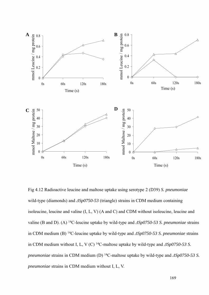
0
0.2
0.4
0.6
0.8
0s 60s 120s 180s
Time (s)
mm
olLe
ucin
e/ m
g pr
otei
n A
0
0.2
0.4
0.6
0.8
0s 60s 120s 180s
mm
olLe
ucin
e/ m
g pr
otei
n
Time (s)
B
0
10
20
30
40
50
0s 60s 120s 180s
Time (s)
mm
olM
alto
se /
mg
prot
ein
mm
olM
alto
se /
mg
prot
ein
0
10
20
30
40
50
0s 60s 120s 180s
Time (s)
DC
Fig 4.12 Radioactive leucine and maltose uptake using serotype 2 (D39) S. pneumoniae
wild-type (diamonds) and ΔSp0750-53 (triangle) strains in CDM medium containing
isoleucine, leucine and valine (I, L, V) (A and C) and CDM without isoleucine, leucine and
valine (B and D). (A) 14C-leucine uptake by wild-type and ΔSp0750-53 S. pneumoniae strains
in CDM medium (B) 14C-leucine uptake by wild-type and ΔSp0750-53 S. pneumoniae strains
in CDM medium without I, L, V (C) 14C-maltose uptake by wild-type and ΔSp0750-53 S.
pneumoniae strains in CDM medium (D) 14C-maltose uptake by wild-type and ΔSp0750-53 S.
pneumoniae strains in CDM medium without I, L, V.
169

170
branched chain amino acids. To ensure that the experimental conditions for the uptake
were correct, 14C-labelled maltose uptake assays were also performed as a positive
control. There was good 14C-labelled maltose uptake in the S. pneumoniae wild-type
both in the presence (Fig 4.12 C) and absence (Fig 4.12 D) of branched chain amino
acids and by the ΔSp0750-53 mutant strain in the CDM medium. However, in the CDM
medium without branched chain amino acids, there was markedly reduced maltose
uptake by the ΔSp0750-53 mutant strain, the reasons are unclear. 14C-labelled
isoleucine, leucine and maltose uptake was also performed, and again showed
negligible uptake both amino acids. Hence, the above results indicate that there is no
significant detectable uptake of BCAA by S. pneumoniae under the in vitro conditions
tested, preventing effective assessment of whether Sp0749-0753 does encode a BCAA
ABC transporter using these assays.
4.4.7 Analysis of substrate specificity by tryptophan fluorescence spectroscopy
In order to investigate whether the Sp0749 lipoprotein binds to branched chain amino
acids (BCAAs), tryptophan fluorescence spectroscopy was performed using purified
and dialysed recombinant His6-Sp0749 lipoprotein (Thomas et al., 2006). Tryptophan
fluorescence spectroscopy analyses the fluorescence emitted as a result of excitation of
electrons by the tryptophan residues present in the protein. In the absence of tryptophan
residues, as is the case for the Sp0749 lipoprotein, this method uses tyrosine residues to
analyse the fluorescence in the proteins. Fig 4.13 A demonstrates that the maximum
emission of fluorescence for the purified His6-Sp0749 lipoprotein occurs at the
wavelength of 309 nm. To test which amino acids induce fluorescence changes in His6-
Sp0749 lipoprotein, branched chain amino acids such as isoleucine, leucine, valine and
other amino acids such as proline, glycine, alanine and threonine were incubated with

171
the recombinant Sp0749 protein. Fig 4.13 B demonstrates that the addition of amino
acids such as proline, glycine, alanine and threonine and valine at a concentration of 50
μM to His6-Sp0749 lipoprotein soon after its stabilization in the spectrofluorimeter had
minor effects on fluorescence. However, the addition of the branched chain amino acid
isoleucine at the concentration of 3.2 μM resulted in a marked change in Sp0749
fluorescence. Further addition of 3.2 μM isoleucine did not result in any quenching of
the changes in fluorescence (data not shown). Therefore to identify the minimum
concentration of the ligand required to obtain maximum fluorescence change,
isoleucine concentrations of 0.64 μM, 0.32 μM (Fig 4.14 A) and 0.16 μM (Fig 4.14 B)
were tested. Addition of 0.16 μM isoleucine did not induce significant changes in
fluorescence of His6-Sp0749, whereas significant changes did occur with the addition
of 0.32 or 0.64 μM isoleucine. No further quenching following the addition of a further
0.64 μM of isoleucine (data not shown). These data suggest that Sp0749 binds to
isoleucine with a high affinity.
To investigate whether His6-Sp0749 lipoprotein binds to leucine, another
potential branched chain amino acid ligand, fluorescence changes upon addition of
leucine at different concentrations were measured. After the addition of leucine at the
final concentration of 3.2 μM, change in fluorescence occurred after a small delay as
shown in Fig 4.15 A, and addition of leucine at the final concentration of 16 μM did not
result in further quenching of fluorescence. When a lower concentration of leucine
(0.64 μM) was tested, only a slow change in fluorescence was observed and further
addition of leucine at 3.2 μM did not result in any fluorescence change. These data
suggest that the His6-Sp0749 lipoprotein binds to leucine with low affinity.
Fluorescence changes upon addition of valine, another branched chain amino
acid ligand was also tested. A marked fluorescence change was observed when valine

0
150000
300000
450000
50 150 250 350 450
Emission wavelength (nm)
Fluo
resc
ence
(arb
itrar
y un
its)A 309 nm
1000
9000
17000
25000
33000
41000
0 100 200 300
Fluo
resc
ence
(arb
itrar
y un
its)
Time (s)
50 μM ProlineB 50 μM Glycine 50 μM Alanine
50 μM Threonine 50 μM Valine
Fig 4.13 Flourescence spectroscopy analysis of the purified His6-Sp0749 lipoprotein (A)
Emission scan of His6-Sp0749 lipoprotein (0.5 μM) demonstrating the maximum flourescence
emission at 309 nm (indicated by an arrow) when excited at the 280 nm wavelength. (B)
Measurement of flourescence change upon addition of 50 μM proline, glycine, alanine,
threonine and valine to 2 μM His6-Sp0749 lipoprotein (point of addition of the amino acid
ligands are indicated by arrows).
172

32000
34000
36000
38000
0 100 200 300 400
Fluo
resc
ence
(arb
itrar
y un
its)
Time (s)
0.32 μM IleuA
0.32 μM Ileu
30000
32000
34000
36000
38000
40000
0 100 200 300
Fluo
resc
ence
(arb
itrar
y un
its)
Time (s)
PO4 buffer B
0.16 μM Ileu 0.32 μM Ileu
Fig 4.14 Flourescence change recorded upon addition of (A) 0.32 μM isoleucine, (B)
0.16 and 0.32 μM isoleucine to 2 μM purified His6-Sp0749 (point addition of ligands
are indicated by arrows).
173

30000
32000
34000
36000
38000
0 200 400 600
Fluo
resc
ence
(arb
itrar
y un
its)
Time (s)
16 μM valine 16 μM valine
B
Fig 4.15 Flourescence change recorded upon addition of (A) 3.2 and 16 μM leucine (B)
16 μM valine to 2 μM purified His6-Sp0749 (point addition of ligands are indicated by
arrows).
22000
24000
26000
28000
0 100 200 300
Fluo
resc
ence
(arb
itrar
y un
its)
Time (s)
3.2 μM LeuA
16 μM Leu
174

175
was added to His6-Sp0749 lipoprotein at the final concentration of 16 μM but
additional valine at the same concentration did not quench the fluorescence change as
shown in Fig 4.15 B. To assess the minimum concentration required for maximum
fluorescence change, 3.2 μM valine was added to the His6-Sp0749 lipoprotein. This
induced only a small fluorescence change. Addition of 50 μM valine in the presence of
50 μM each of a variety of other amino acids failed to induce any changes on
fluorescence, probably due to the large amounts of other amino acids inhibiting binding
the His6-Sp0749 lipoprotein. Overall, the results of the fluorescence binding studies
suggest that the His6-Sp0749 lipoprotein binds to BCAA exhibiting its highest affinity
towards isoleucine, moderate affinity to valine and least affinity towards leucine.
4.4.8 Radioactive substrate binding assay
To further determine the specific binding affinity of the Sp0749 lipoprotein to
isoleucine, leucine and / or valine, radioactive ligand binding assays were performed
using the purified His6-Sp0749 putative lipoprotein. The results suggest that the His6-
Sp0749 lipoprotein bound to 14C-isoleucine, and to a lesser degree to 14C-leucine (p <
0.05) but not to the negative control, 14C-amino isobutyric acid (AIB) (Fig 4.16 A).
Branched chain amino acid transporters (LIV transporters) have been well
characterised in E. coli and P. aeruginosa and have been shown to bind to amino acids
such as L-threonine, L-alanine, L-serine in addition to L-leucine, L-isoleucine and L-
valine (Rahmanian et al., 1973; Hoshino et al., 1992). Therefore His6-Sp0749
lipoprotein was used to investigate if Sp0749 exhibits broader specificity towards
amino acids other than isoleucine, leucine and valine, such as L-threonine. Competitive
ligand binding with leucine, valine, threonine and AIB (negative control) was
performed in the presence of 14C-isoleucine. Fig 4.16 B shows that His6-Sp0749

176
lipoprotein strongly binds to 14C-isoleucine alone and does not bind to 14C-AIB.
However, valine, leucine and threonine inhibited the 14C-isoleucine binding to His6-
Sp0749 lipoprotein and that the His6-Sp0749 lipoprotein bound to valine and threonine
inhibited the 14C-isoleucine binding at similar affinities than leucine. These results
confirm that His6-Sp0749 lipoprotein has greatest affinity towards isoleucine and also
indicates that His6-Sp0749 lipoprotein probably exhibits preferential binding to amino
acid ligands in decreasing order of isoleucine > valine > threonine > leucine. Although
the preferential binding experiment should have been performed with other amino acids
such as serine and alanine in the presence of 14C-isoleucine, it could not be done due to
the lack of purified His6-Sp0749 lipoprotein and time constraints.
4.5 IN VIVO PHENOTYPE ANALYSIS OF THE S. PNEUMONIAE ΔSP0149 AND ΔSP0750-53
DELETION MUTANTS
4.5.1 Competitive index
To investigate the effect on the virulence of S. pneumoniae as the result of Sp0149 and
Sp0750-53 deletion during the pulmonary (IN) and systemic (IP) infection in mouse
models, CIs were performed. Mixed inocula of S. pneumoniae wild-type, ΔSp0149 and
ΔSp0750-53 strains were inoculated in mice by IN and IP routes and the CIs were
determined as described in chapters 2 and 3. Deletion of Sp0149 and Sp0750-53
affected the S. pneumoniae virulence with markedly reduced CIs for the ΔSp0149 and
ΔSp0750-53 strains in both pulmonary (median CIs in lungs: ΔSp0149: 0.028,
ΔSp0750-53: 0.109) and systemic (median CIs in spleen: ΔSp0149: 0.028, ΔSp0750-53:
0.077) models of infection (Fig 4.17). These results are similar to those obtained with
Sp0149- and Sp0750
- IDM mutant strains of S. pneumoniae (mean CI in lungs: Sp0149-:

A
*
Fig 4.16 Radioactive substrate binding assay expressed as pmoles of amino acid bound per
mg of purified His6-Sp0749 lipoprotein (A) Binding affinities of His6-Sp0749 towards 14C-
Labelled isoleucine (Ileu), leucine (Leu) and amino isobutryic acid (AIB) (B) Competitive
amino acid ligand binding of His6-Sp0749 to 14C-AIB (negative control), 14C-isoleucine,
AIB, leucine, valine and threonine in the presence of 14C-isoleucine.
0
0.5
1
1.5
2
2.5
3
3.5
pmol
/ mg
prot
ein
p < 0.05
B
pmol
/ mg
prot
ein
0
0.05
0.1
0.15
0.2
0.2514C-Ileu
177

-5
-4
-3
-2
-1
0Apneumonia: lungs
CI(log10)
∆Sp0149 ∆Sp0750-53
-5
-4
-3
-2
-1
0Bpneumonia: spleens
CI(log10)
∆Sp0149 ∆Sp0750-53
-5
-4
-3
-2
-1
0Csepticaemia: spleens
CI(log10)
∆Sp0149 ∆Sp0750-53
Fig 4.17 In vivo phenotype analysis of ΔSp0149 and ΔSp0750-53 deletion mutants measured
by CIs, expressed as log10 results for the ΔSp0149 and ΔSp0750-53 deletion mutants compared
to the wild-type strain (0100993) in mouse models of pneumonia (A and B), septicaemia (C).
In both the models, each symbol represents the CI obtained from an individual animal.
178

Fig 4.18 Survival of groups of 10 mice were intranasally inoculated with 107 cfu of ΔSp0149
(A) and ΔSp0750-53 (B) deletion mutant strains compared to mice inoculated with the
wild-type S. pneumoniae strains (0100993).
179
0 10 20 30 40 50 60 70 80 90 1000
25
50
75
100WTSp0149
Hours post inoculation
Perc
ent s
urvi
val
0 10 20 30 40 50 60 70 80 90 1000
25
50
75
100WTSp0749
Hours post inoculation
Perc
ent s
urvi
val

180
0.023, Sp0750-: 0.016) (mean CI in spleens: Sp0149
-: 0.067, Sp0750-: 0.017) models of
infection in mice (discussed in chapter 3).
4.5.2 Survival studies
In order to asses the ability of S. pneumoniae strains to cause fatal disease, group of 10
CD1 mice strain were intranasally inoculated with S. pneumoniae (0100993) wild-type,
ΔSp0149 and ΔSp0750-53 strains and their survival curves were determined (Brown et
al., 2001). The mice were inoculated intranasally with the challenge dose of 107 cfu of
the wild-type, ΔSp0149 and ΔSp0750-53 strains and monitored for the progress of
infection. Fig 4.18 demonstrates that there was no difference in the survival of mice
inoculated with the wild-type, ΔSp0149 and ΔSp0750-53 strains. Only 50% of mice
cleared infection and survived after inoculation with the wild-type strain. This was an
unexpectedly high level of clearance, as disease usually progresses into fatal infection
in 80 to 90% of mice when the wild-type S. pneumoniae is inoculated Therefore this
experiment should be repeated to ensure a small survival benefit in mice given the
mutant strains has not been missed.
4.6 SUMMARY
The Sp0148-53 and Sp0749-53 ABC transporters of S. pneumoniae were investigated
in detail for their in vitro and in vivo phenotypes. The relative mRNA expression of
Sp0149 and Sp0749 lipoprotein genes in S. pneumoniae were analysed in THY, human
blood and during infection in mice after IP infection. The results demonstrated that
Sp0149 and Sp0749 genes are expressed during infection in mice, and suggest that the
expression of Sp0749 is actually increased during infection compared to culture in
either THY or blood in the laboratory.

181
From the localisation studies of Sp0149 and Sp0749, Western blot analysis
demonstrated that both Sp0149 and Sp0749 are associated with the membrane
compatible with being lipoproteins. Western blot analysis using the Δlsp and lspc
strains showed that Sp0149 is enzymatically processed by Lsp. Loss of Lsp resulted in
an unusual phenotpye for Sp0749, with reduced band strength in the Δlsp strain,
suggesting Lsp activity is required for efficient attachment of Sp0749. ΔSp0149 and
ΔSp0750-53 deletion mutants were successfully constructed by OEP. In vitro
phenotypes of these strains did not detect any consistent phenotype in growth in
specialised media or laboratory assays associated with the predicted functions of the
corresponding ABC transporter. Uptake assays failed to demonstrate the uptake of
amino acids. However, using the purified His6-Sp0749 lipoprotein, tryptophan
fluorescence spectroscopy and radioactive ligand binding assays demonstrated that
purified Sp0749 binds to BCAAs with a higher affinity towards isoleucine at
submicromolar concentrations.
The in vivo phenotype of ΔSp0149 and ΔSp0750-53 mutant strains was assessed
using CIs, demonstrated a marked decrease in the virulence in pulmonary and
septicaemia models of infection in mice for both strains. However contrasting results
were obtained when survival studies were performed, with no impaired ability of the
ΔSp0149 and ΔSp0750-53 mutant strains to cause fatal disease in a pulmonary model of
infection in mice. Hence loss of these ABC transporters results in a significant
impairment of the mutant strains to compete with the wild-type strain during infection
but the mutants are still able to cause fatal infection.

182
Chapter 5
Investigation of Sp0149 and Sp0749 vaccine
potential against systemic S. pneumoniae disease
The data from chapters 3 and 4 have shown that Sp0149-52 and Sp0749-53 ABC
transporters have role in S. pneumoniae virulence and that the Sp0749 lipoprotein is a
branched chain amino acid binding protein. In this chapter, the potential of the
recombinant Sp0149 and Sp0749 lipoproteins as vaccine candidates are investigated
against S. pneumoniae septicaemia in two different mouse models (outbred CD1 and
inbred BALB/c) using a virulent serotype 2 (D39) as the S. pneumoniae challenge
strain. The immune responses that may be elicited due to intraperitoneal immunisation
of Sp0149 and Sp0749 lipoproteins are also presented.
5.1 EXPRESSION AND PURIFICATION OF SP0149 AND SP0749 LIPOPROTEINS
The phenotype analysis of Sp0149-52 and Sp0749-53 ABC transporters using
Sp0149- and Sp0750
- mutant strains demonstrated marked attenuation in murine
models of septicaemia and pneumonia models compared to the parental wild-type
strain. Hence the lipoprotein components of these ABC transporters were chosen for
investigation as potential novel S. pneumoniae vaccine candidates. This required the
expression and purification of the recombinant lipoproteins of both ABC transporters.
The genes encoding the lipoproteins, Sp0149 for Sp0149-0153 and Sp0749 for
Sp0749-0753, were amplified with the primers listed in chapter 2 using S.
pneumoniae strain 0100993 genomic DNA as the template. To design the primers, the
nucleotide and derived amino acid sequence of Sp0149 and Sp0749 genes were

183
analysed from the available complete genome sequence of a serotype 4 strain of S.
pneumoniae (Tettelin et al., 2001) to identify the lipobox, the conserved amino acid
sequence where the N-terminal signal sequence of the prolipoprotein is enzymatically
cleaved after the attachment to the membrane (Khandavilli et al., 2008; Sutcliffe and
Russell, 1995, Hutchings et al., 2009). The lipobox has the consensus amino acid
sequence [LVI] [ASTVI] [GAS] C, where the leucine is highly conserved and the
cysteine (a site of covalent attachment to the cell membrane phospholipid) is strictly
conserved (Sutcliffe and Russell, 1995). To avoid potential secretion of recombinant
lipoproteins by E. coli, primers were designed to amplify Sp0149 and Sp0749
excluding the N-terminal lipoprotein signal sequence (Table 2.1 and 2.2). Sp0149
primers were designed without flanking restriction sites, and the amplified PCR
product (Fig 5.2 A) ligated into the pQE30UA expression vector (a TA cloning vector
that relies on the addition of terminal thymidine and adenosine residues to the product
of PCR for ligation) (Fig 5.1 A). The Sp0749 gene was amplified using the forward
primer with additional nucleotides encoding a BamHI recognition site, and the
Sp0749 reverse primer with an additional SalI restriction site (Fig 5.2 A). The
amplified PCR product of Sp0749 was ligated into a TA cloning vector (pGEM-T
easy) for efficient restriction digestion, the digested PCR product was gel extracted,
and ligated into the dephosphorylated BamHI and SalI digested pQE30 vector (Fig 5.1
B). The ligation mixture was digested with SacI to linearize the uncut pQE30 vector
(SacI is present between the BamHI and SalI restriction sites) and transformed into
the M15 strain of E. coli. The presence of plasmids containing the correct inserts in
the right orientation was confirmed by PCR using the vector-specific forward primer
(pQE30F) and an insert-specific reverse primer (149Rev and 749Rev) (Fig 5.2 B and
C). The sizes of the PCR products were confirmed by gel electrophoresis. For final

PCR productA
PTS LacO LacO RBS ATG 6x His MCS Stop codonsU
MCSU
Am
pici
llin
pQE30UA3500 bp
ColE
B HI S lIPCR product
B
PTS LacO LacO RBS ATG 6x His MCS Stop codons
SacI
BamHI SalIB
pQE303400 bp
Am
pici
llin
Fig 5.1 (A) Map of Sp0149 lipoprotein gene (800 bp) ligated into prelinearised pQE30UA expression
vector. (B) Map of Sp0749 lipoprotein gene (1000 bp) ligated into BamHI and SalI restriction sites of
ColE
the pQE30 expression vector.
184

Sp0149 Sp0749 -ve control
1000 bp800 bp
A
21 22 23 24 25 26
Clones
1000 bp
B
1 2 3
ClonesC
1000 bp
Fig 5.2 (A) Amplified PCR product of Sp0149 (0.8 kb) and Sp0749 (1 kb) lipoprotein genes from
S. pneumoniae (0100993 strain) genomic DNA used for the ligation into pQE30UA and pQE30
expression vector. (C) Colony PCR of Sp0749 lipoprotein gene using pQE30F and Sp0749 Rev
primers to confirm the orientation of Sp0749 gene after the ligation into pQE30 expression vectorprimers to confirm the orientation of Sp0749 gene after the ligation into pQE30 expression vector.
M15 strain of E. coli clones 1 and 2 showing the correct orientation of Sp0749 gene. (B) E. coli
M15 colony PCR for amplification of inserts containing the Sp0149 lipoprotein gene in the expression
plasmid pQE30UA, using the forward primer pQE30F and the reverse primer Sp0149Rev to confirm
the correct orientation of Sp0149. Clones 21, 23, 24, 26 contain plasmids carrying Sp0149 in the
correct orientation.
185

MKIKKWLGLAALATVAGLA LAAC GNSEKKADNATTIKIANH2
Signal peptide Lipobox Functional domain
A
B
cat cac cat cac cat cac gga tcc cac gtg ata tcc tca atc gct tct tgc gga aac tca gaa aag aaa gca gac aatH H H H H H G S H V I S S I A S C G N S E K K A D N
gca aca act atc aaa atc gca act gtt aac cgt agc ggt tct gaa gaa aaa cgt tgg gac aaa atc caa gaa ttg gtt aaaA T T I K I A T V N R S G S E E K R W D K I Q E L V K
aaa gac gga att acc ttg gaa ttt aca gag ttc aca gac tac tca caa cca aac aaa gca act gct gat ggc gaa gta gatK D G I T L E F T E F T D Y S Q P N K A T A D G E V DK D G I T L E F T E F T D Y S Q P N K A T A D G E V D
ttg aac gct ttc caa cac tat aac ttc ttg aac aac tgg aac aaa gaa aac gga aaa gac ctt gta gcg att gca gat act tacL N A F Q H Y N F L N N W N K E N G K D L V A I A D T Y
atc tct cca atc cgc ctt tac tca ggt ttg aat gga agt gcc aac aag tac act aaa gta gaa gac atc cca gca aac ggaI S P I R L Y S G L N G S A N K Y T K V E D I P A N G
gaa atc gct gta ccg aat gac gct aca aac gaa agc cgt gcg ctt tat ttg ctt caa tca gct ggc ttg att aaa ttg gat gttE I A V P N D A T N E S R A L Y L L Q S A G L I K L D VE I A V P N D A T N E S R A L Y L L Q S A G L I K L D V
tct gga act gct ctt gca aca gtt gcc aac atc aaa gaa aat cca aag aac ttg aaa atc act gaa ttg gac gct agc caaS G T A L A T V A N I K E N P K N L K I T E L D A S Q
aca gct cgt tca ttg tca tca gtt gac gct gcc gtt gta aac aat acc ttc gtt aca gaa gca aaa ttg gac tac aag aaa tcaT A R S L S S V D A A V V N N T F V T E A K L D Y K K S
ctt ttc aaa gaa caa gct gat gaa aac tca aaa caa tgg tac aac atc att gtt gca aaa aaa gat tgg gaa aca tca cct aagL F K E Q A D E N S K Q W Y N I I V A K K D W E T S P KL F K E Q A D E N S K Q W Y N I I V A K K D W E T S P K
gct gat gct atc aag aaa gta atc gca gct tac cac aca gat gac gtg aaa aaa gtt atc gaa gaa tca tca gat ggt ttg gatA D A I K K V I A A Y H T D D V K K V I E E S S D G L D
caa cca gtt tgg taaQ P V W *
Fig 5.3 N-terminal signal sequence and the lipobox of Sp0149 (A) Sp0149 was ligated into pQE30UA
by designing the forward primer from the nucleotide sequence encoding the cysteine residue of the
lipobox. (B) Inframe sequence obtained from sequencing the expression vector (pQE30UA) after
Ligation of Sp0149 into pQE30UA expression vector. Text in bold; 6xHis tag followed by
sequence of pQE30UA.
186
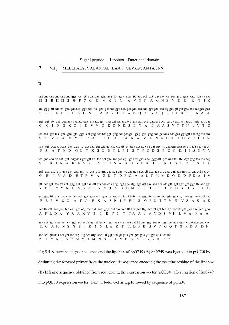
MLLLFALSFVALASVAL LAAC GEVKSGANTAGNSNH2
Signal peptide Lipobox Functional domain
A
B
cat cac cat cac cat cac gga tcc tgt gga gaa gtg aag tct gga gca gtc aac act gct ggt aac tca gta gag gaa aag aca att aaa H H H H H H G F C G E V K S G A V N T A G N S V E E K T I K
atc ggg ttt aac ttt gaa gaa tca ggt tct tta gct gca tac gga aca gct gaa caa aaa ggt gcc caa ttg gct gtt gat gaa atc aat gcc gca I G F N F E E S G S L A A Y G T A E Q K G A Q L A V D E I N A A
ggt ggt atc gat gga aaa caa atc gaa gta gtc gat aaa gat aat aag tct gaa aca gct gag gct gct tca gtt aca act aac ctt gta acc caaG G I D G K Q I E V V D K D N K S E T A E A A S V T T N L V T QG G I D G K Q I E V V D K D N K S E T A E A A S V T T N L V T Q
tct aaa gta tca gca gtc gta gga cct gcg aca tct ggt gcg act gca gct gcg gta gcg aac gct aca aaa gca ggt gtt cca ttg atc tcaS K V S A V V G P A T S G A T A A A V A N A T K A G V P L I S
cca agt gcg act caa gat gga ttg act aaa ggt caa gat tac ctc ttt att gga act ttc caa gat agc ttc caa gga aaa att atc tca aac tat gttP S A T Q D G L T K G Q D Y L F I G T F Q D S F Q G K I I S N Y V
tct gaa aaa tta aat gct aag aaa gtt gtt ctt tac act gac aat gcc agt gac tat gct aaa ggg att gca aaa tct ttc cgc gag tca tac aagS E K L N A K K V V L Y T D N A S D Y A K G I A K S F R E S Y KS E K L N A K K V V L Y T D N A S D Y A K G I A K S F R E S Y K
ggt gaa atc gtt gca gat gaa act ttc gta gca ggt gac aca gac ttc caa gca gcc ctt aca aaa atg aaa ggg aaa gac ttt gat gct atc gttG E I V A D E T F V A G D T D F Q A A L T K M K G K D F D A I V
gtt cct ggt tac tat aat gag gct ggt aaa att gta aac caa gcg cgt ggc atg gga att gac aaa cca atc gtt ggt ggt gat gga ttc aac ggtV P G Y Y N E A G K I V N Q A R G M G I D K P I V G G D G F N G
gag gag ttt gta caa caa gca act gct gaa aaa gca tca aac atc tac ttt atc tca ggc ttc tca act act gta gaa gtt tca gct aaa gct aaaE E F V Q Q A T A E K A S N I Y F I S G F S T T V E V S A K A KE E F V Q Q A T A E K A S N I Y F I S G F S T T V E V S A K A K
gcc ttc ctt gac gct tac cgt gct aag tac aat gaa gag cct tca aca ttt gca gcc ttg gct tat gat tca gtt cac ctt gta gca aac gca gcaA F L D A Y R A K Y N E E P S T F A A L A Y D S V H L V A N A A
aaa ggt gct aaa aat tca ggt gaa atc aag aat aac ctt gct aaa aca aaa gat ttt gaa ggt gta act ggt caa aca agc ttc gat gca gac cacK G A K N S G E I K N N L A K T K D F E G V T G Q T S F D A D H
aac aca gtc aaa act gct tac atg atg acc atg aac aat ggt aaa gtt gaa gca gca gaa gtt gta aaa cca taa*N T V K T A Y M M T M N N G K V E A A E V V K P *
Fig 5.4 N-terminal signal sequence and the lipobox of Sp0749 (A) Sp0749 was ligated into pQE30 by
designing the forward primer from the nucleotide sequence encoding the cysteine residue of the lipobox.
(B) Inframe sequence obtained from sequencing the expression vector (pQE30) after ligation of Sp0749
into pQE30 expression vector. Text in bold; 6xHis tag followed by sequence of pQE30.
187

Induction Purification DialysisA Induction Purification Dialysis
36 KD
U I U I U I CL FT W1 W2 E1 E2 E3 E4
A
Protein concentration: 4.3mg/ml
30KDa including His tag
36 KDa
g
Induction Purification DialysisB
50 KDa
U I U I U I CL FT W1 W2 E1 E2 E3 E4
Protein concentration: 7mg/ml
39.7KDa including His tag
Fig 5.5 Induction, purification and dialysis of recombinant His6-Sp0149 (A) and His6-Sp0749 (B)
lipoproteins from E. coli. Key: U, Uninduced; I, Induced; CL, Cell lysate; FT: Flow through; W1-2,
Washes; E1-4, elutions.; ,
188

189
confirmation of the identity of the PCR products, the PCR products were sequenced
(Fig 5.3 A, B and Fig 5.4 A, B). The recombinant lipoproteins were expressed as
histidine tag fusion proteins, and expressed and purified as described in chapter 2
using conventional techniques. The purity and quantity of His6-Sp0149 and His6-
Sp0749 lipoproteins were checked by running on SDS-PAGE electrophoresis gels.
The obtained molecular weight of His6-Sp0149 and His6-Sp0749 were 30 KDa and 40
KDa respectively, correlating closely with the expected molecular weights after
excluding the N-terminal signal sequence of 29 KDa and 38 KDa respectively. The
purity and concentration of Sp0149 and Sp0749 were improved by dialysis. After
dialysis, His6-Sp0149 and His6-Sp0749 were greater than 95% pure, although faint
high molecular weight bands were observed in both samples (Fig 5.5 A and B), and
had protein concentrations of 4.3 mg ml-1 and 7 mg ml-1 respectively.
For systemic immunisation purposes, PspA and PsaA were used as positive
controls as their efficacy as vaccine candidates against S. pneumoniae infection has
been previously studied.
5.2 CONSERVATION OF SP0149 AND SP0749 GENES IN S. PNEUMONIAE STRAINS
A prerequisite for a protein based vaccine candidate is the conservation and
widespread distribution of the target antigen amongst clinically important capsular
serotypes of S. pneumoniae. Therefore whether the Sp0149 and Sp0749 lipoprotein
genes were present in a range of S. pneumoniae strains was analysed by PCR using
primers Sp0149For and Sp0149Rev, and Sp0749Fwd and Sp0749Rv. Genomic DNA
obtained from strains representative of common multi-locus strain types (MLST)
lineages for each vaccine capsular serotype (3, 4, 6B, 9V, 14, 18C, 19F, 23F, a kind

A
B
C
D
2 3 4 6B 9V 14C 18C 19F 23F His6-Sp0149 protein
Fig 5 6 (A B) Conservation of Sp0149 (A) and Sp0749 (B) genes in the representative strains of
D
2 3 4 6B 9V 14C 18C 19F 23F His6-Sp0749 protein
Fig 5.6 (A, B) Conservation of Sp0149 (A) and Sp0749 (B) genes in the representative strains of
capsular serotypes present in the 7-valent conjugate vaccine. (C, D) Immunoblot of whole cell lysates
of the representative strains of capsular serotypes from the 7-valent conjugate vaccine (plus the ST2,
strain D39 and the ST3 strain 0100993 used for the laboratory studies) probed with anti-Sp0149 (C)
and anti-Sp0749 (D). Identical loading of whole cell lysates were used for (C) and (D).
190

191
gift from Professor Brian Spratt) and from the laboratory strain D39 (capsular
serotype 2) were used as the templates. For both Sp0149 and Sp0749 identical sized
fragments were amplified from all the tested strains, indicating that there was likely to
be a high degree of conservation of these genes amongst S. pneumoniae strains (Fig
5.6 A, B). To further explore whether these proteins are conserved amongst different
S. pneumoniae strains, Western blots were performed against whole cell lysates of the
S. pneumoniae strains discussed above using antisera raised in mice to recombinant
Sp0749 and Sp0149 raised in mice. Anti-Sp0149 antisera gave good signals for all
strains probed, whereas the anti-Sp0749 antisera gave a variable strength signal
between strains, with strong signals only for serotypes 2, 4, 6B and 23F. This suggests
that the expression of Sp0749 lipoprotein varied with strain background in S.
pneumoniae (Fig 5.6 C and D).
5.3 ELISA
To analyse the antibody response after the immunization of mice with the antigens
His6-Sp0149, His6-Sp0749, His6-PspA, and His6-PsaA and with alum, ELISAs were
performed using sera obtained by cardiac puncture from 4 to 5 immunized CD1 or
Balb/c mice 2 weeks after the third booster dose of antigens as described in chapter 2.
Antibody titres of IgG from individual mouse sera were determined. Both His6-
Sp0149 and His6-Sp0749 induced good titres of specific IgG in most mice, albeit with
marked variation between mice. His6-PspA induced IgG titres similar to those
obtained with His6-Sp0149 (median titre of 4.8) and His6-Sp0749 (median titre of 5.4)
(data not shown). Vaccination of Balb/c mice with His6-PsaA, His6-Sp0149 and His6-
Sp0749 also produced good titres of specific IgG antibody with the exception of the
results for one mouse serum versus His6-PsaA (Fig 5.7 A). ELISA was also used to

Fig 5.7 IgG antibody titres measured by ELISA using the sera obtained from Balb/c mice after three
IP immunisations with purified His6-Sp0149, His6-Sp0749 lipoprotein, PsaA (positive control) or alum
alone (negative control). (A) Total IgG titres obtained from the sera of Balb/c mice after the IP
immunisation and cross-reactivity between Sp0149 and Sp0749 lipoproteins (B) IgG1a and IgG2a
antibody titres in the immunised sera against purified His6-Sp0149 and His6-Sp0749 lipoproteins.
0
2
4
6
8
10
12
14AA
ntib
ody
titre
(log 1
0)
His6-Sp0749
Sp0149
His6-Sp0149
Sp0749
His6-PsaA
PsaA
His6-Sp0149
Alum
His6-Sp0749
Sp0749
His6-Sp0149
Sp0149
0
2
4
6
8
10
12
14B
Ant
ibod
y tit
re(lo
g 10)
IgG1a IgG2a
His6-Sp0749
Sp0749
His6-Sp0149
Sp0149
His6-Sp0749
Sp0749
His6-Sp0149
Sp0149
192
cross-reaction

193
demonstrate that there was no cross-reaction of His6-Sp0149 lipoprotein with anti-
Sp0749 IgG antibodies and vice versa, suggesting there was very little or no antigenic
similarity between the two proteins (Fig 5.7 A). As expected no detectable antibody
response was observed in anti-alum antisera. To demonstrate whether the His6-
Sp0149 and His6-Sp0749 lipoproteins elicit a Th1 or a Th2 immune response, IgG1a
and IgG2a antibody titres were analysed (Jomaa et al., 2005). Fig 5.7 B demonstrates
that both antigens mainly stimulate an IgG1a subclass in Balb/c mice following IP
immunisation, suggesting mainly a Th2 response. A similar result was obtained using
CD1 mice sera following IP immunisation. The results of the ELISAs demonstrate
that the purified antigens are immunogenic and induce a good IgG antibody response.
5.4 C3 DEPOSITION
To assess whether anti-Sp0149 and anti-Sp0749 were functional, their effect on the
deposition of C3b/iC3b on several S. pneumoniae strains was analysed by a well-
established flow cytometry assay (Yuste et al., 2005). The pooled sera of anti-Sp0149
and anti-Sp0749 showed a small increase in the C3b/iC3b deposition (Fig 5.8 A), but
the combination of pooled sera containing anti-Sp0149 and anti-Sp0749 increased the
size of the effect (Fig 5.8 B). C3b deposition assay was also performed by incubating
the pooled sera containing anti-Sp0149, anti-Sp0749 and anti-alum with capsular
serotype 3, 6B and 9V (Fig 5.9 A, B, C). Fig 5.9 A and B showed a decrease in the
C3b/iC3b deposition on serotypes 3 and 6B using anti-Sp0149 anti-Sp0749 sera when
compared to anti-alum sera. Fig 5.9 C showed a significantly lower C3b/iC3b
deposition on capsular serotype 9V after incubation in sera containing anti-Sp0749.
Overall, the data suggest that anti-Sp0149 and anti-Sp0749 have a weak effect in

7500
9000A
**
D39
3000
4500
6000
7500
esce
nce
inde
x C
3b
depo
sitio
n alum
anti-Sp0149
anti-Sp0749**
**
0
1500
3000
Flu
ore
1 in 2 dilution1 in 5 dilution
6000
8000
10000
alum
anti-Sp0149 + anti-Sp0749
**
B D39
x C
3b
2000
4000
6000 anti Sp0749
**
Flu
ores
cenc
e in
dex
depo
sitio
n
Fig 5.8 The role of anti-Sp0149 and anti-Sp0749 in the deposition of C3 on the surface of S. pneumoniae
01 in 2 dilution1 in 5 dilution
D39 strain using flow cytometry. The proportion of bacteria positive for the C3 after incubating with
FITC-goat anti-mouse C3 antibody were analysed using Balb/c mice sera. (A) C3 deposition on D39
strain using pooled sera of anti-Sp0149 and anti-Sp0749 on D39 strain (p value <0.01). (B) Effect of C3
deposition on D39 strain upon mixing the pooled anti-Sp0149 and anti-Sp0749 sera (p value ≤ 0.01).
p values were obtained using Student’s-t-test by comparing alum with anti-Sp0149 and anti-Sp0749.
C3 deposition on S. pneumoniae serotypes 3.194

0
50
100
150Fl
oure
scen
cein
dex
C3b
dep
ositi
on
ST3A
0
40
80
120
*
Flou
resc
ence
inde
xC
3b d
epos
ition
alumanti-Sp0149
anti-Sp0749
9VC
Flou
resc
ence
inde
xC
3b d
epos
ition
6BB
0
50
100
150
Fig 5.9 C3 deposition on S. pneumoniae serotypes (A) 3, (B) 6B and (C) 9V (p value < 0.05) using the
pooled sera of the anti-Sp0149 and anti-Sp0749 at 1/2 dilution obtained after the second immunisation
experiment in CD1 mice strain. p values were obtained using Student’s t-test by comparing alum with
anti-Sp0149 and anti-Sp0749.
195

196
increasing deposition of complement on the surface of some S. pneumoniae strains
such as serotypes 3, 6B and 9V.
5.5 OPSONOPHAGOCYTOSIS
Effective antibodies may stimulate phagocytosis directly through Fcγ receptors in
addition to improving opsonisation of bacteria by increasing C3b/iC3b deposition on
their surface. Hence the effect of anti-Sp0149 and anti-Sp0749 on phagocytosis of S.
pneumoniae by human neutrophils was assessed using an in vitro flow cytometry
assay (Lehmann et al., 2000) which measures the association of S. pneumoniae with
phagocytes (Yuste et al., 2008). Fluorescently-labelled bacteria were incubated with
anti-Sp0149 and anti-Sp0749 at different dilutions before incubation with cells from
the neutrophil cell line HL60, and then the association of bacteria and cells measured
by flow cytometry. The role of complement in the in vitro opsonophagocytosis of S.
pneumoniae by HL60 cells was analysed by using heat-treated sera, which inactivates
complement, as a source of antibody in the presence and absence of exogenous
supplementation with purified rabbit complement. Both anti-Sp0149 and anti-Sp0749
increased the proportion of serotype D39 S. pneumoniae associated with HL60 cells
by 40% in 1 in 20 dilution of serum. Heat treatment prevented increased association
of bacteria with HL60 cells for both anti-Sp0149 and anti-Sp0749 sera (Fig 5.10 B).
Similarly, exogenous supplementation with purified rabbit complement also increased
the proportion of S. pneumoniae associated with HL60 cells (Fig 5.10 A), suggesting
the beneficial effect was complement-mediated. Phagocytosis assays were also
performed for representative strains of various S. pneumoniae capsular serotypes (6B,
9V, 23F, 4, 14 and 19) (Fig 5.10 C). In the presence of anti-Sp0149, there was a
statistically significant increase in the association of S. pneumoniae serotype 4 with

40
50A
*
D39
60
80
****
**
B D39
20
30
% a
ssoc
iati
on
**
40
60 **
% a
ssoc
iati
on
0
10
%
1 in 40 1 in 20HBSS0
20
1 in 20 1 in 40HBSS 1 in 20 1 in 40
Role of l t
Role of antibodies l
alum
HBSS
60
80C
** ****
*
**
complement alone
anti-Sp0149
anti-Sp0749
*p-value < 0.05
**p-value < 0.01
40
**
% a
ssoc
iati
on
0
20
9V 46B 23F 14 19
Fig 5.10 (A, B and C) In vitro opsonophagocytosis assay performed using the anti-Sp0149 and
anti-Sp0749 sera obtained after the immunisation experiment in CD1 (A) and Balb/c (B, C) mice
strain. Analysis of the role of complement in the opsonophagocytosis of S. pneumoniae (D39) in the
presence of exogenous addition of purified rabbit complement (A), role of mouse complement andpresence of exogenous addition of purified rabbit complement (A), role of mouse complement and
antibodies (B), and opsonophagocytosis of representative S. pneumoniae vaccine serotypes using anti-
Sp0149 and anti-Sp0749 sera (C). 197

198
HL60 cells compared to alum sera (p < 0.01) but non-significant increases for
serotypes 9V and 14. In the presence of anti-Sp0749, a statistically significant
increase in the association of S. pneumoniae with HL60 cells was observed for
serotypes 6B, 9V, 4, 19 (p < 0.01) and 14 (p < 0.05) compared to sera from mice
vaccinated with alum alone. Overall, these results suggest that anti-Sp0149 and anti-
Sp0749 could improve complement-dependent opsonophagocytosis of S. pneumoniae
and therefore aid immunity against S. pneumoniae infection (Jomaa et al., 2005).
5.6 ACTIVE IMMUNIZATION STUDIES
To assess the efficacy of the purified His6-Sp0149 and His6-Sp0749 lipoproteins as
vaccine candidates, the degree of protection provided by these two lipoproteins in a
septicaemia model of S. pneumoniae infection was evaluated. Three separate IP
immunization experiments were performed using three different challenge doses of
the virulent capsular strain of S. pneumoniae (D39) in naïve outbred CD1 and inbred
Balb/c mice as described in chapter 2.
The initial immunization experiment was performed in male outbred CD1 mice. 10 µg
of the purified His6-Sp0149 and His6-Sp0749 lipoproteins with alum as an adjuvant
and alum alone as the negative control were injected intraperitoneally followed by
two booster doses at 7 days intervals. After 2 weeks of immunization, mice were
challenged by i.p injection of 105 cfu of the virulent capsular serotype 2 S.
pneumoniae strain D39 and the progress of infection monitored over two weeks (Fig
5.11 A, B). Mice immunized with His6-Sp0149 and His6-Sp0749 lipoproteins
exhibited only a small delay of about 4 hours in the progress of infection compared to
alum alone. Of the 19 mice challenged with D39 strain of S. pneumoniae, 100%
mortality was observed in the alum group, while only one mouse was alive in His6-

Fig 5.11 Survival curves after the first IP active immunisation in CD1 mice strain with purified
(A) His6-Sp0149, (B) His6-Sp0749 and alum (negative control) followed by challenge with 105
(B) cfu of S. pneumoniae (D39) for a group of 19 mice.
199
0 10 20 30 40 500
25
50
75
100AlumSp0149
Hours
Perc
ent s
urvi
val
0 10 20 30 40 500
25
50
75
100AlumSp0749
Hours
Perc
ent s
urvi
val
B
A

200
0 10 20 30 40 500
25
50
75
100WTSp0149
Hours
Perc
ent s
urvi
val
0 10 20 30 40 500
25
50
75
100WTSp0749
Hours
Perc
ent s
urvi
val
0 10 20 30 40 500
25
50
75
100WTPspA
Hours
Perc
ent s
urvi
val
Fig 5.12 Survival curves after the second IP active immunisation in CD1 mice strain with (A)
purified His6-Sp0149, (B) His6-Sp0749, (C) PspA (positive control) and alum (negative control)
followed by challenge with 103 cfu of S. pneumoniae (D39) for a group of 19 mice.
B
C
A

0 25 50 75 100 125 1500
25
50
75
100AlumPsaA
Hours
Perc
ent s
urvi
val
0 25 50 75 100 125 1500
25
50
75
100AlumSp0149
Hours
Perc
ent s
urvi
val
0 25 50 75 100 125 1500
25
50
75
100AlumSp0749
Hours
Perc
ent s
urvi
val
Fig 5.13 Survival curves after the third IP active immunisation in Balb/c mice strain with (A)
purified PsaA (positive control), (B) His6-Sp0149, (C) His6-Sp0749 and alum (negative control)
followed by challenge with 104 cfu of S. pneumoniae (D39) for a group of 19 mice201
A
B
C

202
Sp0149 group and 3 mice were alive in His6-Sp0749 group until the end of the
experiment.
To ensure the small differences in the progress of infection in the vaccinated
mice were not due to an overwhelming bacterial challenge, the immunization
experiment was repeated with a lower S. pneumoniae (D39) challenge dose of 103 cfu.
In the second vaccination experiment (Fig 5.12 A, B and C), His6-PspA
(pneumococcal surface protein) was also included as a positive control. His6-Sp0149
and His6-PspA failed to exhibit any protection, but mice immunised with His6-Sp0749
exhibited a non-significant delay (p = 0.08) in the lethal progression of the disease of
approximately 9 hours.
Since the His6-Sp0149 failed to exhibit protection and His6-Sp0749 only exhibited a
delay in the lethal progression of the disease in CD1 mice strain, the immunisation
experiment was repeated in Balb/c mice strain. Balb/c mice were chosen because they
are inbred strain of mice and are relatively resistant to S. pneumoniae infections
unlike the outbred CD1 mouse strain (Gingles et al., 2001). PsaA (pneumococcal
surface adhesion A), a lipoprotein component of S. pneumoniae manganese ABC
transporter was used as positive control and a challenge dose of 104 cfu given by IP
inoculation after immunisation with the purified lipoproteins. Both His6-PsaA and
His6-Sp0149 failed to protect the mice from fatal infection. Again, His6-Sp0749
seemed to delay the lethal progression of infection, although the results were not
statistically significant. After 144 hours only one mouse was alive in the alum group,
3 mice alive in the His6-PsaA and His6-Sp0149 group, and 5 mice alive in His6-
Sp0749 group (Fig 5.13 A, B and C).

203
5.7 SUMMARY
The Sp0149 and Sp0749 lipoproteins were chosen for analysis of their efficacy as
vaccine candidates as the mutant strains affecting the corresponding ABC transporters
had marked attenuation of virulence in murine models of septicaemia and pneumonia.
The PCR–amplified products of the lipoprotein genes, Sp0149 and Sp0749 were
ligated into the expression vectors, pQE30UA and pQE30 respectively (excluding the
N-terminal signal sequence) and the lipoprotein components were successfully
expressed in E. coli and relatively high concentrations of good quality protein
obtained. Vaccination of mice generated high titres of specific antibodies to both
Sp0149 and Sp0749 and there was no evidence of cross reaction of antibodies to
Sp0149 with the protein Sp0749 and vice versa. PCR and western blot analysis
demonstrated that Sp0149 and Sp0749 are conserved in the serotypes present in the 7-
valent conjugate vaccine. In vitro immune assays measuring the effects of polyclonal
murine anti-Sp0149 and anti-Sp0749 on C3b deposition and phagocytosis
demonstrated improvements in both complement deposition and phagocytosis for
several S. pneumoniae strains. Active immunization with the purified Sp0149 and
Sp0749 lipoproteins and challenge with D39 strain of S. pneumoniae indicated that
there was a small but non-significant degree of protection when mice were vaccinated
with recombinant Sp0749.

204
Chapter 6
Investigation of Sp0749 vaccine potential
against S. pneumoniae pneumonia
In this chapter, data regarding the vaccine potential of the recombinant Sp0149 and
Sp0749 lipoproteins against S. pneumoniae pneumonia are presented. Many variations
are adopted in the immunisation-challenge methodology in this chapter compared to
the methodology presented in chapter 5. These variations include mouse strains, use
of adjuvant, S. pneumoniae challenge strains and the route of lipoprotein and S.
pneumoniae administration. In this chapter, the intranasal immunisations and S.
pneumoniae challenge (using 0100993 and D39 strains) experiments were performed
in two different mouse strains (outbred CD1 and inbred CBA/Ca). Cholera toxin was
used as the adjuvant for intranasal immunisation unlike alum which was used as
adjuvant for the intraperitoneal immunisation in chapter 5. The role of Sp0749 alone
and in combination with previously investigated lipoproteins of iron uptake ABC
transporters PiaA and PiuA in eliciting protective immune response was also
investigated and the data are presented in this chapter.
6.1 EXPRESSION AND PURIFICATION OF PIAA AND PIUA LIPOPROTEINS
PiaA and PiuA are lipoproteins components of iron uptake ABC transporters of S.
pneumoniae whose vaccine potential were previously investigated by my supervisor
Dr. Brown. Brown et al previously identified that intraperitoneal and intranasal
immunisation with the recombinant PiaA and PiuA lipoprotein components protected
the mice against S. pneumoniae septicaemia and pneumonia (Brown et al., 2001,

0
1
2
3
4
5
6
Tota
l IgG
and
IgA
titre
(log
10)
IgA (BALF)
IgG (BALF)
His6-PiaA
His6-Sp0749
His6-PiaA
His6-PiaA
His6-Sp0749
His6-Sp0749
CBA miceCD1 mice
IgG (sera)
Fig 6.1 Antibody titres measured by ELISA after IN immunisations with the purified lipoproteins.
Total IgG (serum) and IgA (BALF) antibody titres were determined using purified His6-Sp0749 and
His6-PiaA. Each point represents the anti-PiaA or anti-Sp0749 antibody titre of serum or BALF from
individual mouse. The titres are represented as the reciprocal of the dilution of serum which resulted
in an OD405 ≥ 0.30. No detectable antibody titres were obtained in sera and BALF from PiaA or
PiaA-Sp0749 immunised mice when tested against cholera toxin (CT) (data not shown).
205

206
Jomaa et al., 2005). Therefore the recombinant PiaA and PiuA lipoproteins were used
for the intranasal immunisation and challenge experiments in mice as positive
controls in order to evaluate the vaccine potential of Sp0749. The PiaA and PiuA
lipoproteins were expressed and purified according to (Brown et al., 2001) with some
modifications. The PiaA (Sp1032) and PiuA (Sp1872) lipoprotein genes were
amplified using the primers PiaA F and PiaA R for the PiaA gene and PiuA F and
PiuA R for the PiuA gene and 0100993 genomic DNA as template as described in
chapter 2 and 4. The primers were designed using the TIGR 4 genome to exclude the
DNA sequence encoding the signal peptide as described in chapter 4. The amplified
PCR products were ligated into the expression vector pQE30UA and transformed into
the M15 strain of E. coli. The positive clones for the lipoprotein genes were
confirmed by colony PCR using the vector-specific forward primer (pQE30F) and the
insert specific reverse primer (PiaA R and PiuA R) and by sequencing. The positive
clone for each of the lipoprotein gene was then induced for over expression of His-
tagged lipoproteins and purification of the his-tagged protein performed according to
the QIAexpressionistTM manual. The purified his-tagged lipoproteins were dialysed
and analysed by SDS-PAGE.
6.2 ELISA
In order to analyse the antibody responses after the IN immunisations of mice with
purified lipoproteins (His6-PiaA, and His6-Sp0749), serum and bronchoalveolar
lavage fluid (BALF) were obtained from 4 to 5 immunised CD1 and CBA/Ca mice 2
weeks after the second booster dose of antigens and used for ELISAs (Jomaa et al.,
2005). Cholera toxin (CT) was used as the adjuvant with the lipoproteins and CT
alone was used as the negative control for the immunisation experiments and CT is

207
routinely used as an adjuvant with different S. pneumoniae proteins for IN
immunisations (Pimenta et al., 2006; Shah et al., 2009). Outbred CD1 mice were used
because they are less expensive where as inbred CBA/Ca mice were used because
they are genetically less variable and are susceptible to IN infection (Kadioglu and
Andrew, 2005; Gingles et al., 2001). Purified His6-PiaA lipoprotein was used as
positive control as it has been demonstrated to induce good systemic and mucosal
antibody responses upon pulmonary and systemic immunisations in mice (Brown et
al., 2001; Jomaa et al., 2006). IN immunisation with both His6-PiaA and His6-Sp0749
induced significant serum IgG titres to purified PiaA and Sp0749 in the CD1 mouse
strain (Fig 6.1). Titres of IgG and IgA were measured using BALF obtained from
CBA/Ca mice immunised IN with combination of proteins (His6-PiaA plus His6-
Sp0749). Fig 6.1 demonstrates that His6-PiaA and His6-Sp0749 induced good IgG
titres in BALF, with similar levels to the IgG titres found in sera from CD1 mice.
However, when the IgA titres in the BALF of the CBA/Ca mice were measured,
negligible amounts of anti-Sp0749 IgA were present and anti-PiaA IgA were only
elicited in two mice. These data suggest that after IN immunisation both the proteins
elicit a good systemic and some local IgG response but only a poor mucosal IgA
response.
6.3 BACTERIAL CFUS IN TARGET ORGANS
To further investigate whether IN immunisation of CBA/Ca mice with His6-PiaA and
His6-PiaA plus His6-Sp0749 of CBA/Ca mice protects against S. pneumoniae
challenge, the cfu counts in target organs were determined from the BALF, lungs and
blood. The immunised mice were intranasally challenged with 105 cfu of S.
pneumoniae and the target organs were harvested 48 hours after IN inoculation. The
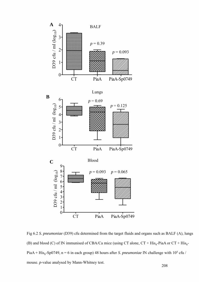
Fig 6.2 S. pneumoniae (D39) cfu determined from the target fluids and organs such as BALF (A), lungs
(B) and blood (C) of IN immunised of CBA/Ca mice (using CT alone, CT + His6-PiaA or CT + His6-
PiaA + His6-Sp0749, n = 6 in each group) 48 hours after S. pneumoniae IN challenge with 105 cfu /
mouse. p-value analysed by Mann-Whitney test.
CT PiaA PiaA-Sp0749 0
1
2
3
4
D39
cfu
/ m
l (lo
g 10)
BALFA
p = 0.39
p = 0.093
CT PiaA PiaA-Sp07490123456789
D39
cfu
/ m
l (lo
g 10)
BloodC
p = 0.093 p = 0.065
208
CT PiaA PiaA-Sp07490
1
2
3
4
5
6
D39
cfu
/ m
l (lo
g 10)
BLungs
p = 0.69p = 0.125

CT
His6-PiaA
His6-PiaA+ His6-Sp0749
BALF Median (IQR)
Lungs Median (IQR)
1.2 (0.93-3.1)
1.1 (0.57-1.09)
0.35 (0-1.09)
4.5 (4.1-4.67)
4.4 (3.3-4.7)
2.7 (1.9-4.0)
Blood Median (IQR)
6.6 (6.05-6.67)
5.7 (5.2-6.16)
4.9 (3.9-6.3)
Table 6.1 Median and interquartile range (IQR) of S. pneumoniae (D39) cfu determined from BALF,
lungs and blood after the IN immunisation of CBA/Ca mice with CT alone, CT + His6-PiaA, CT +
His6-PiaA + His6-Sp0749 (n = 6 in each group) followed by S. pneumoniae challenge 48 hours after
the inoculation of 105 cfu / mouse.
209

210
target organs were then homogenised in sterile PBS, serially diluted and appropriate
dilutions were plated to determine the cfu / ml in the target organs (Fig 6.2 and Table
6.1). Although CBA/Ca mice immunised intranasally with His6-PiaA plus His6-
Sp0749 had reduced S. pneumoniae cfu in BALF (Fig 6.2 A), lung (Fig 6.2 B) and
blood (Fig 6.2 C) compared to mice immunised with CT alone, and mice immunised
with His6-PiaA alone had reduced S. pneumoniae cfu in blood, these reductions failed
to attain statistical significance. However, data were obtained from a relatively small
number of mice and varied widely between mice in the same vaccine group.
6.4 CYTOSPINS OF BALF
BALF cellular content was analysed before and 48 hours after the S. pneumoniae
challenge (D39) of the CBA/Ca mice immunised with CT alone, His6-PiaA and His6-
PiaA plus His6-Sp0749 proteins. Total cell counts in BALF were determined using a
haemocytometer and microscopy of the cytospins was used to assess the proportion of
macrophages, neutrophils and lymphocytes present as described in chapter 2. Total
cell counts of BALF did not demonstrate significant differences before and after the S.
pneumoniae challenge (Table 6.2). The BALF cytospin differential counts (Fig 6.3)
demonstrated significantly higher neutrophil numbers post-challenge with S.
pneumoniae in all three immunised groups (Fig 6.3 B), whereas the proportion of
macrophages and lymphocytes were significantly reduced (Fig 6.3 A and B).
Neutrophil counts were very low in all the three immunisation groups before
challenge and therefore the data are not shown. A statistically significant increase in
the neutrophil counts was obtained in the post-challenge mice immunised with both
His6-PiaA (p = 0.0022) and His6-PiaA plus His6-Sp0749 (p = 0.0043) when compared
to pre-challenge groups.

Table 6.2 Median and IQRs of the total cell counts in BALF and lungs of CBA/Ca mice
after the IN immunisation with CT alone, CT + His6-PiaA and CT + His6-PiaA + His6-
Sp0749 (n = 6) pre and post S. pneumoniae challenge (D39, 105 cfu / mouse).
CT
His6-PiaA
His6-PiaA+His6-Sp0749
BALF Median (IQR)
5.5 (5.2-5.7)
5.5 (5.3-5.5)
5.5 (5.5-5.6)
Pre Post
5.9 (5.7-6.1)
5.8 (5.6-5.8)
5.6 (5.4-5.7)
Lungs Median (IQR)
6.3 (6.3-7.09)
6.0 (5.9-6.2)
6.6 (5.8-7.3)
Pre Post
6.2 (6.0-6.6)
5.9 (5.7-6.2)
5.9 (5.7-6.3)
211

CT
His6-PiaA
His6-PiaA +
His6-Sp0749
** p < 0.01* p < 0.05
Fig 6.3 Differential cell counts of macrophages, lymphocytes and neutrophils present in the
BALF of IN immunised CBA/Ca mice (n = 6 in each group) with CT alone, CT + His6-PiaA,
and CT + His6-PiaA + His6-Sp0749 pre (A) and post (B) S. pneumoniae challenge (D39, 105
cfu / mouse). p value for pre vs post-challenge comparisons analysed by Mann-Whitney test.
A
Pre-challenge
Post-challenge
0
20
40
60
80
100
Macrophages Lymphocytes
BALF
diff
eren
tial c
ount
s
0
25
50
75
100
Macrophages Lymphocytes Neutrophils
BALF
diff
eren
tial c
ell c
ount
s
**
B
**
**
**** *
**
**
**
212

213
6.5 IMMUNE RESPONSE TO CT, HIS6-PIAA AND HIS6-PIAA PLUS HIS6-SP0749 AFTER
IN IMMUNISATION AND S. PNEUMONIAE CHALLENGE
To investigate the effects of immunisation of mice with CT, His6-PiaA or His6-PiaA
plus His6-Sp0749 on the host immune response, leukocytes were isolated from the
BALF and lungs of CBA/Ca mice immediately before and 48 hours after challenge
with S. pneumoniae as described in chapter 2. Total cell counts of the homogenised
lung were determined using a haemocytometer, and cells incubated with
fluorescently-labelled antibodies to lymphocytes and macrophage surface markers
were analysed by flow cytometry to identify the proportion of cells belonging to each
lymphocyte subgroup and the proportion of these cells that are activated (Brown et
al., 2002).
There were only small differences in lung cell counts before and after S.
pneumoniae challenge between the vaccine groups (Table 6.2). The results of the cell
surface marker experiments were highly variable between mice and it was difficult to
identify clear trends. There were no clear differences in the proportions of CD4, CD8
and B cells between the vaccinated groups with the exception of activated CD4 and
CD8 cells in the lungs (Fig 6.5 C, D).
In the BALF, the proportion of activated CD4 and CD8 (CD45RB –ve) cells
(Fig 6.4 C, D) showed a statistically significant decrease in all the three post-
challenge vaccinated groups compared to pre-challenge (for all groups p < 0.01 for
the comparison of results pre and post-challenge of activated CD4 cells) (for all
groups p < 0.05 for the comparison of results pre and post-challenge of activated CD8
cells).
In the lungs, there was a statistically significant increase in the proportion of

Fig 6.4 Flow cytometry analysis of the host’s immune response in the BALF by immune cell surface
staining following the IN immunisation (pre) and subsequent challenge (post). The proportion of (A)
CD4 positive cells, (B) CD8 positive cells, (C) CD4 positive cells (CD45RB –ve) (p < 0.01 for
comparisons pre vs post for all groups), (D) CD8 positve cells (CD45RB-ve) (p < 0.05 for
comparisons pre vs post for all groups). p value were analysed by Kruskal-Wallis test
BALFA
556065707580859095
% C
D8
cells
-ve f
or C
D45
RB
D
Pre
CT PiaA PiaA+Sp0749
Post
CT PiaA PiaA+Sp0749
BALF
Pre
CT PiaA PiaA+Sp0749
01020304050607080
% p
ositi
ve fo
r CD
4
Post
CT PiaA PiaA+Sp0749
BALFB
Pre
CT PiaA PiaA+Sp0749
Post
CT PiaA PiaA+Sp0749
0
10
20
30
40
50
% p
ositi
ve to
CD
8
40
50
60
70
80
90
100
% C
D4
cells
-ve
for C
D45
RB
C
Pre
CT PiaA PiaA+Sp0749
Post
CT PiaA PiaA+Sp0749
BALF**
214

Fig 6.5 Flow cytometry analysis of the host’s immune response in the lungs by immune cell surface
staining following the IN immunisation and challenge. The proportion of (A) CD4 positive cells, (B)
CD8 positive cells (p < 0.05 for comparisons pre Vs post for all groups), (C) CD4 positive cells
(CD45RB –ve) (p < 0.01 for comparisons pre Vs post) and (D) CD8 positive cells (CD45RB -ve) (p <
0.01 for comparisons pre Vs post for all groups). p values were analysed using Kruskal-Wallis test
0
25
50
75
% p
ositi
ve fo
r CD
8
0102030405060708090
% p
ositi
ve fo
r CD
4
A
Pre
CT PiaA PiaA+Sp0749
Post
CT PiaA PiaA+Sp0749
LungsB
Pre
CT PiaA PiaA+Sp0749
Post
CT PiaA PiaA+Sp0749
Lungs*
0
25
50
75
100
% C
D4
cells
-ve
for C
D45
RB
C
CT PiaA PiaA+Sp0749
CT PiaA
Lungs
PiaA+Sp0749
Pre Post
** *
0
25
50
75
100
% C
D8
cells
-ve
for C
D45
RB
D
Pre
CT PiaA PiaA+Sp0749
Post
CT PiaA PiaA+Sp0749
Lungs**
215

Fig 6.6 Flow cytometry analysis of the proportion of B220 positive cells in the (A) BALF and (B)
lungs (p < 0.05 for comparisons pre vs post for all groups) by immune cell surface staining following
the IN immunisation and challenge. p values analysed using Kruskal-Wallis test.
0
10
20
30
40
50
60
% p
osit
ive
for
B22
0
A
Pre
CT PiaA PiaA+Sp0749
Post
CT PiaA PiaA+Sp0749
BALF* **
0
10
20
30
40
50
60
70
% p
ositi
ve fo
r B22
0
B
Pre
CT PiaA PiaA+Sp0749
Post
CT PiaA PiaA+Sp0749
Lungs
*
216

0
10
20
30
40
50
60
% p
ositi
ve fo
r I-A
-I-E
B
Pre
CT PiaA PiaA+Sp0749
Post
CT PiaA PiaA+Sp0749
BALF
Fig 6.7 Flow cytometry analysis of the host’s immune response in the BALF (A, B) and lungs (C, D)
by immune cell surface staining following the IN immunisation and challenge. The proportion of
CD80 positive for macrophages in the (A) BALF (p < 0.05 for comparisons pre vs post for all groups)
and (C) lungs. The proportion of macrophages positive for MHC class II antigens (I-A-I-E) in the
BALF (B) and lungs (D) (p < 0.05 for comparisons pre vs post for all groups). p values were analysed
using Kruskal-Wallis test
0
25
50
75
100
% p
ositi
ve fo
r CD
80
C
Pre
CT PiaA PiaA+Sp0749
Post
CT PiaA PiaA+Sp0749
Lungs
A
Pre
CT PiaA PiaA+Sp0749
Post
CT PiaA PiaA+Sp0749
BALF
01020304050607080
% p
osit
ive
for
CD
80
*
0
25
50
75
100
% p
osit
ive
for
I-A
-I-E
D
Pre
CT PiaA PiaA+Sp0749
Post
CT PiaA PiaA+Sp0749
Lungs*
217

218
CD8 positive lymphocytes in the His6-PiaA plus His6-Sp0749 compared to CT alone
after S. pneumonie challenge (for all groups p < 0.05 for the comparison of results pre
and post-challenge) (Fig 6.5 B). His6-PiaA plus His6-Sp0749 vaccinated group
showed a statistically significant decrease in the proportions of CD4 and CD8
(CD45RB –ve) cells post-challenge compared to pre and post-CT and pre-PiaA (for
all groups p < 0.05 for the comparison of results pre and post-challenge) (Fig 6.5 C,
D). However, the proportion of B220 positive cells may be increased in the lungs of
His6-PiaA plus His6-Sp0749 vaccinated group after S. pneumoniae challenge
compared to pre-CT (for all groups p < 0.05 for the comparison of results pre and
post-challenge) (Fig 6.6 B).
The activation of macrophages before and after the S. pneumoniae challenge
of the immunised mice were analysed using antibodies to the cell surface marker
CD80 and the MHC class II antigen I-A-I-E (Fig 6.7). In the BALF, the proportion of
CD80 positive cells seemed to be similar in the CT group but decrease in the His6-
PiaA plus His6-Sp0749 vaccinated groups upon S. pneumoniae challenge (Fig 6.7 A)
(for all groups p < 0.05 for the comparison of results pre and post-challenge). In
contrast in the lungs, the proportion of CD80 positive cells showed no statistically
significant increase in the His6-PiaA and His6-PiaA plus His6-Sp0749 vaccinated
groups upon S. pneumoniae challenge compared to all the pre-challenge groups and
post-CT (Fig 6.7 C). However, the BALF and lungs results using the (I-A-I-E) surface
marker did not mirror the results for the CD80 cell surface marker (Fig 6.7 B, D).
Therefore the significance of the CD80 marker results remain unconfirmed.
Overall, these results suggest that intranasal immunisation with His6-PiaA and
His6-PiaA plus His6-Sp0749 may affect components of the cellular immune response

219
to S. pneumoniae pneumonia. However these experiments need to be repeated to
identify clear patterns.
6.6 HISTOLOGICAL ANALYSIS OF INFLAMMATION AFTER S. PNEUMONIAE
CHALLENGE OF INTRANASALLY VACCINATED MICE
To investigate the degree of inflammation leading to lung injury of mice vaccinated
IN with His6-PiaA or His6-PiaA plus His6-Sp0749, lungs obtained from the pre and
post, S. pneumoniae challenged mice (48 hours) underwent histological analysis. Each
lung was scored as follows: 1 (visible inflammatory change), 2 (minimal swelling of
alveolar walls and changes in architecture), 3 (increased swelling with presence of
erythrocytes and inflammatory cells and an increase in type II pneumocytes), 4
(considerable haemorrhage with inflammatory cell influx, widespread alveolar
disorganisation with interstitial swelling and pneumocyte proliferation) as described
in chapter 2. A total score of the level of inflammation for each mouse was obtained
by multiplying the percentage of involved lung by the mean score for the areas
analysed, and data presented as medians with IQRs. Table 6.3 shows the levels of
inflammation as medians and interquartile ranges of IN immunised CBA/Ca mice
with His6-PiaA or His6-PiaA plus His6-Sp0749 proteins followed by S. pneumoniae
challenge. Histological analysis of lungs showed there was a statistically significant
increase in the level of inflammation after S. pneumoniae challenge in mice
vaccinated with His6-PiaA or His6-PiaA plus His6-Sp0749 compared to those
vaccinated with CT alone (Table 6.3). These data suggest that vaccination with these
proteins may cause a more rapid inflammatory influx into lungs during subsequent S.
pneumoniae challenge however these experiments needs to be repeated.

CT
His6-PiaA
His6-PiaA+His6-Sp0749
Pre-challenge Median (IQR)
Post-challenge Median (IQR)
11.25 (6.6-25.6)
22.5 (16.6-35.6)
76.25 (45.4-94)
84.2 (56.3-113.3)
185.8 (147.9-375)
130.8 (103.3-300)
Table 6.3 Histological analysis of the level of inflammation in the lungs of CBA/Ca mice
after the IN immunisation with CT alone, CT + PiaA and CT + PiaA + Sp0749 pre and post
S. pneumoniae challenge (D39, 105 cfu / mouse). Degree of lung inflammation before and
after the S. pneumoniae challenge is represented as medians and IQRs. p values were
analysed using Kruskal-Wallis test (CT Vs PiaA or PiaA-Sp0749, p < 0.001 or p < 0.01 or p
< 0.05) and Mann-Whitney test for all groups (Pre-challenge vs post-challenge, p = 0.0022).
220

221
6.7 SURVIVAL OF THE HIS6-SP0149 AND HIS6-SP0749 IMMUNISED MICE AFTER S.
PNEUMONIAE CHALLENGE
In order to asses the efficacy of the purified His6-Sp0149 and His6-Sp0749 as
intranasal vaccine candidates, the degree of protection provided by these two
lipoproteins in the pulmonary models of infection was evaluated. Purified His6-PiaA
and His6-PiuA lipoproteins were used as positive controls as both the lipoproteins
have been investigated as vaccine candidates in the pulmonary and systemic models
of infection in mice (Brown et al., 2001; Jomaa et al., 2006). Four intranasal
immunisation experiments were performed using four different challenge doses of the
virulent capsular strain of S. pneumoniae (D39 and 0100993) in naïve outbred CD1
mice and inbred CBA/Ca mice as described in chapter 2.
Two separate IN immunisations were performed in CD1 mice using 2.5µg of
the purified His6-PiaA, His6-PiuA, His6-Sp0749, His6-PiaA plus His6-Sp0749 and
His6-PiaA plus His6-Sp0149 plus His6-Sp0749 lipoproteins with CT as an adjuvant
and CT alone as the negative control were injected intranasally followed by two
booster doses at 7 days intervals. Two weeks after immunization, mice were
challenged by intranasal injection of 106 cfu of the virulent capsular serotype 2 S.
pneumoniae strain D39 and the progress of infection monitored over two weeks (Fig.
6.1 and 6.2). Fig 6.8 A and B represents pooled data of two separate experiments in
which mice were immunised IN with His6-PiaA and His6-PiaA plus His6-Sp0749 and
D39 challenge experiments in CD1 mice strain (n = 10+10). IN immunisation with
His6-PiaA (p = 0.405) did not exhibit protection, although His6-PiaA plus His6-
Sp0749 exhibited a statistically non-significant delay (p = 0.083) in the progression of
S. pneumoniae infection compared to CT. However the immunisation with His6-PiuA

0 25 50 75 1000
25
50
75
100CTPiaA
Hours
Perc
ent s
urvi
val
0 25 50 75 100 1250
25
50
75
100AlumPiaA+Sp0749
Hours
Perc
ent s
urvi
val
0 25 50 75 100 1250
25
50
75
100CTPiuA
Hours
Perc
ent s
urvi
val
Fig 6.8 Survival curves after the intranasal (IN) immunisation of CD1 mice with cholera toxin CT
(squares), PiaA, PiuA, Sp0749 and combinations of PiaA with Sp0749 (diamonds). IN immunisation
of CD1 mice with CT alone, CT + PiaA (p = 0.405) (A), CT + PiaA + Sp0749 (p = 0.083) (B) and CT
+ PiuA (C) followed by IN challenge with S. pneumoniae D39 (106 cfu / mouse). A and B are the
pooled results of two separate IN immunisation and challenge experiments (n = 10 + 10, n =
number of mice) and p values are analysed by the log rank. 222
C
B
A

A
B
Fig 6.9 Survival curves after the IN immunisation of CD1 mice (n = 10) with CT (squares)
Sp0749 and combinations of PiaA with Sp0149 and Sp0749 (diamonds). IN immunisation of
CD1 mice with CT alone, CT + Sp0749 (A), CT + PiaA + Sp0149 + Sp0749 (B) followed by
IN challenge with S. pneumoniae D39 (106 cfu / mouse). p values are analysed by the log
rank test.
223
0 25 50 75 100 1250
25
50
75
100CTSp0749
Hours
Perc
ent s
urvi
val
0 25 50 75 100 1250
25
50
75
100CTPiaA+Sp0149+Sp0749
Hours
Perc
ent s
urvi
val

A
B
C
Fig 6.10 Survival curves after the IN immunisation of CBA/Ca mice (n = 15) with CT
(squares), PiaA, Sp0749 and combinations of PiaA with Sp0749 (diamonds). IN immunisation
of CBA/Ca mice with CT alone, CT + PiaA (A), CT + PiaA + Sp0749 (B) followed by IN
challenge with S. pneumoniae D39 (105 cfu / mouse). IN immunisation of CBA/Ca mice (n =
20) with CT alone, CT + Sp0749 (C) followed by IN challenge with S. pneumoniae 00100993
strain (105 cfu / mouse). p values are anaysed by the log rank test. 224
0 10 20 30 40 50 600
25
50
75
100CTPiaA
HoursPe
rcen
t sur
viva
l
0 10 20 30 40 50 600
25
50
75
100CTPiaA+Sp0749
Hours
Perc
ent s
urvi
val
0 10 20 30 40 50 600
25
50
75
100CTSp0749
Hours
Perc
ent s
urvi
val

225
(Fig 6.8 C), His6-Sp0749 (Fig 6.9 A), and His6-PiaA plus His6-Sp0149 plus His6-
Sp0749 (Fig 6.9 B) lipoproteins in CD1 mice strain (n = 10) did not show any
protection compared to CT alone.
The third intranasal immunisation experiment was performed in the inbred
CBA/Ca mouse strain .Groups of CBA/Ca mice (n = 15) were immunised with CT
alone, His6-PiaA, His6-PiaA plus His6-Sp0749 followed by intranasal challenge with
105 cfu of S. pneumoniae D39. Mice immunised with His6-PiaA, His6-PiaA plus His6-
Sp0749 showed only delayed progression of disease after S. pneumoniae challenge
(Fig 6.10 A and B). The fourth intranasal immunisation experiment was also
performed in CBA/Ca mice strain with groups of mice (n = 20) immunised with CT
alone, His6-Sp0749 followed by intranasal challenge with 105 cfu of S. pneumoniae
0100993 strain. Fig 6.10 C shows that His6-Sp0749 failed to exhibit any protection
and that the survival curve of His6-Sp0749 was almost similar to mice immunised
with CT alone.
Overall these data suggest there may be some protective effect in these models
after the combined immunisation with PiaA and Sp0749 lipoproteins, but any
protective effect of individual lipoproteins was too weak to be detected by the
infection models used for these experiments.
6.8 SUMMARY
PiaA and PiuA his-tagged lipoproteins were expressed and purified in order to use as
positive controls for the intranasal immunisation studies. Following intranasal
immunisation with PiaA and Sp0749, good IgG titres were demonstrated in sera
obtained from CD1 mice and BALF obtained from CBA/Ca mice. However, anti-
PiaA IgA titres in BALF were variable and no detectable anti-Sp0749 IgA titres were

226
found. When the S. pneumoniae load was determined from the target organs such as
BALF, lungs and blood 48 hours after challenge of vaccinated mice with S.
pneumoniae, statistically non-significant reductions in S. pneumoniae cfu were
demonstrated in the BALF, lungs and blood of mice vaccinated with PiaA plus
Sp0749 compared to CT group. Mice immunised with PiaA alone showed non-
significant reductions of S. pneumoniae cfu in blood only compared to CT group. The
differential cell counts in the BALF of the CT, PiaA and the PiaA plus Sp0749
immunised mice demonstrated a statistically significant increase in the neutrophil
infiltration in PiaA vaccinated mice upon challenge with S. pneumoniae compared to
CT alone. Flow cytometry analysis of the lymphocytes in the BALF and lung
homogenate of the immunised CBA/Ca mice before and after the S. pneumoniae
challenge by cell surface marker staining was performed. Flow cytometry analysis
after immune cell surface staining did not indicate a particular trend and the results
were highly variable between mice. Intranasal immunisation demonstrated that PiaA
in combination with Sp0749 only slightly delayed the progression of lethal S.
pneumoniae infection. In summary, although vaccination with PiaA and Sp0749
causes evidence of an altered immune response to subsequent pneumonic infection,
the protective effects of this seems limited.

227
Chapter 7
Discussion
There has been strong interest in S. pneumoniae proteins as vaccine candidates
because they offer the potential to overcome the limitations of the currently available
S. pneumoniae vaccines based on capsular polysaccharide antigens. Important
features for a protein to qualify as a potential S. pneumoniae vaccine candidate are
surface exposure, immunogenicity, conservation amongst a range of clinically
important serotypes and the ability to elicit a protective immune response. A role for
virulence would be beneficial as this would suggest that the protein candidate is
expressed during systemic infection. The antibodies elicited against the protein in the
host may inhibit the function of the protein thereby affecting the S. pneumoniae
virulence. In addition, an important role in virulence means that the expression of the
protein is necessary for the survival of S. pneumoniae in the host to cause disease.
Genes encoding such proteins which are necessary for the S. pneumoniae survival in
the host rarely undergo mutations. Therefore use of such proteins as vaccine antigens
induces immunity to help prevent immune evasion of S. pneumoniae. Simultaneous
immunisation with several S. pneumoniae surface proteins has been shown to protect
mouse models against S. pneumoniae pulmonary and systemic infections (Briles et
al., 1997; Briles et al., 1996; Brown et al., 2001; Briles et al., 2003; Ogunniyi et al.,
2007) and a vaccine containing several protein antigens is likely to offer advantages
in efficiency and greater serotype coverage.
One group of surface proteins which are important for the survival of S.
pneumoniae in the host, thereby playing important role as virulence determinants, are

228
ABC transporters (Unsworth and Holden, 2000) (Garmory and Titball, 2004) (Benton
et al., 2004). Bioinformatic analysis of the genome of S. pnemoniae has identified as
many as 73 ABC transporters (Harland et al., 2005), many of which are predicted to
be involved in nutrient acquisition from the host (Bergmann and Hammerschmidt,
2006). Several S. pneumoniae ABC transporters are known to be important for
virulence such as PiaA, PiuA (Brown et al., 2004) and PsaA (Dintilhac et al., 1997),
but the role of many S. pneumoniae ABC transporters during virulence has yet to be
investigated. I have therefore chosen 11 ABC transporters from the annotated
completely sequenced genome of capsular serotype 4 of S. pneumoniae (TIGR4) that
have previously not been characterised and investigated their role in virulence and
selected two of these for assessment as potential vaccine candidates. The S.
pneumoniae ABC transporters chosen for these studies were the majority of the
remaining 18 uncharacterised ABC transporters that are organised as operons of three
or more genes containing at least two of the three main components of ABC
transporters (the lipoprotein, ATPase and transmembrane permeases). Disruption of
these ABC transporters was thought to be more likely to give a strong phenotype as
most or all of the components of that ABC transporter would be co-transcribed.
Components of some of the ABC transporters chosen for this study (Sp0092, Sp0150,
Sp2086 and Sp2108) have been identified as potential virulence factors by STM
screening (Hava and Camilli, 2002; Lau et al., 2001). Mutant strains were constructed
by disruption of the predicted first gene of the operon using IDM to try and ensure the
disruption of transcription of the whole operon and therefore the generation of a
stronger phenotype. Nine mutant strains of the chosen 11 ABC transporter operons of
S. pneumoniae were successfully constructed by IDM. However I failed to obtain any
mutant strain for the putative amino acid transporter encoded by Sp0707-0711 even

229
after repeated attempts, possibly because some regions of the genome may be
resistant to transformation or because that disruption of this gene may prove to be
lethal for the bacterium. In another mutant strain, Sp2084-, the putative phosphate
transporter encoded by Sp2084-2087 had an unstable mutation, a relatively common
problem for mutant strains made by IDM due to the duplicated regions flanking the
inverted plasmid allowing homologous recombination to excise the plasmid
containing the gene for antibiotic selection.
BLAST searches indicated that several of the chosen ABC transporters are
likely to be sugar ABC transporters (Sp0090-92, Sp0846-48, Sp1796-98, and Sp2108-
10), which is not surprising given the large range of sugars that S. pneumoniae is
thought to be able to utilise (Tettelin et al., 2001). BLAST searches also indicated that
Sp0607-10, Sp0707-11 and Sp0749-53 are putative amino acid ABC transporters.
Sp0749-53 was identified a putative branched-chain amino acid (BCAA) transporter.
The S. pneumoniae genome also contains genes encoding enzymes required for
BCAA synthesis (Sp0445-50) (Hendriksen et al., 2008) which may partially
compensate for BCAA uptake by S. pneumoniae. Sp0149 was identified as a putative
cation lipoprotein, but other genes of this operon did not correlate with the likely
function of cation uptake. However another transport classification system (Saier, Jr.,
2000) indicated Sp0148-52 as a putative methionine ABC transporter. A similar
discrepancy between the BLAST alignment and the transport classification system
was also observed when predicting the possible substrate for Sp0846-48. BLAST
alignment predicted Sp0846-48 as a putative sugar transporter, whereas the transport
classification system predicted it is a possible nucleoside ABC transporter. Detailed
characterisation of these ABC transporters is needed to confirm their ligand binding
properties.

230
Many ABC transporters seem to be highly conserved amongst different S.
pneumoniae strains (eg PiaA, PsaA and PiuA) (Whalan et al., 2006), (Sampson et al.,
1997). BLAST alignment of the predicted lipoprotein genes of the chosen ABC
transporters to 18 other available S. pneumoniae genomes demonstrated a high degree
of amino acid conservation between S. pneumoniae strains (Basavanna et al., 2009).
BLAST alignment of the chosen ABC transporters showed that 8 of the eleven ABC
transporters have the greatest degree of amino acid homology to various streptococci
(Sp0090-92, Sp0148-52, Sp0607-10, Sp0707-11, Sp0749-53, Sp0846-48, Sp1796-98
and Sp2108-10). However no close homologues amongst streptococci were found for
Sp1688-90 (a putative cation ABC transporter), Sp2084-87 (a putative sugar
transporter), or Sp1824-26 (a putative phosphate ABC transporter). Previously, the
iron uptake ABC transporter PiaA was shown to be highly conserved in S.
pneumoniae strains but absent in oral streptococci, including the S. mitis group
(Whalan et al., 2006), suggesting horizontal gene transfer of this loci into S.
pneumoniae from unrelated bacterial species has occurred, and a similar mechanism
might explain why Sp1688-90, Sp2084-87 and Sp1824-26 are not found amongst
other streptococci.
The genetic organisation of all the 9 ABC transporters were analysed using
TIGR4 genome, and their organisation investigated by RT-PCR using 0100993 strain
of serotype 3 RNA. The RT-PCR analysis using 0100993 strain showed that the
probable transcriptional structure of the Sp0149-152, Sp0749-53, Sp0846-48,
Sp2108-10 loci correlated with the TIGR4 genome. Amplification of cDNA from the
RNA of 0100993 S. pneumoniae grown in THY for these genes was straightforward
suggesting these regions are significantly expressed during growth in THY. However
Sp1794-99 and Sp1822-28 ABC transporter loci were poorly expressed in THY and

231
therefore it was difficult to assess the transcriptional organisation of these operons
using RT-PCR. The transcriptional structure of other ABC transporter loci did not
match well with what would be predicted from the TIGR4 genome (Sp0090-92,
Sp0607-11, Sp1689-91), and sequencing of these regions in the 0100993 S.
pneumoniae strain may help clarify these results.
PCR amplification of the junctions of Sp1794-99 and Sp1822-28 ABC
transporter loci using 0100993 genomic DNA as template showed that some genes of
these ABC transporter loci may be absent. As discussed earlier, the BLAST alignment
of Sp1794-99 ABC transporter loci against 18 available S. pneumoniae genomes
showed that this region was absent in some of the S. pneumoniae strains (Basavanna
et al., 2009). However why some genes of Sp1822-28 ABC transporter loci were
absent in 0100993 strain is not clear. I therefore speculate whether the expression of
Sp1822-28 is induced under certain conditions such as oxidative, osmotic stress, or a
minimal nutrient supply and that the genes of this region may not be constitutively
expressed in THY.
Which transcriptional regulators regulate these S. pneumoniae ABC
transporters warrants further investigation as the information is very limited at
present. In other bacteria such as S. mutans and S. agalactiae, genes of methionine
uptake are under the control of the transcriptional regulator MetR / MtaR respectively
(Sperandio et al., 2007) (Shelver et al., 2003), and whether Sp0149-152 is regulated
in a similar fashion needs to be identified. However, Sp0749-73 and the BCAA
synthesis operon Sp0445-50 have been shown to be negatively regulated by CodY
(Hendriksen et al., 2008), although the relationship between CodY repression of
Sp0749-53 requires further investigation to clarify its functional consequences and
role during bacterial physiology.

232
The phenotype of each of the mutant strains was analysed using in vitro and in
vivo CIs. CI tests the ability of the mutant strain to replicate compared to the wild-
type parental strain in the test condition (as long as the mutant phenotype cannot be
complemented by the extracellular secretion of the missing protein(s) by the wild-type
strain). In vitro CIs performed in a rich complete medium (THY) showed no growth
defects of the mutant strains except a mild impairment for Sp0750-. Similarly, all the
mutant strains grew well in the osmotic stress media (THY + 100 mM NaCl) except
strain Sp0610- which had partial attenuation thereby suggesting a role of this ABC
transporter for aiding growth in high osmotic stress conditions. Osmotic stress is one
physiological stress that pathogens have to overcome in order to cause invasive
infection, and decreased resistance to osmotic stress has been associated with
decreased virulence for some pathogens. For example, expression of the three-gene
operon of S. pneumoniae, phgABC, has been reported to be important for growth
during high osmotic conditions and is required for the full virulence in pulmonary and
systemic infection (Brown et al., 2004), and a proline ABC transporter of S. aureus is
important for the virulence of S. aureus and protects against high osmolarity by
accumulating proline within the cell (Schwan et al., 2004).
To indicate a potential role of the selected S. pneumoniae ABC transporters
during invasive infections, CIs were performed on human blood as an ex vivo culture
system that closely represents at least some of the physiological conditions during
infection such as limited availability of nutrients, pH, and relative hyperosmolarity.
Culture on blood also replicates some of the interaction with different cell types and
immune system components such as neutrophils and complement that occur during
infection (Mereghetti et al., 2008). In this more stringent environment, mutant strains
Sp0090-, Sp0149
-, Sp0610
-, Sp0750-, Sp1824
- were moderately impaired in growth

233
compared to the wild-type, suggesting that these ABC transporters may contribute to
growth of S. pneumoniae in normal physiological fluid. These strains would therefore
be predicted to have impaired virulence and in general, the CIs in human blood
mirrored the in vivo CIs in mouse models of pneumonia and septicaemia. Unlike the
in vitro CIs, where the mixed inocula of wild-type and the mutant strains were grown
for a period of 8 hours in various growth conditions, the in vivo CIs performed in the
mouse models of infection results in the interaction of the mutant and the wild-type
strains in various cells, tissues and body compartments during a period of 24-48
hours. This allows even small differences in virulence between mutant and wild-type
strains to be identified. Of the nine ABC transporter mutant strains successfully
constructed, seven were impaired in full virulence in mouse models of sepsis and / or
pneumonia (Sp0090-, Sp0149
-, Sp0610
-, Sp0750
-, Sp0846
-, Sp1824
- and Sp2108-). Of
the seven attenuated ABC transporter mutant strains, five were partially reduced in
virulence (Sp0090-, Sp0610
-, Sp0846
-, Sp1824
- and Sp2108-) and two were markedly
attenuated (Sp0149- and Sp0750
-) in mouse models of sepsis and / or pneumonia. For
two of the ABC transporter mutant strains (Sp1690-, Sp1796
-) the impairment in
virulence was relatively small, comparable to the effect of loss of a single iron
transporter (Brown et al., 2002), and this maybe because their functions are partially
redundant. For example, BLAST alignments suggest several of the ABC transporters
investigated encode sugar transporters, and more than 30% of S. pneumoniae
transporters are predicted to be sugar transporters which also includes non-ABC
transporter uptake systems such as phosphoenolpyruvate-dependent sugar transporters
(Tettelin et al., 2001). Hence disrupting the function of a single sugar ABC
transporter could be compensated for by the others or by non-ABC transporter uptake
mechanisms such as phosphoenolpyruvate-dependent sugar transporters (Tettelin et

234
al., 2001). Dual mutations in genes encoding components of ABC transporters with
related functions may have a much more marked effect on virulence as has been
shown for the PiuA and PiaA iron transporters (Brown et al., 2001). Two strains
Sp0149- and Sp0750
- were markedly more attenuated in virulence in pneumonia and
septicaemia models of S. pneumoniae infection compared to the other strains. The
functions of proteins encoded by these genes are indicated by BLAST searches to be
methionine / cation and BCAA uptake respectively. Sp0750- had greater degree of
attenuation of virulence in pneumonia model than in the septicaemia model of S.
pneumoniae infection suggesting that this ABC transporter may be particularly
important for the infection of the respiratory tract. As well as ABC transporters
encoded for by groups of genes investigated in this and previous studies, there are
many additional ABC transporters components encoded by isolated single genes or
gene pairs within the TIGR4 genome, some of which STM screens suggest affect
virulence (Hava and Camilli, 2002; Lau et al., 2001). These ABC transporter
components also warrant further investigation, although their specific putative
functions may be in general be even less apparent than those encoded by several
adjacent genes in putative operons.
S. pneumoniae disease process is complex and requires the bacterium to
acclimatise in different environments such as nasopharynx, lungs, blood, ear and
brain. The optimal survival of S. pneumoniae in any of these niches would depend on
the expression of virulence factors (Ogunniyi et al., 2002). Since Sp0149 and Sp0749
ABC transporters have been shown to be important for S. pneumoniae virulence as
demonstrated by in vivo CIs, their relative abundance in different conditions were
investigated. The relative expression of Sp0149 and Sp0749 mRNA, were compared
with the well-characterised virulence protein genes, psaA and Sp1386 (potD,
235
polyamine ABC transporter) (Shah et al., 2008) in the THY, human blood and mice
blood 12 hours after IP infection with S. pneumoniae. Relative mRNA expression of
the virulence genes (Sp0149, Sp0749, psaA and Sp1386) in THY were used as
baseline. psaA was abundantly present in the THY and human blood, therefore I did
not investigate the relative abundance of psaA gene in mouse blood due to limited
recovery of total S. pneumoniae RNA. Also similar work has been performed by
Ogunniyi et al (Ogunniyi et al., 2002) and they have demonstrated that psaA is
expressed abundantly during in vitro conditions and during pulmonary and systemic
S. pneumoniae infection in mice. Sp1386, a lipoprotein component of a polyamine
ABC transporter demonstrated lower expression than psaA and Sp0149 in THY, but
increased mRNA expression was demonstrated in human blood and in the blood of
mice recovered 12 hours after IP inoculation of S. pneumoniae suggesting a role in
this physiological fluid and thereby contributing to S. pneumoniae virulence. Shah et
al have also demonstrated that the expression of the S. pneumoniae polyamine ABC
transporter is up-regulated during oxidative stress and high temperature (Shah et al.,
2008) such results that are compatible with the lower expression of Sp1386 in
favourable growth conditions such as THY.
In the mouse blood, the relative abundance of Sp0149 was increased compared
to THY and human blood. However, Sp0749 was only expressed in mouse blood and
no detectable levels were found in THY and human blood. Previous work by Carlos
Orihuela and collegues has shown that the Sp0149 lipoprotein gene and Sp0750-53
operon are both up-regulated in a rabbit model of meningitis suggesting their gene
expression may contribute towards S. pneumoniae virulence (Orihuela et al., 2004).
The genes of Sp0749-53 are under the negative control of the transcriptional regulator
CodY (Hendriksen et al., 2008) but the relationship between CodY repression, in
236
vitro and in vivo expression of Sp0749-53 and BCAA transport requires further
investigation. There is also a need to repeat the experiment using more sensitive
technique such as real-time PCR, which would allow more accurate quantification of
the differential expression of these ABC transporter genes in both in vitro and in vivo
(systemic and pulmonary murine models).
The subcellular localisation of the lipoproteins Sp0149 and Sp0749 was
investigated using Triton X-114 extracts of the S. pneumoniae membrane associated
proteins. Immunoblots identified that Sp0149 and Sp0749 are membrane localised.
Reports for other Gram positive bacteria such as Mycobacterium tuberculosis (Sander
et al., 2004) Listeria monocytogenes (Reglier-Poupet et al., 2003) and S. suis (De et
al., 2003) have shown that their lipoproteins are processed by Lsp, and that disruption
of lsp in L. monocytogenes (Reglier-Poupet et al., 2003), S. suis (De et al., 2003) and
recently by my supervisor’s group in S. pneumoniae (Khandavilli et al 2008) results
in retention of the N terminal signal peptide by lipopeptides. Hence I investigated the
effects of deletion of lsp on the Sp0149 and Sp0749 lipoproteins. As expected, the
Sp0149 gave a higher molecular weight signal in the whole cell lysate of Δlsp strain
due to incomplete processing of the lipoproteins in the absence of Lsp. However in
the whole cell lysate of lspc strain, the Sp0149 did not revert to the normal wild-type
molecular weight. Whether the differences of Sp0149 is due to the rate of migration
of proteins or due to incomplete Lsp function in the complementation mutant as
described by Khandavilli and collegues (Khandavilli et al 2008) needs to be
investigated. With respect to Sp0749 there was a weak signal in the whole cell lysate
of the Δlsp strain, but the intensity of expression was similar in the wild-type and lspc
strain. At present there is no clear explanation for why there is lower expression of
Sp0749 in the absence of Lsp. However, the localisation of Sp0149 and Sp0749

237
within the membrane and that they are affected by Lsp does indicate that both are
lipoproteins and could be investigated as potential vaccine candidates against S.
pneumoniae. Analysis of the effect of loss of lgt on the processing of Sp0149 and
Sp0749 would also be of interest as this would be predicted to result in loss of both
lipoproteins from the cell membrane associated protein fraction.
The data obtained with the disruption mutant strains suggested that the ABC
transporters encoded by Sp0149-52 and Sp0749-53 have the most crucial roles during
S. pneumoniae infection. For further investigation of the function of the Sp0149-52
and Sp0749-53 ABC transporters, mutant strains were made in the S. pneumoniae
0100993 background in which Sp0149 and Sp0750-53 were deleted from Sp0149-52
and Sp0749-53 operons using constructs made by overlap extension PCR (OEP)
(Shevchuk et al., 2004). OEP is a relatively new technique for constructing deletion
and complemented mutant stains to allow complete replacement of an operon with an
antibiotic resistance cassette. Different techniques have also been used in recent years
to construct mutations in S. pneumoniae such as IDM, in vitro mariner transposon
mutagenesis, and gene replacement using Janus cassette and OEP. IDM however has
been used to for many years to make disruption mutants in S. pneumoniae using
plasmids as it is rapid and a relatively simple technique. However, due to homologous
recombination of duplicated regions flanking the inverted plasmid, the entire plasmid
may be excised allowing the plasmid containing the gene for antibiotic selection to be
excised and this is a relatively common problem with IDM. The advantages of OEP
are that it is a relatively quick method to obtain the desired constructs for mutations
and does not involve plasmids and restriction digestion. However the main
disadvantage of OEP is the induction of random mutation due to amplification by
PCR into the mutant construct. Two other relatively new methods to construct S.

238
pneumoniae mutants are in vitro mariner transposon mutagenesis and gene
replacement using Janus cassettes. In vitro mariner transposon mutagenesis is rapid
and is particularly useful for the identification of genes required for growth and
survival, but causes random insertions into the target sequence rather than a clean
deletion (Akerley et al., 1998). Gene replacement using Janus cassette is a method for
negative selection particularly useful to construct substitutions and inframe deletions
which allows the selection for gene acquisition and loss. Therefore use of Janus
cassette circumvents the problem of accumulation of antibiotic markers in the mutated
strains (Sung et al., 2001), but is more complex than OEP and was not necessary as
multiple mutations in the one strain were not planned. I therefore used OEP to
construct deletion and complementation mutant strains of Sp0149 and Sp0750-53 in
S. pneumoniae as this method is relatively quick, stable and it is possible to construct
in frame mutations. For phenotype analysis of mutant strains it is important if possible
to have a complemented mutant so any phenotypes observed is linked directly to the
genetic manipulation of the gene of interest. However, although after several attempts
I was able to construct the complementation construct of Sp0149 this construct failed
to transform in S. pneumoniae, and I was also unable to obtain the complementation
construct of Sp0750-53 possibly due to its larger size.
The role of S. pneumonaie Sp0149-53 and Sp0749-53 ABC transporters were
investigated in detail for their possible substrate specificity and role in virulence using
the ΔSp0149 and ΔSp0750-53 S. pneumoniae deletion mutants. The role of Sp0149-53
ABC transporter was investigated in vitro by comparing the growth rates of the wild-
type and the ΔSp0149 deletion mutant strains under normal laboratory conditions.
Particular phenotypes associated with the specific functions of the Sp0149-53
(requirement of particular cations, sensitivity to streptonigrin) were also investigated.

239
These assays did not show impairment of growth in the tested conditions such as
cation depleted media, but as discussed earlier recent data suggested that Sp0149-53
is a methionine ABC transporter (Saier, Jr., 2000) rather than a cation transporter.
Further investigation of this transporter is required using uptake assays and ligand
binding assays. Methionine ABC transporters are functionally characterised in Gram
negative bacteria such as E. coli (Kadner, 1974) and S. enterica serovar Typhimurium
(Grundy and Ayling, 1992). To my knowledge, there have been no reports
demonstrating a role for the methionine uptake in the virulence of Gram positive
pathogens including S. pneumoniae. In S. mutans, the Sp0149-53 homologues encode
AtmBDE, an ABC transporter that was identified as a likely methionine uptake
transporter, with functional studies suggesting AtmBDE has a strong affinity for L-
methionine but also transports other sulphur compounds such as selenomethionine, D-
methionine, or homocysteine (Sperandio et al., 2007). MtaR, a transcriptional
regulator, was also identified which not only regulates methionine synthesis and / or
uptake genes (AtmBDE) in S. mutans (Sperandio et al., 2007) and S. agalactiae, but
was also essential for the survival of S. agalactiae in a rat model of septicaemia
therefore contributing towards virulence (Shelver et al., 2003). It would therefore be
interesting to investigate the role of Sp0149-53 as a putative methionine ABC
transporter using methionine as the potentially preferred substrate.
In order to identify particular phenotypes associated with the specific
functions of the Sp0749-53 putative BCAA ABC transporter, toxicity towards
azaleucine, radioactive uptake assays, tryptophan fluorescence spectroscopy and
radioactive binding assay were performed. Loss of Sp0749-53 ABC transporter did
not result in a particular requirement for BCAA in the conditions used such as THY,
and the wild-type strain was resistant to the toxicity of azaleucine, so we were unable

240
to identify significant uptake of the BCAA leucine by S. pneumoniae. Hence in the
conditions used for these experiments (ie., the complete medium THY and the defined
medium CDEM) there seems to be very little BCAA uptake by S. pneumoniae.
Tryptophan fluorescence spectroscopy and radioactive binding assays using 14C-
leucine, isoleucine and valine demonstrated that His6-Sp0749 binds specifically to
BCAA, strongly supporting the hypothesis that Sp0749-53 encodes a BCAA ABC
transporter. Both the tryptophan fluorescence spectroscopy and radioactive binding
assays suggest that His6-Sp0749 has the highest affinity for isoleucine, moderate
affinity for leucine, with the least affinity for valine (Basavanna et al., 2009). These
data suggest strongly that Sp0749 to 0753 does encode a BCAA transporter despite
the lack of evidence for BCAA uptake in vitro.
In mouse models of infection the Δ750-53 and the ΔSp0149 deletion strains were
both significantly out-competed by the wild-type strain. These data suggest there is an
important role for the S. pneumoniae BCAA and putative methionine ABC
transporters specifically during in vivo growth, a conclusion that is supported by
previous publications at least for BCAAs, with disruption of BCAA synthesis
affecting virulence of the unrelated pathogens B. pseudomallei and M. bovis (Atkins
et al., 2002; McAdam et al., 1995). However the effect of loss of Sp0750-53 and
Sp0149 on virulence were only detectable in competitive infection experiments and
after IN inoculation with the ΔSp0149, ΔSp0750-53, or wild-type strain, progression
of infection was similar. Hence, Sp0149-53 and Sp0749-53 ABC transporters do not
have a powerful effect on S. pneumoniae virulence. This could reflect redundancy
associated with S. pneumoniae ABC transporters. For example, the S. pneumoniae
genome contains genes encoding enzymes required for BCAA synthesis (Sp0445-50)
(Tettelin et al., 2001) (Hendriksen et al., 2008) may partially compensate for impaired

241
BCAA uptake in vivo. Whether the loss of Sp0149 is perhaps compensated by other
gene(s) involved in methionine metabolism is not known. Exactly why loss of genes
encoding putative methionine and BCAA affects virulence is not clear. The most
obvious explanation is a nutritional requirement for methionine and BCAA in vivo.
However, the marked difference in CI between growth in blood and during infection
for the Sp0149- and Sp0750
- mutant strains indicates that the loss of virulence maybe
more complex than simple impaired growth under physiological conditions. As stated
above BCAA metabolism and uptake genes in various bacteria have been identified to
be regulated by CodY. CodY, a nutritional repressor identified in L. lactis (Guedon et
al., 2001), C. difficile (Dineen et al., 2007), S. pyogenes (Malke et al., 2006), and L.
monocytogenes (Bennett et al., 2007), S. aureus (Majerczyk et al., 2008) and S.
pneumoniae (Hendriksen et al., 2008) is known to inhibit the genes involved in amino
acid metabolism. It is highly conserved in Gram positive bacteria and responds to
intracellular concentrations of BCAAs and GTP (guanosine triphosphate), thereby
sensing carbon, nitrogen availability and aids the bacterium to adapt to survive under
harsh conditions. Therefore CodY plays a central role in the regulation of bacterial
metabolism (Somerville and Proctor, 2009; Tojo et al., 2008) and will influence
growth and therefore virulence in vivo. In S. pneumonaie, the CodY regulon consists
of genes involved in BCAA and general amino acid metabolism. Upregulation of
BCAA metabolism and uptake genes including Sp0749-53 was observed in a codY
mutant strain suggesting that CodY directly controls the intracellular BCAAs in S.
pneumoniae (Hendriksen et al., 2008).
As the Sp0149-0153 and Sp0749-53 ABC transporters are important for
virulence, and their corresponding lipoprotein genes, Sp0149 and Sp0749 are surface
localised, they were chosen for further investigation as vaccine candidates analogous

242
to other protein antigens such as PiaA, PiuA and PsaA. Western blotting of the cell
lysates of clinically important serotypes responsible for S. pneumoniae infections with
polyclonal antibodies to Sp0149 and Sp0749 demonstrated the presence of the
lipoproteins Sp0149 and Sp0749 in all serotypes investigated. Furthermore, the
proteins were of the same size in all strains and PCR amplified identical sized gene
products. This suggests that both Sp0149 and Sp0749 are identically sized and
conserved amongst S. pneumoniae strains, making them potentially attractive vaccine
candidates. Compatible with the results of the in vitro assays and RT-PCR analysis for
Sp0749 function, the expression of Sp0749 generally seemed to be low compared to
the expression of Sp0149, with probing with anti-Sp0749 giving a variable and much
weaker signal compared to probing with anti-Sp0149 serum. ELISA demonstrated
that Sp0149 and Sp0749 lipoproteins are immunogenic as they elicited good IgG
antibody titres comparable to that of PsaA which was used as positive control. The
antibody results were predominantly IgG1 subclass, although a IgG2a subclass
response was also observed. Hence the lipoproteins may induce a Th2 immune
response similar to that observed upon immunisation with PiaA and PiuA (Jomaa et
al., 2005). No cross reactivity between antibodies to the two proteins was seen
suggesting that both the lipoproteins are antigenically dissimilar. Incubation with anti-
Sp0149 and anti-Sp0749 resulted in small but a significant increase in the deposition
of complement on S. pneumoniae and aided phagocytosis. Antibodies to iron uptake
ABC transporters do not inhibit ABC transporter function (Jomaa et al., 2005), and
although antibodies to Sp0149 and Sp0749 seems to have immunological effects, it is
not known whether anti-Sp0149 and anti-Sp0749 also inhibits the function of Sp0149
and Sp0749 lipoproteins. However this would be difficult to assess due to potential

243
redundancy and the lack of a specific phenotype in vitro that could be used to assess
the function of these ABC transporters (Brown et al., 2001).
Immunisation with Sp0149 or Sp0749 lipoproteins and subsequent challenge
by the intraperitoneal route with D39 strain to simulate a model of S. pneumoniae
septicaemia involving experiments were performed twice in the CD1 mouse strain. In
the initial immunisation experiment, CD1 mice were challenged with 105 cfu of D39
strain of S. pneumoniae. Only a minor delay in the development of fatal infection was
observed in the mice immunised with the lipoprotein groups. Since the lipoproteins
failed to confer strong protection, one of the questions that arose is whether these
lipoproteins are unstable in the laboratory storage conditions and whether they have
degraded from the time of purification to the immunisation or failed to induce good
antibody responses. Western blotting suggested that this was not the case, as
polyclonal antisera raised against the purified Sp0149 and Sp0749 lipoproteins
produced specific signals when used to probe whole cell lysates of S. pneumoniae and
the purified His6-Sp0149 and His6-Sp0749. The disappointingly weak protective
effect of immunisation with His6-Sp0149 and His6-Sp0749 may reflect a challenge
dose that may be overwhelmingly high leading to fast multiplication of S. pneumoniae
in the host even if the vaccine antigens had elicited protective responses.
As well as a negative control group vaccinated with alum alone, the efficacy
of these lipoproteins as vaccines also needs to be evaluated in comparison with a
positive control whose efficacy as a vaccine candidate has been previously
determined. I therefore used PspA, a surface protein known to be protective against
several clinically important serotypes, as a positive control for the second
immunisation experiment. Unfortunately PspA is a weak vaccine candidate against
D39 (McDaniel et al., 1991), but this protein was the only alternative vaccine protein

244
available in our laboratory at the time of second immunisation experiment. A lower
challenge dose of 103 cfu of S. pneumoniae was used in the second immunisation
experiment to increase the likelihood of detecting protective responses. PspA did not
exhibit protection against D39 strain of S. pneumoniae, and the survival of mice
vaccinated with Sp0149 was similar to that of mice immunised with PspA or alum
alone. Sp0749 showed a statistically non-significant delay the development of fatal
infection, perhaps suggesting that Sp0749 may be an alternative protein vaccine
antigen. CD1 mice were used for the immunisation-challenge experiments because
they are an outbred strain, as well as being cheaper, experiments using outbred mice
are more likely to be of greater relevance than positive results obtained with an inbred
mouse population. The immunisation experiment was therefore repeated in the inbred
mice strain Balb/c. Unfortunately, no protection against S. pneumoniae challenge was
observed after immunisation with the Sp0149 or Sp0749 lipoproteins, nor with the
positive control protein PsaA. Why Sp0149, Sp0749 and PsaA did not demonstrate
protection despite the in vitro data suggesting they induce a significant and
immunologically relevant response is not clear. One probable explanation is that the
intraperitoneal route may be too stringent an infection route, and that any protective
response elicited by the lipoproteins may be masked by the severe S. pneumoniae
septicaemia induced by this route of inoculation.
As S. pneumoniae pneumonia is a common disease affecting both the elderly
and infants to which the available vaccines do not provide efficient protection,
intranasal immunisation and challenge studies were performed in mice to investigate
whether immunisation with the protein antigens can protect against S. pneumoniae
pneumonia. The protective efficacy of the purified Sp0149, Sp0749 by itself or in
combination with the previously investigated lipoprotein vaccine candidates PiaA and

245
PiuA (Jomaa et al., 2006) against S. pneumoniae pneumonia was investigated through
the intranasal route (instilling the purified lipoproteins and bacteria into mouse
nostrils, IN). This simulates the natural route of infection in humans. Cholera toxin
was used as the adjuvant as it is well established and used as adjuvant with different
S. pneumoniae proteins for IN immunisation experiments (Jomaa et al., 2006)
(Pimenta et al., 2006; Shah et al., 2009). Experiments assessing survival after IN
immunisation and challenge were performed in outbred CD1 mice due to the cost,
whereas inbred CBA/Ca mice were used for experiments assessing immunological
responses in more detail in order to minimise mouse to mouse variation in results that
may compromise the results of the experiment and its analysis.
The antibody titres, types of immune cell activation and histological
examination of the degree of inflammation in the lungs elicited against the immunised
lipoproteins were analysed. Sp0749 in combination with PiaA elicited good IgG titres
in the sera and respiratory secretions such as BALF after the IN immunisation,
although greater IgG titres were demonstrated in the sera after IP immunisation with
Sp0749. Perhaps surprisingly given the immunisation route Sp0749 specific IgA
antibodies were not detected and PiaA specific IgA antibodies were detected in the
BALF of only two mice immunised with PiaA and Sp0749. Nevertheless, ELISA
demonstrated that the IN immunisation with Sp0749 and PiaA can induce specific
local and systemic IgG antibodies.
To investigate the detailed host immune responses upon S. pneumoniae
challenge, cells were isolated from the BALF and lungs of CT, PiaA and
PiaA+Sp0749 immunised mice before and 48 hours after the S. pneumoniae challenge
and analysed using labelled lymphocyte and macrophage antibody markers and flow
cytometry. The total cell counts in the BALF and lungs determined using a

246
haemocytometer indicated only small differences before and after the S. pneumoniae
challenge between the vaccine groups. The results of the cell surface marker
experiments were highly variable between mice and it was difficult to identify clear
trends. However, there was an indication of an increase in the proportion of B220
cells. As expected, differential cell counts of the BALF cytospins indicated an
overwhelming increase in the neutrophil infiltration upon S. pneumoniae challenge in
all groups. Macrophage activation in the vaccine groups upon S. pneumoniae
challenge was also analysed by flow cytometry using two macrophage surface
markers (CD80 and I-A-I-E). In the lungs, the results suggests both the active vaccine
groups had an increase in CD80 positive cells suggesting a greater recruitment of
activated macrophages in these mice compatible with a positive immunological
benefit of vaccination with lipoproteins. However, flow cytometry using I-A-I-E
surface marker did not mirror the results obtained using the CD80 surface marker,
therefore the significance of the results obtained by CD80 are unconfirmed.
Interestingly the histological examination of the lung tissues from the control
and vaccine groups indicated more inflammation in the vaccine groups than the
control group, with greater degree of inflammation in the PiaA immunised group
upon S. pneumoniae challenge. Why there was increased level of inflammation in the
vaccine groups after the S. pneumoniae challenge is not immediately clear, but could
indicate a more rapid inflammatory response in actively vaccinated mice which could
aid protective immunity. An analysis of the cytokine response would help identify
whether there are differences in the inflammatory response between vaccinated and
control mice after S. pneumoniae challenge. Inflammatory cytokines such as IL-1,
TNF and IL-6 are present in both the patients (Dehoux et al., 1994) and mice with
pulmonary infection caused by S. pneumoniae (Bergeron et al., 1998). Blocking the

247
expression or action of cytokines such as TNF-α, and IL-6 impairs the host’s immune
response to S. pneumoniae with reduced bacterial clearance and reduced survival in
experimental mice models of pneumonia (Takashima et al., 1997; Van der et al.,
1997). However, studies have demonstrated that the host’s cytokine response depends
on the specific strain of S. pneumoniae in addition to the type of infection such as
pneumonia / septicaemia and host’s genetic background (Mohler et al., 2003).
Furthermore, differences in bacterial cfu present at different time points could make
the interpretation of a direct comparison of cytokine levels between groups complex.
Hence a careful selection of the mouse strain and time point for cytokine analysis will
be necessary. To determine which of the observations on differences in the immune
response after IN immunisation with PiaA and Sp0749 or the control group are
consistent and biologically significant and assess their potential importance for
control of S. pneumoniae infection, these experiments will need to be repeated.
Despite the evidence of some modifications of the immunological response to
infection in the pneumonia model in mice vaccinated with the lipoproteins, little
protection was found after intranasal vaccination with the lipoproteins. Sp0149 alone
and in combination with PiaA and PiuA in the triple vaccination gave no protection.
Sp0749 showed a statistically non-significant (p = 0.083) weak level of protection
when combined with PiaA, but not when used alone or in combination with Sp0149
and PiaA in a triple antigen vaccination. The results obtained with PiaA and PiuA
contrast to previously published data on these antigens as intranasal vaccines (Jomaa
et al., 2006). The reasons why both PiaA and PiuA did not protect in both CD1 and
CBA/Ca mice strain when previously they had shown protection against pulmonary S.
pneumonaie infection (Jomaa et al., 2006) are not clear, but could be due to variations
in the serotype of S. pneumoniae used (1x 10⁶ cfu of serotype 1 in CBA/Ca mice).

248
The results of S. pneumoniae cfu enumeration 48 hours after the S. pneumoniae
challenge in the BALF, lungs and blood of immunised CBA/Ca mice were compatible
with the results of the survival experiments. There was a statistically non-significant
decrease in the bacterial counts in BALF, lungs and blood of mice immunised with
PiaA and PiaA+Sp0749 when compared to CT alone, suggesting that the
immunisation with these lipoproteins may perhaps have some weak effect on the
clearance of S. pneumoniae that might not be enough to influence overall survival in
this model. These experiments require repetition to identify whether the differences in
cfu are consistent and to increase the numbers for a more accurate assessment of their
statistical significance.
One explanation for the weak protective effects on S. pneumoniae clearance
from the target organs is that CBA/Ca mice are particularly susceptible to intranasal
infection. Previous work by Gingles et al. demonstrated that the median survival time
of CBA/Ca mice after challenge with 106 cfu of D39 was 28 hours, whereas that of
Balb/c was greater than 168 hours (Gingles et al., 2001). In the future it would be
important to enumerate S. pneumoniae cfu at an earlier time point in CBA/Ca mice
strain or to use Balb/c mice, although these mice may be too resistant to S.
pneumoniae pneumonia for consistent results in the negative control group. When
designing S. pneumoniae vaccination experiments careful consideration of a range of
variables such as the quantity of the antigen, type of adjuvants, route of immunisation,
strain and dose of S. pneumoniae and mice strain is required (Chiavolini et al., 2008;
Tai, 2006). However at present there is a lack of accepted standard mouse strain /
bacterial strain combinations as well as positive controls for the different types of
immunisation-challenge studies for investigating S. pneumoniae vaccine antigens.
Overall the lack of efficacy of the lipoprotein vaccine candidates was disappointing

Table 7.1: Flow chart to summarise the novel results of Sp0149-52 and Sp0749-53 ABC transporters
Chosen 11 ABC transporter operons from TIGR4 genome
Amino acid homology searches using BLAST
Constructed IDM mutants for nine of the eleven ABC transporters in 0100993
Analysis of genetic organisation in 0100993 using RT-PCR
Four of the eleven ABC transporter regions of 0100993 are similar to TIGR4 genome
In vivo phenotype screening by CIs:In vitro phenotype screening by CIs:
Detailed phenotype analysis of Sp0149-53 and Sp0749-53 using ΔSp0149 and ΔSp0750-53 deletion
mutant strains
Investigation of vaccine potential of Sp0149 and Sp0749 lipoproteins
Sp0149-52 and Sp0749-53 are important for S. pneumoniae virulence
In vitro phenotype analysis demonstrated that:
• Sp0149 and Sp0749 are membrane bound lipoproteins• Sp0749 lipoprotein is a branched chain amino acid binding protein.
In vivo phenotype analysis demonstrated that:
• Sp0149 and Sp0749 are differentially expressed in mice after systemic S. pneumoniae infection • Deletion of Sp0149 and Sp0750-53 affects S.
pneumoniae virulence in mixed infection models, but were able to cause fatal disease when given as pure inoculum
• Sp0149 and Sp0749 are present in S. pneumoniae
capsular serotypes present in 7-valent vaccine• Sp0149 and Sp0749 lipoproteins elicits good IgGimmune response, predominantly Th2 type immune response after IP and IN immunisation with Sp0149 and Sp0749.• Anti-Sp0149 and anti-Sp0749 induces small increase in in vitro complement deposition and opsonophagocytosis after IP immunisation with Sp0149 and Sp0749.• IN immunisation with Sp0749 with PiaA may increase the activation of lung macrophages.• IN immunisation with Sp0749 and PiaA increased inflammation in lung sections.• Survival studies after IP and IN immunisation with Sp0149, Sp0749 and PiaA did not protect after S.
pneumoniae challenge
Four mutant strains showed partial growth defect in human blood
Sp0149- and Sp0750- mutant strains were markedly attenuated in systemic and pulmonary S. pneumoniae
infection
Sp0749 is a branched chain amino acid binding lipoprotein and Sp0149 and Sp0749 do not aid protection against
S. pneumonaie septicaemia and pneumonia.
249

250
and suggests that Sp0149 or Sp0749 are not likely to be major vaccine candidates.
They may contribute as an additional antigen for a multivalent vaccine, but any
benefit they might add over and above major vaccine antigens such as PiaA, PspA, or
PsaA would require careful evaluation. Flowchart 7.1 summarises the novel findings
of Sp0149-52 and Sp0749-53 ABC transporters.
7.1 SUMMARY
I selected 11 ABC transporters using the annotated TIGR4 S. pneumoniae genome,
constructed disruption mutant strains for nine of the ABC transporter operons in
0100993 strain. BLAST searches showed that several of the chosen S. pneumoniae
ABC transporters have close homologues in streptococci. However, some discrepancy
in the prediction of the substrate specificity occured using BLAST searches and
transport classification system (Sp0149-52, Sp0846-48). Of the nine ABC transporter
mutant strains successfully constructed, six were impaired in full virulence in mouse
models of sepsis and / or pneumonia (Sp0090-, Sp0149
-, Sp0610
-, Sp0750
-, Sp1824
-
and Sp2108-). Of these six ABC transporter mutant strains, Sp0149
-, and Sp0750
-
were highly attenuated in virulence in mouse models of sepsis and pneumonia
compared to the other strains. These ABC transporters were predicted by BLAST
searches to be methionine and BCAA uptake respectively. Semi-quantitative RT-PCR
indicated that the lipoprotein components of these ABC transporters Sp0149 and
Sp0749 are expressed in mouse blood during systemic infection, suggesting these
transporters are expressed under physiological conditions and supporting a role for
virulence. I also constructed S. pneumoniae ΔSp0149 and ΔSp0750-53 deletion
mutant strains in the 0100993 background, but failed to obtain complemented strains
despite several attempts. Membrane localisation studies using the polyclonal mouse

251
antibodies to recombinant Sp0149 and Sp0749, a lipoprotein processing mutant strain
(Δlsp), and lipoprotein fraction triton-X-114 extracts showed that both Sp0149 and
Sp0749 are membrane bound lipoproteins and therefore could be potential vaccine
candidates against S. pneumoniae. Fluorescence spectroscopy and radioactive binding
assays demonstrated that the Sp0749 lipoprotein is a BCAA binding protein. Similar
experiments needs to be performed to identify if methionine is the preferred substrate
for Sp0149 lipoprotein. After IN inoculation with the ΔSp0149, ΔSp0750-53, or wild-
type strain progression of infection were similar, and the effect of loss of Sp0149 and
Sp0750-53 on virulence were only detectable in competitive infection experiments.
These data suggest that although Sp0149-53 and Sp0749-53 ABC transporters are
required for full virulence these ABC transporters do not have a powerful effect on S.
pneumoniae virulence, perhaps because of functional redundancy of methionine or
BCAA uptake by these ABC transporters.
PCR and Western blot analysis demonstrated that Sp0149 and Sp0749 are
conserved in the S. pneumoniae serotypes present in the 7-valent conjugate vaccine.
Systemic immunisation with Sp0149 and Sp0749 lipoproteins induced good IgG
antibody titres, predominantly of a Th2 type immune response. Good IgG titres were
obtained in the sera and BALF after IN immunisaton with Sp0749, but IgA titres were
not detected. There was a small but significant increase in in vitro complement
deposition and opsonophagocytosis assays in the presence of polyclonal antibody to
Sp0149 and Sp0749. Detailed assessment of the mouse immune response to S.
pneumoniae pneumonia after IN vaccination with Sp0749 plus another lipoprotein
vaccine candidate PiaA suggested a possible increase in the activation of lung
macrophages. Histological analysis of the lung sections also indicated an increased
degree of inflammation in actively vaccinated mice compared to the controls upon S.

252
pneumoniae challenge. However, despite these immunological effects of vaccination
with these lipoproteins , IP and IN immunisations with Sp0149 and Sp0749 did not
significantly delay the progression of infection after systemic or pulmonary S.
pneumoniae challenge in mice, although IN immunisation with Sp0749 in conjunction
with PiaA may perhaps have weak effect on the S. pneumoniae clearance in the
BALF, lungs and blood

253
7.2 POTENTIAL FUTURE DIRECTIONS
To screen for specific substrates as predicted by BLAST searches of the
chosen ABC transporters using tryptophan fluorescence spectroscopy,
radioactive binding and uptake assays and their detailed in vitro and in vivo
characterisation.
To investigate the ligand binding properties of Sp0149 using methionine as a
potentially preferred substrate using different forms of methionine such as D,
L- methionine, selenomethionine and homocysteine.
To investigate the phenotype in vitro and in vivo of a strain containing a
deletion of the Sp0749-53 BCAA ABC transporter and the Sp0445-50 BCAA
synthesis enzymes
To repeat and investigate in more detail the host’s immune response and S.
pneumoniae clearance from the target organs after intranasal immunisation
with Sp0749, perhaps using mouse and bacterial strains that are more likely to
identify differences between groups.
To perform intranasal immunisation-challenge experiments with Sp0749 and
different combination of potential lipoprotein vaccine candidates such as PiaA,
PiuA, PsaA and PotD to identify potential optimal combinations of protein
antigens for a multivalent vaccine.

254
References
Aguiar,S.I., Serrano,I., Pinto,F.R., Melo-Cristino,J., and Ramirez,M. (2008) The presence of the pilus locus is a clonal property among pneumococcal invasive isolates. BMC Microbiol 8: 41.
Akerley,B.J., Rubin,E.J., Camilli,A., Lampe,D.J., Robertson,H.M., and Mekalanos,J.J. (1998) Systematic identification of essential genes by in vitro mariner mutagenesis. Proc Natl Acad Sci U S A 95: 8927-8932.
AlonsoDeVelasco,E., Verheul,A.F., Verhoef,J., and Snippe,H. (1995) Streptococcus
pneumoniae: virulence factors, pathogenesis, and vaccines. Microbiol Rev 59: 591-603.
Alper,C.A., Abramson,N., Johnston,R.B., Jr., Jandl,J.H., and Rosen,F.S. (1970) Studies in vivo and in vitro on an abnormality in the metabolism of C3 in a patient with increased susceptibility to infection. J Clin Invest 49: 1975-1985.
Anderton,J.M., Rajam,G., Romero-Steiner,S., Summer,S., Kowalczyk,A.P., Carlone,G.M. et al. (2007) E-cadherin is a receptor for the common protein pneumococcal surface adhesin A (PsaA) of Streptococcus pneumoniae. Microb Pathog 42: 225-236.
Arbique,J.C., Poyart,C., Trieu-Cuot,P., Quesne,G., Carvalho,M.G., Steigerwalt,A.G. et
al. (2004) Accuracy of phenotypic and genotypic testing for identification of Streptococcus pneumoniae and description of Streptococcus pseudopneumoniae sp. nov. J Clin Microbiol 42: 4686-4696.
Arditi,M., Mason,E.O., Jr., Bradley,J.S., Tan,T.Q., Barson,W.J., Schutze,G.E. et al. (1998) Three-year multicenter surveillance of pneumococcal meningitis in children: clinical characteristics, and outcome related to penicillin susceptibility and dexamethasone use. Pediatrics 102: 1087-1097.
Atkins,T., Prior,R.G., Mack,K., Russell,P., Nelson,M., Oyston,P.C. et al. (2002) A mutant of Burkholderia pseudomallei, auxotrophic in the branched chain amino acid biosynthetic pathway, is attenuated and protective in a murine model of melioidosis. Infect Immun 70: 5290-5294.
Balachandran,P., Brooks-Walter,A., Virolainen-Julkunen,A., Hollingshead,S.K., and Briles,D.E. (2002) Role of pneumococcal surface protein C in nasopharyngeal carriage and pneumonia and its ability to elicit protection against carriage of Streptococcus
pneumoniae. Infect Immun 70: 2526-2534.
Balachandran,P., Hollingshead,S.K., Paton,J.C., and Briles,D.E. (2001) The autolytic enzyme LytA of Streptococcus pneumoniae is not responsible for releasing pneumolysin. J Bacteriol 183: 3108-3116.
Barocchi,M.A., Ries,J., Zogaj,X., Hemsley,C., Albiger,B., Kanth,A. et al. (2006) A pneumococcal pilus influences virulence and host inflammatory responses. Proc Natl
Acad Sci U S A 103: 2857-2862.

255
Basavanna,S., Khandavilli,S., Yuste,J., Cohen,J.M., Hosie,A.H., Webb,A.J. et al. (2009) Screening of Streptococcus pneumoniae ABC transporter mutants demonstrates that LivJHMGF, a branched chain amino acid ABC transporter, is necessary for disease pathogenesis. Infect Immun.
Baumgartner,M., Karst,U., Gerstel,B., Loessner,M., Wehland,J., and Jansch,L. (2007) Inactivation of Lgt allows systematic characterization of lipoproteins from Listeria
monocytogenes. J Bacteriol 189: 313-324.
Beiter,K., Wartha,F., Albiger,B., Normark,S., Zychlinsky,A., and Henriques-Normark,B. (2006) An endonuclease allows Streptococcus pneumoniae to escape from neutrophil extracellular traps. Curr Biol 16: 401-407.
Bennett,H.J., Pearce,D.M., Glenn,S., Taylor,C.M., Kuhn,M., Sonenshein,A.L. et al. (2007) Characterization of relA and codY mutants of Listeria monocytogenes: identification of the CodY regulon and its role in virulence. Mol Microbiol 63: 1453-1467.
Benton,B.M., Zhang,J.P., Bond,S., Pope,C., Christian,T., Lee,L. et al. (2004) Large-scale identification of genes required for full virulence of Staphylococcus aureus. J
Bacteriol 186: 8478-8489.
Bergmann,S., and Hammerschmidt,S. (2006) Versatility of pneumococcal surface proteins. Microbiology 152: 295-303.
Bergmann,S., Rohde,M., and Hammerschmidt,S. (2004) Glyceraldehyde-3-phosphate dehydrogenase of Streptococcus pneumoniae is a surface-displayed plasminogen-binding protein. Infect Immun 72: 2416-2419.
Bergmann,S., Rohde,M., Preissner,K.T., and Hammerschmidt,S. (2005) The nine residue plasminogen-binding motif of the pneumococcal enolase is the major cofactor of plasmin-mediated degradation of extracellular matrix, dissolution of fibrin and transmigration. Thromb Haemost 94: 304-311.
Bergmann,S., Wild,D., Diekmann,O., Frank,R., Bracht,D., Chhatwal,G.S., and Hammerschmidt,S. (2003) Identification of a novel plasmin(ogen)-binding motif in surface displayed alpha-enolase of Streptococcus pneumoniae. Mol Microbiol 49: 411-423.
Bergeron,Y., Ouellet,N., Deslauriers,A.M., Simard,M., Olivier,M., and Bergeron,M.G. (1998) Cytokine kinetics and other host factors in response to pneumococcal pulmonary infection in mice. Infect Immun 66: 912-922.
Berry,A.M., Lock,R.A., and Paton,J.C. (1996) Cloning and characterization of nanB, a second Streptococcus pneumoniae neuraminidase gene, and purification of the NanB enzyme from recombinant Escherichia coli. J Bacteriol 178: 4854-4860.
Berry,A.M., and Paton,J.C. (1996) Sequence heterogeneity of PsaA, a 37-kilodalton putative adhesin essential for virulence of Streptococcus pneumoniae. Infect Immun 64: 5255-5262.

256
Bethe,G., Nau,R., Wellmer,A., Hakenbeck,R., Reinert,R.R., Heinz,H.P., and Zysk,G. (2001) The cell wall-associated serine protease PrtA: a highly conserved virulence factor of Streptococcus pneumoniae. FEMS Microbiol Lett 205: 99-104.
Bogaert,D., de,G.R., and Hermans,P.W. (2004) Streptococcus pneumoniae colonisation: the key to pneumococcal disease. Lancet Infect Dis 4: 144-154.
Boyer,E., Bergevin,I., Malo,D., Gros,P., and Cellier,M.F. (2002) Acquisition of Mn(II) in addition to Fe(II) is required for full virulence of Salmonella enterica serovar
Typhimurium. Infect Immun 70: 6032-6042.
Braun,J.S., Sublett,J.E., Freyer,D., Mitchell,T.J., Cleveland,J.L., Tuomanen,E.I., and Weber,J.R. (2002) Pneumococcal pneumolysin and H(2)O(2) mediate brain cell apoptosis during meningitis. J Clin Invest 109: 19-27.
Breiman,R.F., Spika,J.S., Navarro,V.J., Darden,P.M., and Darby,C.P. (1990) Pneumococcal bacteremia in Charleston County, South Carolina. A decade later. Arch
Intern Med 150: 1401-1405.
Brent,A.J., Ahmed,I., Ndiritu,M., Lewa,P., Ngetsa,C., Lowe,B. et al. (2006) Incidence of clinically significant bacteraemia in children who present to hospital in Kenya: community-based observational study. Lancet 367: 482-488.
Briles,D.E., Ades,E., Paton,J.C., Sampson,J.S., Carlone,G.M., Huebner,R.C. et al. (2000) Intranasal immunization of mice with a mixture of the pneumococcal proteins PsaA and PspA is highly protective against nasopharyngeal carriage of Streptococcus
pneumoniae. Infect Immun 68: 796-800.
Briles,D.E., Hollingshead,S., Brooks-Walter,A., Nabors,G.S., Ferguson,L., Schilling,M. et al. (2000) The potential to use PspA and other pneumococcal proteins to elicit protection against pneumococcal infection. Vaccine 18: 1707-1711.
Briles,D.E., Hollingshead,S.K., Paton,J.C., Ades,E.W., Novak,L., van Ginkel,F.W., and Benjamin,W.H., Jr. (2003) Immunizations with pneumococcal surface protein A and pneumolysin are protective against pneumonia in a murine model of pulmonary infection with Streptococcus pneumoniae. J Infect Dis 188: 339-348.
Briles,D.E., Hollingshead,S.K., Swiatlo,E., Brooks-Walter,A., Szalai,A., Virolainen,A. et al. (1997) PspA and PspC: their potential for use as pneumococcal vaccines. Microb
Drug Resist 3: 401-408.
Briles,D.E., Tart,R.C., Wu,H.Y., Ralph,B.A., Russell,M.W., and McDaniel,L.S. (1996) Systemic and mucosal protective immunity to pneumococcal surface protein A. Ann N Y
Acad Sci 797: 118-126.
Brown,J.S., Gilliland,S.M., Ruiz-Albert,J., and Holden,D.W. (2002) Characterization of pit, a Streptococcus pneumoniae iron uptake ABC transporter. Infect Immun 70: 4389-4398. Brown,J.S., Gilliland,S.M., Basavanna,S., and Holden,D.W. (2004) phgABC, a three-gene operon required for growth of Streptococcus pneumoniae in hyperosmotic medium and in vivo. Infect Immun 72: 4579-4588

257
Brown,J.S., Gilliland,S.M., and Holden,D.W. (2001) A Streptococcus pneumoniae pathogenicity island encoding an ABC transporter involved in iron uptake and virulence. Mol Microbiol 40: 572-585.
Brown,J.S., Ogunniyi,A.D., Woodrow,M.C., Holden,D.W., and Paton,J.C. (2001a) Immunization with components of two iron uptake ABC transporters protects mice against systemic Streptococcus pneumoniae infection. Infect Immun 69: 6702-6706.
Brown,J.S., Hussell,T., Gilliland,S.M., Holden,D.W., Paton,J.C., Ehrenstein,M.R. et al. (2002) The classical pathway is the dominant complement pathway required for innate immunity to Streptococcus pneumoniae infection in mice. Proc Natl Acad Sci U S A 99: 16969-16974.
Brown,J.S., Gilliland,S.M., Spratt,B.G., and Holden,D.W. (2004) A locus contained within a variable region of pneumococcal pathogenicity island 1 contributes to virulence in mice. Infect Immun 72: 1587-1593.
Burnette-Curley,D., Wells,V., Viscount,H., Munro,C.L., Fenno,J.C., Fives-Taylor,P., and Macrina,F.L. (1995) FimA, a major virulence factor associated with Streptococcus
parasanguis endocarditis. Infect Immun 63: 4669-4674.
Cabellos,C., MacIntyre,D.E., Forrest,M., Burroughs,M., Prasad,S., and Tuomanen,E. (1992) Differing roles for platelet-activating factor during inflammation of the lung and subarachnoid space. The special case of Streptococcus pneumoniae. J Clin Invest 90: 612-618.
Camara,M., Boulnois,G.J., Andrew,P.W., and Mitchell,T.J. (1994) A neuraminidase from Streptococcus pneumoniae has the features of a surface protein. Infect Immun 62: 3688-3695.
Camara,M., Mitchell,T.J., Andrew,P.W., and Boulnois,G.J. (1991) Streptococcus
pneumoniae produces at least two distinct enzymes with neuraminidase activity: cloning and expression of a second neuraminidase gene in Escherichia coli. Infect Immun 59: 2856-2858.
Campos,I.B., Darrieux,M., Ferreira,D.M., Miyaji,E.N., Silva,D.A., Areas,A.P. et al. (2008) Nasal immunization of mice with Lactobacillus casei expressing the Pneumococcal Surface Protein A: induction of antibodies, complement deposition and partial protection against Streptococcus pneumoniae challenge. Microbes Infect 10: 481-488.
Chapuy-Regaud,S., Ogunniyi,A.D., Diallo,N., Huet,Y., Desnottes,J.F., Paton,J.C. et al. (2003) RegR, a global LacI/GalR family regulator, modulates virulence and competence in Streptococcus pneumoniae. Infect Immun 71: 2615-2625.
Chastanet,A., Prudhomme,M., Claverys,J.P., and Msadek,T. (2001) Regulation of Streptococcus pneumoniae clp genes and their role in competence development and stress survival. J Bacteriol 183: 7295-7307.
Chiavolini,D., Pozzi,G., and Ricci,S. (2008) Animal models of Streptococcus
pneumoniae disease. Clin Microbiol Rev 21: 666-685.

258
Cockeran,R., Anderson,R., and Feldman,C. (2002) The role of pneumolysin in the pathogenesis of Streptococcus pneumoniae infection. Curr Opin Infect Dis 15: 235-239.
Coulter,S.N., Schwan,W.R., Ng,E.Y., Langhorne,M.H., Ritchie,H.D., Westbrock-Wadman,S. et al. (1998) Staphylococcus aureus genetic loci impacting growth and survival in multiple infection environments. Mol Microbiol 30: 393-404.
Cundell,D.R., Gerard,N.P., Gerard,C., Idanpaan-Heikkila,I., and Tuomanen,E.I. (1995) Streptococcus pneumoniae anchor to activated human cells by the receptor for platelet-activating factor. Nature 377: 435-438.
Cundell,D.R., Weiser,J.N., Shen,J., Young,A., and Tuomanen,E.I. (1995) Relationship between colonial morphology and adherence of Streptococcus pneumoniae. Infect
Immun 63: 757-761.
Darwin,A.J., and Miller,V.L. (1999) Identification of Yersinia enterocolitica genes affecting survival in an animal host using signature-tagged transposon mutagenesis. Mol
Microbiol 32: 51-62.
Dave,S., Brooks-Walter,A., Pangburn,M.K., and McDaniel,L.S. (2001) PspC, a pneumococcal surface protein, binds human factor H. Infect Immun 69: 3435-3437.
Davidson,A.L., Dassa,E., Orelle,C., and Chen,J. (2008) Structure, function, and evolution of bacterial ATP-binding cassette systems. Microbiol Mol Biol Rev 72: 317-64, table.
Davidson,M., Parkinson,A.J., Bulkow,L.R., Fitzgerald,M.A., Peters,H.V., and Parks,D.J. (1994) The epidemiology of invasive pneumococcal disease in Alaska, 1986-1990--ethnic differences and opportunities for prevention. J Infect Dis 170: 368-376.
De,G.A., Hamilton,A., Sutcliffe,I.C., Buys,H., Van,A.L., and Smith,H.E. (2003a) Lipoprotein signal peptidase of Streptococcus suis serotype 2. Microbiology 149: 1399-1407.
Denham,E.L., Ward,P.N., and Leigh,J.A. (2009) In the absence of Lgt, lipoproteins are shed from Streptococcus uberis independently of Lsp. Microbiology 155: 134-141.
Dehoux,M.S., Boutten,A., Ostinelli,J., Seta,N., Dombret,M.C., Crestani,B. et al. (1994) Compartmentalized cytokine production within the human lung in unilateral pneumonia. Am J Respir Crit Care Med 150: 710-716.
Dineen,S.S., Villapakkam,A.C., Nordman,J.T., and Sonenshein,A.L. (2007) Repression of Clostridium difficile toxin gene expression by CodY. Mol Microbiol 66: 206-219.
Dintilhac,A., Alloing,G., Granadel,C., and Claverys,J.P. (1997) Competence and virulence of Streptococcus pneumoniae: Adc and PsaA mutants exhibit a requirement for Zn and Mn resulting from inactivation of putative ABC metal permeases. Mol
Microbiol 25: 727-739.

259
Ehinger,S., Schubert,W.D., Bergmann,S., Hammerschmidt,S., and Heinz,D.W. (2004) Plasmin(ogen)-binding alpha-enolase from Streptococcus pneumoniae: crystal structure and evaluation of plasmin(ogen)-binding sites. J Mol Biol 343: 997-1005.
Elm,C., Rohde,M., Vaerman,J.P., Chhatwal,G.S., and Hammerschmidt,S. (2004) Characterization of the interaction of the pneumococcal surface protein SpsA with the human polymeric immunoglobulin receptor (hpIgR). Indian J Med Res 119 Suppl: 61-65.
Eskola,J., Takala,A.K., Kela,E., Pekkanen,E., Kalliokoski,R., and Leinonen,M. (1992) Epidemiology of invasive pneumococcal infections in children in Finland. JAMA 268: 3323-3327.
Feikin,D.R., Elie,C.M., Goetz,M.B., Lennox,J.L., Carlone,G.M., Romero-Steiner,S. et
al. (2004) Specificity of the antibody response to the pneumococcal polysaccharide and conjugate vaccines in human immunodeficiency virus-infected adults. Clin Diagn Lab
Immunol 11: 137-141.
Feldman,C., Mitchell,T.J., Andrew,P.W., Boulnois,G.J., Read,R.C., Todd,H.C. et al. (1990) The effect of Streptococcus pneumoniae pneumolysin on human respiratory epithelium in vitro. Microb Pathog 9: 275-284.
Feldman,C., Munro,N.C., Jeffery,P.K., Mitchell,T.J., Andrew,P.W., Boulnois,G.J. et al. (1991) Pneumolysin induces the salient histologic features of pneumococcal infection in the rat lung in vivo. Am J Respir Cell Mol Biol 5: 416-423.
Ferreira,D.M., Miyaji,E.N., Oliveira,M.L., Darrieux,M., Areas,A.P., Ho,P.L., and Leite,L.C. (2006) DNA vaccines expressing pneumococcal surface protein A (PspA) elicit protection levels comparable to recombinant protein. J Med Microbiol 55: 375-378.
Fetherston,J.D., Bertolino,V.J., and Perry,R.D. (1999) YbtP and YbtQ: two ABC transporters required for iron uptake in Yersinia pestis. Mol Microbiol 32: 289-299.
Frederiksen,B., and Henrichsen,J. (1988) Throat carriage of Streptococcus pneumoniae and Streptococcus pyogenes among infants and children in Zambia. J Trop Pediatr 34: 114-117.
French,N. (2003) Use of pneumococcal polysaccharide vaccines: no simple answers. J
Infect 46: 78-86.
Froderberg,L., Houben,E.N., Baars,L., Luirink,J., and de Gier,J.W. (2004) Targeting and translocation of two lipoproteins in Escherichia coli via the SRP/Sec/YidC pathway. J Biol Chem 279: 31026-31032.
Ganeshkumar,N., Arora,N., and Kolenbrander,P.E. (1993) Saliva-binding protein (SsaB) from Streptococcus sanguis 12 is a lipoprotein. J Bacteriol 175: 572-574.
Garcia-del,P.F., and Cossart,P. (2007) An important step in listeria lipoprotein research. J Bacteriol 189: 294-297.

260
Garmory,H.S., and Titball,R.W. (2004) ATP-binding cassette transporters are targets for the development of antibacterial vaccines and therapies. Infect Immun 72: 6757-6763.
Geelen,S., Bhattacharyya,C., and Tuomanen,E. (1992) Induction of procoagulant activity on human endothelial cells by Streptococcus pneumoniae. Infect Immun 60: 4179-4183.
Gessner,B.D., Ussery,X.T., Parkinson,A.J., and Breiman,R.F. (1995) Risk factors for invasive disease caused by Streptococcus pneumoniae among Alaska native children younger than two years of age. Pediatr Infect Dis J 14: 123-128.
Giammarinaro,P., and Paton,J.C. (2002) Role of RegM, a homologue of the catabolite repressor protein CcpA, in the virulence of Streptococcus pneumoniae. Infect Immun 70: 5454-5461.
Giebink,G.S., Verhoef,J., Peterson,P.K., and Quie,P.G. (1977) Opsonic requirements for phagocytosis of Streptococcus pneumoniae types VI, XVIII, XXIII, and XXV. Infect
Immun 18: 291-297.
Gillespie,S.H., and Balakrishnan,I. (2000) Pathogenesis of pneumococcal infection. J
Med Microbiol 49: 1057-1067.
Gingles,N.A., Alexander,J.E., Kadioglu,A., Andrew,P.W., Kerr,A., Mitchell,T.J. et al. (2001) Role of genetic resistance in invasive pneumococcal infection: identification and study of susceptibility and resistance in inbred mouse strains. Infect Immun 69: 426-434.
Gratten,M., Montgomery,J., Gerega,G., Gratten,H., Siwi,H., Poli,A., and Koki,G. (1989) Multiple colonization of the upper respiratory tract of Papua New Guinea children with Haemophilus influenzae and Streptococcus pneumoniae. Southeast Asian
J Trop Med Public Health 20: 501-509.
Gross,G.N., Rehm,S.R., and Pierce,A.K. (1978) The effect of complement depletion on lung clearance of bacteria. J Clin Invest 62: 373-378.
Grundy,C.E., and Ayling,P.D. (1992) Fine structure mapping and complementation studies of the metD methionine transport system in Salmonella typhimurium. Genet Res 60: 1-6.
Guedon,E., Serror,P., Ehrlich,S.D., Renault,P., and Delorme,C. (2001) Pleiotropic transcriptional repressor CodY senses the intracellular pool of branched-chain amino acids in Lactococcus lactis. Mol Microbiol 40: 1227-1239.
Hacker,J., and Kaper,J.B. (2000) Pathogenicity islands and the evolution of microbes. Annu Rev Microbiol 54: 641-679.
Hammerschmidt,S. (2006) Adherence molecules of pathogenic pneumococci. Curr
Opin Microbiol 9: 12-20.
Hammerschmidt,S., Agarwal,V., Kunert,A., Haelbich,S., Skerka,C., and Zipfel,P.F. (2007) The host immune regulator factor H interacts via two contact sites with the PspC

261
protein of Streptococcus pneumoniae and mediates adhesion to host epithelial cells. J
Immunol 178: 5848-5858.
Hammerschmidt,S., Talay,S.R., Brandtzaeg,P., and Chhatwal,G.S. (1997) SpsA, a novel pneumococcal surface protein with specific binding to secretory immunoglobulin A and secretory component. Mol Microbiol 25: 1113-1124.
Hardy,G.G., Caimano,M.J., and Yother,J. (2000) Capsule biosynthesis and basic metabolism in Streptococcus pneumoniae are linked through the cellular phosphoglucomutase. J Bacteriol 182: 1854-1863.
Harland,D.N., Garmory,H.S., Brown,K.A., and Titball,R.W. (2005) An association between ATP binding cassette systems, genome sizes and lifestyles of bacteria. Res
Microbiol 156: 434-442.
Hausdorff,W.P. (2002) Invasive pneumococcal disease in children: geographic and temporal variations in incidence and serotype distribution. Eur J Pediatr 161 Suppl 2: S135-S139.
Hava,D.L., and Camilli,A. (2002) Large-scale identification of serotype 4 Streptococcus
pneumoniae virulence factors. Mol Microbiol 45: 1389-1406.
Hava,D.L., Hemsley,C.J., and Camilli,A. (2003) Transcriptional regulation in the Streptococcus pneumoniae rlrA pathogenicity islet by RlrA. J Bacteriol 185: 413-421.
Hava,D.L., LeMieux,J., and Camilli,A. (2003) From nose to lung: the regulation behind Streptococcus pneumoniae virulence factors. Mol Microbiol 50: 1103-1110.
Hendriksen,W.T., Bootsma,H.J., Estevao,S., Hoogenboezem,T., de,J.A., de,G.R. et al. (2008a) CodY of Streptococcus pneumoniae: link between nutritional gene regulation and colonization. J Bacteriol 190: 590-601.
Henneke,P., Dramsi,S., Mancuso,G., Chraibi,K., Pellegrini,E., Theilacker,C. et al. (2008) Lipoproteins are critical TLR2 activating toxins in group B streptococcal sepsis. J Immunol 180: 6149-6158.
Hermans,P.W., Adrian,P.V., Albert,C., Estevao,S., Hoogenboezem,T., Luijendijk,I.H. et al. (2006) The streptococcal lipoprotein rotamase A (SlrA) is a functional peptidyl-prolyl isomerase involved in pneumococcal colonization. J Biol Chem 281: 968-976.
Heumann,D., Barras,C., Severin,A., Glauser,M.P., and Tomasz,A. (1994) Gram-positive cell walls stimulate synthesis of tumor necrosis factor alpha and interleukin-6 by human monocytes. Infect Immun 62: 2715-2721.
Hodges,R.J., Jenkins,R.G., Wheeler-Jones,C.P., Copeman,D.M., Bottoms,S.E., Bellingan,G.J. et al. (2004) Severity of lung injury in cyclooxygenase-2-deficient mice is dependent on reduced prostaglandin E(2) production. Am J Pathol 165: 1663-1676.
Hoffmann,O., Zweigner,J., Smith,S.H., Freyer,D., Mahrhofer,C., Dagand,E. et al. (2006) Interplay of pneumococcal hydrogen peroxide and host-derived nitric oxide. Infect Immun 74: 5058-5066.

262
Holmes,A.R., McNab,R., Millsap,K.W., Rohde,M., Hammerschmidt,S., Mawdsley,J.L., and Jenkinson,H.F. (2001) The pavA gene of Streptococcus pneumoniae encodes a fibronectin-binding protein that is essential for virulence. Mol Microbiol 41: 1395-1408.
Hoshino,T., Kose-Terai,K., and Sato,K. (1992) Solubilization and reconstitution of the Pseudomonas aeruginosa high affinity branched-chain amino acid transport system. J
Biol Chem 267: 21313-21318.
Houldsworth,S., Andrew,P.W., and Mitchell,T.J. (1994) Pneumolysin stimulates production of tumor necrosis factor alpha and interleukin-1 beta by human mononuclear phagocytes. Infect Immun 62: 1501-1503.
Huss,A., Scott,P., Stuck,A.E., Trotter,C., and Egger,M. (2009) Efficacy of pneumococcal vaccination in adults: a meta-analysis. CMAJ 180: 48-58.
Hvalbye,B.K., Aaberge,I.S., Lovik,M., and Haneberg,B. (1999) Intranasal immunization with heat-inactivated Streptococcus pneumoniae protects mice against systemic pneumococcal infection. Infect Immun 67: 4320-4325.
Ishizuka,S., Yamaya,M., Suzuki,T., Nakayama,K., Kamanaka,M., Ida,S. et al. (2001) Acid exposure stimulates the adherence of Streptococcus pneumoniae to cultured human airway epithelial cells: effects on platelet-activating factor receptor expression. Am J Respir Cell Mol Biol 24: 459-468.
Iyer,R., Baliga,N.S., and Camilli,A. (2005) Catabolite control protein A (CcpA) contributes to virulence and regulation of sugar metabolism in Streptococcus
pneumoniae. J Bacteriol 187: 8340-8349.
Janakiraman,A., and Slauch,J.M. (2000) The putative iron transport system SitABCD encoded on SPI1 is required for full virulence of Salmonella typhimurium. Mol
Microbiol 35: 1146-1155.
Janulczyk,R., Iannelli,F., Sjoholm,A.G., Pozzi,G., and Bjorck,L. (2000) Hic, a novel surface protein of Streptococcus pneumoniae that interferes with complement function. J Biol Chem 275: 37257-37263.
Janulczyk,R., Ricci,S., and Bjorck,L. (2003) MtsABC is important for manganese and iron transport, oxidative stress resistance, and virulence of Streptococcus pyogenes. Infect Immun 71: 2656-2664.
Jedrzejas,M.J. (2001) Pneumococcal virulence factors: structure and function. Microbiol
Mol Biol Rev 65: 187-207.
Jefferson,T., and Demicheli,V. (2002) Polysaccharide pneumococcal vaccines. BMJ 325: 292-293.
Jomaa,M., Terry,S., Hale,C., Jones,C., Dougan,G., and Brown,J. (2006) Immunization with the iron uptake ABC transporter proteins PiaA and PiuA prevents respiratory infection with Streptococcus pneumoniae. Vaccine 24: 5133-5139.

263
Jomaa,M., Yuste,J., Paton,J.C., Jones,C., Dougan,G., and Brown,J.S. (2005) Antibodies to the iron uptake ABC transporter lipoproteins PiaA and PiuA promote opsonophagocytosis of Streptococcus pneumoniae. Infect Immun 73: 6852-6859.
Juncker,A.S., Willenbrock,H., Von,H.G., Brunak,S., Nielsen,H., and Krogh,A. (2003) Prediction of lipoprotein signal peptides in Gram-negative bacteria. Protein Sci 12: 1652-1662.
Kadner,R.J. (1974) Transport systems for L-methionine in Escherichia coli. J Bacteriol 117: 232-241.
Kanungo,R., and Rajalakshmi,B. (2001) Serotype distribution & antimicrobial resistance in Streptococcus pneumoniae causing invasive & other infections in south India. Indian J Med Res 114: 127-132.
Kar,U.K., Satpathy,G., Nayak,N., Das,B.K., and Panda,S.K. (2006) Serotype distribution of Streptococcus pneumoniae isolates from ophthalmic & systemic infections & of commensal origin. Indian J Med Res 124: 99-104.
Kawamura,Y., Whiley,R.A., Shu,S.E., Ezaki,T., and Hardie,J.M. (1999) Genetic approaches to the identification of the mitis group within the genus Streptococcus. Microbiology 145 ( Pt 9): 2605-2613.
Kerr,A.R., Adrian,P.V., Estevao,S., de,G.R., Alloing,G., Claverys,J.P. et al. (2004) The Ami-AliA/AliB permease of Streptococcus pneumoniae is involved in nasopharyngeal colonization but not in invasive disease. Infect Immun 72: 3902-3906. Khandavilli,S., Homer,K.A., Yuste,J., Basavanna,S., Mitchell,T., and Brown,J.S. (2008) Maturation of Streptococcus pneumoniae lipoproteins by a type II signal peptidase is required for ABC transporter function and full virulence. Mol Microbiol 67: 541-557.
Kilpi,T., Herva,E., Kaijalainen,T., Syrjanen,R., and Takala,A.K. (2001) Bacteriology of acute otitis media in a cohort of Finnish children followed for the first two years of life. Pediatr Infect Dis J 20: 654-662.
King,S.J., Hippe,K.R., and Weiser,J.N. (2006) Deglycosylation of human glycoconjugates by the sequential activities of exoglycosidases expressed by Streptococcus pneumoniae. Mol Microbiol 59: 961-974.
Koga,T., Lim,J.H., Jono,H., Ha,U.H., Xu,H., Ishinaga,H. et al. (2008) Tumor suppressor cylindromatosis acts as a negative regulator for Streptococcus pneumoniae-induced NFAT signaling. J Biol Chem 283: 12546-12554.
Kolberg,J., Aase,A., Bergmann,S., Herstad,T.K., Rodal,G., Frank,R. et al. (2006) Streptococcus pneumoniae enolase is important for plasminogen binding despite low abundance of enolase protein on the bacterial cell surface. Microbiology 152: 1307-1317.
Kolenbrander,P.E., Andersen,R.N., Baker,R.A., and Jenkinson,H.F. (1998) The adhesion-associated sca operon in Streptococcus gordonii encodes an inducible high-affinity ABC transporter for Mn2+ uptake. J Bacteriol 180: 290-295.

264
Kostyukova,N.N., Volkova,M.O., Ivanova,V.V., and Kvetnaya,A.S. (1995) A study of pathogenic factors of Streptococcus pneumoniae strains causing meningitis. FEMS
Immunol Med Microbiol 10: 133-137.
Kotarsky,H., Hellwage,J., Johnsson,E., Skerka,C., Svensson,H.G., Lindahl,G. et al. (1998) Identification of a domain in human factor H and factor H-like protein-1 required for the interaction with streptococcal M proteins. J Immunol 160: 3349-3354.
Kwon,H.Y., Ogunniyi,A.D., Choi,M.H., Pyo,S.N., Rhee,D.K., and Paton,J.C. (2004) The ClpP protease of Streptococcus pneumoniae modulates virulence gene expression and protects against fatal pneumococcal challenge. Infect Immun 72: 5646-5653.
Lange,R., Wagner,C., de,S.A., Flint,N., Molnos,J., Stieger,M. et al. (1999) Domain organization and molecular characterization of 13 two-component systems identified by genome sequencing of Streptococcus pneumoniae. Gene 237: 223-234.
Lankinen,K.S., Leinonen,M., Tupasi,T.E., Haikala,R., and Ruutu,P. (1994) Pneumococci in nasopharyngeal samples from Filipino children with acute respiratory infections. J Clin Microbiol 32: 2948-2952.
Lau,G.W., Haataja,S., Lonetto,M., Kensit,S.E., Marra,A., Bryant,A.P. et al. (2001) A functional genomic analysis of type 3 Streptococcus pneumoniae virulence. Mol
Microbiol 40: 555-571.
Lawrence,M.C., Pilling,P.A., Epa,V.C., Berry,A.M., Ogunniyi,A.D., and Paton,J.C. (1998) The crystal structure of pneumococcal surface antigen PsaA reveals a metal-binding site and a novel structure for a putative ABC-type binding protein. Structure 6: 1553-1561.
Lee,M.S., Seok,C., and Morrison,D.A. (1998) Insertion-duplication mutagenesis in Streptococcus pneumoniae: targeting fragment length is a critical parameter in use as a random insertion tool. Appl Environ Microbiol 64: 4796-4802. Lehmann,A.K., Sornes,S., and Halstensen,A. (2000) Phagocytosis: measurement by flow cytometry. J Immunol Methods 243: 229-242.
Lei,B., Liu,M., Chesney,G.L., and Musser,J.M. (2004) Identification of new candidate vaccine antigens made by Streptococcus pyogenes: purification and characterization of 16 putative extracellular lipoproteins. J Infect Dis 189: 79-89. Lesinski,G.B., Smithson,S.L., Srivastava,N., Chen,D., Widera,G., and Westerink,M.A. (2001) A DNA vaccine encoding a peptide mimic of Streptococcus pneumoniae serotype 4 capsular polysaccharide induces specific anti-carbohydrate antibodies in Balb/c mice. Vaccine 19: 1717-1726.
Lim,W.S. (2004) Community acquired pneumonia. Clin Med 4: 224-228.
Lloyd-Evans,N., O'Dempsey,T.J., Baldeh,I., Secka,O., Demba,E., Todd,J.E. et al. (1996) Nasopharyngeal carriage of pneumococci in Gambian children and in their families. Pediatr Infect Dis J 15: 866-871.
Lu,L., Ma,Y., and Zhang,J.R. (2006) Streptococcus pneumoniae recruits complement factor H through the amino terminus of CbpA. J Biol Chem 281: 15464-15474.

265
Madan,B.M., and Sankaran,K. (2002) DOLOP--database of bacterial lipoproteins. Bioinformatics 18: 641-643.
Majerczyk,C.D., Sadykov,M.R., Luong,T.T., Lee,C., Somerville,G.A., and Sonenshein,A.L. (2008) Staphylococcus aureus CodY negatively regulates virulence gene expression. J Bacteriol 190: 2257-2265.
Malke,H., Steiner,K., McShan,W.M., and Ferretti,J.J. (2006) Linking the nutritional status of Streptococcus pyogenes to alteration of transcriptional gene expression: the action of CodY and RelA. Int J Med Microbiol 296: 259-275.
Malley,R., Henneke,P., Morse,S.C., Cieslewicz,M.J., Lipsitch,M., Thompson,C.M. et
al. (2003) Recognition of pneumolysin by Toll-like receptor 4 confers resistance to pneumococcal infection. Proc Natl Acad Sci U S A 100: 1966-1971.
Manco,S., Hernon,F., Yesilkaya,H., Paton,J.C., Andrew,P.W., and Kadioglu,A. (2006) Pneumococcal neuraminidases A and B both have essential roles during infection of the respiratory tract and sepsis. Infect Immun 74: 4014-4020.
Mastro,T.D., Nomani,N.K., Ishaq,Z., Ghafoor,A., Shaukat,N.F., Esko,E. et al. (1993) Use of nasopharyngeal isolates of Streptococcus pneumoniae and Haemophilus
influenzae from children in Pakistan for surveillance for antimicrobial resistance. Pediatr Infect Dis J 12: 824-830.
Masuda,K., Matsuyama,S., and Tokuda,H. (2002) Elucidation of the function of lipoprotein-sorting signals that determine membrane localization. Proc Natl Acad Sci U
S A 99: 7390-7395.
Maus,U.A., Srivastava,M., Paton,J.C., Mack,M., Everhart,M.B., Blackwell,T.S. et al. (2004) Pneumolysin-induced lung injury is independent of leukocyte trafficking into the alveolar space. J Immunol 173: 1307-1312.
McAdam,R.A., Weisbrod,T.R., Martin,J., Scuderi,J.D., Brown,A.M., Cirillo,J.D. et al. (1995) In vivo growth characteristics of leucine and methionine auxotrophic mutants of Mycobacterium bovis BCG generated by transposon mutagenesis. Infect Immun 63: 1004-1012.
McCluskey,J., Hinds,J., Husain,S., Witney,A., and Mitchell,T.J. (2004) A two-component system that controls the expression of pneumococcal surface antigen A (PsaA) and regulates virulence and resistance to oxidative stress in Streptococcus
pneumoniae. Mol Microbiol 51: 1661-1675.
McCullers,J.A. (2006) Insights into the interaction between influenza virus and pneumococcus. Clin Microbiol Rev 19: 571-582.
McCullers,J.A., and Tuomanen,E.I. (2001) Molecular pathogenesis of pneumococcal pneumonia. Front Biosci 6: D877-D889.
McDaniel,L.S., Ralph,B.A., McDaniel,D.O., and Briles,D.E. (1994) Localization of protection-eliciting epitopes on PspA of Streptococcus pneumoniae between amino acid residues 192 and 260. Microb Pathog 17: 323-337.

266
McDaniel,L.S., Sheffield,J.S., Delucchi,P., and Briles,D.E. (1991) PspA, a surface protein of Streptococcus pneumoniae, is capable of eliciting protection against pneumococci of more than one capsular type. Infect Immun 59: 222-228.
McKenzie,H., Reid,N., and Dijkhuizen,R.S. (2000) Clinical and microbiological epidemiology of Streptococcus pneumoniae bacteraemia. J Med Microbiol 49: 361-366.
Meli,D.N., Christen,S., Leib,S.L., and Tauber,M.G. (2002) Current concepts in the pathogenesis of meningitis caused by Streptococcus pneumoniae. Curr Opin Infect Dis 15: 253-257.
Mereghetti,L., Sitkiewicz,I., Green,N.M., and Musser,J.M. (2008) Extensive adaptive changes occur in the transcriptome of Streptococcus agalactiae (group B streptococcus) in response to incubation with human blood. PLoS ONE 3: e3143.
Mills,M.F., Marquart,M.E., and McDaniel,L.S. (2007) Localization of PcsB of Streptococcus pneumoniae and its differential expression in response to stress. J
Bacteriol 189: 4544-4546.
Mitchell,T.J., Andrew,P.W., Saunders,F.K., Smith,A.N., and Boulnois,G.J. (1991) Complement activation and antibody binding by pneumolysin via a region of the toxin homologous to a human acute-phase protein. Mol Microbiol 5: 1883-1888.
Miyaji,E.N., Dias,W.O., Gamberini,M., Gebara,V.C., Schenkman,R.P., Wild,J. et al. (2001) PsaA (pneumococcal surface adhesin A) and PspA (pneumococcal surface protein A) DNA vaccines induce humoral and cellular immune responses against Streptococcus pneumoniae. Vaccine 20: 805-812.
Moberley,S.A., Holden,J., Tatham,D.P., and Andrews,R.M. (2008) Vaccines for preventing pneumococcal infection in adults. Cochrane Database Syst Rev: CD000422.
Mohler,J., zoulay-Dupuis,E., mory-Rivier,C., Mazoit,J.X., Bedos,J.P., Rieux,V., and Moine,P. (2003) Streptococcus pneumoniae strain-dependent lung inflammatory responses in a murine model of pneumococcal pneumonia. Intensive Care Med 29: 808-816.
Mold,C., Rodic-Polic,B., and Du Clos,T.W. (2002) Protection from Streptococcus
pneumoniae infection by C-reactive protein and natural antibody requires complement but not Fc gamma receptors. J Immunol 168: 6375-6381.
Moore,Q.C., Bosarge,J.R., Quin,L.R., and McDaniel,L.S. (2006) Enhanced protective immunity against pneumococcal infection with PspA DNA and protein. Vaccine 24: 5755-5761.
Muhe,L., and Klugman,K.P. (1999) Pneumococcal and Haemophilus influenzae meningitis in a children's hospital in Ethiopia: serotypes and susceptibility patterns. Trop Med Int Health 4: 421-427.
Muller-Graf,C.D., Whatmore,A.M., King,S.J., Trzcinski,K., Pickerill,A.P., Doherty,N. et al. (1999) Population biology of Streptococcus pneumoniae isolated from oropharyngeal carriage and invasive disease. Microbiology 145 ( Pt 11): 3283-3293.

267
Munoz-Almagro,C., Jordan,I., Gene,A., Latorre,C., Garcia-Garcia,J.J., and Pallares,R. (2008) Emergence of invasive pneumococcal disease caused by nonvaccine serotypes in the era of 7-valent conjugate vaccine. Clin Infect Dis 46: 174-182.
Nandoskar,M., Ferrante,A., Bates,E.J., Hurst,N., and Paton,J.C. (1986) Inhibition of human monocyte respiratory burst, degranulation, phospholipid methylation and bactericidal activity by pneumolysin. Immunology 59: 515-520.
Nazos,P.M., Mayo,M.M., Su,T.Z., Anderson,J.J., and Oxender,D.L. (1985) Identification of livG, a membrane-associated component of the branched-chain amino acid transport in Escherichia coli. J Bacteriol 163: 1196-1202.
Nelson,A.L., Ries,J., Bagnoli,F., Dahlberg,S., Falker,S., Rounioja,S. et al. (2007) RrgA is a pilus-associated adhesin in Streptococcus pneumoniae. Mol Microbiol 66: 329-340.
Nelson,A.L., Roche,A.M., Gould,J.M., Chim,K., Ratner,A.J., and Weiser,J.N. (2007) Capsule enhances pneumococcal colonization by limiting mucus-mediated clearance. Infect Immun 75: 83-90.
Obaro,S., and Adegbola,R. (2002) The pneumococcus: carriage, disease and conjugate vaccines. J Med Microbiol 51: 98-104.
Ogunniyi,A.D., Folland,R.L., Briles,D.E., Hollingshead,S.K., and Paton,J.C. (2000) Immunization of mice with combinations of pneumococcal virulence proteins elicits enhanced protection against challenge with Streptococcus pneumoniae. Infect Immun 68: 3028-3033.
Ogunniyi,A.D., Giammarinaro,P., and Paton,J.C. (2002) The genes encoding virulence-associated proteins and the capsule of Streptococcus pneumoniae are upregulated and differentially expressed in vivo. Microbiology 148: 2045-2053.
Ogunniyi,A.D., Grabowicz,M., Briles,D.E., Cook,J., and Paton,J.C. (2007) Development of a vaccine against invasive pneumococcal disease based on combinations of virulence proteins of Streptococcus pneumoniae. Infect Immun 75: 350-357.
Oliveira,M.L., Areas,A.P., Campos,I.B., Monedero,V., Perez-Martinez,G., Miyaji,E.N. et al. (2006) Induction of systemic and mucosal immune response and decrease in Streptococcus pneumoniae colonization by nasal inoculation of mice with recombinant lactic acid bacteria expressing pneumococcal surface antigen A. Microbes Infect 8: 1016-1024.
Orihuela,C.J., Radin,J.N., Sublett,J.E., Gao,G., Kaushal,D., and Tuomanen,E.I. (2004) Microarray analysis of pneumococcal gene expression during invasive disease. Infect
Immun 72: 5582-5596.
Ortqvist,A. (2001) Pneumococcal vaccination: current and future issues. Eur Respir J 18: 184-195.
Overweg,K., Kerr,A., Sluijter,M., Jackson,M.H., Mitchell,T.J., de Jong,A.P. et al. (2000) The putative proteinase maturation protein A of Streptococcus pneumoniae is a

268
conserved surface protein with potential to elicit protective immune responses. Infect
Immun 68: 4180-4188.
Paik,S., Brown,A., Munro,C.L., Cornelissen,C.N., and Kitten,T. (2003) The sloABCR operon of Streptococcus mutans encodes an Mn and Fe transport system required for endocarditis virulence and its Mn-dependent repressor. J Bacteriol 185: 5967-5975.
Paterson,G.K., Blue,C.E., and Mitchell,T.J. (2006) Role of two-component systems in the virulence of Streptococcus pneumoniae. J Med Microbiol 55: 355-363.
Paton,J.C. (1996) The contribution of pneumolysin to the pathogenicity of Streptococcus pneumoniae. Trends Microbiol 4: 103-106.
Paton,J.C. (1998) Novel pneumococcal surface proteins: role in virulence and vaccine potential. Trends Microbiol 6: 85-87.
Paton,J.C., and Ferrante,A. (1983) Inhibition of human polymorphonuclear leukocyte respiratory burst, bactericidal activity, and migration by pneumolysin. Infect Immun 41: 1212-1216.
Paton,J.C., Rowan-Kelly,B., and Ferrante,A. (1984) Activation of human complement by the pneumococcal toxin pneumolysin. Infect Immun 43: 1085-1087.
Pennini,M.E., Pai,R.K., Schultz,D.C., Boom,W.H., and Harding,C.V. (2006) Mycobacterium tuberculosis 19-kDa lipoprotein inhibits IFN-gamma-induced chromatin remodeling of MHC2TA by TLR2 and MAPK signaling. J Immunol 176: 4323-4330.
Pericone,C.D., Overweg,K., Hermans,P.W., and Weiser,J.N. (2000) Inhibitory and bactericidal effects of hydrogen peroxide production by Streptococcus pneumoniae on other inhabitants of the upper respiratory tract. Infect Immun 68: 3990-3997.
Pettigrew,M.M., Fennie,K.P., York,M.P., Daniels,J., and Ghaffar,F. (2006) Variation in the presence of neuraminidase genes among Streptococcus pneumoniae isolates with identical sequence types. Infect Immun 74: 3360-3365.
Phalipon,A., and Corthesy,B. (2003) Novel functions of the polymeric Ig receptor: well beyond transport of immunoglobulins. Trends Immunol 24: 55-58.
Pimenta,F.C., Miyaji,E.N., Areas,A.P., Oliveira,M.L., de Andrade,A.L., Ho,P.L. et al. (2006) Intranasal immunization with the cholera toxin B subunit-pneumococcal surface antigen A fusion protein induces protection against colonization with Streptococcus
pneumoniae and has negligible impact on the nasopharyngeal and oral microbiota of mice. Infect Immun 74: 4939-4944.
Pizza,M., Scarlato,V., Masignani,V., Giuliani,M.M., Arico,B., Comanducci,M. et al. (2000) Identification of vaccine candidates against serogroup B meningococcus by whole-genome sequencing. Science 287: 1816-1820.
Polissi,A., Pontiggia,A., Feger,G., Altieri,M., Mottl,H., Ferrari,L., and Simon,D. (1998) Large-scale identification of virulence genes from Streptococcus pneumoniae. Infect
Immun 66: 5620-5629.

269
Pracht,D., Elm,C., Gerber,J., Bergmann,S., Rohde,M., Seiler,M. et al. (2005) PavA of Streptococcus pneumoniae modulates adherence, invasion, and meningeal inflammation. Infect Immun 73: 2680-2689.
Prescott,S.M., Zimmerman,G.A., Stafforini,D.M., and McIntyre,T.M. (2000) Platelet-activating factor and related lipid mediators. Annu Rev Biochem 69: 419-445.
Quin,L.R., Onwubiko,C., Moore,Q.C., Mills,M.F., McDaniel,L.S., and Carmicle,S. (2007) Factor H binding to PspC of Streptococcus pneumoniae increases adherence to human cell lines in vitro and enhances invasion of mouse lungs in vivo. Infect Immun 75: 4082-4087.
Rahmanian,M., Claus,D.R., and Oxender,D.L. (1973) Multiplicity of leucine transport systems in Escherichia coli K-12. J Bacteriol 116: 1258-1266.
Ram,S., Sharma,A.K., Simpson,S.D., Gulati,S., McQuillen,D.P., Pangburn,M.K., and Rice,P.A. (1998) A novel sialic acid binding site on factor H mediates serum resistance of sialylated Neisseria gonorrhoeae. J Exp Med 187: 743-752.
Reglier-Poupet,H., Frehel,C., Dubail,I., Beretti,J.L., Berche,P., Charbit,A., and Raynaud,C. (2003) Maturation of lipoproteins by type II signal peptidase is required for phagosomal escape of Listeria monocytogenes. J Biol Chem 278: 49469-49477.
Reinert,R.R. (2004) Pneumococcal conjugate vaccines--a European perspective. Int J
Med Microbiol 294: 277-294.
Riesenfeld-Orn,I., Wolpe,S., Garcia-Bustos,J.F., Hoffmann,M.K., and Tuomanen,E. (1989) Production of interleukin-1 but not tumor necrosis factor by human monocytes stimulated with pneumococcal cell surface components. Infect Immun 57: 1890-1893.
Rijneveld,A.W., Weijer,S., Florquin,S., Speelman,P., Shimizu,T., Ishii,S., and van der,P.T. (2004) Improved host defense against pneumococcal pneumonia in platelet-activating factor receptor-deficient mice. J Infect Dis 189: 711-716.
Ring,A., Weiser,J.N., and Tuomanen,E.I. (1998) Pneumococcal trafficking across the blood-brain barrier. Molecular analysis of a novel bidirectional pathway. J Clin Invest 102: 347-360.
Ripley-Petzoldt,M.L., Giebink,G.S., Juhn,S.K., Aeppli,D., Tomasz,A., and Tuomanen,E. (1988) The contribution of pneumococcal cell wall to the pathogenesis of experimental otitis media. J Infect Dis 157: 245-255.
Roche,A.M., King,S.J., and Weiser,J.N. (2007) Live attenuated Streptococcus
pneumoniae strains induce serotype-independent mucosal and systemic protection in mice. Infect Immun 75: 2469-2475.
Roggenkamp,A., Ruckdeschel,K., Leitritz,L., Schmitt,R., and Heesemann,J. (1996) Deletion of amino acids 29 to 81 in adhesion protein YadA of Yersinia enterocolitica serotype O:8 results in selective abrogation of adherence to neutrophils. Infect Immun 64: 2506-2514.

270
Romero-Steiner,S., Pilishvili,T., Sampson,J.S., Johnson,S.E., Stinson,A., Carlone,G.M., and Ades,E.W. (2003) Inhibition of pneumococcal adherence to human nasopharyngeal epithelial cells by anti-PsaA antibodies. Clin Diagn Lab Immunol 10: 246-251.
Rosenow,C., Ryan,P., Weiser,J.N., Johnson,S., Fontan,P., Ortqvist,A., and Masure,H.R. (1997) Contribution of novel choline-binding proteins to adherence, colonization and immunogenicity of Streptococcus pneumoniae. Mol Microbiol 25: 819-829.
Rossjohn,J., Gilbert,R.J., Crane,D., Morgan,P.J., Mitchell,T.J., Rowe,A.J. et al. (1998) The molecular mechanism of pneumolysin, a virulence factor from Streptococcus
pneumoniae. J Mol Biol 284: 449-461.
Rossmann,E., Kraiczy,P., Herzberger,P., Skerka,C., Kirschfink,M., Simon,M.M. et al. (2007) Dual binding specificity of a Borrelia hermsii-associated complement regulator-acquiring surface protein for factor H and plasminogen discloses a putative virulence factor of relapsing fever spirochetes. J Immunol 178: 7292-7301.
Rubins,J.B., Duane,P.G., Charboneau,D., and Janoff,E.N. (1992) Toxicity of pneumolysin to pulmonary endothelial cells in vitro. Infect Immun 60: 1740-1746.
Rubins,J.B., Duane,P.G., Clawson,D., Charboneau,D., Young,J., and Niewoehner,D.E. (1993) Toxicity of pneumolysin to pulmonary alveolar epithelial cells. Infect Immun 61: 1352-1358.
Sadziene,A., and Barbour,A.G. (1996) Experimental immunization against Lyme borreliosis with recombinant Osp proteins: an overview. Infection 24: 195-202.
Saier,M.H., Jr. (2000) A functional-phylogenetic classification system for transmembrane solute transporters. Microbiol Mol Biol Rev 64: 354-411.
Sampson,H.A., Walchner,A.M., and Baker,P.J. (1982) Recurrent pyogenic infections in individuals with absence of the second component of complement. J Clin Immunol 2: 39-45.
Sambrook J., E F Fritsch, and Maniatis T (1989) Molecular cloning: a laboratory cloning, 2nd ed. New York: Cold Spring Harbor Laboratory Press.
Sampson,J.S., Furlow,Z., Whitney,A.M., Williams,D., Facklam,R., and Carlone,G.M. (1997) Limited diversity of Streptococcus pneumoniae psaA among pneumococcal vaccine serotypes. Infect Immun 65: 1967-1971.
Sander,P., Rezwan,M., Walker,B., Rampini,S.K., Kroppenstedt,R.M., Ehlers,S. et al. (2004) Lipoprotein processing is required for virulence of Mycobacterium tuberculosis. Mol Microbiol 52: 1543-1552.
Sankaran,K., Gupta,S.D., and Wu,H.C. (1995) Modification of bacterial lipoproteins. Methods Enzymol 250: 683-697.
Schutzman,S.A., Petrycki,S., and Fleisher,G.R. (1991) Bacteremia with otitis media. Pediatrics 87: 48-53.

271
Schwan,W.R., Wetzel,K.J., Gomez,T.S., Stiles,M.A., Beitlich,B.D., and Grunwald,S. (2004) Low-proline environments impair growth, proline transport and in vivo survival of Staphylococcus aureus strain-specific putP mutants. Microbiology 150: 1055-1061.
Seo,J.Y., Seong,S.Y., Ahn,B.Y., Kwon,I.C., Chung,H., and Jeong,S.Y. (2002) Cross-protective immunity of mice induced by oral immunization with pneumococcal surface adhesin a encapsulated in microspheres. Infect Immun 70: 1143-1149.
Shah,P., Briles,D.E., King,J., Hale,Y., and Swiatlo,E. (2009) Mucosal immunization with polyamine transport protein D (PotD) protects mice against nasopharyngeal colonization with Streptococcus pneumoniae. Exp Biol Med (Maywood ) 234: 403-9.
Shah,P., and Swiatlo,E. (2006) Immunization with polyamine transport protein PotD protects mice against systemic infection with Streptococcus pneumoniae. Infect Immun 74: 5888-5892.
Shah,P., Romero,D.G., and Swiatlo,E. (2008) Role of polyamine transport in Streptococcus pneumoniae response to physiological stress and murine septicemia. Microb Pathog 45: 167-72.
Shaper,M., Hollingshead,S.K., Benjamin,W.H., Jr., and Briles,D.E. (2004) PspA protects Streptococcus pneumoniae from killing by apolactoferrin, and antibody to PspA enhances killing of pneumococci by apolactoferrin [corrected]. Infect Immun 72: 5031-5040.
Shelver,D., Rajagopal,L., Harris,T.O., and Rubens,C.E. (2003) MtaR, a regulator of methionine transport, is critical for survival of group B streptococcus in vivo. J
Bacteriol 185: 6592-6599.
Shevchuk,N.A., Bryksin,A.V., Nusinovich,Y.A., Cabello,F.C., Sutherland,M., and Ladisch,S. (2004) Construction of long DNA molecules using long PCR-based fusion of several fragments simultaneously. Nucleic Acids Res 32: e19.
Shoma,S., Tsuchiya,K., Kawamura,I., Nomura,T., Hara,H., Uchiyama,R. et al. (2008) Critical involvement of pneumolysin in production of interleukin-1alpha and caspase-1-dependent cytokines in infection with Streptococcus pneumoniae in vitro: a novel function of pneumolysin in caspase-1 activation. Infect Immun 76: 1547-1557.
Singleton,R.J., Hennessy,T.W., Bulkow,L.R., Hammitt,L.L., Zulz,T., Hurlburt,D.A. et
al. (2007) Invasive pneumococcal disease caused by nonvaccine serotypes among alaska native children with high levels of 7-valent pneumococcal conjugate vaccine coverage. JAMA 297: 1784-1792.
Sleeman,K., Knox,K., George,R., Miller,E., Waight,P., Griffiths,D. et al. (2001) Invasive pneumococcal disease in England and Wales: vaccination implications. J Infect
Dis 183: 239-246. Smith,B.L., and Hostetter,M.K. (2000) C3 as substrate for adhesion of Streptococcus
pneumoniae. J Infect Dis 182: 497-508.

272
Somerville,G.A., and Proctor,R.A. (2009) At the crossroads of bacterial metabolism and virulence factor synthesis in Staphylococci. Microbiol Mol Biol Rev 73: 233-248.
Sperandio,B., Gautier,C., McGovern,S., Ehrlich,D.S., Renault,P., Martin-Verstraete,I., and Guedon,E. (2007) Control of methionine synthesis and uptake by MetR and homocysteine in Streptococcus mutans. J Bacteriol 189: 7032-7044.
Srivastava,A., Henneke,P., Visintin,A., Morse,S.C., Martin,V., Watkins,C. et al. (2005) The apoptotic response to pneumolysin is Toll-like receptor 4 dependent and protects against pneumococcal disease. Infect Immun 73: 6479-6487.
Steere,A.C., Sikand,V.K., Meurice,F., Parenti,D.L., Fikrig,E., Schoen,R.T. et al. (1998) Vaccination against Lyme disease with recombinant Borrelia burgdorferi outer-surface lipoprotein A with adjuvant. Lyme Disease Vaccine Study Group. N Engl J Med 339: 209-215.
Sung,C.K., Li,H., Claverys,J.P., and Morrison,D.A. (2001) An rpsL cassette, janus, for gene replacement through negative selection in Streptococcus pneumoniae. Appl
Environ Microbiol 67: 5190-5196.
Sutcliffe,I.C., and Harrington,D.J. (2002) Pattern searches for the identification of putative lipoprotein genes in Gram-positive bacterial genomes. Microbiology 148: 2065-2077.
Sutcliffe,I.C., and Russell,R.R. (1995) Lipoproteins of gram-positive bacteria. J
Bacteriol 177: 1123-1128.
Swiatlo,E., and Ware,D. (2003) Novel vaccine strategies with protein antigens of Streptococcus pneumoniae. FEMS Immunol Med Microbiol 38: 1-7.
Syrjanen,R.K., Auranen,K.J., Leino,T.M., Kilpi,T.M., and Makela,P.H. (2005) Pneumococcal acute otitis media in relation to pneumococcal nasopharyngeal carriage. Pediatr Infect Dis J 24: 801-806.
Syrjanen,R.K., Kilpi,T.M., Kaijalainen,T.H., Herva,E.E., and Takala,A.K. (2001) Nasopharyngeal carriage of Streptococcus pneumoniae in Finnish children younger than 2 years old. J Infect Dis 184: 451-459.
Tai,S.S. (2006) Streptococcus pneumoniae protein vaccine candidates: properties, activities and animal studies. Crit Rev Microbiol 32: 139-153.
Tai,S.S., Yu,C., and Lee,J.K. (2003) A solute binding protein of Streptococcus
pneumoniae iron transport. FEMS Microbiol Lett 220: 303-308.
Takashima,K., Tateda,K., Matsumoto,T., Iizawa,Y., Nakao,M., and Yamaguchi,K. (1997) Role of tumor necrosis factor alpha in pathogenesis of pneumococcal pneumonia in mice. Infect Immun 65: 257-260.
Tettelin,H., Nelson,K.E., Paulsen,I.T., Eisen,J.A., Read,T.D., Peterson,S. et al. (2001) Complete genome sequence of a virulent isolate of Streptococcus pneumoniae. Science 293: 498-506.
273
Thomas,G.H., Southworth,T., Leon-Kempis,M.R., Leech,A., and Kelly,D.J. (2006) Novel ligands for the extracellular solute receptors of two bacterial TRAP transporters. Microbiology 152: 187-198.
Thorpe,C., Edwards,L., Snelgrove,R., Finco,O., Rae,A., Grandi,G. et al. (2007) Discovery of a vaccine antigen that protects mice from Chlamydia pneumoniae infection. Vaccine 25: 2252-2260. Throup,J.P., Koretke,K.K., Bryant,A.P., Ingraham,K.A., Chalker,A.F., Ge,Y. et al. (2000) A genomic analysis of two-component signal transduction in Streptococcus
pneumoniae. Mol Microbiol 35: 566-576. Tilley,S.J., Orlova,E.V., Gilbert,R.J., Andrew,P.W., and Saibil,H.R. (2005) Structural basis of pore formation by the bacterial toxin pneumolysin. Cell 121: 247-256.
Tojo,S., Satomura,T., Kumamoto,K., Hirooka,K., and Fujita,Y. (2008) Molecular mechanisms underlying the positive stringent response of the Bacillus subtilis ilv-leu operon, involved in the biosynthesis of branched-chain amino acids. J Bacteriol 190: 6134-6147.
Tomasz, A. 1964. Studies on the competence (for genetic transformation) of Diplococcus pneumoniae in a synthetic medium, abstr. G87. Abstr. 64th Gen. Meet. Am. Soc. Microbiol. American Society for Microbiology, Washington, DC. Tu,A.H., Fulgham,R.L., McCrory,M.A., Briles,D.E., and Szalai,A.J. (1999) Pneumococcal surface protein A inhibits complement activation by Streptococcus
pneumoniae. Infect Immun 67: 4720-4724. Tuomanen,E., Hengstler,B., Zak,O., and Tomasz,A. (1986) Induction of meningeal inflammation by diverse bacterial cell walls. Eur J Clin Microbiol 5: 682-684.
Tuomanen,E., Liu,H., Hengstler,B., Zak,O., and Tomasz,A. (1985) The induction of meningeal inflammation by components of the pneumococcal cell wall. J Infect Dis 151: 859-868.
Tuomanen,E., Rich,R., and Zak,O. (1987) Induction of pulmonary inflammation by components of the pneumococcal cell surface. Am Rev Respir Dis 135: 869-874.
Tuomanen,E.I. (2000) Pathogenesis of pneumococcal inflammation: otitis media. Vaccine 19 Suppl 1: S38-S40.
Unsworth,K.E., and Holden,D.W. (2000) Identification and analysis of bacterial virulence genes in vivo. Philos Trans R Soc Lond B Biol Sci 355: 613-622.
van der,F.M., Chhun,N., Wizemann,T.M., Min,J., McCarthy,J.B., and Tuomanen,E.I. (1995) Adherence of Streptococcus pneumoniae to immobilized fibronectin. Infect
Immun 63: 4317-4322.
Van der,P.T., Keogh,C.V., Buurman,W.A., and Lowry,S.F. (1997) Passive immunization against tumor necrosis factor-alpha impairs host defense during pneumococcal pneumonia in mice. Am J Respir Crit Care Med 155: 603-608.

274
Van der,P.T., Keogh,C.V., Guirao,X., Buurman,W.A., Kopf,M., and Lowry,S.F. (1997) Interleukin-6 gene-deficient mice show impaired defense against pneumococcal pneumonia. J Infect Dis 176: 439-444.
Vives,M., Garcia,M.E., Saenz,P., Mora,M.A., Mata,L., Sabharwal,H., and Svanborg,C. (1997) Nasopharyngeal colonization in Costa Rican children during the first year of life. Pediatr Infect Dis J 16: 852-858.
Volkova,M.O., Kostiukova,N.N., and Kvetnaia,A.S. (1994) [The role of hyaluronidase in the occurrence of a generalized pneumococcal infection]. Zh Mikrobiol Epidemiol
Immunobiol Suppl 1: 118-122.
Vollmer,W., and Tomasz,A. (2001) Identification of the teichoic acid phosphorylcholine esterase in Streptococcus pneumoniae. Mol Microbiol 39: 1610-1622.
Walport,M.J. (2001) Complement. First of two parts. N Engl J Med 344: 1058-1066.
Wartha,F., Beiter,K., Albiger,B., Fernebro,J., Zychlinsky,A., Normark,S., and Henriques-Normark,B. (2007a) Capsule and D-alanylated lipoteichoic acids protect Streptococcus pneumoniae against neutrophil extracellular traps. Cell Microbiol 9: 1162-1171.
Wartha,F., Beiter,K., Normark,S., and Henriques-Normark,B. (2007b) Neutrophil extracellular traps: casting the NET over pathogenesis. Curr Opin Microbiol 10: 52-56.
Watson,K., Carville,K., Bowman,J., Jacoby,P., Riley,T.V., Leach,A.J., and Lehmann,D. (2006) Upper respiratory tract bacterial carriage in aboriginal and non-aboriginal children in a semi-arid area of Western Australia. Pediatr Infect Dis J 25: 782-790.
Webb,A.J., Homer,K.A., and Hosie,A.H. (2008) Two closely related ABC transporters in Streptococcus mutans are involved in disaccharide and/or oligosaccharide uptake. J
Bacteriol 190: 168-178.
Weiser,J.N., Austrian,R., Sreenivasan,P.K., and Masure,H.R. (1994) Phase variation in pneumococcal opacity: relationship between colonial morphology and nasopharyngeal colonization. Infect Immun 62: 2582-2589.
Weiser,J.N., Bae,D., Fasching,C., Scamurra,R.W., Ratner,A.J., and Janoff,E.N. (2003) Antibody-enhanced pneumococcal adherence requires IgA1 protease. Proc Natl Acad
Sci U S A 100: 4215-4220.
Whalan,R.H., Funnell,S.G., Bowler,L.D., Hudson,M.J., Robinson,A., and Dowson,C.G. (2006) Distribution and genetic diversity of the ABC transporter lipoproteins PiuA and PiaA within Streptococcus pneumoniae and related streptococci. J Bacteriol 188: 1031-1038.
Whatmore,A.M., Efstratiou,A., Pickerill,A.P., Broughton,K., Woodard,G., Sturgeon,D. et al. (2000) Genetic relationships between clinical isolates of Streptococcus
pneumoniae, Streptococcus oralis, and Streptococcus mitis: characterization of "Atypical" pneumococci and organisms allied to S. mitis harboring S. pneumoniae virulence factor-encoding genes. Infect Immun 68: 1374-1382.

275
Winkelstein,J.A. (1981) The role of complement in the host's defense against Streptococcus pneumoniae. Rev Infect Dis 3: 289-298.
Winkelstein,J.A., and Tomasz,A. (1978) Activation of the alternative complement pathway by pneumococcal cell wall teichoic acid. J Immunol 120: 174-178.
Yuste,J., Botto,M., Paton,J.C., Holden,D.W., and Brown,J.S. (2005) Additive inhibition of complement deposition by pneumolysin and PspA facilitates Streptococcus
pneumoniae septicemia. J Immunol 175: 1813-1819.
Yuste,J., Botto,M., Bottoms,S.E., and Brown,J.S. (2007) Serum amyloid P aids complement-mediated immunity to Streptococcus pneumoniae. PLoS Pathog 3: 1208-1219.
Yuste,J., Sen,A., Truedsson,L., Jonsson,G., Tay,L.S., Hyams,C. et al. (2008) Impaired opsonization with C3b and phagocytosis of Streptococcus pneumoniae in sera from subjects with defects in the classical complement pathway. Infect Immun 76: 3761-3770. Zahner,D., and Hakenbeck,R. (2000) The Streptococcus pneumoniae beta-galactosidase is a surface protein. J Bacteriol 182: 5919-5921. Zhang,J.R., Mostov,K.E., Lamm,M.E., Nanno,M., Shimida,S., Ohwaki,M., and Tuomanen,E. (2000) The polymeric immunoglobulin receptor translocates pneumococci across human nasopharyngeal epithelial cells. Cell 102: 827-837.
Zysk,G., Schneider-Wald,B.K., Hwang,J.H., Bejo,L., Kim,K.S., Mitchell,T.J. et al. (2001) Pneumolysin is the main inducer of cytotoxicity to brain microvascular endothelial cells caused by Streptococcus pneumoniae. Infect Immun 69: 845-852.

276
Appendix
MEDIA
Cden medium To prepare 395 ml Cden medium Cden base 200 ml HYR 50 ml Glutamine (1 mg ml-1) 10 ml (stock solution) Vitamins (without choline 10 ml Sodium pyruvate (2%) 5 ml (stock solution SAC solution 40 ml Supplement 13 ml KPO4 (1 M; pH 8.0) 15 ml Leucine (10 mg ml-1) 10 ml (stock solution) Phenylalanine (10 mg ml-1) 5 ml (stock solution) Lysine (10 mg ml-1) 9 ml (stock solution) Choline chloride (1 mg ml-1) 2 ml (stock solution) Distilled water 26 ml Filter sterilize before use. Cden base
Glycine 190 mg Alanine 350 mg Valine 720 mg Isoleucine 760 mg Proline 1160 mg Serine 590 mg Threonine 450 mg Methionine 310 mg Tryptophan 140 mg Asparagine 720 mg Glutamic acid 2200 mg Cysteine 150 mg Dissolve in the final volume of 2 litres (L), adjust the pH to 7.0 and filter sterilize. HYR (Histidine, Tyrosine, Arginine)
Histidine 640 mg Tyrosine 122 mg Arginine 800 mg Vitamins without choline
Adam’s I solution 12 ml Asparagine (5 mg ml-1) 32 ml (stock solution)

277
Distilled water 36 ml Dissolve and filter sterilize Adam’s I Cden solution
Biotin (0.5 mg ml-1) 60 μl (stock solution) Nicotinic acid 30 mg Pyridoxine 35 mg Calcium pantothenate 120 mg Thiamine hydrochloride 32 mg Riboflavin 14 mg Dissolve in final volume of 200 ml, filter sterilize and store at 4ºC in dark. SAC solution
Sodium chloride 12 g Anhydrous sodium acetate 12 g Dissolve in final volume of 1 L. Supplement
3 in 1 salts 60 ml Glucose (20%) 120 ml (stock solution) Sucrose (50%) 6 ml (stock solution) Adenosine (2 mg ml-1) 120 ml (store at room temperature) Uridine (2 mg ml-1) 120 ml (store at room temperature) Filter sterilize 3 in 1 salts
MgCl2.6H2O 100 g CaCl2 (anhydrous) 0.5 g MnSO4 (0.1 M) 200 μl (stock solution) Dissolve in final volume of 1 L, autoclave or filter sterilize. TE-DOC
Tris 50 mM EDTA 5 mM Sodium deoxycholic acid 0.01% NAES buffer
Sodium acetate 50 mM, pH 5·1 EDTA 10 mM

278
SDS 1%
SDS-PAGE gels
10% resolving gel
30% acrylamide mix 6.7 ml 1M Tris (pH8.8) 7.5 ml 10% SDS 0.2 ml 10% ammonium persulphate 0.2 ml TEMED 0.008 ml Distilled water 5.4 ml Stacking gel
30% acrylamide mix 0.83 ml 1M Tris (pH6.8) 0.63 ml 10% SDS 0.05 ml 10% ammonium persulphate- 0.05 ml TEMED 0.005 ml Distilled water 3.4 ml 10X running buffer for SDS-PAGE
Tris base (0.25 M) 30.3 g Glycine (1.92 M) 144 g SDS (1% ) 10 g Distilled water 1000 ml Adjust the pH to 8.3 5X Laemeli buffer
Tris (312 mM, pH 6.8) 3.125 ml of 1M stock SDS (10%) 1 g Glycerol (20%) 2 ml Dithiothretol (50 mM) 0.5 ml Bromophenol blue Add until dark blue Distilled water 10 ml Coomassie brilliant blue stain (CBB)
CBB R-250 0.025 g Methanol 40 ml Acetic acid 10 ml Distilled water 100 ml Filter through Whatman No 1 paper.

279
Destaining solution
Methanol 40 ml Acetic acid 10 ml Distilled water 100 ml Transfer buffer (1x)
Glycine 2.93 g Tris 5.81 g SDS 0.375 g Methanol 200 ml Dissolve in 1 L of distilled water. Tris buffered saline (10x TBS)
Tris 24.2 g NaCl 80 g Dissolve in 1 L of distilled water and adjust the pH to 7.6. 1x TBS-Tween
Dilute 10x TBS to 1x TBS and add 0.1% Tween-20. Protein purification buffers (E. coli)
Buffer B
NaH2PO4 (100 mM ) 13.8 g TrisCl (10 mM) 1.2 g Urea (8M) 480.5 g Dissolve in 1 L of distilled water and adjust the pH to 8.0 using HCL.
Cell Lysis buffer
NaH2PO4 (50 mM) 6.90 g NaCl (300 mM) 17.54 g Imidazole (10 mM) 0.68 g Dissolve in 1 L of distilled water and adjust the pH to 8.0 using NaOH. Wash buffer
NaH2PO4 (50 mM) 6.90 g NaCl (300 mM) 17.54 g Imidazole (20 mM) 1.36 g

280
Dissolve in 1 L of distilled water and adjust the pH to 8.0 using NaOH. Elution buffer
NaH2PO4 (50 mM) 6.90 g NaCl (300 mM) 17.54 g Imidazole (250 mM) 17 g Dissolve in 1 L of distilled water and adjust the pH to 8.0 using NaOH RBC lysis buffer
NH4Cl 8.29 g KHCO3 1 g EDTA 37.2 mg Dissolve in 1 L of distilled water, adjust the pH to 7.3. Filter sterilize and store at 4ºC. PBS / 1% BSA / 0.1% azide
Sterile PBS (1x) 100 ml BSA 1 g Azide 0.1 g Store at 4ºC. Buffers for ELISA
TSA buffer NaCl (132 mM) 7.72 g Tris-HCl (125 mM) 3.03 g NaN3 0.5 g Dissolve in 1 L of distilled water and adjust the pH to 7.5. 10x ELISA wash buffer
NaCl 43.83 g Tris 3.03 g Triton-X-100 5 ml HCl (1M) 18 ml Dissolve in 5 L of distilled water and adjust the pH to 7.6. BSA-Tween buffer
NaCl 8 g Triethanolamine (0.25 M) 50 ml Tween-20 0.5 ml NaN3 200 mg

281
BSA 200 mg Dissolve in 1 L of distilled water and store in dark. Enzyme diluent (for secondary antibody)
NaCl 8 g Triethanolamine (0.25 M) 50 ml 500 x Mg/Zn 2 ml BSA 200 mg NaN3 200 mg Dissolve in 1 L of distilled water 500 x Mg/Zn (500 mM MgCl2 / 1.25 mM ZnCl2)
1 M MgCl2 in 1 M HCL 50 ml 0.1 M ZnCl2 in 1 M HCL 1.25 ml Add 0.1 M HCL to a final volume of 100 ml. Paraformaldehyde preparation
Paraformaldehyde 3 g CaCl2 (100 mM) 100 µl MgCl2 (100 mM) 100 µl PBS 100 ml Potassium phosphate buffer
50 mM potassium phosphate buffer, pH 7.2, supplemented with 1 mM magnesium chloride
Copyright © 2022 FDOKUMEN