Antibiotic consumption and resistance selection in Streptococcus pneumoniae
Upload
independentCategory
view
2download
0
M O N O C L O N A L A N T I B O D I E S A G A I N S T P R O T E A S E -
S E N S I T I V E P N E U M O C O C C A L A N T I G E N S C A N P R O T E C T
M I C E F R O M F A T A L I N F E C T I O N W I T H STREPTOCOCCUS
P N E U M O N I A E
BY LARRY S. McDANIEL, GERALDINE SCOTT, JOHN F. KEARNEY, AND DAVID E. BRILES
From the Cellular Immunobiology Unit of the Tumor Institute, Department of Microbiology, and the Comprehensive Cancer Center, University of Alabama in Birmingham,
Birmingham, Alabama 35294
The important role of type-specific anticapsular antibodies in host defense against pneumococcal infections is well established (1). The early successes of inducing type-specific protective antibodies (2) gave rise to numerous studies dealing with anticapsular antibodies and the development o f pneumococcal capsular polysaccharide vaccines (1). It has been suggested that antibodies to the pneumococcal cell wall are not opsonic and fail to protect mice from infection (3-5). However, there have been occasional reports o f protective antibody specific for noncapsular pneumococcal antigen(s) (6-9). More recently, it has been demonstra ted that monoclonal antibodies and C-reactive protein directed against phosphocholine (PC), 1 a cell wall determinant of pneumococcal C-car- bohydrate (10), can protect mice from fatal infections with several types of Streptococcus pneumoniae (1 1 - 15).
In the present study we have used monoclonal antibody techniques in an effort to characterize other cell wall determinants that may elicit protective antibody. xid mice, which respond poorly to carbohydrates (16, 17), were injected with an unencapsulated pneumococcus R36A in an effort to optimize the product ion of hybridoma antiprotein antibodies. Two hybridomas were produced which make antibodies that recognized protease-sensitive pneumococcal antigens and pro- tected mice f rom infection with an encapsulated strain of S. pneumoniae.
Mater ia l s a n d M e t h o d s
Mice. CBA/N males (xid) were obtained from Dublin Laboratories, Dominion, VA. CFW Swiss-Webster mice were obtained from Charles River Breeding Laboratories, Inc.,
These studies have been supported by grants CA 16673 and CA 13143, awarded by the National Cancer Institute; AI 14782, AI 15986, and AI 18557, awarded by the National Institute of Allergy and Infectious Diseases. J. F. Kearney and D. E. Briles are recipients of Research Career Development Awards AI 00338 and AI 00498. Address correspondence to L. S. McDaniel, 224 Tumor Institute, University of Alabama in Birmingham, Birmingham, AL 35294.
i Abbreviations used in this paper: PC, phosphocholine; CFU, colony-forming units; AP, alkaline phosphatase; ELISA, enzyme-linked immunosorbent assay; PDs0, median protective dose, i.e., dose of the antibody at which 50% of the animals survive.
3 86 J. Exp. MED. © The Rockefeller University Press • 0022-1007/84/08/0386/12 $1.00 Volume 160 August 1984 386-397
on February 6, 2014
jem.rupress.org
Dow
nloaded from
Published August 1, 1984
McDANIEL ET AL. 387
Wilmington, MA. BALB/c mice were obtained from The Jackson Laboratory, Bar Harbor, ME.
Bacterial Strains and Growth Conditions. S. pneumoniae strains included type 3 strains WU2 (11) and A66 (18), and a type 5 strain (12), which were all kept virulent by mouse passage, and the unencapsulated strain R36A originally derived from the type 2 strain D39 (18). A66 and D39 were obtained from Dr. M. McCarty and R36A from Dr. A. Tomasz at The Rockefeller University. Clinical isolates of types 1, 3, 6A, 8, 9V, 10, 14, 19F, and 23F were obtained from Barry Gray, University of Alabama in Birmingham. Cultures were maintained and grown as previously described (11). Log phase bacteria at 108/ml were harvested from 7 ml of broth (Todd-Hewitt supplemented with 0.5% yeast extract) by centrifugation (4,000 g, 10 min, 4°C) and suspended in Ringer's lactate for injection into mice, or 1% phosphate-buffered saline (PBS) for use in the binding assay. Alternatively, 100 ml of a 10S/ml culture was maintained at 60°C for 60 min and the heat-killed organisms, washed twice with Ringer's lactate, were resuspended at a final concentration of 109 cells/ml as determined by optical density at 420 nm (optical density [OD] of 1 = 2 × 10 s colony-forming units [CFU]/ml), and checked for sterility by plating a sample on blood agar. The killed bacteria were stored as 5-mi aliquots at -70°C.
Preparation ofMonoclonal Antipneuraococcal Antibodies. Mice were given four weekly intravenous injections of 2 x l0 s heat-killed R36A in 0.2 ml of Ringer's lactate. Mice were killed 5 d after the last immunization. Spleen cells from immunized mice were fused with the non-Ig-producing mouse myeloma line P3-X63-Ag.8.653 using polyethylene glycol 4000 (Fisher Scientific Co., Pittsburgh, PA) as described previously (19, 20).
After 2-3 wk, culture supernatants were screened for antibody activity against PC as described previously (21). To assay for antibody to R36A, polystyrene microtiter plates were coated with heat-killed R36A by adding 100 #1 of l0 s cells/ml in PBS to each well. The plate was centrifuged at 550 g for 15 min, washed three times with PBS, and flooded with 1% bovine serum albumin (BSA) in PBS for 4 h at 4°C to block unreacted sites. The remainder of the enzyme-linked immunosorbent assay (ELISA) for anti-R36A antibody was carried out using alkaline phosphatase (AP) anti-mouse Igs as described previously for the anti-PC ELISA (19, 20).
Hybridomas that secreted antibodies reacting with R36A but not PC were chosen for further study and recloned by limiting dilution on syngeneic normal mouse peritoneal feeder cells. The Ig isotypes of the hybridoma antibodies were determined at this stage using AP-labeled goat antibodies specific for mouse #, 3' I, 72a, ~2b, ~,3, a, x, and ~ chain determinants as previously described (19, 20).
After expansion of positive reclones, 10-30 million cells were injected intraperitoneally into either syngeneic or nude mice previously primed with pristane (Aldrich Chemical Co., Milwaukee, WI.). Ascites were harvested and the amount of R36A-binding antibody was determined in a quantitative ELISA assay (22) using anti-PC antibodies as standards. Ascites were ammonium sulfate precipitated and again quantitated for anti-R36A-binding antibody. The anti-PC, T15-positive antibody, 22.1A4, was provided by Dr. Latham Claflin, University of Michigan, Ann Arbor, MI (23).
Assay for Antipneuraococcal Surface Binding Capacity. Hybridoma antibodies were chlor- amine T-labeled with a~sI (24) for use in a surface binding assay described previously (12) to determine their ability to bind to 10 7 CFU in 0.3 ml. In this assay, ~TCoCls chelated with 0.1 mM diethylenetriamine pentaacetic acid was used as a volume marker (12). Various strains and serotypes of viable pneumococci were used to determine their relative abilities to bind the l~I-labeled antibody. In some experiments the heat-killed R36A was treated wth 2.5 mg/ml pepsin at 37°C, pH 2.0, for 3 h or with 2 mg/ml trypsin at 37°C, pH 7.5, for 1 h. Before use in the assay, the pH of the pepsin-treated samples was neutralized with 1 N NaOH (25) and the bacteria were washed three times in PBS. Similarly at the end of the incubation with trypsin, the effectiveness of the trypsin was blocked with soybean trypsin inhibitor (Sigma Chemical Co., St. Louis, MO) and the bacteria were washed three times with PBS. Control bacteria received the same treatments minus the pepsin or trypsin.
This assay was also used to test for competitive binding among the hybridoma antibodies
on February 6, 2014
jem.rupress.org
Dow
nloaded from
Published August 1, 1984
388 ANTIPNEUMOCOCCAL PROTEIN ANTIBODIES
to pneumococci. In this case various unlabeled antibodies were used to inhibit the binding of the labeled antibody to pneumococci.
Inhibition Assay. An ELISA procedure was developed to determine the ability of various pneumococcal cell components to block the binding of the hybridoma antibodies to heat-killed R36A cells. Materials tested included PC (Sigma Chemical Co.), type 3 capsular polysaccharide (from Phillip Baker, NIH), and C-polysaccharide (from John Volanakis, University of Alabama in Birmingham). After coating (with heat-killed R36A) and blocking of microtiter plates with BSA as described above, 100 ttl of 1% BSA were added to all wells. Then 50 ttl of PBS containing a potential inhibitor were added to the first well and diluted threefold through seven wells using 50 #1 dilution wands (Dynatech Laboratories, Inc., Alexandria, VA). Samples were always assayed in duplicate. The eighth well in each row served as an uninhibited control. Next, 50 #1 of a standardized concentration of hybridoma antibody from culture supernatants were added to all wells. Plates were incubated overnight at 4°C, washed three times with PBS, and developed with AP-conjugated goat anti-mouse antibody and AP substrate as described above. The OD, at 405 nm, of each well relative to the OD of the positive control was plotted.
Preparation of Bacterial Lysate. A modification of the procedure described by Saunders and Guild (26) was used to lyse pneumococci. Briefly, R36A was grown to - 1 x 10⁹ cells/ ml in 500 ml of medium. Cells were harvested by centrifugation, washed once with PBS, and resuspended in 1.5 ml of lysis buffer, 0.1% sodium deoxycholate, 0.01% sodium dodecyl sulfate, and 0.15 M sodium citrate. After 5 min at 37 ° C, 2.5 ml of dilution buffer (0.15 M NaC1 and 0.015 M sodium citrate) was added. The lysate was dialyzed against three changes of PBS at 4°C and centrifuged at 10,000 g for 10 min to remove insoluble material. The amount of protein in the lysate was determined by a Bio-Rad microassay (Bio-Rad Laboratories, Richmond, CA). The lysate was aliquoted and stored at - 2 0 ° C until use.
Trypsin Treatment of Lysate. In some experiments, we trypsin treated the R36A lysate before using it in the ELISA inhibition assay. The lysate was divided into two 300-td samples. To one sample, 50 ttl of a 2 mg/ml solution of trypsin was added while the other sample received 50 td of PBS without trypsin. The mixtures were incubated for 2 h at 37 °C, at which time 10 ttl of soybean trypsin inhibitor (Sigma Chemical Co.) was added to both samples.
Mouse Protection Tests. Mice were injected intraperitoneally with various concentra- tions of antibody diluted in 2% fetal calf serum (FCS)-Ringer's lactate 1 h before intravenous challenge with 5-10 times the LD~0 of bacteria. Control mice received diluent alone 1 h before challenge, hnmediately after challenging, samples of the inoculum were diluted and plated to determine the CFU. Deaths were recorded at 24-h intervals. Although most deaths occurred on days 2 and 3, the mice were observed for 10 d. The dose for each of the hybridoma antibodies that protected 50% of infected mice (PD.~0) was calculated by the method of Reed and Muench (27).
Resu l t s
Antibody Characterization. Cells f rom the fusion o f P3-XB3-Ag.8.653 with a n t i - R 3 6 A - i m m u n e C B A / N spleen cells were plated into 192 wells. O f these 192, 170 (88.5%) conta ined growing hybrids. A total o f 42 (21.9%) hybrids p roduced ant ibody that bound to R36A, and 17 of these p roduced ant ibody that did not bind PC. T h r e e of these lat ter hybrids were rec loned and expanded for fu r the r study: Xi64 (IgM,K), Xi69 (IgM,K), and Xi126 (IgG2b,r) .
Using the same procedures , hybr idomas were p roduced f rom the fusion o f spleen cells f rom a BALB/c mouse that had been immunized with R36A. O f 92 growing hybrids, 14 p roduced ant ibody that bound R36A. T h e ant ibody pro- duced by two of these failed to bind to PC. One of these latter hybrids was recloned and designated H P R 3 6 A (IgM,K).
on February 6, 2014
jem.rupress.org
Dow
nloaded from
Published August 1, 1984
McDANIEL ET AL. 389
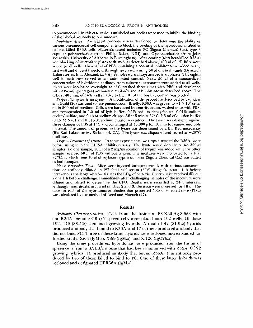
Lack of Reactivity with Polysaccharides. Possible reactivity of Xi126, Xi64, and Xi69 antibodies with pneumococcal cell wall and capsular polysaccharides was assessed by using these materials to attempt inhibition of the binding of the hybridoma antibodies to R36A. By this assay all three antibodies failed to show reactivity with free PC, C-polysaccharide, or type 3 pneumococcal polysaccharide (Fig. 1). An IgG3 anti-PC antibody, 59.6C5 (23), was included as a control. Both PC and C-polysaccharide inhibited the binding of the anti-PC antibody to R36A. The less complete inhibition of the binding of this antibody to R36A by type 3 polysaccharide was probably due to a low level of contaminating C-polysaccharide in the type 3 preparation.
Supernatant from a culture of R36A failed to inhibit the binding of these antibodies, indicating that the antigen they detect is neither released into the medium during bacterial growth, nor acquired from it.
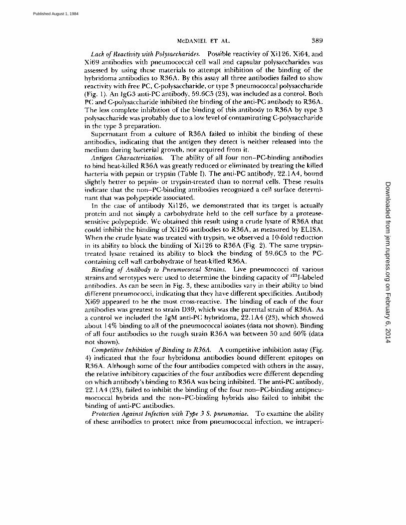
Antigen Characterization. The ability of all four non-PC-binding antibodies to bind heat-killed R36A was greatly reduced or eliminated by treating the killed bacteria with pepsin or trypsin (Table I). The anti-PC antibody, 22.1A4, bound slightly better to pepsin- or trypsin-treated than to normal cells. These results indicate that the non-PC-binding antibodies recognized a cell surface determi- nant that was polypeptide associated.
In the case of antibody Xi126, we demonstrated that its target is actually protein and not simply a carbohydrate held to the cell surface by a protease- sensitive polypeptide. We obtained this result using a crude lysate of R36A that could inhibit the binding of Xi 126 antibodies to R36A, as measured by ELISA. When the crude lysate was treated with trypsin, we observed a 10-fold reduction in its ability to block the binding of Xi126 to R36A (Fig. 2). The same trypsin- treated lysate retained its ability to block the binding of 59.6C5 to the PC- containing cell wall carbohydrate of heat-killed R36A.
Binding of Antibody to Pneumococcal Strains. Live pneumococci of various strains and serotypes were used to determine the binding capacity of ~25I-labeled antibodies. As can be seen in Fig. 3, these antibodies vary in their ability to bind different pneumococci, indicating that they have different specificities. Antibody Xi69 appeared to be the most cross-reactive. The binding of each of the four antibodies was greatest to strain D39, which was the parental strain of R36A. As a control we included the IgM anti-PC hybridoma, 22.1A4 (23), which showed about 14% binding to all of the pneumococcal isolates (data not shown). Binding of all four antibodies to the rough strain R36A was between 50 and 60% (data not shown).
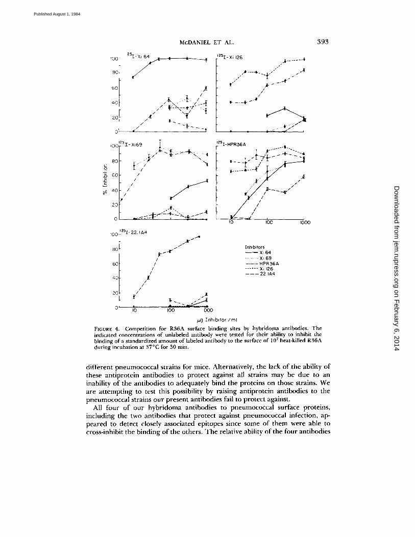
Competitive Inhibition of Binding to R36A. A competitive inhibition assay (Fig. 4) indicated that the four hybridoma antibodies bound different epitopes on R36A. Although some of the four antibodies competed with others in the assay, the relative inhibitory capacities of the four antibodies were different depending on which antibody's binding to R36A was being inhibited. The anti-PC antibody, 22.1A4 (23), failed to inhibit the binding of the four non-PC-binding antipneu- mococcal hybrids and the non-PC-binding hybrids also failed to inhibit the binding of anti-PC antibodies.
Protection Against Infection with Type 3 S. pneumoniae. To examine the ability of these antibodies to protect mice from pneumococcal infection, we intraperi-
on February 6, 2014
jem.rupress.org
Dow
nloaded from
Published August 1, 1984
390 ANTIPNEUMOCOCCAL PROTEIN ANTIBODIES
A E /c, - - . - -o Xi 126 = ~ _ _ ~ Xi 64 ,.n 0.8 ~ " " ' T ' ~ "-' ~- " x i 69 o hit " f - -
"~ 06 ,,'1 0
04
rr igG3 0.2
onti-PC
o 60, ~, ,'o ,~ ,oo ,~oo /zg/ml of Phosphocholine Chloride
, . , B E I'0 ~ ' e - ~ i : ~ - ~ - ~ ~ c ~ , 6 9
i 0,2
i i
o do, o., ,:o ,o ,;o ,~ /.;,~/ml of C- polyseccheride
r... E t . O ~ E
~" 0 . 8
'~ ~ kXi 126
,-'i 0.6 0
m ~ igG3 0.4 _o onfi- PC
02
000t o t t .0 t too IO00 /,,~jlml of Type 3 Polysacchoride
FIGURE 1. Lack of reactivity of anti-R36A hybridoma antibodies with capsular and cell wall antigens. (A) Inhibition of antibody binding to R36A by PC chloride. Absorhance at OD 405 nm (vertical axis) is a measure of the chromophore produced in the ELISA assay that detects antibodies bound to R36A. (B) Inhibition of antibody binding to R36A by C-polysaccharide as described in A. (¢) Inhibition of antibody binding to R36A by type 3 capsular polysaccharide. The inhibition of the anti-PC antibody with type 3 polysaccharide is likely the result of contamination of type 3 polysaccharide with C-polysaccharide.
on February 6, 2014
jem.rupress.org
Dow
nloaded from
Published August 1, 1984
McDANIEL ET AL. 391
TABLE I Binding Properties of Anti-R36A Hybridomas
Hybridoma Isotype Reactivity with PC R36A treated with:
PDs0* Percent bound to: (#g/mouse)
R36A WU2 D39 Pepsin Trypsin
Xi64 lgM - 54 1 0.8 4.5 >200 Xi126 IgG~b -- 16 4 0.5 2.6 112 Xi69 IgM - 10 - 3 0.3 >700 >200 HPR36A IgM - 20 - 5 0.5 >700 ND* 22.1A4 IgM + 60 70 74 11.5 >200 159.4D5 IgM~ + ND ND ND 3.8 40
* PDs0 doses were calculated at 10 d postinfection with I00 type 3 strain WU2 or type 2 strain D- 39.
* Not done.
c
a5.8 cD ,f-'Tr ypsin Tteoled
2 i L-
o l 0 B. c ~5 c~8 Q_ ± 6 E <
~4 o
Y2 Lysate , ~ ,
tO I00 tO00 ,u.9 / mt protein
FIGURE 2. Inhibitors in the crude lysate of R36A that block the binding of Xi126 and 59.6C5 to heat-killed R36A. (A) Susceptibility of the inhibitor of Xi126 binding to trypsin. (B) Resistance of the inhibitor of 59.6C5 binding to trypsin.
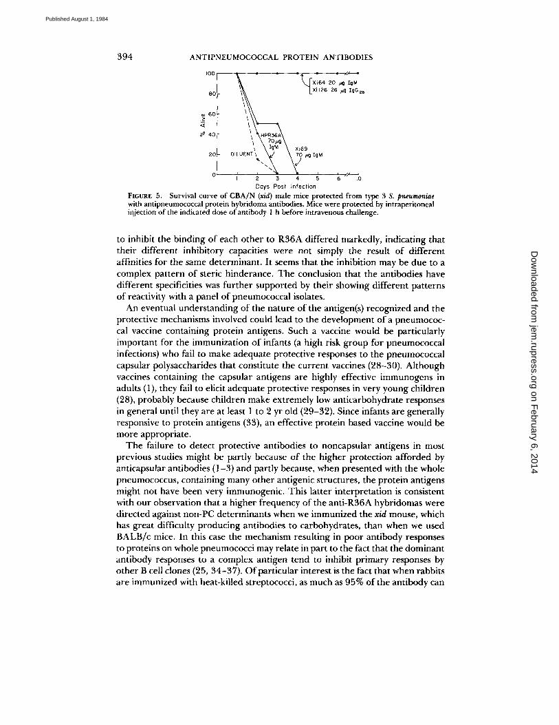
toneally injected various concentrations of antibody 1 h before intravenous challenge with about five times the LDs0 of type 3, strain WU2 pneumococci. All six mice injected with 20 #g of Xi64 and all six injected with 26 #g of Xi126 were alive 10 d after infection (Fig. 5). In contrast, at 3 d all six mice treated with 70 #g of Xi69 were dead and at 2 d all unprotected mice were dead. By combining the data in Fig. 5 with those of subsequent experiments, the doses of antibody ÷equired to protect 50% of mice from fatal infection (PDs0) was determined (Table I). Xi64 and Xi126 were found to be even more protective than the IgM anti-PC antibody, 22.1A4, while Xi69 and HPR36A failed to protect mice even at doses as high as 700 ~g per injection.
In another experiment we demonstrated that the LDs0 of WU2 in CBA/N mice given 20 vg of Xi64 or Xi126 was 104 and 106, respectively, as compared
on February 6, 2014
jem.rupress.org
Dow
nloaded from
Published August 1, 1984
392 ANTIPNEUMOCOCCAL PROTEIN ANTIBODIES
I0l I ~ ~ ~ X i 6 4
I
c~ 5
po[- ~ H P R 3 6 A
i J J
~ 0.3 I I 2* 2 3 t 3 ~t 4 5 6A 8 9V I0 i4 ~gF25F
Serotype
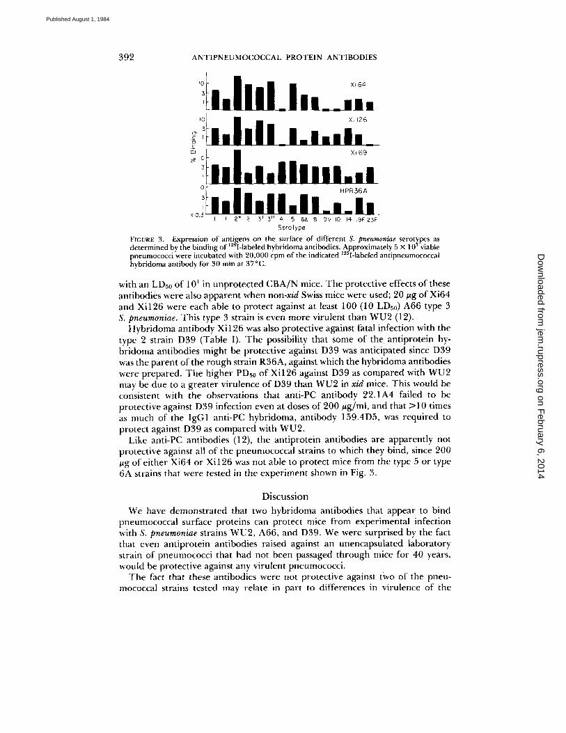
FIGURE 3. Expression of antigens on the surface of different S. pneumoniae serot~pes as 12~'t" / determined by the binding of I-labeled hybridoma antibodies. Approximately 5 x 10 viable
1 2 f f i pneumococci were incubated with 20,000 cpm of the indicated I-labeled antipneumococcal hybridoma antibody for 30 min at 37°C.
with an LDs0 of 101 in unprotected CBA/N mice. The protective effects of these antibodies were also apparent when non-xid Swiss mice were used; 20 #g of Xi64 and Xi126 were each able to protect against at least 100 (10 LDs0) A66 type 3 S. pneumoniae. This type 3 strain is even more virulent than WU2 (12).
Hybridoma antibody Xi 126 was also protective against fatal infection with the type 2 strain D39 (Table I). The possibility that some of the antiprotein hy- bridoma antibodies might be protective against D39 was anticipated since D39 was the parent of the rough strain R36A, against which the hybridoma antibodies were prepared. The higher PDs0 of Xi126 against D39 as compared with WU2 may be due to a greater virulence of D39 than WU2 in xid mice. This would be consistent with the observations that anti-PC antibody 22.1A4 failed to be protective against D39 infection even at doses of 200 #g/ml, and that >10 times as much of the IgG1 anti-PC hybridoma, antibody 159.4D5, was required to protect against D39 as compared with WU2.
Like anti-PC antibodies (12), the antiprotein antibodies are apparently not protective against all of the pneumococcai strains to which they bind, since 200 #g of either Xi64 or Xi126 was not able to protect mice from the type 5 or type 6A strains that were tested in the experiment shown in Fig. 3.
Discussion
We have demonstrated that two hybridoma antibodies that appear to bind pneumococcal surface proteins can protect mice from experimental infection with S. pneumoniae strains WU2, A66, and D39. We were surprised by the fact that even antiprotein antibodies raised against an unencapsulated laboratory strain of pneumococci that had not been passaged through mice for 40 years, would be protective against any virulent pneumococci.
The fact that these antibodies were not protective against tWO of the pneu- mococcal strains tested may relate in part to differences in virulence of the
on February 6, 2014
jem.rupress.org
Dow
nloaded from
Published August 1, 1984
M c D A N I E L E T AL. 393
IO0
80
60
40
20
0
=251-Xi 64
/ /
d¢
4 /
- / - 3- . . . . . . 6 ~--I /~ t~--'~x-- ~. r .... a l / \ • - z
/ t . _
~25I-Xi 126 .~ . . . . . . . ½
. / h . . . . lb. { - - "~
1 .l l'
IO0
8O g
6O
20
IO0
80
60
40
20
rsZ- Xi69 I . :..t". ~ . ! ~. ~.4. ...........
. ~ . ) "t[- \ \
/
i - I11 y
z 5 [ 2 2 IA4 .,.4
/ t"
.4~
/ /
/ /
t / /
/ / .1
. . . . e--""- ~ IO0 IO00
=zSI-HPR36A
. . . . . .
. . . . . . ; - - - ¢ . I - J " . I ~ / / ) I
tO qO0 iO0O
[nhibitors - - Xi 64 ......... Xi 69 . . . . . HPR36A . . . . . . X= 126 - - - - - - 22.~A4
Znh lbdo r /m l
FIGURE 4. Competition for R36A surface binding sites by hybridoma antibodies. The indicated concentrations of unlabeled antibody were tested for their ability to inhibit the binding of a standardized amount of labeled antibody to the surface of 107 heat-killed R 3 6 A during incubation at 37°C for 30 rain.
different pneumococcal strains for mice. Alternatively, the lack of the ability of these antiprotein antibodies to protect against all strains may be due to an inability of the antibodies to adequately bind the proteins on those strains. We are attempting to test this possibility by raising antiprotein antibodies to the pneumococcal strains our present antibodies fail to protect against.
All four of our hybridoma antibodies to pneumococcal surface proteins, including the two antibodies that protect against pneumococcal infection, ap- peared to detect closely associated epitopes since some of them were able to cross-inhibit the binding of the others. The relative ability of the four antibodies
on February 6, 2014
jem.rupress.org
Dow
nloaded from
Published August 1, 1984
394 ANTIPNEUMOCOCCAL PROTEIN ANTIBODIES
I00
80
o~ 6O > <z
40
20
\ : : ; \ F L ,
~ Xl69 o,L UENT %1,
J i ~ l L i .gL--J
I 2 3 4 5 6 tO Doys Post Infection
FIGURE 5. Survival curve of CBA/N (x/d) male mice protected from type 3 S. pneumoniae with antipneumococcal protein hybridoma antibodies. Mice were protected by intraperitoneal injection of the indicated dose of antibody 1 h before intravenous challenge.
to inhibit the binding of each other to R36A differed markedly, indicating that their different inhibitory capacities were not simply the result of different affinities for the same determinant. It seems that the inhibition may be due to a complex pattern of steric hinderance. The conclusion that the antibodies have different specificities was further supported by their showing different patterns of reactivity with a panel of pneumococcal isolates.
An eventual understanding of the nature of the antigen(s) recognized and the protective mechanisms involved could lead to the development of a pneumococ- cal vaccine containing protein antigens. Such a vaccine would be particularly important for the immunization of infants (a high risk group for pneumococcal infections) who fail to make adequate protective responses to the pneumococcal capsular polysaccharides that constitute the current vaccines (28-30). Although vaccines containing the capsular antigens are highly effective immunogens in adults (1), they fail to elicit adequate protective responses in very young children (28), probably because children make extremely low anticarbohydrate responses in general until they are at least 1 to 2 yr old (29-32). Since infants are generally responsive to protein antigens (33), an effective protein based vaccine would be more appropriate.
The failure to detect protective antibodies to noncapsular antigens in most previous studies might be partly because of the higher protection afforded by anticapsular antibodies (1-3) and partly because, when presented with the whole pneumococcus, containing many other antigenic structures, the protein antigens might not have been very immunogenic. This latter interpretation is consistent with our observation that a higher frequency of the anti-R36A hybridomas were directed against non-PC determinants when we immunized the xid mouse, which has great difficulty producing antibodies to carbohydrates, than when we used BALB/c mice. In this case the mechanism resulting in poor antibody responses to proteins on whole pneumococci may relate in part to the fact that the dominant antibody responses to a complex antigen tend to inhibit primary responses by other B cell clones (25, 34-37). Of particular interest is the fact that when rabbits are immunized with heat-killed streptococci, as much as 95% of the antibody can
on February 6, 2014
jem.rupress.org
Dow
nloaded from
Published August 1, 1984
McDANIEL ET AL. 395
be directed against the group-specific polysaccharide, leaving 5% directed against other internal and external molecules on the bacterium (25, 38).
Another possibility is that the capsule may block the antigenicity of cell wall antigens by simply covering them up. We recently observed that when xid mice are immunized with smooth rather than rough pneumococci, very few hybrido- mas producing antibody to either surface proteins or PC were obtained (Mc- Daniel, Scott and Briles, unpublished results).
Regardless of why the protective potential of these antibodies was not observed in previous studies, we hope that a vaccine containing cell wall proteins, free from other pneumococcal antigens will elicit protection against pneumococci. Recently, a similar strategy has been proposed for other pathogens such as Haemophilus influenzae (39), where infants also fail to respond to the polysaccha- ride capsular antigens (32).
S u m m a r y
Monoclonal antibodies were raised against surface determinants of Streptococcus pneumoniae by hyperimmunizing X-linked immunodeficient (xid) CBA/N mice with the heat-killed rough strain R36A. 17 hybridomas produced antibody that bound intact R36A and did not cross-react with phosphocholine, an antigen common in the cell wall of all S. pneumoniae. The antibody produced by at least two of these hybridomas, Xi64 (IgM) and Xi126 (IgG2b), could protect mice from a lethal intravenous challenge of type 3 S. pneumoniae strains WU2 and A66 and of the type 2 strain D39. The minimum amount of antibody required to protect xid mice from 100 WU2 was 4.5 #g/mouse for Xi64 and 2.6 #g/ mouse for Xi126. Free phosphocholine, C-polysaccharide, and type 3 capsular polysaccharide all failed to inhibit the binding of Xi64 or Xi 126 to R36A. These antibodies appeared to bind surface polypeptides, since treatment of R36A with either pepsin or trypsin, or of R36A lysate with trypsin, effectively eliminated the ability of Xi64 and Xi 126 to bind antigens in these preparations. Binding studies indicated that these two antibodies recognized different epitopes that were expressed on several but not all serotypes of pneumococci.
We would like to acknowledge the expert technical assistance of Colynn Forman. We thank Chen-lo Chen, Joyce Lehmeyer, and Linda Hendershot for assistance and advice. Hugh Dillon, Barry Gray, and John Volanakis are acknowledged for the contribution of pneumococcal strains and C-carbohydrate, Latham Claflin for anti-PC hybridoma anti- bodies, and Maclyn McCarty for the strain D39 and for suggestions concerning its use. We are grateful to Ann Brookshire for careful typing of the manuscript and to Maxine Aycock for preparation of the illustrations.
Received for publication 21 November 1983 and in revised form 24 April 1984.
References 1. Austrian, R. 1979. Pneumococcal vaccine: development and prospects. Am. J. Med.
67:547. 2. MacLeod, C. M., R. G. Hodges, M. Heidelberger, and W. G. Bernhard. 1945.
Prevention of pneumococcal pneumonia by immunization with specific capsular polysaccharides. J. Exp. Med. 82:445.
on February 6, 2014
jem.rupress.org
Dow
nloaded from
Published August 1, 1984

396 ANTIPNEUMOCOCCAL PROTEIN ANTIBODIES
3. Heffron, R. 1979. Pneumonia, with Special Reference to Pneumococcus Lobar Pneumonia. Harvard University Press, Cambridge, MA. 805-879.
4. Brown, E.J., S. W. Hosea, C. H. Hammer, C. G. Burch, and M. M. Frank. 1982. A quantitative analysis of the interactions of antipneumococcal antibody and comple- ment in experimental pneumococcal bacteremia.J. Clin. Invest. 69:85.
5. Austrian, R., and C. M. MacLeod. 1949. A type-specific protein from pneumococcus. J. Exp. Med. 89:439.
6. Tillett, W. S. 1928. Active and passive immunity to pneumococcus infection induced in rabbits by immunization with R pneumococci. J. Exp. Med. 48:791.
7. Dubos, R. J. 1938. Immunization of experimental animals with a soluble antigen extracted from pneumococci.J. Exp. Med. 67:799.
8. Sabin, A. B. 1932. On the presence in anti-pneumococcus serum of type-specific protective antibody not neutralized by homologous specific soluble substance. J. Exp. Med. 53:93.
9. Au, C. C., and T. K. Eisenstein. 1981. Nature of the cross-protective antigen in subcellular vaccines of Streptococcus pneumoniae. Infect. lmmun. 31:160.
10. Brundish, D. E., and J. Baddiley. 1968. Pneumococcal C-substance, a ribitol teichoic acid containing choline phosphate. Biochem. J. 110:573.
11. Briles, D. E., M. Nahm, K. Schroer, J. Davie, P. Baker, J. Kearney, and R. Bar|etta. 1981. Antiphosphocholine antibodies found in normal mouse serum are protective against intravenous infection with type 3 Streptococcus pneumoniae. J. Exp. Med. 153:694.
12. Yother, J., C. Forman, B. M. Gray, and D. E. Briles. 1982. Protection of mice from infection with Streptococcus pneumoniae by anti-phosphocholine antibody. Infect. Im- mun. 36:184.
13. Szu, S. C., S. Clarke, and J. B. Robbins. 1983. Protection against pneumococcal infection in mice conferred by phosphocholine-binding antibodies: specificity of the phosphocholine binding and relation to several types. Infect. lmmun. 39:993.
14. Mold, C., S. Nakayama, T. J. Holzer, H. Gerwurz, and T. W. DuC|os. 1981. C- reactive protein is protective against Streptococcus pneumoniae infection in mice. J. Exp. Med. 154:1703.
15. Yother, J., J. E. Volanakis, and D. E. Briles. 1982. Human C-reactive protein is protective against fatal Streptococcus pneumoniae infection in mice. J. lmmunol. 128:2374.
16. Scher, I. 1982. The CBA/N mouse strain: an experimental model illustrating influ- ences of the X-chromosome on immunity. Adv. lmmunol. 33:1.
17. Briles, D. E., M. Nahm, T. N. Marion, R. M. Perlmutter, andJ . M. Davie. 1982. Streptococcal group A carbohydrate has properties of both a thymus-independent (TI-2) and a thymus-dependent antigen. J. lmmunol. 128:2032.
18. Avery, O. T., C. M. MacLeod, and M. McCarty. 1944. Studies of the chemical nature of the substance inducing transformation of pneumococcal types.J. Exp. Med. 79:137.
19. Kearney, J. F., A. Radbuch, B. Liesegang, and K. Rajewsky. 1979. A new mouse myeloma cell line that has lost immunoglobulin expression but permits the construc- tion of antibody-secreting hybrid cell lines. J. Immunol. 123:1548.
20. Hammerling, G.J. , U. Hammerling, and J. F. Kearney. 1981. Appendix. In Mono- clonal Antibodies and T-Cell Hybridomas: Perspectives and Technical Advances. Elsevier/North Holland, Amsterdam, The Netherlands. 574-579.
21. Kearney, J. F., R. Barletta, Z. S. Quan, and J. Quintans. 1981. Monoclonal versus heterogeneous anti-H-8 antibodies in the analysis of the anti-phosphorylcholine response in BALB/c mice. Eur. J. lmmunol. 11:877.
on February 6, 2014
jem.rupress.org
Dow
nloaded from
Published August 1, 1984

McDANIEL ET AL. 397
22. Stohrer, R., M. C. Lee, andJ. F. Kearney. 1983. Analysis of the anti-al ~ 3 dextran response with monoclonal anti-idiotype antibodies. J. Immunol. 131 : 1375.
23. Andres, C. M., A. Maddalena, S. Hudak, N. M. Young, andJ. L. Claflin. 1981. Anti- phosphocholine hybridoma antibodies. II. Functional analysis of binding sites within three antibody families. J. Exp. Med. 154:1584.
24. Greenwood, F. C., W. M. Hunter, and J. S. Giover. 1963. The preparation of lS~l- labeled human growth hormone of high specific radioactivity. Biochem. J. 89:114.
25. Krause, R. M. 1970. The search for antibodies with molecular uniformity. Adv. lmmunol. 12:1.
26. Saunders, C. W., and W. R. Guild. 1980. Properties and transforming activities of two plasmids in Streptococcus pneumoniae. Mol. Gen. Genet. 180:573.
27. Reed, L. J., and H. Muench. 1938. A simple method of estimating fifty percent endpoints. Am. J. Hyg. 27:493.
28. Klein, J. O., D. W. TeeM, J. L. Sloyer, Jr., J. H. Ploussard, V. Howie, P. H. Makela, and P. Karma. 1982. Use of pneumococcal vaccine for prevention of recurrent episodes of otitis media. Semin. Infect. Dis. 4:305.
29. Cowan, M. J., J. Arthur, J. Ammann, D. W. Wara, V. M. Howie, L. Schultz, N. Doyle, and M. Kaplan. 1978. Pneumococcal polysaccharide immunization in infants and children. Pediatrics. 62:721.
30. Douglas, R. M., C. J. Paton, S. J. Duncan, and D. J. Hansman. 1983. Antibody responses to pneumococcal vaccination in children younger than five years of age. J. Infect. Dis. 148:131.
31. Gotschlich, E. G., I. Goldschneider, and M. Lepow. 1977. The immune response to bacterial polysaccharides in man. In Antibodies in Human Diagnosis and Therapy. E. Haber and R. Krause, editors. Raven Press, New York. 391-402.
32. Andersson, P., D. H. Smith, D. L. Ingrain, J. Wilkins, P. F. Wehrle, and V. M. Howie. 1977. Antibody to polyribophosphate of Haemophilus influenzae type b in infants and children. J. Infect. Dis. 136(Suppl):S57.
33. Smith, R. T., D. V. Eitzman, M. E. Catlin, E. O. Wirtz, and B. E. Miller. 1964. The development of the immune response. Characterization of the response of the human infant and adult to immunization with salmonella vaccines. Pediatrics. 33:163.
34. Askonas, B. A., and A. R. Williamson. 1972. Dominance of a cell clone forming antibody to DNP. Nature (Lond.). 238:339.
35. Cramer, M., and D. G. Braun. 1975. Immunological memory: stable IgG patterns determine in vivo responsiveness at the clonal level. Scand. J. Immunol. 4:63.
36. Brown, A. R., C. L. DeWitt, M. H. Bosma, and A. Nisonoff. 1980. Dominance of an immune response by secondary cells. Quantitation by allotypic analysis. J. Immunol. 124:250.
37. Briles, D. E., andJ. M. Davie. 1980. Clonal nature of the immune response. II. The effect of immunization on clonal commitment. J. Exp. Med. 152:151.
38. Braun, D. G., K. Eichmann, and R. M. Krause. 1968. Rabbit antibodies to strepto- coccal carbohydrates, influence of primary and secondary immunization and of possible genetic factors on the antibody response. J. Exp. Med. 129:809.
39. Hansen, E. J., S. M. Robertson, P. A. Gulig, C. F. Frisch, and E.J. Haanes. 1982. Immunoprotection of rats against haemophilus influenzae type B disease mediated by monoclonal antibody against a haemophilus outer-membrane protein. Lancet. 1:366.
on February 6, 2014
jem.rupress.org
Dow
nloaded from
Published August 1, 1984
Copyright © 2022 FDOKUMEN