Mechanical ventilation drives pneumococcal pneumonia into ...
13
RESEARCH Open Access Mechanical ventilation drives pneumococcal pneumonia into lung injury and sepsis in mice: protection by adrenomedullin Holger C Müller-Redetzky 1 , Daniel Will 1 , Katharina Hellwig 1 , Wolfgang Kummer 2,3 , Thomas Tschernig 4 , Uwe Pfeil 2 , Renate Paddenberg 2 , Michael D Menger 5 , Olivia Kershaw 6 , Achim D Gruber 6 , Norbert Weissmann 7 , Stefan Hippenstiel 1 , Norbert Suttorp 1 and Martin Witzenrath 1* Abstract Introduction: Ventilator-induced lung injury (VILI) contributes to morbidity and mortality in acute respiratory distress syndrome (ARDS). Particularly pre-injured lungs are susceptible to VILI despite protective ventilation. In a previous study, the endogenous peptide adrenomedullin (AM) protected murine lungs from VILI. We hypothesized that mechanical ventilation (MV) contributes to lung injury and sepsis in pneumonia, and that AM may reduce lung injury and multiple organ failure in ventilated mice with pneumococcal pneumonia. Methods: We analyzed in mice the impact of MV in established pneumonia on lung injury, inflammation, bacterial burden, hemodynamics and extrapulmonary organ injury, and assessed the therapeutic potential of AM by starting treatment at intubation. Results: In pneumococcal pneumonia, MV increased lung permeability, and worsened lung mechanics and oxygenation failure. MV dramatically increased lung and blood cytokines but not lung leukocyte counts in pneumonia. MV induced systemic leukocytopenia and liver, gut and kidney injury in mice with pneumonia. Lung and blood bacterial burden was not affected by MV pneumonia and MV increased lung AM expression, whereas receptor activity modifying protein (RAMP) 1–3 expression was increased in pneumonia and reduced by MV. Infusion of AM protected against MV-induced lung injury (66% reduction of pulmonary permeability p < 0.01; prevention of pulmonary restriction) and against VILI-induced liver and gut injury in pneumonia (91% reduction of AST levels p < 0.05, 96% reduction of alanine aminotransaminase (ALT) levels p < 0.05, abrogation of histopathological changes and parenchymal apoptosis in liver and gut). Conclusions: MV paved the way for the progression of pneumonia towards ARDS and sepsis by aggravating lung injury and systemic hyperinflammation leading to liver, kidney and gut injury. AM may be a promising therapeutic option to protect against development of lung injury, sepsis and extrapulmonary organ injury in mechanically ventilated individuals with severe pneumonia. Introduction In acute respiratory failure, mechanical ventilation (MV) is a life-saving intervention without alternatives, but MV may cause ventilator-induced lung injury (VILI). Since clinical trials have been highlighting the impact of VILI on acute respiratory distress syndrome (ARDS) mortality [1], lung-protective ventilation has been widely implemented in clinical practice. However, clinical and experimental studies provide evidence that VILI occurs despite low tidal volume ventilation and that particularly preinjured lungs are susceptible for the development of VILI [2,3]. Lung injury worsened in ventilated mice upon infec- tion with Staphylococcus aureus even under protective ventilation strategies [4], which is intriguing as pneu- monia is the leading cause of ARDS and sepsis [1,5]. However a major limitation of this and other studies was that mice were infected after initiation of MV [4,6,7]. Experimental studies investigating the impact * Correspondence: [email protected] 1 Department of Infectious Diseases and Pulmonary Medicine, Charité – Universitätsmedizin Berlin, Charitéplatz 1, 10117 Berlin, Germany Full list of author information is available at the end of the article © 2014 Müller-Redetzky et al.; licensee BioMed Central Ltd. This is an open access article distributed under the terms of the Creative Commons Attribution License (http://creativecommons.org/licenses/by/2.0), which permits unrestricted use, distribution, and reproduction in any medium, provided the original work is properly cited. Müller-Redetzky et al. Critical Care 2014, 18:R73 http://ccforum.com/content/18/2/R73
-
Upload
khangminh22 -
Category
Documents
-
view
1 -
download
0
Transcript of Mechanical ventilation drives pneumococcal pneumonia into ...
Müller-Redetzky et al. Critical Care 2014, 18:R73http://ccforum.com/content/18/2/R73
RESEARCH Open Access
Mechanical ventilation drives pneumococcalpneumonia into lung injury and sepsis in mice:protection by adrenomedullinHolger C Müller-Redetzky1, Daniel Will1, Katharina Hellwig1, Wolfgang Kummer2,3, Thomas Tschernig4, Uwe Pfeil2,Renate Paddenberg2, Michael D Menger5, Olivia Kershaw6, Achim D Gruber6, Norbert Weissmann7,Stefan Hippenstiel1, Norbert Suttorp1 and Martin Witzenrath1*
Abstract
Introduction: Ventilator-induced lung injury (VILI) contributes to morbidity and mortality in acute respiratorydistress syndrome (ARDS). Particularly pre-injured lungs are susceptible to VILI despite protective ventilation. In aprevious study, the endogenous peptide adrenomedullin (AM) protected murine lungs from VILI. We hypothesizedthat mechanical ventilation (MV) contributes to lung injury and sepsis in pneumonia, and that AM may reduce lunginjury and multiple organ failure in ventilated mice with pneumococcal pneumonia.
Methods: We analyzed in mice the impact of MV in established pneumonia on lung injury, inflammation, bacterialburden, hemodynamics and extrapulmonary organ injury, and assessed the therapeutic potential of AM by startingtreatment at intubation.
Results: In pneumococcal pneumonia, MV increased lung permeability, and worsened lung mechanics and oxygenationfailure. MV dramatically increased lung and blood cytokines but not lung leukocyte counts in pneumonia. MV inducedsystemic leukocytopenia and liver, gut and kidney injury in mice with pneumonia. Lung and blood bacterial burden wasnot affected by MV pneumonia and MV increased lung AM expression, whereas receptor activity modifying protein (RAMP)1–3 expression was increased in pneumonia and reduced by MV. Infusion of AM protected against MV-induced lung injury(66% reduction of pulmonary permeability p< 0.01; prevention of pulmonary restriction) and against VILI-induced liver andgut injury in pneumonia (91% reduction of AST levels p< 0.05, 96% reduction of alanine aminotransaminase (ALT) levelsp < 0.05, abrogation of histopathological changes and parenchymal apoptosis in liver and gut).
Conclusions: MV paved the way for the progression of pneumonia towards ARDS and sepsis by aggravatinglung injury and systemic hyperinflammation leading to liver, kidney and gut injury. AM may be a promisingtherapeutic option to protect against development of lung injury, sepsis and extrapulmonary organ injury inmechanically ventilated individuals with severe pneumonia.
IntroductionIn acute respiratory failure, mechanical ventilation (MV)is a life-saving intervention without alternatives, but MVmay cause ventilator-induced lung injury (VILI). Sinceclinical trials have been highlighting the impact of VILI onacute respiratory distress syndrome (ARDS) mortality [1],lung-protective ventilation has been widely implemented
* Correspondence: [email protected] of Infectious Diseases and Pulmonary Medicine, Charité –Universitätsmedizin Berlin, Charitéplatz 1, 10117 Berlin, GermanyFull list of author information is available at the end of the article
© 2014 Müller-Redetzky et al.; licensee BioMedCreative Commons Attribution License (http:/distribution, and reproduction in any medium
in clinical practice. However, clinical and experimentalstudies provide evidence that VILI occurs despite low tidalvolume ventilation and that particularly preinjured lungsare susceptible for the development of VILI [2,3].Lung injury worsened in ventilated mice upon infec-
tion with Staphylococcus aureus even under protectiveventilation strategies [4], which is intriguing as pneu-monia is the leading cause of ARDS and sepsis [1,5].However a major limitation of this and other studieswas that mice were infected after initiation of MV[4,6,7]. Experimental studies investigating the impact
Central Ltd. This is an open access article distributed under the terms of the/creativecommons.org/licenses/by/2.0), which permits unrestricted use,, provided the original work is properly cited.

Müller-Redetzky et al. Critical Care 2014, 18:R73 Page 2 of 13http://ccforum.com/content/18/2/R73
of VILI in established pneumonia – that is, when theimmune system is already activated and lung mechan-ics are changed due to pneumonic infiltrates – wouldbe of particular clinical relevance.VILI has been linked to multiple organ failure [8,9]. Im-
proved understanding of the impact of VILI on the pro-gression of pneumonia towards sepsis with its detrimentalcomplications is desirable. Thus, we implemented a newsecond-hit model of established pneumococcal pneumo-nia and MV.While the risk of ARDS development may be reduced
by lowering tidal volumes, MV with low tidal volumesstill seems to aggravate lung injury and further tidal vol-ume reduction is limited by hypercapnia, which aggra-vates acidosis. Adjuvant pharmacotherapies in additionto protective ventilation are thus needed to further limitVILI. Adrenomedullin (AM), an endogenous 52-amino-acid peptide belonging to the calcitonin gene-related pep-tide family, is crucial for regulation of endothelial barrierintegrity [10]. AM binds to the calcitonin receptor-like re-ceptor (CRLR) assembled with receptor activity modifyingprotein (RAMP)-1 to RAMP3, thereby raising intracellularcAMP levels in endothelial cells and reducing myosin lightchain phosphorylation. Consequently, interendothelial gapformation is prevented [10-12]. Exogenous AM reducedpulmonary hyperpermeability in experimental acute lunginjury and sepsis [13,14], and we identified AM as beingprotective against VILI and associated kidney injury inpreviously healthy mice by stabilizing endothelial barrierfunction and microcirculation [13]. These and other stud-ies gave rise to a recent positive opinion from the Com-mittee for Orphan Medicinal Products of the EuropeanMedicines Agency (EMA), recommending the granting ofthe development of AM as an orphan drug for the treat-ment of acute lung injury/ARDS (EMA/COMP/104704/2010 to SH). However, although AM proved to be benefi-cial in healthy lungs subjected to VILI, evidence is lackingfor a protective effect of AM during MV of individualswith severe pneumonia. Clinical trials with AM are cur-rently being planned, so additional preclinical evidence isdesirable.We therefore conducted the current study to decipher
the contribution of VILI and underlying mechanisms tothe progression of ARDS, sepsis and multiple organ dys-function syndrome in pneumonia, to test the therapeuticimpact of AM in the treatment of VILI-driven lung in-jury in pneumonia, and to investigate potential protect-ive effects of AM on VILI-driven extrapulmonary organdysfunction.
MethodsEthics statementAnimal experiments were approved by the animal ethicscommittee of Charité-Universitätsmedizin Berlin and local
governmental authorities (Landesamt für Gesundheit undSoziales Berlin).
MiceFemale C57Bl/6 mice (8 to 10 weeks; 18 to 20 g; CharlesRiver, Sulzfeld, Germany) were used.
Pneumococcal pneumoniaStreptococcus pneumoniae (serotype 3, strain NCTC7978)was grown to mid log phase. Mice were anesthetized byintraperitoneal ketamine (1.6 mg) and xylazine (0.5 mg)and were transnasally inoculated with 5× 10⁶ colony-forming units of S. pneumoniae diluted in 20 μl sterilephosphate-buffered saline (10 μl into each nostril) asdescribed previously [15]. Noninfected mice were anesthe-tized and transnasally inoculated with 20 μl phosphate-buffered saline.
Mechanical ventilation and adrenomedullin treatmentTwenty-four hours after infection when severe pneumo-nia had devolved, mice were subjected to MV as de-scribed previously [16,17]. Mice were anesthetized byintraperitoneal injections of fentanyl (75 μg/kg), midazo-lam (1.5 mg/kg) and medetomedin (0.75 mg/kg). Repeti-tively, fentanyl (16 μg/kg), midazolam (0.33 mg/kg) andmedetomedin (0.16 mg/kg) were supplied via an intra-peritoneal catheter when required to guarantee adequateanesthesia during the observation period. Body temperaturewas maintained at 37°C by a body temperature-controlledheating pad. Tracheotomy and intubation was performed,and a carotid artery catheter was placed for bloodpressure monitoring and infusion of NaCl 0.9% con-taining 100 mmol/l HCO3
− (350 μl/hour). No additionalfluid support was provided in any experiment. A urin-ary catheter was inserted. The tidal volume, respiratoryrate, airway pressure, and urine output were monitored(Pulmodyn; Hugo-Sachs-Electronics, March-Hugstetten,Germany).After preparation, a recruitment maneuver was per-
formed (airway pressure 35 cmH2O for 5 seconds) andmice were ventilated for 6 hours with a tidal volume of12 ml/kg, a respiratory rate of 120 breaths/minute, and apositive end-expiratory pressure of 2 cmH2O (MiniVent;Hugo-Sachs-Electronics). A second recruitment maneu-ver was performed 5 minutes before termination of theexperiment. All mice survived the protocol. At termin-ation of the experiments, mice were sacrificed by exsan-guination via the carotid artery catheter. Blood sampleswere analyzed for partial arterial pressure of oxygen byblood gas analyzer (ABL-800; Radiometer, Copenhagen,Denmark). The P/F ratio was calculated as partial ar-terial pressure of oxygen/fraction of inspired oxygen.Nonventilated mice served as controls. Murine AM(0.05 mg/kg/hour; Phoenix, Burlingame, CA, USA) was
Müller-Redetzky et al. Critical Care 2014, 18:R73 Page 3 of 13http://ccforum.com/content/18/2/R73
continuously infused via the carotid artery catheter, start-ing with onset of ventilation. The dosage was proven to beeffective without causing relevant hemodynamic changesin mice [13].
Quantitative real-time polymerase chain reactionLungs were flushed and snap-frozen in liquid nitrogen.Total RNA was isolated from the lungs using the RNeasymini kit (Qiagen, Hilden, Germany) according to the man-ufacturer’s instructions. To remove genomic DNA con-tamination, isolated RNA samples were treated with 1 UDNase/μg RNA (Invitrogen, Karlsruhe, Germany) for 15minutes at 37°C. One microgram of total RNA was usedin a 20 μl reaction to synthesize cDNA using SuperscriptH– reverse transcriptase (200 U/μg RNA; Invitrogen) andoligo dTs as primers. Reverse transcription reaction wascarried out for 50 minutes at 42°C. Quantitative real-timepolymerase chain reaction (qRT-PCR) was performedusing the I-Cycler IQ detection system (Bio-Rad, Munich,Germany) in combination with the IQ SYBR GreenReal-Time PCR Supermix (Bio-Rad). The polymerasechain reaction conditions included initial denaturationin one cycle of 10 minutes at 95°C followed by 40 cy-cles of 20 seconds at 95°C, 20 seconds at 60°C, and20 seconds at 72°C. The relative expressions were cal-culated as:
2– ΔCTð Þ � 1=mean control 2– ΔCTð Þ� �
where ΔCT (CT; Threshold Cycle) is calculated as:
ΔCT ¼ CTgene of interest–CThousekeeping gene
Primer sequences are provided in Additional file 1.Regulation of CRLR and RAMP 1, 2 and 3 mRNA in un-infected, spontaneously breathing mice and uninfected,mechanically ventilated mice has been published previ-ously [16].
Lung permeabilityHuman serum albumin (HSA) was injected intraven-ously 90 min prior to the end of the experiment. Afterligation of the left stem bronchus bronchoalveolar lavage(BAL) was performed twice with 400 μl PBS each. Fromeach BAL portion, 250 μl were pooled and BAL andplasma HSA concentration was determined by enzyme-linked immunosorbent assay. Permeability was assessedby calculating the HSA BAL/plasma ratio as described[17].
Hypoxic vasoconstriction in precision-cut lung slicesPrecision-cut lung slices (PCLS) were prepared as de-scribed previously [18]. Briefly, mice were killed by cer-vical dislocation and the airways were filled with 1.5%low melting point agarose. After solidification of the
agarose, the lungs were cut into 200 μm thick slices. Theagarose was removed by incubation of the PCLS in phe-nol red-free minimal essential medium continuouslygassed with 21% oxygen, 5% carbon dioxide (CO2), 74%nitrogen for at least 2 hours at 37°C.To analyze the vasoreactivity of individual cross-sectioned
intra-acinar arteries (minimal inner diameter up to 40 μm),the PCLS were transferred into a flow-through superfusionchamber (Hugo-Sachs-Elektronik). At the beginning ofeach experiment the capability of the vessel to contractin response to the thromboxane analogue U46619 andto dilate after application of the nitric oxide donor so-dium nitroprusside was checked. After washing outthese drugs with normoxic gassed phenol red-freeminimal essential medium (21% oxygen, 5% CO2, 74%nitrogen), the PCLS were incubated with hypoxicgassed medium (1% oxygen, 5% CO2, 94% nitrogen; 0.7ml/minute). After 15 minutes, 500 nM murine AMwas added to the hypoxic medium. After a secondwashout, the PCLS were again challenged with U46619in the presence of 500 nM AM when the hypoxic incu-bation was performed in the presence of AM or solv-ent, respectively.Pictures of the artery were taken every 2 minutes using
an inverted microscope mounted on the superfusionchamber. Changes in the luminal area of the vesselswere evaluated by manually lining the inner boundaries.The luminal area at the beginning of hypoxia was de-fined as 100% and vasoreactivity was expressed as arelative decrease or increase of this area. Only thevalues (mean ± standard error of the mean) obtainedfor the hypoxic incubation followed by the incubationin hypoxic medium ± AM, the second washout phaseand the final challenge with U44619 ± AM are givenbelow.
Leukocytes in lung tissue, bronchoalveolar lavage fluidand bloodThe lungs were flushed. The left lung was digested inRPMI containing collagenase and DNAse for 1 hour.Leukocytes were extracted by meshing the lung tissuethrough a cell strainer (100 μm) and differentiated byflow cytometry according to their side-scatter/forward-scatter properties and CD45, Gr-1 and F4-80 expression(FACSCalibur, BD, Heidelberg, Germany). Blood leuko-cytes were quantified and differentiated by flow cytometryusing TruCount-Tubes (BD, Heidelberg, Germany) accord-ing to cellular side-scatter/forward-scatter properties andCD45, Gr-1 and CD3 expression.
Quantification of cytokinesCytokines were quantified from total protein of flushedhomogenized left lungs and from plasma samples by the

Müller-Redetzky et al. Critical Care 2014, 18:R73 Page 4 of 13http://ccforum.com/content/18/2/R73
multiplex cytokine assay technique (BioRad, Hercules,CA, USA).
Bacterial burdenSerial dilutions of BALF, spleen homogenate and bloodwere plated on blood agar and incubated at 37°C under5% CO2 for 24 hours to count colony-forming units.
Creatinine, aspartate transaminase, alanine transaminaseand neutrophil gelatinase-associated lipocalinCreatinine, aspartate transaminase and alanine transamin-ase plasma levels were quantified by routine laboratorytests. Neutrophil gelatinase-associated lipocalin levels inurine samples collected over the last 2 hours of the MVperiod were measured by enzyme-linked immunosorbentassay (BioPorto, Gentofte, Denmark).
HistologyImmunolabeling for AM was performed by overnight incu-bation at room temperature with previously characterizedantibodies, including double-labeling with biotinylated ratmonoclonal anti-CD31 (1 μg/ml, clone MEC 13.3; BDBiosciences, Heidelberg, Germany), an endothelial marker[19]. Secondary reagents, each applied for 1 hour, were Cy3-conjugated goat anti-human IgG F(ab)2 (1:500; Dianova,Hamburg, Germany), Cy3-conjugated donkey anti-rabbit IgG(1:2,000; Dianova), and fluorescein isothiocyanate-conjugatedstreptavidin (1:500; Sigma, Deisenhofen, Germany). Tis-sue sections depicting all groups were processed simul-taneously and images were taken at the same exposuretime.For analysis of apoptosis, staining against cleaved caspase-
3 (CC3) was used as an indicator of apoptotic cell death asdescribed previously [20]. In brief, paraffin sections were in-cubated overnight with a polyclonal anti-cleaved caspase-3antibody (Asp175, 1:50; Cell Signaling Technology, Frankfurt,Germany). A secondary antibody was added and 3,3′-diaminobenzidine served as chromogen. Apoptotic cellsappeared with a brown color. The sections were counter-stained with hemalaun. In each procedure, sections of thy-mus tissue served as positive control because apoptosis isa constant event in this organ.
Data analysesData are expressed as mean ± standard error of the mean.For comparison between groups, the Mann–Whitney U testwas used. P < 0.05 was considered statistically significant.For the comparison of experiments using PCLS, the areaunder the curve of each phase of every single experimentwas calculated. Comparison between groups for eachphase was performed again by Mann–Whitney U test,and P < 0.05 was considered statistically significant.
ResultsPulmonary expression of adrenomedullin and its receptorcomplexesPneumonia and MV each increased pulmonary AM mRNAexpression (Figure 1A). Immunofluorescent staining of AMin pulmonary tissue revealed that in naïve lungs AM pro-tein was located mainly, although not exclusively, inmacrophages and in pulmonary endothelium, whichwas confirmed by double staining of AM and theendothelial marker CD31 (see Additional file 2). In linewith increased AM mRNA expression after MV, weobserved a MV-induced increase of parenchymal AMprotein. Furthermore, pneumonic infiltrates were posi-tive for AM immunostaining, with recruited leukocytesdisplaying marked immunoreactivity (Figure 1B). AMspecificity of the employed antibody was validated inpre-absorption experiments (see Additional file 3). Regula-tion of the AM receptor components CRLR and RAMP1to RAMP3 was investigated by qRT-PCR analyses. As re-ported previously [16], MV alone had no impact on CRLRor RAMP1 and RAMP2 expression while RAMP3 wasdownregulated. Pneumonia resulted in an increase ofRAMP1 to RAMP3 expression, while MV markedly re-duced mRNA levels of RAMP1 to RAMP3 in pneumo-nia (Figure 1C). Notably, treatment with AM did notalter expression of AM, CRLR or RAMP1 to RAMP3in pneumonia and subsequent MV (see Additional file 4).Regulation of the AM receptor components CRLR and
RAMP1 to RAMP3 was investigated by qRT-PCR ana-lyses. As reported previously [21], MV alone had no im-pact on CRLR or RAMP1 and RAMP2 expressionwhile RAMP3 was downregulated. Pneumonia resultedin an increase of RAMP1 to RAMP3 expression, whileMV markedly reduced mRNA levels of RAMP1 to RAMP3in pneumonia (Figure 1C). Notably, treatment with AM didnot alter expression of AM, CRLR or RAMP1 to RAMP3 inpneumonia and subsequent MV (see Additional file 4).
Mechanical ventilation exacerbated lung injury inpneumonia: protection by adrenomedullinPneumonia as well as MV each increased pulmonary vascu-lar permeability. AM reduced MV-evoked lung permeability.Notably, when mice with pneumonia were subjected to MVa further dramatic increase in lung permeability was ob-served, which was almost completely avoided by AM treat-ment starting with onset of MV (Figure 2A).Under volume-controlled MV, an increase in the peak
inspiratory pressure reflects a decrease of lung compli-ance, which is mostly due to lung edema in the currentmodel. While pneumonia and MV alone had no impacton peak inspiratory pressure as compared with healthymice, MV in infected mice led to a significant increase ofpeak inspiratory pressure after 6 hours of MV, which wasalmost completely impeded by AM treatment (Figure 2B).

NV MV NV MV0
1
2
3
4
5 p 0.055
S.p. - - + +
RA
MP
1re
lativ
e ex
pres
sion
*
NV MV NV MV0
20
40
60
**
**
S.p. - - + +
**
AM
rela
tive
expr
essi
on
NVMVNVMV0
5
10
15
S.p. - - + +
**
******
RA
MP
3re
lativ
e ex
pres
sion
NV MV NV MV0
5
10
S.p. - - + +
CR
LRre
lativ
e ex
pres
sion
NV MV NV MV0
5
10
15
S.p. - - + +
* **
RA
MP
2re
lativ
e ex
pres
sion
A B
C
NV
AM
50 µm
MV
MV + S.p.NV + S.p.
Figure 1 Regulation of adrenomedullin and its receptor complexes due to pneumonia and mechanical ventilation. Pneumococcalpneumonia (S.p.) was induced 24 hours before mechanical ventilation (MV) was performed for 6 hours. Nonventilated (NV) mice were sacrificed30 hours after infection. Regulation of (A) adrenomedullin (AM) and (C) its receptor components calcitonin receptor-like receptor (CRLR) andreceptor activating modifying protein (RAMP)- 1, 2 and 3 were quantified by quantitative real-time polymerase chain reaction in lung homogenate(*P< 0.05; **P< 0.01, n= 5). Note that the receptor expression data for the NV and MV groups without infection have been published previously[21]. (B) Immunofluorescence analysis of lung tissue. AM-immunolabeling was enhanced in MV mice compared with NV mice. In pneumonicinfiltrates, leukocytes showed intense AM-immunolabeling. Representative images from five animals per group are shown.
Müller-Redetzky et al. Critical Care 2014, 18:R73 Page 5 of 13http://ccforum.com/content/18/2/R73
While oxygenation capacity was not impaired due topneumonia or MV alone, the combination of pneumoniaand MV led towards severe deterioration of oxygenation.Although AM reduced lung injury in mechanically venti-lated mice, AM did not ameliorate the deterioration ofoxygenation (Figure 2C). Histology performed 24 hourspost infection confirmed severe necrotizing broncho-pneumonia affecting 40 to 60% of the lung tissue. Changesdue to MV could not be dissected from the already preva-lent severe alteration in the lungs due to pneumonia (seeAdditional file 5). With regard to the missing improvement1in oxygenation despite barrier-stabilizing properties ofAM and preserved lung mechanics in the pneumonia +MV group, we hypothesized that vasodilatory properties inthe lung of AM might have been counteracting the reduc-tion of lung injury by increasing ventilation/perfusion mis-match. Indeed, vasoconstriction due to hypoxia or thethromboxane agonist U46619 was significantly reduced byAM in murine PCLS (Figure 2D).
Mechanical ventilation aggravated pulmonaryinflammation in pneumoniaThe concentrations of the cytokines interleukin (IL)-1β,IL-6, keratinocyte-derived cytokine (KC) and IL-10 in lunghomogenate were increased by pneumonia and, to a lesserextent, by MV. In pneumonia, MV led to a further dramatic
increase of IL-1β, IL-6 and KC, while IL-10 levels remainedunaffected. AM treatment had no impact on pulmonarycytokine levels (Figure 3A).In pneumonia and in MV, pulmonary polymorpho-
nuclear neutrophils (PMN) and Gr-1high monocytes wereincreased. A combination of MV and pneumonia did notfurther increase PMN and Gr-1high monocytes. Notably,AM decreased pulmonary Gr-1high monocyte recruit-ment in uninfected mice subjected to MV, but not inmice with pneumonia subjected to MV (Figure 3B).
Mechanical ventilation had no impact on pulmonarybacterial outgrowth and development of bacteremia inpneumoniaBlood and spleen homogenate bacterial counts wereassessed in BALF. MV and AM each had no impacton pulmonary bacterial burden, bacteremia or dissem-ination to the spleen in pneumonia (Figure 4).
Mechanical ventilation aggravated systemichyperinflammation in pneumoniaPneumonia and MV each increased plasma IL-6, KC andIL-10 levels. In pneumonia, MV caused a further in-crease of systemic cytokine levels. AM treatment had noimpact on cytokine levels (Figure 5A).

0
2
4
6
8
10
Per
mea
bilit
y(c
HS
AB
AL/c
HS
AP
lasm
a)/
1000
*
*
**
*
**
MVS.p.AM
- -- - -+
+ + + +++++- - - -
0
100
200
300
400
500 ****
P/F
MVS.p.AM
- -- - -+
+ + + +++++- - - -
A
0
10
20
PIP
[cm
H2O
]
****
MVS.p.AM
- -- - -+
+ + + +++++- - - -
B C
D
0 15 30 45 60 75 90
405060708090
100110120
solvent AM
Hypoxia wash U44619
AM/solvent
ns ** *** ***
AM/solvent
[time]
Ve
sse
l are
a [%
]
Figure 2 Adrenomedullin protected mice with pneumonia against mechanical ventilation-induced lung injury. Pneumococcalpneumonia (S.p.) was induced 24 hours before mechanical ventilation (MV) was performed for 6 hours. Continuous adrenomedullin (AM) infusion(0.05 mg/kg/hour) started with the onset of MV. Nonventilated (NV) mice were sacrificed 30 hours after infection. (A) Human serum albumin(HSA) was injected 90 minutes prior to termination of the experiment and the HSA concentration (cHSA) in plasma and in bronchoalveolarlavage (BAL) was determined. An increased HSA BAL/plasma ratio indicated enhanced lung permeability (*P < 0.05, **P < 0.01; NV, n = 5; NV + S.p.,n = 4; MV, n = 5; MV + AM, n = 4; MV + S.p., n = 7; MV + S.p. + AM, n = 5). (B) Peak inspiratory pressure (PIP) was analyzed after 6 hours of MVfollowing a final recruitment maneuver (**P < 0.01; NV and NV + S.p., n = 5; all other groups, n = 8 each). (C) Arterial oxygen partial pressure wasdetermined at the end of the experiment and the P/F ratio was calculated (**P < 0.01; NV and NV + S.p., n = 5; MV, n = 6; MV + AM, n = 7; MV +S.p., n = 5; MV + S.p. + AM, n = 8). (D) In precision cut lung slices, vasoconstriction was induced by hypoxia (hypoxic pulmonary vasoconstriction)and by the thromboxane receptor agonist U46619 in the presence or absence of AM (0.5 μM). The luminal areas of single intra-acinar pulmonaryarteries were continuously analyzed by planimetry and the area under the curve (AUC) was calculated for each experiment. Significant AUCdifferences between the two groups were evident under hypoxia, during the wash out phase and after U46619 challenge (ns, not significant;**P < 0.01, ***P < 0.001; n = 10 each).
Müller-Redetzky et al. Critical Care 2014, 18:R73 Page 6 of 13http://ccforum.com/content/18/2/R73
Mechanical ventilation induced leukopenia in pneumoniaPneumonia increased circulating neutrophils and mono-cytes while lymphocyte counts remained unaffected. MVreduced lymphocytes but had no effect on other leukocytepopulations. In infected mice subjected to MV, bloodleukocyte counts were significantly reduced comparedwith nonventilated mice with pneumonia, and lympho-cyte counts dropped significantly below those observedin naïve mice. Besides almost restoring lymphocytelevels in uninfected mice subjected to MV, AM treat-ment had no significant impact on blood leukocyte counts(Figure 5B).
Adrenomedullin protected mice with pneumonia againstMV-related organ failureAspartate aminotransferase and alanine aminotransami-nase levels were not altered by pneumonia or MV, whereasMV in pneumonia dramatically increased transaminase
levels, which was almost completely avoided by AM(Figure 6A). We observed extended liver injury dis-played by necrotic areas and induction of hepatic apop-tosis exclusively in the pneumonia +MV group, which wasundetectable under AM treatment. More specifically,periportal rings of CC3+ cells were highly prominent(Figure 6B) in four out of six livers from the pneumo-nia + MV group and showed weaker prominence intwo livers. In the liver sections of the pneumonia +MV + AM group only small, single islets of CC3+ cellswere found. In all other groups, CC3+ cells were rare(0 to 5 per section). In ileum sections, an enhancednumber of CC3+ cells was almost exclusively observedin the pneumonia + MV group, indicating tissue injurythat was abolished by AM treatment (Figure 6C).In the pneumonia +MV group increased urine neutro-
phil gelatinase-associated lipocalin levels were measured(see Figure S5A in Additional file 6). Pneumonia and

0
200
400
600
****
IL-1
0 [p
g/m
g]
0
10000
20000
30000
40000
****
**
KC
[p
g/m
g]
0
20000
40000
60000
****
p 0.0521
IL-1
[pg/
mg]
0
100
200
2000
7000
12000
****
**
IL-6
[pg
/mg
]
MVS.p.AM
- -- - -+
+ + + +++++- - - -
MVS.p.AM
- -- - -+
+ + + +++++- - - -
MVS.p.AM
- -- - -+
+ + + +++++- - - -
MVS.p.AM
- -- - -+
+ + + +++++- - - -
02
04
06
0 ****
PM
N [
%]
0
10
20
30
****
**
mon
ocyt
es(G
r1hi
gh)
[%]
MVS.p.AM
- -- - -+
+ + + +++++- - - -
MVS.p.AM
- -- - -+
+ + + +++++- - - -
A
B
Figure 3 Mechanical ventilation induced exacerbation of pulmonary inflammation in pneumonia independently of leukocyte counts.Pneumococcal pneumonia (S.p.) was induced 24 hours before mechanical ventilation (MV) was performed for 6 hours. Continuous adrenomedullin(AM) infusion (0.05 mg/kg/hour) started with the onset of MV. Nonventilated mice were sacrificed 30 hours after infection. (A) Cytokine levelsof interleukin (IL)-1β, IL-6, keratinocyte-derived cytokine (KC) and IL-10 were measured in lung homogenate. **P < 0.01; n = 6 each. (B) Leukocytes wereisolated from lung homogenate and differentiated by flow cytometry. **P< 0.01; n=5 each. PMN, polymorphonuclear neutrophils.
10 0
10 1
10 2
10 3
10 4
10 5
CF
U s
plee
n
10 5
10 6
10 7
10 8
10 9
CF
U B
AL
10 -1
10 0
10 1
10 2
10 3
10 4
10 5
10 6
CFU
Blo
od [µ
l-1]
MVS.p.AM
-+ +
+ ++
- - +
MVS.p.AM
-+ +
+ ++
- - +
MVS.p.AM
-+ +
+ ++
- - +
Figure 4 Mechanical ventilation has no impact on lung bacterial outgrowth and systemic bacterial dissemination. Pneumococcalpneumonia (S.p.) was induced 24 hours before mechanical ventilation (MV) was performed for 6 hours. Continuous adrenomedullin (AM) infusion(0.05 mg/kg/hour) started with the onset of MV. Nonventilated mice were sacrificed 30 hours after infection. Serial dilutions of bronchoalveolarlavage (BAL) fluid, spleen homogenate and blood were plated on agar, and colony-forming units (CFU) were counted after 24 hours of incubation(n = 5 to 7).
Müller-Redetzky et al. Critical Care 2014, 18:R73 Page 7 of 13http://ccforum.com/content/18/2/R73

0
1500
3000
4500
6000
**IL
-6 [
pg/µ
l]
0
100
200
**
IL-1
0 [p
g/µl
]
0
500
1000
1500
2000
2500
**
**
KC
[pg/
µl]
MVS.p.AM
- -- - -+
+ + + +++++- - - -
MVS.p.AM
- -- - -+
+ + + +++++- - - -
MVS.p.AM
- -- - -+
+ + + +++++- - - -
A
0
1000
2000
PM
N [µ
l-1]
** *
0
2000
4000
* **
Leuk
ocyt
es [µ
l-1]
0
300
600
900
1200
Mon
ocyt
es [µ
l-1]
** **
MVS.p.AM
- -- - -+
+ + + +++++- - - -
MVS.p.AM
- -- - -+
+ + + +++++- - - -
MVS.p.AM
- -- - -+
+ + + +++++- - - -
MVS.p.AM
- -- - -+
+ + + +++++- - - -
B
0
1000
2000**
*
Lym
phoc
ytes
[µl-1
]
Figure 5 Mechanical ventilation induced systemic hyperinflammation in pneumonia. Pneumococcal pneumonia (S.p.) was induced 24hours before mechanical ventilation (MV) was performed for 6 hours. Continuous adrenomedullin (AM) infusion (0.05 mg/kg/hour) started withthe onset of MV. Nonventilated mice were sacrificed 30 hours after infection. (A) Cytokine levels of IL-6, keratinocyte-derived cytokine (KC) andIL-10 were measured in blood plasma **P < 0.01; n = 5 or 6 each). (B) Whole blood leukocytes were quantified and differentiated by flow cytometry(*P < 0.05, **P < 0.01; n = 5 each). PMN, polymorphonuclear neutrophils.
Müller-Redetzky et al. Critical Care 2014, 18:R73 Page 8 of 13http://ccforum.com/content/18/2/R73
MV each increased blood creatinine levels in compari-son with naïve mice and a trend towards further in-creased creatinine levels in the pneumonia +MV groupwas observed (see Figure S5B in Additional file 6). Urineoutput was not altered (see Figure S5C in Additional file6). These findings suggest early tubular injury due toMV in pneumonia unaffected by AM.Notably, hematocrit in the pneumonia +MV +AM
group was lower compared with the pneumonia +MVgroup, suggesting intravasal plasma conservation due tosystemic stabilization of vascular barrier function by AM(Figure 6D).
DiscussionIn the current experimental study, MV induced lung in-jury in pneumonia and promoted sepsis and multipleorgan injury. AM infusion protected against lung edemaand liver and gut injury without interfering with inflam-matory host responses.
For this study, we launched a novel experimentalmodel to display the relevant interaction of VILI andpre-established pneumonia regarding lung injury, sys-temic inflammation and multiple organ dysfunction.VILI was induced by ventilating mice for 6 hours withmoderately injurious tidal volumes of 12 ml/kg. Al-though 6 ml/kg is recommended for lung-protectiveventilation in humans, the currently applied settingsmeet the requirements for protective ventilation in mice.The tidal volume of 12 ml/kg with a respiratory rate of120 breaths/minute induced only a minor increase inlung permeability and inflammation, while having no im-pact on hepatic or renal injury in healthy mice [17].Lower tidal volumes would require higher respiratoryrates to ensure CO2 removal, which independently con-tributed to VILI [21] and was therefore avoided.We first investigated the expression of AM and its re-
ceptor complexes and observed pulmonary upregulationof AM in each of both MV and pneumonia. MV

0
500
1000
1500
2000
2500
3000 ****A
LT [I
U/l]
MVS.p.AM
- -- - -+
+ + + +++++- - - -
0
500
1000
1500
2000
2500 ****
AS
T [IU
/l]
MVS.p.AM
- -- - -+
+ + + +++++- - - -
A
B
C
NV
NV + S.p. MV + AM
MV MV + S.p.
MV + S.p. + AM
NV
NV + S.p. MV + AM
MV MV + S.p.
MV + S.p. + AM
MV + S.p.
100µm
100µm
MVS.p.AM
++
- +
++
D
20
30
40
50
*
Hct
[%
]
MV + S.p. + AM
100µm
Figure 6 Adrenomedullin protected against mechanical ventilation-induced liver and gut injury in mice with pneumonia. Pneumococcalpneumonia (S.p.) was induced 24 hours before mechanical ventilation (MV) was performed for 6 hours. Continuous adrenomedullin (AM) infusion(0.05 mg/kg/hour) started with the onset of MV. Nonventilated (NV) mice were sacrificed 30 hours after infection. (A) Liver transaminases (alanineaminotransaminase (ALT), aspartate aminotransferase (AST)) were quantified in blood plasma (*P < 0.05, ***P < 0.001; n = 6 to 8 each). Liver andileum sections were stained for cleaved caspase 3 (CC3) and counterstained with hemalaun. (B) In the MV + S.p. group, liver lobular necrosis(white arrow) surrounded by rings of CC3+ apoptotic cells (black arrow) developed, which was absent under AM treatment (MV + S.p. + AM).(C) Apoptotic CC3+ cells were observed in ileum sections of the MV + S.p. group, and to a lesser extent in the MV + S.p. + AM group.Representative images from six animals per group are shown. (D) Hematocrit (Hct) was reduced following AM treatment in ventilated micewith pneumonia (MV + S.p. + AM) as compared with sham-treated mice (MV + S.p.) (*P < 0.05; n = 5 or 6 each).
Müller-Redetzky et al. Critical Care 2014, 18:R73 Page 9 of 13http://ccforum.com/content/18/2/R73
increased AM expression mainly in endothelial cells andmacrophages, while AM expression in pneumonia couldmainly be attributed to invading leukocytes forming pul-monary infiltrates. However, MV tended to downregu-late overall AM expression in pneumonia. AM binds to
CRLR assembled with RAMPs, mainly RAMP2 andRAMP3 [10]. Expression of all three RAMPs was up-regulated in pneumonia, and reduced by additionalMV. In summary, reduced expression of RAMP1 toRAMP3 under MV in pneumonia was observed,

Müller-Redetzky et al. Critical Care 2014, 18:R73 Page 10 of 13http://ccforum.com/content/18/2/R73
suggesting weakened protection of endothelial integritydue to reduced endogenous AM function. Notably,AM therapy had no influence on the expression ofAM, CRLR or RAMP1 to RAMP3.We reported previously that exogenous AM protected
mice against VILI even in the context of hyperoxia anddelayed onset of treatment [13]. However, the appliedmodels did not reproduce the clinically relevant situationwhen patients with respiratory failure due to pneumonianeed MV. In the current study, pneumonia-induced lung in-jury was exacerbated by MV as displayed by increased per-meability and edema and decreased oxygenation capacity.These changes could not be attributed to further pulmonaryleukocyte recruitment due to MV, but were paralleled by adramatic increase of pulmonary inflammatory cytokines.Some studies reported immunomodulating effects of AMoffering protection against lipopolysaccharide-induced sys-temic hyperinflammation and lung injury or against polymi-crobial sepsis in mice [22,23]. In line with these studies,pulmonary Gr-1high monocytes, which have specifically beenlinked to VILI [24], were reduced by AM in mechanicallyventilated mice without pneumonia. However, AM did notalter cytokine levels, overall leukocyte counts or Gr-1high
monocytes in the lungs of mechanically ventilated micewith pneumonia, suggesting that protection by AMwas not primarily mediated by immunomodulation inthis study but targets a central mechanism downstream ofharmful hyperinflammation.Taking the current and previous findings into account,
stabilization of endothelial integrity was most probablythe main mechanism of the observed protective AM ef-fect [11,12,14,25,26]. From the clinical point of view it ispreferable to stabilize lung barrier function independ-ently from the inflammatory state because hyperinflamma-tion is frequently established when it comes to intubationin pneumonia and sepsis, because hyperinflammation mayoverride anti-inflammatory properties of pharmacologictherapies, and because pharmacologic immunosuppressionmay pave the way for secondary infections [27]. Further,while disturbance of microcirculation is a hallmark oforgan failure during septic shock, AM stabilized microcir-culation in inflammation [26,28]. Although not directly evi-denced by the current study, this protective function ofAM possibly contributed to the observed effects.Oxygenation was not improved in AM-treated mice
despite marked reduction of permeability and edema.This contradictory finding may be explained by vasodila-tory AM effects. More specifically, vasoconstriction causedby the thromboxane agonist U44619 or hypoxia were di-minished by AM in lung tissue slices. Although direct evi-dence is not provided, it is tempting to speculate thatreduction of pulmonary vascular resistance by AM resultedin increased shunt perfusion, probably masking improve-ment of oxygenation capacity following reduction of edema
formation in AM-treated mice. Importantly, AM treatmentdid not result in deterioration of mean systemic arterialblood pressure or microcirculatory impairment as assessedby blood lactate.Cyclic stretch of alveolar epithelial cells in MV has been
observed to enhance bacterial growth and bacteremia,thereby augmenting the development of sepsis and organfailure [6,23,29,30]. However, this mainly holds true forGram-negative bacteria, while bacterial growth and trans-location of Gram-positive bacteria such as S. aureus wasnot influenced by cyclic stretch in vitro or by MV in vivo[4,30]. Further, Gram-positive S. pneumoniae actively in-vades lung tissue, so that MV-associated bacterial trans-location may be of inferior relevance particularly inpneumococcal pneumonia [31]. In the current study,all infected mice were bacteriemic and MV did not im-pact pulmonary S. pneumoniae outgrowth or bacteremia.Nevertheless, infected mice subjected to MV developedsevere sepsis, whereas spontaneously breathing mice didnot. Severe sepsis was displayed by dramatically increasedblood cytokines, leukopenia and hepatic, renal and intes-tinal injury, which, most notably, could not be attributedto hemodynamic deterioration.AM protected against hepatic and intestinal injury in
VILI-driven sepsis. These findings support previous stud-ies in which AM protected from liver or gut injury instaphylococcal α-toxin induced shock, in polymicrobialsepsis or in gut ischemia and reperfusion [14,22,32].Again, in the majority of studies, tissue-protective ef-fects of AM have been attributed to anti-inflammatoryproperties, whereas anti-inflammatory effects of AMwere not detected in the current study. Apoptosis maybe crucial for the development of organ failure in sep-sis, and AM holds anti-apoptotic properties [33]. How-ever, whether protection from apoptosis observed hereand elsewhere is the mechanism of or a consequenceof other yet unknown underlying AM functions remainsunclear. The lower hematocrit in AM-treated mice cur-rently observed indicated intravascular plasma conserva-tion due to systemic vascular barrier protection by AM,confirming previous studies demonstrating barrier protec-tion in the liver, ileum and kidney as a central beneficialmechanism of AM in shock [14]. Taken together, AM pro-tected against hepatic and intestinal injury accompaniedby anti-apoptotic and barrier protective effects withoutmodulating hyperinflammation in pneumonia-associatedVILI-driven sepsis. Further characterization of this potentprotective AM function downstream of injurious hyperin-flammation warrants further investigation.One limitation of this study was that AM treatment
could not be investigated in nonventilated mice be-cause this requires vascular catheterization for con-tinuous infusion due to the very short half-life of AM.This catheterization is not feasible in awake mice, and
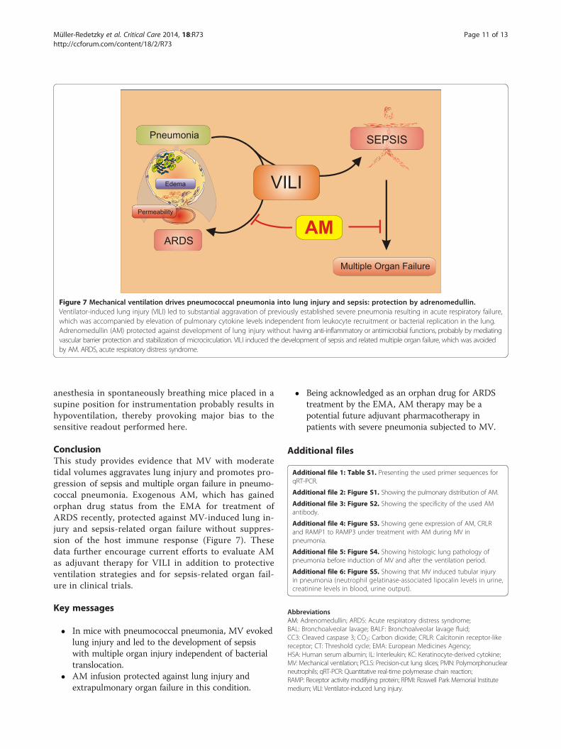
Figure 7 Mechanical ventilation drives pneumococcal pneumonia into lung injury and sepsis: protection by adrenomedullin.Ventilator-induced lung injury (VILI) led to substantial aggravation of previously established severe pneumonia resulting in acute respiratory failure,which was accompanied by elevation of pulmonary cytokine levels independent from leukocyte recruitment or bacterial replication in the lung.Adrenomedullin (AM) protected against development of lung injury without having anti-inflammatory or antimicrobial functions, probably by mediatingvascular barrier protection and stabilization of microcirculation. VILI induced the development of sepsis and related multiple organ failure, which was avoidedby AM. ARDS, acute respiratory distress syndrome.
Müller-Redetzky et al. Critical Care 2014, 18:R73 Page 11 of 13http://ccforum.com/content/18/2/R73
anesthesia in spontaneously breathing mice placed in asupine position for instrumentation probably results inhypoventilation, thereby provoking major bias to thesensitive readout performed here.
ConclusionThis study provides evidence that MV with moderatetidal volumes aggravates lung injury and promotes pro-gression of sepsis and multiple organ failure in pneumo-coccal pneumonia. Exogenous AM, which has gainedorphan drug status from the EMA for treatment ofARDS recently, protected against MV-induced lung in-jury and sepsis-related organ failure without suppres-sion of the host immune response (Figure 7). Thesedata further encourage current efforts to evaluate AMas adjuvant therapy for VILI in addition to protectiveventilation strategies and for sepsis-related organ fail-ure in clinical trials.
Key messages
� In mice with pneumococcal pneumonia, MV evokedlung injury and led to the development of sepsiswith multiple organ injury independent of bacterialtranslocation.
� AM infusion protected against lung injury andextrapulmonary organ failure in this condition.
� Being acknowledged as an orphan drug for ARDStreatment by the EMA, AM therapy may be apotential future adjuvant pharmacotherapy inpatients with severe pneumonia subjected to MV.
Additional files
Additional file 1: Table S1. Presenting the used primer sequences forqRT-PCR.
Additional file 2: Figure S1. Showing the pulmonary distribution of AM.
Additional file 3: Figure S2. Showing the specificity of the used AMantibody.
Additional file 4: Figure S3. Showing gene expression of AM, CRLRand RAMP1 to RAMP3 under treatment with AM during MV inpneumonia.
Additional file 5: Figure S4. Showing histologic lung pathology ofpneumonia before induction of MV and after the ventilation period.
Additional file 6: Figure S5. Showing that MV induced tubular injuryin pneumonia (neutrophil gelatinase-associated lipocalin levels in urine,creatinine levels in blood, urine output).
AbbreviationsAM: Adrenomedullin; ARDS: Acute respiratory distress syndrome;BAL: Bronchoalveolar lavage; BALF: Bronchoalveolar lavage fluid;CC3: Cleaved caspase 3; CO2: Carbon dioxide; CRLR: Calcitonin receptor-likereceptor; CT: Threshold cycle; EMA: European Medicines Agency;HSA: Human serum albumin; IL: Interleukin; KC: Keratinocyte-derived cytokine;MV: Mechanical ventilation; PCLS: Precision-cut lung slices; PMN: Polymorphonuclearneutrophils; qRT-PCR: Quantitative real-time polymerase chain reaction;RAMP: Receptor activity modifying protein; RPMI: Roswell Park Memorial Institutemedium; VILI: Ventilator-induced lung injury.

Müller-Redetzky et al. Critical Care 2014, 18:R73 Page 12 of 13http://ccforum.com/content/18/2/R73
Competing interestsThe authors declare that they have no competing interests.
Authors’ contributionsHCM-R and MW planned and supervised the study, performed in vivoexperiments, analyzed the data and drafted the manuscript. DW and KHperformed animal experiments, flow cytometry and cytokine assays, andcritically revised the manuscript for important intellectual content, WK, MDM,TT, OK and ADG performed histology and critically revised the manuscriptfor important intellectual content. RP performed experiments using PCLSand critically revised the manuscript for important intellectual content. UPperformed qRT-PCR analyses and critically revised the manuscript forimportant intellectual content. SH, NW and NS were involved in thestudy design and participated in drafting the manuscript. All authorsread and approved the final version of the manuscript.
AcknowledgementsThe authors thank Sandra Wienhold, Matthias Felten, Marfa Polikarpova, UteKellermann Anna Goldberg and Petra Merner for skillful technical assistance.
FundingThis study was supported by the Deutsche Forschungsgemeinschaft to WKand NW (Excellence Cluster Cardio-Pulmonary System), to SH (SFB-TR84 C2and DFG HI 789/7-1), to NS (SFB-TR84 Z2), to MW (SFB-TR84 C3, C6 and DFGOP 86/7-1) and to NW (SFB-TR84 C6), and by the German Federal Ministry ofEducation and Research to HCM-R (PROGRESS). The funding sources had noinfluence on study design or publication.
Author details1Department of Infectious Diseases and Pulmonary Medicine, Charité –Universitätsmedizin Berlin, Charitéplatz 1, 10117 Berlin, Germany. 2Institute forAnatomy and Cell Biology, Justus-Liebig-University, Universities of Giessenand Marburg Lung Center, Aulweg 123, 35392 Giessen, Germany. 3Memberof the German Center for Lung Research, Aulweg 130, 35392 Giessen,Germany. 4Institute of Anatomy and Cell Biology, Saarland University, Facultyof Medicine, Kirrberger Str. 100, 66424 Homburg/Saar, Germany. 5Institute forClinical and Experimental Surgery, Saarland University, Faculty of Medicine,Kirrberger Straße 100, 66424 Homburg/SaarHomburg, Germany. 6Departmentof Veterinary Pathology, Freie Universität Berlin, Robert-von-Ostertag-Straße15, 14163 Berlin, Germany. 7Excellencecluster Cardio-Pulmonary System,Department of Internal Medicine, Aulweg 130, 35392 Giessen, Germany.
Received: 4 October 2013 Accepted: 3 April 2014Published: 14 April 2014
References1. Ventilation with lower tidal volumes as compared with traditional tidal
volumes for acute lung injury and the acute respiratory distresssyndrome. The Acute Respiratory Distress Syndrome Network. N Engl JMed 2000, 342:1301–1308.
2. Terragni PP, Del Sorbo L, Mascia L, Urbino R, Martin EL, Birocco A, Faggiano C,Quintel M, Gattinoni L, Ranieri VM: Tidal volume lower than 6 ml/kgenhances lung protection: role of extracorporeal carbon dioxide removal.Anesthesiology 2009, 111:826–835.
3. Wolthuis EK, Vlaar AP, Choi G, Roelofs JJ, Juffermans NP, Schultz MJ:Mechanical ventilation using non-injurious ventilation settings causeslung injury in the absence of pre-existing lung injury in healthy mice.Crit Care 2009, 13:R1.
4. Dhanireddy S, Altemeier WA, Matute-Bello G, O'Mahony DS, Glenny RW,Martin TR, Liles WC: Mechanical ventilation induces inflammation, lunginjury, and extra-pulmonary organ dysfunction in experimental pneumonia.Lab Invest 2006, 86:790–799.
5. Brunkhorst FM, Oppert M, Marx G, Bloos F, Ludewig K, Putensen C, Nierhaus A,Jaschinski U, Meier-Hellmann A, Weyland A, Grundling M, Moerer O, Riessen R,Seibel A, Ragaller M, Buchler MW, John S, Bach F, Spies C, Reill L, Fritz H,Kiehntopf M, Kuhnt E, Bogatsch H, Engel C, Loeffler M, Kollef MH, Reinhart K,Welte T: Effect of empirical treatment with moxifloxacin and meropenem vsmeropenem on sepsis-related organ dysfunction in patients with severesepsis: a randomized trial. JAMA 2012, 307:2390–2399.
6. van Kaam AH, Lachmann RA, Herting E, De Jaegere A, van Iwaarden F,Noorduyn LA, Kok JH, Haitsma JJ, Lachmann B: Reducing atelectasis
attenuates bacterial growth and translocation in experimentalpneumonia. Am J Respir Crit Care Med 2004, 169:1046–1053.
7. van Kaam AH, Lutter R, Lachmann RA, Haitsma JJ, Herting E, Snoek M,De Jaegere A, Kok JH, Lachmann B: Effect of ventilation strategy andsurfactant on inflammation in experimental pneumonia. Eur Respir J 2005,26:112–117.
8. Imai Y, Parodo J, Kajikawa O, de Perrot M, Fischer S, Edwards V, Cutz E, Liu M,Keshavjee S, Martin TR, Marshall JC, Ranieri VM, Slutsky AS: Injurious mechanicalventilation and end-organ epithelial cell apoptosis and organ dysfunction inan experimental model of acute respiratory distress syndrome. JAMA 2003,289:2104–2112.
9. Ranieri VM, Giunta F, Suter PM, Slutsky AS: Mechanical ventilation as amediator of multisystem organ failure in acute respiratory distresssyndrome. JAMA 2000, 284:43–44.
10. Temmesfeld-Wollbruck B, Hocke AC, Suttorp N, Hippenstiel S: Adrenomedullinand endothelial barrier function. Thromb Haemost 2007, 98:944–951.
11. Hippenstiel S, Witzenrath M, Schmeck B, Hocke A, Krisp M, Krull M, Seybold J,Seeger W, Rascher W, Schutte H, Suttorp N: Adrenomedullin reducesendothelial hyperpermeability. Circ Res 2002, 91:618–625.
12. Hocke AC, Temmesfeld-Wollbrueck B, Schmeck B, Berger K, Frisch EM,Witzenrath M, Brell B, Suttorp N, Hippenstiel S: Perturbation of endothelialjunction proteins by Staphylococcus aureus alpha-toxin: inhibition ofendothelial gap formation by adrenomedullin. Histochem Cell Biol 2006,126:305–316.
13. Müller HC, Witzenrath M, Tschernig T, Gutbier B, Hippenstiel S, Santel A,Suttorp N, Rosseau S: Adrenomedullin attenuates ventilator-induced lunginjury in mice. Thorax 2010, 65:1077–1084.
14. Temmesfeld-Wollbruck B, Brell B, David I, Dorenberg M, Adolphs J, Schmeck B,Suttorp N, Hippenstiel S: Adrenomedullin reduces vascular hyperpermeabilityand improves survival in rat septic shock. Intensive Care Med 2007,33:703–710.
15. Witzenrath M, Schmeck B, Doehn JM, Tschernig T, Zahlten J, Loeffler JM,Zemlin M, Müller H, Gutbier B, Schutte H, Hippenstiel S, Fischetti VA, Suttorp N,Rosseau S: Systemic use of the endolysin Cpl-1 rescues mice with fatalpneumococcal pneumonia. Crit Care Med 2009, 37:642–649.
16. Müller-Redetzky HC, Kummer W, Pfeil U, Hellwig K, Will D, Paddenberg R,Tabeling C, Hippenstiel S, Suttorp N, Witzenrath M: Intermedin stabilizedendothelial barrier function and attenuated ventilator-induced lung injuryin mice. PLoS ONE 2012, 7:e35832.
17. Müller HC, Hellwig K, Rosseau S, Tschernig T, Schmiedl A, Gutbier B,Schmeck B, Hippenstiel S, Peters H, Morawietz L, Suttorp N, Witzenrath M:Simvastatin attenuates ventilator-induced lung injury in mice. Crit Care 2010,14:R143.
18. Paddenberg R, Konig P, Faulhammer P, Goldenberg A, Pfeil U, Kummer W:Hypoxic vasoconstriction of partial muscular intra-acinar pulmonaryarteries in murine precision cut lung slices. Respir Res 2006, 7:93.
19. Vecchi A, Garlanda C, Lampugnani MG, Resnati M, Matteucci C, Stoppacciaro A,Schnurch H, Risau W, Ruco L, Mantovani A: Monoclonal antibodies specificfor endothelial cells of mouse blood vessels. Their application in theidentification of adult and embryonic endothelium. Eur J Cell Biol 1994,63:247–254.
20. Sperling J, Schafer T, Ziemann C, Benz-Weiber A, Kollmar O, Schilling MK,Menger MD: Hepatic arterial infusion of bevacizumab in combinationwith oxaliplatin reduces tumor growth in a rat model of colorectal livermetastases. Clin Exp Metastasis 2012, 29:91–99.
21. Vaporidi K, Voloudakis G, Priniannakis G, Kondili E, Koutsopoulos A, Tsatsanis C,Georgopoulos D: Effects of respiratory rate on ventilator-induced lung injuryat a constant PaCO2 in a mouse model of normal lung. Crit Care Med 2008,36:1277–1283.
22. Gonzalez-Rey E, Chorny A, Varela N, Robledo G, Delgado M: Urocortin andadrenomedullin prevent lethal endotoxemia by down-regulating theinflammatory response. Am J Pathol 2006, 168:1921–1930.
23. Itoh T, Obata H, Murakami S, Hamada K, Kangawa K, Kimura H, Nagaya N:Adrenomedullin ameliorates lipopolysaccharide-induced acute lunginjury in rats. Am J Physiol Lung Cell Mol Physiol 2007, 293:L446–L452.
24. Wilson MR, O'Dea KP, Zhang D, Shearman AD, van Rooijen N, Takata M:Role of lung-marginated monocytes in an in vivo mouse model ofventilator-induced lung injury. Am J Respir Crit Care Med 2009,179:914–922.
25. Brell B, Temmesfeld-Wollbruck B, Altzschner I, Frisch E, Schmeck B, Hocke AC,Suttorp N, Hippenstiel S: Adrenomedullin reduces Staphylococcus aureus
Müller-Redetzky et al. Critical Care 2014, 18:R73 Page 13 of 13http://ccforum.com/content/18/2/R73
alpha-toxin-induced rat ileum microcirculatory damage. Crit Care Med 2005,33:819–826.
26. Brell B, Hippenstiel S, David I, Pries AR, Habazettl H, Schmeck B, Suttorp N,Temmesfeld-Wollbruck B: Adrenomedullin treatment abolishes ileal mucosalhypoperfusion induced by Staphylococcus aureus alpha-toxin–an intravitalmicroscopic study on an isolated rat ileum. Crit Care Med 2005, 33:2810–016.
27. Sprung CL, Annane D, Keh D, Moreno R, Singer M, Freivogel K, Weiss YG,Benbenishty J, Kalenka A, Forst H, Laterre PF, Reinhart K, Cuthbertson BH,Payen D, Briegel J: Hydrocortisone therapy for patients with septic shock.N Engl J Med 2008, 358:111–124.
28. Vincent JL, De Backer D: Microvascular dysfunction as a cause of organdysfunction in severe sepsis. Crit Care 2005, 9:S9–S12.
29. Charles PE, Piroth L, Desbiolles N, Lequeu C, Martin L, Portier H, Chavanet P:New model of ventilator-associated pneumonia in immunocompetentrabbits. Crit Care Med 2002, 30:2278–2283.
30. Pugin J, Dunn-Siegrist I, Dufour J, Tissieres P, Charles PE, Comte R: Cyclicstretch of human lung cells induces an acidification and promotes bacterialgrowth. Am J Respir Cell Mol Biol 2008, 38:362–370.
31. Hammerschmidt S, Wolff S, Hocke A, Rosseau S, Muller E, Rohde M:Illustration of pneumococcal polysaccharide capsule during adherenceand invasion of epithelial cells. Infect Immun 2005, 73:4653–4667.
32. Carrizo GJ, Wu R, Cui X, Dwivedi AJ, Simms HH, Wang P: Adrenomedullinand adrenomedullin-binding protein-1 downregulate inflammatory cytokinesand attenuate tissue injury after gut ischemia-reperfusion. Surgery 2007,141:245–253.
33. Yin H, Chao L, Chao J: Adrenomedullin protects against myocardialapoptosis after ischemia/reperfusion through activation of Akt-GSKsignaling. Hypertension 2004, 43:109–116.
doi:10.1186/cc13830Cite this article as: Müller-Redetzky et al.: Mechanical ventilation drivespneumococcal pneumonia into lung injury and sepsis in mice: protectionby adrenomedullin. Critical Care 2014 18:R73.
Submit your next manuscript to BioMed Centraland take full advantage of:
• Convenient online submission
• Thorough peer review
• No space constraints or color figure charges
• Immediate publication on acceptance
• Inclusion in PubMed, CAS, Scopus and Google Scholar
• Research which is freely available for redistribution
Submit your manuscript at www.biomedcentral.com/submit