R Comparative analysis of diguanylate cyclase and phosphodiesterase genes in Klebsiella pneumoniae
Upload
khangminh22Category
view
0download
0
Article
An acquired acyltransfera
se promotes Klebsiellapneumoniae ST258 respiratory infectionGraphical abstract
Highlights
d An acyltransferase (atf3) is prevalent in ST258 K. pneumoniae
d Expression of atf3 enhances glycolysis, increasing bacterial
ATP production and ROS
d With atf3, the airway metabolome was altered with greater
glucose consumption
d K. pneumoniae expressing atf3 has a competitive advantage
in vivo
Ahn et al., 2021, Cell Reports 35, 109196June 1, 2021 ª 2021 The Authors.https://doi.org/10.1016/j.celrep.2021.109196
Authors
Danielle Ahn, Gitanjali Bhushan,
Thomas H. McConville, ...,
Robert K. Ernst, Anne-Catrin Uhlemann,
Alice Prince
In brief
Klebsiella pneumoniae ST258 is a
successful human pathogen. Here, Ahn
et al. identify an acyltransferase of the
superfamily 3 (atf3) concentrated within
one of the ST258 clades. Acquisition of
this gene leads to enhanced bioenergetic
properties, giving the pathogen a
competitive advantage in vivo.
ll

OPEN ACCESS
llArticle
An acquired acyltransferasepromotes Klebsiella pneumoniaeST258 respiratory infectionDanielle Ahn,1,5,* Gitanjali Bhushan,1 Thomas H. McConville,2 Medini K. Annavajhala,2 Rajesh Kumar Soni,3
Tania Wong Fok Lung,1 Casey E. Hofstaedter,4 Shivang S. Shah,1 Alexander M. Chong,2 Victor G. Castano,1
Robert K. Ernst,4 Anne-Catrin Uhlemann,2 and Alice Prince11Department of Pediatrics, Columbia University Irving Medical Center, New York, NY 10032, USA2Department of Medicine, Columbia University Irving Medical Center, New York, NY 10032, USA3Proteomics and Macromolecular Crystallography Shared Resource, Herbert Irving Comprehensive Cancer Center, Columbia University
Irving Medical Center, New York, NY 10032, USA4Department of Microbial Pathogenesis, University of Maryland, Baltimore, Baltimore, MD 21201, USA5Lead contact*Correspondence: [email protected]
https://doi.org/10.1016/j.celrep.2021.109196
SUMMARY
Klebsiella pneumoniae ST258 is a human pathogen associated with poor outcomes worldwide. We identify amember of the acyltransferase superfamily 3 (atf3), enriched within the ST258 clade, that provides a majorcompetitive advantage for the proliferation of these organisms in vivo. Comparison of a wild-type ST258strain (KP35) and a Datf3 isogenic mutant generated by CRISPR-Cas9 targeting reveals greater NADH:ubi-quinone oxidoreductase transcription and ATP generation, fueled by increased glycolysis. The acquisitionof atf3 induces changes in the bacterial acetylome, promoting lysine acetylation of multiple proteins involvedin central metabolism, specifically Zwf (glucose-6 phosphate dehydrogenase). The atf3-mediated metabolicboost leads to greater consumption of glucose in the host airway and increased bacterial burden in the lung,independent of cytokine levels and immune cell recruitment. Acquisition of this acyltransferase enhancesfitness of a K. pneumoniae ST258 isolate and may contribute to the success of this clonal complex as ahealthcare-associated pathogen.
INTRODUCTION
Multi-drug-resistant Gram-negative pathogens are an increasing
threat, limiting the effectiveness of even novel therapeutic strate-
gies (Doi et al., 2017). These organisms are an omnipresent
complication of modern intensive care unit (ICU) care, causing
ventilator-associated pneumonia (Cilloniz et al., 2019), superin-
fection following viral illnesses such as severe acute respiratory
syndrome-coronavirus-2 (SARS-CoV-2) (Gomez-Simmonds
et al., 2020), and bloodstream infections (Satlin et al., 2017). Car-
bapenem-resistant Klebsiella pneumoniae (CRKP) is among the
current epidemic of multi-drug-resistant Gram-negative bacteria
associated with unacceptably high morbidity and mortality (Borer
et al., 2009; Kohler et al., 2017; Xu et al., 2017). Within CRKP, the
sequence type 258 (ST258) has emerged as a persistent and
overrepresented cause of hospital-associated infections in the
United States and worldwide (Marsh et al., 2019; Tzouvelekis
et al., 2013; van Duin et al., 2020). Transposon screens have
been used to identify loci that promote K. pneumoniae pathoge-
nicity (Bachman et al., 2015; Paczosa et al., 2020; Vornhagen
et al., 2019). However, such studies typically use a laboratory
reference isolate (American Type Culture Collection [ATCC]
43816orKPPR1), which is genetically distinct from the highly anti-
This is an open access article under the CC BY-N
biotic resistant ST258 clonal complex (Gomez-Simmonds and
Uhlemann, 2017; Vornhagen et al., 2019). To understand the bac-
terial properties responsible for the success of K. pneumoniae
ST258, we analyzed clinical isolate 35 (KP35) from our own hos-
pital, a representative strain that expresses the most common
cps (capsular protein) and wzi (capsular component) genotypes
(Ahn et al., 2016; Gomez-Simmonds et al., 2015). The median le-
thal dose (LD50) of ST258 isolates is several logs higher than
KPPR1 (Xiong et al., 2015), consistent with our previous work in
which a much higher inoculum of KP35 was required to induce
pathology in a murine model of pneumonia (Ahn et al., 2016).
KP35 displayed kinetics of infection similar to what has been
observed in humans—an indolent but progressive pneumonia
characterized by the recruitment of anti-inflammatory monocytes
that fail to clear infection and are typically present in the resolution
phase of infection (Poe et al., 2013). The influx of thesemonocytes
in response to infection with ST258 strains then elicits a subdued
immune response mediated by interleukin-10 (IL-10) secretion
(Penaloza et al., 2019).
The genetic elements responsible for the global persistence of
ST258 K. pneumoniae have not been identified (Tzouvelekis
et al., 2013). The prevailing wisdom suggests that the acquisition
of antimicrobial resistance elements carries a fitness cost that is
Cell Reports 35, 109196, June 1, 2021 ª 2021 The Authors. 1C-ND license (http://creativecommons.org/licenses/by-nc-nd/4.0/).

Articlell
OPEN ACCESS
often not detected under in vitro conditions (Wong et al., 2019).
This fitness cost is predicted to lead to reversibility in the
absence of antimicrobial pressure (Andersson and Hughes,
2010; Bachman et al., 2015) and is the foundation for the devel-
opment of antimicrobial stewardship programs. However, the
dissemination and persistence of ST258 organisms worldwide,
amidst many other species of opportunistic pathogens, implies
that they may have other properties to promote selection in vivo
(Ernst et al., 2020). Comprehensive genotypic and phenotypic
studies of ST258 isolates have identified interesting but inconsis-
tent virulence determinants such as those involved in iron utiliza-
tion, stress response signaling, biofilm formation, and type IV pili
(Marsh et al., 2019; Pitout et al., 2015). In the absence of a unify-
ing virulence phenotype, it has been postulated that the dynamic
evolution of thesemechanisms of persistence within the host mi-
crobiota is due to the availability of metabolites generated by re-
cruited immune cells as well as by bacteria (Blin et al., 2017;
Vornhagen et al., 2019). We hypothesized that the success of
pathogens like ST258 K. pneumoniae could also be attributable
to less apparent factors such as the metabolic properties of the
bacteria.
In the KP35 genome, we identified an open reading frame
(ORF) that provides KP35 with a selective advantage, enhancing
persistence in the murine lung (Ahn et al., 2016). This ORF,
initially designated in our previous publication as an arginine anti-
porter (arcD), is now annotated in silico as a member of the acyl-
transferase superfamily 3 (atf3), a diverse group of proteins
linked to metabolic pathways. In the experiments detailed
here, we show that this element promotes a global increase in
K. pneumoniae bioenergetics with increased ATP production
through enhanced glycolysis and tricarboxylic acid (TCA) cycle
activity, leading to increased substrate utilization and persistent
pulmonary infection. This augmented metabolic activity may
negate the metabolic expense of maintaining antibiotic resis-
tance genes and provide KP35 with a metabolic boost that con-
tributes to its persistence in the lung.
RESULTS
Acquisition of an acyltransferase superfamily 3 (atf3)is ubiquitous in an ST258 cladeWe determined whether the unique ORF in KP35, annotated in
silico as a member of the acyltransferase superfamily 3 (atf3)
and previously associated with increased fitness in a murine
model of pneumonia (Ahn et al., 2016), was present across
diverse K. pneumoniae clones. Phylogenetic reconstruction of
the 178 publicly available genomes in NCBI showed that atf3
was only present in ST258 isolates (Figure 1A), appreciating
that the data from sequenced isolates may be skewed toward
those associated with clinically important infections or hospital
outbreaks. In phylogenetic analyses focused on only ST258 iso-
lates (n = 100), we found that atf3 is concentrated in clade 1, one
of the twomajor ST258 clades that expresses KPC-2 andwzi 29,
although we observed some isolates with a sporadic loss of the
gene (Figure 1B). Of note, atf3was not present in KPPR1, the lab-
oratory reference strain often used in studies of K. pneumoniae
pathogenicity. The 332-amino acid protein encoded by atf3 is
predicted in silico to be a transmembrane protein, but lacks ho-
2 Cell Reports 35, 109196, June 1, 2021
mology with known proteins. Phylogeny of atf3members as rep-
resented in a sunburst diagram (Pfam), demonstrating the ubiq-
uity and heterogeneity of this ancient enzyme domain across
6,022 species (Figure 1C).
Deletion of atf3 does not alter the physical properties ofKP35Neither the primary sequence nor the predicted protein structure
of the KP35 atf3 gene product indicates its function. However,
atf3 is colocated with multiple genes involved in capsular
production (ugd, gnd; components of the cps cluster in
K. pneumoniae) (Pan et al., 2015) and O-antigen synthesis
(wbgU, tagGH) (Bruchmann et al., 2021; Caboni et al., 2015) (Fig-
ure 2A). To study the role of atf3 in pathogenesis, a null mutant
was constructed using a recently adapted CRISPR-Cas9 sys-
tem, Lambda Red Recombineering genes to improve efficiency,
and Zeocin selection due to the presence of multiple antimicro-
bial resistance elements in the KP35 genome (McConville et al.,
2020). A 122-bp deletion beginning 4 bp upstream of the start
codon was confirmed by whole-genome sequencing. The dele-
tion mutant was complemented with a cloned version of atf3 ex-
pressed on a high copy number plasmid under Zeocin selection.
The basic properties of the three strains were compared in vitro
and found to have equivalent rates of growth in Luria-Bertani (LB)
media (Figure 2B), formation of biofilm (Figure S1A), and suscep-
tibility to antimicrobials (Figure S1B).
Among the many functions ascribed to K. pneumoniae acyl-
transferases, modification of the lipid A component of lipopoly-
saccharide (LPS) is a potential mechanism that could affect
pathogenesis. Using MALDI-TOFmass spectrometry, we estab-
lished thatWT KP35 and theDatf3mutant did not havemajor dif-
ferences in LPS lipid A structure (Figures 2C and 2D). However,
there was an increased abundance of LPSO-antigen side chains
and core protein in the Datf3 mutant (Figures S2C and S2D).
These differences in LPS did not translate into altered suscepti-
bility to polymyxin B or H2O2 (Figures S1B and S1E). These find-
ings indicated that atf3 is conserved within the ST258
K. pneumoniae clonal clade and that its deletion did not signifi-
cantly change the baseline characteristics of this clinical isolate.
The presence of atf3 increases K. pneumoniae
glycolysis and TCA cycle activityThe impact of atf3 on KP35 gene expression was assessed by
RNA sequencing (RNA-seq) comparing KP35 and the Datf3
mutant grown in LB broth overnight (Figure 3A). Kyoto Encyclo-
pedia of Genes and Genomes (KEGG) pathway analysis of the
top 50 genes with increased expression in KP35 as compared
to the Datf3 mutant showed the upregulation of many metabolic
pathways, suggesting that atf3 activity is primarily involved in
enhancing the ability of KP35 to generate energy (Figure 3B).
This was reflected in the increased expression of glycolytic en-
zymes inWTKP35 compared to theDatf3mutant, and to a lesser
extent in the components of the TCA cycle (Figure 3C). To deter-
mine the possible role of atf3 acyltransferase activity in bacterial
metabolism, we compared the assimilation of single carbon
sources that are intermediate steps in glycolysis and the TCA cy-
cle. There was preferential metabolism of glucose and glycolytic
intermediates in minimal media by KP35, as determined by the

Figure 1. A novel acyltransferase of the superfamily 3 (atf3) is prevalent in K. pneumoniae ST258 isolates
(A) A phylogenetic tree of the publicly available assemblies of K. pneumoniae isolates. The inner ring represents the ST group to which the isolate belongs and the
outer ring is the screen for the atf3 gene, with isolates designated as a variant if it contained at least 1 point mutation.
(B) A concentrated phylogenetic tree of ST258 isolates only.
(C) A sunburst visualization of species, considering taxonomic lineage, with members of the acyltransferase superfamily 3.
Articlell
OPEN ACCESS
reduction of tetrazolium blue, relative to the Datf3 mutant or the
laboratory standardKPPR1, included for comparison (Figure 3D).
Static growth in minimal media with increasing concentrations of
selected metabolites showed no major differences in growth to
explain this difference (Figures S2A–S2D). In RPMI, a more
nutrient-rich media, growth of the bacteria with differing concen-
trations of glucose was similar for WT KP35 and Datf3
(Figure S2E).
There was enhancement in the glycolytic activity of KP35
alone as compared to Datf3. Using the Seahorse analyzer, we
observed relatively higher extracellular acidification rates
(ECARs) of KP35 in response to glucose to initiate glycolysis
and to oligomycin to block oxidative phosphorylation and un-
leash glycolytic capacity (Figure 3E). The expected reduction in
glycolytic activity by 2-deoxyglucose was observed for both
bacterial strains. A relative increase in oxygen consumption
Cell Reports 35, 109196, June 1, 2021 3

Figure 2. The growth rate and lipid A structure for KP35 are unchanged with the deletion of the atf3 gene
(A) Schematic of the KP35 atf3 gene locus using Geneious version 10.
(B) Serial optical density at wavelength of 600 nm (OD600) measurements of bacteria were taken over 18 h with intermittent shaking at 37�C in LB. n = 3; each point
is the mean value, with bars representing the SEMs.
(C) Negative ionmodeMALDI-TOFMS lipid A spectra are shown for KP35,Daft3, andDatf3::atf3 (n = 1). The strains were grown at 37�C in LB for 18 h. Additions of
a hydroxyl group (Dm/z 16) and an aminoarabinose group (Dm/z 131) are observed among all 3 strains. The ion atm/z 1972 demonstrates both modifications. In
addition, loss of a phosphate (Dm/z 80) is likely a result of harsh lipid A extraction conditions. The ion atm/z 2063 represents the addition of a palmitate (C16,Dm/z
238) acyl chain.
(D) Representative structures are shown for the ions at m/z 1825 and m/z 1972. The ion at m/z 1956/1972 represents lipid A species with the addition of 1
aminoarabinose moiety, whereas the ion at m/z 2103 represents the addition of 2 aminoarabinose moieties.
Articlell
OPEN ACCESS
rates (OCRs) was also observed in response to glucose in the
parent strain (Figure 3F). Of note, these changes in bacterial
metabolism did not significantly alter the cumulative host and
pathogenmetabolic activity asmeasured in differentiatedmono-
cytes (THP-1s) co-incubated with the bacterial strains (Figures
3G and 3H). The expected consequences of heightened meta-
bolism in the presence of atf3 were documented by the
increased generation of reactive oxygen species (ROS) (Figure 3I)
and levels of ATP produced by bacteria alone (Figure 3J). This
was further supported by the enhanced transcription of virtually
all of the nuo and nqr genes in the parent strain compared to the
mutant. These loci encode the individual NADH:ubiquinone
oxidoreductase (NQO) components of complex 1 (Figures 3K
and 3L). All 12 of the proton-translocating NQO components
(nuoB, -C, -E, -F, -G, -H, -I, -J, -K, -L, -M, and -N) were upregu-
lated in the parent strain with atf3 as were the 6 Na+-translocat-
ing NQOs (nqrA, -B, -C, -D, -E, and -F). These membrane-asso-
ciated proton (or Na+) pumps couple the oxidation of NADH to
the reduction of ubiquinone, generate the transmembrane elec-
4 Cell Reports 35, 109196, June 1, 2021
trochemical gradient, and drive ATP synthesis for bacteria,
sharing many essential properties with complex I of mitochon-
dria (Dibrov et al., 2017; Erhardt et al., 2012). The importance
and complexities of these NQOs in bacterial energetics has
made them a major focus of research for decades (Dimroth,
1987), although a role in pathogenesis has not been specifically
described.
Global lysine acetylation of bacterial proteins isincreased by atf3
Having established a major impact of atf3 on the expression of
metabolic genes by KP35, we hoped to identify the target(s) of
the acyltransferase more specifically. In bacteria, post-transla-
tional modification of proteins, specifically lysine acetylation, is
a major mechanism of metabolic regulation (Christensen et al.,
2019b). Acetyl phosphate and acetyl-coenzyme A (CoA), act
as donors and can non-enzymatically acetylate proteins, a pro-
cess that is dependent upon glucose availability and glycolytic
activity (Christensen et al., 2019b). Glycolytic and TCA cycle

Figure 3. Enhanced metabolic properties of K. pneumoniae ST258 in the presence of atf3
(A) Shotgun RNA-seq of bacteria alone, Illumina platform, heatmap of variance stabilized counts. n = 1.
(B) KEGG pathway analysis showing the top 10 pathways increased in KP35 with respect to the isogenic mutant.
(C) Heatmap of the fold expression of the genes involved in glycolysis and TCA cycle enzymes expressed by KP35 over Datf3 (n= 1).
(D) Single carbon source assimilation (Biolog) (n = 2). KPPR1 = laboratory reference strain ATCC 43816.
(E–H) Glycolysis as measured by (E) extracellular acidification rates (ECARs) and (F) oxygen consumption rates (OCRs) as measured by Seahorse analyzer of
bacteria alone (n = 4) and (G) and (H) with THP1 s (n = 5). 1 = glucose, 2 = oligomycin, 3 = 2-deoxyglucose; each point is the mean value with bars representing the
SEM; *p < 0.05 between KP35 and Δatf3, 2-way ANOVA.
(I–K) Intracellular reactive oxygen species (ROS) measured byMitosox (Invitrogen) dye via flow cytometry (n = 4) (I). (J) Total ATP production (Abcam) (n = 8). For (I)
and (J), columns are mean values with bars representing the SEM; ****p < 0.001, 1-way ANOVA, Tukey’s test for multiple comparisons. Heatmap of the fold
expression of the Na-independent and -dependent NADH:quinone oxidoreductases expressed by KP35 over Datf3 via (K) RNA-seq (n = 1) and confirmed with
standard qRT-PCR (n = 3, 2 technical replicates per sample).
*p < 0.05, **p < 0.01, ***p < 0.005, ****p < 0.001; multiple t tests with a false discovery rate (FDR) of 1%.
Articlell
OPEN ACCESS
enzymes are among the most frequent targets for lysine acetyla-
tion (Nakayasu et al., 2017). We postulated that either the meta-
bolic consequences of atf3 with increased availability of acetyl-
CoA or acetyl phosphate or possibly a specific Atf3 target could
lead to changes in the lysine acetylation of key metabolic en-
zymes. Acetyl-lysine motifs on bacterial proteins were captured
using an immunoaffinity bead kit and abundance measured via
tandem mass spectrometry (MS/MS) (Figure 4). KP35 exhibited
substantially increased site-specific acetylation of many of the
enzymes involved in glycolysis as well as the TCA cycle, as
compared with the Datf3 mutant. Special attention was taken
to understand the role of Zwf (glucose-6 phosphate dehydroge-
nase), Pgi (glucose-6-phosphate isomerase), Edd (phospho-
gluconate dehydratase), and Gnd (6-phosphogluconate dehy-
drogenase), as these enzymes are the metabolic switchboard
for glucose utilization (Callura et al., 2012). Among the 4 proteins,
only Zwf and Pgi had a statistically significant increase in site-
specific lysine acetylation, with no modifications measured in
Cell Reports 35, 109196, June 1, 2021 5

(legend on next page)
6 Cell Reports 35, 109196, June 1, 2021
Articlell
OPEN ACCESS

Articlell
OPEN ACCESS
Edd. The enzyme Zwf is an important regulator of glucose con-
sumption catalyzing the oxidation of glucose-6 phosphate
(glucose-6P) to 6-phosphogluconic acid, a substrate for the Ent-
ner-Doudoroff and pentose phosphate pathways. Pgi converts
glucose-6P to fructose-6P, the first step in the Embden-Meyer-
hof pathway, also known as glycolysis. The acetylation of pro-
teins like Zwf and Pgi generally leads to decreased enzymatic
activity and would therefore decrease the generation of 6-phos-
phogluconic acid and fructose-6P, respectively (Nakayasu et al.,
2017). Exactly how lysine acetylation affects protein function and
whether critical sites for dimerization or the active site itself is
modified can be established for enzymes with solved structures.
However, since the structure of Zwf and Pgi in KP35 is unknown,
we can only demonstrate the likelihood that this critical regulator
of glucose metabolism is altered in bacteria expressing atf3.
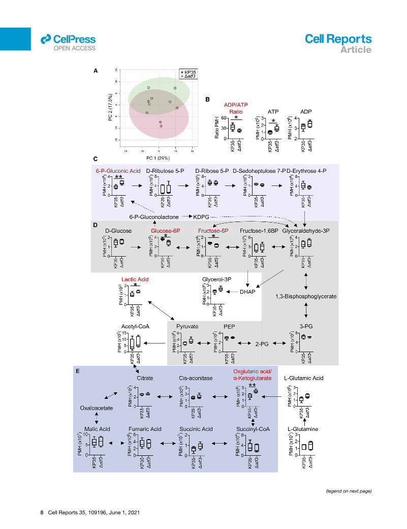
Intracellular metabolite accumulation in KP35represents diminished Zwf activity and enhancedglycolysisTodemonstrate the impact of atf3onbacterial glycolysis, targeted
intracellular metabolomics was performed on KP35 and the Datf3
mutant (Figure 5A). An increase in the intracellular ADP:ATP ratio
in KP35 was consistent with enhanced glycolysis (Figure 5B).
There was relative depletion of ATP inside KP35 as compared to
the mutant, contrary to our previous observation that total ATP
levels were increased in the presence of atf3 (Figure 3J). This sug-
gests greater movement of ATP extracellularly in the parent strain.
Glucose utilization was differential, with its diversion toward the
generation of 6-phosphogluconic acid in the Datf3mutant without
an increase in other components of the pentose phosphate
pathway (Figure 5C). This reduced generation of 6-phosphoglu-
conic acid in KP35 suggests reduced Zwf activity, the expected
effect of increased lysine acetylation of the enzyme (Figure 4). In
line with the enhanced glycolytic activity previously observed,
glucose-6P and fructose-6P levels were relatively elevated within
WT KP35 (Figure 5D). While a significant reduction in fructose-6P
due to the acetylation of Pgi in the parent strain was not observed,
the absolute difference in peak metabolite height (PMH) was
reduced between glucose-6P (3.656 3 106 versus 2.873 3 106,
wild-type (WT) KP35 versus Datf3) and fructose-6P (1.396 3 106
versus 1.155 3 106, WT KP35 versus Datf3), suggesting the rela-
tively reduced production of fructose-6P in KP35.
Reduced levels of a-ketoglutarate derived from glutamic acid
were also measured in the parent strain (Figure 5E). In Escheri-
chia coli, a-ketoglutarate alongside glutamine signals the avail-
ability of carbon and nitrogen, respectively, for metabolic pro-
cesses (Doucette et al., 2011). As a-ketoglutarate
accumulates, as in the case of themutant lacking atf3, glycolysis
is suppressed through the inhibition of enzyme 1, a complex of
proteins that transport and phosphorylate glucose as it is taken
Figure 4. Post-translational acetylation of glycolysis and TCA cycle en
Acetyl-lysine motifs of KP35 and the isogenic mutant were captured with an im
spectrometry (LC/LC/MS) was then performed to measure the abundance
Heatmaps represent fold abundance of KP35 as compared to the isogenic mutan
boxes (n = 5).
*p < 0.05, **p < 0.01, ***p < 0.005, ****p < 0.001; multiple t tests with a FDR of 1%
highlighted in gray boxes are members of the metabolic switchboard for glucose
up into the cell (Doucette et al., 2011). This feedback mechanism
allows the bacteria to sense lower nitrogen availability as a-keto-
glutarate is the direct carbon product of nitrogen assimilation
though glutamate synthesis. These processes support a role
for the atf3 gene in promoting enhanced utilization of the glyco-
lytic pathway, although a single target for enhanced enzymatic
activity is not defined.
Airway metabolomics reflect the consequences of atf3on glucose consumptionWe next wanted to determine whether the infected airway me-
tabolome similarly reflected the impact of atf3 on KP35 meta-
bolic activity in vivo. We found significant depletion of glucose
in KP35 infection relative to the uninfected airway; much less
so in Datf3 infection (Figure 6A). We then performed a targeted
metabolomic analysis of the bronchoalveolar lavage fluid
(BALF) harvested from mice infected with either KP35 or the
Datf3 mutant (Figures 6B and 6C), representing the combined
host and bacterial extracellular metabolic environment. At 48 h
of infection, the respective metabolomes diverged by principal-
component analysis (PCA). There was again a relative depletion
of glucose and accumulation of pyruvate in KP35 infection as
compared to Datf3 (Figure 6D). This is consistent with increased
bacterial glycolytic activity and enhanced glucose consumption
in the airway. Measurable TCA cycle intermediates were not sta-
tistically different from the PBS control (Figure 6E). Among other
major metabolites, carnitine was the only compound to be rela-
tively depleted in KP35 infection compared to the Datf3 mutant
(Figures 6F–6H). The in vivo data support the hypothesis that
atf3 contributes to substrate utilization and subsequent energy
production, providing a metabolic advantage that may benefit
KP35 and its survival in the host lung.
The presence of atf3 provides a competitive advantagein vivo
We next addressed whether the increased metabolic activity
associated with atf3 expression in KP35 contributes to its patho-
genicity. We performed a competitive index experiment in a mu-
rine model of pneumonia, varying the relative amounts of KP35
andDatf3mutant strain inoculated, to determinewhether atf3pro-
vides a fitness advantage in the lung. The recovery of KP35 from
the BALF was significantly greater regardless of increasing pro-
portions of Datf3 (Figure 7A). We also observed greater recovery
of WT KP35 from the lungs at 48 h post-infection as compared
to theDatf3mutant, with similar findings observed for the comple-
mented strain Datf3::atf3 (Figure 7B). The persistence of KP35
in vivo was demonstrated by the increased number of mice with
measurable KP35 colony-forming units (CFU) in the BALF at
168 h post-infection (Figure 7C). We also noted more prominent
disruption of the lung architecture with KP35 infection as
zymes are increased in the presence of atf3
munoaffinity bead kit (Cell Signaling). Tandem liquid chromatography/mass
of acetylation motifs at unique positions (row label) of detected proteins.
t, with statistically significant differences represented with stars within specific
. Student’s t test performed for datasets with a single acetylation site. Proteins
utilization.
Cell Reports 35, 109196, June 1, 2021 7
(legend on next page)
8 Cell Reports 35, 109196, June 1, 2021
Articlell
OPEN ACCESS

Figure 6. The presence of atf3 alters the airway metabolome in KP35 infection
WT C57BL/6J mice were intranasally infected with 1–2 3 108 CFU.
(A) Glucose levels measured in BALF of WT mice infected for 48 h via calorimetric assay (Abcam). n = 5 (PBS) or 9–10 (infected); columns are mean values with
bars representing SEM; p<0.05 (Mann-Whitney), ****p<0.0001 (one-way ANOVA, Tukey’s test for multiple comparisons); recovered CFUs are shown in Figure 7B.
(B) Bacterial CFU recovered from BALF of infected mice of over a time course of infection.
(C) PCA plot for metabolites measured from the BALF supernatant of these mice via targeted LC/MS of polar metabolites (n = 2–3, each point represents 1 mouse).
(D–H) Selected peak metabolite levels involved in (D) glycolysis, (E) TCA cycle, (F) carnitine-related metabolites, (G) choline-related metabolites, and (H) amino
acids measured in BALF at 48 h of infection are shown via heatmap.
*p < 0.05, ***p < 0.005; multiple t tests with a FDR of 1%, compared to PBS control.
Articlell
OPEN ACCESS
compared to Datf3 by histopathology (Figure 7D). The immune
response to infection did not appear to account for the differences
in bacterial recovery. The secreted cytokines in BALF were not
significantly different in the WT KP35 or Datf3 infection (Figures
7E, 7F, and S3), as may be predicted by the absence of major
changes in LPS. The numbers of immune cells recovered were
also similar (Figure 7G). In vitro assays using differentiated mono-
cytes (THP-1s) confirmed no differences in bacterial uptake and
killing (Figure 7H).
Figure 5. Accumulation of intracellular metabolites in KP35 reflect enh
(A) PCA plot of targeted polar intracellular bacterial metabolites measured by LC
(B–E) Individual metabolites (B) ATP/ADP, (C) pentose phosphate pathway (purple
Box and whisker plots with min/max.
*p < 0.05, **p < 0.01; multiple t tests with FDR of 1%.
DISCUSSION
It has been difficult to explain the prevalence of ST258
K. pneumoniae as major healthcare-associated pathogens
worldwide beyond their antimicrobial resistance, a feature
shared by many other healthcare-associated pathogens. We
propose that one factor in their success is the acquisition of a
novel acyltransferase, atf3, a metabolically enhancing protein
that provides a significant growth advantage in vivo over bacteria
anced glycolysis and TCA cycle activity when atf3 is present
/MS. n = 5, each point represents a biologic replicate.
), (D) glycolysis (gray), and (E) the TCA cycle (blue) were then compared (n = 5).
Cell Reports 35, 109196, June 1, 2021 9

Figure 7. atf3 promotes CRKP persistence in the lung
WT C57BL/6J mice were intranasally infected with 1–2 3 108 CFU for 48 h.
(A) In a competition experiment, mice were infected with increasing proportions of KP35:Datf3 (1:1, 1:5, 1:10), or single isolates alone. BALF recovered and CFU
enumerated by serial dilutions, and proportion of bacteria containing atf3 (+) or lacking (�) was measured via PCR amplification with nested primers for atf3. n = 1
for control groups, n = 4 for experimental groups; horizontal lines are median values, and each data point represents an individual mouse; columns are mean
values, with bars representing the SD.
(B) KP35, Datf3, and Datf3::atf3 (comp) clearance from bronchoalveolar lavage fluid (BALF), lung homogenate, and spleen homogenate, #, the lower limit of
detection. Horizontal lines represent median values, and each data point represents an individual mouse. All of the data were compiled from 3 independent
experiments. n = 9–13 per condition.
(C) Percentage of the cohort at 168 h that grew bacteria from the BALF above the limit of detection (102 CFU/mL) (n = 3–9). For the mouse experiments, a Mann-
Whitney test was performed between control and experimental conditions; *p < 0.05, **p < 0.005.
(D) Histopathology of pneumonia with KP35 and Datf3 with PBS control in H&E-stained sections of lung. Scale bars, 500 mm.
(E and F) Selected cytokine and chemokine content of BALF quantified by multiplex assay. The heatmap represents mean values. n = 6 per time point. (Box and
whiskers presented in Figure S3.)
(G) Cellular response to infection in BALF determined by flow cytometry-monocytic myeloid-derived suppressor cells (M-MDSCs) (CD45+CD11b+MHCIIlo-
Ly6ChiLy6Glo) and granulocytic myeloid-derived suppressor cells/neutrophils (G-MDSCs/NEUT) (CD45+CD11b+MHCIIloLy6ChiLy6Ghi). Horizontal lines represent
median values, and each data point represents an individual mouse. All of the data were compiled from 2 independent experiments. n = 3–9.
(H) A gentamicin protection assay was performed using THP-1 cells stimulated with phorbol 12-myristate 13-acetate (PMA) (1 mM)3 24 h. Data are presented as
CFU per live cell, with columns as mean values and bars representing SEMs (n= 4).
Articlell
OPEN ACCESS
lacking this gene. By exploiting CRISPR-Cas9 technology to
generate a knockout mutant in a multi-drug-resistant clinical
isolate, we confirmed that atf3 provides a positive selective
advantage for these clinically important pathogens. This acyl-
transferase was uniquely concentrated within one major clade
of ST258 strains that have publicly available genomes, including
our prototypic K. pneumoniae ST258isolate KP35. Further work
needs to be done to determine whether similar mechanisms of
fitness are also present in clade 2 of ST258 isolates. KP35 are
representative of ST258 strains from our institution (Gomez-Sim-
monds et al., 2015) and those that typically predominate in hos-
pital settings worldwide (Marsh et al., 2019). KP35, along with
othermembers of the important clade of ST258, acquired and re-
tained the novel acyltransferase atf3, suggesting the importance
of enhanced bacterial energetics in clinical infections.
The acquisition of atf3 had profound effects on bacterial meta-
bolism, particularly glucose consumption and enhanced glycol-
ysis. KP35 had a greater expression of proteins involved in glycol-
ysis and the TCA cycle as compared to the Datf3 mutant. The
subsequent generation of ROS and ATP by KP35 was translated
into the upregulation of virtually all of the nuo and nqr genes that
function in electron transport and generation of ATP. The details
10 Cell Reports 35, 109196, June 1, 2021
of the metabolic processes upregulated in KP35 were explored
by quantifying the accumulation of intracellular metabolites, also
consistentwith the conclusion that glycolytic and TCA cycle activ-
ity were significantly increased in the presence of atf3. Of note, we
did not observe a relative growth defect in theDatf3mutant in vitro
with either nutrient-rich media or with differing concentrations of
glucose, indicating that the metabolic changes conferred by
Atf3 were supplementary and did not incur a fitness cost. NQOs
in K. pneumoniae are known to promote bacterial growth, likely
due to the generation of a more favorable intracellular redox state
(Zhang et al., 2018). Effects of Atf3 on growth rates in vivo may
contribute to KP35 persistence in the lung and to the differences
in the metabolites, such as glucose, that accumulate in KP35 and
Datf3 infection. However, the advantage afforded by atf3 was
most apparent in a mouse model of pneumonia. When the gene
was present in only 1/10th the total infecting inoculum, bacteria ex-
pressing atf3 significantly outcompeted the null mutant. We
demonstrate that these organisms with enhanced metabolic
fitness are selected and predominate within the host, even in a
setting lacking antimicrobial pressure.
The annotation of atf3 as a bacterial acyltransferase in
K. pneumoniae did not immediately narrow the search for its

Articlell
OPEN ACCESS
potential target(s) or explain its role in pathogenicity. There are
numerous bacterial acyltransferases with diverse functions
(Rottig and Steinb€uchel, 2013) and >60 independent proteins
identified as targets for acylation (Hentchel and Escalante-Semer-
ena, 2015). Members of the acyltransferase superfamily 3, of
which atf3 of KP35 belongs, are present in prokaryotes and eu-
karyotes, with enrichment of matching orthologous sequences
across Gammaproteobacteria, a class of bacteria to which
K. pneumoniae belongs (Pearson et al., 2020). A notable member
of this family includes OatA, an enzyme that O-acetylates the
peptidoglycan of Staphylococcus aureus, leaving it resistant to
lysozyme degradation (Bera et al., 2006; Herbert et al., 2007).
This family also includes a broad range of enzymes capable of
transferring acyl or acetyl groups other than aminoacyl groups,
leading to a variety of post-translational modifications of integral
proteins and bacterial components. In K. pneumoniae, one role
of acyltransferases is the modification of LPS and subsequent
alteration in their immunogenicity (Insua et al., 2013; Mills et al.,
2017). We noted relatively minor effects of atf3 on LPS lipid A
modifications. These did not affect the immunogenicity of the or-
ganisms, as detected by cytokine induction or changes in immune
cell recruitment. Other consequences of acyltransferases in
K. pneumoniae include the acetylation of aminoglycosides (Min-
shew et al., 1974), capsular protein modification (Hsu et al.,
2016), and toxin:antitoxin systems (Qian et al., 2018), all of which
play a role in the selection of bacterial persisters.
In contrast to these many defined targets of specific bacterial
acyltransferases, we found global effects of atf3. Post-transla-
tional modifications of prokaryotic enzymes are important mech-
anisms of regulating metabolism and can be achieved by the
nonenzymatic donation of acetyl groups or site-specific acetyla-
tion by lysine [K] acetyltransferases (KATs) (Christensen et al.,
2019b). The former is the result of accumulated acetyl-phos-
phate and acetyl-CoA as a consequence of glucose metabolism
(Weinert et al., 2013). The latter is mediated by Nε-acetyltrans-
ferases that affect bacterial metabolism by the interaction of
conserved sequences and acetyl-CoA by reversible lysine acyl-
ation (Hentchel and Escalante-Semerena, 2015). Regardless of
mechanism, acetylation neutralizes the positive charge of the
lysine residue and increases the size of the target enzyme, lead-
ing to decreased activity. The influence of KATs on central meta-
bolism is conserved across taxa, is well described (Nakayasu
et al., 2017), and is regulated by the energy status of the organ-
ism (Hentchel and Escalante-Semerena, 2015). Within the KP35
genome, the KATs YjaB, RimI, and PhnO with robust enzymatic
activity in E. coli are present, but are not homologous to atf3
(Christensen et al., 2019a). While we could not identify Atf3 of
KP35 as having specific homology with known bacterial KATs,
we document global post-translational consequences of Atf3
onmany protein targets, especially those with metabolic activity,
as would be mediated by KATs.
The acquisition of atf3 has afforded KP35 a major metabolic
advantage in the infected host. Many metabolic enzymes had
both increased expression andmore abundant lysine acetylation
in KP35 in an atf3-dependent manner. Reversible lysine acetyla-
tion, as we observed, generally decreases the activity of the
target enzyme, and therefore is an important regulator of these
processes, tempering increased transcription and activity. In
our dataset, KP35-associated acetylation of Zwf and decreased
6-gluconic acid are consistent with diminished Zwf activity, di-
recting more glucose toward glycolysis. It remains unclear
whether Zwf acetylation is a specific target of Atf3 or is one of
many enzymes modified by the global increase in lysine acetyla-
tion as a consequence of increased glucose utilization, glycol-
ysis, and acetyl group availability.
The selection and retention of an ORF like atf3, which provides
a metabolic boost in a clinically important strain within a major
ST258 clade is of both clinical and epidemiological significance.
The metabolic benefit to these pathogens ascribed to the acqui-
sition of a small acyltransferase may, in fact, be sufficient to
negate the theoretical ‘‘fitness cost’’ associated with the mainte-
nance of antibiotic-resistant elements. Nonetheless, we have
shown that the atf3 gene is highly relevant to the ability of these
pathogens to persist in vivo. These studies highlight the impor-
tance of studying current clinical isolates, as even small genomic
changes may have a major impact in pathogenesis and the
epidemiology of infection.
STAR+METHODS
Detailed methods are provided in the online version of this paper
and include the following:
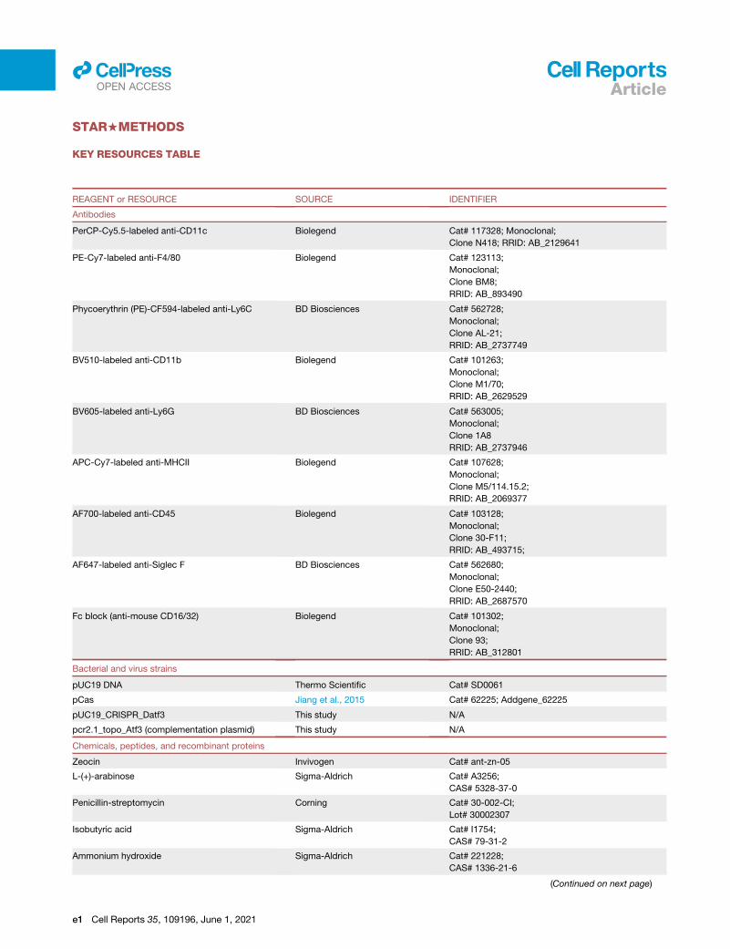
d KEY RESOURCES TABLE
d RESOURCE AVAILABILITY
B Lead contact
B Materials availability
B Data and code availability
d EXPERIMENTAL MODEL AND SUBJECT DETAILS
B Animals
B Microbe strains
B Cell lines
d METHOD DETAILS
B Bacterial genetics and mutagenesis
B Baseline characteristics of bacterial isolates
B RNA-seq and qRT-PCR
B Cell lines
B Mouse studies
B Targeted metabolomics
B Global acetylated lysine proteomics
d QUANTIFICATION AND STATISTICAL ANALYSIS
B Statistics
SUPPLEMENTAL INFORMATION
Supplemental information can be found online at https://doi.org/10.1016/j.
celrep.2021.109196.
ACKNOWLEDGMENTS
This work was supported by NIH K08 HL138289 (to D.A.), NIH R35 HL135800
(to A.P.), NIH R01 AI116939 (to A.-C.U.), NIH K08 AI146284 (to T.H.M.), and
R01 AI123820/AI147314 (to R.K.E.). The proteomics core facility is funded
by NIH P30 CA013696-45 5607 and the CCTI Flow Core by NIH
S10RR027050. We thank Guoan Zhang, PhD, from the Proteomics and Me-
tabolomics Core Facility at Weill Cornell Medicine for performing the targeted
metabolomics with analysis. The graphical abstract was created with
BioRender.
Cell Reports 35, 109196, June 1, 2021 11

Articlell
OPEN ACCESS
AUTHOR CONTRIBUTIONS
D.A. and A.P. designed the experiments, analyzed the data, and wrote the
manuscript. D.A., G.B., T.H.M., M.K.A., R.K.S., T.W.F.L., V.G.C., C.E.H.,
S.S.S., V.C.G., and A.M.C. designed, performed, and analyzed the experi-
ments. A.-C.U. and R.K.E. designed and analyzed the experiments.
DECLARATION OF INTERESTS
The authors declare no competing interests.
Received: November 18, 2020
Revised: February 12, 2021
Accepted: May 10, 2021
Published: June 1, 2021
REFERENCES
Ahn, D., Penaloza, H., Wang, Z., Wickersham, M., Parker, D., Patel, P., Koller,
A., Chen, E.I., Bueno, S.M., Uhlemann, A.C., and Prince, A. (2016). Acquired
resistance to innate immune clearance promotes Klebsiella pneumoniae
ST258 pulmonary infection. JCI Insight 1, e89704.
Andersson, D.I., and Hughes, D. (2010). Antibiotic resistance and its cost: is it
possible to reverse resistance? Nat. Rev. Microbiol. 8, 260–271.
Bachman, M.A., Breen, P., Deornellas, V., Mu, Q., Zhao, L., Wu, W., Cavalcoli,
J.D., and Mobley, H.L. (2015). Genome-Wide Identification of Klebsiella pneu-
moniae Fitness Genes during Lung Infection. MBio 6, e00775.
Bera, A., Biswas, R., Herbert, S., and Gotz, F. (2006). The presence of peptido-
glycan O-acetyltransferase in various staphylococcal species correlates with
lysozyme resistance and pathogenicity. Infect. Immun. 74, 4598–4604.
Blin, C., Passet, V., Touchon, M., Rocha, E.P.C., and Brisse, S. (2017). Meta-
bolic diversity of the emerging pathogenic lineages of Klebsiella pneumoniae.
Environ. Microbiol. 19, 1881–1898.
Borer, A., Saidel-Odes, L., Riesenberg, K., Eskira, S., Peled, N., Nativ, R.,
Schlaeffer, F., and Sherf, M. (2009). Attributable mortality rate for carbape-
nem-resistant Klebsiella pneumoniae bacteremia. Infect. Control Hosp. Epide-
miol. 30, 972–976.
Bruchmann, S., Feltwell, T., Parkhill, J., and Short, F.L. (2021). Identifying viru-
lence determinants of multidrug-resistant Klebsiella pneumoniae in Galleria
mellonella. Pathog. Dis. 79, ftab009.
Caboni, M., Pedron, T., Rossi, O., Goulding, D., Pickard, D., Citiulo, F., Ma-
cLennan, C.A., Dougan, G., Thomson, N.R., Saul, A., et al. (2015). An O antigen
capsule modulates bacterial pathogenesis in Shigella sonnei. PLoS Pathog.
11, e1004749.
Callura, J.M., Cantor, C.R., and Collins, J.J. (2012). Genetic switchboard for
synthetic biology applications. Proc. Natl. Acad. Sci. USA 109, 5850–5855.
Christensen, D.G., Baumgartner, J.T., Xie, X., Jew, K.M., Basisty, N., Schilling,
B., Kuhn, M.L., and Wolfe, A.J. (2019a). Mechanisms, Detection, and Rele-
vance of Protein Acetylation in Prokaryotes. MBio 10, e02708-18.
Christensen, D.G., Xie, X., Basisty, N., Byrnes, J., McSweeney, S., Schilling,
B., and Wolfe, A.J. (2019b). Post-translational Protein Acetylation: An Elegant
Mechanism for Bacteria to Dynamically Regulate Metabolic Functions. Front.
Microbiol. 10, 1604.
Cilloniz, C., Dominedo, C., and Torres, A. (2019). An overview of guidelines for
the management of hospital-acquired and ventilator-associated pneumonia
caused by multidrug-resistant Gram-negative bacteria. Curr. Opin. Infect.
Dis. 32, 656–662.
Clinical and Laboratory Standards Institute (2017). M100. Performance Stan-
dards for Antimicrobial Susceptibility Testing, 27th ed. (Clinical and Laboratory
Standards Institute).
Creecy, J.P., and Conway, T. (2015). Quantitative bacterial transcriptomics
with RNA-seq. Curr. Opin. Microbiol. 23, 133–140.
12 Cell Reports 35, 109196, June 1, 2021
Davis, M.R., Jr., and Goldberg, J.B. (2012). Purification and visualization of
lipopolysaccharide from Gram-negative bacteria by hot aqueous-phenol
extraction. J. Vis. Exp. 63, 3916.
Dibrov, P., Dibrov, E., and Pierce, G.N. (2017). Na+-NQR (Na+-translocating
NADH:ubiquinone oxidoreductase) as a novel target for antibiotics. FEMS Mi-
crobiol. Rev. 41, 653–671.
Dimroth, P. (1987). Sodium ion transport decarboxylases and other aspects of
sodium ion cycling in bacteria. Microbiol. Rev. 51, 320–340.
Doi, Y., Bonomo, R.A., Hooper, D.C., Kaye, K.S., Johnson, J.R., Clancy, C.J.,
Thaden, J.T., Stryjewski, M.E., and van Duin, D.; Gram-Negative Committee of
the Antibacterial Resistance Leadership Group (ARLG) (2017). Gram-Negative
Bacterial Infections: Research Priorities, Accomplishments, and Future Direc-
tions of the Antibacterial Resistance Leadership Group. Clin. Infect. Dis. 64
(Suppl_1), S30–S35.
Doucette, C.D., Schwab, D.J., Wingreen, N.S., and Rabinowitz, J.D. (2011).
-Ketoglutarate coordinates carbon and nitrogen utilization via enzyme I inhibi-
tion. Nat. Chem. Biol. 7, 894–901.
El Hamidi, A., Tirsoaga, A., Novikov, A., Hussein, A., and Caroff, M. (2005). Mi-
croextraction of bacterial lipid A: easy and rapid method for mass spectro-
metric characterization. J. Lipid Res. 46, 1773–1778.
Erhardt, H., Steimle, S., Muders, V., Pohl, T., Walter, J., and Friedrich, T.
(2012). Disruption of individual nuo-genes leads to the formation of partially
assembled NADH:ubiquinone oxidoreductase (complex I) in Escherichia coli.
Biochim. Biophys. Acta 1817, 863–871.
Ernst, C.M., Braxton, J.R., Rodriguez-Osorio, C.A., Zagieboylo, A.P., Li, L., Pir-
onti, A., Manson, A.L., Nair, A.V., Benson,M., Cummins, K., et al. (2020). Adap-
tive evolution of virulence and persistence in carbapenem-resistant Klebsiella
pneumoniae. Nat. Med. 26, 705–711.
Giddins, M.J., Macesic, N., Annavajhala, M.K., Stump, S., Khan, S., McCon-
ville, T.H., Mehta, M., Gomez-Simmonds, A., and Uhlemann, A.C. (2018). Suc-
cessive Emergence of Ceftazidime-Avibactam Resistance through Distinct
Genomic Adaptations in blaKPC-2-Harboring Klebsiella pneumoniae Sequence
Type 307 Isolates. Antimicrob. Agents Chemother. 62, e02101-17.
Gomez-Simmonds, A., and Uhlemann, A.C. (2017). Clinical Implications of
Genomic Adaptation and Evolution of Carbapenem-Resistant Klebsiella pneu-
moniae. J. Infect. Dis. 215 (Suppl_1), S18–S27.
Gomez-Simmonds, A., Greenman, M., Sullivan, S.B., Tanner, J.P., Sowash,
M.G., Whittier, S., and Uhlemann, A.C. (2015). Population Structure of Klebsi-
ella pneumoniae Causing Bloodstream Infections at a New York City Tertiary
Care Hospital: Diversification of Multidrug-Resistant Isolates. J. Clin. Micro-
biol. 53, 2060–2067.
Gomez-Simmonds, A., Annavajhala, M.K., McConville, T.H., Dietz, D.E., Shou-
cri, S.M., Laracy, J.C., Rozenberg, F.D., Nelson, B., Greendyke, W.G., Furuya,
E.Y., et al. (2020). Carbapenemase-producing Enterobacterales causing sec-
ondary infections during the COVID-19 crisis at a New York City hospital.
J. Antimicrob. Chemother. 76, 380–384.
Haas, B.J., Chin, M., Nusbaum, C., Birren, B.W., and Livny, J. (2012). How
deep is deep enough for RNA-Seq profiling of bacterial transcriptomes?
BMC Genomics 13, 734.
Haug, K., Cochrane, K., Nainala, V.C., Williams, M., Chang, J., Jayaseelan, K.V.,
and O’Donovan, C. (2020). MetaboLights: a resource evolving in response to the
needs of its scientific community. Nucleic Acids Res. 48 (D1), D440–D444.
Hentchel, K.L., and Escalante-Semerena, J.C. (2015). Acylation of Biomole-
cules in Prokaryotes: aWidespread Strategy for the Control of Biological Func-
tion and Metabolic Stress. Microbiol. Mol. Biol. Rev. 79, 321–346.
Herbert, S., Bera, A., Nerz, C., Kraus, D., Peschel, A., Goerke, C., Meehl, M.,
Cheung, A., and Gotz, F. (2007). Molecular basis of resistance to muramidase
and cationic antimicrobial peptide activity of lysozyme in staphylococci. PLoS
Pathog. 3, e102.
Hsu, C.R., Liao, C.H., Lin, T.L., Yang, H.R., Yang, F.L., Hsieh, P.F., Wu, S.H.,
and Wang, J.T. (2016). Identification of a capsular variant and characterization
of capsular acetylation in Klebsiella pneumoniae PLA-associated type K57.
Sci. Rep. 6, 31946.

Articlell
OPEN ACCESS
Inouye, M., Dashnow, H., Raven, L.A., Schultz, M.B., Pope, B.J., Tomita, T.,
Zobel, J., and Holt, K.E. (2014). SRST2: rapid genomic surveillance for public
health and hospital microbiology labs. Genome Med. 6, 90.
Insua, J.L., Llobet, E., Moranta, D., Perez-Gutierrez, C., Tomas, A., Garmen-
dia, J., and Bengoechea, J.A. (2013). Modeling Klebsiella pneumoniae patho-
genesis by infection of the wax moth Galleria mellonella. Infect. Immun. 81,
3552–3565.
Jiang, Y., Chen, B., Duan, C., Sun, B., Yang, J., and Yang, S. (2015). Multigene
editing in the Escherichia coli genome via the CRISPR-Cas9 system. Appl. En-
viron. Microbiol. 81, 2506–2514.
Jiang, X., Bomgarden, R., Brown, J., Drew, D.L., Robitaille, A.M., Viner, R., and
Huhmer, A.R. (2017). Sensitive and Accurate Quantitation of Phosphopeptides
Using TMT Isobaric Labeling Technique. J. Proteome Res. 16, 4244–4252.
Kohler, P.P., Volling, C., Green, K., Uleryk, E.M., Shah, P.S., and McGeer, A.
(2017). Carbapenem Resistance, Initial Antibiotic Therapy, and Mortality in
Klebsiella pneumoniae Bacteremia: A Systematic Review and Meta-Analysis.
Infect. Control Hosp. Epidemiol. 38, 1319–1328.
Langmead, B., and Salzberg, S.L. (2012). Fast gapped-read alignment with
Bowtie 2. Nat. Methods 9, 357–359.
Letunic, I., and Bork, P. (2019). Interactive Tree Of Life (iTOL) v4: recent up-
dates and new developments. Nucleic Acids Res. 47 (W1), W256–W259.
Liu, Y.Y., Chandler, C.E., Leung, L.M., McElheny, C.L., Mettus, R.T., Shanks,
R.M.Q., Liu, J.H., Goodlett, D.R., Ernst, R.K., and Doi, Y. (2017). Structural
Modification of Lipopolysaccharide Conferred bymcr-1 in Gram-Negative ES-
KAPE Pathogens. Antimicrob. Agents Chemother. 61, e00580-17.
Love, M.I., Huber, W., and Anders, S. (2014). Moderated estimation of fold
change and dispersion for RNA-seq data with DESeq2. Genome Biol. 15, 550.
Marsh, J.W., Mustapha,M.M., Griffith,M.P., Evans, D.R., Ezeonwuka, C., Pas-
culle, A.W., Shutt, K.A., Sundermann, A., Ayres, A.M., Shields, R.K., et al.
(2019). Evolution of Outbreak-Causing Carbapenem-Resistant Klebsiella
pneumoniae ST258 at a Tertiary Care Hospital over 8 Years. MBio 10,
e01945-19.
McConville, T.H., Annavajhala, M.K., Giddins, M.J., Macesic, N., Herrera,
C.M., Rozenberg, F.D., Bhushan, G.L., Ahn, D., Mancia, F., Trent, M.S., and
Uhlemann, A.C. (2020). CrrB Positively Regulates High-Level Polymyxin Resis-
tance and Virulence in Klebsiella pneumoniae. Cell Rep. 33, 108313.
McConville, T.H., Giddins, M.J., and Uhlemann, A.C. (2021). An efficient and
versatile CRISPR-Cas9 system for genetic manipulation of multi-drug resistant
Klebsiella pneumoniae. STAR Protoc 2, 100373.
Mills, G., Dumigan, A., Kidd, T., Hobley, L., and Bengoechea, J.A. (2017). Iden-
tification and Characterization of Two Klebsiella pneumoniae lpxL Lipid A Late
Acyltransferases and Their Role in Virulence. Infect. Immun. 85, e00068-17.
Minshew, B.H., Holmes, R.K., Sanford, J.P., and Baxter, C.R. (1974). Transfer-
rable resistance to tobramycin in Klebsiella pneumoniae and Enterobacter
cloacae associated with enzymatic acetylation of tobramycin. Antimicrob.
Agents Chemother. 6, 492–497.
Nakayasu, E.S., Burnet, M.C., Walukiewicz, H.E., Wilkins, C.S., Shukla, A.K.,
Brooks, S., Plutz, M.J., Lee, B.D., Schilling, B., Wolfe, A.J., et al. (2017).
Ancient Regulatory Role of Lysine Acetylation in Central Metabolism. MBio
8, e01894-17.
Navarrete-Perea, J., Yu, Q., Gygi, S.P., and Paulo, J.A. (2018). Streamlined
Tandem Mass Tag (SL-TMT) Protocol: An Efficient Strategy for Quantitative
(Phospho)proteome Profiling Using Tandem Mass Tag-Synchronous Precur-
sor Selection-MS3. J. Proteome Res. 17, 2226–2236.
Paczosa, M.K., Silver, R.J., McCabe, A.L., Tai, A.K., McLeish, C.H., Lazinski,
D.W., and Mecsas, J. (2020). Transposon Mutagenesis Screen of Klebsiella
pneumoniae Identifies Multiple Genes Important for Resisting Antimicrobial
Activities of Neutrophils in Mice. Infect. Immun. 88, e00034-20.
Pan, Y.J., Lin, T.L., Chen, C.T., Chen, Y.Y., Hsieh, P.F., Hsu, C.R., Wu, M.C.,
and Wang, J.T. (2015). Genetic analysis of capsular polysaccharide synthesis
gene clusters in 79 capsular types of Klebsiella spp. Sci. Rep. 5, 15573.
Pearson, C.R., Tindall, S.N., Herman, R., Jenkins, H.T., Bateman, A., Thomas,
G.H., Potts, J.R., and Van derWoude, M.W. (2020). Acetylation of Surface Car-
bohydrates in Bacterial Pathogens Requires Coordinated Action of a Two-
Domain Membrane-Bound Acyltransferase. MBio 11. https://doi.org/10.
1128/mBio.01364-20.
Penaloza, H.F., Noguera, L.P., Ahn, D., Vallejos, O.P., Castellanos, R.M., Vaz-
quez, Y., Salazar-Echegarai, F.J., Gonzalez, L., Suazo, I., Pardo-Roa, C., et al.
(2019). Interleukin-10 Produced by Myeloid-Derived Suppressor Cells Pro-
vides Protection to Carbapenem-Resistant Klebsiella pneumoniae Sequence
Type 258 by Enhancing Its Clearance in the Airways. Infect. Immun. 87,
e00665-18.
Pitout, J.D., Nordmann, P., and Poirel, L. (2015). Carbapenemase-Producing
Klebsiella pneumoniae, a Key Pathogen Set for Global Nosocomial Domi-
nance. Antimicrob. Agents Chemother. 59, 5873–5884.
Poe, S.L., Arora, M., Oriss, T.B., Yarlagadda,M., Isse, K., Khare, A., Levy, D.E.,
Lee, J.S., Mallampalli, R.K., Chan, Y.R., et al. (2013). STAT1-regulated lung
MDSC-like cells produce IL-10 and efferocytose apoptotic neutrophils with
relevance in resolution of bacterial pneumonia. Mucosal Immunol. 6, 189–199.
Qian, H., Yao, Q., Tai, C., Deng, Z., Gan, J., and Ou, H.Y. (2018). Identification
and characterization of acetyltransferase-type toxin-antitoxin locus in Klebsi-
ella pneumoniae. Mol. Microbiol. 108, 336–349.
Rottig, A., and Steinb€uchel, A. (2013). Acyltransferases in bacteria. Microbiol.
Mol. Biol. Rev. 77, 277–321.
Satlin, M.J., Chen, L., Patel, G., Gomez-Simmonds, A., Weston, G., Kim, A.C.,
Seo, S.K., Rosenthal, M.E., Sperber, S.J., Jenkins, S.G., et al. (2017). Multi-
center Clinical and Molecular Epidemiological Analysis of Bacteremia Due to
Carbapenem-Resistant Enterobacteriaceae (CRE) in the CRE Epicenter of
the United States. Antimicrob. Agents Chemother. 61, e02349-16.
Stamatakis, A. (2014). RAxML version 8: a tool for phylogenetic analysis and
post-analysis of large phylogenies. Bioinformatics 30, 1312–1313.
Tzouvelekis, L.S., Miriagou, V., Kotsakis, S.D., Spyridopoulou, K., Athanasiou,
E., Karagouni, E., Tzelepi, E., and Daikos, G.L. (2013). KPC-producing, multi-
drug-resistant Klebsiella pneumoniae sequence type 258 as a typical opportu-
nistic pathogen. Antimicrob. Agents Chemother. 57, 5144–5146.
van Duin, D., Arias, C.A., Komarow, L., Chen, L., Hanson, B.M., Weston, G.,
Cober, E., Garner, O.B., Jacob, J.T., Satlin, M.J., et al.; Multi-Drug Resistant
Organism Network Investigators (2020). Molecular and clinical epidemiology
of carbapenem-resistant Enterobacterales in the USA (CRACKLE-2): a pro-
spective cohort study. Lancet Infect. Dis. 20, 731–741.
Vornhagen, J., Sun, Y., Breen, P., Forsyth, V., Zhao, L., Mobley, H.L.T., and
Bachman, M.A. (2019). The Klebsiella pneumoniae citrate synthase gene,
gltA, influences site specific fitness during infection. PLoS Pathog. 15,
e1008010.
Weinert, B.T., Iesmantavicius, V., Wagner, S.A., Scholz, C., Gummesson, B.,
Beli, P., Nystrom, T., and Choudhary, C. (2013). Acetyl-phosphate is a critical
determinant of lysine acetylation in E. coli. Mol. Cell 51, 265–272.
Wong, J.L.C., Romano, M., Kerry, L.E., Kwong, H.S., Low, W.W., Brett, S.J.,
Clements, A., Beis, K., and Frankel, G. (2019). OmpK36-mediated Carbape-
nem resistance attenuates ST258 Klebsiella pneumoniae in vivo. Nat. Com-
mun. 10, 3957.
Xiong, H., Carter, R.A., Leiner, I.M., Tang, Y.W., Chen, L., Kreiswirth, B.N., and
Pamer, E.G. (2015). Distinct Contributions of Neutrophils and CCR2+ Mono-
cytes to Pulmonary Clearance of Different Klebsiella pneumoniae Strains.
Infect. Immun. 83, 3418–3427.
Xu, L., Sun, X., andMa, X. (2017). Systematic review andmeta-analysis ofmor-
tality of patients infected with carbapenem-resistant Klebsiella pneumoniae.
Ann. Clin. Microbiol. Antimicrob. 16, 18.
Zhang, L., Bao, W., Wei, R., Fu, S., and Gong, H. (2018). Inactivating
NADH:quinone oxidoreductases affects the growth and metabolism of
Klebsiella pneumoniae. Biotechnol. Appl. Biochem. 65, 857–864.
Cell Reports 35, 109196, June 1, 2021 13
Articlell
OPEN ACCESS
STAR+METHODS
KEY RESOURCES TABLE
REAGENT or RESOURCE SOURCE IDENTIFIER
Antibodies
PerCP-Cy5.5-labeled anti-CD11c Biolegend Cat# 117328; Monoclonal;
Clone N418; RRID: AB_2129641
PE-Cy7-labeled anti-F4/80 Biolegend Cat# 123113;
Monoclonal;
Clone BM8;
RRID: AB_893490
Phycoerythrin (PE)-CF594-labeled anti-Ly6C BD Biosciences Cat# 562728;
Monoclonal;
Clone AL-21;
RRID: AB_2737749
BV510-labeled anti-CD11b Biolegend Cat# 101263;
Monoclonal;
Clone M1/70;
RRID: AB_2629529
BV605-labeled anti-Ly6G BD Biosciences Cat# 563005;
Monoclonal;
Clone 1A8
RRID: AB_2737946
APC-Cy7-labeled anti-MHCII Biolegend Cat# 107628;
Monoclonal;
Clone M5/114.15.2;
RRID: AB_2069377
AF700-labeled anti-CD45 Biolegend Cat# 103128;
Monoclonal;
Clone 30-F11;
RRID: AB_493715;
AF647-labeled anti-Siglec F BD Biosciences Cat# 562680;
Monoclonal;
Clone E50-2440;
RRID: AB_2687570
Fc block (anti-mouse CD16/32) Biolegend Cat# 101302;
Monoclonal;
Clone 93;
RRID: AB_312801
Bacterial and virus strains
pUC19 DNA Thermo Scientific Cat# SD0061
pCas Jiang et al., 2015 Cat# 62225; Addgene_62225
pUC19_CRISPR_Datf3 This study N/A
pcr2.1_topo_Atf3 (complementation plasmid) This study N/A
Chemicals, peptides, and recombinant proteins
Zeocin Invivogen Cat# ant-zn-05
L-(+)-arabinose Sigma-Aldrich Cat# A3256;
CAS# 5328-37-0
Penicillin-streptomycin Corning Cat# 30-002-CI;
Lot# 30002307
Isobutyric acid Sigma-Aldrich Cat# I1754;
CAS# 79-31-2
Ammonium hydroxide Sigma-Aldrich Cat# 221228;
CAS# 1336-21-6
(Continued on next page)
e1 Cell Reports 35, 109196, June 1, 2021

Continued
REAGENT or RESOURCE SOURCE IDENTIFIER
Norharmane Sigma-Aldrich Cat# N6252;
CAS# 244-63-3
Chloroform Sigma-Aldrich Cat# C606SK-4;
CAS# 67-66-3
Methanol Sigma-Aldrich Cat# A456-1;
CAS# 67-56-1
TRIzol reagent Invitrogen Cat# 15596026;
Lot# 42902
Hydrogen peroxide solution 30% (w/w) Sigma-Aldrich Cat# H1009;
CAS# 7722-84-1;
Lot# MKBZ7948V
Lysozyme Sigma-Aldrich Cat# L6876;
Lot# SLCC4285
D-(+)-glucose Sigma-Aldrich Cat# G7021;
CAS# 50-99-7;
Lot# SLBZ6903
Sodium pyruvate Sigma-Aldrich Cat# P5280;
CAS# 113-24-6;
Lot# SLBP4879V
a-ketoglutaric acid Sigma-Aldrich Cat# 75890;
CAS# 328-50-7
Disodium succinate Sigma-Aldrich Cat# W327700;
CAS# 150-90-3;
Lot# MKCC2485
Protocol crystal violet Fisher Scientific Cat# 255-960B
Phorbol 12-myristate 13-acetate (PMA) Sigma-Aldrich Cat# P8139;
CAS# 16561-29-8
Gentamicin Sigma-Aldrich Cat# G1397;
CAS# 1405-41-0
Saponin Sigma-Aldrich Cat# S4521;
CAS# 8047-15-2;
Lot# 121K7004
Formalin free tissue fixative Sigma-Aldrich Cat# A5472;
Lot# SLBV4022
Methanol, ultrapure, HPLC grade, 99.8+% Alfa Aesar Cat# 22909;
Lot# Q17D708
EPPS Sigma-Aldrich Cat# E9502;
CAS# 16052-06-5;
Lot# SLCB4421
Bicinchoninic acid (BCA) Thermo Scientific Cat# 23225;
Lot# VB297450
Dithiothreitol (DTT) Sigma-Aldrich Cat# D0632;
CAS# 3483-12-3;
Lot# SLBW1508
Tris(2-carboxyethyl)phosphine) (TCEP) Sigma-Aldrich Cat# C4706;
CAS# 51805-45-9;
Lot# SLCB2139
Iodoacetamide (IAA) Sigma-Aldrich Cat# I1149;
CAS# 144-48-9;
Lot# SLCC6164
Lys-C Wako (Fujifilm) Cat# 125-05061;
Lot# SKM6660
Trypsin Promega Cat# V5111;
Lot# 0000459830
(Continued on next page)
Cell Reports 35, 109196, June 1, 2021 e2
Articlell
OPEN ACCESS

Continued
REAGENT or RESOURCE SOURCE IDENTIFIER
Trifluoroacetic acid (TFA) Thermo Scientific Cat# 28904;
CAS# 76-05-1;
Lot# VH312275
Critical commercial assays
Pro-Q emerald 300 lipopolysaccharide gel stain kit Invitrogen Cat# P20495
PM1 phenotype microarray Biolog Cat# 12111;
Lot# 3410131
Seahorse XF24 cell culture microplates Agilent Cat# 100777-004;
Lot# 21720
Seahorse XF glycolysis stress test kit Agilent Cat# 103020-100;
Lot# 17019693
MitoSox Invitrogen Cat# M36008
BacTiter-glo microbial cell viability assay Promega Cat# G8230
E.Z.N.A. Total RNA Kit I Omega Bio-tek Cat# R6834-02;
Lot# R68345371-10
DNA-free DNA removal kit Invitrogen Cat# AM1906
MICROBExpress Ambion, Life Technologies Cat# AM1905
Truseq stranded mRNA library prep Illumina Cat# 20020594
High-capacity cDNA reverse transcription kit Applied Biosystems Cat# 4368814
PowerSYBR green PCR master mix Applied Biosystems Cat# 4368708
Mouse cytokine array / chemokine array 31-plex Eve Technologies Cat# MD31
DNeasy blood and tissue kit QIAGEN Cat# 69504
LIVE/DEAD fixable blue dead cell stain kit, for UV
excitation
Molecular Probes Cat# L23105
Glucose assay kit Abcam Cat# ab65333
Uniform dye microspheres Bang Laboratories Cat# FSO7F;
Lot# 12980
10-plex TMT kit Thermo Scientific Cat# 90111;
Lot# UB279981
PTMScan� acetyl-lysine motif [Ac-K] Cell Signaling Cat# 13416
Microscan Beckman Coulter Cat# NM42
Deposited data
Full bacterial genome sequencing and bacterial
RNA-seq (KP35)
NCBI Short Read Archive (SRA) NCBI: SAMN18588135
Full bacterial genome sequencing and bacterial
RNA-seq (KP35::Datf3)
NCBI Short Read Archive (SRA) NCBI: SAMN18588136
Acetylome (KP35, KP35::Datf3) MassIVE, University of California,
San Diego
https://massive.ucsd.edu/ProteoSAFe/static/
massive.jsp
MassIVE: MSV000087181
Bacterial and BALF metabolomics Metabolights (Haug et al., 2020) https://www.ebi.ac.uk/metabolights/
MetaboLights: MTBLS2640
Experimental models: cell lines
Human monocytes (THP-1 s) ATCC TIB-202
Experimental models: organisms/strains
Mouse: C57BL/6J The Jackson Laboratories JAX: 000664
Klebsiella pneumoniae, clinical isolate 35 Ahn et al., 2016 N/A
Isogenic mutant – KP35::Datf3 This study N/A
Complemented mutant – KP35::Datf3::Datf3 This study N/A
Klebsiella pneumoniae, KPPR1 ATCC Cat# 43816
Pseudomonas aeruginosa (PAO1) Our laboratory N/A
(Continued on next page)
e3 Cell Reports 35, 109196, June 1, 2021
Articlell
OPEN ACCESS

Continued
REAGENT or RESOURCE SOURCE IDENTIFIER
Oligonucleotides
Primers for the competition experiment – atf3
Present FWD (ATG TGG GTG GAT TAT GCT
AAG G)
This study N/A
atf3 Absent FWD (ATG AGT CTA GAA TGT TAT A) This study N/A
atf3 Common REV (GGC ATA TGG AAT GAA TAT
ATA A)
This study N/A
Primers for NADH:ubiquinone oxidoreductases:
see Table S1
This study N/A
Software and algorithms
SRST2 Inouye et al., 2014 https://github.com/katholt/srst2
Snippy https://github.com/tseemann/snippy
RAxML Stamatakis, 2014 https://github.com/stamatak/standard-RAxML
iTOL Letunic and Bork, 2019 https://github.com/iBiology/iTOL
Geneious Biomatters https://www.geneious.com
Bowtie2 Langmead and Salzberg, 2012 http://bowtie-bio.sourceforge.net/bowtie2/index.
shtml
DESeq2 Love et al., 2014 https://github.com/mikelove/DESeq2
Trimmomatic https://github.com/timflutre/trimmomatic
KEGG pathway analysis, KEGG Mapper Kanehisa Laboratories https://www.genome.jp/kegg/
MetaboAnalyst McGill https://www.metaboanalyst.ca/
FlexAnalysis software Bruker Daltonics https://www.bruker.com/en.html; v4.30
FlowJo X software TreeStart Inc. https://www.flowjo.com; ver 10.0.8
Graph pad prism Version 8.4.0 (455), February 20,
2020
https://www.graphpad.com
ImageJ National Institutes of Health https://imagej.nih.gov/ij; Java 1.8.0_172
Endnote X9 Endnote X9.3.2 (Bld 15235) endnote.com
StepOne Software Applied Biosystems Ver 2.2.2
MaxQuant Max Plank Institute of
Biochemistry
https://www.maxquant.org/download_asset/
maxquant/latestv1.6.17.0
Other
Novex 16% tricine gel Invitrogen Cat# EC6695BOX;
Lot# 19100940
M9 minimal salts (2X) GIBCO Cat# A13744-01;
Lot# 2121257
RPMI 1640, 1x with L-glutamine Corning Cat# 10-040-CV;
Lot# 33720004
Fetal bovine serum GIBCO Cat# 26140-079
XF base medium minimal DMEM Agilent Cat# 102353-100;
Lot# 17619002
Difco LB broth, mill (Luria-Bertani) (LB) media BD Cat# 244610;
Lot# 9324045
BBL trypticase soy broth (TS) media BD Cat# 211768;
Lot# 1300142
TrypLE express GIBCO Cat# 12604021;
Lot# 2085370
Articlell
OPEN ACCESS
RESOURCE AVAILABILITY
Lead contactFurther information and requests for resources and reagents should be directed to and will be fulfilled by the lead contact, Danielle
Ahn ([email protected]).
Cell Reports 35, 109196, June 1, 2021 e4

Articlell
OPEN ACCESS
Materials availabilityA standard CRISPR plasmid (pCAS) was used to generate KP35::Datf3 is available at addgene; catalog #62225. https://www.
addgene.org/62225/. The gene specific plasmid (pUC19_CRISPR_Datf3) and complementation plasmid (pcr2.1_topo_Atf3) are
available without restriction upon request.
Data and code availabilityThe RNA-seq datasets and full genome sequencing of KP35 and the Datf3 isogenic mutant generated during this study are available
in the NCBI Short Read Archive (SRA) and are assigned a BioProject ID: PRJNA719112, NCBI SRA: SAMN18588135 (KP35), and
NCBI SRA: SAMN18588136 (KP35::Datf3)
The acetylomes of KP35 and the Datf3 isogenic mutant are available through MassIVE (available through https://massive.ucsd.
edu/ProteoSAFe/static/massive.jsp, MassIVE: MSV000087181 and the metabolomics data are available through Metabolights
(available through https://www.ebi.ac.uk/, MetaboLights: MTBLS2640).
EXPERIMENTAL MODEL AND SUBJECT DETAILS
AnimalsIn vivo experiments were performed using 8-week-old, male C57BL/6J mice (Jackson Laboratories, Stock No. 000664). Sample
sizes were predetermined by power calculations using a 1 log reduction in bacterial burden in the BALF of infected mice. No
mice were excluded and were placed into control or infection groups at random. Treatment groups were not blinded. Experimental
mice were weighed daily and given a body conditioning score. Since the mice were noted to be bright, alert and active with 20%
weight loss (Ahn et al., 2016), after careful review by the veterinarians and IACUC, the animals were allowed to lose 30%of their initial
body weight as long as their body conditioning score remained high. All animal experiments were performed in accordance with the
guidelines of the IACUC at Columbia University (protocol number AAAS4464).
Microbe strainsWe selected a representative K. pneumoniae ST258 clinical isolate 35 (KP35) from a patient with bacteremia which was comprehen-
sively studied in previous publications (Ahn et al., 2016; Gomez-Simmonds and Uhlemann, 2017). Pseudomonas aeruginosa strain
PAO1 and K. pneumoniae KPPR1 (ATCC 43816) were used as controls. All assays were performed on bacteria grown overnight, sub-
cultured to logarithmic phase and normalized to OD600 of 0.5.
Cell linesHuman monocytes (THP-1 s)
ATCC TIB-202:monocytes from a humanmale with acutemonocytic leukemia. THP-1 cells were grown at 37�Cwith 5%CO2 in RPMI
1640 with L-glutamine cell culture medium with 10% heat-inactivated fetal bovine serum. THP-1 s were activated with 1 mM phorbol
12-myristate 13-acetate (PMA) for 24 hours prior to infection and subsequently weaned to antibiotic free media 24 hours prior to
testing.
METHOD DETAILS
Bacterial genetics and mutagenesisScreening for atf3 presence
To determine the prevalence of atf3 in K. pneumoniae, 218 publicly available genome assemblies were downloaded from NCBI.
SRST2 (Inouye et al., 2014) was used for MLST typing and to identify isolates belonging to sequence type 258 (ST258). A custom
SRST2 gene database was then made with the atf3 gene sequence from the KP35 reference genome and used to determine the
presence or absence of the atf3 gene in each genomic assembly.
Phylogenetic analyses
To reconstruct the phylogenetic relationships between all public K. pneumoniae genomes, and within K. pneumoniae ST258, we
used Snippy to identify core genome single nucleotide polymorphisms (SNPs) by mapping each genome against the ST258 KPNIH1
reference genome (GenBank CP008827.1). A maximum-likelihood phylogeny was then created with RAxML (Stamatakis, 2014) with
100 bootstrap replicates based on concatenated core genome SNPs. Phylogenetic trees were visualized and annotated in iTOL
(Letunic and Bork, 2019).
Gene editing using CRISPR-Cas9
To allow for efficient genetic manipulation in multi-drug resistant K. pneumoniae, we utilized our recently optimized single plasmid
CRISPR-Cas9 / lambda red recombineering system (Jiang et al., 2015; McConville et al., 2020; McConville et al., 2021). For the
gene deletion, we first analyzed the wild-type atf3 sequence via the CRISPRdirect website to identify an appropriate N20
sequence, which was incorporated into an atf3 sgRNA. To knockout atf3, the homology was engineered to contain a 122 bp deletion
surrounding the cas9 cut site. The atf3 specific sgRNA and homology cassettes were cloned into the pUC19_CRISPR vector
(pUC19_CRISPR_Datf3). The sequence confirmed plasmid was inserted into the clinical isolate KP35 via electroporation and
e5 Cell Reports 35, 109196, June 1, 2021

Articlell
OPEN ACCESS
appropriate transformants were identified through colony PCR. Transformants were grown at 30�C under Zeocin selection and
induced with 2% L-(+)-arabinose after 2 hours. Following 6+ hours of induction the cultures were diluted 1:100 and plated on low
salt LB with Zeocin and L-(+)-arabinose. Appropriate mutants were identified with colony PCR and sanger sequencing (Genewiz).
Mutants were cured of the CRISPR plasmid with serial passage on non-selective media. To ensure no off-target editing occurred,
we performed whole genome sequencing (WGS) with hybrid assembly utilizing Illumina and Nanopore reads (Giddins et al.,
2018). The complemented isolate was constructed using a high copy number plasmid containing the atf3 genewith a Zeocin resistant
cassette (pcr2.1_topo_Atf3).
Baseline characteristics of bacterial isolatesLPS studies
Lipid A analysis
K. pneumoniae strainswere cultured for 18 hr at 37�Cwith shaking in 5mL Luria-Bertani (LB). Lipid Awas extracted from cell pellets
using an ammonium hydroxide-isobutyric acid-based procedure (El Hamidi et al., 2005; Liu et al., 2017). Briefly, approximately 5 mL
of cell culture was pelleted and resuspended in 400 mL of 70% isobutyric acid and 1 M ammonium hydroxide (5:3 [vol/vol]). Samples
were incubated for 1 h at 100�C and centrifuged at 2,0003 g for 15min. Supernatants were collected, added to endotoxin-free water
(1:1 [vol/vol]), snap-frozen on dry ice, and lyophilized overnight. The resultant material was washed twice with 1 mL methanol, and
lipid A was extracted using 80 mL of a mixture of chloroform, methanol, and water (3:1:0.25 [vol/vol/vol]). Once extracted, 1 mL of the
concentrate was spotted on spotted on a steel re-usableMALDI plate followed by 1 mL of 10mg/ml norharmanematrix in chloroform-
methanol (2:1 [vol/vol]) and then was air-dried. All samples were analyzed on a Bruker Microflex mass spectrometer (Bruker Dalton-
ics, Billerica, MA) in the negative-ion mode with reflectron mode. An electrospray tuning mix (Agilent, Palo Alto, CA) was used for
mass calibration. Spectral data were analyzed with FlexAnalysis software. The resulting spectra were used to estimate the lipid A
structures present in each strain based on their predicted structures and molecular weights.
O-antigen analysis
To visualize the O-antigen ladder of bacteria, LPS was first isolated using a hot phenol extraction (Davis and Goldberg, 2012) from
overnight cultures normalized to OD600 of 0.5 after 1:10 dilution. Samples were treated with DNase and RNase for 30min at 37�C and
Proteinase K at 59�C overnight. After hot phenol extraction with Trizol, the isolated LPS was run on a 16% Tricine gel. The gel was
then stained using Pro-Q Emerald 300 Lipopolysaccharide Gel Stain Kit and imaged using UV light on a Protein Simple imager.
Antimicrobial susceptibility
Antimicrobial susceptibility testing of bacterial isolates were performed according to routine microbiology laboratory protocols us-
ing the Microscan automated system with the gram-negative panel, with additional E-testing as needed. Susceptibility breakpoints
were derived from Clinical and Laboratory Standards Institute guide (Clinical and Laboratory Standards Institute, 2017).
Kinetic growth curves and static growth assays
For kinetic growth assays, isolates were grown to logarithmic phase in LB, normalized and inoculated (1:100) in a round-bottom 96-
well plates with a final volume of 100 mL per well of LB, RPMI media or M9 salts with relevant supplements or antibiotics when indi-
cated. Plates were incubated under agitation at 37�C overnight for kinetic growth curves. OD600 was measured every 15-30 minutes
for 18 hours on a SpectraMax M2 Microplate reader.
Static metabolite growth and biofilm assay
For static growth assays, bacteria supplementedM9Minimal Media (MM) or LBwere standardized to an OD600 of 0.1, pelleted and
washed twice with the respective media. Bacteria were incubated in a 1:100 dilution either in media alone or in media with increasing
concentrations of specific metabolites or H2O2 in round-bottom 96-well plates with a final volume of 100 mL per well. Plates were
incubated statically at 37�C overnight and the next day, OD600 was measured on a Tecan Infinite 200. To measure biofilm formation,
overnight cultures were standardized to an OD of 1, inoculated 1:100 in TS media + 0.5% glucose in a 96-well plate and grown stat-
ically overnight at 37�C. In the morning, the OD600 was measured. After methanol wash, the wells were then stained with 1% crystal
violet, resuspended with 33% acetic acid and the OD540 was measured
Single carbon source assimilation
Bacterial isolates were grown to exponential phase and normalized to an OD600 of 0.5. Bacteria was then added to a PM1 Pheno-
typeMicroarray, a 96-well plate array with a single carbon source in eachwell. The protocol provided by themanufacturer was strictly
followed. Plates were incubated at 37�C for 24-48h.
Seahorse analyzer
A sensor cartridge was hydrated per the manufacturer’s instructions overnight at 37�C. For bacteria alone, 107 CFU was added to
XF24 Cell Culture Microplates with XF base medium supplemented with 2mM glutamine. The extracellular acidification rate and ox-
ygen consumption rate were measured using a XF24 Seahorse analyzer (Agilent). Each measurement cycle consisted of a mixing
time of 3 minutes and a data acquisition period of 3 minutes, generating 12 data points over 73 minutes. Using the XF Glycolysis
Stress Test Kit, Glucose (10 mM) was added to stimulate glycolysis, oligomycin (1 mM) to suppress oxidative phosphorylation and
unleash glycolytic capacity, and 2-deoxyglucose (50 mM) to inhibit glycolysis.
Bacterial ROS production
Single colonies of KP35, Datf3, Datf3::atf3, and PAO1 were grown overnight in M9 MM supplemented with 62.5 mM glucose then
subcultured (1:100) and standardized to OD600 of 0.5, washed twice with and resuspended in M9 MM. The pellet was then stained
Cell Reports 35, 109196, June 1, 2021 e6

Articlell
OPEN ACCESS
with MitoSOX for 20 minutes, before pelleting bacteria and washing twice with PBS. Dye intensity was then quantified on a BDFACS-
Canto II.
ATP assay
Bacteria in logarithmic phasewas standardized to OD600 of 0.5 and resuspended to a concentration of 108 CFU/mL. Equal portions
of BacTiter-Glo Reagent was added to 100 mL bacterial suspension in white-walled 96-well plates. Contents were briefly mixed on a
shaker for 5 minutes before recording luminescence on a SpectraMax M2 Microplate reader.
RNA-seq and qRT-PCRIsolates were grown to mid/late log phase (OD600 0.5) in non-selective LB broth, pelleted, resuspended, and incubated in 50 mg/mL
lysozyme in 50uM Tris-HCl 10 uM EDTA pH 7.5 for 30 minutes at 37�C, followed by addition of TRK lysis buffer. After 10 minutes at
room temperature, 70% ethanol was added and samples were transferred to the columns in the E.Z.N.A. Total RNA Kit I. RNA was
isolated following themanufacturer’s instructions and treated with DNase using the DNA-free DNA removal kit. To enrichmRNA con-
tent, rRNAwas removed from isolated RNA using theMICROBExpress. For RNA-Seq, library prep of enrichedmRNAwas performed
with the Truseq mRNA stranded kit, followed by sequencing using the 150 cycle v2 kit on an Illumina Miseq. Quality-filtered reads
(Trimmomatic) from all isolates weremapped against KP35, whichwas used as a reference genome. This was generated usingNano-
pore sequencing and hybrid assembly as previously described, using Bowtie2 (Langmead and Salzberg, 2012). DESeq2 (Love et al.,
2014) in R was used to identify the significantly up and/or downregulated mRNA transcripts using Benjamini-Hochberg p value
adjustment to obtain FDR values (Creecy and Conway, 2015; Haas et al., 2012). The top 50 upregulated genes in WT KP35 over
the Datf3 mutant were analyzed using KEGG Mapper.
For confirmatory studies, isolated bacterial RNA transcribed to cDNA using a High Capacity cDNA Reverse transcription kit.
Finally, qRT was performed using relevant primers and PowerSYBR Green PCR Master Mix on a StepOnePlus Real-time PCR Sys-
tem (Applied Biosystems), using StepOne Software. The ribosomal gene 16S was used as the house keeping gene and the DDCT
values were then calculated. The primer sequences used are presented in Table S1.
Cell linesSeahorse analyzer
For studies using the Seahorse analyzer (Agilent), the media for plated THP-1 monocytes was replaced with XF base medium sup-
plemented with 2 mM glutamine 1 hour prior to infection. The cells were then infected with the desired bacterial isolate at a MOI of 10
and incubated at 37�C without CO2 for 3 hours. Measurements were then taken as described above.
Gentamicin protection assay
Differentiated and weaned THP-1 cells were infected with bacteria (MOI 10) for 30 minutes and then the media was replaced with
RPMI + 10%HI FBS with Gentamicin 500 mg/mL. After 2 or 8 hours from the start of the infection, cells were recovered using TrypLE
Express, lysed with saponin 0.2% and serial dilutions were plated on LB agar. An aliquot was set aside for cell enumeration and
counted on a Countess (Invitrogen) with Trypan exclusion.
Mouse studiesIn vivo experiments were performed using 8-week-old, male C57BL/6J mice. Mice were anesthetized with 100 mg/kg ketamine and
5 mg/kg xylazine given i.p. and infected intranasally with respective isolates (108 CFU in 50 mL of PBS). Bacterial load of each
compartment was quantified by serial dilutions on LB agar plates.
Competition experiment
Genomic DNAwas extracted and purified from the BAL fluid or lung homogenate of C57BL/6Jmice infectedwith KP35 orDatf3 alone
or 1:1, 1:5 or 1:10 ratios of KP35:Datf3 K. pneumoniae using the QIAGEN DNeasy Blood and Tissue kit. Genomic DNA was diluted
with nuclease-free water to 0.05 to 0.005 ng/ml. The presence of either KP35 or Datf3 was measured by qPCR using Power SYBR
Green Master Mix on the StepOne Real-Time PCR system (Applied Biosystems) with the primers listed below. Raw Ct values
were standardized to house-keeping gene 16S and the proportion of either KP35 or Datf3 was calculated.
atf3 Present FWD ATG TGG GTG GAT TAT GCT AAG G
atf3 Absent FWD ATG ATG AGT CTA GAA TGT TAT A
atf3 Common REV GGC ATA TGG AAT GAA TAT ATA A
Histopathology and whole lung imaging
After euthanasia, tracheas were cannulated and lungs were inflated with Formalin Free Tissue Fixative at 5 cm of H2O. Whole mouse
lung was fixed with the same fixation solution for 24 hours, 70%ethanol for 24 hours, and then placed in paraffin blocks. H&E staining
was performed in the usual fashion on 5 mm sections for gross pathology. Tissue sections were then digitally scanned using a Leica
SCN400 whole slide digital imager.
e7 Cell Reports 35, 109196, June 1, 2021
Articlell
OPEN ACCESS
Bronchoalveolar lavage fluid (BALF) assays
BALF was obtained by instilling aliquots of sterile PBS with calcium and magnesium into a cannulated trachea. Serial dilutions for
bacterial enumeration were performed on the BALF prior to centrifuging. The supernatant of the first mL of BALF was set aside
for cytokine array, 31-plex mouse discovery assay. The total cellular content was set aside for flow cytometry (FC).
Analysis of immune cell populations
To further delineate immune cell populations, analysis of cell populations in BALF or single cell suspension of lung homogenate was
conducted using multi-color FC on a BD LSR II. Cells were labeled with a combination of PerCP-Cy5.5-labeled anti-CD11c, PE-Cy7-
labeled anti-F4/80, phycoerythrin (PE)-CF594-labeled anti-Ly6C, BV510-labeled anti-CD11b, BV605-labeled anti-Ly6G, APC-Cy7-
labeled anti-MHCII, AF700-labeled anti-CD45 and AF647-labeled anti-Siglec F. LIVE/DEAD Fixable Blue Dead Cell Stain Kit, for UV
excitation and Fc block (anti-mouse CD16/32) was added to each sample.
Uniform dye microspheres were added to calculate the concentration of cellular components. All flow data was analyzed on
FlowJo. Research reported in this publication was performed in the CCTI Flow Cytometry Core, supported in part by the Office of
the Director, National Institutes of Health under award S10RR027050. The content is solely the responsibility of the authors and
does not necessarily represent the official views of the National Institutes of Health.
Glucose assay on murine BALF
The glucose concentration in BALF fromWT KP35 and Datf3, and Datf3::atf3 infected C57BL/J6 mice was determined using a color-
imetric glucose assay kit. The glucose reaction mix was added to BALF samples and then read at OD570 on a Tecan Infinite 200. A
standard curve was generated to determine the final concentration of glucose in the BALF.
Targeted metabolomicsTargeted polar metabolomics were performed at the shared resource at Cornell University Medical Center, Metabolomics core on
BALF harvested at multiple time points post inoculation and intracellular bacterial metabolites. For intracellular metabolites, bacteria
were lysed with multiple freeze thaws cycles with liquid nitrogen and 100% ethanol with dry ice. Metabolites were extracted using
80%methanol. Targeted LC/MS analyses were performed on a Q Exactive Orbitrap mass spectrometer (Thermo Scientific) coupled
to a Vanquish UPLC system (Thermo Scientific). The Q Exactive operated in polarity-switching mode. A Sequant ZIC-HILIC column
(2.1 mm i.d.3 150 mm, Merck) was used for separation of metabolites. The flow rate was set at 150 mL/min. The buffers consisted of
100% acetonitrile for mobile A, and 0.1%NH4OH/20 mMCH3COONH4 in water for mobile B. The gradient ran from 85% to 30%A in
20 min followed by a wash with 30% A and re-equilibration at 85% A. Metabolites were identified on the basis of exact mass within 5
ppm and standard retention times. Relative metabolite quantitation was performed based on peak area for each metabolite. PCA
component analysis was performed using either MetaboAnalyst or independently generated R script.
Global acetylated lysine proteomicsTissue lysis and in-solution digestion
Overnight cultures were re-inoculated to anOD600 0.5 andwashedwith PBS�/� x 2. Bacterial cell pellets were lysed/homogenized by
bead-beating in 8 M urea, 1% SDS, 200 mM EPPS (pH 8.5), and protease inhibitors. Lysates were cleared by centrifugation at
21,000 g for 30 min at 4�C, and the protein concentration was measured by bicinchoninic acid (BCA). Proteins were reduced with
5 mM tris(2-carboxyethyl)phosphine) (TCEP), alkylated with 10 mM iodoacetamide (IAA), and quenched with 10 mM dithiothreitol
(DTT). A total of 1 mg of protein was chloroform�methanol precipitated. Proteins were then reconstituted in 200 mM EPPS (pH
8.5) and digested by Lys-C overnight and trypsin for 6 h, both at a 1:50 protease-to-peptide ratio. Digested peptides were quantified
using a Nanodrop at 280 nm, and 500 mg of peptide from each sample were labeledwith 800 mg tandemmass tag (TMT) reagent using
a 10-plex TMT kit (Navarrete-Perea et al., 2018). TMT labels were checked, 100 ng of each sample was pooled and desalted and
analyzed by short SPS-MS3method, and using normalization factor samples were bulkmixed at 1:1 across all channels and desalted
using a 500 mg Sep-Pak solid-phase extraction column and dried using vacuum centrifugation.
Acetylated lysine peptides enrichment
Desalted dried isobaric labeled peptides (5 mg) were resuspended in 1.4 mL of ice-cold IAP buffer (50 mM MOPS (pH 7.2), 10 mM
sodium phosphate and 50 mM NaCl) and centrifuged at maximum speed for 5 min at 4�C to remove any insoluble material. Super-
natants (pH �7.5) were incubated with the washed PTMScan� Acetyl-Lysine Motif [Ac-K] antibody beads for 2 hours at 4�C with
gentle end-over-end rotation. After centrifugation at 2000 x g for 1 min, beads were washed three times with ice-cold IAP buffer
and three times with ice-cold HPLC water. The acetylated lysine peptides were eluted twice with 0.15% trifluoroacetic acid (TFA),
desalted using a homemade SDB-RP StageTip, and dried via vacuum centrifugation.
LC-MS/MS analysis
The isobaric labeled, dried, acetylated lysine peptides were dissolved in 10 mL of (3%acetonitrile/ 0.1% formic acid), and analyzed on
an Orbitrap Fusion mass spectrometer coupled to a Dionex Ultimate 3000 (ThermoFisher Scientific) using the TMT-SPS-MS3
method (Jiang et al., 2017). Peptides were separated on an EASY-Spray C18 50cm column (Thermo Scientific). Peptides elution
and separation were achieved at a non-linear flow rate of 250 nL/min using a gradient of 5%–30% of buffer B (0.1% (v/v) formic
acid, 100% acetonitrile) for 110 minutes with a temperature of the column maintained at 50�C during the entire experiment. MS1
data were collected using the Orbitrap (120,000 resolution; maximum injection time 50 ms; AGC 43 105). Determined charge states
between 2 and 5 were required for sequencing and a 45 s dynamic exclusion window was used. Data-dependent top10 MS2 scans
Cell Reports 35, 109196, June 1, 2021 e8

Articlell
OPEN ACCESS
were performed in the ion trap with collision-induced dissociation (CID) fragmentation (Turbo; NCE 35%; maximum injection time
50 ms; AGC 1 3 104). MS3 quantification scans were performed using the multi-notch MS3-based TMT method (ten SPS ions;
50,000 resolution; NCE 65%, maximum injection time 105 ms; AGC 1x10⁵) using the Orbitrap.
Data analysis
Rawmass spectrometric data were analyzed usingMaxQuant to perform database search and TMT reporter ions quantification. TMT
tags on lysine residues and peptide N termini (+229.163 Da) and the carbamidomethylation of cysteine residues (+57.021 Da) was set
as static modifications, while the oxidation of methionine residues (+15.995 Da), deamidation (+0.984) on asparagine and glutamine
and acetyl (+42.010 Da) on lysine were set as a variable modification. Data were searched against the KP35 database with peptide-
spectrummatch (PSMs) and protein-level FDR at 1%FDR. Acetylated lysine peptides identification and quantification were imported
into GraphPad Prism for t test statistical analysis (FDR < 0.05) to identify proteins demonstrating statistically significant changes in
abundance.
QUANTIFICATION AND STATISTICAL ANALYSIS
StatisticsAll statistical analysis was performed usingGraphPadPrism Version 8.4.0 (February 20, 2020). Formost datasets, either a one-way or
two-way ANOVA was performed with post test analysis for multiple comparisons. For datasets in which only two datasets could be
compared, two-tailed Mann-Whitney test was performed. For larger datasets (metabolome and acetylome), multiple t tests with a
FDR at 1%was used. A p value of < 0.05was considered significant. Grubb’s test was performed on all datasets to remove significant
outliers with an a value of 0.05. Experiment specific tests, exact value of n and what it represents, and definition of center and disper-
sion are described in the individual figure legends. Power calculations were performed a priori for the in vivo experiments. The animals
were randomized to groups at random and no mice were excluded.
e9 Cell Reports 35, 109196, June 1, 2021

Cell Reports, Volume 35
Supplemental information
An acquired acyltransferase
promotes Klebsiella pneumoniae
ST258 respiratory infection
Danielle Ahn, Gitanjali Bhushan, Thomas H. McConville, Medini K. Annavajhala, RajeshKumar Soni, Tania Wong Fok Lung, Casey E. Hofstaedter, Shivang S. Shah, AlexanderM. Chong, Victor G. Castano, Robert K. Ernst, Anne-Catrin Uhlemann, and Alice Prince

Supplemental Figure 1 – The basic properties of CRKP ST258 are unchanged with the deletion of the atf3 gene. A) Biofilm formation was assessed using a crystal violet assay (n=4), columns are mean values and bars represent the SEM B) Antibiogram of each isolate using Microscan, Red = Resistant, Yellow = Intermediate and Green = Sensitive C) O-antigen ladder on LPS extracted using hot phenol, tricine 16% gel, stained with Pro-Q Emerald glycoprotein stain (n=3) D) Densitometry using J-Image, each dot represents one lane in blot, horizontal bars represent median values, **p<0.01, ***p<0.005 between groups with horizontal bars, Kruskal-Wallis test, one-way ANOVA, Dunn’s correction for multiple comparisons. E) Overnight cultures were inoculated 1:200 LB with the desired H2O2 concentration and statically grown overnight (n=3). Wells were considered positive (black) if above the average blank OD. Related to Figure 2.
0
23.4 47 94 188
375
750
1.5 3 6 12 24
KP35Δatf3
0
1
AE
←C+1
KΔatf3KP35
←C
O
C C+1O
KP35
Δatf3
KP35
Δatf3
KP35
Δatf3
0
50
100
150
Densitometry
***** **
C D
PAO1
KP35
Δatf3
Δatf3::atf3
0
1
2
3
OD540/OD600
Supplemental Figure 1B
E
μM mM
H2O2(Conc)
ABX KP35 ∆atf3 compA/S >16/8 >16/8 >16/8AK 32 32 32AM >16 >16 >16AUG >16/8 >16/8 >16/8AZT >16 >16 >16CAX >32 >32 >32CAZ >16 >16 >16CF >16 >16 >16CFT >32 >32 >32CFX >16 >16 >16CFZ >16 >16 >16CP >4 >4 >4CPE >16 >16 >16CRM >16 >16 >16DOR >2 >2 >2ETP >4 >4 >4FD >64 >64 >64GM >8 >8 >8IMP >8 8 >8LVX >4 >4 >4MER >8 >8 >8P/T >64 >64 >64T/S >2/38 >2/38 >2/38TE 4 4 4TGC ≤1 ≤1 ≤1TO >8 >8 >8POLY B 0.5 0.5 0.5

Supplemental Figure 2 – Lack of atf3 does not change growth in vitro. Overnight cultures of bacteria grown in LB were normalized to an OD of 0.1, washed twice with M9 minimal media, and inoculated 1:100 in a 96 well microtiter plate with increasing concentrations (1.95, 3.91, 7.81, 15.6, 31.3, 62.5, 125 mM) of A) Glucose, B) Pyruvate, C) α-Ketoglutarate, D) Succinate. OD600 was then measured overnight after static growth at 37oC. Each dot represents the mean value, bars are SD, (n=2) E) Serial OD600 measurements were taken over 18 hours with intermittent shaking at 37ºC in RPMI supplemented with differential concentrations of glucose, each dot is the mean value with the bars representing SEM, (n=4). Related to Figure 3D.
KP35 Δatf3 PAO1 KPPR1
0.0
0.2
0.4
0.6O
D60
0M9 ± α-D-Glucose
Supplemental Figure 2A
KP35 Δatf3 PAO1 KPPR1
0.0
0.2
0.4
0.6
OD
600
M9 ± Succinate
KP35 Δatf3 PAO1 KPPR1
0.0
0.2
0.4
0.6
OD
600
M9 ± α-Ketoglutarate
KP35 Δatf3 PAO1 KPPR1
0.0
0.2
0.4
0.6
OD
600
M9 ± PyruvateB
C
D
0:00:0
0
2:00:0
0
4:00:0
0
6:00:0
0
8:00:0
0
10:00
:00
12:00
:000.0
0.5
1.0
OD
600
Glucose 31.3 mM
0:00:0
0
2:00:0
0
4:00:0
0
6:00:0
0
8:00:0
0
10:00
:00
12:00
:000.0
0.5
1.0
OD
600
Glucose 3.13 mM
0:00:0
0
2:00:0
0
4:00:0
0
6:00:0
0
8:00:0
0
10:00
:00
12:00
:000.0
0.5
1.0
OD
600
Glucose 156.5 µM
KPPR1KP35Δatf3
0:00:0
0
2:00:0
0
4:00:0
0
6:00:0
0
8:00:0
0
10:00
:00
12:00
:000.0
0.5
1.0
OD
600
Glucose 313 µM
E
0 2 4 6 8 10 12 0 2 4 6 8 10 12
0 2 4 6 8 10 12 0 2 4 6 8 10 12
Time (hr)à
Time (hr)à
0:00:0
0
2:00:0
0
4:00:0
0
6:00:0
0
8:00:0
0
10:00
:00
12:00
:000.0
0.5
1.0
OD
600
Glucose 156.5 µM
KPPR1KP35Δatf3

Supplemental Figure 3 – Major cytokines in the bronchoalveolar lavage fluid are not atf3 dependent.WT C57BL/6J mice were intranasally infected with 1-2 x 10⁸ CFU for 48 hours. Selected cytokine and chemokine content of BALF quantified by multiplex assay, n = 6 per time point, Box-and-Whiskers with min/max presented, One-way ANOVA, Kruskal-Wallis test, Dunn’s test for multiple comparisons, *p<0.05, **p<0.01, colored stars are compared to PBS, other statistically significant differences are presented with a horizontal bar with black star(s) between two groups. Related to Figure 7E-F.
PBS
KP3548
HR
Δatf 4
8 HR
Δatf::
atf 48
HR
0
100
200
300
400
RANTES/CCL5
pg/m
L
PBS
KP3548
HR
Δatf 4
8 HR
Δatf::
atf 48
HR
0
5000
10000
15000
MIP1β/CCL4
pg/m
L
PBS
KP3548
HR
Δatf 4
8 HR
Δatf::
atf 48
HR
0
5000
10000
15000MIP1α/CCL3
pg/m
L
PBS
KP3548
HR
Δatf 4
8 HR
Δatf::
atf 48
HR
0
5000
10000
15000
20000
pg/m
L
MCP-1/CCL2
PBS
KP3548
HR
Δatf 4
8 HR
Δatf::
atf 48
HR
0
200
400
600
800VEGF
pg/m
L
PBS
KP3548
HR
Δatf 4
8 HR
Δatf::
atf 48
HR
0
1×10³
2×10³
3×10³
4×10³
pg/m
L
CXCL10/IP10
PBS
KP3548
HR
Δatf 4
8 HR
Δatf::
atf 48
HR
0
100
200
300
400MIG/CXCL9
pg/m
L
PBS
KP3548
HR
Δatf 4
8 HR
Δatf::
atf 48
HR
0
500
1000
1500LIX/CXCL5
pg/m
L
PBS
KP3548
HR
Δatf 4
8 HR
Δatf::
atf 48
HR
0
5000
10000
15000
20000
pg/m
LMIP2/CXCL2
PBS
KP3548
HR
Δatf 4
8 HR
Δatf::
atf 48
HR
10-1
100
101
102
103
104
pg/m
L
KC/CXCL1
PBS
KP3548
HR
Δatf 4
8 HR
Δatf::
atf 48
HR
0
100
200
300
400
pg/m
L
GM-CSF
PBS
KP3548
HR
Δatf 4
8 HR
Δatf::
atf 48
HR
01000
10000
12500
15000
pg/m
L
Copy of G-CSF
PBS
KP3548
HR
Δatf 4
8 HR
Δatf::
atf 48
HR
0
10
20
30
pg/m
L
IL-17
PBS
KP3548
HR
Δatf 4
8 HR
Δatf::
atf 48
HR
0.11
10
100
1000
pg/m
L
IL-1β
PBS
KP3548
HR
Δatf 4
8 HR
Δatf::
atf 48
HR
0
6000
12000
pg/m
L
IL-6
PBS
KP3548
HR
Δatf 4
8 HR
Δatf::
atf 48
HR
0
50
100
150
200IL-1α
pg/m
L
PBS
KP3548
HR
Δatf 4
8 HR
Δatf::
atf 48
HR
0
200
400
600
800
pg/m
L
TNF
Supplemental Figure 3 PBS
KP35
Δatf3
comp
PBS
KP35
Δatf3
comp
PBS
KP35
Δatf3
comp
PBS
KP35
Δatf3
comp
PBS
KP35
Δatf3
comp
PBS
KP35
Δatf3
comp
PBS
KP35
Δatf3
comp
PBS
KP35
Δatf3
comp
PBS
KP35
Δatf3
comp
PBS
KP35
Δatf3
comp
PBS
KP35
Δatf3
comp
PBS
KP35
Δatf3
comp
PBS
KP35
Δatf3
comp
PBS
KP35
Δatf3
comp
PBS
KP35
Δatf3
comp
PBS
KP35
Δatf3
comp
PBS
KP35
Δatf3
comp
TNFα IL-1α IL-1β IL-6 IL-17 G-CSF
GM-CSF
pg/mL
pg/mL
KC/CXCL1 MIP2/CXCL2 LIX/CXCL5
PBS
KP3548
HR
Δatf 4
8 HR
Δatf::
atf 48
HR
0
500
1000
1500
IL-10
pg/m
L
PBS
KP35
Δatf3
comp
MIG/CXCL9 IP-10/CXCL10
VEGF
pg/mL
MCP-1/CCL2 MIP1α/CCL3 MIP1β/CCL4 RANTES/CCL5 IL-10
**
** ** * **
*
* * *
*
** *
* *
*
**
* * * * *** ****
* *

Gene Primer sequences (5’ – 3’) nuoB Forward: GTTACCCCCTGCAGAAACAG
Reverse: ATGACGTCACCATTTCCACA nuoC Forward: CAGCCTATACCGCCAAAGAG
Reverse: CTGGCGCAGCTCTTCTACTT nuoE Forward: CACGAGAAGCACCACTACGA
Reverse: GCGGAAAATCTGGCTGTAGA nuoF Forward: CCTATAAAGACCGCCTGCTG
Reverse: CACGAACAGCTCGAAGTCAA nuoG Forward: GAATTGCGCCGTACTACCAT
Reverse: TCCACGCTAAAGGAGAGCAT nuoH Forward: GGCATGAAATTCGGTCTGTT
Reverse: CATACCTTCCAGCCAAAGGA nuoI Forward: CGTCAGGATCTGGTGTACGA
Reverse: ACAGGCTCTTGACGTCGATT nuoJ Forward: GCTGCTGGTGGTTATCGTTT
Reverse: TTTGTCGCTGTCGTTCAGAC nuoK Forward: CCTGTTTGTGCTTGGTCTGA
Reverse: GCTGATGGCGAGGATATACA nuoL Forward: GCGTCGACTTTTTCGCTAAC
Reverse: CTGGCGATAAACAGGTTGGT nuoM Forward: GAGCGGTGATCCAGATGATT
Reverse: GATCGTCGGCACCACTTTAT nuoN Forward: CAACAACTGATCGCACTGCT
Reverse: AGCACCAGACCGGTGTAAAG nqrA Forward: GATTATTCTGGCCGAACGTG
Reverse: CGCGTCCTGATAATTGAGGT nqrB Forward: CATGGTACTGGCATCTGGTG
Reverse: AAGAGGTTGGCGAAGAGGAT nqrC Forward: CGAACTGCTGGAGAAAGACC
Reverse: AGCACCACCTGCTCAATCTT nqrD Forward: GGCGAGCTTTATGGATGGTA
Reverse: ACTCCTTTTCCTGCTGCTCA nqrE Forward: GGTGTCGAAAAAGGTCTCCA
Reverse: GGATCATCTCGAGGATCTGC nqrF Forward: GACGCCGAGATGGTGTTTAT
Reverse: GCCAGCTGTTCAAACTCCTC
Supplemental Table 1 – Primers used for standard qRT-PCR of nqo and nqr subunits. Related to Figure 3L.
Copyright © 2022 FDOKUMEN