Molecular Characterization of Extended-Spectrum Beta-lactamases in Escherichia coli and Klebsiella...
10
Advances in Life Science and Technology www.iiste.org ISSN 2224-7181 (Paper) ISSN 2225-062X (Online) Vol.21, 2014 1 Molecular Characterization of Extended-Spectrum Beta- Lactamases in Escherichia coli and Klebsiella pneumoniae in Accra, Ghana *Henry Kwadwo Hackman 1 , Charles A Brown 2 , Richard Harry Asmah 2 , Christian Adabor Badu 2 , Gloria Amegashi 2 , Kingsley Twum-Danso 3 1. Department of Science Laboratory Technology, School of Applied Sciences and Arts, Accra Polytechnic 2. Department of Medical Laboratory Technology, School of Allied Health Sciences, University of Ghana, Korle Bu 3. Department of Microbiology, University of Ghana Medical School, Korle Bu *Email of corresponding author: [email protected] Abstract Extended-spectrum beta-lactamases (ESBLs) are plasmid-mediated beta lactamases that are capable of hydrolysing beta-lactams except carbapenems and cephamycins. The ESBL types include SHV, TEM and CTX- M, OXA, PER and VEB-1. The most common ones isolated from clinical specimen are the CTX-M, SHV and TEM. The specific ESBL-producing organisms have different genetic characteristics which mark their identification at the molecular level. This work sought to determine the genetic characterization of ESBL- producing K. pneumoniae and E. coli in Accra. The molecular investigations of the ESBL-coding genes included extraction of 100 DNA templates of phenotypic ESBL-producing isolates by boiling method, preparation of the PCR reaction mixture using appropriate primers, standard PCR reaction in a thermocycler, agarose gel electrophoresis, bands visualization by ultraviolet trans-illumination and bands photography using a Kodak EDAS 290 gel documentation system. The results significantly (p<0.05) indicated that of the 100 ESBL producers, 90(90%) possess CTX-M genes and 25(25%) had TEM genes. None of the ESBL producers possesses SHV genes. Seventy (70%) of the ESBL producers possess only CTX-M genes and 5(5%) had only TEM genes. Twenty (20%) of the isolates had both CTX-M and TEM genes. Of the 100 ESBL phenotypes, 78(78%) and 2(2%) were positive for CTX-M-1group and CTX-M-9group ESBL genes respectively. Organisms producing CTX-M-type ESBL are more prevalent in Accra than other ESBL types. CTX-M-1group producing isolates dominated the ESBL phenotypes with CTX-M-15 likely to be the dominate CTX-M-type ESBL. There is the need for further studies into the characteristic transmission, pathogenesis, antibiotic resistance expression, and infection control of CTX-M-type ESBL and TEM-type ESBL in Accra. Keywords: Extended spectrum beta-lactamase, CTX-M genes, TEM genes, SHV genes, Molecular 1.0 Introduction Extended-spectrum beta-lactamases (ESBLs) are plasmid-mediated beta lactamases that are capable of hydrolysing beta-lactams except carbapenems and cephamycins. They are inhibited by beta-lactamase inhibitors such as clavulanic acid, sulbactam and tazobactam. They have been found in the Enterobacteriaceae and other Gram-negative bacilli. ESBL producing isolates are predominantly Klebsiella pneumoniae and Escherichia coli (Paterson and Bonomo, 2005). The ESBL types include SHV, TEM and CTX-M, OXA, PER and VEB-1. The most common ESBL genes isolated from clinical specimen are the CTX-M, SHV and TEM (Paterson and Bonomo, 2005). It has been observed that the same organism may harbour two or more ESBL genes, which may change the antibiotic resistance phenotype (Yamasaki et al., 2003). The CTX-M enzymes are replacing SHV and TEM enzymes as the prevalent type of ESBLs in urinary tract infections, bloodstream and intra-abdominal infections (Falagas and Karageorgopoulos, 2009). The phylogenic study reveals five major groups of acquired CTX-M enzymes (Bonnet, 2004). The CTX-M-1group (Group I) includes CTX-M- 1 , CTX-M- 3 , CTX-M- 10 , CTX-M- 12 , CTX-M- 15 , CTX-M- 22 , CTX-M- 23 , CTX-M- 28 , CTX-M- 29 CTX-M- 30 and CTX-M- 68 . The CTX-M-2group (Group II) includes CTX-M- 2 , CTX-M- 4 , CTX-M- 5 , CTX-M- 6 , CTX-M- 7 and CTX-M- 20 . The CTX-M-8group (Group III) includes one plasmid-mediated member, CTX-M- 8 . The CTX-M-9group (Group IV) includes nine plasmid-mediated enzymes (CTX-M- 9 , CTX-M- 13 , CTX-M- 14 , CTX-M- 16 , CTX-M- 17 , CTX-M- 19 , CTX-M- 21 , CTX-M- 24 and CTX-M- 27 ). The CTX-M-25group (Group V) includes the CTX-M- 25 and CTX-M- 26 enzymes (Bonnet, 2004). The CTX-M- 15 and CTX-M- 14 seems to be the most widespread globally, while many of the other CTX-M ESBLs tend to be more limited in their distribution (Heffernan et al., 2007).
Transcript of Molecular Characterization of Extended-Spectrum Beta-lactamases in Escherichia coli and Klebsiella...

Advances in Life Science and Technology www.iiste.org
ISSN 2224-7181 (Paper) ISSN 2225-062X (Online)
Vol.21, 2014
1
Molecular Characterization of Extended-Spectrum Beta-
Lactamases in Escherichia coli and Klebsiella pneumoniae in
Accra, Ghana
*Henry Kwadwo Hackman1, Charles A Brown
2, Richard Harry Asmah
2, Christian Adabor Badu
2, Gloria
Amegashi2,
Kingsley Twum-Danso3
1. Department of Science Laboratory Technology, School of Applied Sciences and Arts, Accra
Polytechnic
2. Department of Medical Laboratory Technology, School of Allied Health Sciences, University of Ghana,
Korle Bu
3. Department of Microbiology, University of Ghana Medical School, Korle Bu
*Email of corresponding author: [email protected]
Abstract
Extended-spectrum beta-lactamases (ESBLs) are plasmid-mediated beta lactamases that are capable of
hydrolysing beta-lactams except carbapenems and cephamycins. The ESBL types include SHV, TEM and CTX-
M, OXA, PER and VEB-1. The most common ones isolated from clinical specimen are the CTX-M, SHV and
TEM. The specific ESBL-producing organisms have different genetic characteristics which mark their
identification at the molecular level. This work sought to determine the genetic characterization of ESBL-
producing K. pneumoniae and E. coli in Accra. The molecular investigations of the ESBL-coding genes included
extraction of 100 DNA templates of phenotypic ESBL-producing isolates by boiling method, preparation of the
PCR reaction mixture using appropriate primers, standard PCR reaction in a thermocycler, agarose gel
electrophoresis, bands visualization by ultraviolet trans-illumination and bands photography using a Kodak
EDAS 290 gel documentation system. The results significantly (p<0.05) indicated that of the 100 ESBL
producers, 90(90%) possess CTX-M genes and 25(25%) had TEM genes. None of the ESBL producers
possesses SHV genes. Seventy (70%) of the ESBL producers possess only CTX-M genes and 5(5%) had only
TEM genes. Twenty (20%) of the isolates had both CTX-M and TEM genes. Of the 100 ESBL phenotypes,
78(78%) and 2(2%) were positive for CTX-M-1group and CTX-M-9group ESBL genes respectively. Organisms
producing CTX-M-type ESBL are more prevalent in Accra than other ESBL types. CTX-M-1group producing
isolates dominated the ESBL phenotypes with CTX-M-15 likely to be the dominate CTX-M-type ESBL. There
is the need for further studies into the characteristic transmission, pathogenesis, antibiotic resistance expression,
and infection control of CTX-M-type ESBL and TEM-type ESBL in Accra.
Keywords: Extended spectrum beta-lactamase, CTX-M genes, TEM genes, SHV genes, Molecular
1.0 Introduction
Extended-spectrum beta-lactamases (ESBLs) are plasmid-mediated beta lactamases that are capable of
hydrolysing beta-lactams except carbapenems and cephamycins. They are inhibited by beta-lactamase inhibitors
such as clavulanic acid, sulbactam and tazobactam. They have been found in the Enterobacteriaceae and other
Gram-negative bacilli. ESBL producing isolates are predominantly Klebsiella pneumoniae and Escherichia coli
(Paterson and Bonomo, 2005). The ESBL types include SHV, TEM and CTX-M, OXA, PER and VEB-1. The
most common ESBL genes isolated from clinical specimen are the CTX-M, SHV and TEM (Paterson and
Bonomo, 2005). It has been observed that the same organism may harbour two or more ESBL genes, which may
change the antibiotic resistance phenotype (Yamasaki et al., 2003).
The CTX-M enzymes are replacing SHV and TEM enzymes as the prevalent type of ESBLs in urinary tract
infections, bloodstream and intra-abdominal infections (Falagas and Karageorgopoulos, 2009). The phylogenic
study reveals five major groups of acquired CTX-M enzymes (Bonnet, 2004). The CTX-M-1group (Group I)
includes CTX-M-1, CTX-M-3, CTX-M-10, CTX-M-12, CTX-M-15, CTX-M-22, CTX-M-23, CTX-M-28, CTX-M-29
CTX-M-30 and CTX-M-68. The CTX-M-2group (Group II) includes CTX-M-2, CTX-M-4, CTX-M-5, CTX-M-6,
CTX-M-7 and CTX-M-20. The CTX-M-8group (Group III) includes one plasmid-mediated member, CTX-M-8.
The CTX-M-9group (Group IV) includes nine plasmid-mediated enzymes (CTX-M-9, CTX-M-13, CTX-M-14,
CTX-M-16, CTX-M-17, CTX-M-19, CTX-M-21, CTX-M-24 and CTX-M-27). The CTX-M-25group (Group V)
includes the CTX-M-25 and CTX-M-26 enzymes (Bonnet, 2004). The CTX-M-15 and CTX-M-14 seems to be the
most widespread globally, while many of the other CTX-M ESBLs tend to be more limited in their distribution
(Heffernan et al., 2007).

Advances in Life Science and Technology www.iiste.org
ISSN 2224-7181 (Paper) ISSN 2225-062X (Online)
Vol.21, 2014
2
The specific ESBL-producing organisms have different genetic characteristics which mark their identification at
the molecular level. This genetic diversity in the various ESBL-producing organisms may reflect characteristic
differences in relation to pathogenesis, antibiotic resistance expression, response to therapy, transmission and
infection control. Genetic characterization of ESBL-producing organisms is also essential for epidemiological
use. This work seeks to determine the genetic characterization of ESBL-producing K. pneumoniae and E. coli in
Accra.
2.0 Materials and Methods 2.1 Materials
Glycerol broth, blood agar and MacConkey agar were prepared according to manufacturers’ guidelines. Vitek 2
Compact System (bioMérieux, Marcy I’Etoile, France) was used to identify the isolates and determine ESBL
phenotypes. Water bath was used to heat the colony suspension and centrifuge was used to spin the suspension to
extract the bacteria DNA. BIOR GenePro thermocycler was used to perform the polymerase chain reaction
(PCR) under controlled reaction conditions with specific primers. PCR products were used to perform agarose
gel electrophoresis with 1X TAE buffer, 2% agarose gel and 0.5µg/ml ethidium bromide at 120V for 45minutes.
The bands on the gels were visualized by ultraviolet trans-illumination and photographed using a Kodak EDAS
290 gel documentation system.
2.2 Study Sites
Lactose fermenting bacterial isolates were collected from the Central Laboratory of the Korle Bu Teaching
Hospital (KBTH) and Advent Clinical Laboratories; both in the Accra Metropolis, Ghana. The PCR was
performed at the molecular biology laboratory of the School of Allied Health Sciences, University of Ghana and
the bands on the gels were visualized and photographed at the molecular biology laboratory of the Microbiology
Department of University of Ghana Medical School.
2.3 Sample Size
A sample size of 100 DNA templates extracted from ESBL- producing K. pneumoniae and E. coli isolates.
2.4 Inclusion Criteria
Non-duplicate pure cultures of ESBL-producing K. pneumoniae and E. coli isolates were used in the work.
2.5 Exclusion Criteria
All isolates not confirmed as ESBL-producing K. pneumoniae and E. coli were excluded.
2.6 Determination of ESBL Phenotypes
The isolates were identified as K. pneumoniae and E. coli based on their gram stain reaction and biochemical
reaction characteristics in the ID test cards wells using Vitek 2 system (bioMérieux, Marcy I’Etoile, France).
The Vitek 2 system was used to concurrently determine the ESBL phenotypes based on the simultaneous
assessment of the inhibitory effects of cefotaxime, ceftazidime and cefepime, alone and in the presence of
clavulanic acid in accordance with the CLSI.
2.7 Molecular Analysis of ESBL-coding Genes
The molecular investigations of the ESBL-coding genes included extraction of DNA templates of phenotypic
ESBL-producing isolates by boiling method, preparation of the PCR reaction mixture using appropriate primers,
standard PCR reaction in a thermocycler, agarose gel electrophoresis, bands visualization by ultraviolet trans-
illumination and bands photography using a Kodak EDAS 290 gel documentation system.
2.7.1 Genomic DNA Extraction of K. pneumoniae and E. coli
DNA template was extracted by a simple boiling method (Heffernan et al., 2007). A loopful of bacterial colony
was picked from each isolate and suspended in 100µl of double distilled H2O in Eppendorf tube. The DNA
suspension was incubated at 99°C for 5 minutes and snapped cold on ice for 10 minutes. The cell lysate was then
centrifuged briefly at high speed (12.000 rpm for 3 min), and the supernatant containing genomic DNA was
transferred into sterile Eppendorf and 5µl of the supernatant was used for PCR reaction. The extracted DNA was
stored at -21°C until required for PCR.
2.7.2 PCR Detection of ESBL-encoding Gene
PCR of ESBL-encoding genes was carried out using BIOER GenePro thermocycler. A typical 25µl PCR
reaction mixture for a primer set was prepared as shown in table 3. The primers used were already published

Advances in Life Science and Technology www.iiste.org
ISSN 2224-7181 (Paper) ISSN 2225-062X (Online)
Vol.21, 2014
3
primers as shown in table 1 for CTX-M, TEM and SHV genes and their corresponding PCR conditions (table 2).
Further PCRs using the primers for CTX-M-1group and CTX-M-9group were performed with isolates that were
positive with the universal CTX-M primers (Heffernan et al., 2007). Sterile distilled water as negative controls
were included in each round of PCR.
Table 1: Primers used for the detection of ESBL genes (Heffernan et al., 2007) Primer Name Sequence (5'-3') Target Gene Size (bp)
SHV-F GCCGGGTTATTCTTATTTGTCCG SHV 1007
SHV-R ATGCCGCCGCCAGTCA
TEM-F GTATCCGCTCATGAGACAATA TEM 966
TEM-R TCTAAAGTATATATGAGTAAAC
CTX-M-F TTTGCGATGTGCAGTACCAGTAA CTX-M 590
CTX-M-R CGATATCGTTGGTGGTGCCATA
CTX-M-1-F CCCATGGTTAAAAAATCACTG CTX-M-1 group 891
CTX-M-1-R CCGTTTCCGCTATTACAAAC
CTX-M-9-F GTGACAAAGAGAGTGCAACGG CTX-M-9 group 857
CTX-M-9-R ATGATTCTCGCCGCTGAAGCC
Table 2: PCR conditions used for the detection of ESBL genes (Heffernan et al., 2007) Target Gene PCR Conditions SHV Initial denaturation for 5min at 94ºC 1 cycle 94ºC for 30s 35 cycles 68ºC for 30s 72ºC for 60s Final extension at 72ºC for 7min 1 cycle TEM Initial denaturation for 5min at 94ºC 1 cycle 94ºC for 60s 35 cycles 55ºC for 30s 72ºC for 60s Final extension at 72ºC for 10min 1 cycle CTX-M Initial denaturation for 5min at 94ºC 1 cycle 94ºC for 30s 30 cycles 60ºC for 30s 72ºC for 60s Final extension at 72ºC for 7min 1 cycle CTX-M-1group Initial denaturation for 10min at 94ºC 1 cycle 94ºC for 60s 30 cycles 55ºC for 60s 72ºC for 2mins Final extension at 72ºC for 5min 1 cycle CTX-M-9group Initial denaturation for 10min at 94ºC 1 cycle 94ºC for 30s 25 cycles 55ºC for 30s 72ºC for 60s Final extension at 72ºC for 10min 1 cycle
Table 3: PCR reaction mixture Reagent Volume (µl) Final concentration Nuclease-free water 16.175 - 10X PCR buffer + MgCl2 2.5 1X 10mM DNTP mix 0.4 200 µM each 10µM forward primer 0.4 0.2µM 10µM reverse primer 0.4 0.2µM 5U/µl Taq polymerase 0.125 0.5U Template DNA 5 (≤1 µg/reaction)
TOTAL volume 25
2.7.3 Agarose Gel Electrophoresis
The buffer (1XTAE buffer) was prepared and subsequently used to prepare 2% agarose gel. The suspension was
boiled in a microwave for 2 minutes. The molten agarose was allowed to cool to 60°C and stained with 3µl of
0.5µg/ml ethidium bromide (which absorbs invisible UV light and transmits the energy as visible orange light).
A comb was inserted into the slots of the casting tray and the molten agarose was poured into the tray. The gel
was allowed to solidify for 20 minutes to form the wells. The 1XTAE buffer was poured into the gel tank to
barely submerge the gel. Two microliter (2µl) of 10X blue gel loading dye (which gives colour and density to the
samples to make it easy to load into the wells and monitor the progress of the gel) was added to 10µl of each
PCR product and loaded into the wells. The 100bp DNA ladder was loaded into well 1. The gel was
electrophoresed at 120V for 45 minutes using either a midi or a maxi gel system. The bands on the gels were

Advances in Life Science and Technology www.iiste.org
ISSN 2224-7181 (Paper) ISSN 2225-062X (Online)
Vol.21, 2014
4
visualized by ultraviolet trans-illumination and photographed using a Kodak EDAS 290 gel documentation
system. The sizes of the PCR products were estimated by comparison with the mobility of a 100bp molecular
weight ladder that was ran alongside experimental samples in the gel.
2.8 Statistical Analyses
The data from the work was collated and statistically analysed using using the chi-square test. P values < 0.05
were considered significant.
3.0 Results
Of the 100 ESBL producers, 90% possessed CTX-M genes with bands amplicon size of 590bp as in plate 1 and
25% had TEM genes with bands amplicon size of 966bp as shown in plate 2. None of the ESBL producers
possessed SHV genes as indicated in table 4. Seventy percent (70%) of the ESBL producers possessed only
CTX-M genes and 5% had only TEM genes. Twenty percent (20%) of the isolates had both CTX-M and TEM
genes. Of the 100 ESBL phenotypes, 78% and 2% were positive for CTX-M-1group and CTX-M-9group ESBL
genes respectively as demonstrated in table 5. CTX-M-1group ESBL genes with bands amplicon size of 891bp
and CTX-M-9group with band amplicon size of 857bp are indicated in plate 3 and plate 4 respectively.
Table 4: Occurrence of ESBL-coding genes in K. pneumoniae and E. coli Isolates by
Polymerase Chain Reaction
Number (%)
ESBL-genes Isolates (n= 100)
CTX-M 90(90)
TEM 25(25)
SHV 0(0)
CTX-M Only 70(70)
TEM Only 5(5)
CTX-M and TEM 20(20)
Neither CTX-M or TEM 5(5)
Table 5: Occurrence of CTX-M-group ESBL coding genes in K. pneumoniae and E. coli
Isolates by Polymerase Chain Reaction
Number (%)
ESBL-genes Isolates (n= 100)
CTX-M-1group 78(78)
CTX-M-9group 2(2)
Advances in Life Science and Technology www.iiste.org
ISSN 2224-7181 (Paper) ISSN 2225-062X (Online)
Vol.21, 2014
5
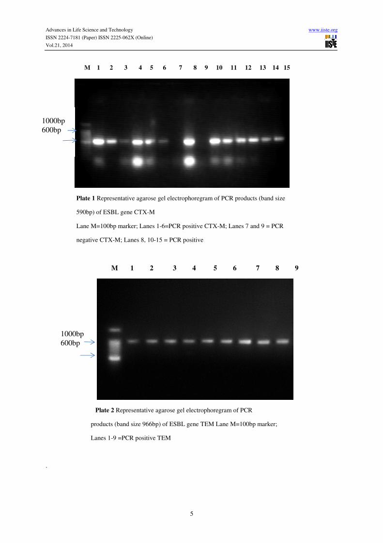
M 1 2 3 4 5 6 7 8 9 10 11 12 13 14 15
Plate 1 Representative agarose gel electrophoregram of PCR products (band size
590bp) of ESBL gene CTX-M
Lane M=100bp marker; Lanes 1-6=PCR positive CTX-M; Lanes 7 and 9 = PCR
negative CTX-M; Lanes 8, 10-15 = PCR positive
M 1 2 3 4 5 6 7 8 9
Plate 2 Representative agarose gel electrophoregram of PCR
products (band size 966bp) of ESBL gene TEM Lane M=100bp marker;
Lanes 1-9 =PCR positive TEM
.
1000bp
600bp
1000bp
600bp

Advances in Life Science and Technology www.iiste.org
ISSN 2224-7181 (Paper) ISSN 2225-062X (Online)
Vol.21, 2014
6
M 1 2 3 4 5 6 7 8 9 10 11 12 13 14 15
Plate 3 Representative agarose gel electrophoregram of PCR products
(band size 891bp) of ESBL gene CTX-M-G1
Lane M=100bp marker; Lanes 1-6=PCR positive CTX-M-G1; Lanes 7
and 8 = PCR negative CTX-M-G1; Lanes 9-15 = PCR positive CTX-M-G1
M 1 2 3 4 5 6 7 8 9 10 11 12
Plate 4 Representative agarose gel electrophoregram of PCR products (band size
857bp) of ESBL gene CTX-M-G9
Lane M=100bp marker; Lanes 7 and 9 =PCR positive CTX-M-G9; Lanes 1-6
and 10-12 = PCR negative CTX-M-G9
4.0 Discussion CTX-M-type, TEM-type and SHV-type ESBLs are the main types of ESBL produced by bacteria. Some
researchers have suggested that CTX-M-type ESBLs are now the most frequent ESBL type worldwide as
compared to SHV and TEM-type ESBLs (Paterson and Bonomo, 2005). In this study 90% isolates produced
1000bp
600bp
1000bp
600bp

Advances in Life Science and Technology www.iiste.org
ISSN 2224-7181 (Paper) ISSN 2225-062X (Online)
Vol.21, 2014
7
CTX-M-type ESBL confirming that CTX-M is the dominant ESBL-type in Accra as it is in the rest of world
(Falagas and Karageorgopoulos, 2009).
The percentage of isolates producing CTX-M-type ESBL in this present study is consistent with the
study in Kumasi, Ghana by Feglo (2013) who reported 94.4% CTX-M-type ESBL. However, while Feglo (2013)
reported that 64.5% of the ESBL isolates possess two genes and 29.9% possess three genes; only 20% of ESBL
isolates in this present study had two genes and none possessed three ESBL genes. In a study in Southwest
Nigeria, 30 selected multidrug-resistant K. pneumoniae strains isolated from patients with urinary tract infections
showed 57% CTX-M enzymes (Olysegun et al., 2006) which were slightly lower than the 87.1% of CTX-M
ESBLs observed in K. pneumoniae strains in this study. Reports from Indonesia confirmed the high prevalence
(94.5%) of CTX-M-type ESBL (Severin et al., 2010). This was also corroborated by Tham and others (2010)
who observed that 90% ESBL-positive genes were of CTX-M ESBL-type. This study was consistent with the
finding by Heffernan and colleagues (2007) in New Zealand who observed that 96% E. coli and K. pneumoniae
isolates produced CTX-M ESBL genes which is also corroborated by a study done in Canada (Pitout et al.,
2007). So in this study, Ghana joins North America, South America, Western Europe, Asia and other African
nations with high prevalent CTX-M-ESBL producers (Paterson and Bonomo, 2005).
This study has shown that 78% of the CTX-M-type ESBLs were CTX-M-1group with 2% belonging to
CTX-M-9group corroborating the study of Tham and colleagues in 2010 who reported that CTX-M-1group was
prevalent followed by the CTX-M-9group as do the conclusions of Heffernan and colleagues who also showed
that CTX-M-1group (CTX-M-15) and CTX-M-9group (CTX-M-14) seem to be the most widespread (Heffernan
et al., 2007). However, this study is at variance with the work of Pitout and colleagues in 2007 who found 48%
and 37% CTX-M-1group and CTX-M-9group respectively.
The high prevalence of CTX-M-1group ESBL observed in this study confirms the studies in other parts
of Africa. In Tanzania, Blomberg and colleagues (2005) discovered the presence of CTX-M-1group (CTX-M-
15) ESBL-producing organism for the first time in Africa. In Tunisia, 43 of 47 isolates showed CTX-M-1group
(CTX-M-15) and 2 CTX-M-9group (CTX-M-14) (Elhani et al., 2010). This consensus of CTX-M ESBL genes
dominating the ESBL types was contradicted by the study of Feglo (2013) in Kumasi, Ghana which reported
TEM-type ESBL as the dominating ESBL gene.
In this study 25% of the bacterial isolates produced TEM-type ESBL, a result which is lower as
reported in Canada (Pitout et al., 2007) and New Zealand (Heffernan et al., 2007). Furthermore, Severin and
colleagues (2010) in a study of 73 ESBL-positive E. coli and 72 K. pneumoniae strains, TEM-type ESBLs were
not detected in any of the isolates. Interestingly Feglo in 2013, reported a high prevalent of 96.2% of TEM-type
ESBL phenotypes in a study in Kumasi in contrast to the findings of this study and other reports.
Regarding SHV-type ESBL, none was seen in 100 strains of ESBL phenotypes in this study confirming
a study in Algiers by Ramdani-Bouguessa and colleagues in 2006. Low rates were reported in New Zealand
(Heffernan et al., 2007) and North Lebanon (Sana et al., 2011) which contradicts findings in this study.
However, higher rates of 32.5% SHV-type ESBL were reported by Feglo (2013) in Kumasi contradicting this
work also in Ghana, 38.7% in Cameroon Gangoué-Piéboji et al., 2005) while Jones and others also reported that
SHV genes were found in 41% and 28% of the ESBL-positive K. pneumoniae and E. coli isolates respectively
(Jones et al., 2009) in contrast to this study.
5.0 Conclusion
The findings of this work suggest that organisms producing CTX-M-type ESBL are more prevalent in Accra
than other ESBL types. The present study indicated that the CTX-M-1group producing isolates dominated the
ESBL phenotypes and CTX-M-15 may be the dominant CTX-M-type ESBL. There is the need for further
studies into the characteristic transmission, pathogenesis, antibiotic resistance expression, response to therapy
and infection control of CTX-M-type ESBL and TEM-type ESBL in Accra.
Acknowledgements The authors appreciate the grant support from College of Health Sciences, University of Ghana. We express our
profound gratitude to the management and staff of Advent Clinical Laboratories, Central Laboratory, Korle Bu
Teaching Hospital and Molecular Biology Laboratories of School of Allied Health Sciences and Department of
Microbiology, University of Ghana Medical School.

Advances in Life Science and Technology www.iiste.org
ISSN 2224-7181 (Paper) ISSN 2225-062X (Online)
Vol.21, 2014
8
References
Blomberg B, Jureen R, Manji KP, Tamim BS, Mwakagile DSM, Urassa WK, Fataki M, Msangi V, Tellevik MG,
Maselle SY, Langeland N (2005). High rate of fatal cases of pediatric septicemia caused by Gram-negative
bacteria with extended-spectrum β-lactamases in Dar es Salaam, Tanzania. Journal of Clinical Microbiology; 43:
745-749.
Bonnet R (2004). Growing group of extended-spectrum β-lactamases: the CTX-M enzymes. Antimicrob Agents
Chemother; 48: 1-14.
Elhani D, Bakir L, Aouni M, Passet V, Arlet G, Brisse S, Weill FX (2010). Molecular epidemiology of
extended-spectrum beta-lactamase-producing Klebsiella pneumoniae strains in a university hospital in Tunis,
Tunisia, 1999-2005. J Clinical Microbiology; 16(2): 157-164.
Falagas ME, Karageorgopoulos DE (2009). Extended-spectrum beta-lactamase-producing organisms. J Hosp
Infect; 73(4):345-54.
Feglo PK (2013). Molecular epidemiology of extended spectrum β-lactamase (ESBL) producing
enterobacteriaceae at the Komfo Anokye Taching Hospital. Thesis Abstract.
Gangoué-Piéboji J, Branka B, Koulla-Shiro S, Randegger C, Adiogo D,
Ngassam P, Ndumbe P, Hächler H
(2005). Extended-Spectrum-ß-Lactamase-Producing Enterobacteriaceae in Yaounde, Cameroon. Journal of
Clinical Microbiology; 43(7): 3273-3277.
Heffernan H, Pope C and Carter P (2007). Identification of extended spectrum β-lactamase types, plasmid-
mediated Ampc β-lactamases and strains among urinary Escherichia coli and Klebsiella in New Zealand in
2006. Communicable Disease Group, ESR. FW07103.
Jones CH, Tuckman M, Keeney D, Ruzin A, Bradford PA (2009). Characterization and Sequence Analysis of
Extended-Spectrum-_-Lactamase-Encoding Genes from Escherichia coli, Klebsiella pneumoniae, and Proteus
mirabilis Isolates Collected during Tigecycline Phase 3 Clinical Trials. Antimicrobial Agents and Chemotherapy;
53(2) 465–475.
Olysegun SO, Queenan AM, Ojo KK., Adeniyi BA, Roberts MC (2006). CTX-M-15 extended-spectrum β
lactamase
from Nigerian Klebsiella pneumoniae. J. Antimicrob. Chemother. 57: 24-30.
Paterson DL, Bonomo RA (2005). Extended-spectrum ß-lactamases: a clinical update. Clinical Microbiology
Reviews; 18(4) 657-686.
Pitout JD, Church DL, Gregson DB (2007). Molecular epidemiology of CTX-M-producing Escherichia coli in
the Calgary Health Region: emergence of CTX-M-15-producing isolates. Antimicrob Agents Chemother; 51:
1281–86.
Ramdani-Bouguessa N, Mendonça N, Leitão J, Ferreira E, Mohamed Tazir M, Caniça M (2006). CTX-M-3 and
CTX-M-15 Extended-Spectrum β-Lactamases in Isolates of Escherichia coli from a Hospital in Algiers, Algeria.
Antimicrob Agents Chemother; 45: 1730–36
Sana T, Rami1 K, Racha1 B, Fouad D, Marcel A, Hassan M, Sani1 H, Hamze H (2011). Detection of genes
TEM, OXA, SHV and CTX-M in 73 clinical isolates of Escherichia coli producers of extended spectrum Beta-
lactamases and determination of their susceptibility to antibiotics. International Arabic Journal of Antimicrobial
Agents; 1(5): 704
Severin JA, Mertaniasih NM, Kuntaman K, Lestari ES, Purwanta M, Lemmens-Den Toom N, Duerink DO, Hadi
U, van Belkum A, Verbrugh HA, Goessens WH (2010). Molecular characterization of extended-spectrum beta-
lactamases in clinical Escherichia coli and Klebsiella pneumoniae isolates from Surabaya, Indonesia. J
Antimicrob Chemother; 65(3): 465-469.
Tham J, Odenholt I , Walder M, Brolund A, Ahl J, Melande E (2010).. Extended-spectrum beta-lactamase-
producing Escherichia coli in patients with travellers’ diarrhoea. Scandinavian Journal of Infectious Disease.

Advances in Life Science and Technology www.iiste.org
ISSN 2224-7181 (Paper) ISSN 2225-062X (Online)
Vol.21, 2014
9
Yamasaki K, Komatsu M, Yamashita T, Shimakawa K, Ura T, Nishio H, Satoh K, Washidu R, Kinoshita S,
Aihara M (2003). Production of CTX-M-3 extended-spectrum beta-lactamase and IMP-1 metallo beta-lactamase
by five Gram-negative bacilli: survey of clinical isolates from seven laboratories collected in 1998 and 2000, in
the Kinki region of Japan. J. Antimicrob. Chemother; 51:631-638.

The IISTE is a pioneer in the Open-Access hosting service and academic event
management. The aim of the firm is Accelerating Global Knowledge Sharing.
More information about the firm can be found on the homepage:
http://www.iiste.org
CALL FOR JOURNAL PAPERS
There are more than 30 peer-reviewed academic journals hosted under the hosting
platform.
Prospective authors of journals can find the submission instruction on the
following page: http://www.iiste.org/journals/ All the journals articles are available
online to the readers all over the world without financial, legal, or technical barriers
other than those inseparable from gaining access to the internet itself. Paper version
of the journals is also available upon request of readers and authors.
MORE RESOURCES
Book publication information: http://www.iiste.org/book/
IISTE Knowledge Sharing Partners
EBSCO, Index Copernicus, Ulrich's Periodicals Directory, JournalTOCS, PKP Open
Archives Harvester, Bielefeld Academic Search Engine, Elektronische
Zeitschriftenbibliothek EZB, Open J-Gate, OCLC WorldCat, Universe Digtial
Library , NewJour, Google Scholar