Genotypes and virulence in serotype K2 Klebsiella pneumoniae from liver abscess and non-infectious...
7
RESEARCH Open Access Genotypes and virulence in serotype K2 Klebsiella pneumoniae from liver abscess and non-infectious carriers in Hong Kong, Singapore and Taiwan Jung-Chung Lin 1 , Tse Hsien Koh 2 , Nelson Lee 3 , Chang-Phone Fung 4 , Feng-Yee Chang 1 , Yu-Kuo Tsai 5 , Margaret Ip 6* and L Kristopher Siu 1,5,7* Abstract In Klebsiella pneumoniae liver abscess (KP-LA), K. pneumoniae K2 is the most frequently isolated serotype after K1, but this serotype has been much less studied. In the present study, the molecular types sequences type (MLST) of serotype K2 isolates from three different regions in Asia were identified and the virulence of these isolates was investigated. Eight different MLSTs were found among 26 isolates (ST 65, 66, 86, 373, 374, 375, 380, and 434). There were two major MLST groups, ST-65-like (42%) and ST86-like (46%). No isolates contained allS while all isolates contained rmpA. The prevalence of aerobactin gene and kfu were 25/26 (96%) and 3/26 (11.5%) respectively. Although liver abscess isolates were generally more resistant (11/15 isolates) to serum killing, there was no specific distribution of serum killing resistant or susceptible ST types between stool carriage and liver abscess isolates. Neutrophil phagocytosis showed that the liver abscess and carriage isolates varied in their susceptibility to phagocytosis. Strains with resistance to both neutrophil phagocytosis and serum killing were generally hypervirulent with lethality at LD 50 < 10 3 colony forming units by intraperitoneal injection. In conclusion, Anti-phagocytosis and resistance to serum killing are two parameters that most predict hyperviurlence in serotype K2 isolates. Unlike serotype K1 KP-LA that mainly belong to ST-23, ST-65-like and -86-like are the two major MLST types among serotype K2 isolates from Singapore, Hong Kong and Taiwan. Keywords: Liver abscess, Klebsiella pneumoniae, MLST Introduction Klebsiella pneumoniae liver abscess (KP-LA) has beco- ming a global emerging disease [1,2]. The etiology of this disease has been investigated by different study groups [1,3-5]. Several bacterial virulence factors have been investigated and virulence has often been found to be capsule related [2,6,7]. K. pneumoniae serotype K1 cau- sing liver abscesses LA has been observed worldwide and is the most prevalent type among all 77 serotypes [1,6,8]. Previous molecular and virulence analysis in K. pneumoniae serotype K1 has shown that ST23 was predominant in serotype K1 K. pneumoniae isolates causing liver abscess and carried in stools of uninfected subjects in Hong Kong, Singapore and Taiwan [9]. In addition, serotype K1 isolates with ST23 could have different mice lethal dose (LD 50 ) indicating normally expressed K1 capsule is not the sole factor for hyperviru- lence but its phagocytic resistance and carriage of the aerobactin gene were two independent determinants contributing to mouse lethality [2,9]. Although kfu (gene encoding an iron uptake system) and allS (a gene asso- ciated with allantoin metabolism) have been docu- mented as virulence factors contributing to virulence, all invasive serotype K1 isolates contained these two deter- minants in a previous study [10-12]. Although non-K1 serotypes have been observed in KP- LA, they were less frequently encountered. Serotype K2 KP is the second most commonly isolated serotype in KP-LA in Taiwan and has also been reported in Asia and US [1,13-15]. Relatively few studies have specifically * Correspondence: [email protected]; [email protected] 6 Department of Microbiology, Faculty of Medicine, Chinese University of Hong Kong, Hong Kong, SAR, People’s Republic of China 1 Division of Infectious Diseases and Tropical Medicine, Department of Internal Medicine, Tri-Service General Hospital, National Defense Medical Center, Taipei, Taiwan Full list of author information is available at the end of the article © 2014 Lin et al.; licensee BioMed Central Ltd. This is an Open Access article distributed under the terms of the Creative Commons Attribution License (http://creativecommons.org/licenses/by/4.0), which permits unrestricted use, distribution, and reproduction in any medium, provided the original work is properly credited. The Creative Commons Public Domain Dedication waiver (http://creativecommons.org/publicdomain/zero/1.0/) applies to the data made available in this article, unless otherwise stated. Lin et al. Gut Pathogens 2014, 6:21 http://www.gutpathogens.com/content/6/1/21
Transcript of Genotypes and virulence in serotype K2 Klebsiella pneumoniae from liver abscess and non-infectious...

Lin et al. Gut Pathogens 2014, 6:21http://www.gutpathogens.com/content/6/1/21
RESEARCH Open Access
Genotypes and virulence in serotype K2 Klebsiellapneumoniae from liver abscess and non-infectiouscarriers in Hong Kong, Singapore and TaiwanJung-Chung Lin1, Tse Hsien Koh2, Nelson Lee3, Chang-Phone Fung4, Feng-Yee Chang1, Yu-Kuo Tsai5,Margaret Ip6* and L Kristopher Siu1,5,7*
Abstract
In Klebsiella pneumoniae liver abscess (KP-LA), K. pneumoniae K2 is the most frequently isolated serotype after K1,but this serotype has been much less studied. In the present study, the molecular types sequences type (MLST) ofserotype K2 isolates from three different regions in Asia were identified and the virulence of these isolates wasinvestigated. Eight different MLSTs were found among 26 isolates (ST 65, 66, 86, 373, 374, 375, 380, and 434). Therewere two major MLST groups, ST-65-like (42%) and ST86-like (46%). No isolates contained allS while all isolatescontained rmpA. The prevalence of aerobactin gene and kfu were 25/26 (96%) and 3/26 (11.5%) respectively.Although liver abscess isolates were generally more resistant (11/15 isolates) to serum killing, there was no specificdistribution of serum killing resistant or susceptible ST types between stool carriage and liver abscess isolates.Neutrophil phagocytosis showed that the liver abscess and carriage isolates varied in their susceptibility tophagocytosis. Strains with resistance to both neutrophil phagocytosis and serum killing were generally hypervirulentwith lethality at LD50 < 103 colony forming units by intraperitoneal injection. In conclusion, Anti-phagocytosis andresistance to serum killing are two parameters that most predict hyperviurlence in serotype K2 isolates. Unlikeserotype K1 KP-LA that mainly belong to ST-23, ST-65-like and −86-like are the two major MLST types amongserotype K2 isolates from Singapore, Hong Kong and Taiwan.
Keywords: Liver abscess, Klebsiella pneumoniae, MLST
IntroductionKlebsiella pneumoniae liver abscess (KP-LA) has beco-ming a global emerging disease [1,2]. The etiology of thisdisease has been investigated by different study groups[1,3-5]. Several bacterial virulence factors have beeninvestigated and virulence has often been found to becapsule related [2,6,7]. K. pneumoniae serotype K1 cau-sing liver abscesses LA has been observed worldwideand is the most prevalent type among all 77 serotypes[1,6,8]. Previous molecular and virulence analysis inK. pneumoniae serotype K1 has shown that ST23 waspredominant in serotype K1 K. pneumoniae isolates
* Correspondence: [email protected]; [email protected] of Microbiology, Faculty of Medicine, Chinese University ofHong Kong, Hong Kong, SAR, People’s Republic of China1Division of Infectious Diseases and Tropical Medicine, Department ofInternal Medicine, Tri-Service General Hospital, National Defense MedicalCenter, Taipei, TaiwanFull list of author information is available at the end of the article
© 2014 Lin et al.; licensee BioMed Central LtdCommons Attribution License (http://creativecreproduction in any medium, provided the orDedication waiver (http://creativecommons.orunless otherwise stated.
causing liver abscess and carried in stools of uninfectedsubjects in Hong Kong, Singapore and Taiwan [9]. Inaddition, serotype K1 isolates with ST23 could havedifferent mice lethal dose (LD50) indicating normallyexpressed K1 capsule is not the sole factor for hyperviru-lence but its phagocytic resistance and carriage of theaerobactin gene were two independent determinantscontributing to mouse lethality [2,9]. Although kfu (geneencoding an iron uptake system) and allS (a gene asso-ciated with allantoin metabolism) have been docu-mented as virulence factors contributing to virulence, allinvasive serotype K1 isolates contained these two deter-minants in a previous study [10-12].Although non-K1 serotypes have been observed in KP-
LA, they were less frequently encountered. Serotype K2KP is the second most commonly isolated serotype inKP-LA in Taiwan and has also been reported in Asiaand US [1,13-15]. Relatively few studies have specifically
. This is an Open Access article distributed under the terms of the Creativeommons.org/licenses/by/4.0), which permits unrestricted use, distribution, andiginal work is properly credited. The Creative Commons Public Domaing/publicdomain/zero/1.0/) applies to the data made available in this article,
Lin et al. Gut Pathogens 2014, 6:21 Page 2 of 7http://www.gutpathogens.com/content/6/1/21
focused on this serotype. Little is known about the STsand the prevalence of virulence factors in serotype K2causing KP-LA in different countries.In this study, we determined the molecular types
(MLST) of K. pneumoniae serotype K2 isolates fromliver abscesses and from carriers without a history ofKP-LA and assessed the virulence of isolates with diffe-rent MLST types from Hong Kong, Singapore, andTaiwan.
Material and methodsBacterial strainsK. pneumoniae strains that were isolated from liver ab-scess and stool from healthy subjects, hospitalized patientswithout history of liver abscess or patients admitted withnoninfectious diseases were collected at Prince of Waleshospital in Hong Kong, Singapore General Hospital,National University hospital in Singapore and Tri-ServiceGeneral Hospital in Taiwan from 2002 to 2009. Oneisolate was collected from each patient will liver abscess,healthy subjects, hospitalized patients without history ofliver abscess or patients admitted with noninfectiousdiseases. The diagnosis of liver abscess was confirmed byabdominal ultrasonography and/or computerized tomo-graphy. Identification of the isolates was according tostandard clinical microbiologic methods.
Serotyping, kfu, alls, rmpA and aerobactin gene detectionby PCRIsolates were serotyped by PCR as previously described[16]. PCRs to determine the presence of the specific genesfor serotype K1, K2 and K5 [17], rmpA, alls, kfu and the
Table 1 Specific primers used for amplification of the target g
Serotype (target gene) Primer
K1 (wzyKPK1) 5′-GGTGCTCTTTACATCATTGC-3′
5′-GCAATGGCCATTTGCGTTAG-3′
K2 (wzyKPK2) 5′-GACCCGATATTCATACTTGACAGAG
5′-CCTGAAGTAAAATCGTAAATAGATG
K5 (wzxKPK5) 5′-TGGTAGTGATGCTCGCGA-3′
5′-CCTGAACCCACCCCAATC-3′
RmpA 5′-ACTGGGCTACCTCTGCTTCA-3′
5′-CTTGCATGAGCCATCTTTCA-3′
Aerobactin 5′-GCATAGGCGGATACGAACAT-3′
5′-CACAGGGCAATTGCTTACCT-3′
Aerobactin 5′-CTGTCGGCATCGGTTTTATT-3′
5′-TGGCGTGTCGATTATTACCA-3′
Alls 5′-CCGAAACATTACGCACCTTT-3′
5′-ATCACGAAGAGCCAGGTCAC-3′
Kfu 5′- ATAGTAGGCGAGCACCGAGA-3′
5′-AGAACCTTCCTCGCTGAACA-3′
aerobactin gene [5] were performed using primers aslisted in Table 1. A bacterial colony from an overnight-culture was added to 300 μl water and boiled for 15 minto release DNA template. The reaction mixture was keptat 95°C for 5 min, followed by 40 temperature cycles of95°C for1 min, 50°C for 1 min, and 72°C for 2 min, and72°C for 7 min. The expected PCR products were 641 bpfor wzyKPK2, 535 bp for rmpA, 520 bp for kfu, 508 bp forallS and either 556 or 531 bp for aerobactin in length(Table 1).
Multilocus sequence typing (MLST)MLST were performed according to Turton et al., [4].Sequences of seven housekeeping genes were obtainedfor isolates from liver abscess patients and carriers. Se-quence information was compared with that availablefrom the MLST website (http://www.pasteur.fr/mlst/)developed by Keith Jolley. Alleles and sequence types(STs) were assigned accordingly. Sequences of any allelesthat were not in the database were submitted to thecurator and a new allele number obtained. A differencein two or more alleles was considered to indicate thatthe sequence types being compared were unrelated.
Fluorescence labeling of bacteriaLabeling was performed as previously described [18]. TheKP isolate and control suspensions were individually incu-bated overnight at 37°C. The concentration was appro-ximated using photospectrometry (Olympus, US). Thepercentage of bacterial viability in an aliquot of eachpopulation was determined by quantitative plate coun-ting. The FITC-labeled bacteria were resuspended at a
enes of K. pneumoniae in this study
Size of PCR product (bp) Reference
1283 [17]
-3′ 641 [17]
GC-3′
280 [17]
536 [5]
556 [5]
531 [5]
[5]
[5]
Lin et al. Gut Pathogens 2014, 6:21 Page 3 of 7http://www.gutpathogens.com/content/6/1/21
concentration of 2 × 10⁸ cells/mL in PBS, divided intoequal volumes, and stored at −70°C. Aliquots werethawed just prior to use.
Phagocytosis assayPhagocytosis was measured using a standard assay. Normalhuman serum pooled from healthy volunteers was dividedinto equal volumes and stored at −70°C. Serum was thawedimmediately prior to use and stored on ice until added tothe phagocytosis assay. Briefly, for the assay, 100 μL of aneutrophil suspension (representing 1 × 10⁶ cells), 100 μLof freshly thawed pooled normal human serum (10% v/vopsonization), and 600 μL PBS was added to sealable10 × 75 mm Falcon™ polypropylene tubes (BD, FranklinLakes, NJ). The suspension was pre-warmed with shakingfor 5 min at 37°C. Multiple volumes of 200 μL FITC-labeled bacteria (representing 4 × 10⁷ colony forming units[cfu]/mL) were added to 800 μL to produce a final volumeof 1.0 mL. Each tube was capped and incubated in ashaking water bath at 37°C with continuous agitation for15 min. An unincubated tube served as the 0-min timepoint. At each designated time, samples were removedand placed in an ice bath. The cells in each suspensionwere removed by centrifugation at 250 g for 6 min, andthe cell pellet was resuspended in 1.0 mL of ice-cold PBSand maintained at 4°C. A 600 μL volume of the suspen-sion was transferred into a new tube, and ethidium brom-ide was added to a final concentration of 50 mg/L beforemeasurement. Excess ethidium bromide was used to sup-press the extracellular fluorescence. Bacteria that were notlocalized in neutrophils appeared red in color upon micro-scopic examination (see below).
Phagocytosis assay using flow cytometryA FACScan, emitting an argon laser beam at 488 nm(Becton Dickinson Immunocytometry Systems, San Jose,Calif.), was used to detect FITC fluorescence. The side-way scatter (SSC) threshold was 52. The detector wasset at E00, 350, and 427 for forward scatter (FSC), SSC,and fluorescence 1 (FL1-H, green), respectively. Fluores-cence values were collected after gating the detector onthe FSC and SSC combination. A total of 10,000 cellswere processed using the Cellquest version 1.0 software.Fluorescence distribution data collected using a logarith-mic amplifier was displayed as single histograms forFL1-H. By processing unstained and FITC-stained bac-terial phagocytosis mixtures, the boundary of positiveand negative fluorescence was determined. The percen-tage of ingested bacteria was assessed after the additionof ethidium bromide.
Susceptibility to serum killingSerum bactericidal activity was measured using the me-thod of Hughes et al. [19] as modified by Podschun et al.
[20]. The viable bacterial concentration was adjusted to1 × 106 colony forming units/mL. Twenty-five microlitersof bacteria were added to 75 μL of pooled human seracontained in a 10 × 75 mm Falcon polypropylene tube(BD Biosciences, Franklin Lakes, New Jersey). Tubes wereagitated for 0, 60, 120, or 180 min. To determine the num-ber of viable bacteria after exposure to serum, an aliquotof each bacterial suspension was removed at the desig-nated time point, diluted 10-fold by addition of Mueller-Hinton broth, plated on Mueller-Hinton agar, and assayedas described immediately below.Results were expressed as percentage of inoculums,
and responses in terms of viable counts were gradedfrom 1–6 as described previously [20]. Each strain wastested at least three times. A strain was consideredserum resistant or serum sensitive if the grading was thesame in all experiments. Each isolate was classified ashighly sensitive (grades 1 or 2), intermediately sensitive(grades 3 or 4), or resistant (grades 5 or 6).
Mice lethality testIn determination of LD50 in mice, six mice were used asa sample population for each bacterial concentration.Bacterial concentration was calculated by cell formingunit (cfu). Intraperitoneal (i.p.) injection was used to as-sess virulence. Mice used in this study was approved byanimal used committee with NHRI-IACUC-103014-A.Symptoms and signs of infection were observed for14 days. Survival of the inoculated mice was recordedand the LD50 was calculated using SigmaPlot version7.0 from SPSS Inc. (Chicago, IL).
ResultsMLST profiles of isolates from Hong Kong, Singapore andTaiwanA total of 26 serotype K2 isolates were confirmed byserotyping and PCR and selected for this study. Fifteenand 11 KPs were isolated from liver abscess patients andstool of non-infectious carriers (Table 2) respectively.Eight different MLSTs were identified including ST 65,66, 86, 373, 374, 375, 380, and 434. Two major MLSTgroups, ST-65-like and ST86-like groups were obtainedbased on minimum-spanning tree analysis (Figure 1).ST373 (a single locus variant, SLV to ST86) and ST374were new ST types found in this study (Table 2). The STtypes of liver abscess isolates obtained from Hong Kongwere more diverse. There was no specific distribution ofST types between those isolated from liver abscess andfrom stool carriage.
Serum killing resistance and neutrophil phagocytosis ofall K2 K. pneumoniae isolatesSerum killing resistant and susceptible isolates were foundin both ST65-like and ST86-like isolates. Although liver

Table 2 MLST of serotype K2 isolates from liver abscess patients and from stool carriage of non-liver abscess subjectsin Hong Kong, Singapore and Taiwan
Group* ST type (no. of isolates) No. of isolates Allelic profiles
HK† SG TW gapA inf mdh pgi pho rpoB tonB
LA¶ ST LA ST LA ST
1 65 (8) 2 2 1 2 1 2 1 2 1 10 4 13
1 375 (1) 1 43 1 2 1 10 4 13
1 66 (2) 2 2 3 2 1 10 1 13
2 86 (11) 1 2 2 1 2 3 9 4 2 1 1 1 27
2 373 (1) 1 9 4 2 26 1 1 27
3 374 (1) 1 2 3 58* 37 10 27 9
4 380 (1) 1 2 1 1 1 1 4 19
5 434 (1) 1 2 3 2 4 9 4 118
*Grouping was referred to the results obtained from minimum-spanning tree (Figure 2). 373 and 375 were single locus variant to ST86 and 65.†Isolates from: HK, Hong Kong; SG, Singapore; TW, Taiwan.¶Isolates from: LA, liver abscess; ST, stool carriage.NA: Not available; Bold: New ST types in this study.
Lin et al. Gut Pathogens 2014, 6:21 Page 4 of 7http://www.gutpathogens.com/content/6/1/21
abscess isolates were generally more resistant (11/15 iso-lates) to serum killing, there was no specific distributionof serum killing resistant or susceptible ST types betweenstool carriage and liver abscess isolates (Table 3).Liver abscess and carriage isolates had variable suscep-
tibility to phagocytosis. Likewise, there was no differencebetween the two major MLST types, ST65-like andST86-like (Figure 2). Although ST other than ST65-likeand ST86-like were all more resistant to phagocytosis,there was only one isolate for each type.
Comparative analysis with respect to mouse lethality,susceptibility to neutrophil phagocytosis, serum killingand virulence-associated genesPCR for virulence associated genes revealed that all iso-lates contained rmpA while none harbored allS gene. The
Figure 1 Minimum-spanning tree of 26 isolates of KP serotype K2 strST number, Orange indicates ST group 1; yellow, ST group 2; blue, singleto
prevalence of the aerobactin gene and kfu in all K2 isolateswere 25/26 (96%) and 3/26 (11.5%) respectively. Fifteenisolates from either liver or stool carriage showed resis-tance to both neutrophil phagocytosis and serum killing.Twelve were hypervirulent with LD50 ≤ 10
2 CFU. Two iso-lates with LD50 equal to 1.0 × 10³ CFU and 1 with LD50
5.5 × 10³ CFU were also defined as virulent strains. Fourisolates showed susceptibility to both neutrophil phagocy-tosis and serum killing. These four isolates demonstratedlethality with LD50 that ranged from <3.7 × 10² - 2.2 × 10⁶
CFU in the infection model. These two parameters alonewere not able to reflect the virulence of the strains. Otherfactors that may contribute to virulence are not furtherassessed in this study. One ST86 strain was hypervirulentwith a mouse lethality LD50 of <3.7 × 10² CFU, sugges-ting other unknown and major virulent factor might
ains generated by MLST allelic data (analysis in MLST website).n.
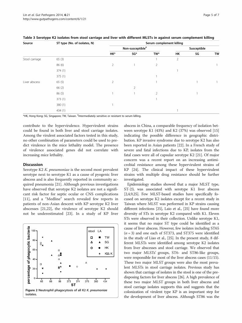
Table 3 Serotype K2 isolates from stool carriage and liver with different MLSTs in against serum complement killing
Source ST type (No. of isolates, N) Serum complement killing
Non-susceptible# Susceptible
HK* SG* TW* HK SG TW
Stool carriage 65 (3) 1 2
86 (6) 2 1 2 1
374 (1) 1
375 (1) 1
Liver abscess 65 (5) 2 1 1 1
66 (2) 2
86 (5) 1 1 2 1
373 (1) 1
380 (1) 1
434 (1) 1
*HK, Hong Kong; SG, Singapore; TW, Taiwan. #Intermediately sensitive or resistant to serum killing.
Lin et al. Gut Pathogens 2014, 6:21 Page 5 of 7http://www.gutpathogens.com/content/6/1/21
contribute to the hypervirulence. Hypervirulent strainscould be found in both liver and stool carriage isolates.Among the virulent associated factors tested in this study,no other combination of parameters could be used to pre-dict virulence in the mice lethality model. The presenceof virulence associated genes did not correlate withincreasing mice lethality.
DiscussionSerotype K2 K. pneumoniae is the second most prevalentserotype next to serotype K1 as a cause of pyogenic liverabscess and is also frequently reported in community ac-quired pneumonia [21]. Although previous investigationshave observed that serotype K2 isolates are not a signifi-cant risk factor for septic ocular or CNS complications[11], and a “Medline” search revealed few reports inpatients of non-Asian descent with KP serotype K2 liverabscesses [21,22], the virulence of serotype K2 shouldnot be underestimated [23]. In a study of KP liver
Figure 2 Neutrophil phagocytosis of all K2 K. pneumoniaeisolates.
abscess in China, a comparable frequency of isolation bet-ween serotype K1 (43%) and K2 (37%) was observed [15]indicating the possible difference in geographic distri-bution. KP invasive syndrome due to serotype K2 has alsobeen reported in Asian patients [22]. In a French study ofsevere and fatal infections due to KP, isolates from thefatal cases were all of capsular serotype K2 [21]. Of majorconcern was a recent report on an increasing antimi-crobial resistance among these hypervirulent strains ofKP [24]. The clinical impact of these hypervirulentstrains with multiple drug resistance should be furtherinvestigated.Epidemiology studies showed that a major MLST type,
ST-23, was associated with serotype K1 liver abscess[2,4,9,25]. Few MLST-based studies have specifically fo-cused on serotype K2 isolates except for a recent study inTaiwan where MLST was performed in KP strains causingdifferent infections [25], Laio et al., [25] have found morediversity of STs in serotype K2 compared with K1. ElevenSTs were observed in their collection. Unlike serotype K1,it seems that no major ST type could be identified as acause of liver abscess. However, few isolates including ST65(n = 3) and one each of ST373, and ST375 were identifiedin the study of Liao et al., [25]. In the present study, 8 dif-ferent MLSTs were identified among serotype K2 isolatesfrom liver abscesses and stool carriage. We observed thattwo major MLSTs’ groups, ST6- and ST86-like groups,were responsible for most of the liver abscess cases (11/15).These two major MLST groups were also the most preva-lent MLSTs in stool carriage isolates. Previous study hasshown that carriage of isolates in the stool is one of the pre-disposing factors for liver abscess [26]. A high prevalence ofthese two major MLST groups in both liver abscess andstool carriage isolates supports this and suggests that thecolonization of virulent type KP is an important step forthe development of liver abscess. Although ST86 was the
Lin et al. Gut Pathogens 2014, 6:21 Page 6 of 7http://www.gutpathogens.com/content/6/1/21
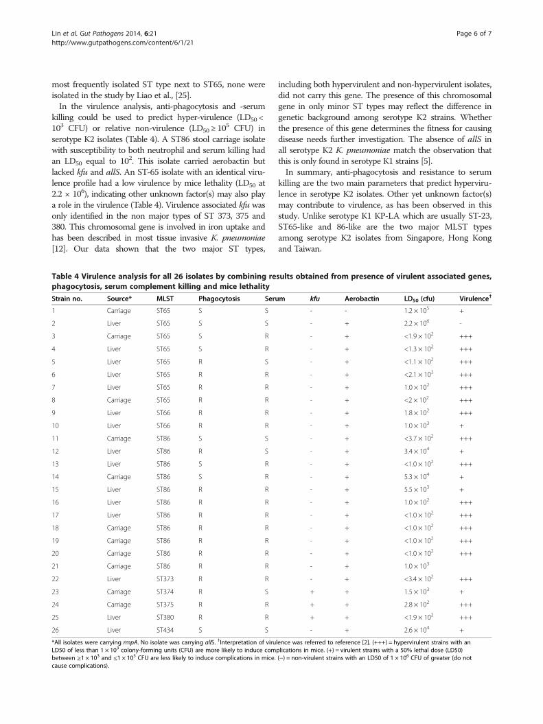
most frequently isolated ST type next to ST65, none wereisolated in the study by Liao et al., [25].In the virulence analysis, anti-phagocytosis and -serum
killing could be used to predict hyper-virulence (LD50 <103 CFU) or relative non-virulence (LD50 ≥ 10
5 CFU) inserotype K2 isolates (Table 4). A ST86 stool carriage isolatewith susceptibility to both neutrophil and serum killing hadan LD50 equal to 102. This isolate carried aerobactin butlacked kfu and allS. An ST-65 isolate with an identical viru-lence profile had a low virulence by mice lethality (LD50 at2.2 × 106), indicating other unknown factor(s) may also playa role in the virulence (Table 4). Virulence associated kfu wasonly identified in the non major types of ST 373, 375 and380. This chromosomal gene is involved in iron uptake andhas been described in most tissue invasive K. pneumoniae[12]. Our data shown that the two major ST types,
Table 4 Virulence analysis for all 26 isolates by combining rephagocytosis, serum complement killing and mice lethality
Strain no. Source* MLST Phagocytosis Seru
1 Carriage ST65 S S
2 Liver ST65 S S
3 Carriage ST65 S R
4 Liver ST65 S R
5 Liver ST65 R S
6 Liver ST65 R R
7 Liver ST65 R R
8 Carriage ST65 R R
9 Liver ST66 R R
10 Liver ST66 R R
11 Carriage ST86 S S
12 Liver ST86 R S
13 Liver ST86 S R
14 Carriage ST86 S R
15 Liver ST86 R R
16 Liver ST86 R R
17 Liver ST86 R R
18 Carriage ST86 R R
19 Carriage ST86 R R
20 Carriage ST86 R R
21 Carriage ST86 R R
22 Liver ST373 R R
23 Carriage ST374 R S
24 Carriage ST375 R R
25 Liver ST380 R R
26 Liver ST434 S S
*All isolates were carrying rmpA. No isolate was carrying allS. †Interpretation of viruLD50 of less than 1 × 10³ colony-forming units (CFU) are more likely to induce combetween ≥1 × 10³ and ≤1 × 10⁵ CFU are less likely to induce complications in mice.cause complications).
including both hypervirulent and non-hypervirulent isolates,did not carry this gene. The presence of this chromosomalgene in only minor ST types may reflect the difference ingenetic background among serotype K2 strains. Whetherthe presence of this gene determines the fitness for causingdisease needs further investigation. The absence of allS inall serotype K2 K. pneumoniae match the observation thatthis is only found in serotype K1 strains [5].In summary, anti-phagocytosis and resistance to serum
killing are the two main parameters that predict hyperviru-lence in serotype K2 isolates. Other yet unknown factor(s)may contribute to virulence, as has been observed in thisstudy. Unlike serotype K1 KP-LA which are usually ST-23,ST65-like and 86-like are the two major MLST typesamong serotype K2 isolates from Singapore, Hong Kongand Taiwan.
sults obtained from presence of virulent associated genes,
m kfu Aerobactin LD50 (cfu) Virulence†
- - 1.2 × 10⁵ +
- + 2.2 × 10⁶ -
- + <1.9 × 10² +++
- + <1.3 × 10² +++
- + <1.1 × 10² +++
- + <2.1 × 10² +++
- + 1.0 × 10² +++
- + <2 × 10² +++
- + 1.8 × 10² +++
- + 1.0 × 10³ +
- + <3.7 × 10² +++
- + 3.4 × 10⁴ +
- + <1.0 × 10² +++
- + 5.3 × 10⁴ +
- + 5.5 × 10³ +
- + 1.0 × 10² +++
- + <1.0 × 10² +++
- + <1.0 × 10² +++
- + <1.0 × 10² +++
- + <1.0 × 10² +++
- + 1.0 × 10³
- + <3.4 × 10² +++
+ + 1.5 × 10³ +
+ + 2.8 × 10² +++
+ + <1.9 × 10² +++
- + 2.6 × 10⁴ +
lence was referred to reference [2]. (+++) = hypervirulent strains with anplications in mice. (+) = virulent strains with a 50% lethal dose (LD50)(−) = non-virulent strains with an LD50 of 1 × 10⁶ CFU of greater (do not

Lin et al. Gut Pathogens 2014, 6:21 Page 7 of 7http://www.gutpathogens.com/content/6/1/21
Competing interestsThe authors declared that they have no competing interests.
Authors’ contributionsJCL, LKS MI,TSK designed study and drafted the manuscript, CPF, JCL, NL,FYC, MI, TSK enrolled the patients and collected the isolates in this study. MI,TSK and LKS proof read and edited the manuscript. All authors read andapproved the final manuscript.
FundingThis study was supported by research grants from National Science Council(NSC102-2314-B016-013-MY-3) and National Health Research Institutes.
Author details1Division of Infectious Diseases and Tropical Medicine, Department ofInternal Medicine, Tri-Service General Hospital, National Defense MedicalCenter, Taipei, Taiwan. 2Department of Pathology, Singapore GeneralHospital, Singapore, Singapore. 3Department of Medicine and Therapeutics,Faculty of Medicine, Chinese University of Hong Kong, Hong Kong, SAR,People’s Republic of China. 4Department of Medicine, Section of InfectiousDiseases, Taipei Veterans General Hospital and National Yang-Ming University,Taipei, Taiwan. 5National Institute of Infectious Diseases and Vaccinology,National Health Research Institutes, 35 Keyan Road, Zhunan, Miaoli County350, Taiwan. 6Department of Microbiology, Faculty of Medicine, ChineseUniversity of Hong Kong, Hong Kong, SAR, People’s Republic of China.7Graduate Institute of Basic Medical Science, China Medical University,Taichung, Taiwan.
Received: 10 April 2014 Accepted: 20 May 2014Published: 12 June 2014
References1. Fung CP, Chang FY, Lee SC, Hu BS, Kuo BI, Liu CY, Ho M, Siu LK:
A global emerging disease of Klebsiella pneumoniae liver abscess: isserotype K1 an important factor for complicated endophthalmitis? Gut 2002,50(3):420–424.
2. Siu LK, Yeh KM, Lin JC, Fung CP, Chang FY: Klebsiella pneumoniae liverabscess: a new invasive syndrome. Lancet Infect Dis 2012, 12(11):881–887.
3. Fang CT, Chuang YP, Shun CT, Chang SC, Wang JT: A novel virulence genein Klebsiella pneumoniae strains causing primary liver abscess andseptic metastatic complications. J Exp Med 2004, 199(5):697–705.
4. Turton JF, Englender H, Gabriel SN, Turton SE, Kaufmann ME, Pitt TL:Genetically similar isolates of Klebsiella pneumoniae serotype K1 causingliver abscesses in three continents. J Med Microbiol 2007, 56(Pt 5):593–597.
5. Yu WL, Ko WC, Cheng KC, Lee CC, Lai CC, Chuang YC: Comparison ofprevalence of virulence factors for Klebsiella pneumoniae liver abscessesbetween isolates with capsular K1/K2 and non-K1/K2 serotypes. DiagnMicrobiol Infect Dis 2008, 62(1):1–6.
6. Yeh KM, Kurup A, Siu LK, Koh YL, Fung CP, Lin JC, Chen TL, Chang FY,Koh TH: Capsular serotype K1 or K2, rather than magA and rmpA, is amajor virulence determinant for Klebsiella pneumoniae liver abscess inSingapore and Taiwan. J Clin Microbiol 2007, 45(2):466–471.
7. Yeh KM, Lin JC, Yin FY, Fung CP, Hung HC, Siu LK, Chang FY: Revisiting theimportance of virulence determinant magA and its surrounding genes inKlebsiella pneumoniae causing pyogenic liver abscesses: exact role inserotype K1 capsule formation. J Infect Dis 2010, 201(8):1259–1267.
8. Chung DR, Lee SS, Lee HR, Kim HB, Choi HJ, Eom JS, Kim JS, Choi YH,Lee JS, Chung MH, Kim YS, Lee H, Lee MS, Park CK, Korean Study Group forLiver Abscess: Emerging invasive liver abscess caused by K1 serotypeKlebsiella pneumoniae in Korea. J Infect 2007, 54(6):578–583.
9. Siu LK, Fung CP, Chang FY, Lee N, Yeh KM, Koh TH, Ip M: Molecular typingand virulence analysis of serotype K1 Klebsiella pneumoniae strainsisolated from liver abscess patients and stool samples fromnoninfectious subjects in Hong Kong, Singapore, and Taiwan. J ClinMicrobiol 2011, 49(11):3761–3765.
10. Chou HC, Lee CZ, Ma LC, Fang CT, Chang SC, Wang JT: Isolation of achromosomal region of Klebsiella pneumoniae associated with allantoinmetabolism and liver infection. Infect Immun 2004, 72(7):3783–3792.
11. Fang CT, Lai SY, Yi WC, Hsueh PR, Liu KL, Chang SC: Klebsiella pneumoniaegenotype K1: an emerging pathogen that causes septic ocular or central
nervous system complications from pyogenic liver abscess. Clin Infect Dis2007, 45(3):284–293.
12. Ma LC, Fang CT, Lee CZ, Shun CT, Wang JT: Genomic heterogeneity inKlebsiella pneumoniae strains is associated with primary pyogenic liverabscess and metastatic infection. J Infect Dis 2005, 192(1):117–128.
13. Doud MS, Grimes-Zeppegno R, Molina E, Miller N, Balachandar D, SchneperL, Poppiti R, Mathee K: A k2A-positive Klebsiella pneumoniae causes liverand brain abscess in a Saint Kitt’s man. Int J Med Sci 2009, 6(6):301–304.
14. Rivero A, Gomez E, Alland D, Huang DB, Chiang T: K2 serotype Klebsiellapneumoniae causing a liver abscess associated with infectiveendocarditis. J Clin Microbiol 2010, 48(2):639–641.
15. Shen DX, Wang J, Li DD: Klebsiella pneumoniae liver abscesses. LancetInfect Dis 2013, 13(5):390–391.
16. Fung CP, Hu BS, Chang FY, Lee SC, Kuo BI, Ho M, Siu LK, Liu CY:A 5-year study of the seroepidemiology of Klebsiella pneumoniae: highprevalence of capsular serotype K1 in Taiwan and implication for vaccineefficacy. J Infect Dis 2000, 181(6):2075–2079.
17. Turton JF, Baklan H, Siu LK, Kaufmann ME, Pitt TL: Evaluation of a multiplexPCR for detection of serotypes K1, K2 and K5 in Klebsiella sp. andcomparison of isolates within these serotypes. FEMS Microbiol Lett 2008,284(2):247–252.
18. Lin JC, Chang FY, Fung CP, Xu JZ, Cheng HP, Wang JJ, Huang LY, Siu LK:High prevalence of phagocytic-resistant capsular serotypes of Klebsiellapneumoniae in liver abscess. Microbes Infect 2004, 6(13):1191–1198.
19. Hughes C, Phillips R, Roberts AP: Serum resistance among Escherichia colistrains causing urinary tract infection in relation to O type and thecarriage of hemolysin, colicin, and antibiotic resistance determinants.Infect Immun 1982, 35(1):270–275.
20. Podschun R, Teske E, Ullmann U: Serum resistance properties of Klebsiellapneumoniae and K. oxytoca isolated from different sources. Zentralbl HygUmweltmed 1991, 192(3):279–285.
21. Decre D, Verdet C, Emirian A, Le Gourrierec T, Petit JC, Offenstadt G, MauryE, Brisse S, Arlet G: Emerging severe and fatal infections due to Klebsiellapneumoniae in two university hospitals in France. J Clin Microbiol 2011,49(8):3012–3014.
22. Pourcine F, Baudel JL, Decre D, Dahoumane R, Bige N, Alves M, Ait-OufellaH, Offenstadt G, Maury E: Klebsiella pneumoniae liver abscesses. LancetInfect Dis 2013, 13(5):392–393.
23. Fung CP, Siu LK: Virulence of Klebsiella pneumoniae serotype K2 shouldnot be underestimated in K. pneumoniae liver abscess. Clin Infect Dis2007, 45(11):1530–1531. author reply 1532–1533.
24. Li W, Sun G, Yu Y, Li N, Chen M, Jin R, Jiao Y, Wu H: Increasing occurrenceof antimicrobial-resistant hypervirulent (hypermucoviscous) Klebsiellapneumoniae isolates in China. Clin Infect Dis 2014, 58(2):225–232.
25. Liao CH, Huang YT, Chang CY, Hsu HS, Hsueh PR: Capsular serotypes andmultilocus sequence types of bacteremic Klebsiella pneumoniae isolatesassociated with different types of infections. Eur J Clin Microbiol Infect Dis2014, 33(3):365–369.
26. Fung CP, Lin YT, Lin JC, Chen TL, Yeh KM, Chang FY, Chuang HC, Wu HS,Tseng CP, Siu LK: Klebsiella pneumoniae in gastrointestinal tract andpyogenic liver abscess. Emerg Infect Dis 2012, 18(8):1322–1325.
doi:10.1186/1757-4749-6-21Cite this article as: Lin et al.: Genotypes and virulence in serotype K2Klebsiella pneumoniae from liver abscess and non-infectious carriers inHong Kong, Singapore and Taiwan. Gut Pathogens 2014 6:21.