Supramolecular Organization of the Repetitive Backbone Unit of the Streptococcus pneumoniae Pilus
10
Supramolecular Organization of the Repetitive Backbone Unit of the Streptococcus pneumoniae Pilus Glen Spraggon 1 *, Eric Koesema 1 , Maria Scarselli 2 , Enrico Malito 1,2 , Massimiliano Biagini 2 , Nathalie Norais 2 , Carla Emolo 2 , Miche ` le Anne Barocchi 2 , Fabiola Giusti 3 , Markus Hilleringmann 2 , Rino Rappuoli 2 , Scott Lesley 1 , Antonello Covacci 2 , Vega Masignani 2 , Ilaria Ferlenghi 2 * 1 Genomics Institute of the Novartis Research Foundation, San Diego, California, United States of America, 2 Novartis Vaccines and Diagnostics, Siena, Italy, 3 Department of Evolutionary Biology, University of Siena, Siena, Italy Abstract Streptococcus pneumoniae, like many other Gram-positive bacteria, assembles long filamentous pili on their surface through which they adhere to host cells. Pneumococcal pili are formed by a backbone, consisting of the repetition of the major component RrgB, and two accessory proteins (RrgA and RrgC). Here we reconstruct by transmission electron microscopy and single particle image reconstruction method the three dimensional arrangement of two neighbouring RrgB molecules, which represent the minimal repetitive structural domain of the native pilus. The crystal structure of the D2-D4 domains of RrgB was solved at 1.6 A ˚ resolution. Rigid-body fitting of the X-ray coordinates into the electron density map enabled us to define the arrangement of the backbone subunits into the S. pneumoniae native pilus. The quantitative fitting provide evidence that the pneumococcal pilus consists uniquely of RrgB monomers assembled in a head-to-tail organization. The presence of short intra-subunit linker regions connecting neighbouring domains provides the molecular basis for the intrinsic pilus flexibility. Citation: Spraggon G, Koesema E, Scarselli M, Malito E, Biagini M, et al. (2010) Supramolecular Organization of the Repetitive Backbone Unit of the Streptococcus pneumoniae Pilus. PLoS ONE 5(6): e10919. doi:10.1371/journal.pone.0010919 Editor: Georg Ha ¨cker, Technical University Munich, Germany Received November 25, 2009; Accepted May 4, 2010; Published June 15, 2010 Copyright: ß 2010 Spraggon et al. This is an open-access article distributed under the terms of the Creative Commons Attribution License, which permits unrestricted use, distribution, and reproduction in any medium, provided the original author and source are credited. Funding: This work was supported by internal funding from Novartis Vaccines and Diagnostics. The funders had no role in study design, data collection and analysis, decision to publish, or preparation of the manuscript. Competing Interests: M.S., E.M., M.B., N.N., C.E., M.A.B., M.H., R.R., A.C., V.M. and I.F. are employees of Novartis Vaccines and Diagnostics s.r.l. which funded the work. R.R., A.C., M.A.B., and V.M. hold company stock options. All other authors report no potential conflicts. No other relevant declaration relating to employment, consultancy, patents, products in development or policies on sharing data and materials is needed. All authors confirm that this does not alter their adherence to all the PLoS ONE policies on sharing data and materials. * E-mail: [email protected] (GS); [email protected] (IF) Introduction Most bacterial pathogens have long filamentous structures, known as pili or fimbriae, extending from their surface. These structures are often involved in the initial adhesion of the bacteria to host tissues during colonization. Over the past five decades, several distinct pilus types have been identified, most of which were described and characterized in Gram-negative bacteria. Gram-negative pili are typically formed by non-covalent interactions between identical copies of pilin subunits, thus generating the pilus shaft. They have been reported to be involved in adherence to host cells, induction of cell signalling [1], transfer of genetic material [2,3] and motility [4,5]. In Gram-positive bacteria, these surface appendages were first detected in Corynebacterium renale by electron microscopy [6]. More recently, pili were reported in many diverse species such as Actinomyces naeslundii, Corynebacterium diphtheriae, and many pathogenic Strepto- coccus spp [7,8,9,10]; [11]; [12]). During the past several years, pili of pathogenic streptococci have been under intense investigation, where they were shown to be fundamental for the adhesion/ invasion process and pathogenesis [2,13,14,15,16]. Gram-positive pili differ from those of Gram-negative bacteria by the presence of covalently linked subunits containing a conserved LPXTG motif or a variant of it. This motif is the target of sortase enzymes which during pilus formation catalyse the covalent attachment of the consecutive backbone pilins by means of intermolecular isopeptide bonds formed between the Thr of the LPxTG motif and a Lys residue located at the N-terminus of the next subunit [17]. In S. pneumoniae, the genes coding for the pilus are contained in a 12 Kb pathogenicity island (the rlrA islet), consisting of seven genes of which rrgA, rrgB, and rrgC encode LPXTG-containing proteins [14,18]. Only RrgB is strictly necessary for the pilus formation while the other two are ancillary proteins [2]. The major ancillary protein RrgA has been shown to be the pilus adhesin [19]. Consistently with its role in adhesion, recent data suggest that RrgA is located at the tip of the pilus, whereas the minor ancillary protein RrgC serves as the pilus anchor and is located at the base of the shaft [20,56]). For many years, no information on the structure and assembly of these pili were available. Novel awareness of the mechanism of Gram-positive pilus assembly arose from crystal structure determination of single pilus subunits of S. agalactiae (GBS) [21] and S. pyogenes (GAS) [22], however the determination of the macromolecular architecture of these important structures still remained elusive. Here we present for the first time the three dimensional structure of the S. pneumoniae native pilus obtained using a combination of electron microscopy (EM) and single-particle PLoS ONE | www.plosone.org 1 June 2010 | Volume 5 | Issue 6 | e10919
-
Upload
independent -
Category
Documents
-
view
0 -
download
0
Transcript of Supramolecular Organization of the Repetitive Backbone Unit of the Streptococcus pneumoniae Pilus

Supramolecular Organization of the Repetitive BackboneUnit of the Streptococcus pneumoniae PilusGlen Spraggon1*, Eric Koesema1, Maria Scarselli2, Enrico Malito1,2, Massimiliano Biagini2, Nathalie
Norais2, Carla Emolo2, Michele Anne Barocchi2, Fabiola Giusti3, Markus Hilleringmann2, Rino Rappuoli2,
Scott Lesley1, Antonello Covacci2, Vega Masignani2, Ilaria Ferlenghi2*
1 Genomics Institute of the Novartis Research Foundation, San Diego, California, United States of America, 2 Novartis Vaccines and Diagnostics, Siena, Italy, 3 Department
of Evolutionary Biology, University of Siena, Siena, Italy
Abstract
Streptococcus pneumoniae, like many other Gram-positive bacteria, assembles long filamentous pili on their surface throughwhich they adhere to host cells. Pneumococcal pili are formed by a backbone, consisting of the repetition of the majorcomponent RrgB, and two accessory proteins (RrgA and RrgC). Here we reconstruct by transmission electron microscopyand single particle image reconstruction method the three dimensional arrangement of two neighbouring RrgB molecules,which represent the minimal repetitive structural domain of the native pilus. The crystal structure of the D2-D4 domains ofRrgB was solved at 1.6 A resolution. Rigid-body fitting of the X-ray coordinates into the electron density map enabled us todefine the arrangement of the backbone subunits into the S. pneumoniae native pilus. The quantitative fitting provideevidence that the pneumococcal pilus consists uniquely of RrgB monomers assembled in a head-to-tail organization. Thepresence of short intra-subunit linker regions connecting neighbouring domains provides the molecular basis for theintrinsic pilus flexibility.
Citation: Spraggon G, Koesema E, Scarselli M, Malito E, Biagini M, et al. (2010) Supramolecular Organization of the Repetitive Backbone Unit of the Streptococcuspneumoniae Pilus. PLoS ONE 5(6): e10919. doi:10.1371/journal.pone.0010919
Editor: Georg Hacker, Technical University Munich, Germany
Received November 25, 2009; Accepted May 4, 2010; Published June 15, 2010
Copyright: � 2010 Spraggon et al. This is an open-access article distributed under the terms of the Creative Commons Attribution License, which permitsunrestricted use, distribution, and reproduction in any medium, provided the original author and source are credited.
Funding: This work was supported by internal funding from Novartis Vaccines and Diagnostics. The funders had no role in study design, data collection andanalysis, decision to publish, or preparation of the manuscript.
Competing Interests: M.S., E.M., M.B., N.N., C.E., M.A.B., M.H., R.R., A.C., V.M. and I.F. are employees of Novartis Vaccines and Diagnostics s.r.l. which funded thework. R.R., A.C., M.A.B., and V.M. hold company stock options. All other authors report no potential conflicts. No other relevant declaration relating toemployment, consultancy, patents, products in development or policies on sharing data and materials is needed. All authors confirm that this does not alter theiradherence to all the PLoS ONE policies on sharing data and materials.
* E-mail: [email protected] (GS); [email protected] (IF)
Introduction
Most bacterial pathogens have long filamentous structures,
known as pili or fimbriae, extending from their surface. These
structures are often involved in the initial adhesion of the bacteria
to host tissues during colonization. Over the past five decades,
several distinct pilus types have been identified, most of which
were described and characterized in Gram-negative bacteria.
Gram-negative pili are typically formed by non-covalent
interactions between identical copies of pilin subunits, thus
generating the pilus shaft. They have been reported to be involved
in adherence to host cells, induction of cell signalling [1], transfer
of genetic material [2,3] and motility [4,5]. In Gram-positive
bacteria, these surface appendages were first detected in
Corynebacterium renale by electron microscopy [6]. More recently,
pili were reported in many diverse species such as Actinomyces
naeslundii, Corynebacterium diphtheriae, and many pathogenic Strepto-
coccus spp [7,8,9,10]; [11]; [12]). During the past several years, pili
of pathogenic streptococci have been under intense investigation,
where they were shown to be fundamental for the adhesion/
invasion process and pathogenesis [2,13,14,15,16].
Gram-positive pili differ from those of Gram-negative bacteria
by the presence of covalently linked subunits containing a
conserved LPXTG motif or a variant of it. This motif is the
target of sortase enzymes which during pilus formation catalyse the
covalent attachment of the consecutive backbone pilins by means
of intermolecular isopeptide bonds formed between the Thr of the
LPxTG motif and a Lys residue located at the N-terminus of the
next subunit [17].
In S. pneumoniae, the genes coding for the pilus are contained in a
12 Kb pathogenicity island (the rlrA islet), consisting of seven genes
of which rrgA, rrgB, and rrgC encode LPXTG-containing proteins
[14,18]. Only RrgB is strictly necessary for the pilus formation
while the other two are ancillary proteins [2]. The major ancillary
protein RrgA has been shown to be the pilus adhesin [19].
Consistently with its role in adhesion, recent data suggest that
RrgA is located at the tip of the pilus, whereas the minor ancillary
protein RrgC serves as the pilus anchor and is located at the base
of the shaft [20,56]).
For many years, no information on the structure and assembly
of these pili were available. Novel awareness of the mechanism of
Gram-positive pilus assembly arose from crystal structure
determination of single pilus subunits of S. agalactiae (GBS) [21]
and S. pyogenes (GAS) [22], however the determination of the
macromolecular architecture of these important structures still
remained elusive.
Here we present for the first time the three dimensional
structure of the S. pneumoniae native pilus obtained using a
combination of electron microscopy (EM) and single-particle
PLoS ONE | www.plosone.org 1 June 2010 | Volume 5 | Issue 6 | e10919

image reconstruction method. The rigid body fitting of the
RrgBD2-D4 X-ray coordinates into the pilus electron density map
highlights a head-to-tail arrangement made exclusively by the
RrgB protein. These results provide insights into the molecular
structure of the repetitive backbone unit of the pilus as well as on
the regions that are exposed on its surface and may be important
for the development of therapeutic inhibitory molecules and for
the next generation of protein vaccines.
Results
Microscopy and image analysis of native pneumococcalTIGR4 pili
To shed light on the molecular architecture of native pili we
examined the 3D architecture of S. pneumoniae pili by applying the
single-particle reconstruction method to low-dose transmission
electron microscopy (TEM) images of negative stained native pili.
When observed by TEM purified S. pneumoniae TIGR4 pili
preparation appeared as long and thin filaments. Random
presence of pili aggregated into bundles of various diameters or
twines were also observed. On TEM micrographs (Figure 1A,
arrowheads) S. pneumoniae pili appeared as faint, gently curved
filaments with a variable length ranging from 100 nm up to
1000 nm and with an average width of 5 nm. High magnification
TEM images revealed pili as elongated structures with a ‘‘pearl
necklace’’ organization.
To understand pilus morphology at molecular level over 5000
independent segments of linear fibres were manually selected from
digitized micrographs and boxed into a 1646164 pixel box in
order to generate the three-dimensional reconstruction of the
pilus. The final structure was solved at 22 A resolution applying
the single-particle approach [23].
Boxed segments were pre-aligned to a future-less reference
cylinder centred into the image box (Figure 1B). The correctly pre-
aligned segments were then subjected to multivariate statistical
analysis (MSA) [23,24] for classification. The resulting class-
averages determined by angular reconstruction applying a C1
point-group symmetry [25] showed S. pneumoniae pili as fibres with
an irregular ‘‘pearl necklace’’ appearance. Visual inspection
allowed us to observe that most of the class averages displayed
pili composed by individual elongated beads organized in a head-
to-tail arrangement (Figure 1C). Depending on the orientation a
lateral concavity could be detected in some of the beads, giving
thus them a ‘‘bean’’ appearance. Since the concavity was not
always detectable along the pilus this suggested that the beads did
not follow a defined helical rule of assembly. In order to confirm
the data a final set of class averages were compared to re-
projections of the 3D reconstruction obtained (Figure 1D). The
electron density map of the reconstructed full-length RrgB scaffold
(Figure 2) resulted in an extremely compact structure clearly
showing a contiguous organization of individual subunits, with
each single subunit made by a thin connecting region (,3 nm
width) followed by larger (,5.2 nm width) and smaller (,5 nm
width) globular densities separated by a lateral concavity (Figure 2).
The filament interior is tightly-packed. A twist along the
longitudinal axis was observed between two neighbouring subunits
along the length of the pilus. Different degrees of twist ranging
between 17u and 22u were measured. The refinement of the pilus
structure was performed using a 3D model of the fibre composed
of 2 averaged subunits. The projections around the long axis of the
averaged pilus were used for multi-reference alignment (MRA)
[26] of the different pilus segments. Two EM reconstructions of
the pilus were generated, which differed in the number of filament
segments used, the defocus range, and the approach to contrast
transfer function (CTF) correction. Comparison of these two
reconstructions by Fourier shell correlation method [27] (FSC,
threshold 0.50) provided a resolution estimated 22 A.
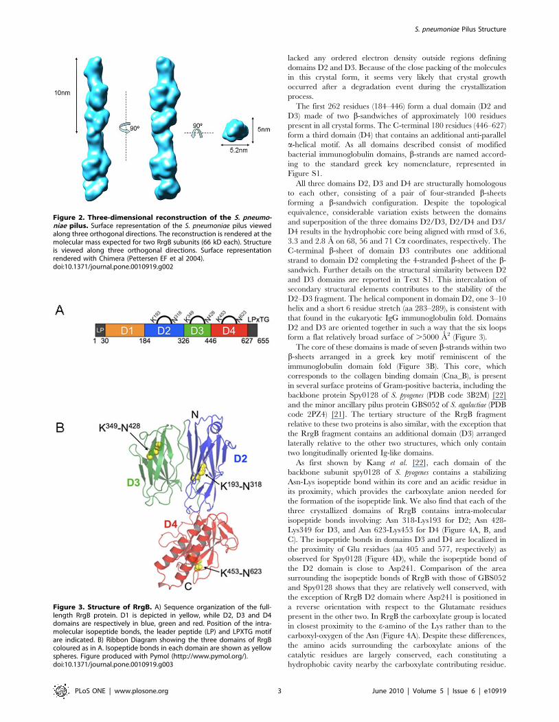
Structure of major pneumococcal pilin RrgB fragmentAttempts to obtain full-length RrgB crystals were made with no
success. Crystallisable construct of fragments of RrgB were derived
from limited proteolysis and mass spectroscopy experiments (see
Materials and Methods construct design for details). The fragment
of RrgB designated RrgBD2-D4 (residues 184–627, Figure 3A)
was crystallized in three different crystal forms (Table 1) (in space
groups P212121, P6122 and C2221). All the three forms of
RrgBD2–D4 possess identical tertiary structure and all exhibited
root mean square deviations (rmsds) with each other on aligned
carbon alpha atoms of less than 7 A (Table S1). Due to this,
reference to the RrgBD2–D4 will be made to the orthorhombic
crystal structure only, the coordinate set used in the analysis.
Each RrgBD2–D4 crystal form contained one molecule in the
asymmetric unit, consisting of an elongated polypeptide (84 A in
length, 50 A in width) made of three immunoglobulin-like
domains (named D2, D3 and D4).
A further crystal form designated RrgBD2–D3 (residues 141–
592) was also derived via the construct design process. Crystals
grown from these constructs belong to the monoclinic (space group
C2) crystal form and diffracted to high resolution (Table 1) but
Figure 1. Negative stain EM of purified S. pneumoniae pili.(A) The micrograph shows a network of elongated thin structures indicated by whitearrows. (B) Set of raw images of purified S. pneumoniae pili. (C) Representative class averages of purified S. pneumoniae pili segments. (D) Re-projection of the 3D reconstruction of the pilus. Scale bar in panel A 20 nm, scale bar in panels B, C and D 10 nm.doi:10.1371/journal.pone.0010919.g001
S. pneumoniae Pilus Structure
PLoS ONE | www.plosone.org 2 June 2010 | Volume 5 | Issue 6 | e10919
lacked any ordered electron density outside regions defining
domains D2 and D3. Because of the close packing of the molecules
in this crystal form, it seems very likely that crystal growth
occurred after a degradation event during the crystallization
process.
The first 262 residues (184–446) form a dual domain (D2 and
D3) made of two b-sandwiches of approximately 100 residues
present in all crystal forms. The C-terminal 180 residues (446–627)
form a third domain (D4) that contains an additional anti-parallel
a-helical motif. As all domains described consist of modified
bacterial immunoglobulin domains, b-strands are named accord-
ing to the standard greek key nomenclature, represented in
Figure S1.
All three domains D2, D3 and D4 are structurally homologous
to each other, consisting of a pair of four-stranded b-sheets
forming a b-sandwich configuration. Despite the topological
equivalence, considerable variation exists between the domains
and superposition of the three domains D2/D3, D2/D4 and D3/
D4 results in the hydrophobic core being aligned with rmsd of 3.6,
3.3 and 2.8 A on 68, 56 and 71 Ca coordinates, respectively. The
C-terminal b-sheet of domain D3 contributes one additional
strand to domain D2 completing the 4-stranded b-sheet of the b-
sandwich. Further details on the structural similarity between D2
and D3 domains are reported in Text S1. This intercalation of
secondary structural elements contributes to the stability of the
D2–D3 fragment. The helical component in domain D2, one 3–10
helix and a short 6 residue stretch (aa 283–289), is consistent with
that found in the eukaryotic IgG immunoglobulin fold. Domains
D2 and D3 are oriented together in such a way that the six loops
form a flat relatively broad surface of .5000 A2 (Figure 3).
The core of these domains is made of seven b-strands within two
b-sheets arranged in a greek key motif reminiscent of the
immunoglobulin domain fold (Figure 3B). This core, which
corresponds to the collagen binding domain (Cna_B), is present
in several surface proteins of Gram-positive bacteria, including the
backbone protein Spy0128 of S. pyogenes (PDB code 3B2M) [22]
and the minor ancillary pilus protein GBS052 of S. agalactiae (PDB
code 2PZ4) [21]. The tertiary structure of the RrgB fragment
relative to these two proteins is also similar, with the exception that
the RrgB fragment contains an additional domain (D3) arranged
laterally relative to the other two structures, which only contain
two longitudinally oriented Ig-like domains.
As first shown by Kang et al. [22], each domain of the
backbone subunit spy0128 of S. pyogenes contains a stabilizing
Asn-Lys isopeptide bond within its core and an acidic residue in
its proximity, which provides the carboxylate anion needed for
the formation of the isopeptide link. We also find that each of the
three crystallized domains of RrgB contains intra-molecular
isopeptide bonds involving: Asn 318-Lys193 for D2; Asn 428-
Lys349 for D3, and Asn 623-Lys453 for D4 (Figure 4A, B, and
C). The isopeptide bonds in domains D3 and D4 are localized in
the proximity of Glu residues (aa 405 and 577, respectively) as
observed for Spy0128 (Figure 4D), while the isopeptide bond of
the D2 domain is close to Asp241. Comparison of the area
surrounding the isopeptide bonds of RrgB with those of GBS052
and Spy0128 shows that they are relatively well conserved, with
the exception of RrgB D2 domain where Asp241 is positioned in
a reverse orientation with respect to the Glutamate residues
present in the other two. In RrgB the carboxylate group is located
in closest proximity to the e-amino of the Lys rather than to the
carboxyl-oxygen of the Asn (Figure 4A). Despite these differences,
the amino acids surrounding the carboxylate anions of the
catalytic residues are largely conserved, each constituting a
hydrophobic cavity nearby the carboxylate contributing residue.
Figure 2. Three-dimensional reconstruction of the S. pneumo-niae pilus. Surface representation of the S. pneumoniae pilus viewedalong three orthogonal directions. The reconstruction is rendered at themolecular mass expected for two RrgB subunits (66 kD each). Structureis viewed along three orthogonal directions. Surface representationrendered with Chimera (Pettersen EF et al 2004).doi:10.1371/journal.pone.0010919.g002
Figure 3. Structure of RrgB. A) Sequence organization of the full-length RrgB protein. D1 is depicted in yellow, while D2, D3 and D4domains are respectively in blue, green and red. Position of the intra-molecular isopeptide bonds, the leader peptide (LP) and LPXTG motifare indicated. B) Ribbon Diagram showing the three domains of RrgBcoloured as in A. Isopeptide bonds in each domain are shown as yellowspheres. Figure produced with Pymol (http://www.pymol.org/).doi:10.1371/journal.pone.0010919.g003
S. pneumoniae Pilus Structure
PLoS ONE | www.plosone.org 3 June 2010 | Volume 5 | Issue 6 | e10919

In domain D2, Asp241 is surrounded by Phe277, Phe249, Ile300,
Ile224 and Val230; in domain D3 Glu405 is surrounded by
Val426, Phe367, Ala365 and Ile408; finally in domain D4
Glu577 is surrounded by Leu587, Phe466, Phe563, Phe451 and
Ala464 (Figure 4A, B and C).
The presence and nature of the isopeptide bonds in D2, D3 and
D4 were also supported by mass spectrometry data (Texts S2 and
S3 and Figure S2).
Gram positive pili contain different types of backbonesubunits
Sequence comparison was performed between RrgB and pilus
backbone proteins of other streptococcal pili. Significant
similarity resulted between RrgB and the backbone proteins of
S. pyogenes and S. agalactiae (Figure 5). In particular RrgB is more
similar to backbone subunits of S. agalactiae pilus islands 1 and 2a
[28] and to serotype M4 S. pyogenes backbone protein Spy0116
[11], where the level of sequence conservation ranges from 36 to
44% of sequence identity. With only one exception, the residues
involved in the formation of the intra-molecular isopeptide
bonds present in D2, D3 and D4 are also conserved in backbone
proteins belonging to pilus island 1 and 2a of GBS and in M4
GAS Spy0116 (Figure 5A), indicating that these subunits might
also share a similar global folding with RrgB. In contrast, the
overall similarity between RrgB and the pilus backbone of pilus
island 2b of GBS and most GAS pilus backbone proteins
(including the crystallized spy0128) revealed only a very limited
sequence conservation (data not shown). In line with this, while
RrgB consists of four independently folded domains, spy0128
has only two distinct domains and is much shorter in length
(665aa of RrgB versus 340 aa of spy0128). These diversities
suggest that Gram-positive pili can adopt a similar overall
architecture despite using different types of molecules as major
building blocks.
Table 1. Crystal forms of RrgBD2–D4 and of RrgBD2–D3 fragments.
RrgB-184-627 RrgB-184-627 RrgB-184-627 RrgB-187-448
Data collection
PDB ID 2X9W 2X9X 2X9Y 2X9Z
Spacegroup P212121 P6122 C2221 C2
No. Mol. in ASU 1 1 1 1
Unit Cell (A) a/b/c 67.36/74.06/104.46 142.56/142.56/89.40 43.67/157.75/147.67 94.97/46.63/64.38 b= 115.48
Wavelength (A) 0.9795 0.9795 1.54 0.9795
Resolution (A) 50-1.9 50-1.5 50.0-2.3 50.0-1.3
Total Reflections unique,(no observations)
40672 (529986) 85582 (812884) 21514 (135832) 61593 (192302)
Completeness %(highest shell)
99.4 (89.3) 99.7 (98.3) 96.3 (86.3) 98.0 (90.7)
Rmerge, % (highest shell) 0.071 (0.93) 0.062 (0.91) 0.2 (0.705) 0.09 (0.71)
Highest Res. Shell, (A) 1.95-1.92 1.55-1.5 2.38-2.34 1.32-1.3
Mean I/s (I), (highest shell) 26.0 (1.4) 35.9(1.28) 8.7 (1.3) 24.6 (1.0)
Mean Redundancy(highest shell)
13.0 (5.8) 9.5 (4.5) 6.3 (4.1) 3.3 (1.8)
Phasing
Mean Figure of meritfor resolution range (A)
0.2 (50.0 2.3) - - -
Refinement
No. refs. working set 70612 85061 21265 56542
No. refs. test set 3579 4253 1092 2866
Rcryst (Rfree*){ 0.17 (0.23) 0.168 (0.2) 0.2 (0.25) 0.15 (0.17)
Rmsd bonds, A 0.012 0.004 0.004 0.005
Rmsd angles, u 1.294 0.849 0.764 1.033
Average B, A2 29.9 20 35.3 16.2
Maximum Likelihoodbased on Rfree, A{
0.25 0.23 0.67 0.16
Ramachandran Plot
Most Favored, % 90.9 91.6 90.6 90.4
Additional Allowed, % 8.8 8.1 9.2 9.6
Generously Allowed, % 0.3 0.3 0.3 0.0
Disallowed, % 0.0 0.0 0.0 0.0
{Rfactor~PH
Pi
jIi Hð Þ{vIi Hð Þwj�P
H
Pi
Ii Hð Þ where Ii is the scaled intensity of the ith measurement, and ,Ii. is the mean intensity for that reflection.
*Rfree = as for Rfactor, but for 5.0% of the total reflections chosen at random and omitted from refinement.doi:10.1371/journal.pone.0010919.t001
S. pneumoniae Pilus Structure
PLoS ONE | www.plosone.org 4 June 2010 | Volume 5 | Issue 6 | e10919

Fitting of the RrgB D2-D4 crystal coordinates into thepilus density map
In order to investigate subunit arrangement and interactions in
the S. pneumoniae pilus, a rigid-body fitting of two RrgBD2–D4
crystal fragments into the electron density map was performed by
using CHIMERA [29] (Figure 6A). The C-terminal immunoglob-
ulin domain (D4) of the crystal structure matched well into the
smaller globular density present below the groove with the core of
seven b-strands placed internally the filament and the additional
anti-parallel a-helical motif exposed on the surface. The fitting
confirmed that pilus volume (,1556e3 A3) and dimensions (52 A
in width and 252 A in length) could accommodate two RrgBD2–
D4 molecules organized in a head-to-tail arrangement with
rotations ranging between 17u and 22u along the vertical axis of
the lower RrgBD2–D4 subunit in respect to the upper one. Here
both the flattened surface of the D2–D3 dual domain and the D4
Figure 4. Isopeptide Bonds of RrgB and Spy0128. A) and B) the isopeptide bond between residues Lys193 and Asn318 in domain 2 (D2) ofRrgB. Dashed lines show the distance of the carboxylate anion provided by Asp241; the isopeptide bond between Lys 349 and Asn428 in domain 3(D3) and the position of Glu405 contributing the carboxylate anion; the isopeptide bond between Lys 453 and Asn623 and the position of Glu577 indomain 4 (D4). C) Sigma-a weighted 2Fo–Fc map contoured at 1s level is shown for the isopeptide bond in D4 of RrgB. D) Isopeptide bond betweenLys36 and Asn168 in Spy0128 (drawn from PDB 3b2m). Carbon atoms are coloured green for residues involved in the formation of the isopeptidebonds, yellow for neighbouring hydrophobic residues, and gray for residues adjacent to the isopeptide bond. Oxygen and nitrogen atoms arecoloured red and blue, respectively. Figure produced by Pymol.doi:10.1371/journal.pone.0010919.g004
Figure 5. Sequence alignments of related pili proteins from Gram-positive bacteria. A) Sequence alignment showing intra-molecularisopeptide bond formation which involves conserved residues Lysine (K), Aspartic Acid (D), Glutamic Acid (E) and Asparagine (N). Residues arehighlighted and coloured according to the domain localization. UniProtKB/TrEMBL accession codes are shown; RrgB-TIGR4, RrgB-6BSP and RrgB-23FTW represent the sequences of the three RrgB alleles of S. pneumoniae; SAG0645 (also known as GBS80) belongs to PI-1 of Streptococcusagalactiae; GBS1477, SAL1486, SAG1407 and SAI1511 belong to GBS PI-2a, whereas SAK0776 belongs to GBS PI-2b; Spy0116 belongs to the M4serotype of Streptococcus pyogenes. The sequence alignment was obtained and edited by Jalview/ClustalW [55]. B) Cartoon representation of RrgBsingle domains coloured as in 1A) is shown on the bottom. Yellow spheres depict the residues forming the isopeptide bonds.doi:10.1371/journal.pone.0010919.g005
S. pneumoniae Pilus Structure
PLoS ONE | www.plosone.org 5 June 2010 | Volume 5 | Issue 6 | e10919

domain could interface with the D1 domain of the neighbouring
subunit. Furthermore the fitting suggested that the inter-subunit
density (7 A thick) could accommodate the 8-residue C-terminal
tail not present in the RrgB crystal. Notably, no information
about the neighbouring subunits or the missing residues in the
truncated pilin model was introduced into the fitting at any
stage. Therefore, the packing of the 84 A long molecule within
the 60 A diameter EM density without significant collision
independently validates the EM reconstruction and docking
procedures.
Moreover, the rigid body fitting performed with the electron
density map countered at the same threshold levels that
corresponded to the molecular mass of two RrgB subunits
(,132 kD total mass) showed that the two RrgBD2–D4 crystal
structures occupied a total volume of ,1176e3 A3 leaving two
regions of unoccupied volume (,216e3 A3) in the density map.
The two remaining extra unoccupied volumes, both present above
the N-terminus of each fitted RrgBD2–D4 crystal fragment
(Figure 6A), easily accommodated the volume of two computer
modelled D1 domains (RrgBD1) (Text S4, Figure S3A), each
containing 156 residues. Each D1 computer model was first fitted
manually using CHIMERA by placing as much of the atomic
structure as possible fully into the EM density map, approximately
in the position thought to be correct. This step was then followed
by a rigid body fitting using CHIMERA and optimized, as
previously, for the spatial frequency band of 22–60 A (Figure 6B).
The low correlation values (,0.55) and the absence of clashes
were both indications of the correct orientation of the D1
computer models into the 3D pilus density in respect to the
RrgBD2–D4 crystal fragment orientation (Figure S3B). Finally,
the packing of the RrgB subunits within the EM density was the
only one validating the EM reconstruction. Any attempt of fitting
done with the computer models of the two ancillary proteins at the
spatial frequency band of 22–60 A did not satisfy the spatial
restrains and generated a too high level of collisions.
Discussion
Fibrillar structures have been recently found in Gram-positive
bacteria complementing the wide range of Gram-negative
pathogens that since long have been known to express pili on
their surface. Pili were first observed in the Gram-positive species
Corynebacterium renale by electron microscopy, than followed by their
detection on the surface of other Gram-positive bacteria such as
Corynebacterium diphtheriae, Streptococcus salivarius and Streptococcus
sanguis. Recently these elongated appendages have been found
on the surface of the principal streptococcal pathogens including
Group A streptococci, Group B streptococci and S. pneumoniae. In
addition to colonization and adhesion, Gram-positive pili have
also been associated to other functions among which biofilm
formation and immune evasion [15,17,30]. Pilus subunits are
immunogenic in humans [13] and able to elicit a protective
response when tested in mouse models of infection [12,31]. Pilus
expression increases pathogenicity in animal models [32,33], and
enhances adhesion to epithelial cells [14,34,35].
To accurately define the structure and assembly mechanism of
the pneumococcal pilus we determined both a low-dose EM
reconstruction of the pilus filament and a high resolution crystal
structure of the backbone subunit.
Here the crystal structure of the RrgBD2–D4 backbone subunit
fitted into the EM reconstruction of the S. pneumoniae pilus reveals
for the first time the polymeric architecture of a Gram-positive
pilus indicating a head-to-tail organization of individual backbone
subunits. The measured unoccupied volume present between two
neighbouring subunits is compatible with the predicted density of
the flexible 8-residue C-terminus sequence 624KKVTIPQT631
Figure 6. Fitting of the RrgBD2–D4 crystal structure and of the RrgBD1 computer model into the 3D map of the S. pneumoniae pilus.A) Semi transparent rendered surface representations of the 3-D map of the S. pneumoniae pilus viewed along the z-axis tilted by 90u showing thedocking of two copies of the atomic resolution RrgBD2–D4 subunit into the pilus reconstruction. Empty densities present above the two fittedRrgBD2–D4 copies have a volume of ,216e3 A3 each. B) Same semi-transparent rendered surface representations as in panel A showing the dockinginto the pilus reconstruction of two copies of the RrgBD2–D4 subunit and contemporaneously of two computer model RrgBD1 domains. The dockingreveals that the inter-subunit density (7 A thick) present between the upper and lower subunits could accommodate the 8-residue C- terminal tail,not present in the RrgBD2–D4 crystal. Crystal structures are in cartoon representation and the three domains are coloured following thenomenclature of Figure 3. Figure produced with Pymol (http://www.pymol.org/). Surface representation and molecule rendered with Chimera [29].doi:10.1371/journal.pone.0010919.g006
S. pneumoniae Pilus Structure
PLoS ONE | www.plosone.org 6 June 2010 | Volume 5 | Issue 6 | e10919

that is missing from the crystal structure. This region contains
Thr631, implicated in the formation of the inter-molecular
isopeptide bond that links the C-terminus of one subunit to the
N-terminal region of the next one in the row. Sequence analysis of
RrgB indicates the presence of two lysines potentially implicated in
the isopeptide bond formation: i) Lys183 which is part of a
canonical pilin motif (180VYPKN184); ii) Lys162 which can be
nicely aligned with Lys161 of Spy0128, previously identified by
Kang et al. [22]. When mapped onto the predicted model of the
RrgBD1 domain, both residues were well exposed and located on
the same face of the molecule. However, according to the model,
only Lys162 could be close enough to the presumed position of the
LPXTG motif of the neighbouring RrgB molecule to be involved
in the formation of an inter-molecular isopeptide bond. Never-
theless, experimental evidence is still needed to discriminate the
essential Lys.
The analysis of the rigid body fitting indicates that not only the
surface-surface interaction between neighbouring subunits but also
the presence of the inter-molecular isopeptide bond constrains the
flexibility of the pilus. The limited curviness observed in the pili
could be conferred by the hinge region of 2–4 residues which links
D1 to D2 in each individual subunit. This internal flexibility is
suggested also by the proteolysis experiments where the presence
of a proteolytic cleavage site between D1 and D2 indicates that a
mobile loop connects the two domains.
Another important aspect of pilus biogenesis is the understand-
ing of how the ancillary proteins are incorporated into the pilus
backbone. Originally, two distinct mechanisms were hypothesized.
The first assumed that the ancillary proteins are incorporated in
the pilus shaft in a similar manner as the backbone subunits, either
interspersed between the backbone subunits or located at the
extremities of the fiber. The second one sustains that the ancillary
proteins are associated laterally to the pilus shaft generating a
branched structure.
Previous reports [36] [19] [37] [20] showed by Immunogold
EM that RrgA and RrgC were distributed in clusters along pili
when organized in bundles. The single pilus structure presented in
this work and in Hilleringmann et al. [56] clearly show that the
pilus shaft is made by multiple copies of RrgB organized in a head-
to-tail linear structure and with the two opposite tips decorated by
the two ancillary proteins. Thus the presence of the ancillary
proteins clusters observed along the bundles could be a
consequence of the disposition of single pili along the bundle.
Recent works on GBS and C. diphtheriae have suggested that the
minor pilin may anchor the pili on the cell wall [30] [38], whereas
another recent paper shows that in GAS the major ancillary
protein is only attached at the tip of the fiber, consistently with its
role in adhesion [39]. These data are in agreement with our
observations that in S. pneumoniae the ancillary proteins are not
appended laterally, conferring to the pilus a pearl on a string
appearance of identical subunits bound to each other. Moreover
rigid-body fitting clearly indicates that the pilus density can
correspond only to a linear assembly of RrgB monomers,
excluding that other molecules, apart from RrgB, are incorporated
into the pilus shaft or appended laterally. Therefore, as in the case
of C. diphtheriae the most probable scenario for the pilus of S.
pneumoniae is the one that contemplate the presence of the major
ancillary protein RrgA at the distal tip where it could be more
available for adhesion, whereas the RrgB backbone provides the
structure with the elasticity required to reach the host cell
receptors.
The work described above shows the powerful synergy and
mechanistic insights that can result from a combined EM, X-ray
crystallography and Mass Spectrometry approach. The three-
dimensional structure of the pilus generated from TEM images
fitted with high resolution crystal structure of the major fragment
of RrgB have provided a detailed molecular view of the backbone
of S. pneumoniae pilus, and could be a key-model for the study of the
assembly, attachment and function of the pili in Gram-positive
bacteria.
Materials and Methods
Bacterial strains and culture conditionsS. pneumoniae type 4 strain TIGR4 was employed [40]. The
pneumococcal strains were stored at –80uC in 12% glycerol and
routinely grown at 37uC in 5% CO2 on Tryptic Soy Agar (Becton
Dickinson) supplemented with 5% defibrinated sheep blood or in
Tryptic Soy Broth (Becton Dickinson). When appropriate,
erythromycin and kanamycin (Sigma-Aldrich) as selection marker
were used.
Native TIGR4 pili purificationS. pneumoniae TIGR4 strain was chosen as starting material as
the bacteria belong to a clinical relevant serotype 4 isolate, the
sequence of which is known [40].
Native pili of TIGR4 and TIGR4DrrgA were purified essentially
according a protocol described by Hilleringmann et al. [37].
Purified pili fractions were judged to be homogeneous based on
electron microscopy and SDS-PAGE. Samples were stored at –
80uC or liquid nitrogen until further use.
Electron microscopyA 5 ml aliquot of purified pili preparation with a final
concentration of 0.052 mg/ ml was applied to 200-square mesh
copper grids coated with a thin carbon film and let stand for
5 min. Excess of solution was blotted by Whatman filter paper.
The grids were first washed by streaming several drops of PBS
over the grids. They were subsequently negatively stained by two
drops of 1% buffered PTA (pH 6.5). The last drop was left on the
grids for 17 s. Finally the grids were washed with several drops of
ddH2O, the excess of liquid was soaked off by Whatman filter
paper and air dried. The grids were observed using a CM200 FEG
Philips Electron Microscope (FEI, Eindhoven, The Netherlands),
equipped with a GATAN GIF 2002 post column energy filter
(Gatan, Pleasanton, California, United States). All images were
collected at an accelerating voltage of 200 kV and a nominal
magnification of 500006, on Kodak SO163 film. Micrographs
where checked for astigmatism and drift on an optical diffractom-
eter prior to digitisation.
Image processingMicrographs taken at 500006 of magnification were digitized
on an IMACON 949 scanner at spacing of 7.95 mm resulting in a
nominal sampling of 1.6 A/pixel-1. Analysis of defocus and
Contrast Transfer Function (CTF) using the Medical Research
Council (MRC) program CTFFIND3 [41] and IMAGIC 5 [26]
showed that the first zero corresponds to ,17–19 A. Since only a
moderate resolution of the 3-D reconstruction of the S. pneumoniae
pilus was required in order to identify the arrangement of the
backbone subunits, the final 3-D map was obtained at 22 A
resolution using the 0.5 threshold of the Fourier shell correlation
(FSC) [27]. Pili segments were picked manually from digitized
images using the command ‘‘helixboxer’’ from the software
EMAN [42]. Digitized pili images were cut into individual repeats
by using boxes of 1646164 pixels, with overlapping ends, using 10
pixel shift for each box, so that adjacent boxes had 90% overlap.
Images were band-pass filtered at 17–200 A to remove back-
S. pneumoniae Pilus Structure
PLoS ONE | www.plosone.org 7 June 2010 | Volume 5 | Issue 6 | e10919
ground and normalized. The individual pili segments were treated
as single particles. In a first analysis, the segments were selected
and pre-aligned interactively, subsequently the pre-aligned repeats
were aligned using alignments with only limited angular ranges
(25u, +5u), finally a vertical alignment has been performed using
as a future-less reference the projection of a model cylinder, with a
5 nm width that corresponds to the width of the pilus measured in
the images, followed by translational alignment perpendicular to
the cylinder axis only. All the aligned and filtered images were
consistent: they all presented centred rods with similar diameters.
The only major differences were the surrounding stain distribu-
tions. Aligned pili segments were than classified by MSA to sort
images into class averages with similar features. The class averages
obtained have an improved signal-to-noise ratio and represent
characteristic molecular views of the pilus. Most class averages
showed pili with subunit-like features. Several iterations of
alignments and MSA classification led to homogeneous class
averages showing pili with globular subunits arranged linearly.
The initial model was determined from four side views of the pilus
and one end view [43]. In a first approximation, the end view was
taken as rotationally symmetrised average. The 3-D map was than
refined by adding class averages of the side views and a
reprojection along the z-axis as the end view. Re-projections of
the final 3-D were compared for consistency with input class
averages to check the accuracy of the Euler angles assigned [23].
Image processing of the pilus was performed using software
IMAGIC-5 [26]. The final 3-D map of the S. pneumoniae pilus was
refined at 22 A resolution (FSC = 0.5) [27] by iterating procedures
of alignment and classification. 3D rendered surface representa-
tions were visualized in UCSF Chimera [29].
Construct DesignFull length RrgB (aa1-665) was expressed and purified in E.coli
strain HK100, according to standard protocols [44]. The resultant
protein was purified by Ni-NTA IMAC (Quiagen) and eluted by a
HEPES elution buffer, Buffer A (20 mM HEPES pH 7.3,
150 mM NaCl, 1 mM TCEP). 80 ml TPCK-Trypsin (Pierce)
was washed three times with 400 ml Buffer A, and then
resuspended in 800 ml of the same buffer. 25 ml of the TPCK-
Trypsin suspension was added to 50 ml protein (0.5–1.5 mg/ml)
and incubated at 37uC, 250 rpm for 4 hours. The immobilized
trypsin was removed by centrifugation and the proteolyzed
samples were then submitted for LCMS. Mass spec data were
analyzed by PAWS (Genomic Solutions Inc.) to determine possible
truncation boundaries. The resultant constructs were cloned in the
pSpeedET vector by the PIPE cloning method [45]. Positive
clones were verified by DNA sequence analysis, and expressed in
an identical way to the full length construct [46].In all, ten
constructs were generated via this methodology with boundaries,
24–227, 52–460, 109–562, 139–590,141–592, 184–627,163–
615,191–337 and 281–484 of the full length RrgB construct.
CrystallizationAll crystallization experiments were carried out in 96 well low
profile Greiner crystallization plates in a nanodroplet sitting drop
vapour diffusion format with 480 conditions screens performed at
both 4 and 20u [46]. Equal volumes of protein concentrated to
10 mg/ml were added to the reservoir solutions to create a total
drop volume of 500 nl. Three crystal forms of the RrgBD2–D4
(containing residues 184–627) constructs were produced belonging
to spacegroups P212121, P6122 and C2221 in conditions 0.05
potassium dihydrogen phosphate, 20% PEG-8000 pH 4.5 4uC,
1.0 M Sodium Citrate 0.1 M Imidazole pH 8.0 at 20uC and
0.2 M Lithium Sulfate, 30% Peg-4000 0.1 M Tris pH 8.5 4uC
respectively. The other constructs which ultimately produced a
two domain version of the structures RrgBD2–D3, (containing
residues 141–592 of full length RrgB) were produced with identical
crystallization screens in crystal condition 30% PEG-6000 0.1 M
Citrate pH 5.0 at 20uC. All crystals were mounted using 20%
glycerol as a cryo-protectant prior to cooling to 100uK for data
collection.
Data Collection and Structure SolutionData were collected at beamline 5.0.2 and 5.0.3 of the ALS and
were processed with the HKL2000 package [47] . Data collection
for phasing was performed on the orthorhombic crystal form of
RrgB184–627 at the Selenium edge with an inverse beam strategy.
Substructure solution, phasing, density modification and initial
model building was performed with SOLVE and RESOLVE
[48,49] on the primitive orthorhombic crystal form. Given that
only one Seleno-Methionine residue existed in the 441 residues
present in the asymmetric unit, the anomalous signal was relatively
weak (DF/sDF,1.2 on all data between 50 and 2.3 A) but
implementation of a brute force searching strategy over various
resolution ranges and redundancies resulted in the location of the
substructure and initial phases, capable of building the model.
Subsequent refinement and building was performed with Phenix
and Coot [50,51]. All other crystallographic manipulations were
carried out with the CCP4 package [52]. Solutions of all other
crystal forms were performed by molecular replacement using the
orthorhombic crystal form as a search model and Phaser [53]
followed by refinement and building with Phenix and Coot
[50,54]. The geometry of all structures is excellent and all residues
are in allowed regions of the Ramachandran plot (Table 1).
Fitting of the X-ray coordinates into the electron densitymap
The fitting was carried out independently for two individual
RrgBD2–D4 crystals and optimized for the spatial frequency band
of 22–60 A. The correlation values between the fitted atomic
structures of two copies of RrgBD2–D4 and the 3-D map
corresponding to the upper and lower subunits of the pilus
reconstruction were both 0.66. All other orientations of two
subunits into the 3-D map resulted in lower correlation values
(,0.5 for each single subunit). Moreover, the alternative checked
orientations did not satisfy the spatial restrains on the distance
between neighbouring subunits. Alternative fittings of two adjacent
subunits had tilts and rotations that increased the distance between
the N-terminus of one fragment and the C-terminus of the next
one. Basically the first two modified immunoglobulin domains D2
and D3 of the crystal structure aligned well with the larger
globular density present in the pilus reconstruction, placing the
pair of four-stranded b-sheets parallel to the filament axis and
facing outward.
Supporting Information
Figure S1 Structure of RrgB. A) Ribbon Diagram showing the
three domains of RrgB coloured as in Figure 3B. Intra-isopeptide
bonds in each domain are shown as yellow spheres. Figure
produced with Pymol (http://www.pymol.org/). Greek-key rep-
resentation of the secondary structure organization of RrgB D2-
D4 domains (panel B) and the prototypic immunoglobulin fold
(panel C).
Found at: doi:10.1371/journal.pone.0010919.s001 (3.08 MB TIF)
Figure S2 Peptide mass fingerprinting of RrgB Domain 4. Each
signal is labelled with an m/z ratio and the amino acid position of
S. pneumoniae Pilus Structure
PLoS ONE | www.plosone.org 8 June 2010 | Volume 5 | Issue 6 | e10919

the corresponding tryptic peptide. A) Signals labelled in red are
consistent with peptides linked by an isopeptide bound between
Lys453 and Asp623. The signal at m/z 1675.94 is consistent with
one trypsin missed cleavage while the signal at m/z 1290.74 is
consistent with no missed cleavage. Signal labelled with an asterisk
is consistent with the tryptic N-terminal and C-terminal peptide of
the cloned D4 domain of sequence MASVTYGK (m/z 856.50)
and ITLEHHHHHH (m/z 1297.64), respectively. Signal labelled
with a T is generated by trypsin autolysis products. B) MS/MS
spectrum of parental ion at m/z 1675.94. Only b and y series are
reported. A scheme of the fragmentation is shown at the top of the
figure.
Found at: doi:10.1371/journal.pone.0010919.s002 (3.34 MB TIF)
Figure S3 Fitting of the RrgBD1 computer model into the 3D
map of the S. pneumoniae pilus. A) Computer model of the D1
domain and sequence alignment of RrgB D1 to the N-terminal
domain of Spy0128 (PDB code 3B2M). Gold rectangles depict the
localization of beta strands on the crystal structure. B) The overall
protein fold is represented as a ribbon; the side-chains of Lys41
and Asn184, involved in the intra-molecular isopeptide bond are
highlighted. Lys162, with putative involvement in the inter-
molecular isopeptide bond is evidenced in grey. B) After fitting, D1
and D2-D4 coordinates were merged into a single file and
overlapping atoms were removed. The resulting RrgBD1-D4
model was visually inspected for absence of steric conflicts and
minimized with the same protocol used for D1. Threading was
performed with SwissPDBViewer, surface representation and
molecule rendered with Chimera. Crystal structures are in cartoon
representation and the three domains are coloured following the
nomenclature of Figure 3. Figure produced with Pymol (http://
www.pymol.org/). Surface representation and molecule rendered
with Chimera.
Found at: doi:10.1371/journal.pone.0010919.s003 (10.23 MB
TIF)
Table S1 Root mean square deviations (rmsd) of the three
crystal forms of RrgBD2-D4.
Found at: doi:10.1371/journal.pone.0010919.s004 (0.03 MB
DOC)
Text S1 Structural relationship between D2 and D3 domains.
Definition of the reciprocal orientation of domains D2 and D3.
Found at: doi:10.1371/journal.pone.0010919.s005 (0.03 MB
DOC)
Text S2 Presence of intra-molecular isopeptide bonds in D2, D3
and D4 are confirmed by MS. Details on the characterization of
isopeptide bonds.
Found at: doi:10.1371/journal.pone.0010919.s006 (0.03 MB
DOC)
Text S3 In-gel digestion and mass spectrometric analysis. Details
on mass spectroscopy analysis.
Found at: doi:10.1371/journal.pone.0010919.s007 (0.03 MB
DOC)
Text S4 Computer modelling of the RrgBD1 domain. Details on
the generation of the D1 computer model.
Found at: doi:10.1371/journal.pone.0010919.s008 (0.03 MB
DOC)
Acknowledgments
The authors wish to thank Peter Schultz for continued support. The work
in this paper is based on experiments conducted at beamline 5.0.3 of the
advanced light source (ALS). The ALS is supported by the Director, Office
of Science, Office of Basic Energy Sciences, Material Sciences Division of
the U.S. Department of Energy under contract No. DE-AC03-76SF00098
at Lawrence Berkeley National Laboratory. The Department of Evolu-
tionary Biology of the University of Siena, Italy (Prof. Dallai R.) for the use
of microscopy facilities and for all the valuable help.
The Structures have been deposited in the Protein Databank RrgBD2–D4
(PDB code 2x9w (P212121), 2x9x (P6122), 2x9y (C2221)) RrgBD3–D4 2x9z
(C2). The EM density map for the S. pneumoniae pilus is available in the
Electron Microscopy Data Bank (http://www.ebi.ac.uk/pdbe).
Author Contributions
Conceived and designed the experiments: MS MB RR SAL AC VM IF.
Performed the experiments: GS EK EM MB NN CE FG MH IF.
Analyzed the data: GS MS EM MB NN IF. Contributed reagents/
materials/analysis tools: IF. Wrote the paper: GS MS NN IF. Global Head
of Systems Biology for Novartis Vaccines and Diagnostics: AC. Global
Head of Vaccine Reasearch for Novartis Vaccines and Diagnostics: RR.
Project Leader: VM.
References
1. Kallstrom H, Islam M, Berggren PO, Jonsson AB (1998) Cell signaling bythe type IV pili of pathogenic Neisseria. Biol Chem 273: 21777–
21782.
2. Telford JL, Barocchi MA, Margarit I, Rappuoli R, Grandi G (2006) Pili inGram-positive pathogens. 4: 509–519.
3. Elena SF, Whittam TS, Winkworth CL, Riley MA, Lenski RE (2005) Genomic
divergence of Escherichia coli strains: evidence for horizontal transfer andvariation in mutation rates. Int Microbiol 8: 271–278.
4. Mattick JS (2002) Type IV pili and twitching motility. Annu Rev Microbiol 56:
289–314.
5. Wenyuan S, Hong S (2002) Type IV Pilus-Dependent Motility and Its Possible
Role in Bacterial Pathogenesis. Infection and Immunity 70: 1–4.
6. Abe S, Saito T, Koga T, Ono E, Yanagawa R, et al. (1990) Cloning andexpression of a pili gene of Corynebacterium renale in Escherichia coli. Nippon
Juigaku Zasshi 52: 11–18.
7. Mishra A, Das A, Cisar JO, Ton-That H (2007) Sortase-catalyzed assembly ofdistinct heteromeric fimbriae in Actinomyces naeslundii. J Bacteriol 189:
3156–3165.
8. Ton-That H, Schneewind O (2003) Assembly of pili on the surface of
Corynebacterium diphtheriae. Mol Microbiol 50: 1429–1438.
9. Gaspar AH, Ton-That H (2006) Assembly of distinct pilus structures onthe surface of Corynebacterium diphtheriae. J Bacteriol 188: 1526–
1533.
10. Lauer P, Rinaudo CD, Soriani M, Margarit I, Maione D, et al. (2005) Genomeanalysis reveals pili in Group B Streptococcus. Science 309: 105.
11. Falugi F, Zingaretti C, Pinto V, Mariani M, Amodeo L, et al. (2008) Sequence
variation in group A Streptococcus pili and association of pilus backbone types
with lancefield T serotypes. J Infect Dis 198: 1834–1841.
12. Rosini R, Rinaudo CD, Soriani M, Lauer P, Mora M, et al. (2006) Identificationof novel genomic islands coding for antigenic pilus-like structures in
Streptococcus agalactiae. Mol Microbiol 61: 126–141.
13. Mora M, Bensi G, Capo S, Falugi F, Zingaretti C, et al. (2005) Group A
Streptococcus produce pilus-like structures containing protective antigens andLancefield T antigens. Proc Natl Acad Sci U S A 102: 15641–15646.
14. Barocchi MA, Ries J, Zogaj X, Hemsley C, Albiger B, et al. (2006) Apneumococcal pilus influences virulence and host inflammatory responses. Proc
Natl Acad Sci U S A 103: 2857–2862.
15. Konto-Ghiorghi Y, Mairey E, Mallet A, Dumenil G, Caliot E, et al. (2009) Dual
role for pilus in adherence to epithelial cells and biofilm formation inStreptococcus agalactiae. PLoS Pathog 5: e1000422.
16. Bagnoli F, Moschioni M, Donati C, Dimitrovska V, Ferlenghi I, et al. (2008) A
Second Pilus Type in Streptococcus pneumoniae Is Prevalent in Emerging
Serotypes and Mediates Adhesion to Host Cells . J Bacteriol 190: 5480–5492.
17. Proft T, Baker EN (2009) Pili in Gram-negative and Gram-positive bacteria -structure, assembly and their role in disease. Cell Mol Life Sci 66: 613–635.
18. Moschioni M, Donati C, Muzzi A, Masignani V, Censini S, et al. (2008)Streptococcus pneumoniae contains 3 rlrA pilus variants that are clonally
related. J Infect Dis 197: 888–896.
19. Nelson AL, Ries J, Bagnoli F, Dahlberg S, Falker S, et al. (2007) RrgA is a pilus-
associated adhesin in Streptococcus pneumoniae. Mol Microbiol 66: 329–340.
20. Falker S, Nelson AL, Morfeldt E, Jonas K, Hultenby K, et al. (2008) Sortase-
mediated assembly and surface topology of adhesive pneumococcal pili. MolMicrobiol 70: 595–607.
21. Krishnan V, Gaspar AH, Ye N, Mandlik A, Ton-That H, et al. (2007) An IgG-
like Domain in the Minor Pilin GBS52 of Streptococcus agalactiae Mediates
Lung Epithelial Cell Adhesion. Structure 15: 893–903.
S. pneumoniae Pilus Structure
PLoS ONE | www.plosone.org 9 June 2010 | Volume 5 | Issue 6 | e10919

22. Kang HJ, Coulibaly F, Clow F, Proft T, Baker EN (2007) Stabilizing Isopeptide
Bonds Revealed in Gram-Positive Bacterial Pilus Structure. Science 318:
1625–1628.
23. van Heel M, Gowen B, Matadeen R, Orlova EV, Finn R, et al. (2000) Single-
particle electron cryo-microscopy: towards atomic resolution. Q Rev Biophys 33:
307–369.
24. van Heel M (1984) Multivariate statistical classification of noisy images
(randomly oriented biological macromolecules). Ultramicroscopy 13: 165–183.
25. van Heel M (1987) Angular reconstitution: a posteriori assignment of projection
directions for 3D reconstruction. Ultramicroscopy 21: 111–123.
26. van Heel M, Harauz G, Orlova EV, Schmidt R, Schatz M (1996) A New
Generation of the IMAGIC Image Processing System. Journal of Structural
Biology 116: 17–24.
27. Harauz G, van Heel M (1986) Exact filters for general geometry three-
dimensional reconstruction. Optik 73: 146–156.
28. Margarit I, Rinaudo CD, Galeotti CL, Maione D, Ghezzo C, et al. (2009)
Preventing bacterial infections with pilus-based vaccines: the group B
streptococcus paradigm. J Infect Dis 199: 108–115.
29. Pettersen EF, Goddard TD, Huang CC, Couch GS, Greenblatt DM, et al.
(2004) UCSF Chimera-a visualization system for exploratory research and
analysis. J Comput Chem 25: 1605–1612.
30. Mandlik A, Swierczynski A, Das A, Ton-That H (2008) Pili in Gram-positive
bacteria: assembly, involvement in colonization and biofilm development.
Trends in Microbiology 16: 33–40.
31. Gianfaldoni C, Censini S, Hilleringmann M, Moschioni M, Facciotti C, et al.
(2007) Streptococcus pneumoniae pilus subunits protect mice against lethal
challenge. Infect Immun 75: 1059–1062.
32. Abbot E, Smith WD, Siou GP, Chiriboga C, Smith RJ, et al. (2007) Pili mediate
specific adhesion of Streptococcus pyogenes to human tonsil and skin. Cell
Microbiol 9: 1822–1833.
33. Hava DL, Camilli A (2002) Large-scale identification of serotype 4 Streptococ-
cus pneumoniae virulence factors. Mol Microbiol 45: 1389–1406.
34. Dramsi S, Caliot E, Bonne I, Guadagnini S, Prevost MC, et al. (2006) Assembly
and role of pili in group B streptococci. Mol Microbiol 60: 1401–1413.
35. Maisey HC, Hensler M, Nizet V, Doran KS (2007) Group B streptococcal pilus
proteins contribute to adherence to and invasion of brain microvascular
endothelial cells. J Bacteriol 189: 1464–1467.
36. LeMieux J, Hava DL, Basset A, Camilli A (2006) RrgA and RrgB Are
Components of a Multisubunit Pilus Encoded by the Streptococcus pneumoniae
rlrA Pathogenicity Islet. Infect Immun 74: 2453–2456.
37. Hilleringmann M, Giusti F, Baudner BC, Masignani V, Covacci A, et al. (2008)
Pneumococcal pili are composed of protofilaments exposing adhesive clusters of
Rrg A. PLoS Pathog 4: e1000026.
38. Nobbs AH, Rosini R, Rinaudo CD, Maione D, Grandi G, et al. (2008) Sortase
A Utilizes an Ancillary Protein Anchor for Efficient Cell Wall Anchoring of Pili
in Streptococcus agalactiae. Infect Immun 76: 3550–3560.
39. Quigley BR, Zahner D, Hatkoff M, Thanassi DG, Scott JR (2009) Linkage of T3
and Cpa pilins in the Streptococcus pyogenes M3 pilus. Mol Microbiol 72:1379–1394.
40. Tettelin H, Nelson KE, Paulsen IT, Eisen JA, Read TD, et al. (2001) Complete
genome sequence of a virulent isolate of Streptococcus pneumoniae. Science293: 498–506.
41. Mindell JA, Grigorieff N (2003) Accurate determination of local defocus andspecimen tilt in electron microscopy. Journal of Structural Biology 142:
334–347.
42. Ludtke SJ, Baldwin PR, Chiu W (1999) EMAN: Semiautomated Software forHigh-Resolution Single-Particle Reconstructions. Journal of Structural Biology
128: 82–97.43. Salih O, Remaut H, Waksman G, Orlova EV (2008) Structural Analysis of the
Saf Pilus by Electron Microscopy and Image Processing. Journal of MolecularBiology 379: 174–187.
44. Lesley S, Kuhn P, Godzik A, Deacon AM, Mathews I, et al. (2002) Structural
genomics of the Thermotoga maritima proteome implemented in a high-throughput structure determination pipeline. Proc Natl Acad Sci U S A 99:
11664–11669.45. Klock HE, Koesema EJ, Knuth MW, Lesley SA (2008) Combining the
polymerase incomplete primer extension method for cloning and mutagenesis
with microscreening to accelerate structural genomics efforts. Proteins 71:982–994.
46. Lesley M, Floyd J, Oermann M (2002) Use of MindMapper software forresearch domain mapping. Comput Inform Nurs 20: 229–235.
47. Otwinowski Z, Minor W (1997) Processing of X-ray diffraction data collected inoscillation mode. Meth Enzymol 276: 307–326.
48. Terwilliger T (2001) Maximum-likelihood density modification using pattern
recognition of structural motifs. Acta Crystallogr D Biol Crystallogr 57:1755–1762.
49. Terwilliger T (1999) Reciprocal-space solvent flattening. Acta Crystallogr D BiolCrystallogr 55: 1863–1871.
50. Emsley P, Cowtan K (2004) Coot: model-building tools for molecular graphics.
Acta Crystallogr D Biol Crystallogr 60: 2126–2132.51. Adams PD, Grosse-Kunstleve R, Hung LW, Ioerger TR, McCoy AJ, et al.
(2002) PHENIX: building new software for automated crystallographic structuredetermination. Acta Crystallogr D Biol Crystallogr 59: 1948–1954.
52. Collaborative Computational Project N (1994) The CCP4 suite: programs forprotein crystallography. Acta Crystallogr D Biol Crystallogr 50: 760–763.
53. Read R (2001) Pushing the boundaries of molecular replacement with maximum
likelihood. Acta Crystallogr D Biol Crystallogr 57: 1373–1382.54. Brunger A (1992) Free R value: a novel statistical quantity for assessing the
accuracy of crystal structures. Nature 355: 472–475.55. Clamp M, Cuff J, Searle SM, Barton GJ (2004) The Jalview Java alignment
editor. Bioinformatics 20: 426–427.
56. Hilleringmann M, Ringler P, Muller SA, De Angelis G, Rappuoli R, et al. (2009)Molecular architecture of Streptococcus pneumoniae TIGR4 pili. EMBO J 16:
28 (24): 3921–30.
S. pneumoniae Pilus Structure
PLoS ONE | www.plosone.org 10 June 2010 | Volume 5 | Issue 6 | e10919