Chlamydia pneumoniae infections in mouse models
11
Review Chlamydia pneumoniae infections in mouse models: relevance for atherosclerosis research Martijn D. de Kruif a,b, * , Eric C.M. van Gorp a,b , Tymen T. Keller a , Jacobus M. Ossewaarde c , Hugo ten Cate a,d a Laboratory of Experimental Internal Medicine, Academic Medical Center, University of Amsterdam, Amsterdam, The Netherlands b Department of Internal Medicine, Slotervaart Hospital, Amsterdam, The Netherlands c Department of Medical Microbiology and Infectious Diseases, Erasmus Medical Center, Erasmus University, Rotterdam, The Netherlands d Department of Internal Medicine, Academic Hospital Maastricht, University Maastricht, Maastricht, The Netherlands Received 28 July 2004; received in revised form 20 September 2004; accepted 29 September 2004 Available online 22 October 2004 Time for primary review 26 days Abstract Mouse models have been frequently used in the study of Chlamydia pneumoniae (also known as Chlamydophila pneumoniae ) infections. This gram-negative obligate intracellular bacterium causes respiratory infections, followed by dissemination of the bacterium to various organs throughout the body, including cardiovascular tissues, supporting the current hypothesis of a relationship between C. pneumoniae and atherosclerosis. Recently, clinical trials evaluated the effect of antichlamydial antibiotics on secondary cardiovascular events. Although small studies showed some effect, the large WIZARD study did not confirm these results, and the role of antichlamydial antibiotics in prevention of secondary events was questioned. To address these issues, data obtained from mouse models were systematically reviewed here. C. pneumoniae infections showed atherogenic properties in mice that were reproducible and confirmed by different research groups. However, antibiotic therapy was of limited value in these mouse models. Antibiotic therapy effectively cleared the acute infection, but did not influence the atherogenic properties of C. pneumoniae unless the therapy was started early during the acute infection. The results summarized here may help to better understand the results of the clinical antibiotic trials. D 2004 European Society of Cardiology. Published by Elsevier B.V. All rights reserved. Keywords: Chlamydia pneumoniae ; Chlamydia psittaci strain TWAR; Chlamydophila pneumoniae; Mouse models; Mice; Cardiovascular diseases; Atherosclerosis; Antibiotics 1. Introduction Atherosclerosis is considered an inflammatory disease [1]. The precise trigger for inflammation is not known, but several infectious organisms have been suggested to play a role in this process [2]. The most prominent of these organisms is the gram-negative, obligate intracellular bacterium Chlamydia (C.) pneumoniae (officially known as Chlamydophila pneumoniae [3], but more commonly called Chlamydia pneumoniae ). C. pneumoniae enters the body as a respiratory pathogen, and causes infections in a wide range of severity, from asymptomatic disease to severe pneumonia [4,5]. The acute infection is sometimes followed by a prolonged period of malaise and coughing, suggesting a chronic reaction to the organism [4,6]. Notably, the effects of C. pneumoniae infection are not limited to the respiratory tract. Cases of myocarditis and endocarditis due to C. pneumoniae have been described [7,8]. In addition, C. pneumoniae infection has been associated with chronic diseases, including atherosclerosis, asthma, arthritis, hyper- tension and multiple sclerosis in merely epidemiologic studies [4,5,9]. 0008-6363/$ - see front matter D 2004 European Society of Cardiology. Published by Elsevier B.V. All rights reserved. doi:10.1016/j.cardiores.2004.09.031 * Corresponding author. Laboratory of Experimental Internal Medi- cine, G2-132, Academic Medical Center, University of Amsterdam, Meibergdreef 9, 1105 AZ, Amsterdam, The Netherlands. Tel.: +31 20 5667906; fax: +31 20 697 7192. E-mail address: [email protected] (M.D. de Kruif). Cardiovascular Research 65 (2005) 317 – 327 www.elsevier.com/locate/cardiores Downloaded from https://academic.oup.com/cardiovascres/article/65/2/317/306499 by guest on 30 May 2022
-
Upload
khangminh22 -
Category
Documents
-
view
0 -
download
0
Transcript of Chlamydia pneumoniae infections in mouse models

www.elsevier.com/locate/cardiores
Cardiovascular Research
Dow
nloaded from https://academ
Review
Chlamydia pneumoniae infections in mouse models:
relevance for atherosclerosis research
Martijn D. de Kruif a,b,*, Eric C.M. van Gorpa,b, Tymen T. Kellera,
Jacobus M. Ossewaardec, Hugo ten Catea,d
aLaboratory of Experimental Internal Medicine, Academic Medical Center, University of Amsterdam, Amsterdam, The NetherlandsbDepartment of Internal Medicine, Slotervaart Hospital, Amsterdam, The Netherlands
cDepartment of Medical Microbiology and Infectious Diseases, Erasmus Medical Center, Erasmus University, Rotterdam, The NetherlandsdDepartment of Internal Medicine, Academic Hospital Maastricht, University Maastricht, Maastricht, The Netherlands
Received 28 July 2004; received in revised form 20 September 2004; accepted 29 September 2004
Available online 22 October 2004
Time for primary review 26 days
ic.oup.com/cardiovascres/article/65/2/317/306499 by guest on 30
Abstract
Mouse models have been frequently used in the study of Chlamydia pneumoniae (also known as Chlamydophila pneumoniae) infections.
This gram-negative obligate intracellular bacterium causes respiratory infections, followed by dissemination of the bacterium to various
organs throughout the body, including cardiovascular tissues, supporting the current hypothesis of a relationship between C. pneumoniae and
atherosclerosis. Recently, clinical trials evaluated the effect of antichlamydial antibiotics on secondary cardiovascular events. Although small
studies showed some effect, the large WIZARD study did not confirm these results, and the role of antichlamydial antibiotics in prevention of
secondary events was questioned. To address these issues, data obtained from mouse models were systematically reviewed here. C.
pneumoniae infections showed atherogenic properties in mice that were reproducible and confirmed by different research groups. However,
antibiotic therapy was of limited value in these mouse models. Antibiotic therapy effectively cleared the acute infection, but did not influence
the atherogenic properties of C. pneumoniae unless the therapy was started early during the acute infection. The results summarized here may
help to better understand the results of the clinical antibiotic trials.
D 2004 European Society of Cardiology. Published by Elsevier B.V. All rights reserved.
Keywords: Chlamydia pneumoniae; Chlamydia psittaci strain TWAR; Chlamydophila pneumoniae; Mouse models; Mice; Cardiovascular diseases;
Atherosclerosis; Antibiotics
May 2022
1. Introduction
Atherosclerosis is considered an inflammatory disease
[1]. The precise trigger for inflammation is not known, but
several infectious organisms have been suggested to play a
role in this process [2]. The most prominent of these
organisms is the gram-negative, obligate intracellular
bacterium Chlamydia (C.) pneumoniae (officially known
0008-6363/$ - see front matter D 2004 European Society of Cardiology. Publishe
doi:10.1016/j.cardiores.2004.09.031
* Corresponding author. Laboratory of Experimental Internal Medi-
cine, G2-132, Academic Medical Center, University of Amsterdam,
Meibergdreef 9, 1105 AZ, Amsterdam, The Netherlands. Tel.: +31 20
5667906; fax: +31 20 697 7192.
E-mail address: [email protected] (M.D. de Kruif).
as Chlamydophila pneumoniae [3], but more commonly
called Chlamydia pneumoniae). C. pneumoniae enters the
body as a respiratory pathogen, and causes infections in a
wide range of severity, from asymptomatic disease to severe
pneumonia [4,5]. The acute infection is sometimes followed
by a prolonged period of malaise and coughing, suggesting
a chronic reaction to the organism [4,6]. Notably, the effects
of C. pneumoniae infection are not limited to the respiratory
tract. Cases of myocarditis and endocarditis due to C.
pneumoniae have been described [7,8]. In addition, C.
pneumoniae infection has been associated with chronic
diseases, including atherosclerosis, asthma, arthritis, hyper-
tension and multiple sclerosis in merely epidemiologic
studies [4,5,9].
65 (2005) 317–327
d by Elsevier B.V. All rights reserved.

Table 1
Methods in studies using mouse models for the study of C. pneumoniae infection
Authors Year Mouse strain Clpn strain Dose�106 IFUa Inoculation
(week)bMultiple
inoculationscInterval
(week)
Reinfection
(week postinfection)dFollow-up
(week)eMain topic
Kishimoto [30] ’90 ICR TW-183 – 6–7 – – – 3 Course of infection
Yang et al. [32] ’93 Swiss Webster, Icr, BALB/c C,
C57Bl/6, C3H/HeN, B6C3F1
AR-39 30 4–5 – – 10, 14 2/7 15 2/7 Course of infection
Kaukoranta-Tolvanen
et al. [31]
’93 NIHS, Swiss Webster, BALB/c K 6, H12, TW-183 0.2–2 6–8 – – – 6 Course of infection
Kimura [44] ’94 ICR TW-183 – 6–7 – – 5 3 Course of infection
Nakata et al. [45] ’94 MCH-ICR K 6, IOL-207 0.0001–0.1 – – – – 3 Antibiotics
Kaukoranta-Tolvanen
et al. [46]
’95 NIHS K 6 1 6–8 – – 4, 10 14 Course of infection
Yang et al. [47] ’95 Swiss Webster AR-39 30 4–5 – – – 1 4/7 Course of infection
Malinverni et al. [48] ’95 Swiss Webster AR 388, AR-39 1.5–13 4 – – – 14 4/7 Antibiotics
Masson et al. [49] ’95 MF1, NIHS, BALB/c, C3H/He TW-183 0.0017–0.045 4–6 – – – 4 1/7 Antibiotics
Malinverni et al. [50] ’95 Swiss Webster AR-39 1.5 4 – – – 5 4/7 Course of infection
Laitinen et al. [51] ’96 NIHS K 6 1 6 – – – 6 2/7 Course of infection
Peterson et al. [37] ’96 BALB/c, C57Bl/6 TW-183 10 6 – – – 9 4/7 Immunology
Moazed et al. [42] ’96 Apo E, C57Bl/6 AR-39 30 8 3� 2 – 4 Course of infection
Moazed et al. [38] ’98 C57Bl/6 AR-39 30 6–8 – – – 1 Course of infection
Penttila et al. [52] ’98 BALB/c K 6 1 6–8 – – 4–8 16 6/7 Course of infection
Penttila et al. [53] ’98 BALB/c, NIHS, C57Bl/6 K 6 1–1.5 6–7 – – 6 12 Immunology
Penttila et al. [54] ’98 BALB/c, nude K 6 1–10 6–8 – – 5–8 10 5/7 Immunology
Peterson et al. [55] ’98 Swiss Webster AR-39 0.002–20 4–6 – – – 5/7 Immunology
Rottenberg et al. [56] ’99 Multiple KO mice K 6 1 6–10 – – – 8 4/7 Immunology
Wolf and
Malinverni [57]
’99 NMRI AR-39 50 3–4 – – – 2 6/7 Antibiotics
Hu et al. [16] ’99 LDL R�/� AR-39 5–10 4–5 9� 4 – 43 4/7 Atherosclerosis
Moazed et al. [17] ’99 Apo E �/� AR-39 30 8 3� 1 – 10 Atherosclerosis
Blessing et al. [43] ’00 C57Bl/6 AR-39 30 8 3� 1 – 20 Atherosclerosis
Liu et al. [18] ’00 LDL R�/� AR-39 5–10 4–5 12� 2 – 23 4/7 Atherosclerosis
Liuba et al. [58] ’00 Apo E �/�, C57Bl/6 IOL-207 0.4 8 4� 2 – 10 Atherosclerosis
Vuola et al. [59] ’00 C57Bl/6, BALB/c K 6 1 6–8 – – 4 5/7, 9 10 5/7 Immunology
Geng et al. [60] ’00 BALB/c, 129sv, IFN-y R�/� TW-183 0.03 6–8 – – – 2 2/7 Immunology
Rottenberg et al. [61] ’00 Multiple KO micef K 6 1 6–10 – – – 8 4/7 Immunology
M.D.deKruifet
al./Cardiovascu
larResea
rch65(2005)317–327
318
Dow
nloaded from https://academ
ic.oup.com/cardiovascres/article/65/2/317/306499 by guest on 30 M
ay 2022

Pal et al. [62] ’00 129 sv, Nramp1�/� TW-183 0.1 7–9 – – – 1 3/7 Immunology
Erkkila et al. [41] ’00 NIHS, C57Bl/6 K 6, K 7 1.2 –1.8 6–8 – – – 3 Techniques
Meijer et al. [63] ’00 C57Bl/6 AR-39 1–430 4–6 – – – 3 Techniques
Svanholm et al. [64] ’00 Multiple KO micef K 6 1 10–14 – – – 1 Immunology
Murdin et al. [65] ’00 BALB/c AR-39 0.05–0.5 – – – – 3 1/7 Immunology
Bin et al. [66] ’00 NMRI AR-39 50 4 – – – 9 Antibiotics
Rothstein et al. [21] ’01 Apo E �/� A-03 5 10 2� 2 – 14 Antibiotics
Penttil7 et al. [67] ’01 BALB/c K 6 1 11–13 – – – 1 3/7 Immunology
Blessing et al. [19] ’01 C57Bl/6 AR-39 30 8 3� 2 – 16 Atherosclerosis
Aalto-Set7l7 et al. [68] ’01 Apo E �/� K 7 0.1–3 8 4� 3–4 – 18 Atherosclerosis
Caligiuri et al. [69] ’01 Apo E �/�, C57Bl/6 K 6 1 6–8 – – 18 22 Atherosclerosis
Burian et al. [70] ’01 BALB/c – 0.02–0.04 6–8 3� – – 7 Atherosclerosis
Burnett et al. [20] ’01 Apo E �/� – – 6 2� 2 – 10 Atherosclerosis
Blessing et al. [71] ’02 C57Bl/6 AR-39 30 8 3� 2 – 18 Atherosclerosis
Ezzahiri et al. [39] ’02 Apo E3-Leiden �/� TWAR 2043 60 9 2� 2 9, 32 36 Atherosclerosis
Erkkila et al. [72] ’02 NIHS K 6 1 6–8 – – 4 2/7 8 4/7 Techniques
Tiirola et al. [73] ’02 NIHS K 7 0.5 8–9 – – – 2 6/7 Lipids
Bandholtz et al. [74] ’02 Multiple KO micef K 6 1 6–10 – – – 3 Immunology
Airaksinen et al. [75] ’03 BALB/c K 6 1 6–8 – – 8 9 3/7 Immunology
Burian et al. [76] ’03 BALB/c, HDC �/�, CD1 TW-183 1 6–8 – – – 4 3/7 Immunology
Shi et al. [77] ’03 ICR CWL-029 7–9.7 8 – – – 8 4/7 Course of infection
Ezzahiri et al. [40] ’03 Apo E �/� and LDL R �/� TWAR 2043 5–50 16 6� 4 – 24 Atherosclerosis
Liuba et al. [78] ’03 Apo E �/� IOL-207 0.2 7 3x 2 – 10 Atherosclerosis
Chesebro et al. [22] ’03 C57BL/6, iNOS�/�, eNOS�/� AR-39 24 8 3� 2 – 18 Atherosclerosis
Rothfuchs et al. [79] ’04 Multiple KO micef K 6 1 6–10 – – – 3 4/7 Immunology
Mygind et al. [80] ’04 C57BL/6 VR 1310 0.25–2 – – – – 12 Immunology
Little et al. [81] ’04 C57BL/6 96-41 0.02–0.04 12 – – – 12 Amyloid depositions
Tfrm7kangas et al. [82] ’04 C57BL/6 K 7 0.6 8 – – – 3 Antibiotics
a IFUs: inclusion forming units.b Inoculation: age of mice during first inoculation.c Multiple inoculations: subsequent infections aimed at increasing the chance of establishing a chronic infection; number of inoculations and intervals in weeks.d Reinfection: occasional extra inoculation.e Follow-up: total duration of main experiment in weeks after first infection.f Multiple KO mice: multiple gene knockout mouse strains (C57BL/6 background).
M.D.deKruifet
al./Cardiovascu
larResea
rch65(2005)317–327
319
Dow
nloaded from https://academ
ic.oup.com/cardiovascres/article/65/2/317/306499 by guest on 30 M
ay 2022

M.D. de Kruif et al. / Cardiovascular Research 65 (2005) 317–327320
Dow
nloaded from https://academ
ic.oup.com/cardiovascres/article/65/2/317/306499 by guest on 30 M
ay 2022
A serologic relationship of C. pneumoniae infection with
atherosclerosis was first demonstrated in 1988 [10]. Saikku
et al. showed that patients with coronary artery disease had
elevated antibody titers against C. pneumoniae. Although
this serologic relationship has been disputed [11], it
stimulated further research. In atheromatous tissues from
human vessels, C. pneumoniae was detected by several in
situ methods [12,13]. In vitro data suggested a role for
chlamydial heat shock protein 60 (cHSP60) in oxidation of
low-density lipoproteins and a role for chlamydial lip-
opolysaccharide (LPS) in foam cell formation [14,15]. In
mice, atherosclerotic lesion development was accelerated by
C. pneumoniae infection, and reactive infiltration of inflam-
matory cells in intimal plaques was demonstrated [16–22]. In
rabbits, acceleration of lesion development by C. pneumo-
niae could be inhibited by antibiotics after infection [23].
This accumulating evidence for a relationship between C.
pneumoniae and atherosclerosis led to the hypothesis that
antibiotic therapy might influence the course of cardiovas-
cular disease [24]. Initially, a number of small studies
showed some beneficial effects of antichlamydial antibiotics
in the outcomes of various cardiovascular complications,
including myocardial infarction and angina pectoris [25].
However, the WIZARD study, a recently published large
randomized trial that included 7747 patients with previous
myocardial infarction and elevated C. pneumoniae titers,
showed no beneficial effects of a 3-month course of
azithromycin on secondary cardiac events after a median
follow-up of 14 months [26]. Two other large trials, ACES
and PROVE IT [27,28], were recently preliminary presented
at the European society of cardiologists congress 2004,
which was held in Munich, Germany, from August 28 to
September 1st, 2004, and showed similar negative results.
The negative results of these studies prompted a lot of
questions, related to the pathogenetic involvement of C.
pneumoniae in cardiovascular disease, on the one hand, and
its susceptibility to antibiotic treatment on the other hand
[29]. Many of these questions can be discussed on the basis
of studies that have been performed in mouse models, which
have been the predominant models for studying athero-
genesis during the past several years. Here, we present a
systematic overview of C. pneumoniae research in mice,
emphasizing its relationship with atherosclerosis.
2. Methods
Citations were retrieved from Pub Med and MEDLINE
databases. Using the single terms bChlamydophila pneumo-
niaeQ, bChlamydia pneumoniaeQ and bChlamydia psittaci
strain TWARQ, and combinations of these terms with
bmouseQ or bmiceQ, or batherosclerosisQ, or bantibioticsQ,titles, abstracts and references were scanned for the use of
mouse models in C. pneumoniae research. A total of 56
articles using mouse models was retrieved, from 1990 to
2004.
3. Mouse models: general aspects
The first mouse models for C. pneumoniae infection
were developed in the beginning of the 1990s, when
evidence became clear that C. pneumoniae was a major
respiratory pathogen [30–32]. The models used several
mouse strains that differed in susceptibility to infection.
Swiss Webster mice and NIH/S mice were highly suscep-
tible, followed by C57BL/6 mice, whereas BALB/c mice
were least susceptible [31,32]. In addition, C. pneumoniae
strains differed in virulence, although they are known to
show very little genetic diversity [33]. For each strain, a
specific dose was established (Table 1). The most widely
used C. pneumoniae strain was AR-39, a strain originally
isolated from a patient’s throat swab in 1983 and known to
be host to a bacteriophage with possible pathogenic
properties [34,35]. Other commonly used strains were
TW-183, which was the first C. pneumoniae isolate and
was isolated from a trachoma patient in Taiwan in 1965 and
was propagated afterwards for a long time in a yolk sac [5],
and Kajaani 6, which was isolated from pharyngeal swabs in
an epidemic of C. pneumoniae infections in military
conscripts in 1987 [36].
The mice were inoculated intranasally by droplets of
0.5 ml administered onto the nostrils, or sometimes
intraperitoneally, when the respiratory tract infection was
not the main focus of attention [22,37–40]. Anesthetics
used during inoculation differed greatly. One study showed
methoxyflurane anesthesia to be superior to CO2 anes-
thesia, resulting in more homogeneously infected animals
[41]. In some models, multiple inoculations were intro-
duced in order to mimic chronic infection; generally 2–4
intranasal inoculations were given at 1- to 2-week intervals
[42].
Under normal conditions, mice do not develop athero-
sclerosis. However, wild-type C57BL/6 mice can develop
early atherosclerotic lesions in the aortic root when they are
being fed an atherogenic diet [43]. Several genetically
modified mouse strains, mostly with a C57BL/6 back-
ground, were used, such as LDL receptor knockout mice
and Apo E3 Leiden transgenic mice [16,39]. These mice are
more prone to develop atherosclerosis than wild-type mice
but still need to be fed an atherogenic diet. Apo E knockout
mice do not need a special diet. They are characterized by
disturbed LDL uptake by the liver and develop atheroscle-
rosis spontaneously from the first stage of macrophage
adhesions at the age of 10 weeks to the development of
mature atheromas at 24 weeks [42].
4. Course of infection
Infection of mice with C. pneumoniae resulted in a self-
limiting pneumonia [30–32,77]. Depending on the dose
given, mice displayed symptoms, like dyspnea, weakness
and weight loss. The symptoms reached a maximum within

Fig. 1. Immunological reaction to C. pneumoniae infection. The innate
immunity, represented by polymorphonuclear cells and macrophages, plays
an important role in the first days of infection. Subsequently, CD8+T
lymphocytes are predominant, mainly by stimulating the powerful
antichlamydial cytokine IFN-g. Antibody production by B lymphocytes
plays a minor role (see also text). Abbreviations: PMN=polymorphonuclear
cell, MB=macrophage, CP-IB=C. pneumoniae inclusion body (intracellu-
lar), CP-EB=C. pneumoniae elementary body (extracellular).
M.D. de Kruif et al. / Cardiovascular Research 65 (2005) 317–327 321
Dow
nloaded from https://academ
ic.oup.com/cardiovascres/article/65/2/317/306499 by guest on
2 to 4 days and rarely lasted longer than 1 week.
Histological examination revealed peribronchial and peri-
vascular inflammation, with exudates in bronchi and alveoli
in severe cases [32,59,77]. Proliferation of lymphoid tissue
and peribronchial fibrosis was observed after 1–2 weeks
[31,32,46,63]. The infiltrates consisted of polymorphonu-
clear cells in the first days, which were gradually replaced
by mononuclear cells within two weeks. At 3–6 weeks, no
lesions were left in the lungs [31,32,42,49,77].
Isolation of viable C. pneumoniae organisms from the
lungs was generally possible up to 4 weeks after infection and
occasionally up to 6 weeks [31,32,42,47,49,52,54,66,72].
However, C. pneumoniae antigens and DNA were detected
in the lungs for a much longer period, up to 20 weeks after
infection [42]. The presence of C. pneumoniae antigens in
the lungs was limited to macrophages in alveoli and
bronchus-associated lymphoid tissue [63]. These findings
suggested a latent persistence of the organisms, which was
confirmed by reactivation of pulmonary infection using
cortisone-induced immunosuppression experiments [50,51].
When mice were administered multiple inoculations, the
histopathological changes lasted much longer, up to 16
weeks [42]. In reinfection experiments, a partial immunity
developed, since less organisms were isolated [32,44,46,52].
Despite this clinical improvement, histopathological
changes were similar to those in primary infection, except
for a more pronounced lymphoid reaction indicative of
induced immunological memory [46,52].
C. pneumoniae infection was not limited to the respira-
tory tract only. Following local infection, spreading of C.
pneumoniae to multiple organs throughout the body was
detected by PCR and immunohistochemical staining up to
20 weeks [42]. The dissemination was probably mediated
by peripheral blood monocytes as they were positive both
by PCR and isolation in some studies, whereas blood
plasma was negative [38,47]. Transiently raised levels of
triglycerides were found in three studies [21,68,73], but
three other studies reported to have found no differences in
lipid levels [16,18,19].
30 May 2022
5. Immunological reaction
Like other chlamydiae, C. pneumoniae challenges the
host defense mechanisms in a complicated manner [83]. The
intracellular location of the bacterium provides relative
protection against the immune system. The bacterium
probably hides from detection by down-regulating process-
ing and presentation of its own antigens [84,85] and the
bacterium is able to prevent spontaneous apoptosis of its
host cell [86,87].
The immunological reaction to C. pneumoniae infection
in mice proceeded in two stages, a nonspecific reaction of
the innate immune system followed by a specific reaction a
few days later. In the specific reaction, CD8+ T lympho-
cytes played a major role [54,56] (see Fig. 1). The function
of these cells was essential for resolution of the infection,
although in C. pneumoniae infection, they did not act as
perforin mediated cytolytic cells, since mice lacking
perforin did not show more severe infection [56]. Probably,
the function of CD8+ cells was mediated by their ability to
release cytokines, thus stimulating a Th1 response and
inhibiting a Th2 response. In addition, the CD8+ cells were
important for C. pneumoniae specific memory [54]. In
contrast to the CD8+ cells, CD4+ cells contributed to
protection only in the later stages of infection. In the first
weeks, CD4+ cells even played a potentially harmful role
[54,56].
The cytokine release was characterized by a Th1-type
response with elevated levels of IFN- g, TNFa, IL-12 and
IL-10 and unaltered IL4 levels [60]. The most important
cytokine was IFN-g, both in specific and in nonspecific
immunity [59]. IFN-g stimulated several antibacterial
mechanisms, including stimulation of inducible nitric oxide
M.D. de Kruif et al. / Cardiovascular Research 65 (2005) 317–327322
oxidase, and stimulated IL-12, which in turn stimulated
IFN-g [56,61].
Antibody production played only a minor role in the
neutralization of C. pneumoniae infection [56]. C. pneumo-
niae specific IgG titers started to rise from 10 to 12 days
after infection, reaching a maximum at 4 weeks and
gradually disappearing by 16 weeks [31,32,42,72]. IgM
production started from 1 week after infection, reaching a
maximum in 2–3 weeks, and gradually subsided until 6
weeks [30,31,43].
Dow
nloaded from https://academ
ic.oup.com/cardiovascres/article/65/2/317/306
6. Antichlamydial strategies
A few short-termed antibiotic trials were conducted in
mouse models. There were no apparent differences found
among doxycycline, azithromycin, erythromycin, amoxicil-
lin-clavunalate, telithromycin and ciprofloxacin treatments,
as they were all efficacious in clearing the active infection
within 2 weeks [48,49,82]. Sparfloxacin was superior to
clarithromycin, tosufloxacin, ofloxacin and minocycline in a
model of leukopenic mice [45]. A combination of azithro-
mycin and rifampin treated the infection better than
amoxyciline or azithromycin treatment alone [57,66]. After
therapy, C. pneumoniae DNA could still be detected by
PCR, but viable bacteria could not be isolated from the
tissues, which suggested the persistence of nonviable or
nonreplicating bacteria [48,82].
A vaccine should theoretically circumvent the problem of
persisting bacteria [83]. Partially effective immunizations
were reported in C. pneumoniae infection using DNA
vaccines encoding for several C. pneumoniae proteins,
including MOMP, Omp2, Hsp60 and an ADP/ATP translo-
case [37,64,65,67,74,75]. Current research focuses on
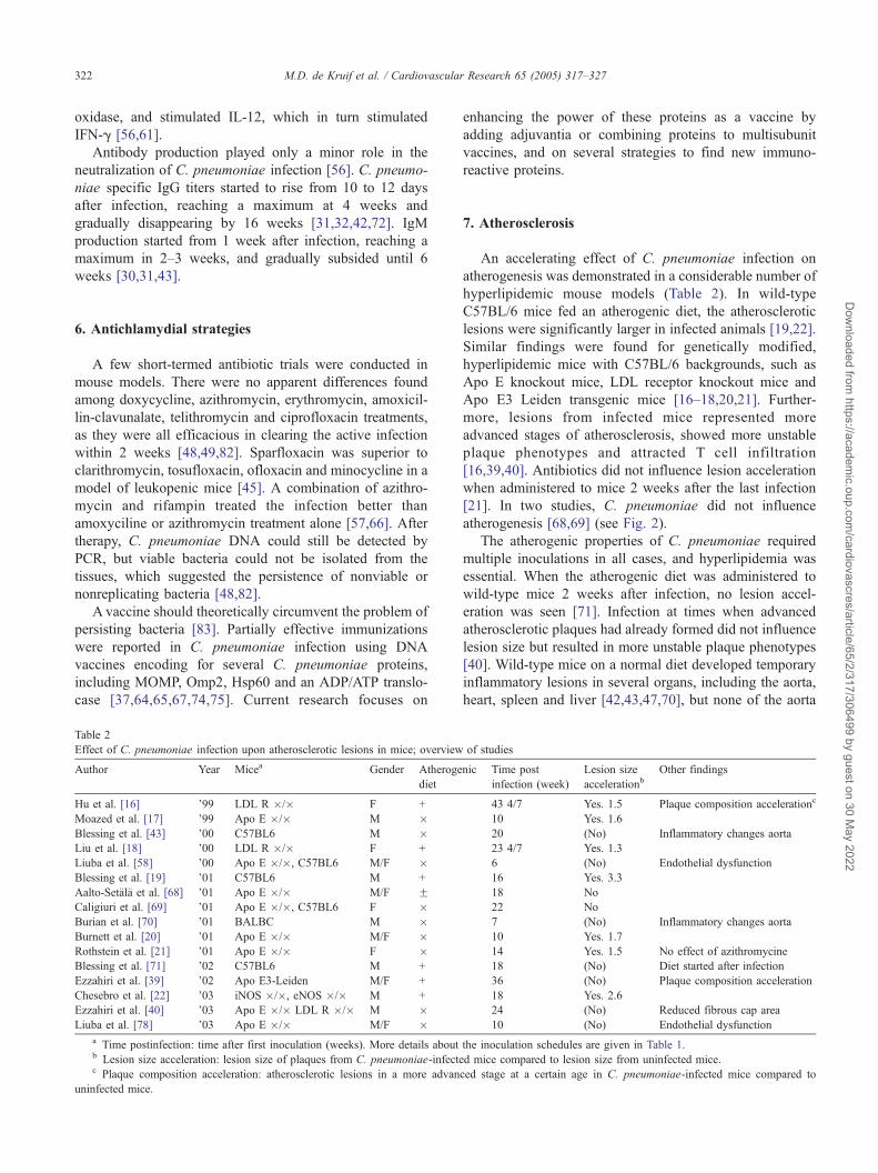
Table 2
Effect of C. pneumoniae infection upon atherosclerotic lesions in mice; overview
Author Year Micea Gender Atheroge
diet
Hu et al. [16] ’99 LDL R �/� F +
Moazed et al. [17] ’99 Apo E �/� M �Blessing et al. [43] ’00 C57BL6 M �Liu et al. [18] ’00 LDL R �/� F +
Liuba et al. [58] ’00 Apo E �/�, C57BL6 M/F �Blessing et al. [19] ’01 C57BL6 M +
Aalto-Set7l7 et al. [68] ’01 Apo E �/� M/F FCaligiuri et al. [69] ’01 Apo E �/�, C57BL6 F �Burian et al. [70] ’01 BALBC M �Burnett et al. [20] ’01 Apo E �/� M/F �Rothstein et al. [21] ’01 Apo E �/� F �Blessing et al. [71] ’02 C57BL6 M +
Ezzahiri et al. [39] ’02 Apo E3-Leiden M/F +
Chesebro et al. [22] ’03 iNOS �/�, eNOS �/� M +
Ezzahiri et al. [40] ’03 Apo E �/� LDL R �/� M �Liuba et al. [78] ’03 Apo E �/� M/F �
a Time postinfection: time after first inoculation (weeks). More details aboutb Lesion size acceleration: lesion size of plaques from C. pneumoniae-infectec Plaque composition acceleration: atherosclerotic lesions in a more advan
uninfected mice.
enhancing the power of these proteins as a vaccine by
adding adjuvantia or combining proteins to multisubunit
vaccines, and on several strategies to find new immuno-
reactive proteins.
7. Atherosclerosis
An accelerating effect of C. pneumoniae infection on
atherogenesis was demonstrated in a considerable number of
hyperlipidemic mouse models (Table 2). In wild-type
C57BL/6 mice fed an atherogenic diet, the atherosclerotic
lesions were significantly larger in infected animals [19,22].
Similar findings were found for genetically modified,
hyperlipidemic mice with C57BL/6 backgrounds, such as
Apo E knockout mice, LDL receptor knockout mice and
Apo E3 Leiden transgenic mice [16–18,20,21]. Further-
more, lesions from infected mice represented more
advanced stages of atherosclerosis, showed more unstable
plaque phenotypes and attracted T cell infiltration
[16,39,40]. Antibiotics did not influence lesion acceleration
when administered to mice 2 weeks after the last infection
[21]. In two studies, C. pneumoniae did not influence
atherogenesis [68,69] (see Fig. 2).
The atherogenic properties of C. pneumoniae required
multiple inoculations in all cases, and hyperlipidemia was
essential. When the atherogenic diet was administered to
wild-type mice 2 weeks after infection, no lesion accel-
eration was seen [71]. Infection at times when advanced
atherosclerotic plaques had already formed did not influence
lesion size but resulted in more unstable plaque phenotypes
[40]. Wild-type mice on a normal diet developed temporary
inflammatory lesions in several organs, including the aorta,
heart, spleen and liver [42,43,47,70], but none of the aorta
of studies
nic Time post
infection (week)
Lesion size
accelerationbOther findings
43 4/7 Yes. 1.5� Plaque composition accelerationc
10 Yes. 1.6�20 (No) Inflammatory changes aorta
23 4/7 Yes. 1.3�6 (No) Endothelial dysfunction
16 Yes. 3.3�18 No
22 No
7 (No) Inflammatory changes aorta
10 Yes. 1.7�14 Yes. 1.5� No effect of azithromycine
18 (No) Diet started after infection
36 (No) Plaque composition acceleration
18 Yes. 2.6�24 (No) Reduced fibrous cap area
10 (No) Endothelial dysfunction
the inoculation schedules are given in Table 1.
d mice compared to lesion size from uninfected mice.
ced stage at a certain age in C. pneumoniae-infected mice compared to
499 by guest on 30 May 2022

Fig. 2. Scheme summarizing the limitations of antibiotic therapy of C. pneumoniae infection in mice. Antibiotic therapy is effective in clearing the acute active
infection in lungs, but a number of organisms are able to survive in the lungs in a persistent state. Furthermore, inhibition of the pro-atherogenic effects of C.
pneumoniae is only possible when the antibiotics are given early during the infection. It is not clear what the precisemechanism is of the pro-atherogenic properties
of C. pneumoniae in mice, and it is not clear whether antibiotics can affect the presence of C. pneumoniae antigens in atherosclerotic plaques (see also text).
M.D. de Kruif et al. / Cardiovascular Research 65 (2005) 317–327 323
Dow
nloaded from https://academ
ic.oup.com/cardiovascres/article/65/2/317/306499
inflammatory lesions progressed to atherosclerotic lesions
[43,70]. Murine cytomegalovirus infection produced similar
inflammatory lesions, and superinfection with C. pneumo-
niae had an impressive additional effect upon the size of
these lesions [70]. However, no such additional effects were
found in Apo E knockout mice, although each pathogen on
itself accelerated lesion development [20]. Infection with C.
pneumoniae resulted in increased endothelial dysfunction
and enhanced VCAM-1 expression in Apo E knockout
mice, and coinfection with Helicobacter pylori stimulated
these effects [78]. In one study, lesion acceleration was
found in male mice only, which suggested a gender-
dependent effect [20].
by guest on 30 May 2022
8. Discussion
Mouse models provided detailed information about C.
pneumoniae infections. Many of these details were rather
uncommon to a respiratory tract infection. The histopatho-
logical changes in the lungs were considerable, although the
clinical disease was rather mild [31,32]. The host defense,
characterized by a Th1 response requiring IFN-g for clearing
the bacteria, was only partly successful in controlling the
infection [56]. Afterwards, the C. pneumoniae organisms
persisted in the lungs in a latent phase that could be
reactivated by the administration of cortisone [50,51]. This
phenomenon could be related to the capability of IFN-g to
induce persistence of the bacteria. The addition of IFN-g toC.
pneumoniae-infected monocytic cell lines induced a similar
kind of persistence of nonreplicating, viable bacteria [88].
Another uncommon finding of a respiratory tract
infection is that C. pneumoniae disseminated throughout
the body and caused inflammatory lesions in several organs,
including the heart and the vessel walls [47]. The dissem-
ination was generally mediated by leukocytes, in which C.
pneumoniae has found a way of surviving [38]. Notably,
only during a short period following infection, viable
organisms were isolated from the organ tissues [42]. Also
in human atherosclerotic tissue specimens, isolation of
viable organisms generally was a rare event. In these tissue
specimens, membrane components of C. pneumoniae were
detected by in situ methods, but no inclusions were found
that resembled viable organisms [89]. Probably, viable
organisms were capable of reaching the atheroma, but did
not survive there for long, and left nonviable, potentially
pathogenic remnants behind. It is also possible that C.
pneumoniae persisted in the atheroma in a latent state as had
been found in lung tissue, but no such inclusions were
detected in mice. In either case, antibiotic treatment is of
limited value, because it aims at viable, replicating bacteria.
The presence of C. pneumoniae in the atheroma seems to
be pathogenic. Atherogenesis was accelerated in a number
of mouse models, and the results were reproducible and
confirmed by different research groups (Table 2). At first,
acceleration of atherogenesis was demonstrated in genet-
ically modified mice only [16,17]. These results were
difficult to interpret because the genetic modification in
itself could have influenced the inflammatory process [90].
Additional evidence came from normal C57BL/6 mice fed
an atherogenic diet [19,71]. It became clear that multiple
inoculations and a hyperlipidemic environment were essen-
tial, and some evidence suggested a gender-dependent effect
[43,71]. In two studies, the conclusion was drawn that C.
pneumoniae did not accelerate atherogenesis. In one of
these studies [69], no reinfection after the first inoculation
had been given until 18 weeks after infection, which could
explain the lack of association with atherogenesis, arguing
M.D. de Kruif et al. / Cardiovascular Research 65 (2005) 317–327324
Dow
nloaded from https://academ
ic.oup.com/cardiovascres/article/65/2/317/306499 by guest on 30 M
ay 2022
that a model of chronic infection is necessary. Furthermore,
these investigators used a lower dose of C. pneumoniae
[91]. In the other study by Aalto-Set7l7 et al. [68], Apo E
knockout mice on an FVB background were used. These
mice are known to develop atherosclerosis more slowly than
Apo E knockout mice on C57BL/6 background, which may
have influenced the findings [92]. The investigators used C.
pneumoniae strain Kajaani 7, which is known to be cleared
quickly [41], so it may not have been able to induce chronic
infection. On the other hand, this strain was very virulent in
acute disease, and many mice died, up to 80% in a group,
leaving 1 to 5 mice in a group. The conclusions drawn from
this study are therefore difficult to interpret.
The atherogenic properties of C. pneumoniae in mice
have also been demonstrated in a few studies in rats, rabbits
and pigs, suggesting that the results of these animal models
can be extrapolated to human beings [93–95]. The
mechanism is not known, but several pathways were
proposed for C. pneumoniae-enhanced atherosclerosis
[96]. These mechanisms include (1) direct stimulation of
monocyte migration to the atheromas [97]; (2) direct
interference with intracellular metabolism within the plaque,
as C. pneumoniae was able to infect all cell types present in
a plaque in vitro, thereby inducing atherogenic processes
such as foam cell formation and LDL oxidation [14,15]; (3)
the lesions could be complicated by an immunopathogenic
role of C. pneumoniae, attracting inflammatory cells
causing tissue damage. T cell infiltration which was
demonstrated both in murine and human atherosclerotic
plaques [39,98]; (4) triggering of an autoimmune reaction to
human Hsp60 by antigenic mimicry could be interfering
with atherogenesis [14,99]; and (5) apart from the athero-
genic properties, C. pneumoniae could complicate athero-
sclerotic disease further by destabilizing plaques by
reducing the fibrous cap area and stimulating matrix
degrading metalloproteinases [40], or by enhancing throm-
bogenicity by stimulating coagulation factors and tissue
factor expression [100,101].
Antibiotics were successful for the treatment of the acute
pulmonary infection, but had no effect on the atherogenic
properties of C. pneumoniae when they were administered 2
weeks after infection [21]. In addition, in rabbits, antibiotics
inhibited atherogenesis only slightly when they were
administered 2 weeks after infection, but they were much
more effective when they were administered within 5 days
after infection [23,102]. Furthermore, antibiotics failed to
completely eradicate C. pneumoniae from the organ tissues
[48,103]. These findings may help to understand the
negative results of the human WIZARD study, because
the antibiotics in animals were apparently only effective
when they were given during the actual infection itself. The
acute C. pneumoniae infection in humans frequently passes
clinically unnoticed.
C. pneumoniae was one of the first and therefore most
frequently studied infectious organisms that showed athero-
genic properties in mice. The effect seemed to be specific to
C. pneumoniae because C. trachomatis did not influence
atherogenesis [104]. However, other organisms that were
not clearly related did show atherogenic properties in similar
mouse models. These organisms included various bacteria,
viruses and protozoa, such as Porphyromonas gingivalis,
murine cytomegalovirus, murine g-herpesvirus-68, Toxo-
plasma gondii and Trypanosoma cruzi [105–110]. The
contribution of these single pathogens is still being
researched. Additional atherogenic properties to C. pneumo-
niae infection were found in two out of three coinfection
studies with H. pylori and murine cytomegalovirus [70].
After all, the impact of the pathogen burden as a whole may
still be more important than the impact of single pathogens
alone.
In conclusion, the evidence for a pathogenic role of C.
pneumoniae in atherosclerosis in mouse models seems
convincing, because the results were reproducible and
confirmed by different research groups. However, the precise
mechanisms of this relationship remain unclear, although
many theories exist. Temptations to eradicate C. pneumoniae
from the body, both by the immune system and by antibiotic
treatment, generally encountered many difficulties which
could be related to the unusual gram-negative, obligate
intracellular characteristics of the bacterium. The value of the
currently available antibiotics should not be overestimated,
because they were only effective in preventing the long-term
atherogenic effects of C. pneumoniae infection when they
were given during the acute infection, which is often
asymptomatic or aspecific in humans. Therefore, the focus
of future research should merely shift back to basic research.
The precise mechanisms by which C. pneumoniae is
interfering with atherogenesis should be further elucidated
to enable more specific strategies to interfere with this
process, e.g. by immunosuppressive agents. In addition, the
role of other infectious pathogens should be further exam-
ined, and especially the impact of the pathogen burden as a
whole. The development of an effective vaccine would be the
final strategy to prevent chlamydial disease and its compli-
cations, but not to prevent the impact of the pathogen burden.
References
[1] Ross R. Atherosclerosis—an inflammatory disease. N Engl J Med
1999;340:115–26.
[2] Espinola-Klein C, Rupprecht HJ, Blankenberg S, Bickel C, Kopp H,
Rippin G. Impact of infectious burden on extent and long-term
prognosis of atherosclerosis. Circulation 2002;105:15–21.
[3] Everett KD, Andersen AA. Identification of nine species of the
Chlamydiaceae using PCR-RFLP. Int J Syst Bacteriol 1999;49(Pt 2):
803–13.
[4] Kuo CC, Jackson LA, Campbell LA, Grayston JT. Chlamydia
pneumoniae (TWAR). Clin Microbiol Rev 1995;8:451–61.
[5] Grayston JT. Background and current knowledge of Chlamydia
pneumoniae and atherosclerosis. J Infect Dis 2000;181(Suppl. 3):
S402–10.
[6] Wright SW, Edwards KM, Decker MD, Grayston JT, Wang S.
Prevalence of positive serology for acute Chlamydia pneumoniae

M.D. de Kruif et al. / Cardiovascular Research 65 (2005) 317–327 325
Dow
nloaded from https://academ
ic.oup.com/cardiovascres/article/65/2/317/306499 by guest on 30 M
ay 2022
infection in emergency department patients with persistent cough.
Acad Emerg Med 1997;4:179–83.
[7] Odeh M, Oliven A. Chlamydial infections of the heart. Eur J Clin
Microbiol Infect Dis 1992;11:885–93.
[8] Gnarpe H, Gnarpe J, Gastrin B, Hallander H. Chlamydia
pneumoniae and myocarditis. Scand J Infect Dis, Suppl 1997;
104:50–2.
[9] Swanborg RH, Whittum-Hudson JA, Hudson AP. Human herpesvi-
rus 6 and Chlamydia pneumoniae as etiologic agents in multiple
sclerosis—a critical review. Microbes Infect 2002;4:1327–33.
[10] Saikku P, Leinonen M, Mattila K, Ekman MR, Nieminen MS,
Makela PH, et al. Serological evidence of an association of a novel
Chlamydia, TWAR, with chronic coronary heart disease and acute
myocardial infarction. Lancet 1988;2:983–6.
[11] Danesh J, Whincup P, Walker M, Lennon L, Thomson A, Appleby P,
et al. Chlamydia pneumoniae IgG titres and coronary heart disease:
prospective study and meta-analysis. BMJ 2000;321:208–13.
[12] Kuo CC, Shor A, Campbell LA, Fukushi H, Patton DL, Grayston JT.
Demonstration of Chlamydia pneumoniae in atherosclerotic lesions
of coronary arteries. J Infect Dis 1993;167:841–9.
[13] Taylor-Robinson D, Thomas BJ. Chlamydia pneumoniae in athero-
sclerotic tissue. J Infect Dis 2000;181(Suppl. 3):S437–40.
[14] Kol A, Sukhova GK, Lichtman AH, Libby P. Chlamydial heat shock
protein 60 localizes in human atheroma and regulates macrophage
tumor necrosis factor-alpha and matrix metalloproteinase expression.
Circulation 1998;98:300–7.
[15] Kalayoglu MV, Indrawati, Morrison RP, Morrison SG, Yuan,
Byrne GI. Chlamydial virulence determinants in atherogenesis: the
role of chlamydial lipopolysaccharide and heat shock protein 60 in
macrophage–lipoprotein interactions. J Infect Dis 2000;181(Suppl.
3):S483–9.
[16] Hu H, Pierce GN, Zhong G. The atherogenic effects of chlamydia are
dependent on serum cholesterol and specific to Chlamydia pneumo-
niae. J Clin Invest 1999;103:747–53.
[17] Moazed TC, Campbell LA, Rosenfeld ME, Grayston JT, Kuo CC.
Chlamydia pneumoniae infection accelerates the progression of
atherosclerosis in apolipoprotein E-deficient mice. J Infect Dis
1999;180:238–41.
[18] Liu L, Hu H, Ji H, Murdin AD, Pierce GN, Zhong G. Chlamydia
pneumoniae infection significantly exacerbates aortic atherosclerosis
in an LDLR�/� mouse model within six months. Mol Cell Biochem
2000;215:123–8.
[19] Blessing E, Campbell LA, Rosenfeld ME, Chough N, Kuo CC.
Chlamydia pneumoniae infection accelerates hyperlipidemia induced
atherosclerotic lesion development in C57BL/6J mice. Atheroscle-
rosis 2001;158:13–7.
[20] Burnett MS, Gaydos CA, Madico GE, Glad SM, Paigen B, Quinn
TC, et al. Atherosclerosis in apoE knockout mice infected with
multiple pathogens. J Infect Dis 2001;183:226–31.
[21] Rothstein NM, Quinn TC, Madico G, Gaydos CA, Lowenstein CJ.
Effect of azithromycin on murine arteriosclerosis exacerbated by
Chlamydia pneumoniae. J Infect Dis 2001;183:232–8.
[22] Chesebro BB, Blessing E, Kuo CC, Rosenfeld ME, Puolakkainen
LA, Campbell LA. Nitric oxide synthase plays a role in Chlamydia
pneumoniae-induced atherosclerosis. Cardiovasc Res 2003;
60:170–4.
[23] Muhlestein JB, Anderson JL, Hammond EH, Zhao L, Trehan S,
Schwobe EP, et al. Infection with Chlamydia pneumoniae accelerates
the development of atherosclerosis and treatment with azithromycin
prevents it in a rabbit model. Circulation 1998;97:633–6.
[24] Dunne MW. Rationale and design of a secondary prevention trial of
antibiotic use in patients after myocardial infarction: the WIZARD
(weekly intervention with zithromax [azithromycin] for atheroscle-
rosis and its related disorders) trial. J Infect Dis 2000;181(Suppl 3):
S572–8.
[25] Grayston JT. Antibiotic treatment of atherosclerotic cardiovascular
disease. Circulation 2003;107:1228–30.
[26] O’Connor CM, Dunne MW, Pfeffer MA, Muhlestein JB, Yao L,
Gupta S, et al. Azithromycin for the secondary prevention of
coronary heart disease events: the WIZARD study: a randomized
controlled trial. JAMA 2003;290:1459–66.
[27] Jackson LA. Description and status of the azithromycin and coronary
events study (ACES). J Infect Dis 2000;181(Suppl. 3):S579–81.
[28] Cannon CP, McCabe CH, Belder R, Breen J, Braunwald E. Design
of the pravastatin or atorvastatin evaluation and infection therapy
(PROVE IT)-TIMI 22 trial. Am J Cardiol 2002;89:860–1.
[29] Pislaru SV, Van de Werf F. Antibiotic therapy for coronary artery
disease: can a WIZARD change it all? JAMA 2003;290:1515–6.
[30] Kishimoto T. Studies on Chlamydia pneumoniae, strain TWAR,
infection: I. Experimental infection of C. pneumoniae in mice and
serum antibodies against TWAR by MFA. Kansenshogaku Zasshi
1990;64:124–31.
[31] Kaukoranta-Tolvanen SS, Laurila AL, Saikku P, Leinonen M,
Liesirova L, Laitinen K. Experimental infection of Chlamydia
pneumoniae in mice. Microb Pathog 1993;15:293–302.
[32] Yang ZP, Kuo CC, Grayston JT. A mouse model of Chlamydia
pneumoniae strain TWAR pneumonitis. Infect Immun 1993;61:
2037–40.
[33] Meijer A, Morre SA, van den Brule AJ, Savelkoul PH, Ossewaarde
JM. Genomic relatedness of Chlamydia isolates determined by
amplified fragment length polymorphism analysis. J Bacteriol
1999;181:4469–75.
[34] Read TD, Brunham RC, Shen C, Gill SR, Heidelberg JF, White
O, et al. Genome sequences of Chlamydia trachomatis MoPn
and Chlamydia pneumoniae AR39. Nucleic Acids Res 2000;
28:1397–406.
[35] Karunakaran KP, Blanchard JF, Raudonikiene A, Shen C, Murdin
RC, Brunham RC. Molecular detection and seroepidemiology of the
Chlamydia pneumoniae bacteriophage (PhiCpn1). J Clin Microbiol
2002;40:4010–4.
[36] Ekman MR, Grayston JT, Visakorpi R, Kleemola M, Kuo CC,
Saikku P. An epidemic of infections due to Chlamydia pneumoniae
in military conscripts. Clin Infect Dis 1993;17:420–5.
[37] Peterson EM, Cheng X, Qu Z, de La Maza LM. Characterization of
the murine antibody response to peptides representing the variable
domains of the major outer membrane protein of Chlamydia
pneumoniae. Infect Immun 1996;64:3354–9.
[38] Moazed TC, Kuo CC, Grayston JT, Campbell LA. Evidence of
systemic dissemination of Chlamydia pneumoniae via macrophages
in the mouse. J Infect Dis 1998;177:1322–5.
[39] Ezzahiri R, Nelissen-Vrancken HJ, Kurvers HA, Stassen FR, Vliegen
GE, Grauls GE, et al. Chlamydophila pneumoniae (Chlamydia
pneumoniae) accelerates the formation of complex atherosclerotic
lesions in Apo E3-Leiden mice. Cardiovasc Res 2002;56:269–76.
[40] Ezzahiri R, Stassen FR, Kurvers HA, van Pul MM, Kitslaar
PJ, Bruggeman PJ. Chlamydia pneumoniae infection induces an
unstable atherosclerotic plaque phenotype in LDL-receptor,
ApoE double knockout mice. Eur J Vasc Endovasc Surg
2003;26:88–95.
[41] Erkkila L, Rottenberg ME, Laitinen K. Comparison of anesthetics for
inoculation of mice with Chlamydia pneumoniae. Comp Med
2000;50:46–8.
[42] Moazed TC, Kuo C, Grayston JT, Campbell LA. Murine models of
Chlamydia pneumoniae infection and atherosclerosis. J Infect Dis
1997;175:883–90.
[43] Blessing E, Lin TM, Campbell LA, Rosenfeld ME, Lloyd D, Kuo C.
Chlamydia pneumoniae induces inflammatory changes in the heart
and aorta of normocholesterolemic C57BL/6J mice. Infect Immun
2000;68:4765–8.
[44] Kimura M. Experimental study on the mechanisms of Chlamydia
pneumoniae respiratory infection in mice with former chlamydial
exposure. Kansenshogaku Zasshi 1994;68:50–8.
[45] Nakata K, Okazaki Y, Hattori H, Nakamura S. Protective effects of
sparfloxacin in experimental pneumonia caused by Chlamydia
M.D. de Kruif et al. / Cardiovascular Research 65 (2005) 317–327326
Dow
nloaded from https://academ
ic.oup.com/cardiovascres/article/65/2/317/306499 by guest on 30 M
ay 2022
pneumoniae in leukopenic mice. Antimicrob Agents Chemother
1994;38:1757–62.
[46] Kaukoranta-Tolvanen SE, Laurila AL, Saikku P, Leinonen M,
Laitinen K. Experimental Chlamydia pneumoniae infection in mice:
effect of reinfection and passive immunization. Microb Pathog
1995;18:279–88.
[47] Yang ZP, Kuo CC, Grayston JT. Systemic dissemination of
Chlamydia pneumoniae following intranasal inoculation in mice. J
Infect Dis 1995;171:736–8.
[48] Malinverni R, Kuo CC, Campbell LA, Lee A, Grayston JT. Effects
of two antibiotic regimens on course and persistence of experimental
Chlamydia pneumoniae TWAR pneumonitis. Antimicrob Agents
Chemother 1995;39:45–9.
[49] Masson ND, Toseland CD, Beale AS. Relevance of Chlamydia
pneumoniae murine pneumonitis model to evaluation of antimicro-
bial agents. Antimicrob Agents Chemother 1995;39:1959–64.
[50] Malinverni R, Kuo CC, Campbell LA, Grayston JT. Reactivation of
Chlamydia pneumoniae lung infection in mice by cortisone. J Infect
Dis 1995;172:593–4.
[51] Laitinen K, Laurila AL, Leinonen M, Saikku P. Reactivation of
Chlamydia pneumoniae infection in mice by cortisone treatment.
Infect Immun 1996;64:1488–90.
[52] Penttila JM, Anttila M, Puolakkainen M, Laurila A, Varkila K,
Sarvas M, et al. Local immune responses to Chlamydia pneumoniae
in the lungs of BALB/c mice during primary infection and
reinfection. Infect Immun 1998;66:5113–8.
[53] Penttila JM, Pyhala R, Sarvas M, Rautonen N. Expansion of a
novel pulmonary CD3(�) CD4(+) CD8(+) cell population in mice
during Chlamydia pneumoniae infection. Infect Immun 1998;66:
3290–4.
[54] Penttila JM, Anttila M, Varkila K, Puolakkainen M, Sarvas M,
Makela PH, et al. Depletion of CD8+ cells abolishes memory in
acquired immunity against Chlamydia pneumoniae in BALB/c mice.
Immunology 1999;97:490–6.
[55] Peterson EM, de la Maza LM, Brade L, Brade H. Characterization
of a neutralizing monoclonal antibody directed at the lipopoly-
saccharide of Chlamydia pneumoniae. Infect Immun 1998;66:
3848–55.
[56] Rottenberg ME, Gigliotti Rothfuchs AC, Gigliotti D, Svanholm C,
Bandholtz L, Wigzell H. Role of innate and adaptive immunity in
the outcome of primary infection with Chlamydia pneumoniae, as
analyzed in genetically modified mice. J Immunol 1999;162:
2829–36.
[57] Wolf K, Malinverni R. Effect of azithromycin plus rifampin versus
that of azithromycin alone on the eradication of Chlamydia
pneumoniae from lung tissue in experimental pneumonitis. Anti-
microb Agents Chemother 1999;43:1491–3.
[58] Liuba P, Karnani P, Pesonen E, Paakkari I, Forslid A, Johansson L,
et al. Endothelial dysfunction after repeated Chlamydia pneumoniae
infection in apolipoprotein E-knockout mice. Circulation 2000;102:
1039–44.
[59] Vuola JM, Puurula V, Anttila M, Makela PH, Rautonen N. Acquired
immunity to Chlamydia pneumoniae is dependent on gamma
interferon in two mouse strains that initially differ in this respect
after primary challenge. Infect Immun 2000;68:960–4.
[60] Geng Y, Berencsi K, Gyulai Z, Valyi-Nagy T, Gonczol E, Trinchieri
G. Roles of interleukin-12 and gamma interferon in murine
Chlamydia pneumoniae infection. Infect Immun 2000;68:2245–53.
[61] Rottenberg ME, Gigliotti Rothfuchs A, Gigliotti D, Ceausu M, Une
C, Levitsky V, et al. Regulation and role of IFN-gamma in the innate
resistance to infection with Chlamydia pneumoniae. J Immunol
2000;164:4812–8.
[62] Pal S, Peterson EM, de La Maza LM. Role of Nramp1 deletion in
Chlamydia infection in mice. Infect Immun 2000;68:4831–3.
[63] Meijer A, Roholl PJ, Gielis-Proper SK, Meulenberg YF, Ossewaarde
JM. Chlamydia pneumoniae in vitro and in vivo: a critical evaluation
of in situ detection methods. J Clin Pathol 2000;53:904–10.
[64] Svanholm C, Bandholtz L, Castanos-Velez E, Wigzell H, Rottenberg
ME. Protective DNA immunization against Chlamydia pneumoniae.
Scand J Immunol 2000;51:345–53.
[65] Murdin AD, Dunn P, Sodoyer R, Wang J, Caterini J, Brunham RC,
et al. Use of a mouse lung challenge model to identify antigens
protective against Chlamydia pneumoniae lung infection. J Infect
Dis 2000;181(Suppl 3):S544–51.
[66] Bin XX, Wolf K, Schaffner T, Malinverni R. Effect of azithromycin
plus rifampin versus amoxicillin alone on eradication and inflam-
mation in the chronic course of Chlamydia pneumoniae pneumonitis
in mice. Antimicrob Agents Chemother 2000;44:1761–4.
[67] Penttila T, Vuola JM, Puurula V, Anttila M, Sarvas M, Rautonen N,
et al. Immunity to Chlamydia pneumoniae induced by vaccination
with DNA vectors expressing a cytoplasmic protein (Hsp60) or outer
membrane proteins (MOMP and Omp2). Vaccine 2000;19:1256–65.
[68] Aalto-Setala K, Laitinen K, Erkkila L, Leinonen M, Jauhiainen M,
Ehnholm C, et al. Chlamydia pneumoniae does not increase
atherosclerosis in the aortic root of apolipoprotein E-deficient mice.
Arterioscler Thromb Vasc Biol 2001;21:578–84.
[69] Caligiuri G, Rottenberg M, Nicoletti A, Wigzell H, Hansson GK.
Chlamydia pneumoniae infection does not induce or modify
atherosclerosis in mice. Circulation 2001;103:2834–8.
[70] Burian K, Berencsi K, Endresz V, Gyulai Z, Valyi-Nagy T, Valyi-
Nagy I, et al. Chlamydia pneumoniae exacerbates aortic inflamma-
tory foci caused by murine cytomegalovirus infection in normocho-
lesterolemic mice. Clin Diagn Lab Immunol 2001;8:1263–6.
[71] Blessing E, Campbell LA, Rosenfeld ME, Kuo CC. Chlamydia
pneumoniae and hyperlipidemia are co-risk factors for atheroscle-
rosis: infection prior to induction of hyperlipidemia does not
accelerate development of atherosclerotic lesions in C57BL/6J mice.
Infect Immun 2002;70:5332–4.
[72] Erkkila L, Laitinen K, Laurila A, Saikku P, Leinonen M.
Experimental Chlamydia pneumoniae infection in NIH/S mice:
effect of reinoculation with chlamydial or cell preparation on culture,
PCR and histological findings of lung tissue. Vaccine 2002;
20:2318–24.
[73] Tiirola T, Erkkila L, Laitinen K, Leinonen M, Saikku P, Bloigu A,
et al. Effect of acute Chlamydia pneumoniae infection on
lipoprotein metabolism in NIH/S mice. Scand J Clin Lab Invest
2002;62:477–84.
[74] Bandholtz L, Kreuger MR, Svanholm C, Wigzell H, Rottengerg ME.
Adjuvant modulation of the immune responses and the outcome of
infection with Chlamydia pneumoniae. Clin Exp Immunol 2002;
130:393–403.
[75] Airaksinen U, Penttila T, Wahlstrom E, Vuola JM, Puolakkainen M,
Sarvas M. Production of Chlamydia pneumoniae proteins in Bacillus
subtilis and their use in characterizing immune responses in the
experimental infection model. Clin Diagn Lab Immunol 2003;10:
367–75.
[76] Burian K, Hegyesi H, Buzas E, Endresz V, Kis Z, Falus A, et al.
Chlamydophila (Chlamydia) pneumoniae induces histidine decar-
boxylase production in the mouse lung. Immunol Lett 2003;
89:229–36.
[77] Shi Y, Yin J, Zhan H, Feng G, Zhang X, Su X, et al. The
pathogenesis of Chlamydia pneumoniae-type pneumonitis in mice.
Chin Med J (Engl) 2003;116:328–32.
[78] Liuba P, Pesonen E, Paakkari I, Batra S, Andersen L, Forslid A, et al.
Co-infection with Chlamydia pneumoniae and Helicobacter pylori
results in vascular endothelial dysfunction and enhanced VCAM-1
expression in apoE-knockout mice. J Vasc Res 2003;40:115–22.
[79] Rothfuchs AG, Kreuger MR, Wigzell H, Rottenberg ME. Macro-
phages, CD4+ or CD8+ cells are each sufficient for protection
against Chlamydia pneumoniae infection through their ability to
secrete IFN-gamma. J Immunol 2004;172:2407–15.
[80] Mygind T, Vandahl B, Pedersen AS, Christiansen G, Hollsberg P,
Birkelund S. Identification of an in vivo CD4+ T cell-mediated
response to polymorphic membrane proteins of Chlamydia pneumo-

M.D. de Kruif et al. / Cardiovascular Research 65 (2005) 317–327 327
Dow
nloaded from https://academ
ic.oup.com/cardiovascres/article/65/2/317/306499 by guest on 30 M
ay 2022
niae during experimental infection. FEMS Immunol Med Microbiol
2004;40:129–37.
[81] Little CS, Hammond CJ, MacIntyre A, Balin BJ, Appelt DM.
Chlamydia pneumoniae induces Alzheimer-like amyloid plaques in
brains of BALB/c mice. Neurobiol Aging 2004;25:419–29.
[82] Tormakangas L, Saario E, Bem David D, Bryskier A, Leinonen M,
Saikku P. Treatment of acute Chlamydia pneumoniae infection with
telithromycin in C57BL/6J mice. J Antimicrob Chemother 2004;
53:1101–4.
[83] Igietseme JU, Black CM, Caldwell HD. Chlamydia vaccines:
strategies and status. BioDrugs 2002;16:19–35.
[84] Caspar-Bauguil S, Puissant B, Nazzal D, Lefevre JC, Thomsen N,
Salvayre R, et al. Chlamydia pneumoniae induces interleukin-10
production that down-regulates major histocompatibility complex
class I expression. J Infect Dis 2000;182:1394–401.
[85] Fan P, Dong F, Huang Y, Zhong G. Chlamydia pneumoniae secretion
of a protease-like activity factor for degrading host cell transcription
factors required for [correction of factors is required for] major
histocompatibility complex antigen expression. Infect Immun 2002;
70:345–9.
[86] Fischer SF, Hacker G. Characterization of antiapoptotic activities of
Chlamydia pneumoniae in infected cells. Ann NY Acad Sci
2003;1010:565–7.
[87] van Zandbergen G, Gieffers J, Kothe H, Rupp J, Bollinger A, Aga E,
et al. Chlamydia pneumoniae multiply in neutrophil granulocytes and
delay their spontaneous apoptosis. J Immunol 2004;172:1768–76.
[88] Hammerschlag MR. The intracellular life of chlamydiae. Semin
Pediatr Infect Dis 2002;13:239–48.
[89] Meijer A, Roholl PJ, Gielis-Proper SK, Ossewaarde JM. Chlamydia
pneumoniae antigens, rather than viable bacteria, persist in athero-
sclerotic lesions. J Clin Pathol 2000;53:911–6.
[90] de Bont N, Netea MG, Demacker PN, Kullberg BJ, van der Meer
AF, Stalenhoef AF. Apolipoprotein E-deficient mice have an
impaired immune response to Klebsiella pneumoniae. Eur J Clin
Investig 2000;30:818–22.
[91] Kuo CC, Campbell LA, Rosenfeld ME. Chlamydia pneumoniae
infection and atherosclerosis: methodological considerations. Circu-
lation 2002;105:E34.
[92] Dansky HM, Shu P, Donavan M, Montagno J, Nagle DL, Smutko JS,
et al. A phenotype-sensitizing Apoe-deficient genetic background
reveals novel atherosclerosis predisposition loci in the mouse.
Genetics 2002;160:1599–608.
[93] Fong IW, Chiu B, Viira E, Jang D, Mahony JB. De novo induction of
atherosclerosis by Chlamydia pneumoniae in a rabbit model. Infect
Immun 1999;67:6048–55.
[94] Pislaru SV, Van Ranst M, Pislaru C, Szelid Z, Theilmeier G,
Ossewaarde JM, et al. Chlamydia pneumoniae induces neointima
formation in coronary arteries of normal pigs. Cardiovasc Res
2003;57:834–42.
[95] Herrera VL, Shen L, Lopez LV, Didishvili T, Zhang YX, Ruiz-Opazo
N. Chlamydia pneumoniae accelerates coronary artery disease
progression in transgenic hyperlipidemia-genetic hypertension rat
model. Mol Med 2003;9:135–42.
[96] Kalayoglu MV, Libby P, Byrne GI. Chlamydia pneumoniae as an
emerging risk factor in cardiovascular disease. JAMA 2002;
288:2724–31.
[97] May AE, Redecke V, Gruner S, Schmidt R, Massberg S, Miethke T,
et al. Recruitment of Chlamydia pneumoniae-infected macrophages
to the carotid artery wall in noninfected, nonatherosclerotic mice.
Arterioscler Thromb Vasc Biol 2003;23:789–94.
[98] van der Wal AC. Chlamydia pneumoniae inside the atherosclerotic
plaque—does it affect plaque inflammation and plaque progression?
Cardiovasc Res 2002;56:178–80.
[99] Bachmaier K, Neu N, de la Maza LM, Pal S, Hessel A, Penninger
JM. Chlamydia infections and heart disease linked through antigenic
mimicry. Science 1999;283:1335–9.
[100] Dechend R, Maass M, Gieffers J, Dietz R, Scheidereit C, Leutz A,
et al. Chlamydia pneumoniae infection of vascular smooth muscle
and endothelial cells activates NF-kappaB and induces tissue factor
and PAI-1 expression: a potential link to accelerated arteriosclerosis.
Circulation 1999;100:1369–73.
[101] Virok D, Loboda A, Kari L, Nebozhyn M, Chang C, Nichols C, et al.
Infection of U937 monocytic cells with Chlamydia pneumoniae
induces extensive changes in host cell gene expression. J Infect Dis
2003;188:1310–21.
[102] Fong IW. Antibiotics effects in a rabbit model of Chlamydia
pneumoniae-induced atherosclerosis. J Infect Dis 2000;181
(Suppl. 3):S514–8.
[103] Gieffers J, Fullgraf H, Jahn J, Klinger M, Dalhoff K, Katus HA, et al.
Chlamydia pneumoniae infection in circulating human monocytes is
refractory to antibiotic treatment. Circulation 2001;103:351–6.
[104] Blessing E, Nagano S, Campbell LA, Rosenfeld ME, Kuo CC. Effect
of Chlamydia trachomatis infection on atherosclerosis in apolipo-
protein E-deficient mice. Infect Immun 2000;68:7195–7.
[105] Sunnemark D, Harris RA, Frostegard J, Orn A. Induction of early
atherosclerosis in CBA/J mice by combination of Trypanosoma
cruzi infection and a high cholesterol diet. Atherosclerosis 2000;
153:273–82.
[106] Alber DG, Vallance P, Powell KL. Enhanced atherogenesis is not an
obligatory response to systemic herpesvirus infection in the apoE-
deficient mouse: comparison of murine gamma-herpesvirus-68 and
herpes simplex virus-1. Arterioscler Thromb Vasc Biol 2002;
22:793–8.
[107] Lalla E, Lamster IB, Hofmann MA, Bucciarelli L, Jerud AP, Tucker
S, et al. Oral infection with a periodontal pathogen accelerates early
atherosclerosis in apolipoprotein E-null mice. Arterioscler Thromb
Vasc Biol 2003;23:1405–11.
[108] Naghavi M, Wyde P, Litovsky S, Madjid M, Akhtar A, Naguib S,
et al. Influenza infection exerts prominent inflammatory and
thrombotic effects on the atherosclerotic plaques of apolipoprotein
E-deficient mice. Circulation 2003;107:762–8.
[109] Portugal LR, Fernandes LR, Cesar GC, Santiago HC, Oliveira DR,
Silva NM, et al. Infection with Toxoplasma gondii increases
atherosclerotic lesion in ApoE-deficient mice. Infect Immun 2004;
72:3571–6.
[110] Vliegen I, Duijvestijn A, Grauls G, Herngreen S, Bruggeman C,
Stassen F. Cytomegalovirus infection aggravates atherogenesis in
apoE knockout mice by both local and systemic immune activation.
Microbes Infect 2004;6:17–24.