The Puzzle of Faith - ScholarWorks @ Georgia State University

Molecular Microbiology (2001) 39(6), 1651±1660
The puzzle of zmpB and extensive chain formation,autolysis defect and non-translocation of choline-binding proteins in Streptococcus pneumoniae
Mathieu BergeÂ,1 Pedro GarcõÂa,2 Francesco Iannelli,3
Marie FrancËoise PreÁre,1,4 Chantal Granadel,1
Alessandra Polissi5 and Jean-Pierre Claverys1*1Laboratoire de Microbiologie et GeÂneÂtique MoleÂculaire,
UMR5100 CNRS±Universite Paul Sabatier, 118 route de
Narbonne, 31062 Toulouse Cedex, France.2Centro de Investigaciones BioloÂgicas, CSIC, VelaÂzquez
144, 28006 Madrid, Spain.3Dipartimento di Biologia Molecolare, Universita' di Siena,
53100 Siena, Italy.4Laboratoire de Microbiologie, HoÃpital Purpan, Place du
Docteur-Baylac, 31059 Toulouse Cedex, France.5Department of Microbiology, Glaxo Wellcome SpA, Via
Fleming 4, 37100 Verona, Italy.
Summary
Choline-binding proteins (CBPs) from Streptococcus
pneumoniae are involved in several important pro-
cesses. Inactivation of zmpB, a gene that encodes a
surface-located putative zinc metalloprotease, in a S.
pneumoniae serotype 4 strain was recently reported
to reveal a composite phenotype, including extensive
chain formation, lysis defect and transformation
deficiency. This phenotype was associated with the
lack of surface expression of several CBPs, including
the major autolysin LytA. LytA, normally 36 kDa in
size, was reported to form an SDS-resistant 80 kDa
complex with CinA. ZmpB was therefore proposed to
control translocation of CBPs to the surface, possibly
through the proteolytic release of CBPs (and RecA)
from CinA. Based on the use of 12 independent
mariner insertions in the zmpB gene of the well-
characterized R6 laboratory strain, we could not
confirm several of these observations. Our zmpB
mutants: (i) did not form chains; (ii) lysed normally in
the presence of deoxycholate, which indicates the
presence of a functional autolysin; (iii) transformed at
normal frequency; and (iv) contained bona fide CinA
and LytA species. Polymorphism of ZmpB between
R6 and the serotype 4 isolate could not account for
the discrepancy, as inactivation of zmpB (through
replacement by transposon-inactivated zmpB R6
alleles) in the latter strain did not affect separation
of daughter cells and autolysis. The conflicting
observations could be explained by our finding
that the reportedly serotype 4 zmpB `mutant' differed
from its S. pneumoniae parent in lacking capsule
and in exhibiting characteristic traits of the Strepto-
coccus viridans group, including resistance to
optochin.
Introduction
The Gram-positive bacterium Streptococcus pneumoniae,
a human commensal and a pathogen, contains choline
residues incorporated into the cell wall teichoic acids and
in the membrane-bound lipoteichoic acid (Tomasz, 1967).
Choline residues serve to anchor a family of surface-
exposed proteins that share a specialized choline-binding
domain (SaÂnchez-Puelles et al., 1990), the choline-
binding proteins (CBPs; for a review, see GarcõÂa et al.,
1998). Choline residues bound to teichoic acid have also
been shown to be absolutely required for the activity of
LytA (Holtje and Tomasz, 1975). LytA is a 36 kDa N-
acetylmuramoyl-L-alanine amidase, the major autolytic
enzyme of S. pneumoniae (for a review, see Tomasz,
1984; GarcõÂa et al., 1986) and the first identified CBP.
During a search for surface-exposed proteins of S.
pneumoniae involved in adhesion, Novak et al. (2000)
identified a putative zinc metalloprotease, ZmpB. A global
loss of cell surface expression of the CBPs (CbpA, CbpE,
CbpF and CbpJ) was observed in a ZmpB-deficient
mutant. Immunoelectron microscopy failed to detect any
intracellular or extracellular CbpE, CbpF and CbpJ.
However, the majority of CbpA was found trapped in the
cytoplasm of the zmpB mutant. LytA was also detected
mostly in the cytoplasm. This phenomenon was tenta-
tively attributed to alteration in the translocation of CBPs
to the surface. In addition, no 36 kDa bona fide LytA band
could be detected by Western blot. Two immunoreactive
species at 80 kDa and 60 kDa were detected instead
(Novak et al., 2000). The 80 kDa species was concluded
to correspond to a `complex' between LytA and CinA, a
protein induced when cells become competent for genetic
transformation (Masure et al., 1998; Mortier-BarrieÁre et al.,
1998), because (i) immunoelectron microscopy sug-
gested that the two proteins co-localized in the cytoplasm;
Q 2001 Blackwell Science Ltd
Accepted 15 January, 2001. *For correspondence. E-mail [email protected]; Tel. (133) 561 33 59 11; Fax (133) 561 33 58 86.
(ii) addition of the molecular weight of CinA and LytA
results in a molecular weight (81 kDa) potentially corre-
sponding to that of the putative complex; and (iii) Western
blot analysis using anti-CinA antibody detected an 80 kDa
species instead of the 45 kDa CinA band in the parent
strain. Detection of the `complex' on SDS210% poly-
acrylamide gels (Novak et al., 2000) suggested covalent
linkage of the proteins. Although cinA and lytA are part of
an operon expressed at the onset of competence for
genetic transformation (Mortier-BarrieÁre et al., 1998), they
are separated by two genes, recA and dinF. Therefore,
the putative CinA±LytA complex could not be produced
by a simple frameshift.
LytA is responsible for the sensitivity of S. pneumoniae
to deoxycholate (DOC), which is considered as a species
characteristic trait. DOC, an allosteric activator of LytA,
triggers autolysis. In line with the postulated defect in the
translocation of CBPs, including LytA, the zmpB mutant
did not undergo autolysis. Resistance to autolysis was
observed (i) in the stationary phase of growth; (ii) after the
addition of a 10-fold excess of DOC; or (iii) when treated
with up to 10� the minimum inhibitory concentration of
penicillin (Novak et al., 2000). The zmpB mutant was also
reported to grow in chains of 2000±3000 cells, whereas
S. pneumoniae normally grows mainly as diplococci
(hence its former name Diplococcus). This could be
explained by the loss of the LytB CBP, a murein hydrolase
essential for the separation of daughter cells (GarcõÂa et al.,
1999).
Finally, based on a previous report that CinA formed a
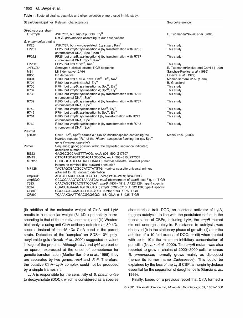
Table 1. Bacterial strains, plasmids and oligonucleotide primers used in this study.
Strain/plasmid/primer Relevant characteristics Source/reference
Streptococcus strainET-zmpB JNR.7/87, but zmpB::pJDC9; EryR E. Tuomanen/Novak et al. (2000)
Not S. pneumoniae according to our observationsS. pneumoniae strains
FP25 JNR.7/87, but non-capsulated, Dcps::kan; KanR This studyFP251 FP25, but zmpB::spc insertion a (by transformation with R736
chromosomal DNA); SpcR, KanRThis study
FP252 FP25, but zmpB::spc insertion d (by transformation with R737chromosomal DNA); SpcR, KanR
This study
FP253 FP25, but str41; SmR, KanR This studyJNR.7/87 Serotype 4 clinical isolate; TIGR sequence E. Tuomanen/Bricker and Camilli (1999)M31 M11 derivative, DlytA SaÂnchez-Puelles et al. (1986)R800 R6 derivative LefeÁvre et al. (1979)R304 R800, but str41, rif23, nov1; SmR, RifR, NovR Mortier-BarrieÁre et al. (1998)R704 R800, but comA::ermAM; EryR B. GrossiordR736 R704, but zmpB::spc insertion a; SpcR, EryR This studyR737 R704, but zmpB::spc insertion d; SpcR, EryR This studyR738 R800, but zmpB::spc insertion a (by transformation with R736
chromosomal DNA); SpcRThis study
R739 R800, but zmpB::spc insertion d (by transformation with R737chromosomal DNA); SpcR
This study
R742 R704, but zmpB::spc insertion l; SpcR, EryR This studyR745 R704, but zmpB::spc insertion h; SpcR, EryR This studyR761 R800, but zmpB::spc insertion l (by transformation with R742
chromosomal DNA); SpcRThis study
R762 R800, but zmpB::spc insertion h (by transformation with R745chromosomal DNA); SpcR
This study
PlasmidpR412 ColE1, ApR, SpcR; carries a 1146 bp minitransposon containing the
inverted repeats (IRs) of the Himar1 transposon flanking the spc SpcR
gene (`mariner cassette')
Martin et al. (2000)
Primer Sequence; gene; position within the deposited sequence indicated;accession number
BG23 GAGGCGCCAAGTTTACG; recA; 606±590; Z17307BM15 CTTCATGCAGTTGCACAAGCGCA; recA; 293±315; Z17307MP127 CCGGGGACTTATCAGCCAACC; mariner cassette universal primer;
internal to terminal IRs; outward orientationMP128 TACTAGCGACGCCATCTATGTG; mariner cassette universal primer;
adjacent to IRL, outward orientationzmpBUP AGTCTTACCCAAGCTGGTCC; hk09; 2120±2139; SPAJ6398zmpBDO CCCCCAAAGTCCTAAAATCA; pabS (downstream of zmpB; see Fig. 1); TIGR7693 CAACAGCTTCACGTTCCAAT; zmpB; 4831±4812; AF221126; type 4 specific8594 CGACCTGAAAGTGTGCCTGT; zmpB; 5732±5713; AF221126; type 4 specificOF889 GGCCCGGGAACTATTCAC; 16S rDNA; 1393±1375; TIGROF890 TCAAAKGAATTGACGGGGGC; 16S rDNA; 916±935; TIGR
1652 M. Berge et al.
Q 2001 Blackwell Science Ltd, Molecular Microbiology, 39, 1651±1660

complex with RecA that was translocated to the
membrane upon induction of competence (Masure et al.,
1998), Novak et al. (2000) examined the cellular distribution
of RecA. They could not observe trafficking of RecA to the
membrane in the zmpB mutant, which they attributed to the
trapping of CinA in the CinA±LytA complex (see above). In
addition, the zmpB mutant was observed to exhibit a 99%
reduction in genetic transformation efficiency, which was
tentatively attributed to an alteration in RecA trafficking.
Collectively, these observations led Novak et al. (2000)
to propose that ZmpB is important for the CinA-dependent
transport of RecA, LytA and other CBPs to the cell
membrane, possibly required for the proteolytic release of
CBPs and RecA from CinA.
We have studied the role of CinA and failed to confirm
the existence of the specific interaction between CinA and
RecA reported by Masure et al. (1998) (Mortier-BarrieÁre,
1999; I. Mortier-BarrieÁre, M. Prudhomme, L. Fontaine, B.
Martin and J.-P. Claverys, in preparation). We therefore
set out to examine the reported formation of a complex
between CinA and LytA in a zmpB mutant. A straightfor-
ward test would be the absence of the putative 80 kDa
CinA±LytA complex from a ZmpB-deficient strain after the
introduction of a cinA null mutation. We therefore
generated a series of zmpB mutants by in vitro mariner
mutagenesis (Akerley et al., 1998). Our results failed to
confirm that the translocation of CBPs to the cell surface
is altered in zmpB mutants of S. pneumoniae and
provided no evidence for the existence of a ZmpB
protease-dependent regulatory mechanism governing
the translocation of CinA and the CBPs or the hypothesis
that CinA is involved in the transport of LytA to the cell
membrane.
Results
In vitro mariner mutagenesis of the zmpB region
A 6845 bp fragment was amplified from the wild-type S.
pneumoniae strain R800 by polymerase chain reaction
(PCR) with the zmpBUP±zmpBDO primer pair (Table 1),
designed from the S. pneumoniae type 4 sequence (http://
www.tigr.org). This fragment was mutagenized in vitro
using purified Himar1 transposase and plasmid pR412
(Table 1) as a source of the spectinomycin resistance
(SpcR) mariner minitransposon; transposition products
were used as donors in the transformation of an S.
pneumoniae recipient strain (Experimental procedures).
Twelve independent SpcR transformants were isolated.
For each carefully subcloned putative insertion mutant,
the orientation and the site of insertion of the spc cassette
in the chromosome were deduced from the presence or
absence and the size of PCR fragments generated with
the cassette-specific primer MP128 (Table 1) combined
with zmpBUP or zmpBDO. Seven insertions corre-
sponded to the co-transcribed orientation of the cassette
with respect to zmpB (Fig. 1). Among 12 insertions in
zmpB, seven led to more severe truncations of ZmpB than
in the insertion±duplication mutant used by Novak et al.
(2000), and two generated similarly sized truncated
proteins (insertions l and k; Fig. 1). For eight insertions,
Fig. 1. Location of mariner cassette insertions in the zmpB gene ofS. pneumoniae. Cassette location and orientation was firstestablished by PCR and confirmed by sequencing one of the twochromosome±cassette junctions. Letters (a±n) identify independentspc insertions. b, f, g and d insertions occurred, respectively, afterpositions 108, 183, 536 and 1008 with respect to the start of zmpB(accession no. AF221126). The zmpB region that differs insequence between the serotype 4 (strain JNR.7/87) and theserotype 19F (strain G54) isolates (see Fig. 5) is indicated by anopen rectangle below the map. Letters above and below the mapcorrespond to cassette orientation co-transcribed andantitranscribed with respect to zmpB respectively. The location ofprimers used to amplify the zmpB region (zmpUP and zmpBDO)and of the type 4-specific primers (7693 and 8594) is indicated byarrows below the map. The black rectangle within zmpB representsthe fragment used for insertion±duplication mutagenesis of zmpBby Novak et al. (2000). rr09 and hk09 (Lange et al., 1999), alsonamed 488rr and 488hk (Throup et al., 2000) or zmpR and zmpS(Novak et al., 2000), encode the response regulator and thehistidine kinase of a two-component regulatory system; no evidencefor a functional relationship to zmpB was reported (Novak et al.,2000). ywlG is homologous to a Bacillus subtilis orf with unknownfunction, and pabB probably encodes subunit A of para-aminobenzoate synthase.
Fig. 2. Inactivation of zmpB by cassette insertion. The zmpB regionwas PCR amplified from chromosomal DNA of the wild type and oftwo zmpB mutants (insertions a and d; strain R738 and R739respectively; Table 1) with the zmpBUP±zmpBDO primer pair. Thepredicted size of PCR fragments is 6845 bp for the wild type and7993 bp for mariner cassette insertion mutants. Similar patternswere obtained for 12 independent zmpB insertion mutants (data notshown).
The puzzle of zmpB of Streptococcus pneumoniae 1653
Q 2001 Blackwell Science Ltd, Molecular Microbiology, 39, 1651±1660
cassette±S. pneumoniae DNA junctions were character-
ized by sequencing PCR products with MP128 as primer
(Experimental procedures; Fig. 1 and see below).
The structure of each insertion was controlled by PCR
amplification of the zmpB chromosomal region with the
zmpBUP±zmpBDO primer pair for each mutant. A fragment
exhibiting the size expected after insertion of the mariner
cassette was obtained in all cases (Fig. 2; data not shown).
Disappearance of the wild-type fragment in mariner insertion
mutants (Fig. 2) indicated that no rearrangement had
occurred and ruled out possible artifacts such as the
presence of an intact copy of the zmpB gene in the mutants
through duplication of the region.
Cell morphology
Surprisingly, microscopic examination of the cell morphol-
ogy of this series of zmpB mutants revealed that they all
grew in THY or in C1Y medium mainly as diplococci (data
not shown). No extensive chain formation of the sort
described by Novak et al. (2000) was observed. This
suggested that at least one CBP, LytB, which is required
for separation of daughter cells (GarcõÂa et al., 1999), was
not affected by disruption of the zmpB gene.
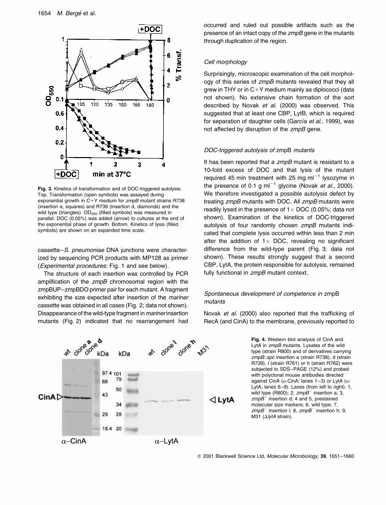
DOC-triggered autolysis of zmpB mutants
It has been reported that a zmpB mutant is resistant to a
10-fold excess of DOC and that lysis of the mutant
required 45 min treatment with 25 mg ml21 lysozyme in
the presence of 0.1 g ml21 glycine (Novak et al., 2000).
We therefore investigated a possible autolysis defect by
treating zmpB mutants with DOC. All zmpB mutants were
readily lysed in the presence of 1� DOC (0.05%; data not
shown). Examination of the kinetics of DOC-triggered
autolysis of four randomly chosen zmpB mutants indi-
cated that complete lysis occurred within less than 2 min
after the addition of 1� DOC, revealing no significant
difference from the wild-type parent (Fig. 3; data not
shown). These results strongly suggest that a second
CBP, LytA, the protein responsible for autolysis, remained
fully functional in zmpB mutant context.
Spontaneous development of competence in zmpB
mutants
Novak et al. (2000) also reported that the trafficking of
RecA (and CinA) to the membrane, previously reported to
Fig. 3. Kinetics of transformation and of DOC-triggered autolysis.Top. Transformation (open symbols) was assayed duringexponential growth in C1Y medium for zmpB mutant strains R738(insertion a, squares) and R739 (insertion d, diamonds) and thewild type (triangles). OD550 (filled symbols) was measured inparallel. DOC (0.05%) was added (arrow) to cultures at the end ofthe exponential phase of growth. Bottom. Kinetics of lysis (filledsymbols) are shown on an expanded time scale.
Fig. 4. Western blot analysis of CinA andLytA in zmpB mutants. Lysates of the wildtype (strain R800) and of derivatives carryingzmpB::spc insertion a (strain R738), d (strainR739), l (strain R761) or h (strain R762) weresubjected to SDS±PAGE (12%) and probedwith polyclonal mouse antibodies directedagainst CinA (a-CinA; lanes 1±3) or LytA (a-LytA; lanes 6±9). Lanes (from left to right): 1,wild type (R800); 2, zmpB2 insertion a; 3,zmpB2 insertion d; 4 and 5, prestainedmolecular size markers; 6, wild type; 7,zmpB2 insertion l; 8, zmpB2 insertion h; 9,M31 (DlytA strain).
1654 M. Berge et al.
Q 2001 Blackwell Science Ltd, Molecular Microbiology, 39, 1651±1660

Fig. 5. Comparison of type 4 and G54 ZmpB sequences. Gaps indicated by dashes were introduced by the CLUSTALW program. Identical aminoacids are in white letters on a black background. The proposed signal peptidase cleavage site (positions 33±34), which conforms to the (23,21) rule, the putative N-terminal cell wall anchor motif (LPNTG; positions 73±77) and the putative C-terminal zinc-binding sequence (HETTH;positions 1562±1566 in ZmpB type 4 and 1540±1544 in ZmpB-G54, followed by an E residue 20 amino acids downstream) are indicated. Theregion between residues 277 and 315 (underlined) is almost exactly repeated in ZmpB type 4 (amino acids 361±402), but not in ZmpB-G54.
The puzzle of zmpB of Streptococcus pneumoniae 1655
Q 2001 Blackwell Science Ltd, Molecular Microbiology, 39, 1651±1660

occur in competent cells of S. pneumoniae (Masure et al.,
1998), was not observed in their zmpB mutant. In line with
this observation, they reported a 99% reduction in the
efficiency of genetic transformation of the zmpB mutant
compared with the parent strain. The addition of compe-
tence stimulating peptide (CSP; HaÊvarstein et al., 1995)
did not improve transformation efficiency.
Although we could not confirm a differential cellular
localization of RecA (and CinA) between competent and
non-competent wild-type cells (Mortier-BarrieÁre, 1999; I.
Mortier-BarrieÁre, M. Prudhomme, L. Fontaine, B. Martin
and J.-P. Claverys, in preparation), spontaneous compe-
tence of the zmpB mutants was assayed under standard
conditions in C1Y medium (Experimental procedures).
Our zmpB mutants displayed normal competence profiles
and efficiencies of genetic transformation similar to those
of the parental strain (Fig. 3, top). These observations
indicated that the RecA protein remained fully functional in
zmpB mutants. They also strongly suggested that
translocation of a third CBP, CBP3, which was recently
shown to be required for transformation (Rimini et al.,
2000), was not affected by disruption of the zmpB gene.
Analysis of CinA and LytA in zmpB mutants
The finding that our zmpB mutants displayed normal cell
morphology, autolysis and genetic transformation effi-
ciency, three properties relying on the presence of at least
three functional CBPs, LytB, LytA and CBP3, strongly
suggested that secretion or translocation of CBPs was
unaffected in the mutants. Nevertheless, we carried out a
Western blot analysis of LytA. In contrast to Novak et al.
(2000), who reported the absence of a band displaying the
apparent molecular mass expected for LytA (36 kDa), we
readily detected this protein with anti-LytA antibodies in
cell extracts of four of the zmpB mutants chosen at
random (Fig. 4; data not shown).
As a last control, we decided to investigate the CinA
protein. According to Novak et al. (2000), no `free' CinA
species could be detected in extracts from a zmpB mutant
on SDS gels. Using standard methods (see Experimental
procedures), we detected the expected 45 kDa CinA
species in extracts of competent cells of both parental and
zmpB mutants using anti-CinA antibodies (Fig. 4; data not
shown), with no hint of an 80 kDa CinA±LytA complex
similar to that described by Novak et al. (2000).
Collectively, these data demonstrate that the secretion
and translocation of the CBPs is unaltered in our zmpb
mutants of S. pneumoniae, in contrast to the report of
Novak et al. (2000) based on their zmpb mutant.
Polymorphism of the zmpB gene in S. pneumoniae
Only four out of eight sequenced mariner cassette±zmpB
junctions could be mapped at the nucleotide level using
the published zmpB type 4 sequence (insertions b, f, g
and d; Fig. 1; data not shown). Other insertions (a, l, h and
m; Fig. 1) could be mapped only with respect to the
sequence of the zmpB gene of strain G54 (EMBL
accession no. AL449926), a 19F capsular-type clinical
isolate (Rimini et al., 2000; data not shown). This
observation suggested the existence of a polymorphism
between the zmpB gene of the type 4 clinical isolate and
of R6, and that R6 and strain G54 share similar zmpB
alleles. Comparison of the sequences of ZmpB type 4
[1906 amino acids, instead of 1882 amino acids as
indicated previously by Novak et al. (2000), because of an
incorrect assignment of the start] and of ZmpB-G54 (1870
amino acids) revealed that the proteins are 99% identical
over the 335 first amino acids and 42% identical in the
polymorphic region (Fig. 5). Although no significant
identity is detected at the DNA level in the polymorphic
region, except for five small (from 37 to 218 nucleotides
long) segments with an average identity of 78%,
sequence identity resumes 20 nucleotides upstream of
the pabB start codon (Fig. 1). Polymorphism of surface-
exposed proteins as observed for ZmpB is not unprece-
dented in S. pneumoniae, as sequence variability has
been reported in several instances (e.g. PspA and PspC/
CbpA/SpsA; Brooks-Walter et al., 1999; Hollingshead
et al., 2000). We observed the existence of a peptide of 33
amino acids with typical features of a prokaryotic export
signal sequence at the N-terminus of ZmpB (Fig. 5),
confirmed the presence of a cell wall anchor domain and
of a zinc-binding motif in the N-terminal and in the C-
terminal region, respectively (Fig. 5), but failed to detect
the previously reported `10 nearly identical tandem
repeats of a 25-mer of unknown function' in either protein.
After communication of our observations, E. Tuomanen
suggested that, although located at the same chromosomal
position between ywlG and pabB (Fig. 1), zmpB-R6 and
zmpB type 4 could constitute two different genes, with
different functions. This seemed rather unlikely in view of the
overall conservation of the ZmpB proteins (Fig. 5). In
addition, taking into account the central role proposed for
ZmpB in the trafficking of CBPs (Novak et al., 2000) and the
importance of CBPs for S. pneumoniae, one would expect
that the function is conserved in the species. Nevertheless,
to resolve the apparently conflicting data, we inactivated the
zmpB gene in the type 4 strain. Substitution of zmpB type 4
by an inactivated copy of zmpB-R6 was readily obtained in
two independent experiments by transformation of strain
FP25, a non-capsulated derivative of the serotype 4 isolate
(Table 1), with chromosomal DNA from strains R736
(zmpB::spc insertion a) and R737 (zmpB::spc insertion d).
The structure of two independent transformants in each
experiment was controlled by PCR amplification of the zmpB
chromosomal region with the zmpBUP±zmpBDO primer
1656 M. Berge et al.
Q 2001 Blackwell Science Ltd, Molecular Microbiology, 39, 1651±1660
pair. A fragment exhibiting the size predicted after substitu-
tion of the zmpB type 4 gene by the cassette-containing
zmpB-R6 gene was obtained in all cases (data not shown,
but see Fig. 2). Disappearance of the wild-type fragment in
the transformants indicated that no rearrangement had
occurred and ruled out possible artifacts such as the
presence of an intact copy of the zmpB type 4 gene in the
transformants through duplication of the region (data not
shown, but see Fig. 2). The presence and location of the
mariner cassette in the transformants was confirmed by
PCR using the cassette-specific primers MP127 or MP128
(Table 1) combined with zmpBDO or zmpBUP (Fig. 6). In
addition, the disappearance of zmpB type 4-specific frag-
ments in the transformants was monitored by PCR using
primer zmpBUP in combination with primers 7693 (Fig. 6A)
or 8594 (data not shown); the latter two primers are type 4
specific (Table 1 and Fig. 1).
None of the two zmpB mutants of the type 4 strain
generated this way displayed extensive chain formation of
the sort described by Novak et al. (2000); they both grew
mainly as diplococci (data not shown). In addition,
examination of the kinetics of DOC-triggered autolysis
revealed no significant difference from the wild-type
parent (data not shown). Thus, the polymorphism of the
zmpB gene between R6 and the serotype 4 isolate cannot
account for the conflicting observations.
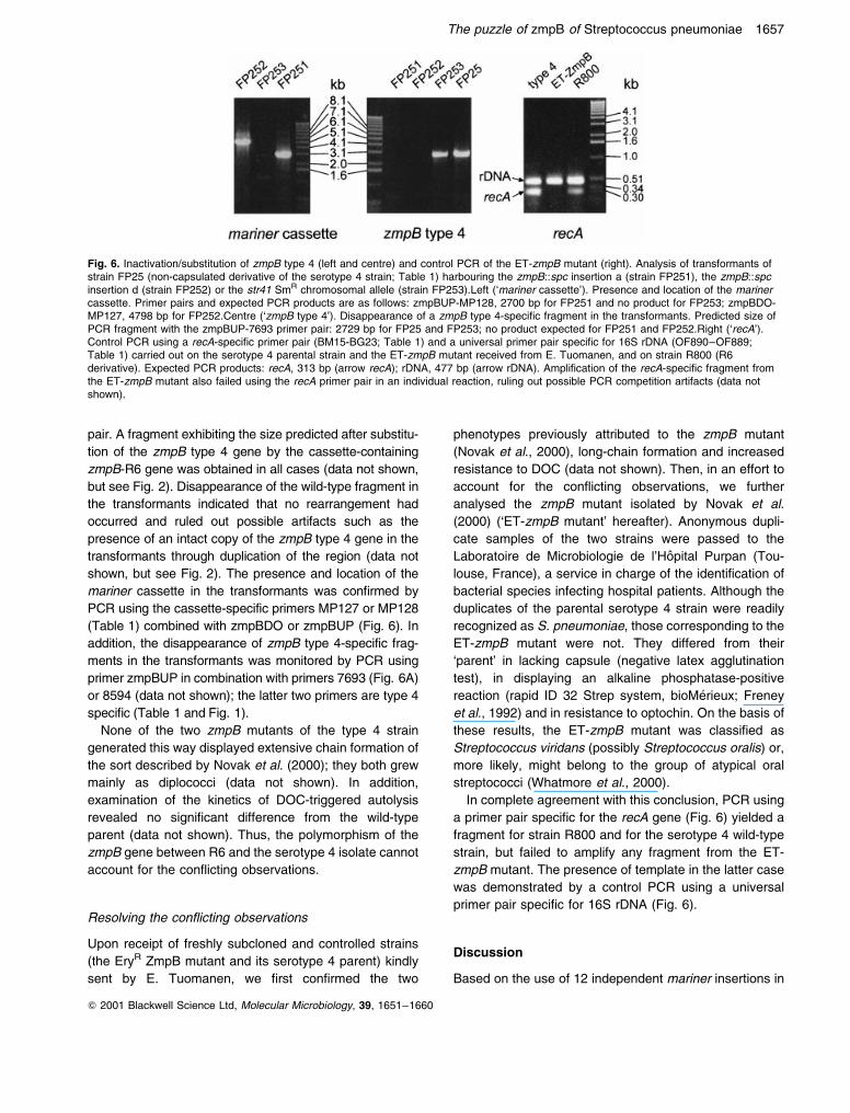
Resolving the conflicting observations
Upon receipt of freshly subcloned and controlled strains
(the EryR ZmpB mutant and its serotype 4 parent) kindly
sent by E. Tuomanen, we first confirmed the two
phenotypes previously attributed to the zmpB mutant
(Novak et al., 2000), long-chain formation and increased
resistance to DOC (data not shown). Then, in an effort to
account for the conflicting observations, we further
analysed the zmpB mutant isolated by Novak et al.
(2000) (`ET-zmpB mutant' hereafter). Anonymous dupli-
cate samples of the two strains were passed to the
Laboratoire de Microbiologie de l'HoÃpital Purpan (Tou-
louse, France), a service in charge of the identification of
bacterial species infecting hospital patients. Although the
duplicates of the parental serotype 4 strain were readily
recognized as S. pneumoniae, those corresponding to the
ET-zmpB mutant were not. They differed from their
`parent' in lacking capsule (negative latex agglutination
test), in displaying an alkaline phosphatase-positive
reaction (rapid ID 32 Strep system, bioMeÂrieux; Freney
et al., 1992) and in resistance to optochin. On the basis of
these results, the ET-zmpB mutant was classified as
Streptococcus viridans (possibly Streptococcus oralis) or,
more likely, might belong to the group of atypical oral
streptococci (Whatmore et al., 2000).
In complete agreement with this conclusion, PCR using
a primer pair specific for the recA gene (Fig. 6) yielded a
fragment for strain R800 and for the serotype 4 wild-type
strain, but failed to amplify any fragment from the ET-
zmpB mutant. The presence of template in the latter case
was demonstrated by a control PCR using a universal
primer pair specific for 16S rDNA (Fig. 6).
Discussion
Based on the use of 12 independent mariner insertions in
Fig. 6. Inactivation/substitution of zmpB type 4 (left and centre) and control PCR of the ET-zmpB mutant (right). Analysis of transformants ofstrain FP25 (non-capsulated derivative of the serotype 4 strain; Table 1) harbouring the zmpB::spc insertion a (strain FP251), the zmpB::spcinsertion d (strain FP252) or the str41 SmR chromosomal allele (strain FP253).Left (`mariner cassette'). Presence and location of the marinercassette. Primer pairs and expected PCR products are as follows: zmpBUP-MP128, 2700 bp for FP251 and no product for FP253; zmpBDO-MP127, 4798 bp for FP252.Centre (`zmpB type 4'). Disappearance of a zmpB type 4-specific fragment in the transformants. Predicted size ofPCR fragment with the zmpBUP-7693 primer pair: 2729 bp for FP25 and FP253; no product expected for FP251 and FP252.Right (`recA').Control PCR using a recA-specific primer pair (BM15-BG23; Table 1) and a universal primer pair specific for 16S rDNA (OF890±OF889;Table 1) carried out on the serotype 4 parental strain and the ET-zmpB mutant received from E. Tuomanen, and on strain R800 (R6derivative). Expected PCR products: recA, 313 bp (arrow recA); rDNA, 477 bp (arrow rDNA). Amplification of the recA-specific fragment fromthe ET-zmpB mutant also failed using the recA primer pair in an individual reaction, ruling out possible PCR competition artifacts (data notshown).
The puzzle of zmpB of Streptococcus pneumoniae 1657
Q 2001 Blackwell Science Ltd, Molecular Microbiology, 39, 1651±1660

the zmpB gene of the well-characterized R6 laboratory
strain, we observed that the mutants (i) do not show
extensive chain formation, which indicates the presence
of a functional LytB CBP; (ii) undergo normal DOC-
triggered autolysis, which relies on the presence of a
functional LytA CBP; (iii) transform at a normal frequency,
which requires the presence of at least a functional CBP3;
and (iv) contain bona fide CinA and LytA species. In
addition, polymorphism of the zmpB gene between strain
R6 (R800) and the serotype 4 strain used by Novak et al.
(2000) could not account for the discrepancy, as we
observed that inactivation of zmpB in the latter does not
affect the separation of daughter cells and autolysis.
Thus, our findings do not confirm that the secretion and
translocation of CBPs to the cell surface is altered after
inactivation of the zmpB gene of S. pneumoniae.
A possible explanation for the discrepancy was
suggested to us by the striking similarity between the
pleiotropic phenotype reported for the zmpB mutant
(Novak et al., 2000) and that previously attributed to
PsaD mutants (Novak et al., 1998). The PsaD phenotype
included (i) reduced sensitivity to the lytic and killing
effects of penicillin; (ii) growth in chains of 200±300 cells;
(iii) autolysis defect and loss of sensitivity to low
concentrations of DOC; (iv) absence of LytA; (v) almost
complete loss of CBPs; (vi) loss of transformability. The
only difference is in the Mn21 requirement for growth in a
chemically defined medium displayed by PsaD mutant
cells. As with the ZmpB mutant phenotype, the phenotypic
traits of Psa mutants could not be confirmed by other
laboratories (Claverys et al., 1999). These observations
suggest that the phenotypes might not result from the
introduced mutations but from low-level contamination of
pneumococcal laboratory stocks by a distinct but trans-
formable streptococcal species. This species would be
related closely enough to S. pneumoniae so as to accept
psa or zmpB knock-out plasmids, but would also display
inherently the phenotypes described ± extensive chain
formation, absence of autolysis and global modification of
CBPs. This hypothesis is consistent with the observation
of an unexplained increase in the size of RecA in some of
the mutants (Novak et al., 1998), whereas DNA diver-
gence could explain the apparent low transformability of
the mutants. The emergence of low-level contaminants
could readily be accounted for by selection for penicillin
tolerance in one case (Novak et al., 1998). In the other
case (Novak et al., 2000), it could be relevant that the S.
pneumoniae serotype 4 isolate used is notoriously difficult
to transform (Bricker and Camilli, 1999).
In the case of the zmpB mutant used by Novak et al.
(2000), the contaminant hypothesis was supported by our
finding that this strain does not belong to the S.
pneumoniae species. The `zmpB mutant' differed from
its S. pneumoniae serotype 4 parent in displaying
resistance to optochin, although sensitivity to optochin is
a characteristic trait of the species, and in lacking a
capsule. The strain was tentatively classified as belonging
to the S. viridans group. The contamination of S.
pneumoniae cultures by related species is not unprece-
dented, as this occurred at an early stage of the TIGR S.
pneumoniae genome sequence project (Adams and
Venter, 1996).
Collectively, our findings do not confirm that the
translocation of CBPs to the cell surface is altered in
zmpB mutants of S. pneumoniae. They provide no
evidence for the existence of the ZmpB protease-
dependent regulatory mechanism governing the translo-
cation of CinA and the CBPs of S. pneumoniae postulated
by Novak et al. (2000). They also do not support the
hypothesis that CinA is involved in the transport of LytA to
the cell membrane. The functions of ZmpB and of CinA in
S. pneumoniae remain to be established.
Experimental procedures
Bacterial strains, growth conditions and competence
The bacterial strains and plasmids used are listed in Table 1.Competence profiles, which assess spontaneous transform-ability, were generated from cultures grown in C1Y mediumas described previously (Alloing et al., 1996). In short, stocksof bacteria grown in CAT medium to an OD550 of 0.4 werediluted 20-fold in C1Y medium and incubated at 378C.Samples were withdrawn at 15 min intervals, from 90 min to180 min after inoculation, and incubated with R304 donorDNA for 20 min. For CSP-induced competence, precompe-tent cells prepared as described previously (Martin et al.,1995) but in THY (30 g l21 Todd±Hewitt, 5 g l21 yeastextract; Difco) instead of CTM 1 were incubated withsynthetic CSP (25 ng ml21; CSP2 for strain FP25) in CTM2 (Martin et al., 1995) at 378C for 10±15 min before theaddition of DNA.
Cells were incubated at 308C during DNA uptake.Transformants were determined by plating in 10 ml of CATagar, followed by challenge with a 10 ml overlay containingthe appropriate antibiotic after phenotypic expression for120 min at 378C. Antibiotic concentrations used for theselection of transformants were 0.05 mg ml21 for erythromy-cin (Ery), 250 mg ml21 for kanamycin (Kan) and 200 mg ml21
for spectinomycin (Spc) and streptomycin (Sm).
In vitro mariner mutagenesis
Transposition reactions were performed using purifiedHimar1 transposase as described previously (Lampe et al.,1996). The target for transposition of the spc minitransposoncarried by plasmid pR412 (Table 1) was a zmpB PCRproduct. Gaps in transposition products were repaired asdescribed previously (Akerley et al., 1998), and repairedtransposition products were used as donors in transforma-tion.
1658 M. Berge et al.
Q 2001 Blackwell Science Ltd, Molecular Microbiology, 39, 1651±1660

PCR reactions and characterization of mariner cassette±
S. pneumoniae DNA junctions
PCR was performed using hot Tub DNA polymerase(Amersham), primers (1 mM) and 30 cycles of amplification(30 s denaturation at 948C, 30 s annealing at 558C and 7±12 min extension at 728C). For DNA sequencing, the zmpbchromosomal region was amplified with the zmpBUP±zmpBDO primer pair. PCR products were purified with theQiaquick DNA cleanup system (Qiagen) and sequenced withthe ThermoSequenase cycle sequencing kit (USB) using thecassette-specific primer MP128.
Proteins
Preparation of cell extracts was as described previously(Martin et al., 1995). Samples were subjected to 12% SDS±PAGE, followed by immunoblot transfer with anti-CinA oranti-LytA antibodies as described previously (Martin et al.,1995).
Acknowledgements
We thank Dr Elaine Tuomanen for helpful discussions and forproviding us with the S. pneumoniae serotype 4 strain andthe zmpB mutant, and Dr Olivier Fayet for the gift of OF889±OF890 primers. We also thank The Institute for GenomicResearch and the Research Department of Glaxo Wellcome(Spain) for granting access to the partially completed genomesequence of S. pneumoniae type 4 isolate (http://www.ti-gr.org) and type 19F isolate (http://srs.ebi.ac.uk) respectively.The work was funded by grants from the Programme deRecherche Fondamentale en Microbiologie, Maladies Infec-tieuses et Parasitaires and by the European Union (grantBIO4-CT98-0424) (to J.-P.C.), from DireccioÂn General deInvestigacioÂn CientõÂfica y TeÂcnica (PB960809) (to P.G.), andfrom Ministero d ell'Universita' e della Ricerca Scientifica eTecnologica (Cofin, 1999).
References
Adams, M.D., and Venter, J.C. (1996) Should non-peer-reviewed raw DNA sequence data release be forced on thescientific community? Science 274: 534±536.
Akerley, B.J., Rubin, E.J., Camilli, A., Lampe, D.J., Robert-son, H.M., and Mekalanos, J.J. (1998) Systematic identi-fication of essential genes by in vitro mariner mutagenesis.Proc Natl Acad Sci USA 95: 8927±8932.
Alloing, G., Granadel, C., Morrison, D.A., and Claverys, J.P.(1996) Competence pheromone, oligopeptide permeaseand induction of competence in Streptococcus pneumo-niae. Mol Microbiol 21: 471±478.
Bricker, A.L., and Camilli, A. (1999) Transformation of type 4encapsulated strain of Streptococcus pneumoniae. FEMSMicrobiol Lett 172: 131±135.
Brooks-Walter, A., Briles, D.E., and Hollingshead, S.K.(1999) The pspC gene of Streptococcus pneumoniaeencodes a polymorphic protein, PspC, which elicits cross-reactive antibodies to PspA and provides immunity topneumococcal bacteremia. Infect Immun 67: 6533±6542.
Claverys, J.P., Granadel, C., Berry, A.M., and Paton, J.C.(1999) Penicillin tolerance in Streptococcus pneumoniae,autolysis and the Psa ABC Mn-permease. Mol Microbiol32: 881±883.
Freney, J., Bland, S., Etienne, J., Desmonceaux, M.,Boeufgras, J.M., and Fleurette, F. (1992) Description andevaluation of the semiautomated 4-hour rapid ID 32 Strepmethod for identification of streptococci and members ofrelated genera. J Clin Microbiol 30: 2657±2661.
GarcõÂa, J.L., SaÂnchez-Beato, A.R., Medrano, F.J., andLoÂpez, R. (1998) Versatility of choline-binding domain.Microb Drug Res 4: 25±36.
GarcõÂa, P., GarcõÂa, J.L., GarcõÂa, E., and LoÂpez, R. (1986)Nucleotide sequence and expression of the pneumococcalautolysin gene from its own promoter in Escherichia coli.Gene 43: 265±272.
GarcõÂa, P., Gonza lez, M.P., GarcõÂa, E., LoÂpez, R., andGarcõÂa, J.L. (1999) LytB, a novel pneumococcal mureinhydrolase essential for cell separation. Mol Microbiol 31:1275±1277.
HaÊvarstein, L.S., Coomaraswamy, G., and Morrison, D.A.(1995) An unmodified heptadecapeptide pheromoneinduces competence for genetic transformation in Strepto-coccus pneumoniae. Proc Natl Acad Sci USA 92: 11140±11144.
Hollingshead, S.K., Becker, R., and Briles, D.E. (2000)Diversity of PspA: mosaic genes and evidence for pastrecombination in Streptococcus pneumoniae. Infect Immun68: 5889±5900.
Holtje, J.-V., and Tomasz, A. (1975) Specific recognition ofcholine residues in the cell wall teichoic acid by the N-acetylmuramyl-L-alanine amidase of pneumococcus. J BiolChem 250: 6072±6076.
Lampe, D.J., Churchill, M.E.A., and Robertson, H.M. (1996)A purified mariner transposase is sufficient to mediatetransposition in vitro. EMBO J 15: 5470±5479.
Lange, R., Wagner, C., de Saizieu, A., Flint, N., Molnos, J.,Stieger, M., et al. (1999) Domain organization andmolecular characterization of 13 two-component systemsidentified by genome sequencing of Streptococcus pneu-moniae. Gene 237: 223±234.
LefeÁvre, J.C., Claverys, J.P., and Sicard, A.M. (1979) Donordeoxyribonucleic acid length and marker effect in pneu-mococcal transformation. J Bacteriol 138: 80±86.
Martin, B., GarcõÂa, P., Castanie , M.P., and Claverys, J.P.(1995) The recA gene of Streptococcus pneumoniae is partof a competence-induced operon and controls lysogenicinduction. Mol Microbiol 15: 367±379.
Martin, B., Prudhomme, M., Alloing, G., Granadel, C., andClaverys, J.P. (2000) Cross-regulation of competencepheromone production and export in the early control oftransformation in Streptococcus pneumoniae. Mol Micro-biol 38: 867±878.
Masure, H.R., Pearce, B.J., Shio, H., and Spellerberg, B.(1998) Membrane targeting of RecA during genetictransformation. Mol Microbiol 27: 845±852.
Mortier-BarrieÁre, I. (1999) Controà le geÂne tique de la compeÂ-tence chez la bacteÂrie aÁ Gram positif Streptococcuspneumoniae: eÂtude de l'opeÂron tardif cinA. PhD Disserta-tion, University P. Sabatier, Toulouse, France.
Mortier-BarrieÁre, I., de Saizieu, A., Claverys, J.P., and Martin,
The puzzle of zmpB of Streptococcus pneumoniae 1659
Q 2001 Blackwell Science Ltd, Molecular Microbiology, 39, 1651±1660

B. (1998) Competence-specific induction of recA isrequired for full recombination proficiency during transfor-mation in Streptococcus pneumoniae. Mol Microbiol 27:159±170.
Novak, R., Braun, J.S., Charpentier, E., and Tuomanen, E.(1998) Penicillin tolerance genes of Streptococcus pneu-moniae: the ABC-type manganese permease complexPsa. Mol Microbiol 29: 1285±1296.
Novak, R., Charpentier, E., Braun, J.S., Park, E., Murti, S.,Tuomanen, E., and Masure, R. (2000) Extracellulartargeting of choline-binding proteins in Streptococcuspneumoniae by a zinc metalloprotease. Mol Microbiol 36:366±376.
Rimini, R., Jansson, B., Feger, G., Roberts, T.C., deFrancesco, M., Gozzi, A., et al. (2000) Global analysis oftranscription kinetics during competence development inStreptococcus pneumoniae. Mol Microbiol 36: 1279±1292.
SaÂnchez-Puelles, J.M., Ronda, C., GarcõÂa, J.L., GarcõÂa, P.,LoÂpez, R., and GarcõÂa, E. (1986) Searching for autolysinfunctions. Characterization of a pneumococcal mutantdeleted in the lytA gene. Eur J Biochem 158: 289±293.
SaÂnchez-Puelles, J.M., Sanz, J.M., GarcõÂa, J.L., and GarcõÂa,
E. (1990) Cloning and expression of gene fragmentsencoding the choline-binding domain of pneumococcalmurein hydrolases. Gene 89: 69±75.
Throup, J.P., Koretke, K.K., Bryant, A.P., Ingraham, K.A.,Chalker, A.F., Ge, Y., et al. (2000) A genomic analysis oftwo-component signal transduction in Streptococcus pneu-moniae. Mol Microbiol 35: 566±576.
Tomasz, A. (1967) Choline in the cell wall of a bacterium:novel type of polymer-linked choline in pneumococcus.Science 157: 694±697.
Tomasz, A. (1984) Building and breaking of bonds in the cellwall of bacteria ± the role of autolysins. In Microbial CellWall Synthesis and Autolysis. Nombela, C. (ed.). Amster-dam: Elsevier, pp. 3±12.
Whatmore, A.M., Efstratiou, A., Pickerill, A.P., Broughton, K.,Woodard, G., Sturgeon, D., et al. (2000) Genetic relation-ships between clinical isolates of Streptococcus pneumo-niae, Streptococcus oralis, and Streptococcus mitis:characterization of ``atypical'' pneumococci and organismsallied to S. mitis harboring S. pneumoniae virulence factor-encoding genes. Infect Immun 68: 1374±1382.
1660 M. Berge et al.
Q 2001 Blackwell Science Ltd, Molecular Microbiology, 39, 1651±1660
Copyright © 2022 FDOKUMEN


![Magnetic ionic plastic crystal: choline[FeCl4]](https://static.fdokumen.com/doc/165x107/634545c06cfb3d4064099b55/magnetic-ionic-plastic-crystal-cholinefecl4.jpg)