Current trends in capsular polysaccharide biosynthesis of Streptococcus pneumoniae
Upload
independentCategory
view
5download
0
Early biofilm formation on microtiter plates is not correlated withthe invasive disease potential of Streptococcus pneumoniae
Anel Lizcano1, Tiffany Chin2, Karin Sauer3, Elaine I. Tuomanen2, and Carlos J. Orihuela11Department of Microbiology and Immunology, The University of Texas Health Science Center atSan Antonio, San Antonio, Texas 78229.2Department of Infectious Diseases, St. Jude Children’s Research Hospital, Memphis, Tennessee38105.3Department of Biological Sciences, Binghamton University SUNY, Binghamton, New York 13902.
AbstractBiofilm formation has been suggested to play an important role during Streptococcus pneumoniaenasopharyngeal colonization and may facilitate progression to pneumonia. To test whether the abilityof S. pneumoniae to form biofilms was important for virulence we screened the ability of 30 invasiveand 22 non-invasive clinical isolates of serotype 6A and 6B to form early biofilms on polystyrenemicrotiter plates and infect mice following intranasal and intratracheal challenge. We first determinedthat no correlation existed between the ability to form early biofilms and whether isolates werecollected from healthy carriers or individuals with invasive disease. A disconnect between biofilmforming ability and the capacity to colonize the nasopharynx, cause pneumonia, and enter thebloodstream was also observed in mice. Importantly, S. pneumoniae mutants deficient in theestablished virulence determinants pneumolysin, CbpA, and hydrogen peroxide formed biofilmsnormally. Incidentally, we determined that robust biofilm production was dependent on the formationand coalescing of bacteria aggregates on a thin layer of bacteria attached to the plate surface. Insummary, these studies suggest that the ability to form early biofilms in vitro does not reflect virulencepotential. More complex studies are required to determine if biofilm formation is important forvirulence.
KeywordsStreptococcus pneumoniae; adhesion; microtiter plate; biofilm; virulence
INTRODUCTIONSince the discovery that Pseudomonas aeruginosa forms biofilms in cystic fibrotic lungs,considerable attention has been placed on the role of bacteria biofilms during infectiousdiseases. To date bacteria in biofilms have been shown to have differences in metabolism,
© 2010 Elsevier Ltd. All rights reserved.Corresponding Author: Carlos J. Orihuela, PhD., Department of Microbiology and Immunology, University of Texas Health ScienceCenter at San Antonio, 7703 Floyd Curl Drive, MC7758, San Antonio, TX 78229-3900, Office: (210) 567-3973, Fax: (210) 567-6612,[email protected]'s Disclaimer: This is a PDF file of an unedited manuscript that has been accepted for publication. As a service to our customerswe are providing this early version of the manuscript. The manuscript will undergo copyediting, typesetting, and review of the resultingproof before it is published in its final citable form. Please note that during the production process errors may be discovered which couldaffect the content, and all legal disclaimers that apply to the journal pertain.
NIH Public AccessAuthor ManuscriptMicrob Pathog. Author manuscript; available in PMC 2011 March 1.
Published in final edited form as:Microb Pathog. 2010 ; 48(3-4): 124–130. doi:10.1016/j.micpath.2010.01.002.
NIH
-PA Author Manuscript
NIH
-PA Author Manuscript
NIH
-PA Author Manuscript

virulence gene expression, and protein production that contribute to surface adhesion andpersistence of an infection. Because biofilm bacteria are enmeshed within an extracellularmatrix, and in some instances metabolically inert, biofilm bacteria are more resistant to killingby leukocytes and antimicrobials, and serve as an recalcitrant source [of bacteria] duringpersistent infections [1,2].
Considerable evidence suggests that biofilm formation is the underlying mechanismresponsible for chronic otitis media. In regards to Streptococcus pneumoniae, the leading causeof otitis media, pneumococci have been detected on the surface of adenoid and mucosalepithelial cells biopsied from children with recurrent middle ear infections [3–5], occludedtympanostomy tubes isolated from the latter [6], and middle ear sections taken from challengedchinchillas [7,8]. More recently, experimental evidence has been collected that suggests a rolefor pneumococcal biofilms during nasopharyngeal colonization and pneumonia. For example,Sanderson et al. found pneumococcal biofilms in biopsies of sinuses from individuals withchronic rhinosinusitis [9]. Following an in vitro screen of transposon mutants Munoz-Elias etal. identified 23 genes necessary for biofilm formation that were also required fornasopharyngeal colonization of mice [10]. Trappetti et al. found that treatment of mice withsialic acid, a condition that enhanced pneumococcal biofilm formation in vitro, increasedbacterial counts in the nasopharynx of mice and instigated translocation of the bacteria into thelungs [11]. Finally, Oggioni and colleagues found that biofilm pneumococci had geneexpression profiles similar to those of bacteria isolated from the lungs of mice; these profileswere distinct from planktonic bacteria isolated from either blood or culture media [12]. Thus,considerable evidence suggests that biofilms are an important aspect of pneumococcalpathogenesis.
To date many investigators have used the static polystyrene microtiter plate system to examinethe molecular mechanisms underlying bacterial attachment to abiotic surfaces and early eventsduring pneumococcal biofilm formation [10–16]. Advantages of this model system includethat it is easy to establish, is applicable to high-throughput screens, and allows visualizationof biofilm structures using an inverted microscope. Based on existing evidence supporting arole for biofilm formation during middle ear infection and nasopharyngeal colonization, wehypothesized that the ability to form biofilms might also contribute towards the ability to causeinvasive pneumococcal disease (IPD). To test this hypothesis, we examined the ability of 30invasive and 22 non-invasive low-passage clinical isolates of serotype 6A and 6B to attach toand form early biofilms (≤18 hours old) on untreated polystyrene 96-well microtiter plates andinfect mice. In this manuscript we show that no correlation was found between biofilmproduction and the source of the clinical isolate, the ability of an isolate to colonize thenasopharynx, or cause invasive disease in mice. We conclude that the ability to form earlybiofilms in vitro had no correlation with virulence in mice and that extrapolations regardingin vitro biofilm formation with virulence are tenuous. These findings emphasize the importanceof testing suspected pathogenic mechanisms using validated model systems along with adiverse panel of clinical isolates.
MATERIALS AND METHODSBacteria strains
Clinical isolates of S. pneumoniae were collected at The University of Texas SouthwesternMedical Center in Dallas County, Texas, from February 1999 to January 2003. A total of 52clinical isolates were examined, 23 serotype 6A isolates and 29 serotype 6B isolates. Non-invasive isolates were obtained from nasopharyngeal swabs of healthy carriers (strains 6A1–6A10 and 6B1–6B12). Invasive isolates were obtained from blood, cerebrospinal fluid, oraspirates of normally sterile sites from individuals with invasive disease. Phylogeneticrelationships between the clinical isolates were extrapolated using comparative genomic
Lizcano et al. Page 2
Microb Pathog. Author manuscript; available in PMC 2011 March 1.
NIH
-PA Author Manuscript
NIH
-PA Author Manuscript
NIH
-PA Author Manuscript

hybridization data previously obtained for these strains [17]. TIGR4 is a virulent laboratorystrain [18]. Isogenic mutants of TIGR4 deficient in pneumolysin (T4 Δpln), Choline bindingprotein A (T4 ΔcpbA), and hydrogen peroxide production (T4 ΔspxB) were made by insertionduplication mutagenesis with the suicide vector pJDC9 using previously described constructs[19]. Mutants were maintained with 1 µg/ml erythromycin.
In vitro screening for biofilm formationWe tested biofilm formation in the static polystyrene microtiter well system previouslydescribed by Allegrucci et al. [16]. Bacteria were streaked from blood agar plates (Remel,Lenexa, KS) into Todd Hewitt Broth (THB) (Difco, Detroit, MI) and grown at 37° C in 5%CO2. At mid-logarithmic phase growth (OD620=0.5) bacteria were diluted 1:20 in mediacontaining 10% glycerol and frozen stocks were created. 96-well polystyrene microtiter plate(CELLSTAR, Greiner Bio-One, Monroe, NC) wells containing 270 µl of THB were inoculatedwith 30 µl of thawed stocks. Plates were incubated at 37° C, 10% CO2, overnight for 18 hours.The next day plates were washed with phosphate buffered saline (PBS), biofilms stained with150 µl 0.5% crystal violet (CV) for 30 minutes, washed with PBS, and allowed to air dry.Biofilm formation was quantified by solubilizing the CV stain with 150 µl of 95% ethanol andmeasuring absorbance with a spectrophotometer at 540 nm (CV540). Images of the biofilmswere captured using a Leica inverted microscope at 15X and 200X magnification prior todrying. Additional wells not inoculated with bacteria served as controls for inadvertentbacterial contamination of the media and served as background levels for CV staining.Experiments were done in triplicate with ≥3 replicate wells tested per strain in each experiment.
Animal infectionsFemale BALB/cJ mice (4–5 weeks old; The Jackson Laboratory) were maintained in animalbiosafety level 2 facilities. For the initial virulence screen of all 52 isolates, a minimum of 2replicate experiments were performed, each with 2–3 mice, for no less than 5 mice sampledfor each clinical isolate tested. Briefly, mice were anesthetized with 2.5% inhaled isoflurane(Baxter Healthcare) and challenged intranasally with 107 colony forming units (CFU)suspended in 25 µL of PBS. Post-infection, blood was collected from the tail vein to test forbacteremia and nasal lavage performed to determine bacterial titers in the nasopharynx [20].For long-term nasopharyngeal colonization studies, cohorts of 6–12 mice were challengedintranasally with 105 CFU in 10 µl PBS and examined over a 28-day period. For intratrachealchallenge experiments cohorts of 6 mice were used. Mice were anesthetized, hung upright bytheir incisors, and 105 CFU in 100 µL of PBS placed in their throats. Aspiration was inducedby gently pulling the tongue outward and covering the nostrils. Lung and blood samples werecollected 2 days later. For blood and nasal lavage samples, serial dilution in PBS followed byplating and colony counting was used to determine the bacterial burden. For lung samples, thelungs were weighed and homogenized in 1 ml of PBS. Bacterial titers in the lungs were assessedper gram of homogenized tissue following serial dilution of the homogenate and colonycounting. In all instances, the infectious dose administered was confirmed.
Statistical analysesDifferences between the highest and lowest biofilm producers in regards to biofilm production,blood, and nasopharyngeal bacterial titers were tested for significance using a two-tailedStudent’s t-test. Comparisons regarding the incidence of positive blood cultures were doneusing a two-tailed Fisher’s exact test. Simple linear regression analysis was performed usingSigmaStat 3.1 software (Systat Software Inc. Point Richmond, CA). For comparison of strainsin the long-term nasopharyngeal colonization studies and the intratracheal challenge studiesstatistical analysis were performed using a One-Way ANOVA.
Lizcano et al. Page 3
Microb Pathog. Author manuscript; available in PMC 2011 March 1.
NIH
-PA Author Manuscript
NIH
-PA Author Manuscript
NIH
-PA Author Manuscript
RESULTSEarly biofilm formation in vitro
The static 96-well microtiter plate model tests for bacterial attachment to an abiotic surfaceand early biofilm formation. All clinical isolates tested were able to attach to the polystyrenesurface; however, individual isolates had differential abilities to form early biofilms asmeasured by CV540, an indicator of biofilm biomass (Figure 1). For example, in serotype 6Athe lowest early biofilm producer was 6A16 which had a CV540 = 0.241 ± 0.047, whereas thegreatest biofilm producer was 6A10 which had a CV540= 2.64 ± 0.133 (6A16 versus 6A10;p<0.001). Likewise for 6B the lowest early biofilm producer was 6B26 which had a CV540 =0.376 ± 0.060 and the highest was 6B8 with a CV540 = 1.457 ± 0.377 (6B26 versus 6B8;p=0.03). Due to the exceptionally high and divergent ability of 6A10 to produce early biofilms,no significant differences in early biofilm production were observed between serotype 6A and6B when compared as a whole. For all 6A isolates the mean CV540 detected was 0.674 ± 0.099;whereas for all 6B isolates the mean CV540 was 0.768 ± 0.051 (p=0.375). Excluding 6A10, astatistically significant difference was observed between serotype 6A and 6B, with 6B isolateshaving 31% higher mean CV540 values (p=0.018).
As exemplified by 6A10, the ability to form biofilms was highly diverse between individualclinical isolates. Closely related strains, including those determined to be clonal (i.e. withinthe same phylogenetic clade) by previous comparative genomic hybridization studies [17],varied considerably in their attachment and early biofilm forming ability. For example, 6A17and 6A18 differed by 2.4-fold (6A17 CV540 = 0.873 ± 0.100; 6A18 CV540 = 0.365 ± 0.044;p=0.009) despite belonging to the same phylogenetic clade. Importantly, no significantdifferences were observed for attachment/early biofilm formation when comparing clinicalisolates from individuals with invasive disease versus those collected from healthy carriers (6Anon-invasive CV540 = 0.825 ± 0.217; 6A invasive CV540 = 0.564 ± 0.066; p=0.199) (6B non-invasive CV540 = 0.809 ± 0.098; 6B invasive CV540 = 0.739 ± 0.058; p=0.513). Thus, wedetermined that initial attachment and early biofilm production was not correlated with thedisease state of the individuals from whom the isolates were obtained, and that sufficientindividual variation existed between clinical isolates (as much as 10-fold) that no statisticallysignificant difference in early biofilm formation occurred between serotype 6A and 6B.
Early biofilm formation is not correlated with the ability to colonize the nasopharynx or causeinvasive disease in mice
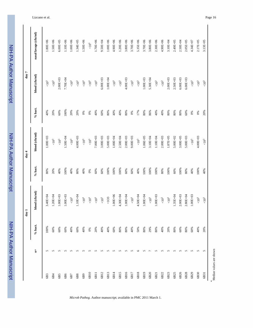
To determine if in vitro early biofilm formation was correlated with in vivo fitness, we firstinfected cohorts of Balb/cJ mice with 107 CFU of each strain and determined their ability tocolonize the nasopharynx and cause bacteremia (Table 1). With exception of 6B10 that failedto colonize mice, we observed stable nasopharyngeal colonization for all isolates on day 7 thatwas comparable with levels observed in previous studies [20,21]. Between strains, colonizationlevels were determined to be uniform with no statistical differences between the highest andlowest colonizers of each serotype (6A21, 6A23, respectively; p=0.111) (6B29, 6B2,respectively; p=0.194). Likewise no significant differences were observed between serotype6A and 6B when compared as groups (average value of medians: 6A = 5.04 ± 0.944 × 10⁵
CFU/ml nasal elute, 6B =3.11 ± 1.47 × 10⁶ CFU/ml; p=0.111). Again, no differences whereobserved when comparing invasive disease isolates versus asymptomatic carrier isolates(average value of medians: 6A non-invasive = 3.33 ± 0.843 × 10⁵ CFU/ml; 6A invasive = 5.82± 1.43 × 10⁵ CFU/ml nasal elute; 6A invasive vs. noninvasive: p=0.192; 6B non-invasive =9.68 ± 1.91 × 10⁵ CFU/ml; 6B invasive = 4.65 ± 2.54 × 10⁵ CFU/ml; 6B invasive vs. non-invasive: p=0.244). Thus excluding 6B10, clinical isolates from serotype 6A and 6B colonizedmice stably and equally despite a differential ability in abiotic surface colonization.
Lizcano et al. Page 4
Microb Pathog. Author manuscript; available in PMC 2011 March 1.
NIH
-PA Author Manuscript
NIH
-PA Author Manuscript
NIH
-PA Author Manuscript

To assess invasiveness, the same mice were tail bled on days 1, 4, and 7 post-challenge andbacteria burden in the blood determined (Table 1). Most mice infected with 6A isolates werenot bacteremic during the 7 days of observation (blood culture positive on day 1: 9/23, day 4:4/23, day 7: 9/23); whereas in contrast the majority of 6B isolates were able to cause bacteremia(day 1: 16/29, day 4: 20/29, day 7 11/29). On day 4, the number of isolates able to causebacteremia was determined to be statistically significant between the serotypes using a two-tailed Fisher’s Exact test (p<0.001). In general bacterial titers in the blood were low with littleto no mortality due to infection. Of the 148 mice infected with serotype 6B only 9 died: 2 for6B6, and 1 for 6B3, 6B14, 6B19, 6B20, 6B25, 6B28, and 6B31. Whereas for 6A, only 15 ofthe 120 mice infected died: 3 for 6A4 and 6A24, 2 for 6A3 and 6A14, and 1 for 6A7, 6A10and 6A15. Importantly, the ability to cause bacteremia was not correlated with the source ofthe clinical isolate (Day 4, invasive vs. asymptomatic carrier: 6A p=1.0; 6B p=0.105),suggesting that a discrepancy also exists between the disease potential of S. pneumoniae inhumans and mice. We next directly tested whether early biofilm formation on a microtiter platewas positively correlated with virulence in the mice. To do this, we performed linear regressionanalysis using CV540 biofilm production measures and nasal lavage and blood bacterial titers.For serotype 6A we used bacterial titers from day 7, as more mice were blood culture positiveat this later time point. For serotype 6B we used day 4 bacterial titers for the same reason.Figure 2 shows that biofilm formation on microtiter plates in vitro was not correlated with theability to colonize the nasopharynx or cause bacteremia for either serotype 6A or 6B.
To more robustly confirm the observed disconnect between biofilm formation and colonizationand invasion, we tested the ability of the 3 highest (6A4, 6A10, 6A13) and 2 lowest (6A16,6A21) biofilm producers of serotype 6A to colonize the nasopharynx in a long-term model(Figure 3A) and following intratracheal challenge cause pneumonia (Figure 3B). Followingintranasal challenge of mice, we observed no significant differences between levels of the 5isolates on days 1, 3, 7, 14, 21 and 28. Thus the highest biofilm producers colonized thenasopharynx equally to those that formed little to no biofilms. Similarly, no differences wereobserved in the lungs of mice infected intratracheally with 6A4, 6A10, and 6A21. Of note,mice infected with 6A13 had no bacteria detectable in the lungs, as did the majority of miceinfected with the low biofilm producing strain 6A16. Thus, in separate experiments, weconfirmed that early in vitro biofilm forming capability was not correlated with the ability tocolonize or cause pneumococcal disease. Finally, we also tested the reverse; whether earlybiofilm formation was affected by the deletion of established virulence determinants knownto be required in the nasopharynx and lungs [19]. Isogenic mutants deficient in the toxinpneumolysin, the adhesin CbpA, and hydrogen peroxide synthesis, all formed biofilmscomparable to the parent strain TIGR4 (T4 WT CV540 = 0.849 ± 0.121; T4 Δpln CV540 = 0.964± 0.124; T4 ΔcbpA CV540 = 0.737 ± 0.072; T4 ΔspxB CV540 = 1.093 ± 0.132). Thus earlybiofilm formation was determined not to be associated with these virulence determinants.
Bacteria aggregation contributes to early biofilm biomassIncidentally, microscopic inspection of the surface attached biofilm biomass determined thatthe ability to form robust surface attached communities was associated with the formation oflarge aggregates on the polystyrene surface (Figure 4). Low early biofilm producers (e.g. 6A16,6B14) formed only a thin layer on the microtiter well bottom composed primarily of individualdiplococci, while mid level biofilm producers formed a similar bacterial “carpet” accentuatedby the presence of small bacterial aggregates (e.g. 6A3, 6B22). As the respective CV540 valuesincreased, the size of the bacterial aggregates also increased, ultimately coalescing intomicrocolonies ranging in size between 100 and 500 µm (e.g. 6A13, 6B25). Finally in the highestearly biofilm producers, the microcolonies converged forming a thick confluent layer ofbacteria along the bottom of the well (e.g. 6A10, 6B7). Thus, increased CV540 values were
Lizcano et al. Page 5
Microb Pathog. Author manuscript; available in PMC 2011 March 1.
NIH
-PA Author Manuscript
NIH
-PA Author Manuscript
NIH
-PA Author Manuscript

associated with an incremental ability to form bacterial aggregates following initial attachmentto the plastic surface.
DISCUSSIONBecause biofilm formation is a mechanism for the pneumococcus to evade the host-defenseand resist antimicrobials, it is reasonable to hypothesize that strains better able to form biofilmsare more virulent. To test this hypothesis, we examined the ability of 32 clinical isolates fromindividuals with IPD and 20 isolates from healthy asymptomatic carriers to form biofilms invitro as well as cause IPD in mice. If our hypothesis were true, we would have predicted thatthe clinical isolates that caused invasive disease in humans, as well as in mice, were those thatbest formed biofilms in vitro. In fact, this was not observed and instead we determined that theability of serotype 6A and 6B isolates to form biofilms in vitro was not correlated with thedisease condition of the host (i.e. invasive or non-invasive), the ability to colonize thenasopharynx, or the ability to cause invasive disease in mice. Thus our experimental resultssuggested that the ability to form early biofilms was not important for IPD.
This study has important limitations. Foremost, we examined the ability of S. pneumoniae toform biofilms in vitro and thus our findings regarding virulence were correlative. Biofilmformation in vivo occurs under selective pressure and environmental signals that are distinctfrom in vitro. Environmental signals such as limited nutrients, the presence of antimicrobialhost factors, and bacteria density may serve as signals for the expression of biofilm relatedgenes. Previously we have shown that gene expression during bacterial growth in media isdistinct from that which occurs in vivo, and from bacteria attached to epithelial cells in vitro[21]. Thus it is possible that biofilm factors are differentially expressed in vitro and that biofilmformation occurs differently in vivo. Furthermore, studies have shown that differences in therichness of growth media, bacteria seeding, and strain capsulation, affect biofilm formation onmicrotiter plates [10,12–14,16]. Had we used different media or altered our growth conditionswe may have observed distinct results. Finally, we only tested serotype 6 clinical isolates.Capsule types 6A and 6B are near identical but the linkage between a component rhamnoseand ribitol is 1→3 for serotype 6A and 1→4 for serotype 6B [22]. Perhaps for other serotypes,biofilm production under these conditions would have been positively correlated with invasivedisease potential.
Currently two model systems are used to grow biofilms in vitro, a static microtiter plate systemand a continuous flow system. Static systems such as the microtiter plate system permit growthof the bacteria in a vessel without replacement of media. Advantages of this model system arethat it is easily amendable to high through-put screens, particularly when using a 96-wellpolystyrene plates. However, this model is short-term as nutrients are depleted and metabolicwastes accumulate; typically growth of the biofilm stops between 8 to 16 hours followed byloss of adhered bacteria presumably due to autolytic properties [13]. Recent findings alsosuggest the accumulation of a biofilm dispersion signal, cis-2-decenoic acid [23]; thus, withoutfrequent exchanges of media, the static system does not allow for the formation of maturebiofilms. Importantly, differences between the microtiter plate system and continuous flowreactor in pneumococcal biofilm formation have been reported. Studies by Allegruci et al.,have shown that the inability to form early biofilms in a microtiter plate is not correlated withthe ability to form biofilms in a flow-through reactor over an extended period [24]. Similarobservations have been made for various other biofilm forming bacteria including Gram-negative pili mutants. While pili-deficient mutants have been shown to be defective in initialattachment to non-coated abiotic surfaces, most retain the capability to form biofilms (althoughwith altered biofilm architecture compared to wild type) following several days of growth underflowing conditions [27–31]. Thus the continuous flow through model emphasizes distinctphysiological properties and may have distinct in vivo correlates. It is therefore possible that
Lizcano et al. Page 6
Microb Pathog. Author manuscript; available in PMC 2011 March 1.
NIH
-PA Author Manuscript
NIH
-PA Author Manuscript
NIH
-PA Author Manuscript

had we used the continuous flow through reactor we would have observed distinct results. Thispossibility highlights the arbitrary nature of the current in vitro biofilm models.
Despite these considerations, our finding that the capability to initiate surface attached growthon microtiter plates was not correlated with disease suggests that for at least serotype 6A and6B the ability to form early biofilms does not model pneumococcal events necessary for thedevelopment of invasive disease in humans or mice. Importantly, biofilm formation onpolystyrene [or: in microtiter plates] with media would be distinct from events in vivo due tothe fact that the bacteria would not be interacting with host components such as mucin,fibronectin, or laminin which may act as bridging molecules between individual bacteria andeukaryotic cells. Of note, finding that TIGR4 mutants deficient in pneumolysin, CbpA, andhydrogen peroxide synthesis were able to form biofilms was consistent with previous findingsby Munoz-Elias et al. which showed that only deletion of lytC, a gene encoding a mureinhydrolase, and cps4E, a capsule synthesis enzyme, affected early biofilm production followinga screen of 6,500 TIGR4 mutants [10].
Interestingly, Munoz-Elias found 49 genes including cbpA that contributed to biofilmproduction in an acapsular strain. One explanation provided for the discrepancy betweencapsulated and unencapsulated mutants in biofilm production was increased bacterialinteractions in the absence of capsule. Of note, 23 of the 49 genes identified by Munoz-Eliaswere subsequently shown to be important for nasopharyngeal colonization in TIGR4 [10].Thus, when using unencapsulated pneumococci and a mutant with its isogenic parent, the earlybiofilm model provided important information regarding bacteria to bacteria interactions thatwere pertinent to the disease process. One important caveat is that clinical isolates of S.pneumoniae are almost always encapsulated, the exception being those collected fromindividuals with conjunctivitis [34]. Also that we observed contradictory results, strains unableto form dense biofilms colonized normally and in some instances were able to cause diseasein mice. Thus these strains lacked biofilm determinants that were not required in vivo;alternatively, the presence of capsule alters the role of surface expressed proteins.
Despite the absence of an invasive disease correlation, our studies were the first to show thatstrains within a single serotype have a diverse ability to form early biofilms. While numerousstudies have shown that the ability to form biofilms is nutrient dependent and enhanced by theabsence of capsule [10,13,15,35], our studies are the first to rigorously show that bacterialcomponents other than capsule play important roles. The finding that phylogenetically similarstrains (i.e. clonal derivatives), as determined by genetic content using microarrays, havediverse early biofilm forming abilities suggests that the ability to form biofilms may beregulated at the transcriptional level and not due to the presence or absence of genes [17].Importantly, this study does not attempt to identify which genes are responsible for alteredbiofilm formation or correlate the serotype 6 clinical isolates with virulence gene expression.Our studies also corroborate earlier work by Hall-Stoodly et al. showing that the formation ofmicrocolonies occurs and that they are important for biofilm biomass accumulation [14].Examination of biofilm images revealed that early pneumococcal biofilm formation asdetermined by CV540 stating intensity coincided with the formation of bacterial aggregatessuperimposed on a bacterial “lawn”; suggesting that the formation of pneumococcal aggregateson top of a bacterial layer is a critical event during early biofilm formation.
In summary, it is clear that the ability to form early biofilms in vitro under the growth conditionstested were not positively correlated with the ability of clinical isolates to cause invasivedisease. This suggests that the ability to form biofilms is not important for invasive disease.However, the discussed limitations in the model system and the fact that these studies arecorrelative, still leaves open the possibility that biofilm formation in vivo is important for
Lizcano et al. Page 7
Microb Pathog. Author manuscript; available in PMC 2011 March 1.
NIH
-PA Author Manuscript
NIH
-PA Author Manuscript
NIH
-PA Author Manuscript

invasive disease. These findings emphasize a need for future studies examining biofilmformation in animals during invasive disease.
AcknowledgmentsThis work was supported by National Institute of Health Grant AI078972.
REFERENCES1. Costerton JW, Stewart PS, Greenberg EP. Bacterial biofilms: A common cause of persistent infection.
Science 1999;284:1318–1322. [PubMed: 10334980]2. Hall-Stoodley L, Stoodley P. Evolving concepts in biofilm infections. Cell Microbiol 2009;11:1034–
1043. [PubMed: 19374653]3. Hoa M, Tomovic S, Nistico L, Hall-Stoodley L, Stoodley P, Sachdeva L, et al. Identification of adenoid
biofilms with middle ear pathogens in otitis-prone children utilizing SEM and FISH. Int J PediatrOtorhinolaryngol. 2009
4. Coates H, Thornton R, Langlands J, Filion P, Keil AD, Vijayasekaran S, et al. The role of chronicinfection in children with otitis media with effusion: evidence for intracellular persistence of bacteria.Otolaryngol Head Neck Surg 2008;138:778–781. [PubMed: 18503854]
5. Hall-Stoodley L, Hu FZ, Gieseke A, Nistico L, Nguyen D, Hayes J, et al. Direct detection of bacterialbiofilms on the middle-ear mucosa of children with chronic otitis media. JAMA 2006;296:202–211.[PubMed: 16835426]
6. Mehta AJ, Lee JC, Stevens GR, Antonelli PJ. Opening plugged tympanostomy tubes: effect of biofilmformation. Otolaryngol Head Neck Surg 2006;134:121–125. [PubMed: 16399191]
7. Reid SD, Hong W, Dew KE, Winn DR, Pang B, Watt J, et al. Streptococcus pneumoniae forms surface-attached communities in the middle ear of experimentally infected chinchillas. J Infect Dis2009;199:786–794. [PubMed: 19434911]
8. Hoa M, Syamal M, Sachdeva L, Berk R, Coticchia J. Demonstration of nasopharyngeal and middleear mucosal biofilms in an animal model of acute otitis media. Ann Otol Rhinol Laryngol2009;118:292–298. [PubMed: 19462851]
9. Sanderson AR, Leid JG, Hunsaker D. Bacterial biofilms on the sinus mucosa of human subjects withchronic rhinosinusitis. Laryngoscope 2006;116:1121–1126. [PubMed: 16826045]
10. Munoz-Elias EJ, Marcano J, Camilli A. Isolation of Streptococcus pneumoniae biofilm mutants andtheir characterization during nasopharyngeal colonization. Infect Immun 2008;76:5049–5061.[PubMed: 18794289]
11. Trappetti C, Kadioglu A, Carter M, Hayre J, Iannelli F, Pozzi G, et al. Sialic acid: a preventable signalfor pneumococcal biofilm formation, colonization, and invasion of the host. J Infect Dis2009;199:1497–1505. [PubMed: 19392624]
12. Oggioni MR, Trappetti C, Kadioglu A, Cassone M, Iannelli F, Ricci S, et al. Switch from planktonicto sessile life: a major event in pneumococcal pathogenesis. Mol Microbiol 2006;61:1196–1210.[PubMed: 16925554]
13. Moscoso M, Garcia E, Lopez R. Biofilm formation by Streptococcus pneumoniae: role of choline,extracellular DNA, and capsular polysaccharide in microbial accretion. J Bacteriol 2006;188:7785–7795. [PubMed: 16936041]
14. Hall-Stoodley L, Nistico L, Sambanthamoorthy K, Dice B, Nguyen D, Mershon WJ, et al.Characterization of biofilm matrix, degradation by DNase treatment and evidence of capsuledownregulation in Streptococcus pneumoniae clinical isolates. BMC Microbiol 2008;8:173.[PubMed: 18842140]
15. Allegrucci M, Sauer K. Formation of Streptococcus pneumoniae non-phase-variable colony variantsis due to increased mutation frequency present under biofilm growth conditions. J Bacteriol2008;190:6330–6339. [PubMed: 18658260]
16. Allegrucci M, Sauer K. Characterization of colony morphology variants isolated from Streptococcuspneumoniae biofilms. J Bacteriol 2007;189:2030–2038. [PubMed: 17189375]
Lizcano et al. Page 8
Microb Pathog. Author manuscript; available in PMC 2011 March 1.
NIH
-PA Author Manuscript
NIH
-PA Author Manuscript
NIH
-PA Author Manuscript

17. Obert C, Sublett J, Kaushal D, Hinojosa E, Barton T, Tuomanen EI, et al. Identification of a candidateStreptococcus pneumoniae core genome and regions of diversity correlated with invasivepneumococcal disease. Infect Immun 2006;74:4766–4777. [PubMed: 16861665]
18. Tettelin H, Nelson KE, Paulsen IT, Eisen JA, Read TD, Peterson S, et al. Complete genome sequenceof a virulent isolate of Streptococcus pneumoniae. Science 2001;293:498–506. [PubMed: 11463916]
19. Orihuela CJ, Gao G, Francis KP, Yu J, Tuomanen EI. Tissue-specific contributions of pneumococcalvirulence factors to pathogenesis. J Infect Dis 2004;190:1661–1669. [PubMed: 15478073]
20. Embry A, Hinojosa E, Orihuela CJ. Regions of Diversity 8, 9 and 13 contribute to Streptococcuspneumoniae virulence. BMC Microbiol 2007;7:80. [PubMed: 17723151]
21. Orihuela CJ, Radin JN, Sublett JE, Gao G, Kaushal D, Tuomanen EI. Microarray analysis ofpneumococcal gene expression during invasive disease. Infect Immun 2004;72:5582–5596.[PubMed: 15385455]
22. Mavroidi A, Godoy D, Aanensen DM, Robinson DA, Hollingshead SK, Spratt BG. Evolutionarygenetics of the capsular locus of serogroup 6 pneumococci. J Bacteriol 2004;186:8181–8192.[PubMed: 15576766]
23. Davies DG, Marques CNH. A Fatty Acid Messenger Is Responsible for Inducing Dispersion inMicrobial Biofilms. J. Bacteriol 2009;191:1393–1403. [PubMed: 19074399]
24. Allegrucci M, Hu FZ, Shen K, Hayes J, Ehrlich GD, Post JC, et al. Phenotypic characterization ofStreptococcus pneumoniae biofilm development. J Bacteriol 2006;188:2325–2335. [PubMed:16547018]
25. Donlan RM, Piede JA, Heyes CD, Sanii L, Murga R, Edmonds P, et al. Model system for growingand quantifying Streptococcus pneumoniae biofilms in situ and in real time. Appl Environ Microbiol2004;70:4980–4988. [PubMed: 15294838]
26. Budhani RK, Struthers JK. Interaction of Streptococcus pneumoniae and Moraxella catarrhalis:investigation of the indirect pathogenic role of beta-lactamase-producing moraxellae by use of acontinuous-culture biofilm system. Antimicrob Agents Chemother 1998;42:2521–2526. [PubMed:9756750]
27. Shime-Hattori A, Iida T, Arita M, Park K-S, Kodama T, Honda T. Two type IV pili of Vibrioparahaemolyticus play different roles in biofilm formation. FEMS Microbiology Letters2006;264:89–97. [PubMed: 17020553]
28. Morgan R, Kohn S, Hwang S-H, Hassett DJ, Sauer K. BdlA, a Chemotaxis Regulator Essential forBiofilm Dispersion in Pseudomonas aeruginosa. J. Bacteriol 2006;188:7335–7343. [PubMed:17050921]
29. O'Toole GA, Kolter R. Flagellar and twitching motility are necessary for Pseudomonas aeruginosabiofilm development. Molecular Microbiology 1998;30:295–304. [PubMed: 9791175]
30. O'Toole GA, Kolter R. Initiation of biofilm formation in Pseudomonas fluorescens WCS365 proceedsvia multiple, convergent signalling pathways: a genetic analysis. Molecular Microbiology1998;28:449–461. [PubMed: 9632250]
31. Gohl O, Friedrich A, Hoppert M, Averhoff B. The Thin Pili of Acinetobacter sp. Strain BD413Mediate Adhesion to Biotic and Abiotic Surfaces. Appl. Environ. Microbiol 2006;72:1394–1401.[PubMed: 16461692]
32. Orihuela CJ, Mahdavi J, Thornton J, Mann B, Wooldridge KG, Abouseada N, et al. Laminin receptorinitiates bacterial contact with the blood brain barrier in experimental meningitis models. J Clin Invest2009;119:1638–1646. [PubMed: 19436113]
33. Holmes AR, McNab R, Millsap KW, Rohde M, Hammerschmidt S, Mawdsley JL, et al. The pavAgene of Streptococcus pneumoniae encodes a fibronectin-binding protein that is essential forvirulence. Mol Microbiol 2001;41:1395–1408. [PubMed: 11580843]
34. Martin M, Turco JH, Zegans ME, Facklam RR, Sodha S, Elliott JA, et al. An outbreak of conjunctivitisdue to atypical Streptococcus pneumoniae. N Engl J Med 2003;348:1112–1121. [PubMed:12646668]
35. McEllistrem MC, Ransford JV, Khan SA. Characterization of in vitro biofilm-associatedpneumococcal phase variants of a clinically relevant serotype 3 clone. J Clin Microbiol 2007;45:97–101. [PubMed: 17093036]
Lizcano et al. Page 9
Microb Pathog. Author manuscript; available in PMC 2011 March 1.
NIH
-PA Author Manuscript
NIH
-PA Author Manuscript
NIH
-PA Author Manuscript
Figure 1. Biofilm formation on 96-well polystyrene microtiter plates by individual isolates of S.pneumoniaeClinical isolates belonging to serotype 6A and 6B were collected from individuals with invasivedisease (bold font) and healthy asymptomatic carriers (regular font). Media in the wells wereinoculated with ~105 CFU and incubated overnight at 37° C at 10% CO2. Plates were washed,and biofilm formation was assessed by crystal violet staining (CV540) as described in themethods. Shown is the average of three independent experiments, each with >3 replicate wellsfor each clinical isolate tested. Error bars indicate the standard error of means. Phylogenetictrees on the left of the isolate name are based on comparative genomic analyses done usingmicroarrays [17]. Branches indicate phylogenetic relationships between the clinical isolates incontext of TIGR4, a serotype 4 isolate, from whose genomic DNA the microarray wasdesigned.
Lizcano et al. Page 10
Microb Pathog. Author manuscript; available in PMC 2011 March 1.
NIH
-PA Author Manuscript
NIH
-PA Author Manuscript
NIH
-PA Author Manuscript
Figure 2. Biofilm formation is not correlated with nasopharyngeal colonization or bacteremiaLinear regression analysis was used to determine if biofilm formation was correlated with theability to colonize the nasopharynx and cause invasive disease. For nasopharyngealcolonization, CV540 values were plotted against median bacterial titers in nasal lavage elutesfrom day 7. For 6A invasive disease, CV540 values was plotted against median bacteremiatiters at day 7, the day the majority of mice had bacteria in the blood. For 6B invasive disease,CV540 values was plotted against median bacteremia titers at day 4 for the same reason. Forboth 6A and 6B no correlation was found between biofilm formation and invasive disease ondays 1, 4 or 7 (data not shown).
Lizcano et al. Page 11
Microb Pathog. Author manuscript; available in PMC 2011 March 1.
NIH
-PA Author Manuscript
NIH
-PA Author Manuscript
NIH
-PA Author Manuscript

Figure 3. Ability of high and low serotype 6A biofilm producers to colonize the nasopharynx long-term and cause IPDA) Average bacterial titers in nasal lavage samples collected from mice colonized with the highbiofilm producing strains (black shapes) 6A10, 6A13 and 6A21 as well as the low biofilmproducing strains (white shapes) 6A21 and 6A16. Mice were challenged with 105 CFU in 10µl PBS and bacterial titers in the nasopharynx determined on days 1, 3, 7, 14, 21 and 28. Nostatistical differences were observed between the groups using One-Way ANOVA. Error barsindicate ± SEM. B) Bacterial titers in the lungs and blood 48 hours after intratracheal challengeof mice with the same 6A isolates. Mice were infected with 105 CFU in 100 µl PBS. Individual
Lizcano et al. Page 12
Microb Pathog. Author manuscript; available in PMC 2011 March 1.
NIH
-PA Author Manuscript
NIH
-PA Author Manuscript
NIH
-PA Author Manuscript

shapes represent distinct mice. Horizontal bars designate the median bacterial value. Statisticalanalyses between the groups were performed using One-Way ANOVA.
Lizcano et al. Page 13
Microb Pathog. Author manuscript; available in PMC 2011 March 1.
NIH
-PA Author Manuscript
NIH
-PA Author Manuscript
NIH
-PA Author Manuscript

Figure 4. Increased biofilm biomass corresponds to the formation of increasingly larger bacterialaggregatesRepresentative micrographs of pneumococcal biofilms forming on the bottom of thepolystyrene microtiter plate wells. Images were taken with at 200X with an invertedmicroscope. For 6A10 we include an insert of a well taken at 15X magnification. This is todemonstrate that the 6A10 biofilm has completely covered the well bottom. Strains wereselected based on their increasing CV540 value.
Lizcano et al. Page 14
Microb Pathog. Author manuscript; available in PMC 2011 March 1.
NIH
-PA Author Manuscript
NIH
-PA Author Manuscript
NIH
-PA Author Manuscript

NIH
-PA Author Manuscript
NIH
-PA Author Manuscript
NIH
-PA Author Manuscript
Lizcano et al. Page 15
Tabl
e 1
Perc
enta
ge o
f mic
e w
ith p
ositi
ve b
lood
cul
ture
s fol
low
ing
intra
nsal
cha
lleng
e an
d ba
cter
ial b
urde
n in
the
bloo
d an
d na
soph
aryn
x*
day
1da
y 4
day
7
n=%
bac
t.bl
ood
(cfu
/ml)
% b
act.
bloo
d (c
fu/m
l)%
bac
t.bl
ood
(cfu
/ml)
nasa
l lav
age
(cfu
/ml)
6A1
50%
<103
0%<1
0320
%<1
038.
00E+
05
6A2
50%
<103
40%
<103
20%
<103
3.00
E+05
6A3
560
%7.
00E+
0310
0%4.
40E+
0680
%1.
97E+
054.
20E+
04
6A4
580
%3.
00E+
0310
0%1.
00E+
0660
%5.
00E+
085.
45E+
04
6A5
50%
<103
20%
<103
20%
<103
6.00
E+05
6A6
50%
<103
0%<1
030%
<103
2.00
E+05
6A7
50%
<103
20%
<103
60%
8.70
E+04
6.50
E+05
6A8
50%
<103
40%
<103
0%<1
032.
00E+
05
6A9
50%
<103
0%<1
030%
<103
1.11
E+05
6A10
560
%4.
00E+
0320
%<1
0320
%<1
033.
68E+
05
6A11
560
%2.
00E+
0360
%2.
00E+
0320
%<1
037.
00E+
05
6A12
50%
<103
0%<1
030%
<103
1.00
E+06
6A13
520
%<1
030%
<103
0%<1
032.
00E+
05
6A14
540
%<1
0340
%<1
0380
%4.
20E+
061.
00E+
05
6A15
560
%2.
00E+
0340
%<1
0360
%1.
68E+
054.
50E+
05
6A16
50%
<103
0%<1
030%
<103
6.00
E+05
6A17
560
%1.
00E+
0320
%<1
030%
<103
2.00
E+05
6A18
580
%1.
50E+
0480
%5.
70E+
0480
%9.
00E+
038.
50E+
04
6A20
80%
<103
0%<1
0313
%<1
034.
50E+
05
6A21
540
%<1
0320
%<1
0360
%1.
00E+
031.
70E+
06
6A22
650
%3.
00E+
0317
%<1
0350
%1.
00E+
031.
55E+
06
6A23
540
%<1
0320
%<1
0320
%<1
031.
29E+
05
6A24
667
%9.
34E+
0523
%<1
0383
%2.
53E+
084.
00E+
05
6B1
580
%4.
00E+
0340
%<1
0320
%<1
031.
20E+
06
6B2
540
%<1
0310
0%2.
00E+
0320
%<1
032.
00E+
04
Microb Pathog. Author manuscript; available in PMC 2011 March 1.
NIH
-PA Author Manuscript
NIH
-PA Author Manuscript
NIH
-PA Author Manuscript
Lizcano et al. Page 16
day
1da
y 4
day
7
n=%
bac
t.bl
ood
(cfu
/ml)
% b
act.
bloo
d (c
fu/m
l)%
bac
t.bl
ood
(cfu
/ml)
nasa
l lav
age
(cfu
/ml)
6B3
510
0%3.
40E+
0480
%1.
00E+
0340
%<1
031.
80E+
06
6B4
560
%1.
20E+
0420
%<1
0320
%<1
031.
50E+
06
6B5
560
%1.
00E+
0340
%<1
0360
%2.
00E+
036.
00E+
05
6B6
560
%1.
00E+
0310
0%1.
50E+
0410
0%7.
70E+
041.
10E+
06
6B7
540
%<1
0340
%<1
0320
%<1
031.
00E+
06
6B8
560
%1.
10E+
0480
%4.
00E+
0320
%<1
031.
34E+
05
6B9
540
%<1
0340
%<1
030%
<103
1.50
E+06
6B10
50%
<103
0%<1
030%
<103
<103
6B11
520
%<1
0360
%7.
00E+
0340
%<1
031.
70E+
06
6B12
540
%<1
0360
%3.
00E+
0360
%6.
00E+
039.
50E+
04
6B13
540
%<1
O3
100%
5.00
E+03
80%
1.00
E+04
1.00
E+06
6B14
560
%1.
00E+
0610
0%1.
00E+
0440
%<1
034.
90E+
06
6B15
580
%4.
30E+
0410
0%2.
50E+
0440
%<1
031.
20E+
06
6B16
580
%1.
00E+
0460
%2.
00E+
0380
%4.
00E+
033.
80E+
06
6B17
540
%<1
0360
%9.
00E+
030%
<103
3.70
E+06
6B18
650
%4.
50E+
0440
%<1
0317
%<1
035.
35E+
06
6B19
580
%1.
00E+
0410
0%1.
06E+
0580
%1.
00E+
033.
70E+
06
6B20
729
%<1
0310
0%3.
10E+
0486
%5.
30E+
043.
80E+
06
6B21
580
%1.
00E+
0310
0%1.
10E+
0440
%<1
032.
30E+
06
6B22
540
%<1
0380
%2.
00E+
0340
%<1
034.
90E+
06
6B23
520
%<1
0310
0%1.
87E+
0580
%2.
00E+
033.
10E+
04
6B25
580
%1.
35E+
0460
%5.
00E+
0280
%2.
50E+
032.
49E+
05
6B26
560
%2.
00E+
0310
0%3.
00E+
0580
%6.
00E+
032.
50E+
04
6B28
580
%2.
80E+
0480
%5.
00E+
0360
%6.
00E+
032.
05E+
05
6B29
560
%1.
00E+
0340
%<1
030%
<103
4.34
E+07
6B30
540
%<1
0310
0%4.
00E+
030%
<103
2.17
E+05
6B31
520
%<1
0340
%<1
0320
%<1
033.
13E+
05
* Med
ian
valu
es a
re sh
own
Microb Pathog. Author manuscript; available in PMC 2011 March 1.
Copyright © 2022 FDOKUMEN