
The Cholesterol-Dependent Cytolysin Pneumolysin from Streptococcus pneumoniae Binds to Lipid Raft...
11
The Cholesterol-Dependent Cytolysin Pneumolysin from Streptococcus pneumoniae Binds to Lipid Raft Microdomains in Human Corneal Epithelial Cells Sidney D. Taylor, Melissa E. Sanders, Nathan A. Tullos, Stephen J. Stray, Erin W. Norcross, Larry S. McDaniel, Mary E. Marquart* Department of Microbiology, University of Mississippi Medical Center, Jackson, Mississippi, United States of America Abstract Streptococcus pneumoniae (pneumococcus) is an opportunistic bacterial pathogen responsible for causing several human diseases including pneumonia, meningitis, and otitis media. Pneumococcus is also a major cause of human ocular infections and is commonly isolated in cases of bacterial keratitis, an infection of the cornea. The ocular pathology that occurs during pneumococcal keratitis is partly due to the actions of pneumolysin (Ply), a cholesterol-dependent cytolysin produced by pneumococcus. The lytic mechanism of Ply is a three step process beginning with surface binding to cholesterol. Multiple Ply monomers then oligomerize to form a prepore. The prepore then undergoes a conformational change that creates a large pore in the host cell membrane, resulting in cell lysis. We engineered a collection of single amino acid substitution mutants at residues (A370, A406, W433, and L460) that are crucial to the progression of the lytic mechanism and determined the effects that these mutations had on lytic function. Both Ply WT and the mutant Ply molecules (Ply A370G , Ply A370E , Ply A406G , Ply A406E , Ply W433G , Ply W433E , Ply W433F , Ply L460G , and Ply L460E ) were able to bind to the surface of human corneal epithelial cells (HCECs) with similar efficiency. Additionally, Ply WT localized to cholesterol-rich microdomains on the HCEC surface, however, only one mutant (Ply A370G ) was able to duplicate this behavior. Four of the 9 mutant Ply molecules (Ply A370E , Ply W433G , Ply W433E , and Ply L460E ) were deficient in oligomer formation. Lastly, all of the mutant Ply molecules, except Ply A370G , exhibited significantly impaired lytic activity on HCECs. The other 8 mutants all experienced a reduction in lytic activity, but 4 of the 8 retained the ability to oligomerize. A thorough understanding of the molecular interactions that occur between Ply and the target cell, could lead to targeted treatments aimed to reduce the pathology observed during pneumococcal keratitis. Citation: Taylor SD, Sanders ME, Tullos NA, Stray SJ, Norcross EW, et al. (2013) The Cholesterol-Dependent Cytolysin Pneumolysin from Streptococcus pneumoniae Binds to Lipid Raft Microdomains in Human Corneal Epithelial Cells. PLoS ONE 8(4): e61300. doi:10.1371/journal.pone.0061300 Editor: Eliane Namie Miyaji, Instituto Butantan, Brazil Received August 6, 2012; Accepted March 11, 2013; Published April 5, 2013 Copyright: ß 2013 Taylor et al. This is an open-access article distributed under the terms of the Creative Commons Attribution License, which permits unrestricted use, distribution, and reproduction in any medium, provided the original author and source are credited. Funding: This work was supported by Public Health Services Grant R01EY016195 (MEM), National Eye Institute, National Institutes of Health. The funders had no role in study design, data collection and analysis, decision to publish, or preparation of the manuscript. Competing Interests: The authors have declared that no competing interests exist. * E-mail: [email protected] Introduction Streptococcus pneumoniae (pneumococcus) is a worldwide pathogen responsible for both invasive and noninvasive infections, including pneumonia, meningitis, bacteremia, and otitis media [1–5]. Additionally, pneumococcus is known to be the etiologic agent of several ocular infections including conjunctivitis, endophthal- mitis, and keratitis [6–14]. Pneumococcus is typically among the top three most commonly isolated species from cases of bacterial keratitis, an infection of the cornea of the eye [13,15,16]. Pneumococcal keratitis can be a sight-threatening infection if left untreated or if treatment is delayed. Corneal ulceration occurs during the course of the infection and often results in an opaque scarification of the corneal surface after the infection is cleared. The lytic action of pneumolysin (Ply), a 53 kilodalton (kDa) virulence protein produced by S. pneumoniae, is responsible for the formation of corneal ulcers and is a major contributor to pneumococcal virulence as a whole [17–23]. Ply is a member of the cholesterol-dependent cytolysin (CDC) family of proteins, a group of pore-forming proteins from several gram positive bacterial genera including Streptococcus, Listeria, Bacillus, and Clostridium [24]. All CDCs are thought to share a common lytic mechanism which is 100% dependent on the presence of cholesterol in the target cell membrane [24]. In the case of Ply, cholesterol serves as the initial binding ligand which anchors Ply to the host cell surface [25]. After binding to cholesterol, surface- bound Ply monomers are oriented perpendicular to the cell surface and begin to diffuse laterally across the host membrane [26]. Eventually Ply monomers will interact with one another and oligomerize to form a large multimeric prepore structure consisting of 34–50 monomers [24,27]. The prepore structure then undergoes a synchronized conformational change that causes a vertical collapse of the entire complex and the insertion of two b- hairpin structures from each monomer into the host membrane [28]. The b-hairpins collectively form a large b-barrel pore approximately 25 nm in diameter that traverses the cell mem- brane resulting in osmotic disregulation and cell death [28,29]. Much of what is known about Ply has been extrapolated from previous findings focusing on the lytic mechanism of perfringolysin (Pfo), the CDC produced by Clostridium perfringens. The tertiary PLOS ONE | www.plosone.org 1 April 2013 | Volume 8 | Issue 4 | e61300
Transcript of The Cholesterol-Dependent Cytolysin Pneumolysin from Streptococcus pneumoniae Binds to Lipid Raft...

The Cholesterol-Dependent Cytolysin Pneumolysin fromStreptococcus pneumoniae Binds to Lipid RaftMicrodomains in Human Corneal Epithelial CellsSidney D. Taylor, Melissa E. Sanders, Nathan A. Tullos, Stephen J. Stray, Erin W. Norcross,
Larry S. McDaniel, Mary E. Marquart*
Department of Microbiology, University of Mississippi Medical Center, Jackson, Mississippi, United States of America
Abstract
Streptococcus pneumoniae (pneumococcus) is an opportunistic bacterial pathogen responsible for causing several humandiseases including pneumonia, meningitis, and otitis media. Pneumococcus is also a major cause of human ocular infectionsand is commonly isolated in cases of bacterial keratitis, an infection of the cornea. The ocular pathology that occurs duringpneumococcal keratitis is partly due to the actions of pneumolysin (Ply), a cholesterol-dependent cytolysin produced bypneumococcus. The lytic mechanism of Ply is a three step process beginning with surface binding to cholesterol. MultiplePly monomers then oligomerize to form a prepore. The prepore then undergoes a conformational change that creates alarge pore in the host cell membrane, resulting in cell lysis. We engineered a collection of single amino acid substitutionmutants at residues (A370, A406, W433, and L460) that are crucial to the progression of the lytic mechanism and determinedthe effects that these mutations had on lytic function. Both PlyWT and the mutant Ply molecules (PlyA370G, PlyA370E, PlyA406G,PlyA406E, PlyW433G, PlyW433E, PlyW433F, PlyL460G, and PlyL460E) were able to bind to the surface of human corneal epithelial cells(HCECs) with similar efficiency. Additionally, PlyWT localized to cholesterol-rich microdomains on the HCEC surface, however,only one mutant (PlyA370G) was able to duplicate this behavior. Four of the 9 mutant Ply molecules (PlyA370E, PlyW433G,PlyW433E, and PlyL460E) were deficient in oligomer formation. Lastly, all of the mutant Ply molecules, except PlyA370G,exhibited significantly impaired lytic activity on HCECs. The other 8 mutants all experienced a reduction in lytic activity, but4 of the 8 retained the ability to oligomerize. A thorough understanding of the molecular interactions that occur betweenPly and the target cell, could lead to targeted treatments aimed to reduce the pathology observed during pneumococcalkeratitis.
Citation: Taylor SD, Sanders ME, Tullos NA, Stray SJ, Norcross EW, et al. (2013) The Cholesterol-Dependent Cytolysin Pneumolysin from Streptococcus pneumoniaeBinds to Lipid Raft Microdomains in Human Corneal Epithelial Cells. PLoS ONE 8(4): e61300. doi:10.1371/journal.pone.0061300
Editor: Eliane Namie Miyaji, Instituto Butantan, Brazil
Received August 6, 2012; Accepted March 11, 2013; Published April 5, 2013
Copyright: � 2013 Taylor et al. This is an open-access article distributed under the terms of the Creative Commons Attribution License, which permitsunrestricted use, distribution, and reproduction in any medium, provided the original author and source are credited.
Funding: This work was supported by Public Health Services Grant R01EY016195 (MEM), National Eye Institute, National Institutes of Health. The funders had norole in study design, data collection and analysis, decision to publish, or preparation of the manuscript.
Competing Interests: The authors have declared that no competing interests exist.
* E-mail: [email protected]
Introduction
Streptococcus pneumoniae (pneumococcus) is a worldwide pathogen
responsible for both invasive and noninvasive infections, including
pneumonia, meningitis, bacteremia, and otitis media [1–5].
Additionally, pneumococcus is known to be the etiologic agent
of several ocular infections including conjunctivitis, endophthal-
mitis, and keratitis [6–14]. Pneumococcus is typically among the
top three most commonly isolated species from cases of bacterial
keratitis, an infection of the cornea of the eye [13,15,16].
Pneumococcal keratitis can be a sight-threatening infection if left
untreated or if treatment is delayed. Corneal ulceration occurs
during the course of the infection and often results in an opaque
scarification of the corneal surface after the infection is cleared.
The lytic action of pneumolysin (Ply), a 53 kilodalton (kDa)
virulence protein produced by S. pneumoniae, is responsible for the
formation of corneal ulcers and is a major contributor to
pneumococcal virulence as a whole [17–23]. Ply is a member of
the cholesterol-dependent cytolysin (CDC) family of proteins, a
group of pore-forming proteins from several gram positive
bacterial genera including Streptococcus, Listeria, Bacillus, and
Clostridium [24]. All CDCs are thought to share a common lytic
mechanism which is 100% dependent on the presence of
cholesterol in the target cell membrane [24]. In the case of Ply,
cholesterol serves as the initial binding ligand which anchors Ply to
the host cell surface [25]. After binding to cholesterol, surface-
bound Ply monomers are oriented perpendicular to the cell surface
and begin to diffuse laterally across the host membrane [26].
Eventually Ply monomers will interact with one another and
oligomerize to form a large multimeric prepore structure
consisting of 34–50 monomers [24,27]. The prepore structure
then undergoes a synchronized conformational change that causes
a vertical collapse of the entire complex and the insertion of two b-
hairpin structures from each monomer into the host membrane
[28]. The b-hairpins collectively form a large b-barrel pore
approximately 25 nm in diameter that traverses the cell mem-
brane resulting in osmotic disregulation and cell death [28,29].
Much of what is known about Ply has been extrapolated from
previous findings focusing on the lytic mechanism of perfringolysin
(Pfo), the CDC produced by Clostridium perfringens. The tertiary
PLOS ONE | www.plosone.org 1 April 2013 | Volume 8 | Issue 4 | e61300
structure of domain 4 of Pfo contains 4 peptide loops that were
found to directly enter the lipid environment upon interaction with
cholesterol containing membranes [26]. One of these loops is
commonly referred to as the undecapeptide sequence and was
originally hypothesized to interact directly with cholesterol and
facilitate membrane anchoring since the sequence is highly
conserved in most CDCs [30,31]. However, intermedilysin (Ily),
of Streptococcus intermedius, has been found to have an altered
undecapeptide sequence which directly targets human CD59
(protectin) as the initial binding target [32]. Despite the modified
undecapeptide, Ily is similar to Pfo in that it still contains the other
3 hydrophobic loops, commonly referred to as L1–L3, and these
L1–L3 loops enter the lipid environment of cholesterol-containing
membranes in the same manner as seen in Pfo. The behavior of
the L1–L3 loops indicates that one or more of these loops likely
interact with cholesterol in the host membrane [33]. Recently, two
residues found within the L1 loop (T490 and L491 in Pfo) have
been proposed to be the cholesterol recognition motif for all
CDCs, as the two residues are 100% conserved across the CDC
family and mutagenesis of these two residues results in drastic
reductions in cholesterol binding [34].
Within the host cell membrane, cholesterol is found at a higher
concentration within self-segregating regions known as lipid raft
microdomains [35,36]. When compared to the surrounding
phospholipid membrane, also known as the liquid disordered
phase (Ld), lipid rafts typically contain a higher concentration of
sphingolipids intercalated with cholesterol [35,36]. Numerous
studies suggest that the segregation of lipid raft microdomains
from the surrounding bilayer functions to compartmentalize or
concentrate specific proteins in order to perform cellular functions,
including endocytosis and intracellular signaling by second
messenger pathways [37–40]. Since cholesterol serves as the initial
binding target for Ply, it is hypothesized that Ply will localize and
interact with lipid raft microdomains on the host cell surface.
Furthermore, lipid rafts may function to congregate Ply monomers
and increase the probability of oligomerization.
Traditionally, the Ply mechanism of action has been studied
using either human red blood cells or synthetic membranes, but
Ply has never been examined using cells of the human corneal
epithelium, which are an example of a cell that is directly and
measurably affected by Ply during the course of an infection.
Investigating the lytic mechanism of Ply in the context of ocular
pathogenesis could lead to the development of targeted treatments
that aim to prevent the pathology caused by Ply during
pneumococcal keratitis.
Materials and Methods
Structural DiagramsPly structural cartoons were generated using MacPyMOL
version 1.3 based on the primary sequence of Ply from the S.
pneumoniae D39 genome (GenBank Accession Number
CP000410.1).
Plasmids, Bacterial Strains, and Cell Growth ConditionsThe wild-type ply gene (plyWT) was amplified from the
chromosomal DNA of S. pneumoniae D39, and cloned into the
pET101D expression vector (Invitrogen, Carlsbad, CA) as
previously described and was subsequently named pET101D-
plyWT [41]. All recombinant Ply expression vectors were trans-
formed in Escherichia coli BL21, and grown at 37uC with aeration
at 200 rpm in Luria-Bertani medium (LB) containing 50 mg/ml
carbenicillin.
Human corneal epithelial cells (HCECs) were a kind gift from
Dr. Haydee Bazan (Louisiana State University Eye Center, New
Orleans, LA) and were originally established by Dr. Roger
Beuerman (Singapore Eye Research Institute). The first use of the
HCEC line was described by Sharma et al. in 2003 [42]. HCECs
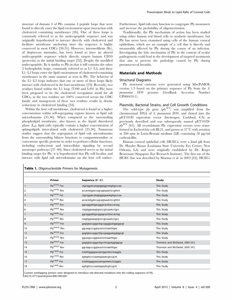
Table 1. Oligonucleotide Primers for Mutagenesis.
Primer Sequence (59–39) Study
1 PlyA370G For ctgctggatcatagtggtggctatgttgcccaa This Study
2 PlyA370G Rev accactatgatccagcagtaaatctccgtttct This Study
3 PlyA370E For ctgctggatcatagtggtgaatatgttgcccaa This Study
4 PlyA370E Rev accactatgatccagcagtaaatctccgtttct This Study
5 PlyA406G For ggcaggatttgacgggtcactttaccactag This Study
6 PlyA406G Rev ctagtggtaaagtgacccgtcaaatcctgcc This Study
7 PlyA406E For ggcaggatttgacggagcactttaccactag This Study
8 PlyA406E Rev ctagtggtaaagtgctccgtcaaatcctgcc This Study
9 PlyW433G For gagtgtaccgggcttgccggggaatggtggcgt This Study
10 PlyW433G Rev ggcaagcccggtacactctctaattttgac This Study
11 PlyW433E For gagtgtaccgggcttgccgaggaatggtggcgt This Study
12 PlyW433E Rev ggcaagcccggtacactctctaattttgac This Study
13 PlyW433F For gagtgtaccgggcttgccttcgaatggtggcgt Thornton and McDaniel, 2005 [41]
14 PlyW433F Rev ggcaagcccggtacactctctaattttgac Thornton and McDaniel, 2005 [41]
15 PlyL460G For tctatttggggaacaactggctatcctcaggta This Study
16 PlyL460G Rev agttgttccccaaatagaaatcgtccgctt This Study
17 PlyL460E For tctatttggggaacaactgaatatcctcaggta This Study
18 PlyL460E Rev agttgttccccaaatagaaatcgtccgctt This Study
Custom overlapping primers were designed to introduce site-directed mutations into the coding sequence of Ply.doi:10.1371/journal.pone.0061300.t001
Pneumolysin Binds to Lipid Rafts of Corneal Cells
PLOS ONE | www.plosone.org 2 April 2013 | Volume 8 | Issue 4 | e61300
were cultivated in serum-free Clonetics keratinocyte media (KGM;
Clonetics BioWhittaker Europe, Verviers, Belgium) supplemented
with growth factors and antibiotics as previously described [42,43].
Site-Directed MutagenesisSingle amino acid substitutions were introduced into the
pET101D-plyWT vector, specifically A370G, A370E, A406G,
A406E, W433G, W433E, L460G, and L460E. The pET101D-
plyW433F vector was previously engineered by Thornton and
McDaniel using the Genetailor site-directed mutagenesis kit
(Invitrogen, Carlsbad, CA), and the same methodology was used
to engineer the other mutants with the exception of pET101D-
plyA406G and pET101D-plyA406E [41]. Both A406 mutants were
engineered using PCR QuikChange site-directed mutagenesis
(Stratagene, La Jolla, CA). Custom primers were designed for each
mutagenesis reaction (Table 1).
Recombinant Pneumolysin PurificationE. coli BL21 was grown in LB plus 50 mg/ml carbenicillin to an
OD600 of 0.5, which corresponds to 1x10⁹ cfu/ml. Isopropyl-b-D-
thiogalactopyranoside (IPTG) was added to the culture to a final
concentration of 1 mM to induce Ply expression. The culture was
grown for an additional 4–5 h. Bacterial cells were collected by
centrifugation for 10 min at 8,000 x g and 4uC. The remainder of
the purification was performed on ice. The bacterial pellet was
suspended in 40 ml of extraction/wash buffer (50 mM sodium
phosphate, 300 mM sodium chloride) and lysed by sonication (5
intervals; 30 sec per interval, with alternating 30 sec rest periods).
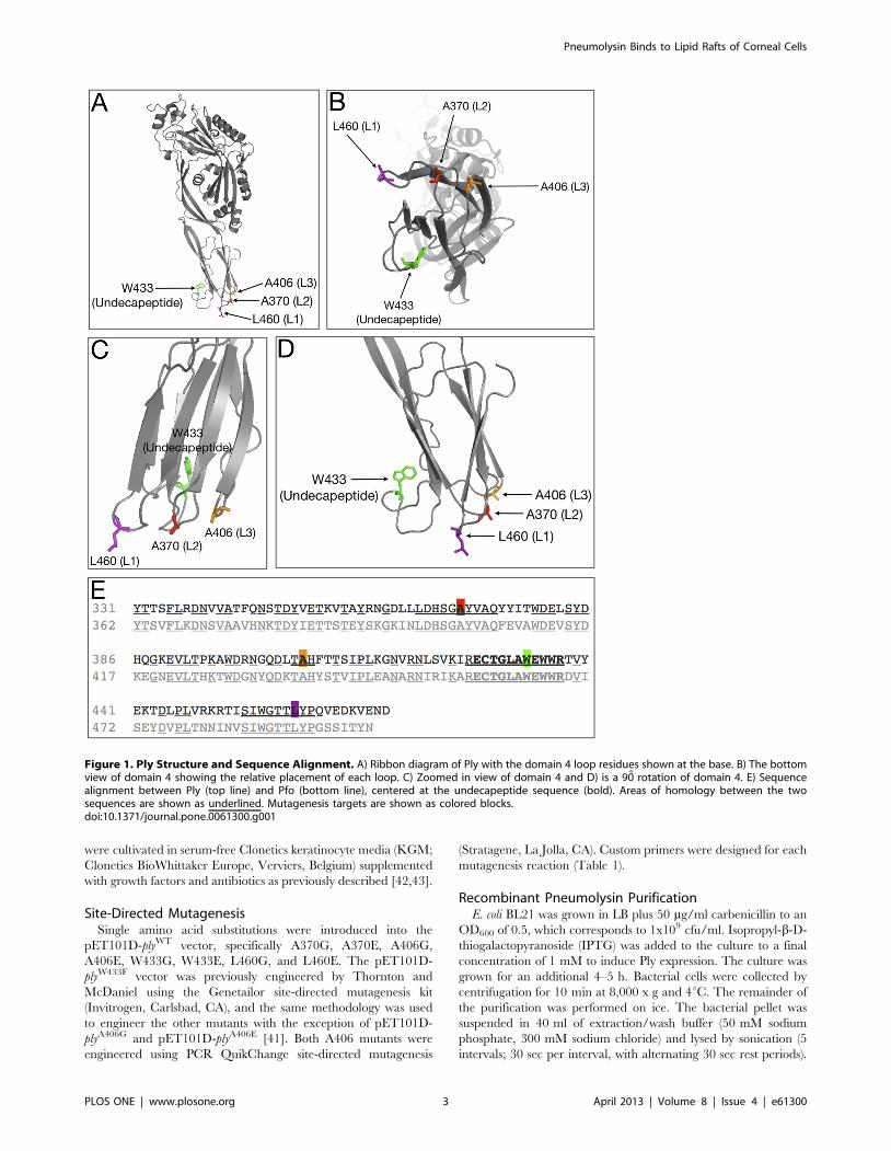
Figure 1. Ply Structure and Sequence Alignment. A) Ribbon diagram of Ply with the domain 4 loop residues shown at the base. B) The bottomview of domain 4 showing the relative placement of each loop. C) Zoomed in view of domain 4 and D) is a 90̊ rotation of domain 4. E) Sequencealignment between Ply (top line) and Pfo (bottom line), centered at the undecapeptide sequence (bold). Areas of homology between the twosequences are shown as underlined. Mutagenesis targets are shown as colored blocks.doi:10.1371/journal.pone.0061300.g001
Pneumolysin Binds to Lipid Rafts of Corneal Cells
PLOS ONE | www.plosone.org 3 April 2013 | Volume 8 | Issue 4 | e61300
The cellular debris was removed by centrifugation for 20 min at
20,000 x g and 4uC. Ply was purified from the soluble intracellular
contents by Talon metal affinity resin chromatography (BD
Biosciences, Franklin Lakes, NJ), which selectively binds to the 6X
histidine tag added by the pET101D expression vector. After
elution from the resin, Ply was extensively dialyzed in 4 L of
phosphate-buffered saline (PBS; 150 mM sodium chloride,
2.3 mM monobasic sodium phosphate, 7.7 mM dibasic sodium
phosphate, pH 7.2). Purified Ply was confirmed via SDS-PAGE as
a single 53 kDa band (data not shown), and was quantified by
detecting the absorbance at 280 nm with an extinction coefficient
of 72,000 cm21M21.
Cytotoxicity AssayPly cytotoxicity on HCECs was measured using the Live/Dead
cytotoxicity kit (Invitrogen, Carlsbad, CA). HCECs were grown to
confluency in 96-well tissue culture plates. Each monolayer was
washed twice with PBS and labeled with various concentrations of
either PlyWT or mutant Ply for 3 h at 37uC. The cells were washed
with PBS twice and labeled with a final concentration of 2 mM
calcein AM, a selective intracellular label for healthy cells. The
cells were placed in the dark for 30 min at 37uC. The cells were
then washed with PBS, and fluorescence was detected by
fluorescence spectrometry at 530 nm. Triton X-100 (TX100)
treated HCECs served as the positive control representing 100%
lysis, and PBS treated cells served as the negative control. Each
experiment was performed in triplicate, and relative percent
survival was calculated as (Asample2ATX100)/(APBS2ATX100)6100.
Data was presented as relative percent survival.
Hemolysis AssayVarious concentrations (480–0.2 nM) of PlyWT or mutant Ply
were incubated with 2% rabbit red blood cells (RBCs) in a round
bottom 96-well plate for 30 min at 37uC. The plate was
centrifuged for 5 min at 700 x g to collect any intact RBCs. The
supernatant from each well was transferred to a flat-bottom 96-
well plate and RBC lysis was quantified based on hemoglobin
concentration in the supernatant. Hemoglobin was quantified by
measuring absorbance at 450 nm by spectrophotometry. Each
assay was performed in triplicate. RBCs and 1% TX100 served as
the positive control and represents 100% lysis. All results are
presented as percent hemolysis relative to the positive control.
Relative percent hemolysis was calculated as (Asample2APBS)/
(ATX1002APBS)6100. Rabbit RBCs were collected from New
Zealand white rabbits (Charles River, Wilmington, MA). Our
protocol was approved by and the rabbits were maintained
according to the guidelines of the University of Mississippi Medical
Center Institutional Animal Care and Use Committee (IACUC)
and the tenets of the Association for Research in Vision and
Ophthalmology (ARVO) Resolution on the Use of Animals in
Ophthalmic and Vision Research. Rabbits were anesthetized by
intramuscular injection of 100 mg of ketamine prior to drawing
blood via ear vein. The rabbits were provided enrichment items
for entertainment during housing. The animals were not
euthanized at the end of the study.
Western Blot (Oligomerization Assay)The oligomerization assay protocol was modified from a
previously published protocol performed by Soltani et al. [33].
Five hundred ng of PlyWT or mutant Ply was incubated either in
the presence or absence of 106 HCECs for 30 min at 37uC in PBS
to a total volume of 20 ml. Each cell suspension was mixed 1:1 with
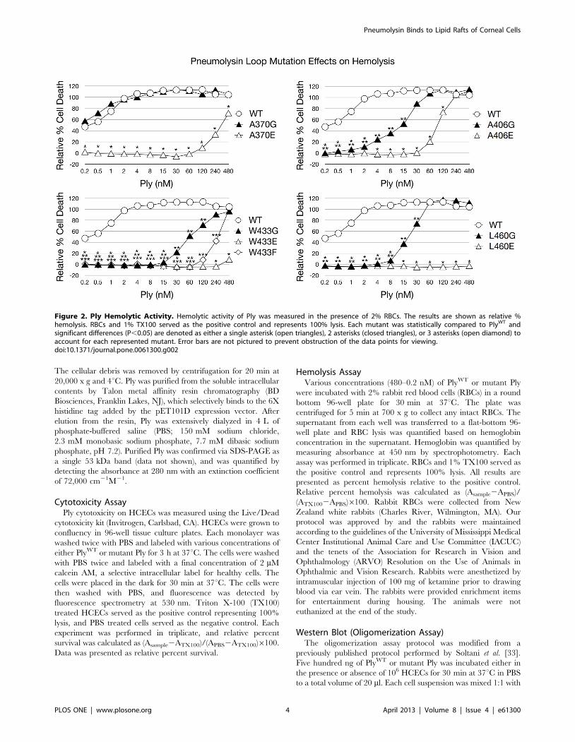
Figure 2. Ply Hemolytic Activity. Hemolytic activity of Ply was measured in the presence of 2% RBCs. The results are shown as relative %hemolysis. RBCs and 1% TX100 served as the positive control and represents 100% lysis. Each mutant was statistically compared to PlyWT andsignificant differences (P,0.05) are denoted as either a single asterisk (open triangles), 2 asterisks (closed triangles), or 3 asterisks (open diamond) toaccount for each represented mutant. Error bars are not pictured to prevent obstruction of the data points for viewing.doi:10.1371/journal.pone.0061300.g002
Pneumolysin Binds to Lipid Rafts of Corneal Cells
PLOS ONE | www.plosone.org 4 April 2013 | Volume 8 | Issue 4 | e61300

2X SDS Loading Buffer (100 mM Tris-HCl, 200 mM dithiothre-
itol, 4% w/v SDS, 0.2% bromophenol blue, 20% v/v glycerol)
and was subjected to 6% SDS-PAGE. The gel was then
electroblotted to a PVDF membrane, and blocked in 5% skim
milk in NP-40 buffer (50 mM Tris-HCl, 150 mM NaCl, 5 mM
EDTA, 0.05% v/v Nonidet P-40) for 1 h at 25uC. The membrane
was rinsed with NP-40 buffer and then labeled with rabbit
polyclonal anti-Ply serum (1:200 dilution in NP-40 buffer)
produced as described previously [44]. After a 3 h incubation at
25uC, the membrane was washed 3 times in NP-40 buffer and
labeled with horseradish peroxidase (HRP) conjugated goat anti-
rabbit IgG for 2 h at 25uC. The membrane was then washed 3
times with NP-40 buffer and developed with Pierce ECL western
blotting substrate.
Alexa Fluor 488 LabelingBoth PlyWT and mutant Ply molecules were conjugated with
Alexa Fluor 488 (AF488) tags in order to facilitate fluorescent
detection. The conjugation reactions were carried out using the
Alexa Fluor microscale protein labeling kit (Invitrogen Molecular
Probes) according to the manufacturers instructions. The degree of
labeling for each Ply type was optimized to be 3 AF488 tags per
molecule of Ply. No significant difference was observed between
the cytotoxicities of AF488-labeled PlyWT and unlabeled PlyWT
(data not shown).
Flow CytometryAdherent HCECs were detached from the culture flask with
0.1% trypsin-EDTA for 10 min, washed 2 times with PBS, and
fixed in 3.7% paraformaldehyde (PFA) for 15 min at 25uC. The
fixed cells were washed 3 times with PBS and suspended in PBS at
a concentration of 106 cells/ml. Fluorescent labeling was per-
formed as described above. The fixed cell suspension was divided
into 300 ml aliquots and labeled with either AF488-labeled PlyWT,
mutant Ply, heat inactivated Ply, or BSA and incubated for 30 min
at 37uC. After incubation, the cells were centrifuged at 500 x g for
3 min, and the supernatant was removed. The cells were then
washed 3 times with PBS before being suspended in a final 300 ml
of PBS and transferred to 5 ml dilution tubes. Ply surface binding
was detected using a Gallios flow cytometer with Kaluza software
version 1.1 (Beckman Coulter, Miami, FL). Each experimental
condition was performed in triplicate with 5,000 events per
experiment.
Sucrose Density Gradient CentrifugationFive x 106 HCECs were collected and washed 3 times in Tris-
buffered saline (TBS; 25 mM Tris/HCl, 140 mM NaCl, pH 7.5).
TBS was removed and the cells were suspended in 1 ml of
incubation buffer (25 mM Tris/HCl, 140 mM NaCl, 1 mM
EDTA, 1 mM PMSF, Roche Complete Protease Inhibitor
cocktail, pH 8) plus 25 mg of Ply and 5 mg of biotinylated cholera
Figure 3. Ply Cytotoxicity on HCECs. Ply cytotoxicity on HCECs was measured by staining with calcein AM, a selective marker for healthy cellswith intact membranes. Percent survival was normalized using PBS (negative) and TX100 (positive) treated cells as controls. The results are shown as% survival. Each mutant was statistically compared to PlyWT and significant differences (P,0.05) are denoted as a single asterisk (open triangles), 2asterisks (closed triangles), or 3 asterisks (open diamond) to account for each represented mutant. Error bars are not pictured to prevent obstructionof the data points for viewing.doi:10.1371/journal.pone.0061300.g003
Pneumolysin Binds to Lipid Rafts of Corneal Cells
PLOS ONE | www.plosone.org 5 April 2013 | Volume 8 | Issue 4 | e61300
toxin b (CTxb), which binds to the common lipid raft constituent
ganglioside GM1 of eukaryotic cell membranes. The cell suspen-
sion was incubated on ice for 4 h after which 40 ml of 25% TX100
was added and mixed thoroughly (final concentration 1% TX100).
The mixture was incubated an additional 1 h on ice. The solution
was passed through a small gauge needle 20 times and then
centrifuged at 10,000 x g for 5 min at 4uC. The supernatant was
transferred to a new microcentrifuge tube, and 70% sucrose was
added to a final concentration of 40% sucrose. The sample was
layered under a 10–30% discontinuous sucrose gradient in a 5 ml
ultracentrifuge tube at a ratio of 1.5:2.5:1. The gradient was
centrifuged at 4uC for 18 h at 300,000 x g. Fractions were
collected from the top in 400 ml increments. Fractions were
spotted on a nitrocellulose membrane, blocked with 5% skim milk
in NP-40 buffer for 1 h, and labeled with either HRP-conjugated
streptavidin to detect CTxb, or anti-Ply rabbit polyclonal serum
followed by HRP-conjugated goat anti-rabbit IgG to detect Ply.
The blot was visualized using Pierce ECL western blotting
substrate.
Statistical AnalysesExperimental results were analyzed using the statistical analysis
system (SAS) for computers (SAS Institute, Cary, NC) version 9.2.
All experimental groups were compared using a nonparametric
one-way analysis of variance, and any P-value , 0.05 was
considered significant.
Results
Previous studies that focused on Pfo, a related CDC that shares
42% amino acid homology with Ply, have pinpointed several
important amino acids that are involved in interacting with the
lipid environment of the host membrane during initial binding
[26,45]. These residues include A401, A437, W464, and L491,
and correspond to A370, A406, W433, and L460 in Ply [26,46]. In
Pfo, each of these amino acid residues are located at the tip of one
of 4 loops structures found in domain 4 which extend out from the
protein to interact with the host membrane. A sequence alignment
of domain 4 from Ply and Pfo reveals that the loop residues from
Pfo are conserved in Ply, and many of the surrounding residues
around both A370 and L460 are also conserved (Figure 1E).
Structural diagrams show the positions of the domain 4 loops
relative to one another (Figure 1A-D). The R-groups from the
highlighted amino acids of L1-L3 extend away from the interior of
the molecule and presumably enter the lipophilic environment of
the host cell membrane. Based on the observed homology and
relative positions of each amino acid, we engineered 2 amino acid
substitution mutants at the apex of each loop structure. We chose
to substitute both glutamate and glycine in order to 1.) prevent the
loop from entering the lipid environment of the host membrane
(glutamate), and 2.) observe the effect of removing the R-group
from each mutation site (glycine). We also included PlyW433F since
this mutation is classically studied and a wide array of information
is readily available on its lytic behavior in various models. Each Ply
Figure 4. Ply Surface Binding to HCECs. Flow cytometry was used to assess surface binding to HCECs. Various concentrations of AF488 labeledPly were incubated in the presence of 3x10⁵ fixed HCECs. AF488 labeled bovine serum albumin (BSA) and AF488 labeled heat inactivated (HI) PlyWT
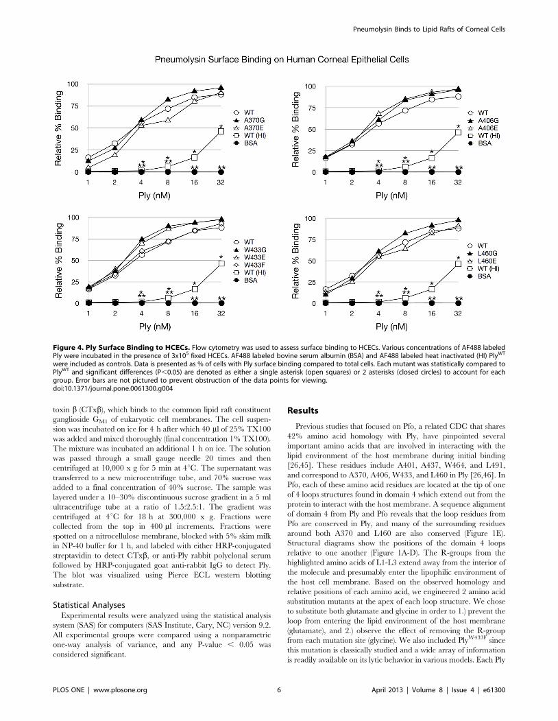
were included as controls. Data is presented as % of cells with Ply surface binding compared to total cells. Each mutant was statistically compared toPlyWT and significant differences (P,0.05) are denoted as either a single asterisk (open squares) or 2 asterisks (closed circles) to account for eachgroup. Error bars are not pictured to prevent obstruction of the data points for viewing.doi:10.1371/journal.pone.0061300.g004
Pneumolysin Binds to Lipid Rafts of Corneal Cells
PLOS ONE | www.plosone.org 6 April 2013 | Volume 8 | Issue 4 | e61300
variant was analyzed for cytotoxicity, ability to oligomerize, and
ability to bind to the HCEC surface, specifically to cholesterol-rich
membranes.
The Effects of the Ply Loop Mutations on CytolysisThe introduction of each amino acid substitution had a direct
effect on the cytotoxicity of Ply on both RBCs and HCECs
(Figures 2 and 3). It was previously reported that PlyW433F has a
99% reduction in hemolytic activity, and our findings are in
agreement [21]. When examining the glutamate substitution
mutants, both PlyW433E and PlyL460E possessed the least cytolytic
power, having significantly lower hemolytic activity at all
concentrations examined when compared to PlyWT. At 480 nM,
the highest Ply concentration in the hemolysis assay, PlyL460E was
found to be completely deficient in lytic activity. The other
glutamate substitution mutants, PlyA370E, PlyA406E, and PlyW433E,
resulted a relative cell death percentage of 71, 110, and 8
respectively. The glycine substitution mutants each exhibited a less
pronounced effect than their glutamate counterparts on lytic
ability, which can be expected given the polar nature of glutamate.
PlyW433G had the least lytic power of the glycine substitution
mutants, with all hemolytic activity ceasing at 8 nM. Likewise
PlyL460G lost all hemolytic activity at a slightly lower concentration
of 4 nM. PlyA406G remained active at a concentration as low as
0.5 nM, but PlyA370G showed no signs of hemolytic loss and
remained as active as PlyWT at all concentrations examined. The
same results were found when examining the cytotoxic effect of Ply
on HCECs. Based on the lytic activity observed on both RBCs and
HCECs, the relative cytotoxicity of each Ply variant can be ranked
from most cytotoxic to least cytotoxic in the following order:
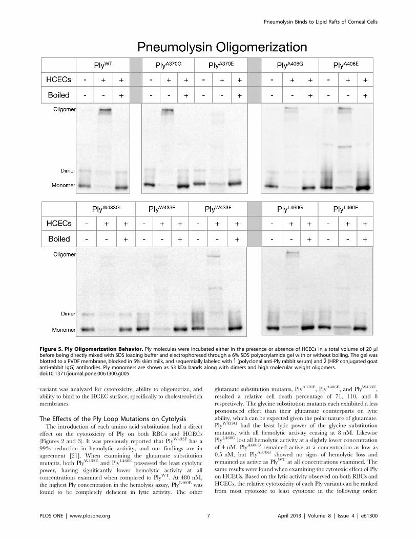
Figure 5. Ply Oligomerization Behavior. Ply molecules were incubated either in the presence or absence of HCECs in a total volume of 20 mlbefore being directly mixed with SDS loading buffer and electrophoresed through a 6% SDS polyacrylamide gel with or without boiling. The gel wasblotted to a PVDF membrane, blocked in 5% skim milk, and sequentially labeled with 1̊ (polyclonal anti-Ply rabbit serum) and 2̊ (HRP conjugated goatanti-rabbit IgG) antibodies. Ply monomers are shown as 53 kDa bands along with dimers and high molecular weight oligomers.doi:10.1371/journal.pone.0061300.g005
Pneumolysin Binds to Lipid Rafts of Corneal Cells
PLOS ONE | www.plosone.org 7 April 2013 | Volume 8 | Issue 4 | e61300

PlyWT, PlyA370G, PlyA406G, PlyL460G, PlyW433G, PlyA406E, PlyW433F,
PlyA370E, PlyW433E, and PlyL460E.
The Effects of the Ply Loop Mutations on Ply SurfaceBinding
Alexa Fluor 488 labeled PlyWT and all mutant variants were
readily detectable as bound to the HCEC surface. Flow cytometry
analysis of Ply surface binding to HCECs revealed that PlyWT and
all 9 Ply mutants were able to bind to the corneal cell surface, and
this binding was not significantly different in any of the domain 4
loop mutants including the glutamate substitution mutants
(Figure 4).
The Effects of the Ply Loop Mutations on PlyOligomerization
Ply oligomeric complexes are SDS resistant and can be detected
by western blot as high molecular mass bands if the samples are
not boiled prior to electrophoresis [47]. After each Ply variant was
incubated in the presence of HCECs, oligomeric complexes were
readily detectable by western blot for PlyWT, PlyA370G, PlyA406G,
PlyA406E, PlyW433F and PlyL460G, but not PlyA370E, PlyW433G,
PlyW433E, and PlyL460E (Figure 5). PlyWT and PlyA370G exhibited
the darkest high molecular weight oligomer band in agreement
with the fact that PlyA370G retained full lytic activity. PlyA406G,
PlyA406E, PlyW433F, and PlyL460G all retained their ability to
oligomerize in the presence of cholesterol rich membranes
although at a visibly diminished efficiency when compared to
PlyWT. Interestingly, both PlyA370 and PlyL460 followed a similar
pattern where glutamate substitution rendered both mutants
unable to oligomerize, but glycine substitution allowed for the
retention of oligomerization ability, albeit reduced from PlyWT.
The A406 loop is unique in that neither substitution resulted in a
loss of oligomerization. The W433 loop was only able to
oligomerize with the phenylalanine substitution, which is a
conservative substitution. The loss of either the W433 loop
insertion into the membrane or the loss of the R-group resulted in
no oligomer formation.
The Effects of the Ply Loop Mutations on Lipid RaftColocalization
We performed sucrose density gradient centrifugations with
solubilized HCECs incubated with both Ply and CTxbBiotinylated in
order to separate the low density lipid rafts from the high density
phospholipid bilayer (Figure 6). The sucrose gradients revealed
that both PlyWT and CTxb did in fact localize to the low density
lipid raft fractions at the top of the sucrose gradient (fractions 3-5).
However, of the 9 mutants, only PlyA370G was found in the low
density lipid raft fractions. The remaining mutants were only
found in the high density fractions (fractions 9-12) and were unable
to localize to the lipid raft fractions of the gradient.
Figure 6. Ply Localization to Lipid Raft Microdomains. Sucrose density gradients were performed to isolate lipid raft microdomains from thephospholipid membrane and to visualize whether Ply localized with the lipid raft fractions. Gradient fractions were blotted onto a nitrocellulosemembrane, and Ply or CTxb were detected by chemiluminescence. Fractions are numbered from least dense to most dense.doi:10.1371/journal.pone.0061300.g006
Pneumolysin Binds to Lipid Rafts of Corneal Cells
PLOS ONE | www.plosone.org 8 April 2013 | Volume 8 | Issue 4 | e61300
Discussion
The fact that 8 of the 9 mutant Ply variants exhibit a reduction
in cytotoxicity when compared to PlyWT, but none of the mutants
are deficient in HCEC surface binding indicates that the domain 4
loops are, in some capacity, involved in initiating oligomerization
and/or the prepore to pore conversion. The sequence alignment
of domain 4 Pfo and Ply shows nearly 75% sequence homology
between the two molecules (Figure 1). The undecapeptide (W433)
along with the L460 loop and the A370 loop are 100% conserved
between Ply and Pfo, however, there is sequence variability when
comparing the A406 loop. This lack of homology at the A406 loop
may indicate that this loop is of less importance than the other
more conserved loops in terms of contributing to the progression
of the lytic mechanism. Additionally, substitution of glycine or
glutamate at the A406 position has a smaller effect on cytotoxicity
than those same mutations at any of the other loops, with the only
exception being PlyA370G. The fact that PlyA370G resulted in no
reduction in cytotoxicity, but PlyA370E did result in significant
reduction in cytotoxicity indicates that the native alanine at
position 370 is likely not involved in any direct molecular
interactions with the membrane constituents, but rather simply
supplies a stabilizing effect that still occurs when A370 is
substituted with glycine. Additionally, the fact that PlyA370E
prevents oligomerization indicates that the A370 loop must
interact with the membrane in order for oligomerization to occur.
Ply and Pfo as a whole have been shown to be largely conserved
in structure and amino acid sequence, so findings regarding one
are likely to be at least partly applicable to another. However, we
have discovered some behavioral differences between Ply and Pfo
with regard to surface binding. Previous research conducted by
Ramachandran et al. has shown that all 4 homologous domain 4
loops of Pfo interact with the lipid environment during cholesterol
binding, and Soltani et al. observed that when the loop residues of
Pfo are individually mutated to aspartate, then surface binding to
cholesterol containing membranes is almost completely abolished
[26,46]. Our surface binding results for Ply and HCECs are
unique from the findings observed for Pfo. When the domain 4
loops of Ply are mutated to glutamate, then surface binding to
HCECs is unaffected as all mutants bind with the same efficiency
as PlyWT. This difference from the observed findings for Pfo
indicates one of three possibilities: 1) aspartate and glutamate
result in two different outcomes when substituted at the loop
residues, 2) Pfo and Ply have different binding behaviors which
react differently to the presence of a charged polar amino acid in
the loops of domain 4, or 3) the observed difference is due to the
use of HCECs as the target cell.
A recent study by Farrand et al. reported that the CDC
cholesterol recognition motif for several CDCs including Pfo and
Ply is a threonine-leucine amino acid pair found in domain 4,
corresponding to T459 and L460 in Ply [34]. They found that
double glycine substitutions of these residues dramatically reduced
cholesterol binding on RBCs, and this threonine-leucine pair is
conserved across all CDCs. Interestingly, our results indicate that
when PlyL460 is substituted with glutamate, it still retains its ability
to bind to the surface of HCECs at an undiminished capacity
when compared to PlyWT. This binding behavior was not expected
due to the previous results showing that T459 and L460 comprised
the cholesterol recognition motif for Ply when exposed to
cholesterol-rich liposomes. Our results indicate that it is unlikely
that L460 is part of the cholesterol recognition motif of Ply when
targeting HCECs, since the addition of a polar charged residue or
removal of the R-group has no observed effect on surface binding
to HCECs. Likewise, in addition to PlyL460E, flow cytometry
revealed that the other glutamate substitution mutants, PlyA370E,
PlyA406E, and PlyW433E were also capable of binding to the surface
of HCECs with no significant differences when compared to
PlyWT. These results indicate that cholesterol recognition and
binding by Ply is likely carried out not by a single loop structure,
but rather a concerted effort between 2 or more of the loops.
The oligomerization behaviors of our Ply variants also yielded
some unique results when compared to other CDCs. We observed
that PlyW433G was unable to form oligomeric complexes under our
experimental conditions. However, a previous study that exam-
ined the oligomerization behavior of Ily found that IlyW491A, the
Ily mutant corresponding to same position as PlyW433G, was able
to form high molecular weight oligomeric complexes [33].
Interestingly, PlyW433F was also found to be capable of oligomer-
ization indicating that W433 is likely involved in a molecular
interaction required for oligomerization to occur, since a
conservative substitution, tryptophan to phenylalanine, resulted
in the retention of oligomerization ability. The same study by
Soltani et al. observed that IlyL518D was able to oligomerize,
although at a markedly reduced capacity when compared to
IlyWT. Our Ply mutant with a similar mutation, PlyL460E, was
unable to oligomerize at any detectible level. In this particular case
we substituted glutamate rather than aspartate and this may
account for the differing oligomerization behavior. However,
IlyA428D, compared to the homologous Ply mutant PlyA370E,
behaved in a similar manner where neither mutant was able to
oligomerize, and likewise the same might be said for IlyA464D and
PlyA406E [33]. The oligomerization deficiency caused by PlyA370E
and PlyL460E indicates that these amino acids must be within the
lipid membrane environment in order for oligomerization to occur
on the HCEC surface. They likely function in a stabilizing role,
since mutation of both of these residues to glycine did not prevent
oligomerization. Therefore, the R-groups of A370 and L460 are
not likely involved in specific molecular interactions that are
required for the oligomerization of Ply on HCECs. The reduction
in oligomerization efficiency may be due to destabilization caused
by the presence of glycine rather than the native residues.
We then questioned where on the HCEC surface Ply was
localizing and if there was any difference between PlyWT and the
loop mutants. Previous research investigating both Pfo and
listeriolysin (Llo) have shown that they preferentially localized to
lipid raft microdomains on the surface of human lymphoblastic
cells (MOLT-4) and mouse macrophage (J774 cells) respectively
[48,49]. Specifically, Llo has been shown to cause lipid raft
markers, including ganglioside GM1, to aggregate on the surface of
J774 macrophages [48]. The same study by Gekara et al.
discovered that lipid raft aggregation by Llo can be blocked if
the toxin is pretreated with a monoclonal antibody that blocks
oligomerization, but not cholesterol recognition. Therefore they
proposed that oligomerization of Llo is responsible for lipid raft
aggregation and may facilitate other cellular functions such as
endocytosis or the initiation of intracellular signaling. Very little is
known regarding Ply and whether it interacts with lipid raft
microdomains on the HCEC surface. We performed sucrose
density gradient centrifugations of HCEC membranes in order to
separate the lipid rafts from the surrounding bilayer, after labeling
the cells with CTxb and Ply. We observed that both PlyWT and
CTxb were detectable in both the low (raft) and high (non-raft)
density fractions. Interestingly, all of the mutant Ply molecules
were only detectable in the high density fractions except for
PlyA370G, the only fully lytic mutant. The fact that the majority of
the mutant Ply molecules were only detectable in the high density
fractions indicates that although initial binding is not affected, the
loop mutations do influence the ability of the molecules to localize
Pneumolysin Binds to Lipid Rafts of Corneal Cells
PLOS ONE | www.plosone.org 9 April 2013 | Volume 8 | Issue 4 | e61300

to lipid raft microdomains on the HCEC surface. The nature of
this disruption is still not fully understood. The fact that only the
fully active Ply variants (PlyWT and PlyA370G) were detected in the
raft fractions indicates that either full oligomerization capability is
necessary or the ability to convert the prepore to mature pore is
required for the raft colocalization to occur. Of the Ply mutants
that were found to be oligomerization capable (PlyA370G, PlyA406G,
PlyA406E, PlyW433F, and PlyL460G), PlyA370G is the only mutant that
is as efficient as PlyWT at oligomer formation and was also the only
mutant that localized to the raft fractions of the sucrose gradient.
Perhaps the diminished capacity to oligomerize results in an
inability to colocalize with lipid rafts. The process of oligomeri-
zation may induce the formation of raft microdomains, or
alternatively, lipid rafts could fill a critical role in the process of
oligomerization, such as stabilization of the monomers on the cell
surface. In either case, the loop mutations result in both a
reduction in oligomerization and lipid raft colocalization.
In conclusion, the domain 4 loop mutations are clearly involved
in the lytic mechanism of Ply since the cytotoxicity of each mutant
was found to be considerably reduced as compared to PlyWT
(except for PlyA370G). None of the loop mutations prevented
binding to the surface of HCECs, although PlyA370E, PlyW433G,
PlyW433E, and PlyL460E did prevent the process of oligomerization.
The binding behavior of the mutants indicates that cholesterol
recognition is likely not carried out by a single loop, but rather a
concerted process involving multiple loops, since preventing any
single loop from membrane interaction by glutamate substitution
never resulted in a loss of surface binding. None of the Ply loop
mutants were found to localize with the low density lipid raft
fractions of HCEC sucrose density gradients (again except
PlyA370G), where PlyWT was detectable. These findings may be
unique to Ply or to the interaction of Ply specifically with HCECs.
Although the exact relationship between the domain 4 loops and
their interaction with cholesterol and lipid rafts is not fully
understood, it is apparent that lipid raft microdomains play a
critical role in the lytic mechansim of Ply.
Acknowledgments
The authors would like to thank Dr. Don Sittman (University of Mississippi
Medical Center, Jackson, MS) for helpful advice and assistance in the
experimental methods, and Dr. Dwight D. Cavanagh (Southwestern
Medical Center, Dallas, TX) for providing the initial inspiration for this
project.
Author Contributions
Conceived and designed the experiments: ST MS NT MM. Performed the
experiments: ST MS NT SS EN LM. Analyzed the data: ST NT SS EN
MM. Contributed reagents/materials/analysis tools: SS LM MM. Wrote
the paper: ST MS NT SS EN LM MM.
References
1. O’Brien KL, Wolfson LJ, Watt JP, Henkle E, Deloria-Knoll M, et al. (2009)
Burden of disease caused by Streptococcus pneumoniae in children younger than 5
years: global estimates. Lancet 374: 893–902.
2. Ahern JW, Raszka WV, Jr (2009) Meningitis from an uncommon serotype of
Streptococcus pneumoniae in a young child. South Med J 102: 1189.
3. Antao VC, Hausdorff WP (2009) Global epidemiology of pneumococcal
disease--new prospects for vaccine control. Adv Exp Med Biol 634: 19–29.
4. Kisakye A, Makumbi I, Nansera D, Lewis R, Braka F, et al. (2009) Surveillance
for Streptococcus pneumoniae meningitis in children aged ,5 years: implications for
immunization in Uganda. Clin Infect Dis 48 Suppl 2: S153–161.
5. Yoshioka CR, Martinez MB, Brandileone MC, Ragazzi SB, Guerra ML, et al.
(2011) Analysis of invasive pneumonia-causing strains of Streptococcus pneumoniae:
serotypes and antimicrobial susceptibility. J Pediatr (Rio J) 87: 70–75.
6. Crum NF, Barrozo CP, Chapman FA, Ryan MA, Russell KL (2004) An
outbreak of conjunctivitis due to a novel unencapsulated Streptococcus pneumoniae
among military trainees. Clin Infect Dis 39: 1148–1154.
7. Zegans ME, Sanchez PA, Likosky DS, Allar RT, Martin M, et al. (2009) Clinical
features, outcomes, and costs of a conjunctivitis outbreak caused by the ST448
strain of Streptococcus pneumoniae. Cornea 28: 503–509.
8. Soriano F, Perez-Trallero E, Pallares R, Meseguer MA, Fleites A, et al. (2006)
Streptococcus pneumoniae endophthalmitis: a study of 36 cases with special reference
to antibiotic resistance and treatment options. Clin Microbiol Infect 12: 519–
526.
9. Cid M, Sabates NR (1997) Penicillin-resistant Streptococcus pneumoniae endoph-
thalmitis. Am J Ophthalmol 123: 133–135.
10. Bhave P, Chamie G (2008) Streptococcus pneumoniae keratitis. J Hosp Med 3: 353.
11. Dada T, Sharma N, Dada VK, Vajpayee RB (2000) Pneumococcal keratitis
after laser in situ keratomileusis. J Cataract Refract Surg 26: 460–461.
12. Lifshitz T, Levy J, Raiskup F, Klemperer I, Frucht-Pery J (2005) Two cases of
pneumococcal keratitis following myopic LASIK. J Refract Surg 21: 498–501.
13. Parmar P, Salman A, Kalavathy CM, Jesudasan CA, Thomas PA (2003)
Pneumococcal keratitis: a clinical profile. Clin Experiment Ophthalmol 31: 44–
47.
14. Ramirez M, Hernandez-Quintela E, Beltran F, Naranjo-Tackman R (2002)
Pneumococcal keratitis at the flap interface after laser in situ keratomileusis.
J Cataract Refract Surg 28: 550–552.
15. Kunimoto DY, Sharma S, Reddy MK, Gopinathan U, Jyothi J, et al. (1998)
Microbial keratitis in children. Ophthalmology 105: 252–257.
16. Green M, Apel A, Stapleton F (2008) Risk factors and causative organisms in
microbial keratitis. Cornea 27: 22–27.
17. Marquart ME, Monds KS, McCormick CC, Dixon SN, Sanders ME, et al.
(2007) Cholesterol as treatment for pneumococcal keratitis: cholesterol-specific
inhibition of pneumolysin in the cornea. Invest Ophthalmol Vis Sci 48: 2661–
2666.
18. Sanders ME, Tullos NA, Taylor SD, Norcross EW, King LB, et al. (2010)
Moxifloxacin and cholesterol combined treatment of pneumococcal keratitis.
Curr Eye Res 35: 1142–1147.
19. Alexander JE, Berry AM, Paton JC, Rubins JB, Andrew PW, et al. (1998) Amino
acid changes affecting the activity of pneumolysin alter the behaviour of
pneumococci in pneumonia. Microb Pathog 24: 167–174.
20. Alexander JE, Lock RA, Peeters CC, Poolman JT, Andrew PW, et al. (1994)
Immunization of mice with pneumolysin toxoid confers a significant degree of
protection against at least nine serotypes of Streptococcus pneumoniae. Infect Immun
62: 5683–5688.
21. Berry AM, Alexander JE, Mitchell TJ, Andrew PW, Hansman D, et al. (1995)
Effect of defined point mutations in the pneumolysin gene on the virulence of
Streptococcus pneumoniae. Infect Immun 63: 1969–1974.
22. Berry AM, Ogunniyi AD, Miller DC, Paton JC (1999) Comparative virulence of
Streptococcus pneumoniae strains with insertion-duplication, point, and deletion
mutations in the pneumolysin gene. Infect Immun 67: 981–985.
23. Hirst RA, Kadioglu A, O’Callaghan C, Andrew PW (2004) The role of
pneumolysin in pneumococcal pneumonia and meningitis. Clin Exp Immunol
138: 195–201.
24. Tweten RK (2005) Cholesterol-dependent cytolysins, a family of versatile pore-
forming toxins. Infect Immun 73: 6199–6209.
25. Johnson MK, Geoffroy C, Alouf JE (1980) Binding of cholesterol by sulfhydryl-
activated cytolysins. Infect Immun 27: 97–101.
26. Ramachandran R, Heuck AP, Tweten RK, Johnson AE (2002) Structural
insights into the membrane-anchoring mechanism of a cholesterol-dependent
cytolysin. Nat Struct Biol 9: 823–827.
27. Hotze EM, Tweten RK (2011) Membrane assembly of the cholesterol-
dependent cytolysin pore complex. Biochim Biophys Acta.
28. Dang TX, Hotze EM, Rouiller I, Tweten RK, Wilson-Kubalek EM (2005)
Prepore to pore transition of a cholesterol-dependent cytolysin visualized by
electron microscopy. J Struct Biol 150: 100–108.
29. Czajkowsky DM, Hotze EM, Shao Z, Tweten RK (2004) Vertical collapse of a
cytolysin prepore moves its transmembrane beta-hairpins to the membrane.
EMBO J 23: 3206-3215.
30. Jacobs T, Cima-Cabal MD, Darji A, Mendez FJ, Vazquez F, et al. (1999) The
conserved undecapeptide shared by thiol-activated cytolysins is involved in
membrane binding. FEBS Lett 459: 463–466.
31. Sekino-Suzuki N, Nakamura M, Mitsui KI, Ohno-Iwashita Y (1996)
Contribution of individual tryptophan residues to the structure and activity of
theta-toxin (perfringolysin O), a cholesterol-binding cytolysin. Eur J Biochem
241: 941–947.
32. Giddings KS, Zhao J, Sims PJ, Tweten RK (2004) Human CD59 is a receptor
for the cholesterol-dependent cytolysin intermedilysin. Nat Struct Mol Biol 11:
1173–1178.
33. Soltani CE, Hotze EM, Johnson AE, Tweten RK (2007) Specific protein-
membrane contacts are required for prepore and pore assembly by a cholesterol-
dependent cytolysin. J Biol Chem 282: 15709–15716.
34. Farrand AJ, LaChapelle S, Hotze EM, Johnson AE, Tweten RK (2010) Only
two amino acids are essential for cytolytic toxin recognition of cholesterol at the
membrane surface. Proc Natl Acad Sci U S A 107: 4341–4346.
Pneumolysin Binds to Lipid Rafts of Corneal Cells
PLOS ONE | www.plosone.org 10 April 2013 | Volume 8 | Issue 4 | e61300

35. Brown DA, Rose JK (1992) Sorting of GPI-anchored proteins to glycolipid-
enriched membrane subdomains during transport to the apical cell surface. Cell68: 533–544.
36. Simons K, van Meer G (1988) Lipid sorting in epithelial cells. Biochemistry 27:
6197–6202.37. McMahon KA, Zhu M, Kwon SW, Liu P, Zhao Y, et al. (2006) Detergent-free
caveolae proteome suggests an interaction with ER and mitochondria.Proteomics 6: 143–152.
38. Sprenger RR, Speijer D, Back JW, De Koster CG, Pannekoek H, et al. (2004)
Comparative proteomics of human endothelial cell caveolae and rafts using two-dimensional gel electrophoresis and mass spectrometry. Electrophoresis 25: 156–
172.39. Foster LJ, De Hoog CL, Mann M (2003) Unbiased quantitative proteomics of
lipid rafts reveals high specificity for signaling factors. Proc Natl Acad Sci U S A100: 5813-5818.
40. Brown DA (2006) Lipid rafts, detergent-resistant membranes, and raft targeting
signals. Physiology (Bethesda) 21: 430–439.41. Thornton J, McDaniel LS (2005) THP-1 monocytes up-regulate intercellular
adhesion molecule 1 in response to pneumolysin from Streptococcus pneumoniae.Infect Immun 73: 6493–6498.
42. Sharma GD, He J, Bazan HE (2003) p38 and ERK1/2 coordinate cellular
migration and proliferation in epithelial wound healing: evidence of cross-talkactivation between MAP kinase cascades. J Biol Chem 278: 21989–21997.
43. Kakazu A, Sharma G, Bazan HE (2008) Association of protein tyrosine
phosphatases (PTPs)-1B with c-Met receptor and modulation of cornealepithelial wound healing. Invest Ophthalmol Vis Sci 49: 2927–2935.
44. Norcross EW, Sanders ME, Moore QC, 3rd, Taylor SD, Tullos NA, et al. (2011)
Active Immunization with Pneumolysin versus 23-Valent PolysaccharideVaccine for Streptococcus pneumoniae Keratitis. Invest Ophthalmol Vis Sci 52:
9232–9243.45. Tweten RK (1988) Nucleotide sequence of the gene for perfringolysin O (theta-
toxin) from Clostridium perfringens: significant homology with the genes for
streptolysin O and pneumolysin. Infect Immun 56: 3235–3240.46. Soltani CE, Hotze EM, Johnson AE, Tweten RK (2007) Structural elements of
the cholesterol-dependent cytolysins that are responsible for their cholesterol-sensitive membrane interactions. Proc Natl Acad Sci U S A 104: 20226–20231.
47. Shepard LA, Shatursky O, Johnson AE, Tweten RK (2000) The mechanism ofpore assembly for a cholesterol-dependent cytolysin: formation of a large
prepore complex precedes the insertion of the transmembrane beta-hairpins.
Biochemistry 39: 10284–10293.48. Gekara NO, Jacobs T, Chakraborty T, Weiss S (2005) The cholesterol-
dependent cytolysin listeriolysin O aggregates rafts via oligomerization. CellMicrobiol 7: 1345–1356.
49. Shimada Y, Maruya M, Iwashita S, Ohno-Iwashita Y (2002) The C-terminal
domain of perfringolysin O is an essential cholesterol-binding unit targeting tocholesterol-rich microdomains. Eur J Biochem 269: 6195–6203.
Pneumolysin Binds to Lipid Rafts of Corneal Cells
PLOS ONE | www.plosone.org 11 April 2013 | Volume 8 | Issue 4 | e61300