Cryoprotective properties of completely synthetic polyampholytes via reversible...
16
This article was downloaded by: [Hokuriku Sentan Kagaku] On: 10 October 2013, At: 00:31 Publisher: Taylor & Francis Informa Ltd Registered in England and Wales Registered Number: 1072954 Registered office: Mortimer House, 37-41 Mortimer Street, London W1T 3JH, UK Journal of Biomaterials Science, Polymer Edition Publication details, including instructions for authors and subscription information: http://www.tandfonline.com/loi/tbsp20 Cryoprotective properties of completely synthetic polyampholytes via reversible addition-fragmentation chain transfer (RAFT) polymerization and the effects of hydrophobicity Robin Rajan a b , Minkle Jain a b & Kazuaki Matsumura a a School of Materials Science, Japan Advanced Institute of Science and Technology , 1-1 Asahidai, Ishikawa, Nomi , 923-1292 , Japan b Department of Chemistry , University of Delhi , Delhi , 110007 , India Published online: 30 May 2013. To cite this article: Robin Rajan , Minkle Jain & Kazuaki Matsumura (2013) Cryoprotective properties of completely synthetic polyampholytes via reversible addition-fragmentation chain transfer (RAFT) polymerization and the effects of hydrophobicity, Journal of Biomaterials Science, Polymer Edition, 24:15, 1767-1780, DOI: 10.1080/09205063.2013.801703 To link to this article: http://dx.doi.org/10.1080/09205063.2013.801703 PLEASE SCROLL DOWN FOR ARTICLE Taylor & Francis makes every effort to ensure the accuracy of all the information (the “Content”) contained in the publications on our platform. However, Taylor & Francis, our agents, and our licensors make no representations or warranties whatsoever as to the accuracy, completeness, or suitability for any purpose of the Content. Any opinions and views expressed in this publication are the opinions and views of the authors, and are not the views of or endorsed by Taylor & Francis. The accuracy of the Content should not be relied upon and should be independently verified with primary sources of information. Taylor and Francis shall not be liable for any losses, actions, claims, proceedings, demands, costs, expenses, damages, and other liabilities whatsoever or howsoever caused arising directly or indirectly in connection with, in relation to or arising out of the use of the Content. This article may be used for research, teaching, and private study purposes. Any substantial or systematic reproduction, redistribution, reselling, loan, sub-licensing,
Transcript of Cryoprotective properties of completely synthetic polyampholytes via reversible...
This article was downloaded by: [Hokuriku Sentan Kagaku]On: 10 October 2013, At: 00:31Publisher: Taylor & FrancisInforma Ltd Registered in England and Wales Registered Number: 1072954 Registeredoffice: Mortimer House, 37-41 Mortimer Street, London W1T 3JH, UK
Journal of Biomaterials Science,Polymer EditionPublication details, including instructions for authors andsubscription information:http://www.tandfonline.com/loi/tbsp20
Cryoprotective properties ofcompletely synthetic polyampholytesvia reversible addition-fragmentationchain transfer (RAFT) polymerizationand the effects of hydrophobicityRobin Rajan a b , Minkle Jain a b & Kazuaki Matsumura aa School of Materials Science, Japan Advanced Institute of Scienceand Technology , 1-1 Asahidai, Ishikawa, Nomi , 923-1292 , Japanb Department of Chemistry , University of Delhi , Delhi , 110007 ,IndiaPublished online: 30 May 2013.
To cite this article: Robin Rajan , Minkle Jain & Kazuaki Matsumura (2013) Cryoprotectiveproperties of completely synthetic polyampholytes via reversible addition-fragmentation chaintransfer (RAFT) polymerization and the effects of hydrophobicity, Journal of Biomaterials Science,Polymer Edition, 24:15, 1767-1780, DOI: 10.1080/09205063.2013.801703
To link to this article: http://dx.doi.org/10.1080/09205063.2013.801703
PLEASE SCROLL DOWN FOR ARTICLE
Taylor & Francis makes every effort to ensure the accuracy of all the information (the“Content”) contained in the publications on our platform. However, Taylor & Francis,our agents, and our licensors make no representations or warranties whatsoever as tothe accuracy, completeness, or suitability for any purpose of the Content. Any opinionsand views expressed in this publication are the opinions and views of the authors,and are not the views of or endorsed by Taylor & Francis. The accuracy of the Contentshould not be relied upon and should be independently verified with primary sourcesof information. Taylor and Francis shall not be liable for any losses, actions, claims,proceedings, demands, costs, expenses, damages, and other liabilities whatsoever orhowsoever caused arising directly or indirectly in connection with, in relation to or arisingout of the use of the Content.
This article may be used for research, teaching, and private study purposes. Anysubstantial or systematic reproduction, redistribution, reselling, loan, sub-licensing,
systematic supply, or distribution in any form to anyone is expressly forbidden. Terms &Conditions of access and use can be found at http://www.tandfonline.com/page/terms-and-conditions
Dow
nloa
ded
by [
Hok
urik
u Se
ntan
Kag
aku]
at 0
0:31
10
Oct
ober
201
3
Cryoprotective properties of completely synthetic polyampholytes viareversible addition-fragmentation chain transfer (RAFT)
polymerization and the effects of hydrophobicity
Robin Rajana,b, Minkle Jaina,b and Kazuaki Matsumuraa*
aSchool of Materials Science, Japan Advanced Institute of Science and Technology, 1-1 Asahidai,Ishikawa, Nomi 923-1292, Japan; bDepartment of Chemistry, University of Delhi, Delhi 110007,
India
(Received 12 March 2013; accepted 25 April 2013)
A completely synthetic polyampholyte cryoprotectant was developed with cationicand anionic monomers by reversible addition-fragmentation chain transfer polymeri-zation. The neutralized random polyampholyte, which had an equal compositionratio of monomers, showed high cryoprotective properties in mammalian cells. Intro-duction of a small amount of hydrophobic monomer enhanced cell viability aftercryopreservation, indicating the importance of hydrophobicity. Leakage experimentsconfirmed that these polyampholytes protected the cell membrane during cryopreser-vation. Due to low cytotoxicity, this polyampholyte has the potential to replace theconvention cryoprotective agent dimethyl sulfoxide. The present study is the first toshow that we can design a polymeric cryoprotectant that will protect the cellmembrane during freezing using appropriate polymerization techniques.
Keywords: biocompatibility; cryopreservation; polyampholyte; reversible addition-fragmentation chain transfer (RAFT)
1. Introduction
Cryopreservation is a process through which different types of cells, tissues, or organs arepreserved at very low temperatures in such a way as to allow them to be restored with alltheir original functions whenever required. Cryopreservation is of paramount importancein various medicinal and biological contexts. Polge et al. were the first to report the preser-vation of living cells at very low temperatures after the accidental discovery of the cryo-protective properties of glycerol on fowl sperm.[1] Some years later, the cryopreservationof red blood cells was achieved using dimethyl sulfoxide (DMSO) as a cryoprotectant.[2]These common cell membrane-penetrating cryoprotectants (i.e. glycerol and DMSO)protect cells from lethal damage caused by the formation of intracellular ice duringfreezing and thawing. However, the cryoprotective properties of glycerol are relativelyweak, and DMSO shows high toxicity [3] and affects the differentiation of various typesof cells.[4–6] Thus, there is a great need to develop newer cryoprotective agents,especially in applications of regenerative medicine.
The polyampholyte carboxylated poly-l-lysine (COOH-PLL) shows excellentpost-thaw survival efficiency [7–9] and cryoprotective properties against human
*Corresponding author. Email: [email protected]
Journal of Biomaterials Science, Polymer Edition, 2013Vol. 24, No. 15, 1767–1780, http://dx.doi.org/10.1080/09205063.2013.801703
� 2013 Taylor & Francis
Dow
nloa
ded
by [
Hok
urik
u Se
ntan
Kag
aku]
at 0
0:31
10
Oct
ober
201
3
mesenchymal stem cells, while retaining the cells’ full differentiation capacity withoutthe addition of any other low-molecular-weight cryoprotectants or proteins.[10]However, the mechanisms through which a non-membrane-penetrating polymer, such asCOOH-PLL, could exhibit substantial cryoprotective properties are still not clear. Fromour previous studies demonstrating that polyampholytes are absorbed on the cellmembrane during freezing,[10] we hypothesized that the mechanisms of suchpolyampholytes are likely related to the induction of membrane protection againstmechanical damage from ice formation.
Controlled radical polymerization techniques, such as atom transfer radicalpolymerization,[11,12] nitroxide-mediated polymerization,[13,14] and reversible addi-tion-fragmentation chain transfer (RAFT) [15–17] polymerization, have been studiedextensively in recent years. Among them, RAFT polymerization has the advantage thatit can be applied to a wide range of functional and nonfunctional monomers under avariety of conditions and solvents to yield polymers with predetermined molecularweights, narrow molecular weight distributions, and complex architecture.[18,19] More-over, it does not require the use of any toxic organometallic catalysts. Recently, manyresearchers have attempted to achieve RAFT polymerization in aqueous media.[20–23]
In the present study, we sought to examine whether a completely synthetic polyam-pholyte (different from COOH-PLLs), synthesized using RAFT polymerization, couldexhibit cryoprotective properties in murine L929 cells, which would allow us toelucidate the molecular mechanisms of these effects by synthesizing various polymers.We also investigated the membrane-protective properties of the newly synthesizedpolyampholyte and evaluated the effects of hydrophobicity on enhanced membraneprotection and cryopreservation. This report is the first to reveal that a completely syn-thetic polymer possesses cryoprotective properties and to demonstrate the relationshipbetween the cryoprotective properties of a polymer and the cell membrane protection ofthese polyampholytes.
2. Materials and methods
2.1. Materials
2-(Dimethylamino)ethyl methacrylate (DMAEMA), methacrylic acid (MAA), andn-butyl methacrylate (Bu-MA) were purchased from Wako Pure Chemical IndustriesLtd (Osaka, Japan). N-Octyl methacrylate (Oc-MA) was purchased from NOF Corpora-tion (Tokyo, Japan). All of these monomers were distilled under reduced pressure priorto use to remove inhibitors. 2-(Dodecylthiocarbonothioylthio)-2-methylpropionic acid(RAFT agent) and carboxyfluorescein (CF), obtained from Sigma–Aldrich (St. Louis,MO, USA), were used as provided without further purification. 4–4’-Azobis-(4-cyano-valeric acid) (V-501, initiator) was purchased from TCI (Tokyo, Japan). All otherreagents were reagent grade and were used without further purification.
2.2. Synthesis of polyampholytes
We synthesized various amphoteric copolymers and terpolymers by changing the mono-mer ratios of the components added or by changing the molar ratio of total monomer/RAFT agent/initiator to obtain polymers with various molecular weights. To synthesizea 1:1 copolymer, DMAEMA (4mmol), MAA (4mmol), 2-(dodecylthiocarbonothioyl-thio)-2-methylpropionic acid (0.2mmol), and V-501 (0.04mmol) were added to a
1768 R. Rajan et al.
Dow
nloa
ded
by [
Hok
urik
u Se
ntan
Kag
aku]
at 0
0:31
10
Oct
ober
201
3
reaction vial, and 20mL of water–methanol mixture (1:1 [v/v]) was then added. Tointroduce hydrophobic moieties to the polyampholytes, 1–10% of the total monomeramount of Bu-MA or Oc-MA was added in the reaction mixture. Alternatively, 5%HE-MA was added to the reaction mixture to introduce hydrophilicity. The solution wasthen purged with nitrogen gas for 1 h and stirred at 70 °C. Samples were removed peri-odically (25 μL), and the conversion at each reaction time was obtained by 1H NMR(400MHz, Bruker). After 24 h, the reaction mixture was precipitated using 2-propanol,the precipitates were collected by centrifugation, and the compound was dried overvacuum. The molecular weight and distribution (polydispersity index, PDI) of the poly-mers was determined by gel permeation chromatography (GPC, column, BioSep-s2000,Phenomenex, Inc., CA, USA) and was measured on a Shimadzu high-performanceliquid chromatography data system using a refractive index detector. Phosphate buffersolution (pH 7.4, 0.1M) was used as the mobile phase (flow rate, 1mL/min) andpullulan was used as the standard. The chemical structures of the compounds wereconfirmed by 1H NMR using D2O as solvent.
2.3. Cell culture
L929 cells (American Type Culture Collection, Manassas, VA, USA) were cultured inDulbecco’s modified Eagle’s medium (DMEM, Sigma Aldrich) supplemented with 10%fetal bovine serum (FBS). Cells were cultured at 37 °C in a CO2 incubator in a humidi-fied atmosphere. When the cells were confluent, they were washed with phosphatebuffered saline (PBS) and then treated with trypsin solution (0.25% [w/v] trypsincontaining 0.02% [w/v] ethylenediaminetetraacetic acid in PBS) to detach the cells. Thecell pellet was then collected by centrifugation, mixed with fresh DMEM, andsubsequently transferred to a new culture plate for subculture.
2.4. Cryopreservation of cells
Polyampholyte solutions were prepared in DMEM without FBS at 5–15% concentra-tions. The pH of the solution was adjusted to 7.4, and the osmotic pressure wasadjusted to 500mmol/kg by the addition of sodium chloride using a vapor pressureOsmometer (VAPRO Model 5660, WESCOR Biomedical Systems, UT, USA). Thesesolutions were filter sterilized using a MILLEX GP Filter Unit 0.22 μm (MilliporeCorp., Billerica, MA, USA). One million L929 cells were suspended in 1mL of thiscryopreservation solution and stored at �80 °C without controlling the cooling rate.[7,8]
2.5. Cell viability and proliferation assay
After 24 h, the cells were thawed by immersing the vial into a water bath at 37 °C. Thecell suspension was then diluted 10-fold with DMEM followed by centrifugation at1000 rpm for 5min. The supernatant was discarded, and fresh DMEM was added. Thecells were then centrifuged again, and the cell pellet was suspended in a small amountof fresh DMEM. A portion of the suspension was then removed to determine cellviability, which was determined by staining with trypan blue. The remaining cells wereplated in 6-well culture plates at a cell density of 1�104/cm2 (n= 5). To determine cellsurvival, the medium, including dead floating cells, was collected, and the attached cellswere trypsinized. All cells were stained with trypan blue and counted using a haemocy-tometer immediately post-thawing (0 h) and at 6 h after thawing. The reported values
Journal of Biomaterials Science, Polymer Edition 1769
Dow
nloa
ded
by [
Hok
urik
u Se
ntan
Kag
aku]
at 0
0:31
10
Oct
ober
201
3
are the ratios of living cells to the total number of cells. To study cell proliferationcapacity after thawing, the cells were seeded in 24-well culture plates at a cell densityof 5.0� 104/cm2 with 2mL of DMEM (n= 3). Cell numbers were counted at 2, 4, 5,and 7 days after seeding.[7]
2.6. Cytotoxicity assay
L929 cells suspended in 0.1mL medium at a concentration of 1.0� 104/mL wereplaced in 96-well culture plates. After 72 h of incubation at 37 °C, 0.1mL mediumcontaining different concentrations of polyampholyte solution was added to the cells,followed by incubation for 48 h. To evaluate cell viability, the supernatant wasdiscarded from each of the wells, and 10 μL of Cell Counting Kit-8 reagent (DojindoMolecular Technologies, Inc., Kumamoto, Japan) and 90 μL DMEM were added to thecultured cells. Cells were then further incubated for 3 h at 37 °C. The resulting colorintensity, which was proportional to the number of viable cells, was recorded at 450 nmusing a microplate reader (MTP-300 Corona Electric). The cytotoxicity was representedas the concentration of copolymers that caused a 50% decrease in the reduction ofWST-8 (in CCK-8) by dehydrogenases present in the cell and was compared with theuntreated control culture (IC50).
2.7. Liposome preparation
Lecithin (12mg) was isolated from egg (Wako), dissolved in diethyl ether, and pouredinto a glass test tube. Organic solvent was then evaporated under a gentle stream of N2,and the precipitate was dried under a vacuum overnight. The resulting lipid film washydrated in 300 μL of 0.1M CF/PBS solution, and liposomes were formed using amini-extruder set (Avanti Polar Lipids) and membranes with a 0.1-μm pore size.[24]
2.8. Leakage experiment
The external CF from the liposomes was removed by passing the solution obtained afterextrusion through a Sephadex G-25 column (NAP-5, GE Healthcare). Liposomes wereeluted from the column using PBS and collected in a small vial. The liposomes werethen suspended in polyampholyte solutions of various concentrations and stored at�80 °C. After 24 h, the solution was thawed by immersing the vial in a 37 °C waterbath. The solution was then diluted 1000-fold. CF fluorescence was measured using aFluololog-3 instrument (Horiba Jobin-Yvon, Japan) with an excitation wavelength of450 nm and a detection wavelength of 520 nm at 25 °C. When liposomes remain intact,the fluorescence is strongly quenched, but when liposomes are damaged, thefluorescence increases as CF is released into the surrounding buffer. The maximum CFfluorescence (around 100% leakage) was determined after lysis of the liposomes withTriton X-100, and the percent leakage was calculated relative to these values.[25]
2.9. Statistical analysis
All data are expressed as the mean ± standard deviation (SD). All experiments were con-ducted in triplicate. To compare data among more than 3 groups, the Tukey–Kramertest was used. To compare data between two groups, Student’s t-test was used.
1770 R. Rajan et al.
Dow
nloa
ded
by [
Hok
urik
u Se
ntan
Kag
aku]
at 0
0:31
10
Oct
ober
201
3
3. Results and discussion
3.1. Characterization
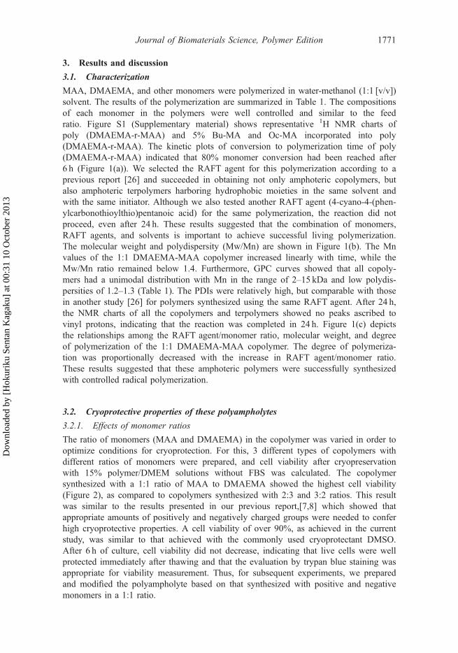
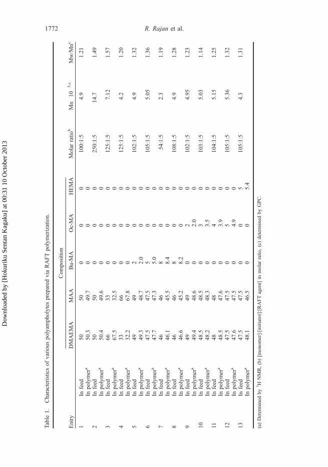
MAA, DMAEMA, and other monomers were polymerized in water-methanol (1:1 [v/v])solvent. The results of the polymerization are summarized in Table 1. The compositionsof each monomer in the polymers were well controlled and similar to the feedratio. Figure S1 (Supplementary material) shows representative 1H NMR charts ofpoly (DMAEMA-r-MAA) and 5% Bu-MA and Oc-MA incorporated into poly(DMAEMA-r-MAA). The kinetic plots of conversion to polymerization time of poly(DMAEMA-r-MAA) indicated that 80% monomer conversion had been reached after6 h (Figure 1(a)). We selected the RAFT agent for this polymerization according to aprevious report [26] and succeeded in obtaining not only amphoteric copolymers, butalso amphoteric terpolymers harboring hydrophobic moieties in the same solvent andwith the same initiator. Although we also tested another RAFT agent (4-cyano-4-(phen-ylcarbonothioylthio)pentanoic acid) for the same polymerization, the reaction did notproceed, even after 24 h. These results suggested that the combination of monomers,RAFT agents, and solvents is important to achieve successful living polymerization.The molecular weight and polydispersity (Mw/Mn) are shown in Figure 1(b). The Mnvalues of the 1:1 DMAEMA-MAA copolymer increased linearly with time, while theMw/Mn ratio remained below 1.4. Furthermore, GPC curves showed that all copoly-mers had a unimodal distribution with Mn in the range of 2–15 kDa and low polydis-persities of 1.2–1.3 (Table 1). The PDIs were relatively high, but comparable with thosein another study [26] for polymers synthesized using the same RAFT agent. After 24 h,the NMR charts of all the copolymers and terpolymers showed no peaks ascribed tovinyl protons, indicating that the reaction was completed in 24 h. Figure 1(c) depictsthe relationships among the RAFT agent/monomer ratio, molecular weight, and degreeof polymerization of the 1:1 DMAEMA-MAA copolymer. The degree of polymeriza-tion was proportionally decreased with the increase in RAFT agent/monomer ratio.These results suggested that these amphoteric polymers were successfully synthesizedwith controlled radical polymerization.
3.2. Cryoprotective properties of these polyampholytes
3.2.1. Effects of monomer ratios
The ratio of monomers (MAA and DMAEMA) in the copolymer was varied in order tooptimize conditions for cryoprotection. For this, 3 different types of copolymers withdifferent ratios of monomers were prepared, and cell viability after cryopreservationwith 15% polymer/DMEM solutions without FBS was calculated. The copolymersynthesized with a 1:1 ratio of MAA to DMAEMA showed the highest cell viability(Figure 2), as compared to copolymers synthesized with 2:3 and 3:2 ratios. This resultwas similar to the results presented in our previous report,[7,8] which showed thatappropriate amounts of positively and negatively charged groups were needed to conferhigh cryoprotective properties. A cell viability of over 90%, as achieved in the currentstudy, was similar to that achieved with the commonly used cryoprotectant DMSO.After 6 h of culture, cell viability did not decrease, indicating that live cells were wellprotected immediately after thawing and that the evaluation by trypan blue staining wasappropriate for viability measurement. Thus, for subsequent experiments, we preparedand modified the polyampholyte based on that synthesized with positive and negativemonomers in a 1:1 ratio.
Journal of Biomaterials Science, Polymer Edition 1771
Dow
nloa
ded
by [
Hok
urik
u Se
ntan
Kag
aku]
at 0
0:31
10
Oct
ober
201
3
Table
1.Characteristicsof
variou
spo
lyam
pholytes
prepared
viaRAFTpo
lymerization.
Entry
Com
positio
n
Molar
ratio
bMn�
10�3,c
Mw/M
ncDMAEMA
MAA
Bu-MA
Oc-MA
HEMA
1In
feed
5050
00
010
0:1:5
4.9
1.21
Inpo
lymer
a50
.349
.70
00
2In
feed
5050
00
025
0:1:5
14.7
1.49
Inpo
lymer
a50
.449
.60
00
3In
feed
6633
00
012
5:1:5
7.12
1.57
Inpo
lymer
a67
.532
.50
00
4In
feed
3366
00
012
5:1:5
4.2
1.20
Inpo
lymer
a32
.267
.80
00
5In
feed
4949
20
010
2:1:5
4.9
1.32
Inpo
lymer
a49
.348
.72.0
00
6In
feed
47.5
47.5
50
010
5:1:5
5.05
1.36
Inpo
lymer
a47
.747
.35.0
00
7In
feed
4646
80
054
:1:5
2.3
1.19
Inpo
lymer
a46
.145
.58.4
00
8In
feed
4646
80
010
8:1:5
4.9
1.28
Inpo
lymer
a46
.645
.28.2
00
9In
feed
4949
02
010
2:1:5
4.95
1.23
Inpo
lymer
a49
.448
.60
2.0
010
Infeed
48.5
48.5
03
010
3:1:5
5.03
1.14
Inpo
lymer
a48
.248
.30
3.5
011
Infeed
4848
04
010
4:1:5
5.15
1.25
Inpo
lymer
a48
.547
.60
3.9
012
Infeed
47.5
47.5
05
010
5:1:5
5.36
1.32
Inpo
lymer
a47
.647
.50
4.9
013
Infeed
47.5
47.5
00
510
5:1:5
4.3
1.31
Inpo
lymer
a48
.146
.50
05.4
(a)Determined
by1H
NMR,(b)[m
onom
er]:[initiator]:[RAFTagent]in
molar
ratio
,(c)determ
ined
byGPC.
1772 R. Rajan et al.
Dow
nloa
ded
by [
Hok
urik
u Se
ntan
Kag
aku]
at 0
0:31
10
Oct
ober
201
3
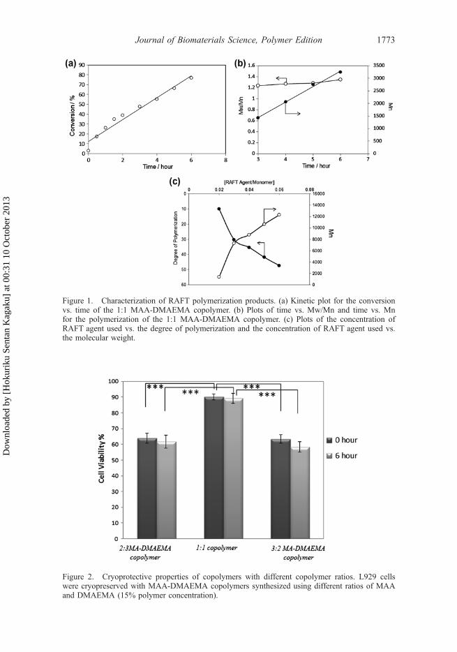
Figure 2. Cryoprotective properties of copolymers with different copolymer ratios. L929 cellswere cryopreserved with MAA-DMAEMA copolymers synthesized using different ratios of MAAand DMAEMA (15% polymer concentration).
Figure 1. Characterization of RAFT polymerization products. (a) Kinetic plot for the conversionvs. time of the 1:1 MAA-DMAEMA copolymer. (b) Plots of time vs. Mw/Mn and time vs. Mnfor the polymerization of the 1:1 MAA-DMAEMA copolymer. (c) Plots of the concentration ofRAFT agent used vs. the degree of polymerization and the concentration of RAFT agent used vs.the molecular weight.
Journal of Biomaterials Science, Polymer Edition 1773
Dow
nloa
ded
by [
Hok
urik
u Se
ntan
Kag
aku]
at 0
0:31
10
Oct
ober
201
3
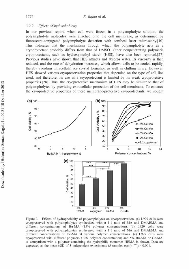
3.2.2. Effects of hydrophobicity
In our previous report, when cell were frozen in a polyampholyte solution, thepolyampholyte molecules were attached onto the cell membrane, as determined byfluorescent-conjugated polyampholyte detection with confocal laser microscopy.[10]This indicates that the mechanism through which the polyampholyte acts as acryoprotectant probably differs from that of DMSO. Other nonpenetrating polymericcryoprotectants, such as hydroxymethyl starch (HES), have also been reported.[27]Previous studies have shown that HES attracts and absorbs water. Its viscosity is thenreduced, and the rate of dehydration increases, which allows cells to be cooled rapidly,thereby avoiding intracellular ice crystal formation as well as chilling injury. However,HES showed various cryopreservation properties that depended on the type of cell lineused, and therefore, its use as a cryoprotectant is limited by its weak cryoprotectiveproperties.[28] Thus, the cryoprotective mechanism of HES may be similar to that ofpolyampholytes by providing extracellular protection of the cell membrane. To enhancethe cryoprotective properties of these membrane-protective cryoprotectants, we sought
Figure 3. Effects of hydrophobicity of polyampholytes on cryopreservation. (a) L929 cells werecryopreserved with polyampholytes synthesized with a 1:1 ratio of MA and DMAEMA anddifferent concentrations of Bu-MA (15% polymer concentration). (b) L929 cells werecryopreserved with polyampholytes synthesized with a 1:1 ratio of MA and DMAEMA anddifferent concentrations of Oc-MA at various polymer concentrations. (c) L929 cells werecryopreserved with different polymers (10% polymer concentration) and 5% Bu-MA or Oc-MA.A comparison with a polymer containing the hydrophilic monomer HEMA is shown. Data areexpressed as the mean ± SD of 3 independent experiments (5 samples each). ⁄⁄⁄p < 0.001.
1774 R. Rajan et al.
Dow
nloa
ded
by [
Hok
urik
u Se
ntan
Kag
aku]
at 0
0:31
10
Oct
ober
201
3
to increase the cell membrane interaction of the polyampholytes by the introduction ofhydrophobic group(s) in the polymer backbone. The alkyl chain is highly hydrophobic,and amphiphilic polymers, such as polyethylene glycol and polyvinyl alcohol, in whichthe alkyl chain was introduced, show cell membrane attachment via hydrophobic inter-actions between membrane lipids and the alkyl chain.[29] To identify the effects ofhydrophobicity, a small amount of a hydrophobic alkyl chain monomer, i.e., n-butylmethacrylate (Bu-MA) or n-octyl methacrylate (Oc-MA), was introduced into the 1:1copolymer. Different amounts of Bu-MA were introduced, and cell viability aftercryopreservation was measured. As shown in Figure 3(a), the viability of L929 cellscryopreserved with 15% poly (DMAEMA-co-MAA) solution including various concen-trations of Bu-MA was enhanced. A similar effect was observed in cells frozen in 7.5%polymer with 5% Oc-MA (Figure 3(b)), which showed almost double the viability ofcells frozen in the polyampholyte without the hydrophobic moiety. Figure 3(c) showsthe cell viability after cryopreservation with hydrophilic and hydrophobic polyampho-lyte solutions at the same polymer concentration (10%). Under these conditions, andwith the same ratio of hydrophilic and hydrophobic moieties, the cryoprotectiveproperties of the solution were strongly correlated with hydrophobicity.
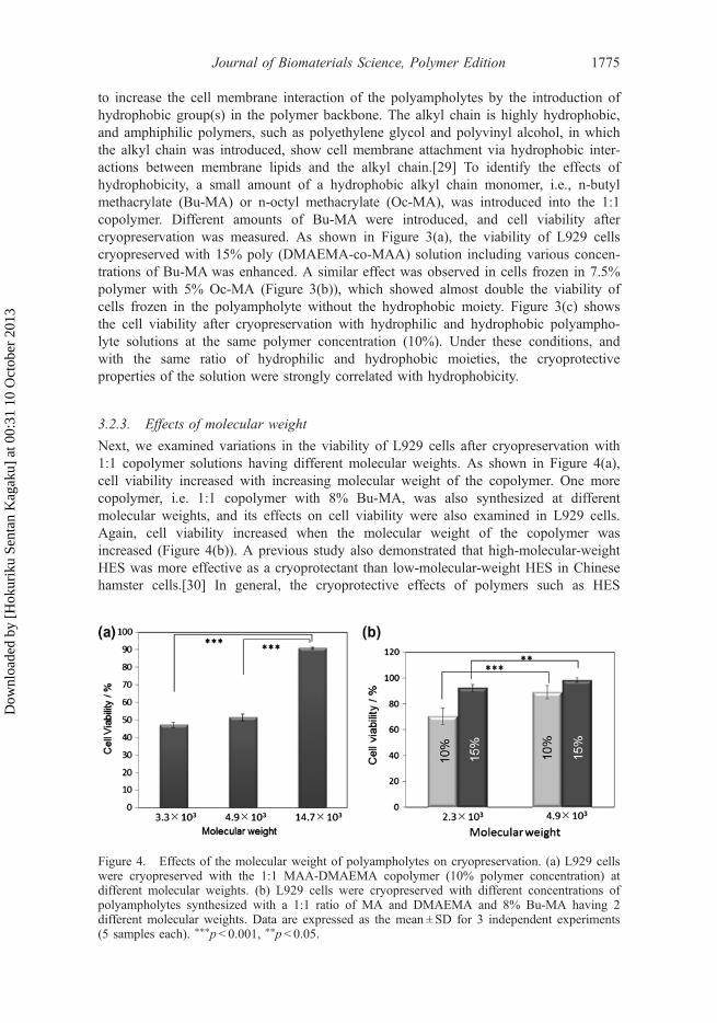
3.2.3. Effects of molecular weight
Next, we examined variations in the viability of L929 cells after cryopreservation with1:1 copolymer solutions having different molecular weights. As shown in Figure 4(a),cell viability increased with increasing molecular weight of the copolymer. One morecopolymer, i.e. 1:1 copolymer with 8% Bu-MA, was also synthesized at differentmolecular weights, and its effects on cell viability were also examined in L929 cells.Again, cell viability increased when the molecular weight of the copolymer wasincreased (Figure 4(b)). A previous study also demonstrated that high-molecular-weightHES was more effective as a cryoprotectant than low-molecular-weight HES in Chinesehamster cells.[30] In general, the cryoprotective effects of polymers such as HES
Figure 4. Effects of the molecular weight of polyampholytes on cryopreservation. (a) L929 cellswere cryopreserved with the 1:1 MAA-DMAEMA copolymer (10% polymer concentration) atdifferent molecular weights. (b) L929 cells were cryopreserved with different concentrations ofpolyampholytes synthesized with a 1:1 ratio of MA and DMAEMA and 8% Bu-MA having 2different molecular weights. Data are expressed as the mean ± SD for 3 independent experiments(5 samples each). ⁄⁄⁄p< 0.001, ⁄⁄p< 0.05.
Journal of Biomaterials Science, Polymer Edition 1775
Dow
nloa
ded
by [
Hok
urik
u Se
ntan
Kag
aku]
at 0
0:31
10
Oct
ober
201
3
depend on its ability to absorb water molecules and keep these thermally inert in aglassy state without experiencing any phase transitions during cooling. Absorption ofwater molecules depends on the molecular weight and concentration of the molecule.Although further research should be conducted, our current data strongly support thehypothesis that the protective properties of these cryoprotectants may be related towater absorption, which is dependent on the molecular weight of the compound. Thus,careful control of molecular weight using RAFT polymerization may be effective in thedevelopment of polyampholyte cryoprotectants.
3.3. Leakage experiment
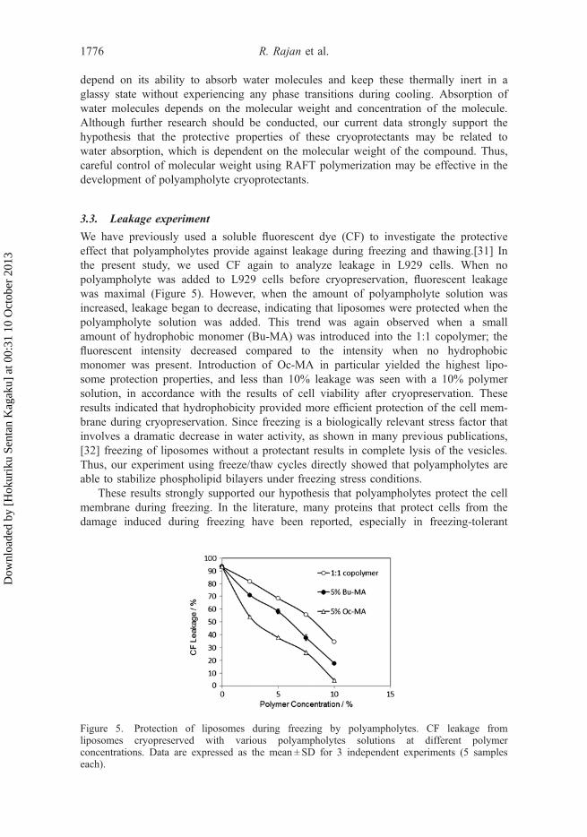
We have previously used a soluble fluorescent dye (CF) to investigate the protectiveeffect that polyampholytes provide against leakage during freezing and thawing.[31] Inthe present study, we used CF again to analyze leakage in L929 cells. When nopolyampholyte was added to L929 cells before cryopreservation, fluorescent leakagewas maximal (Figure 5). However, when the amount of polyampholyte solution wasincreased, leakage began to decrease, indicating that liposomes were protected when thepolyampholyte solution was added. This trend was again observed when a smallamount of hydrophobic monomer (Bu-MA) was introduced into the 1:1 copolymer; thefluorescent intensity decreased compared to the intensity when no hydrophobicmonomer was present. Introduction of Oc-MA in particular yielded the highest lipo-some protection properties, and less than 10% leakage was seen with a 10% polymersolution, in accordance with the results of cell viability after cryopreservation. Theseresults indicated that hydrophobicity provided more efficient protection of the cell mem-brane during cryopreservation. Since freezing is a biologically relevant stress factor thatinvolves a dramatic decrease in water activity, as shown in many previous publications,[32] freezing of liposomes without a protectant results in complete lysis of the vesicles.Thus, our experiment using freeze/thaw cycles directly showed that polyampholytes areable to stabilize phospholipid bilayers under freezing stress conditions.
These results strongly supported our hypothesis that polyampholytes protect the cellmembrane during freezing. In the literature, many proteins that protect cells from thedamage induced during freezing have been reported, especially in freezing-tolerant
Figure 5. Protection of liposomes during freezing by polyampholytes. CF leakage fromliposomes cryopreserved with various polyampholytes solutions at different polymerconcentrations. Data are expressed as the mean ± SD for 3 independent experiments (5 sampleseach).
1776 R. Rajan et al.
Dow
nloa
ded
by [
Hok
urik
u Se
ntan
Kag
aku]
at 0
0:31
10
Oct
ober
201
3
plants, anhydrobiotic invertebrates, and fungi.[33] Interestingly, these proteins sharemany common characteristics, including highly charged polyampholytes with hydropho-bic moieties.[34,35] According to a report by Tolleter et al. [31] polyampholitic protein,which is classified as late embryogenesis abundant (LEA) protein, protects the liposomefrom desiccation and freezing damage with membrane attachments via electrostaticinteractions between LEA protein molecules, which have many positive and negativecharges, and phospholipid molecules. Although the mechanisms of protection arestill unknown, a relationship may exist between these proteins and our synthesizedpolyampholytes.
3.4. Biocompatibility of the novel polyampholytes
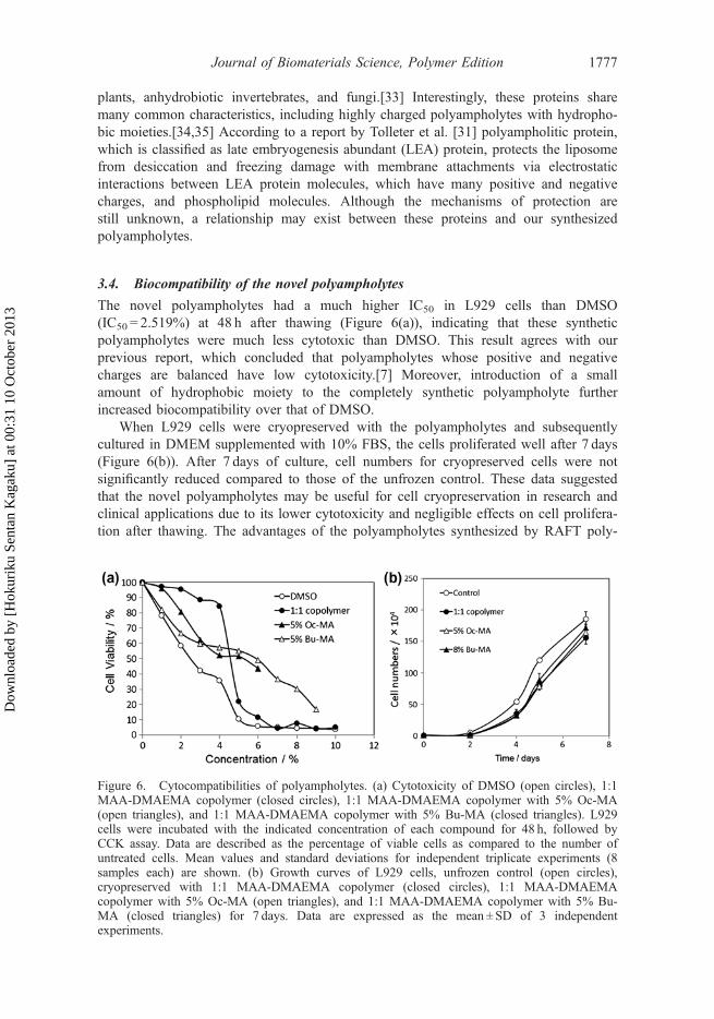
The novel polyampholytes had a much higher IC50 in L929 cells than DMSO(IC50 = 2.519%) at 48 h after thawing (Figure 6(a)), indicating that these syntheticpolyampholytes were much less cytotoxic than DMSO. This result agrees with ourprevious report, which concluded that polyampholytes whose positive and negativecharges are balanced have low cytotoxicity.[7] Moreover, introduction of a smallamount of hydrophobic moiety to the completely synthetic polyampholyte furtherincreased biocompatibility over that of DMSO.
When L929 cells were cryopreserved with the polyampholytes and subsequentlycultured in DMEM supplemented with 10% FBS, the cells proliferated well after 7 days(Figure 6(b)). After 7 days of culture, cell numbers for cryopreserved cells were notsignificantly reduced compared to those of the unfrozen control. These data suggestedthat the novel polyampholytes may be useful for cell cryopreservation in research andclinical applications due to its lower cytotoxicity and negligible effects on cell prolifera-tion after thawing. The advantages of the polyampholytes synthesized by RAFT poly-
Figure 6. Cytocompatibilities of polyampholytes. (a) Cytotoxicity of DMSO (open circles), 1:1MAA-DMAEMA copolymer (closed circles), 1:1 MAA-DMAEMA copolymer with 5% Oc-MA(open triangles), and 1:1 MAA-DMAEMA copolymer with 5% Bu-MA (closed triangles). L929cells were incubated with the indicated concentration of each compound for 48 h, followed byCCK assay. Data are described as the percentage of viable cells as compared to the number ofuntreated cells. Mean values and standard deviations for independent triplicate experiments (8samples each) are shown. (b) Growth curves of L929 cells, unfrozen control (open circles),cryopreserved with 1:1 MAA-DMAEMA copolymer (closed circles), 1:1 MAA-DMAEMAcopolymer with 5% Oc-MA (open triangles), and 1:1 MAA-DMAEMA copolymer with 5% Bu-MA (closed triangles) for 7 days. Data are expressed as the mean ± SD of 3 independentexperiments.
Journal of Biomaterials Science, Polymer Edition 1777
Dow
nloa
ded
by [
Hok
urik
u Se
ntan
Kag
aku]
at 0
0:31
10
Oct
ober
201
3
merization in terms of cryoprotective properties were clearly indicated, and we expectthat the control of dispersity, composition, and precise polymerization, such as blockcopolymerization, may enable researchers to prepare cryoprotective agents with highcell viability and to elucidate the mechanisms of cryoprotection of polyampholytes.
4. Conclusion
We have successfully demonstrated that synthetic polyampholytes made of methacrylicacid and 2-(dimethylamino)ethyl methacrylate can efficiently cryopreserve various typesof cells without the requirement for any other cryoprotectants. Additionally, introductionof hydrophobicity and an increase in molecular weight promoted cell viability afterthawing. Leakage experiments suggested that polyampholytes protected the cellmembrane during cryopreservation, and this effect was enhanced by increasedhydrophobicity. Moreover, due to low cytotoxicity, these polyampholytes have thepotential to replace the conventionally used cryoprotective agent DMSO. The presentstudy is the first to show that we can design a polymeric cryoprotectant that will protectthe cell membrane during freezing using appropriate polymerization techniques.
Disclosures
The authors have no conflicts of interest to declare.
AcknowledgementsThis study was supported in part by a grant from The Canon Foundation (K11-N-028). We thankProf. H. Kitano and M. Gemmei-Ide, University of Toyama, for critical comments for RAFTpolymerization. We also acknowledge Prof. T. Hohsaka and Dr T. Watanabe, Japan AdvancedInstitute of Science and Technology, for assistance in fluorescence measurements.
References[1] Polge C, Smith AU, Parkes AS. Revival of spermatozoa after vitrification and dehydration
at low temperatures. Nature. 1949;164:666.[2] Lovelock JE, Bishop MW. Prevention of freezing damage to living cells by dimethyl
sulphoxide. Nature. 1959;183:1394–1395.[3] Fahy GM. The relevance of cryoprotectant “toxicity” to cryobiology. Cryobiology.
1986;23:1–13.[4] Oh JE, Raja KK, Shin JH, Pollak A, Hengstschlager M, Lubec G. Cytoskeleton changes
following differentiation of N1E–115 neuroblastoma cell line. Amino Acids. 2006;31:289–298.[5] Young DA, Gavrilov S, Pennington CJ, Nuttall RK, Edwards DR, Kitsis RN, Clark IM.
Expression of metalloproteinases and inhibitors in the differentiation of P19CL6 cells intocardiac myocytes. Biochem. Biophys. Res. Commun. 2004;322:759–765.
[6] Jiang GS, Bi KH, Tang TH, Wang JW, Zhang YK, Zhang W, Ren HQ, Bai HQ, Wang YS.Down-regulation of TRRAP-dependent hTERT and TRRAP-independent CAD activation byMyc/Max contributes to the differentiation of HL60 cells after exposure to DMSO. Int.Immunopharmacol. 2006;6:1204–1213.
[7] Matsumura K, Hyon SH. Polyampholytes as low toxic efficient cryoprotective agents withantifreeze protein properties. Biomaterials. 2009;30:4842–4849.
[8] Matsumura K, Bae JY, Hyon SH. Polyampholytes as cryoprotective agents for mammaliancell cryopreservation. Cell Transplant. 2010;19:691–699.
[9] Vrana N, Matsumura K, Hyon SH, Geever L, Kennedy J, Lyons J, Higginbotham CL, CahillPA, McGuinness GB. Cell encapsulation and cryostorage in PVA/gelatin cryogels: incorporationof carboxylated ɛ-poly-L-lysine as cryoprotectant. J. Tissue. Eng. Regen. Med. 2012;6:280–290.
1778 R. Rajan et al.
Dow
nloa
ded
by [
Hok
urik
u Se
ntan
Kag
aku]
at 0
0:31
10
Oct
ober
201
3
[10] Matsumura K, Hayashi F, Nagashima T, Hyon SH. Long-term cryopreservation of humanmesenchymal stem cells using carboxylated poly-L-lysine without the addition of proteinsor dimethyl sulfoxide. J. Biomate. Sce. Polym. Ed. in press. doi:10.1080/09205063.2013.771318.
[11] Wang JS, Matyjaszewski K. Controlled/“living” radical polymerization. Atom transfer radicalpolymerization in the presence of transition-metal complexes. J. Am. Chem. Soc.1995;117:5614–5615.
[12] Kato M, Kamigaito M, Sawamoto M, Higashimura T. Polymerization of methyl methacrylatewith the carbon tetrachloride/dichlorotris- (triphenylphosphine)ruthenium(II)/methylaluminumbis(2,6-di-tert-butylphenoxide) initiating system: possibility of living radical polymerization.Macromolecules. 1995;28:1721–1723.
[13] Georges MK, Veregin RPN, Kazmaier PM, Hamer GK. Narrow molecular weight resins bya free-radical polymerization process. Macromolecules. 1993;26:2987–2988.
[14] Nicolas J, Guillaneuf Y, Lefay C, Bertin D, Gigmes D, Charleux B. Nitroxide-mediatedpolymerization. Prog. Polym. Sci. 2013;38:63–235.
[15] Quinn JF, Davis TP, Rizzardo E. Ambient temperature reversible addition-fragmentationchain transfer polymerization. Chem. Commun. 2001;11:1044–1045.
[16] Goto A, Sato K, Tsujii Y, Fukuda T, Moad G, Rizzardo E, Thang SH. Mechanism andkinetics of RAFT-based living radical polymerizations of styrene and methyl methacrylate.Macromolecules. 2001;34:402–408.
[17] Chiefari J, Chong YK, Ercole F, Krstina J, Jeffery J, Le TP, Mayadunne RTA, Meijs GF,Moad CL, Moad G, Rizzardo E, Thang SH. Living free-radical polymerization by reversibleaddition-fragmentation chain transfer: the RAFT process. Macromolecules. 1998;31:5559–5562.
[18] Mitsukami Y, Donovan MS, Lowe AB, McCormick CL. Water-soluble polymers. 81. Directsynthesis of hydrophilic styrenic-based homopolymers and block copolymers in aqueoussolution via RAFT. Macromolecules. 2001;34:2248–2256.
[19] Donovan MS, Lowe AB, Sanford TA, McCormick CL. Sulfobetaine-containing diblock andtriblock copolymers via reversible addition-fragmentation chain transfer polymerization inaqueous media. J. Polym. Sci. Polym. Chem. 2003;41:1262–1281.
[20] McCormick CL, Lowe AB. Aqueous RAFT polymerization: Recent developments in synthe-sis of functional water-soluble (co)polymers with controlled structures. Acc. Chem. Res.2004;37:312–325.
[21] Thomas DB, Sumerlin BS, Lowe AB, McCormick CL. Conditions for facile, controlledRAFT polymerization of acrylamide in water. Macromolecules. 2003;36:1436–1439.
[22] Ladavière C, Dörr N, Claverie JP. Controlled radical polymerization of acrylic acid in proticmedia. Macromolecules. 2001;34:5370–5372.
[23] Sumerlin BS, Lowe AB, Thomas DB, McCormick CL. Aqueous solution properties ofpH-responsive AB diblock acrylamido copolymers synthesized via aqueous RAFT.Macromolecules. 2003;36:5982–5987.
[24] MacDonald RC, MacDonald RI, Menco BPM, Takeshita K, Subbarao NK, Hu LR.Small-volume extrusion apparatus for preparation of large, unilamellar vesicles. Biochim.Biophys. Acta. 1991;1061:297–303.
[25] Hincha DK, Zuther E, Hellwege EM, Heyer AG. Specific effects of fructo- and gluco-oligo-saccharides in the preservation of liposomes during drying. Glycobiology. 2002;12:103–110.
[26] Kitano H, Kondo T, Kamada T, Iwanaga S, Nakamura M, Ohno K. Anti-biofouling proper-ties of an amphoteric polymer brush constructed on a glass substrate. Colloids Surf. B.2011;88:455–462.
[27] Stolzing A, Naaldijk Y, Fedorova V, Sethe S. Hydroxyethylstarch in cryopreservation –Mechanisms, benefits and problems. Transfus. Apher. Sci. 2012;46:137–147.
[28] Stiff PJ, Murgo AJ, Zaroulis CG, DeRisi MF, Clarkson BD. Unfractionated human marrowcell cryopreservation using dimethylsulfoxide and hydroxyethyl starch. Cryobiology.1983;20:17–24.
[29] Inui O, Teramura Y, Iwata H. Retention dynamics of amphiphilic polymers PEG-lipids andPVA-alkyl on the cell surface. ACS Appl. Mater. Interfaces. 2010;2:1514–1520.
[30] Luo K, Wu G, Wang Q, Sun Y, Liu H. Effect of dimethylsulfoxide and hydroxyethyl starchin the preservation of fractionated human marrow cells. Cryobiology. 1994;31:349–354.
Journal of Biomaterials Science, Polymer Edition 1779
Dow
nloa
ded
by [
Hok
urik
u Se
ntan
Kag
aku]
at 0
0:31
10
Oct
ober
201
3
[31] Tolleter D, Hincha DK, Macherel D. A mitochondrial late embryogenesis abundantprotein stabilizes model membranes in the dry state. Biochim. Biophys. Acta. 2010;1798:1926–1933.
[32] Hincha DK, Popova AV, Cacela C. Effects of sugars on the stability and structure of lipidmembranes during drying. Vol. 3. In: Tien HT, Ottova-Leitmannova A, editors. Advancesin planar lipid bilayers and liposomes. Amsterdam: Elsevier; 2006. 189–217.
[33] Tunnacliffe A, Wise MJ. The continuing conundrum of the LEA proteins. Naturwissenschaften.2007;94:791–812.
[34] Shimizu T, Kanamori Y, Furuki T, Kikawada T, Okuda T, Takahashi T, Mihara H, SakuraiM. Desiccation-induced structuralization and glass formation of group 3 late embryogenesisabundant protein model peptides. Biochemistry. 2010;16:1093–1104.
[35] Furuki T, Shimizu T, Chakrabortee S, Yamakawa K, Hatanaka R, Takahashi T, Kikawada T,Okuda T, Mihara H, Tunnacliffe A, Sakurai M. Effects of group 3 LEA protein model pep-tides on desiccation-induced protein aggregation. Biochim. Biophys. Acta.2012;1824:891–897.
1780 R. Rajan et al.
Dow
nloa
ded
by [
Hok
urik
u Se
ntan
Kag
aku]
at 0
0:31
10
Oct
ober
201
3