Detergent-like actions of linear amphipathic cationic antimicrobial peptides
Upload
independentCategory
view
1download
0
Oleic and Docosahexaenoic Acid Differentially Phase Separate fromLipid Raft Molecules: A Comparative NMR, DSC, AFM, andDetergent Extraction Study
Saame Raza Shaikh,*y Alfred C. Dumaual,* Alicia Castillo,z Daniel LoCascio,* Rafat A. Siddiqui,z
William Stillwell,*y and Stephen R. Wassall{y
*Department of Biology, Indiana University-Purdue University, Indianapolis, Indiana; yMedical Biophysics Program, Indiana UniversitySchool of Medicine, Indianapolis, Indiana; zCellular Biochemistry Laboratory, Methodist Research Institute at Clarian Health,Indianapolis, Indiana; and {Department of Physics, Indiana University-Purdue University, Indianapolis, Indiana
ABSTRACT We have previously suggested that the v-3 polyunsaturated fatty acid, docosahexaenoic acid (DHA) may in partfunction by enhancing membrane lipid phase separation into lipid rafts. Here we further tested for differences in the molecularinteractions of an oleic (OA) versus DHA-containing phospholipid with sphingomyelin (SM) and cholesterol (CHOL) utilizing 2HNMR spectroscopy, differential scanning calorimetry, atomic force microscopy, and detergent extractions in model bilayermembranes. 2H NMR and DSC (differential scanning calorimetry) established the phase behavior of the OA-containing1-[2H31]palmitoyl-2-oleoyl-sn-glycero-3-phosphoethanolamine (16:0-18:1PE-d31)/SM (1:1) and the DHA-containing 1-[2H31]-palmitoyl-2-docosahexaenoyl-sn-glycero-3-phosphoethanolamine (16:0-22:6PE-d31)/SM (1:1) in the absence and presence ofequimolar CHOL. CHOL was observed to affect the OA-containing phosphatidylethanolamine (PE) more than the DHA-containing PE, as exemplified by.23 greater increase in order measured for the perdeuterated palmitic chain in 16:0-18:1PE-d31/SM (1:1) compared to 16:0-22:6PE-d31/SM (1:1) bilayers in the liquid crystalline phase. Atomic force microscopy (AFM)experiments showed less lateral phase separation between 16:0-18:1PE-rich and SM/CHOL-rich raft domains in 16:0-18:1PE/SM/CHOL (1:1:1) bilayers than was observed when 16:0-22:6PE replaced 16:0-18:1PE. Differences in the molecular interactionof 16:0-18:1PE and 16:0-22:6PE with SM/CHOL were also found using biochemical detergent extractions. In the presence ofequimolar SM/CHOL, 16:0-18:1PE showed decreased solubilization in comparison to 16:0-22:6PE, indicating greater phaseseparation with the DHA-PE. Detergent experiments were also conducted with cardiomyocytes fed radiolabeled OA or DHA.Although both OA and DHA were found to be largely detergent solubilized, the amount of OA that was found to be associatedwith raft-rich detergent-resistant membranes exceeded DHA by almost a factor of 2. We conclude that the OA-PE phaseseparates from rafts far less than DHA-PE, which may have implications for cellular signaling.
INTRODUCTION
It is well recognized that dietary intake of v-3 poly-
unsaturated fatty acids (PUFAs) has profound benefits on
normal health and prevention of chronic disease states,
although the molecular mechanism responsible remains
unclear. One possibility is that v-3 PUFAs are incorporated
into the plasma membrane, influencing membrane structure
and function (Jump, 2002; Stillwell, 2000). Of special
importance for human health is docosahexaenoic acid
(DHA), the longest and most unsaturated fatty acid
commonly found in the plasma membrane. With 22 carbons
and 6 cis double bonds located at positions 4, 7, 10, 13, 16,
and 19, DHA’s unique molecular structure (Eldho et al.,
2003) renders it a primary candidate for affecting properties
of the membrane. It has been proposed by us (Brzustowicz
et al., 2002a; Shaikh et al., 2002) and others (Huster et al.,
1998; Mitchell and Litman, 1998) that DHA may be
specifically involved in inducing lateral phase separations
into DHA-rich/cholesterol-poor and DHA-poor/cholesterol-
rich lipid microdomains. A reduced affinity between the
DHA acyl chain and cholesterol has been hypothesized to
promote phase separations, perhaps affecting lipid rafts
(Stillwell and Wassall, 2003).
Lipid rafts are postulated to be specialized liquid-ordered
(lo) lipid microdomains rich in sphingolipids and cholesterol.
They are involved in serving as platforms for cellular
signaling events (Ahmed et al., 1997; Edidin, 1993, 2003a;
Hooper, 1999) by accumulating specific proteins, including
GPI-anchored proteins and the src-family of kinases. In
addition, rafts are thought to influence lipid trafficking
events, pathogen entry, and cytoskeletal organization
(Munro, 2003). The supposed stability of lipid rafts is
attributed to the favorable interaction between the amide of
the sphingosine backbone and the hydroxyl of an adjacent
sphingolipid as well as hydrogen bonding between the 3-OH
of cholesterol and the sphingosine amide. Saturated acyl
chains are thought to promote formation of rafts since they
are more extended than unsaturated chains and pack well
amongst themselves and with cholesterol. Consequently,
phospholipids containing highly disordered polyunsaturated
Submitted April 16, 2004, and accepted for publication June 18, 2004.
Address reprint requests to William Stillwell, Dept. of Biology, Indiana
University-Purdue University, 723 W. Michigan St., Indianapolis, IN
46202-5132. Tel.: 317-274-0580; Fax: 317-274-2846; E-mail: wstillwe@
iupui.edu.
� 2004 by the Biophysical Society
0006-3495/04/09/1752/15 $2.00 doi: 10.1529/biophysj.104.044552
1752 Biophysical Journal Volume 87 September 2004 1752–1766

acyl chains that exhibit low affinity to cholesterol would be
expected to phase separate from the rafts.
We suggested in a preliminary investigation that an oleic
acid (OA)-containing phosphatidylethanolamine (PE) and a
DHA-containing PE may phase separate differently from the
lipid raft molecules sphingomyelin (SM) and cholesterol
(CHOL) in monolayer and bilayer membranes (Shaikh et al.,
2002). Here we elucidate the effect that replacing the
OA-containing 1-palmitoyl-2-oleoyl-sn-glycero-3-phosphoeth-anolamine (16:0-18:1PE) with the DHA-containing 1-palmi-
toyl-2-docosahexaenoyl-sn-glycero-3-phosphoethanolamine
(16:0-22:6PE) has on phase separation from SM and CHOL.
PE was chosen as our model phospholipid since it is the
second most abundant polar lipid found in mammalian
plasma membranes and it is often in PE that DHA accu-
mulates (Simopoulos et al., 1986; Zerouga et al., 1996).
Partial lipid asymmetry is a characteristic of all membranes.
Approximately one-sixth of plasma membrane PE is found
in the outer leaflet where it would interact with the abundant
SM (approximately five-sixths of the total SM). In the inner
leaflet, the abundant PE (approximately five-sixths of the
total PE) would interact with the less abundant SM
(approximately one-sixth of the total SM). Therefore, in
both membrane leaflets there are sufficient quantities of both
PE and SM to affect membrane structure (Simons and Vaz,
2004). PEs normally exist as heteroacid sn-1 saturated, sn-2unsaturated species. We employ 16:0-18:1PE as our
‘‘typical’’ phospholipid because oleic acid is the most
abundant unsaturated fatty acid in membranes and 16:
0-22:6PE constitutes our ‘‘modified’’ phospholipid.
In this study we explore differences in lateral phase
separation between 16:0-18:1PE/SM/CHOL (1:1:1) and
16:0-22:6PE/SM/CHOL (1:1:1) bilayers employing solid
state 2H NMR spectroscopy, differential scanning calorim-
etry (DSC), atomic force microscopy (AFM), and detergent
extraction. 2H NMR and DSC are first used to characterize
the phase behavior of our model membrane systems in the
absence and presence of cholesterol. Subsequent analysis of2H NMR spectra in the lamellar liquid crystalline phase,
AFM measurements, and detergent-resistant experiments in
model membranes demonstrate marked differences in the
molecular interactions of 16:0-18:1PE versus 16:0-22:6PE
with raft molecules. We also extend our studies to include
detergent-resistant studies with living cardiomyocytes.
These studies are discussed in relation to the physiological
importance of rafts.
MATERIALS AND METHODS
Materials
Egg SM, 1-palmitoyl-2-oleoyl-sn-glycero-3-phosphoethanolamine (16:
0-18:1PE), 1-[2H31]palmitoyl-2-oleoyl-sn-glycero-3-phosphoethanolamine
(16:0-18:1PE-d31), 1-palmitoyl-2-docosahexaenoyl-sn-glycero-3-phosphoe-
thanolamine (16:0-22:6PE), and 1-[2H31]palmitoyl-2-docosahexaenoyl-
sn-glycero-3-phosphoethanolamine (16:0-22:6PE-d31) were purchased from
Avanti Polar Lipids (Alabaster, AL). CHOL was obtained from Sigma
Chemical Co. (St. Louis, MO). Lipid purity was assessed with thin layer
chromatography. Lipid and cholesterol concentrations were quantified using
phosphate and gravimetric analyses, respectively. Deuterium-depleted H2O
used in the 2H NMR studies was obtained from Isotec (Miamisburg, OH).
High-performance thin layer chromatography (HPTLC) plates were
purchased from Alltech (Deerfield, IL). All organic solvents were HPLC
grade and obtained from Fisher Scientific (Pittsburgh, PA). Molecular
Probes (Eugene, OR) was the source of cholera toxin subunit B, horseradish
peroxidase conjugate. The radiolabeled fatty acids [14C]DHA and [3H]OA
were obtained from PerkinElmer (Boston, MA) The cardiomyocyte isolation
kit was purchased from Worthington Biochemical (Lakewood, NJ). Horse
and fetal bovine serums were from Hyclone (Logan, UT).
2H NMR sample preparation
2H NMR samples were prepared taking stringent precautions to prevent
oxidation, as described before (Shaikh et al., 2002). Lipid mixtures
containing 100 mg total lipid were codissolved in chloroform, dried under
argon gas, and placed under vacuum overnight. After vacuum pumping, the
mixtures were hydrated to 50 wt % with 50 mM Tris buffer (pH 7.4) and
vigorously vortexed for ;5 min. The pH was corrected in the presence of
2 mL additional deuterium-depleted H2O, and the samples were then frozen
and lyophilized multiple times at ;50 mTorr to reduce the 2H NMR signal
from residual 2HHO. After hydrating the powders to 50 wt %, the resultant
aqueous multilamellar dispersions were transferred to 5-mm glass NMR
tubes and stored at �20�C when not in use. All samples were allowed to
equilibrate to room temperature for ;1 h before experimentation. As
previously reported (Brzustowicz et al., 2002b; Shaikh et al., 2002), we have
compared phospholipid/cholesterol samples prepared as described here and
by the low-temperature trapping method (Huang et al., 1999) designed to
ensure that lipids do not demix during removal of the solvent. Both methods
yielded essentially identical results.
2H NMR spectroscopy
Solid-state 2H NMR spectra were acquired on a home-built spectrometer
operating at a resonance frequency of 27.6 MHz. A double resonance probe
(Cryomagnet Systems, Indianapolis, IN) with a 5-mm transverse-mounted
coil was utilized. Temperature was controlled (60.5�C)with a Love Controls(1600 series) temperature controller (MichiganCity, IN). Spectral parameters
were as stated previously (McCabe et al., 1994; Shaikh et al., 2003a).
Analysis of 2H NMR spectra
Moments Mn were calculated from 2H NMR spectra for 16:0-18:1PE-d31 or
16:0-22:6PE-d31 in PE/SM (1:1) and PE/SM/CHOL (1:1:1) mixtures with
Mn ¼RN
�N jvnjf ðvÞdvRN
�N f ðvÞdv ; (1)
where v is the frequency with respect to the central Larmor frequency v0,
f(v) is the lineshape, and n is the order of the spectral moment (Davis, 1983).
In practice the integral is a summation over the digitized data. The
expression
M1 ¼pffiffiffi3
p e2qQ
h
� �jSCDj (2)
relates the first moment M1 to the average order parameter SCD of the
perdeuterated palmitic sn-1 chain via the static quadrupolar coupling
constant (e2qQ/h) ¼ 167 kHz in the lamellar liquid crystalline phase.
OA versus DHA-Raft Phase Separations 1753
Biophysical Journal 87(3) 1752–1766

Spectra were also fast Fourier transform (FFT) depaked to enhance
resolution in the lamellar liquid crystalline phase (McCabe and Wassall,
1997). The depaking procedure numerically deconvolutes the powder
pattern signal to a spectrum representative of a planar membrane of single
alignment. The depaked spectra consist of doublets with quadrupolar
splittings DnðqÞ that equate to order parameters by
DnðuÞ ¼ 3
2
e2qQ
h
� �jSCDjP2ðcos uÞ; (3)
where q ¼ 0� is the angle the membrane normal makes with the magnetic
field and P2ðcosqÞ is the second-order Legendre polynomial. Smoothed
profiles of order along the perdeuterated palmitoyl sn-1 chain were then
constructed on the basis of integrated intensity assuming monotonic
variation toward the disordered center of the bilayer (Lafleur et al., 1989).
Differential scanning calorimetry
DSC experiments were conducted as previously described (Shaikh et al.,
2001). Briefly, lipid mixtures, codissolved in chloroform, were dried under
argon gas followed by ;12 h under vacuum to ensure the removal of
residual organic solvent. Samples containing DHA were prepared under
dark conditions in a glovebox with an argon atmosphere to control for
oxidation. Multilamellar vesicles (MLVs) were made by hydrating the
appropriate phospholipids at 5 mg/ml in 10 mM sodium phosphate buffer
(pH 7.4) with subsequent removal of oxygen. MLV solutions were frozen in
dry ice and thawed three times in a water bath above the gel-to-liquid
crystalline phase transition temperature (Tm) of the lipids. The MLV
solutions (500 mL) were added to each of the three chambers of
a Calorimetry Sciences differential scanning calorimeter (American Fork,
Utah) whereas the fourth chamber contained 500 mL of the buffer. Heating
and cooling scans were made at 0.125�C/min. Only the cooling scans are
presented although data derived from both scans appeared nearly identical.
Deconvolution of the multicomponent 16:0-18:1PE/SM (1:1) scan was
accomplished with Microcal Origin Software (Northampton, MA).
AFM sample preparation
Lipid mixtures codissolved in chloroform were dried under argon gas and
placed in a vacuum overnight. Two mL of buffer solution (10 mM MOPS,
200 mM NaCl, 0.1 mM EGTA, pH 7.4) warmed above the phase transition
was added to the dried lipid film at a final concentration of 1 mM to make
MLVs as described above. Large unilamellar vesicles (LUVs) of 0.1 mm
diameter were formed by initially extruding the MLV suspension multi-
ple times through a 1.0-mm pore-sized nucleopore polycarbonate filter
(Whatman, Clifton, NJ) and subsequently through a 0.1-mm nucleopore
filter using a miniextruder (Avanti Polar Lipids). Fifty-nm diameter small
unilamellar vesicle (SUV) lipid suspensions were made by additionally
passing the LUV suspension through a 0.05-mm nucleopore filter. Lipid
preparation and AFM experiments were conducted in the dark and under
degassed, argon gas-saturated conditions to prevent oxidation.
Supported bilayers
Supported bilayer formation using LUV suspensions was adapted from
Tokumasu et al (2003). Briefly, 20 mL of the LUV suspension was placed on
a freshly cleaved mica surface, incubated for 30–60 s, and then washed with
buffer. Excess solution was removed and fresh degassed buffer solution was
added to the sample and allowed to incubate for 30–60 min before imaging.
Alternatively, supported bilayers were also formed using an SUV
suspension (Rinia et al., 2002). In this case, 75 mL of the SUV suspension
with a lipid concentration of ;0.5 mM was added to the freshly cleaved
mica surface. Lipid vesicles were allowed to absorb to the mica surface for
a period of 10 h at 4�C. Afterwards, the sample was rinsed with buffer
solution and warmed above the phase transition temperature for 60 min and
then allowed to cool to room temperature. Samples were rinsed once more
and fresh, degassed buffer solution was added before imaging. Images from
LUV and SUV vesicle deposition showed nearly identical results.
Atomic force microscopy
Supported bilayer samples were placed in a liquid cell holder attached to
a Bioscope AFM (Digital Instruments, Santa Barbara, CA) set on a vibration-
free air table. Bilayer surfaces were imaged using the tapping mode at
a scanning rate of 0.5–1.0 Hz and a tapping frequency of ;7.8 kHz using
DNP-S20 oxide-sharpened tips with a spring constant of 0.06 N/m (Digital
Instruments). All images were scanned over a 10 3 10-mm area at room
temperature (23�C). Images were acquired from four separate experiments
for each lipid composition with a minimum of four images (from separate
areas of the bilayer) per experiment for analysis. Image analysis was
conducted with software provided by Digital Instruments in addition to NIH
Image J software.
Detergent-resistant studies
Detergent-resistant studies were conducted as described by others
(Gandhavadi et al., 2002; Schroeder et al., 1994). Lipid mixtures (2 mg)
of either 16:0-18:1PE/SM/CHOL (1:1:1) or 16:0-22:6PE/SM/CHOL (1:1:1)
were codissolved in chloroform and dried under a gentle stream of argon
gas. Samples were then placed in a vacuum for a minimum of ;6 h to
remove trace amounts of organic solvent. MLVs were prepared by adding
1 mL of phosphate-buffered saline (PBS) (Cambrex, East Rutherford, NJ)
into the samples and hydrating them for 30 min at 45�C with intermit-
tent vortexing. MLVs were then incubated at 4 or 40�C for 30 min in the
presence of 1% Triton X-100 and subsequently centrifuged at the
appropriate temperature in a Beckman (Fullerton, CA) table-top centrifuge
(TL100) for 1 h at 200,000 g. Supernatants (detergent soluble membranes,
DSMs) and pellets (detergent resistant membranes, DRMs) were separated
and washed for 90 min with SM-2 Adsorbent Bio-Beads (Bio-Rad,
Hercules, CA) to remove Triton X-100 (Gandhavadi et al., 2002). DSMs and
DRMs were lyophilized for ;10 h to remove H20 and then dissolved in
chloroform and subjected to HPTLC analysis.
HPTLC analysis
SM, CHOL, and PE lipids were separated as described (Graham and
Higgins, 1997) on HPTLC plates. Plates were developed (halfway up the
plate) twice in chloroform-methanol-water (60:30:5 by volume) followed by
a third development (to the top of the plate) with hexane-diethyl ether-acetic
acid (80:20:1.5 by volume). They were then charred (;190�C) in 5%
CuSO4/4% H3PO4 solution for 10 min and analyzed using a Kodak Image
Station.
Isolation of cardiomyocytes and cell culture
Neonatal cardiomyocytes were isolated using a cell isolation system from
Worthington Biochemical (Lakewood, NJ). Hearts were isolated from 1- to
3-day-old Wistar rats. Connective tissue and atria were removed from the
hearts and ventricles were subsequently minced into ;1-mm blocks and
digested with trypsin overnight. Trypsin activity was neutralized the
following day and tissues were further digested with collagenase. Single-cell
isolation was achieved by filtering the cell suspension through a 70-mm
filter. Cells were then preplated to remove fibroblasts and cardiomyocytes
were isolated using the isolation protocol accompanying the kit (Worthing-
ton). Dead cells and cellular debris were removed by centrifugation on a
1754 Shaikh et al.
Biophysical Journal 87(3) 1752–1766

5-mL Opti-Prep density gradient solution (Axis-Shiel PoC, As, Oslo,
Norway). Isolated neonatal cardiomyocytes were cultured for 24 h in
a humidified incubation chamber in the presence of 95% O2/5% CO2 on
laminin- and collagen-coated plates in F-10 media (containing 10% horse
serum, 5% fetal bovine serum, 100 units/mL penicillin, 100 mg/mL
streptomyocin, and 0.1 mM bromodeoxyuridine).
Incorporation of radiolabeled fatty acids
Cardiomyocytes were washed with serum-free media twice and then treated
with 5 mM DHA or OA in the presence of trace amounts of [14C]DHA (0.2
mCi/mL) and [3H]OA (1mCi/mL), respectively, under serum-free conditions
for 24 h. Free fatty acid solutions containing radiolabeled fatty acids were
prepared freshly each time from stock solutions by dissolving the compounds
inEtOHwith a final EtOHconcentration inwarmed culturemedium#0.05%.
The concentration of free fatty acids used was based on cell viability
measurements using WST-1 (Roche Biochemicals, Indianapolis, IN).
Isolation of detergent-resistant membranes
Cells were scraped and dissolved in 1 mL of cold Mes-buffered saline
(MBS; 150 mM NaCl, 25 mM Mes, pH 6.5) in the presence of 1% (w/v)
Triton X-100. Cells were incubated for 30 min at 4�C and then homogenized
on ice. Cold MBS (1.5 mL) was added to the homogenate and 2 mL of this
suspension was mixed with 2 mL of 90% (w/v) sucrose in MBS. The
mixture was then added onto a sucrose gradient consisting of 8 mL of
5–35% (w/v) sucrose in MBS. All solutions were supplemented with
a protease inhibitor cocktail (Roche Biochemicals). Sucrose gradients were
then subjected to centrifugation in a Beckman SW40 (Fullerton, CA)
swinging bucket rotor at 200,000 g for 20 h at 4�C. Fractions of 1 mL each
were collected from the top to the bottom of the tubes, vortexed, and stored
at �80�C until further use. Fifty mL of each isolated sucrose gradient
fraction containing [3H]OA or [14C]DHA was placed in a scintillation vial,
mixed with scintillation fluid, and counted using a Beckman scintillation
counter (LS 6000 IC).
b-cholera toxin binding to GM1
Dot blots were obtained to test for localization of GM1, a raft marker (Brown
and London, 2000), in sucrose gradient fractions of cardiomyocytes. Twenty
mL of each fraction was loaded onto a nitrocellulose paper and placed under
a vacuum for 2 h. The nitrocellulose was then blocked in Blocker Blotto
(Pierce, Rockford, IL) for 1 h and then incubated in peroxidase labeled
b-cholera toxin overnight (1:1000) in TTBS (50 mM Tris-HCl, 150 mM
NaCl, 0.05% Tween-20, pH 7.4) in the cold. Blots were then washed three
times in TTBS buffer and developed using an enhanced-chemiluminescence
kit and x-ray film. X-ray films were quantified using a Kodak Image Station.
RESULTS
Characterization of phase behavior for16:0-18:1PE-d31/SM (1:1) versus 16:0-22:6PE-d31/SM (1:1) in the absence andpresence of equimolar CHOL
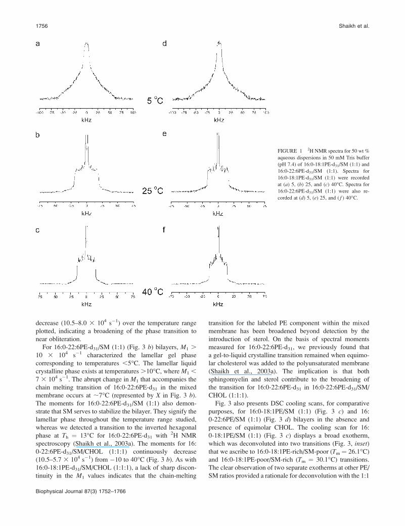
Fig. 1 presents 2H NMR spectra for aqueous multilamellar
dispersions of 16:0-18:1PE-d31/SM (1:1) and 16:0-22:6PE-
d31/SM (1:1) at 5, 25, and 40�C. The approach is to use the
perdeuterated sn-1 chain as an intrinsic probe of molecular
organization for the phospholipid in the manner advocated
by, for example, Petrache et al. (2001) who examined DHA-
containing phosphatidylcholines (PCs) with a series of
saturated sn-1 chains. Both 16:0-18:1PE-d31/SM (1:1) (Fig.
1 a) and 16:0-22:6PE-d31/SM (1:1) (Fig. 1 d) at 5�C show
featureless broad spectra characteristic of the gel phase with
shoulders at 663 kHz. The spectral shape is rendered
nonaxially symmetric (asymmetry parameter h 6¼ 0) by slow
rotational diffusion of the all trans 16:0 acyl chains (Wassall
et al., 1986). Increasing the temperature to 25�C results in
a narrowing of the spectra to 619 kHz for 16:0-18:1PE-d31/
SM (1:1) (Fig. 1 b) and 616 kHz for 16:0-22:6PE-d31/SM
(Fig. 1 e), indicating fast axial rotation associated with the
onset of lamellar liquid crystalline phase. Spectra recorded
at 40�C for 16:0-18:1PE-d31/SM (1:1) (Fig. 1 c) and 16:
0-22:6PE-d31/SM (1:1) (Fig. 1 f) are characterized by
spectral edges at 615 kHz, and are representative of the
lamellar liquid crystalline phase in which there is rapid
isomerization about the carbon-carbon bonds.
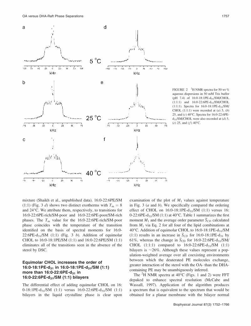
Fig. 2 illustrates the effect of equimolar CHOL on the
phase behavior of 16:0-18:1PE-d31/SM (1:1) and 16:0-22:
6PE-d31/SM (1:1) bilayers at 5, 25, and 40�C. All samples at
5, 25, and 40�C produce powder patterns characteristic of the
liquid crystalline state. Although spectral peaks at 5�C are
poorly resolved for 16:0-18:1PE-d31/SM/CHOL (1:1:1)
(Fig. 2 a) and 16:0-22:6PE-d31/SM/CHOL (1:1:1) (Fig. 2
d), particularly in the former system, the PE component in
either lipid mixture displays spectral edges at 628 kHz,
indicative of fast axial rotation. Upon increasing the
temperature to 25 or 40�C enhanced spectral resolution
becomes apparent and a differential reduction in width is
observed between 16:0-18:1PE-d31/SM/CHOL (1:1:1) (Fig.
2, b and c) and 16:0-22:6PE-d31/SM/CHOL (1:1:1) (Fig. 2, eand f). The sharp edges of the spectrum for 16:0-18:1PE-d31/
SM/CHOL (1:1:1) bilayers occur at 625 kHz (25�C) and623 kHz (40�C) in comparison to622 kHz (25�C) and619
kHz (40�C) for 16:0-22:6PE-d31/SM/CHOL (1:1:1). A
greater increase in order due to the sterol for the OA-con-
taining component than the DHA-containing component in
the mixtures with SM is indicated.
Figs. 1 and 2 only present examples of spectra at selected
temperatures. However, spectra for 16:0-18:1PE-d31/SM
(1:1), 16:0-18:1PE-d31/SM/CHOL (1:1:1), 16:0-22:6PE-d31/
SM (1:1), and 16:0-22:6PE-d31/SM/CHOL (1:1:1) were
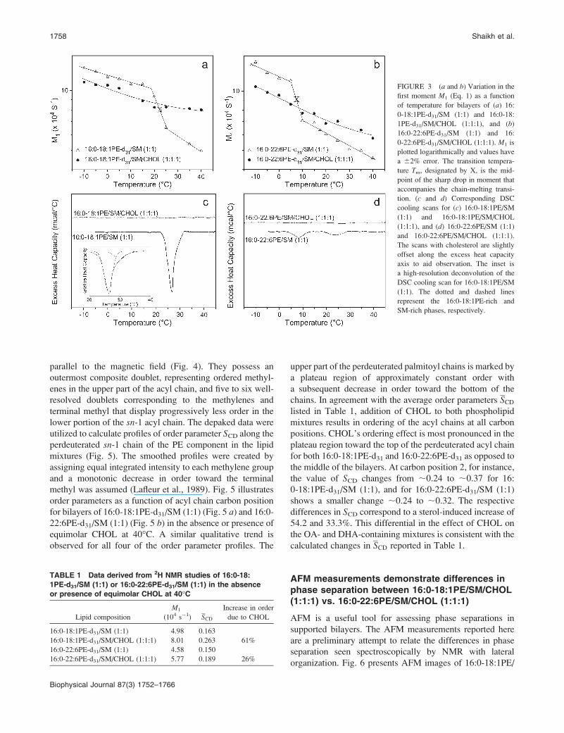
acquired over a wide temperature range from �10 to 40�C.To illustrate the effect of temperature on phase behavior, Fig.
3, a and b, presents the first moment M1 as a function of
temperature for all four of the lipid mixtures. For 16:
0-18:1PE-d31/SM (1:1) bilayers (Fig. 3 a), adoption of gel
phase by 16:0-18:1PE-d31 is signified byM1 . 103 104 s�1
for temperatures ,20�C. The liquid crystalline state,
characterized by M1 , 6.5 3 104 s�1, was adopted at
temperatures .25�C. The sharp drop in M1 value that
accompanies the gel-to-liquid crystalline phase transition is
centered at;23�C (represented by X in Fig. 3 a). There is nolonger a discontinuity in the temperature variation of the first
moment after the addition of equimolar CHOL to 16:
0-18:1PE-d31/SM (1:1) (Fig. 3 a). The values gradually
OA versus DHA-Raft Phase Separations 1755
Biophysical Journal 87(3) 1752–1766
decrease (10.5–8.0 3 104 s�1) over the temperature range
plotted, indicating a broadening of the phase transition to
near obliteration.
For 16:0-22:6PE-d31/SM (1:1) (Fig. 3 b) bilayers, M1 .
10 3 104 s�1 characterized the lamellar gel phase
corresponding to temperatures ,5�C. The lamellar liquid
crystalline phase exists at temperatures .10�C, where M1 ,
7 3 104 s�1. The abrupt change in M1 that accompanies the
chain melting transition of 16:0-22:6PE-d31 in the mixed
membrane occurs at ;7�C (represented by X in Fig. 3 b).The moments for 16:0-22:6PE-d31/SM (1:1) also demon-
strate that SM serves to stabilize the bilayer. They signify the
lamellar phase throughout the temperature range studied,
whereas we detected a transition to the inverted hexagonal
phase at Th ¼ 13�C for 16:0-22:6PE-d31 with 2H NMR
spectroscopy (Shaikh et al., 2003a). The moments for 16:
0-22:6PE-d31/SM/CHOL (1:1:1) continuously decrease
(10.5–5.7 3 104 s�1) from �10 to 40�C (Fig. 3 b). As with16:0-18:1PE-d31/SM/CHOL (1:1:1), a lack of sharp discon-
tinuity in the M1 values indicates that the chain-melting
transition for the labeled PE component within the mixed
membrane has been broadened beyond detection by the
introduction of sterol. On the basis of spectral moments
measured for 16:0-22:6PE-d31, we previously found that
a gel-to-liquid crystalline transition remained when equimo-
lar cholesterol was added to the polyunsaturated membrane
(Shaikh et al., 2003a). The implication is that both
sphingomyelin and sterol contribute to the broadening of
the transition for 16:0-22:6PE-d31 in 16:0-22:6PE-d31/SM/
CHOL (1:1:1).
Fig. 3 also presents DSC cooling scans, for comparative
purposes, for 16:0-18:1PE/SM (1:1) (Fig. 3 c) and 16:
0-22:6PE/SM (1:1) (Fig. 3 d) bilayers in the absence and
presence of equimolar CHOL. The cooling scan for 16:
0-18:1PE/SM (1:1) (Fig. 3 c) displays a broad exotherm,
which was deconvoluted into two transitions (Fig. 3, inset)that we ascribe to 16:0-18:1PE-rich/SM-poor (Tm ¼ 26.1�C)and 16:0-18:1PE-poor/SM-rich (Tm ¼ 30.1�C) transitions.The clear observation of two separate exotherms at other PE/
SM ratios provided a rationale for deconvolution with the 1:1
FIGURE 1 2H NMR spectra for 50 wt %
aqueous dispersions in 50 mM Tris buffer
(pH 7.4) of 16:0-18:1PE-d31/SM (1:1) and
16:0-22:6PE-d31/SM (1:1). Spectra for
16:0-18:1PE-d31/SM (1:1) were recorded
at (a) 5, (b) 25, and (c) 40�C. Spectra for
16:0-22:6PE-d31/SM (1:1) were also re-
corded at (d) 5, (e) 25, and ( f ) 40�C.
1756 Shaikh et al.
Biophysical Journal 87(3) 1752–1766
mixture (Shaikh et al., unpublished data). 16:0-22:6PE/SM
(1:1) (Fig. 3 d) shows two distinct exotherms with Tm ¼ 8
and 24�C. We attribute them, respectively, to transitions for
16:0-22:6PE-rich/SM-poor and 16:0-22:6PE-poor/SM-rich
phases. The Tm value for the 16:0-22:6PE-rich/SM-poor
phase coincides with the temperature of the transition
identified on the basis of spectral moments for 16:0-
22:6PE-d31/SM (1:1) (Fig. 3 b). Addition of equimolar
CHOL to 16:0-18:1PE/SM (1:1) and 16:0-22:6PE/SM (1:1)
eliminates all of the transitions seen in the absence of the
sterol by DSC.
Equimolar CHOL increases the order of16:0-18:1PE-d31 in 16:0-18:1PE-d31/SM (1:1)more than 16:0-22:6PE-d31 in16:0-22:6PE-d31/SM (1:1) bilayers
The differential effect of adding equimolar CHOL on 16:
0-18:1PE-d31/SM (1:1) versus 16:0-22:6PE-d31/SM (1:1)
bilayers in the liquid crystalline phase is clear upon
examination of the plot of M1 values against temperature
in Fig. 3 (a and b). We specifically compared the ordering
effect of CHOL on 16:0-18:1PE-d31/SM (1:1) versus 16:
0-22:6PE-d31/SM (1:1) at 40�C. Table 1 summarizes the first
moment M1 and the average order parameter SCD calculated
from M1 via Eq. 2 for all four of the lipid combinations at
40�C. Addition of equimolar CHOL to 16:0-18:1PE-d31/SM
(1:1) results in an increase in SCD for 16:0-18:1PE-d31 by
61%, whereas the change in SCD for 16:0-22:6PE-d31/SM/
CHOL (1:1:1) compared to 16:0-22:6PE-d31/SM (1:1)
bilayers is ;26%. Although these values represent a pop-
ulation-weighted average over all coexisting environments
between which the deuterated PE molecules exchange,
greater interaction of the sterol with the OA- than the DHA-
containing PE may be unambiguously inferred.
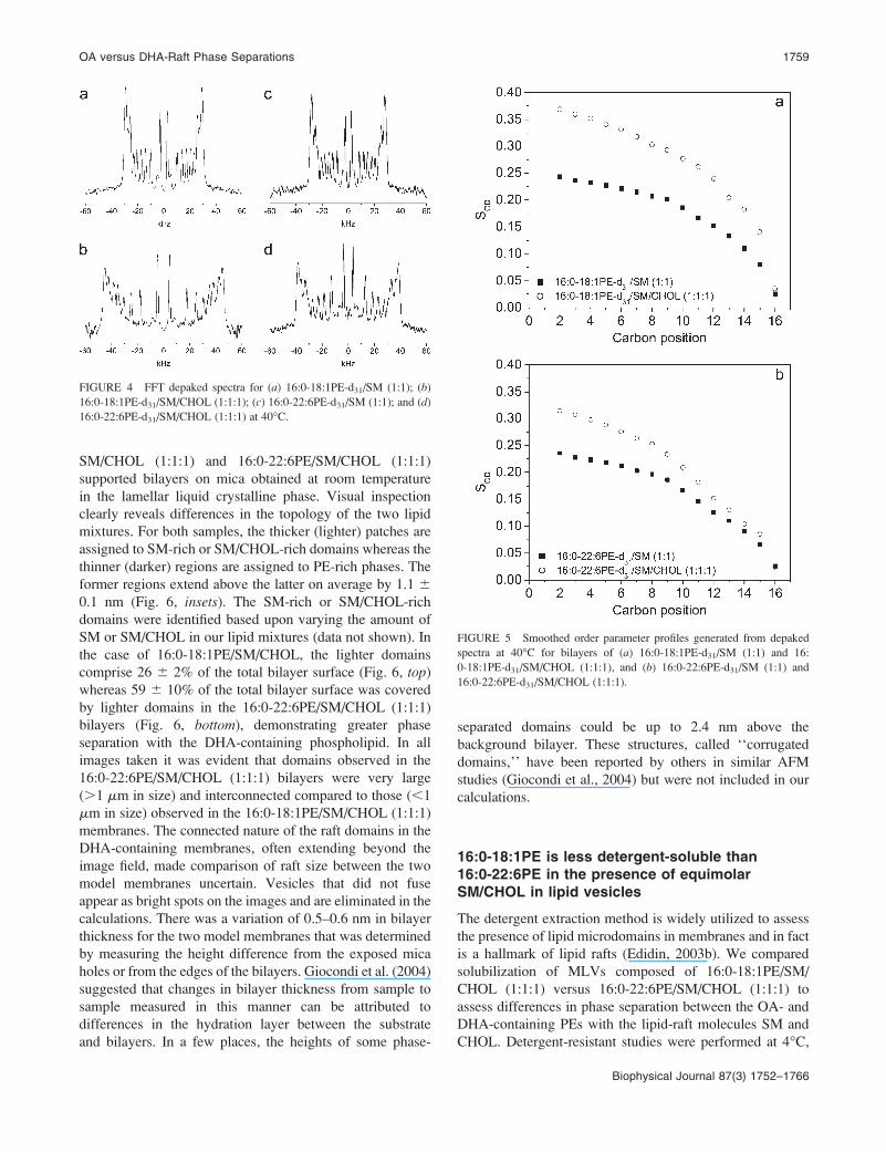
The 2H NMR spectra at 40�C (Figs. 1 and 2) were FFT
depaked to enhance spectral resolution (McCabe and
Wassall, 1997). Application of the algorithm produces
a spectrum that is equivalent to the spectrum that would be
obtained for a planar membrane with the bilayer normal
FIGURE 2 2H NMR spectra for 50 wt %
aqueous dispersions in 50 mM Tris buffer
(pH 7.4) of 16:0-18:1PE-d31/SM/CHOL
(1:1:1) and 16:0-22:6PE-d31/SM/CHOL
(1:1:1). Spectra for 16:0-18:1PE-d31/SM/
CHOL (1:1:1) were recorded at (a) 5, (b)
25, and (c) 40�C. Spectra for 16:0-22:6PE-d31/SM/CHOL were also recorded at (d) 5,(e) 25, and ( f ) 40�C.
OA versus DHA-Raft Phase Separations 1757
Biophysical Journal 87(3) 1752–1766
parallel to the magnetic field (Fig. 4). They possess an
outermost composite doublet, representing ordered methyl-
enes in the upper part of the acyl chain, and five to six well-
resolved doublets corresponding to the methylenes and
terminal methyl that display progressively less order in the
lower portion of the sn-1 acyl chain. The depaked data were
utilized to calculate profiles of order parameter SCD along the
perdeuterated sn-1 chain of the PE component in the lipid
mixtures (Fig. 5). The smoothed profiles were created by
assigning equal integrated intensity to each methylene group
and a monotonic decrease in order toward the terminal
methyl was assumed (Lafleur et al., 1989). Fig. 5 illustrates
order parameters as a function of acyl chain carbon position
for bilayers of 16:0-18:1PE-d31/SM (1:1) (Fig. 5 a) and 16:0-22:6PE-d31/SM (1:1) (Fig. 5 b) in the absence or presence ofequimolar CHOL at 40�C. A similar qualitative trend is
observed for all four of the order parameter profiles. The
upper part of the perdeuterated palmitoyl chains is marked by
a plateau region of approximately constant order with
a subsequent decrease in order toward the bottom of the
chains. In agreement with the average order parameters SCDlisted in Table 1, addition of CHOL to both phospholipid
mixtures results in ordering of the acyl chains at all carbon
positions. CHOL’s ordering effect is most pronounced in the
plateau region toward the top of the perdeuterated acyl chain
for both 16:0-18:1PE-d31 and 16:0-22:6PE-d31 as opposed to
the middle of the bilayers. At carbon position 2, for instance,
the value of SCD changes from ;0.24 to ;0.37 for 16:
0-18:1PE-d31/SM (1:1), and for 16:0-22:6PE-d31/SM (1:1)
shows a smaller change ;0.24 to ;0.32. The respective
differences in SCD correspond to a sterol-induced increase of
54.2 and 33.3%. This differential in the effect of CHOL on
the OA- and DHA-containing mixtures is consistent with the
calculated changes in SCD reported in Table 1.
AFM measurements demonstrate differences inphase separation between 16:0-18:1PE/SM/CHOL(1:1:1) vs. 16:0-22:6PE/SM/CHOL (1:1:1)
AFM is a useful tool for assessing phase separations in
supported bilayers. The AFM measurements reported here
are a preliminary attempt to relate the differences in phase
separation seen spectroscopically by NMR with lateral
organization. Fig. 6 presents AFM images of 16:0-18:1PE/
FIGURE 3 (a and b) Variation in the
first moment M1 (Eq. 1) as a function
of temperature for bilayers of (a) 16:
0-18:1PE-d31/SM (1:1) and 16:0-18:
1PE-d31/SM/CHOL (1:1:1), and (b)
16:0-22:6PE-d31/SM (1:1) and 16:
0-22:6PE-d31/SM/CHOL (1:1:1). M1 is
plotted logarithmically and values have
a 62% error. The transition tempera-
ture Tm, designated by X, is the mid-
point of the sharp drop in moment that
accompanies the chain-melting transi-
tion. (c and d) Corresponding DSC
cooling scans for (c) 16:0-18:1PE/SM
(1:1) and 16:0-18:1PE/SM/CHOL
(1:1:1), and (d) 16:0-22:6PE/SM (1:1)
and 16:0-22:6PE/SM/CHOL (1:1:1).
The scans with cholesterol are slightly
offset along the excess heat capacity
axis to aid observation. The inset is
a high-resolution deconvolution of the
DSC cooling scan for 16:0-18:1PE/SM
(1:1). The dotted and dashed lines
represent the 16:0-18:1PE-rich and
SM-rich phases, respectively.
TABLE 1 Data derived from 2H NMR studies of 16:0-18:
1PE-d31/SM (1:1) or 16:0-22:6PE-d31/SM (1:1) in the absence
or presence of equimolar CHOL at 40�C
Lipid composition
M1
(104 s�1) SCD
Increase in order
due to CHOL
16:0-18:1PE-d31/SM (1:1) 4.98 0.163
16:0-18:1PE-d31/SM/CHOL (1:1:1) 8.01 0.263 61%
16:0-22:6PE-d31/SM (1:1) 4.58 0.150
16:0-22:6PE-d31/SM/CHOL (1:1:1) 5.77 0.189 26%
1758 Shaikh et al.
Biophysical Journal 87(3) 1752–1766
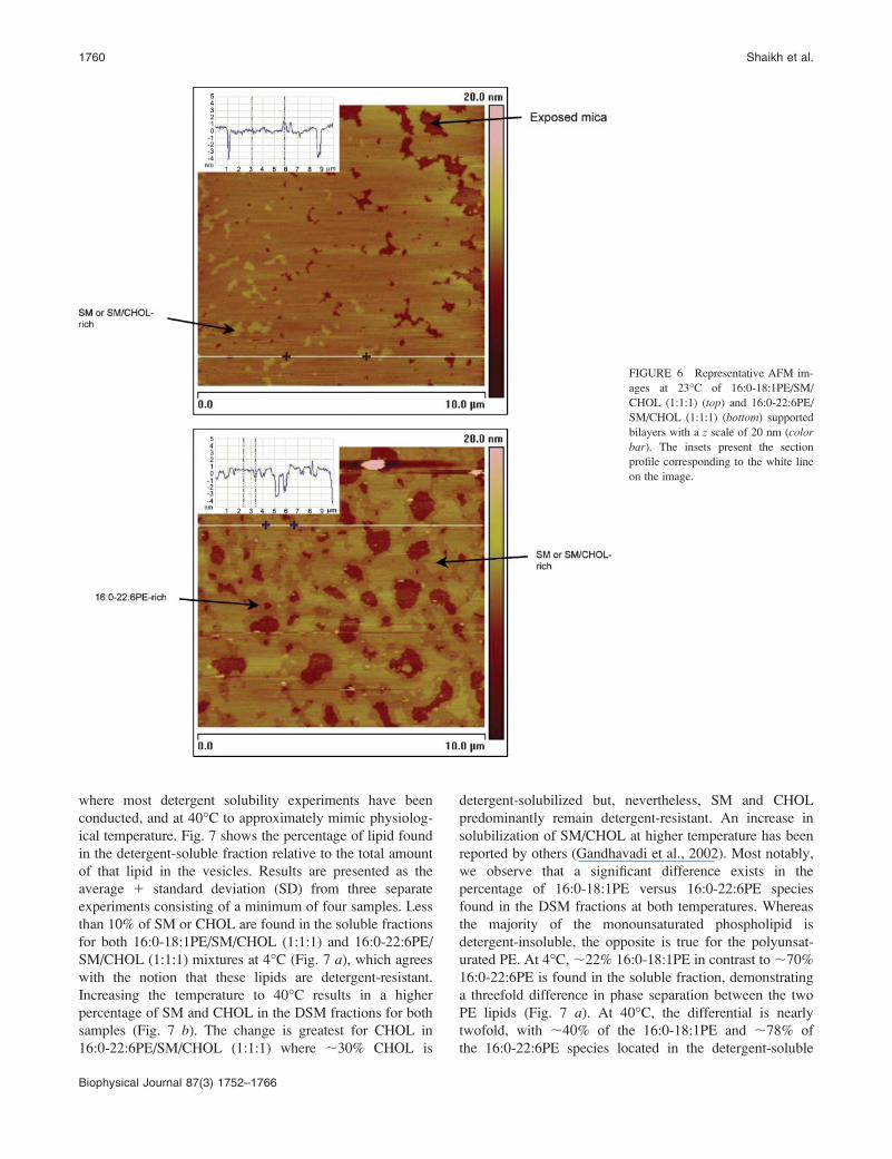
SM/CHOL (1:1:1) and 16:0-22:6PE/SM/CHOL (1:1:1)
supported bilayers on mica obtained at room temperature
in the lamellar liquid crystalline phase. Visual inspection
clearly reveals differences in the topology of the two lipid
mixtures. For both samples, the thicker (lighter) patches are
assigned to SM-rich or SM/CHOL-rich domains whereas the
thinner (darker) regions are assigned to PE-rich phases. The
former regions extend above the latter on average by 1.1 6
0.1 nm (Fig. 6, insets). The SM-rich or SM/CHOL-rich
domains were identified based upon varying the amount of
SM or SM/CHOL in our lipid mixtures (data not shown). In
the case of 16:0-18:1PE/SM/CHOL, the lighter domains
comprise 26 6 2% of the total bilayer surface (Fig. 6, top)whereas 59 6 10% of the total bilayer surface was covered
by lighter domains in the 16:0-22:6PE/SM/CHOL (1:1:1)
bilayers (Fig. 6, bottom), demonstrating greater phase
separation with the DHA-containing phospholipid. In all
images taken it was evident that domains observed in the
16:0-22:6PE/SM/CHOL (1:1:1) bilayers were very large
(.1 mm in size) and interconnected compared to those (,1
mm in size) observed in the 16:0-18:1PE/SM/CHOL (1:1:1)
membranes. The connected nature of the raft domains in the
DHA-containing membranes, often extending beyond the
image field, made comparison of raft size between the two
model membranes uncertain. Vesicles that did not fuse
appear as bright spots on the images and are eliminated in the
calculations. There was a variation of 0.5–0.6 nm in bilayer
thickness for the two model membranes that was determined
by measuring the height difference from the exposed mica
holes or from the edges of the bilayers. Giocondi et al. (2004)
suggested that changes in bilayer thickness from sample to
sample measured in this manner can be attributed to
differences in the hydration layer between the substrate
and bilayers. In a few places, the heights of some phase-
separated domains could be up to 2.4 nm above the
background bilayer. These structures, called ‘‘corrugated
domains,’’ have been reported by others in similar AFM
studies (Giocondi et al., 2004) but were not included in our
calculations.
16:0-18:1PE is less detergent-soluble than16:0-22:6PE in the presence of equimolarSM/CHOL in lipid vesicles
The detergent extraction method is widely utilized to assess
the presence of lipid microdomains in membranes and in fact
is a hallmark of lipid rafts (Edidin, 2003b). We compared
solubilization of MLVs composed of 16:0-18:1PE/SM/
CHOL (1:1:1) versus 16:0-22:6PE/SM/CHOL (1:1:1) to
assess differences in phase separation between the OA- and
DHA-containing PEs with the lipid-raft molecules SM and
CHOL. Detergent-resistant studies were performed at 4�C,
FIGURE 4 FFT depaked spectra for (a) 16:0-18:1PE-d31/SM (1:1); (b)
16:0-18:1PE-d31/SM/CHOL (1:1:1); (c) 16:0-22:6PE-d31/SM (1:1); and (d)16:0-22:6PE-d31/SM/CHOL (1:1:1) at 40�C.
FIGURE 5 Smoothed order parameter profiles generated from depaked
spectra at 40�C for bilayers of (a) 16:0-18:1PE-d31/SM (1:1) and 16:
0-18:1PE-d31/SM/CHOL (1:1:1), and (b) 16:0-22:6PE-d31/SM (1:1) and
16:0-22:6PE-d31/SM/CHOL (1:1:1).
OA versus DHA-Raft Phase Separations 1759
Biophysical Journal 87(3) 1752–1766
where most detergent solubility experiments have been
conducted, and at 40�C to approximately mimic physiolog-
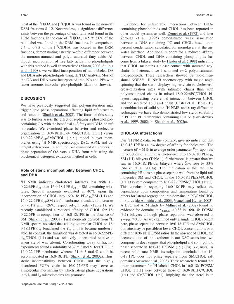
ical temperature. Fig. 7 shows the percentage of lipid found
in the detergent-soluble fraction relative to the total amount
of that lipid in the vesicles. Results are presented as the
average 1 standard deviation (SD) from three separate
experiments consisting of a minimum of four samples. Less
than 10% of SM or CHOL are found in the soluble fractions
for both 16:0-18:1PE/SM/CHOL (1:1:1) and 16:0-22:6PE/
SM/CHOL (1:1:1) mixtures at 4�C (Fig. 7 a), which agrees
with the notion that these lipids are detergent-resistant.
Increasing the temperature to 40�C results in a higher
percentage of SM and CHOL in the DSM fractions for both
samples (Fig. 7 b). The change is greatest for CHOL in
16:0-22:6PE/SM/CHOL (1:1:1) where ;30% CHOL is
detergent-solubilized but, nevertheless, SM and CHOL
predominantly remain detergent-resistant. An increase in
solubilization of SM/CHOL at higher temperature has been
reported by others (Gandhavadi et al., 2002). Most notably,
we observe that a significant difference exists in the
percentage of 16:0-18:1PE versus 16:0-22:6PE species
found in the DSM fractions at both temperatures. Whereas
the majority of the monounsaturated phospholipid is
detergent-insoluble, the opposite is true for the polyunsat-
urated PE. At 4�C, ;22% 16:0-18:1PE in contrast to ;70%
16:0-22:6PE is found in the soluble fraction, demonstrating
a threefold difference in phase separation between the two
PE lipids (Fig. 7 a). At 40�C, the differential is nearly
twofold, with ;40% of the 16:0-18:1PE and ;78% of
the 16:0-22:6PE species located in the detergent-soluble
FIGURE 6 Representative AFM im-
ages at 23�C of 16:0-18:1PE/SM/
CHOL (1:1:1) (top) and 16:0-22:6PE/
SM/CHOL (1:1:1) (bottom) supported
bilayers with a z scale of 20 nm (color
bar). The insets present the section
profile corresponding to the white line
on the image.
1760 Shaikh et al.
Biophysical Journal 87(3) 1752–1766
fractions (Fig. 7 b). Increasing the temperature results in
greater solubilization of both PE lipids as is the case with
SM and CHOL.
OA and DHA are mostly detergent-solubilizedin cells
We utilized detergent-resistant assays with neonatal cardio-
myocytes to look for differences in the localization (raft
versus non-raft) of OA and DHA in a cellular system.
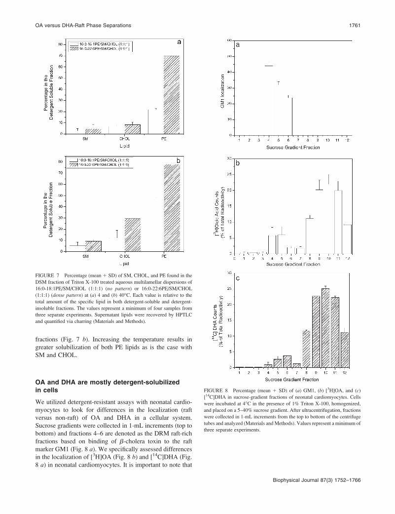
Sucrose gradients were collected in 1-mL increments (top to
bottom) and fractions 4–6 are denoted as the DRM raft-rich
fractions based on binding of b-cholera toxin to the raft
marker GM1 (Fig. 8 a). We specifically assessed differences
in the localization of [3H]OA (Fig. 8 b) and [14C]DHA (Fig.
8 a) in neonatal cardiomyocytes. It is important to note that
FIGURE 7 Percentage (mean 1 SD) of SM, CHOL, and PE found in the
DSM fraction of Triton X-100 treated aqueous multilamellar dispersions of
16:0-18:1PE/SM/CHOL (1:1:1) (no pattern) or 16:0-22:6PE/SM/CHOL
(1:1:1) (dense pattern) at (a) 4 and (b) 40�C. Each value is relative to the
total amount of the specific lipid in both detergent-soluble and detergent-
insoluble fractions. The values represent a minimum of four samples from
three separate experiments. Supernatant lipids were recovered by HPTLC
and quantified via charring (Materials and Methods).
FIGURE 8 Percentage (mean 1 SD) of (a) GM1, (b) [3H]OA, and (c)
[14C]DHA in sucrose-gradient fractions of neonatal cardiomyocytes. Cells
were incubated at 4�C in the presence of 1% Triton X-100, homogenized,
and placed on a 5–40% sucrose gradient. After ultracentrifugation, fractions
were collected in 1-mL increments from the top to bottom of the centrifuge
tubes and analyzed (Materials andMethods). Values represent a minimum of
three separate experiments.
OA versus DHA-Raft Phase Separations 1761
Biophysical Journal 87(3) 1752–1766
most of the [3H]OA and [14C]DHA was found in the non-raft
DSM fractions 8–12. Nevertheless, a significant difference
exists between the percentage of each fatty acid found in the
DRM fractions. In the case of [3H]OA, 14.5 6 2.6% of the
radiolabel was found in the DRM fractions. In comparison,
7.4 6 0.9% of the [14C]DHA was located in the DRM
fractions, demonstrating a nearly twofold difference between
the monounsaturated and polyunsaturated fatty acids. Al-
though incorporation of free fatty acids into phospholipids
with this method is well characterized (Munro, 2003; Stulnig
et al., 1998), we verified incorporation of radiolabeled OA
and DHA into phospholipids using HPTLC analysis. Most of
the OA and DHA were incorporated into PCs and PEs with
lesser amounts into other phospholipids (data not shown).
DISCUSSION
We have previously suggested that polyunsaturation may
trigger lipid phase separations affecting lipid raft structure
and function (Shaikh et al., 2002). The focus of this study
was to further assess the effect of replacing a phospholipid-
containing OA with the beneficial v-3 fatty acid DHA on raft
molecules. We examined phase behavior and molecular
organization in 16:0-18:1PE-d31/SM/CHOL (1:1:1) versus
16:0-22:6PE-d31/SM/CHOL (1:1:1) model bilayer mem-
branes using 2H NMR spectroscopy, DSC, AFM, and de-
tergent extractions. In addition, we evaluated differences in
phase separation of OA versus DHA from rafts using the
biochemical detergent extraction method in cells.
Role of steric incompatibility between CHOLand DHA
2H NMR indicates cholesterol interacts less with 16:
0-22:6PE-d31 than 16:0-18:1PE-d31 in SM-containing mix-
tures. Spectral moments evaluated at 40�C upon the
incorporation of CHOL into 16:0-18:1PE-d31/SM (1:1) and
16:0-22:6PE-d31/SM (1:1) membranes translate to increases
of ;61% and ;26%, respectively, in order (Table 1). We
recently established a reduced affinity of CHOL for 16:
0-22:6PE in comparison to 16:0-18:1PE in the absence of
SM (Shaikh et al., 2003a). First moments derived from 2H
NMR spectra revealed that adding equimolar CHOL to 16:
0-18:1PE-d31 broadened the Tm until it became unobserv-
able. In contrast, the transition was detected in 16:0-22:6PE-
d31/CHOL (1:1) and was relatively unperturbed relative to
when sterol was absent. Corroborating x-ray diffraction
experiments found a solubility of 326 3 mol % for CHOL in
16:0-22:6PE membranes whereas 51 6 3 mol % could be
accommodated in 16:0-18:1PE (Shaikh et al., 2003a). Thus,
steric incompatibility between CHOL and the highly
disordered PUFA chain in 16:0-22:6PE may serve as
a molecular mechanism by which lateral phase separations
into lo and ld microdomains are promoted.
Evidence for unfavorable interactions between DHA-
containing phospholipids and CHOL has been reported in
other model systems as well. Demel et al. (1972) and later
Zerouga et al. (1995) demonstrated weak association
between a DHA-containing PC and CHOL based upon
percent condensation calculated for monolayers at the air-
water interface. Additional support for a reduced affinity
between CHOL and DHA-containing phospholipids has
come from a bilayer study by Huster et al. (1998) indicating
that CHOL maintains a closer contact with saturated acyl
chains in heteroacid sn-1 saturated sn-2 polyunsaturated
phospholipids. These researchers showed by two-dimen-
sional NOESY 1H NMR spectroscopy with magic angle
spinning that the sterol displays higher chain-to-cholesterol
cross-relaxation rates with saturated chains than with
polyunsaturated chains in mixed 18:0-22:6PC/CHOL bi-
layers, suggesting preferential interaction between CHOL
and the saturated 18:0 sn-1 chain (Huster et al., 1998). By
a combination of solid-state 2H NMR and x-ray diffraction
techniques we have also demonstrated low sterol solubility
in PC and PE membranes containing PUFAs (Brzustowicz
et al., 1999, 2002a,b; Shaikh et al., 2003a).
CHOL-OA interactions
Our 2H NMR data, on the contrary, give no indication that
16:0-18:1PE has a low degree of affinity for cholesterol. The
increase of ;61% in average order parameter SCD upon the
introduction of equimolar cholesterol into 16:0-18:1PE-d31/
SM (1:1) bilayers (Table 1), furthermore, is greater than we
saw in 16:0-18:1PE-d31 bilayers where SCD rose by 33%
(Shaikh et al, 2003a). The implication is that the OA-
containing PE does not phase separate well from the lipid-raft
molecules SM and CHOL in the 16:0-18:1PE/SM/CHOL
(1:1:1) system compared to 16:0-22:6PE/SM/CHOL (1:1:1).
This conclusion regarding 16:0-18:1PE may reflect the
dependence upon composition and temperature found by
others for lateral segregation into domains in PC/SM/CHOL
mixtures (de Almeida et al., 2003; Veatch and Keller, 2003).
A DSC and AFM study by Milhiet et al. (2002) found no
evidence for domains at xCHOL $0.33 in 16:0-18:1PC/SM
(3:1) bilayers although phase separation was observed at
xCHOL #0.33. As we examined only a single CHOL content
here, phase separation between 16:0-18:1PE and SM/CHOL
domains may be possible at lower CHOL concentrations or at
different 16:0-18:1PE/SM ratios. In the absence ofCHOL, the
deconvolution of the exotherm in our DSC scans into two
components does suggest that phospholipid and sphingolipid
phase separate in 16:0-18:1PE/SM (1:1) (Fig. 3 c, inset). Arecent solid-state NMR investigation concluded that 16:
0-18:1PC does not phase separate from SM/CHOL raft
domains (Aussenac et al., 2003). These researchers found that
order parameters for 2H-labeled CHOL in 16:0-18:1PC/SM/
CHOL (1:1:1) were between those of 16:0-18:1PC/CHOL
(1:1) and SM/CHOL (1:1), implying that the sterol is in
1762 Shaikh et al.
Biophysical Journal 87(3) 1752–1766
intimate contact with both 16:0-18:1PC and SM in the mixed
membrane. Explanations include homogeneous mixing and
fast exchange of labeled cholesterol between 16:0-18:1PC/
CHOL and SM/CHOL microdomains. A higher partition
coefficient of CHOL in an OA-containing PC in comparison
to a DHA-containing PC was also recently reported. Using
a novel vesicle-cyclodextrin system to measure CHOL’s
partition coefficient in unilamellar vesicles, Niu and Litman
(2002) measured a 2.6-fold higher partitioning of CHOL in
16:0-18:1PC compared to 16:0-22:6PC.
Phase separation observed with AFM
Our AFM results with 16:0-18:1PE/SM/CHOL (1:1:1) show
that most of the bilayer is homogeneous in thickness with
a few (;26% of the total surface area) phase-separated
domains rich in SM or SM/CHOL (Fig. 6). We propose that
CHOL interacts with both SMand 16:0-18:1PE,which results
in a homogenous lo phase with a few SM- or SM/CHOL-rich
light domains protruding from the bilayer surface. This
finding is consistent with our 2HNMRand detergent-resistant
observations that CHOL interacts strongly with 16:0-18:1PE-
d31 in the presence of equimolar SM. In addition, 2H NMR
studies have shown that 16:0-18:1PE/CHOL can form an lophase (Pare and Lafleur, 1998). In the case of 16:0-22:6PE/
SM/CHOL (1:1:1) bilayers (Fig. 6), more phase-separated
light (SM- or SM/CHOL-rich) domains are observed (;59%
of the total surface area). The dark thinner domains are
attributed to the ld phase rich in 16:0-22:6PE. Again, this
finding is consistent with our 2HNMRand detergent-resistant
data that suggest enhanced phase separation in the presence
of polyunsaturation. A continuing problem in membrane
structural studies is determining the actual size of lipid
domains. Although our earlier NMR work on mixed PCs is
consistent with diffusion-mediated fast exchange of choles-
terol between microdomains ,160 A in size (Brzustowicz
et al., 2002b), our AFMmeasurements here identify domains
in the micron range. Any attempt to reconcile this apparent
discrepancy must take into account that, due to very different
timescales, the two methods are inherently sensitive to
inequivalent length scales. The timescale of the 2H NMR
approach is ;10�5 s, whereas AFM is much slower. Our
AFM images were obtained in ;2 min, during which time
lateral diffusion of lipid would prevent observation of
individual microdomains. One possible explanation that we
are pursuing is that the very large domains observed with
AFM are actually composed of clusters of much smaller
domains implied by NMR.
Detergent-solubilization studies in vesicles
Detergent-solubilization studies were conducted at 4 and
40�C to further evaluate phase separations in our model
membrane systems. They were undertaken at higher and
lower temperature because the validity of detergent
extractions that are routinely performed at 4�C to assess
phase separation has been questioned and may not reflect the
phase state of the lipid mixture at physiological temperature
(Shogomori and Brown, 2003). However, our 2H NMR and
DSC data for 16:0-18:1PE/SM/CHOL (1:1:1) and 16:
0-22:6PE/SM/CHOL (1:1:1) (Figs. 2 and 3) establish that
each mixture is liquid crystalline at both low and high tem-
peratures. The results identify that ;60–80% 16:0-18:1PE
versus ;20–30% 16:0-22:6PE are found in the DRM
fraction at either 4 or 40�C (Fig. 7). Although it is plausible
that Triton may be inducing domain formation (Heerklotz,
2002), our detergent extraction finding is in agreement with
our NMR observation that CHOL increases the order of both
PEs in the presence of SM but exerts much greater effect on
16:0-18:1PE-d31 than on 16:0-22:6PE-d31 (Table 1 and Fig.
3). The significant difference in the amount of 16:0-18:1PE
in the DSM fraction compared to 16:0-22:6PE for both
temperatures, moreover, suggests phase separation that is
driven by the presence of the polyunsaturated acyl chain in
agreement with our AFM data. The differences in phase
separation are also correlated to the differences in Tm of the
two PEs examined, in accord with previous findings
(Ferguson, 1999; Shaikh et al., 2003b). Phase separation
driven by the presence of acyl chain unsaturation, as assessed
by detergent extraction, was identified in an 18:1-18:1PC/
SM/CHOL (1:1:1) model system by McIntosh and co-
workers (Gandhavadi et al., 2002; McIntosh et al., 2003).
These researchers saw a greater proportion of 18:1-18:1PC
in the DSM fraction at 4 and 37�C in the presence of
equimolar SM and CHOL, and suggested that mechanical
properties of DRMs and DSMs may have implications for
peptide sorting (McIntosh et al., 2003).
Detergent-solubilization studies in cells
As seen in our model membrane studies, a significant
difference in the amount of OA versus DHA was found in
DRM fractions isolated from cardiomyocytes. However, our
cellular studies reveal that the vast majority of OA and DHA
are localized in the DSMs, whereas our model membrane
studies would predict that most of the OA would be raft-
localized in cells. This inconsistency suggests that our model
membrane studies do not correlate perfectly with our cellular
model. Perhaps a three-component lipid system does not
realistically reflect the complex heterogeneities that may
arise in the plasma membrane due to interactions amongst
various lipids, proteins, and even the cytoskeleton. Although
this supposition has validity, we believe that the discrepancy
in the OA data between model membranes and cells can be
explained better if one takes into account the limitations of
the detergent extraction approach. Lipid phase behavior is
highly temperature-dependent and complicated in a real cell
membrane that is composed of hundreds of different lipid
molecular species. Thus, the reduction in temperature could
have resulted in changes in lipid organization that would be
OA versus DHA-Raft Phase Separations 1763
Biophysical Journal 87(3) 1752–1766
difficult to model in simple two- or three-component
vesicles. A recent study by Schuck et al. (2003) furthermore
showed that different detergents show considerable differ-
ences in their ability to solubilize membrane lipids and
proteins. This underscores the difficulty of interpreting
detergent extractions as a tool for understanding membrane
organization. These researchers suggested that the presence
of certain lipids or proteins in the DRM fraction does not
necessarily indicate an association with the same micro-
domain in the biological membrane (Schuck et al., 2003).
Another major limitation of the detergent-solubilization
technique is that partial solubilization may not be the result
of differences in solubility between microdomains but rather
due to differences in the solubility of the two leaflets.
Perhaps OA and DHA are localizing to different leaflets.
Indeed Knapp et al. (1994) reported that DHA is preferen-
tially accepted into the inner leaflet of PEs and phosphati-
dylserines of the erythrocyte plasma membrane. Further
investigation into this matter is clearly of interest. Finally,
different cell lines may show different results. We examined
changes in OA and DHA localization in MDA-MB-231
breast cancer cells where differences were smaller than those
reported for cardiomyocytes (Shaikh et al., unpublished
data).
Physiological relevance of DHA-lipid raftphase separations
Since v-3 fatty acids are increasingly being utilized
clinically as adjuvant immunosuppressants in the treatment
of inflammatory diseases, it is becoming evident that PUFAs
modulate immune responses by suppressing T-cell activation
in lymphoid cells. The suggestion has been made that
inhibition of T-cell signaling may be mediated by modifi-
cation of lipid raft microdomains upon incorporation of
various PUFAs including eicosapentaenoic acid (20:5) and
arachidonic acid (20:4) (Webb et al., 2000; Zeyda et al.,
2002). Although relatively little is known about the
molecular interactions between PUFAs and lipid rafts,
a few studies have emerged to explain the physiological
importance of the PUFA-lipid raft relationship at a cellular
level.
A recent investigation showed that acylated proteins found
in lipid rafts were displaced due to the incorporation of
eicosapentaenoic acid in Jurkat T cells (Stulnig et al., 2001).
In another study, it was demonstrated that DHA stimulates
phospholipase D1 activity in stimulated human peripheral
blood mononuclear cells, which may be responsible for some
of the DHA-induced immunosuppression (Diaz et al., 2002).
These researchers found that phospholipase D1 (PLD1),
located in the DRM fractions of sucrose gradients, is lipid
raft-localized in the absence of DHA treatment and that its
activity is impaired (Diaz et al., 2002). Treatment of the
peripheral blood mononuclear cells with DHA resulted in
changing its localization from DRMs to DSMs and an
increase in activity was observed. Both studies have
suggested that altered lipid raft formation may be responsible
for the known inhibitory effects of PUFAs on T-cell ac-
tivation. However, the molecular mechanism by which this
occurs remains elusive.
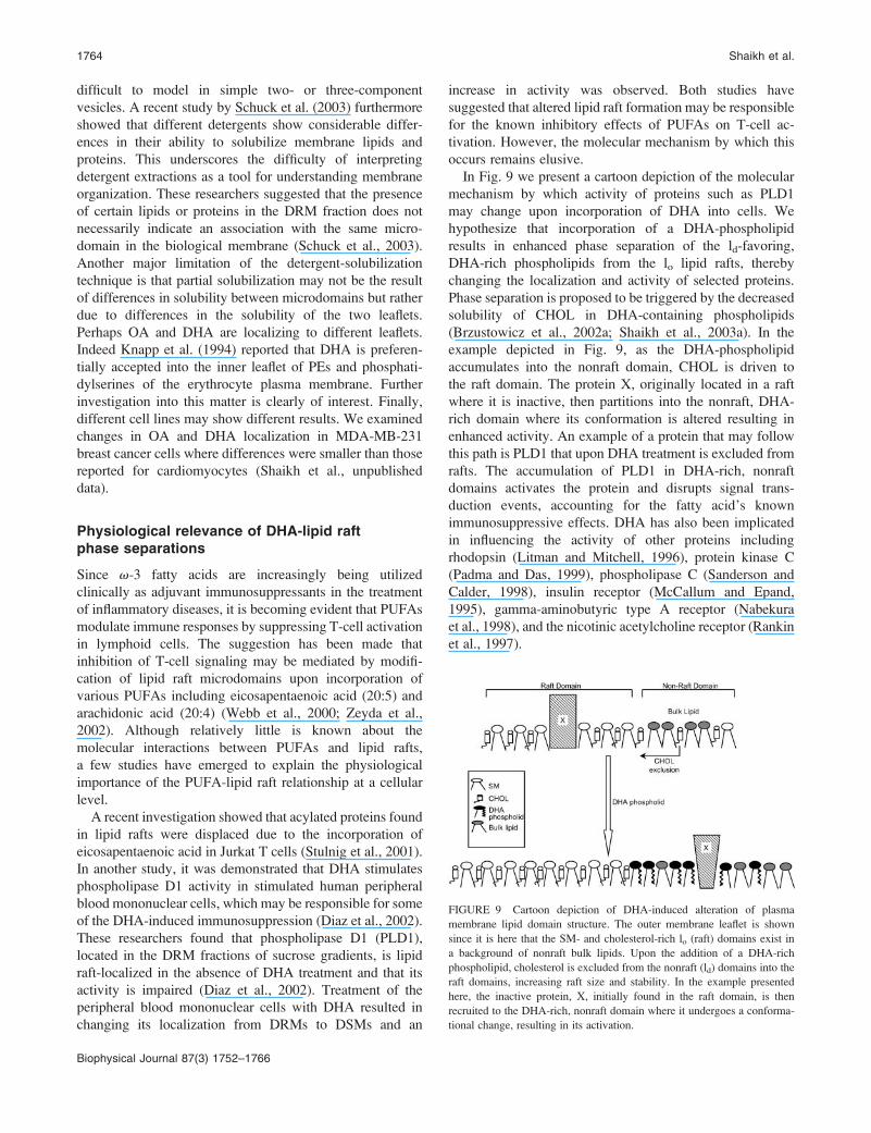
In Fig. 9 we present a cartoon depiction of the molecular
mechanism by which activity of proteins such as PLD1
may change upon incorporation of DHA into cells. We
hypothesize that incorporation of a DHA-phospholipid
results in enhanced phase separation of the ld-favoring,
DHA-rich phospholipids from the lo lipid rafts, thereby
changing the localization and activity of selected proteins.
Phase separation is proposed to be triggered by the decreased
solubility of CHOL in DHA-containing phospholipids
(Brzustowicz et al., 2002a; Shaikh et al., 2003a). In the
example depicted in Fig. 9, as the DHA-phospholipid
accumulates into the nonraft domain, CHOL is driven to
the raft domain. The protein X, originally located in a raft
where it is inactive, then partitions into the nonraft, DHA-
rich domain where its conformation is altered resulting in
enhanced activity. An example of a protein that may follow
this path is PLD1 that upon DHA treatment is excluded from
rafts. The accumulation of PLD1 in DHA-rich, nonraft
domains activates the protein and disrupts signal trans-
duction events, accounting for the fatty acid’s known
immunosuppressive effects. DHA has also been implicated
in influencing the activity of other proteins including
rhodopsin (Litman and Mitchell, 1996), protein kinase C
(Padma and Das, 1999), phospholipase C (Sanderson and
Calder, 1998), insulin receptor (McCallum and Epand,
1995), gamma-aminobutyric type A receptor (Nabekura
et al., 1998), and the nicotinic acetylcholine receptor (Rankin
et al., 1997).
FIGURE 9 Cartoon depiction of DHA-induced alteration of plasma
membrane lipid domain structure. The outer membrane leaflet is shown
since it is here that the SM- and cholesterol-rich lo (raft) domains exist in
a background of nonraft bulk lipids. Upon the addition of a DHA-rich
phospholipid, cholesterol is excluded from the nonraft (ld) domains into the
raft domains, increasing raft size and stability. In the example presented
here, the inactive protein, X, initially found in the raft domain, is then
recruited to the DHA-rich, nonraft domain where it undergoes a conforma-
tional change, resulting in its activation.
1764 Shaikh et al.
Biophysical Journal 87(3) 1752–1766

CONCLUSION
Our results identify major differences in the molecular inter-
actions of OA versus DHA with raft molecules. These
findings provide further evidence in support of the hypothe-
sis that DHA-containing phospholipids will phase separate
into DHA-rich, SM/CHOL-poor and DHA-poor, SM/
CHOL-rich microdomains. Studies of DHA-lipid raft phase
separations may contribute to understanding more complex
signaling pathways involving proteins. Perhaps DHA exerts
its beneficial health effects on numerous disease states
including depression, heart disease, various cancers, schizo-
phrenia, and arthritis by influencing lipid raft-mediated cell
signaling events.
This work was supported by a grant from the National Institutes of Health
(RO1CA57212).
REFERENCES
Ahmed, S. N., D. A. Brown, and E. London. 1997. On the origin ofsphingolipid/cholesterol-rich detergent insoluble cell membranes: phys-iological concentrations of cholesterol and sphingolipid induce formationof a detergent-insoluble, liquid-ordered lipid phase in model membranes.Biochemistry. 36:10944–10953.
Aussenac, F., M. Tavares, and E. J. Dufourc. 2003. Cholesterol dynamics inmembranes of raft composition: a molecular point of view from 2H and31P solid-state NMR. Biochemistry. 42:1383–1390.
Brown, D. A., and E. London. 2000. Structure and function of sphingolipidand cholesterol-rich membrane rafts. J. Biol. Chem. 275:17221–17224.
Brzustowicz, M. R., V. Cherezov, M. Caffrey, W. Stillwell, and S. R.Wassall. 2002b. Molecular organization of cholesterol in polyunsaturatedmembranes: microdomain formation. Biophys. J. 82:285–298.
Brzustowicz, M. R., V. Cherezov, M. Zerouga, M. Caffrey, W. Stillwell,and S. R. Wassall. 2002a. Controlling membrane cholesterol content. Arole for polyunsaturated (docosahexaenoate) phospholipids. Biochemis-try. 41:12509–12519.
Brzustowicz, M. R., W. Stillwell, and S. R. Wassall. 1999. Molecularorganization of cholesterol in polyunsaturated phospholipid membranes:a solid state 2H NMR investigation. FEBS Lett. 451:197–202.
Davis, J. H. 1983. The description of membrane lipid conformation, orderand dynamics by 2H-NMR. Biochim. Biophys. Acta. 737:117–171.
de Almeida, R. F. M., A. Fedorov, and M. Prieto. 2003. Sphingomyelin/phosphatidylcholine/cholesterol phase diagram: boundaries and compo-sition of lipid rafts. Biophys. J. 85:2406–2416.
Demel, R. A., W. S. M. Geurts van Kessel, and L. L. M. van Deenen. 1972.The properties of polyunsaturated lecithins in monolayers and liposomesand the interactions of these lecithins with cholesterol. Biochim. Biophys.Acta. 266:26–40.
Diaz, O., A. Berquand, M. Dubois, S. Di Agostino, C.C. Sette, S. Bourgoin,M. Lagarde, G. Nemoz, and A.-F. Prigent. 2002. The mechanism ofdocosahexaenoic acid-induced phospholipase D activation in humanlymphocytes involves exclusion of the enzyme from lipid rafts. J. Biol.Chem. 277:39368–39378.
Edidin, M. 1993. Patches and fences: probing for plasma membranedomains. J. Cell Sci. 17:165–169.
Edidin, M. 2003a. Lipids on the frontier: a century of cell-membranebilayers. Nat. Rev. Mol. Cell Biol. 4:414–418.
Edidin, M. 2003b. The state of lipid rafts: from model membranes to cells.Annu. Rev. Biophys. Biomol. Struct. 32:257–283.
Eldho, N. V., S. E. Feller, S. Tristram-Nagle, I. V. Polozov, and K.Gawrisch. 2003. Polyunsaturated docosahexaenoic vs docosapentaenoic
acid—differences in lipid matrix properties from the loss of one doublebond. J. Am. Chem. Soc. 125:6409–6421.
Ferguson, M. A. 1999. The structure, biosynthesis and functions ofglycosylphosphatidylinositol anchors, and the contributions of trypano-some research. J. Cell Sci. 112:2799–2809.
Gandhavadi, M., D. Allende, A. Vidal, S. A. Simon, and T. J. McIntosh.2002. Structure, composition, and peptide binding properties of deter-gent-soluble bilayers and detergent resistant rafts. Biophys. J. 82:1469–1482.
Giocondi, M.-C., P. E. Milhiet, P. Dosset, and C. L. Grimellec. 2004. Useof cyclodextrin for AFM monitoring of model raft formation. Biophys. J.86:861–869.
Graham, J. M., and J. A. Higgins. 1997. Membrane Analysis. Springer-Verlag, New York.
Heerklotz, H. 2002. Triton promotes domain formation in lipid raftmixtures. Biophys. J. 83:2693–2701.
Hooper, N. 1999. Detergent-insoluble glycosphingolipid-rich membranedomains, lipid rafts and caveolae (review). Mol. Membr. Biol. 16:145–156.
Huang, J., J. T. Buboltz, and G. W. Feigenson. 1999. Maximum solubilityof cholesterol in phosphatidylcholine and phosphatidylethanolaminebilayers. Biochim. Biophys. Acta. 1417:89–100.
Huster, D., K. Arnold, and K. Gawrisch. 1998. Influence of docosahex-aenoic acid and cholesterol on lateral lipid organization in phospholipidmixtures. Biochemistry. 37:17299–17308.
Jump, D. B. 2002. The biochemistry of n-3 polyunsaturated fatty acids.J. Biol. Chem. 277:8755–8758.
Knapp, H., F. Hullin, and N. Salem, Jr. 1994. Asymmetric incorporation ofdietary n-3 fatty acids into membrane aminophospholipids of humanerythrocytes. J. Lipid Res. 35:1283–1291.
Lafleur, M., B. Fine, E. Sternin, P. R. Cullis, and M. Bloom. 1989.Smoothed orientational order profile of lipid bilayers by 2H-nuclearmagnetic resonance. Biophys. J. 56:1037–1041.
Litman, B. J., and D. C. Mitchell. 1996. A role for phospholipidpolyunsaturation in modulating membrane protein function. Lipids.31(Suppl):S193–S197.
McCabe, M. A., G. L. Griffith, W. D. Ehringer, W. Stillwell, and S. R.Wassall. 1994. 2H NMR studies of isomeric w3 and w6 polyunsaturatedphospholipid membranes. Biochemistry. 33:7203–7210.
McCabe, M. A., and S. R. Wassall. 1997. Rapid deconvolution of NMRpowder spectra by weighted fast Fourier transformation. Solid State Nucl.Mag. Reson. 10:53–61.
McCallum, C. D., and R. M. Epand. 1995. Insulin receptor autophosphor-ylation and signaling is altered by modulation of membrane physicalproperties. Biochemistry. 34:1815–1824.
McIntosh, T. J., A. Vidal, and S. A. Simon. 2003. Sorting of lipids andtransmembrane peptides between detergent-soluble bilayers and de-tergent-resistant rafts. Biophys. J. 85:1656–1666.
Milhiet, P. E., M. C. Giocondi, and C. L. Grimellec. 2002. Cholesterol isnot crucial for the existence of microdomains in kidney-brush bordermembrane models. J. Biol. Chem. 22:875–878.
Mitchell, D. C., and B. J. Litman. 1998. Molecular order and dynamics inbilayers consisting of highly polyunsaturated phospholipids. Biophys. J.74:879–891.
Munro, S. 2003. Lipid rafts: elusive or illusive? Cell. 115:377–388.
Nabekura, J., K. Noguchi, M. R. Witt, M. Nielsen, and N. Akaike. 1998.Functional modulation of human recombinant gamma-aminobutyric acidtype A receptor by docosahexaenoic acid. J. Biol. Chem. 273:11056–11061.
Niu, S. L., and B. J. Litman. 2002. Determination of membrane cholesterolpartition coefficient using a lipid vesicle-cyclodextrin binary system:effect of phospholipid acyl chain unsaturation and headgroup composi-tion. Biophys. J. 83:3408–3415.
Padma, M., and U. N. Das. 1999. Effect of cis-unsaturated fatty acids on theactivity of protein kinases and protein phosphorylation in macrophage
OA versus DHA-Raft Phase Separations 1765
Biophysical Journal 87(3) 1752–1766
tumor (AK-5) cells in vitro. Prostaglandins Leukot. Essent. Fatty Acids.60:55–63.
Pare, C., and M. Lafleur. 1998. Polymorphism of POPE/cholesterol system:A 2H nuclear magnetic resonance and infrared spectroscopic investiga-tion. Biophys. J. 74:899–909.
Petrache, H. E., A. Salmon, and M. F. Brown. 2001. Structural properties ofdocosahexaenoyl phospholipid bilayers investigated by solid-state 2HNMR spectroscopy. J. Am. Chem. Soc. 123:12611–12622.
Rankin, S. E., G. H. Addona, M. A. Kloczewiak, B. Bugge, and K. W.Miller. 1997. The cholesterol dependence of activation and fast desen-sitization of the nicotinic acetylcholine receptor. Biophys. J. 73:2446–2455.
Rinia, H. A., J. W. Boots, D.T. Rijkers, R.A. Kik, M.M. Snel, R.A. Demel,J.A. Killian, J.P. van der Eerden, and B. de Kruijff. 2002. Domainformation in phosphatidylcholine bilayers containing transmembranepeptides: specific effects of flanking residues. Biochemistry. 41:2814–2824.
Sanderson, P., and P. C. Calder. 1998. Dietary fish oil appears to preventthe activation of phospholipase C-gamma in lymphocytes. Biochim.Biophys. Acta. 1392:300–308.
Schroeder, R., E. London, and D. Brown. 1994. Interactions betweensaturated acyl chains confer detergent resistance on lipids andglycosylphosphatidylinositol (GPI)-anchored proteins: GPI-anchoredproteins in liposomes and cells show similar behavior. Proc. Natl. Acad.Sci. USA. 91:12130–12134.
Schuck, S., M. Honsho, K. Ekroos, A. Shevchenko, and K. Simons. 2003.Resistance of cell membranes to different detergents. Proc. Natl. Acad.Sci. USA. 100:5795–5800.
Shaikh, S. R., M. R. Brzustowicz, N. Gustafson, W. Stillwell, and S. R.Wassall. 2002. Monounsaturated PE does not phase-separate from thelipid raft molecules sphingomyelin and cholesterol: role for polyunsatu-ration? Biochemistry. 41:10593–10602.
Shaikh, S. R., V. Cherezov, M. Caffrey, W. Stillwell, and S. R. Wassall.2003a. Interaction of cholesterol with a docosahexaenoic acid-containingphosphatidylethanolamine: trigger for microdomain/raft formation? Bio-chemistry. 42:12028–12037.
Shaikh, S. R., A. C. Dumaual, L. J. Jenski, and W. Stillwell. 2001. Lipidphase separation in phospholipid bilayers and monolayers modeling theplasma membrane. Biochim. Biophys. Acta. 1512:317–328.
Shaikh, S. R., A. C. Dumaual, D. LoCassio, R. A. Siddiqui, and W.Stillwell. 2003b. Acyl chain unsaturation in PEs modulates phaseseparation from lipid raft molecules. Biochem. Biophys. Res. Commun.311:793–796.
Shogomori, H., and D. A. Brown. 2003. Use of detergents to studymembrane rafts: The good, the bad, and the ugly. Biol. Chem. 384:1259–1263.
Simons, K., and W. L. C. Vaz. 2004. Model systems, lipid rafts, and cellmembranes. Annu. Rev. Biophys. Biomol. Struct. 33:269–295.
Simopoulos, A. P., R. R. Kifer, and R. E. Martin. 1986. Health effects ofpolyunsaturated fatty acids in seafood. Academic Press, Orlando.
Stillwell, W. 2000. Docosahexaenoic acid and membrane lipid domains.Curr. Org. Chem. 4:1169–1183.
Stillwell, W., and S. R. Wassall. 2003. Docosahexaenoic acid: membraneproperties of a unique fatty acid. Chem. Phys. Lipids. 126:1–27.
Stulnig, T. M., M. Berger, T. Sigmund, D. Raederstorff, H. Stockinger, andW. Waldhausl. 1998. Polyunsaturated fatty acids inhibit T cell signaltransduction by modification of detergent-insoluble membrane domains.J. Cell Biol. 143:637–644.
Stulnig, T. M., J. Huber, N. Leitinger, E.-M. Imre, P. Angelisova, P.Nowotny, and W. Waldhausl. 2001. Polyunsaturated eicosapentaenoicacid displaces proteins from membrane rafts by altering raft lipidcomposition. J. Biol. Chem. 276:37335–37340.
Tokumasu, F., A. J. Jin, G. W. Feigenson, and J. A. Dvorak. 2003.Nanoscopic lipid domain dynamics revealed by atomic force microscopy.Biophys. J. 84:2609–2618.
Veatch, S. L., and S. L. Keller. 2003. A closer look at the canonical ‘raftmixture’ in model membrane studies. Biophys. J. 84:725–726.
Wassall, S. R., J. L. Thewalt, L. Wong, H. Gorrissen, and R. J. Cushley.1986. Deuterium NMR study of the interaction of alpha-tocopherol witha phospholipid model membrane. Biochemistry. 25:319–326.
Webb, Y., L. Hermida-Matsumoto, and M. D. Resh. 2000. Inhibition ofprotein palmitoylation, raft localization, and T cell signaling by2-bromopalmitate and polyunsaturated fatty acids. J. Biol. Chem. 275:261–270.
Zerouga, M., L. J. Jenski, and W. Stillwell. 1995. Comparison ofphosphatidylcholines containing one or two docosahexaenoic acyl chainson properties of phospholipid monolayers and bilayers. Biochim.Biophys. Acta. 1236:266–272.
Zerouga, M., W. Stillwell, J. Stone, A. Powner, and L. J. Jenski. 1996.Phospholipid class as a determinant in docosahexaenoic acid’s effect ontumor cell viability. Anticancer Res. 16:2863–2868.
Zeyda, M., G. Staffler, V. Horejsi, W. Waldhausl, and T. M. Stulnig. 2002.LAT displacement from lipid rafts as a molecular mechanism for theinhibition of T cell signaling by polyunsaturated fatty acids. J. Biol.Chem. 277:28418–28423.
1766 Shaikh et al.
Biophysical Journal 87(3) 1752–1766
Copyright © 2022 FDOKUMEN