Dynamics of putative raft-associated proteins at the cell surface
12
The Journal of Cell Biology JCB Article 735 The Journal of Cell Biology, Volume 165, Number 5, June 7, 2004 735–746 http://www.jcb.org/cgi/doi/10.1083/jcb.200312170 Dynamics of putative raft-associated proteins at the cell surface Anne K. Kenworthy, 1,2,3 Benjamin J. Nichols, 1,4 Catha L. Remmert, 2 Glenn M. Hendrix, 2 Mukesh Kumar, 5 Joshua Zimmerberg, 5 and Jennifer Lippincott-Schwartz 1 1 Cell Biology and Metabolism Branch, National Institute of Child Health and Human Development, National Institutes of Health, Bethesda, MD 20895 2 Department of Molecular Physiology and Biophysics and 3 Department of Cell and Developmental Biology, Vanderbilt University School of Medicine, Nashville, TN 37232 4 Medical Research Council Laboratory of Molecular Biology, Cambridge CB2 2QH, England, UK 5 Laboratory of Cellular and Molecular Biophysics, National Institute of Child Health and Human Development, National Institutes of Health, Bethesda, MD 20817 ipid rafts are conceptualized as membrane micro- domains enriched in cholesterol and glycosphingolipid that serve as platforms for protein segregation and signaling. The properties of these domains in vivo are unclear. Here, we use fluorescence recovery after photobleaching to test if raft association affects a protein’s ability to laterally diffuse large distances across the cell surface. The diffusion coefficients (D) of several types of putative raft and nonraft proteins were systematically measured under steady-state conditions and in response to raft perturbations. Raft L proteins diffused freely over large distances (4 m), exhibiting Ds that varied 10-fold. This finding indicates that raft proteins do not undergo long-range diffusion as part of discrete, stable raft domains. Perturbations reported to affect lipid rafts in model membrane systems or by biochemical fractionation (cholesterol depletion, decreased temperature, and cholesterol loading) had similar effects on the diffusional mobility of raft and nonraft proteins. Thus, raft association is not the dominant factor in determining long-range protein mobility at the cell surface. Introduction Biological membranes contain regions of lateral inhomoge- neity known as microdomains (Maxfield, 2002; Edidin, 2003). The most studied class of putative microdomains are cholesterol and glycosphingolipid-enriched lipid rafts. These domains are thought to act as platforms with which proteins can selectively associate, leading to their lateral segregation (Simons and Ikonen, 1997; Simons and Toomre, 2000; Maxfield, 2002; Edidin, 2003). Proteins recovered in buoyant, detergent-resistant membrane (DRM) fractions after extraction of cells with certain detergents are commonly defined as raft associated. These proteins include glyco- sylphosphatidylinositol (GPI)-anchored proteins, acylated proteins, and a subset of transmembrane proteins (Simons and Ikonen, 1997; Brown and London, 1998a). Caveolae, a subset of lipid rafts, are additionally enriched in the protein caveolin (Rothberg et al., 1992). Lipid rafts are proposed to function in a large number of cellular functions ranging from protein and lipid sorting during post-Golgi sorting and endocytosis to regulation of cell signaling and viral entry and budding (Simons and Ikonen, 1997; Simons and Toomre, 2000; Ikonen, 2001; Edidin, 2003; Hancock, 2003). Despite this, a consensus view of the structure and dynamics of lipid rafts has yet to emerge (for review see Anderson and Jacobson, 2002; Maxfield, 2002). One area of widespread interest is how rafts affect a protein’s ability to laterally diffuse across cell membranes. In model membranes, the lateral mobility of lipids in a liquid-ordered phase is slower than in a liquid-disordered phase (Brown and London, 1998b; Korlach et al., 1999; Dietrich et al., 2002; Kahya et al., 2003). Indeed, several studies suggest Address correspondence to J. Lippincott-Schwartz, Cell Biology and Metabolism Branch, National Institute of Child Health and Human Development, National Institutes of Health, Bldg. 18T, Rm. 101, 18 Library Dr., Bethesda, MD 20895. Tel.: (301) 402-1010. Fax: (301) 402-0078. email: [email protected] Key words: lipid rafts; membrane microdomains; lateral diffusion; fluorescence recovery after photobleaching; cholesterol Abbreviations used in this paper: CTXB, cholera toxin B subunit; D, diffusion coefficient; DRM, detergent-resistant membrane; GPI, glyco- sylphosphatidylinositol; MCD, methyl -cyclodextrin; M f , mobile fraction; NPC, Niemann-Pick type C; NRK, normal rat kidney; TX-100, Triton X-100. on December 7, 2013 jcb.rupress.org Downloaded from Published June 1, 2004
Transcript of Dynamics of putative raft-associated proteins at the cell surface

The
Jour
nal o
f Cel
l Bio
logy
JCB
Article
735The Journal of Cell Biology, Volume 165, Number 5, June 7, 2004 735–746http://www.jcb.org/cgi/doi/10.1083/jcb.200312170
Dynamics of putative raft-associated proteins at the cell surface
Anne K. Kenworthy,
1,2,3
Benjamin J. Nichols,
1,4
Catha L. Remmert,
2
Glenn M. Hendrix,
2
Mukesh Kumar,
5
Joshua Zimmerberg,
5
and Jennifer Lippincott-Schwartz
1
1
Cell Biology and Metabolism Branch, National Institute of Child Health and Human Development, National Institutes of Health, Bethesda, MD 20895
2
Department of Molecular Physiology and Biophysics and
3
Department of Cell and Developmental Biology, Vanderbilt University School of Medicine, Nashville, TN 37232
4
Medical Research Council Laboratory of Molecular Biology, Cambridge CB2 2QH, England, UK
5
Laboratory of Cellular and Molecular Biophysics, National Institute of Child Health and Human Development, National Institutes of Health, Bethesda, MD 20817
ipid rafts are conceptualized as membrane micro-domains enriched in cholesterol and glycosphingolipidthat serve as platforms for protein segregation and
signaling. The properties of these domains in vivo are unclear.Here, we use fluorescence recovery after photobleachingto test if raft association affects a protein’s ability to laterallydiffuse large distances across the cell surface. The diffusioncoefficients (D) of several types of putative raft and nonraftproteins were systematically measured under steady-stateconditions and in response to raft perturbations. Raft
L
proteins diffused freely over large distances (
�
4
�
m),exhibiting Ds that varied 10-fold. This finding indicates thatraft proteins do not undergo long-range diffusion as part ofdiscrete, stable raft domains. Perturbations reported to affectlipid rafts in model membrane systems or by biochemicalfractionation (cholesterol depletion, decreased temperature,and cholesterol loading) had similar effects on the diffusionalmobility of raft and nonraft proteins. Thus, raft associationis not the dominant factor in determining long-range proteinmobility at the cell surface.
Introduction
Biological membranes contain regions of lateral inhomoge-neity known as microdomains (Maxfield, 2002; Edidin,2003). The most studied class of putative microdomains arecholesterol and glycosphingolipid-enriched lipid rafts. Thesedomains are thought to act as platforms with which proteinscan selectively associate, leading to their lateral segregation(Simons and Ikonen, 1997; Simons and Toomre, 2000;Maxfield, 2002; Edidin, 2003). Proteins recovered inbuoyant, detergent-resistant membrane (DRM) fractionsafter extraction of cells with certain detergents are commonlydefined as raft associated. These proteins include glyco-sylphosphatidylinositol (GPI)-anchored proteins, acylatedproteins, and a subset of transmembrane proteins (Simonsand Ikonen, 1997; Brown and London, 1998a). Caveolae, a
subset of lipid rafts, are additionally enriched in the proteincaveolin (Rothberg et al., 1992). Lipid rafts are proposed tofunction in a large number of cellular functions rangingfrom protein and lipid sorting during post-Golgi sorting andendocytosis to regulation of cell signaling and viral entry andbudding (Simons and Ikonen, 1997; Simons and Toomre,2000; Ikonen, 2001; Edidin, 2003; Hancock, 2003). Despitethis, a consensus view of the structure and dynamics of lipidrafts has yet to emerge (for review see Anderson and Jacobson,2002; Maxfield, 2002).
One area of widespread interest is how rafts affect a protein’sability to laterally diffuse across cell membranes. In modelmembranes, the lateral mobility of lipids in a liquid-orderedphase is slower than in a liquid-disordered phase (Brownand London, 1998b; Korlach et al., 1999; Dietrich et al.,2002; Kahya et al., 2003). Indeed, several studies suggest
Address correspondence to J. Lippincott-Schwartz, Cell Biology andMetabolism Branch, National Institute of Child Health and HumanDevelopment, National Institutes of Health, Bldg. 18T, Rm. 101, 18Library Dr., Bethesda, MD 20895. Tel.: (301) 402-1010. Fax: (301)402-0078. email: [email protected]
Key words: lipid rafts; membrane microdomains; lateral diffusion;fluorescence recovery after photobleaching; cholesterol
Abbreviations used in this paper: CTXB, cholera toxin B subunit; D,diffusion coefficient; DRM, detergent-resistant membrane; GPI, glyco-sylphosphatidylinositol; M
�
CD, methyl
�
-cyclodextrin; M
f
, mobilefraction; NPC, Niemann-Pick type C; NRK, normal rat kidney; TX-100,Triton X-100.
on Decem
ber 7, 2013jcb.rupress.org
Dow
nloaded from
Published June 1, 2004

736 The Journal of Cell Biology
|
Volume 165, Number 5, 2004
proteins and lipids undergo constrained and/or slowed dif-fusion within rafts (Sheets et al., 1997; Jacobson and Die-trich, 1999; Schütz et al., 2000; Niv et al., 2002; Shvarts-man et al., 2003). Other measurements imply that raftproteins are stably associated over minutes with discrete do-mains, which themselves can either diffuse across the cellsurface (Pralle et al., 2000) or are immobile, such as cell sur-face caveolae (Pelkmans et al., 2001; Thomsen et al., 2002).However, individual raft proteins do not appear to undergocorrelated diffusion with one another (Vrljic et al., 2002),and the GPI-anchored protein CD59 shows similar behaviorto a nonraft lipid in single molecule tracking studies (Sub-czynski and Kusumi, 2003). Thus, conflicting evidence existsas to whether raft domains are mobile or immobile struc-tures, if protein associations with rafts are stable or transient,or how perturbations of raft structure affect the dynamics ofindividual proteins.
The relative importance of raft association compared withother factors known to modulate protein diffusional mobilityis also uncertain. The lateral diffusion of proteins is typically10–100-fold slower in cell membranes than in model mem-brane systems, even for lipid-binding proteins such as choleratoxin B subunit (CTXB; Bacia et al., 2002). In addition tothe presence of membrane microdomains, several factorsmay contribute to the slowing of protein diffusion within bio-logical membranes, including cytoskeletal barriers, interac-tions between protein ectodomains, and molecular crowding(Sheets et al., 1995; Edidin, 1996; Kusumi and Sako, 1996;Saxton, 1999; Lippincott-Schwartz et al., 2001). However,many of the studies defining these barriers to diffusion wereperformed before the development of the lipid raft model,and the role of lipid rafts in regulating membrane protein dif-fusion has not been systematically investigated.
We have measured the cell surface diffusion of putativeraft-associated proteins tagged with GFP using FRAP. FRAPhas been used extensively to characterize protein and lipiddiffusional mobility and the domain structure of the plasmamembrane, typically by monitoring recoveries into smallspots (
�
1
�
m; Edidin, 1994). In our experiments, webleached and monitored protein recovery into an area of themembrane much larger than the proposed size of lipid raftdomains. In these measurements, diffusional recovery wouldrequire either the diffusion of entire rafts or the diffusion of
individual proteins outside of raft domains. We tested sev-eral models for the stability and organization of lipid raft do-mains by comparing the diffusional mobility of several kindsof raft proteins (glycolipid-binding, transmembrane, GPI-anchored, acylated, and prenylated proteins). We also exam-ined the effects of cholesterol depletion, cholesterol loading,and reduced temperature on protein mobility. Our resultsindicate that putative raft-associated proteins are freely mo-bile and do not diffuse as part of discrete, stable domainsacross the cell surface. We also find that perturbations re-ported to affect lipid rafts have similar effects on the diffu-sional mobility of raft and nonraft proteins. This findingfurther indicates that raft association is not the dominantfactor in determining protein mobility at the cell surface.Thus, if raft domains exist, raft proteins must rapidly parti-tion into and out of them. Alternatively, raft domains maynot be a significant feature of the cell surface under steady-state conditions.
Results
Models for lipid raft dynamics tested in this paper
To understand the properties of lipid rafts, we consideredfour simple models for how “raft” proteins might diffuseacross the cell surface (Fig. 1). In model 1, lipid rafts are rel-atively stable, immobile structures, similar to caveolae (Pelk-mans et al., 2001; Thomsen et al., 2002). In this model,rafts are unable to diffuse and proteins associate with raftsfor long times. This predicts that raft proteins should exhibitlow mobile fractions (M
f
) and/or very slow diffusion coeffi-cients (Ds). In model 2, rafts diffuse as stable entities acrossthe cell surface (Pralle et al., 2000). A lipid raft domain itselfshould in principle be able to diffuse across the cell surface.Under conditions where viscosity is limiting, lateral diffu-sion varies inversely with the logarithm of the radius ofthe transmembrane portion of the diffusing species (Saff-man and Delbrück, 1975). In a hop diffusion model wherethe membrane is compartmentalized by cytoskeletal cor-rals, long-range diffusion would be highly sensitive to size(Kusumi and Sako, 1996; Subczynski and Kusumi, 2003).In either case, this model predicts that the diffusion of raftdomains of comparable dimensions will be independent oftheir protein constituents. So, different raft proteins should
Figure 1. Models for lipid raft dynamics and protein diffusional mobility. Models for diffusional mobility of lipid rafts (yellow), raft-associated proteins (red), and nonraft proteins (blue). (1) Stable, immobile rafts. Hypothetical barriers to lipid raft diffusion are depicted by the red lines. (2) Stable, mobile rafts. (3) Dynamic partitioning of raft proteins. (4) No rafts. For simplicity, putative barriers to individual protein diffusion are not depicted. See text for further details.
on Decem
ber 7, 2013jcb.rupress.org
Dow
nloaded from
Published June 1, 2004

Lateral diffusion of raft proteins |
Kenworthy et al. 737
exhibit similar Ds. In model 3, dynamic partitioning of raftproteins into and out of lipid raft domains occurs (Sheets etal., 1997; Jacobson and Dietrich, 1999; Schütz et al., 2000;Dietrich et al., 2002; Niv et al., 2002; Shvartsman et al.,2003). This partitioning would permit proteins to tran-siently populate raft domains as well as to undergo diffusionoutside of rafts. D for any given protein should reflect theaverage of the diffusion within these two environments,which in turn depends on the protein’s partitioning coeffi-cient for rafts, the fraction of raftlike membrane, and otherbarriers to protein diffusion that exist in the raft and nonraftdomains. In model 4, raft proteins diffuse independently ofone another due to the absence of significant raft domains.In this model, lipid raft domains by themselves make a nom-inal contribution to protein diffusion. Instead, each proteinexperiences its own distinct set of constraints to its diffusion,giving rise to different Ds for different proteins.
Protein markers for raft and nonraft domains
To test these various models, we examined the diffusionalmobility of plasma membrane proteins tagged with the fluo-rescent proteins GFP or YFP. We examined a number of
different proteins in our work to test how the properties ofraft proteins compare with one another as well as with non-raft proteins (Fig. 2 A). Proteins were chosen based on pre-vious biochemical fractionation studies identifying themas either raft-associated or nonraft (Skibbens et al., 1989;Scheiffele et al., 1997; Orlandi and Fishman, 1998; Zhanget al., 1998b; Pralle et al., 2000; Keller et al., 2001; Nich-ols et al., 2001; Prior et al., 2001; Niv et al., 2002). Theraft proteins studied include two transmembrane proteins(HA-GFP and LAT-GFP), three GPI-anchored proteins(GFP-GPI, YFP-GL-GPI, and GFP-CD59), and two cyto-plasmically oriented proteins (Fyn-GFP and GFP-HRas).Transient expression of CFP/YFP-GPI causes a fivefold in-crease in the total amount of GPI anchors in COS-7 cells(Glebov and Nichols, 2004). As a marker for raft lipids, welabeled cells with fluorescently tagged CTXB (Cy3-CTXB).Nonraft proteins used as negative controls included twotransmembrane proteins, VSVG-GFP and YFP-GT46 (alsoknown as LYFPGT46), and a cytoplasmically oriented pro-tein, GFP-KRas. As an internal control, we compared twoversions of VSVG-GFP that vary only in the linker region(Toomre et al., 1999; Keller et al., 2001). Control experi-
Figure 2. Proteins used in this work. (A) Membrane topology of GFP/YFP chimeras and fluorescently labeled toxin used in this work. The barrel indicates the position of the GFP. (B) Extraction of COS-7 cells grown on coverslips with 1% TX-100 at 4�C confirms that Fyn-GFP, LAT-GFP, and HA-GFP are present in DRM. YFP-GT46, a nonraft protein, is essentially completely solubilized under identical conditions. Bar, 10 �m.
on Decem
ber 7, 2013jcb.rupress.org
Dow
nloaded from
Published June 1, 2004

738 The Journal of Cell Biology
|
Volume 165, Number 5, 2004
ments verified that upon extraction of cells grown on cover-slips with 1% ice-cold Triton X-100 (TX-100), putative raftproteins were largely present in the DRM remnants, whereasnonraft proteins were efficiently solubilized (Fig. 2 B andnot depicted). These observations are consistent with previ-ous biochemical experiments based on the low buoyant den-sity of DRMs.
Large-scale diffusional mobility assay measured by confocal FRAP
To measure the diffusional mobility of raft-associated pro-teins, we used a variation on FRAP, confocal FRAP (Ellen-berg et al., 1997; Zaal et al., 1999; Nehls et al., 2000). Inthese experiments, we monitored fluorescence of the proteinof interest using low-intensity laser excitation. A defined re-gion was photobleached using high-intensity light by tran-siently increasing the laser power, and the diffusive exchangeof bleached proteins with nearby unbleached molecules wasthen followed (Fig. 3 A). Recovery into the bleached regioncan be described by two parameters, an apparent D and M
f
(Edidin, 1994; Lippincott-Schwartz et al., 2001). D pro-vides a measure of how fast recovery occurs, whereas M
f
re-ports the fraction of fluorescent molecules that are able to re-cover into the bleached area over the time course of theexperiment.
To detect possible diffusion of raft domains themselves aswell as the diffusion of individual proteins, the bleached re-gion used, a strip 4
�
m wide, was much larger than the pro-posed size of lipid rafts (0–700 nm in diameter; Andersonand Jacobson, 2002). Such large-scale measurements are alsoof interest because experiments visualizing DRM suggest
lipid rafts comprise a significant fraction of the plasmamembrane (Mayor and Maxfield, 1995; Kenworthy andEdidin, 1998; Hao et al., 2001; Nichols et al., 2001). Re-coveries in a strip this size occurred over tens of seconds, al-lowing us to readily quantitate recovery into the bleached re-gion while collecting images of the surrounding area.
Control experiments verified that fluorescence recoveryhad the characteristics of lateral diffusion. Recovery did notoccur in samples fixed with 3.7% PFA (unpublished data).We also confirmed that the characteristic recovery time wasproportional to the size of the bleached region, as expectedfor a protein undergoing lateral diffusion. To test this, wecompared recoveries for a given protein into two differentsize bleach boxes (Fig. 3 B). Recovery was faster in thesmaller bleach box, yet identical Ds were obtained for bothand were well described by the simulation data for diffu-sive recoveries (unpublished data). Although dimerizationof GFP-tagged membrane proteins has been observed byFRET (Zacharias et al., 2002), introduction of point muta-tions to eliminate dimerization had no detectable effect onlateral diffusion (unpublished data).
To compare the diffusional mobility of different proteins,we performed FRAP experiments using identically sizedbleach boxes. Under these conditions, differences in the kinet-ics and extent of recovery will reflect the diffusional mobilityof the individual proteins. Recovery curves for a population ofcells expressing a given protein were similar, but differences inthe kinetics of recovery were apparent between proteins (Fig.3 C). Only a small fraction of proteins were immobile, as M
f
was typically
�
85% (Fig. 3 C). In contrast, caveolin-1-GFPwas nearly 100% immobile (unpublished data).
Figure 3. Large-scale lateral diffusion measurements by confocal microscopy. (A) Selected images from a confocal FRAP experiment at 37�C of GFP-KRas expressed in COS-7 cells. Bleach box, 4 �m wide. Bar, 10 �m. (B) Kinetics of recovery for 1.4- (circles) versus 4-�m-wide (squares) bleach box. Calculated D and t1/2 values are indicated. Data shown are for GFP-KRas expressed in COS-7 cells at 37�C. (C) Kinetics of recovery for YFP-GT46 (triangles), YFP-GL-GPI (squares), and GFP-KRas (circles) in COS-7 cells at 37�C using a 4-�m-wide bleach box. Each curve shows the mean � SD from seven to nine cells from a single experiment. The calculated Ds were as follows: GFP-KRas, 1.01 � 0.11 �m2/s; YFP-GL-GPI, 0.47 � 0.07 �m2/s; YFP-GT46, 0.23 � 0.02 �m2/s.
on Decem
ber 7, 2013jcb.rupress.org
Dow
nloaded from
Published June 1, 2004
Lateral diffusion of raft proteins |
Kenworthy et al. 739
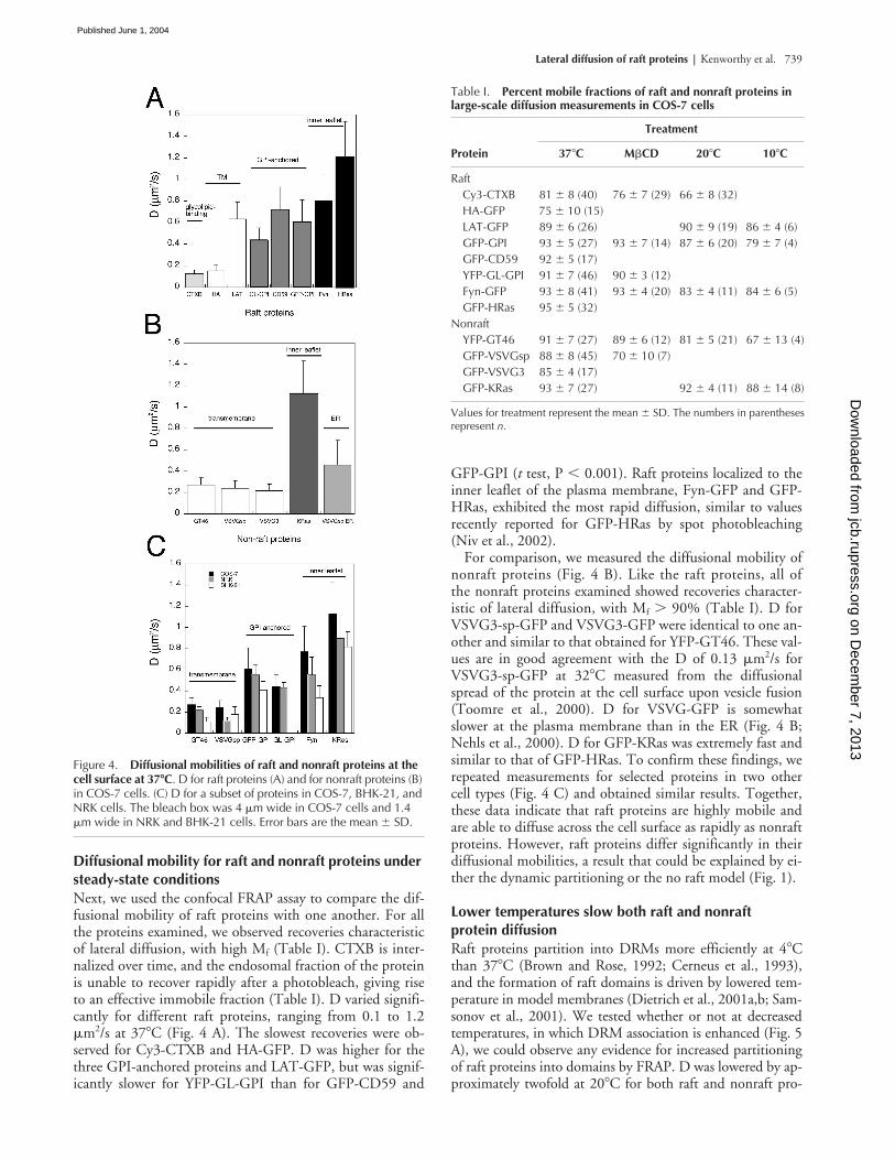
Diffusional mobility for raft and nonraft proteins under steady-state conditions
Next, we used the confocal FRAP assay to compare the dif-fusional mobility of raft proteins with one another. For allthe proteins examined, we observed recoveries characteristicof lateral diffusion, with high M
f
(Table I). CTXB is inter-nalized over time, and the endosomal fraction of the proteinis unable to recover rapidly after a photobleach, giving riseto an effective immobile fraction (Table I). D varied signifi-cantly for different raft proteins, ranging from 0.1 to 1.2
�
m
2
/s at 37
�
C (Fig. 4 A). The slowest recoveries were ob-served for Cy3-CTXB and HA-GFP. D was higher for thethree GPI-anchored proteins and LAT-GFP, but was signif-icantly slower for YFP-GL-GPI than for GFP-CD59 and
GFP-GPI (
t
test, P
�
0.001). Raft proteins localized to theinner leaflet of the plasma membrane, Fyn-GFP and GFP-HRas, exhibited the most rapid diffusion, similar to valuesrecently reported for GFP-HRas by spot photobleaching(Niv et al., 2002).
For comparison, we measured the diffusional mobility ofnonraft proteins (Fig. 4 B). Like the raft proteins, all ofthe nonraft proteins examined showed recoveries character-istic of lateral diffusion, with M
f
�
90% (Table I). D forVSVG3-sp-GFP and VSVG3-GFP were identical to one an-other and similar to that obtained for YFP-GT46. These val-ues are in good agreement with the D of 0.13
�
m
2
/s forVSVG3-sp-GFP at 32
�
C measured from the diffusionalspread of the protein at the cell surface upon vesicle fusion(Toomre et al., 2000). D for VSVG-GFP is somewhatslower at the plasma membrane than in the ER (Fig. 4 B;Nehls et al., 2000). D for GFP-KRas was extremely fast andsimilar to that of GFP-HRas. To confirm these findings, werepeated measurements for selected proteins in two othercell types (Fig. 4 C) and obtained similar results. Together,these data indicate that raft proteins are highly mobile andare able to diffuse across the cell surface as rapidly as nonraftproteins. However, raft proteins differ significantly in theirdiffusional mobilities, a result that could be explained by ei-ther the dynamic partitioning or the no raft model (Fig. 1).
Lower temperatures slow both raft and nonraft protein diffusion
Raft proteins partition into DRMs more efficiently at 4
�
Cthan 37
�
C (Brown and Rose, 1992; Cerneus et al., 1993),and the formation of raft domains is driven by lowered tem-perature in model membranes (Dietrich et al., 2001a,b; Sam-sonov et al., 2001). We tested whether or not at decreasedtemperatures, in which DRM association is enhanced (Fig. 5A), we could observe any evidence for increased partitioningof raft proteins into domains by FRAP. D was lowered by ap-proximately twofold at 20
�
C for both raft and nonraft pro-
Figure 4. Diffusional mobilities of raft and nonraft proteins at the cell surface at 37�C. D for raft proteins (A) and for nonraft proteins (B) in COS-7 cells. (C) D for a subset of proteins in COS-7, BHK-21, and NRK cells. The bleach box was 4 �m wide in COS-7 cells and 1.4 �m wide in NRK and BHK-21 cells. Error bars are the mean � SD.
Table I.
Percent mobile fractions of raft and nonraft proteins in large-scale diffusion measurements in COS-7 cells
Treatment
Protein 37
�
C M
�
CD 20
�
C 10
�
C
RaftCy3-CTXB 81
�
8 (40) 76
�
7 (29) 66
�
8 (32)HA-GFP 75
�
10 (15)LAT-GFP 89
�
6 (26) 90
�
9 (19) 86
�
4 (6)GFP-GPI 93
�
5 (27) 93
�
7 (14) 87
�
6 (20) 79
�
7 (4)GFP-CD59 92
�
5 (17)YFP-GL-GPI 91
�
7 (46) 90
�
3 (12)Fyn-GFP 93
�
8 (41) 93
�
4 (20) 83
�
4 (11) 84
�
6 (5)GFP-HRas 95
�
5 (32)Nonraft
YFP-GT46 91
�
7 (27) 89
�
6 (12) 81
�
5 (21) 67
�
13 (4)GFP-VSVGsp 88
�
8 (45) 70
�
10 (7)GFP-VSVG3 85
�
4 (17)GFP-KRas 93
�
7 (27) 92
�
4 (11) 88
�
14 (8)
Values for treatment represent the mean
�
SD. The numbers in parenthesesrepresent
n
.
on Decem
ber 7, 2013jcb.rupress.org
Dow
nloaded from
Published June 1, 2004
740 The Journal of Cell Biology
|
Volume 165, Number 5, 2004
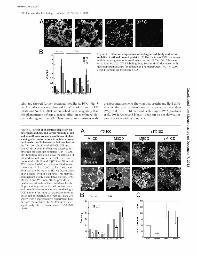
teins and showed further decreased mobility at 10
�
C (Fig. 5B). A similar effect was observed for VSVG-GFP in the ER(Reits and Neefjes, 2001; unpublished data), suggesting thatthis phenomenon reflects a general effect on membrane vis-cosity throughout the cell. These results are consistent with
previous measurements showing that protein and lipid diffu-sion at the plasma membrane is temperature dependent(Wey et al., 1981; Hillman and Schlessinger, 1982; Jacobsonet al., 1984; Aroeti and Henis, 1988) but do not show a sim-ple correlation with raft domains.
Figure 5. Effect of temperature on detergent solubility and lateral mobility of raft and nonraft proteins. (A) The fraction of DRM decreases with increasing temperature of extraction in 1% TX-100. DRM was visualized by Cy3-CTXB labeling. Bar, 10 �m. (B) D decreases with decreasing temperature for both raft and nonraft proteins. **, P � 0.0001, t test. Error bars are the mean � SD.
Figure 6. Effect of cholesterol depletion on detergent solubility and lateral mobility of raft and nonraft proteins, and quantitation of filipin staining after perturbations in cellular choles-terol levels. (A) Cholesterol depletion enhances the TX-100 solubility of YFP-GL-GPI and Cy3-CTXB. A similar effect was observed for other raft proteins (not depicted). Bar, 10 �m. (B) Cholesterol depletion slows the diffusion of raft and nonraft proteins at 37�C. Cells were pretreated with 10 mM M�CD for 30 min at 37�C before TX-100 extraction or FRAP mea-surements. **, P � 0.0001; *, P � 0.01, t test. Error bars are the mean � SD. (C) Quantitation of cholesterol by filipin staining. This method, although not strictly quantitative (Severs, 1997; Maxfield and Wüstner, 2002), provides a qualitative estimate of the cholesterol levels. Filipin staining was performed on fixed cells and quantified from images obtained using a CCD camera for identical exposure times as described in Materials and methods. Data are shown from a representative experiment. Error bars are the mean � SD. All treatments are significantly different than controls (P � 0.0001, t test).
on Decem
ber 7, 2013jcb.rupress.org
Dow
nloaded from
Published June 1, 2004
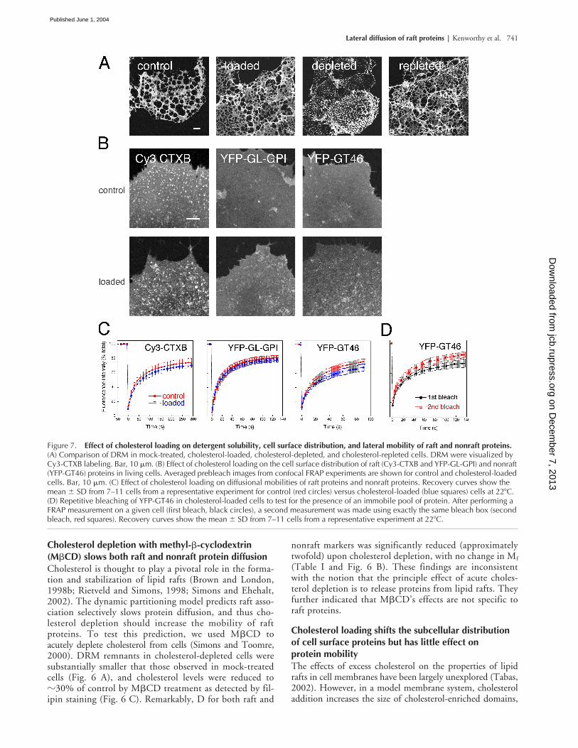
Lateral diffusion of raft proteins | Kenworthy et al. 741
Cholesterol depletion with methyl-�-cyclodextrin (M�CD) slows both raft and nonraft protein diffusionCholesterol is thought to play a pivotal role in the forma-tion and stabilization of lipid rafts (Brown and London,1998b; Rietveld and Simons, 1998; Simons and Ehehalt,2002). The dynamic partitioning model predicts raft asso-ciation selectively slows protein diffusion, and thus cho-lesterol depletion should increase the mobility of raftproteins. To test this prediction, we used M�CD toacutely deplete cholesterol from cells (Simons and Toomre,2000). DRM remnants in cholesterol-depleted cells weresubstantially smaller that those observed in mock-treatedcells (Fig. 6 A), and cholesterol levels were reduced to�30% of control by M�CD treatment as detected by fil-ipin staining (Fig. 6 C). Remarkably, D for both raft and
nonraft markers was significantly reduced (approximatelytwofold) upon cholesterol depletion, with no change in Mf
(Table I and Fig. 6 B). These findings are inconsistentwith the notion that the principle effect of acute choles-terol depletion is to release proteins from lipid rafts. Theyfurther indicated that M�CD’s effects are not specific toraft proteins.
Cholesterol loading shifts the subcellular distribution of cell surface proteins but has little effect on protein mobilityThe effects of excess cholesterol on the properties of lipidrafts in cell membranes have been largely unexplored (Tabas,2002). However, in a model membrane system, cholesteroladdition increases the size of cholesterol-enriched domains,
Figure 7. Effect of cholesterol loading on detergent solubility, cell surface distribution, and lateral mobility of raft and nonraft proteins. (A) Comparison of DRM in mock-treated, cholesterol-loaded, cholesterol-depleted, and cholesterol-repleted cells. DRM were visualized by Cy3-CTXB labeling. Bar, 10 �m. (B) Effect of cholesterol loading on the cell surface distribution of raft (Cy3-CTXB and YFP-GL-GPI) and nonraft (YFP-GT46) proteins in living cells. Averaged prebleach images from confocal FRAP experiments are shown for control and cholesterol-loaded cells. Bar, 10 �m. (C) Effect of cholesterol loading on diffusional mobilities of raft proteins and nonraft proteins. Recovery curves show the mean � SD from 7–11 cells from a representative experiment for control (red circles) versus cholesterol-loaded (blue squares) cells at 22�C. (D) Repetitive bleaching of YFP-GT46 in cholesterol-loaded cells to test for the presence of an immobile pool of protein. After performing a FRAP measurement on a given cell (first bleach, black circles), a second measurement was made using exactly the same bleach box (second bleach, red squares). Recovery curves show the mean � SD from 7–11 cells from a representative experiment at 22�C.
on Decem
ber 7, 2013jcb.rupress.org
Dow
nloaded from
Published June 1, 2004
742 The Journal of Cell Biology | Volume 165, Number 5, 2004
eventually leading to a continuous raftlike phase (Kahya etal., 2003; Lawrence et al., 2003). Assuming the same occursin cell membranes, the partitioning model would predict adecrease in D of raft proteins due to enhanced partitioninginto raft domains, accompanied by immobilization of non-raft proteins due to their exclusion from increasingly largeraft domains. To test these predictions, we used water-solu-ble cholesterol (M�CD–cholesterol complexes) to introducecholesterol into the plasma membrane (Christian et al.,1997; Lange et al., 1999; Feng et al., 2003). Loading cellswith approximately twofold higher cholesterol levels than inmock-treated cells (Fig. 6 C) did not substantially changethe morphology of DRM remnants (Fig. 7 A).
FRAP recovery curves for Cy3-CTXB, YFP-GL-GPI,and YFP-GT46 yielded similar halftimes in control andcholesterol-loaded cells, whereas Mf of YFP-GL-GPI andYFP-GT46 were significantly decreased (Fig. 7 C, Table II,and not depicted). Cholesterol loading also caused the ac-cumulation of YFP-GT46, and to a lesser extent YFP-GL-GPI, in patchy and/or punctate structures (Fig. 7 B). Thesepunctate structures appeared to represent an endocytosedpool of protein based on two findings. First, immunofluo-rescence labeling experiments using anti-GFP antibodiesshowed that the YFP-GT46–positive structures were inac-cessible to labeling in nonpermeabilized cells (unpublisheddata). Second, the punctate structures did not recover overshort time periods, as expected for a vesicular pool. By per-forming a second bleach of the same region of interest incholesterol-loaded cells, an increased Mf of YFP-GT46 wasobserved as a result of eliminating this intracellular immo-bile fraction (Fig. 7 D and Table II). This observation sug-gested that the effects of cholesterol loading on the appar-ent diffusional mobility of YFP-GT46 are due to theredistribution of the protein to an intracellular pool ratherthan altered lateral mobility within the membrane. Thus,cholesterol addition to cell membranes does not have ef-fects analogous to those observed in model membrane sys-tems. However, loading does appear to increase the extentof protein internalization.
DiscussionRaft proteins are mobile and do not diffuse as part of stable structuresIn this work, we used FRAP to investigate the dynamicproperties of lipid rafts and the overall effect of lipid rafts onprotein diffusional mobility. In our experiments, we mea-
sured recoveries in an area much larger than the proposedsize of lipid rafts to monitor possible diffusion of raft do-mains themselves as well as diffusion of proteins outside ofraft domains. Our data revealed that raft proteins are able todiffuse large distances across the cell membrane and thatthey have high Mf. Yet, D for individual raft proteins variedover an order of magnitude depending on the protein exam-ined (Fig. 4). These results are incompatible with two of themodels for lipid raft structure and dynamics described inFig. 1. The first is the stable, immobile raft model. Exceptfor the specialized case of caveolin (Pelkmans et al., 2001;Thomsen et al., 2002), this model is not generally consistentwith the behavior of raft proteins as observed in our experi-ments. The second model, implied by photonic force mi-croscopy measurements (Pralle et al., 2000), is one in whichrafts diffuse as stable entities across the cell surface. Arguingagainst this model are two findings: those here showing thelack of correlation in Ds of different raft proteins and previ-ous data from single molecule tracking showing that individ-ual raft proteins diffuse independently of one another (Vrljicet al., 2002).
Diffusional mobility is more strongly correlated with membrane anchorage than with raft associationOur finding that Ds varied tenfold for different raft proteinsis consistent with previous observations showing that a vari-ety of barriers to protein diffusional mobility exist in cellmembranes (Sheets et al., 1995; Edidin, 1996; Kusumiand Sako, 1996; Saxton, 1999; Lippincott-Schwartz et al.,2001). These results also indicate that raft association is notthe dominant factor in determining the overall mobility of aparticular protein, a possibility consistent with either the dy-namic partitioning or no raft models (Fig. 1). Instead, weobserved a correlation between protein diffusional mobilityand the mode of membrane anchorage, with Dacylated or
prenyalated � DGPI-anchored proteins � Dtransmembrane regardless ofwhether or not a particular protein is raft associated (Fig. 4).
The high mobility of GPI-anchored proteins has beenpreviously noted and is thought to reflect the lipid-based an-chor of these proteins (Ishihara et al., 1987; Edidin andStroynowski, 1991) but is modulated by the ectodomain(Zhang et al., 1991). This finding could explain why we ob-served significantly higher Ds for GPI-anchored proteinsthan a transmembrane form of HA, in contrast to a recentreport (Shvartsman et al., 2003). Until now, the diffusionalproperties of acylated and prenylated proteins have re-mained largely unexplored because of their inaccessibility tolabeling with exogenous fluorescent probes. The high Ds ofthese proteins suggest that their lipid anchors facilitate theirrelatively free diffusion and/or that the microenvironment ofthe inner leaflet contains fewer barriers to protein diffusionthan the outer leaflet where GPI-anchored proteins arefound.
LAT-GFP and Cy3-CTXB are two exceptions to the cor-relation between D and membrane anchorage (Fig. 4). Wespeculate that, given the effective absence of an ectodomain(Zhang et al., 1998a), LAT is more directly comparable toproteins localized to the inner leaflet of the plasma mem-brane than other transmembrane proteins. The slow diffu-sion of CTXB, a homo-pentamer, could potentially arise
Table II. Percent mobile fraction of raft and nonraft proteins in cholesterol-loaded COS-7 cells at 22�C
Treatment
Protein Control Loaded Rebleachcontrol Rebleachloaded
Cy3-CTXB 66 � 8 (23) 64 � 8 (22)YFP-GL-GPI 77 � 7 (27) 72 � 6a (27)YFP-GT46 79 � 5 (28) 69 � 12a (29) 80 � 8 (30) 79 � 9 (30)
Values for treatment represent the mean � SD. The numbers in parenthesesrepresent n.aP � 0.05 compared to control, t test.
on Decem
ber 7, 2013jcb.rupress.org
Dow
nloaded from
Published June 1, 2004

Lateral diffusion of raft proteins | Kenworthy et al. 743
from cross-linking and trapping other cell surface proteinsthat can interact with the cytoskeleton, similar to that pro-posed for cross-linked GPI-anchored proteins (Suzuki andSheetz, 2001). Association of CTXB with immobile caveolaebefore its eventual internalization may also slow its diffu-sion.
The effects of reduced temperature and cholesterol depletion on protein diffusion are not limited to alterations of raft dynamicsTo distinguish between the dynamic partitioning (Fig. 1,Model 3) and no raft domain (Fig. 1, Model 4) models, wefirst investigated the sensitivity of protein mobility to twoconditions previously shown to alter partitioning of raft pro-teins into DRMs, temperature reduction and cholesterol de-pletion. We found that both temperature reduction andcholesterol depletion decreased the diffusional mobility ofboth raft and nonraft proteins to a similar extent (Figs. 5 Band 6 B). This observation does not completely eliminatethe possibility of altered partitioning of raft proteins intorafts in response to these treatments, but indicates that otherfactors play a more dominant role in determining a protein’slateral mobility under these conditions. Interestingly, an im-mobilizing effect of cholesterol depletion also has beennoted in a recent study of MHC-class I lateral mobility(Kwik et al., 2003). This effect was linked to changes in theorganization of the actin cytoskeleton in response to M�CDtreatment, which have also been noted in other studies(Grimmer et al., 2002; Hering et al., 2003; see also the re-traction of cells during this treatment in Fig. 6 A). Given thepotential connection between lipid rafts and the actin cyto-skeleton (Maxfield, 2002), it is unclear if these effects occurin addition to, or as a consequence of, disruption of raft do-mains. Nevertheless, these findings raise questions about thespecificity of acute cholesterol depletion on lipid domainstructure/function and highlight the possibility that cyto-skeletal reorganization might account for some of the ob-served functional effects of M�CD treatment.
Effects of cholesterol loading, another method to explore membrane domain structure and function on raft protein dynamicsTo further attempt to distinguish the effect of raft perturba-tions on protein mobility, we used cholesterol loading.Given that major cellular defects are associated with condi-tions of aberrant cholesterol accumulation, such as hyper-cholesterolemia and Niemann-Pick type C (NPC) disease,understanding the effects of excess cholesterol on lipid raftstructure and function is of physiological importance. Basedon work in model systems (Kahya et al., 2003; Lawrence etal., 2003), we hypothesized that cholesterol loading shouldlead to the formation of additional lipid rafts and/or to anincrease in size of existing rafts. However, little change in thehalftime of recovery was seen for any of the proteins studiedin cholesterol-loaded cells (Fig. 7). These data suggest that ifcholesterol loading indeed increases the fraction of raftlikemembrane, diffusion of raft proteins within this environ-ment is not significantly slowed under these conditions.However, we did observe decreased Mf in response to cho-
lesterol loading. This effect appears to be due to enhancedendocytosis of proteins in response to cholesterol loading,potentially resulting from proteins being driven out of an ex-panded raft phase. The identity of these putative endocyticstructures remains to be determined. Both clathrin-mediatedand nonclathrin endocytic pathways are sensitive to mem-brane cholesterol levels (Rodal et al., 1999; Subtil et al.,1999; Nichols and Lippincott-Schwartz, 2001). Moreover,defects in the trafficking of glycolipids internalized by non-clathrin pathways have been correlated with the accumula-tion of excess cholesterol in multiple sphingolipid storagediseases including NPC (Puri et al., 1999). Our observationsof continued CTXB uptake in cholesterol-loaded cells (Fig.7 B) are in line with observations that CTXB is able to enterendocytic structures in NPC1-deficient cells (Sugimoto etal., 2001; Choudhury et al., 2002). It will be of interest todetermine the nature of the trafficking defects induced byacute cholesterol loading and to discern if the mechanismsare similar to those observed in NPC-deficient cells.
Implications for models for lipid raftsOur data indicate that raft association is not the major deter-minant of plasma membrane protein diffusional mobilityunder steady-state conditions. Thus, we can rule out severalmodels for raft dynamics, including stable immobile raftsand stable mobile rafts, on the basis of our findings. It ispossible that a small fraction of proteins are associated withstable immobile domains, as we observed an immobile frac-tion of 10–15% for the majority of the proteins examined(Table I). However, the absence of an effect of cholesteroldepletion on the size of the immobile fractions that we andothers (Lommerse et al., 2004) have detected, and the simi-lar immobile fractions of raft and nonraft proteins, suggeststhat the immobile fraction is not generated by recruitmentto lipid rafts. It is also possible that overexpression of ourmarker proteins may overwhelm and mask a small amountof behavior corresponding to models 1 or 2. However, diffu-sion of the raft marker GFP-HRas was previously shown tobe independent of expression levels (Niv et al., 2002). Tech-niques with single molecule sensitivity such as fluorescencecorrelation spectroscopy could provide insight into this issuein the future.
Our findings also reveal that treatments shown to perturbthe rafts detected in model membrane systems (cholesteroldepletion, temperature reduction, and cholesterol addition)have effects on protein diffusion in cells that cannot be ex-plained solely by predictions of the raft hypothesis. How-ever, these treatments may give rise to pleiotropic effects thatmask underlying changes in raft properties. Therefore, onthe basis of our current data we cannot definitively distin-guish between the dynamic partitioning model and the ab-sence of detectable raft domains (Fig. 1). Interestingly, thepredictions of the dynamic partitioning and no raft modelsconverge under conditions where lipid rafts comprise a smallfraction of the cell surface and proteins spend only a smallamount of time visiting them, or if the fraction of raftlikemembrane is normally large but protein diffusion within araft is not very different than in nonraft regions of the mem-brane. Our understanding of how rafts function in vivo nowneeds to take into account the fact that incorporation into
on Decem
ber 7, 2013jcb.rupress.org
Dow
nloaded from
Published June 1, 2004

744 The Journal of Cell Biology | Volume 165, Number 5, 2004
biochemically defined rafts is not indicative of incorporationinto stable microdomains in the plasma membrane.
Materials and methodsDNA constructs, cell transfections, and fluorescent probesUnless otherwise indicated, all chemicals were obtained from Sigma-Aldrich. For simplicity, EGFP is referred to as GFP throughout. GFP-GPI andGFP-CD59 were as described previously (Nichols et al., 2001). GFP-HRas,GFP-KRas, and Fyn-GFP (Choy et al., 1999) were provided by M. Philips(New York University School of Medicine, New York, NY). LAT-GFP (Bun-nell et al., 2002) was obtained from L. Samelson (National Institutes ofHealth, Bethesda, MD). YFP-GL-GPI (Keller et al., 2001), YFP-GT46 (Pralleet al., 2000), VSVG3-sp-GFP (Keller et al., 2001), and VSVG-3-GFP(Toomre et al., 1999) were provided by P. Keller and K. Simons (MaxPlanck Institute of Molecular Cell Biology and Genetics, Dresden, Ger-many). YFP-GT46 is an artificial secretory protein containing a signal se-quence, YFP, a consensus N-glycosylation site, the transmembrane domainof the LDL receptor, and the cytoplasmic tail of CD46 (Pralle et al., 2000).To construct HA-GFP, the influenza hemagglutinin gene was PCR amplifiedfrom the plasmid pCB6HA (a gift from M.G. Roth, University of TexasSouthwestern Medical Center, Dallas, TX) and cloned into the plasmidpEGFP-N1 (CLONTECH Laboratories, Inc.). In control experiments, con-structs containing monomeric (Zacharias et al., 2002) forms of fluorescentprotein were examined (mYFP-GL-GPI, mYFP-GT46, mCitFP-LAT, andVSVG-mYFP; Glebov and Nichols, 2004).
Cells were grown in DME (COS-7 and normal rat kidney [NRK] cells) orEMEM (BHK-21) supplemented with 10% FCS, glutamine, penicillin, andstreptomycin (Biofluids). Transient transfections were performed using Fu-GENE 6 (Roche Molecular Biochemicals). CTXB was fluorescently labeledwith Cy3 (Amersham Biosciences) as per the manufacturer’s instructionsand was used at a final concentration of 1 �g/ml.
Fluorescence microscopy and diffusional mobility measurementsFilipin fluorescence was imaged using a wide field microscope (model Ax-iophot; Carl Zeiss MicroImaging, Inc.). Fluorescence was excited with amercury arc lamp, and emission was detected using a DAPI filter set. Im-ages were collected using a 40� 1.3 NA Plan-Neofluar objective (CarlZeiss MicroImaging, Inc.) and captured using a MicroMax CCD camera(Princeton Instruments) and MetaMorph acquisition software. Images wereobtained using identical exposure times for cells subjected to various treat-ments in each experiment. Quantitation of filipin fluorescence was per-formed using NIH Image.
All other fluorescence images were obtained using a confocal micro-scope (model LSM 510; Carl Zeiss MicroImaging, Inc.). Fluorescence emis-sion resulting from 488-nm excitation for GFP, 488- or 514-nm excitationfor YFP, and 543-nm excitation for Cy3 was detected using filter sets sup-plied by the manufacturer. Cells were held at 37�C on the microscopestage unless otherwise indicated. For FRAP measurements, a 40� 1.3 NAPlan-Neofluar objective or 100� 1.4 NA Plan-Apochromat objective (CarlZeiss MicroImaging, Inc.) was used with the confocal pinhole set at 1–2Airy units. Photobleaching of GFP, YFP, or Cy3 was performed using 5–20scans with the 488-nm laser line at full power in a rectangular region of in-terest 4 �m wide (COS-7 cells) or 1.4 �m wide (BHK-21 and NRK cells).Pre- and postbleach images were monitored at low laser intensity. Fluores-cence recoveries in the bleached region and overall photobleaching in thewhole cell during the time series were quantitated using the LSM software(Carl Zeiss MicroImaging, Inc.). Photobleaching of Cy3 did not inducephotodamage to the membrane, as evidenced by control experimentsshowing identical recoveries for GFP-HRas in cells labeled with Cy3-CTXBversus unlabeled CTXB. For presentation purposes, LSM 510 images wereexported in TIFF format and their contrast and brightness optimized inPhotoshop.
Ds were calculated from the photobleaching data using Diffuse, a pro-gram that simulates diffusive recovery into the bleached region using theseries of images collected after the photobleaching episode (Ellenberg etal., 1997; Zaal et al., 1999; Nehls et al., 2000; Siggia et al., 2000; programavailable upon request). LSM 510 images were exported in TIFF formatand converted to PPM format for analysis with the Diffuse program. Statis-tical differences were evaluated using the t test. Mf was calculated as de-scribed previously (Ellenberg et al., 1997).
TX-100 extractionTX-100 extractions were performed as described previously (Nichols et al.,2001) except that cells were incubated in TX-100 for 15 min and after fixa-
tion subsequently labeled with Cy3-CTXB before imaging. Where indicated,TX-100 extractions were performed at 12�, 22�, or 37�C instead of 4�C.
Filipin stainingFixed cells were stained with 5 �g/ml filipin as described previously (Neu-feld et al., 1999). Filipin fluorescence was quantified from regions of inter-est containing plasma membrane and underlying cytoplasmic structuresusing NIH Image.
Cholesterol depletion, loading, and repletionSolutions for cholesterol depletion, repletion, and loading were made inDME or RPMI supplemented with 25 mM Hepes and 1 mg/ml BSA. Forcholesterol depletion, cells were briefly washed, and then incubated in 10mM M�CD for 30 min at 37�C before TX-100 extraction or FRAP. Choles-terol loading was performed using water-soluble cholesterol (M�CD–cho-lesterol complexes at �6:1 molar ratio; Sigma-Aldrich). Previous work hasestablished that efficient loading of cells occurs when incubated withM�CD–cholesterol complexes prepared at this ratio (Christian et al., 1997;Sheets et al., 1999). M�CD–cholesterol complexes were prepared as astock solution, 300 mM in M�CD, diluted directly onto the cells to 10 mMin M�CD, and incubated for 30 min at 37�C. Incubation for longer times(1–2 h) led to increased sensitivity to TX-100 extraction in DRM experi-ments. Cholesterol-loaded cells imaged live for FRAP experiments did notinitially exhibit any gross morphological differences from control cells, butover time cells showed a tendency to round up.
We thank Eric Siggia and Stefan Bekiranov for providing the Diffuse simu-lation and Michael Edidin for sharing results before publication. MichaelRoth, Mark Philips, Larry Samelson, Patrick Keller, and Kai Simons gener-ously shared reagents. We thank George Patterson, Christy Hendrix, Kim-berly Drake, and J. Shawn Goodwin for assistance and Erik Snapp and Ni-hal Altan for comments on the manuscript.
Experiments were performed in part through the use of the VanderbiltUniversity Medical Center Cell Imaging Core Resource (supported by Na-tional Institutes of Health grants CA68485, DK20593, and DK58404). A.K.Kenworthy was supported by a fellowship from the National ResearchCouncil, and B.J. Nichols was funded by an International Prize TravellingResearch Fellowship from the Wellcome Trust.
Submitted: 24 December 2003Accepted: 26 April 2004
ReferencesAnderson, R.G.W., and K. Jacobson. 2002. A role for lipid shells in targeting pro-
teins to caveolae, rafts and other lipid domains. Science. 296:1821–1825.Aroeti, B., and Y.I. Henis. 1988. Effects of fusion temperature on the lateral mobil-
ity of Sendai virus glycoproteins in erythrocyte membranes and on cell fu-sion indicate that glycoprotein mobilization is required for cell fusion. Bio-chemistry. 27:5654–5661.
Bacia, K., I.V. Majoul, and P. Schwille. 2002. Probing the endocytic pathway inlive cells using dual-color fluorescence cross-correlation analysis. Biophys. J.83:1184–1193.
Brown, D.A., and J.K. Rose. 1992. Sorting of GPI-anchored proteins to glycolipid-enriched membrane subdomains during transport to the apical cell surface.Cell. 68:533–544.
Brown, D.A., and E. London. 1998a. Functions of lipid rafts in biological mem-branes. Annu. Rev. Cell Dev. Biol. 14:111–136.
Brown, D.A., and E. London. 1998b. Structure and origin of ordered lipid do-mains in biological membranes. J. Membr. Biol. 164:103–114.
Bunnell, S.C., D.I. Hong, J.R. Kardon, T. Yamazaki, C.J. McGlade, V.A. Barr,and L.E. Samelson. 2002. T cell receptor ligation induces the formation ofdynamically regulated signaling assemblies. J. Cell Biol. 158:1263–1275.
Cerneus, D.P., E. Ueffing, G. Posthuma, G.J. Strous, and A. van der Ende. 1993.Detergent insolubility of alkaline phosphatase during biosynthetic transportand endocytosis. Role of cholesterol. J. Biol. Chem. 268:3150–3155.
Choudhury, A., M. Dominguez, V. Puri, D.K. Sharma, K. Narita, C.L. Wheatley,D.L. Marks, and R.E. Pagano. 2002. Rab proteins mediate Golgi transportof caveola-internalized glycosphingolipids and correct lipid trafficking inNiemann-Pick C cells. J. Clin. Invest. 109:1541–1550.
Choy, E., V.K. Chiu, J. Silletti, M. Feoktistov, T. Morimoto, D. Michaelson, I.E.Ivanov, and M.R. Philips. 1999. Endomembrane trafficking of Ras: theCAAX motif targets proteins to the ER and Golgi. Cell. 98:69–80.
on Decem
ber 7, 2013jcb.rupress.org
Dow
nloaded from
Published June 1, 2004
Lateral diffusion of raft proteins | Kenworthy et al. 745
Christian, A.E., M.P. Haynes, M.C. Phillips, and G.H. Rothblat. 1997. Use of cy-clodextrins for manipulating cellular cholesterol content. J. Lipid Res. 38:2264–2272.
Dietrich, C., L.A. Bagatolli, Z.N. Volovyk, N.L. Thompson, M. Levi, K. Jacobson,and E. Gratton. 2001a. Lipid rafts reconstituted in model systems. Biophys.J. 80:1417–1428.
Dietrich, C., Z.N. Volovyk, M. Levi, N.L. Thompson, and K. Jacobson. 2001b.Partitioning of Thy-1, GM1, and crosslinked phospholipid analogs intolipid rafts reconstituted in supported membrane monolayers. Proc. Natl.Acad. Sci. USA. 98:10642–10647.
Dietrich, C., B. Yang, T. Fujiwara, A. Kusumi, and K. Jacobson. 2002. Relation-ship of lipid rafts to transient confinement zones detected by single particletracking. Biophys. J. 82:274–284.
Edidin, M. 1994. Fluorescence photobleaching and recovery, FPR, in the analysisof membrane structure and dynamics. In Mobility and Proximity in Biologi-cal Membranes. S. Damjanovish, M. Edidin, J. Szollosi, and L. Tron, edi-tors. CRC Press, Boca Raton, FL. 109–135.
Edidin, M. 1996. Getting there is only half the fun. Current Topics in Membranes.43:1–13.
Edidin, M. 2003. The state of lipid rafts: from model membranes to cells. Annu.Rev. Biophys. Biomol. Struct. 32:257–283.
Edidin, M., and I. Stroynowski. 1991. Differences between the lateral organizationof conventional and inositol phospholipid-anchored membrane proteins. Afurther definition of micrometer scale membrane domains. J. Cell Biol. 112:1143–1150.
Ellenberg, J., E.D. Siggia, J.E. Moreira, C.L. Smith, J.F. Presley, H.J. Worman,and J. Lippincott-Schwartz. 1997. Nuclear membrane dynamics and reas-sembly in living cells: targeting of an inner nuclear membrane protein in in-terphase and mitosis. J. Cell Biol. 138:1193–1206.
Feng, B., P.M. Yao, Y. Li, C.M. Devlin, D. Zhang, H.P. Harding, M. Sweeney,J.X. Rong, G. Kuriakose, E.A. Fisher, et al. 2003. The endoplasmic reticu-lum is the site of cholesterol-induced cytotoxicity in macrophages. Nat. CellBiol. 5:781–792.
Glebov, O.O., and B.J. Nichols. 2004. Lipid raft proteins have a random distri-bution during localized activation of the T-cell receptor. Nat. Cell Biol.6:238–243.
Grimmer, S., B. van Deurs, and K. Sandvig. 2002. Membrane ruffling and macro-pinocytosis in A431 cells require cholesterol. J. Cell Sci. 115:2953–2962.
Hancock, J.F. 2003. Ras proteins: different signals from different locations. Nat.Rev. Mol. Cell Biol. 4:373–384.
Hao, M., S. Mukherjee, and F.R. Maxfield. 2001. Cholesterol depletion induceslarge scale domain segregation in living cell membranes. Proc. Natl. Acad.Sci. USA. 98:13072–13077.
Hering, H., C.C. Lin, and M. Sheng. 2003. Lipid rafts in the maintenance of syn-apses, dendritic spines, and surface AMPA receptor stability. J. Neurosci. 23:3262–3271.
Hillman, G.M., and J. Schlessinger. 1982. Lateral diffusion of epidermal growthfactor complexed to its surface receptors does not account for the thermalsensitivity of patch formation and endocytosis. Biochemistry. 21:1667–1672.
Ikonen, E. 2001. Role of lipid rafts in membrane transport. Curr. Opin. Cell Biol.13:470–477.
Ishihara, A., Y. Hou, and K. Jacobson. 1987. The Thy-1 antigen exhibits rapid lat-eral diffusion in the plasma membrane of rodent lymphoid cells and fibro-blasts. Proc. Natl. Acad. Sci. USA. 84:1290–1293.
Jacobson, K., and C. Dietrich. 1999. Looking at lipid rafts? Trends Cell Biol.9:87–91.
Jacobson, K., D. O’Dell, and J.T. August. 1984. Lateral diffusion of an 80,000-dalton glycoprotein in the plasma membrane of murine fibroblasts: relation-ships to cell structure and function. J. Cell Biol. 99:1624-33.
Kahya, N., D. Scherfeld, K. Bacia, B. Poolman, and P. Schwille. 2003. Probinglipid mobility of raft-exhibiting model membranes by fluorescence correla-tion spectroscopy. J. Biol. Chem. 278:28109–28115.
Keller, P., D. Toomre, E. Diaz, J. White, and K. Simons. 2001. Multicolour im-aging of post-Golgi sorting and trafficking in live cells. Nat. Cell Biol.3:140–149.
Kenworthy, A.K., and M. Edidin. 1998. Distribution of a glycosylphosphatidyl-inositol-anchored protein at the apical surface of MDCK cells examined at aresolution of �100 Å using imaging fluorescence resonance energy transfer.J. Cell Biol. 142:69–84.
Korlach, J., P. Schwille, W.W. Webb, and G.W. Feigenson. 1999. Characteriza-tion of lipid bilayer phases by confocal microscopy and fluorescence correla-tion spectroscopy. Proc. Natl. Acad. Sci. USA. 96:8461–8466.
Kusumi, A., and Y. Sako. 1996. Cell surface organization by the membrane skele-ton. Curr. Opin. Cell Biol. 8:566–574.
Kwik, J., S. Boyle, D. Fooksman, L. Margolis, M.P. Sheetz, and M. Edidin. 2003.Membrane cholesterol, lateral mobility, and the phosphatidylinositol 4,5-bisphosphate-dependent organization of cell actin. Proc. Natl. Acad. Sci.USA. 100:13964–13969.
Lange, Y., J. Ye, M. Rigney, and T.L. Steck. 1999. Regulation of endoplasmicreticulum cholesterol by plasma membrane cholesterol. J. Lipid Res. 40:2264–2270.
Lawrence, J.C., D.E. Saslowsky, J.M. Edwardson, and R.M. Henderson. 2003.Real-time analysis of the effects of cholesterol on lipid raft behavior usingatomic force microscopy. Biophys. J. 84:1827–1832.
Lippincott-Schwartz, J., E. Snapp, and A.K. Kenworthy. 2001. Studying proteindynamics in living cells. Nat. Rev. Mol. Cell Biol. 2:444–456.
Lommerse, P.H., G.A. Blab, L. Cognet, G.S. Harms, B.E. Snaar-Jagalska, H.P.Spaink, and T. Schmidt. 2004. Single-molecule imaging of the H-Ras mem-brane-anchor reveals domains in the cytoplasmic leaflet of the cell mem-brane. Biophys. J. 86:609–616.
Maxfield, F.R. 2002. Plasma membrane microdomains. Curr. Opin. Cell Biol. 14:483–487.
Maxfield, F.R., and D. Wüstner. 2002. Intracellular cholesterol transport. J. Clin.Invest. 110:891–898.
Mayor, S., and F.R. Maxfield. 1995. Insolubility and redistribution of GPI-anchored proteins at the cell surface after detergent treatment. Mol. Biol.Cell. 6:929–944.
Nehls, S., E.L. Snapp, N.B. Cole, K.J. Zaal, A.K. Kenworthy, T.H. Roberts, J. El-lenberg, J.F. Presley, E. Siggia, and J. Lippincott-Schwartz. 2000. Dynamicsand retention of misfolded proteins in native ER membranes. Nat. Cell Biol.2:288–295.
Neufeld, E.B., M. Wastney, S. Patel, S. Suresh, A.M. Cooney, N.K. Dwyer, C.F.Roff, K. Ohno, J.A. Morris, E.D. Carstea, et al. 1999. The Niemann-PickC1 protein resides in a vesicular compartment linked to retrograde transportof multiple lysosomal cargo. J. Biol. Chem. 274:9627–9635.
Nichols, B.J., and J. Lippincott-Schwartz. 2001. Endocytosis without clathrincoats. Trends Cell Biol. 11:406–412.
Nichols, B.J., A.K. Kenworthy, R.S. Polishchuk, R. Lodge, T.H. Roberts, K.Hirschberg, R.D. Phair, and J. Lippincott-Schwartz. 2001. Rapid cycling oflipid raft markers between the cell surface and Golgi complex. J. Cell Biol.153:529–541.
Niv, H., O. Gutman, Y. Kloog, and Y.I. Henis. 2002. Activated K-Ras and H-Rasdisplay different interactions with saturable nonraft sites at the surface of livecells. J. Cell Biol. 157:865–872.
Orlandi, P.A., and P.H. Fishman. 1998. Filipin-dependent inhibition of choleratoxin: evidence for toxin internalization and activation through caveolae-likedomains. J. Cell Biol. 141:905–915.
Pelkmans, L., J. Kartenbeck, and A. Helenius. 2001. Caveolar endocytosis of sim-ian virus 40 reveals a new two-step vesicular-transport pathway to the ER.Nat. Cell Biol. 3:473–483.
Pralle, A., P. Keller, E.L. Florin, K. Simons, and J.K. Horber. 2000. Sphingolipid-cholesterol rafts diffuse as small entities in the plasma membrane of mamma-lian cells. J. Cell Biol. 148:997–1008.
Prior, I.A., A. Harding, J. Yan, J. Sluimer, R.G. Parton, and J.F. Hancock. 2001.GTP-dependent segregation of H-ras from lipid rafts is required for biologi-cal activity. Nat. Cell Biol. 3:368–375.
Puri, V., R. Watanabe, M. Dominguez, X. Sun, C.L. Wheatley, D.L. Marks, andR.E. Pagano. 1999. Cholesterol modulates membrane traffic along the en-docytic pathway in sphingolipid-storage diseases. Nat. Cell Biol. 1:386–388.
Reits, E.A.J., and J.J. Neefjes. 2001. From fixed to FRAP: measuring protein mo-bility and activity in living cells. Nat. Cell Biol. 3:E145–E147.
Rietveld, A., and K. Simons. 1998. The differential miscibility of lipids as the basisfor the formation of functional membrane rafts. Biochim. Biophys. Acta.1376:467–479.
Rodal, S.K., G. Skretting, O. Garred, F. Vilhardt, B. van Deurs, and K. Sandvig.1999. Extractions of cholesterol with methyl-�-cyclodextrin perturbs forma-tion of clathrin-coated endocytic vesicles. Mol. Biol. Cell. 10:961–974.
Rothberg, K.G., J.E. Heuser, W.C. Donzell, Y.S. Ying, J.R. Glenney, and R.G.Anderson. 1992. Caveolin, a protein component of caveolae membranecoats. Cell. 68:673–682.
Saffman, P.G., and M. Delbrück. 1975. Brownian motion in biological mem-branes. Proc. Natl. Acad. Sci. USA. 72:3111–3113.
Samsonov, A.V., I. Mihalyov, and F.S. Cohen. 2001. Characterization of choles-terol-sphingomyelin domains and their dynamics in bilayer membranes. Bio-
on Decem
ber 7, 2013jcb.rupress.org
Dow
nloaded from
Published June 1, 2004

746 The Journal of Cell Biology | Volume 165, Number 5, 2004
phys. J. 81:1488–1500.Saxton, M.P. 1999. Lateral diffusion of lipids and proteins. Current Topics in Mem-
branes. 48:229–282.Scheiffele, P., M.G. Roth, and K. Simons. 1997. Interaction of influenza virus
haemagglutinin with sphingolipid-cholesterol membrane domains via itstransmembrane domain. EMBO J. 16:5501–5508.
Schütz, G.J., G. Kada, V.P. Pastushenko, and H. Schindler. 2000. Properties oflipid microdomains in a muscle cell membrane visualized by single moleculemicroscopy. EMBO J. 19:892–901.
Severs, N.J. 1997. Cholesterol cytochemistry in cell biology and disease. Subcell.Biochem. 28:477–505.
Sheets, E.D., R. Simson, and K. Jacobson. 1995. New insights into membrane dy-namics from the analysis of cell surface interactions by physical methods.Curr. Opin. Cell Biol. 7:707–714.
Sheets, E.D., G.M. Lee, R. Simson, and K. Jacobson. 1997. Transient confinementof a glycosylphosphatidylinositol-anchored protein in the plasma membrane.Biochemistry. 36:12449–12458.
Sheets, E.D., D. Holowka, and B. Baird. 1999. Critical role for cholesterol in Lyn-mediated tyrosine phosphorylation of Fc�RI and their association with de-tergent-resistant membranes. J. Cell Biol. 145:877–887.
Shvartsman, D.E., M. Kotler, R.D. Tall, M.G. Roth, and Y.I. Henis. 2003. Differ-ently anchored influenza hemagglutinin mutants display distinct interactiondynamics with mutual rafts. J. Cell Biol. 8:879–888.
Siggia, E.D., J. Lippincott-Schwartz, and S. Bekiranov. 2000. Diffusion in inho-mogeneous media: theory and simulations applied to whole cell photobleachrecovery. Biophys. J. 79:1761–1770.
Simons, K., and E. Ikonen. 1997. Functional rafts in cell membranes. Nature. 387:569–572.
Simons, K., and D. Toomre. 2000. Lipid rafts and signal transduction. Nat. Rev.Mol. Cell Biol. 1:31–41.
Simons, K., and R. Ehehalt. 2002. Cholesterol, lipid rafts, and disease. J. Clin. In-vest. 110:597–603.
Skibbens, J.E., M.G. Roth, and K.S. Matlin. 1989. Differential extractability of in-fluenza hemagglutinin during intracellular transport in polarized epithelialcells and nonpolar fibroblasts. J. Cell Biol. 108:821–832.
Subczynski, W.K., and A. Kusumi. 2003. Dynamics of raft molecules in the celland artificial membranes: approaches by pulse EPR spin labeling and singlemolecule optical microscopy. Biochim. Biophys. Acta. 1610:231–243.
Subtil, A., I. Gaidarov, K. Kobylarz, M. Lampson, J. Keen, and T. McGraw. 1999.Acute cholesterol depletion inhibits clathrin-coated pit budding. Proc. Natl.Acad. Sci. USA. 96:6775–6780.
Sugimoto, Y., H. Ninomiya, Y. Ohsaki, K. Higaki, J.P. Davies, Y.A. Ioannou, andK. Ohno. 2001. Accumulation of cholera toxin and GM1 ganglioside in theearly endosome of Niemann-Pick C1-deficient cells. Proc. Natl. Acad. Sci.USA. 98:12391–12396.
Suzuki, K., and M.P. Sheetz. 2001. Binding of cross-linked glycosylphosphatidyl-inositol-anchored proteins to discrete actin-associated sites and cholesterol-dependent domains. Biophys. J. 81:2181–2189.
Tabas, I. 2002. Consequences of cellular cholesterol accumulation: basic conceptsand physiological implications. J. Clin. Invest. 110:905–911.
Thomsen, P., K. Roepstorff, M. Stahlhut, and B. van Deurs. 2002. Caveolae arehighly immobile plasma membrane microdomains, which are not involvedin constitutive endocytic trafficking. Mol. Biol. Cell. 13:238–250.
Toomre, D., P. Keller, J. White, J.-C. Olivo, and K. Simons. 1999. Dual-color vi-sualization of trans-Golgi network to plasma membrane traffic along micro-tubules in living cells. J. Cell Sci. 112:21–33.
Toomre, D., J.A. Steyer, P. Keller, W. Almers, and K. Simons. 2000. Fusion ofconstitutive membrane traffic with the cell surface observed by evanescentwave microscopy. J. Cell Biol. 149:33–40.
Vrljic, M., S.Y. Nishimura, S. Brasselet, W.E. Moerner, and H.M. McConnell.2002. Translational diffusion of individual Class II MHC membrane pro-teins in cells. Biophys. J. 83:2681–2692.
Wey, C.L., R.A. Cone, and M.A. Edidin. 1981. Lateral diffusion of rhodopsin inphotoreceptor cells measured by fluorescence photobleaching and recovery.Biophys. J. 33:225–232.
Zaal, K.J.M., C.L. Smith, R.S. Polishchuk, N. Altan, N.B. Cole, J. Ellenberg, K.Hirschberg, J.F. Presley, T.H. Roberts, E. Siggia, et al. 1999. Golgi mem-branes are absorbed into and reemerge from the ER during mitosis. Cell. 99:589–601.
Zacharias, D.A., J.D. Violin, A.C. Newton, and R.Y. Tsien. 2002. Partitioning oflipid-modified monomeric GFPs into membrane microdomains of live cells.Science. 296:913–916.
Zhang, F., B. Crise, B. Su, Y. Hou, J.K. Rose, A. Bothwell, and K. Jacobson. 1991.Lateral diffusion of membrane-spanning and glycosylphosphatidylinositol-linked proteins: toward establishing rules governing the lateral mobility ofmembrane proteins. J. Cell Biol. 115:75–84.
Zhang, W., J. Sloan-Lancaster, J. Kitchen, R.P. Trible, and L.E. Samelson. 1998a.LAT: the ZAP-70 tyrosine kinase substrate that links T cell receptor to cellu-lar activation. Cell. 92:83–92.
Zhang, W.G., R.P. Trible, and L.E. Samelson. 1998b. LAT palmitoylation: its es-sential role in membrane microdomain targeting and tyrosine phosphoryla-tion during T cell activation. Immunity. 9:239–246.
on Decem
ber 7, 2013jcb.rupress.org
Dow
nloaded from
Published June 1, 2004