EGFP-tagged core and linker histones diffuse via distinct mechanisms within living cells

Linker for Activation of T-cell Family Member2(LAT2) a Lipid Raft Adaptor Protein for AKTSignaling, Is an Early Mediator ofAlkylphospholipid Anti-leukemic Activity*□S
Carolina H. Thome‡§¶, Guilherme A. dos Santos‡¶�, Germano A. Ferreira‡**,Priscila S. Scheucher‡, Clarice Izumi‡**, Andreia M. Leopoldino‡‡, Ana Maria Simao§§,Pietro Ciancaglini§§, Kleber T. de Oliveira¶¶, Alice Chin��, Samir M. Hanash��,Roberto P. Falcao‡�, Eduardo M. Rego‡�, Lewis J. Greene‡**, and Vitor M. Faca‡ab
Lipid rafts are highly ordered membrane domains rich incholesterol and sphingolipids that provide a scaffold forsignal transduction proteins; altered raft structure has alsobeen implicated in cancer progression. We have shown that25 �M 10-(octyloxy) decyl-2-(trimethylammonium) ethylphosphate (ODPC), an alkylphospholipid, targets high cho-lesterol domains in model membranes and induces apopto-sis in leukemia cells but spares normal hematopoietic andepithelial cells under the same conditions. We performed aquantitative (SILAC) proteomic screening of ODPC targetsin a lipid-raft-enriched fraction of leukemic cells to identifyearly events prior to the initiation of apoptosis. Six proteins,three with demonstrated palmitoylation sites, were reducedin abundance. One, the linker for activation of T-cell familymember 2 (LAT2), is an adaptor protein associated with lipidrafts in its palmitoylated form and is specifically expressedin B lymphocytes and myeloid cells. Interestingly, LAT2 isnot expressed in K562, a cell line more resistant to ODPC-induced apoptosis. There was an early loss of LAT2 in the
lipid-raft-enriched fraction of NB4 cells within 3 h followingtreatment with 25 �M ODPC. Subsequent degradation ofLAT2 by proteasomes was observed. Twenty-five �M ODPCinhibited AKT activation via myeloid growth factors, andLAT2 knockdown in NB4 cells by shRNA reproduced thiseffect. LAT2 knockdown in NB4 cells also decreased cellproliferation and increased cell sensitivity to ODPC (7.5�),perifosine (3�), and arsenic trioxide (8.5�). Taken together,these data indicate that LAT2 is an early mediator of theanti-leukemic activity of alkylphospholipids and arsenic tri-oxide. Thus, LAT2 may be used as a target for the design ofdrugs for cancer therapy. Molecular & Cellular Proteom-ics 11: 10.1074/mcp.M112.019661, 1898–1912, 2012.
The development of resistance to drugs that inhibit signal-ing pathways in cancer cells has emerged as a major limita-tion of targeted therapy. While the major mechanism of ac-quired resistance is the emergence of additional mutations orgrowth factor receptor overexpression (1), recent studies haveshown an interesting mechanism of constitutional resistanceto epidermal growth factor receptor inhibitors in breast cancercells, which involves structural alterations in lipid rafts and isindependent of the kinase itself (2).
Lipid rafts or membrane rafts are highly ordered membranedomains that are rich in cholesterol and sphingolipids whichfunction by compartmentalizing diverse cellular processes (3, 4),including signal transduction (5–7). Emerging evidence associ-ates altered raft structure with cancer progression (8–10).Therefore, the development of therapeutic strategies for dis-rupting raft-based cell signaling in cancer represents a poten-tially useful approach. We and others have presented evidencethat alkylphospholipid (APL)1 drugs target raft structure in leu-
From the ‡Instituto Nacional de Ciencia e Tecnologia em Celulas-Tronco e Terapia Celular, Fundacao Hemocentro de Ribeirao Preto,14051-140, Ribeirao Preto, SP, Brazil; §Departamento de Bioquímica,Escola Paulista de Medicina, Universidade Federal de Sao Paulo,04039-002, Sao Paulo, SP, Brazil; �Departamento de Clinica Medica,Faculdade de Medicina de Ribeirao Preto, Universidade de SaoPaulo, 14049-900, Ribeirao Preto, SP, Brazil; **Departamento deBiologia Celular e Molecular e Bioagentes Patogenicos, Faculdadede Medicina de Ribeirao Preto, Universidade de Sao Paulo, 14049-900, Ribeirao Preto, SP, Brazil; ‡‡Departamento de Analises Clínicas,Toxicologicas e Bromatologicas, Faculdade de Ciencias Farmaceu-ticas de Ribeirao Preto, Universidade de Sao Paulo, 14040-903, Ri-beirao Preto, SP, Brazil; §§Departamento de Química, Faculdade deFilosofia Ciencias e Letras de Ribeirao Preto, Universidade de SaoPaulo,14040-901, Ribeirao Preto, SP, Brazil; ¶¶Departamento deQuímica, Centro de Ciencias Exatas e Tecnologia, Universidade Federalde Sao Carlos, 13565-905, Sao Carlos, SP, Brazil; ��Fred HutchinsonCancer Research Center, Seattle, WA 98109; aDepartamento de Bio-química e Imunologia, Faculdade de Medicina de Ribeirao Preto, Uni-versidade de Sao Paulo, 14049-900, Ribeirao Preto, SP, Brazil
Received April 13, 2012, and in revised form, September 18, 2012Published, MCP Papers in Press, September 22, 2012, DOI
10.1074/mcp.M112.019661
1 The abbreviations used are: AML, acute myeloid leukemia; APL,alkylphospholipid; ATO, arsenic trioxide; DMSO, dimethyl sulfoxide;DRM, detergent-resistant membrane; DT, doubling time; GR, growthrate; LAB, linker for activation of B-cells; LAT2, linker for activation ofT cells-2; MCD, methyl-�-cyclodextrin; MGF, myeloid growth factor;NTAL, non-T-cell activation linker; ODPC, 10-(octyloxy) decyl-2-(tri-methylammonium) ethyl phosphate; RP, ribosomal proteins.
Research© 2012 by The American Society for Biochemistry and Molecular Biology, Inc.This paper is available on line at http://www.mcponline.org
1898 Molecular & Cellular Proteomics 11.12
kemia (11) and lymphoma cells (12). One such APL, perifosine,is currently in clinical trials as an anti-cancer therapeutic agent(13).
We demonstrated that 10-(octyloxy) decyl-2-(trimethylam-monium) ethyl phosphate (ODPC) targets high cholesterolraft-like domains in model membranes and induces apoptosisin leukemia cells, with an effective dose of 25 �M after 24 h inNB4 cells, but has no effect on normal hematopoietic andepithelial cells under the same conditions (11).
Here we present evidence based on quantitative proteo-mics (14) that the APL ODPC targets proteins recovered in alipid raft-enriched fraction of leukemic cells. Proteins withpredicted palmitoylation sites located in lipid rafts are re-duced in abundance after treatment with ODPC. We provideevidence that an adaptor protein for cell signaling, linker foractivation of T-cells-2 (LAT2)/non-T-cell activation linker(NTAL)/linker for activation of B-cells (LAB) (15), is involved inearly events of ODPC anti-leukemic activity. Additionally, weshow that LAT2 knockdown cells obtained with shRNA havesuppressed AKT activation, decreased cell proliferation, andincreased cell sensitivity to drugs such as ODPC, perifosine,and arsenic trioxide (ATO), indicating that LAT2 is a potentialtarget for the design of drugs for cancer therapy.
EXPERIMENTAL PROCEDURES
Cell Culture and Viability Measurements—The human cell lines NB4(acute promyelocytic leukemia) (16), U937 (histiocytic lymphoma withmyeloid markers) (17), and K562 (chronic myeloid leukemia in blastcrisis) (18) were cultured at 37 °C with 5% CO2 in RPMI 1640 mediumsupplemented with 10% fetal bovine serum. Cell viability was deter-mined via trypan blue assay, and only cultures with �95% viabilitywere used. ODPC was synthesized as described elsewhere (11, 19)and tested at 25 �M concentration for 3, 6, 12, and 24 h with phos-phate buffered saline (PBS) as the vehicle control. Apoptotic eventswere detected via annexin-V and propidium iodide assays using flowcytometry (11). All cell lines were purchased from the American TissueCulture Collection (Rockville, MD). Perifosine was purchased fromSelleck Chemicals (Houston, TX) and dissolved in PBS. The effectivedose (ED50) of perifosine for the inhibition of 50% of the proliferationof leukemic cells was determined by means of median dose effectanalysis using commercially available software (Calcusyn) (Biosoft,Ferguson, MO) (20).
Effect of ODPC on the PI3K/AKT Pathway—NB4 cells were main-tained serum-free overnight (18 h). ODPC (25 �M), Wortmannin (1 �M)(positive control for inhibition of phosphoinositide 3-kinase (PI3K)), orPBS (negative control) was then added for 15 min, and cells werestimulated with a mixture of myeloid growth factors (10 ng/ml each ofhr-IL-3, hr-GM-CSF, hr-FLT3-L, and hr-SCF) (PeproTech, MexicoCity, Mexico). Aliquots were removed 5, 15, and 30 min after stimu-lation, and phosphorylation of Ser 473 of AKT was measured viaWestern blotting.
Effect of the Inhibition of Palmitoyl Transferase and CholesterolDepletion on LAT2—Palmitoyl transferases were inhibited via theincubation of NB4 cells with 100 �M 2-bromopalmitic acid (2-BrPA)(Sigma, St. Louis, MO), or its vehicle (0.1% v/v dimethyl sulfoxide(DMSO)) as a negative control, for 1 h to deplete palmitoyl residues inraft proteins (21). The cells were collected after 3, 6, 12, and 24 h. Forcholesterol depletion, 2.5 � 105 cells were treated with 2.5 mg/mlmethyl-�-cyclodextrin (MCD) (product number C4555, lot number054K01461V; Sigma) for 30 min in serum-free medium (22). After
incubation the cells were washed three times with PBS and sus-pended in complete culture medium containing 25 �M ODPC or PBSas a vehicle control.
Effect of Proteasome Inhibition on APL Induction of LAT2 Degra-dation—NB4 cells were treated with 10 �M MG132 (Sigma) or 0.1%DMSO (v/v) as a vehicle control for 3 h. Cells were then treated with25 �M ODPC or PBS as a vehicle control and harvested at 3, 6, 12,and 24 h for LAT2 analysis via Western blotting. Alternatively, NB4cells were treated with 5 �M MLN9708 (Selleck Chemicals, Houston,TX) for 30 min or 0.1% DMSO (v/v) as a vehicle control and thenexposed to 25 �M ODPC or 25 �M perifosine or PBS as a vehiclecontrol and harvested at 3, 6, 12, and 24 h for LAT2 analysis viaWestern blotting.
Caspase-3 Activity Assay—The tetrapeptide Asp-Glu-Val-Asp(DEVD), modified by N-acetylation of the N terminus and with p-nitroanilide (pNA) to form an alpha peptide bond with the C-terminalaspartic acid residue (Sigma), was used as a substrate. DEVD-de-pendent protease activity was determined via spectrophotometricmeasurement of pNA at 405 nm released from the substrate. Caspaseactivity is reported as nmol substrate hydrolyzed/min. Briefly, follow-ing the induction of apoptosis, between 1 � 106 and 2 � 106 cellswere rinsed twice with PBS, resuspended in lysis buffer (50 mM
HEPES, 100 mM NaCl, 0.1% Chaps, 1 mM DTT, 100 �M EDTA, pH7.4), incubated on ice for 5 min, and centrifuged at 20,000 � g for 30min at 4 °C, and the supernatants were denoted total cell lysates (heldon ice until use). The protein concentration was determined accordingto the Bradford method (Bio-Rad, Hercules, CA), using bovine serumalbumin as a standard. Thirty micrograms of proteins were incubatedwith 200 �M substrate for 1 h at 37 °C in reaction buffer (50 mM
HEPES, 100 mM NaCl, 0.1% Chaps, 10 mM DTT, 100 �M EDTA, and10% glycerol, pH 7.4).
SILAC Labeling—NB4 cells were cultured with SILAC, RPMI 1640medium Kit (Life Tech, Carlsbad, CA), containing light lysine (naturalL-Lys) or heavy lysine ([U13C-L-Lys) and supplemented with 10% (v/v)dialyzed fetal bovine serum plus 0.01% (w/v) penicillin/streptomycinas described elsewhere (14). Cells underwent at least seven duplica-tion cycles. The “heavy” labeled NB4 cells were treated with 25 �M
ODPC for 3 h, and the “light” labeled NB4 cells were treated with PBSas a control. For each treatment, 2.0 � 108 cells were used.
Cell Fractionation—For total cell extracts, equal amounts of light(control) or heavy (ODPC-treated) NB4 cells were mixed, washedtwice with cold PBS, and resuspended in buffer (0.1 ml 25 mM
Tris-HCl pH 8.5, 2% SDS plus a protease inhibitor mixture (productnumber P8340; Sigma)). A D-130 tissue homogenizer (Biosystem, SaoJose dos Pinhais, PR, Brazil) was used at 15,000 rpm for 2 min to lysethe cells. Lysates were centrifuged at 20,000 � g for 30 min at 4 °C,and the supernatants were denoted total cell lysates. Lipid raft-enriched fractions, designated as detergent-resistant membranes(DRMs), were isolated using a successive detergent extractionmethod exactly as described elsewhere (23), except for the use of a25-gauge needle instead of a Dounce homogenizer to disrupt thecells. Briefly, NB4 cells were resuspended in buffer M (50 mM HEPES,pH 7.4, 10 mM NaCl, 5 mM MgCl2, 0.1 mM EDTA plus a proteaseinhibitor mixture, 1 mM Na3VO4, 1 mM NaF, and 1 mM Na4P2O7.10dH2O) and broken by being passed through a 25-gauge needle 20times and centrifuged at 500 � g for 10 min at 4 °C to pellet nucleiand intact cells. The supernatant was centrifuged at 16,000 � g for 20min at 4 °C to pellet membranes. The pellets were resuspended inbuffer A (25 mM MES (2-(N-morpholino)-ethanesulfonic acid), 150 mM
NaCl, pH 6.5) and samples combined with an equal volume of bufferA containing 2% Triton X-100 and protease inhibitor. Samples wereincubated on ice for 60 min and centrifuged at 16,000 � g for 20 minat 4 °C, and the supernatant (the Triton-soluble material) was desig-nated as DSM. Pellets were rinsed briefly with buffer A and resus-
LAT2 Mediates Alkylphospholipid Anti-leukemic Activity
Molecular & Cellular Proteomics 11.12 1899
pended in buffer B (10 mM Tris-Cl, pH 7.6, 150 mM NaCl, 60 mM
�-octyl glucoside and phosphatase and protease inhibitor). Sampleswere incubated on ice for 30 min and centrifuged at 16,000 � g for 20min at 4 °C, and supernatants were collected as the lipid raft-enrichedfraction that was designated as DRM. Western blot analysis wasperformed to determine the level of efficacy of enrichment with thelipid raft marker Lyn, the non-raft marker Ergic-53, and the nucleusmarker histone H4.
Sucrose Density Gradient Centrifugation—NB4 cells (4 � 108) werewashed twice with cold PBS and lysed in 2 ml of 25 mM MES buffer(pH 6.5), 150 mM NaCl containing a protease inhibitor mixture (prod-uct number P8340; Sigma), and 1% Triton X-100 for 30 min at 4 °C.Cells were disrupted by being passed through a 25-gauge needle 20times followed by use of a D-130 tissue homogenizer (Biosystem)(15,000 rpm) for 2 min. The suspension of disrupted cells was thencentrifuged at 500 � g for 10 min at 4 °C to pellet nuclei and intactcells. About 1 ml of supernatant was layered onto a continuoussucrose density gradient (0%–63% (w/v), 13 ml) in 25 mM MES buffer(pH 6.5) containing 150 mM NaCl, and centrifugation was carried outfor 4 h at 180,000 � g using a Hitachi vertical rotor (P65VT3) at 4 °C.Fractions of 1 ml were collected and assayed for protein content andrefractive index. The protein concentration was determined accordingto the Bradford method (Bio-Rad) using bovine serum albumin asstandard. Five micrograms of protein from each fraction were sub-jected to SDS-polyacrylamide gel electrophoresis and Western blotanalysis.
LC-MS/MS Analysis—The proteins present in the total protein ex-tract or DRM fraction were partially separated by 12.5% SDS-PAGE.Each lane was cut into 6 or 10 pieces, washed, and digested withtrypsin as described elsewhere (24). Tryptic peptides were succes-sively extracted with 0.1% formic acid and 50% acetonitrile and then70% acetonitrile and dried with a SpeedVac apparatus (Thermo Sci-entific, Marietta, OH). Peptide mixtures were dissolved in 45 �l 4%acetonitrile, 0.1% formic acid, and 0.5 M urea and analyzed viaLC-MS/MS in a nanoflow reversed-phase HPLC system connected toan LTQ Orbitrap mass spectrometer (Thermo Scientific). Chromatog-raphy was carried out with an in-house packed 75-�m inner diameter(New Objectives) � 25-cm long C18 column packed with Magic C18resin at 250 nl/min with 90 min linear gradients from 5% to 40%acetonitrile in 0.1% formic acid. MS/MS scans of the five mostabundant doubly or triply charged peaks in the FT-MS scan wererecorded in a data-dependent mode in the linear ion trap (25). Twoadditional verification replicate runs were performed using the sameamount of DRM extract mixtures. The data for these replicates werecollected as described above. Peptides and proteins were identifiedwith the Computational Proteomics Analysis System (26) using theX!Tandem search engine (January 2007 release) (27) and PeptideProphet (28) and Protein Prophet (29) algorithms for the statisticalvalidation of data and protein grouping. MS data were searched in thehuman International Protein Index (IPI version 3.52; 73,950 entries).Search parameters for tryptic peptides included up to two missedcleavages, mass allowances of 0.5 Da for fragment ions, fixed cys-teine modification with carbamidomethylation (�57.02146), variablemethionine oxidation (�15.99491), and variable lysine modification(�6.020129) to account for both heavy and light SILAC labels. Onlypeptides with a Peptide Prophet score above 0.90 and precursor ionswith a delta mass less than 20 ppm were considered for proteinidentification and quantification. The list of proteins was generatedwith a Protein Prophet cut-off value of 0.9, representing an overallprotein false discovery rate of �2% based on the Protein Prophetestimate and including proteins identified based on single peptidehits. Proteins were quantitated as previously described, using the Q3algorithm to measure SILAC peak intensities (30, 31).
Western Blotting—NB4, U937, or K562 cells were washed twice incold PBS; lysed with lysis buffer, 50 mM Tris-HCl, pH 8.5, 2% SDS, 1mM Na3VO4 containing the protease inhibitor mixture (product numberP8340; Sigma); and homogenized in a D-130 tissue homogenizer(15,000 rpm) (Biosystems, Sao Jose dos Pinhais, PR, Brazil) for 2 min onice. Lysates were centrifuged at 20,000 � g for 30 min at 4 °C, and thesupernatants were designated as total cell lysates. The protein concen-tration was determined according to the Bradford method (Bio-Rad).Proteins were submitted to SDS-PAGE and transferred to polyvi-nylidene fluoride membranes (GE Lifesciences, Pittsburgh, PA). Mem-branes were blocked with 5% nonfat milk in 0.05% Tween-TBS andincubated with the specific antibodies. Mouse anti-�-actin (sc-81178),goat anti-Lyn(44)-G (sc-15G), goat anti-ERGIC-53 (sc-32442), rabbitanti-histone H4 (H-97) (sc-10810), rabbit anti-AKT 1(5G12) (sc-81435),rabbit anti-phospho-AKT 1/2/3 (Ser-473) (sc-101629), rabbit anti-PTEN(C-20)-R (sc-6817-R), and horseradish peroxidase-conjugated second-ary donkey anti-goat IgG (sc-2033) were purchased from Santa CruzBiotechnology (Santa Cruz, CA). Rabbit anti-phospho-PTEN (Ser-380/Thr-382/383) (#9549), rabbit anti-caspase-3 (#9662), rabbit anti-cleavedcaspase-3 (#9661), rabbit anti-PARP (#9542), rabbit anti-cleaved PARP(#9541), rabbit anti-caspase-7 (#9492), rabbit anti-cleaved caspase-7(#9491), rabbit anti-caspase-9 (#9502), rabbit anti-cleaved caspase-9(#9501), rabbit anti-cleaved caspase-8 (#9496), rabbit anti-NTAL/LAB(#9533), rabbit anti-phospho-S6 ribosomal protein (Ser-235/236)(#4858), rabbit anti-AKT (#9272), rabbit anti-phospho-AKT (Ser-473)(#4058), and horseradish peroxidase-conjugated secondary anti-body goat anti-rabbit IgG were purchased from Cell Signaling (Beverly,MA). Goat anti-mouse IgG (NA931VS) was purchased from GE Health-care Life Sciences. The apparent molecular weights reported in thefigures were obtained via comparison with a biotinylated protein ladder(#7727 and #7075) (Cell Signaling, Beverly, MA). The antibody–proteincomplex was detected using ECL Western blotting detection reagents(GE Healthcare Life Sciences). Western blotting experiments werequantified with image analysis software (ImageQuant TL) (GE Health-care Life Sciences).
Lentiviral Transduction for Knockdown of LAT2—Stable NB4 cellline knockdown of LAT2 was obtained using MISSION lentiviralshRNA transduction particles (catalogue number SHCLNV-NM_014146; Sigma) according to the manufacturer’s protocol. TheshRNA sequence against LAT2 used was CCGGGAAGATGAG-GAATCTGAGGATCTCGAGATCCTCAGATTCCTCATCTTCTTTTTTG(TRCN0000129029). MISSION TurboGFP™ Control TransductionParticles (Sigma) were used as a control for transduction efficiency.Empty vector virus (pLKO.1, catalogue number SHC003V; Sigma)was used as a negative control for LAT2 knockdown. RPMI 1640medium was used for viral transduction of NB4 cells in the presenceof polybrene (8 �g/ml) (Sigma). NB4 cells were transduced at amultiplicity of infection of 0.25. After 16 to 18 h, cells were cultured infresh medium, and 1 week after transduction cells were selected withpuromycin (3 �g/ml) (Sigma) for 1 week to obtain a stable cell line.Reduced basal LAT2 expression was detected via Western blottingafter puromycin selection. NB4 cells transfected with an empty vectormaintained the same behavior as the parental nontransfected NB4cell line and were used as NB4 wild type cells (NB4 WT) in allcomparisons with the NB4 LAT2 knockdown (NB4 LAT2KD) cell line.
Effect of LAT2 on AKT Activation—NB4 WT and NB4 LAT2KD cellswere maintained serum-free overnight (18 h). Cells were assayed viatrypan blue exclusion assay, and only cultures with viability � 95%were used. Cells were stimulated with a mixture of myeloid growthfactors (10 ng/ml each of hr-IL-3, hr-GM-CSF, hr-FLT3-L, and hr-SCF) (PeproTech, Mexico City, Mexico). Aliquots were obtained at 5and 15 min after stimulation and assayed for AKT phosphorylation atSer 473 (product number 4058; Cell Signaling) via Western blotting.
LAT2 Mediates Alkylphospholipid Anti-leukemic Activity
1900 Molecular & Cellular Proteomics 11.12

Population Doubling Assay of NB4 WT and NB4 LAT2KD Cells—Onday 0, the cell concentrations of exponentially growing NB4 WT andNB4 LAT2KD cells were determined in a Coulter counter (T890 auto-matic cell counter) (Beckman Coulter, Brea, CA), and cells were thenplated in 25 cm2 culture flasks in triplicate at a cell concentration of5 � 105 cells/ml with a final volume of 5 ml, where they were culturedfor a total of 10 days. At 48-h intervals, the cells were counted andre-plated in fresh flasks under the same conditions. Nonlinear regres-sion analysis was used to determine the doubling time (DT) and thegrowth rate (GR) of both cell lines. DT is defined as the time in hoursneeded for one population doubling, and GR is the number of dou-blings in 1 day. DT values were determined using Graph Pad Prism 5software (GraphPad Software, Inc., La Jolla, CA) to carry out nonlin-ear regression using the least squares method and the exponentialgrowth equation as a model. The comparison of the two growth rateswas made via the extra sum-of-squares F test.
The values of the cell concentrations determined each 48 h wereused as inputs for y, and y0 was fixed at 5 � 105 cells/ml. Theconstant k was equal to the GR that was reported as an inverse oftime and was calculated by the equation: y � yo.exp�kx�, where x is thetime. The value of DT was a transformation of the growth rate:
DT �In 2
kDose-effect Analysis with Arsenic Trioxide, ODPC, or Perifosine on
NB4 WT or NB4 LAT2KD Cells—For the measurements of cytotoxicitywe used the Annexin V apoptosis detection kit APC (e-Biosciences,San Diego, CA) as recommended by the manufacturer. We acquiredthe samples using a FACSCalibur flow cytometer (BD Biosciences,Heidelberg, Germany) and analyzed the data using FlowJo software(TreeStar Inc., San Carlos, CA). We considered a toxic effect to beindicated by the externalization of phosphatidylserine (annexin-posi-tive cells) and/or the loss of plasma membrane integrity (propidiumiodide-positive cells). The double negative fraction of cells was con-sidered not affected. NB4 WT or NB4 LAT2KD in exponential growthwere plated in 24-well plates at a density of 5 � 105 cell/ml with a finalvolume of 1 ml per well. Cells were treated for 24 h with ATO at finalconcentrations of 0.0625, 0.125, 0.25, 0.5, 1, 2, and 4 �M; ODPC at6.25, 12.5, 25, 50, and 100 �M; and perifosine at 2.5, 5, 10, 20, and 40�M. Dose-effect analysis is based on the median effect equation ofChou and Talalay (32). We used the Calcusyn software (Biosoft Inc.,Cambridge, UK) to calculate the data and create a dose-effect tableand graphics for each drug in each cell line. The effective-dose 50%(ED50), the 95% confidence interval (95% CI), and the regressioncoefficient (r) are reported.
RESULTS
Induction of Apoptosis in NB4 and U937 Leukemia Cells byODPC—In order to identify the early events of ODPC treat-ment of acute myeloid leukemia (AML) cells, we determinedthe time of appearance of some of the proteins that partici-pate in apoptosis after treatment with 25 �M ODPC. Theconcentration of ODPC used was the ED50 for these cells for24 h (11). In a time course study, NB4 (acute promyelocyticleukemia cells) and U937 (myelomonocytic leukemic cells)were treated with 25 �M ODPC, initiating apoptosis after 6 h,based on caspase activation (caspase-3, -7, -8, and -9) asindicated by Western blotting (Fig. 1A) and caspase-3 enzymeactivity (Fig. 1B). These results were also confirmed by mea-suring annexin-V and propidium iodide staining using flowcytometry in NB4 cells (Fig. 1C). In contrast, there was noevidence of apoptosis following treatment of the K562 chronic
myeloid leukemia cell line with 25 �M ODPC for up to 24 h(Figs. 1A and 1B). To identify changes in protein abundanceinduced by ODPC, we carried out mass spectrometric anal-ysis of stable isotope-labeled proteins of a lipid raft-enrichedmembrane fraction from NB4 cells.
Isolation of the Lipid Raft-enriched Detergent-resistantMembrane Fraction from NB4 Cells—DRMs were isolated onthe basis of their differential detergent solubility (23) (see Fig.2A). The selectivity of the method is demonstrated in Fig. 2Bby the low level of cross-contamination in blots preparedwith antibodies to LYN, a specific marker of lipid rafts (33),to ERGIC-53, a marker of endoplasmic reticulum Golgi in-termediate compartment (34), and to the nuclear markerhistone H4 (35).
Quantitative Proteomics of Identified ODPC Targets in De-tergent-resistant Membranes—Cultured cells were grownwith SILAC reagents (with complete incorporation of labeledamino acids) before ODPC treatment. Mass spectrometry wasused to identify the proteins whose abundance was modifiedby ODPC treatment. A 3-h exposure to 25 �M ODPC was usedbecause apoptosis had not yet been initiated at that early time(Figs. 1A and 1B). Both the whole cell lysate and the DRMfraction were analyzed via GEL-LC-MS/MS, and 1842 pro-teins were identified with less than a 2% false discovery ratebased on Protein Prophet Algorithm estimations (supplemen-tal Table S1). Fig. 2C shows that a total of 621 proteins wereidentified exclusively in the DRM fraction extract (i.e. not in thetotal cell extract) and that 721 proteins were identified exclu-sively in the total cell extract. The fact that only 500 proteinswere detected in both extracts was somewhat surprising,given that the DRM fraction was prepared from the total celllysate. Cellular compartment ontology of proteins was used toidentify proteins exclusively in the total cell extract, exclu-sively in the DRM fraction, or in both fractions. Annotation wasobtained with Ingenuity Pathway Analysis software. The DRMfraction sub-proteome was, as expected, relatively enrichedwith membrane and membrane-associated proteins relativeto the total cell lysate (Fig. 2D).
For quantitative analysis based on a discovery SILAC ex-periment, proteins were considered to be regulated only if atleast two lysine-containing tryptic peptides were identifiedwith peptide prophet scores � 0.90 and the ratio of ODPC-treated versus non-treated NB4 cells was greater than 2-fold.Based on these criteria, ODPC treatment increased the abun-dance of only one protein in the total cell extract, small nu-clear ribonucleoprotein. In contrast, six proteins were signifi-cantly decreased in abundance in the DRM fraction afterODPC treatment, as shown in Table I. Two additional repli-cates were performed for verification purposes. The replicatesconfirmed a significant decrease in abundance (�1.3-fold) ofall six proteins initially selected in the discovery SILAC exper-iment. The proteins reduced in abundance were two ribo-somal proteins (RPL23 and RPL38), one ATP-dependent pro-tease (CLPP), one membrane chlorine channel (CLIC1), one
LAT2 Mediates Alkylphospholipid Anti-leukemic Activity
Molecular & Cellular Proteomics 11.12 1901

FIG. 1. Kinetics of apoptosis induced in leukemia cells by treatment with ODPC. A, apoptosis was initiated within 6 h of exposure to25 �M ODPC in AML cell lines. Antibodies were used to detect activated caspases, pro-caspase, or cleaved fragments. B, colorimetriccaspase-3 activity assay using the specific peptide substrate (AcDEVD-pNA) for activated caspase-3. Measurements were carried outusing 30 �g total protein; p � 0.05 compared with control (analysis of variance). C, annexin V and propidium iodide staining was measuredvia flow cytometry of NB4 cells. The K562 cell line showed a pattern of resistance compared with the U937 and NB4 cell lines (A and B).On the basis of these results, we used NB4 cells treated with 25 �M ODPC for 3 h to perform the proteomic screening. Arrows indicatethe cleaved fragments.
LAT2 Mediates Alkylphospholipid Anti-leukemic Activity
1902 Molecular & Cellular Proteomics 11.12

adaptor protein for signal transduction named LAT2, and oneSplicing factor 3A subunit 1 (SF3A1). A complete summary ofour quantitative data analysis based on SILAC is presented insupplemental Table S2.
Of these six downregulated proteins, only LAT2 has beenidentified as a lipid raft protein (36). LAT2, RPL23, and CLIC1contain sites of palmitoylation (37, 38), a post-translationalmodification that is believed to target proteins to lipid rafts.Furthermore, when these six proteins were evaluated withrespect to tissue expression using BioGPS (39), LAT2 wasshown to be specifically expressed in hematopoietic tis-sues. In these analyses, the normal counterparts of B-acutelymphoblastic leukemia, AML, and myelomonocytic leuke-mia presented LAT2 mRNA expression levels almost 100
times greater than the basal gene expression (supplementalFig. S1). LAT2 was selected for in-depth study as a target ofODPC on the basis of these considerations.
LAT2 as a Target of APLs—LAT2, an adaptor protein forsignal transduction in lipid rafts of mast cells (40), was reducedin abundance by treatment of NB4 cells with ODPC (Table I) andin U937 cells, based on Western blotting. However, LAT2 wasnot detected in K562 cells (Fig. 3A), which were relatively moreresistant to ODPC treatment than NB4 and U937 cells (Figs. 1Aand 1B). Perifosine, an APL currently in clinical trials for cancertreatment, also induced a reduction of LAT2 abundance in NB4and U937 cells (Fig. 3B). Interestingly, ODPC induced a reduc-tion of LAT2 in the DRM fraction of NB4 cells after only 3 h oftreatment (Fig. 3C). A similar reduction of LAT2 was obtained in
FIG. 2. Isolation of detergent-resis-tant membrane (DRM) and detergent-soluble membrane (DSM) fractionsfrom NB4 cells and the identificationof proteins by proteomics. A, succes-sive detergent extraction method to iso-late Triton X-100-resistant membranes(DRM) that are rich in lipid rafts. B, qual-ity control Western blots of specificmarkers for lipid rafts, non-raft mem-branes, and nuclear proteins. Lyn, a raft-associated kinase, appears only in theDRM fraction; ERGIC-53, an endoplas-mic reticulum-Golgi network protein, ap-pears only in the non-raft membranes;and histone-H4 is present only in thenuclear fraction. C, number of proteinsidentified exclusively in the total cell ex-tract, the DRM fraction, or both frac-tions. D, cellular compartment ontologyidentified proteins exclusively in the totalcell extract, exclusively in the DRM frac-tion, or in both fractions. Annotation wasobtained with Ingenuity Pathway Analy-sis software.
LAT2 Mediates Alkylphospholipid Anti-leukemic Activity
Molecular & Cellular Proteomics 11.12 1903
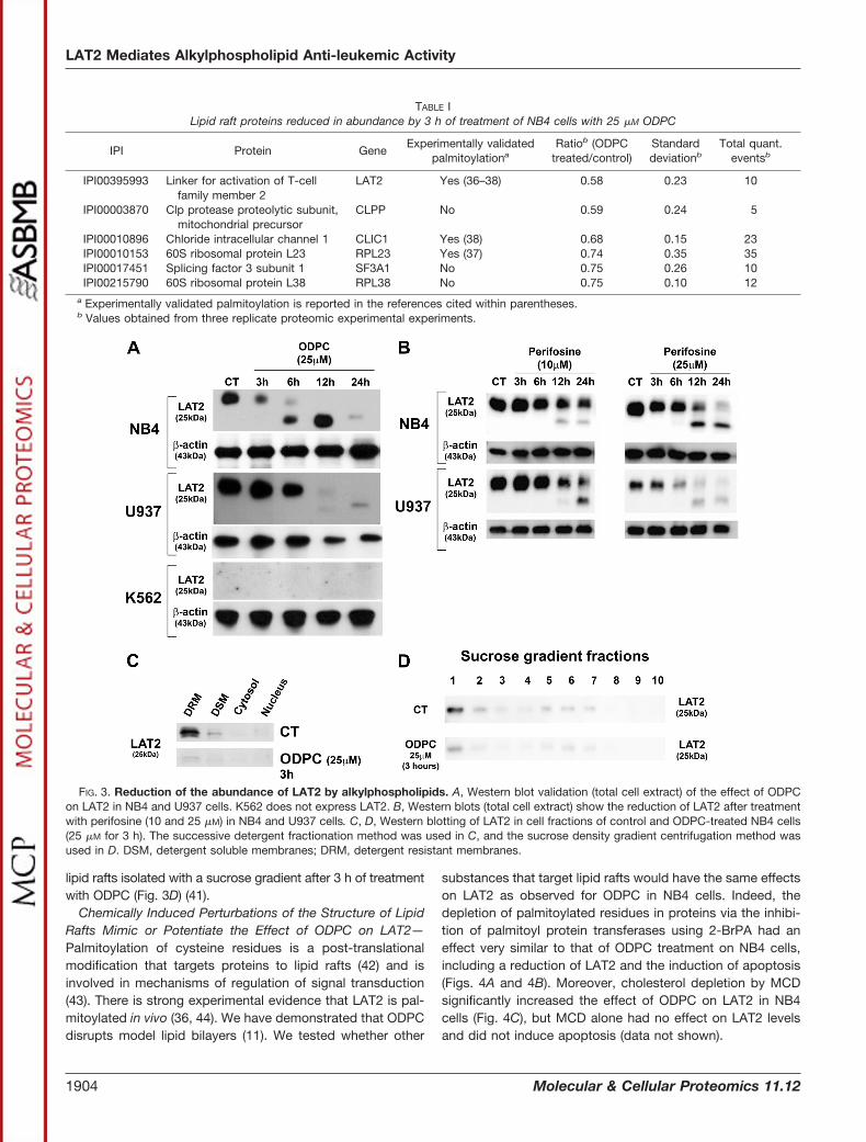
lipid rafts isolated with a sucrose gradient after 3 h of treatmentwith ODPC (Fig. 3D) (41).
Chemically Induced Perturbations of the Structure of LipidRafts Mimic or Potentiate the Effect of ODPC on LAT2—Palmitoylation of cysteine residues is a post-translationalmodification that targets proteins to lipid rafts (42) and isinvolved in mechanisms of regulation of signal transduction(43). There is strong experimental evidence that LAT2 is pal-mitoylated in vivo (36, 44). We have demonstrated that ODPCdisrupts model lipid bilayers (11). We tested whether other
substances that target lipid rafts would have the same effectson LAT2 as observed for ODPC in NB4 cells. Indeed, thedepletion of palmitoylated residues in proteins via the inhibi-tion of palmitoyl protein transferases using 2-BrPA had aneffect very similar to that of ODPC treatment on NB4 cells,including a reduction of LAT2 and the induction of apoptosis(Figs. 4A and 4B). Moreover, cholesterol depletion by MCDsignificantly increased the effect of ODPC on LAT2 in NB4cells (Fig. 4C), but MCD alone had no effect on LAT2 levelsand did not induce apoptosis (data not shown).
TABLE ILipid raft proteins reduced in abundance by 3 h of treatment of NB4 cells with 25 �M ODPC
IPI Protein GeneExperimentally validated
palmitoylationaRatiob (ODPC
treated/control)Standarddeviationb
Total quant.eventsb
IPI00395993 Linker for activation of T-cellfamily member 2
LAT2 Yes (36–38) 0.58 0.23 10
IPI00003870 Clp protease proteolytic subunit,mitochondrial precursor
CLPP No 0.59 0.24 5
IPI00010896 Chloride intracellular channel 1 CLIC1 Yes (38) 0.68 0.15 23IPI00010153 60S ribosomal protein L23 RPL23 Yes (37) 0.74 0.35 35IPI00017451 Splicing factor 3 subunit 1 SF3A1 No 0.75 0.26 10IPI00215790 60S ribosomal protein L38 RPL38 No 0.75 0.10 12
a Experimentally validated palmitoylation is reported in the references cited within parentheses.b Values obtained from three replicate proteomic experimental experiments.
FIG. 3. Reduction of the abundance of LAT2 by alkylphospholipids. A, Western blot validation (total cell extract) of the effect of ODPCon LAT2 in NB4 and U937 cells. K562 does not express LAT2. B, Western blots (total cell extract) show the reduction of LAT2 after treatmentwith perifosine (10 and 25 �M) in NB4 and U937 cells. C, D, Western blotting of LAT2 in cell fractions of control and ODPC-treated NB4 cells(25 �M for 3 h). The successive detergent fractionation method was used in C, and the sucrose density gradient centrifugation method wasused in D. DSM, detergent soluble membranes; DRM, detergent resistant membranes.
LAT2 Mediates Alkylphospholipid Anti-leukemic Activity
1904 Molecular & Cellular Proteomics 11.12

LAT2 Is Degraded by Proteasomes Following Treatmentwith APLs—The regulation of LAT2 through degradation byproteasomes has been reported (36). To determine whetherproteasome-mediated degradation follows LAT2 displace-ment from lipid rafts by ODPC treatment, we exposed NB4cells to MG132, a potent and specific cell-permeable protea-some inhibitor, followed by treatment with 25 �M ODPC. LAT2was accumulated in ODPC-treated cells in the presence of theproteasome inhibitor MG132, suggesting that ODPC treat-ment makes LAT2 susceptible to proteasomal degradation(Fig. 5A). To further evaluate the role of the proteasome inLAT2 degradation following APL exposure, we used a secondproteasome inhibitor (MLN9708) on ODPC- or perifosine-treated cells. In these experiments, the presence of MLN9708caused an accumulation of LAT2 after ODPC (Fig. 5B) orperifosine (Fig. 5C) treatment, confirming that LAT2 is de-graded by proteasomes after treatment with APL.
Reduction of the Abundance of LAT2 by ODPC Is Associ-ated with Inhibition of the AKT Signaling Pathway—There isevidence that LAT2 functions as an adaptor molecule thatpositively regulates mast cell survival by inhibiting the recruit-ment of protein phosphatases to lipid rafts, leading to AKThyperphosphorylation and activation (45). In addition, perifos-ine has been shown to be an inhibitor of AKT signaling (46).On the basis of these considerations, we determined whetherODPC treatment had an effect on the AKT signaling pathway.Treatment with 25 �M ODPC reduced AKT and phospho-AKTlevels after 3 h of treatment (Fig. 6A). There was no effect onPTEN, the main negative regulator of the AKT pathway, ex-cept at 24 h of treatment, and this effect occurred after theonset of apoptosis. Thus there was no evidence for the re-duction of PTEN at earlier times. We also observed that the
C-terminal region of PTEN was dephosphorylated after 3 h inthe presence of ODPC (Fig. 6A). C-terminal dephosphory-lation has been considered as an indicator of PTEN phospha-tase activity (47) that negatively regulates the AKT pathway.
Given prior evidence linking LAT2 to signaling in myeloidcells (15, 48), we next determined whether ODPC could impairAKT activation and alter LAT2 levels as a result of treatmentwith myeloid growth factors (MGFs). In order to maintain a
FIG. 4. Chemically induced perturbations of lipid raft structure mimic or potentiate the effect of ODPC on LAT2. Inhibition of proteinpalmitoylation by 100 �M 2-bromopalmitic acid (2-BrPA) has an effect on LAT2 abundance (A) and apoptosis induction (B) that is very similarto that of 25 �M ODPC treatment. NB4 cells were exposed to 2-BrPA. LAT2 abundance was evaluated via Western blot, and apoptosisinduction via annexin-V and propidium iodide staining by flow cytometry at the indicated times. C, cholesterol depletion by methyl-�-cyclodextrin (MCD) increases the effect of ODPC on LAT2 abundance; however, MCD has a very limited effect when used alone. NB4 cells wereexposed to MCD (2.5 mg/ml) in serum-free medium for 30 min, and subsequently MCD was washed out and complete medium was restored.LAT2 abundance was evaluated via Western blot at the indicated times.
FIG. 5. LAT2 is degraded by proteasomes after treatment withalkylphospholipids. A, 3-h treatment of NB4 cells before exposure tothe proteasome inhibitor MG132 (10 �M) prevented the reduction ofLAT2 induced by 25 �M ODPC. B, C, a similar effect was observedafter exposure (30 min) of NB4 cells to the proteasome inhibitorMLN9708 (5 �M) followed by treatment with 25 �M ODPC (B) or 25 �M
perifosine (C).
LAT2 Mediates Alkylphospholipid Anti-leukemic Activity
Molecular & Cellular Proteomics 11.12 1905

state of hypophosphorylation of AKT, NB4 cells were main-tained in serum-free medium overnight and were subse-quently treated with 25 �M ODPC or its vehicle control (PBS)for 15 min. The functionality of the pathway was tested by theaddition of a growth factor mixture containing 10 ng/ml eachof GM-CSF, IL-3, FLT3-L, and SCF. Analysis of AKT phos-phorylation at Ser 473, a cancer cell survival signal (49),showed that ODPC inhibited AKT activation (phosphorylationat Ser 473) in the first minutes of the test (Fig. 6B) and to thesame extent as treatment with Wortmannin, a classical irre-versible inhibitor of the upstream kinase PI3K (50). The ab-sence of AKT activation in ODPC- and Wortmannin-treatedcells was supported by hypophosphorylation of one of itsdownstream targets, S6-ribosomal protein (Fig. 6B). Thephosphorylated state of S6-ribosomal protein is essential forproper translation of proteins by the ribosome (51). In ourexperiments, LAT2 was rapidly synthesized after MGF addi-tion to control-treated cells but suppressed in ODPC- andWortmannin-treated cells, suggesting that LAT2 mRNA trans-lation is a highly regulated process that depends on the AKTpathway.
The reduction of the abundance of LAT2 and the inhibitionof AKT signaling observed upon treatment with ODPC (Figs.3A, 3B, and 6B) or perifosine (52) suggest that LAT2 contrib-utes to the cytotoxic effect of these drugs. To further test thishypothesis, we created a LAT2 knockdown cell line derivedfrom NB4 by the stable expression of shRNA that targets
LAT2 expression. Fig. 6C shows a representative Westernblot that confirms the reduction of LAT2 protein by more than70% in the NB4 LAT2KD.
The Knockdown of LAT2 in NB4 Cells Impairs AKT Activa-tion by MGF—To further determine whether LAT2 is essen-tial for AKT activation, we compared the response of NB4WT with NB4 LAT2KD to AKT activation triggered by MGF.We observed an absence of AKT phosphorylation at Set 473in NB4 LAT2KD after the MGF stimulus (Fig. 6D), supportingthe importance of this adaptor protein for AKT activation(45).
The Knockdown of LAT2 in the NB4 Cell Line Results in aReduction of Cell Proliferation—We were able to maintainNB4 cells as viable despite the LAT2 knockdown. However,NB4 LAT2KD presented a lower rate of cell proliferation asdetermined by a cell population doubling assay. The meanand 95% CI for the GR, reported as the number of cellduplications per day, were 0.776 (0.767–0.786) for NB4 WTand 0.655 (0.641–0.669) for NB4 LAT2KD. The DT waslower in the parental NB4 cell line (NB4 WT) than in NB4LAT2KD (Fig. 7A). This result demonstrates that the knock-down of LAT2 reduces the proliferation potential of NB4cells, probably by inducing AKT suppression (53, 54).
The Knockdown of LAT2 in NB4 Cells Results in IncreasedSensitivity to the Action of APLs and ATO—Because AKT is aconserved pro-survival pathway (55), we hypothesized thatthe down-regulation of LAT2 and consequent AKT pathway
FIG. 6. Reduction of the abundance of LAT2 by ODPC or LAT2 knockdown by shRNA (shRNA LAT2) is associated with lack ofactivation of the AKT pathway. A, ODPC (25 �M) reduced AKT and phospho-AKT levels after 3 h of treatment; however, PTEN wasreduced only at 24 h, when apoptosis was fully installed. ODPC treatment induced a state of hypophosphorylation at the C terminus ofPTEN, which has been reported to be required in order to initiate phosphatase activity (41). B, ODPC inhibited AKT activation by myeloidgrowth factors (MGFs) in a manner similar to Wortmannin, a specific PI3K inhibitor. ODPC inhibited the up-regulation of LAT2 induced byMGFs under the same conditions and inhibited ribosomal S6P phosphorylation induced by MGFs. NB4 cells were maintained serum-free(SF) overnight, treated with PBS as control with 25 �M ODPC or 1 �M Wortmannin for 15 min, stimulated with an MGF mixture (10 ng/mlIL-3, GM-CSF, L-FLT-3, and SCF), and harvested at the indicated times. C, LAT2 stable knockdown was obtained from NB4 cells vialentiviral transduction of shRNA that targets LAT2. D, AKT activation was suppressed in LAT2 stable knockdown NB4 cells. Both cell lineswere maintained SF overnight and subsequently stimulated with an MGF mixture (same as described above) and harvested at the indicatedtimes.
LAT2 Mediates Alkylphospholipid Anti-leukemic Activity
1906 Molecular & Cellular Proteomics 11.12

impairment might induce a state of increased sensitivity todrug action, irrespective of the mechanism of action of theagent. We tested this hypothesis by comparing NB4 WT andNB4 LAT2KD cell lines with respect to sensitivity to ATO.
Additionally, we compared the sensitivity of the cell lines tothe APLs ODPC and perifosine. Figs. 7B–7D show dose-response curves for the three agents, based on the dose-effect equation described by Chou and Talalay (32), and
FIG. 7. LAT2 knockdown NB4 cells (NB4 LAT2KD) have a lower proliferation potential and a higher sensitivity to APLs or ATO. A, NB4LAT2KD cells have a lower proliferation potential than the parental NB4 cells, as determined by the doubling time, which is higher in NB4 LAT2KD.B–D, NB4 LAT2KD showed greater sensitivity to apoptosis induction by ATO (B), ODPC (C), and perifosine (D). Values in parentheses indicate the95% CI.
LAT2 Mediates Alkylphospholipid Anti-leukemic Activity
Molecular & Cellular Proteomics 11.12 1907

show that NB4 LAT2KD presented greater sensitivity to allthree treatments. Values of ED50 and the respective in-creased sensitivity to the treatment are presented in TableII. Note that the sensitivity of NB4 LAT2KD cells to thesedrugs is 3- to 8-fold greater than that of the NB4 WTcells.
DISCUSSION
ODPC induces apoptosis in the AML cells NB4 and U937,but K562 leukemia cells are relatively more resistant. Normalcells are also resistant to ODPC at the same concentrationsthat initiate apoptosis in leukemia cells (11). We have demon-strated here via a quantitative proteomic analysis that somelipid raft proteins are reduced in abundance in the DRMfraction by ODPC treatment. This result illustrates the impor-tance of the use of subcellular fractionation to enrich andidentify raft proteins. For example, we identified 621 proteinsin the DRM fraction that were not demonstrable in the totalcell extract from which DRM was prepared. The simplestexplanation for this is that the preparation of the DRM fractionenriched the proteins sufficiently for them to be detected, butthey were too dilute to be detected in the total cell extract.Quantitative mass spectrometric analysis of the DRM fractionindicated that LAT2 and five other proteins are potential tar-gets of ODPC in leukemia cells. In addition, it is important toemphasize the efficacy of the method recently described byAdam et al. (25) for obtaining the DRM fraction enriched withlipid raft proteins.
After the proteomic discovery replication experiments,LAT2 was selected for extensive study based on its significantregulation and its specificity for hematopoietic tissues, asshown in supplemental Fig. S1. LAT2 is a 25- to 30-kDatransmembrane adaptor protein associated with lipid rafts. Ithas been experimentally demonstrated that the location ofLAT2 in the lipid raft is due to a double Cys-palmitoylation ata C25-V-R-C28 site (36, 44). In myeloid cells and B-cells,extracellular signals bind to transmembrane receptors, whichtrigger LAT2 phosphorylation that recruits signaling moleculessuch as Grb2, Gab2, and GADS into receptor-signaling com-plexes (15, 48). LAT2 function has been studied in detail inmast cells, in which this protein functions as a negative reg-ulator of LAT-induced degranulation signals (56). LAT was the
first protein of this family to be described (57). However, therole of LAT2 in the survival signaling of cancer cells has notbeen reported previously. Here, we demonstrated the role ofLAT2 in the early events that induce apoptosis in leukemiacells by means of functional experiments. We have previouslypresented preliminary evidence that ODPC induces a state ofdisorganization in the membranes of model systems. Ourexperiments with mimetic membrane models indicated thatODPC has a primary action on the lipid bilayer, and theseeffects were more pronounced in liposomes containing cho-lesterol, a model of lipid raft-enriched membranes (11). Sim-ilarly, 2-BrPA and MCD were used as tools to induce similarperturbations of lipid rafts in order to characterize the behav-ior of LAT2. We showed that 2-BrPA, an inhibitor of palmitoyltransferase, caused an effect similar to that of ODPC byreducing the abundance of LAT2 and triggering apoptosis.MCD, which disrupts lipid rafts by scavenging cholesterol,potentiated the effect of ODPC on LAT2 abundance. Theseresults emphasize the importance of the post-translationalmodification palmitoylation for stabilizing adaptor proteins inlipid rafts, as previously reported (58, 59). Our data suggestthat the main effect of ODPC on membranes appears to beinterference with the interaction of lipid rafts and palmitoy-lated proteins, which leads to an inefficient scaffold for main-taining signal transduction proteins.
We demonstrated that LAT2 is degraded by proteasomesafter experimentally induced perturbations in lipid raft struc-ture. Proteasome regulation of protein stability is expected inthe case of a highly regulated protein involved in cell signaling.Indeed, Brdicka et al. (36) have reported that B-cell receptoractivation in a B-cell line induces LAT2 phosphorylation andubiquitination and that the E3-ubiquitin ligase c-Cbl can in-teract with phosphorylated LAT2 after stimulation of THP-1cells. Moreover, we consistently detected the appearance of alower apparent molecular weight band of LAT2 in ODPC- and2-BrPA-treated cells before the complete degradation ofLAT2. This same band also increased in the presence ofMG132 or MLN9708, two proteasome inhibitors. This sug-gests that LAT2 might be first cleaved by a non-proteasomemechanism that generates a protein with a mass almost 3KDa lower. Whether this cleaved product has lost its N-ter-minal portion, including its palmitoylation sites, or repre-
TABLE IIEffective dose 50% (ED50) of ATO, ODPC, or perifosine for the inhibition of the proliferation of NB4 wild type (NB4 WT) and LAT2 knockdown
NB4 (NB4 LAT2KD) cells
NB4 WT NB4 LAT2KD Relative drugsusceptibilitya
ED50 (�M) 95% CIb r ED50 (�M) 95% CIb r NB4 WT/NB4LAT2KD
ATO 8.76 5.36 to 14.34 0.94 1.03 0.92 to 1.15 0.98 8.50ODPC 23.49 19.79 to 27.88 0.95 3.12 1.83 to 5.28 0.92 7.53Perifosine 18.67 16.48 to 21.17 0.97 6.10 4.45 to 8.37 0.91 3.06
a Ratio calculated by averaging ED50.b 95% CI is the 95% confidence interval for ED50, expressed in �M.
LAT2 Mediates Alkylphospholipid Anti-leukemic Activity
1908 Molecular & Cellular Proteomics 11.12

sents another cleaved product was not determined. How-ever, the complete loss of LAT2 in the DRM fraction orlighter membrane fraction from the sucrose gradient afteronly 3 h of exposure to ODPC (Figs. 3C and 3D) suggeststhat the most probable peptide lost in this LAT2 processingstep is the N-terminal portion that is essential for lipid raftassociation.
Although we have confirmed the involvement of LAT2 andother proteins of the AKT pathway in the process of apoptosistriggered by ODPC, we cannot conclude that LAT2 is the onlytarget of ODPC or perifosine, because APLs have a broadrange of actions on diverse cancer subtypes (60) and LAT2 isan adaptor protein expressed specifically in hematopoieticcells (15, 48). However, we demonstrated that the down-regulation of LAT2 by shRNA in NB4 cells mimics the action ofODPC, impairing AKT activation by MGFs (Fig. 6D). BecauseAKT is a well-described pro-survival pathway in cancer cells(55), we hypothesized that LAT2 down-regulation and conse-quent AKT impairment might induce cell death or cause re-duction in cell proliferation. We could maintain NB4 cells asviable even after a stable reduction of more than 70% of LAT2protein levels. In fact, NB4 LAT2 knockdown cells exhibited alower level of cell proliferation (Fig. 7A) and an increasedsensitivity to APLs and ATO (Figs. 7B–7D). This finding high-lights LAT2 and possible other adaptor proteins as potentialcancer therapeutic targets.
Most studies of the mechanisms of action of APLs, espe-cially those using edelfosine, have focused on altered phos-phatidylcholine biosynthesis as the major mechanism of ac-tion of the drug (61, 62). In the case of perifosine, the mainfocus was on the consequences of AKT signaling inhibition(63–65). However, our results indicate that cell signaling dis-ruption by APLs is probably due to a primary action on theorganization of lipids in rafts and that protein function isconsequently altered (Fig. 8). ODPC inhibited AKT activationin acute leukemia cells by MGFs after a few minutes of incu-bation in a way similar to that of the specific PI3K inhibitorWortmannin. However, there is no evidence that APLs func-tion as direct kinase inhibitors, although there is evidence thatproper lipid raft assembly is essential for the highly constitu-tive AKT activity of cancer cells (66). Importantly, we demon-strated that a lack of LAT2 impairs AKT activation by MGFs inleukemia cells. This indicates that some adaptor proteinsmight be essential for signal transduction in cancer cells andsuggests that ODPC and probably other APLs have theirprimary action on cell signaling pathways by inhibiting raftassembly. A proposed model of the mechanism of action ofAPLs is shown in Fig. 8.
Supplementing serum-free culture medium with MGF in-duced a rapid up-regulation of the expression of LAT2 invehicle control-treated NB4 cells. This effect was partiallyreduced by ODPC treatment and was suppressed by Wort-mannin. This rapid up-regulation of LAT2 after exposure toMGF and its reduction by a PI3K inhibitor suggest that LAT2
mRNA translation depends on a functional AKT pathway.Indeed, phosphorylation of 4EBP-1 and ribosomal S6 kinase(RS6K) and up-regulation of cap-dependent translation in theribosome is downstream of PI3K/AKT/mTOR (51, 67, 68). Tosupport this hypothesis, we showed that S6RP, the maintarget of RS6K, was hyperphosphorylated by the addition ofMGF to control cells but was hypophosphorylated in thepresence of ODPC or Wortmannin. Additionally, the up-regu-lation of LAT2 induced by MGF was blocked by two classicalinhibitors of translation (cycloheximide and rapamycin) butnot by a classical transcription inhibitor, actinomycin D (datanot shown). These results suggest that ODPC might have adirect or indirect inhibitory effect on the translation apparatus.Interestingly, we have shown here that the ribosomal proteins(RP) L23 and L38 are reduced in abundance in the lipid raftfraction of ODPC-treated cells. This finding confirms a previ-ous report stating that a raft disruption treatment with MCDcauses down-regulation of several RP (69). Moreover, Yang etal. (38), using a specific chemical approach to evaluate pal-mitoylated proteins, have reported that palmitoylation mighttarget ribosome proteins to lipid rafts. They have demon-strated via immunofluorescence that the ribosomal proteinL10A partially co-localizes with lipid rafts and that 2-BrPAtreatment reverses this association. Whether this representsonly a mechanism that mediates the association betweenRP and kinases/phosphatases of lipid rafts, in order toregulate the function of RP before the final ribosomal as-sembly, or the physical association between ribosomes andlipid rafts is not known. This inhibition of translation byODPC might be caused by the inhibition of AKT/mTORactivation or by direct down-regulation of RP in lipid rafts, orby both mechanisms. This might be associated with thespecificity of ODPC for malignant cells, as previous reportshave linked an altered translational control to the malignantphenotype (70, 71).
The induction of apoptosis by APLs has been reported tobe predominantly related to Fas/CD95 death receptor activa-tion in lipid rafts with secondary activation of the extrinsiccaspase pathway (72–74). In the present study we showedthat both intrinsic and extrinsic caspase pathways are acti-vated after ODPC treatment. This result is consistent with thereport of Chiarini et al. (75) that the proapoptotic action ofperifosine is only partially dependent on Fas/CD95 and is alsomediated by the mitochondrial intrinsic pathway.
Our results are relevant to the mechanisms of action ofAPLs. We emphasize the importance of palmitoylation forcell signaling in lipid rafts and provide evidence that theAPLs, ODPC and perifosine, create a state of disorganiza-tion of these microdomains that has a profound impact oncell function. Moreover, lipid raft resident adaptor proteinsthat function in signal transduction, here exemplified byLAT2, emerge as potential therapeutic targets in humancancer.
LAT2 Mediates Alkylphospholipid Anti-leukemic Activity
Molecular & Cellular Proteomics 11.12 1909

FIG. 8. Proposed mechanism of alkylphospholipid (ODPC)-induced raft-based inhibition of cell signaling. A, lipid rafts are membranedomains enriched in shingolipids (green) and cholesterol (blue) that serve as a scaffold for signaling through growth factor receptors (purple).The correct assembly of lipid rafts containing adaptor proteins such as LAT2 and Grb2 is essential for proper signaling of the AKT survivalpathway (shown in A). Alkylphospholipids (ODPC) disrupt the assembly of lipid rafts, displacing raft-associated adaptor proteins. As a result,signal transduction is inhibited (shown in B). ODPC, 10-(octyloxy) decyl-2-(trimethylammonium) ethyl phosphate; PI3K, phosphatidylinositol3-kinases; Grb2, growth factor receptor-bound protein 2; LAT2, linker for activation of T-cell family member 2; PTEN, phosphatase and tensinhomolog; PDK1, phosphoinositide-dependent protein kinase 1); AKT, v-AKT murine thymoma viral oncogene homolog 1; PIP2, phosphatidyl-inositol (3,4)-bisphosphate; PIP3, phosphatidylinositol (3,4,5)-trisphosphate.
LAT2 Mediates Alkylphospholipid Anti-leukemic Activity
1910 Molecular & Cellular Proteomics 11.12

Acknowledgments—We thank Prof. Dr. Stefano Servi, Diparti-mento di Chimica, Materiali, Ingegneria Chimica “Giullio Natta,” Po-litecnico di Milano, for providing a sample of the original compoundthat was used as a standard for ODPC synthesis. We are grateful toDr. Elettra Greene for assistance in revising the manuscript.
* This research was supported by FAPESP, FINEP, and CNPq.C.H.T., G.A.S., and P.S.S. received fellowships from FAPESP Proc.No. 07/58649-1, 2011/07387-2, and 2011/09718-6, respectively.G.A.F. received a fellowship from CAPES.
□S This article contains supplemental material.¶ These authors contributed equally to this work.b To whom correspondence should be addressed: E-mail:
REFERENCES
1. Kosaka, T., Yamaki, E., Mogi, A., and Kuwano, H. (2011) Mechanisms ofresistance to EGFR TKIs and development of a new generation of drugsin non-small-cell lung cancer. J. Biomed. Biotechnol. 2011, 165214
2. Irwin, M. E., Mueller, K. L., Bohin, N., Ge, Y., and Boerner, J. L. (2011) Lipidraft localization of EGFR alters the response of cancer cells to the EGFRtyrosine kinase inhibitor gefitinib. J. Cell. Physiol. 226, 2316–2328
3. Pike, L. J. (2006) Rafts defined: a report on the Keystone Symposium onLipid Rafts and Cell Function. J. Lipid Res. 47, 1597–1598
4. Lingwood, D., and Simons, K. (2010) Lipid rafts as a membrane-organizingprinciple. Science 327, 46–50
5. Seminario, M. C., and Bunnell, S. C. (2008) Signal initiation in T-cell recep-tor microclusters. Immunol. Rev. 221, 90–106
6. Gupta, N., and DeFranco, A. L. (2007) Lipid rafts and B cell signaling.Semin. Cell Dev. Biol. 18, 616–626
7. Jury, E. C., Flores-Borja, F., and Kabouridis, P. S. (2007) Lipid rafts in T cellsignalling and disease. Semin. Cell Dev. Biol. 18, 608–615
8. Jahn, K. A., Su, Y., and Braet, F. (2011) Multifaceted nature of membranemicrodomains in colorectal cancer. World J. Gastroenterol. 17, 681–690
9. Patra, S. K. (2008) Dissecting lipid raft facilitated cell signaling pathways incancer. Biochim. Biophys. Acta 1785, 182–206
10. Hitosugi, T., Sato, M., Sasaki, K., and Umezawa, Y. (2007) Lipid raft specificknockdown of SRC family kinase activity inhibits cell adhesion and cellcycle progression of breast cancer cells. Cancer Res. 67, 8139–8148
11. dos Santos, G. A., Thome, C. H., Ferreira, G. A., Yoneda, J. S., Nobre, T. M.,Daghastanli, K. R., Scheucher, P. S., Gimenes-Teixeira, H. L., Constan-tino, M. G., de Oliveira, K. T., Faca, V. M., Falcao, R. P., Greene, L. J.,Rego, E. M., and Ciancaglini, P. (2010) Interaction of 10-(octyloxy) decyl-2-(trimethylammonium) ethyl phosphate with mimetic membranes andcytotoxic effect on leukemic cells. Biochim. Biophys. Acta 1798,1714–1723
12. van der Luit, A. H., Vink, S. R., Klarenbeek, J. B., Perrissoud, D., Solary, E.,Verheij, M., and van Blitterswijk, W. J. (2007) A new class of anticanceralkylphospholipids uses lipid rafts as membrane gateways to induceapoptosis in lymphoma cells. Mol. Cancer Ther. 6, 2337–2345
13. Pinton, G., Manente, A. G., Angeli, G., Mutti, L., and Moro, L. (2012)Perifosine as a potential novel anti-cancer agent inhibits EGFR/MET-AKTaxis in malignant pleural mesothelioma. PLoS ONE 7, e36856
14. Ong, S. E., Blagoev, B., Kratchmarova, I., Kristensen, D. B., Steen, H.,Pandey, A., and Mann, M. (2002) Stable isotope labeling by amino acidsin cell culture, SILAC, as a simple and accurate approach to expressionproteomics. Mol. Cell. Proteomics 1, 376–386
15. Iwaki, S., Jensen, B. M., and Gilfillan, A. M. (2007) Ntal/Lab/Lat2. Int.J. Biochem. Cell Biol. 39, 868–873
16. Lanotte, M., Martin-Thouvenin, V., Najman, S., Balerini, P., Valensi, F., andBerger, R. (1991) NB4, a maturation inducible cell line with t(15;17)marker isolated from a human acute promyelocytic leukemia (M3). Blood77, 1080–1086
17. Strefford, J. C., Foot, N. J., Chaplin, T., Neat, M. J., Oliver, R. T., Young,B. D., and Jones, L. K. (2001) The characterisation of the lymphoma cellline U937, using comparative genomic hybridisation and multi-plex FISH.Cytogenet. Cell Genet. 94, 9–14
18. Naumann, S., Reutzel, D., Speicher, M., and Decker, H. J. (2001) Completekaryotype characterization of the K562 cell line by combined application
of G-banding, multiplex-fluorescence in situ hybridization, fluorescencein situ hybridization, and comparative genomic hybridization. Leuk. Res.25, 313–322
19. Agresta, M., D’Arrigo, P., Fasoli, E., Losi, D., Pedrocchi-Fantoni, G., Riva,S., Servi, S., and Tessaro, D. (2003) Synthesis and antiproliferative ac-tivity of alkylphosphocholines. Chem. Phys. Lipids 126, 201–210
20. Chou, T. C. (2006) Theoretical basis, experimental design, and computer-ized simulation of synergism and antagonism in drug combination stud-ies. Pharmacol. Rev. 58, 621–681
21. Resh, M. D. (2006) Use of analogs and inhibitors to study the functionalsignificance of protein palmitoylation. Methods 40, 191–197
22. Vink, S. R., Luit, A. H. v. d., Klarenbeek, J. B., Verheij, M., and Blitterswijk,W. J. v. (2007) Lipid rafts and metabolic energy differentially determineuptake of anti-cancer alkylphospholipids in lymphoma versus carcinomacells. Biochem. Pharmacol. 74, 1456–1465
23. Adam, R. M., Yang, W., Di Vizio, D., Mukhopadhyay, N. K., and Steen, H.(2008) Rapid preparation of nuclei-depleted detergent-resistant mem-brane fractions suitable for proteomics analysis. BMC Cell Biol. 9, 30
24. Pereira, S. R., Faca, V. M., Gomes, G. G., Chammas, R., Fontes, A. M.,Covas, D. T., and Greene, L. J. (2005) Changes in the proteomic profileduring differentiation and maturation of human monocyte-derived den-dritic cells stimulated with granulocyte macrophage colony stimulatingfactor/interleukin-4 and lipopolysaccharide. Proteomics 5, 1186–1198
25. Faca, V. M., Ventura, A. P., Fitzgibbon, M. P., Pereira-Faca, S. R., Pitteri,S. J., Green, A. E., Ireton, R. C., Zhang, Q., Wang, H., O’Briant, K. C.,Drescher, C. W., Schummer, M., McIntosh, M. W., Knudsen, B. S., andHanash, S. M. (2008) Proteomic analysis of ovarian cancer cells revealsdynamic processes of protein secretion and shedding of extra-cellulardomains. PLoS One 3, e2425
26. Rauch, A., Bellew, M., Eng, J., Fitzgibbon, M., Holzman, T., Hussey, P.,Igra, M., Maclean, B., Lin, C. W., Detter, A., Fang, R., Faca, V., Gafken,P., Zhang, H., Whiteaker, J., Whitaker, J., States, D., Hanash, S., Pau-lovich, A., and McIntosh, M. W. (2006) Computational Proteomics Anal-ysis System (CPAS): an extensible, open-source analytic system forevaluating and publishing proteomic data and high throughput biologicalexperiments. J. Proteome Res. 5, 112–121
27. MacLean, B., Eng, J. K., Beavis, R. C., and McIntosh, M. (2006) Generalframework for developing and evaluating database scoring algorithmsusing the TANDEM search engine. Bioinformatics 22, 2830–2832
28. Nesvizhskii, A. I., Keller, A., Kolker, E., and Aebersold, R. (2003) A statisticalmodel for identifying proteins by tandem mass spectrometry. Anal.Chem. 75, 4646–4658
29. Keller, A., Nesvizhskii, A. I., Kolker, E., and Aebersold, R. (2002) Empiricalstatistical model to estimate the accuracy of peptide identifications madeby MS/MS and database search. Anal. Chem. 74, 5383–5392
30. Faca, V., Coram, M., Phanstiel, D., Glukhova, V., Zhang, Q., Fitzgibbon, M.,McIntosh, M., and Hanash, S. (2006) Quantitative analysis of acrylamidelabeled serum proteins by LC-MS/MS. J. Proteome Res. 5, 2009–2018
31. Faca, V. M., Song, K. S., Wang, H., Zhang, Q., Krasnoselsky, A. L., New-comb, L. F., Plentz, R. R., Gurumurthy, S., Redston, M. S., Pitteri, S. J.,Pereira-Faca, S. R., Ireton, R. C., Katayama, H., Glukhova, V., Phanstiel,D., Brenner, D. E., Anderson, M. A., Misek, D., Scholler, N., Urban, N. D.,Barnett, M. J., Edelstein, C., Goodman, G. E., Thornquist, M. D., McIn-tosh, M. W., DePinho, R. A., Bardeesy, N., and Hanash, S. M. (2008) Amouse to human search for plasma proteome changes associated withpancreatic tumor development. PLoS Med. 5, e123
32. Chou, T. C., Talaly, P., Chou, T. C., and Talaly, P. (1977) A simple gener-alized equation for the analysis of multiple inhibitions of Michaelis-Men-ten kinetic systems. Journal of Biological Chemistry 252, 6438–6442
33. Suzuki, K. G., Fujiwara, T. K., Sanematsu, F., Iino, R., Edidin, M., andKusumi, A. (2007) GPI-anchored receptor clusters transiently recruit Lynand G alpha for temporary cluster immobilization and Lyn activation:single-molecule tracking study 1. J. Cell Biol. 177, 717–730
34. Appenzeller-Herzog, C., and Hauri, H. P. (2006) The ER-Golgi intermediatecompartment (ERGIC): in search of its identity and function. J. Cell Sci.119, 2173–2183
35. Burgess, R. J., and Zhang, Z. (2010) Histones, histone chaperones andnucleosome assembly. Protein Cell 1, 607–612
36. Brdicka, T., Imrich, M., Angelisova, P., Brdickova, N., Horvath, O., Spicka,J., Hilgert, I., Luskova, P., Draber, P., Novak, P., Engels, N., Wienands, J.,Simeoni, L., Osterreicher, J., Aguado, E., Malissen, M., Schraven, B., and
LAT2 Mediates Alkylphospholipid Anti-leukemic Activity
Molecular & Cellular Proteomics 11.12 1911
Horejsí, V. (2002) Non-T cell activation linker (NTAL): a transmembraneadaptor protein involved in immunoreceptor signaling. J. Exp. Med. 196,1617–1626
37. Wilson, J. P., Raghavan, A. S., Yang, Y. Y., Charron, G., and Hang, H. C.(2011) Proteomic analysis of fatty-acylated proteins in mammalian cellswith chemical reporters reveals S-acylation of histone H3 variants. Mol.Cell. Proteomics 10, M110.001198
38. Yang, W., Di Vizio, D., Kirchner, M., Steen, H., and Freeman, M. R. (2010)Proteome scale characterization of human S-acylated proteins in lipidraft-enriched and non-raft membranes. Mol. Cell. Proteomics 9, 54–70
39. Wu, C., Orozco, C., Boyer, J., Leglise, M., Goodale, J., Batalov, S., Hodge,C. L., Haase, J., Janes, J., Huss, J. W., and Su, A. I. (2009) BioGPS: anextensible and customizable portal for querying and organizing geneannotation resources. Genome Biol. 10, R130
40. Tkaczyk, C., Horejsi, V., Iwaki, S., Draber, P., Samelson, L. E., Satterth-waite, A. B., Nahm, D. H., Metcalfe, D. D., and Gilfillan, A. M. (2004) NTALphosphorylation is a pivotal link between the signaling cascades leadingto human mast cell degranulation following Kit activation and Fc epsilonRI aggregation. Blood 104, 207–214
41. Delmas, D., Rebe, C., Lacour, S., Filomenko, R., Athias, A., Gambert, P.,Cherkaoui-Malki, M., Jannin, B., Dubrez-Daloz, L., Latruffe, N., andSolary, E. (2003) Resveratrol-induced apoptosis is associated with Fasredistribution in the rafts and the formation of a death-inducing signalingcomplex in colon cancer cells. J. Biol. Chem. 278, 41482–41490
42. Salaun, C., Greaves, J., and Chamberlain, L. H. (2010) The intracellulardynamic of protein palmitoylation. J. Cell Biol. 191, 1229–1238
43. Sorek, N., Bloch, D., and Yalovsky, S. (2009) Protein lipid modifications insignaling and subcellular targeting. Curr. Opin. Plant Biol. 12, 714–720
44. Janssen, E., Zhu, M., Zhang, W., and Koonpaew, S. (2003) LAB: a newmembrane-associated adaptor molecule in B cell activation. Nat. Immu-nol. 4, 117–123
45. Roget, K., Malissen, M., Malbec, O., Malissen, B., and Daeron, M. (2008)Non-T cell activation linker promotes mast cell survival by dampening therecruitment of SHIP1 by linker for activation of T cells. J. Immunol. 180,3689–3698
46. Elrod, H. A., Lin, Y. D., Yue, P., Wang, X., Lonial, S., Khuri, F. R., and Sun,S. Y. (2007) The alkylphospholipid perifosine induces apoptosis of hu-man lung cancer cells requiring inhibition of Akt and activation of theextrinsic apoptotic pathway. Mol. Cancer Ther. 6, 2029–2038
47. Vazquez, F., Ramaswamy, S., Nakamura, N., and Sellers, W. R. (2000)Phosphorylation of the PTEN tail regulates protein stability and function.Mol. Cell. Biol. 20, 5010–5018
48. Orr, S. J., and McVicar, D. W. (2011) LAB/NTAL/Lat2: a force to be reck-oned with in all leukocytes? J. Leukoc. Biol. 89, 11–19
49. Carracedo, A., and Pandolfi, P. P. (2008) The PTEN-PI3K pathway: offeedbacks and cross-talks. Oncogene 27, 5527–5541
50. Knight, Z. A. (2010) Small molecule inhibitors of the PI3-kinase family. Curr.Top. Microbiol. Immunol. 347, 263–278
51. Mamane, Y., Petroulakis, E., LeBacquer, O., and Sonenberg, N. (2006)mTOR, translation initiation and cancer. Oncogene 25, 6416–6422
52. Bhatt, A. P., Bhende, P. M., Sin, S.-H., Roy, D., Dittmer, D. P., Damania, B.,Bhatt, A. P., Bhende, P. M., Sin, S.-H., Roy, D., Dittmer, D. P., andDamania, B. (2010) Dual inhibition of PI3K and mTOR inhibits autocrineand paracrine proliferative loops in PI3K/Akt/mTOR-addicted lympho-mas. Blood 115, 4455–4463
53. Ramaswamy, S., Nakamura, N., Vazquez, F., Batt, D. B., Perera, S., Rob-erts, T. M., Sellers, W. R., Ramaswamy, S., Nakamura, N., Vazquez, F.,Batt, D. B., Perera, S., Roberts, T. M., and Sellers, W. R. (1999) Regu-lation of G1 progression by the PTEN tumor suppressor protein is linkedto inhibition of the phosphatidylinositol 3-kinase/Akt pathway. Proc. Natl.Acad. Sci. U.S.A. 96, 2110–2115
54. Fei, H. R., Chen, G., Wang, J. M., and Wang, F. Z. (2010) Perifosine inducescell cycle arrest and apoptosis in human hepatocellular carcinoma celllines by blockade of Akt phosphorylation. Cytotechnology 62, 449–460
55. Vivanco, I., and Sawyers, C. L. (2002) The phosphatidylinositol 3-kinase-AKT pathway in human cancer. Nat. Rev. Cancer 2, 489–501
56. Volna, P., Lebduska, P., Draberova, L., Símova, S., Heneberg, P., Boubelík,M., Bugajev, V., Malissen, B., Wilson, B. S., Horejsí, V., Malissen, M., andDraber, P. (2004) Negative regulation of mast cell signaling and functionby the adaptor LAB/NTAL. J. Exp. Med. 200, 1001–1013
57. Zhang, W., Sloan-Lancaster, J., Kitchen, J., Trible, R. P., and Samelson,
L. E. (1998) LAT: the ZAP-70 tyrosine kinase substrate that links T cellreceptor to cellular activation. Cell 92, 83–92
58. Resh, M. D. (2006) Palmitoylation of ligands, receptors, and intracellularsignaling molecules. Sci. STKE 2006, re14
59. Pechlivanis, M., and Kuhlmann, J. (2006) Hydrophobic modifications of Rasproteins by isoprenoid groups and fatty acids—more than just mem-brane anchoring. Biochim. Biophys. Acta 1764, 1914–1931
60. van Blitterswijk, W. J., and Verheij, M. (2008) Anticancer alkylphospholipids:mechanisms of action, cellular sensitivity and resistance, and clinicalprospects. Curr. Pharm. Des. 14, 2061–2074
61. Boggs, K. P., Rock, C. O., and Jackowski, S. (1995) Lysophosphatidylcho-line and 1-O-octadecyl-2-O-methyl-rac-glycero-3-phosphocholine in-hibit the CDP-choline pathway of phosphatidylcholine synthesis at theCTP:phosphocholine cytidylyltransferase step. J. Biol. Chem. 270,7757–7764
62. Caskey, C. T. (1975) Identifiying inherited disease through the family his-tory. Am. Fam. Physician 11, 118–127
63. Li, Z., Tan, F., Liewehr, D. J., Steinberg, S. M., and Thiele, C. J. (2010) Invitro and in vivo inhibition of neuroblastoma tumor cell growth by AKTinhibitor perifosine. J. Natl. Cancer Inst. 102, 758–770
64. Engel, J. B., Schonhals, T., Hausler, S., Krockenberger, M., Schmidt, M.,Horn, E., Koster, F., Dietl, J., Wischhusen, J., and Honig, A. (2011)Induction of programmed cell death by inhibition of AKT with the alkyl-phosphocholine perifosine in in vitro models of platinum sensitive andresistant ovarian cancers. Arch. Gynecol. Obstet. 283, 603–610
65. Cirstea, D., Hideshima, T., Rodig, S., Santo, L., Pozzi, S., Vallet, S., Ikeda,H., Perrone, G., Gorgun, G., Patel, K., Desai, N., Sportelli, P., Kapoor, S.,Vali, S., Mukherjee, S., Munshi, N. C., Anderson, K. C., and Raje, N.(2010) Dual inhibition of akt/mammalian target of rapamycin pathway bynanoparticle albumin-bound-rapamycin and perifosine induces antitu-mor activity in multiple myeloma. Mol. Cancer Ther. 9, 963–975
66. Elhyany, S., Assa-Kunik, E., Tsory, S., Muller, T., Fedida, S., Segal, S., andFishman, D. (2004) The integrity of cholesterol-enriched microdomains isessential for the constitutive high activity of protein kinase B in tumourcells. Biochem. Soc. Trans. 32, 837–839
67. She, Q. B., Halilovic, E., Ye, Q., Zhen, W., Shirasawa, S., Sasazuki, T., Solit,D. B., and Rosen, N. (2010) 4E-BP1 is a key effector of the oncogenicactivation of the AKT and ERK signaling pathways that integrates theirfunction in tumors. Cancer Cell 18, 39–51
68. Jastrzebski, K., Hannan, K. M., Tchoubrieva, E. B., Hannan, R. D., andPearson, R. B. (2007) Coordinate regulation of ribosome biogenesis andfunction by the ribosomal protein S6 kinase, a key mediator of mTORfunction. Growth Factors 25, 209–226
69. Foster, L. J., De Hoog, C. L., and Mann, M. (2003) Unbiased quantitativeproteomics of lipid rafts reveals high specificity for signaling factors.Proc. Natl. Acad. Sci. U.S.A. 100, 5813–5818
70. Barna, M., Pusic, A., Zollo, O., Costa, M., Kondrashov, N., Rego, E., Rao,P. H., and Ruggero, D. (2008) Suppression of Myc oncogenic activity byribosomal protein haploinsufficiency. Nature 456, 971–975
71. Hsieh, A. C., Costa, M., Zollo, O., Davis, C., Feldman, M. E., Testa, J. R.,Meyuhas, O., Shokat, K. M., and Ruggero, D. (2010) Genetic dissectionof the oncogenic mTOR pathway reveals druggable addiction to trans-lational control via 4EBP-eIF4E. Cancer Cell 17, 249–261
72. Mollinedo, F., de la Iglesia-Vicente, J., Gajate, C., Estella-Hermoso deMendoza, A., Villa-Pulgarin, J. A., de Frias, M., Roue, G., Gil, J., Colomer,D., Campanero, M. A., and Blanco-Prieto, M. J. (2010) In vitro and in vivoselective antitumor activity of edelfosine against mantle cell lymphomaand chronic lymphocytic leukemia involving lipid rafts. Clin. Cancer Res.16, 2046–2054
73. Gajate, C., Gonzalez-Camacho, F., and Mollinedo, F. (2009) Involvement ofraft aggregates enriched in Fas/CD95 death-inducing signaling complex inthe antileukemic action of edelfosine in Jurkat cells. PLoS One 4, e5044
74. Gajate, C., and Mollinedo, F. (2007) Edelfosine and perifosine induce se-lective apoptosis in multiple myeloma by recruitment of death receptorsand downstream signaling molecules into lipid rafts. Blood 109, 711–719
75. Chiarini, F., Del Sole, M., Mongiorgi, S., Gaboardi, G. C., Cappellini, A.,Mantovani, I., Follo, M. Y., McCubrey, J. A., and Martelli, A. M. (2008) Thenovel Akt inhibitor, perifosine, induces caspase-dependent apoptosisand downregulates P-glycoprotein expression in multidrug-resistant hu-man T-acute leukemia cells by a JNK-dependent mechanism. Leukemia22, 1106–1116
LAT2 Mediates Alkylphospholipid Anti-leukemic Activity
1912 Molecular & Cellular Proteomics 11.12
Copyright © 2022 FDOKUMEN