A CD44v6 peptide reveals a role of CD44 in VEGFR-2 signaling and angiogenesis
Upload
independentCategory
view
4download
0
IntroductionDuring embryonic development, hematopoietic andearly endothelial cells (angioblasts) originate from acommon precursor cell known as hemangioblast.Given this common origin, several signaling path-ways are shared by both hematopoietic and vascularcells. One such pathway is the VEGFR-2 signalingpathway. VEGFR-2 (KDR, human homologue; Flk-1,murine homologue) binds to several soluble factorsincluding VEGF, which exerts proliferative andmigratory effects on endothelium. VEGFR-2 wasthought to be exclusively expressed by adult endothe-lial cells; however, it was only recently shown to bepresent on a subset of multipotent hematopoieticstem cells (1). Later studies further revealed that cer-tain leukemic cells also expressed VEGFR-2 (2). Con-sidering this new evidence, neoplastic transformationof hematopoietic stem cells into malignant leukemiccells may be associated with recapitulated expressionof hemangioblast-associated signaling tyrosinekinases such as VEGFR-2. Because VEGF is also pro-duced by leukemic cells (2–4), coinciding expression
of VEGF receptors may result in the generation of anautocrine loop that supports the proliferation andsurvival of leukemic cells.
The two primary signaling tyrosine kinase receptorsthat mediate the various biologic effects of VEGF areVEGFR-2 and VEGFR-1 (Flt-1). Although the bindingaffinity of VEGFR-1 to VEGFR is very high, with IC50
values of 10–70 pM (5), most studies have shown thatVEGFR-2 is the critical receptor for transmitting cellu-lar signals for the proliferation and differentiation ofendothelial cells (6), whereas VEGFR-1 may be moreimportant for vascular remodeling. The relative signif-icance of VEGF receptors in the regulation of vasculo-genesis and angiogenesis has been established in stud-ies in which the VEGFR-2 and VEGFR-1 genes weredisrupted in murine embryonic stem cells by homolo-gous recombination. Mice deficient in VEGFR-2 haddrastic defects in vasculogenesis, angiogenesis, andhematopoiesis (7). In contrast, VEGFR-1 knockoutmice developed abnormal vascular channels, suggest-ing a role for this receptor in the regulation of endothe-lial cell-cell or cell-matrix interactions (8). Disruption
The Journal of Clinical Investigation | August 2000 | Volume 106 | Number 4 511
Autocrine stimulation of VEGFR-2 activates humanleukemic cell growth and migration
Sergio Dias,1 Koichi Hattori,1,2 Zhenping Zhu,3 Beate Heissig,2 Margaret Choy,1
William Lane,1 Yan Wu,3 Amy Chadburn,4 Elizabeth Hyjek,4 Muhammad Gill,1
Daniel J. Hicklin,3 Larry Witte,3 M.A.S. Moore,2 and Shahin Rafii1
1Weill Medical College of Cornell University, Hematology/Oncology Division, New York, New York, USA2Sloan Kettering Cancer Institute, New York, New York, USA3ImClone Systems Inc., New York, New York, USA4Weill Medical College of Cornell University, Division of Pathology, New York, New York, USA
Address correspondence to: Shahin Rafii, Division of Hematology/Oncology, Room C-606, 1300 York Avenue, New York, New York 10021, USA. Phone: (212) 746-2070; Fax: (212) 746-8866; E-mail: [email protected].
Received for publication November 23, 1999, and accepted in revised form July 11, 2000.
Emerging data suggest that VEGF receptors are expressed by endothelial cells as well as hematopoi-etic stem cells. Therefore, we hypothesized that functional VEGF receptors may also be expressed inmalignant counterparts of hematopoietic stem cells such as leukemias. We demonstrate that cer-tain leukemias not only produce VEGF but also express functional VEGFR-2 in vivo and in vitro,resulting in the generation of an autocrine loop that may support leukemic cell survival and pro-liferation. Approximately 50% of freshly isolated leukemias expressed mRNA and protein forVEGFR-2. VEGF165 induced phosphorylation of VEGFR-2 and increased proliferation of leukemiccells, demonstrating these receptors were functional. VEGF165 also induced the expression of MMP-9 by leukemic cells and promoted their migration through reconstituted basement membrane. Theneutralizing mAb IMC-1C11, specific to human VEGFR-2, inhibited leukemic cell survival in vitroand blocked VEGF165-mediated proliferation of leukemic cells and VEGF-induced leukemic cellmigration. Xenotransplantation of primary leukemias and leukemic cell lines into immunocom-promised nonobese diabetic mice resulted in significant elevation of human, but not murine, VEGFin plasma and death of inoculated mice within 3 weeks. Injection of IMC-1C11 inhibited prolifera-tion of xenotransplanted human leukemias and significantly increased the survival of inoculatedmice. Interruption of signaling by VEGFRs, particularly VEGFR-2, may provide a novel strategy forinhibiting leukemic cell proliferation.
J. Clin. Invest. 106:511–521 (2000).
of VEGFR-2 signaling resulted in inhibition of tumorgrowth and tumor metastasis. In fact, neutralizingmAb to murine VEGFR-2 inhibited tumor growth andmetastasis in murine models (9, 10). Furthermore,glioblastoma growth was inhibited in mice dominant-negative for VEGFR-2 (11).
Leukemias originate from hematopoietic stem cells atdifferent stages of their maturation and differentiation.It is now well established that acute leukemias originatefrom immature hematopoietic stem cells that have thecapacity to undergo self-renewal, whereas less aggressiveleukemias such as chronic leukemias seem to originatefrom the more mature committed hematopoietic pro-genitor cells. Several studies have shown that VEGF isalmost invariably expressed by all established leukemiccell lines, including the well-studied HL-60 leukemiccell line (2, 3), as well as freshly isolated humanleukemias. Using RT-PCR, several studies have alsoshown that VEGFR-2 and VEGFR-1 are expressed bycertain human leukemias (2, 3). However, none of thesestudies have shown whether expression of these recep-tors is associated with any functional response.
In this report, we demonstrate that VEGF receptorsexpressed on leukemic cells are functional and conveysignals similar to those on endothelial cells such asincreasing proliferation, MMP activation, and trans-basement membrane migration. Moreover, using aneutralizing mAb specific for human VEGFR-2, wedemonstrate that inhibition of VEGFR-2 either in vitroor in vivo inhibits proliferation of VEGFR-2+ humanleukemias. Therefore, blocking VEGFR-2 signaling mayrepresent a novel therapeutic approach for the treat-ment of a subset of acute leukemias.
MethodsAll chemicals and reagents were obtained from SigmaChemical Co. (St. Louis, Missouri, USA) unless stat-ed otherwise.
Monoclonal neutralizing antibodies to VEGF receptors. Weused an immunoneutralizing mAb to KDR/VEGFR-2, IMC-1C11 (ImClone Systems Inc.), to blockVEGF/VEGFR-2 interactions in vitro and in vivo. Aneutralizing mAb to VEGFR-1/Flt-1 was also used invitro (clone 6.12; ImClone Systems Inc.). These anti-bodies have been previously shown to block VEGF-induced endothelial cell proliferation and receptorphosphorylation (12, 13). Importantly, IMC-1C11 hasbeen shown not to cross react with the murine homo-logue of KDR, flk-1 (14).
Collection and culture of primary leukemia samples. Periph-eral blood samples from leukemic patients (diagnosedwith acute leukemia) were collected by phlebotomy, dilut-ed 1/2 in Hanks’ buffered saline (Life Technologies Inc.,Grand Island, New York, USA), and overlaid on 5 mL ofLymphoprep (Ficoll; Accurate Chemical and ScientificCorp., Westbury, New York, USA). Each sample was spunat 2,000 g for 30 minutes, and the mononuclear cell inter-phase was collected into a fresh tube and washed twicewith Hanks’ for 5 minutes at 780 g. The resulting cell pel-
let was finally resuspended in RPMI/10% FCS and cul-tured overnight in RPMI/10% FCS, to eliminate possiblecontamination with monocytes/macrophages. These willadhere readily to the tissue culture flasks, and the cellsremaining in suspension consist mainly of leukemicblasts. These leukemic cells were subsequently transferredto another flask and, before the addition of VEGF and/orVEGFR antibodies, were serum starved for 16 hoursovernight, in serum free RPMI, as already described here.
Culture of leukemic cell lines. The three leukemic celllines used in this study, HL-60 (pro-myelomonocytic),HEL (megakaryocytic), and K562 (CML/erythroid)were cultured in RPMI with 10% FCS, penicillin (100U/mL), streptomycin (100 µg/mL), and fungizone(0.25 µg/mL). Before incubation with VEGF and/orantibodies, cells were serum starved for 16 hours inRPMI alone. For proliferation experiments, cells werecultured in six-well plates (Corning-Costar Corp., Cam-bridge, Massachusetts, USA), at a cell density of 1 × 10⁵
cells per well, in serum-free RPMI. Cells were treated(10–50 ng/mL VEGF) or untreated (RPMI alone) andwere cultured in the presence or absence of 500 ng to 1µg/mL of IMC-1C11, or an mAb to FLT-1/VEGFR-1,clone 6.12. After 24, 48, 72, and 96 hours, viable cells (asdetermined by trypan blue exclusion) were counted intriplicate using a hemocytometer. Each experimentalcondition was done in triplicate, and experiments withthe leukemic cell lines were repeated three times.
RNA extraction, cDNA synthesis, and RT-PCR. Total RNAwas extracted using TRI-reagent, following the manu-facturer’s instructions. cDNA was subsequently syn-thesized from total RNA using the Ready-to-Go Kit(Amersham Pharmacia Biotech, Piscataway, New Jersey,USA), and PCR was performed using a PCR thermalcycler (MWG Biotech, High Point, North Carolina,USA). The PCR program used to amplify VEGFR-2,VEGFR-1, and β-actin consisted of a precycle of 5 min-utes at 94°C, 45 seconds at 60°C, and 45 seconds at72°C. After this initial cycle, the reaction was contin-ued for 35 cycles of 1 minute at 94°C, 45 seconds at65°C, and 2 minutes at 72°C and concluded with 7minutes at 72°C. The primer sequences were as follows:β-actin forward primer: TCATGTTTGAGACCTTCAA; β-actin reverse primer: GTCTTTGCGGATGTCCACG (β-actin PCR product: 513 bp); VEGFR-2 forward primer:GTGACCAACATGGAGTCGTG; VEGFR-2 reverse primer:CCAGAGATTCCATGCCACTT (VEGFR-2 PCR product:660 bp); VEGFR-1 forward primer: ATTTGTGATTTTG-GCCTTGC; VEGFR-1 reverse primer: CAGGCTCAT-GAACTTGAAAGC (VEGFR-1 PCR product: 550 bp).Endothelial cell cDNA was used as positive control forall three sets of primers.
Protein extraction and Western blotting. PhosphorylatedVEGF receptors (VEGFR-1 and VEGFR-2) were detect-ed by Western blotting, after cell incubation with 20ng/mL VEGF165 (R&D Systems Inc., Minneapolis, Min-nesota, USA) for 10 minutes at 37°C. After this briefstimulation, total protein extracts from primaryleukemic cells and cell lines were obtained by lysing the
512 The Journal of Clinical Investigation | August 2000 | Volume 106 | Number 4

cells in cold RIPA buffer (50 mM Tris, 5 mM EDTA, 1%Triton X-114, 0.4% sodium cacodylate, and 150 mMNaCl), in the presence of protease inhibitors (1 mg/mLaprotinin, 10 mg/mL leupeptin, 1 mM β-glycerophos-phate, 1mM sodium orthovanadate, and 1 mM PMSF).Embryonic fibroblasts (the MRC5 cell line) were usedas negative control. After centrifugation to remove celldebris, supernatants (a total protein minimum of 500ng) were immunoprecipitated overnight at 4°C withprotein-G agarose beads and an anti-phosphotyrosineantibody (Santa Cruz Biotechnology Inc., Santa Cruz,California, USA) to precipitate phosphorylated pro-teins. These were resuspended in loading buffer andsubjected to SDS-PAGE-acrylamide gel electrophoresis(7.5% gels) under reducing conditions (in the presenceof β-mercaptoethanol). Proteins were subsequentlyblotted onto a nitrocellulose membrane following con-ventional protocols. Finally, blots were blocked in 1%BSA/PBS-1% Tween-20 for 1 hour at room temperaturefollowed by incubation with primary and secondaryantibodies. Rabbit polyclonal anti-VEGFR-2 (SantaCruz Biotechnology Inc.) and goat monoclonal anti-VEGFR-1 (R&D Systems Inc.) antibodies were used ata concentration of 1 µg/mL, and secondary anti-rabbitIgG-HRP (for VEGFR-2) or anti-goat IgG-HRP (forVEGFR-1) were used at 1:6,000. The ECL chemilumi-nescence detection system and ECL film (AmershamPharmacia Biotech) were used to visualize the presenceof proteins on the nitrocellulose blots.
Gelatinolytic zymography. Supernatants from leukemiccell lines and primary leukemia cultures were collectedafter overnight incubation in serum-free medium, withor without VEGF165, and their metalloproteinase activi-ty was measured by gelatinolytic zymography, asdescribed previously (15). Briefly, cell culture super-natants were treated with gelatin-agarose beads, to con-centrate the gelatinases, and processed through SDS-PAGE-acrylamide gels containing 1% gelatin. The gelswere subsequently incubated in 2.5% Triton X-100 for 1hour at room temperature, rinsed in distilled water, andplaced in low-salt collagenase buffer (50 mM Tris [pH7.6], 0.2 M NaCl, 5 mM CaCl2, and 0.2% vol/vol Brij-35)at 37°C for 18 hours. Bands of gelatinolytic activity werevisualized after staining the gels with 10 mL of a 0.2%Coomassie blue solution and 190 mL destain (distilledwater, methanol, and glacial acetic acid; 6:3:1) for 30minutes to 1 hour at room temperature. For each exper-iment, supernatants from 1 × 10⁶ cells were collected,and each experiment was done in triplicate. The AdobePhotoshop 4.0 software application (Adobe Systems,Mountain View, California, USA) and a Umax Astrascanner (Umax, Fremont, California, USA) were used toscan the gels and the intensity of the gelatinolytic bandswas assessed using NIH Image (version 1.58).
Migration experiments. Freshly isolated leukemic cellsand cell lines were resuspended in serum-free RPMI,and a stock of 106 cells/mL was prepared. A modifiedversion of a transwell migration technique describedpreviously (16) was used. Briefly, leukemic cell aliquots
(100 µL) were added to 8-µm–pore transwell inserts,coated with 25 µg of growth factor–depleted Matrigel(Becton Dickinson Immunocytometry Systems, SanJose, California, USA), and placed into the wells of a 24-well plate. The lower compartment contained serum-free RPMI with or without 200 ng/mL VEGF165. For thepurpose of blocking migration, each condition was pre-pared in a separate aliquot and incubated with IMC-1C11 (1 µg/condition), anti–VEGFR-1 (clone 6.12; 1µg/condition), or rhTIMP-1 (Oncogene Research Prod-ucts, Cambridge, Massachusetts, USA). The migrationwas carried out at 37°C and 5% CO2 for 14–18 hours.Migrated cells were collected from the lower compart-ment, spun down at 4650 g and counted using a hemo-cytometer. Only live cells, as determined by trypan blueexclusion, were considered in the quantification. Exper-iments were done in triplicate, and results are shown asthe number of cells migrated in response to VEGF.
Detection of KDR/VEGFR-2, FLT-1/VEGFR-1, and VEGFon ectopically implanted leukemias (chloromas) by immuno-histochemistry. Paraffin-embedded chloroma sectionswere immunohistochemically stained for VEGFR-1and VEGFR-2, following conventional protocols. Theantibodies used were mouse mAb to VEGFR-2/Flk-1(Santa Cruz Biotechnology Inc.) used at 300 ng/mL;rabbit polyclonal antibody to VEGFR-1 (R&D SystemsInc.), used at 200 ng/mL; vWF polyclonal antibody,used at 200 ng/mL; and VEGF polyclonal antibody(Zymed Laboratories Inc., South San Francisco, Cali-fornia, USA), used at 200 ng/mL. For single immuno-histochemical staining, peroxidase-labeled secondaryantibodies (against mouse and rabbit immunoglobu-lins) were used at a 1/6,000 dilution. ForVEGF/VEGFR-2 double immunostaining, two detec-tion systems were used. Incubation with anti–VEGFR-2/Flk-1 was followed by goat anti-mouse IgG (1:200)and PAP complexes. The peroxidase reaction was devel-
The Journal of Clinical Investigation | August 2000 | Volume 106 | Number 4 513
Table 1VEGF production and VEGF receptor expression of three leukemiccell lines (HL-60, HEL, and K562) and ten primary leukemia samples(samples 1–10)
Leukemic cells VEGF VEGFR-1 VEGFR-2(pg/mL) (mRNA) (mRNA)
HL-60 144 ± 35 + +HEL 94 ± 15 + +Sample 1 120 ± 16 + +Sample 2 196 ± 20 + +Sample 3 185 ± 16 + +Sample 4 192 ± 25 + +Sample 5 100 ± 20 + +K562 85 ± 28 + –Sample 6 88 ± 18 + –Sample 7 90 ± 12 – –Sample 8 93 ± 21 + –Sample 9 77 ± 14 – –Sample 10 78 ± 22 + –
VEGF production was determined by ELISA. VEGF receptor expression wasdetermined by RT-PCR. See Methods.

oped with a diaminobenzidine substrate. Immunode-tection of VEGF was demonstrated using the Vectorblue alkaline phosphatase substrate (Vector Laborato-ries Inc., Burlingame, California, USA). Sections withsingle immunohistochemical staining were counter-stained with hematoxylin and eosin. All sections wereobserved under a light microscope.
In vivo experiments with the HL-60 and HEL cell lines anda primary leukemia. Nonobese diabetic immunocom-promised (NOD-SCID) mice were used in all experi-ments. These were age and sex matched. In the begin-ning of each experiment, HL-60 (3 × 10⁶/mouse), HEL(5 × 10⁶/mouse), or primary leukemia (5 × 10⁶/mouse)cells were injected (intravenously) into 24 NOD-SCIDmice, and 4 days after injection the mice were dividedinto three groups of eight mice. One group was treatedintraperitoneally with IMC-1C11 (400 µg three times aweek), whereas the control group was injected withPBS/1% BSA (diluent control for the antibody), and athird group was treated intraperitoneally with 400 µg
of unrelated human IgG (Bayer Corp., Elkhart, Indiana,USA) for the duration of the experiment.
At days 14–15 after the start of the experiment,three mice from each group were sacrificed, their plas-ma collected into capillary tubes, and their organs col-lected and processed for histology, to determine theexistence of metastasis.
Quantification of VEGF levels in cell culture supernatantsand in mouse plasma. An ELISA kit (R&D Systems Inc.)specific for human VEGF was used, to quantify thelevels of VEGF produced by leukemic cells in vitro.Cells were cultured in serum-free conditions, and theculture supernatants collected after 24 hours Thesewere used without further dilution. Each sample wasmeasured in triplicate.
Two ELISA kits (both from R&D Systems Inc.), spe-cific for human and murine VEGF, were used to deter-mine VEGF concentrations in the plasma of leukemia-inoculated mice. Plasma samples were collected atdifferent time points after cell injection and used with-out any further dilution. Each sample was assayed intriplicate, and the measurements were done in two sep-arate experiments. Both assays have a sensitivity limitof 7.5 pg/mL and were developed according to themanufacturer’s instructions.
Quantification of leukemic cell engraftment in peripheralblood and bone marrow of NOD-SCID mice. On HL-60–and HEL–injected mice, the percentage of humanleukemic cells in the bone marrow was quantified bystaining for human specific antigens and analyzed byflow cytometry. Briefly, tibias were surgicallyremoved from mice on day 15 after the start of theexperiment, and their cellular content was flushedusing insulin syringes with 27-gauge needles (BectonDickinson Immunocytometry Systems) into serum-free medium (RPMI). Bone marrow cells were stainedfollowing conventional protocols. In the case of theprimary leukemia sample, PBMCs from inoculatedmice were analyzed for the presence of human spe-cific markers, following conventional staining pro-tocols. The following antibodies were used: mABagainst human CD15 (PE-labeled, Clone DU-HL60-3; Sigma Chemical Co.); mAb against human CD45(PE-labeled, Clone KC56; Coulter Corp., Miami,Florida, USA); mAb against human VEGFR-2 (FITC-labeled, clone 6.64; ImClone Systems Inc.); and anisotypic control, MSIgG1 (FITC/PE-labeled, clone2T8-2F5; Coulter Corp.). The percentage of humanleukemic cells in mouse bone marrow and peripher-
514 The Journal of Clinical Investigation | August 2000 | Volume 106 | Number 4
Figure 1Human chloromas express FLT-1/VEGFR-1 and KDR/VEGFR-2.Sections from human chloromas were stained by immunohisto-chemistry, as described in Methods. (a) Control IgG, showing lit-tle nonspecific staining (×400); (b) factor VIII staining, showingspecific blood vessel staining (×100); (c) KDR/VEGFR-2 staining,on leukemic cell areas (×400); (d) FLT-1/VEGFR-1 expression, alsodetected on a subset of leukemic cells (×400). (e and f) Doubleimmunohistochemical staining, showing leukemic cells stain forVEGFR-2 and VEGF (red arrows). Stromal cells stain for VEGF (yel-low arrows), whereas endothelial cells stain solely for VEGFR-2(black arrows). ×400. Insets show stain for VEGFR-2 alone or VEGFalone. These results are representative of four different samples.
Figure 2Representative RT-PCR analysisfor VEGFR-2 and VEGFR-1, onleukemic cell lines. The cell linesHL-60 and HEL express VEGFR-1and VEGFR-2, as detected by RT-PCR. K562 cells were VEGFR-2negative. β-actin was used as aninternal control.
al blood samples was determined using a CoulterElite flow cytometer (Coulter Corp.).
ResultsHuman chloromas express VEGF and VEGF receptors. Humanleukemias are not only localized to the bone marrow andperipheral circulation but may also infiltrate tissues andform solid masses referred to as chloromas.
Immunohistochemical staining of human chloromaswith antibodies specific for VEGFR-1 or VEGFR-2revealed that, besides staining the endothelial lining ofblood vessels, these receptors were also expressed by asubset of the leukemic cells (Figure 1, a and b). The stain-ing localized to the cell membrane, and positive cellsappeared scattered throughout the sections (Figure 1, cand d). Overall, there were more VEGFR-2–positive thanVEGFR-1–positive areas in the sections analyzed. Thesereceptors were primarily detected in different areas of thetumor, suggesting that the staining pattern of VEGFR-1 and VEGFR-2 may identify different cell populations.Furthermore, using a double immunostaining tech-nique, it was shown that, on the sections analyzed,VEGFR-2–positive leukemic cells also stained for VEGF(Figure 1, e and f). Importantly, stromal cells did notstain for VEGFR-2 but expressed VEGF (Figure 1e),whereas endothelium shows a reverse staining pattern,staining for VEGFR-2 but not for VEGF (Figure 1f).
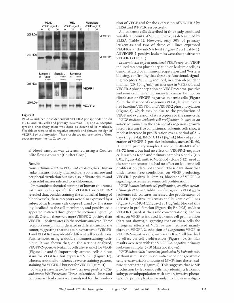
Primary leukemias and leukemic cell lines produce VEGFand express VEGF receptors. Three leukemic cell lines andten primary leukemias were analyzed for the produc-
tion of VEGF and for the expression of VEGFR-2 byELISA and RT-PCR, respectively.
All leukemic cells described in this study producedvariable amounts of VEGF in vitro, as determined byELISA (Table 1). However, only 50% of primaryleukemias and two of three cell lines expressedVEGFR-2 at the mRNA level (Figure 2 and Table 1).All VEGFR-2–positive leukemias were also positive forVEGFR-1 (Table 1).
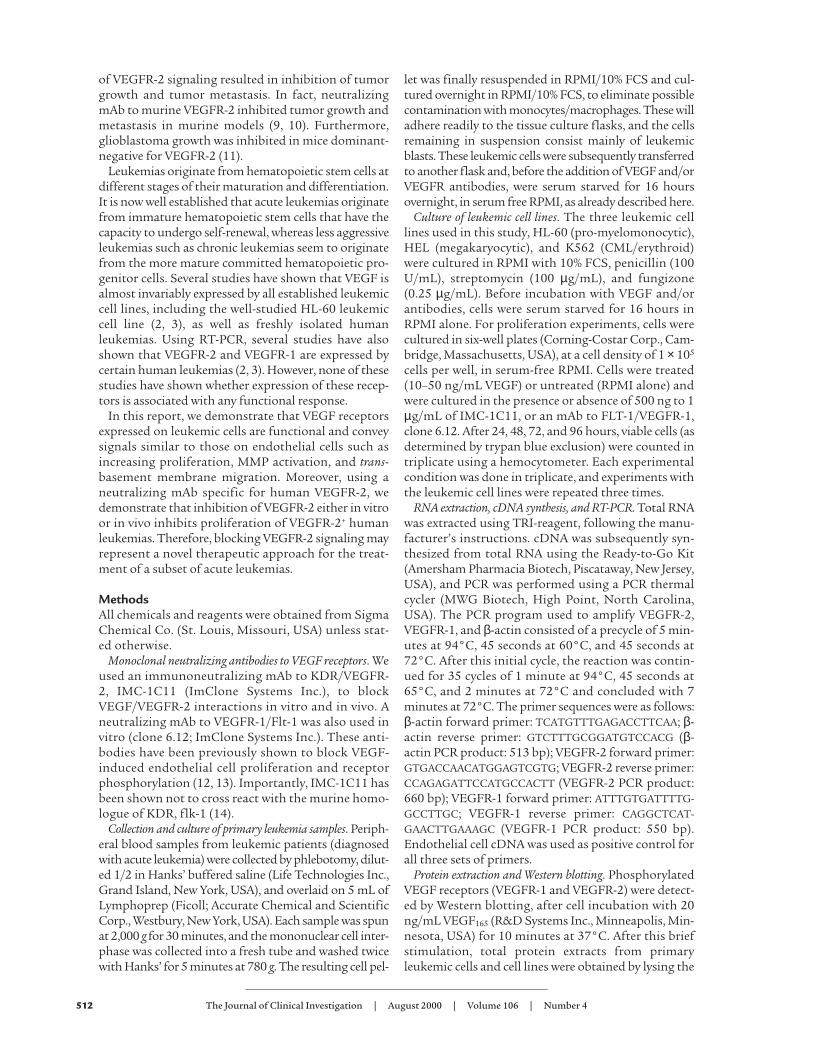
Leukemic cells express functional VEGF receptors. VEGFinduced receptor phosphorylation on leukemic cells, asdemonstrated by immunoprecipitation and Westernblotting, confirming that these are functional, signal-ing receptors. VEGF165 induced, in a dose-dependentmanner (20–50 ng/mL), an increase in VEGFR-1 andVEGFR-2 phosphorylation on VEGF receptor–positiveleukemic cell lines and primary leukemias, but not onfibroblasts or VEGFR-negative leukemic cells (Figure3). In the absence of exogenous VEGF, leukemic cellshad baseline VEGFR-1 and VEGFR-2 phosphorylation(Figure 3), which may be due to the production ofVEGF and expression of its receptors by the same cells.
VEGF mediates leukemic cell proliferation in vitro in anautocrine manner. In the absence of exogenous growthfactors (serum-free conditions), leukemic cells show amodest increase in proliferation over a period of 2–3days (Figure 4a). IMC-1C11 (1 µg/mL) blocked prolif-eration of VEGFR-2–positive leukemias, such as HL-60,HEL, and primary samples 1 and 2, by 40–60% after48–72 hours, but had no effect on VEGFR-2–negativecells such as K562 and primary samples 6 and 7 (P <0.01; Figure 4a). mAb to VEGFR-1 (clone 6.12), used atthe same concentration, had no effect on leukemic cellproliferation (data not shown). These data show thatunder serum-free conditions, on VEGF-producing,VEGFR-2–positive leukemias, blockade of VEGFR-2signaling decreases leukemic cell proliferation.
VEGF induces leukemic cell proliferation, an effect mediat-ed through VEGFR-2. Addition of exogenous VEGF165 toleukemic cell cultures increased the proliferation ofVEGFR-2–positive leukemias and leukemic cell lines(Figure 4b). IMC-1C11, used at 1 µg/mL, blocked thisincrease in proliferation (Figure 4b; P < 0.05). mAb toVEGFR-1 (used at the same concentrations) had noeffect on VEGF165-induced leukemic cell proliferation(data not shown), suggesting that on these cells themitogenic effects of VEGF165 are mediated mainlythrough VEGFR-2. Addition of exogenous VEGF toVEGFR-2–negative cells, such as the K562 cell line, hadno effect on cell proliferation (Figure 4b). Similarresults were seen with the VEGFR-2–negative primaryleukemic samples 6–10 (data not shown).
VEGF induces MMP secretion/production by leukemic cells.Without stimulation, in serum-free conditions, leukemiccells release variable amounts of MMPs into the cell cul-ture supernatant (Figure 5). This suggests that MMPproduction by leukemic cells may identify a leukemicsubtype or subpopulation with a more invasive pheno-type. On primary leukemias and/or cell lines investigat-
The Journal of Clinical Investigation | August 2000 | Volume 106 | Number 4 515
Figure 3VEGF165 induced dose-dependent VEGFR-2 phosphorylation onHL-60 and HEL cells and primary leukemias 1, 2, and 3. Receptortyrosine phosphorylation was done as described in Methods.Fibroblasts were used as negative controls and showed no sign ofVEGFR-2 phosphorylation. These results are representative of threeseparate experiments. C, control.

ed, the myelo-monocytic subtypes (shown in Figure 5:HL-60 cells, samples 2 and 3) have consistently shown ahigher level of basal MMP production and release. Incu-bation with VEGF165 increased MMP-9 release byleukemic cells (Figure 5a) over an 18-hour period. Quan-tification of VEGF165-induced increases in secretedMMP-9, by densitometry, indicated that this effect wassignificant (Figure 5b). On primary leukemias, MMP-9was the main MMP released into the culture super-natants (Figure 5). The levels of TIMP-1 produced byleukemic cells were also investigated, by Western blot-ting, but showed only a minor variation after stimula-tion with VEGF165 (data not shown).
VEGF induces leukemic cell migration through Matrigel-coated transwells, an effect mediated through VEGFR-1 andVEGFR-2. We investigated whether VEGF165 induced amore invasive phenotype on leukemic cells. This wasdemonstrated using a migration system in which tran-swell inserts were coated with a thin layer of Matrigel,a model of invasion through the basement membrane.VEGF165 induced trans-basement membrane migrationof HL-60 cells and primary leukemias (Figure 6). Thisprocess requires MMP production and activation, asthe VEGF-induced cell migration was blocked byrecombinant human TIMP-1 (Figure 6). TIMP-1 hadno effect on leukemic cell migration through bare(uncoated) transwells (data not shown).
As shown here, the mitogenic effects of VEGF165 onleukemic cells were mediated through VEGFR-2. How-ever, in the migration system used in our studies, incu-bation of HL-60 cells with IMC-1C11 (at 1 µg/mL)could only partially (40%) block VEGF165-inducedmigration through Matrigel (Figure 6). On the otherhand, an mAb to VEGFR-1, used at the same concen-tration, significantly blocked (60–70%) HL-60 cell
migration (Figure 6; P < 0.05), suggesting that VEGF165
may induce MMP activation and cell migration byinteracting with both receptors. In fact, incubation ofHL-60 cells with VEGFR-1 mAb and IMC-1C11blocked VEGF165-induced migration more effectively(70–80%) than either antibody alone (Figure 6). On pri-mary leukemias, both VEGFR-1 mAb and IMC-1C11
516 The Journal of Clinical Investigation | August 2000 | Volume 106 | Number 4
Figure 5VEGF165 induces MMP secretion by leukemic cells. (a) Zymographicanalysis of leukemic cell supernatants, with or without VEGF165 stim-ulation for 18 hours. (b) Quantification of the gelatinolytic activitydetected on the culture supernatants. Incubation of leukemic cellswith VEGF165 had a significant (P < 0.05) effect on the level of MMPproduction in vitro, as detected by zymography. The results shownare representative of three independent experiments.
Figure 4(a) Leukemic cell growth in serum-free media. Treatment of VEGFR-2–positive cells (HL-60, HEL, and primary samples 1 and 2), butnot of VEGFR-2–negative cells (K562 and samples 6 and 7), with IMC-1C11 decreases cell growth (AP < 0.005, at 48 and 72 hours).The results shown are representative of three separate experiments. (b) VEGF165 induced proliferation of a subset of leukemias, an effectmediated through KDR/VEGFR-2. VEGF165 induced a significant (AP < 0.003) increase in proliferation of all VEGFR-2–positive leukemias(two cell lines and two primary leukemias are shown). This effect was blocked by IMC-1C11 (BP < 0.04), as described in Methods. Theresults shown are representative of three separate experiments.

showed, overall, comparable migration-blocking effects(Figure 6). As shown for the HL-60 cells, incubation ofprimary leukemias with both antibodies was moreeffective than either antibody alone, blocking VEGF165-induced migration by 80% (Figure 6).
Leukemic cells release VEGF in vivo. Using NOD-SCIDmice as an in vivo model, we investigated whetherVEGFR-2–positive leukemias, HL-60, HEL, and a pri-mary leukemia, released VEGF in vivo. As shown in Fig-ure 7a, human VEGF plasma levels increased 7 and 14days after HL-60 cell injection. Importantly, murineVEGF plasma levels remained very low (at or below theassay detection level; Figure 7a) throughout the experi-ment. Similar results were obtained using the HEL cellline (data not shown) and a primary leukemia sample(Figure 7b). If left untreated, mice injected with this pri-mary leukemia had increased circulating human VEGFlevels 14 days after inoculation (Figure 7b). However,similarly to the HL-60 model, murine VEGF plasma lev-els did not increase in these mice (data not shown).
IMC-1C11 blocks leukemic growth in vivo. In the absenceof any treatment, or treated with unrelated human IgG,mice injected with 3 × 10⁶ HL-60 cells (intravenously)survived only 14–25 days (n = 8 for each treatment),highlighting the aggressive nature of these leukemiccells. However, mice treated with IMC-1C11 survivedfive times longer than untreated mice (Figure 8a; P <0.005) and, as shown in Figure 7a, had reduced humanVEGF plasma levels throughout the experiment. Simi-larly, untreated HEL-inoculated mice died within 45days, whereas those treated with IMC-1C11 survivedsignificantly longer (P < 0.05; Figure 8b).
These observations were extended to include a pri-mary leukemia. In the absence of any treatment, miceinoculated with 5 × 10⁶ primary leukemic cells diedwithin 3 weeks. However, as shown for the two leukemiccell lines, IMC-1C11 prolonged the survival of primaryleukemia-bearing mice (Figure 8c). This increase in sur-vival was accompanied by a reduction in human VEGFplasma levels (Figure 7b). Given that IMC-1C11 is spe-cific for human VEGFR-2 (KDR) and does not crossreact with murine flk-1, the data suggest that treatmentof inoculated mice with IMC-1C11 blocks leukemicgrowth by impeding leukemia-derived VEGF frominteracting with VEGFR-2 on leukemic cells.
Decreased metastasis in mice treated with IMC-1C11. Asshown in vitro, VEGF can promote an invasive pheno-type on VEGFR-2–positive leukemic cells, which maybe one mechanism by which extramedullary leukemicmasses are established. Using the in vivo modelsdescribed here, it was next investigated whetherVEGFR-2 blockade affected metastatic behavior.
In untreated HL-60 inoculated mice, histologicalanalysis of the liver and spleen revealed the presence ofmetastatic lesions in both organs (Figure 9, b and d).Liver sections had several areas occupied by leukemiccells, with signs of active cellular proliferation (Figure9b). Furthermore, the white pulp area of the spleen ofthese mice was largely replaced by leukemic cells (Fig-ure 9d). However, 14 days after start of the experiment,
The Journal of Clinical Investigation | August 2000 | Volume 106 | Number 4 517
Figure 6VEGF165 induces leukemic cell migration through Matrigel-coatedtranswells, a process mediated through VEGFR-2 and VEGFR-1.Migration of HL-60 cells and four primary leukemias throughMatrigel-coated transwells, in response to 200 ng/mL VEGF165, isshown. This process requires MMP activity, as shown by the effectsof rhTIMP-1. IMC-1C11 partially blocked leukemic cell migration,but incubation of the cells with VEGFR-1 mAb and IMC-1C11 wasmore effective than either antibody alone (AP < 0.05). These resultsare representative of three independent experiments.
Figure 7(a) HL-60 injection into NOD-SCID mice induces high levels ofhuman but not murine VEGF in mouse plasma. Mice were inject-ed with 3 × 10⁶ HL-60 cells (intravenously), and the human andmurine VEGF plasma levels were determined at different timepoints after injection. Mice treated with IMC-1C11 had reducedcirculating human VEGF levels (P < 0.01). The results shown arerepresentative of three mice per time point and of two separateexperiments. (b) Inoculation of a primary leukemia into NOD-SCID mice increased human VEGF levels in mouse plasma. Micewere injected with 5 × 10⁶ leukemic cells (intravenously) and thehuman VEGF plasma levels were quantified by ELISA, asdescribed in Methods. Mice treated with IMC-1C11 had reducedlevels of circulating human VEGF (P < 0.05). The results shownare representative of three mice per time point.

IMC-1C11–treated mice had normal liver and spleenhistology (Figure 9, a and c). In these mice, there was noevidence of metastatic lesions in the liver (Figure 9a),and the spleen appeared normal, with evidence ofapoptosis (Figure 9c). Mice inoculated with the HELcell line showed similar results. In control mice, 15 days
after the start of the experiment, there were numerousmetastatic foci in the spleen and lungs (more thanthree metastatic foci seen at 10× magnification). How-ever, mice treated with IMC-1C11 had no evidence ofmetastasis (data not shown).
Decreased circulating leukemic cells and reduced bone mar-row leukemic cell engraftment in mice treated with IMC-1C11. In HL-60–injected mice, untreated or treatedwith unrelated human IgG, 15 days after the start ofthe experiment, there were 6% human CD15-positivecells in the bone marrow, of which approximately 4%were VEGFR-2 positive (Figure 10a). However, micetreated with IMC-1C11 had tenfold less human cellsin the bone marrow (Figure 10a). Similar results wereobtained with the HEL leukemia cell line (Figure 10a).These cells, contrarily to HL-60, do not express CD15,but express VEGFR-2/KDR. As shown in Figure 10a,15 days after the start of the experiment, there weretenfold more VEGFR-2–positive HEL cells in the bonemarrow of untreated mice compared with those treat-ed with IMC-1C11 (P < 0.005).
We also quantified the number of circulatingleukemic cells in mice inoculated with a primaryleukemia. Fourteen days after the start of the experi-ment, untreated mice had 12% circulatingCD45+VEGFR-2+ leukemic cells (Figure 10b). However,those treated with IMC-1C11, at the same time point,had less than 1% circulating leukemic cells (Figure 10b).Similar results were obtained with the two cell lines HL-60 and HEL (data not shown).
DiscussionIt is now well established that most human leukemiccell lines as well as primary isolated leukemias have thecapacity to elaborate VEGF (2, 3). Several studies havealso demonstrated expression of VEGFR-2 in certainleukemias (3, 4), and one recent study correlated thelevels of VEGF production by lymphoid cells with theirability to form subcutaneous tumors in vivo (17).
In this report, we demonstrate that primaryleukemias and leukemic cell lines produce VEGF, andsubsets of human acute leukemias express functionalVEGF receptors, namely, VEGFR-2. On VEGFR-2–positive leukemias, blockade of VEGF binding to itsreceptor by a neutralizing mAb to VEGFR-2, IMC-1C11, resulted in decreased leukemic cell growth inserum-free conditions, suggesting the existence of anautocrine loop between leukemia-derived VEGF andits VEGFR-2. Moreover, as shown for endothelial cells,addition of exogenous VEGF induced VEGFR-2 phos-phorylation and proliferation of subsets of acuteleukemias primarily by signaling through VEGFR-2.VEGF also induced MMP production and promotedtrans-basement membrane migration of leukemic cellsby interacting with VEGFR-2 and VEGFR-1. Thisstudy provides the first demonstration that blockadeof VEGF/VEGFR-2 interactions decreases leukemiccell growth in serum-free (in the absence of exogenousgrowth factors) conditions. It also shows for the first
518 The Journal of Clinical Investigation | August 2000 | Volume 106 | Number 4
Figure 8(a) Mouse survival (%) after intravenous HL-60 injection. Mice wereinjected with 3 × 10⁶ cells intravenously, and 4 days after leukemiccell injection they were left untreated (control, n = 8), or treated (n =8) intraperitoneally with IMC-1C11 (400 µg/dose) or with an unre-lated human IgG (n = 8, 400 µg/dose) three times a week. The resultsshown are representative of three separate experiments. (b) Mousesurvival (%) after intravenous injection of HEL cells. Mice were inject-ed with 5 × 10⁶ cells intravenously, and 4 days after leukemic cellinjection they were treated with PBS/BSA (n = 8) or with IMC-1C11(n = 8, intraperitoneal injection, 400 µg/dose), three times a week.The results shown are representative of two separate experiments. (c)Mouse survival (%) after intravenous injection of primary humanleukemia cells. Mice were injected with 5 × 10⁶ cells intravenously,and 4 days after leukemic cell injection were treated with PBS/BSA(n = 8) or with IMC-1C11 (n = 8, intraperitoneal injection, 400µg/dose), three times a week. The results shown are representativeof two separate experiments.

time that VEGF receptors expressed by leukemic cellsare functional, signaling receptors, conveying signalssimilar to those induced on endothelial cells.
To define the role of VEGF/VEGFR-2 autocrine loopin the regulation of leukemic cell growth in vivo, wehave used a well-established xenotransplantationmodel in which human leukemic cells are engraftedinto sublethally irradiated immunodeficient (NOD-SCID) mice (18–20). We used three VEGFR-2–positiveleukemias, the HL-60 and HEL cell lines and a primaryleukemia sample, for these experiments. Injection ofleukemic cells into NOD-SCID mice resulted in theengraftment and formation of leukemic foci in thebone marrow, spleen, liver, and peripheral circulationof NOD-SCID mice. We demonstrate that leukemiccells produce human VEGF that could be detected athigh levels in the plasma of leukemia xenotransplant-ed mice, as determined by a human specific VEGFELISA. Rising levels of human VEGF were detected inplasma of the xenotransplanted mice and increasedover time as the leukemic cells proliferated in theNOD-SCID mice. In contrast, plasma levels of murineVEGF produced were insignificant and did not increaseduring in vivo proliferation of any of the cell lines orprimary sample tested.
In the absence of any intervention, xenograftedNOD-SCID mice succumb to metastatic proliferationof leukemic cells within 2–3 weeks of inoculation. How-ever, treatment of inoculated mice with IMC-1C11,inhibited proliferation of HL-60, HEL, and primaryleukemic cells in NOD-SCID mice and promoted theirlong-term survival. Histological examination of liverand spleen demonstrated that treatment of xenotrans-planted mice with IMC-1C11 effectively inhibits pro-liferation and establishment of leukemic infiltrates. Intreated mice, the number of VEGFR-2+ leukemic cellsin the bone marrow and peripheral blood was also dra-matically reduced. Furthermore, a reduction inleukemic cells and subsequent increase in mouse sur-
vival was accompanied by a decrease in circulatinghuman VEGF levels. These data strongly suggest thatendogenously produced human VEGF by leukemiccells promotes their growth, survival, and capacity tometastasize through interaction with human VEGFR-2. Collectively, these data illustrate the plasticity ofVEGFRs signaling within the vascular and hematopoi-etic lineages and their potential to support the prolif-eration certain subsets of VEGFR-2+ acute leukemias.
In humans, acute leukemias are classified based onprogenitor cell type and differentiation state of thestem cell they originated from (M1 to M7). The major-ity of leukemias are due to malignant transformationof the myeloid progenitors (M1, M2, M3, M4, M5),which are mostly localized to the bone marrow and theperipheral circulation. However, leukemic cells occa-sionally acquire the capacity to invade various tissues,setting up niches for the formation of large tumormasses referred to as chloromas. Based on our in vivodata, most of the human chloroma samples tested todate express VEGFR-2 and VEGFR-1. Given that for-mation of chloromas, similar to vessel formation,requires sequential invasion, proliferation, and estab-lishment of a neovasculature, it is not surprising thatleukemic cells with the potential to form chloromasproduce VEGF and may also express VEGFRs. In thisregard, it has been recently reported that the levels ofVEGF produced by lymphoid cells are strictly correlat-ed with their ability to form subcutaneous tumors invivo (17). For instance, HL-60 cells release VEGF,express VEGF receptors, and represent one leukemicmodel with the capacity to metastasize and to form asolid, vascularized tumor if implanted subcutaneous-ly. Therefore, leukemias destined to form chloromasmay utilize VEGFRs to upregulate the expression ofMMPs and facilitate invasion into the tissues. Expres-sion of VEGFR-2 may subsequently promote leukemiccell proliferation. In the absence of counterregulatoryfactors, the leukemias may continue to proliferate.
The Journal of Clinical Investigation | August 2000 | Volume 106 | Number 4 519
Figure 9Histology of liver (a and b) and spleen (c and d) fromHL-60–injected mice. Untreated (control) mice hadevidence of metastatic disease in the liver (white andred arrows, b) and spleen (d). In contrast, mice treat-ed with IMC-1C11 showed no evidence of leukemiccells in the liver (a), and the spleen had evidence ofapoptosis (blue arrows, c) but normal histology.

Under physiological conditions, most tissues havethe capacity to produce VEGF. Elevated levels of VEGFhave been detected in the peripheral circulation ofpatients with various malignancies (21, 22). Therefore,it is conceivable that increases in VEGF secretion intothe peripheral circulation of leukemic patients mayresult in mobilization of leukemic cells to the bonemarrow extravascular space.
Malignant transformation of leukemic cells may beassociated with the acquisition of specific tyrosinekinases that may be involved in the self-renewalprocess. Therefore, reactivation of VEGFR-2 may beone mechanism by which certain subsets of leukemiccells establish their autonomous growth. Recentdemonstration that VEGFR-2 signaling is critical forhematopoietic stem cell proliferation underscores
the significance of VEGFR-2 in leukemic cell prolif-eration. Other studies have also shown that solidtumors such as breast, ovarian, and prostate carcino-mas express VEGFR-2 mRNA (23–25). However, acti-vation of VEGFR-2 has not been shown to convey anyphysiological signal in these malignancies possiblydue to lack of downstream signaling pathways inthese cells. Because CD34+VEGFR-2+ stem cells, asresult of oncogenic transformation, may give rise toacute leukemias, it is not surprising that VEGFR-2 isfunctional on these cells.
It is now well established that growth of solid tumorsis angiogenesis dependent (26). A previous report alsosuggested that progression of leukemias may be asso-ciated with increases in bone marrow endothelial cellmass (27). This process may be mediated through therelease of VEGF, which will induce endothelial prolif-eration and stimulate the release of hematopoieticgrowth factors to support leukemic cell growth (2). Inthe present report, it is hypothesized that in liquidtumors such as leukemias, VEGF may serve two func-tions, one to enhance endothelial cell proliferation andthe production of hematopoietic growth factors andanother to promote leukemic cell growth through anautocrine loop (Figure 11). Based on our in vivo data,the growth of leukemic cells xenotransplanted intoNOD-SCID mice was inhibited by human specific neu-tralizing mAb to VEGFR-2, suggesting that anautocrine loop generated by VEGF/VEGFR-2, in addi-tion to paracrine loops, may mediate leukemic cell pro-liferation (Figure 11).
Recent studies have shown that leukemic cells arecapable of producing other forms of VEGF includingVEGF-C (28) and possibly VEGF-D. VEGF-C andVEGF-D signal through both VEGFR-2 and VEGFR-3(Flt-4). However, VEGF (VEGF-A) as well as VEGF-C
520 The Journal of Clinical Investigation | August 2000 | Volume 106 | Number 4
Figure 10(a) Leukemic cell engraftment (%) in the bone marrow of HL-60– and HEL-inoculated mice. Day 15 after the start of the experiment, bonemarrow cells from inoculated mice were stained for human CD15 and human VEGFR-2/KDR and quantified by flow cytometry. IMC-1C11–treated mice had a significantly lower percentage of human cells in the bone marrow than untreated mice (P < 0.001). These resultsare representative of three mice per cell line. (b) Quantification of primary leukemic cells in the peripheral blood of inoculated mice. PBMCsfrom primary leukemia-inoculated mice were stained for human CD45 and human VEGFR-2 and quantified by flow cytometry, as describedin Methods. Fourteen days after the start of treatment, IMC-1C11–treated mice had significantly less circulating CD45+VEGFR-2+ humanleukemic cells than untreated mice (P < 0.05). These results are representative of three mice.
Figure 11On VEGFR-2–positive leukemias, VEGF may support leukemic cellgrowth through paracrine (by increasing bone marrow endotheliumcell mass) and autocrine (supporting leukemic cell survival) mecha-nisms. Antibodies against human VEGFRs may block the autocrineloop induced by leukemia-derived human VEGF.

and VEGF-D promote the proliferation of the targetcells primarily through VEGFR-2. Therefore, inhibitionof VEGRF-2 signaling may provide an effective meansof blocking the endogenous signaling induced by vari-ous forms of VEGFs.
Using mAb’s that could differentially block murine orhuman VEGFR-2 and various forms of VEGFs may ulti-mately define the relative significance of the autocrineversus the paracrine role of VEGF in supporting thegrowth of VEGF-dependent leukemias, in particularthose that have the capacity to form chloromas. Thesestudies may lay the foundation for a novel therapeuticapproach using a combination of mAb’s to VEGF andVEGFRs for the treatment of VEGFR-2+ leukemias.
AcknowledgmentsS. Rafii is supported by National Heart, Lung, andBlood Institute (NHLBI) grants R01 HL-58707 andR01 HL-61849, an American Heart Association Grant-In-Aid, the Dorothy Rodbell Foundation for SarcomaResearch, and the Rich Foundation. M.A.S. Moore issupported by NHLBI grant R01 HL-61401 and the GarReichman Fund of the Cancer Research Institute.
1. Ziegler, B.L., et al. 1999. KDR receptor: a key marker defininghematopoietic stem cells. Science. 285:1553–1558.
2. Fiedler, W., et al. 1997. Vascular endothelial growth factor, a possibleparacrine growth factor in human acute myeloid leukemia. Blood.89:1870–1875.
3. Bellamy, W.T., Richter, L., Frutiger, Y., and Grogan, T.M. 1999. Expres-sion of vascular endothelial growth factor and its receptors inhematopoietic malignancies. Cancer Res. 59:728–733.
4. Katoh, O., Tauchi, H., Kawaishi, K., Kimura, A., and Satow, Y. 1995.Expression of the vascular endothelial growth factor (VEGF) receptorgene, KDR, in hematopoietic cells and inhibitory effect of VEGF onapoptotic cell death caused by ionizing radiation. Cancer Res.55:5687–5692.
5. Klagsbrun, M., and D’Amore, P.A. 1996. Vascular endothelial growth fac-tor and its receptors. Cytokine Growth Factor Rev. 7:259–270.
6. Ortega, N., et al. 1997. Systemic activation of the vascular endothelialgrowth factor receptor KDR/flk-1 selectively triggers endothelial cellswith an angiogenic phenotype. Am. J. Pathol. 151:1215–1224.
7. Shalaby, F., et al. 1995. Failure of blood-island formation and vasculo-genesis in Flk-1–deficient mice. Nature. 376:62–66.
8. Fong, G.H., Rossant, J., Gertsenstein, M., and Breitman, M.L. 1995. Roleof the Flt-1 receptor tyrosine kinase in regulating the assembly of vas-cular endothelium. Nature. 376:66–70.
9. Skobe, M., Rockwell, P., Goldstein, N., Vosseler, S., and Fusenig, N.E.1997. Halting angiogenesis suppresses carcinoma cell invasion. Nat. Med.
3:1222–1227.10. Prewett, M., et al. 1999. Antivascular endothelial growth factor receptor
(fetal liver kinase 1) monoclonal antibody inhibits tumor angiogenesisand growth of several mouse and human tumors. Cancer Res.59:5209–5218.
11. Millauer, B., Shawver, L.K., Plate, K.H., Risau, W., and Ullrich, A. 1994.Glioblastoma growth inhibited in vivo by a dominant-negative Flk-1mutant. Nature. 367:576–579.
12. Strawn, L.M., et al. 1996. Flk-1 as a target for tumor growth inhibition.Cancer Res. 56:3540–3545.
13. Zhu, Z., et al. 1998. Inhibition of vascular endothelial growth factor-induced receptor activation with anti-kinase insert domain-containingreceptor single-chain antibodies from a phage display library. Cancer Res.58:3209–3214.
14. Lu, D., et al. 2000. Identification of the residues in the extracellular regionof KDR important for interaction with vascular endothelial growth factorand neutralizing anti-KDR antibodies. J. Biol. Chem. 275:14321–14330.
15. Leber, T.M., and Balkwill, F.R. 1997. Zymography: a single-step stainingmethod for quantitation of proteolytic activity on substrate gels. Anal.Biochem. 249:24–28.
16. Hamada, T., et al. 1998. Transendothelial migration of megakaryocytesin response to stromal cell–derived factor 1 (SDF-1) enhances plateletformation. J. Exp. Med. 188:539–548.
17. Fusetti, L., et al. 2000. Human myeloid and lymphoid malignancies inthe non-obese diabetic/severe immunocompromised mouse model: fre-quency of apoptotic cells in solid tumors and efficiency and speed ofengraftment correlate with vascular endothelial growth factor produc-tion. Cancer Res. 60:2527–2534.
18. Clutterbuck, R.D., et al. 1998. Inhibitory effect of simvastatin on the pro-liferation of human myeloid leukaemia cells in severe combined immun-odeficient (SCID) mice. Br. J. Haematol. 102:522–527.
19. Terpstra, W., et al. 1995. Conditions for engraftment of human acutemyeloid leukemia (AML) in SCID mice. Leukemia. 9:1573–1577.
20. Machado, E.A., et al. 1984. Proliferation and differentiation of humanmyeloid leukemic cells in immunodeficient mice: electron microscopyand cytochemistry. Blood. 63:1015–1022.
21. Salven, P., Manpaa, H., Orpana, A., Alitalo, K., and Joensuu, H. 1997.Serum vascular endothelial growth factor is often elevated in dissemi-nated cancer. Clin. Cancer Res. 3:647–651.
22. Obermair, A., et al. 1998. Concentration of vascular endothelial growthfactor (VEGF) in the serum of patients with suspected ovarian cancer.Br. J. Cancer. 77:1870–1874.
23. Kranz, A., Mattfeldt, T., and Waltenberger, J. 1999. Molecular mediatorsof tumor angiogenesis: enhanced expression and activation of vascularendothelial growth factor receptor KDR in primary breast cancer. Int. J.Cancer. 84:293–298.
24. Abu-Jawdeh, G.M., et al. 1996. Strong expression of vascular permeabil-ity factor (vascular endothelial growth factor) and its receptors in ovar-ian borderline and malignant neoplasms. Lab. Invest. 74:1105–1115.
25. Ferrer, F.A., et al. 1999. Expression of vascular endothelial growth factorreceptors in human prostate cancer. Urology. 54:567–572.
26. Folkman, J. 1997. Angiogenesis and angiogenesis inhibition: an overview.EXS. 79:1–8.
27. Perez-Atayde, A.R., et al. 1997. Spectrum of tumor angiogenesis in thebone marrow of children with acute lymphoblastic leukemia. Am. J.Pathol. 150:815–821.
28. Fielder, W., et al. 1997. Expression of FLT4 and its ligand VEGF-C inacute myeloid leukemia. Leukemia. 11:1234–1237.
The Journal of Clinical Investigation | August 2000 | Volume 106 | Number 4 521
Copyright © 2022 FDOKUMEN